WO2015159982A1 - 骨芽細胞の調製方法及び骨芽細胞誘導剤 - Google Patents
骨芽細胞の調製方法及び骨芽細胞誘導剤 Download PDFInfo
- Publication number
- WO2015159982A1 WO2015159982A1 PCT/JP2015/061893 JP2015061893W WO2015159982A1 WO 2015159982 A1 WO2015159982 A1 WO 2015159982A1 JP 2015061893 W JP2015061893 W JP 2015061893W WO 2015159982 A1 WO2015159982 A1 WO 2015159982A1
- Authority
- WO
- WIPO (PCT)
- Prior art keywords
- osteoblasts
- cells
- bone
- medium
- culture
- Prior art date
- Legal status (The legal status is an assumption and is not a legal conclusion. Google has not performed a legal analysis and makes no representation as to the accuracy of the status listed.)
- Ceased
Links
Images



Classifications
-
- C—CHEMISTRY; METALLURGY
- C12—BIOCHEMISTRY; BEER; SPIRITS; WINE; VINEGAR; MICROBIOLOGY; ENZYMOLOGY; MUTATION OR GENETIC ENGINEERING
- C12N—MICROORGANISMS OR ENZYMES; COMPOSITIONS THEREOF; PROPAGATING, PRESERVING, OR MAINTAINING MICROORGANISMS; MUTATION OR GENETIC ENGINEERING; CULTURE MEDIA
- C12N5/00—Undifferentiated human, animal or plant cells, e.g. cell lines; Tissues; Cultivation or maintenance thereof; Culture media therefor
- C12N5/06—Animal cells or tissues; Human cells or tissues
- C12N5/0602—Vertebrate cells
- C12N5/0652—Cells of skeletal and connective tissues; Mesenchyme
- C12N5/0654—Osteocytes, Osteoblasts, Odontocytes; Bones, Teeth
-
- A—HUMAN NECESSITIES
- A61—MEDICAL OR VETERINARY SCIENCE; HYGIENE
- A61K—PREPARATIONS FOR MEDICAL, DENTAL OR TOILETRY PURPOSES
- A61K35/00—Medicinal preparations containing materials or reaction products thereof with undetermined constitution
- A61K35/12—Materials from mammals; Compositions comprising non-specified tissues or cells; Compositions comprising non-embryonic stem cells; Genetically modified cells
- A61K35/35—Fat tissue; Adipocytes; Stromal cells; Connective tissues
-
- A—HUMAN NECESSITIES
- A61—MEDICAL OR VETERINARY SCIENCE; HYGIENE
- A61K—PREPARATIONS FOR MEDICAL, DENTAL OR TOILETRY PURPOSES
- A61K35/00—Medicinal preparations containing materials or reaction products thereof with undetermined constitution
- A61K35/12—Materials from mammals; Compositions comprising non-specified tissues or cells; Compositions comprising non-embryonic stem cells; Genetically modified cells
- A61K35/36—Skin; Hair; Nails; Sebaceous glands; Cerumen; Epidermis; Epithelial cells; Keratinocytes; Langerhans cells; Ectodermal cells
-
- C—CHEMISTRY; METALLURGY
- C12—BIOCHEMISTRY; BEER; SPIRITS; WINE; VINEGAR; MICROBIOLOGY; ENZYMOLOGY; MUTATION OR GENETIC ENGINEERING
- C12N—MICROORGANISMS OR ENZYMES; COMPOSITIONS THEREOF; PROPAGATING, PRESERVING, OR MAINTAINING MICROORGANISMS; MUTATION OR GENETIC ENGINEERING; CULTURE MEDIA
- C12N5/00—Undifferentiated human, animal or plant cells, e.g. cell lines; Tissues; Cultivation or maintenance thereof; Culture media therefor
- C12N5/06—Animal cells or tissues; Human cells or tissues
- C12N5/0602—Vertebrate cells
- C12N5/0652—Cells of skeletal and connective tissues; Mesenchyme
- C12N5/0653—Adipocytes; Adipose tissue
-
- A—HUMAN NECESSITIES
- A61—MEDICAL OR VETERINARY SCIENCE; HYGIENE
- A61K—PREPARATIONS FOR MEDICAL, DENTAL OR TOILETRY PURPOSES
- A61K35/00—Medicinal preparations containing materials or reaction products thereof with undetermined constitution
- A61K35/12—Materials from mammals; Compositions comprising non-specified tissues or cells; Compositions comprising non-embryonic stem cells; Genetically modified cells
- A61K35/32—Bones; Osteocytes; Osteoblasts; Tendons; Tenocytes; Teeth; Odontoblasts; Cartilage; Chondrocytes; Synovial membrane
-
- C—CHEMISTRY; METALLURGY
- C12—BIOCHEMISTRY; BEER; SPIRITS; WINE; VINEGAR; MICROBIOLOGY; ENZYMOLOGY; MUTATION OR GENETIC ENGINEERING
- C12N—MICROORGANISMS OR ENZYMES; COMPOSITIONS THEREOF; PROPAGATING, PRESERVING, OR MAINTAINING MICROORGANISMS; MUTATION OR GENETIC ENGINEERING; CULTURE MEDIA
- C12N2500/00—Specific components of cell culture medium
- C12N2500/30—Organic components
-
- C—CHEMISTRY; METALLURGY
- C12—BIOCHEMISTRY; BEER; SPIRITS; WINE; VINEGAR; MICROBIOLOGY; ENZYMOLOGY; MUTATION OR GENETIC ENGINEERING
- C12N—MICROORGANISMS OR ENZYMES; COMPOSITIONS THEREOF; PROPAGATING, PRESERVING, OR MAINTAINING MICROORGANISMS; MUTATION OR GENETIC ENGINEERING; CULTURE MEDIA
- C12N2501/00—Active agents used in cell culture processes, e.g. differentation
- C12N2501/01—Modulators of cAMP or cGMP, e.g. non-hydrolysable analogs, phosphodiesterase inhibitors, cholera toxin
-
- C—CHEMISTRY; METALLURGY
- C12—BIOCHEMISTRY; BEER; SPIRITS; WINE; VINEGAR; MICROBIOLOGY; ENZYMOLOGY; MUTATION OR GENETIC ENGINEERING
- C12N—MICROORGANISMS OR ENZYMES; COMPOSITIONS THEREOF; PROPAGATING, PRESERVING, OR MAINTAINING MICROORGANISMS; MUTATION OR GENETIC ENGINEERING; CULTURE MEDIA
- C12N2501/00—Active agents used in cell culture processes, e.g. differentation
- C12N2501/065—Modulators of histone acetylation
-
- C—CHEMISTRY; METALLURGY
- C12—BIOCHEMISTRY; BEER; SPIRITS; WINE; VINEGAR; MICROBIOLOGY; ENZYMOLOGY; MUTATION OR GENETIC ENGINEERING
- C12N—MICROORGANISMS OR ENZYMES; COMPOSITIONS THEREOF; PROPAGATING, PRESERVING, OR MAINTAINING MICROORGANISMS; MUTATION OR GENETIC ENGINEERING; CULTURE MEDIA
- C12N2501/00—Active agents used in cell culture processes, e.g. differentation
- C12N2501/30—Hormones
- C12N2501/36—Somatostatin
-
- C—CHEMISTRY; METALLURGY
- C12—BIOCHEMISTRY; BEER; SPIRITS; WINE; VINEGAR; MICROBIOLOGY; ENZYMOLOGY; MUTATION OR GENETIC ENGINEERING
- C12N—MICROORGANISMS OR ENZYMES; COMPOSITIONS THEREOF; PROPAGATING, PRESERVING, OR MAINTAINING MICROORGANISMS; MUTATION OR GENETIC ENGINEERING; CULTURE MEDIA
- C12N2501/00—Active agents used in cell culture processes, e.g. differentation
- C12N2501/30—Hormones
- C12N2501/38—Hormones with nuclear receptors
- C12N2501/39—Steroid hormones
-
- C—CHEMISTRY; METALLURGY
- C12—BIOCHEMISTRY; BEER; SPIRITS; WINE; VINEGAR; MICROBIOLOGY; ENZYMOLOGY; MUTATION OR GENETIC ENGINEERING
- C12N—MICROORGANISMS OR ENZYMES; COMPOSITIONS THEREOF; PROPAGATING, PRESERVING, OR MAINTAINING MICROORGANISMS; MUTATION OR GENETIC ENGINEERING; CULTURE MEDIA
- C12N2501/00—Active agents used in cell culture processes, e.g. differentation
- C12N2501/70—Enzymes
- C12N2501/72—Transferases [EC 2.]
-
- C—CHEMISTRY; METALLURGY
- C12—BIOCHEMISTRY; BEER; SPIRITS; WINE; VINEGAR; MICROBIOLOGY; ENZYMOLOGY; MUTATION OR GENETIC ENGINEERING
- C12N—MICROORGANISMS OR ENZYMES; COMPOSITIONS THEREOF; PROPAGATING, PRESERVING, OR MAINTAINING MICROORGANISMS; MUTATION OR GENETIC ENGINEERING; CULTURE MEDIA
- C12N2501/00—Active agents used in cell culture processes, e.g. differentation
- C12N2501/70—Enzymes
- C12N2501/72—Transferases [EC 2.]
- C12N2501/727—Kinases (EC 2.7.)
-
- C—CHEMISTRY; METALLURGY
- C12—BIOCHEMISTRY; BEER; SPIRITS; WINE; VINEGAR; MICROBIOLOGY; ENZYMOLOGY; MUTATION OR GENETIC ENGINEERING
- C12N—MICROORGANISMS OR ENZYMES; COMPOSITIONS THEREOF; PROPAGATING, PRESERVING, OR MAINTAINING MICROORGANISMS; MUTATION OR GENETIC ENGINEERING; CULTURE MEDIA
- C12N2506/00—Differentiation of animal cells from one lineage to another; Differentiation of pluripotent cells
- C12N2506/13—Differentiation of animal cells from one lineage to another; Differentiation of pluripotent cells from connective tissue cells, from mesenchymal cells
- C12N2506/1307—Differentiation of animal cells from one lineage to another; Differentiation of pluripotent cells from connective tissue cells, from mesenchymal cells from adult fibroblasts
Definitions
- the present invention relates to an osteoblast preparation method and an osteoblast inducer. Furthermore, the present invention relates to a kit for inducing somatic cells into osteoblasts.
- Bone remodeling does not function well in various diseases such as fractures associated with osteoporosis, joint destruction associated with rheumatoid arthritis, extensive bone loss associated with severe trauma and bone tumor resection, and alveolar bone resorption associated with severe periodontal disease There are cases where bone destruction progresses or bone defects remain. Particularly in elderly people, there are many cases in which bone regeneration after a fracture is delayed, resulting in disability and long-term bedridden, which is a major medical and social problem.
- Osteoblasts are cells that play a central role in bone formation and remodeling. In osteoporosis and rheumatoid arthritis, osteoclasts are activated to reduce bone density and strength. However, if osteoblast number and function can be enhanced, it can be a useful preventive or therapeutic method. In addition, autologous bone marrow cells have been transplanted into bone defect lesions, and osteoblasts differentiated from bone marrow mesenchymal stem cells are thought to promote bone regeneration. However, the current situation is that there are many cases where the invasion to the patient during bone marrow collection is large and the number of cells obtained is insufficient. If a large number of patient-derived osteoblasts can be prepared and autografted into a bone defect site, it may be an effective treatment for the above diseases.
- Non-patent document 1 introduces Lentivirus vector introduction of Osterix into human ES cells + induces differentiation into osteoblasts in Osteogenic medium. Induction of osteoblasts by such gene introduction is a risk of canceration. There was sex.
- osteoblast precursor cells such as ES cells, MC3T3-E1 cells, and bone marrow stromal cells induce differentiation into osteoblasts, mineralization, and production of bone-related proteins by various compounds. Yes.
- Non-Patent Documents 2 and 3 describe that MC3T3-E1, which is an osteoblast progenitor cell, is caused to act on a statin compound to induce differentiation into osteoblasts.
- Non-Patent Document 4 describes that simvastatin acts on mouse ES cells to induce differentiation into osteoblasts.
- Non-Patent Document 5 describes that oxysterol is allowed to act on mouse bone marrow stromal cells to induce differentiation into osteoblasts.
- Non-Patent Document 6 describes that phenamyl acts on mouse bone marrow stromal cells to induce differentiation into osteoblasts and calcification.
- Non-Patent Document 7 describes that simvastatin and atorvastatin promote the production of type I collagen and osteocalcin in primary human osteoblasts and osteosarcoma cell line (MG63).
- Non-Patent Document 8 describes that simvastatin induces differentiation of human periodontal ligament stem cells into osteoblasts.
- Non-Patent Document 9 discloses that various statin compounds increase bone density.
- An object of the present invention is to provide a technique for converting differentiated somatic cells into osteoblasts without performing gene transfer.
- the present invention includes the following osteoblast preparation method, osteoblast inducer and kit.
- Item 1 mammalian differentiated somatic cells in a medium, (1) a statin compound, (2) casein kinase 1 inhibitor, (3) culturing in the presence of at least one compound selected from the group consisting of a cAMP inducer and (4) a histone methyltransferase inhibitor to convert the somatic cells into osteoblasts , A method of preparing osteoblasts.
- Item 2 The method according to Item 1, wherein the somatic cells are fibroblasts, gingival cells, or adipocytes.
- Item 3 The method according to Item 1 or 2, wherein the medium is an osteoblast induction medium.
- statin compound (2) casein kinase 1 inhibitor,
- An inducer for inducing differentiated somatic cells into osteoblasts comprising (3) a cAMP inducer and (4) at least one compound selected from the group consisting of histone methyltransferase inhibitors.
- statin compound (2) casein kinase 1 inhibitor, (3) A kit for inducing differentiated somatic cells into osteoblasts comprising at least one compound selected from the group consisting of a cAMP inducer and (4) a histone methyltransferase inhibitor, and a medium.
- Item 6 The kit according to Item 5, wherein the medium is an osteoblast induction medium.
- osteoblasts can be provided in a short time from somatic cells differentiated by the action of a low molecular compound. Since these osteoblasts can be easily derived from the somatic cells of the individual to be transplanted, problems such as immunological rejection response do not occur even when osteoblasts themselves or bone tissue prepared therefrom are transplanted. In addition, since osteoblasts can be directly induced from somatic cells without going through iPS cells or ES cells, problems caused by pluripotent stem cells such as canceration can be avoided. On the other hand, it is possible to create a bank in advance and use it for allo-transplantation and xenotransplantation to patients.
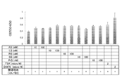
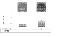
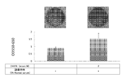
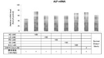
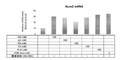
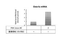
- An Alizarin Red S stained image is shown. (28 days) An Alizarin Red S stained image is shown. (28 days) The AlizarinzarRedzarS staining intensity is shown. An Alizarin Red S stained image is shown. (28 days) The staining intensity of Alizarin Red S is shown. An Alizarin Red S stained image and staining intensity are shown. An Alizarin Red S stained image and staining intensity are shown. The mRNA expression level of Runx2, Osteocalcin (OC), and ALP by real-time RT-PCR is shown.
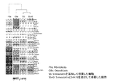
- FBs Fibroblasts
- OBs Osteoblasts
- SS cells cultured with addition of Simvastatin
- SS + D cells cultured with addition of Simvastatin and D4476, respectively.
- the result of a DNA microarray is shown.
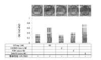
- the staining intensity of Alizarin Red S is shown.
- 28 days An Alizarin Red S stained image and intensity are shown.
- 28 days An Alizarin Red S stained image and intensity are shown.
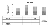
- the expression level of ALP mRNA by real-time RT-PCR is shown.
- 28 days The expression level of ALP mRNA by real-time RT-PCR is shown.
- 28 days The expression level of ALP mRNA by real-time RT-PCR is shown.
- the expression level of ALP mRNA by real-time RT-PCR is shown.
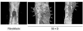
- FBs Femurs with bone defects and transplanted with fibroblasts.
- SS + D A femur with bone defects and then transplanted with cells cultured with Simvastatin and D4476.
- the triangle mark indicates the site where an artificial bone defect is made and transplanted, and the bone defect remains.
- the arrow indicates the site where the artificial bone defect has been made and transplanted, and the regenerated bone has been made and the defect has been repaired.
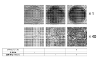
- An Alizarin Red S stained image is shown. (Human normal white fat precursor cells, 21days)
- the present invention provides at least one selected from the group consisting of (1) a statin compound, (2) a casein kinase inhibitor, (3) a cAMP inducer, and (4) a histone methyltransferase inhibitor in a medium of differentiated somatic cells. It is characterized in that osteoblasts are obtained from differentiated somatic cells by coexisting with a low molecular weight compound.
- the differentiated somatic cells that are directly reprogrammed into osteoblasts by the method of the present invention are not particularly limited as long as they do not include osteoblasts.
- Preferred examples include fibroblasts, keratinocytes, oral mucosal epithelial cells, gingival cells, leukocytes, lymphocytes, osteoclasts, and adipocytes.
- another compound can be further used in combination to promote direct reprogramming to osteoblasts.
- examples of such compounds include (i) low molecular weight compounds that promote iPS cell induction and cell reprogramming, (ii) compounds that induce Oct4, (iii) methyltransferase inhibitors, histone demethylase inhibitors, And epigenetic regulatory compounds such as histone deacetylase inhibitors.
- Statin compounds broadly encompass HMG-CoA reductase inhibitors, and are not particularly limited. Examples include crilvastatin, dalvastatin, glenvastatin, fluindostatin, verostatin, mevastatin, rivastatin, cilivastatin, CI-981 and the like. All the statin compounds developed in the future are included in the statin compounds of the present invention.
- statin compounds promote direct reprogramming into osteoblasts, particularly through increased expression of the Runx2 gene. It is also thought to contribute to the promotion of calcium deposition.
- Casein kinase inhibitors broadly include inhibitors for casein kinase in which subtypes such as casein kinase 1 and casein kinase 2 exist. From the viewpoint that the effect of inducing osteoblasts is high, a casein kinase 1 inhibitor is preferable.
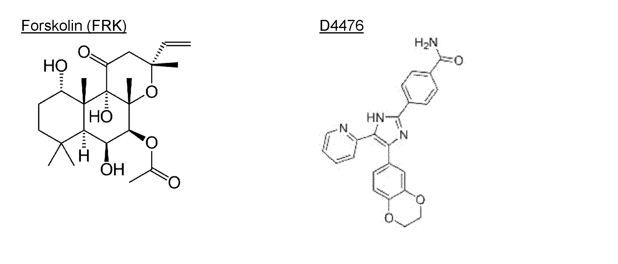
- Casein kinase 1 inhibitors include compounds such as D4476, IC261, CK1-7, A3, SB-431542, DRB, hymenardicin, mataleidinol, 5-iodotuberucine, meridienine, SB-203580, and the like (casein kinase). A compound that specifically inhibits 1).).
- casein kinase 2 inhibitor is CX-4945.
- a casein kinase 1 inhibitor a derivative thereof can be used instead of the above compound. Derivatives do not necessarily have activity to inhibit casein kinase.
- the casein kinase 1 inhibitor D4476 (4- [4- (2,3-dihydro-1,4-benzodioxin-6-yl) -5- (2-pyridinyl) 1H-imidazol-2-yl]- In place of benzamide), a derivative of D4476 represented by the following formula (I) described in WO00 / 61576 can be used:
- R 1 is halogen, C 1-6 alkoxy (—O—C 1-6 alkyl), C 1-6 alkylthio (—S—C 1-6 alkyl), C 1-6 alkyl, —O — (CH 2 ) n —Ph, —S— (CH 2 ) n —Ph, cyano, phenyl (Ph), and CO 2 R, where R is hydrogen or C 1-6 alkyl, and n is , 0, 1, 2 or 3), which is optionally substituted with one or more substituents selected from the group consisting of naphthyl, anthracenyl, or phenyl; or R 1 is N, Phenyl fused to a 5 to 7 membered aromatic or non-aromatic ring optionally containing up to 2 heteroatoms independently selected from O and S; R 2 is H, NH (CH 2 ) n —Ph or NH—C 1-6 alkyl (where n is 0, 1, 2 or 3); R 3 is
- C 1-6 alkyl is linear or branched alkyl having 1 to 6 carbon atoms, specifically, methyl, ethyl, n-propyl, isopropyl, n-butyl, isobutyl, sec-butyl, Mention may be made of tert-butyl, n-pentyl, isopentyl, n-hexyl and isohexyl.
- C 3-7 cycloalkyl examples include cyclopropyl having 3 to 7 carbon atoms, specifically, cyclopropyl, cyclobutyl, cyclopentyl, cyclohexyl, and cycloheptyl.
- R 1 is phenyl fused to a 5-7 membered aromatic or non-aromatic ring optionally containing up to 2 heteroatoms independently selected from N, O and S
- Examples of such derivatives of D4476 include the following compounds: 4- [4- (4-fluorophenyl) -5- (2-pyridyl) -1-hydroxy-1H-imidazol-2-yl] benzonitrile; 4- [4- (4-fluorophenyl) -5- (2-pyridyl) -1H-imidazol-2-yl] benzonitrile; 4- [4- (4-fluorophenyl) -5- (2-pyridyl) -1H-imidazol-2-yl] benzoic acid; Methyl 4- [4- (4-fluorophenyl) -5- (2-pyridyl) -1H-imidazol-2-yl] benzoate; 4- [4- (4-fluorophenyl) -5- (2-pyridyl) -1H-imidazol-2-yl] ethyl benzoate; 4- (4-Benzo [1,3] dioxol-5-yl
- casein kinase 1 inhibitors are believed to promote direct reprogramming into osteoblasts, particularly through increased expression of ALP (alkaline phosphatase), promotion of calcium deposition, and the like. .
- ALP alkaline phosphatase
- the cAMP inducer (which can also be referred to as an adenylate cyclase activator) widely includes compounds that increase intracellular cAMP (cyclic AMP) levels by activating the adenylate cyclase, for example, Examples include forskolin (FRK) and isoproterenol.
- adenylate cyclase activator widely includes compounds that increase intracellular cAMP (cyclic AMP) levels by activating the adenylate cyclase, for example, Examples include forskolin (FRK) and isoproterenol.
- the cAMP inducer promotes direct reprogramming into osteoblasts, particularly through increased expression of the Runx2 gene. It is also thought to contribute to the promotion of calcium deposition.
- histone methyltransferase inhibitors examples include DZNep (3-deazaneplanocin A) and BIX-01294.
- the cAMP inducer promotes direct reprogramming into osteoblasts, particularly through promotion of calcium deposition.
- the concentration of the statin compound in the medium is not particularly limited as long as osteoblasts can be induced.
- the concentration is about 100 pM to 10 ⁇ M, preferably about 500 pM to 5 ⁇ M, more preferably about 1 nM to 1 ⁇ M, and more preferably about 10 to 100 nM. It is.
- the concentration of casein kinase 1 inhibitor in the medium is not particularly limited as long as osteoblasts can be induced, but is, for example, about 0.01 to 100 ⁇ M, preferably about 0.1 to 50 ⁇ M, more preferably about 1 to 10 ⁇ M.
- the concentration of the cAMP inducer in the medium is not particularly limited as long as osteoblasts can be induced.
- it is about 0.01 to 100 ⁇ M, preferably about 0.1 to 50 ⁇ M, more preferably about 1 to 10 ⁇ M.
- the concentration of the histone methyltransferase inhibitor in the medium is not particularly limited as long as osteoblasts can be induced, but for example, about 100 pM to 50 ⁇ M, preferably about 1 nM to 10 ⁇ M, more preferably about 5 nM to 1 ⁇ M, and further preferably 10 About 100nM.
- the low molecular compounds that coexist in the medium may be used alone or in combination of two or more.
- a combination is not specifically limited. From the viewpoint that the effect of inducing osteoblasts is high, a combination of a statin compound and a casein kinase 1 inhibitor is exemplified as a preferred combination.
- a combination of a statin compound and a casein kinase 1 inhibitor may be further combined with a cAMP inducer and / or a histone methyltransferase inhibitor.
- two or more kinds of low molecular compounds When two or more kinds of low molecular compounds are used in combination, two or more kinds may coexist simultaneously during the culture period, or different low molecular compound (s) may be used for each part during the culture period.
- cells cultured with one or more low molecular weight compounds added and cells cultured with another one or more low molecular weight compounds added may be mixed.
- the medium for inducing osteoblasts is not particularly limited, but an induction medium is preferable.
- an induction medium ascorbic acid; ⁇ -Glycerophosphate; at least one selected from the group consisting of glucocorticoids such as dexamethasone and hydrocortisone; serum components (Fetal Bovine Serum (FBS), Human serum (HS), etc.); streptomycin Examples include those obtained by adding an antibiotic such as DMEM (Dulbecco's Modified Eagle's Medium), EMEM (Eagle's minimal essential medium), etc.).
- DMEM Dulbecco's Modified Eagle's Medium
- EMEM Eagle's minimal essential medium
- the culture temperature is about 37 ° C., and the culture period is about 1 to 6 weeks, preferably about 2 to 5 weeks, more preferably about 3 to 4 weeks.
- a solvent such as DMSO may be used for the medium.
- the kit of the present invention contains an osteoblast medium and a statin compound, and, if necessary, a casein kinase inhibitor, a histone methyltransferase inhibitor, and the like.
- pre-osteoblasts pre-osteoblasts, immature osteoblasts, mature osteoblasts, bone cells and the like can be prepared. In the present specification, these are all called osteoblasts for simplicity.
- Examples of diseases to be treated using osteoblasts (transplant material) obtained by the present invention include bone tumors associated with bone tumors, trauma, osteomyelitis, etc., bone defects after fractures such as bone tumors, fractures, Osteoporosis, periodontal disease, alveolar bone resorption, rheumatoid arthritis, idiopathic femoral head necrosis, osteoarthritis, lumbar spondylotic spondylosis, spinal canal stenosis, intervertebral disc herniation, spondylolysis, spondylolisthesis, spine Destroyed by complicated fractures such as scoliosis, cervical spondylotic myelopathy, posterior longitudinal ligament ossification, spinal cord injury, hip osteoarthritis, knee osteoarthritis, femoral head slippery, osteomalacia, mandibular reconstruction Examples include bone fracture reconstruction, bone repair after surgery (sternal repair after cardiac surgery, etc.), defect repair associated with artificial
- osteoblasts are transplanted, there is a possibility that the therapeutic effect can be enhanced in combination with bone grafts, artificial bone grafts, artificial joints and implants. It is also possible to treat the above-mentioned diseases by culturing osteoblasts using a three-dimensional scaffold or the like to create various forms of bone tissue outside the body and transplanting the bone tissue. In addition, various diseases related to osteoblast deficiency, deficiency or reduced function are targeted.
- treatment intends a treatment performed while a patient is suffering from a particular disease or disorder, whereby the severity of the disease or disorder, or one or It means that a plurality of its symptoms are alleviated or the progression of the disease or disorder is delayed or slowed down.
- treatment includes “prevention”.
- the osteoblasts obtained by the present invention can also be used not only for treatment of diseases but also for cosmetic purposes.
- a bone matrix can be produced to repair the deficient site and become plump and inconspicuous.
- treatment for humans is also referred to as treatment in this specification for convenience, and “patient” can be read as “healthy” or “human”, and “disease” can be read as “beauty”.
- the present invention can also be used to treat not only humans but also mammals including pets such as dogs and cats and livestock such as cows, horses, pigs, sheep and chickens.
- pets such as dogs and cats
- livestock such as cows, horses, pigs, sheep and chickens.
- “patient” is read as “patient” or “mammal”.
- the transplant material refers to a material containing osteoblasts that is introduced into a living body for the repair and reconstruction of bone tissue.
- the transplant material includes a material in which bone tissue is regenerated partially or completely in vitro and transplanted to the same or another individual.
- the osteoblast obtained by this invention can be used for preparation of a transplant material. Osteoblasts themselves are also transplant material. Therefore, osteoblasts can be transplanted to patients as cell preparations, transplanted with a base material (scaffold) made of artificial material such as hydroxyapatite or bioabsorbable ceramic, or transplanted after culturing with the scaffold. Can do. In these cases, the scaffold can have various three-dimensional shapes depending on the purpose of implantation.
- Somatic cells may be derived from mammals. When transplanting osteoblasts into a living body, it is preferable to use somatic cells (autologous cells) derived from the transplanted subject in order to reduce the risk of infection, rejection response, and the like.
- somatic cells autologous cells
- osteoblasts prepared in advance from somatic cells of another person or another animal can be used for transplantation instead of autologous cells.
- osteoblasts can be prepared from somatic cells of other people or other animals prepared in advance and used for transplantation. That is, an osteoblast bank or an osteoblast precursor cell bank can be prepared and used for transplantation purposes.
- MHC can be typed in advance in order to reduce the risk of a rejection response or the like.
- the osteoblast character and tumorigenicity can be confirmed in advance.
- mammals include mice, rats, hamsters, humans, dogs, cats, monkeys, rabbits, cows, horses, pigs, and the like, and particularly humans.
- the present invention can also be used for various researches and technical developments using osteoblasts. For example, it is useful for basic research such as analysis of bone development and aging, morphogenesis, remodeling mechanisms, mechanical stress on these, nutrition, immunity, nerves, and hormone effects. It is also useful for analyzing the effects of radioactive materials such as strontium 90 on internal bones and developing a technique for removing strontium 90 from bone.
- basic research such as analysis of bone development and aging, morphogenesis, remodeling mechanisms, mechanical stress on these, nutrition, immunity, nerves, and hormone effects.
- radioactive materials such as strontium 90 on internal bones and developing a technique for removing strontium 90 from bone.
- osteoblasts can be established easily, quickly and inexpensively from humans and animals having various diseases and genetic backgrounds, so that osteoblast abnormalities related to diseases and genetic backgrounds can be biochemically detected. It can be analyzed by molecular biology, immunology, etc., and this can be used for research such as elucidation of the pathogenesis of diseases and development of diagnostic methods. If such osteoblasts are used for drug development, drug toxicity tests, etc., it can be used for development of new treatments for various diseases.
- the osteoblasts were obtained by measuring ALP (alkaline phosphatase), osteocalcin (OC) ⁇ ⁇ , osteopontin, and Runx2 mRNA by real-time PCR, staining with alizarin red S (production of mineralized bone matrix) ), Etc.
- ALP alkaline phosphatase
- osteocalcin (OC) ⁇ ⁇ osteopontin
- Runx2 mRNA by real-time PCR, staining with alizarin red S (production of mineralized bone matrix) ), Etc.
- Runx2 is an essential transcription factor for bone formation. Runx2 plays an essential role in the differentiation of mesenchymal stem cells into osteoblasts in vivo. Forced expression of Runx2 in mesenchymal stem cells increases osteoblast-specific genes such as OC (osteocalcin), BSP (Bone sialo-protein), ALP (alkaline phosphatase), and COL1A1. Runx2 KO mice show no membranous or endochondral ossification due to the loss of mature osteoblasts. However, these mouse-derived mesenchymal stem cells have the ability to induce adipocytes and chondrocytes.
- ALP alkaline phosphatase
- Osteocalcin is expressed specifically in osteoblasts and is thought to contribute to the promotion of bone formation.
- Staining with alizarin red S or von Kossa staining can detect the production of mineralized bone matrix, that is, calcium deposition, which is one of the important elements of bone formation.
- Example 1 Alizarin Red S staining
- Human normal skin fibroblast cell line HDFs are cultured in 35 mm culture dishes at a concentration of 5 ⁇ 10 4 cells / well, and 100 nM simvastatin (SS) or 100 nM simvastatin (SS) and 1 ⁇ M DZnep Culture was performed under standard conditions in the added induction medium.
- the composition of the induction medium is 50 ⁇ g / ml ascorbic acid, 10 mM ⁇ -Glycerophosphate, 100 nM dexamethasone (all final concentrations), 10% FBS, 1% antibiotic, 1% NEAA, ⁇ -MEM. Standard conditions are 37 ° C, 5% CO 2 , 95% humidified air.
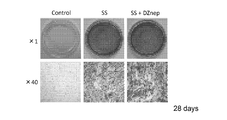
- the medium was changed about once every four days and cultured for 28 days. Aspirate the culture medium from the culture dish, wash twice with distilled water, and fix with 10% formalin. After washing with sterilized distilled water, add Alizarin Red S staining solution and let stand at room temperature for 20 minutes. After washing with sterilized distilled water, it was observed with the naked eye and a microscope. The results are shown in Figure 1. Red staining indicates calcified bone matrix. It can be seen that the addition of SS, or SS and DZnep, transformed fibroblasts into osteoblasts that produce a large amount of calcified bone matrix.
- the staining reaction solution was recovered, washed with sterile distilled water, and observed with the naked eye and a microscope.
- the results are shown in FIG.
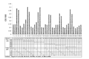
- the absorbance (550 nm) of the staining reaction solution collected from each well was measured using a microplate reader.
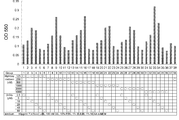
- the results are shown in FIG. 100 nM simvastatin (SS) induces fibroblasts into osteoblasts in the presence of dexamethasone and ⁇ -Glycerophosphate, with 25-800 nM dexamethasone and 10-20 ⁇ M ⁇ -Glycerophosphate It can be seen that the concentration is desirable.
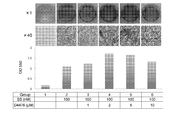
- the absorbance (550 nm) of the collected staining reaction solution was measured using a microplate reader. The results are shown in FIG. It can be seen that when 2-5 ⁇ M D4476 coexists with 100 nM simvastatin, the induction efficiency to osteoblasts is further increased.
- Real-time PCR Master Mix Taqman probes, Specific Primers, and cDNA are mixed, Real-time RT-PCR is performed using AB7300 Real-time PCR system, and Runx2, osteocalcin (OC), alkaline phosphatase (ALP) gene mRNA was quantified.
- OC osteocalcin
- ALP alkaline phosphatase
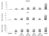
- Osteoblasts derived from fibroblasts with 100 nM simvastatin express Runx2, osteocalcin (OC), alkaline phosphatase (ALP), and in the presence of 2-5 ⁇ M D4476, their expression is equivalent or It can be seen that it increases.
- Example 2 2-1 Alizarin Red S staining ( Figure 10) Human normal skin fibroblast cell line HDFs were seeded on a 24-well plate at a concentration of 5 ⁇ 10 3 cells / well (day 0). On the next day, the culture medium in each well was discarded and replaced with a new medium of 500 ⁇ l / well.
- the induction medium is Dulbecco's modified Eagle's medium (DMEM), 50 ⁇ g / ml ascorbic acid, 10 mM ⁇ -glycerophospahte, 100 nM Dexamethasone plus 10% fetal bovine serum (FBS).
- DMEM Dulbecco's modified Eagle's medium
- FBS fetal bovine serum
- AS is Atorvastatin
- LS is Lovastatin
- RS Rosuvastatin
- PiS is Pitavastatin
- SS is Simvastatin
- PrS is Pravastatin
- FSK is forskolin.
- AS for LKT Laboratories (St Pau, USA), LS for Cayman chemical (Ann Arbor, USA), RS for Cayman chemical (Ann Arbor, USA), PiS for Cayman chemical (Ann Arbor, USA), SS for Sigma (St Louis , USA), PrS (Cayman chemical (Ann Arbor, USA), and FSK from Sigma (St Louis, USA), respectively.
- statin compounds could be observed by culturing with 10 nM PiS added.
- ALP alkaline phosphatase
- FIG. 13 The results are shown in FIG. 13 as relative values with the value of normal human fibroblasts as 1. It can be seen that the expression of ALP gene mRNA was induced by the culture with addition of AS, LS, RS, SS, PrS, and D4476.
- the ALP gene mRNA expression in the cells of each well was analyzed by Real-time RT-PCR in the same manner as in Example 3.
- total RNA was extracted from normal human bone-derived osteoblasts and analyzed in the same manner.
- RNA expression of the ALP gene in cells in each well was analyzed by Real-time RT-PCR in the same manner as in “2-3,” above.
- total RNA was extracted from normal human bone-derived osteoblasts and analyzed in the same manner.
- Real-time PCR Master-Mix, Taqman probe specific to human osteocalcin (OC) gene and Primers were added, and Real-time RT-PCR was performed using AB7300 Real-time PCR system.
- Real-time RT-PCR was performed using AB7300 Real-time PCR-system using Real-time PCR Master-Mix and Taqman probe specific to human Runx2 gene and Primers.
- mice were euthanized, the thighs were excised, fixed with neutral formalin, and then micro-computed tomography ( ⁇ CT using X-ray CT device (Scan Xmate-L090, Com Scan Techno, Yokohama, Japan) )did.
- ⁇ CT using X-ray CT device Scan Xmate-L090, Com Scan Techno, Yokohama, Japan
- Figure 21 shows a 3D-constructed ⁇ CT image.
- Black arrow Regenerated bone is created and the defect is repaired at the site where the artificial bone defect is created and transplanted.
- White arrow Regenerated bone is created at the site transplanted around the bone.
- Arrowhead A bone defect remains at the site where an artificial bone defect has been made and transplanted.
- Cells cultured by adding simvastatin (SS) + D4476 (D) were shown to have osteogenic ability in vivo.
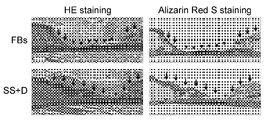
- Figure 22 shows the results. Cells cultured with SS + D4476 were shown to have bone forming ability in vivo.
- HWPs Human normal white fat precursor cells
- the normal medium is a medium in which 10% FBS is added to Dulbecco's modified Eagle's medium (DMEM), and the induction medium is 50 ⁇ g / mL ascorbic acid, 10 mM ⁇ -glycerophospahte, 100 nM Dexamethasone and 10% FBS in DMEM. Is.
- the culture medium was changed once every 3 to 4 days and cultured.
Landscapes
- Health & Medical Sciences (AREA)
- Engineering & Computer Science (AREA)
- Life Sciences & Earth Sciences (AREA)
- Biomedical Technology (AREA)
- Biotechnology (AREA)
- Zoology (AREA)
- Chemical & Material Sciences (AREA)
- Cell Biology (AREA)
- Bioinformatics & Cheminformatics (AREA)
- Genetics & Genomics (AREA)
- Organic Chemistry (AREA)
- Wood Science & Technology (AREA)
- General Health & Medical Sciences (AREA)
- Rheumatology (AREA)
- Orthopedic Medicine & Surgery (AREA)
- Biochemistry (AREA)
- General Engineering & Computer Science (AREA)
- Microbiology (AREA)
- Medicinal Chemistry (AREA)
- Developmental Biology & Embryology (AREA)
- Immunology (AREA)
- Virology (AREA)
- Pharmacology & Pharmacy (AREA)
- Epidemiology (AREA)
- Animal Behavior & Ethology (AREA)
- Public Health (AREA)
- Veterinary Medicine (AREA)
- Dermatology (AREA)
- Pharmaceuticals Containing Other Organic And Inorganic Compounds (AREA)
- Medicines That Contain Protein Lipid Enzymes And Other Medicines (AREA)
- Micro-Organisms Or Cultivation Processes Thereof (AREA)
Abstract
Description
(1)スタチン化合物、
(2)カゼインキナーゼ1阻害剤、
(3)cAMP誘導剤、及び
(4)ヒストンメチルトランスフェラーゼ阻害剤
からなる群から選択される少なくとも1種の化合物の存在下に培養して前記体細胞を骨芽細胞に変換することを特徴とする、骨芽細胞を調製する方法。
(2)カゼインキナーゼ1阻害剤、
(3)cAMP誘導剤、及び
(4)ヒストンメチルトランスフェラーゼ阻害剤
からなる群から選択される少なくとも1種の化合物のを含む、分化した体細胞を骨芽細胞に誘導するための誘導剤。
(2)カゼインキナーゼ1阻害剤、
(3)cAMP誘導剤、及び
(4)ヒストンメチルトランスフェラーゼ阻害剤
からなる群から選択される少なくとも1種の化合物、並びに、培地を含む分化した体細胞を骨芽細胞に誘導するためのキット。
カゼインキナーゼ1阻害剤としては、上記の化合物に替えてその誘導体を使用することもできる。誘導体は、必ずしも、カゼインキナーゼを阻害する活性を有さなくてもよい。例えば、カゼインキナーゼ1阻害剤であるD4476(4-[4-(2,3-dihydro-1,4-benzodioxin-6-yl)-5-(2-pyridinyl)1H-imidazol-2-yl]-benzamide)に替えて、WO00/61576号に記載の下記式(I)で表されるD4476の誘導体を使用することができる:

R2は、H、NH(CH2)n-PhまたはNH-C1-6アルキルであり(ここで、nは、0、1、2または3である);
R3は、CO2H、CONH2、CN、NO2、C1-6アルキルチオ、-SO2-C1-6アルキル、C1-6アルコキシ、SONH2、CONHOH、NH2、CHO、CH2OH、CH2NH2、またはCO2Rであり(ここで、Rは、水素またはC1-6アルキルである);
X1およびX2のうち一方は、NまたはCR'であり、他方は、NR'またはCHR'である(ここで、R'は、水素、OH、C1-6アルキル、またはC3-7シクロアルキルである)か;またはX1およびX2のうち一方がNまたはCR'である場合、他方は、SまたはOであってもよい]。
4-[4-(4-フルオロフェニル)-5-(2-ピリジル)-1-ヒドロキシ-1H-イミダゾール-2-イル]ベンゾニトリル;
4-[4-(4-フルオロフェニル)-5-(2-ピリジル)-1H-イミダゾール-2-イル]ベンゾニトリル;
4-[4-(4-フルオロフェニル)-5-(2-ピリジル)-1H-イミダゾール-2-イル]安息香酸;
4-[4-(4-フルオロフェニル)-5-(2-ピリジル)-1H-イミダゾール-2-イル]安息香酸メチル;
4-[4-(4-フルオロフェニル)-5-(2-ピリジル)-1H-イミダゾール-2-イル]安息香酸エチル;
4-(4-ベンゾ[1,3]ジオキソール-5-イル-1-ヒドロキシ-5-ピリジン-2-イル-1H-イミダゾール-2-イル)ベンゾニトリル;
4-(4-ベンゾ[1,3]ジオキソール-5-イル-5-ピリジン-2-イル-1H-イミダゾール-2-イル)ベンゾニトリル;
4-(4-ベンゾ[1,3]ジオキソール-5-イル-5-ピリジン-2-イル-1H-イミダゾール-2-イル)安息香酸;
2-[4-ベンゾ[1,3]ジオキソール-5-イル-2-(4-ニトロフェニル)-1H-イミダゾール-5-イル]ピリジン;
3-(4-ベンゾ[1,3]ジオキソール-5-イル-5-ピリジン-2-イル-1H-イミダゾール-2-イル)フェニルアミン;
4-[4-(4-フルオロフェニル)-2-(4-ニトロフェニル)-1H-イミダゾール-5-イル]ピリジン;
4-[4-(4-フルオロフェニル)-5-ピリジン-2-イル-1H-イミダゾール-2-イル)フェニルアミン;
4-(4-ベンゾ[1,3]ジオキソール-5-イル-5-ピリジン-2-イル-1H-イミダゾール-2-イル)フェニル]メタノール;
4-(4-ベンゾ[1,3]ジオキソール-5-イル-5-ピリジン-2-イル-1H-イミダゾール-2-イル)ベンズアミド;
4-[4-(2,3-ジヒドロ-ベンゾ[1,4]ジオキシン-6-イル)-5-ピリジン-2-イル-1H-イミダゾール-2-イル]-ベンゾニトリル;
4-[4-(2,3-ジヒドロ-ベンゾフラン-5-イル)-5-ピリジン-2-イル-1H-イミダゾール-2-イル]ベンズアミド;
3-[4-ベンゾ[1,3]ジオキソール-5-イル-5-ピリジン-2-イル-1H-イミダゾール-2-イル)ベンゾニトリル;
4-[4-(2,3-ジヒドロ-ベンゾフラン-6-イル)-5-ピリジン-2-イル-1H-イミダゾール-2-イル]ベンゾニトリル;
4-[4-(2,3-ジヒドロ-ベンゾフラン-6-イル)-5-ピリジン-2-イル-1H-イミダゾール-2-イル]ベンズアミド;
3-(4-ベンゾ[1,3]ジオキソール-5-イル-5-ピリジン-2-イル-1H-イミダゾール-2-イル)安息香酸;
4-[4-(4-メトキシフェニル)-5-(2-ピリジル)-1H-イミダゾール-2-イル]ベンゾニトリル;
4-[4-(2,2-ジフルオロ-ベンゾ[1,3]ジオキソール-5-イル)-5-ピリジン-2-イル-1H-イミダゾール-2-イル]ベンズアミド;
4-[4-(2,3-ジヒドロ-ベンゾ[1,4]ジオキシン-6-イル)-1-メチル-5-ピリジン-2-イル-1H-イミダゾール-2-イル]ベンズアミド;
4-[5-(2,3-ジヒドロ-ベンゾ[1,4]ジオキシン-6-イル)-1-メチル-4-ピリジン-2-イル-1H-イミダゾール-2-イル]ベンズアミド;
4-(5-ベンゾ[1,3]ジオキソール-5-イル-4-ピリジン-2-イル-オキサゾール-2-イル)ベンゾニトリル;
4-(5-ベンゾ[1,3]ジオキソール-5-イル-4-ピリジン-2-イル-オキサゾール-2-イル)ベンズアミド;および 4-(4-ベンゾ[1,3]ジオキソール-5-イル-5-ピリジン-2-イル-1H-ピロール-2-イル)ベンズアミド。
1-1、Alizarin Red S染色(図1)
ヒト正常皮膚線維芽細胞株HDFsを、5×10⁴ 細胞/ウェルの濃度で35 mm培養ディッシュに培養し、100 nMのシンバスタチン(SS)、または、100 nMのシンバスタチン(SS)と1μMのDZnepを添加した誘導培地にて、標準条件下で培養した。誘導培地の組成は、50 μg/ml アスコルビン酸、10 mM β-Glycerophosphate、100 nM dexamethasone(いずれも最終濃度)、10% FBS、1% 抗生剤、1% NEAA 、α-MEMである。標準条件とは、37°C、5%CO2, 95% humidified airである。約4日に1回培地交換を行い、28日間培養した。培養ディッシュから培養液を吸引除去し、蒸留水で2回洗浄を行い、10%ホルマリンで固定。滅菌蒸留水で洗浄した後、アリザリンレッドS染色液を加え、室温で20分間静置。滅菌蒸留水で洗浄した後、肉眼および顕微鏡で観察した。結果を図1に示す。赤い染色は石灰化骨基質を示している。SS、またはSSとDZnepの添加により、線維芽細胞が石灰化骨基質を多量に産生する骨芽細胞に転換したことがわかる。
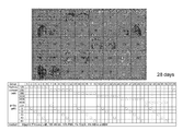
ヒト正常皮膚線維芽細胞株HDFsを、24-well plateに培養し、100 nMのシンバスタチン(SS)と、種々の濃度のハイドロキシコルチゾンおよびβ-グリセロフォスフェイトを添加した誘導培地にて、標準条件下で培養した(表中、〇は各濃度の化合物を添加したことを示す)。約4日に1回培地交換を行い、28日間培養した。プレートから培養液を吸引除去し、蒸留水で2回洗浄を行い、10%ホルマリンで固定。滅菌蒸留水で洗浄した後、アリザリンレッドS染色液を加え、室温で静置。20分後、染色反応液を回収した後、滅菌蒸留水で洗浄した、肉眼および顕微鏡で観察した。結果を図2に示す。また各ウェルから回収した染色反応液の吸光度(550 nm)をマイクロプレートリーダーを用いて測定した。結果を図3に示す。100 nMのシンバスタチン(SS)は、ハイドロキシコルチゾンとβ-Glycerophosphate の存在下に、線維芽細胞を骨芽細胞に転換すること、その際ハイドロキシコルチゾンは125~4000 nM、β-Glycerophosphate は10~20 μMの濃度であることが望ましいことが分かる。
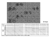
ヒト正常皮膚線維芽細胞株HDFsを、24-well plateに培養し、100 nMのシンバスタチン(SS)と、種々の濃度のデキサメサゾン(Dex)およびβ-グリセロフォスフェイトを添加した誘導培地にて、標準条件下で培養した(表中、〇は各濃度の化合物を添加したことを示す)。約4日に1回培地交換を行い、28日間培養した。プレートから培養液を吸引除去し、蒸留水で2回洗浄を行い、10%ホルマリンで固定。滅菌蒸留水で洗浄した後、アリザリンレッドS染色液を加え、室温で静置。20分後、染色反応液を回収した後、滅菌蒸留水で洗浄した、肉眼および顕微鏡で観察した。結果を図4に示す。また各ウェルから回収した染色反応液の吸光度(550 nm)をマイクロプレートリーダーを用いて測定した。結果を図5に示す。100 nMのシンバスタチン(SS)は、デキサメサゾンと、β-Glycerophosphate の存在下に、線維芽細胞を骨芽細胞に誘導すること、その際デキサメサゾンは25~800 nM、β-Glycerophosphate は10~20 μMの濃度であることが望ましいことが分かる。
ヒト正常皮膚線維芽細胞株HDFsを、24-well plateに培養し、種々の濃度のシンバシタチン(SS)を添加した誘導培地にて、標準条件下で培養した。約4日に1回培地交換を行い、28日間培養した。培養プレートから培養液を吸引除去し、蒸留水で2回洗浄を行い、10%ホルマリンで固定。滅菌蒸留水で洗浄した後、アリザリンレッドS染色液を加え、室温で静置。20分後の染色反応液を回収した後、滅菌蒸留水で洗浄した、肉眼および顕微鏡で観察した。また回収した染色反応液の吸光度(550 nm)をマイクロプレートリーダーを用いて測定した。結果を図6に示す。シンバスタチンが骨芽細胞を誘導する望ましい濃度は10~100nMであることが分かる。
ヒト正常皮膚線維芽細胞株HDFsを、24-well plateに培養し、種々の濃度のシンバスタチン(SS)とD4476(カゼインキナーゼインヒビター)を添加した誘導培地にて、標準条件下で培養した。約4日に1回培地交換を行い、28日間培養した。その後培養プレートから培養液を吸引除去し、蒸留水で2回洗浄を行い、10%ホルマリンで固定。滅菌蒸留水で洗浄した後、アリザリンレッドS染色液を加え、室温で静置。20分後の染色反応液を回収した後、滅菌蒸留水で洗浄した、肉眼および顕微鏡で観察した。また回収した染色反応液の吸光度(550 nm)をマイクロプレートリーダーを用いて測定した。結果を図7に示す。100 nMのシンバスタチンに、2~5 μMのD4476が共存すると、骨芽細胞への誘導効率がより高まることが分かる。
ヒト正常皮膚線維芽細胞株HDFsを、12-well plateに培養し、100 nMのシンバスタチン(SS)と種々の濃度のD4476(カゼインキナーゼインヒビター)を添加した誘導培地にて、標準条件下で培養した(カッコ内の数字はμM)。約4日に1回培地交換を行い、21日間培養した。細胞からISOGEN IIにてtotal RNAを回収し、Rever Tra Ace qPCR RT Master Mixを用いてcDNAを作製した。Real-time PCR Master Mix、Taqman probes、Specific PrimersおよびcDNAを混和し、AB7300 Real-time PCR systemを用いてReal-time RT-PCRを行い、Runx2、オステオカルシン(OC)、アルカリフォスファターゼ(ALP)遺伝子のmRNAを定量した。コントロールとしてシンバスタチンを添加しない培養を行った線維芽細胞からRNAを採取し同様の解析を行った。またヒト骨芽細胞からRNAを採取し同様の解析を行った。結果を図8に示す。縦軸は、コントロールの細胞のmRNAレベルを1としたときの各遺伝子のmRNAの相対値である。100nMのシンバスタチンによって線維芽細胞から誘導された骨芽細胞が、Runx2、オステオカルシン(OC)、アルカリフォスファターゼ(ALP)を発現すること、また2~5 μMのD4476が共存すると、それらの発現が同等もしくはより高まることが分かる。
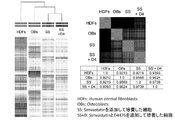
ヒト正常皮膚線維芽細胞株HDFsを、60 mm培養ディシュに培養し、100 nMのシンバスタチン(SS)、または100 nMのシンバスタチンと2μMのD4476(SS+D4)を添加した誘導培地にて、標準条件下で培養した。約4日に1回培地交換を行い、21日間培養した。細胞からISOGEN IIにてtotal RNAを回収した。ヒト線維芽細胞(HDFs)とヒト骨芽細胞(OBs)からも同様にtotal RNAを回収した。各細胞のmRNA発現パターンをaffymetrix 社のDNAチップを用いてゲノムワイドに解析した。結果を図9に示す。SS、SS+D4とも、線維芽細胞よりも骨芽細胞に類似の網羅的遺伝子発現パターンを示し、SS+D4の方がSSよりもより骨芽細胞に類似の網羅的遺伝子発現パターンを示すことが分かる。
2-1、Alizarin Red S染色(図10)
ヒト正常皮膚線維芽細胞株HDFsを、5×10³ 細胞/ウェルの濃度で24 well plateに播種した(day 0)。翌日、各wellの培養液を棄て、500 μl/wellの新しい培地に交換した。誘導培地はDulbecco's modified Eagle's medium (DMEM)、50 μg/mlアスコルビン酸、10 mM β-glycerophospahte、100 nM Dexamethasoneに10% ウシ胎仔血清(FBS)を添加したものである。また記載の小分子化合物を加えた。ただし、ASはAtorvastatin、LSはLovastatin、RSはRosuvastatin、PiSはPitavastatin、SSはSimvastatin、PrSはPravastatin、FSKはforskolinを示す。ASはLKT Laboratories(St Pau, USA)、LSはCayman chemical(Ann Arbor, USA)、RSはCayman chemical(Ann Arbor, USA)、PiSはCayman chemical(Ann Arbor, USA)、SSはSigma(St Louis, USA)、PrSは(Cayman chemical(Ann Arbor, USA)、FSKはSigma(St Louis, USA)からそれぞれ購入した。3~4日に1度、培養液を交換して培養した。

ヒト正常皮膚線維芽細胞株HDFsを、5×10³ 細胞/ウェルの濃度で24 well plateに播種した(day 0)。翌日、各wellの培養液を棄て、500 μl/wellの新しい培地に交換した。誘導培地はDulbecco's modified Eagle's medium (DMEM)、50 μg/mlアスコルビン酸、10 mM β-glycerophospahte、100 nM Dexamethasoneに10% FBSを添加したものである。また記載の小分子化合物を加えた。3~4日に1度、培養液を交換して培養した。
ヒト正常皮膚線維芽細胞株HDFsを、5×10³ 細胞/ウェルの濃度で24 well plateに播種した(day 0)。翌日、各wellの培養液を棄て、500 μl/wellの新しい培地に交換した。誘導培地はDulbecco's modified Eagle's medium (DMEM)、50 μg/mlアスコルビン酸、10 mM β-glycerophospahte、100 nM Dexamethasoneに5% Human Serumを添加したものである。また記載の小分子化合物を加えた。3~4日に1度、培養液を交換して培養した。
上記「2-1、」と同様の培養を、記載の化合物を添加して行った。
上記「2-1、」と同様の培養を、記載の化合物を添加して行った。
ヒト正常皮膚線維芽細胞株HDFsを、5×10³ 細胞/ウェルの濃度で24 well plateに播種した(day 0)。翌日、各wellの培養液を棄て、500 μl/wellの新しい培地に交換した。誘導培地はDulbecco's modified Eagle's medium (DMEM)、50 μg/mlアスコルビン酸、10 mM β-glycerophospahte、100 nM Dexamethasoneに5% Human Serumを添加したものである。また記載の小分子化合物を加えた。3~4日に1度、培養液を交換して培養した。
上記「2-6、」と同様の培養を、記載の化合物を添加して行った。
上記「1、」と同様の培養を、記載の化合物を添加して行った。
上記「2-1、」と同様の培養を、記載の化合物を添加して行った。
実施例1と同様の培養を、記載の化合物を添加して行った。DZnepは、Cayman chemical (Ann Arbor, USA)、CX-4945はBiovision(Zurich, Switzerland)から購入して用いた。
ヒト正常皮膚線維芽細胞株HDFsを、60 mm培養ディシュに培養し、100 nMのシンバスタチン(SS)、または100 nMのシンバスタチンと2μMのD4476(SS+D4)を添加した誘導培地にて、標準条件下で培養した。3~4日に1回培地交換を行い、21日間培養した。
細胞からISOGEN IIにてtotal RNAを回収した。ヒト線維芽細胞(HDFs)とヒト骨芽細胞(OBs)からも同様にtotal RNAを回収した。各細胞のmRNA発現パターンをaffymetrix 社のDNAチップを用いてゲノムワイドに解析した。
動物実験は、京都府立医科大学の認可を得て行った。8週齢オスのNOD/SCIDマウス(Charles River)の腹腔内にペントバルビタールを注射し麻酔した。注水下に歯科ドリルを用いて左大腿骨骨幹に直径約4mmの部分骨欠損を作成した。実施例11と同様の方法で、HDFsをシンバスタチンとD4476存在下で21日間培養した細胞を、50 μLの培地と50 μLのマトリゲル (BD Bioscience, San Jose, CA)の1:1混合液に懸濁し、骨欠損部とその周辺の骨表面に5×10⁵ 個 /マウスの濃度で移植した. また、同様に骨欠損を作ったのち線維芽細胞を移植したマウスも準備した。21日後にマウスを安楽死させ、大腿を切除し、中性ホルマリンで固定後、X-ray CT device (Scan Xmate-L090, Com Scan Techno, Yokohama, Japan)を用いてマイクロ・コンピューター断層撮影(μCT)した。
動物実験は、京都府立医科大学の認可を得て行った。上記「2-12、」と同様に移植実験を行った。また、線維芽細胞を移植したマウスも準備した。21日後にマウスを安楽死させ、実施例11と同様に大腿を切除し、中性ホルマリンで固定後、骨組織をSCEM (Leica Microsystem) compoundで包埋し、急速凍結した。6 μmの切片にスライス後、連続切片をヘマトキシリン・エオジン (H&E)(左)およびAlizarin Red S (右)で染色した。
ヒト正常白色脂肪前駆細胞(HWPs)を、5×10⁴ 細胞/ディシュの濃度で35mmディシュに播種した(day 0)。翌日、各wellの培養液を棄て、2 mL/wellの新しい培地(通常培地、または、記載の小分子化合物を加えた誘導培地)に交換した。通常培地は、Dulbecco's modified Eagle's medium (DMEM)に10% FBSを添加した培地であり、誘導培地はDMEMに50 μg/mLアスコルビン酸、10 mM β-glycerophospahte、100 nM Dexamethasoneおよび10% FBSを添加したものである。3~4日に1度、培養液を交換して培養した。
Claims (6)
- 哺乳動物の分化した体細胞を培地中、
(1)スタチン化合物、
(2)カゼインキナーゼ1阻害剤、
(3)cAMP誘導剤、及び
(4)ヒストンメチルトランスフェラーゼ阻害剤
からなる群から選択される少なくとも1種の化合物の存在下に培養して前記体細胞を骨芽細胞に変換することを特徴とする、骨芽細胞を調製する方法。 - 前記体細胞が線維芽細胞、歯肉細胞または脂肪細胞である、請求項1に記載の方法。
- 前記培地が、骨芽細胞の誘導培地である、請求項1または2に記載の方法。
- (1)スタチン化合物、
(2)カゼインキナーゼ1阻害剤、
(3)cAMP誘導剤、及び
(4)ヒストンメチルトランスフェラーゼ阻害剤
からなる群から選択される少なくとも1種の化合物のを含む、分化した体細胞を骨芽細胞に誘導するための誘導剤。 - (1)スタチン化合物、
(2)カゼインキナーゼ1阻害剤、
(3)cAMP誘導剤、及び
(4)ヒストンメチルトランスフェラーゼ阻害剤
からなる群から選択される少なくとも1種の化合物、並びに、培地を含む分化した体細胞を骨芽細胞に誘導するためのキット。 - 前記培地が、骨芽細胞の誘導培地である、請求項5に記載のキット。
Priority Applications (5)
| Application Number | Priority Date | Filing Date | Title |
|---|---|---|---|
| JP2016513843A JP6705115B2 (ja) | 2014-04-18 | 2015-04-17 | 骨芽細胞の調製方法及び骨芽細胞誘導剤 |
| KR1020167032139A KR102346489B1 (ko) | 2014-04-18 | 2015-04-17 | 골아 세포의 조제 방법 및 골아 세포 유도제 |
| US15/304,287 US11174464B2 (en) | 2014-04-18 | 2015-04-17 | Method for preparing osteoblasts and osteoblast inducer |
| EP15779733.3A EP3133149B1 (en) | 2014-04-18 | 2015-04-17 | Method for preparing osteoblasts and osteoblast inducer |
| CN201580023967.4A CN106459903B (zh) | 2014-04-18 | 2015-04-17 | 成骨细胞的制备方法及成骨细胞诱导剂 |
Applications Claiming Priority (2)
| Application Number | Priority Date | Filing Date | Title |
|---|---|---|---|
| JP2014-086757 | 2014-04-18 | ||
| JP2014086757 | 2014-04-18 |
Publications (1)
| Publication Number | Publication Date |
|---|---|
| WO2015159982A1 true WO2015159982A1 (ja) | 2015-10-22 |
Family
ID=54324183
Family Applications (1)
| Application Number | Title | Priority Date | Filing Date |
|---|---|---|---|
| PCT/JP2015/061893 Ceased WO2015159982A1 (ja) | 2014-04-18 | 2015-04-17 | 骨芽細胞の調製方法及び骨芽細胞誘導剤 |
Country Status (6)
| Country | Link |
|---|---|
| US (1) | US11174464B2 (ja) |
| EP (1) | EP3133149B1 (ja) |
| JP (1) | JP6705115B2 (ja) |
| KR (1) | KR102346489B1 (ja) |
| CN (1) | CN106459903B (ja) |
| WO (1) | WO2015159982A1 (ja) |
Cited By (2)
| Publication number | Priority date | Publication date | Assignee | Title |
|---|---|---|---|---|
| WO2018062269A1 (ja) * | 2016-09-30 | 2018-04-05 | 京都府公立大学法人 | 体細胞を製造する方法、体細胞、及び組成物 |
| JP2019198278A (ja) * | 2018-05-17 | 2019-11-21 | 日産化学株式会社 | 骨形成促進材 |
Families Citing this family (5)
| Publication number | Priority date | Publication date | Assignee | Title |
|---|---|---|---|---|
| TWI611714B (zh) * | 2015-01-30 | 2018-01-11 | 財團法人資訊工業策進會 | 用於載波聚合通訊系統的基地台與上行鏈路控制資訊排程方法 |
| WO2019213505A1 (en) * | 2018-05-04 | 2019-11-07 | Spinalcyte, Llc | Enhancement of fibroblast plasticity for treatment of disc degeneration |
| CN114129565A (zh) * | 2021-12-09 | 2022-03-04 | 上海交通大学医学院附属第九人民医院 | 组蛋白甲基转移酶ezh2抑制剂在促使骨代谢进入高转换以修复骨缺损产品中的应用 |
| WO2024053957A1 (ko) * | 2022-09-07 | 2024-03-14 | 단국대학교 천안캠퍼스 산학협력단 | 케미컬로 유도된 골형성세포와 이의 질병모델링에의 이용 및 골 이식용 스캐폴드 |
| KR102905414B1 (ko) * | 2022-09-07 | 2025-12-29 | 단국대학교 천안캠퍼스 산학협력단 | 케미컬로 유도된 골형성세포 및 이의 질병모델링에의 이용 |
Citations (4)
| Publication number | Priority date | Publication date | Assignee | Title |
|---|---|---|---|---|
| JP2002527386A (ja) * | 1998-10-15 | 2002-08-27 | メルク エンド カムパニー インコーポレーテッド | 骨形成調節方法 |
| JP2008044956A (ja) * | 1996-12-13 | 2008-02-28 | Zymogenetics Inc | 骨の成長を刺激する組成物および方法 |
| JP2011073998A (ja) * | 2009-09-29 | 2011-04-14 | Osaka Univ | 軟骨増大促進剤 |
| WO2013013105A2 (en) * | 2011-07-19 | 2013-01-24 | Vivoscript,Inc. | Compositions and methods for re-programming cells without genetic modification for repairing cartilage damage |
Family Cites Families (5)
| Publication number | Priority date | Publication date | Assignee | Title |
|---|---|---|---|---|
| DE60001229T2 (de) | 1999-04-09 | 2003-10-30 | Smithkline Beecham Corp., Philadelphia | Triarylimidazole |
| WO2011084685A2 (en) | 2009-12-17 | 2011-07-14 | Children's Medical Center Corporation | Saposin-a derived peptides and uses thereof |
| EP2799538A4 (en) * | 2011-12-27 | 2015-06-03 | Univ Osaka | METHOD FOR INDUCING DIFFERENTIATION BY TUMOR GENE ACTIVATION OF SUPPRESSIVE IPS CELLS |
| BR112016001487A2 (pt) | 2013-07-26 | 2017-08-29 | Kyoto Prefectural Public Univ Corp | Osteoblasto e método para preparar o mesmo |
| CN105062963B (zh) * | 2015-03-23 | 2018-05-25 | 云南济慈再生医学研究院有限公司 | 一种促进人皮肤成纤维细胞向成骨细胞转分化的培养方法 |
-
2015
- 2015-04-17 EP EP15779733.3A patent/EP3133149B1/en active Active
- 2015-04-17 US US15/304,287 patent/US11174464B2/en active Active
- 2015-04-17 WO PCT/JP2015/061893 patent/WO2015159982A1/ja not_active Ceased
- 2015-04-17 KR KR1020167032139A patent/KR102346489B1/ko active Active
- 2015-04-17 CN CN201580023967.4A patent/CN106459903B/zh active Active
- 2015-04-17 JP JP2016513843A patent/JP6705115B2/ja active Active
Patent Citations (4)
| Publication number | Priority date | Publication date | Assignee | Title |
|---|---|---|---|---|
| JP2008044956A (ja) * | 1996-12-13 | 2008-02-28 | Zymogenetics Inc | 骨の成長を刺激する組成物および方法 |
| JP2002527386A (ja) * | 1998-10-15 | 2002-08-27 | メルク エンド カムパニー インコーポレーテッド | 骨形成調節方法 |
| JP2011073998A (ja) * | 2009-09-29 | 2011-04-14 | Osaka Univ | 軟骨増大促進剤 |
| WO2013013105A2 (en) * | 2011-07-19 | 2013-01-24 | Vivoscript,Inc. | Compositions and methods for re-programming cells without genetic modification for repairing cartilage damage |
Non-Patent Citations (5)
| Title |
|---|
| HIROYUKI SUZUKI ET AL.: "An in vitro study on the characteristics of osteoblastic cells derived from human mandibular periosteum", JOURNAL OF THE JAPANESE STOMATOLOGICAL SOCIETY, vol. 40, no. 1, 1991, pages 89 - 108, XP055359664 * |
| HIROYUKI SUZUKI ET AL.: "An In Vitro Study on the Ectopic Calcification, Human Gingival Fibroblast Culture", JOURNAL OF THE JAPANESE STOMATOLOGICAL SOCIETY, vol. 41, no. 2, 1992, pages 268 - 274, XP055359659 * |
| HOU P. ET AL.: "Pluripotent Stem Cells Induced from Mouse Somatic Cells by Small-Molecule Compounds.", SCIENCE, vol. 341, no. 6146, 2013, pages 651 - 654, XP055079363, ISSN: 0036-8075 * |
| See also references of EP3133149A4 * |
| ZHOU Y. ET AL.: "The role of simvastatin in the osteogenesis of injectable tissue-engineered bone based on human adipose-derived stromal cells and platelet-rich plasma.", BIOMATERIALS, vol. 31, no. 20, 2010, pages 5325 - 5335, XP055231477, ISSN: 0142-9612 * |
Cited By (3)
| Publication number | Priority date | Publication date | Assignee | Title |
|---|---|---|---|---|
| WO2018062269A1 (ja) * | 2016-09-30 | 2018-04-05 | 京都府公立大学法人 | 体細胞を製造する方法、体細胞、及び組成物 |
| JP2019198278A (ja) * | 2018-05-17 | 2019-11-21 | 日産化学株式会社 | 骨形成促進材 |
| JP7143629B2 (ja) | 2018-05-17 | 2022-09-29 | 日産化学株式会社 | 骨形成促進材 |
Also Published As
| Publication number | Publication date |
|---|---|
| EP3133149A1 (en) | 2017-02-22 |
| US20170037374A1 (en) | 2017-02-09 |
| CN106459903A (zh) | 2017-02-22 |
| KR102346489B1 (ko) | 2021-12-31 |
| JP6705115B2 (ja) | 2020-06-03 |
| EP3133149B1 (en) | 2022-06-22 |
| KR20160145174A (ko) | 2016-12-19 |
| CN106459903B (zh) | 2021-04-23 |
| JPWO2015159982A1 (ja) | 2017-04-13 |
| EP3133149A4 (en) | 2017-12-06 |
| US11174464B2 (en) | 2021-11-16 |
Similar Documents
| Publication | Publication Date | Title |
|---|---|---|
| JP6705115B2 (ja) | 骨芽細胞の調製方法及び骨芽細胞誘導剤 | |
| Hemming et al. | EZH2 deletion in early mesenchyme compromises postnatal bone microarchitecture and structural integrity and accelerates remodeling | |
| Lu et al. | GDF11 inhibits bone formation by activating Smad2/3 in bone marrow mesenchymal stem cells | |
| CN108495930A (zh) | 棕色脂肪细胞的制备方法 | |
| JP2016522167A (ja) | ヒト間葉系幹細胞に対するダイゼイン類似体の骨形成活性の向上 | |
| WO2017069222A1 (ja) | 細胞の調製方法 | |
| US11771803B2 (en) | Enhancement of osteogenic potential of bone grafts | |
| KR20170108325A (ko) | 성체줄기세포의 연골세포 분화용 조성물 및 분화 방법 | |
| US20180133211A1 (en) | Methods and materials for promoting bone formation | |
| Min et al. | Effect of radiopaque Portland cement on mineralization in human dental pulp cells | |
| JPWO2006123699A1 (ja) | 幹細胞に対する骨芽細胞への分化促進剤 | |
| US20120213826A1 (en) | Use of Pro-Inflammatory Compounds for Promoting Bone Formation | |
| Zhang et al. | SDF-1 induces directional chemotaxis of endogenous BMSCs at the intervertebral fusion site and promotes osteogenic differentiation by regulating Wnt/ß-catenin in the bone marrow chimera spinal intervertebral fusion mouse model | |
| JP2008061569A (ja) | 幹細胞の培養方法及びその培地 | |
| Lyu et al. | Sirt1 Runx2 Signaling Pathway Promotes Healing of Tibial Fracture in Rats | |
| CN117244070A (zh) | Nr4a1激动剂/过表达制剂在成骨中的应用 | |
| Wang et al. | Wnt/β-catenin signaling plays an essential role in bone sialoprotein-elicited osteoblast differentiation and mineralization | |
| KR20170044334A (ko) | 이소함네틴을 유효성분으로 포함하는 지방세포 분화 억제 및 골세포 형성 촉진용 조성물 |
Legal Events
| Date | Code | Title | Description |
|---|---|---|---|
| 121 | Ep: the epo has been informed by wipo that ep was designated in this application |
Ref document number: 15779733 Country of ref document: EP Kind code of ref document: A1 |
|
| DPE2 | Request for preliminary examination filed before expiration of 19th month from priority date (pct application filed from 20040101) | ||
| ENP | Entry into the national phase |
Ref document number: 2016513843 Country of ref document: JP Kind code of ref document: A |
|
| WWE | Wipo information: entry into national phase |
Ref document number: 15304287 Country of ref document: US |
|
| NENP | Non-entry into the national phase |
Ref country code: DE |
|
| REEP | Request for entry into the european phase |
Ref document number: 2015779733 Country of ref document: EP |
|
| WWE | Wipo information: entry into national phase |
Ref document number: 2015779733 Country of ref document: EP |
|
| ENP | Entry into the national phase |
Ref document number: 20167032139 Country of ref document: KR Kind code of ref document: A |