WO2015166640A1 - 異種ポリペプチド発現カセット - Google Patents
異種ポリペプチド発現カセット Download PDFInfo
- Publication number
- WO2015166640A1 WO2015166640A1 PCT/JP2015/002132 JP2015002132W WO2015166640A1 WO 2015166640 A1 WO2015166640 A1 WO 2015166640A1 JP 2015002132 W JP2015002132 W JP 2015002132W WO 2015166640 A1 WO2015166640 A1 WO 2015166640A1
- Authority
- WO
- WIPO (PCT)
- Prior art keywords
- bifidobacterium
- chain antibody
- amino acid
- scfv
- antibody
- Prior art date
- Legal status (The legal status is an assumption and is not a legal conclusion. Google has not performed a legal analysis and makes no representation as to the accuracy of the status listed.)
- Ceased
Links
Images











Classifications
-
- C—CHEMISTRY; METALLURGY
- C12—BIOCHEMISTRY; BEER; SPIRITS; WINE; VINEGAR; MICROBIOLOGY; ENZYMOLOGY; MUTATION OR GENETIC ENGINEERING
- C12N—MICROORGANISMS OR ENZYMES; COMPOSITIONS THEREOF; PROPAGATING, PRESERVING, OR MAINTAINING MICROORGANISMS; MUTATION OR GENETIC ENGINEERING; CULTURE MEDIA
- C12N15/00—Mutation or genetic engineering; DNA or RNA concerning genetic engineering, vectors, e.g. plasmids, or their isolation, preparation or purification; Use of hosts therefor
- C12N15/09—Recombinant DNA-technology
- C12N15/63—Introduction of foreign genetic material using vectors; Vectors; Use of hosts therefor; Regulation of expression
- C12N15/74—Vectors or expression systems specially adapted for prokaryotic hosts other than E. coli, e.g. Lactobacillus, Micromonospora
- C12N15/746—Vectors or expression systems specially adapted for prokaryotic hosts other than E. coli, e.g. Lactobacillus, Micromonospora for lactic acid bacteria (Streptococcus; Lactococcus; Lactobacillus; Pediococcus; Enterococcus; Leuconostoc; Propionibacterium; Bifidobacterium; Sporolactobacillus)
-
- A—HUMAN NECESSITIES
- A61—MEDICAL OR VETERINARY SCIENCE; HYGIENE
- A61K—PREPARATIONS FOR MEDICAL, DENTAL OR TOILETRY PURPOSES
- A61K35/00—Medicinal preparations containing materials or reaction products thereof with undetermined constitution
- A61K35/66—Microorganisms or materials therefrom
- A61K35/74—Bacteria
-
- A—HUMAN NECESSITIES
- A61—MEDICAL OR VETERINARY SCIENCE; HYGIENE
- A61K—PREPARATIONS FOR MEDICAL, DENTAL OR TOILETRY PURPOSES
- A61K35/00—Medicinal preparations containing materials or reaction products thereof with undetermined constitution
- A61K35/66—Microorganisms or materials therefrom
- A61K35/74—Bacteria
- A61K35/741—Probiotics
- A61K35/744—Lactic acid bacteria, e.g. enterococci, pediococci, lactococci, streptococci or leuconostocs
- A61K35/745—Bifidobacteria
-
- A—HUMAN NECESSITIES
- A61—MEDICAL OR VETERINARY SCIENCE; HYGIENE
- A61K—PREPARATIONS FOR MEDICAL, DENTAL OR TOILETRY PURPOSES
- A61K39/00—Medicinal preparations containing antigens or antibodies
- A61K39/395—Antibodies; Immunoglobulins; Immune serum, e.g. antilymphocytic serum
-
- A—HUMAN NECESSITIES
- A61—MEDICAL OR VETERINARY SCIENCE; HYGIENE
- A61P—SPECIFIC THERAPEUTIC ACTIVITY OF CHEMICAL COMPOUNDS OR MEDICINAL PREPARATIONS
- A61P35/00—Antineoplastic agents
-
- A—HUMAN NECESSITIES
- A61—MEDICAL OR VETERINARY SCIENCE; HYGIENE
- A61P—SPECIFIC THERAPEUTIC ACTIVITY OF CHEMICAL COMPOUNDS OR MEDICINAL PREPARATIONS
- A61P37/00—Drugs for immunological or allergic disorders
- A61P37/02—Immunomodulators
- A61P37/04—Immunostimulants
-
- C—CHEMISTRY; METALLURGY
- C07—ORGANIC CHEMISTRY
- C07K—PEPTIDES
- C07K16/00—Immunoglobulins [IG], e.g. monoclonal or polyclonal antibodies
- C07K16/18—Immunoglobulins [IG], e.g. monoclonal or polyclonal antibodies against material from animals or humans
- C07K16/28—Immunoglobulins [IG], e.g. monoclonal or polyclonal antibodies against material from animals or humans against receptors, cell surface antigens or cell surface determinants
- C07K16/2803—Immunoglobulins [IG], e.g. monoclonal or polyclonal antibodies against material from animals or humans against receptors, cell surface antigens or cell surface determinants against the immunoglobulin superfamily
- C07K16/2818—Immunoglobulins [IG], e.g. monoclonal or polyclonal antibodies against material from animals or humans against receptors, cell surface antigens or cell surface determinants against the immunoglobulin superfamily against CD28 or CD152
-
- C—CHEMISTRY; METALLURGY
- C07—ORGANIC CHEMISTRY
- C07K—PEPTIDES
- C07K16/00—Immunoglobulins [IG], e.g. monoclonal or polyclonal antibodies
- C07K16/18—Immunoglobulins [IG], e.g. monoclonal or polyclonal antibodies against material from animals or humans
- C07K16/28—Immunoglobulins [IG], e.g. monoclonal or polyclonal antibodies against material from animals or humans against receptors, cell surface antigens or cell surface determinants
- C07K16/2896—Immunoglobulins [IG], e.g. monoclonal or polyclonal antibodies against material from animals or humans against receptors, cell surface antigens or cell surface determinants against molecules with a "CD"-designation, not provided for elsewhere
-
- C—CHEMISTRY; METALLURGY
- C07—ORGANIC CHEMISTRY
- C07K—PEPTIDES
- C07K2317/00—Immunoglobulins specific features
- C07K2317/10—Immunoglobulins specific features characterized by their source of isolation or production
- C07K2317/14—Specific host cells or culture conditions, e.g. components, pH or temperature
-
- C—CHEMISTRY; METALLURGY
- C07—ORGANIC CHEMISTRY
- C07K—PEPTIDES
- C07K2317/00—Immunoglobulins specific features
- C07K2317/60—Immunoglobulins specific features characterized by non-natural combinations of immunoglobulin fragments
- C07K2317/62—Immunoglobulins specific features characterized by non-natural combinations of immunoglobulin fragments comprising only variable region components
- C07K2317/622—Single chain antibody (scFv)
-
- C—CHEMISTRY; METALLURGY
- C07—ORGANIC CHEMISTRY
- C07K—PEPTIDES
- C07K2319/00—Fusion polypeptide
- C07K2319/01—Fusion polypeptide containing a localisation/targetting motif
- C07K2319/02—Fusion polypeptide containing a localisation/targetting motif containing a signal sequence
-
- C—CHEMISTRY; METALLURGY
- C12—BIOCHEMISTRY; BEER; SPIRITS; WINE; VINEGAR; MICROBIOLOGY; ENZYMOLOGY; MUTATION OR GENETIC ENGINEERING
- C12N—MICROORGANISMS OR ENZYMES; COMPOSITIONS THEREOF; PROPAGATING, PRESERVING, OR MAINTAINING MICROORGANISMS; MUTATION OR GENETIC ENGINEERING; CULTURE MEDIA
- C12N2830/00—Vector systems having a special element relevant for transcription
- C12N2830/36—Vector systems having a special element relevant for transcription being a transcription termination element
Definitions
- the present invention relates to an expression cassette that is expressed in Bifidobacterium, which sequentially contains promoter DNA, DNA encoding a secretory signal peptide, DNA encoding a linker peptide, DNA encoding a heterologous polypeptide, and terminator DNA.
- the present invention also relates to a vector containing the above expression cassette, a Bifidobacterium bacterium transformed with the vector, a pharmaceutical composition containing the bacterium, and the like.
- a promoter a DNA encoding a signal sequence, a DNA encoding a polypeptide, or an expression cassette (for example, see Patent Document 2) containing a cloning site for inserting the DNA, a signal peptide, a single chain antibody, and one
- a recombinant obligate anaerobic gram-positive bacterium see, for example, Patent Document 3 containing a nucleic acid encoding a fusion protein containing two or more heterologous polypeptides has been proposed.
- the signal peptide proposed in the above document is a signal peptide found in membrane proteins and secreted proteins, and it is expected to specify a signal peptide with better secretion efficiency.
- An object of the present invention is to produce a heterologous polypeptide by a Bifidobacterium genus bacterium, a signal peptide that can secrete such a heterologous polypeptide out of the cell with high efficiency, and a heterologous polypeptide using such a signal peptide.
- a heterologous polypeptide expression cassette capable of efficiently secreting the peptide outside the cell, a heterologous polypeptide expression vector incorporating the expression cassette, a heterologous polypeptide expression vector transformed with the heterologous polypeptide, It is to provide a Bifidobacterium that can be secreted.
- the present inventors have specifically accumulated in solid tumors transformed with a vector incorporating a cytosine deaminase gene, which is an enzyme that converts 5-fluorocytosine into 5-fluorouracil having antitumor activity, An anti-cancer therapy using a growing Bifidobacterium has already been developed.
- the present inventors are able to treat various diseases by locally expressing and secreting antibody peptides and heterologous polypeptides that specifically recognize target cells in Bifidobacterium. Based on the knowledge, an attempt was made to identify a secretory signal peptide capable of expressing and secreting a heterologous polypeptide more efficiently than conventionally known secretory signal peptides applicable to Bifidobacterium.
- a plurality of secretory signal peptides including a secretory signal peptide named SP7 were found. Furthermore, a promoter DNA that functions in a Bifidobacterium genus, a DNA that encodes the above-mentioned secretory signal peptide, a DNA that encodes a linker peptide, and a single-chain antibody (single chain chain fragment variable antibody: scF antigen) having antitumor activity ) And a Bifidobacterium longum transformed with a vector inserted with an expression cassette sequentially containing a terminator DNA that functions in a bacterium belonging to the genus Bifidobacterium is a single-chain antibody.
- the single-chain antibody secreted outside the cell body has a secreted amount outside the cell body due to the difference in the type of the secretory signal peptide sequence and the number of amino acids in the linker peptide sequence downstream from the type. It was confirmed that there was a difference in the ability of the present chain antibody to bind to the antigen and the ability to inhibit competitive binding, and the present invention was completed.
- An expression cassette that is expressed in Bifidobacterium characterized by sequentially including the following DNAs (1) to (4): (1) Promoter DNA that functions in Bifidobacterium genus bacteria; (2) DNA encoding a secretory signal peptide consisting of the amino acid sequence shown in the following a) or b); a) the amino acid sequence shown in any of SEQ ID NOs: 6, 7, 8, 9, 10, 11, 12, 13, 14, 15, and 16; b) In the amino acid sequence shown in any of SEQ ID NOs: 6, 7, 8, 9, 10, 11, 12, 13, 14, 15, and 16, one or several amino acids are deleted, substituted, or added.
- amino acid sequence wherein the peptide comprising the amino acid sequence functions as a secretory signal peptide in a bacterium belonging to the genus Bifidobacterium; (3) DNA encoding a heterologous polypeptide; (4) Terminator DNA that functions in bacteria of the genus Bifidobacterium; [2] The expression cassette according to the above [1], wherein a DNA encoding a linker peptide is linked downstream of a DNA encoding a secretory signal peptide. [3] The linker peptide has 0 to 29 amino acid residues from the C-terminus of the amino acid sequence shown in any one of SEQ ID NOs: 25, 26, 27, 28, 29, 30, 31, 32, 33, 34, and 35.
- the linker peptide has 10 to 29 amino acid residues from the C-terminus of the amino acid sequence shown in any of SEQ ID NOs: 25, 26, 27, 28, 29, 30, 31, 32, 33, 34, and 35.
- the single chain antibody is an anti-PD-1 antibody.
- the single chain antibody is an anti-CTLA-4 antibody.
- [8] A vector comprising the expression cassette according to any one of [1] to [7] above.
- [9] A Bifidobacterium bacterium transformed with the vector of [8] above.
- a pharmaceutical composition comprising the Bifidobacterium genus bacterium according to [9] or [10] above.
- [12] A secretory signal peptide consisting of the amino acid sequence shown in any one of SEQ ID NOs: 6, 7, 8, 9, 10, 11, 12, 13, 14, 15, and 16.
- [13] A DNA encoding the secretion signal peptide of [12] above.
- a therapeutic method comprising administering to a subject an effective amount of the Bifidobacterium genus described in [9] or [10]; the Bifidobacterium described in [9] or [10] Examples include the use of the genus Umbrella for preparing a therapeutic agent; the Bifidobacterium genus described in [9] or [10] above for use in the treatment of diseases; and the like.
- the present invention by using an expression cassette excellent in the secretion of a heterologous polypeptide, it is possible to provide a Bifidobacterium bacterium that can efficiently secrete a heterologous polypeptide outside the cell.
- the peptide is an antibody having an anticancer activity such as PD-1 or CTLA-4, such a bacterium belonging to the genus Bifidobacterium is very useful as an anticancer agent.
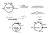
- (A) shows the case where an anti-PD-1 antibody is used as a single chain antibody
- (b) shows the case where an anti-CTLA-4 antibody is used as a single chain antibody.
- Anti-PD-1 or anti-CTLA-4 single chain antibody secretion plasmids pHuSP3L22-scFv-PD-1-1, pHuSP7L20-scFv-PD-1-1, pHuSP23L27-scFv-PD-1-1, pHuSP7L20-scFv- It is a figure which shows the preparation outline
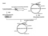
- FIG. 3 is a view showing an outline of the production of an anti-PD-1-1 single chain antibody secretion plasmid when various signal peptide-linker conjugates are used.
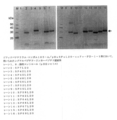
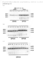
- Secreted into the culture supernatant of a Bifidobacterium bacterium transformed with a vector in which an expression cassette containing various signal peptide-linker peptide conjugates and an anti-PD-1-1 single chain antibody is inserted It is a figure which shows the result by the Western analysis of an anti- PD-1-1 single chain antibody.
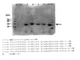
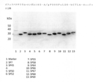
- Anti-PD-1- secreted into the culture supernatant of a Bifidobacterium bacterium transformed with a vector in which an expression cassette containing various signal sequences and an anti-PD-1-1 single chain antibody is inserted It is a figure which shows the result by the Western analysis of 1 single chain antibody. It is a figure which shows the result by the Western analysis of the various antibody secreted in the culture supernatant of Bifidobacterium genus bacteria.
- 2 is a graph showing the inhibition rate of binding of human CD80 to human CTLA-4 of anti-CTLA-4-1 single chain antibody and anti-CTLA-4-2 single chain antibody.
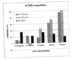
- 2 is a graph showing the inhibition rate of binding of human CD86 to human CTLA-4 by anti-CTLA-4-1 single chain antibody and anti-CTLA-4-2 single chain antibody.
- 2 is a graph showing the competitive inhibitory activity of an anti-mouse PD-1 single chain antibody on the binding reaction between mouse PD-1 and mouse PD-L1.
- the vertical axis represents inhibitory activity, and the horizontal axis represents antibody concentration. The figure by a flow cytometry analysis is shown.
- anti-mouse PD-1 antibody was reacted with unstimulated CD4 positive cells and detected by FITC.
- anti-mouse PD-1 antibody was reacted with CD3 / CD28 stimulated CD positive cells and detected by FITC.
- unstimulated CD4 positive cells were reacted with anti-mouse PD-1 single chain antibody and its His. Alexa 488-anti-His-tag antibody was reacted with and detected.
- anti-mouse PD-1 single-chain antibody was reacted with CD3 / CD28-stimulated CD-positive cells, and Alexais488-anti-His-tag antibody was reacted with the His to detect.
- A is an immunohistological staining diagram of tumor tissue of mice administered with anti-mouse PD-1 single chain antibody-secreting bifidobacteria
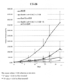
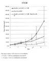
- B is an immunohistological staining diagram of tumor tissue of untreated mice. It is a graph which shows CT26 tumor volume transition of a single agent administration group and an untreated group. It is a graph which shows CT26 tumor volume transition of a single agent administration group, a 2 agent combination group, and an untreated group.
- Binding by ELISA of PD-L1 that was not competitively inhibited by scFv secreted by Bifidobacterium longum 105-A / pHuSP69L5-hPD-1 scFv03 and Bifidobacterium longum 105-A / pHuSP69L20-hPD-1 scFv03 It is a figure which shows quantity.
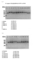
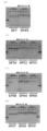
- (A) and (b) are diagrams showing the presence or absence of antibody secretion of each Bifidobacterium longum 105-A / pHuSPxL20-hPD-1scFv03 strain.
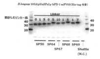
- (A) to (c) are diagrams showing the presence or absence of antibody secretion of each Bifidobacterium longum 105-A / pHuSPxLy-hPD-1scFv03 strain. It is a figure which shows the presence or absence of antibody secretion of each Bifidobacterium longum 105-A / pHuSPxLy-hPD-1scFv03 strain.
- FIG. 1 It is a figure which shows the presence or absence of antibody secretion of each Bifidobacterium longum 105-A / pP30SPxL20-hCTLA-4scFv02 strain.
- (A) to (c) are diagrams showing the presence or absence of antibody secretion of each Bifidobacterium longum 105-A / pP30SPxLy-hCTLA-4scFv02 strain.
- the expression cassette of the present invention includes (1) a promoter DNA that functions in Bifidobacterium, (2) a DNA that encodes a secretion signal peptide, (3) a DNA that encodes a linker peptide, (4) a heterologous polypeptide (5)
- Terminator DNA that functions in Bifidobacterium, and the secretory signal peptide is a) SEQ ID NOs: 6, 7, 8, 9, 10, 11, 12 , 13, 14, 15, and 16; or b) any of SEQ ID NOs: 6, 7, 8, 9, 10, 11, 12, 13, 14, 15, and 16.
- a peptide comprising the amino acid sequence is An amino acid sequence that functions as a secretory signal peptide in bacteria belonging to the genus Bifidobacterium, and is not particularly limited as long as it is an expression cassette for use in the expression of a heterologous polypeptide in a bacterium belonging to the genus Bifidobacterium.
- the 3 ′ end and the 5 ′ end of each downstream DNA may not be directly linked as long as the effects of the present invention can be obtained, but are preferably directly linked.
- the promoter DNA is not particularly limited as long as it is a promoter DNA that functions in a Bifidobacterium genus bacterium, and Hu is a promoter related to the expression of a gene encoding a histone-like DNA binding protein derived from Bifidobacterium longum.
- Promoter DNA Bifidobacterium breve-derived Gap gene promoter DNA (Biotechnol. Lett. 2008 30: 1983-1988), Bifidobacterium longum-derived AmyB gene promoter DNA (Biotechnol. Lett. 2006 28: 163-168) and, 16SrRNA promoter DNA (Biotechnol Lett 2008 30:. .
- amino acid sequence in which one or several amino acids are deleted, substituted or added is, for example, 1 to 5, preferably 1 to 3, more preferably 1 to 2, further preferably 1
- An amino acid sequence in which amino acids are deleted, substituted or added, and the amino acid sequence of the mutant secretion signal peptide includes SEQ ID NOs: 6, 7, 8, 9, 10, 11, 12, 13, 14, 15, and The sequence identity with any one of the amino acid sequences shown in FIG. 16 is preferably 90% or more, more preferably 95% or more, and further preferably 98% or more.
- the DNA encoding the secretory signal peptide consisting of the amino acid sequence shown in the above a) is not particularly limited as long as it has a base sequence corresponding to the amino acid sequence shown in the above a).
- DNA having the base sequence shown in any of SEQ ID NOS: 116, 117, 118, 119, 120, 121, 122, 123, 124, 125, and 126 can be mentioned.
- SEQ ID NOS: 116, 117, 118, 119, 120, 121, 122, 123, 124, 125, and 126 can be mentioned.
- These DNAs can be prepared by methods known to those skilled in the art, such as chemical synthesis and genetic engineering techniques.
- the DNA encoding the secretory signal peptide consisting of the amino acid sequence shown in b) above is not particularly limited as long as it has a base sequence corresponding to the amino acid sequence shown in b) above. Although different DNAs are included, these DNAs can also be prepared by methods known to those skilled in the art, such as chemical synthesis, genetic engineering techniques, and mutagenesis.
- a method in which a DNA having a base sequence represented by any of SEQ ID NOs: 116, 117, 118, 119, 120, 121, 122, 123, 124, 125, and 126 is contacted with a drug that is a mutagen
- the mutant DNA can be obtained by introducing a mutation into these DNAs using a method of irradiating ultraviolet rays, a genetic engineering technique, or the like.
- Site-directed mutagenesis which is one of the genetic engineering methods, is useful because it can introduce a specific mutation at a specific position.
- Molecular Cloning A laboratory Manual, 2nd Ed., Cold Spring Harbor Laboratory , Cold Spring Harbor, NY., 1989., Current Protocols in Molecular Biology, Supplements 1-38, John Wiley & Sons (1987-1997) and the like.
- the secretory signal peptide is preferably used as a signal peptide-linker conjugate linked to a linker peptide from the viewpoint of the expression / secretion efficiency of the heterologous polypeptide.
- the linker peptide is not particularly limited as long as it is a peptide that is used by linking to the C-terminus of the secretion signal peptide and can enhance the expression / secretion efficiency of the heterologous polypeptide.
- Preferable examples include peptides consisting of sequences.
- any one of the first to 30th of each sequence A linker peptide consisting of an amino acid sequence, for example, a linker peptide consisting of the first to fifth, first to tenth, first to fifteenth, first to twentieth amino acid sequences can be mentioned.
- Linker peptide sequence shown in SEQ ID NO: 25 (downstream of SP7); 2) The linker peptide sequence shown in SEQ ID NO: 26 (downstream of SP45); 3) The linker peptide sequence shown in SEQ ID NO: 27 (downstream of SP50); 4) The linker peptide sequence shown in SEQ ID NO: 28 (downstream of SP52); 5) The linker peptide sequence shown in SEQ ID NO: 29 (downstream of SP55); 6) The linker peptide sequence shown in SEQ ID NO: 30 (downstream of SP58); 7) The linker peptide sequence shown in SEQ ID NO: 31 (downstream of SP64); 8) The linker peptide sequence shown in SEQ ID NO: 32 (downstream of SP66); 9) The linker peptide sequence shown in SEQ ID NO: 33 (downstream of SP67); 10) The linker peptide sequence shown in SEQ ID NO: 34 (downstream of SP68); 11) The linker peptide sequence shown in SEQ ID NO: 35 (
- the DNA encoding the secretory signal peptide-linker conjugate of the present invention includes (2) from the 5 ′ end of the DNA encoding the secretory signal peptide to (3) the 3 ′ end of the DNA encoding the linker peptide. (2) 3 'end of DNA encoding secretory signal peptide and (3) 5' end of DNA encoding linker peptide, so that secretory signal peptide-linker conjugate May be referred to as SPxLy (where x is the number of the secretory signal peptide in the present specification and y is the number of amino acid residues of the linker peptide). Specific examples of such secretory signal peptide-linker conjugates include the following 11 types.
- the 5 ′ end of the encoding DNA is linked to the 3 ′ end of the DNA encoding the secretory signal peptide consisting of the amino acid sequence shown in SEQ ID NO: 6, and similarly, the amino acid sequence shown in any of SEQ ID NOs: 26 to 35
- the 5 ′ end of each DNA encoding a linker peptide lacking 0-29, for example, 10-29, 15-29, 20-29, or 25-29, in the C-terminal amino acid residue of the linker peptide consisting of At the 3 ′ end of each DNA encoding a secretory signal peptide consisting of the amino acid sequence shown in any of 7-16 It is binding.
- DNAs encoding such secretory signal peptide-linker conjugates are, for example, SEQ ID NO: 116 + SEQ ID NO: 127 (SP7L30), SEQ ID NO: 117 + SEQ ID NO: 128 (SP45L30), SEQ ID NO: 118 + SEQ ID NO: 129 (SP50L30), SEQ ID NO: 119+ sequence.
- SEQ ID NO: 130 (SP52L30), SEQ ID NO: 120 + SEQ ID NO: 131 (SP55L30), SEQ ID NO: 121 + SEQ ID NO: 132 (SP58L30), SEQ ID NO: 122 + SEQ ID NO: 133 (SP64L30), SEQ ID NO: 123 + SEQ ID NO: 134 (SP66L30), SEQ ID NO: 124 + SEQ ID NO: Based on the base sequence information shown in any of No. 135 (SP67L30), SEQ ID No. 125 + SEQ ID No. 136 (SP68L30), SEQ ID No. 126 + SEQ ID No. 137 (SP69L30), Growth, can be prepared by methods known to those skilled in the art, such as genetic engineering techniques.
- y in Ly is 30, 29, 28, 27, 26, 25, 24, 23, 22, 21, 20, 19, 18, 17, 16, 15, 14 , 13, 12, 11, 10, 9, 8, 7, 6, 5, 4, 3, 2, 1.
- anti-hPD- Specific examples of secretory signal peptide-linker conjugates confirmed to secrete 1scFv03 include SP69L1, SP69L2, SP69L3, SP69L4, SP69L5, SP69L6, SP69L7, SP69L8, SP69L9, SP69L10, SP69L15, SP69L20, SP7L20, SP45L20, SP50L SP52L20, SP55L20, SP58L20, SP64L20, SP66L20, SP67L20, SP68L20, SP7L5, SP7L10, S 7L15, SP45L5, SP45L10, SP45L15, SP50L5, SP50L10, SP50L15, SP52L5, SP52L10, SP52L15, SP52L15, SP52L15, SP52L15, SP
- SP7L5, SP7L20, SP68L5, SP68L20, SP69L5, SP69L20, SP45L20, SP50L20, SP52L20, SP55L20, SP58L20, SP64L20, SP66L20, SP67L20 can be mentioned, and the amount of secreted anti-hPD-1scFv03 bound to PD-1
- the linker conjugate include SP69L5, SP69L20, SP50L5, SP64L5, SP67L10, SP68L1, SP68L5, SP69L1 and SP69L7, and SP50L5, SP68L1 and SP69L1 are preferable, and the bond between hPD-1 and PD-L1 is particularly preferable.
- SP50L5 which has a low antibody concentration (IC50) that inhibits 50%, is particularly preferred.
- anti-hCTLA- Specific examples of secretory signal peptide-linker conjugates confirmed to secrete 4scFv02 include SP7L20, SP45L20, SP50L20, SP52L20, SP55L20, SP58L20, SP64L20, SP66L20, SP67L20, SP68L20, SP69L20, SP45L0, SP45L1, SP45L4, SP45L1, SP45L4 , SP45L5, SP50L0, SP50L1, SP50L2, SP50L3, SP50L4, SP50L5, SP64L0, P64L1, SP64L2, SP64L3, SP64L4, SP64L5, SP68L1, SP68L2, SP68L3, SP68L4, SP68L5, SP69L0
- the DNA encoding the heterologous polypeptide is not particularly limited as long as it is a polypeptide derived from other than Bifidobacterium that can be expressed in the expression cassette of the present invention.
- DNA encoding an angiogenesis inhibitor such as ostatin
- antibodies used for rheumatic therapeutics targeting interleukin-6 (IL-6) receptor examples include antibodies used for multiple sclerosis therapeutics targeting ⁇ 4-integrin, Examples thereof include antibodies having anticancer activity targeting CD20, CD33, PD-1, CTLA-4, CD80, and CD86.
- anti-PD-1 antibodies bind to PD-1 receptors expressed on activated lymphocytes (T cells, B cells), thereby causing PD-L1 and PD-L2 and PD expressed on cancer cells.
- T cells, B cells activated lymphocytes
- anti-CTLA-4 antibody enhances the antitumor immune response by suppressing the function of CTLA-4, which is known as a suppressor molecule of autoimmune function, and to CD80 and CD86 expressed in antigen-presenting cells. This is preferably exemplified because it is thought to prevent the negative down-regulation of the immune response induced by the interaction of these molecules by blocking the binding of CTLA-4.
- DNA encoding the antibody polypeptide examples include DNA encoding a chimeric antibody, humanized antibody, Fab, Fab ′, F (ab ′) 2 , single chain antibody, etc.
- DNA encoding a heterologous polypeptide containing these antibodies can be prepared by publicly known methods such as chemical synthesis, genetic engineering techniques, etc. by appropriately obtaining the sequence information from publicly known literatures or databases such as GenBank. .
- the terminator DNA is not particularly limited as long as it is a terminator DNA that functions in bacteria belonging to the genus Bifidobacterium. Specifically, a Hu terminator derived from Bifidobacterium longum, a T572 terminator (J Microbiol Biotechnol. 2012 Dec ; 22 (12): 1714-23).
- an expression cassette containing DNA encoding a linker peptide preferably an expression cassette containing DNA encoding a linker peptide, (1) Promoter DNA that functions in Bifidobacterium genus bacteria; (2) DNA encoding a secretory signal peptide consisting of the amino acid sequence shown in the following a) or b); a) the amino acid sequence shown in any of SEQ ID NOs: 6, 7, 8, 9, 10, 11, 12, 13, 14, 15, and 16; b) In the amino acid sequence shown in any of SEQ ID NOs: 6, 7, 8, 9, 10, 11, 12, 13, 14, 15, and 16, one or several amino acids are deleted, substituted, or added.
- amino acid sequence wherein the peptide comprising the amino acid sequence functions as a signal peptide in a bacterium belonging to the genus Bifidobacterium; (3) DNA encoding a linker peptide; (4) DNA encoding a heterologous polypeptide; (5) Terminator DNA that functions in bacteria of the genus Bifidobacterium; A method of preparing each of the DNAs of (1) as an upstream side and (5) as a downstream side so as to sequentially contain the DNAs of (1) to (5) can be exemplified.
- the vector of the present invention is not particularly limited as long as it is a vector capable of expressing a heterologous polypeptide when the expression cassette of the present invention is inserted and transformed into a bacterium belonging to the genus Bifidobacterium.
- One or more expression cassettes can be inserted into the vector.
- Specific examples of plasmid replication units that function in the Bifidobacterium genus contained in the vector of the present invention include pTB6 (BioscinBiotechnol Biochem. 2005 Feb; 69 (2): 422-5), pMB1 (Lett Appl Microbiol.
- pTB4 Structured plasmid pTB4 derived from Bifidobacterium longum Poster presentation Program-Japan Molecular Biology Society, 1994
- pFI2576 J Microbiol Biotechnol. 2009 Apr; 19 (4): 403-8
- pCIBAO Appl Environ Microbiol. 2007 Dec; 73 (24): 7858-66
- pBC1 Plasmid.
- the Tsu door can be preferably mentioned.
- the vector can also contain a marker gene such as a drug resistance gene.
- marker gene examples include spectinomycin, chloramphenicol, erythromycin, ampicillin and the like.
- An example of a method for introducing the vector of the present invention into a bacterium belonging to the genus Bifidobacterium includes an electroporation method.
- Bifidobacterium bacteria in the present invention include Bifidobacterium longum, Bifidobacterium breve (B. breve), Bifidobacterium addresscentis (B. adolescentis), Bifidobacterium bifidum (B. bifidum), Bifidobacterium pseudolongum, B. thermophirum, B. infantis, Bifidobacterium -B. ⁇ ⁇ animalis, B. angulatum, B. asteroides, B. boum, Bifidobacterium catenuratam (B. catenulatum), B. ⁇ ⁇ choerinum , B. coryneforme, B.
- B. denticolens cuniculi, B. denticolens, B. dentium , B. gallicum, B. gallinarum, B. globosum, B. indicum, B. B. inopinatum, B. lactis, B. lactentis, B. magnum, Bifido B. merycicum, B. ⁇ minimum, Biff B. Mongolia Enns, B. parvulorum, B. pseudocatenulatum, Bifidobacterium cyclaerophyllum (B. psychraerophilum, B. ⁇ pullorum, B. ruminale, B. ruminantium, B. saeculare ), Bifidobacterium skardovi (B.
- Bifidobacterium longum, Bifidobacterium breve, Bifidobacterium addresscentis, Bifidobacterium bifidum are known to be resident in the human intestine regardless of age.
- Bifidobacterium infantis is preferably used as the host cell, and Bifidobacterium longum is more preferably used. All of these bacteria are commercially available or can be easily obtained from depository institutions.
- each strain is not particularly limited.
- Bifidobacterium longum Bifidobacterium longum 105-A strain, Bifidobacterium longum aE-194b strain, Bifidobacterium longum Longgam bs-601 strain, Bifidobacterium longum M101-2 strain, Bifidobacterium longum ATCC-15707 strain can be mentioned, among which Bifidobacterium longum 105-A strain is preferable.
- Bifidobacterium breve include Bifidobacterium breve standard strain (JCM1192), Bifidobacterium breve aS-1 strain, and Bifidobacterium breve I-53-8W strain.
- Bifidobacterium breve standard strain and Bifidobacterium breve aS-1 strain are preferable.
- Bifidobacterium infantis include Bifidobacterium infantis standard strain (JCM1222) and Bifidobacterium infantis I-10-5 strain.
- Bifidobacterium lactentis include Bifidobacterium lactentis standard strain (JCM1210).
- strain of Bifidobacterium bifidum include Bifidobacterium bifidum ATCC-11863 strain.
- the transformed Bifidobacterium bacterium does not grow in normal tissues but grows only in tumor tissues in an anaerobic environment, and can express a polypeptide useful for treatment in the tumor tissues. Therefore, such a transformed Bifidobacterium is effective for the treatment of solid tumors that bring about tumor growth-inhibiting action, tumor volume-inhibiting action, preferably complete regression action on tumors having an anaerobic environment. It can be used as a pharmaceutical composition, especially as an anticancer agent. Accordingly, the pharmaceutical composition of the present invention is not particularly limited as long as it contains the above-mentioned Bifidobacterium of the present invention capable of secreting a heterologous polypeptide, preferably an antibody having anticancer activity, as an active ingredient. As long as the action and effect of the secreted polypeptide are not hindered, pharmacologically acceptable carriers, excipients, diluents and the like as optional components may be included.
- the target of application is colon cancer, head and neck cancer, breast cancer, lung cancer, esophageal cancer, stomach cancer, liver cancer, gallbladder cancer, bile duct cancer Pancreatic cancer, islet cell cancer, choriocarcinoma, colon cancer, renal cell cancer, adrenocortical cancer, bladder cancer, testicular cancer, prostate cancer, testicular cancer, ovarian cancer, uterus Cancer, choriocarcinoma, thyroid cancer, squamous cell carcinoma, skin cancer, brain tumor, malignant carcinoid tumor, osteosarcoma, soft tissue sarcoma, neuroblastoma, Wilms tumor, retinoblastoma, melanoma Can do.
- Examples of the dosage form of the pharmaceutical composition of the present invention include a liquid agent and a solid preparation.
- the liquid preparation is prepared by purifying the Bifidobacterium bacterium culture solution of the present invention and adding an appropriate physiological saline solution or a supplement solution or a pharmaceutical additive to the ampule or a vial as necessary. can do.
- For solid preparations add an appropriate protective agent to the solution and fill it into ampoules or vials and freeze-dry it, or add an appropriate protective agent to the solution and freeze-dry it, then add it to an ampoule or vial. It can be manufactured by filling.
- As a method for administering the pharmaceutical composition of the present invention both oral administration and parenteral administration are possible, but parenteral administration is preferred, and examples thereof include intravenous administration and topical administration.
- the dosage of the pharmaceutical composition of the present invention is not particularly limited as long as the Bifidobacterium bacterium of the present invention is an amount sufficient to grow at a disease site and to express an effective therapeutic amount of an active antibody.
- the amount is appropriately selected according to the degree of disease, the patient's weight, age, sex, and can be appropriately increased or decreased according to the degree of improvement, but from the viewpoint of avoiding economic viewpoints and side effects as much as possible, It is preferable that the amount is as small as possible within a range where a necessary therapeutic effect can be obtained.
- the formulation for injection at the lowest concentration possible should be divided into multiple doses or an appropriate fluid replacement It is preferable to dilute with continuous injection.
- the cells of the genus Bifidobacterium of the present invention are divided into 10 4 to 10 12 cfu per kg of body weight 1 to several times a day, continuously or at appropriate intervals for 1 to several days.
- a preparation containing 10 4 to 10 10 cfu / mL of Bifidobacterium of the present invention is diluted 1 to 1000 mL per adult directly or with an appropriate replacement fluid. Divide once to several times a day for 1 to several consecutive days.
- the cells of the genus Bifidobacterium of the present invention may be 10 4 to 10 12 cfu per kg of body weight 1 to several times a day, continuously 1 to several days as necessary, or Administer at appropriate intervals.
- a preparation containing 10 4 to 10 10 cfu / mL of the Bifidobacterium genus bacterium of the present invention is required directly from 0.1 to 100 mL per adult several times a day. Depending on the dosage, administer continuously for 1 to several days.
- Example 1 [Production of pHuSP3L22-scFv-PD-1-1, pHuSP7L20-scFv-PD-1-1, pHuSP23L27-scFv-PD-1-1]
- An anti-PD-1-1 single chain antibody secretion plasmid was prepared using a known secretion signal peptide and a novel secretion signal peptide.
- the expression cassette of the anti-PD-1 single chain antibody is a Hu promoter DNA (derived from Bifidobacterium longum), a DNA encoding a secretory signal peptide, and a linker following the secretory signal peptide.
- the anti-PD-1 single chain antibody (including heavy chain sequence, linker ((GGGGS) 3 ), base sequence including light chain sequence) in each expression cassette is shown in Table 3 below as shown below. Two types of anti-human PD-1 single chain antibodies and one type of anti-mouse PD-1 single chain antibody were used with reference to the literature.
- the anti-PD-1 single chain antibody genes of scFv-PD-1-1, scFv-PD-1-2, and scFv-PD-1-3 were subcloned into the plasmid pUC57 for Escherichia coli at Genscript Japan Co., Ltd. Then, artificial genes were synthesized as plasmids pUC57-scFv-PD-1-1, pUC57-scFv-PD-1-2 and pUC57-scFv-PD-1-3.
- Secretory signal peptide-linker conjugates SP3L22, SP7L20, and SP23L27 were used as secretory signal peptide sequences.
- SP3L22 a known sequence described as SEQ ID NO: 8 in International Publication Number WO2011 / 093467 was used, and as a SP23L27, a known sequence described as SEQ ID NO: 25 in International Publication Number WO2011 / 093467 was used.
- SP7L20 is based on the nucleotide sequence of SEQ ID NO: 12 described in International Publication No. WO2011 / 093467 and further added to the base sequence starting from ATG 66 nucleotides upstream of 5 ′: ATGGCGTTGATGATGAGCGTTAAGACTATTATTTCCACATCAGTGGCGATTATCGCCACGGGTGCC SEQ ID NO: 81) was used.
- SP7 analyzes the signal sequence prediction program SignalP 4.1 server (http://www.cbs.dtu.dk/services/SignalP/) that predicts the presence or absence of a secretory signal. This is because it was found to have a secretory signal peptide sequence at the N-terminus.
- the linker peptide sequence following SP7 was 20 amino acid residues on the C-terminal side of SP7.
- FIG. 1 An outline of the preparation of the anti-PD-1 single chain antibody secretion plasmid is shown in FIG.
- scFv-gene is PD-1
- PCR amplification was performed using 500 pg of plasmid pUC57-scFv-PD-1-1 as a template and using the lns_PD-1-1_F1 primer and the lns_PD-1-1_R1 primer set shown in Tables 1 and 2.
- the primer sequence was designed so that the insert fragment overlaps the end 15 bp of the vector fragment.
- PCR amplification was performed using a PrimeSTAR HS (Premix) (manufactured by Takara Bio Inc.) kit with 0.2 ⁇ M of each primer and a reaction volume of 30 ⁇ L.
- Vector fragment preparation Using the vector linearized fragment 500 pg described in SEQ ID NO: 20 as a template, using the primer set consisting of the TGA_Hu_Terminator_F primer and the vec-SP3L20-PD-1-1_R1 primer described in Table 1 and Table 2 above PCR amplification was performed using PrimeSTAR HS (Premix) kit with a primer of 0.2 ⁇ M and a reaction volume of 30 ⁇ L. As an amplification program, 30 cycles were performed with 98 ° C. for 10 seconds, 55 ° C. for 5 seconds, and 72 ° C. for 5 minutes as one cycle.
- PrimeSTAR HS Premix
- the amplified PCR product was electrophoresed on a 0.8% agarose gel, then purified using QIAquick Gel Extraction Kit (Qiagen), and 5′-Hu terminator-pTB6rep unit-SPCMr-pUCori-Hu promoter- SP3L20-3 ′ vector fragment was prepared.
- the vector linearized fragment is constituted as follows in the base sequence from the 4th to the 3845th of SEQ ID NO: 20.
- Hu terminator nucleotide sequence from the 4th to 117th sequence of SEQ ID NO: 20
- Bifidobacterium genus replication origin pTB6 rep unit: nucleotide sequence from the 124th sequence to 1719 of the SEQ ID NO: 20
- spectinomycin resistance gene SPCMr: nucleotide sequence from SEQ ID NO: 20 from 1726 to 2804
- pUCori nucleotide sequence from SEQ ID NO: 20 from 2811 to 3478
- Hu promoter SEQ ID NO: 20 from 3485 to Base sequence up to 3845.
- Infusion reaction The vector fragment prepared above and the anti-PD-1-1 single chain antibody insert were ligated using In-Fusion (registered trademark) HD Cloning Kit (Takara Bio Inc.). That is, the vector and the insert were added to the microtube at a molar ratio of 1: 5, 2 ⁇ L of 5 ⁇ In-Fusion HD Enzyme premix was added, and the reaction solution volume was adjusted to 10 ⁇ L. This was kept at 50 ° C. for 15 minutes. For other procedures, an infusion reaction solution was prepared in accordance with the product instructions of the kit.
- In-Fusion registered trademark
- HD Cloning Kit Tekara Bio Inc.
- Escherichia coli transformation and plasmid DNA sequence confirmation Using 5 ⁇ L of the infusion reaction solution, transformation of E. coli HST08 competent cells (manufactured by Takara Bio Inc.) was performed according to the product instructions. The bacterial suspension after the transformation was applied to an LB agar medium containing 75 ⁇ g / mL spectinomycin and statically cultured at 37 ° C. overnight. The Escherichia coli colonies formed on the agar medium were cultured overnight at 30 ° C. in an LB liquid medium containing 75 ⁇ g / mL spectinomycin, and a plasmid was extracted therefrom using QIAprep Spin Miniprep Kit (manufactured by Qiagen).
- pHuSP3L22-scFv-PD-1-1 vector linearized fragment preparation Using the plasmid pHuSP3L20-scFv-PD-1-1 500 pg as a template and a primer set consisting of the Ins_PD-1-1_F1 phosphorylated primer and SP3L22_R1 phosphorylated primer, the final concentration was 0.2 ⁇ M and the reaction volume was 30 ⁇ L.
- PCR amplification was performed using the PrimeSTAR HS (Premix) kit. As an amplification program, 30 cycles were performed with 98 ° C. for 10 seconds, 55 ° C. for 5 seconds, and 72 ° C. for 5 minutes as one cycle.
- the amplified PCR product was electrophoresed on a 0.8% agarose gel and then purified using a QIAquick Gel Extraction Kit to prepare a pHuSP3L22-scFv-PD-1-1 vector linearized fragment.
- the vector linearized fragment was subjected to ligation reaction using T4 DNA Ligase (manufactured by Thermo Fisher Scientific Co., Ltd.) according to the product instructions.
- T4 DNA Ligase manufactured by Thermo Fisher Scientific Co., Ltd.
- the attached 10 ⁇ buffer for T4 DNA ligase (2 ⁇ L) was added to make the reaction solution volume 20 ⁇ L, and this was subjected to ligation reaction at 22 ° C. for 10 minutes to prepare a ligation reaction solution.
- E. coli HST08 competent cells were transformed with 10 ⁇ L of the ligation reaction solution.
- the bacterial suspension after the transformation was applied to a 75 ⁇ g / mL spectinomycin-containing LB agar medium and statically cultured at 37 ° C. overnight.
- the transformation conditions followed the product instructions for E. coli HST08 competent cells, but are outlined below.
- the E. coli colonies formed on the agar medium were cultured overnight at 30 ° C. in an LB liquid medium containing 75 ⁇ g / mL spectinomycin, and a plasmid was extracted therefrom using QIAprep Spin Miniprep Kit.
- the extracted plasmid was named pHuSP3L22-scFv-PD-1-1.
- transformation of Bifidobacterium Using the 1000 ng plasmid pHuSP3L22-scFv-PD-1-1 extracted from the transformed Escherichia coli, transformation of Bifidobacterium longum 105-A strain was performed using an electroporation system (Gene Pulser II, Bio-Rad). Laboratories, Inc.). After electric shock (2 kV, 25 ⁇ F, 200 ⁇ ), immediately add a mixture of 800 ⁇ L of IMR liquid medium and 50 ⁇ L of vitamin C supplement to a cuvette (2 mm gap) and collect it in a sterile 2 mL microtube. did.
- Each bacterial suspension after incubation was applied to an IMR agar medium containing 75 ⁇ g / mL spectinomycin. These plates were placed in a closed container together with the deoxygenation / carbon dioxide generating agent and cultured in an incubator set at 37 ° C. for 2 days.
- the colonies formed on the spectinomycin-containing IMR agar medium were picked and streaked on a 75 ⁇ g / mL spectinomycin-containing BL-bS agar medium (BL agar medium not containing equine defibrinated blood). It was placed in a closed container together with a carbon dioxide generator and cultured in an incubator set at 37 ° C. for 1 day to obtain Bifidobacterium longum 105-A / pHuSP3L22-scFv-PD-1-1 strain.
- Vector fragment preparation Using the vector linearized fragment 500 pg described in SEQ ID NO: 21 as a template, a primer set consisting of the TGA_Hu_Terminator_F primer and the vec-SP7L20-PD-1-1_R1 primer described in Table 1 and Table 2 above is used. PCR amplification was performed using a PrimeSTAR HS (Premix) kit at a final concentration of 0.2 ⁇ M and a reaction volume of 30 ⁇ L. As an amplification program, 30 cycles were performed with 98 ° C. for 10 seconds, 55 ° C. for 5 seconds, and 72 ° C. for 5 minutes as one cycle.
- PrimeSTAR HS Premix
- the amplified PCR product was electrophoresed on a 0.8% agarose gel, purified using QIAquick Gel Extraction Kit, and 5′-Hu terminator-pTB6rep unit-SPCMr-pUCori-Hu promoter-SP7L20-3 ′ vector. Fragment (1) was prepared.
- the vector linearized fragment is constituted as follows in the base sequence from the 4th to the 3845th of SEQ ID NO: 21.
- Hu terminator nucleotide sequence from SEQ ID NO: 21 to 4 to 117 Bifidobacterium replication origin
- pTB6 rep unit nucleotide sequence from SEQ ID NO: 21 to 1719 spectinomycin resistance gene
- SPCMr nucleotide sequence from 1726 to 2804 of SEQ ID NO: 21
- E. coli replication origin pUCori: nucleotide sequence from 2811 to 3478 of SEQ ID NO: 21
- Hu promoter 3485 to 3485 of SEQ ID NO: 21 Base sequence up to 3845.
- Vector fragment preparation Using the vector linearized fragment 500 pg described in SEQ ID NO: 22 as a template, a primer set consisting of TGA_Hu_Terminator_F primer and vec-SP23L20-PD-1-1_R1 primer described in Table 1 and Table 2 above is used. PCR amplification was performed using a PrimeSTAR HS (Premix) kit at a final concentration of 0.2 ⁇ M and a reaction volume of 30 ⁇ L. As an amplification program, 30 cycles were performed with 98 ° C. for 10 seconds, 55 ° C. for 5 seconds, and 72 ° C. for 5 minutes as one cycle.
- PrimeSTAR HS Premix
- the amplified PCR product was electrophoresed on a 0.8% agarose gel, purified using QIAquick Gel Extraction Kit, and 5′-Hu terminator-pTB6rep unit-SPCMr-pUCori-Hu promoter-SP23L20-3 ′ vector. Fragments were prepared.
- the vector linearized fragment is constituted as follows in the base sequence from the 4th to the 3845th of SEQ ID NO: 22.
- Hu terminator nucleotide sequence from SEQ ID NO: 22 to 4 to 117 Bifidobacterium replication origin
- pTB6 rep unit nucleotide sequence from SEQ ID NO: 124 to 1719 spectinomycin resistance gene
- SPCMr nucleotide sequence from SEQ ID NO: 22 from 1726 to 2804
- E. coli replication origin E. coli replication origin
- pUCori nucleotide sequence from SEQ ID NO: 22 from 2811 to 3478
- Hu promoter 3485 from SEQ ID NO: 22 Base sequence up to 3845.
- the amplified PCR product was electrophoresed on a 0.8% agarose gel, and then the PCR product was purified using a QIAquick Gel Extraction Kit to prepare a pHuSP23L27-scFv-PD-1-1 vector linearized fragment.
- Example 2 (Examination of the presence or absence of single-chain antibody secretion in recombinant Bifidobacterium 1) Three types of recombinant Bifidobacterium bacteria (Bifidobacterium longum 105-A / pHuSP3L22-scFv-) using the three types of anti-PD-1-1 single chain antibody secretion plasmids prepared in Example 1 above.
- MRS MRS was mixed in a ratio of 9: 1 to 20 mL of medium, and vitamin C addition solution 100 ⁇ L and spectinomycin were added to 75 ⁇ g / mL. 100 ⁇ L of the activated culture solution was inoculated. This was anaerobically cultured at 37 ° C. for 18 hours.
- the above culture supernatant concentrate (equivalent to 1 mL of culture solution) was electrophoresed on a Mini-PROTEAN (registered trademark) TGX TM gel (4-20%) (Bio-Rad), and the gel was PVDF membrane (iBlot Transfer Stacks, Life Transferred to Technologies) using an iBlot transfer device (Life Technologies).
- the primary antibody is mouse histidine tag antibody (THE HIS Tag Antibody, mAb, Mouse, Genscript Japan)
- ECL-peroxidase-labeled anti-mouse antibody GE Healthcare Japan Co., Ltd.
- Western Lightning Ultra Perkin Elmer Co.
- Lane M is a marker
- lane 1 is a negative control
- lane 2 is a secreted antibody from a Bifidobacterium that is transformed with pHuSP3L22-scFv-PD-1-1
- lane 3 is pHuSP7L20-scFv-PD-1-
- lane 4 shows the antibody secreted from the bacterium belonging to the genus Bifidobacterium that has been transformed by pHuSP23L27-scFv-PD-1-1.
- Example 3 [Examination of 33 types of secretory signal peptide-linker conjugates]
- pHuSP7L20-scFv-PD-1-1 33 types of anti-secretory peptide-linker conjugate SP7L20 were substituted with 33 types of secretory signal peptide-linker conjugates (SP42L20 to SP46L20, SP48L20 to SP75L20), respectively.
- a PD-1-1 single chain antibody secretion plasmid was constructed. An outline of the construction method is shown in FIG. Details of the primers used are shown in Table 4 and Table 5 below.
- TM transmembrane region
- each secretory protein is secreted from the N-terminal of each protein to the amino acid sequence estimated as a protease cleavage site by SignalP analysis.
- the signal peptide sequence was used.
- the linker peptide sequence was 20 amino acid residues that continued from the C-terminus of each signal sequence.
- PCR amplification was performed using Bifidobacterium longum 105-A strain 120 pg as a template and each primer set described in Tables 4 and 5. Each primer sequence was designed so that the insert fragment overlaps the end 15 bp of the vector fragment.
- PCR amplification was performed using a PrimeSTAR HS (Premix) kit at a primer concentration of 0.2 ⁇ M and a reaction volume of 20 ⁇ L. As the amplification program, 30 cycles were performed with 98 ° C. for 10 seconds, 65 ° C. for 5 seconds, and 72 ° C. for 20 seconds, followed by 30 seconds of extension reaction at 72 ° C. A variety of secretory signal peptide-linker conjugate inserts were amplified.
- the vector fragment prepared above and 33 kinds of secretory signal peptide-linker conjugate inserts were ligated using In-Fusion (registered trademark) HD Cloning Kit. Specifically, the vector and the insert were added to the microtube at a molar ratio of 1: 2, 5 ⁇ In-Fusion HD Enzyme premix 2 ⁇ L in the kit was added, and 0.1 ⁇ TE buffer (1 mM Tris-HCl, 0.1 mM EDTA) was added. , PH 7.5) to 10 ⁇ L. This was kept at 37 ° C. for 15 minutes, and further kept at 50 ° C. for 15 minutes. For other procedures, 33 types of infusion reaction solutions were prepared according to the product instructions of the kit.
- Escherichia coli HST16CR competent cells (manufactured by Takara Bio Inc.) were transformed with 0.8 ⁇ L of the 33 kinds of infusion reaction solutions.
- the bacterial suspension after the transformation was applied to an LB agar medium containing 75 ⁇ g / mL spectinomycin and statically cultured at 37 ° C. overnight. Transformation conditions followed the product instructions for E. coli HST16CR competent cells, but are outlined below.
- the Escherichia coli colonies formed on the agar medium were cultured overnight at 37 ° C.
- plasmid was extracted therefrom using QIAprep Spin Miniprep Kit.
- a sequencing reaction was performed using BigDye (registered trademark) Terminator v3.1 Cycle Sequencing Kit. went.
- transformation was performed with a plasmid in which each of the secretory signal peptide SP-42L20, SP48L20 and SP62L20 secretory signal peptide-linker conjugates consisting of SP42, SP48 and SP62 secretory signal peptide sequences followed by a linker peptide sequence consisting of 20 amino acid residues was inserted.
- secretion of anti-PD-1-1 single chain antibody was not observed.
- Bifidobacterium which has been able to secrete a single chain antibody by the above examination is characterized by using an expression cassette containing SP45L20, SP50L20, SP52L20, SP55L20, SP58L20, SP64L20, SP66L20, SP67L20, SP68L20, SP69L20. Only transformed bacteria. It was confirmed that the other 23 types of sequences were not secreted or were insufficiently secreted. As examples of secretory signal peptide-linker conjugates that are not secreted or insufficiently secreted, the sequences of SP42L20, SP48L20, and SP62L20 are shown in SEQ ID NOs: 17, 18, and 19, respectively.
- Vector fragment preparation Using 500 pg of the vector linearized fragment described in SEQ ID NO: 23 as a template, a primer set consisting of the TGA_Hu_Terminator_F primer and the vec-SP7L20-PD-1-2_R1 primer described in Table 1 and Table 2 above is used. PCR amplification was performed using the PrimeSTAR HS (Premix) kit at a final concentration of 0.2 ⁇ M and a reaction volume of 30 ⁇ L. The amplification program was 30 cycles, with 98 ° C. for 10 seconds, 55 ° C. for 5 seconds, and 72 ° C. for 5 minutes as one cycle.
- PrimeSTAR HS Premix
- the amplified PCR product was electrophoresed on a 0.8% agarose gel, then purified using QIAquick Gel Extraction Kit, and 5′-Hu terminator-pTB6rep unit-SPCMr-pUCori-Hu promoter-SP7L20-3 ′ vector Fragment (2) was prepared.
- the vector linearized fragment is constituted as follows in the base sequence from the 4th to the 3845th of SEQ ID NO: 23.
- Hu terminator nucleotide sequence from the 4th to 117th sequence of SEQ ID NO: 23
- Bifidobacterium genus replication origin pTB6 rep unit: nucleotide sequence from the 124th sequence to 1719 of the SEQ ID NO: 23
- spectinomycin resistance gene SPCMr: nucleotide sequence from 1726 to 2804 of SEQ ID NO: 23
- pUCori nucleotide sequence from 2811 to 3478 of SEQ ID NO: 23 Hu promoter: 3485 to 3485 of SEQ ID NO: 23 Base sequence up to 3845.
- Anti-PD-1-3 single chain antibody is selected as a heterologous peptide, and anti-PD-1-3 single chain antibody secretion plasmid pHuSP7L20-scFv-PD-1-3 having a secretory signal peptide-linker conjugate (SP7L20) Built. Details are as follows.
- Vector fragment preparation A primer set consisting of the TGA_Hu_Terminator_F primer and the vec-SP7L20-PD-1-3_R1 primer described in Table 1 and Table 2 above was used using the vector linearized fragment 500 pg described in SEQ ID NO: 24 as a template. Otherwise, the 5′-Hu terminator-pTB6rep unit-SPCMr-pUCori-Hu promoter-SP7L20-3 ′ vector Fragment (3) was prepared.
- the vector linearized fragment is composed of the following base sequence in the base sequence from the 4th to the 3845th of SEQ ID NO: 24.
- Hu terminator nucleotide sequence from SEQ ID NO: 24 to 4 to 117 Bifidobacterium replication origin
- pTB6 rep unit nucleotide sequence from SEQ ID NO: 24 to 1719 spectinomycin resistance gene
- SPCMr base sequence from 1726 to 2804 of SEQ ID NO: 24
- pUCori base sequence from 2811 to 3478 of SEQ ID NO: 24
- Hu promoter from 3485 to 3485 of SEQ ID NO: 24 Base sequence up to 3845.
- the expression cassette of the anti-CTLA-4 single chain antibody is a Hu promoter DNA (derived from Bifidobacterium longum), a DNA encoding a secretory signal peptide, and a linker following the secretory signal peptide.
- DNA encoding peptide, DNA encoding anti-CTLA-4 single chain antibody including heavy chain sequence, linker ((GGGGS) 3 ), light chain sequence), His tag sequence, and Hu terminator (Bifidobacterium • Longong origin).
- the anti-CTLA-4 single chain antibody (including heavy chain sequence, linker ((GGGGGS) 3 ), light chain sequence-containing base sequence) in each expression cassette is shown in Table 6 below as shown below. With reference to the literature, two types of anti-human CTLA-4 single chain antibodies were used.
- PCR amplification was performed using the primer set consisting of the lns_CTLA-4-1_F1 primer and the lns_CTLA-4-1_R1 primer described in Tables 1 and 2 above. went.
- the primer sequence was designed so that the insert fragment overlaps the end 15 bp of the vector fragment.
- the PCR reaction solution was made to have a primer of 0.2 ⁇ M and a reaction volume of 30 ⁇ L, and was amplified using a PrimeSTAR HS (Premix) (Takara Bio Inc.) kit.
- the amplification program was performed 30 cycles, with 98 ° C. for 10 seconds, 55 ° C. for 5 seconds, and 72 ° C. for 60 seconds.
- the amplified insert PCR product was electrophoresed on a 2% agarose gel and then purified using the QIAquick Gel Extraction Kit to prepare an anti-CTLA-4-1 single chain antibody insert.
- Vector fragment preparation The 5′-Hu terminator-pTB6rep unit-SPCMr-pUCori-Hu promoter-SP7L20-3 ′ vector fragment (with the same procedure as in (Preparation of vector fragment) in [Preparation of pHuSP7L20-scFv-PD-1-1]) 1) was prepared.
- Example 9 (Examination of the presence or absence of single-chain antibody secretion in recombinant Bifidobacterium 3) Bifidobacterium longum 105-A / pHuSP7L20-scFv-PD-1, which is a recombinant Bifidobacterium bacterium with anti-PD-1 single chain antibody secretion plasmid and anti-CTLA-4 single chain antibody secretion plasmid -1 strain, Bifidobacterium longum 105-A / pHuSP7L20-scFv-PD-1-2 strain, Bifidobacterium longum 105-A / pHuSP7L20-scFv-PD-1-3 strain, Bifidobacterium Longum 105-A / pHuSP7L20-scFv-CTLA-4-1 strain and Bifidobacterium longum 105-A / pHuSP7L20-scFv-CTLA-4-2 strain described above (in
- Example 10 [Purification of anti-PD-1 and anti-CTLA-4 single-chain antibodies from recombinant Bifidobacterium strains] Bifidobacterium longum 105-A / pHuSP7L20-scFv-PD-1-1 strain, Bifidobacterium longum 105-A / pHuSP7L20-scFv-PD-1-2 strain, Bifidobacterium longum 105- A / pHuSP7L20-scFv-PD-1-3 strain, Bifidobacterium longum 105-A / pHuSP7L20-scFv-CTLA-4-1 strain and Bifidobacterium longum 105-A / pHuSP7L20-scFv-CTLA- For strain 4-2, purification of the single chain antibody was performed by the method shown below.
- MRS MRS (9: 1) medium, spectinomycin (final concentration 75 ⁇ g / mL) and a vitamin C addition solution (500 ⁇ L per 100 mL of medium) were added, and the culture solution was adjusted to a medium amount of 0. 5% of the inoculum was inoculated and anaerobically cultured at 37 ° C for 18 hours.
- a portion of the purified single-chain antibody is subjected to SDS-PAGE, followed by Coomassie brilliant blue staining (SimplyBlue (registered trademark) Safe Stain, Life Technologies), and scFv-PD-1 with a purity of about 90%.
- scFv-PD-1-2, scFv-PD-1-3, scFv-CTLA4-1, and scFv-CTLA4-2 were confirmed to be purified.
- Example 11 [Confirmation of binding between human PD-1 and anti-PD-1 single chain antibody or anti-CTLA-4 single chain antibody] Binding of anti-PD-1-2 single chain antibody purified from the culture supernatant of Bifidobacterium longum 105-A / pHuSP7L20-scFv-PD-1-2 to human PD-1 (hPD-1) The presence or absence of was confirmed by the ELISA method.
- hPD-1 Recombinant Human PD-1 Fc Chimera, manufactured by R & D ⁇ Systems
- hPD-1 Recombinant Human PD-1 Fc Chimera, manufactured by R & D ⁇ Systems
- Dispense 100 ⁇ L of hPD-1 Adjust to 1 ⁇ g / mL with 1 ⁇ PBS in a 96-well plate and let stand at 4 ° C. overnight. Solidified. After removing the liquid, 350 ⁇ L of 1 ⁇ PBS was dispensed to remove the liquid. This was washed three times repeatedly. Blocking was performed by dispensing 350 ⁇ L of 1% BSA solution onto the plate and leaving it at room temperature for 2 hours. After removing the liquid, 350 ⁇ L of 1 ⁇ PBS was dispensed to remove the liquid. This was washed three times repeatedly.
- the anti-PD-1-2 single chain antibody was adjusted to 1000 ng / mL, 100 ng / mL, 10 ng / mL and 1 ng / mL with a signal enhancement reagent (Signal Enhancer HIKARI, manufactured by nacalai tesque), and the blocking was completed by 100 ⁇ L each. Dispense into rear plates. As negative controls, anti-CTLA-4-1 single-chain antibody and anti-CTLA-4-2 single-chain antibody purified from Bifidobacterium were prepared in the same manner as described above. Only 100 ⁇ L of the signal enhancement reagent was dispensed into the blank well.
- the plate was sealed and allowed to stand at room temperature for 2 hours, after which the immobilized hPD-1 reacted with each single-chain antibody. After removing the liquid, 350 ⁇ L of 1 ⁇ PBS was dispensed to remove the liquid. This was washed three times repeatedly.
- the His-Tag Antibody manufactured by GenScript was diluted 2500 times with a signal enhancement reagent, and 100 ⁇ L of this was dispensed. The plate was sealed and left at room temperature for 2 hours. After removing the liquid, 350 ⁇ L of 1 ⁇ PBS was dispensed to remove the liquid. This was washed three times repeatedly.
- the tertiary antibody (Biotin anti-mouse IgG, manufactured by Biolegend) was diluted 20,000 times with a signal enhancement reagent, and 100 ⁇ L of this was dispensed. The plate was sealed and allowed to stand at room temperature for 30 minutes. After removing the liquid, 350 ⁇ L of 1 ⁇ PBS was dispensed to remove the liquid. This was washed three times repeatedly.
- an avidin-biotin-labeled enzyme complex (Vectastain® ABC® Kit, manufactured by Vector)
- 3 drops of solution A and solution B were added to 7.5 mL of the signal enhancement reagent. After 100 ⁇ L of this was dispensed, the plate was sealed and allowed to stand at room temperature for 30 minutes. After removing the liquid, 350 ⁇ L of 1 ⁇ PBS was dispensed to remove the liquid. This was washed three times repeatedly.
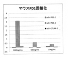
- Color Solution A and Color Solution B (manufactured by R & D Systems) were mixed in equal amounts, dispensed in 200 ⁇ L portions, allowed to stand at room temperature for 20 minutes in the dark. After 20 minutes exactly, StopSsolution (manufactured by R & D Systems) was added by 50 ⁇ L at a time to stop the color reaction. The measurement was performed by measuring the absorbance at 450 nm with 570 nm as a control. Presence or absence of binding of anti-PD-1-2 single-chain antibody, anti-CTLA-4-1 single-chain antibody, anti-CTLA-4-2 single-chain antibody to human PD-1 solid-phase plate using ELISA method The results are shown in Table 7 below and FIG.
- Example 13 (Confirmation of binding of mouse PD-1 and anti-PD-1-3 single chain antibody 1)
- Anti-PD-1-3 single chain antibody purified from the culture supernatant of Bifidobacterium longum 105-A / pHuSP7L20-scFv-PD-1-3 and mouse PD-1 (mPD-1) The presence or absence of binding was confirmed by the following ELISA method.
- mPD-1 Recombinant Mouse PD-1 Fc Chimera, manufactured by R & D Systems
- Anti-PD-1-3 single chain antibody purified from Bifidobacterium was prepared with a signal enhancement reagent (Signal Enhancer HIKARI, manufactured by nacalai tesque) to 1000 ng / mL, 100 ng / mL and 10 ng / mL, 100 ⁇ L each was dispensed to the plate after the blocking.
- a signal enhancement reagent Signal Enhancer HIKARI, manufactured by nacalai tesque
- anti-PD-1-2 single-chain antibody purified from Bifidobacterium and anti-CTLA-4-2 single-chain antibody purified from Bifidobacterium were prepared in the same manner as described above. did. Only 100 ⁇ L of the signal enhancement reagent was dispensed into the blank well.
- the plate was sealed and allowed to stand at room temperature for 2 hours, after which the immobilized mPD-1 reacted with the anti-PD-1-3 single chain antibody. After removing the liquid, 350 ⁇ L of 1 ⁇ PBS was dispensed to remove the liquid. This was washed three times repeatedly.
- Color Solution A and Color Solution B (manufactured by R & D Systems) were mixed in equal amounts, dispensed in 200 ⁇ L portions, and allowed to stand at room temperature for 20 minutes in the dark. After 20 minutes exactly, StopSsolution (manufactured by R & D Systems) was added by 50 ⁇ L at a time to stop the color reaction.
- Example 14 (Confirmation of binding between mouse PD-1 and anti-PD-1-3 single chain antibody 2)
- Anti-PD-1-3 single chain antibody purified from the culture supernatant of Bifidobacterium longum 105-A / pHuSP7L20-scFv-PD-1-3 The specificity of binding was confirmed.
- the same test as in [Example 13] was carried out using hCTLA-4 (Recombinant Human CTLA-4-Fc Chimera carrier-free, BioLegend) as the protein to be immobilized on the plate. The results are shown in Table 10 and FIG.
- the anti-CTLA-4-2 single chain antibody which is a positive control, binds to hCTLA-4 in a concentration-dependent manner. The antibody did not bind. Also, anti-PD-1-2 single chain antibody, which is an anti-human PD-1 antibody, did not bind. From these results, it was shown that the binding of mPD-1 and anti-PD-1-3 single chain antibody in [Example 13] is specific.
- Example 15 (Confirmation of binding between anti-PD-1-2 single-chain antibody and human PD-1 by flow cytometry analysis) Binding of anti-PD-1-2 single-chain antibody to human PD-1 overexpressing cells Jurkat cells overexpressed human PD-1 (provided by Professor Shinichiro Taniguchi, Shinshu University Molecular Oncology), and human PD-1 And HEK293T cells (provided by Professor Shunichiro Taniguchi, Department of Molecular Oncology, Shinshu University).
- Human PD-1 overexpressing Jurkat cells were placed in 100 mm Petri dishes (Ina Optica) in RPMI 1640 medium containing 5 ⁇ 10 6 cells / 10 mL medium (inactivated 10% fetal calf serum, 50 ⁇ M 2-mercaptoethanol and 8 mM HEPES). ) / dish, and human PD-1 overexpressing HEK293T cells were seeded in 2 ⁇ 10 6 cells / 10 mL medium (DMEM medium containing inactivated 10% fetal bovine serum) in a 100 mm dish (manufactured by Greiner Japan). .
- DMEM medium containing inactivated 10% fetal bovine serum
- human PD-1 overexpressing Jurkat cells were collected in a 15 mL falcon tube (Becton Dickinson), then the number of cells was counted with a hemocytometer, so that 1 ⁇ 10 5 cells / mL / tube 1 Dispensed into a 5 mL tube (manufactured by Ina Optica).
- Human PD-1 overexpressing HEK293T cells were removed from the culture supernatant, washed twice with PBS (Ca 2+ , Mg 2+ free phosphate buffer), and diluted 10-fold with PBS (trypsin / EDTA solution ( 1 mL of Wako (manufactured by Wako) was added, and the mixture was allowed to stand at room temperature for 1 minute. After that, 10 mL of inactivated DMEM medium containing 10% fetal bovine serum was added and transferred to a 15 mL falcon tube.
- PBS Ca 2+ , Mg 2+ free phosphate buffer
- PBS trypsin / EDTA solution
- Wako manufactured by Wako
- PD-1 overexpressing Jurkat cells and HEK293T cells dispensed into 1.5 mL tubes were centrifuged at 5000 rpm and 4 ° C. for 1 minute in a microcooled centrifuge (manufactured by Tommy Seiko), and the supernatant was removed after centrifugation.
- the cell pellet remaining in the tube was washed twice with 0.5 mL of PBS, and then anti-PD-1-2 single-chain antibody and LEAF purified anti-human PD-1 antibody (clone: EH12.2H7) as a positive control. ) (Manufactured by BioLegend) was added at a concentration of 10 ⁇ g / mL, and allowed to stand on ice for 30 minutes.
- FACS buffer PBS containing 1% BSA and 0.1% NaN 3
- 50 ⁇ L of biotinylated protein L manufactured by Pierce
- 500 ⁇ L of FACS buffer PBS containing 1% BSA and 0.1% NaN 3
- Example 16 (Confirmation of competitive binding inhibitory activity of anti-CTLA-4 single chain antibody with CD80 and CD86 against human CTLA-4 using ELISA method) Using the anti-CTLA-4-1 single-chain antibody and anti-CTLA-4-2 single-chain antibody secreted by the recombinant Bifidobacterium genus bacteria produced in Examples 7 and 8 above, human CTLA-4 ( Competitive binding inhibitory activity for binding to human CD80 (hCD80) or human CD86 (hCD86) to hCTLA-4) was confirmed. An anti-PD-1-2 single chain antibody was used as a negative control.
- hCTLA-4 Recombinant Human CTLA-4-Fc Chimera, carrier-free, BioLegend
- 1 ⁇ PBS 1 ⁇ PBS
- Blocking was performed by dispensing 350 ⁇ L of 1% BSA solution onto the plate and allowing to stand at room temperature for 2 hours. After removing the liquid, 350 ⁇ L of 1 ⁇ PBS was dispensed to remove the liquid. This was washed three times repeatedly.
- hCD80 Recombinant Human B7-1 / CD80 Fc Chimera, manufactured by R & D Systems
- hCD86 Recombinant Human B7-2 / CD86 Fc Chimera, manufactured by R & D Systems
- anti-PD-1-2 single chain antibody purified from Bifidobacterium was prepared in the same manner as described above. Only 100 ⁇ L of the signal enhancement reagent was dispensed into the blank well.
- hCD80 and hCD86 mixed with anti-CTLA-4 single-chain antibody and anti-CTLA-4-2 single-chain antibody against solid-phased hCTLA-4 Reacted. After removing the liquid, 350 ⁇ L of 1 ⁇ PBS was dispensed to remove the liquid. This was washed three times repeatedly.
- Color Solution A and Color Solution B (manufactured by R & D Systems) were mixed in equal amounts, dispensed in 200 ⁇ L portions, and allowed to stand at room temperature for 20 minutes in the dark. After 20 minutes exactly, StopSsolution (manufactured by R & D Systems) was added by 50 ⁇ L at a time to stop the color reaction. The absorbance was measured at 450 nm, and the absorbance at 570 nm was measured as a control. The measurement results are shown in Tables 11 and 12, and the inhibition rates are shown in FIGS.
- the inhibition rate of hCD80 to hCTLA-4 binding was 61.7% and 73.1% with 1 ⁇ g / mL anti-CTLA-4-1 single-chain antibody and anti-CTLA-4-2 single-chain antibody, At 10 ⁇ g / mL, they were 94.1% and 99.3%, respectively.
- the inhibition rate of hCD86 binding to hCTLA-4 was 83.7% and 90.9% for 1 ⁇ g / mL anti-CTLA-4-1 single-chain antibody and anti-CTLA-4-2 single-chain antibody, respectively. At 10 ⁇ g / mL, they were 96.0% and 97.7%, respectively.
- anti-CTLA-4-1 single-chain antibody and anti-CTLA-4-2 single-chain antibody are both 1 ⁇ g / mL for binding of 1 ⁇ g / mL of hCD80 and hCD86 to hCTLA-4. It was shown that binding was inhibited competitively at these concentrations.
- Example 17 [Competitive inhibitory activity of anti-mouse PD-1 single-chain antibody for binding reaction between mouse PD-1 and mouse PD-L1] Using the anti-mouse PD-1 single chain antibody secreted by Bifidobacterium longum 105-A / pHuSP7L20-scFv-PD-1-3 prepared in Example 6, mouse PD-1 (mPD-1 ) And mouse PD-L1 (mPD-L1) were confirmed for competitive inhibitory activity using the ELISA method. Anti-human CTLA-4-2 single chain antibody was used as a negative control.
- MPD-L1 Recombinant Mouse B7-H1 / PD-L1 Fc Chimera, manufactured by R & D Systems
- a signal enhancing reagent Synchronization Enhancer HIKARI, manufactured by nacalaitesque
- Anti-mouse PD-1 single chain antibody purified from Bifidobacterium longum 105-A / pHuSP7L20-scFv-PD-1-3 strain was tested with a signal enhancement reagent at 20000 ng / mL, 2000 ng / mL, 200 ng / mL and The amount was adjusted to 20 ng / mL, mixed with an equal amount of mPD-L1, and 100 ⁇ L was dispensed on the plate after the blocking was completed.
- anti-human CTLA-4-2 single chain antibody purified from Bifidobacterium was prepared in the same manner as described above. Only 100 ⁇ L of the signal enhancement reagent was dispensed into the blank well.
- a seal was attached to the plate and allowed to stand at room temperature for 2 hours, and mPD-L1 mixed with an anti-mouse PD-1 single-chain antibody was reacted with the immobilized mPD-1. After removing the liquid, 350 ⁇ L of 1 ⁇ PBS was dispensed to remove the liquid. This was washed three times repeatedly.
- mPD-L1 Secondary antibody against mPD-L1 (Biotin anti-mouse CD274, manufactured by Biolegend) was adjusted to 10 ng / mL with a signal enhancement reagent, and 100 ⁇ L each was dispensed. A seal was affixed to the plate and allowed to stand at room temperature for 30 minutes. After removing the liquid, 350 ⁇ L of 1 ⁇ PBS was dispensed to remove the liquid. This was washed three times repeatedly. As the avidin-biotin-labeled enzyme complex (Vectastain® ABC® Kit, manufactured by Vector), A solution and B solution were added dropwise to 2.5 mL of the signal enhancement reagent.
- Vectastain® ABC® Kit As the avidin-biotin-labeled enzyme complex (Vectastain® ABC® Kit, manufactured by Vector), A solution and B solution were added dropwise to 2.5 mL of the signal enhancement reagent.
- Color Solution A and Color Solution B (manufactured by R & D Systems) were mixed in equal amounts, dispensed in 200 ⁇ L portions and allowed to stand at room temperature for 20 minutes with light shielding. After 20 minutes exactly, StopSsolution (manufactured by R & D Systems) was added by 50 ⁇ L at a time to stop the color reaction. The absorbance was measured at 450 nm, and the absorbance at 570 nm was measured as a control. The measurement results are shown in Table 13 below and FIG.
- Example 18 [Confirmation of binding between anti-mouse PD-1 single-chain antibody and mouse T-cell (CD3 / CD28-stimulated CD4-positive cell)] Anti-mouse PD-1 single-chain antibody-secreting Bifidobacterium spp. Culture supernatants of anti-mouse PD-1 single-chain antibody and mPD-1-expressing cells were examined for the presence or absence of binding by CD3 and CD28 stimulation. It was examined by flow cytometry analysis using CD4-positive cells in which mPD-1 expression was induced (hereinafter also referred to as “CD3 / CD28-stimulated CD4-positive cells”).
- CD4 positive cells were labeled with Biotin anti-mouse CD4 Antibody (Biolegend). Excess antibody was removed by washing, and Streptavidin Particles Plus-DM (manufactured by Nippon Becton Dickinson) was added to bind the magnetic beads to the CD4-positive cells.
- CD4-positive cells This was separated with a magnet, and the cells attracted to the magnet were collected as CD4-positive cells, suspended in RPMI-1640 medium containing 10% fetal bovine serum, 8 mM HEPES, 50 ⁇ M 2-mercaptoethanol, and 2 ⁇ 10 6 cells. / ML CD4 positive cells were prepared.
- Anti-CD3 antibody Purified anti-mouse CD3 ⁇ antibody (Biolegend, hereinafter also referred to as “anti-CD3 antibody”) adjusted to 2 ⁇ g / mL was dispensed 110 ⁇ L / well into a 48-well plate, and allowed to stand at 4 ° C. overnight. Phased. The CD4-positive cells were dispensed at 500 ⁇ L / well onto a solid phase plate. Furthermore, 1 ⁇ L / well of Functional Grade Anti-Mouse CD28 (manufactured by eBioscience, hereinafter also referred to as “anti-CD28 antibody”) adjusted to 1 mg / mL was added to obtain CD3 / CD28-stimulated CD4-positive cells. Similarly, CD4 positive cells were also dispensed into wells where anti-CD3 antibody was not immobilized, to give unstimulated CD4 positive cells. This plate was cultured at 37 ° C. under the condition of 5% CO 2 for 2 days.
- the cells cultured for 2 days were peeled off by pipetting, transferred to a 1.5 mL tube, centrifuged at 5000 rpm for 1 minute at 4 ° C., and the supernatant was removed.
- the procedure of adding 1 mL of PBS and centrifuging and removing the supernatant was washed twice, then 0.9 mL of FACS buffer (PBS containing 1% BSA) was added, and centrifugation and removal of the supernatant were similarly performed.
- 20 ⁇ L of TruStain TM fcX manufactured by Biolegend
- Sorting buffer PBS containing 0.5% BSA
- CD3 / CD28-stimulated CD4 positive cells Such cells were divided into two, one was used for confirmation of anti-mouse PD-1 single chain antibody binding, and the other was used for confirmation of PD-1 expression.
- FIGS. 16A and 16B the anti-mPD-1 antibody was specifically bound to CD3 / CD28 stimulated CD positive cells, and PD-1 expression on the cell surface was confirmed. Further, as shown in FIGS. 16C and 16D, anti-mouse PD-1 single chain antibody purified from anti-mouse PD-1 single chain antibody-secreting Bifidobacterium genus is expressed in mPD-1 expressing cells (CD3 / CD28 stimulated CD4 positive cells) was confirmed to bind specifically.
- Example 19 [Anti-mouse PD-1 single chain antibody secretion engraftment in tumor-bearing mice of Bifidobacterium bacteria and secretion of anti-mouse PD-1 single chain antibody]
- Bifidobacterium longum 105-A / pHuSP7L20-scFv-PD-1-3 strain is administered intravenously using a tumor-bearing mouse of mouse colon cancer cell line CT26 to confirm the engraftment of bacteria in the tumor And the localization (secretion) of the anti-mouse PD single chain antibody was confirmed by immunohistochemical staining.
- Mouse colon cancer cell line CT-26 was cultured in RPMI 1640 medium (manufactured by Wako Pure Chemical Industries, Ltd.) containing 10% FBS (EQUITECH-BIO, INC.), And 6-week-old female BALB was cultured.
- / C mice (manufactured by Japan SLC Co., Ltd.) were transplanted to prepare cancer-bearing mice.
- the tumor-bearing mice having a tumor size of 30.18 to 138.16 mm 3 were divided into 3 groups (3 mice in each group) as shown in the table below (Day 1).
- Group 1 and Group 2 are for confirmation of anti-mouse PD-1 single chain antibody secretion from recombinant Bifidobacterium in tumor tissue
- Group 3 is recombinant Bifidobacterium in tumor tissue It was for engraftment confirmation of genus bacteria.
- the second group and the third group include a simplified strain of Bifidobacterium longum 105-A / pHuSP7L20-scFv-PD-1-3, which is an anti-mouse PD-1 single-chain antibody-secreting Bifidobacterium genus bacterium.
- the frozen formulation was administered at a dose of 1.0 ⁇ 10 9 cfu from the tail vein.
- the sliced slide was air-dried and fixed by dipping in 4% PFA (manufactured by Wako Pure Chemical Industries, Ltd.) for 10 minutes. After fixation, the plate was washed with pure water for 1 minute, and washed with 1 ⁇ PBS ( ⁇ ) for 5 minutes three times. Moisture around the tissue was wiped off, the periphery of the tissue was surrounded with Dako pen (manufactured by Dako), 3% BSA-PBS was dropped onto the tissue and reacted for 60 minutes to inhibit nonspecific binding.
- PFA manufactured by Wako Pure Chemical Industries, Ltd.
- Example 20 [Study of pharmacological (antitumor) effect of anti-mouse PD-1 single-chain antibody-secreting Bifidobacterium spp. On CT26 tumor-bearing model mice] A CT26 tumor-bearing model mouse was prepared in the same manner as in Example 19. The tumor-bearing mice having a tumor size of 40.51 to 88.03 mm 3 were divided into 5 groups (7 mice in each group) as shown in Table 15 below (Day 0).
- the first group is an untreated control group
- the second group is an anti-mouse PD-1 single chain antibody secreting strain (Bifidobacterium longum 105-A / pHuSP7L20-scFv-PD-1-3 strain) single agent (i V.)
- group 3 is anti-mPD-1 antibody (BioXcell, Clone: RPM1-14) single agent administration (it) group
- group 4 is anti-mCTLA-4 antibody (BioXcell) Manufactured by Clone: 9D9) single agent (it) administration group
- group 5 includes anti-mouse PD-1 single chain antibody secreting Bifidobacterium (iv) and anti-mCTLA-4 antibody (It) two-drug combination administration group.
- the administration date, dosage and administration route of the drug are as shown in Table 15 below.
- 10% maltose solution was intraperitoneally administered from Day 1 to Day 5 and from Day 8 to Day 12 at a frequency of 1 mL twice a day.
- the tumor diameter was measured regularly (Day 0, 3, 7, 10, 14, 17, 22), the tumor was excised on Day 22, and the tumor weight was measured.
- Changes in tumor volume are shown in Table 16, statistical tests of tumor volume in CT26-bearing mice are shown in Table 17, and changes in CT26 tumor volume in the single-agent administration group and untreated group are shown in FIG.
- the anti-mPD-1 antibody single agent administration group (group 3) a significant inhibitory effect was observed at all time points in the tumor volume compared to the untreated group (group 1), and T / C was Day 10 Thereafter, it was 0.15 or less.
- the anti-mCTLA-4 antibody single agent administration group (Group 4) administered intratumorally showed a significant inhibitory effect at all time points in the tumor volume compared to the untreated group, and T / C was It was 0.22 or less after Day10.
- the tumor volume at all time points compared to the untreated group Significant difference was observed, and T / C was the strongest antitumor action of 0.10 or less after Day 10 or later. Comparing the two-drug combination group and the single-agent group, Day 10 significantly increased for each single-agent group, and Day 14 and later significantly decreased for the anti-mouse PD-1 single-chain antibody-secreting Bifidobacterium genus group. A strong inhibitory effect with a difference was observed (see Table 16, Table 17, Table 18, FIG. 19 and FIG. 20). In the anti-mCTLA-4 antibody administration group and the two-drug combination group, complete regression of the tumor was observed in one mouse each.
- T / C was administered to anti-mouse PD-1 single-chain antibody-secreting Bifidobacterium.
- Group, anti-mPD-1 antibody administration group, anti-mCTLA-4 antibody administration group and two-drug combination group were 0.31, 0.12, 0.10 and 0.04, respectively, It was the lowest value (see Table 19 and Table 20).
- anti-mouse PD-1 single-chain antibody-secreting Bifidobacterium spp. was engrafted in the tumor of CT-26 tumor-bearing mice by intravenous administration (Example 19), and anti-mouse PD-1 A single chain antibody was secreted to exert a tumor growth inhibitory effect, which was almost the same as that obtained when the reagent antibody anti-mPD-1 antibody was directly administered into the tumor.
- the anti-mouse PD-1 single chain antibody secreted from Bifidobacterium has an activity to competitively inhibit the binding between mPD-1 and mPD-L1 in an in vitro test ( In Example 17), Tcell was activated by blocking negative signaling between PD-1 on T cells and PD-L1 on tumor cells by a similar mechanism in tumors, and as a result, antitumor effects were exhibited. It was estimated that it was demonstrated. In addition, since the combined effect of anti-mouse PD-1 single-chain antibody-secreting Bifidobacterium and anti-mCTLA-4 antibody was confirmed, single-chain antibody that inhibits CTLA-4 and PD-1 were inhibited. These results suggest the usefulness of co-expressing Bifidobacterium that secretes single-chain antibodies simultaneously.
- Example 21 [Preparation of anti-human PD-1scFv03 single-chain antibody-secreting Bifidobacterium with various linker lengths] (Overview) An anti-human PD-1 scFv03 secretion plasmid pHuSP7L20-hPD-1scFv03 having a secretion signal of SP7 and a linker length of 20 amino acid residues following the secretion signal was constructed.
- An anti-human PD-1 scFv03 secretion plasmid pHuSP69L20-hPD-1scFv03 was constructed by replacing the secretion signal of this plasmid and the linker following the secretion signal with a linker (20 amino acid residues) corresponding to SP69 and SP69. Furthermore, plasmids were also constructed in which the linker length was changed from 20 to L0 to 10, 15 (L0, L1, L2, L3, L4, L5, L6, L7, L8, L9, L10 and L15).
- Plasmid pHuSP7L20-hPD-1scFv03 Preparation of plasmid pHuSP7L20-hPD-1scFv03.
- a plasmid pHuSP7L20-hPD-1scFv03 which is an E. coli-Bifidobacterium shuttle vector, containing an expression cassette (anti-human PD-1scFv03 secretion expression cassette) that secretes anti-hPD-1scFv03 was prepared.
- the primers used are shown in Table 21 below.
- anti-human PD-1 scFv03 secretion expression cassette (1) Hu promoter DNA, (2) DNA encoding a signal peptide-linker conjugate SP7L20, (3) amino acid sequence (heavy chain sequence, linker (GGGGGS) of anti-hPD-1 scFv03 ) 3 , including a light chain sequence), (4) DNA encoding a His tag sequence, (5) Hu terminator DNA, (1) upstream (5 ′ end) side, (5) As a downstream (3 ′ end) side, a cassette (including the heavy chain sequence in the amino acid sequence of the anti-hPD-1scFv03, linker (GGGGGS) 3 , and light chain sequence) sequentially containing the DNAs of (1) to (5) is encoded
- the base sequence of DNA to be referred was referred to the literature shown in Table 22 below.
- the anti-hPD-1 scFv03 shown in SEQ ID NO: 86 was subcloned into the plasmid pUC57 for Escherichia coli at Genscript Japan, and an artificial gene was synthesized as plasmid pUC57-hPD-1scFv03.
- the amplified insert PCR product was electrophoresed on a 2% agarose gel and then purified using a QIAquick Gel Extraction kit (Qiagen) to prepare an approximately 0.7 kbp anti-hPD-1 scFv03 insert fragment (1). .
- the amplified PCR product was electrophoresed on a 0.8% agarose gel and then purified using a QIAquick Gel Extraction kit, and approximately 4.0 kbp of 5′-Hu terminator-pTB6rep unit-SPCMr-pUCori-Hu promoter- An SP7L20-3 ′ vector fragment (1) was prepared.
- the vector fragment (1) prepared above and the anti-hPD-1scFv03 insert fragment (1) were ligated using an In-Fusion (registered trademark) HD Cloning kit (manufactured by Takara Bio Inc.). That is, after adding the above-mentioned vector and the insert in the kit at a molar ratio of 1: 5, add 2 ⁇ L of 5 ⁇ In-Fusion (registered trademark) HD Enzyme premix (manufactured by Takara Bio Inc.) to the microtube. The reaction volume was adjusted to 10 ⁇ L. This was kept at 50 ° C. for 15 minutes. For other procedures, an infusion reaction solution 1 was prepared according to the product instructions of the kit.
- In-Fusion registered trademark
- HD Cloning kit manufactured by Takara Bio Inc.
- E. coli HST16CR competent cells were transformed according to the product instructions.
- the bacterial suspension after the transformation was applied to an LB agar medium containing 75 ⁇ g / mL spectinomycin and statically cultured at 37 ° C. overnight.
- the colony formed on the agar medium was cultured with shaking in an LB liquid medium containing 75 ⁇ g / mL spectinomycin at 30 ° C. overnight, and a plasmid was extracted from the colony using QIAprep Spin Miniprep Kit (manufactured by Qiagen). .
- the vector fragment was prepared using a plasmid pHuSP7L20-hPD-1scFv03 as a template and a primer set consisting of hPD-1scFv03_Vec_F1 primer and Hu-mCCL21_Vec_R1 primer described in Table 23 (V1) and Table 24 below, with a primer concentration of 0.
- PCR amplification was performed using a PrimeSTAR HS (Premix) kit with a reaction volume of 2 ⁇ M and a reaction volume of 50 ⁇ L.
- the amplification program was 30 cycles at 98 ° C. for 10 seconds, 65 ° C. for 5 seconds, 72 ° C. for 4 minutes and 45 seconds, then 72 ° C. for 30 seconds.
- the amplified PCR product was designated as vector fragment V1.
- PCR amplification was performed using Bifidobacterium longum 105-A genomic DNA 800 pg as a template and a primer set consisting of SP69-ins_F1 primer and SP69-ins_R1_hPD1_03 primer described in Table 23 (Ins-1) and Table 24 below. went.
- the primer sequence was designed so that the insert fragment overlaps the end 15 bp of the vector fragment.
- PCR amplification was performed as in the above vector. However, the extension reaction time at 72 ° C. was 20 seconds.
- the amplified PCR product was designated as an insert fragment Ins-1.
- the vector fragment V1 prepared above and the insert fragment Ins-1 were ligated using the In-Fusion HD Cloning Kit. That is, the vector fragment and the insert fragment were added to a microtube at a molar ratio of 1: 2, and then 5 ⁇ In-Fusion (registered trademark) HD Enzyme premix 2 ⁇ L and Cloning Enhancer 1 ⁇ L were added to adjust the reaction volume to 10 ⁇ L. . This was incubated at 37 ° C. for 15 minutes and then at 50 ° C. for 15 minutes. For other procedures, an infusion reaction solution was prepared according to the product instructions of the kit.
- E. coli HST16CR competent cells (manufactured by Takara Bio Inc.) were transformed according to the product instructions.
- the bacterial suspension after the transformation was applied to an LB agar medium containing 75 ⁇ g / mL spectinomycin and statically cultured at 37 ° C. overnight.
- the colony formed on the agar medium was cultured with shaking at 37 ° C. overnight in an LB liquid medium containing 75 ⁇ g / mL spectinomycin, and a plasmid was extracted from the colony using QIAprep Spin Miniprep Kit (manufactured by Qiagen). .
- the vector fragment V1 was used as the vector fragment.
- the preparation of the insert fragment was the same as the preparation of the insert fragment Ins-1 described above using the plasmid pHuSP69L20-hPD-1scFv03 as a template and the primer set described in Table 23 (Ins-L0 to 10, Ins-L15). It was prepared by the method.
- the amplified PCR product was inserted into insert fragments Ins-L0, Ins-L1, Ins-L2, Ins-L3, Ins-L4, Ins-L5, Ins-L6, Ins-L7, Ins-L8, Ins-L9, Ins-L10. And Ins-L15.
- the protein in the culture supernatant is precipitated with trichloroacetic acid (TCA, manufactured by Wako Pure Chemical Industries, Ltd.), washed with acetone, dissolved in SDS-PAGE buffer, heat-treated at 95 ° C. for 3 minutes, and cultured. A supernatant concentrate was obtained.
- TCA trichloroacetic acid
- the above-mentioned culture supernatant concentrate (equivalent to 1 mL of culture solution) was electrophoresed on a Mini-PROTEAN (registered trademark) TGX TM gel (4 to 20%) (manufactured by Bio-Rad), The product was transferred using Trans-Blot Turbo (manufactured by Bio-Rad).
- Mini-PROTEAN registered trademark
- TGX TM gel 4 to 20%
- anti-hPD-1 scFv03 was detected in all strains except for Bifidobacterium longum 105-A / pHuSP69L0-hPD-1scFv03, whose linker is L0. Further, the amount of anti-hPD-1 scFv03 secretion tended to decrease as the linker length decreased.
- Example 23 [Confirmation of binding of anti-hPD-1scFv03 with different linker length to human PD-L1 by ELISA] (Purification of anti-hPD-1scFv03)
- Two types of Bifidobacterium having different linker lengths Bifidobacterium longum 105-A / pHuSP69L20-hPD-1scFv03 and Bifidobacterium longum 105-A / pHuSP69L5-hPD-1scFv03 Culture was performed in the same manner as in No.22. However, the culture was scaled up to 200 mL.
- hPD-1 Recombinant Human PD-1 Fc Chimera, manufactured by R & D Systems
- hPD-1 Recombinant Human PD-1 Fc Chimera, manufactured by R & D Systems
- Solidified After removing the liquid, 350 ⁇ L of 1 ⁇ PBS was dispensed to remove the liquid. This was washed three times repeatedly.
- Blocking was performed by dispensing 350 ⁇ L of 1% BSA solution onto the plate and leaving it at room temperature for 2 hours. After removing the liquid, 350 ⁇ L of 1 ⁇ PBS was dispensed to remove the liquid. This was washed three times repeatedly.
- Anti-hPD-1 scFv03 was adjusted to 5 ng / mL, 10 ng / mL, 20 ng / mL, and 40 ng / mL with a signal enhancement reagent (Signal Enhancer HIKARI, manufactured by nacalai tesque), and 100 ⁇ L each was dispensed to the plate after the blocking was completed. . Only 100 ⁇ L of the signal enhancement reagent was dispensed into the blank well. The plate was sealed and allowed to stand at room temperature for 2 hours, after which the immobilized hPD-1 reacted with each single-chain antibody. After removing the liquid, 350 ⁇ L of 1 ⁇ PBS was dispensed to remove the liquid. This was washed three times repeatedly.
- a signal enhancement reagent Signal Enhancer HIKARI, manufactured by nacalai tesque
- FIG. 22 shows the result of anti-hPD-1 scFv03 binding ELISA.
- Anti-hPD-1 scFv03 bound to human PD-1 regardless of the linker lengths of L5 and L20, and the binding amount was dependent on the scFv concentration.
- Example 24 [Confirmation of competitive binding inhibitory activity of anti-hPD-1scFv03 with different linker length for the binding reaction between human PD-1 and PD-L1] Using the anti-hPD-1 scFv03 having a different linker length purified in Example 23, competitive binding inhibitory activity for human PD-L1 binding to human PD-1 was confirmed by ELISA. Anti-human CTLA-4-2 single chain antibody was used as a negative control.
- hPD-1 RecombinantcomHuman PD-1 Fc Chimera, manufactured by R & D Systems
- 1 ⁇ PBS 100 ⁇ L of hPD-1 (RecombinantcomHuman PD-1 Fc Chimera, manufactured by R & D Systems) adjusted to 1 ⁇ g / mL with 1 ⁇ PBS was dispensed into a 96-well plate and allowed to stand overnight at 4 ° C. Phased. After removing the liquid, 350 ⁇ L of 1 ⁇ PBS was dispensed to remove the liquid. This was washed three times repeatedly. Blocking was performed by dispensing 350 ⁇ L of 1% BSA solution onto the plate and allowing to stand at room temperature for 2 hours. After removing the liquid, 350 ⁇ L of 1 ⁇ PBS was dispensed to remove the liquid. This was washed three times repeatedly.
- hPD-L1 (R & D Systems, Recombinant Human B7-H1, hPDL1) was adjusted to 40 nM with
- the anti-human PD-1 single chain antibody purified from each Bifidobacterium was adjusted to 1280 nM, 640 nM, 320 nM, 160 nM, 160 nM, 80 nM, 40 nM, 20 nM and 10 nM with a signal enhancement reagent, and 40 nM hPD-L1 and Equal amounts were mixed. 100 ⁇ L of this scFv / hPD-L1 mixed solution was dispensed to the plate after the blocking.
- anti-human CTLA-4-2 single chain antibody purified from Bifidobacterium was also operated in the same manner as described above.
- hPD-L1 solution adjusted to 20 nM with a signal enhancement reagent was dispensed as wells without scFv added (no competitive inhibition). Only 100 ⁇ L of the signal enhancement reagent was dispensed into the blank well. A seal was attached to the plate and allowed to stand at room temperature for 2 hours, and hPD-L1 mixed with anti-hPD-1 scFv was reacted with the immobilized hPD-1. After removing the liquid, 350 ⁇ L of 1 ⁇ PBS was dispensed to remove the liquid. This was washed three times repeatedly.
- hPD-L1 Secondary antibody against hPD-L1 (Biotin-anti-human CD274, PD-L1, manufactured by Biolegend) was adjusted to 200 ng / mL with a signal enhancement reagent, and 100 ⁇ L of this was dispensed. A seal was affixed to the plate and allowed to stand at room temperature for 30 minutes. After removing the liquid, 350 ⁇ L of 1 ⁇ PBS was dispensed to remove the liquid. This was washed three times repeatedly. As an avidin-biotin-labeled enzyme complex (Vectastain ABC Kit, manufactured by Vector), 4 drops of solution A and solution B were added to 10 mL of signal enhancement reagent.
- Vectastain ABC Kit an avidin-biotin-labeled enzyme complex
- Color Solution A and Color Solution B (manufactured by R & D Systems) were mixed in equal amounts, dispensed in 200 ⁇ L portions, allowed to stand at room temperature for 20 minutes in the dark. After exactly 20 minutes had elapsed, Stop solution (manufactured by R & D Systems) was added in an amount of 50 ⁇ L each to stop the color reaction. The measurement was performed by measuring the absorbance at 450 nm with 570 nm as a control. The regression value was obtained from the measured value using free software Image J, and the antibody concentration (IC50) that inhibits the binding of PD-1 and PD-L1 by 50% was calculated. The measurement and analysis results are shown in Table 25 below and FIG.
- anti-hPD-1 scFv03 showed competitive inhibitory activity against the binding of human PD-L1 to human PD-1 regardless of the linker length of L5 and L20.
- SP69L5 and IC 50 of the anti-hPD-1scFv03 of SP69L20 are each 18.7nM and 51.7NM, towards shorter linker length SP69L5 showed lower concentrations in competitive inhibition activity. No competitive inhibition was observed with the negative control anti-human CTLA-4-2 single chain antibody.
- the Bifidobacterium longum 105-A strain into which the secretory signal peptide-linker conjugates of SP50L20, SP64L20, SP68L20, and SP69L20 were inserted had a relatively large amount of secretion.
- Example 26 [Confirmation of binding of anti-hPD-1scFv03 to human PD-1 by ELISA] Using the various anti-hPD-1-scFv03 purified from each culture supernatant of Bifidobacterium longum 105-A / pHuSPxL20-hPD-1-scFv03, binding to human PD-1 was confirmed. The measurement was performed by measuring the absorbance at 450 nm with 570 nm as a control. The results of the anti-hPD-1 scFv03 binding ELISA are shown in Table 26 below.
- Example 27 [Confirmation of competitive binding inhibitory activity of anti-hPD-1 scFv03 for the binding reaction between human PD-1 and PD-L1]
- various anti-hPD-1 scFv03 purified from the above Bifidobacterium longum 105-A / pHuSPxL20-hPD-1-scFv03 strain
- various anti-hPD-1 scFv03 were prepared on a microplate on which human PD-1 was immobilized. A mixture of (10 ⁇ g / mL) and human PD-L1 (1 ⁇ g / mL) was added, and the amount of PD-L1 bound to human PD-1 was measured.
- the PD-L1 binding inhibition rate when anti-hPD-1scFv03 was not added was taken as 0%, and the competitive inhibition rate (%) for PD-1 / PD-L1 binding when anti-hPD-1scFv03 coexisted was calculated.
- the results are shown in Table 27 below.
- each secretion signal peptide having various linker lengths for each of 11 types of secretion signal peptides SP7, SP45, SP50, SP52, SP55, SP58, SP64, SP66, SP67, SP68, SP69
- An anti-hPD-1 scFv03 secretion plasmid into which a signal peptide-linker conjugate was inserted was constructed, and various Bifidobacterium longum 105-A strains transformed with the plasmid were prepared.
- SP7 was detected to have the same molecular size regardless of the designed linker length.
- SP45 was detected to have a molecular size secretion according to the designed linker length.
- SP50 was confirmed to be a secretion having a molecular size corresponding to the designed linker length. Even when the designed linker length is L20, SP58 has a small molecular size of the secretion, and when the linker is shortened (L10, L5), it is not secreted (see FIG. 25 (c)). The molecular size in Western analysis of single chain antibody and the designed linker length did not always match.
- transformation is performed with an anti-hPD-1scFv03 secretion plasmid into which any of the secretory signal peptide-linker conjugates of SP50L0, SP50L1, SP50L5, SP64L0, SP64L5, SP67L10, SP68L1, SP68L5, SP69L1, and SP69L7 has been inserted.
- secretion of anti-hPD-1-scFv03 antibody was also confirmed.
- Example 32 (Confirmation of competitive binding inhibitory activity of anti-hPD-1 scFv03 for the binding reaction between human PD-1 and PD-L1) Similar to the procedure of Example 24, competitive binding inhibitory activity of PD-1 / PD-L1 binding was confirmed by scFv purified from each Bifidobacterium secreting hPD-1 scFv03. The measurement was performed by measuring the absorbance at 450 nm with 570 nm as a control. The scFv concentration (IC50) that inhibits the binding of PD-1 and PD-L1 by 50% was calculated assuming that the binding amount of PD-L1 when scFv was not added was 100%. The measurement results are shown in Table 29 below.
- Biacore AB Biacore AB, Uppsala, Sweden
- human PD-1 Fc chimera manufactured by R & D
- Acetate pH 4.5 manufactured by GE Healthcare.
- This solution was immobilized by a standard protein immobilization method (amine coupling method) on a sensor chip in which dextran having a CM group introduced was coated on the gold film surface.
- Anti-human PD-1 single-chain antibody serving as an analyte was prepared to 12.5, 25, 50, 100 and 200 nM with HBS-EP buffer (manufactured by GE Healthcare).
- the kinetics measurement was carried out by the single cycle method from the low concentration of each analyte under the conditions of addition time 60 seconds, dissociation time 60 seconds, and flow rate 30 ⁇ L / min.
- the dissociation conditions of the sample after the measurement were Glycin 1.5 (manufactured by GE Healthcare) with an addition time of 60 seconds and a flow rate of 30 ⁇ L / min.
- the running buffer for this test was HBS-EP buffer. The results are shown in Table 30 below.
- anti-hCTLA into which each of the following 11 types (SP7L20, SP45L20, SP50L20, SP52L20, SP55L20, SP58L20, SP64L20, SP66L20, SP67L20, SP68L20, SP69L20) is inserted is inserted.
- Antibody secretion was observed in the Bifidobacterium longum strain 105-A transformed with the ⁇ 4 scFv02 secretion plasmid.
- Example 35 [Confirmation of anti-hCTLA-4scFv02 binding to human CTLA-4 by ELISA]
- the binding to human CTLA-4 was confirmed using various anti-hCTLA-4scFv02 purified from each culture supernatant of Bifidobacterium longum 105-A / pP30SPxL20-hCTLA-4scFv02 strain. The measurement was performed by measuring the absorbance at 450 nm with 570 nm as a control.
- the results of ELISA binding to human CTLA-4 are shown in Table 31 below.
- Example 36 [Confirmation of competitive binding inhibitory activity for anti-hCTLA-4scFv02 binding reaction with human CTLA-4 / human CD80 or human CTLA-4 / human CD86]
- anti-hCTLA-4scFv02 obtained from Bifidobacterium longum 105-A / pP30SPxL20-hCTLA-4scFv02 strain to a microplate on which human CTLA-4 is immobilized
- purified scFv (10 ⁇ g / mL) A mixture with human CD80 or human CD86 (1 ⁇ g / mL) was added and the amount of human CD80 or human CD86 bound to human CTLA-4 was measured.
- Human CD80 or human CD86 binding inhibition rate when anti-hCTLA-4scFv02 is not added is defined as 0%, with respect to human CTLA-4 / human CD80 binding or human CTLA-4 / human CD86 binding when anti-hCTLA-4scFv02 coexists.
- the competitive inhibition rate (%) was calculated. The results are shown in Table 32 below.
- the Bifidobacterium or pharmaceutical composition of the present invention is useful in the medical field.
Landscapes
- Health & Medical Sciences (AREA)
- Life Sciences & Earth Sciences (AREA)
- Chemical & Material Sciences (AREA)
- Organic Chemistry (AREA)
- Immunology (AREA)
- Genetics & Genomics (AREA)
- General Health & Medical Sciences (AREA)
- Medicinal Chemistry (AREA)
- Engineering & Computer Science (AREA)
- Molecular Biology (AREA)
- Microbiology (AREA)
- Bioinformatics & Cheminformatics (AREA)
- Mycology (AREA)
- Animal Behavior & Ethology (AREA)
- Public Health (AREA)
- Veterinary Medicine (AREA)
- Pharmacology & Pharmacy (AREA)
- Biochemistry (AREA)
- Biophysics (AREA)
- Zoology (AREA)
- Biomedical Technology (AREA)
- Wood Science & Technology (AREA)
- Biotechnology (AREA)
- General Engineering & Computer Science (AREA)
- Epidemiology (AREA)
- Proteomics, Peptides & Aminoacids (AREA)
- Plant Pathology (AREA)
- Physics & Mathematics (AREA)
- Chemical Kinetics & Catalysis (AREA)
- Nuclear Medicine, Radiotherapy & Molecular Imaging (AREA)
- General Chemical & Material Sciences (AREA)
- Medicines Containing Material From Animals Or Micro-Organisms (AREA)
- Peptides Or Proteins (AREA)
- Micro-Organisms Or Cultivation Processes Thereof (AREA)
- Medicines Containing Antibodies Or Antigens For Use As Internal Diagnostic Agents (AREA)
- Preparation Of Compounds By Using Micro-Organisms (AREA)
- Saccharide Compounds (AREA)
Abstract
Description
[1]以下の(1)~(4)のDNAを順次含むことを特徴とするビフィドバクテリウム属細菌内で発現する発現カセット。
(1)ビフィドバクテリウム属細菌で機能するプロモーターDNA;
(2)以下のa)又はb)に示されるアミノ酸配列からなる分泌シグナルペプチドをコードするDNA;
a)配列番号6、7、8、9、10、11、12、13、14、15、及び16のいずれかに示されるアミノ酸配列;
b)配列番号6、7、8、9、10、11、12、13、14、15、及び16のいずれかに示されるアミノ酸配列において、1又は数個のアミノ酸が欠失、置換又は付加されたアミノ酸配列であって、該アミノ酸配列からなるペプチドがビフィドバクテリウム属細菌において分泌シグナルペプチドとして機能するアミノ酸配列;
(3)異種ポリペプチドをコードするDNA;
(4)ビフィドバクテリウム属細菌で機能するターミネーターDNA;
[2]分泌シグナルペプチドをコードするDNAの下流にリンカーペプチドをコードするDNAが連結されていることを特徴とする上記[1]記載の発現カセット。
[3]リンカーペプチドが、配列番号25、26、27、28、29、30、31、32、33、34、及び35のいずれかに示されるアミノ酸配列のC末端からアミノ酸残基が0~29欠損した各アミノ酸配列からなることを特徴とする上記[2]記載の発現カセット。
[4]リンカーペプチドが、配列番号25、26、27、28、29、30、31、32、33、34、及び35のいずれかに示されるアミノ酸配列のC末端からアミノ酸残基が10~29欠損した各アミノ酸配列からなることを特徴とする上記[2]記載の発現カセット。
[5]異種ポリペプチドが、一本鎖抗体であることを特徴とする上記[1]~[4]のいずれか記載の発現カセット。
[6]一本鎖抗体が、抗PD-1抗体であることを特徴とする上記[5]記載の発現カセット。
[7]一本鎖抗体が、抗CTLA-4抗体であることを特徴とする上記[5]記載の発現カセット。
[8]上記[1]~[7]のいずれか記載の発現カセットを含むベクター。
[9]上記[8]記載のベクターで形質転換されたビフィドバクテリウム属細菌。
[10]ビフィドバクテリウム・ロンガムである上記[9]記載のビフィドバクテリウム属細菌。
[11]上記[9]又は[10]記載のビフィドバクテリウム属細菌を含む医薬組成物。
[12]配列番号6、7、8、9、10、11、12、13、14、15、及び16のいずれかに示されるアミノ酸配列からなる分泌シグナルペプチド。
[13]上記[12]記載の分泌シグナルペプチドをコードするDNA。
[14]配列番号105、106、107、108、109、110、111、112、113、114及び115のいずれかに示されるアミノ酸配列のC末端からアミノ酸残基が0~29欠損した各アミノ酸配列からなる分泌シグナルペプチド-リンカー連結体。
[15]配列番号105、106、107、108、109、110、111、112、113、114及び115のいずれかに示されるアミノ酸配列のC末端からアミノ酸残基が10~29欠損した各アミノ酸配列からなることを特徴とする上記[14]記載の分泌シグナルペプチド-リンカー連結体。
[16]上記[14]又は[15]記載の分泌シグナルペプチド-リンカー連結体をコードするDNA。
その他の態様として、上記[9]又は[10]記載のビフィドバクテリウム属細菌の有効量を対象に投与することを特徴とする治療方法;上記[9]又は[10]記載のビフィドバクテリウム属細菌の、治療薬を調製するための使用;疾病の治療に用いるための上記[9]又は[10]記載のビフィドバクテリウム属細菌;等を挙げることができる。
2)配列番号26に示されるリンカーペプチド配列(SP45の下流);
3)配列番号27に示されるリンカーペプチド配列(SP50の下流);
4)配列番号28に示されるリンカーペプチド配列(SP52の下流);
5)配列番号29に示されるリンカーペプチド配列(SP55の下流);
6)配列番号30に示されるリンカーペプチド配列(SP58の下流);
7)配列番号31に示されるリンカーペプチド配列(SP64の下流);
8)配列番号32に示されるリンカーペプチド配列(SP66の下流);
9)配列番号33に示されるリンカーペプチド配列(SP67の下流);
10)配列番号34に示されるリンカーペプチド配列(SP68の下流);
11)配列番号35に示されるリンカーペプチド配列(SP69の下流);
2)配列番号106に示される分泌シグナルペプチド-リンカー連結体(SP45L30);
3)配列番号107に示される分泌シグナルペプチド-リンカー連結体(SP50L30);
4)配列番号108に示される分泌シグナルペプチド-リンカー連結体(SP52L30);
5)配列番号109に示される分泌シグナルペプチド-リンカー連結体(SP55L30);
6)配列番号110に示される分泌シグナルペプチド-リンカー連結体(SP58L30);
7)配列番号111に示される分泌シグナルペプチド-リンカー連結体(SP64L30);
8)配列番号112に示される分泌シグナルペプチド-リンカー連結体(SP66L30);
9)配列番号113に示される分泌シグナルペプチド-リンカー連結体(SP67L30);
10)配列番号114に示される分泌シグナルペプチド-リンカー連結体(SP68L30);
11)配列番号115に示される分泌シグナルペプチド-リンカー連結体(SP69L30);
(1)ビフィドバクテリウム属細菌で機能するプロモーターDNA;
(2)以下のa)又はb)に示されるアミノ酸配列からなる分泌シグナルペプチドをコードするDNA;
a)配列番号6、7、8、9、10、11、12、13、14、15、及び16のいずれかに示されるアミノ酸配列;
b)配列番号6、7、8、9、10、11、12、13、14、15、及び16のいずれかに示されるアミノ酸配列において、1又は数個のアミノ酸が欠失、置換又は付加されたアミノ酸配列であって、該アミノ酸配列からなるペプチドがビフィドバクテリウム属細菌においてシグナルペプチドとして機能するアミノ酸配列;
(3)リンカーペプチドをコードするDNA;
(4)異種ポリペプチドをコードするDNA;
(5)ビフィドバクテリウム属細菌で機能するターミネーターDNA;
の各DNAを、(1)を上流側として(5)を下流側として、(1)~(5)のDNAを順次含むように作製する方法を例示することができ、本発明の発現カセットの作製は、市販の実験書、例えば、遺伝子マニュアル講談社、高木康敬編遺伝子操作実験法講談社、モレキュラー・クローニング [コールド・スプリング・ハーバー・ラボラトリー(1982)]、モレキュラー・クローニング第2版 [コールド・スプリング・ハーバー・ラボラトリー(1989)]、メソッズ・イン・エンザイモロジー 194(1991)、実験医学別冊・酵母による遺伝子実験法、羊土社(1994)等に記載された方法にしたがって行うことができる。
[pHuSP3L22-scFv-PD-1-1、pHuSP7L20-scFv-PD-1-1、pHuSP23L27-scFv-PD-1-1の作製]
公知の分泌シグナルペプチドと新規の分泌シグナルペプチドとを用いて、抗PD-1-1一本鎖抗体分泌プラスミドを作製した。

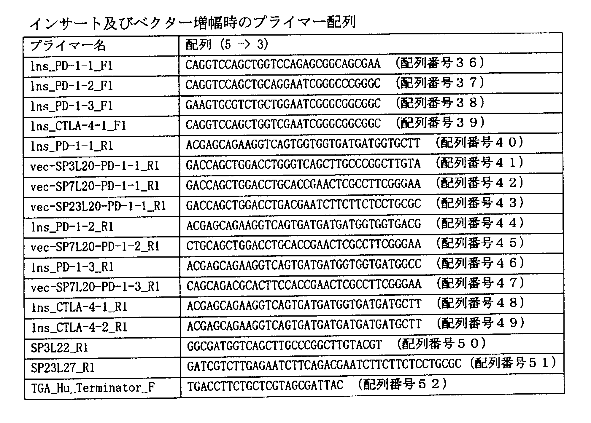
抗PD-1一本鎖抗体の発現カセットは図1(a)に示されるとおり、HuプロモーターDNA(ビフィドバクテリウム・ロンガム由来)、分泌シグナルペプチドをコードするDNAと前記分泌シグナルペプチドに続くリンカーペプチドをコードするDNA、抗PD-1一本鎖抗体(重鎖配列、リンカー((GGGGS)3)、軽鎖配列を含む)をコードするDNA、Hisタグ配列、及びHuターミネーター(ビフィドバクテリウム・ロンガム由来)で構成した。各発現カセット中の抗PD-1一本鎖抗体(重鎖配列、リンカー((GGGGS)3)、軽鎖配列を含む塩基配列を含む)は、以下に示すとおり、以下の表3に示される文献を参照し、二種類の抗ヒトPD-1一本鎖抗体と一種類の抗マウスPD-1一本鎖抗体を使用した。
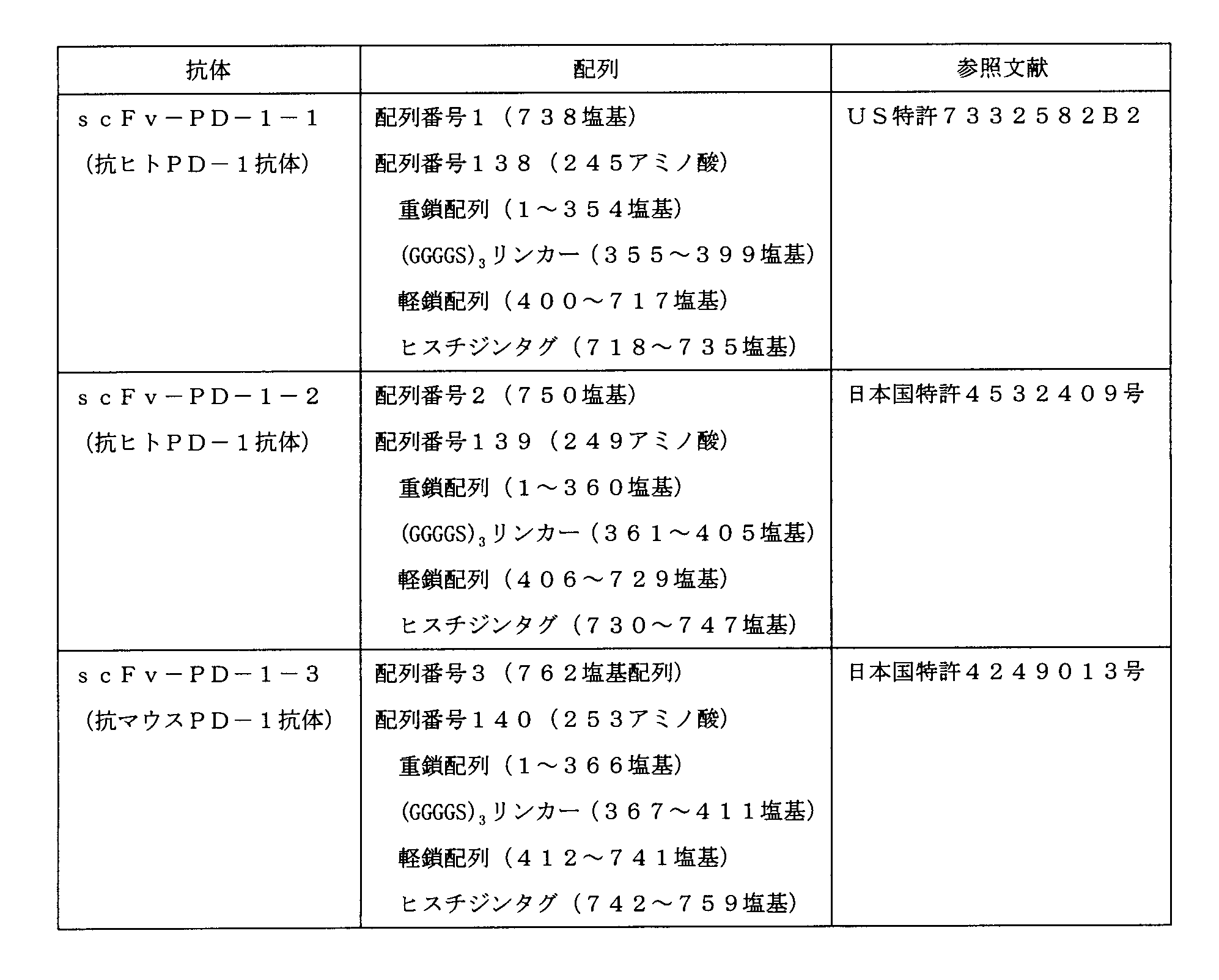
プラスミドpHuSP3L22-scFv-PD-1-1の作製にあたり、まず、プラスミドpHuSP3L20-scFv-PD-1-1を作製した。
(抗PD-1-1一本鎖抗体インサート調製)
プラスミドpUC57-scFv-PD-1-1 500pgを鋳型として、表1及び表2記載のlns_PD-1-1_F1プライマー及びlns_PD-1-1_R1プライマーセットを用いてPCR増幅を行った。プライマー配列は、インサート断片とベクター断片の末端15bpが重複するように設計された。各プライマー0.2μM、反応容量30μLとして、PrimeSTAR HS(Premix) (タカラバイオ株式会社製)キットを用いてPCR増幅を行った。増幅のプログラムとしては、98℃にて10秒、55℃にて5秒、72℃にて60秒を1サイクルとして、30サイクル行った。増幅されたPCR産物を2%アガロースゲルにて電気泳動した後、QIAquick Gel Extraction Kit(キアゲン社製)を用いて精製し、抗PD-1-1一本鎖抗体インサートを調製した。
配列番号20に記載されているベクター直鎖化断片500pgを鋳型として、上記表1及び表2に記載されている、TGA_Hu_Terminator_Fプライマー及びvec-SP3L20-PD-1-1_R1プライマーからなるプライマーセットを用いて、プライマー0.2μM、反応容量30μLとして、PrimeSTAR HS(Premix)キットを用いてPCR増幅を行った。増幅のプログラムとしては、98℃にて10秒、55℃にて5秒、72℃にて5分を1サイクルとして、30サイクル行った。増幅されたPCR産物を0.8%アガロースゲルにて電気泳動した後、QIAquick Gel Extraction Kit(キアゲン社製)を用いて精製し、5’-Huターミネーター-pTB6rep unit-SPCMr-pUCori-Huプロモーター-SP3L20-3’ベクター断片を調製した。
Huターミネーター:配列番号20の第4番から第117番までの塩基配列
ビフィドバクテリウム属細菌複製起点pTB6 rep unit: 配列番号20の第124番から第1719番までの塩基配列
スペクチノマイシン耐性遺伝子SPCMr:配列番号20の第1726番から第2804番までの塩基配列
大腸菌複製起点、pUCori: 配列番号20の第2811番から第3478番までの塩基配列
Huプロモーター: 配列番号20の第3485番から第3845番までの塩基配列。
上記で調製されたベクター断片と上記抗PD-1-1一本鎖抗体インサートとをIn-Fusion(登録商標) HD Cloning Kit(タカラバイオ株式会社製)を用いて連結した。即ち、マイクロチューブに、上記ベクターとインサートを1:5のモル比にて添加後、2μLの5×In-Fusion HD Enzyme premixを添加し、反応液容量を10μLに調整した。これを50℃にて15分間保温した。その他の手順は、キットの製品説明書にしたがい、インフュージョン反応液を調製した。
上記インフュージョン反応液5μLを用いて、大腸菌HST08コンピテント細胞(タカラバイオ株式会社製)の形質転換を製品説明書にしたがい行った。形質転換後の菌懸濁液は、75μg/mLスペクチノマイシン含有LB寒天培地に塗布し、37℃にて一晩静置培養した。上記寒天培地上に形成された大腸菌コロニーを75μg/mLスペクチノマイシン含有LB液体培地にて30℃にて一晩培養し、これよりQIAprep Spin Miniprep Kit(キアゲン社製)を用いてプラスミドを抽出した。抽出したプラスミドにおける抗PD-1-1一本鎖抗体発現カセット(HuプロモーターからHuターミネーターまでを含む)配列を決定するため、BigDye(登録商標) Terminator v3.1 Cycle Sequencing Kit (アプライドバイオシステムズ社製)を用いてシーケンス反応を行った。抽出したプラスミドをpHuSP3L20-scFv-PD-1-1と名付けた。
(リン酸化プライマーの調製)
表1及び表2記載のIns_PD-1-1_F1プライマー、SP3L22_R1プライマーを、T4ポリヌクレオチドキナーゼ(タカラバイオ株式会社製)を用いて、製品説明書にしたがい、各プライマーをリン酸化した。Ins_PD-1-1_F1リン酸化プライマー及びSP3L22_R1リン酸化プライマーを、それぞれ0.1×TEバッファー(1mM Tris-HCl、0.1mM EDTA、pH7.5)で終濃度20μMに調整した。
上記プラスミドpHuSP3L20-scFv-PD-1-1 500pgを鋳型として、上記Ins_PD-1-1_F1リン酸化プライマー及びSP3L22_R1リン酸化プライマーからなるプライマーセットを用いて、最終濃度0.2μM、反応容量30μLにて、PrimeSTAR HS(Premix)キットを用いてPCR増幅を行った。増幅のプログラムとしては、98℃にて10秒、55℃にて5秒、72℃にて5分を1サイクルとして、30サイクル行った。増幅されたPCR産物を0.8%アガロースゲルにて電気泳動した後、QIAquick Gel Extraction Kitを用いて精製し、pHuSP3L22-scFv-PD-1-1ベクター直鎖化断片を調製した。
上記ベクター直鎖化断片について、T4 DNA Ligase(サーモフィッシャーサイエンティフィク株式会社製)を用いて、製品説明書にしたがって、ライゲーション反応を行った。添付の10×バッファー for T4 DNAリガーゼ 2μLを添加して反応液容量を20μLとし、これを22℃にて10分間ライゲーション反応を行い、ライゲーション反応液を調製した。
上記ライゲーション反応液10μLを用いて、大腸菌HST08コンピテント細胞の形質転換を行った。形質転換後の菌懸濁液は、75μg/mLスペクチノマイシン含有LB寒天培地に塗布し、37℃にて一晩静置培養した。
形質転換条件は、大腸菌HST08コンピテント細胞についての製品説明書にしたがったが、以下に概要を記載する。上記寒天培地上に形成した大腸菌コロニーを、75μg/mLスペクチノマイシン含有LB液体培地にて30℃にて一晩培養し、これよりQIAprep Spin Miniprep Kitを用いてプラスミドを抽出した。抽出したプラスミドの抗PD-1-1一本鎖抗体発現カセット(HuプロモーターからHuターミネーターまでを含む)配列を決定するため、BigDye(登録商標) Terminator v3.1 Cycle Sequencing Kitを用いてシーケンス反応を行った。抽出したプラスミドをpHuSP3L22-scFv-PD-1-1と名付けた。
上記形質転換した大腸菌より抽出した1000ngのプラスミドpHuSP3L22-scFv-PD-1-1を用いて、ビフィドバクテリウム・ロンガム105-A株の形質転換をエレクトロポレーションシステム(ジーンパルサーII、バイオ・ラッドラボラトリーズ株式会社製)により行なった。電気ショック(2kV、25μF、200Ω)後は、キュベット(2mmギャップ)に800μLのIMR液体培地と50μLのビタミンC添加液との混合液を即時に添加し、これを滅菌済みの2mLマイクロチューブに回収した。各チューブについて同様の操作を行い、これら2mLチューブの蓋をゆるめて、脱酸素・炭酸ガス発生剤(アネロパック(登録商標)・ケンキ、三菱ガス化学株式会社製)とともに密閉容器に入れ、37℃に設定したインキュベータにて3時間保温した。
(抗PD-1-1一本鎖抗体インサート調製)
上記[プラスミドpHuSP3L20-scFv-PD-1-1の作製]における(抗PD-1-1一本鎖抗体インサート調製)と同様の手順で抗PD-1-1一本鎖抗体インサートを調製した。
配列番号21に記載されているベクター直鎖化断片500pgを鋳型として、上記表1及び表2に記載されている、TGA_Hu_Terminator_Fプライマー及びvec-SP7L20-PD-1-1_R1プライマーからなるプライマーセットを用いて、最終濃度0.2μM、反応容量30μLとして、PrimeSTAR HS(Premix)キットを用いてPCR増幅を行った。増幅のプログラムとしては、98℃にて10秒、55℃にて5秒、72℃にて5分を1サイクルとして、30サイクル行った。増幅されたPCR産物を0.8%アガロースゲルにて電気泳動した後、QIAquick Gel Extraction Kitを用いて精製し、5’-Huターミネーター-pTB6rep unit-SPCMr-pUCori-Huプロモーター-SP7L20-3’ベクター断片(1)を調製した。
Huターミネーター:配列番号21の第4番から第117番までの塩基配列
ビフィドバクテリウム属細菌複製起点pTB6 rep unit: 配列番号21の第124番から第1719番までの塩基配列
スペクチノマイシン耐性遺伝子SPCMr:配列番号21の第1726番から第2804番までの塩基配列
大腸菌複製起点、pUCori: 配列番号21の第2811番から第3478番までの塩基配列
Huプロモーター: 配列番号21の第3485番から第3845番までの塩基配列。
上記で調製されたベクター断片と上記抗PD-1-1一本鎖抗体インサートとを用いたことの他は、上記[プラスミドpHuSP3L20-scFv-PD-1-1の作製]におけるインフュージョン反応と同様の手順でインフュージョン反応液を調製した。
上記で調製されたインフュージョン反応液5μLを用いたことの他は、上記[プラスミドpHuSP3L20-scFv-PD-1-1の作製]の(大腸菌の形質転換及びプラスミドDNAの配列確認)におけると同様の手順にて、大腸菌HST08コンピテント細胞の形質転換を行い、抽出したプラスミドにおける抗PD-1-1一本鎖抗体発現カセット(HuプロモーターからHuターミネーターまでを含む)配列を決定するためシーケンス反応を行った。抽出したプラスミドをpHuSP7L20-scFv-PD-1-1と名付けた。
上記形質転換した大腸菌より抽出したプラスミドpHuSP7L20-scFv-PD-1-1 1000ngのDNAを用いたことの他は、上記[pHuSP3L22-scFv-PD-1-1の作製]における(ビフィドバクテリウム属細菌の形質転換)と同様の方法で、ビフィドバクテリウム・ロンガム105-A株の形質転換を行い、ビフィドバクテリウム・ロンガム105-A/pHuSP7L20-scFv-PD-1-1株を取得した。
プラスミドpHuSP23L27-scFv-PD-1-1の作製にあたり、まず、プラスミドpHuSP23L20-scFv-PD-1-1を作製した。
(抗PD-1-1一本鎖抗体インサート調製)
上記[プラスミドpHuSP3L20-scFv-PD-1-1の作製]における(抗PD-1-1一本鎖抗体インサート調製)と同様の手順で抗PD-1-1一本鎖抗体インサートを調製した。
配列番号22に記載されているベクター直鎖化断片500pgを鋳型として、上記表1及び表2に記載されている、TGA_Hu_Terminator_Fプライマー及びvec-SP23L20-PD-1-1_R1プライマーからなるプライマーセットを用いて、最終濃度0.2μM、反応容量30μLとして、PrimeSTAR HS(Premix)キットを用いてPCR増幅を行った。増幅のプログラムとしては、98℃にて10秒、55℃にて5秒、72℃にて5分を1サイクルとして、30サイクル行った。増幅されたPCR産物を0.8%アガロースゲルにて電気泳動した後、QIAquick Gel Extraction Kitを用いて精製し、5’-Huターミネーター-pTB6rep unit-SPCMr-pUCori-Huプロモーター-SP23L20-3’ベクター断片を調製した。
Huターミネーター:配列番号22の第4番から第117番までの塩基配列
ビフィドバクテリウム属細菌複製起点pTB6 rep unit: 配列番号22の第124番から第1719番までの塩基配列
スペクチノマイシン耐性遺伝子SPCMr:配列番号22の第1726番から第2804番までの塩基配列
大腸菌複製起点、pUCori: 配列番号22の第2811番から第3478番までの塩基配列
Huプロモーター: 配列番号22の第3485番から第3845番までの塩基配列。
上記で調製したベクター断片と抗PD-1-1一本鎖抗体インサートとを用いたことの他は、上記[プラスミドpHuSP3L20-scFv-PD-1-1の作製]におけるインフュージョン反応と同様の手順でインフュージョン反応液を調製した。
上記で調製されたインフュージョン反応液5μLを用いたことの他は、上記[プラスミドpHuSP3L20-scFv-PD-1-1の作製]の(大腸菌の形質転換及びプラスミドDNAの配列確認)におけると同様の手順にて、大腸菌HST08コンピテント細胞の形質転換を行い、抽出したプラスミドにおける抗PD-1-1一本鎖抗体発現カセット(HuプロモーターからHuターミネーターまでを含む)配列を決定するためシーケンス反応を行い、抽出したプラスミドをpHuSP23L20-scFv-PD-1-1と名付けた。
(リン酸化プライマーの調製)
表1及び表2記載のIns_PD-1-1_F1プライマー、SP23L27_R1プライマーを、T4ポリヌクレオチドキナーゼを用いて、製品説明書にしたがい、各プライマーをリン酸化した。Ins_PD-1-1_F1リン酸化プライマー及びSP23L27_R1リン酸化プライマーを、それぞれ0.1×TEバッファー(1mM Tris-HCl、0.1mM EDTA、pH7.5)で終濃度20μMに調整した。
上記プラスミドpHuSP23L20-scFv-PD-1-1 500pgを鋳型として、上記Ins_PD-1-1_F1リン酸化プライマー及びSP23L27_R1リン酸化プライマーからなるプライマーセットを用いて、最終濃度0.2μM、反応容量30μLにて、PrimeSTAR HS(Premix)キットを用いてPCR増幅を行った。増幅のプログラムとしては、98℃にて10秒、55℃にて5秒、72℃にて5分を1サイクルとして、30サイクル行った。増幅されたPCR産物を0.8%アガロースゲルにて電気泳動した後、PCR産物をQIAquick Gel ExtractionKitを用いて精製し、pHuSP23L27-scFv-PD-1-1ベクター直鎖
化断片を調製した。
上記[プラスミドpHuSP3L22-scFv-PD-1-1の作製]における(ライゲーション反応)と同様の手順にて、上記ベクター断片について、ライゲーション反応を行い、ライゲーション反応液を調製した。
上記[プラスミドpHuSP3L22-scFv-PD-1-1の作製]における(大腸菌の形質転換及びプラスミドDNAの配列確認)と同様の手順にて、上記ライゲーション反応液10μLを用いて、大腸菌HST08コンピテント細胞の形質転換を行い、シーケンス反応を行った。抽出したプラスミドをpHuSP23L27-scFv-PD-1-1と名付けた。
上記形質転換した大腸菌より抽出した1000ngのプラスミドpHuSP23L27-scFv-PD-1-1のDNAを用いて、上記[プラスミドpHuSP3L22-scFv-PD-1-1の作製]における(ビフィドバクテリウム属細菌の形質転換)と同様の手順にて、ビフィドバクテリウム・ロンガム105-A株の形質転換を行い、ビフィドバクテリウム・ロンガム105-A/pHuSP23L27-scFv-PD-1-1株を取得した。
(組換えビフィドバクテリウム属細菌における一本鎖抗体の分泌の有無の検討1)
上記実施例1にて作製した三種類の抗PD-1-1一本鎖抗体分泌プラスミドによる3種類の組換えビフィドバクテリウム属細菌(ビフィドバクテリウム・ロンガム105-A/pHuSP3L22-scFv-PD-1-1株、ビフィドバクテリウム・ロンガム105-A/pHuSP7L20-scFv-PD-1-1株、ビフィドバクテリウム・ロンガム105-A/pHuSP23L27-scFv-PD-1-1株)のそれぞれについて、抗PD-1-1一本鎖抗体の分泌確認をウェスタン解析により以下のとおり実施した。
図4からも明らかなとおり、分泌シグナルペプチド配列SP7とこれに続く20アミノ酸配列からなる、新規の分泌シグナルペプチド-リンカー連結体SP7L20が挿入されているpHuSP7L20-scFv-PD-1-1により形質転換されたビフィドバクリウム属細菌においては、抗PD-1-1一本鎖抗体の分泌が認められた。一方、pHuSP3L22-scFv-PD-1-1又はpHuSP23L27-scFv-PD-1-1により形質転換されたビフィドバクリウム属細菌においては、抗PD-1-1一本鎖抗体の分泌は認められなかった。
[33種類の分泌シグナルペプチド-リンカー連結体についての検討]
上記pHuSP7L20-scFv-PD-1-1について、分泌シグナルペプチド-リンカー連結体SP7L20を、33種類の分泌シグナルペプチド-リンカー連結体(SP42L20~SP46L20、SP48L20~SP75L20)にそれぞれ置換した、33種類の抗PD-1-1一本鎖抗体分泌プラスミドを構築した。構築方法の概要を図3に示す。用いたプライマーの詳細は以下の表4及び表5のとおりである。
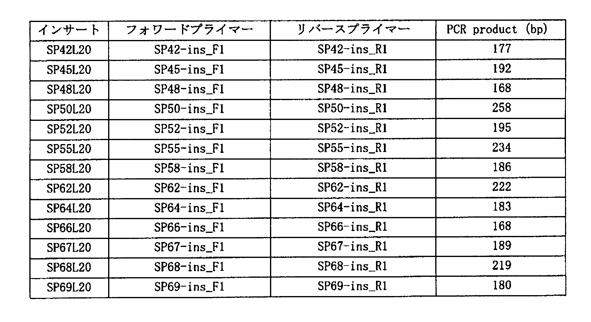
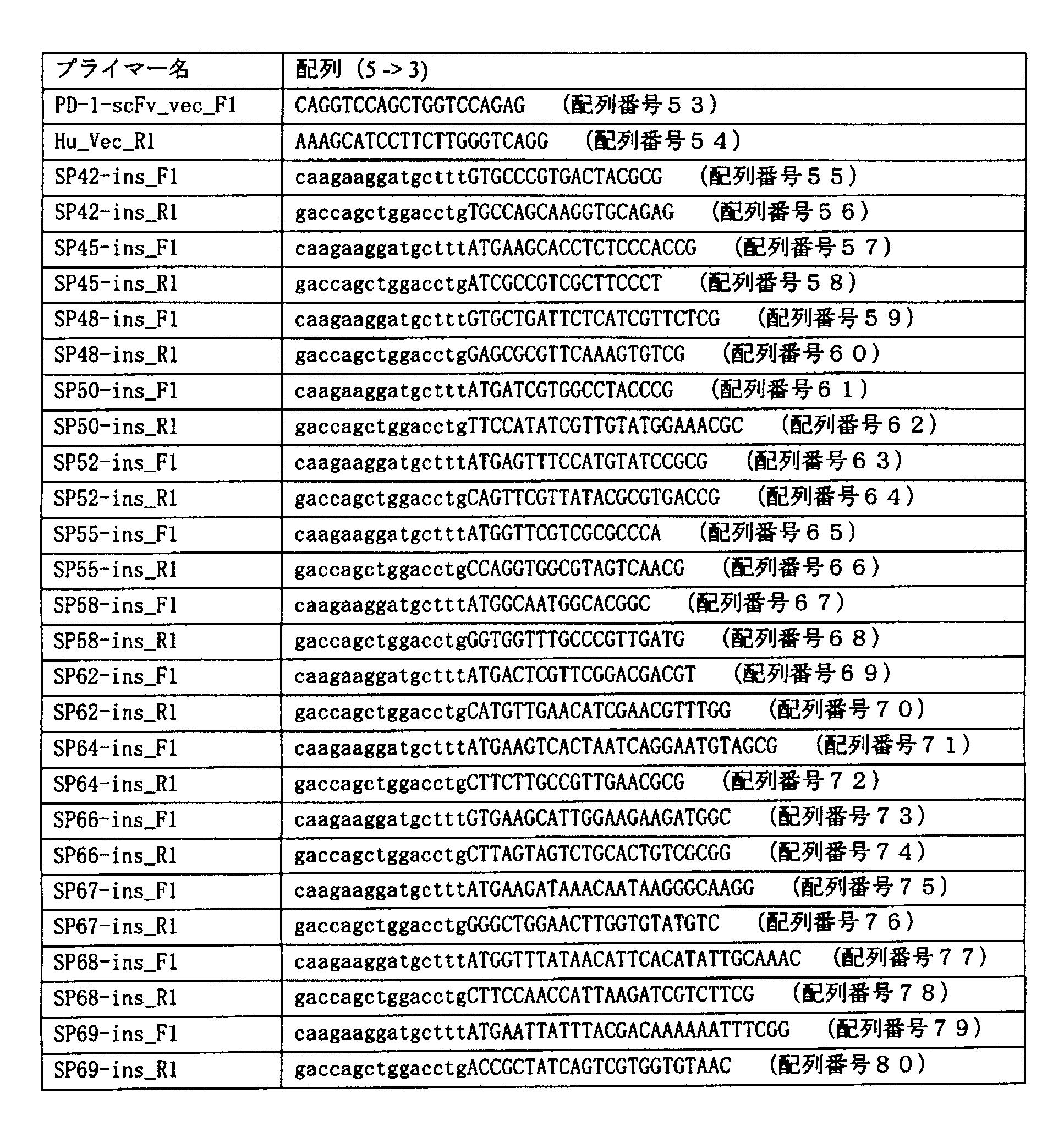
NCBIのゲノムデータベース(http://www.ncbi.nlm.nih.gov/genome/)に登録されているビフィドバクテリウム・ロンガムNCC2705株由来タンパクの全アミノ酸配列を分泌シグナルの有無を予測するシグナル配列予測プログラムSignalP 4.1 server (http://www.cbs.dtu.dk/services/SignalP/)で解析した。この解析により、タンパクのN末端に分泌シグナルペプチド配列を有すると予測されたタンパクの一部のアミノ酸配列に対し、膜貫通領域(TM)を推定するプログラムTMHMM Server v. 2.0 (http://www.cbs.dtu.dk/services/TMHMM-2.0/)にてトポロジー解析を行った。このトポロジー解析結果より、タンパク質のN末端にTMが存在すると推定されたタンパク質として33種のタンパク質を選定した。この33種のタンパク質を、TMを有すると予想されるビフィドバクテリウム・ロンガムNCC2705株由来の分泌シグナルペプチドとして選定した。選定した33種のペプチドのアミノ酸配列に対応するビフィドバクテリウム・ロンガム105A株のアミノ酸配列において、各タンパク質のアミノ酸配列N末端からSignalP解析によってプロテアーゼ切断推定部位と推定されたアミノ酸配列までを各分泌シグナルペプチド配列とした。また、リンカーペプチド配列は、各シグナル配列のC末端より続く20アミノ酸残基とした。
(SPxL20インサート調製)
ビフィドバクテリウム・ロンガム105-A株120pgを鋳型として、表4及び表5に記載されている各プライマーセットを用いてPCR増幅を行った。各プライマー配列は、インサート断片とベクター断片の末端15bpが重複するように設計した。プライマー濃度0.2 μM、反応容量20μLにて、PrimeSTAR HS(Premix)キットを用いてPCR増幅を行った。増幅のプログラムとしては、98℃にて10秒、65℃にて5秒、72℃にて20秒を1サイクルとして、30サイクル行った後、72℃にて30秒伸長反応を行い、上記33種類の分泌シグナルペプチド-リンカー連結体インサートが増幅された。
上記pHuSP7L20-scFv-PD-1-1 2.5ngを鋳型として、上記表4及び表5に記載されているPD-1-scFv_vec_F1プライマー及びHu_Vec_R1プライマーからなるプライマーセットを用いて、最終濃度0.2μM、反応容量50μLにて、PrimeSTAR HS(Premix)キットを用いてPCR増幅を行った。増幅のプログラムとしては、98℃にて10秒、65℃にて5秒、72℃にて4分45秒を1サイクルとして、30サイクル行った後、72℃にて30秒伸長反応を行った。かかる処理により、5’-scFv-PD-1-1-Huターミネーター-pTB6rep unit-SPCMr-pUCori-Huプロモーター-3’のベクター断片が調製された。
上記で調製したベクター断片と33種類の各分泌シグナルペプチド-リンカー連結体インサートをIn-Fusion(登録商標) HD Cloning Kitを用いて連結した。即ち、マイクロチューブに、ベクターとインサートを1:2のモル比にて添加し、キット内の5xIn-Fusion HD Enzyme premix 2μLを添加、0.1×TEバッファー(1mM Tris-HCl、 0.1mM EDTA、 pH7.5)にて10μLに調整した。これを、37℃にて15分間保温した後、さらに50℃にて15分間保温した。その他の手順は、キットの製品説明書にしたがい、33種類のインフュージョン反応液を調製した。
上記33種類のインフュージョン反応液0.8μLを用いて大腸菌HST16CRコンピテント細胞(タカラバイオ株式会社製)の形質転換をそれぞれ行った。形質転換後の菌懸濁液は、75μg/mLスペクチノマイシン含有LB寒天培地に塗布し、37℃にて一晩静置培養した。形質転換条件は、大腸菌HST16CRコンピテント細胞についての製品説明書にしたがったが、以下に概要を記載する。上記寒天培地上に形成した大腸菌コロニーを75μg/mLスペクチノマイシン含有LB液体培地にて37℃にて一晩培養し、これよりQIAprep Spin Miniprep Kitを用いてプラスミドを抽出した。抽出したプラスミドの抗PD-1-1一本鎖抗体発現カセット(HuプロモーターからHuターミネーターまでを含む)配列を決定するため、BigDye(登録商標) Terminator v3.1 Cycle Sequencing Kitを用いてシーケンス反応を行った。抽出したプラスミドをpHuSPxL20-scFv-PD-1-1(但し、x=42~46、48~75)とした。
上記形質転換した大腸菌より抽出したプラスミドpHuSPxL20-scFv-PD-1-1(但し、x=42~46、48~75)について、各々500ngのDNAを用いて、ビフィドバクテリウム・ロンガム105-A株の形質転換を、上記[プラスミドpHuSP3L22-scFv-PD-1-1の作製]における(ビフィドバクテリウム属細菌の形質転換)と同様の方法で行い、ビフィドバクテリウム・ロンガム105-A/pHuSPxL20-scFv-PD-1-1(但し、x=42~46、48~75)のそれぞれの株を取得した。
(組換えビフィドバクテリウム属細菌における一本鎖抗体の分泌の有無の検討2)
上記実施例3で取得した組換えビフィドバクテリウム属細菌である、ビフィドバクテリウム・ロンガム105-A/pHuSPxL20-scFv-PD-1-1株(但し、x=42~46、48~75)のそれぞれについて、上記(組換えビフィドバクテリウム属細菌における一本鎖抗体の分泌の有無の検討1)と同様の方法にて抗PD-1-1一本鎖抗体の分泌確認をウェスタン解析により実施した。結果を図5に示す。
[pHuSP7L20-scFv-PD-1-2の作製]
異種ペプチドとして上記抗PD-1-2一本鎖抗体を用い、分泌シグナルペプチド-リンカー連結体SP7L20を有する抗PD-1-2一本鎖抗体分泌プラスミドpHuSP7L20-scFv-PD-1-2を構築した。詳細は以下のとおりである。
前記人工的に合成されたプラスミドpUC57-scFv-PD-1-2 500pgを鋳型として、上記表1及び表2に記載されている、lns_PD-1-2_F1プライマー及びlns_PD-1-2_R1プライマーからなるプライマーセットを用いてPCR増幅を行った。プライマー配列は、インサート断片とベクター断片の末端15bpが重複するように設計された。PCR反応液は、PrimeSTAR HS(Premix)キットを用いて、プライマー0.2μM、反応容量30μLとした。増幅のプログラムは、98℃にて10秒、55℃にて5秒、72℃にて60秒を1サイクルとして、30サイクル行った。増幅されたPCR産物を2%アガロースゲルにて電気泳動した後、QIAquick Gel Extraction Kitを用いて精製し、抗PD-1-2一本鎖
抗体インサートを調製した。
配列番号23に記載されているベクター直鎖化断片500pgを鋳型として、上記表1及び表2に記載されている、TGA_Hu_Terminator_Fプライマー及びvec-SP7L20-PD-1-2_R1プライマーからなるプライマーセットを用いて、最終濃度0.2μM、反応容量30μLにて、PrimeSTAR HS(Premix)キットを用いてPCR増幅を行った。増幅のプログラムは、98℃で10秒、55℃で5秒、72℃で5分を1サイクルとして、30サイクル行った。増幅されたPCR産物を0.8%アガロースゲルにて電気泳動した後、QIAquick Gel Extraction Kitを用いて精製し、5’-Huターミネーター-pTB6rep unit-SPCMr-pUCori-Huプロモーター-SP7L20-3’ベクター断片(2)を調製した。
Huターミネーター:配列番号23の第4番から第117番までの塩基配列
ビフィドバクテリウム属細菌複製起点pTB6 rep unit: 配列番号23の第124番から第1719番までの塩基配列
スペクチノマイシン耐性遺伝子SPCMr:配列番号23の第1726番から第2804番までの塩基配列
大腸菌複製起点、pUCori: 配列番号23の第2811番から第3478番までの塩基配列
Huプロモーター: 配列番号23の第3485番から第3845番までの塩基配列。
上記で調製されたベクター断片と抗PD-1-2一本鎖抗体インサートとを用いたことの他は、上記[プラスミドpHuSP3L20-scFv-PD-1-1の作製]におけるインフュージョン反応と同様の手順でインフュージョン反応液を調製した。
上記で調製されたインフュージョン反応液5μLを用いた他は、前記実施例1の[プラスミドpHuSP3L20-scFv-PD-1-1の作製]の(大腸菌の形質転換及びプラスミドDNAの配列確認)と同様の手順にて、大腸菌HST08コンピテント細胞の形質転換を行い、抽出したプラスミドにおける抗PD-1-2一本鎖抗体発現カセット(HuプロモーターからHuターミネーターまでを含む)配列を決定するためシーケンス反応を行い、抽出したプラスミドをpHuSP7L20-scFv-PD-1-2と名付けた。
上記形質転換した大腸菌より抽出した1000ngのプラスミドpHuSP7L20-scFv-PD-1-2のDNAを用いて、上記[プラスミドpHuSP3L22-scFv-PD-1-1の作製]における(ビフィドバクテリウム属細菌の形質転換)と同様の手順にて、ビフィドバクテリウム・ロンガム105-A株の形質転換を行い、ビフィドバクテリウム・ロンガム105-A/pHuSP7L20-scFv-PD-1-2株を取得した。
[pHuSP7L20-scFv-PD-1-3の作製]
異種ペプチドとして抗PD-1-3一本鎖抗体を選択し、分泌シグナルペプチド-リンカー連結体(SP7L20)を有する抗PD-1-3一本鎖抗体分泌プラスミドpHuSP7L20-scFv-PD-1-3を構築した。詳細は以下のとおりである。
前記人工的に合成されたプラスミドpUC57-scFv-PD-1-3 500pgを鋳型として、上記表1及び表2に記載されている、lns_PD-1-3_F1プライマー及びlns_PD-1-3_R1プライマーからなるプライマーセットを用いたことの他は、上記[pHuSP7L20-scFv-PD-1-2の作製]の(抗PD-1-2一本鎖抗体インサート調製)と同様の手順にて、抗PD-1-3一本鎖抗体インサートを調製した。
配列番号24に記載されているベクター直鎖化断片500pgを鋳型として、上記表1及び表2に記載されている、TGA_Hu_Terminator_Fプライマー及びvec-SP7L20-PD-1-3_R1プライマーからなるプライマーセットを用いたことの他は、pHuSP7L20-scFv-PD-1-2の作製]の(ベクター断片調製)と同様の手順で、5’-Huターミネーター-pTB6rep unit-SPCMr-pUCori-Huプロモーター-SP7L20-3’ベクター断片(3)を調製した。
Huターミネーター:配列番号24の第4番から第117番までの塩基配列
ビフィドバクテリウム属細菌複製起点pTB6 rep unit: 配列番号24の第124番から第1719番までの塩基配列
スペクチノマイシン耐性遺伝子SPCMr:配列番号24の第1726番から第2804番までの塩基配列
大腸菌複製起点、pUCori: 配列番号24の第2811番から第3478番までの塩基配列
Huプロモーター: 配列番号24の第3485番から第3845番までの塩基配列。
上記で調製したベクター断片と抗PD-1-3一本鎖抗体インサートとを用いたことの他は、上記[プラスミドpHuSP3L20-scFv-PD-1-1の作製]におけるインフュージョン反応と同様の手順でインフュージョン反応液を調製した。
上記で調製されたインフュージョン反応液5μLを用いた他は、前記実施例1の[プラスミドpHuSP3L20-scFv-PD-1-1の作製]の(大腸菌の形質転換及びプラスミドDNAの配列確認)におけると同様の手順にて、大腸菌HST08コンピテント細胞の形質転換を行い、抽出したプラスミドにおける抗PD-1-3一本鎖抗体発現カセット(HuプロモーターからHuターミネーターまでを含む)配列を決定するためシーケンス反応を行い、抽出したプラスミドをpHuSP7L20-scFv-PD-1-3と名付けた。
上記形質転換した大腸菌より抽出した1000ngのプラスミドpHuSP7L20-scFv-PD-1-3のDNAを用いて、上記[プラスミドpHuSP3L22-scFv-PD-1-1の作製]における(ビフィドバクテリウム属細菌の形質転換)と同様の手順にて、ビフィドバクテリウム・ロンガム105-A株の形質転換を行い、ビフィドバクテリウム・ロンガム105-A/pHuSP7L20-scFv-PD-1-3株を取得した。
[pHuSP7L20-scFv-CTLA-4-1の作製]
分泌シグナルペプチド-リンカー連結体SP7L20を有する抗CTLA-4-1一本鎖抗体分泌プラスミドpHuSP7L20-scFv-CTLA-4-1を構築した。
抗CTLA-4一本鎖抗体分泌プラスミドの作製概要を図2に示す。図2左側において、scFv-geneは、CTLA-4であり、pHuSP7L20-scFv-CTLA-4-1の作製において、x=7、m=1であり、pHuSP7L20-scFv-CTLA-4-2の作製において、x=7、m=2である。
詳細は以下のとおりである。
抗CTLA-4一本鎖抗体の発現カセットは図1(b)に示されるとおり、HuプロモーターDNA(ビフィドバクテリウム・ロンガム由来)、分泌シグナルペプチドをコードするDNAと前記分泌シグナルペプチドに続くリンカーペプチドをコードするDNA、抗CTLA-4一本鎖抗体(重鎖配列、リンカー((GGGGS)3)、軽鎖配列を含む)をコードするDNA、Hisタグ配列、及びHuターミネーター(ビフィドバクテリウム・ロンガム由来)で構成した。各発現カセット中の抗CTLA-4一本鎖抗体(重鎖配列、リンカー((GGGGS)3)、軽鎖配列を含む塩基配列を含む)は、以下に示すとおり、以下の表6に示される文献を参照し、二種類の抗ヒトCTLA-4一本鎖抗体を使用した。
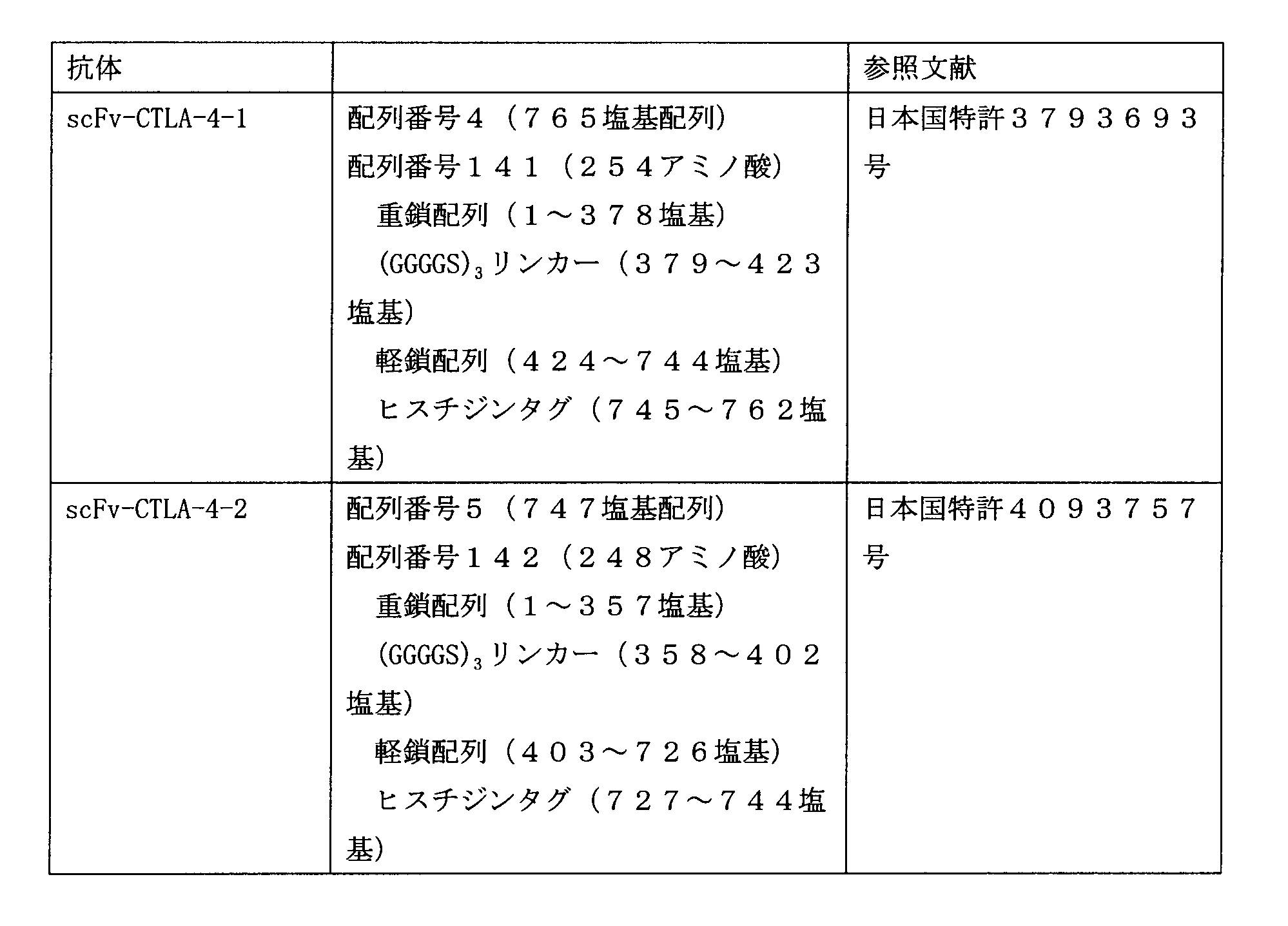
プラスミドpUC57-scFv-CTLA-4-1 500pgを鋳型として、上記表1及び表2に記載されている、lns_CTLA-4-1_F1プライマー及びlns_CTLA-4-1_R1プライマーからなるプライマーセットを用いてPCR増幅を行った。プライマー配列は、インサート断片とベクター断片の末端15bpが重複するように設計した。PCR反応液は、プライマー0.2 μM、反応容量30μLとし、PrimeSTAR HS (Premix) (タカラバイオ株式会社)キットを用いて増幅をおこなった。増幅のプログラムは、98℃で10秒、55℃で5秒、72℃で60秒を1サイクルとして、30サイクル行った。増幅されたインサートPCR産物を2%アガロースゲルにて電気泳動した後、QIAquick Gel Extraction Kitを用いて精製し、抗CTLA-4-1一本鎖抗体インサートを調製した。
前記[pHuSP7L20-scFv-PD-1-1の作製]の(ベクター断片調製)と同様の手順にて、5’-Huターミネーター-pTB6rep unit-SPCMr-pUCori-Huプロモーター-SP7L20-3’ベクター断片(1)を調製した。
上記で調製されたベクター断片と上記抗CTLA-4-1一本鎖抗体インサートとを用いたことの他は、上記[プラスミドpHuSP3L20-scFv-PD-1-1の作製]におけるインフュージョン反応と同様の手順でインフュージョン反応液を調製した。
上記で調製されたインフュージョン反応液5μLを用いたことの他は、上記[プラスミドpHuSP3L20-scFv-PD-1-1の作製]の(大腸菌の形質転換及びプラスミドDNAの配列確認)と同様の手順にて、大腸菌HST08コンピテント細胞の形質転換を行い、抽出したプラスミドにおける抗CTLA-4-1一本鎖抗体発現カセット(HuプロモーターからHuターミネーターまでを含む)配列を決定するためシーケンス反応を行い、抽出したプラスミドをpHuSP7L20-scFv-CTLA-4-1と名付けた。
上記形質転換した大腸菌より抽出したプラスミドpHuSP7L20-scFv-CTLA-4-1 1000ngのDNAを用いたことの他は、上記[pHuSP3L22-scFv-PD-1-1の作製]における(ビフィドバクテリウム属細菌の形質転換)と同様の方法で、ビフィドバクテリウム・ロンガム105-A株の形質転換を行い、ビフィドバクテリウム・ロンガム105-A/pHuSP7L20-scFv-CTLA-4-1株を取得した。
[pHuSP7L20-scFv-CTLA-4-2の作製]
分泌シグナルペプチド-リンカー連結体(SP7L20)を有する抗CTLA-4-2一本鎖抗体分泌プラスミドpHuSP7L20-scFv-CTLA-4-2を構築した。詳細は以下のとおりである。
上記プラスミドpUC57-scFv-CTLA-4-2を鋳型として、上記表1及び表2に記載されている、lns_CTLA-4-1_F1プライマー及びlns_CTLA-4-2_R1プライマーセットからなるプライマーセットを用いたことの他は、上記(抗CTLA-4-1一本鎖抗体インサート調製)と同様に手順にて、抗CTLA-4-2一本鎖抗体インサートを精製した。
前記[pHuSP7L20-scFv-PD-1-1の作製]の(ベクター断片調製)と同様の手順にて5’-Huターミネーター-pTB6rep unit-SPCMr-pUCori-Huプロモーター-SP7L20-3’ベクター断片(1)を調製した。
上記で調製されたベクター断片と上記抗CTLA-4-2一本鎖抗体インサートとを用いたことの他は、上記[プラスミドpHuSP3L20-scFv-PD-1-1の作製]におけるインフュージョン反応と同様の手順でインフュージョン反応液を調製した。
上記で調製されたインフュージョン反応液5μLを用いた他は、前記実施例1の[プラスミドpHuSP3L20-scFv-PD-1-1の作製]の(大腸菌の形質転換及びプラスミドDNAの配列確認)と同様の手順にて、大腸菌HST08コンピテント細胞の形質転換を行い、抽出したプラスミドにおける抗CTLA-4-2一本鎖抗体発現カセット(HuプロモーターからHuターミネーターまでを含む)配列を決定するためシーケンス反応を行い、抽出したプラスミドをpHuSP7L20-scFv-CTLA-4-2と名付けた。
上記形質転換した大腸菌より抽出した1000ngのプラスミドpHuSP7L20-scFv-CTLA-4-2を用いたことの他は、上記[pHuSP3L22-scFv-PD-1-1の作製]における(ビフィドバクテリウム属細菌の形質転換)と同様の方法で、ビフィドバクテリウム・ロンガム105-A株の形質転換を行い、ビフィドバクテリウム・ロンガム105-A/pHuSP7L20-scFv-CTLA-4-2株を取得した。
(組換えビフィドバクテリウム属細菌における一本鎖抗体の分泌の有無の検討3)
抗PD-1一本鎖抗体分泌プラスミド及び抗CTLA-4一本鎖抗体分泌プラスミドによる組換えビフィドバクテリウム属細菌である、ビフィドバクテリウム・ロンガム105-A/pHuSP7L20-scFv-PD-1-1株、ビフィドバクテリウム・ロンガム 105-A/pHuSP7L20-scFv-PD-1-2株、ビフィドバクテリウム・ロンガム 105-A/pHuSP7L20-scFv-PD-1-3株、ビフィドバクテリウム・ロンガム105-A/pHuSP7L20-scFv-CTLA-4-1株及びビフィドバクテリウム・ロンガム105-A/pHuSP7L20-scFv-CTLA-4-2株について、上記(組換えビフィドバクテリウム属細菌における一本鎖抗体の分泌の有無の検討1)と同様の方法にて抗PD-1一本鎖抗体及び抗CTLA-4一本鎖抗体の分泌確認をウェスタン解析により実施した。但し、培養時間は15時間とした。結果を図6に示す。
図6から明らかなとおり、ビフィドバクテリウム・ロンガム105-A/pHuSP7L20-scFv-PD-1-1株の培養上清で抗PD-1-1一本鎖抗体の分泌が認められ、ビフィドバクテリウム・ロンガム105-A/pHuSP7L20-scFv-PD-1-2株の培養上清で抗PD-1-2一本鎖抗体の分泌が認められ、ビフィドバクテリウム・ロンガム 105-A/pHuSP7L20-scFv-PD-1-3株の培養上清で抗PD-1-3一本鎖抗体の分泌が確認され、ビフィドバクテリウム・ロンガム 105-A/pHuSP7L20-scFv-CTLA-4-1株の培養上清で抗CTLA-4-1一本鎖抗体の分泌が認められ、ビフィドバクテリウム・ロンガム 105-A/pHuSP7L20-scFv-CTLA-4-2株の培養上清で抗CTLA-4-2一本鎖抗体の分泌が確認された。
[組換えビフィドバクテリウム属細菌株からの抗PD-1及び抗CTLA-4一本鎖抗体の精製]
ビフィドバクテリウム・ロンガム105-A/pHuSP7L20-scFv-PD-1-1株、ビフィドバクテリウム・ロンガム105-A/pHuSP7L20-scFv-PD-1-2株、ビフィドバクテリウム・ロンガム105-A/pHuSP7L20-scFv-PD-1-3株、ビフィドバクテリウム・ロンガム105-A/pHuSP7L20-scFv-CTLA-4-1株及びビフィドバクテリウム・ロンガム 105-A/pHuSP7L20-scFv-CTLA-4-2株について、一本鎖抗体の精製を以下に示す方法で行った。
上記嫌気培養後の培養液を遠心分離して得た培養上清を撹拌しながら、硫酸アンモニウムを80%飽和になるよう少しずつ添加し、4℃にて一晩撹拌し塩析を行った。これを遠心分離後、沈殿を回収し、ヒスチジンタグ付きタンパク質の精製キット(TALON resin、タカラバイオ株式会社製)にてヒスチジンタグ融合タンパクを精製した。この精製溶液を限外ろ過(アミコンウルトラ-0.5、メルクミリポア社製)にて濃縮した。精製タンパク質の濃度は、Bradford法にて測定した(Coomassie Plus Protein Assay、Thermo Scientific社製)。上記精製一本鎖抗体の一部を用いてSDS-PAGE後、クーマシーブリリアントブルー染色(SimplyBlue(登録商標) Safe Stain、ライフテクノロジーズ社製)を行い、約9割の純度でscFv-PD-1-1、scFv-PD-1-2、scFv-PD-1-3、scFv-CTLA4-1、及びscFv-CTLA4-2の各一本鎖抗体が精製されていることを確認した。
[ヒトPD-1と、抗PD-1一本鎖抗体又は抗CTLA-4一本鎖抗体との結合の有無の確認]
ビフィドバクテリウム・ロンガム105-A/pHuSP7L20-scFv-PD-1-2株の培養上清より精製した抗PD-1-2一本鎖抗体についてヒトPD-1(hPD-1)との結合の有無の確認を、ELISA法により行った。
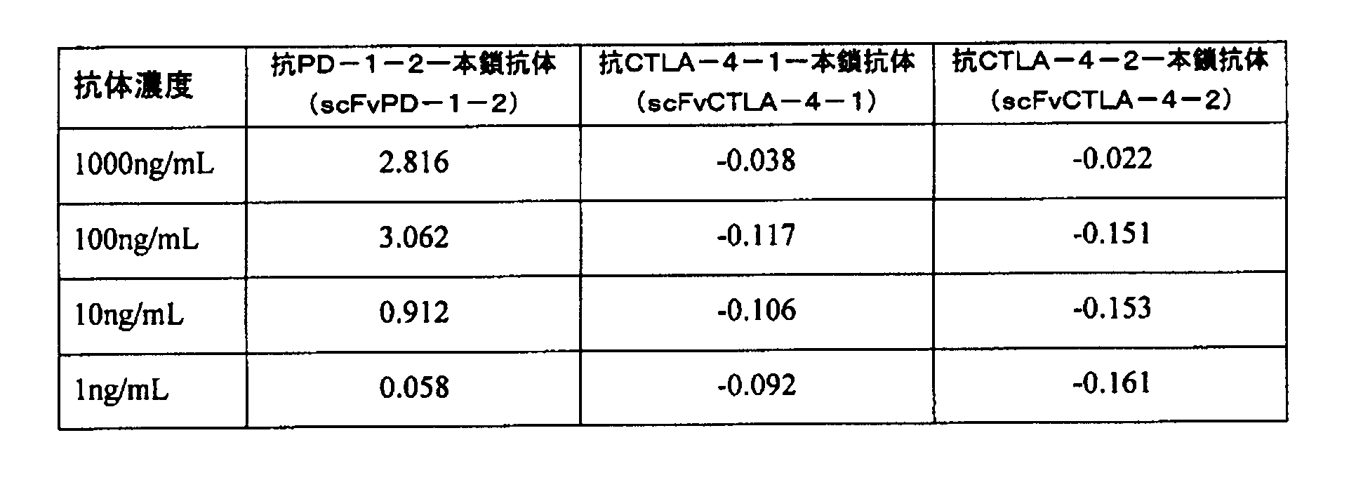
表7及び図7から明らかなとおり、ビフィドバクテリウム属細菌より精製された抗PD-1-2一本鎖抗体は、濃度依存的にhPD-1と結合した。一方、ビフィドバクテリウム属細菌より同様に精製された抗CTLA-4-1一本鎖抗体及び抗CTLA-4-2一本鎖抗体は、hPD-1に結合しなかった。かかる結果により、ビフィドバクテリウム属細菌が分泌した抗PD-1-2一本鎖抗体(抗ヒトPD-1抗体)は、hPD-1に特異的に結合していることが確認された。
[ヒトCTLA-4と、抗PD-1一本鎖抗体又は抗CTLA-4一本鎖抗体との結合の有無の確認]
上記培養上清より精製した抗PD-1-2一本鎖抗体と、抗CTLA-4-1一本鎖抗体及び抗CTLA-4-2一本鎖抗体とを用いて、ヒトCTLA-4(hCTLA-4)(Recombinant Human CTLA-4-Fc Chimera、carrier-free BioLegend社製)との結合の有無の確認を上記[ヒトPD-1と、抗PD-1一本鎖抗体又は抗CTLA-4一本鎖抗体との結合の有無の確認]と同様の手順で行った。ヒトCTLA-4固相化プレートへ抗PD-1-2一本鎖抗体と、抗CTLA-4-1一本鎖抗体及び抗CTLA-4-2一本鎖抗体の結合の有無の結果を以下の表8及び図8に示す。
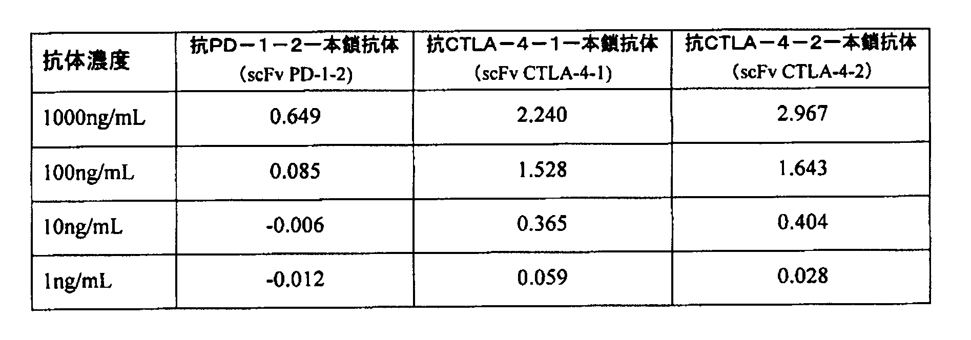
表8及び図8から明らかなとおり、抗CTLA-4-1一本鎖抗体及び抗CTLA-4-2一本鎖抗体は、濃度依存的にhCTLA-4と結合した。一方、ビフィドバクテリウム属細菌より同様に精製された抗PD-1-2一本鎖抗体は、hCTLA-4に結合しなかった。かかる結果により、ビフィドバクテリウム属細菌が分泌した抗CTLA-4-1一本鎖抗体及び抗CTLA-4-2一本鎖抗体(共に抗ヒトCTLA-4抗体)は、hCTLA-4に特異的に結合していることが確認された。
(マウスPD-1と抗PD-1-3一本鎖抗体の結合の有無の確認1)
上記ビフィドバクテリウム・ロンガム105-A/pHuSP7L20-scFv-PD-1-3株の培養上清より精製した抗PD-1-3一本鎖抗体とマウスPD-1(mPD-1)との結合の有無の確認を以下のELISA法にて行った。
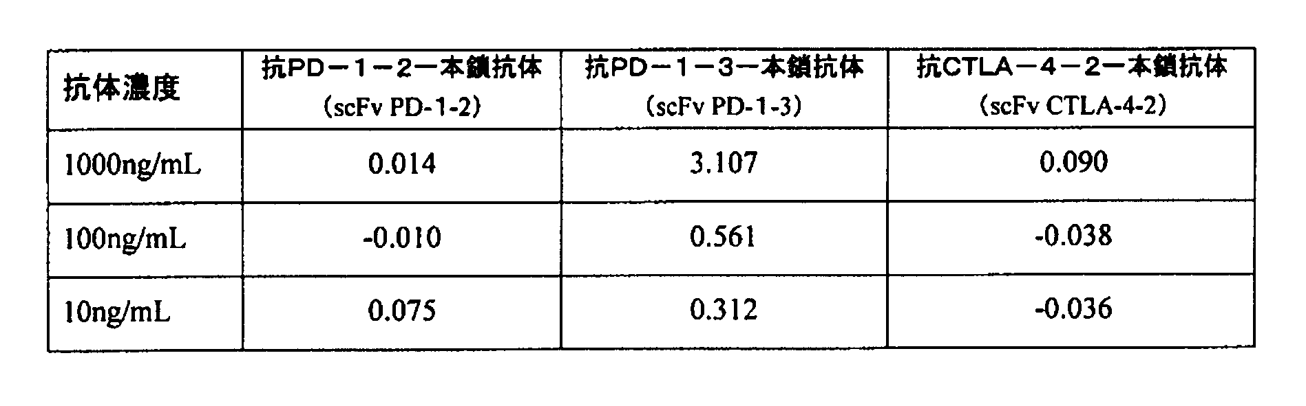
抗PD-1-3一本鎖抗体は、濃度依存的にmPD-1と結合した。一方、同様に精製した抗PD-1-2一本鎖抗体及び抗CTLA-4-2一本鎖抗体は、mPD-1に結合しなかった。これよりビフィドバクテリウム属細菌が分泌した抗PD-1-3一本鎖抗体(抗マウスPD-1抗体)は、mPD-1に特異的に結合していることが示された。
(マウスPD-1と抗PD-1-3一本鎖抗体との結合の有無の確認2)
ビフィドバクテリウム・ロンガム105-A/pHuSP7L20-scFv-PD-1-3株の培養上清より精製した抗PD-1-3一本鎖抗体について、マウスPD-1(mPD-1)への結合の特異性を確認した。プレートに固相化するタンパク質をhCTLA-4 (Recombinant Human CTLA-4-Fc Chimera carrier-free、BioLegend社製)として、上記[実施例13]と同様の試験を実施した。結果を表10と図10に示す。
表10と図10からも明らかなとおり、陽性コントロールである抗CTLA-4-2一本鎖抗体は、濃度依存的にhCTLA-4と結合しているが、抗PD-1-3一本鎖抗体は、結合しなかった。また、抗ヒトPD-1抗体である抗PD-1-2一本鎖抗体も、結合しなかった。これらの結果からも上記[実施例13]でのmPD-1と抗PD-1-3一本鎖抗体の結合は特異的であることが示された。
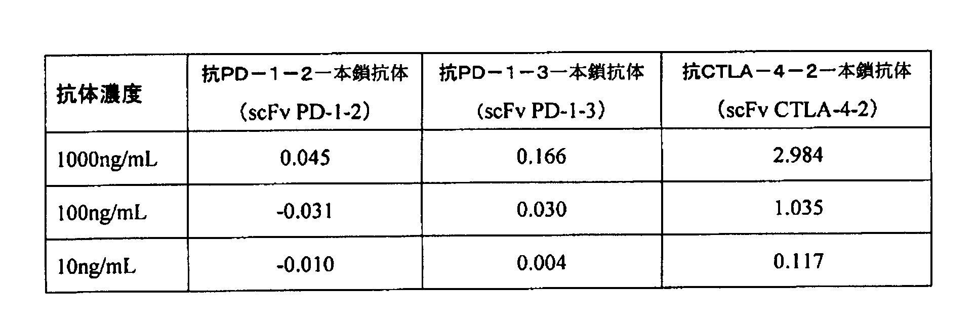
(抗PD-1-2一本鎖抗体とヒトPD-1とのフローサイトメトリー解析による結合の有無の確認)
抗PD-1-2一本鎖抗体のヒトPD-1過剰発現細胞との結合
ヒトPD-1を過剰発現したJurkat細胞(信州大学分子腫瘍学講座谷口俊一郎教授より供与)、及びヒトPD-1を過剰発現したHEK293T細胞(信州大学分子腫瘍学講座谷口俊一郎教授より供与)を使用して検討した。ヒトPD-1過剰発現Jurkat細胞は100mmペトリディッシュ(イナ・オプティカ社)に5×10⁶cells/10mL培地(非働化した10%ウシ胎児血清、50μMの2-メルカプトエタノール及び8mM HEPESを含むRPMI1640培地)/dishに播き、ヒトPD-1過剰発現HEK293T細胞は100mmディッシュ(グライナー・ジャパン社製)に2×106cells/10mL培地(非働化した10%ウシ胎児血清を含むDMEM培地)に播いた。翌日、ヒトPD-1過剰発現Jurkat細胞は、15mLファルコンチューブ(ベクトン・ディッキンソン社製)に回収後、細胞数を血球計算盤にて計測し、1×105cells/mL/チューブになるよう1.5mLチューブ(イナ・オプティカ社製)に分注した。ヒトPD-1過剰発現HEK293T細胞は、培養上清を除去し、PBS(Ca2+、Mg2+フリー・リン酸緩衝液)にて2度洗浄し、PBSで10倍に希釈したトリプシン・EDTA溶液(Wako社製)1mLを加え1分間室温で静置後、10mLの非働化した10%ウシ胎児血清を含むDMEM培地を加え、15mLファルコンチューブに移した。低速遠心機(トミー精工社製)で1000rpm、5分間遠心後、上清を除去し1mLの非働化した10%ウシ胎児血清を含むDMEM培地を加え、細胞数を計測した。さらにDMEM培地を添加し、1×105cells/mLに細胞懸濁液を調製し、1.5mLチューブ(イナ・オプティカ社)に1×105cells/mL/tubeになるように分注した。
図11から明らかなとおり、PD-1を過剰発現するJurkat細胞に抗PD-1-2一本鎖抗体が結合していることが確認された。一方、PD-1を発現していないJurkat-Mock細胞においては、抗PD-1-2一本鎖抗体の結合は認められなかった。また、図12から明らかなとおり、ヒトPD-1を過剰発現したHEK293T-PD-1細胞へ、抗PD-1-2一本鎖抗体及び抗ヒトPD-1抗体(クローン:EH12.2H7)の結合が確認された。一方、PD-1を発現していないHEK293T-Mock細胞への抗PD-1-2一本鎖抗体及び抗ヒトPD-1抗体(クローン:EH12.2H7)の結合は確認されなかった。
(ELISA法を用いた抗CTLA-4一本鎖抗体のヒトCTLA-4に対するCD80及びCD86との競合的結合阻害活性の確認)
上記実施例7及び8で作製した組換えビフィドバクテリウム属細菌が分泌する抗CTLA-4-1一本鎖抗体及び抗CTLA-4-2一本鎖抗体を用いて、ヒトCTLA-4(hCTLA-4)に対するヒトCD80(hCD80)又はヒトCD86(hCD86)との結合に対する競合的結合阻害活性確認を行った。陰性コントロールとして抗PD-1-2一本鎖抗体を用いた。
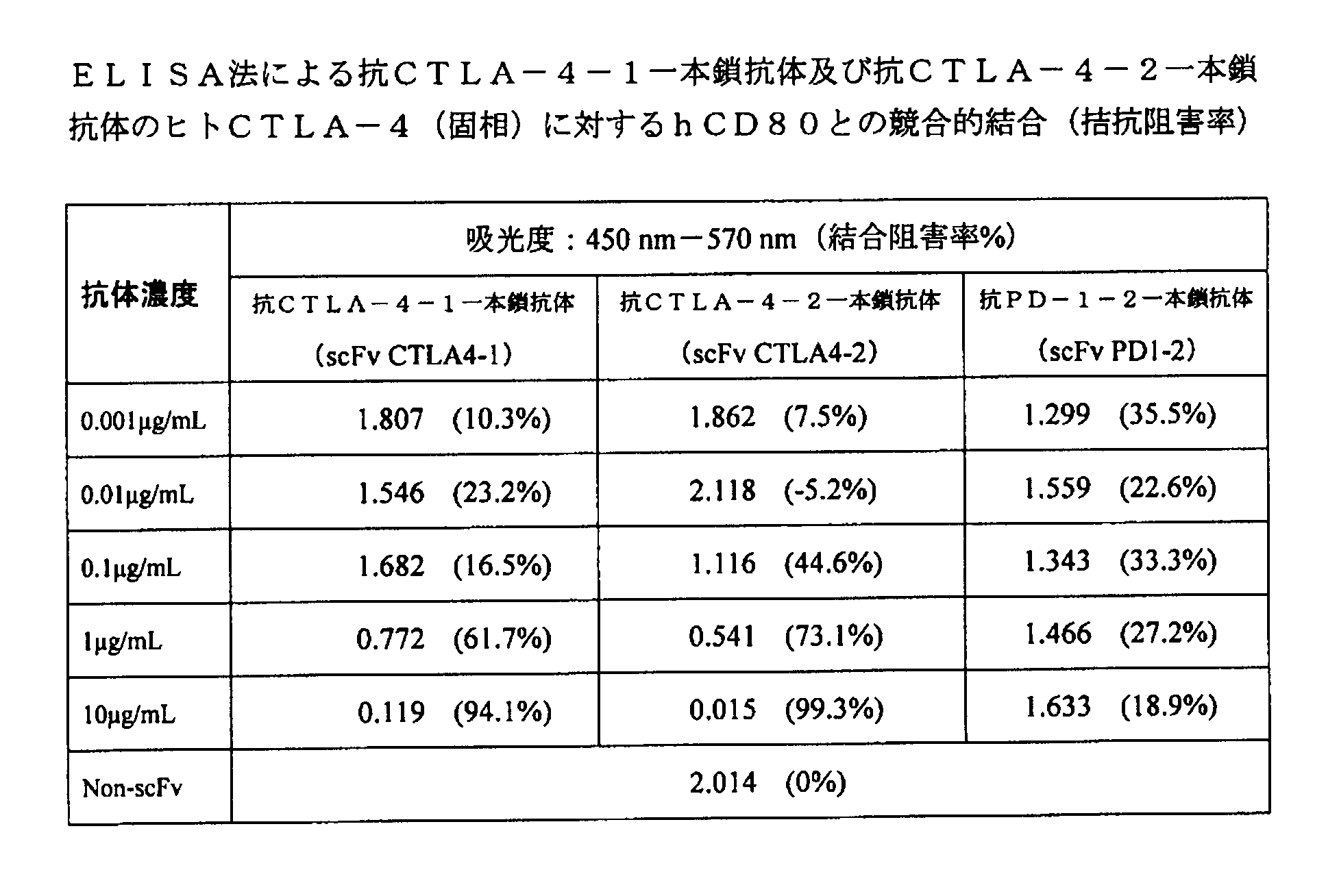

hCD80のhCTLA-4への結合に対する阻害率は、1μg/mLの抗CTLA-4-1一本鎖抗体及び抗CTLA-4-2一本鎖抗体でそれぞれ61.7%及び73.1%、10μg/mLではそれぞれ94.1%及び99.3%であった。またhCD86のhCTLA-4への結合に対する阻害率は、1μg/mLの抗CTLA-4-1一本鎖抗体及び抗CTLA-4-2一本鎖抗体でそれぞれ83.7%及び90.9%、10μg/mLではそれぞれ96.0%及び97.7%であった。以上の結果より、1μg/mLのhCD80及びhCD86のhCTLA-4への結合に対して、抗CTLA-4-1一本鎖抗体及び抗CTLA-4-2一本鎖抗体はいずれも1μg/mL以上の濃度で競合的に結合阻害することが示された。
[マウスPD-1とマウスPD-L1との結合反応に対する抗マウスPD-1一本鎖抗体の競合的阻害活性]
実施例6で作製したビフィドバクテリウム・ロンガム105-A/pHuSP7L20-scFv-PD-1-3株が分泌する抗マウスPD-1一本鎖抗体を用いて、マウスPD-1(mPD-1)とマウスPD-L1(mPD-L1)との結合に対する競合的阻害活性確認を、ELISA法を用いて行った。陰性コントロールとして抗ヒトCTLA-4-2一本鎖抗体を用いた。
96ウェルプレートに、1×PBSにて1μg/mLに調整した前記mPD-1を100μLずつ分注し4℃で一晩静置することで固相化した。液を除去後、1×PBSを350μLずつ分注し、液を除去した。これを3回繰り返し洗浄した。上記プレートに1%BSA溶液を350μLずつ分注し室温で2時間静置することでブロッキングを行った。液を除去後、1×PBSを350μLずつ分注し、液を除去した。これを3回繰り返し洗浄した。

表13及び図15から明らかなとおり、固相化したmPD-1に1μg/mLのmPD-L1を添加した場合その結合反応に対する一本鎖抗体の競合的阻害率は、10μg/mLの抗マウスPD-1一本鎖抗体で51.4%であった。対照とした抗ヒトCTLA-4-2一本鎖抗体では競合阻害は見られなかった(-6.4%)。以上の結果より、1μg/mLのmPD-L1に対して10μg/mL以上の抗マウスPD-1一本鎖抗体が競合阻害活性を示すことが確認された。
[抗マウスPD-1一本鎖抗体とマウスT-細胞(CD3/CD28刺激CD4陽性細胞)との結合の有無の確認]
抗マウスPD-1一本鎖抗体分泌ビフィドバクテリウム属細菌の培養上清より精製した抗マウスPD-1一本鎖抗体とmPD-1発現細胞との結合の有無を、CD3及びCD28刺激によりmPD-1を発現誘導したCD4陽性細胞(以下、「CD3/CD28刺激CD4陽性細胞」ともいう)を用いてフローサイトメトリー解析により検討した。
Balb/cマウスより脾臓を摘出し脾細胞を回収した後、Biotin anti-mouse CD4 Antibody(Biolegend社)でCD4陽性細胞をラベルした。余剰の抗体を洗浄により除いた後、Streptavidin Particles Plus-DM(日本ベクトン・ディッキンソン社製)を加え、CD4陽性細胞に磁気ビーズを結合させた。これをマグネットにより分離し、磁石に引き寄せられた細胞をCD4陽性細胞として回収し、10%ウシ胎児血清、8mM HEPES、50μM 2-メルカプトエタノールを含むRPMI-1640培地に懸濁し、2×10⁶cells/mLのCD4陽性細胞を調製した。
抗マウスPD-1一本鎖抗体の細胞への結合確認は、マウス脾細胞から得られた未刺激CD4陽性細胞とCD3/CD28刺激CD4陽性細胞とを、使用した。この細胞を回収後、1mLの1×PBSバッファーで遠心分離し上清を除去する操作を2回行い培地成分を洗浄した。さらにFACSバッファーを加え遠心分離し上清を除去した。ここにFACSバッファーで50倍希釈したTruStain fcX(Biolegend社製)を20μL添加し、よく撹拌した後氷上で10分間静置した。次にFACSバッファーで200倍希釈したAlexa Fluor 647 anti-mouse CD4 Antibody(Biolegend社製)を20μL添加し、良く撹拌した後氷上で20分間静置した。反応後、1mLのFACSバッファーで遠心分離し上清を除去する操作を2回行い洗浄した。次に、FACSバッファーで200倍希釈したPE anti-mouse CD69 Antibody(Biolegend社製)を20μL添加し、良く撹拌した後氷上で20分間静置した。反応後、1mLのFACSバッファーで遠心分離し情勢を除去する操作を2回行い洗浄した。FACSバッファーで10μg/mLに調整した抗マウスPD-1一本鎖抗体20μLを添加し、撹拌後氷上で20分間静置した。FACSバッファーを1mL加え遠心分離後、上清を除去する操作を2回繰り返して、洗浄を行った。次にFACSバッファーで1000倍希釈したAnti-His-Tag Alexa Fluor 488 Antibody(MBL社)を20μL添加し、撹拌後氷上で20分間静置した。FACSバッファーを1mL加え遠心分離し、上清を除去する操作を、2回繰り返し洗浄を行った。1mLのFACSバッファーを添加後、5mLポリスチレンラウンドボトムチューブ(ベクトン・ディッキンソン社製)にFACSバッファーにて懸濁した細胞を移した。
使用した細胞にPD-1が発現しているか否かの確認は、マウス脾細胞から得られた未刺激CD4陽性細胞とCD3/CD28刺激CD4陽性細胞を、CD4とCD69を抗体で蛍光ラベル処理したものを使用した。この細胞にFACSバッファーで100倍希釈したFITC anti-mouse CD279(PD-1)抗体(Biolegend社製)20μLを添加し撹拌後氷上で20分間静置した。これに、FACSバッファー1mLを加え遠心分離し、上清を除去する操作を2回繰り返して洗浄を行った。1mLのFACSバッファーを添加後、5mLポリスチレンラウンドボトムチューブ(ベクトン・ディッキンソン社製)にFACSバッファーにて懸濁した細胞を移した。
図16Aと図16Bから明らかなとおり、CD3/CD28刺激CD陽性細胞にて特異的に抗mPD-1抗体が結合しており、細胞表面でのPD-1発現が確認された。また、図16Cと図16Dに示したように、抗マウスPD-1一本鎖抗体分泌ビフィドバクテリウム属細菌より精製した抗マウスPD-1一本鎖抗体は、mPD-1発現細胞(CD3/CD28刺激CD4陽性細胞)に特異的に結合することが確認された。
[抗マウスPD-1一本鎖抗体分泌ビフィドバクテリウム属細菌の担癌マウスの腫瘍内での生着及び抗マウスPD-1一本鎖抗体の分泌]
マウス大腸癌細胞株CT26の担癌マウスを用いて、ビフィドバクテリウム・ロンガム105-A/pHuSP7L20-scFv-PD-1-3株を静脈内に投与し、腫瘍内への菌の生着確認及び抗マウスPD一本鎖抗体の局在(分泌)を免疫組織染色で確認した。
凍結した腫瘍組織に嫌気性希釈液を添加し、ホモジナイズ処理をした。このホモジネートを嫌気性希釈液にて適宜希釈した後、BLFS寒天培地(250μg/mL5-フルオロウラシル、30μg/mLスペクチノマイシン含有BL寒天培地)に塗布し、嫌気条件下で37℃にて3日間培養した。BLFS寒天培地上に形成されたコロニー数を数え、腫瘍中の組換えビフィドバクテリウム属細菌の生菌数を算出した。結果を以下の表14に示す。
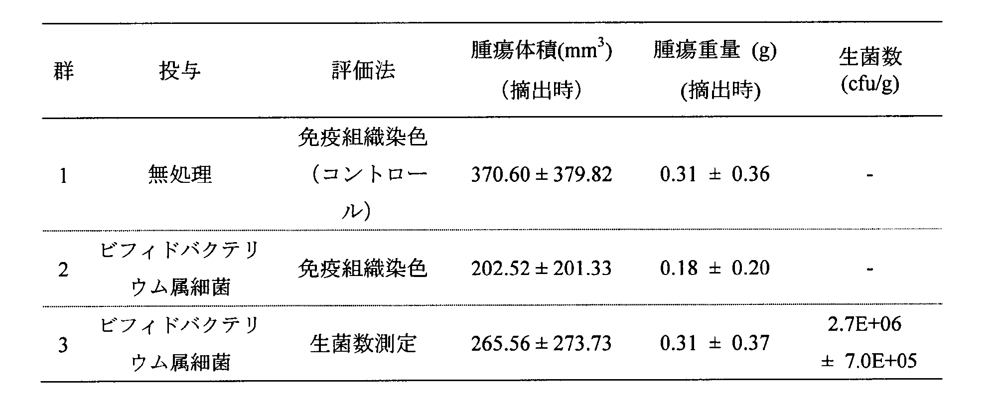
表14から明らかなとおり、腫瘍1gあたり2.7x10⁶cfuの生菌が認められた。
薄切したスライドを風乾し、4%PFA(和光純薬工業社製)に10分間浸漬し固定した。固定後、純水にて1分間洗浄し、1×PBS(-)で5分間の洗浄を3回実施した。組織の周辺の水分を拭き取り、Dako pen(Dako社製)で組織の周囲を囲み、3%BSA-PBSを組織に滴下し60分間反応させ、非特異的結合の阻害を実施した。Anti-His-tag mAb-Alexa Fluor(登録商標)488(MBL社製)抗体反応液を3% BSA-PBSで1000倍希釈した溶液を、組織に滴下した。これを4℃にて一晩反応させた。抗体反応後、1×PBS(-)にて5分間の洗浄を3回実施し、VECTASHIELD(R) Mounting Medium with DAPIで封入した。上記の染色した切片を顕微鏡DM5000B(Leica社製)で鏡検し画像を撮影した。結果を図17に示す。
図17から明らかなとおり、抗マウスPD-1一本鎖抗体分泌ビフィドバクテリウム属細菌を投与したマウス(図17A)では、抗マウスPD-1一本鎖抗体(緑色に染色)が腫瘍組織内にびまん性に陽性を示した。
[抗マウスPD-1一本鎖抗体分泌ビフィドバクテリウム属細菌のCT26担癌モデルマウスに対する薬理(抗腫瘍)効果の検討]
CT26担癌モデルマウスを実施例19と同様の方法にて作製した。腫瘍サイズが40.51から88.03mm3の担癌マウスを下記表15のとおり5群(各群7匹)に分けた(Day0)。第1群は無処理対照群、第2群は抗マウスPD-1一本鎖抗体分泌株(ビフィドバクテリウム・ロンガム105-A/pHuSP7L20-scFv-PD-1-3株)単剤(i.v.)投与群、第3群は抗mPD-1抗体(BioXcell社製、Clone:RPM1-14)単剤投与(i.t.)群、第4群は抗mCTLA-4抗体(BioXcell社製、Clone:9D9)単剤(i.t.)投与群、第5群は、抗マウスPD-1一本鎖抗体分泌ビフィドバクテリウム属細菌(i.v.)と抗mCTLA-4抗体(i.t.)の2剤併用投与群とした。薬剤の投与日、投与量及び投与経路は以下の表15のとおりである。また、第2群及び第5群については、10%マルトース溶液を1mLずつ1日2回の頻度でDay1からDay5及びDay8からDay12に腹腔内投与した。各群共に定期的に腫瘍径を測定(Day0,3,7,10,14,17,22)し、Day22に腫瘍を摘出して腫瘍重量を測定した。腫瘍体積の推移を表16に、CT26担癌マウスにおける腫瘍体積の統計学的検定を表17に、単剤投与群と無処理群のCT26腫瘍体積推移を図18に示した。

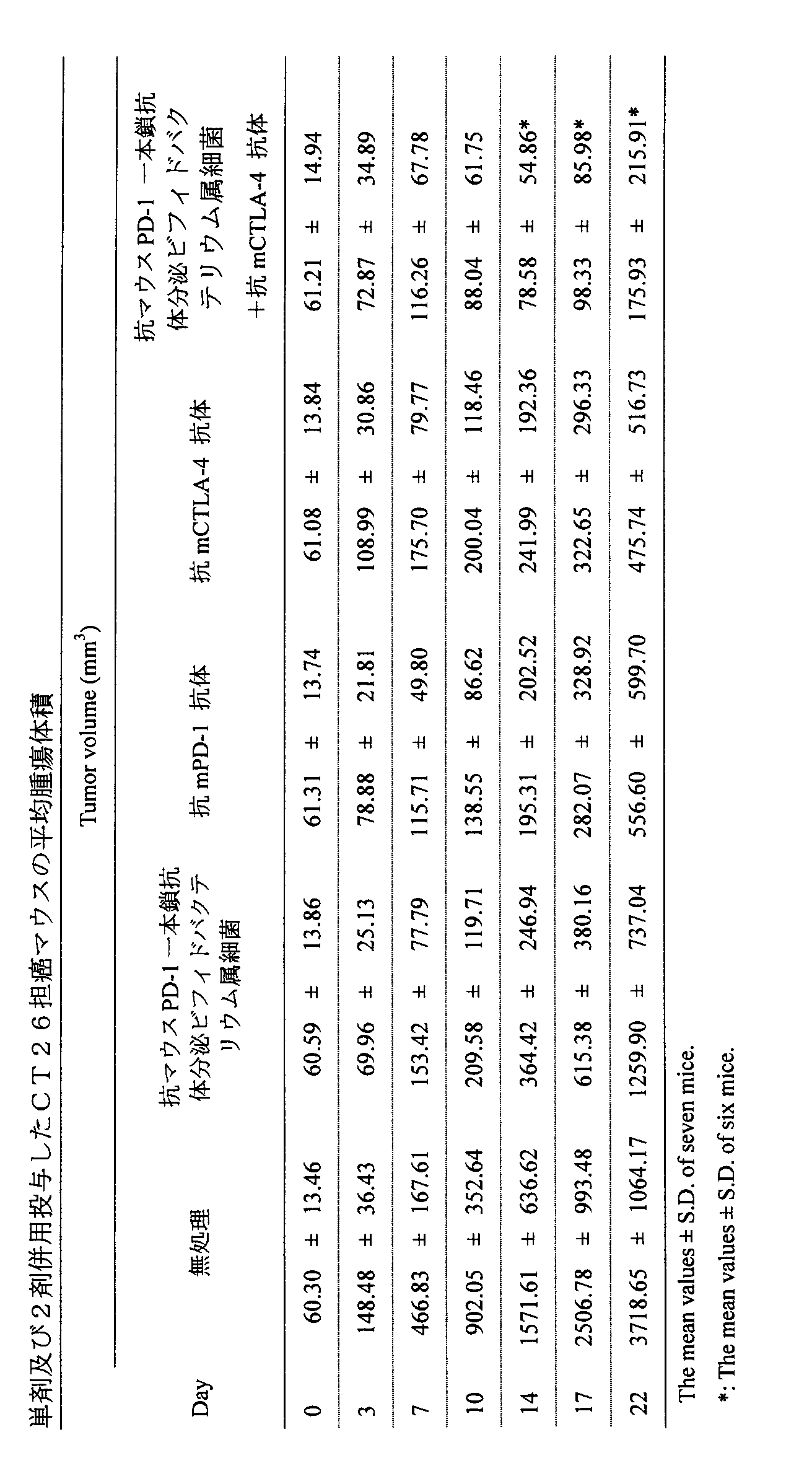


表17、図18から明らかなとおり、抗マウスPD-1一本鎖抗体分泌ビフィドバクテリウム属細菌投与群(第2群)では、無処理群(第1群)と比較して全ての時点で低い腫瘍体積推移を示しており、全ての時点で有意差のある増殖抑制効果が認められた(表17)。また、その腫瘍増殖抑制は無処理対照に対する処理群の比(T/C)で見るとDay10以降で0.34以下であった。
抗mPD-1抗体単剤投与群(第3群)では、無処理群(第1群)と比較して腫瘍体積において全ての時点で有意差のある抑制作用が認められ、T/CはDay10以降で0.15以下であった。なお腫瘍内に投与した抗mCTLA-4抗体単剤投与群(第4群)は、無処理群と比較して腫瘍体積において全ての時点で有意差のある抑制作用が認められ、T/CはDay10以降で0.22以下であった。

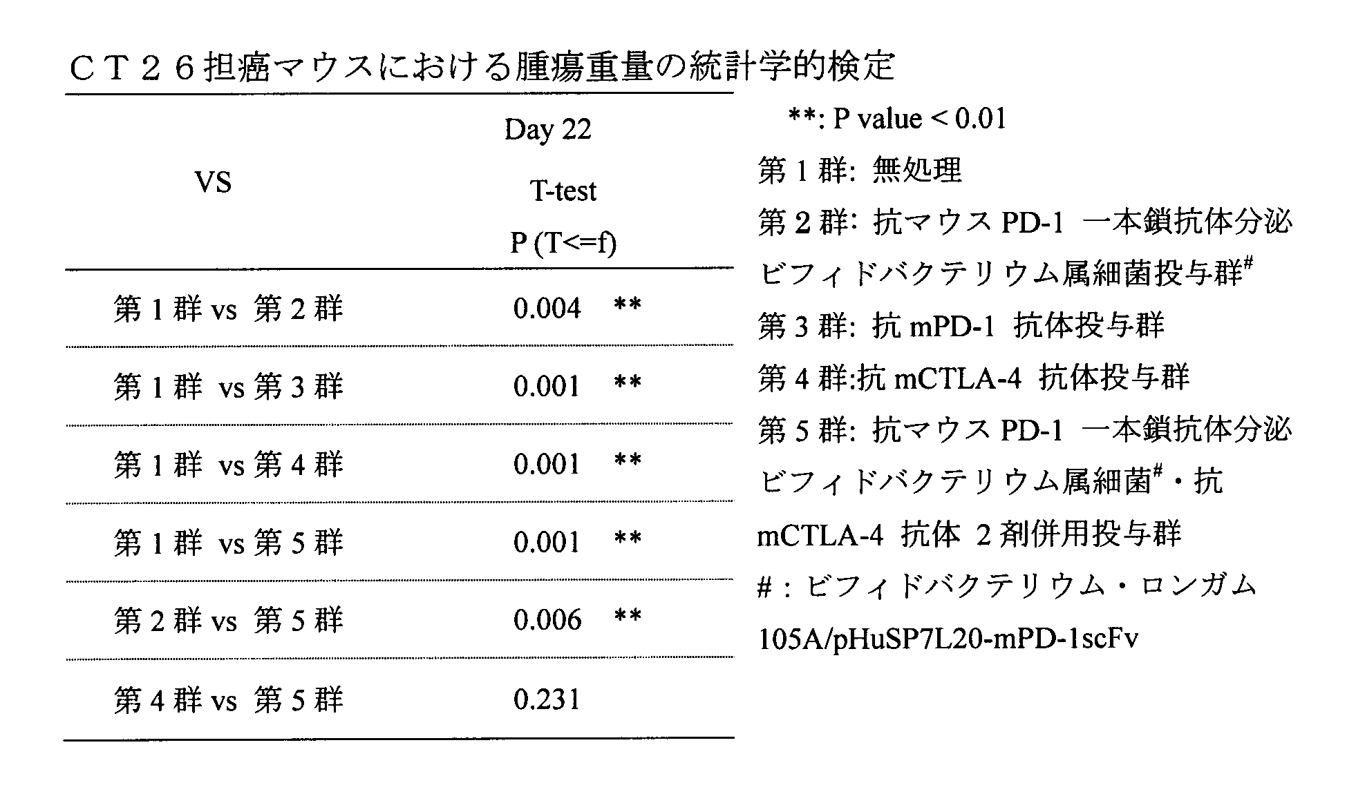
以上の結果より、抗マウスPD-1一本鎖抗体分泌ビフィドバクテリウム属細菌は静脈内投与でCT-26担癌マウスの腫瘍内に生着し(実施例19)、抗マウスPD-1一本鎖抗体を分泌して腫瘍増殖抑制効果を発揮し、その効果は試薬抗体である抗mPD-1抗体を腫瘍内に直接投与した場合とほぼ同程度であった。ビフィドバクテリウム属細菌から分泌された抗マウスPD-1一本鎖抗体は、in vitroの試験にてmPD-1とmPD-L1との結合を競合的に阻害する活性を有しており(実施例17)、腫瘍内でも同様な機序でT細胞上のPD-1と腫瘍細胞上のPD-L1間の負のシグナル伝達を遮断することによりTcellが活性化した結果、抗腫瘍効果が発揮されたものと推定された。また抗マウスPD-1一本鎖抗体分泌ビフィドバクテリウム属細菌と抗mCTLA-4抗体との併用効果が確認されたことから、CTLA-4を阻害する一本鎖抗体とPD-1を阻害する一本鎖抗体とを同時に分泌する共発現ビフィドバクテリウム属細菌の有用性が示唆された。
[各種リンカー長の抗ヒトPD-1scFv03一本鎖抗体分泌ビフィドバクテリウム属細菌の作製]
(概要)
分泌シグナルがSP7、分泌シグナルに続くリンカー長が20アミノ酸残基である抗ヒトPD-1scFv03分泌プラスミドpHuSP7L20-hPD-1scFv03を構築した。このプラスミドの分泌シグナル及び分泌シグナルに続くリンカーを、SP69及びSP69に対応するリンカー(20アミノ酸残基)に置換した抗ヒトPD-1scFv03分泌プラスミドpHuSP69L20-hPD-1scFv03を構築した。さらにリンカーの長さを20から、L0~10、15(L0、L1、L2、L3、L4、L5、L6、L7、L8、L9、L10及びL15)に変更したプラスミドも構築した。
(概要)
抗hPD-1scFv03を分泌する発現カセット(抗ヒトPD-1scFv03分泌発現カセット)を含み、大腸菌-ビフィドバクテリウム属細菌シャトルベクターであるプラスミドpHuSP7L20-hPD-1scFv03を作製した。使用したプライマーを以下の表21に示す。
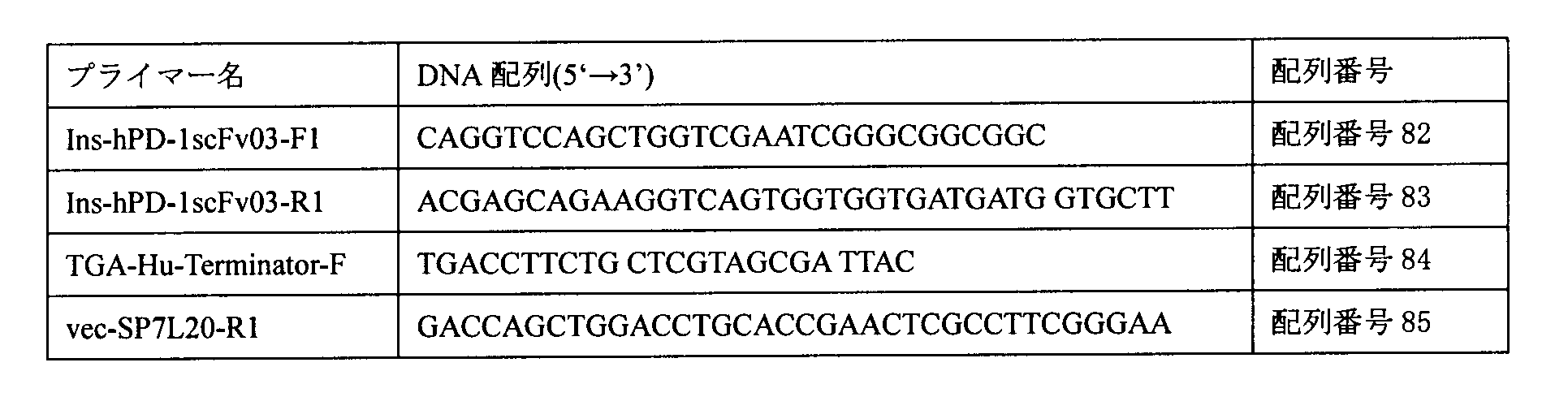
抗ヒトPD-1scFv03分泌発現カセットとして、(1)HuプロモーターDNA、(2)シグナルペプチド-リンカー連結体SP7L20をコードするDNA、(3)抗hPD-1scFv03のアミノ酸配列(重鎖配列、リンカー(GGGGS)3、軽鎖配列を含む)をコードするDNA、及び(4)Hisタグ配列をコードするDNA、(5)HuターミネーターDNAを、(1)を上流(5’末端)側として(5)を下流(3’末端)側として、(1)~(5)のDNAを順次含むカセット(上記抗hPD-1scFv03のアミノ酸配列における重鎖配列、リンカー(GGGGS)3、軽鎖配列を含む)をコードするDNAの塩基配列は、以下の表22に示される文献を参照した。
配列番号86に示された抗hPD-1scFv03については、ジェンスクリプトジャパン社にて、大腸菌用プラスミドpUC57にサブクローニングされ、プラスミドpUC57-hPD-1scFv03として人工遺伝子合成された。

上記プラスミドpUC57-hPD-1scFv03(500pg)を鋳型として、上記表21に記載のIns-hPD-1scFv03-F1プライマー(フォワード)及びIns-hPD-1scFv03-R1プライマー(リバース)のプライマーセットを用いてPCR増幅を行った。プライマー配列は、インサート断片とベクター断片の末端15bpが重複するように設計した。各プライマー濃度を0.2μM、反応容量30μLとして、前記PrimeSTAR HS(Premix)キット(タカラバイオ社製)を用いてPCR増幅を行った。増幅のプログラムとしては、98℃にて10秒、55℃にて5秒、72℃にて60秒を1サイクルとして、30サイクル行った。増幅されたインサートPCR産物を2%アガロースゲルにて電気泳動した後、QIAquick Gel Extractionキット(キアゲン社製)を用いて精製し、約0.7kbpの抗hPD-1scFv03インサート断片(1)を調製した。
配列番号87に記載されているベクター直鎖化断片(500pg)を鋳型として、上記表21に記載されている、TGA-Hu-Terminator-Fプライマー(フォワード)及びvec-SP7L20-R1プライマー(リバース)のプライマーセットを用いてPCR増幅を行った。各プライマー濃度を0.2μM、反応容量30μLとして、PrimeSTAR HS(Premix)キットを用いてPCR増幅を行った。増幅のプログラムは、98℃にて10秒、55℃にて5秒、72℃にて5分を1サイクルとして、30サイクル行った。増幅されたPCR産物を0.8%アガロースゲルにて電気泳動した後、QIAquick Gel Extractionキットを用いて精製し、約4.0kbpの5’-Huターミネーター-pTB6rep unit-SPCMr-pUCori-Huプロモーター-SP7L20-3’ベクター断片(1)を調製した。
上記で調製されたベクター断片(1)と上記抗hPD-1scFv03インサート断片(1)とをIn-Fusion(登録商標)HD Cloningキット(タカラバイオ社製)を用いて連結した。すなわち、マイクロチューブに、キット内の上記ベクターとインサートとを1:5のモル比にて添加後、2μLの5×In-Fusion(登録商標)HD Enzyme premix(タカラバイオ社製)を添加し、反応液容量を10μLに調整した。これを50℃にて15分間保温した。その他の手順は、キットの製品説明書にしたがい、インフュージョン反応液1を調製した。
上記インフュージョン反応液5μLを用いて、大腸菌HST16CRコンピテント細胞の形質転換を製品説明書に従い行った。形質転換後の菌懸濁液は、75μg/mLスペクチノマイシン含有LB寒天培地に塗布し、37℃にて一晩静置培養した。寒天培地上に形成したコロニーを、75μg/mLスペクチノマイシン含有LB液体培地中で30℃にて一晩振とう培養し、これよりQIAprep Spin Miniprep Kit(キアゲン社製)を用いてプラスミドを抽出した。抽出したプラスミドにおける抗ヒトPD-1scFv03分泌発現カセット(5’-Huプロモーター-SP7L20-抗hPD-1scFv03-Hisタグ-Huターミネーター-3’)の配列決定するため、BigDye(登録商標)Terminator v3.1 Cycle Sequencing Kit(アプライドバイオシステムズ社製)を用いてシーケンス反応を行った。配列確認したプラスミドをpHuSP7L20-hPD-1scFv03と名付けた。pHuSP7L20-hPD-1scFv03の配列を配列番号88に示す。
上記抗ヒトPD-1scFv03分泌プラスミドpHuSP7L20-hPD-1scFv03のシグナルペプチド配列及びシグナルペプチド配列に続くリンカーペプチド配列をSP7L20からSP69L20に置換したプラスミドpHuSP69L20-hPD-1scFv03を以下のように構築した。
ベクター断片の調製は、プラスミドpHuSP7L20-hPD-1scFv03を鋳型として、下記表23(V1)及び表24に記載されているhPD-1scFv03_Vec_F1プライマー及びHu-mCCL21_Vec_R1プライマーからなるプライマーセットを用いて、プライマー濃度0.2μM、反応容量50μLとして、PrimeSTAR HS(Premix)キットを用いてPCR増幅を行った。増幅プログラムは、98℃にて10秒、65℃にて5秒、72℃にて4分45秒を1サイクルとして、30サイクル行った後、72℃にて30秒とした。増幅したPCR産物をベクター断片V1とした。
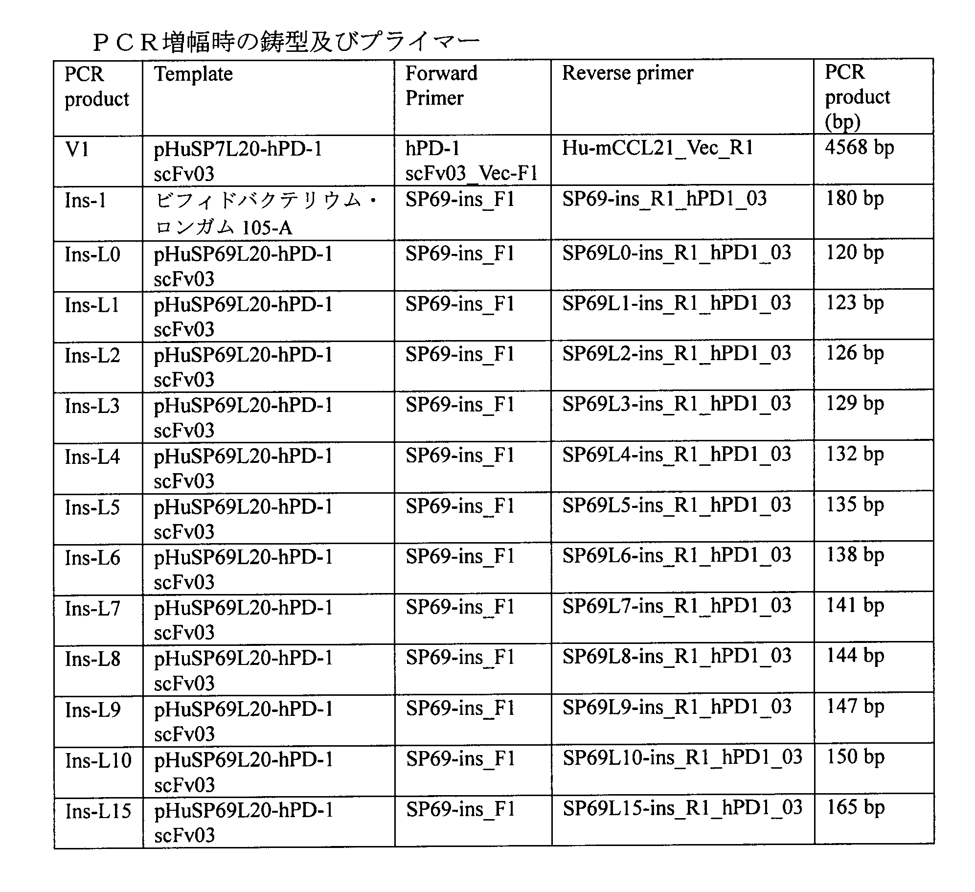
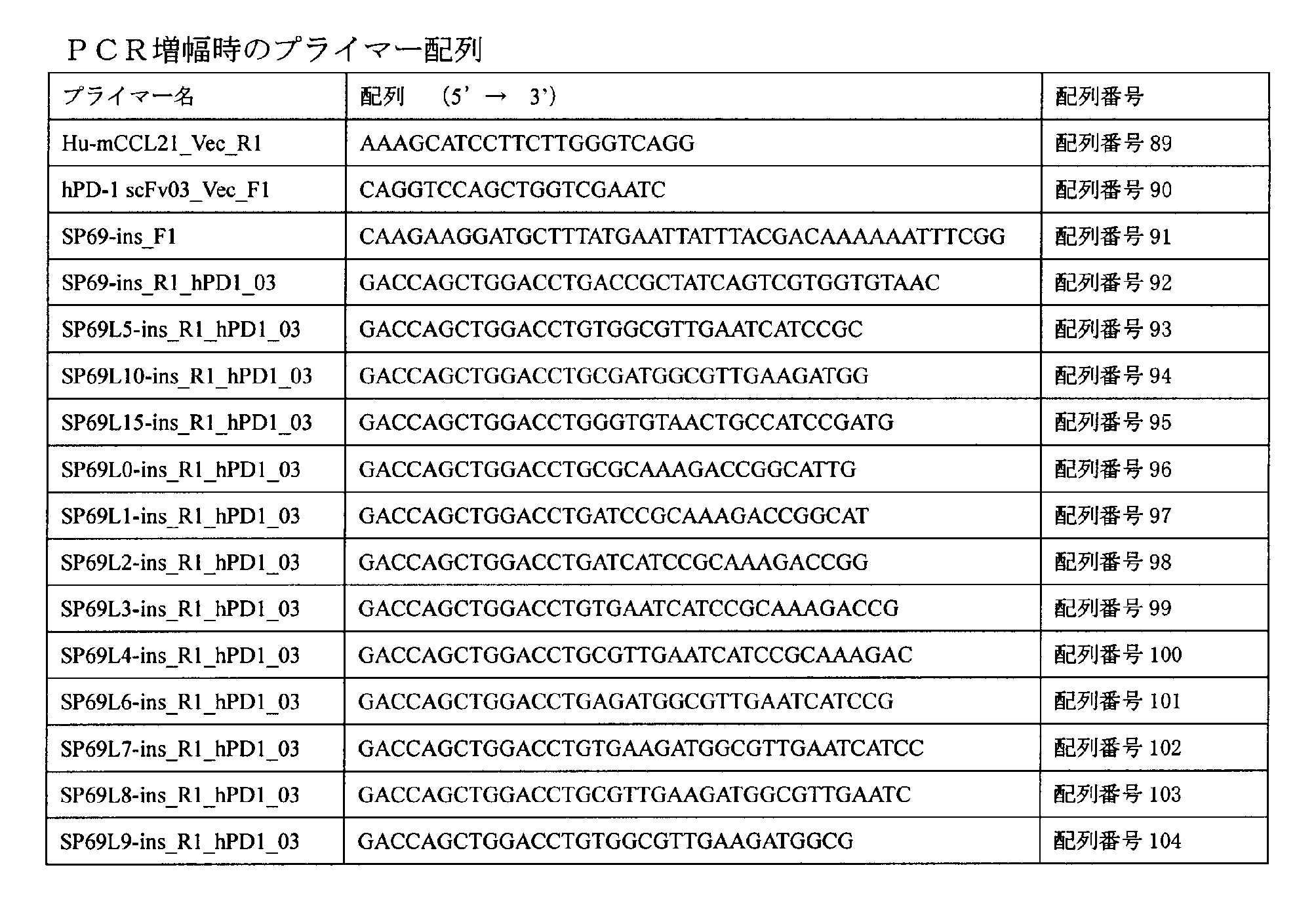
上記で調整したベクター断片V1及びインサート断片Ins-1をIn-Fusion HD Cloning Kitを用いて連結した。即ち、マイクロチューブに、上記ベクター断片とインサート断片を1:2のモル比にて添加後、5xIn-Fusion(登録商標)HD Enzyme premix 2μL、Cloning Enhancer 1μLを添加し、反応容量を10μLに調整した。これを37℃にて15分、その後50℃にて15分間保温した。その他の手順は、キットの製品説明書に従いインフュージョン反応液を調製した。
上記インフュージョン反応液1μLを用いて、大腸菌HST16CRコンピテント細胞(タカラバイオ社製)の形質転換を製品説明書に従い行った。形質転換後の菌懸濁液は、75μg/mLスペクチノマイシン含有LB寒天培地に塗布し、37℃にて一晩静置培養した。寒天培地上に形成したコロニーを、75μg/mLスペクチノマイシン含有LB液体培地中で37℃にて一晩振とう培養し、これよりQIAprep Spin Miniprep Kit(キアゲン社製)を用いてプラスミドを抽出した。抽出したプラスミドにおける抗ヒトPD-1 scFv03発現カセット(HuプロモーターからHuターミネーターまでを含む)配列を決定するため、BigDye(登録商標)Terminator v3.1 Cycle Sequencing Kit(アプライドバイオシステムズ社製)を用いてシーケンス反応を行った。配列確認したプラスミドをpHuSP69L20-hPD-1scFv03と名付けた。
上記pHuSP69L20-hPD-1scFv03のリンカーを3’末端より短くして0~10及び15アミノ酸をコードするDNAとしたプラスミド、pHuSP69Ly-hPD-1scFv03(y=0、1、2、3、4、5、6、7、8、9、10、15)を以下のように作製した。
ベクター断片は、上記ベクター断片V1を使用した。インサート断片の調製は、プラスミドpHuSP69L20-hPD-1scFv03を鋳型として、表23(Ins-L0~10、Ins-L15)に記載されているプライマーセットを用いて、上記インサート断片Ins-1の調製と同様の方法にて調製した。増幅したPCR産物をインサート断片Ins-L0、Ins-L1、Ins-L2、Ins-L3、Ins-L4、Ins-L5、Ins-L6、Ins-L7、Ins-L8、Ins-L9、Ins-L10及びIns-L15とした。
上記で調製したベクター断片V1及び各インサート断片Ins-Ly(y=0~10,15)を、上記pHuSP69L20-hPD-1scFv03作製時と同様の方法にてインフュージョン反応にてつなげた。
上記インフュージョン反応液を用いてpHuSP69L20-hPD-1scFv03作製時と同様の方法にて大腸菌の形質転換及びプラスミドDNAの配列確認を行った。
上記にて作製した13種類のプラスミドpHuSP69Ly-hPD-1scFv03(L0、L1、L2、L3、L4、L5、L6、L7、L8、L9、L10、L15、及びL20)を用いて、ビフィドバクテリウム・ロンガム105-A株の形質転換をエレクトロポレーションシステム(ジーンパルサーII、バイオ・ラッドラボラトリーズ社製)により行った。電気ショック(2kV、25μF、200Ω)後はキュベット(2mmギャップ)に800μLのIMR液体培地と50μLのビタミンC添加液との混合液を即時に添加し、これを滅菌済みの2mLマイクロチューブに回収した。各チューブについて同様の操作を行い、これら2mLチューブの蓋をゆるめて、脱酸素・炭酸ガス発生剤(アネロパック(登録商標)・ケンキ、三菱ガス化学社製)とともに密閉容器に入れ、37℃に設定したインキュベータにて3時間保温した。保温後の各菌懸濁液を75μg/mLスペクチノマイシン含有IMR寒天培地に塗布した。これらのプレートを上記脱酸素・炭酸ガス発生剤とともに密閉容器に入れ、37℃に設定したインキュベータにて2日間培養した。
[各種リンカー長の抗ヒトPD-1scFv03分泌ビフィドバクテリウム属細菌のscFv発現解析]
実施例21にて作製したビフィドバクテリウム・ロンガム105-A/pHuSP69Ly-hPD-1scFv03株(y=0、1、2、3、4、5、6、7、8、9、10、15及び20)より分泌される抗hPD-1scFv03は、C末端にヒスチジンタグを融合しているため、抗ヒスチジンタグ抗体を用いたウェスタンブロッティングにより一本鎖抗体分泌の解析を行なった。
上記各組換えビフィドバクテリウム属細菌株の75μg/mLスペクチノマイシン含有BL-bS寒天培地上の画線培養物を、スペクチノマイシン(終濃度75μg/mL)及びビタミンC添加液(100mLあたりアスコルビン酸35g、L-システイン塩酸塩一水和物2g及び炭酸ナトリウム11gを含む溶液)100μLを添加したMRS(ベクトン・ディッキンソン社製)液体培地10mLに植菌し、37℃にて24時間嫌気培養し、活性化培養液とした。次に、DMEM(CatNo.12320-032:ライフテクノロジーズ社製):MRSを9:1の割合で混合した培地20mLに、ビタミンC添加液100μL、スペクチノマイシンを75μg/mLになるよう添加し、上記活性化培養液100μLを植菌した。これを37℃にて18時間嫌気培養した。
かかる嫌気培養後の培養液を遠心分離後、培養上清を回収した。この培養上清中のタンパク質をトリクロロ酢酸(TCA、和光純薬工業社製)により沈殿させ、アセトンにて洗浄後、SDS-PAGE用バッファーに溶解し、95℃にて3分間熱処理を行い、培養上清濃縮物とした。
図21から明らかなとおり、リンカーがL0であるビフィドバクテリウム・ロンガム105-A/pHuSP69L0-hPD-1scFv03株を除いて全ての菌株にて抗hPD-1scFv03が検出された。また、抗hPD-1scFv03分泌量は、リンカー長が短くなると少なくなる傾向があった。
[リンカー長の異なる抗hPD-1scFv03のヒトPD-L1へのELISAによる結合確認]
(抗hPD-1scFv03の精製)
リンカー長の異なる2種類のビフィドバクテリウム属細菌、ビフィドバクテリウム・ロンガム105-A/pHuSP69L20-hPD-1scFv03株及びビフィドバクテリウム・ロンガム105-A/pHuSP69L5-hPD-1scFv03を上記実施例22と同様の方法で培養した。ただし培養は200mLにスケールアップした。
上記で精製したビフィドバクテリウム・ロンガム105-A/pHuSP69L20-hPD-1scFv03株及びビフィドバクテリウム・ロンガム105-A/pHuSP69L5-hPD-1scFv03由来の抗hPD-1scFv03について、ヒトPD-1(hPD-1)との結合確認をELISA法により行った。
抗hPD-1scFv03の結合ELISAの結果を図22に示す。抗hPD-1scFv03は、L5、L20のリンカー長に関わらずヒトPD-1に結合し、結合量はscFv濃度に依存的であった。
[リンカー長の異なる抗hPD-1scFv03のヒトPD-1とPD-L1との結合反応に対する競合的結合阻害活性の確認]
上記実施例23にて精製したリンカー長の異なる抗hPD-1scFv03を用いて、ヒトPD-1に対するヒトPD-L1の結合に対する競合的結合阻害活性の確認をELISA法により行った。陰性コントロールとして抗ヒトCTLA-4-2一本鎖抗体を用いた。
測定は450nmの吸光度を、570nmを対照として測定した。測定値をフリーソフトウェアImage Jを用いて回帰式を求め、PD-1とPD-L1の結合を50%阻害する抗体濃度(IC50)を算出した。測定及び解析結果を以下の表25及び図23に示した。

表25及び図23から明らかなとおり、抗hPD-1scFv03は、L5、L20のリンカー長に関わらずヒトPD-L1のヒトPD-1への結合に対して競合阻害活性を示した。SP69L5及びSP69L20の抗hPD-1scFv03のIC50はそれぞれ18.7nM及び51.7nMであり、リンカー長の短いSP69L5の方がより低濃度で競合阻害活性を示した。なお、陰性コントロールの抗ヒトCTLA-4-2一本鎖抗体では競合阻害は認められなかった。
[各種シグナルペプチドを組み込んだ抗ヒトPD-1scFv03分泌ビフィドバクテリウム属細菌のscFv発現解析]
実施例21の「プラスミドpHuSP7L20-hPD-1scFv03の作製」と同様の方法で、ビフィドバクテリウム・ロンガム105-A/pHuSPxL20-hPD-1-scFv03株(但し、x=7、45、50、52、55、58、64、66、67、68、69)のそれぞれについて、分泌される抗hPD-1scFv03は、C末端にヒスチジンタグを融合しているため、抗ヒスチジンタグ抗体を用いたウェスタンブロッティングにより一本鎖抗体分泌の解析を行なった。結果を図24(a)及び(b)に示す。
[抗hPD-1scFv03のヒトPD-1へのELISAによる結合確認]
ビフィドバクテリウム・ロンガム105-A/pHuSPxL20-hPD-1-scFv03株の各培養上清より精製した上記各種抗hPD-1-scFv03を用いて、ヒトPD-1への結合確認を行った。測定は450nmの吸光度を、570nmを対照として測定した。抗hPD-1scFv03の結合ELISAの結果を以下の表26に示す。

上記表26より明らかなとおり、各抗hPD-1scFv03は、シグナルペプチドの種類に関わらずヒトPD-1に結合したが、なかでもSP7、SP50、SP64、SP67の場合に結合量が多かった。
[抗hPD-1scFv03のヒトPD-1とPD-L1との結合反応に対する競合的結合阻害活性の確認]
上記ビフィドバクテリウム・ロンガム105-A/pHuSPxL20-hPD-1-scFv03株から精製された各種抗hPD-1scFv03を用いて、ヒトPD-1を固相化したマイクロプレートに、各種抗hPD-1scFv03(10μg/mL)とヒトPD-L1(1μg/mL)との混合物を添加し、ヒトPD-1に結合したPD-L1量を測定した。抗hPD-1scFv03非添加の場合のPD-L1結合阻害率を0%として、抗hPD-1scFv03を共存させたときのPD-1/PD-L1結合に対する競合阻害率(%)を算出した。結果を以下の表27に示す。
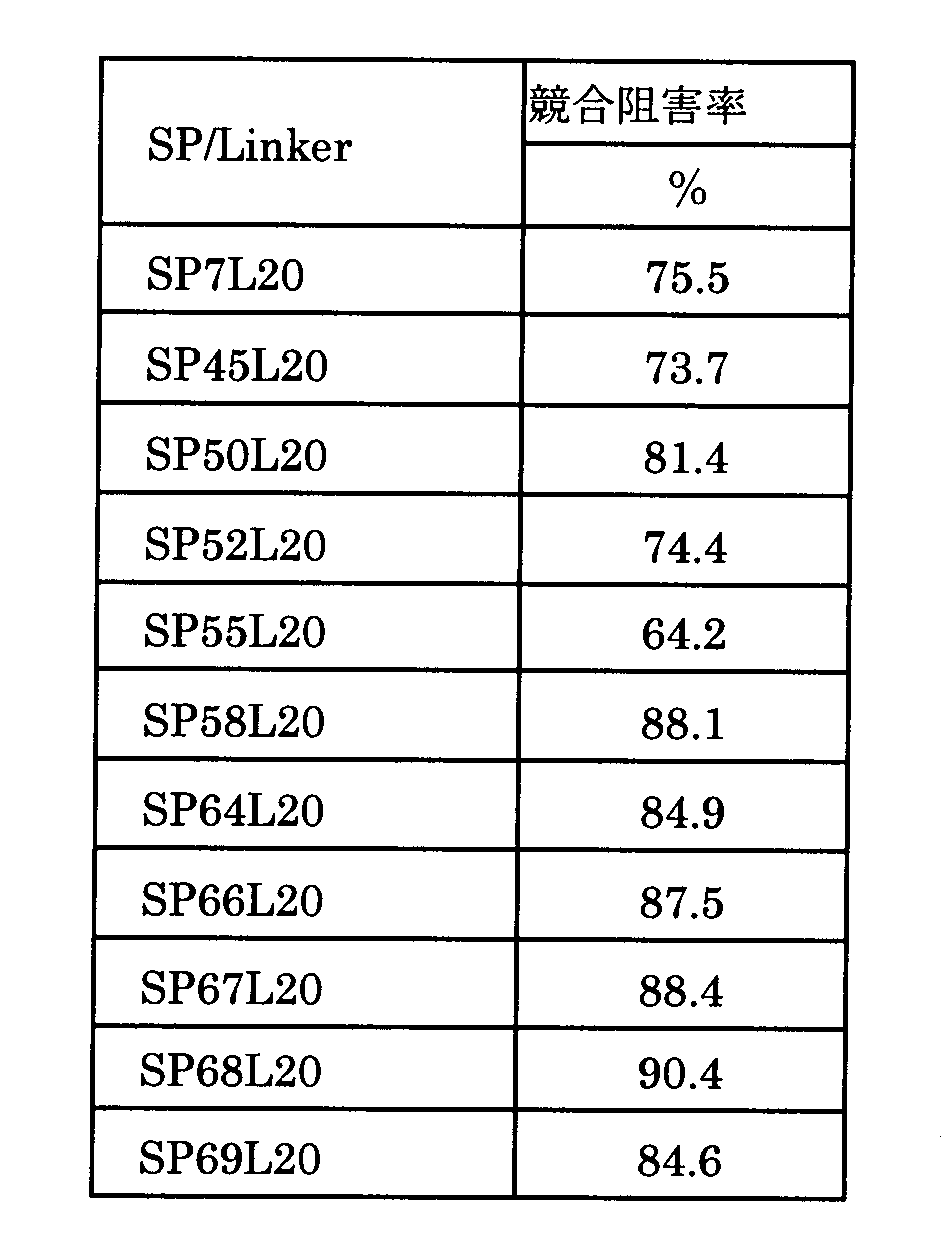
表27より明らかなとおり、シグナルペプチドの種類によって競合阻害率の値に大きな相違はなかったが、SP55L20を用いた場合、競合阻害率はいくぶん低かった。
[リンカー長の検討]
ビフィドバクテリウム・ロンガム105-A/pHuSPxLy-hPD-1-scFv03株(但し、x=7、45、50、52、55、58、64、66、67、68、69)のそれぞれについて、様々なリンカー長(Ly)において検討を行った。
図25(a)~(d)から明らかなとおり、SP58L5,SP58L10を除き、SP7L5、SP7L10、SP7L15,SP7L20、SP45L5、SP45L10、SP45L15,SP45L20、SP50L5、SP50L10、SP50L15,SP50L20、SP52L5、SP52L10、SP52L15,SP52L20、SP55L5、SP55L10、SP55L15,SP55L20、SP58L15,SP58L20、SP64L5、SP64L10、SP64L15,SP64L20、SP66L5、SP66L10、SP66L15,SP66L20、SP67L5、SP67L10、SP67L15,SP67L20、SP68L5、SP68L10、SP68L15,SP68L20、SP69L5、SP69L10、SP69L15,SP69L20の各分泌シグナルペプチド-リンカー連結体が挿入されている抗hPD-1scFv03分泌プラスミドで形質転換された各種ビフィドバクテリウム・ロンガム105-A株から一本鎖抗体が分泌された。
[抗hPD-1scFv03のヒトPD-1へのELISAによる結合確認]
ビフィドバクテリウム・ロンガム105-A/pHuSPxLy-hPD-1-scFv03株(但し、x=7、68、69)各培養上清より精製した抗hPD-1scFv03抗体を1μg/mL添加し、ヒトPD-1(hPD-1)との結合量の確認を、ELISA法により行った。測定は450nmの吸光度を、570nmを対照として測定した。ELISA法を用いたヒトPD-1固相化プレートヘの抗hPD-1scFv03抗体の結合量を以下の表28に示す。
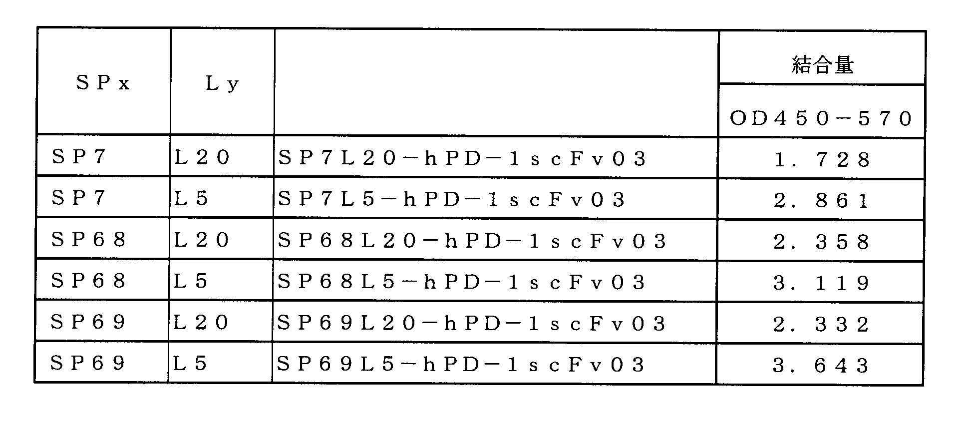
表28から明らかなとおり、SP7、SP68、SP69のいずれの場合においても,リンカー長20(L20)よりもリンカー長5(L5)において、抗hPD-1scFv03抗体のヒトPD-1固相化プレートヘの結合量が多かった。したがって、リンカー長の最適化により、PD-1への抗hPD-1scFv03抗体の結合量を向上できる可能性が示された。
(リンカー長の検討 L=0~10)
SP45、SP50、SP64、SP68、SP69の5種類のシグナルペプチドについて、リンカー長について検討をすすめた。SPxLy(ただし、x=45の場合、y=0、1、2、3、5;x=50、64、68の場合、y=0、1、2、3、4、5;x=69の場合、y=0、1、2、3、4、5,6,7,8,9,10)の各分泌シグナルペプチド-リンカー連結体が挿入されている抗hPD-1scFv03分泌プラスミドで形質転換されたビフィドバクテリウム・ロンガム105-A株を作製し、抗ヒスチジンタグ抗体を用いたウェスタンブロッティングにより一本鎖抗体分泌の解析を行なった。結果を図26に示す。
図26から明らかなとおり、SP69L0の分泌シグナルペプチド-リンカー連結体が挿入されている抗hPD-1scFv03分泌プラスミドで形質転換されたビフィドバクテリウム・ロンガム105-A株では分泌が認められなかった(図26(c)参照)以外は、SP45L0、SP45L1、SP45L2、SP45L3、SP45L5、SP50L0、SP50L1、SP50L2、SP50L3、SP50L4、SP50L5、SP64L0、SP64L1、SP64L2、SP64L3、SP64L4、SP64L5、SP68L0、SP68L1、SP68L2、SP68L3、SP68L4、SP68L5、SP69L1、SP69L2、SP69L3、SP69L4、SP69L5、SP69L6、SP69L7、SP69L8、SP69L9、SP69L10の各分泌シグナルペプチド-リンカー連結体が挿入されている抗hPD-1scFv03分泌プラスミドで形質転換されたビフィドバクテリウム・ロンガム105-A株において分泌が認められた。SP68L0やSP68L1が分泌シグナルペプチド-リンカー連結体として用いられている場合は、分泌量が少なかった(図26(b)参照)。
SP50Ly(ただしy=0、1、5)、SP64Ly(ただしy=0、5)、SP67Ly(ただしy=10)、SP68Ly(ただしy=1、5)、SP69Ly(ただしy=1、7)の各分泌シグナルペプチド-リンカー連結体が挿入されている抗hPD-1scFv03分泌プラスミドで形質転換されたビフィドバクテリウム・ロンガム105-A株を作製し、抗ヒスチジンタグ抗体を用いたウェスタンブロッティングにより一本鎖抗体分泌の解析を行なった。結果を図27に示す。
図27から明らかなとおり、SP50L0、SP50L1、SP50L5、SP64L0、SP64L5、SP67L10、SP68L1、SP68L5、SP69L1、SP69L7のいずれの分泌シグナルペプチド-リンカー連結体が挿入されている抗hPD-1scFv03分泌プラスミドで形質転換された株においても、抗hPD-1-scFv03抗体の分泌が確認された。
(抗hPD-1scFv03のヒトPD-1とPD-L1との結合反応に対する競合的結合阻害活性の確認)
実施例24の手順と同様に、hPD-1scFv03分泌各ビフィドバクテリウム属細菌より精製したscFvによるPD-1/PD-L1結合の競合的結合阻害活性の確認を行った。測定は450nmの吸光度を、570nmを対照として測定した。scFv非添加時のPD-L1の結合量を100%とし、PD-1とPD-L1の結合を50%阻害するscFv濃度(IC50)を算出した。測定結果を以下の表29に示した。

表29から明らかなとおり、SP50L5が最も低濃度でPD-1とPD-L1との結合を阻害した。同一SPでリンカー長の異なるSP68L1とSP68L5、及びSP69L1とSP69L7とを比べるとどちらもリンカー長が短い方(L1)がより低濃度においてもPD-1とPD-L1との結合を阻害することが示された。
(結合親和性)
抗PD-1抗体は、ビアコア分析(Biacore AB、Uppsala、Sweden)によって、以下の表30に示すNo1~No7の7つのサンプルをそれぞれn=5で、結合親和性を解析した。リガンドとしてヒトPD-1 Fcキメラ(R&D社製)をAcetate pH4.5(GEヘルスケア社製)にて5μg/mLに調製した。この液を、CM基を導入したデキストランを金膜表面にコートしたセンサーチップ上に標準のタンパク質固定化法(アミンカップリング法)により固定化した。アナライトとなる抗ヒトPD-1一本鎖抗体はHBS-EPバッファー(GEヘルスケア社製)にて12.5、25、50、100及び200nMに調製した。カイネティクス測定は、各アナライトを低濃度から添加時間60秒、解離時間60秒、流速30μL/分の条件でシングルサイクル法にて実施した。測定後の試料の解離条件はGlycin 1.5(GEヘルスケア社製)を添加時間60秒、流速30μL/分とした。本試験のランニング緩衝液はHBS-EPバッファーとした。結果を以下の表30に示す。

表30から明らかなとおり、抗体としての機能としては、結合が強く(KD値が小さい)、解離が遅い(kd値が低い)SP50L5が最も優れていた。次点はNo3、4、6のSP67L10、SP68L1、SP69L1であった。
[各種シグナルペプチドを組み込んだ抗hCTLA-4scFv02-FLAG分泌ビフィドバクテリウム属細菌のscFv発現解析]
ビフィドバクテリウム・ロンガム105-A/pP30SPxL20-hCTLA-4scFv02株(但し、x=7、45、50、52、55、58、64、66、67、68、69)のそれぞれについて、18時間培養の培養液からサンプル調製し、培養上清80μLを用いて、抗FLAGタグ抗体を用いたウェスタンブロッティングにより一本鎖抗体分泌の解析を行なった。結果を図28に示す。
[抗hCTLA-4scFv02のヒトCTLA-4へのELISAによる結合確認]
ビフィドバクテリウム・ロンガム105-A/pP30SPxL20-hCTLA-4scFv02株の各培養上清より精製した各種抗hCTLA-4scFv02を用いて、ヒトCTLA-4への結合確認を行った。測定は450nmの吸光度を、570nmを対照として測定した。ヒトCTLA-4への結合ELISAの結果を以下の表31に示す。
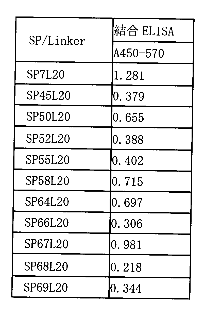
表31から明らかなとおり、SP/Linkerの種類によりscFvの結合量は異なった。SP7L20及びSP67L20が比較的高値であった。
[抗hCTLA-4scFv02のヒトCTLA-4/ヒトCD80との結合反応又はヒトCTLA-4/ヒトCD86との結合反応に対する競合的結合阻害活性の確認]
ビフィドバクテリウム・ロンガム105-A/pP30SPxL20-hCTLA-4scFv02株から得られた抗hCTLA-4scFv02を用いて、ヒトCTLA-4を固相化したマイクロプレートに、精製scFv(10μg/mL)と、ヒトCD80又はヒトCD86(1μg/mL)との混合物を添加し、ヒトCTLA-4に結合したヒトCD80又はヒトCD86の量を測定した。抗hCTLA-4scFv02非添加の場合のヒトCD80又はヒトCD86結合阻害率を0%として、抗hCTLA-4scFv02を共存させたときのヒトCTLA-4/ヒトCD80結合又はヒトCTLA-4/ヒトCD86結合に対する競合阻害率(%)を算出した。結果を以下の表32に示した。
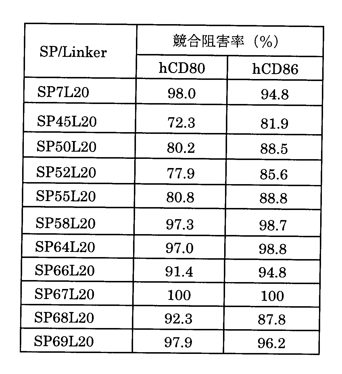
リンカー長が20の場合、シグナルペプチドによる大きな違いはなかった。
抗hCTLA-4scFv02抗体について、SP45、SP50、SP64、SP68、SP69のシグナルペプチドについて、リンカー長について検討をすすめた。SPxLy(ただし、x=45、50、64、68の場合、y=0、1、2、3、4、5;x=69の場合、y=0、1、2、3、4、5,6,7,8,9,10)の各分泌シグナルペプチド-リンカー連結体が挿入されている抗hCTLA-4scFv02分泌プラスミドで形質転換されたビフィドバクテリウム・ロンガム105-A株を作製し、18時間培養の培養液からサンプル調製し、培養上清100μLを用いて、抗FLAGタグ抗体を用いたウェスタンブロッティングにより一本鎖抗体分泌の解析を行なった。結果を図29(a)~(c)に示す。
図29から明らかなとおり、S68L0以外の、SP45L0、SP45L1、SP45L2、SP45L3、SP45L4、SP45L5、SP50L0、SP50L1、SP50L2、SP50L3、SP50L4、SP50L5、SP64L0、SP64L1、SP64L2、SP64L3、SP64L4、SP64L5、SP68L1、SP68L2、SP68L3、SP68L4、SP68L5、SP69L0、SP69L1、SP69L2、SP69L3、SP69L4、SP69L5、SP69L6、SP69L7、SP69L8、SP69L9、SP69L10の分泌シグナルペプチドや分泌シグナルペプチド-リンカー連結体の下流に抗hCTLA-4scFv02が組み込まれた発現カセットを含むベクターで形質転換されたビフィドバクテリウム・ロンガム105-A株において、抗hCTLA-4scFv02の分泌が確認された。
Claims (16)
- 以下の(1)~(4)のDNAを順次含むことを特徴とするビフィドバクテリウム属細菌内で発現する発現カセット。
(1)ビフィドバクテリウム属細菌で機能するプロモーターDNA;
(2)以下のa)又はb)に示されるアミノ酸配列からなる分泌シグナルペプチドをコードするDNA;
a)配列番号6、7、8、9、10、11、12、13、14、15、及び16のいずれかに示されるアミノ酸配列;
b)配列番号6、7、8、9、10、11、12、13、14、15、及び16のいずれかに示されるアミノ酸配列において、1又は数個のアミノ酸が欠失、置換又は付加されたアミノ酸配列であって、該アミノ酸配列からなるペプチドがビフィドバクテリウム属細菌において分泌シグナルペプチドとして機能するアミノ酸配列;
(3)異種ポリペプチドをコードするDNA;
(4)ビフィドバクテリウム属細菌で機能するターミネーターDNA; - 分泌シグナルペプチドをコードするDNAの下流にリンカーペプチドをコードするDNAが連結されていることを特徴とする請求項1記載の発現カセット。
- リンカーペプチドが、配列番号25、26、27、28、29、30、31、32、33、34、及び35のいずれかに示されるアミノ酸配列のC末端からアミノ酸残基が0~29欠損した各アミノ酸配列からなることを特徴とする請求項2記載の発現カセット。
- リンカーペプチドが、配列番号25、26、27、28、29、30、31、32、33、34、及び35のいずれかに示されるアミノ酸配列のC末端からアミノ酸残基が10~29欠損した各アミノ酸配列からなることを特徴とする請求項2記載の発現カセット。
- 異種ポリペプチドが、一本鎖抗体であることを特徴とする請求項1~4のいずれか記載の発現カセット。
- 一本鎖抗体が、抗PD-1抗体であることを特徴とする請求項5記載の発現カセット。
- 一本鎖抗体が、抗CTLA-4抗体であることを特徴とする請求項5記載の発現カセット。
- 請求項1~7のいずれか記載の発現カセットを含むベクター。
- 請求項8記載のベクターで形質転換されたビフィドバクテリウム属細菌。
- ビフィドバクテリウム・ロンガムである請求項9記載のビフィドバクテリウム属細菌。
- 請求項9又は10記載のビフィドバクテリウム属細菌を含む医薬組成物。
- 配列番号6、7、8、9、10、11、12、13、14、15、及び16のいずれかに示されるアミノ酸配列からなる分泌シグナルペプチド。
- 請求項12記載の分泌シグナルペプチドをコードするDNA。
- 配列番号105、106、107、108、109、110、111、112、113、114及び115のいずれかに示されるアミノ酸配列のC末端からアミノ酸残基が0~29欠損した各アミノ酸配列からなる分泌シグナルペプチド-リンカー連結体。
- 配列番号105、106、107、108、109、110、111、112、113、114及び115のいずれかに示されるアミノ酸配列のC末端からアミノ酸残基が10~29欠損した各アミノ酸配列からなる分泌シグナルペプチド-リンカー連結体。
- 請求項14又は15記載の分泌シグナルペプチド-リンカー連結体をコードするDNA。
Priority Applications (7)
| Application Number | Priority Date | Filing Date | Title |
|---|---|---|---|
| US15/308,179 US10443061B2 (en) | 2014-05-01 | 2015-04-17 | Heterologous polypeptide expression cassette |
| KR1020167030618A KR20170002403A (ko) | 2014-05-01 | 2015-04-17 | 이종 폴리펩티드 발현 카세트 |
| CN201580029678.5A CN107002090B (zh) | 2014-05-01 | 2015-04-17 | 异源多肽表达盒 |
| CA2947525A CA2947525C (en) | 2014-05-01 | 2015-04-17 | Heterologous polypeptide expression cassette |
| EP15785407.6A EP3141602A4 (en) | 2014-05-01 | 2015-04-17 | Heterogeneous polypeptide expression cassette |
| SG11201609032WA SG11201609032WA (en) | 2014-05-01 | 2015-04-17 | Heterogeneous polypeptide expression cassette |
| JP2016515853A JP6634625B2 (ja) | 2014-05-01 | 2015-04-17 | 異種ポリペプチド発現カセット |
Applications Claiming Priority (2)
| Application Number | Priority Date | Filing Date | Title |
|---|---|---|---|
| JP2014-094869 | 2014-05-01 | ||
| JP2014094869 | 2014-05-01 |
Publications (1)
| Publication Number | Publication Date |
|---|---|
| WO2015166640A1 true WO2015166640A1 (ja) | 2015-11-05 |
Family
ID=54358386
Family Applications (1)
| Application Number | Title | Priority Date | Filing Date |
|---|---|---|---|
| PCT/JP2015/002132 Ceased WO2015166640A1 (ja) | 2014-05-01 | 2015-04-17 | 異種ポリペプチド発現カセット |
Country Status (8)
| Country | Link |
|---|---|
| US (1) | US10443061B2 (ja) |
| EP (1) | EP3141602A4 (ja) |
| JP (1) | JP6634625B2 (ja) |
| KR (1) | KR20170002403A (ja) |
| CN (1) | CN107002090B (ja) |
| CA (1) | CA2947525C (ja) |
| SG (2) | SG11201609032WA (ja) |
| WO (1) | WO2015166640A1 (ja) |
Cited By (5)
| Publication number | Priority date | Publication date | Assignee | Title |
|---|---|---|---|---|
| WO2016088376A1 (ja) * | 2014-12-03 | 2016-06-09 | 株式会社アネロファーマ・サイエンス | 共発現プラスミド |
| WO2017123675A1 (en) * | 2016-01-11 | 2017-07-20 | Synlogic, Inc. | Microorganisms programmed to produce immune modulators and anti-cancer therapeutics in tumor cells |
| WO2018129404A1 (en) * | 2017-01-06 | 2018-07-12 | Synlogic, Inc. | Microorganisms programmed to produce immune modulators and anti-cancer therapeutics in tumor cells |
| WO2021038975A1 (ja) | 2019-08-28 | 2021-03-04 | 株式会社アネロファーマ・サイエンス | ダイアボディ型BsAbを発現・分泌するビフィドバクテリウム属細菌 |
| WO2025037535A1 (ja) * | 2023-08-11 | 2025-02-20 | 株式会社Co2資源化研究所 | ヒドロゲノフィラス属細菌由来の分泌シグナルペプチド遺伝子 |
Families Citing this family (4)
| Publication number | Priority date | Publication date | Assignee | Title |
|---|---|---|---|---|
| US10344093B2 (en) | 2014-05-02 | 2019-07-09 | Shinshu University | Antibody gene expression-secretion system |
| CA2974333C (en) | 2015-01-19 | 2018-09-25 | Shinshu University | Therapeutic agent for ischemic disease |
| CN110747154A (zh) * | 2019-09-17 | 2020-02-04 | 广州维生君生物科技有限公司 | Pd-1人源化单链抗体融合基因转化乳酸菌及其应用 |
| CN116554344A (zh) * | 2022-01-30 | 2023-08-08 | 中国科学院生物物理研究所 | 一种pd1抗体与白介素2融合的双功能分子 |
Citations (3)
| Publication number | Priority date | Publication date | Assignee | Title |
|---|---|---|---|---|
| JP2004531245A (ja) * | 2001-01-30 | 2004-10-14 | ソシエテ デ プロデユイ ネツスル ソシエテ アノニム | Ncc2705−ビフィドバクテリウムのゲノム |
| WO2011093468A1 (ja) * | 2010-01-29 | 2011-08-04 | 株式会社アネロファーマ・サイエンス | 形質転換用プラスミド |
| WO2014010758A1 (ja) * | 2012-07-13 | 2014-01-16 | 学校法人帝京平成大学 | 抗腫瘍剤、腫瘍検出用マーカー及び経口ワクチン剤 |
Family Cites Families (8)
| Publication number | Priority date | Publication date | Assignee | Title |
|---|---|---|---|---|
| US6051227A (en) * | 1995-07-25 | 2000-04-18 | The Regents Of The University Of California, Office Of Technology Transfer | Blockade of T lymphocyte down-regulation associated with CTLA-4 signaling |
| DE10101793A1 (de) * | 2001-01-17 | 2002-08-01 | Manfred Nilius | Verwendung von SLPI zur Behandlung chronisch-entzündlicher Darmerkrankungen |
| IL149701A0 (en) * | 2001-05-23 | 2002-11-10 | Pfizer Prod Inc | Use of anti-ctla-4 antibodies |
| US20040241745A1 (en) * | 2001-07-31 | 2004-12-02 | Tasuku Honjo | Substance specific to pd-1 |
| WO2006109619A1 (ja) | 2005-04-08 | 2006-10-19 | Anaeropharma Science Inc. | 5-フルオロウラシル耐性菌およびその作製方法 |
| US9730968B2 (en) * | 2008-04-17 | 2017-08-15 | Anaeropharma Science, Inc. | Therapeutic agent for ischemic diseases |
| JPWO2010126073A1 (ja) | 2009-04-28 | 2012-11-01 | 協和発酵キリン株式会社 | ポリペプチドの分泌発現のための発現カセットおよびその使用 |
| CN101608172B (zh) * | 2009-07-24 | 2013-01-23 | 南京华贞生物医药科技有限公司 | 一种用于减肥降脂的转化体及其制备方法 |
-
2015
- 2015-04-17 WO PCT/JP2015/002132 patent/WO2015166640A1/ja not_active Ceased
- 2015-04-17 CN CN201580029678.5A patent/CN107002090B/zh active Active
- 2015-04-17 US US15/308,179 patent/US10443061B2/en active Active
- 2015-04-17 KR KR1020167030618A patent/KR20170002403A/ko not_active Ceased
- 2015-04-17 JP JP2016515853A patent/JP6634625B2/ja active Active
- 2015-04-17 SG SG11201609032WA patent/SG11201609032WA/en unknown
- 2015-04-17 CA CA2947525A patent/CA2947525C/en active Active
- 2015-04-17 SG SG10201809450VA patent/SG10201809450VA/en unknown
- 2015-04-17 EP EP15785407.6A patent/EP3141602A4/en not_active Withdrawn
Patent Citations (4)
| Publication number | Priority date | Publication date | Assignee | Title |
|---|---|---|---|---|
| JP2004531245A (ja) * | 2001-01-30 | 2004-10-14 | ソシエテ デ プロデユイ ネツスル ソシエテ アノニム | Ncc2705−ビフィドバクテリウムのゲノム |
| WO2011093468A1 (ja) * | 2010-01-29 | 2011-08-04 | 株式会社アネロファーマ・サイエンス | 形質転換用プラスミド |
| WO2011093465A1 (ja) * | 2010-01-29 | 2011-08-04 | 株式会社アネロファーマ・サイエンス | 形質転換用プラスミド |
| WO2014010758A1 (ja) * | 2012-07-13 | 2014-01-16 | 学校法人帝京平成大学 | 抗腫瘍剤、腫瘍検出用マーカー及び経口ワクチン剤 |
Non-Patent Citations (7)
| Title |
|---|
| DATABASE GenBank [O] 17 June 2013 (2013-06-17), OBERG,T.S: "Autotransporter adhesin [Bifidobacterium longum D2957", XP055235303, retrieved from ncbi Database accession no. EPE38071 * |
| DATABASE GenBank [O] 29 January 2011 (2011-01-29), FUKUDA,S.: "putative cell surface protein [Bifidobacterium longum subsp. infantis 157F]", XP055235299, retrieved from ncbi Database accession no. BAJ72090 * |
| DATABASE GenBank [O] 31 January 2014 (2014-01-31), HAO,Y.: "Hypothetical protein BBMN68_1126 [Bifidobacterium longum subsp. longum BBMN68", XP055235297, retrieved from ncbi Database accession no. ADQ02103 * |
| DATABASE GenBank [O] 4 May 2012 (2012-05-04), SHKOPOROV,A.N.: "hypothetical protein HMPREF1313_0372 [Bifidobacterium longum subsp. longum 1-6B", XP055235294, retrieved from ncbi Database accession no. EIJ22210 * |
| DATABASE genbank [O] 4 May 2012 (2012-05-04), SHKOPOROV,A.N: "G5 domain protein [Bifidobacterium longum subsp. longum 35B]", XP055235306, retrieved from ncbi Database accession no. EIJ27376 * |
| DATABASE GenBank [O] 4 May 2012 (2012-05-04), SHKOPOROV,A.N: "NlpC/P60 family protein [Bifidobacterium longum subsp. longum 35B]", XP055235291, retrieved from ncbi Database accession no. EIJ27286 * |
| JOURNAL OF CLINICAL AND EXPERIMENTAL MEDICINE, vol. 244, no. 9, 2013, pages 809 - 815, XP008184468 * |
Cited By (8)
| Publication number | Priority date | Publication date | Assignee | Title |
|---|---|---|---|---|
| WO2016088376A1 (ja) * | 2014-12-03 | 2016-06-09 | 株式会社アネロファーマ・サイエンス | 共発現プラスミド |
| US10563208B2 (en) | 2014-12-03 | 2020-02-18 | Anaeropharma Science, Inc. | Co-expression plasmid |
| WO2017123675A1 (en) * | 2016-01-11 | 2017-07-20 | Synlogic, Inc. | Microorganisms programmed to produce immune modulators and anti-cancer therapeutics in tumor cells |
| US11723932B2 (en) | 2016-01-11 | 2023-08-15 | Synlogic Operating Company, Inc. | Microorganisms programmed to produce immune modulators and anti-cancer therapeutics in tumor cells |
| WO2018129404A1 (en) * | 2017-01-06 | 2018-07-12 | Synlogic, Inc. | Microorganisms programmed to produce immune modulators and anti-cancer therapeutics in tumor cells |
| US11471494B2 (en) | 2017-01-06 | 2022-10-18 | Synlogic Operating Company, Inc. | Microorganisms programmed to produce immune modulators and anti-cancer therapeutics in tumor cells |
| WO2021038975A1 (ja) | 2019-08-28 | 2021-03-04 | 株式会社アネロファーマ・サイエンス | ダイアボディ型BsAbを発現・分泌するビフィドバクテリウム属細菌 |
| WO2025037535A1 (ja) * | 2023-08-11 | 2025-02-20 | 株式会社Co2資源化研究所 | ヒドロゲノフィラス属細菌由来の分泌シグナルペプチド遺伝子 |
Also Published As
| Publication number | Publication date |
|---|---|
| CN107002090A (zh) | 2017-08-01 |
| US20170183667A1 (en) | 2017-06-29 |
| KR20170002403A (ko) | 2017-01-06 |
| US10443061B2 (en) | 2019-10-15 |
| JPWO2015166640A1 (ja) | 2017-04-20 |
| EP3141602A4 (en) | 2017-11-29 |
| CA2947525C (en) | 2023-09-05 |
| SG10201809450VA (en) | 2018-11-29 |
| CA2947525A1 (en) | 2015-11-05 |
| SG11201609032WA (en) | 2016-12-29 |
| EP3141602A1 (en) | 2017-03-15 |
| CN107002090B (zh) | 2021-09-10 |
| JP6634625B2 (ja) | 2020-01-22 |
Similar Documents
| Publication | Publication Date | Title |
|---|---|---|
| JP6634625B2 (ja) | 異種ポリペプチド発現カセット | |
| JP7700159B2 (ja) | Axlまたはror2に対するキメラ抗原受容体およびその使用方法 | |
| JP6161098B2 (ja) | Car発現ベクター及びcar発現t細胞 | |
| CN109862912A (zh) | 携带双特异性T细胞衔接器(BiTE)的腺病毒 | |
| CN115925989B (zh) | 长效的双靶点嵌合抗原受体、核酸分子、重组载体、细胞及其应用 | |
| JP6755465B2 (ja) | 共発現プラスミド | |
| US20190099461A1 (en) | Rna viruses for immunovirotherapy | |
| CN118284689A (zh) | 表达免疫检查点抑制剂的超级dc及其用途 | |
| CN117534767A (zh) | 靶向cldn6嵌合抗原受体巨噬细胞及其制备方法和应用 | |
| US20220281998A1 (en) | Bifidobacterium spp. expressing and secreting diabody-type bsab | |
| JP2021536247A (ja) | 併用療法用dnaワクチンを標的とするネオアンチゲン | |
| CN106467576B (zh) | 一种抗体融合蛋白及其制备方法与应用 | |
| US10344093B2 (en) | Antibody gene expression-secretion system | |
| EP4458956A1 (en) | Anti-mesothelin nanobody chimeric antigen receptor and use thereof | |
| WO2022056141A1 (en) | Chimeric antigen receptor (car) with cd28 transmembrane domain | |
| EP3833743B1 (en) | Recombinant cd1-restricted t cells for treating tuberculosis | |
| WO2023145961A1 (ja) | IGF1Rを標的とするpre-pro前駆体型キメラ抗原受容体発現細胞 |
Legal Events
| Date | Code | Title | Description |
|---|---|---|---|
| 121 | Ep: the epo has been informed by wipo that ep was designated in this application |
Ref document number: 15785407 Country of ref document: EP Kind code of ref document: A1 |
|
| ENP | Entry into the national phase |
Ref document number: 2016515853 Country of ref document: JP Kind code of ref document: A |
|
| ENP | Entry into the national phase |
Ref document number: 2947525 Country of ref document: CA |
|
| REEP | Request for entry into the european phase |
Ref document number: 2015785407 Country of ref document: EP |
|
| WWE | Wipo information: entry into national phase |
Ref document number: 2015785407 Country of ref document: EP |
|
| ENP | Entry into the national phase |
Ref document number: 20167030618 Country of ref document: KR Kind code of ref document: A |
|
| WWE | Wipo information: entry into national phase |
Ref document number: 15308179 Country of ref document: US |
|
| NENP | Non-entry into the national phase |
Ref country code: DE |