WO2016002742A1 - Composite de résine-métal, substance de marquage, procédé d'immuno-essai, réactif d'immunoessai, procédé permettant la mesure d'un analyte, kit de mesure d'analyte et bandelette d'essai immunochromatographique sur membrane - Google Patents
Composite de résine-métal, substance de marquage, procédé d'immuno-essai, réactif d'immunoessai, procédé permettant la mesure d'un analyte, kit de mesure d'analyte et bandelette d'essai immunochromatographique sur membrane Download PDFInfo
- Publication number
- WO2016002742A1 WO2016002742A1 PCT/JP2015/068758 JP2015068758W WO2016002742A1 WO 2016002742 A1 WO2016002742 A1 WO 2016002742A1 JP 2015068758 W JP2015068758 W JP 2015068758W WO 2016002742 A1 WO2016002742 A1 WO 2016002742A1
- Authority
- WO
- WIPO (PCT)
- Prior art keywords
- resin
- particles
- metal
- gold
- analyte
- Prior art date
- Legal status (The legal status is an assumption and is not a legal conclusion. Google has not performed a legal analysis and makes no representation as to the accuracy of the status listed.)
- Ceased
Links
Images





Classifications
-
- C—CHEMISTRY; METALLURGY
- C08—ORGANIC MACROMOLECULAR COMPOUNDS; THEIR PREPARATION OR CHEMICAL WORKING-UP; COMPOSITIONS BASED THEREON
- C08J—WORKING-UP; GENERAL PROCESSES OF COMPOUNDING; AFTER-TREATMENT NOT COVERED BY SUBCLASSES C08B, C08C, C08F, C08G or C08H
- C08J3/00—Processes of treating or compounding macromolecular substances
- C08J3/12—Powdering or granulating
- C08J3/128—Polymer particles coated by inorganic and non-macromolecular organic compounds
-
- C—CHEMISTRY; METALLURGY
- C08—ORGANIC MACROMOLECULAR COMPOUNDS; THEIR PREPARATION OR CHEMICAL WORKING-UP; COMPOSITIONS BASED THEREON
- C08K—Use of inorganic or non-macromolecular organic substances as compounding ingredients
- C08K3/00—Use of inorganic substances as compounding ingredients
- C08K3/02—Elements
- C08K3/08—Metals
-
- G—PHYSICS
- G01—MEASURING; TESTING
- G01N—INVESTIGATING OR ANALYSING MATERIALS BY DETERMINING THEIR CHEMICAL OR PHYSICAL PROPERTIES
- G01N33/00—Investigating or analysing materials by specific methods not covered by groups G01N1/00 - G01N31/00
- G01N33/48—Biological material, e.g. blood, urine; Haemocytometers
- G01N33/50—Chemical analysis of biological material, e.g. blood, urine; Testing involving biospecific ligand binding methods; Immunological testing
- G01N33/53—Immunoassay; Biospecific binding assay; Materials therefor
- G01N33/543—Immunoassay; Biospecific binding assay; Materials therefor with an insoluble carrier for immobilising immunochemicals
-
- G—PHYSICS
- G01—MEASURING; TESTING
- G01N—INVESTIGATING OR ANALYSING MATERIALS BY DETERMINING THEIR CHEMICAL OR PHYSICAL PROPERTIES
- G01N33/00—Investigating or analysing materials by specific methods not covered by groups G01N1/00 - G01N31/00
- G01N33/48—Biological material, e.g. blood, urine; Haemocytometers
- G01N33/50—Chemical analysis of biological material, e.g. blood, urine; Testing involving biospecific ligand binding methods; Immunological testing
- G01N33/53—Immunoassay; Biospecific binding assay; Materials therefor
- G01N33/543—Immunoassay; Biospecific binding assay; Materials therefor with an insoluble carrier for immobilising immunochemicals
- G01N33/54366—Apparatus specially adapted for solid-phase testing
- G01N33/54386—Analytical elements
- G01N33/54387—Immunochromatographic test strips
- G01N33/54388—Immunochromatographic test strips based on lateral flow
-
- G—PHYSICS
- G01—MEASURING; TESTING
- G01N—INVESTIGATING OR ANALYSING MATERIALS BY DETERMINING THEIR CHEMICAL OR PHYSICAL PROPERTIES
- G01N33/00—Investigating or analysing materials by specific methods not covered by groups G01N1/00 - G01N31/00
- G01N33/48—Biological material, e.g. blood, urine; Haemocytometers
- G01N33/50—Chemical analysis of biological material, e.g. blood, urine; Testing involving biospecific ligand binding methods; Immunological testing
- G01N33/53—Immunoassay; Biospecific binding assay; Materials therefor
- G01N33/543—Immunoassay; Biospecific binding assay; Materials therefor with an insoluble carrier for immobilising immunochemicals
- G01N33/551—Immunoassay; Biospecific binding assay; Materials therefor with an insoluble carrier for immobilising immunochemicals the carrier being inorganic
- G01N33/553—Metal or metal coated
-
- C—CHEMISTRY; METALLURGY
- C08—ORGANIC MACROMOLECULAR COMPOUNDS; THEIR PREPARATION OR CHEMICAL WORKING-UP; COMPOSITIONS BASED THEREON
- C08K—Use of inorganic or non-macromolecular organic substances as compounding ingredients
- C08K3/00—Use of inorganic substances as compounding ingredients
- C08K3/02—Elements
- C08K3/08—Metals
- C08K2003/0831—Gold
Definitions
- the present invention relates to a resin-metal complex that can be used for, for example, a labeling substance that can be used for immunological measurement, a labeling substance using the same, an immunological measurement method, an immunological measurement reagent, and an analyte measurement.
- the present invention relates to a method, an analyte measurement kit, and a test strip for lateral flow chromatography.
- immunoassay This is also called an immunological measurement method, and is a method for qualitatively and quantitatively analyzing a trace component by utilizing a specific reaction between an antigen and an antibody, which is one of immune reactions.
- the antigen-antibody reaction is widely used in the above field because of its high sensitivity and high selectivity of the reaction.
- EIA enzyme immunoassay
- RIA radioimmunoassay
- CLIA chemiluminescence immunoassay
- FIA fluorescence immunoassay
- LIA latex agglutination
- ICA immunochromatography
- HA hemagglutination method
- HI hemagglutination inhibition method
- an antigen or antibody is detected qualitatively or quantitatively from a change (a change in the concentration of the antigen, antibody or complex) when the antigen and the antibody react to form a complex.
- a change a change in the concentration of the antigen, antibody or complex
- the detection sensitivity is increased by binding a labeling substance to the antibody, antigen or complex. Therefore, it can be said that the labeling ability of the labeling substance is an important factor affecting the detection ability in the immunoassay.
- red blood cells in the case of HA
- latex particles in the case of LIA
- fluorescent dyes in the case of FIA
- radioactive elements in the case of RIA
- enzymes in the case of EIA
- Chemiluminescent materials in the case of CLIA
- colored fine particles when used as the labeling substance, detection can be confirmed by visual observation without using a special analyzer, so that simpler measurement is expected.
- colored fine particles include colloidal particles of metals and metal oxides, latex particles colored with a pigment, and the like (Patent Document 1, Patent Document 4 and the like).
- Patent Document 1 Patent Document 4 and the like.
- the color tone of the colloidal particles is determined depending on the particle diameter and preparation conditions, it is difficult to obtain a desired vivid dark color tone, that is, there is a problem that visibility is insufficient.
- the colored latex particles have a problem that the coloring effect by the coloring matter is low and the visual judgment is insufficient.
- the labeling substance is further modified with other metals.
- An immunochromatographic method for amplifying a substance detection sensitivity is disclosed (Patent Documents 2 and 5).
- Patent Documents 2 and 5 An immunochromatographic method for amplifying a substance detection sensitivity is disclosed (Patent Documents 2 and 5).
- the operation is complicated and stable amplification is difficult.
- the measurement cost is high, for example, a special device is required, it is considered that applicable applications and use environments are limited.
- the colored latex which consists of a gold nanoparticle couple
- the gold nanoparticles themselves serve as a colorant to improve visual judgment and detection sensitivity.
- the gold nanoparticles themselves also bind to antigens or antibodies. Since it is excellent, it is said that a sufficient amount of antigen or antibody can be bound even if gold nanoparticles are bound to a sufficiently dark color.
- the colored latex is one in which gold nanoparticles are bonded to the surface of the latex by irradiating a dispersion of HAuCl, which is a precursor of styrene-acrylic acid copolymer latex and gold nanoparticles, with gamma rays.
- HAuCl a precursor of styrene-acrylic acid copolymer latex and gold nanoparticles
- Patent Document 4 discloses polymer latex particles coated with metallic gold, suggesting application to reagents that can be used in microscopy and immunoassay methods.
- the polymer latex particles coated with the above metal gold do not disclose the material and particle size of the polymer latex particles. Furthermore, there is no verification of the effect as a reagent that can be used in immunoassay methods. Therefore, the effect as a reagent in metal gold and polymer latex particles is unknown.
- Non-Patent Document 1 discloses a microgel in which gold nanoparticles are supported on poly-2-vinylpyridine latex particles, and the pH responsiveness of the particle size of the microgel is determined based on the localized surface plasmon of the gold nanoparticles. Confirmed from changes in absorption behavior.
- gold nanoparticles are supported in a single layer near the surface layer of the latex particles. Therefore, it can be considered that the amount of gold nanoparticles supported is small and a dark color tone effective for immunoassay cannot be obtained.
- the material, structure, composition, etc. of the microgel have not been studied, and the effect on specific uses such as immunological measurement reagents is unknown.
- latex particles to which gold nanoparticles are bound or coated are expected as reagents for immunological measurement, but the durability and visibility are not sufficient with conventional techniques. Moreover, even if the visibility is high, applicable applications and use environments are limited.
- An object of the present invention is a resin-metal that is excellent in durability and visibility in applications such as immunological measurement materials, and that enables highly sensitive determination without requiring the addition of a special device or work process. It is to provide a complex.
- the resin-metal composite of the present invention is a resin-metal composite having a structure in which a plurality of metal particles are fixed to resin particles, and the plurality of metal particles are First particles having a portion exposed outside the resin particles; Second particles entirely encapsulated in the resin particles; Contains Among the first particles and the second particles, at least some of the particles are three-dimensionally distributed in the surface layer portion of the resin particles.
- the supported amount of the metal particles may be in the range of 5 wt% to 70 wt% with respect to the weight of the resin-metal composite.
- the resin-metal composite of the present invention 60 wt% to 100 wt% of the metal particles are present in the surface layer portion, and 5 wt% to 90 wt% of the metal particles present in the surface layer portion are the first particles. May be.
- the resin particles may be polymer particles having a structure having a substituent capable of adsorbing metal ions.
- the average particle diameter of the metal particles may be in the range of 1 to 80 nm.
- the resin-metal composite of the present invention may have an average particle diameter in the range of 100 to 1000 nm.
- the metal particles may be gold particles.
- the labeling substance of the present invention includes any one of the above resin-metal composites.
- the labeling substance of the present invention may be used by adsorbing an antigen or antibody on the surface of the resin-metal complex.
- the immunological measurement method of the present invention uses any of the above-mentioned labeling substances.
- the reagent for immunological measurement of the present invention comprises any one of the above resin-metal complexes.
- the analyte measurement method of the present invention is an analyte measurement method for detecting or quantifying an analyte contained in a sample.
- the method for measuring an analyte of the present invention uses a lateral flow type chromatographic test strip including a membrane and a determination unit in which a capture ligand that specifically binds to the analyte is fixed to the membrane.
- the analyte measuring method of the present invention comprises the following steps (I) to (III): Step (I): The labeled antibody labeled with the resin-metal complex according to any one of claims 1 to 7, wherein the analyte contained in the sample and the antibody that specifically binds to the analyte are labeled with the labeled antibody.
- Step (II) a step of bringing the complex containing the analyte and the labeled antibody formed in Step (I) into contact with a capture ligand in the determination unit;
- Step (III) a step of measuring a color intensity derived from localized surface plasmon resonance of the resin-metal complex, The process including this is performed.
- the analyte measurement kit of the present invention is an analyte measurement kit for detecting or quantifying an analyte contained in a sample using a lateral flow type chromatographic test strip.
- the analyte measurement kit of the present invention includes a membrane, and a test strip for lateral flow type chromatography including a determination unit in which a capture ligand that specifically binds to the analyte is fixed to the membrane;
- a detection reagent comprising a labeled antibody labeled with the resin-metal complex according to any one of claims 1 to 7, wherein the antibody that specifically binds to the analyte; Is included.
- the test strip for lateral flow chromatography of the present invention is It is a test strip for lateral flow type chromatography for detecting or quantifying an analyte contained in a sample.
- the test strip for lateral flow chromatography of the present invention is A membrane, A determination unit in which a capture ligand that specifically binds to the analyte is fixed to the membrane in a direction in which the sample is developed, A reaction part containing a labeled antibody labeled with the resin-metal complex according to any one of claims 1 to 7, wherein an antibody that specifically binds to the analyte is upstream of the determination part; Is included.
- the resin-metal composite of the present invention at least a part of the metal particles is three-dimensionally distributed in the surface layer portion of the resin particles, and a part of the three-dimensionally distributed metal particles is partly described above. It is exposed outside the resin particles, and the remaining part is encapsulated in the resin particles. For this reason, there is a large amount of metal particles that express localized surface plasmon absorption in resin particles. Further, the metal particles are not easily detached from the resin particles. Therefore, the resin-metal composite of the present invention is a material having excellent durability, visibility, visual judgment, and detection sensitivity, for example, EIA, RIA, CLIA, FIA, LIA, PA, ICA, HA, HI, etc.
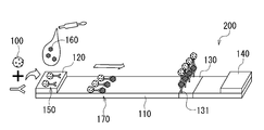
- FIG. 1 is a schematic diagram showing a cross-sectional structure of a resin-metal composite according to an embodiment of the present invention. It is explanatory drawing which shows the outline
- mold chromatography which concerns on one embodiment of this invention.
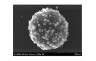
- 2 is a scanning electron microscope (SEM) photograph of the resin-gold composite obtained in Example 1.
- FIG. 3 is a scanning electron microscope (SEM) photograph of the resin-gold composite obtained in Example 2.
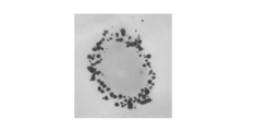
- FIG. 4 is a scanning transmission electron microscope (STEM) photograph of a cross section of the resin-gold composite obtained in Example 2.
- FIG. 3 is a scanning electron microscope (SEM) photograph of the resin-gold composite obtained in Example 3.
- FIG. 4 is a scanning transmission electron microscope (STEM) photograph of a cross section of the resin-gold composite obtained in Example 3.
- FIG. 2 is a scanning electron microscope (SEM) photograph of the resin-gold composite obtained in Example 10.
- FIG. 1 is a schematic sectional view of a resin-metal composite according to an embodiment of the present invention.
- the resin-metal composite 100 includes resin particles 10 and metal particles 20.
- the metal particles 20 are dispersed or fixed to the resin particles 10.
- a part of the metal particles 20 is three-dimensionally distributed in the surface layer portion 60 of the resin particle 10, and a part of the three-dimensionally distributed metal particles 20 is partially.
- the resin particles 10 are exposed outside, and the remaining part is encapsulated in the resin particles 10.
- the metal particles 20 include metal particles completely encapsulated in the resin particles 10 (hereinafter also referred to as “encapsulated metal particles 30”), portions embedded in the resin particles 10, and outside the resin particles 10.
- Metal particles having an exposed portion hereinafter also referred to as “partially exposed metal particles 40” and metal particles adsorbed on the surfaces of the resin particles 10 (hereinafter also referred to as “surface-adsorbed metal particles 50”).
- the partially exposed metal particles 40 and the surface-adsorbed metal particles 50 correspond to “first particles” in the present invention
- the encapsulated metal particles 30 correspond to “second particles” in the present invention.
- the resin-metal complex 100 when used as a labeling substance for immunological measurement or a reagent for immunological measurement, an antibody or an antigen is immobilized on the partially exposed metal particles 40 or the surface-adsorbed metal particles 50. use. At this time, the antibody or antigen is immobilized on the partially exposed metal particles 40 and the surface-adsorbed metal particles 50, but not immobilized on the encapsulated metal particles 30. However, since all of the metal particles 20 including the encapsulated metal particles 30 exhibit localized surface plasmon absorption, not only the partially exposed metal particles 40 and the surface adsorbed metal particles 50 but also the encapsulated metal particles 30 are immunological.
- the partially exposed metal particles 40 and the encapsulated metal particles 30 have a large contact area with the resin particles 10 as compared with the surface-adsorbed metal particles 50, and also have a physical adsorption force such as an anchor effect due to the embedded state. Strong and difficult to desorb from the resin particles 10. Therefore, the durability and stability of the immunological measurement labeling substance and the immunological measurement reagent using the resin-metal complex 100 can be improved.
- the resin-metal complex 100 is applied to an immunological measurement labeling substance (hereinafter also simply referred to as “labeling substance”) or an immunological measurement reagent (hereinafter also simply referred to as “reagent”).
- labeling substance an immunological measurement labeling substance
- reagent an immunological measurement reagent
- the encapsulated metal particles 30 are all covered with the resin constituting the resin particles 10 on the entire surface.
- the partially exposed metal particles 40 are those in which 5% or more and less than 100% of the surface area is covered with the resin constituting the resin particles 10. From the viewpoint of durability for each of the above uses, the lower limit is preferably 20% or more of the surface area, and more preferably 30% or more.
- the surface-adsorbed metal particles 50 are covered with the resin constituting the resin particles 10 with more than 0% and less than 5% of the surface area thereof.
- the amount of the metal particles 20 (the total of the encapsulated metal particles 30, the partially exposed metal particles 40, and the surface adsorbed metal particles 50) supported on the resin-metal composite 100 is based on the weight of the resin-metal composite 100. It is preferably 5 wt% to 70 wt%. Within this range, the resin-metal composite 100 is excellent in visibility as a labeling substance, visual judgment and detection sensitivity. When the loading amount of the metal particles 20 is less than 5 wt%, the amount of antibody or antigen immobilized is decreased, and the detection sensitivity tends to decrease.
- the supported amount of the metal particles 20 is more preferably 15 wt% to 70 wt%.
- 10 wt% to 90 wt% of the metal particles 20 are preferably partially exposed metal particles 40 and surface adsorbed metal particles 50. Within this range, a sufficient amount of antibody or antigen immobilized on the metal particles 20 can be secured, and the sensitivity as a labeling substance is high. More preferably, 20 wt% to 80 wt% of the metal particles 20 are the partially exposed metal particles 40 and the surface adsorbed metal particles 50, and from the viewpoint of durability, the surface adsorbed metal particles 50 are more preferably 20 wt% or less. .
- the resin-metal complex 100 when used for immunological measurement, in order to obtain excellent detection sensitivity, 60 wt% to 100 wt%, preferably 75 to 100 wt% of the metal particles 20 is more preferable. Is preferably in the range of 40% of the particle radius in the depth direction from the surface of the resin particle 10 in the surface layer portion 60.
- 5 wt% to 90 wt% of the metal particles 20 present on the surface layer portion 60 are partially exposed metal particles 40 or surface adsorbed metal particles 50, which indicates that the amount of antibody or antigen immobilized on the metal particles 20 is fixed. Since it can be secured sufficiently, the sensitivity as a labeling substance is increased, which is preferable. In other words, it is preferable that 10 wt% to 95 wt% of the metal particles 20 existing in the surface layer portion 60 are the encapsulated metal particles 30.
- the “surface layer portion” refers to the resin particle 10 based on the outermost position of the resin-metal composite 100 (that is, the protruding end portion of the partially exposed metal particle 40 or the surface-adsorbed metal particle 50). Means a range of 50% of the particle radius in the depth direction.
- the “three-dimensional distribution” means that the metal particles 20 are dispersed not only in the surface direction of the resin particles 10 but also in the depth direction.
- the resin particles 10 are preferably polymer particles having a substituent in the structure capable of adsorbing metal ions.
- nitrogen-containing polymer particles are preferable.
- the nitrogen atom in the nitrogen-containing polymer is preferable because it is easy to chemisorb an anionic metal ion that is a precursor of metal particles such as gold and palladium, which has excellent visibility and can easily immobilize an antigen or antibody.
- metal ions adsorbed in the nitrogen-containing polymer are reduced to form metal nanoparticles, so that part of the generated metal particles 20 becomes encapsulated metal particles 30 or partially exposed metal particles 40. .
- carboxylic acid and the like can adsorb cationic metal ions, so it is easy to adsorb cationic metal ions that are precursors of metal particles such as silver, nickel, copper, etc.
- the metal particles 20 such as nickel and copper can be formed, and an alloy with the metal such as gold and palladium can be formed.
- resin particles other than the nitrogen-containing polymer having a substituent capable of adsorbing metal ions, such as polystyrene it is difficult to adsorb the metal ions inside the resin. As a result, most of the generated metal particles 20 become surface-adsorbed metal particles 50.
- the nitrogen-containing polymer is a resin having a nitrogen atom in the main chain or side chain, and examples thereof include polyamine, polyamide, polypeptide, polyurethane, polyurea, polyimide, polyimidazole, polyoxazole, polypyrrole, and polyaniline.
- Preferred are polyamines such as poly-2-vinylpyridine, poly-3-vinylpyridine, poly-4-vinylpyridine, and the like.
- an acrylic resin, a phenol resin, an epoxy resin, etc. can be used widely.
- silver, nickel, copper, gold, palladium or the like can be applied to the metal particles 20.
- Gold and palladium are preferable because they are excellent in visibility and can easily immobilize antigens or antibodies. These are preferable because they express absorption derived from localized surface plasmon resonance. More preferably, it is gold with good storage stability.
- These metals can be used as a simple substance or a composite such as an alloy.
- a gold alloy means an alloy composed of gold and a metal species other than gold and containing 10% by weight or more of gold.
- the average particle diameter of the metal particles 20 measured by scanning electron microscope (SEM) observation is preferably 1 to 80 nm, for example.
- SEM scanning electron microscope
- the average particle diameter of the metal particles 20 is less than 1 nm or exceeds 80 nm, the localized surface plasmon is less likely to be expressed, and thus the sensitivity tends to decrease.
- the metal particle 20 is a gold particle
- the average particle diameter of the metal particle 20 is preferably 20 nm or more and less than 70 nm, and more preferably 22 nm or more and less than 50 nm.
- the average particle diameter of the resin-metal composite 100 is preferably 100 to 1000 nm, for example. If the average particle size of the resin-metal composite 100 is less than 100 nm, for example, when gold particles are used as the metal particles 20, the amount of gold particles tends to be reduced, so that coloring is more likely than gold particles of the same size. When it exceeds 1000 nm, when it is used as a labeling substance or reagent, it tends to be clogged in the pores of a chromatographic medium such as a membrane filter, and the dispersibility tends to decrease.
- the average particle diameter of the resin-metal composite 100 is preferably 100 nm or more and less than 700 nm, and more preferably 340 nm or more and less than 650 nm.
- the particle diameter of the resin-metal composite 100 means a value obtained by adding the length of the protruding portion of the partially exposed metal particle 40 or the surface-adsorbed metal particle 50 to the particle diameter of the resin particle 10. / Measured by scattering method, dynamic light scattering method, or centrifugal sedimentation method.
- the method for producing the resin-metal composite 100 is not particularly limited.
- a solution containing metal ions is added to a dispersion of resin particles 10 produced by an emulsion polymerization method, and the metal ions are adsorbed on the resin particles 10 (hereinafter referred to as “metal ion adsorption resin particles”).
- metal ion adsorption resin particles a solution containing metal ions is added to a dispersion of resin particles 10 produced by an emulsion polymerization method, and the metal ions are adsorbed on the resin particles 10 (hereinafter referred to as “metal ion adsorption resin particles”).
- metal ion adsorption resin particles the metal ions are reduced to generate the metal particles 20, and the resin-metal composite 100 is obtained.
- examples of the solution containing metal ions include chloroauric acid (HAuCl 4 ) aqueous solution.
- Hauric acid HuCl 4
- water-containing alcohol or alcohol such as methanol, ethanol, n-propanol, isopropanol, n-butanol, sec-butanol, t-butanol, alcohol, hydrochloric acid, sulfuric acid, nitric acid, instead of water
- An acid such as the above may be used.
- water-soluble polymer compounds such as polyvinyl alcohol, surfactants, alcohols; ethers such as tetrahydrofuran, diethyl ether, diisopropyl ether; alkylene glycol, polyalkylene glycol, these Additives such as various water-miscible organic solvents such as polyols such as monoalkyl ether or dialkyl ether, glycerol, etc .; ketones such as acetone and methyl ethyl ketone may be added. Such an additive is effective for accelerating the reduction reaction rate of metal ions and controlling the size of the metal particles 20 to be produced.
- a well-known thing can be used for a reducing agent.
- sodium borohydride, dimethylamine borane, citric acid, sodium hypophosphite, hydrazine hydrate, hydrazine hydrochloride, hydrazine sulfate, formaldehyde, sucrose, glucose, ascorbic acid, erythorbic acid, sodium phosphinate, hydroquinone, Rochelle Examples include salts.
- sodium borohydride, dimethylamine borane, and citric acid are preferable.
- a surfactant can be added to the reducing agent solution as necessary, and the pH of the solution can be adjusted.
- the pH can be adjusted with a buffer such as boric acid or phosphoric acid, an acid such as hydrochloric acid or sulfuric acid, or an alkali such as sodium hydroxide or potassium hydroxide.
- a buffer such as boric acid or phosphoric acid, an acid such as hydrochloric acid or sulfuric acid, or an alkali such as sodium hydroxide or potassium hydroxide.
- the particle size of the metal particles to be formed can be controlled by adjusting the reduction rate of the metal ions depending on the temperature of the reducing agent solution.
- the metal ion adsorption resin particles may be added to the reducing agent solution, or the reducing agent may be added to the metal ion adsorption resin. Although it may be added to the particles, the former is preferable from the viewpoint of easy generation of the encapsulated metal particles 30 and the partially exposed metal particles 40.
- the dispersibility of the resin-metal composite 100 in water for example, citric acid, poly-L-lysine, polyvinyl pyrrolidone, polyvinyl pyridine, polyvinyl alcohol, DISPERBYK194, DISPERBYK180, DISPERBYK184 (Big Chemy Japan)
- a dispersant may be added.
- the dispersibility can be maintained by adjusting the pH with a buffer such as boric acid or phosphoric acid, an acid such as hydrochloric acid or sulfuric acid, or an alkali such as sodium hydroxide or potassium hydroxide.
- the resin-metal complex 100 having the above configuration is used as a labeling substance, for example, by adsorbing an antigen or antibody on the surface of the metal particle 20, for example, EIA, RIA, CLIA, FIA, LIA, PA, ICA, HA. It is preferably applicable to immunological measurement methods such as HI. In particular, it can be preferably applied as a labeling substance for immunological measurement or a reagent for immunological measurement excellent in visual determinability in a low concentration region (high sensitivity region).
- the form of the immunological measurement labeling substance or immunological measurement reagent is not particularly limited. For example, a dispersion in which the resin-metal complex 100 is dispersed in water or a pH-adjusted buffer. Can be used as
- the method for adsorbing an antigen or antibody on the surface of the metal particle 20 is not particularly limited, and known physical adsorption and chemical adsorption methods can be used.
- physical adsorption such as immersing the resin-metal complex 100 in a buffer containing the antigen or antibody and incubating it, or introducing SH groups into the antigen or antibody and reacting with the resin-metal complex 100 to cause Au
- chemisorption such as formation of —SH bond. Of these, chemisorption is preferred because the bond between the metal particles 20 and the antigen or antibody is strong.
- the resin-metal complex 100 can be preferably applied as a solid catalyst, pigment, paint, conductive material, electrode, or sensor element for uses other than the labeling substance for immunological measurement or the reagent for immunological measurement.
- test strip for lateral flow chromatography
- test strip a lateral flow type chromatographic test strip (test strip) according to an embodiment of the present invention will be described with reference to FIG.
- the test strip 200 can be preferably used in the analyte measuring method of one embodiment of the present invention.
- the test strip 200 includes a membrane 110.
- the membrane 110 is provided with a sample addition unit 120, a determination unit 130, and a liquid absorption unit 140 in order in the sample development direction.
- a membrane used as a membrane material in a general test strip can be applied.
- the membrane 110 is formed of an inert substance (a substance that does not react with the analyte 160, various ligands, etc.) made of a microporous material that exhibits a capillary phenomenon and develops the sample at the same time as the sample is added. It is.
- Specific examples of the membrane 110 include a fibrous or non-woven fibrous matrix composed of polyurethane, polyester, polyethylene, polyvinyl chloride, polyvinylidene fluoride, nylon, cellulose derivatives, etc., membrane, filter paper, glass fiber filter paper, cloth, Cotton etc. are mentioned.
- membranes composed of cellulose derivatives and nylon, filter paper, glass fiber filter paper, etc. are preferably used, more preferably nitrocellulose membrane, mixed nitrocellulose ester (mixture of nitrocellulose and cellulose acetate) membrane, nylon membrane Filter paper is used.
- the test strip 200 preferably includes a support that supports the membrane 110 in order to make the operation easier.
- a support for example, plastic can be used.
- the test strip 200 may have a sample adding unit 120 for adding a sample including the analyte 160.
- the sample addition unit 120 is a part for receiving a sample including the analyte 160 in the test strip 200.
- the sample addition unit 120 may be formed on the membrane 110 upstream of the determination unit 130 in the direction in which the sample is developed, or, for example, cellulose filter paper, glass fiber, polyurethane, polyacetate, cellulose acetate, nylon
- a sample addition pad made of a material such as cotton cloth may be provided on the membrane 110 to constitute the sample addition unit 120.
- a capture ligand 131 that specifically binds to the analyte 160 is fixed to the determination unit 130.
- the capture ligand 131 can be used without particular limitation as long as it forms a specific bond with the analyte 160.
- an antibody against the analyte 160 can be preferably used.
- the capture ligand 131 is immobilized so as not to move from the determination unit 130 even when a sample is provided to the test strip 200.
- the capture ligand 131 may be fixed directly or indirectly to the membrane 110 by physical or chemical bonding or adsorption.
- the determination unit 130 is not particularly limited as long as the complex 170 including the labeled antibody 150 and the analyte 160 is in contact with the capture ligand 131 that specifically binds to the analyte 160.
- the capture ligand 131 may be directly fixed to the membrane 110, or the capture ligand 131 may be fixed to a pad made of cellulose filter paper, glass fiber, nonwoven fabric, or the like fixed to the membrane 110. .
- the liquid-absorbing part 140 is formed of a pad of a water-absorbing material such as cellulose filter paper, non-woven fabric, cloth, cellulose acetate or the like.
- the moving speed of the sample after the development front (front line) of the added sample reaches the liquid absorption part 140 varies depending on the material, size, and the like of the liquid absorption part 140. Therefore, the optimum speed for detection / quantification of the analyte 160 can be set by selecting the material, size, etc. of the liquid absorption part 140.
- the liquid absorption part 140 is an arbitrary configuration and may be omitted.
- the test strip 200 may further include arbitrary parts such as a reaction part and a control part as necessary.
- the test strip 200 may be formed with a reaction part including the labeled antibody 150 on the membrane 110.
- the reaction unit can be provided upstream of the determination unit 130 in the direction in which the sample flows.
- the test strip 200 has a reaction part, if the sample containing the analyte 160 is supplied to the reaction part or the sample addition part 120, the analyte 160 contained in the sample and the labeled antibody 150 can be brought into contact with each other in the reaction part. it can.
- the complex 170 containing the analyte 160 and the labeled antibody 150 can be formed by simply supplying the sample to the reaction part or the sample addition part 120, so-called one-step type immunochromatography is possible. Become.
- the reaction part is not particularly limited as long as it includes the labeled antibody 150 that specifically binds to the analyte 160, but may be one in which the labeled antibody 150 is directly applied to the membrane 110.
- the reaction part may be formed by, for example, immobilizing the membrane 110 with a pad (conjugate pad) made of cellulose filter paper, glass fiber, nonwoven fabric, or the like impregnated with the labeled antibody 150.
- the test strip 200 may be formed with a control unit in which a capture ligand that specifically binds to the labeled antibody 150 is fixed to the membrane 110 in the direction in which the sample is developed.
- a control unit By measuring the color intensity at the control unit together with the determination unit 130, the sample provided for the test strip 200 is developed and reaches the reaction unit and the determination unit 130 to confirm that the test has been performed normally. be able to.
- the control unit is prepared in the same manner as the determination unit 130 described above except that another type of capture ligand that specifically binds to the labeled antibody 150 is used instead of the capture ligand 131. The configuration can be taken.
- the measuring method of the analyte 160 of the present embodiment is a measuring method of the analyte 160 that detects or quantifies the analyte 160 contained in the sample.
- the method for measuring the analyte 160 according to the embodiment uses the test strip 200 including the membrane 110 and the determination unit 130 in which the capture ligand 131 that specifically binds to the analyte 160 is fixed to the membrane 110.
- Step (I) is a step of bringing the analyte 160 contained in the sample into contact with the labeled antibody 150.
- the mode of contact is not particularly limited.
- the sample may be provided to the sample addition unit 120 or the reaction unit (not shown) of the test strip 200, and the analyte 160 may be brought into contact with the labeled antibody 150 in the reaction unit, or before the sample is applied to the test strip 200.
- the analyte 160 in the sample may be brought into contact with the labeled antibody 150.
- the composite 170 formed in step (I) expands and moves on the test strip 200 and reaches the determination unit 130.
- step (II) in the determination unit 130 of the test strip 200, the complex 170 formed in step (I) and including the analyte 160 and the labeled antibody 150 is brought into contact with the capture ligand 131.
- the capture ligand 131 specifically binds to the analyte 160 of the complex 170.
- the complex 170 is captured by the determination unit 130.
- the capture ligand 131 does not specifically bind to the labeled antibody 150, when the analyte 160 and the unbound labeled antibody 150 reach the determination unit 130, the analyte 160 and the unbound labeled antibody 150 are unbound. Passes through the determination unit 130.
- a control unit (not shown) to which another capture ligand that specifically binds to the labeled antibody 150 is fixed is formed on the test strip 200, the labeled antibody 150 that has passed through the determination unit 130 is developed. Then, bind to the other capture ligand at the control unit. As a result, the labeled antibody 150 that does not form the complex 170 with the analyte 160 is captured by the control unit.
- step (II) if necessary, before step (III), for example, the test strip 200 is washed with a buffer solution commonly used in biochemical tests such as water, physiological saline, and phosphate buffer solution.
- a cleaning step may be performed.
- the labeled antibody 150 (the labeled antibody 150 that is not bound to the analyte 160 and does not form the complex 170) that has not been captured by the determination unit 130 or the determination unit 130 and the control unit is removed. be able to.
- the background Color development intensity can be reduced, the signal / background ratio can be increased, and detection sensitivity and quantitativeness can be further improved.
- Step (III) is a step of measuring the color intensity derived from the localized surface plasmon resonance of the resin-metal composite 100. After the step (II) or the washing step as necessary, the color intensity derived from the localized surface plasmon resonance of the resin-metal composite 100 is measured on the test strip 200.
- step (II) When a control part is formed on the test strip 200, in step (II), the labeled antibody 150 is captured by another capture ligand in the control part to form a complex. Therefore, in step (III), in the test strip 200, color development by localized surface plasmon resonance can be generated not only in the determination unit 130 but also in the control unit. In this way, by measuring the color intensity in the control unit as well as the determination unit 130, it is possible to confirm whether the sample provided for the test strip 200 has developed normally and reached the reaction unit and the determination unit 130.
- the sample in the analyte measurement method of the present embodiment is not particularly limited as long as it includes a substance that can be an antigen such as a protein as the analyte 160.
- a biological sample containing the analyte 160 of interest ie, whole blood, serum, plasma, urine, saliva, sputum, nasal or throat swab, cerebrospinal fluid, amniotic fluid, nipple discharge, tears, sweat, skin exudate , Extracts from tissues, cells and stool, etc.
- food extracts ie, whole blood, serum, plasma, urine, saliva, sputum, nasal or throat swab, cerebrospinal fluid, amniotic fluid, nipple discharge, tears, sweat, skin exudate , Extracts from tissues, cells and stool, etc.
- the analyte 160 contained in the sample is pretreated prior to the step (I). May be.
- the pretreatment include chemical treatment using various chemicals such as acid, base, and surfactant, and physical treatment using heating, stirring, ultrasonic waves, and the like.
- the analyte 160 is a substance that is not normally exposed on the surface, such as an influenza virus NP antigen, it is preferable to perform treatment with a surfactant or the like.
- a nonionic surfactant can be used in consideration of specific binding reaction, for example, binding reactivity between the ligand 160 and the analyte 160 such as antigen-antibody reaction. .
- the sample may be appropriately diluted with a solvent (water, physiological saline, buffer, or the like) used in a normal immunological analysis method or a water-miscible organic solvent.
- a solvent water, physiological saline, buffer, or the like
- analyte 160 examples include tumor markers, signaling substances, proteins such as hormones (including polypeptides and oligopeptides), nucleic acids (single-stranded or double-stranded DNA, RNA, polynucleotides, oligos) Other molecules such as nucleotides, PNA (including peptide nucleic acids)) or nucleic acid-containing substances, sugars (including oligosaccharides, polysaccharides, sugar chains, etc.) or sugar chains, lipids, and the like.
- proteins such as hormones (including polypeptides and oligopeptides), nucleic acids (single-stranded or double-stranded DNA, RNA, polynucleotides, oligos)
- Other molecules such as nucleotides, PNA (including peptide nucleic acids)) or nucleic acid-containing substances, sugars (including oligosaccharides, polysaccharides, sugar chains, etc.) or
- CEA carcinoembryonic antigen
- HER2 protein HER2 protein
- PSA prostate specific antigen
- CA19-9 ⁇ -fetoprotein
- IPA Immunosuppressive acidic protein
- CA15-3 CA125
- estrogen receptor progesterone Scepter
- HCG human chorionic gonadotropin
- LH luteinizing hormone
- FSH follicle stimulating hormone
- syphilis antibody influenza virus human hemoglobin, chlamydia antigen
- examples include group A ⁇ -streptococcal antigens, HBs antibodies, HBs antigens, rotaviruses, adenoviruses, albumin, glycated albumin and the like.
- antigens solubilized by nonionic surfactants are preferable, and
- the labeled antibody 150 is used to contact the analyte 160 contained in the sample to form a complex 170 including the analyte 160 and the labeled antibody 150.
- the labeled antibody 150 is obtained by labeling an antibody that specifically binds to the analyte 160 with the resin-metal complex 100 having a structure in which a plurality of metal particles 20 are immobilized on the resin particles 10.
- labeling means that the resin-metal complex 100 is directly attached to the antibody to the extent that the resin-metal complex 100 is not detached from the labeled antibody 150 in steps (I) to (III). It means that it is fixed indirectly by chemical or physical bonding or adsorption.
- the labeled antibody 150 may be one in which the resin-metal complex 100 is directly bonded to the antibody, or the antibody and the resin-metal complex 100 are bonded via any linker molecule. Or those fixed to insoluble particles.
- the “antibody” is not particularly limited.
- a monoclonal antibody an antibody obtained by genetic recombination, an antibody fragment [for example, an H chain , L chain, Fab, F (ab ′) 2 etc.] can be used.
- IgG, IgM, IgA, IgE, or IgD may be used as the immunoglobulin.
- the animal species that produce the antibody may be humans or animals other than humans (eg, mice, rats, rabbits, goats, horses, etc.).
- Specific examples of the antibody include anti-PSA antibody, anti-AFP antibody, anti-CEA antibody, anti-adenovirus antibody, anti-influenza virus antibody, anti-HCV antibody, anti-IgG antibody, and anti-human IgE antibody.
- the production of the labeled antibody 150 is at least the following step A; Step A) including the step of obtaining the labeled antibody 150 by mixing and binding the resin-metal complex 100 with the antibody under a first pH condition, and preferably, further includes Step B; Step B) A step of treating the labeled antibody 150 under a second pH condition can be included.
- Step A the labeled antibody 150 is obtained by mixing the resin-metal complex 100 with an antibody under the first pH condition.
- the solid resin-metal complex 100 is preferably contacted with the antibody in a state dispersed in the liquid phase.
- the first pH condition varies depending on the metal species of the metal particle 20 in the resin-metal composite 100.
- the first pH condition is that the binding with the antibody is the resin-metal composite 100.
- the metal particles 20 of the resin-metal composite 100 are gold particles (including gold alloy particles; the same applies hereinafter)
- the first pH condition is that the binding with the antibody is the resin-metal composite 100.
- conditions within the range of pH 2 to 7 are preferable, and acidic conditions such as pH 2.5 to 5.5 are preferred. Within the range is more preferable.
- the metal particles 20 are gold particles, if the resin-metal complex 100 and the antibody are bonded under conditions where the pH is less than 2, the antibody may be altered and deactivated due to strong acidity.
- the metal complex 100 and the antibody are mixed, they aggregate and become difficult to disperse. However, if the antibody is not inactivated by strong acidity, the treatment can be performed even at a pH of less than 2.
- the first pH condition is that the binding with the antibody is the resin-metal complex 100.
- the metal particles 20 of the resin-metal complex 100 are particles other than gold, such as palladium or an alloy thereof
- the first pH condition is that the binding with the antibody is the resin-metal complex 100.
- the metal particles 20 are particles other than gold
- the antibody may be denatured and deactivated due to strong acidity when the conditions for bonding the resin-metal complex 100 and the antibody are less than pH 2, If the pH exceeds 10, the resin-metal complex 100 and the antibody are aggregated when mixed, making dispersion difficult.
- the treatment can be performed even at a pH of less than 2.
- Step A is preferably performed in a binding buffer adjusted to the first pH condition.
- a predetermined amount of the resin-metal complex 100 is mixed in the binding buffer adjusted to the above pH and mixed thoroughly.
- a boric acid solution adjusted to a predetermined concentration can be used as the binding buffer.
- the pH of the binding buffer can be adjusted using, for example, hydrochloric acid or sodium hydroxide.
- a labeled antibody-containing liquid can be obtained by adding a predetermined amount of the antibody to the obtained mixed liquid, sufficiently stirring and mixing.
- the labeled antibody-containing liquid thus obtained can be fractionated only with the labeled antibody 150 as a solid part by solid-liquid separation means such as centrifugation.
- Step B the labeled antibody 150 obtained in the process A is treated under the second pH condition to perform blocking that suppresses nonspecific adsorption to the labeled antibody 150.
- the labeled antibody 150 separated by the solid-liquid separation means is dispersed in the liquid phase under the second pH condition.
- This blocking condition varies depending on the metal species of the metal particle 20 in the resin-metal composite 100.
- the second pH condition is, for example, within the range of pH 2 to 9 from the viewpoint of maintaining the activity of the antibody and suppressing the aggregation of the labeled antibody 150.
- acidic conditions for example, in the range of pH 2 to 6, are more preferable. If the blocking condition is less than pH 2, the antibody may be denatured and deactivated due to strong acidity, and if it exceeds pH 9, the labeled antibody 150 aggregates, making dispersion difficult.
- the second pH condition is, for example, from pH 2 to pH 2 from the viewpoint of maintaining antibody activity and suppressing aggregation of the labeled antibody 150.
- the range of 10 is preferable, and the range of pH 5 to 9 is more preferable from the viewpoint of suppressing nonspecific adsorption of the labeled antibody 150.
- the blocking condition is less than pH 2, the antibody may be denatured and deactivated due to strong acidity, and if it exceeds pH 10, the labeled antibody 150 aggregates, making dispersion difficult.
- Step B is preferably performed using a blocking buffer adjusted to the second pH condition.
- the blocking buffer adjusted to the above pH is added to a predetermined amount of labeled antibody 150, and the labeled antibody 150 is uniformly dispersed in the blocking buffer.
- the blocking buffer for example, a protein solution that does not bind to the detection target is preferably used.
- proteins that can be used in the blocking buffer include bovine serum albumin, ovalbumin, casein, and gelatin. More specifically, it is preferable to use a bovine serum albumin solution adjusted to a predetermined concentration.
- the pH of the blocking buffer can be adjusted using, for example, hydrochloric acid or sodium hydroxide.
- a dispersing means such as ultrasonic treatment. In this way, a dispersion in which the labeled antibody 150 is uniformly dispersed is obtained.
- a dispersion of labeled antibody 150 is obtained. From this dispersion, only the labeled antibody 150 can be fractionated as a solid part by solid-liquid separation means such as centrifugation. Moreover, a cleaning process, a preservation
- a washing buffer solution is added to the labeled antibody 150 sorted by the solid-liquid separation means, and the labeled antibody 150 is uniformly dispersed in the washing buffer solution.
- a dispersion means such as ultrasonic treatment is preferably used.
- the washing buffer is not particularly limited, and for example, a Tris buffer solution, a glycinamide buffer solution, an arginine buffer solution, or the like having a predetermined concentration adjusted within a pH range of 8 to 9 may be used. it can.
- the pH of the washing buffer can be adjusted using, for example, hydrochloric acid or sodium hydroxide.
- the washing treatment of the labeled antibody 150 can be repeated a plurality of times as necessary.
- a storage buffer is added to the labeled antibody 150 collected by the solid-liquid separation means, and the labeled antibody 150 is uniformly dispersed in the storage buffer.
- a dispersion means such as ultrasonic treatment is preferably used.
- the storage buffer for example, a solution obtained by adding a predetermined concentration of an anti-aggregation agent and / or stabilizer to the washing buffer can be used.
- the aggregation inhibitor for example, saccharides typified by sucrose, maltose, lactose, trehalose, polyhydric alcohols typified by glycerin, polyvinyl alcohol, and the like can be used.
- the stabilizer is not particularly limited. For example, proteins such as bovine serum albumin, ovalbumin, casein, and gelatin can be used.
- save process of the labeled antibody 150 can be performed.
- a surfactant or a preservative such as sodium azide or paraoxybenzoate can be used as necessary.
- An analyte measurement kit uses, for example, a lateral flow type chromatographic test strip 200 and an analyte 160 contained in a sample based on the analyte measurement method of the present embodiment. It is a kit for detecting or quantifying.
- the kit of this embodiment is A lateral flow type chromatographic test strip 200 including a membrane 110 and a determination unit 130 in which a capture ligand that specifically binds to the analyte 160 is fixed to the membrane 110; A detection reagent comprising a labeled antibody 150 in which an antibody that specifically binds to the analyte 160 is labeled with a resin-metal complex 100 having a structure in which a plurality of metal particles 20 are immobilized on resin particles 10; Is included.
- the kit of the present embodiment may further include other components as necessary.
- the reaction section or the sample addition section 120 of the test strip 200 is performed.
- the step (II) and the step (III) may be sequentially performed by supplying a sample.
- a detection reagent is applied to the upstream side of the determination unit 130 of the test strip 200 and appropriately dried to form a reaction part, the formed reaction part or a position upstream of the reaction part (for example, The sample may be added to the sample addition unit 120) and the steps (I) to (III) may be performed sequentially.
- the absorbance of the resin-metal composite was measured by placing a resin-metal composite dispersion liquid (dispersion medium: water) prepared at 0.01 wt% into an optical white glass cell (optical path length 10 mm), and an instantaneous multi-photometry system (Otsuka In the case of gold, absorbance at 570 nm was measured using MCPD-3700) manufactured by Denki Co. In the case of gold, the absorbance at 570 nm is 0.9 (good), 0.5 to less than 0.9 is ⁇ (good), and less than 0.5 is x (impossible).
- Solid content concentration (wt%) [weight after drying (g) / weight before drying (g)] ⁇ 100
- Metal loading (wt%) [Weight after heat treatment at 500 ° C. (g) / Weight before heat treatment at 500 ° C. (g)] ⁇ 100
- the average particle size of the metal particles was measured by dropping a resin-metal composite dispersion liquid onto a metallic mesh with a carbon support film, using a field emission scanning electron microscope (STEM; manufactured by Hitachi High-Technologies Corporation, SU The area average diameter of the metal particles was measured from the image observed by ⁇ 9000).
- the evaluation was performed using a monochrome screen for influenza A evaluation (manufactured by Adtech), and the coloring levels after 5 minutes, 10 minutes and 15 minutes were compared. In the performance evaluation, a 2-fold dilution series (1-fold to 1024-fold) of influenza A positive control (APC) was used as the antigen (the virus concentration before APC dilution was 5000 FFU / ml).
- APC influenza A positive control
- ⁇ Evaluation of immunochromatography> 100 ⁇ g of influenza antibody was mixed with 1 ml of the obtained gold colloid (OD 10) and stirred at room temperature for about 3 hours to bind the antibody to the gold colloid.
- the bovine serum albumin solution was added so that the final concentration was 1%, and the mixture was stirred at room temperature for 2 hours to block the colloidal gold surface.
- the gold colloid-labeled antibody was prepared by centrifuging at 12000 rpm and 4 ° C. for 5 minutes and then suspending in a buffer containing 0.2% bovine serum albumin. Using the prepared colloidal gold labeled antibody, the performance of the colloidal gold was evaluated by measuring by the following immunochromatography method.
- Evaluation was performed in the same manner as in Comparative Example 1.
- Evaluation procedure 3 ⁇ l of colloidal gold-labeled antibody was added to each well of a 96-well plate, and 100 ⁇ l each of a 2-fold dilution series (1-fold to 1024-fold) of APC and a negative control was mixed. Next, 50 ⁇ l was added to a monochrome screen for influenza A evaluation, and the color development level after 5 minutes, 10 minutes and 15 minutes was evaluated. The results are shown below.
- the 2.5 wt% gold ion adsorption resin particle dispersion (42.4 ml) is added to 1580 ml of pure water, and a 528 mM dimethylamine borane aqueous solution (10 ml) is added over 2 minutes while stirring at 160 rpm and 20 ° C. After the dropwise addition, the mixture was stirred at room temperature for 2 hours to obtain a resin-gold composite having an average particle size of 438 nm. After the resin-gold complex is precipitated by centrifugation (3100 rpm, 60 minutes), the supernatant is removed, and the operation of re-dispersing in pure water is repeated three times, followed by purification by dialysis and concentration adjustment.
- a 1 wt% resin-gold composite dispersion was obtained.
- the absorbance of the produced resin-gold composite was 0.98 as a result of measurement according to the above method.
- the formed gold particles had an average particle size of 25.0 nm and a gold loading amount of 54.7 wt%.
- a scanning electron microscope (SEM) photograph of the surface of the obtained resin-gold composite is shown in FIG.
- the gold particles are: encapsulated particles that are completely encapsulated in the resin particles; partially exposed gold particles having a portion embedded in the resin particle and a portion exposed outside the resin particle; Surface adsorbed gold particles adsorbed on the surface of the resin particles, and at least some of the gold particles were three-dimensionally distributed in the surface layer portion of the resin particles.
- the 2.5 wt% gold ion adsorption resin particle dispersion (42.4 ml) is added to 1580 ml of pure water, and a 528 mM dimethylamine borane aqueous solution (10 ml) is added over 2 minutes while stirring at 160 rpm and 20 ° C. After the dropwise addition, the mixture was stirred at room temperature for 2 hours to obtain a resin-gold composite having an average particle size of 399 nm. After the resin-gold complex is precipitated by centrifugation (3100 rpm, 60 minutes), the supernatant is removed, and the operation of re-dispersing in pure water is repeated three times, followed by purification by dialysis and concentration adjustment.
- a 1 wt% resin-gold composite dispersion was obtained.
- the absorbance of the produced resin-gold composite was 0.96 as a result of measurement according to the above method.
- the formed gold particles had an average particle size of 25.0 nm and a gold loading amount of 53.2 wt%.
- a scanning electron microscope (SEM) photograph of the surface of the obtained resin-gold composite is shown in FIG. 4, and a scanning transmission electron microscope (STEM) photograph of the cross section thereof is shown in FIG.
- the gold particles are: encapsulated metal particles that are completely encapsulated in the resin particles; Surface adsorbed gold particles adsorbed on the surface of the resin particles, and at least some of the gold particles were three-dimensionally distributed in the surface layer portion of the resin particles. In addition, 100% of gold particles were present in the surface layer portion.
- the 2.5 wt% gold ion adsorption resin particle dispersion (42.4 ml) is added to 1580 ml of pure water, and a 528 mM dimethylamine borane aqueous solution (10 ml) is added over 2 minutes while stirring at 160 rpm and 20 ° C. After the dropwise addition, the mixture was stirred at room temperature for 2 hours to obtain a resin-gold composite having an average particle size of 393 nm. After the resin-gold complex is precipitated by centrifugation (3100 rpm, 60 minutes), the supernatant is removed, and the operation of re-dispersing in pure water is repeated three times, followed by purification by dialysis and concentration adjustment.
- a 1 wt% resin-gold composite dispersion was obtained.
- the absorbance of the produced resin-gold composite was 0.92 as a result of measurement according to the above method.
- the formed gold particles had an average particle diameter of 14.9 nm and a gold loading amount of 55.8 wt%.
- a scanning electron microscope (SEM) photograph of the surface of the obtained resin-gold composite is shown in FIG. 6, and a scanning transmission electron microscope (STEM) photograph of the cross section thereof is shown in FIG.
- the gold particles are: encapsulated particles that are completely encapsulated in the resin particles; partially exposed gold particles having a portion embedded in the resin particle and a portion exposed outside the resin particle; Surface adsorbed gold particles adsorbed on the surface of the resin particles, and at least some of the gold particles were three-dimensionally distributed in the surface layer portion of the resin particles.
- the gold particles were present at 71% in the surface layer portion.
- Example 2 Comparing the cross-sectional images of FIG. 5 of Example 2 and FIG. 7 of Example 3, 75 to 100% of the gold particles are present in the range of 50% of the particle radius in the depth direction from the surface of the resin particles. In Example 2, the immunochromatographic detection sensitivity was superior.
- the 2.5 wt% gold ion adsorbing resin particle dispersion (42.4 ml) is added to 1580 ml of pure water and stirred at 160 rpm at 20 ° C., 528 mM dimethylamine borane aqueous solution (10 ml) and 528 mM boric acid.
- a mixed solution of an aqueous solution (10 ml) was added dropwise over 4 minutes and then stirred at room temperature for 2 hours to obtain a resin-gold composite having an average particle size of 250 nm.
- the gold particles are: encapsulated particles that are completely encapsulated in the resin particles; partially exposed gold particles having a portion embedded in the resin particle and a portion exposed outside the resin particle; Surface adsorbed gold particles adsorbed on the surface of the resin particles, and at least some of the gold particles were three-dimensionally distributed in the surface layer portion of the resin particles.
- Example 5 Aliquat 336 (manufactured by Aldrich) (1.00 g) and polyethylene glycol methyl ethyl ether methacrylate (PEGMA, 10.00 g) were dissolved in 300 g of pure water, and then 2-vinylpyridine (2-VP, 48.00 g) and Divinylbenzene (DVB, 2.00 g) was added, and the mixture was stirred at 150 rpm, 30 ° C. for 50 minutes, and then at 60 ° C. for 30 minutes under a nitrogen stream.
- PEGMA polyethylene glycol methyl ethyl ether methacrylate
- the 2.5 wt% gold ion adsorption resin particle dispersion (42.4 ml) is added to 1580 ml of pure water, and a 528 mM dimethylamine borane aqueous solution (10 ml) is added over 2 minutes while stirring at 160 rpm and 20 ° C. After the dropwise addition, the mixture was stirred at room temperature for 2 hours to obtain a resin-gold composite having an average particle size of 510 nm. After the resin-gold complex is precipitated by centrifugation (3100 rpm, 60 minutes), the supernatant is removed, and the operation of re-dispersing in pure water is repeated three times, followed by purification by dialysis and concentration adjustment.
- a 1 wt% resin-gold composite dispersion was obtained.
- the absorbance of the produced resin-gold composite was 1.01 as a result of measurement according to the above method.
- the formed gold particles had an average particle size of 46.3 nm and a gold loading amount of 54.2 wt%.
- the gold particles are: encapsulated particles that are completely encapsulated in the resin particles; partially exposed gold particles having a portion embedded in the resin particle and a portion exposed outside the resin particle; Surface adsorbed gold particles adsorbed on the surface of the resin particles, and at least some of the gold particles were three-dimensionally distributed in the surface layer portion of the resin particles.
- the 2.5 wt% gold ion adsorption resin particle dispersion (42.4 ml) is added to 1580 ml of pure water, and 528 mM dimethylamine borane aqueous solution (10 ml) is added over 2 minutes while stirring at 160 rpm at 3 ° C. After the dropwise addition, the mixture was stirred at room temperature for 2 hours to obtain a resin-gold composite having an average particle size of 625 nm. After the resin-gold complex is precipitated by centrifugation (3100 rpm, 60 minutes), the supernatant is removed, and the operation of re-dispersing in pure water is repeated three times, followed by purification by dialysis and concentration adjustment.
- a 1 wt% resin-gold composite dispersion was obtained.
- the absorbance of the produced resin-gold composite was 0.98 as a result of measurement according to the above method.
- the formed gold particles had an average particle diameter of 25.0 nm and a gold loading amount of 55.3 wt%.
- the gold particles are: encapsulated particles that are completely encapsulated in the resin particles; partially exposed gold particles having a portion embedded in the resin particle and a portion exposed outside the resin particle; Surface adsorbed gold particles adsorbed on the surface of the resin particles, and at least some of the gold particles were three-dimensionally distributed in the surface layer portion of the resin particles.
- Example 2 ⁇ Evaluation of immunochromatography> The same operation as in Example 1 was performed to prepare a resin-gold complex labeled antibody dispersion. Using the prepared resin-gold complex labeled antibody dispersion, measurement by immunochromatography was performed to evaluate the performance of the resin-gold complex dispersion. The results are shown below.
- Example 7 Aliquat 336 (manufactured by Aldrich) (1.00 g) and polyethylene glycol methyl ethyl ether methacrylate (PEGMA, 10.00 g) were dissolved in 300 g of pure water, and then 4-vinylpyridine (4-VP, 48.00 g) and Divinylbenzene (DVB, 2.00 g) was added, and the mixture was stirred at 150 rpm, 30 ° C. for 50 minutes, and then at 60 ° C. for 30 minutes under a nitrogen stream.
- PEGMA polyethylene glycol methyl ethyl ether methacrylate
- the 2.5 wt% gold ion adsorption resin particle dispersion (42.4 ml) is added to 1580 ml of pure water, and 528 mM dimethylamine borane aqueous solution (10 ml) is added over 2 minutes while stirring at 160 rpm at 3 ° C. After the dropwise addition, the mixture was stirred at room temperature for 2 hours to obtain a resin-gold composite having an average particle size of 448 nm. After the resin-gold complex is precipitated by centrifugation (3100 rpm, 60 minutes), the supernatant is removed, and the operation of re-dispersing in pure water is repeated three times, followed by purification by dialysis and concentration adjustment.
- a 1 wt% resin-gold composite dispersion was obtained.
- the absorbance of the produced resin-gold composite was measured according to the above method and was 0.99.
- the formed gold particles had an average particle diameter of 24.0 nm and a gold loading amount of 55.7 wt%.
- the gold particles are: encapsulated particles that are completely encapsulated in the resin particles; partially exposed gold particles having a portion embedded in the resin particle and a portion exposed outside the resin particle; Surface adsorbed gold particles adsorbed on the surface of the resin particles, and at least some of the gold particles were three-dimensionally distributed in the surface layer portion of the resin particles.
- Example 2 ⁇ Evaluation of immunochromatography> The same operation as in Example 1 was performed to prepare a resin-gold complex labeled antibody dispersion. Using the prepared resin-gold complex labeled antibody dispersion, measurement by immunochromatography was performed to evaluate the performance of the resin-gold complex dispersion. The results are shown below.
- Example 8 Aliquat 336 (manufactured by Aldrich) (1.00 g) and polyethylene glycol methyl ethyl ether methacrylate (PEGMA, 10.00 g) were dissolved in 300 g of pure water, and then 3-vinylpyridine (3-VP, 48.00 g) and Divinylbenzene (DVB, 2.00 g) was added, and the mixture was stirred at 150 rpm, 30 ° C. for 50 minutes, and then at 60 ° C. for 30 minutes under a nitrogen stream.
- PEGMA polyethylene glycol methyl ethyl ether methacrylate
- the 2.5 wt% gold ion adsorption resin particle dispersion (42.4 ml) is added to 1580 ml of pure water, and 528 mM dimethylamine borane aqueous solution (10 ml) is added over 2 minutes while stirring at 160 rpm at 3 ° C. After the dropwise addition, the mixture was stirred at room temperature for 2 hours to obtain a resin-gold composite having an average particle size of 436 nm. After the resin-gold complex is precipitated by centrifugation (3100 rpm, 60 minutes), the supernatant is removed, and the operation of re-dispersing in pure water is repeated three times, followed by purification by dialysis and concentration adjustment.
- a 1 wt% resin-gold composite dispersion was obtained.
- the absorbance of the produced resin-gold composite was 1.03 as a result of measurement according to the above method.
- the formed gold particles had an average particle size of 24.3 nm and a gold loading amount of 55.5 wt%.
- the gold particles are: encapsulated particles that are completely encapsulated in the resin particles; partially exposed gold particles having a portion embedded in the resin particle and a portion exposed outside the resin particle; Surface adsorbed gold particles adsorbed on the surface of the resin particles, and at least some of the gold particles were three-dimensionally distributed in the surface layer portion of the resin particles.
- Example 2 ⁇ Evaluation of immunochromatography> The same operation as in Example 1 was performed to prepare a resin-gold complex labeled antibody dispersion. Using the prepared resin-gold complex labeled antibody dispersion, measurement by immunochromatography was performed to evaluate the performance of the resin-gold complex dispersion. The results are shown below.
- Example 9 2- (Diisopropylamino) ethyl methacrylate (DPA, 10.3 g), poly (propylene glycol) diacrylate (0.2 g) and polyethylene glycol methyl ethyl ether methacrylate (PEGMA, 2.0 g) were dissolved in 85 g of pure water. Thereafter, the mixture was stirred at 150 rpm at 30 ° C. for 50 minutes and then at 70 ° C. for 30 minutes under a nitrogen stream. After stirring, ammonium peroxodisulfate (ASP, 0.10 g) dissolved in 2.00 g of pure water was added dropwise over 2 minutes, and the mixture was stirred at 150 rpm and 70 ° C.
- DPA Diisopropylamino) ethyl methacrylate
- PEGMA polyethylene glycol methyl ethyl ether methacrylate
- the 2.5 wt% gold ion adsorption resin particle dispersion (42.4 ml) is added to 1580 ml of pure water, and 528 mM dimethylamine borane aqueous solution (10 ml) is added over 2 minutes while stirring at 160 rpm at 3 ° C. After the dropwise addition, the mixture was stirred at room temperature for 2 hours to obtain a resin-gold composite having an average particle size of 345 nm. After the resin-gold complex is precipitated by centrifugation (3100 rpm, 60 minutes), the supernatant is removed, and the operation of re-dispersing in pure water is repeated three times, followed by purification by dialysis and concentration adjustment.
- a 1 wt% resin-gold composite dispersion was obtained.
- the absorbance of the produced resin-gold composite was 0.96 as a result of measurement according to the above method.
- the formed gold particles had an average particle diameter of 24.6 nm and a gold loading amount of 48.5 wt%.
- the gold particles are: encapsulated particles that are completely encapsulated in the resin particles; partially exposed gold particles having a portion embedded in the resin particle and a portion exposed outside the resin particle; Surface adsorbed gold particles adsorbed on the surface of the resin particles, and at least some of the gold particles were three-dimensionally distributed in the surface layer portion of the resin particles.
- Tables 12 and 13 collectively show the measurement results of absorbance at 570 nm in Examples 1 to 9 and Comparative Examples 1 and 2 described above.
- This gold ion-adsorbed resin particle dispersion (45 g) was dropped into a 10 mM dimethylamine borane aqueous solution (450 ml) over 8 minutes, and then stirred at room temperature for 2 hours to obtain a resin-gold composite having an average particle size of 0.6 ⁇ m. Got the body.
- This resin-gold complex is precipitated by centrifugation (3000 rpm, 120 minutes), and after removing the supernatant, it is dispersed again in 37 g of pure water and purified with an ultrafiltration membrane to obtain 1 wt% resin-gold.
- a composite dispersion was obtained.
- the absorbance of the resin-gold complex in this resin-gold complex dispersion was 1.20 as a result of measurement according to the above method.
- the average particle size of the gold particles in the resin-gold composite thus obtained was 22.0 nm, and the amount of gold supported was 49.4 wt%.
- a scanning electron microscope (SEM) photograph of the obtained resin-gold composite
- Example 11 In the same manner as in Example 10, except that 10 mM aqueous chloroauric acid solution (56 g) was added to the resin particle dispersion (3.05 g) obtained in Example 10, gold ion-adsorbed resin particle dispersion, average particle A resin-gold composite having a diameter of 0.6 ⁇ m and a 1 wt% resin-gold composite dispersion were obtained. The absorbance of the resin-gold complex in this resin-gold complex dispersion was 1.04. The average particle size of gold particles in the resin-gold composite in the resin-gold composite dispersion thus obtained was 7.61 nm, and the amount of gold supported was 36.8 wt%.
- This gold ion-adsorbed resin particle dispersion (1.5 g) was dropped into a 10 mM dimethylamine borane aqueous solution (65 ml) over 2 minutes, and then stirred at room temperature for 2 hours to give a resin having an average particle size of 0.12 ⁇ m. A gold composite was obtained.
- 600 ⁇ l of 10 wt% dispersant (BYK194) was added and stirred for 1 hour, and then precipitated by centrifugation (9000 rpm, 10 minutes), and the supernatant was removed. Thereafter, an appropriate amount of pure water was added and dispersed again, followed by purification with an ultrafiltration membrane to obtain a 1 wt% resin-gold composite dispersion.
- the absorbance of the resin-gold complex in the resin-gold complex dispersion thus obtained was 0.84.
- the average particle size of the gold particles in the resin-gold composite in the obtained resin-gold composite dispersion was 8.80 nm, and the amount of gold supported was 28.8 wt%.
- Example 13 A 30 mM chloroauric acid aqueous solution (72.4 g) was added to the resin particle dispersion (0.33 g) obtained in Example 12, and left at room temperature for 24 hours. Thereafter, the resin particles are precipitated by centrifugation (3000 rpm, 10 minutes), and the supernatant is removed to remove excess chloroauric acid, and then dispersed again in 20 g of pure water. Prepared. This gold ion-adsorbed resin particle dispersion (20 g) was dropped into a 10 mM dimethylamine borane aqueous solution (200 ml) over 7 minutes, and then stirred at room temperature for 2 hours to obtain a resin-gold composite having an average particle size of 0.12 ⁇ m.
- Example 14 ⁇ Synthesis of resin particles> Resin particles having an average particle diameter of 0.20 ⁇ m and a resin particle dispersion of 10 wt% were obtained in the same manner as in Example 12 except that 0.100 g of DVB was added.
- Example 15 ⁇ Synthesis of resin-gold composite> In the same manner as in Example 12, except that a 30 mM aqueous chloroauric acid solution (198 g) was added to the resin particle dispersion (5.0 g) obtained in Example 14, the gold ion adsorption resin particle dispersion, the average particle A resin-gold composite having a diameter of 0.22 ⁇ m and a 1 wt% resin-gold composite dispersion were obtained.
- the absorbance of the resin-gold complex in the resin-gold complex dispersion thus obtained was 1.12.
- the average particle size of gold particles in the resin-gold composite in the obtained resin-gold composite dispersion was 22.6 nm, and the amount of gold supported was 37.0 wt%.
- Table 14 summarizes the measurement results of absorbance at 570 nm in Examples 10 to 15 and Comparative Example 1 described above.
- a resin-gold composite D-1 having a thickness of 38 ⁇ m was obtained.
- the D-1 is precipitated by centrifugation (3000 rpm, 120 minutes), the supernatant is removed, an appropriate amount of pure water is added and dispersed again, and purified by ultrafiltration membrane to obtain 1 wt% resin-gold.
- a composite dispersion E-1 was obtained.
- the absorbance of the resin-gold complex F-1 in E-1 was 1.0 as a result of measurement according to the above method. Further, the average particle size of gold particles in F-1 was 22.0 nm, and the amount of gold supported was 49.1 wt%.
- the gold particles are: encapsulated particles that are completely encapsulated in the resin particles; partially exposed gold particles having a portion embedded in the resin particle and a portion exposed outside the resin particle; Surface adsorbed gold particles adsorbed on the surface of the resin particles, and at least some of the gold particles were three-dimensionally distributed in the surface layer portion of the resin particles.
- Binding buffer a 100 mM A boric acid solution was adjusted to pH ⁇ 3 with HCl.
- Binding buffer b 100 mM boric acid solution was adjusted to pH ⁇ 4 with HCl.
- Binding buffer c 100 mM boric acid solution was adjusted to pH ⁇ 5 with HCl.
- Binding buffer d 100 mM boric acid solution pH ⁇ 6.5
- Binding buffer e 100 mM boric acid solution was adjusted to pH ⁇ 7.5 with NaCl.
- Binding buffer f 100 mM boric acid solution was adjusted to pH ⁇ 8.5 with NaCl.
- Binding buffer g 50 mM 2-morpholinoethanesulfonic acid solution pH ⁇ 3.8
- Blocking buffer a 1% by weight bovine serum albumin solution was adjusted to pH ⁇ 5 with HCl.
- Blocking buffer b A 1% by weight bovine serum albumin solution was adjusted to pH ⁇ 7 with HCl.
- Blocking buffer c 1% by weight bovine serum albumin solution was adjusted to pH ⁇ 8.5 with HCl.
- Blocking buffer d A 1% by weight bovine serum albumin solution was adjusted to pH ⁇ 9.5 with HCl.
- Washing buffer A 5 mM Tris solution was adjusted to pH ⁇ 8.5 with HCl.
- Influenza A positive control Influenza A virus inactivated antigen (manufactured by Adtech Co., Ltd.) was prepared by diluting 100 times with a sample treatment solution (manufactured by Adtech Co., Ltd.). The antigen concentration of APC corresponds to 5000 FFU / ml.
- Negative control Sample treatment solution (manufactured by Adtec Corporation)
- AuNCP beads Resin-gold composite obtained in Preparation Example 1 (1% by weight; average particle size 380 nm)
- Test Example 2 In the binding step of Test Example 1, labeled antibody-containing solutions A-2, B-2, and C-2 were obtained in the same manner as in Test Example 1 except that the binding buffer b was used instead of the binding buffer a. .
- Test Example 3 Labeled antibody-containing solutions A-3, B-3, and C-3 were obtained in the same manner as in Test Example 1 except that the binding buffer c was used instead of the binding buffer a in the binding step of Test Example 1. .
- Test Example 4 In the binding step of Test Example 1, labeled antibody-containing solutions A-4, B-4, and C-4 were obtained in the same manner as in Test Example 1 except that the binding buffer d was used instead of the binding buffer a. .
- Test Example 5 Labeled antibody-containing solutions B-5 and C-5 were obtained in the same manner as in Test Example 1 except that the block buffer b was used instead of the block buffer a in the block step of Test Example 1.
- Test Example 7 In the binding step of Test Example 1, labeled antibody-containing solutions A-7, B-7, and C-7 were obtained in the same manner as in Test Example 1 except that the binding buffer g was used instead of the binding buffer a. .
- ⁇ Evaluation method> The evaluation was performed using a monochrome screen for influenza A evaluation (manufactured by Adtech), and the coloring levels after 5 minutes, 10 minutes and 15 minutes were compared. The color development level was determined using a color sample for gold colloid determination (manufactured by Adtech).
- the influenza A positive control (APC) was used as the antigen.
- the antigen used was a 2-fold dilution series (1-fold to 1024-fold dilution) of APC.
- SYMBOLS 10 Resin particle, 20 ... Metal particle, 30 ... Encapsulated metal particle, 40 ... Partially exposed metal particle, 50 ... Surface adsorbed metal particle, 60 ... Surface layer part, 100 ... Resin-metal composite, 110 ... Membrane, 120 ... Sample addition unit, 130 ... determination unit, 131 ... capture ligand, 140 ... liquid absorption part, 150 ... labeled antibody, 160 ... analyte, 170 ... complex, 200 ... test strip
Landscapes
- Health & Medical Sciences (AREA)
- Immunology (AREA)
- Life Sciences & Earth Sciences (AREA)
- Chemical & Material Sciences (AREA)
- Engineering & Computer Science (AREA)
- Biomedical Technology (AREA)
- Urology & Nephrology (AREA)
- Molecular Biology (AREA)
- Hematology (AREA)
- Medicinal Chemistry (AREA)
- Microbiology (AREA)
- Pathology (AREA)
- Cell Biology (AREA)
- Biotechnology (AREA)
- General Physics & Mathematics (AREA)
- General Health & Medical Sciences (AREA)
- Biochemistry (AREA)
- Food Science & Technology (AREA)
- Physics & Mathematics (AREA)
- Analytical Chemistry (AREA)
- Inorganic Chemistry (AREA)
- Chemical Kinetics & Catalysis (AREA)
- Polymers & Plastics (AREA)
- Organic Chemistry (AREA)
- Investigating Or Analysing Materials By The Use Of Chemical Reactions (AREA)
- Investigating Or Analysing Materials By Optical Means (AREA)
- Processes Of Treating Macromolecular Substances (AREA)
- Compositions Of Macromolecular Compounds (AREA)
Abstract
Priority Applications (5)
| Application Number | Priority Date | Filing Date | Title |
|---|---|---|---|
| JP2016531372A JP6381642B2 (ja) | 2014-07-01 | 2015-06-30 | 樹脂−金属複合体、標識物質、免疫学的測定法、免疫学的測定用試薬、アナライトの測定方法、アナライト測定用キット、及び、ラテラルフロー型クロマト用テストストリップ |
| US15/323,101 US11366112B2 (en) | 2014-07-01 | 2015-06-30 | Resin-metal composite, marker, immunoassay method, immunoassay reagent, method for measuring analyte, analyte measurement kit, and lateral-flow chromatographic test strip |
| ES15814324T ES2913099T3 (es) | 2014-07-01 | 2015-06-30 | Material compuesto de resina-metal, marcador, método de inmunoensayo, reactivo de inmunoensayo, método para medir un analito, kit de medición de analito, y tira reactiva cromatográfica de flujo lateral |
| EP15814324.8A EP3165560B1 (fr) | 2014-07-01 | 2015-06-30 | Composite de résine-métal, marqueur, procédé d'immuno-essai, réactif d'immunoessai, procédé permettant la mesure d'un analyte, kit de mesure d'analyte et bandelette d'essai immunochromatographique sur membrane |
| CN201580035764.7A CN106661238B (zh) | 2014-07-01 | 2015-06-30 | 树脂-金属复合体及其在标记物质、免疫学、分析物及测试条的应用 |
Applications Claiming Priority (2)
| Application Number | Priority Date | Filing Date | Title |
|---|---|---|---|
| JP2014136355 | 2014-07-01 | ||
| JP2014-136355 | 2014-07-01 |
Publications (1)
| Publication Number | Publication Date |
|---|---|
| WO2016002742A1 true WO2016002742A1 (fr) | 2016-01-07 |
Family
ID=55019274
Family Applications (1)
| Application Number | Title | Priority Date | Filing Date |
|---|---|---|---|
| PCT/JP2015/068758 Ceased WO2016002742A1 (fr) | 2014-07-01 | 2015-06-30 | Composite de résine-métal, substance de marquage, procédé d'immuno-essai, réactif d'immunoessai, procédé permettant la mesure d'un analyte, kit de mesure d'analyte et bandelette d'essai immunochromatographique sur membrane |
Country Status (7)
| Country | Link |
|---|---|
| US (1) | US11366112B2 (fr) |
| EP (1) | EP3165560B1 (fr) |
| JP (1) | JP6381642B2 (fr) |
| CN (1) | CN106661238B (fr) |
| ES (1) | ES2913099T3 (fr) |
| TW (1) | TWI694257B (fr) |
| WO (1) | WO2016002742A1 (fr) |
Cited By (11)
| Publication number | Priority date | Publication date | Assignee | Title |
|---|---|---|---|---|
| JP2017181368A (ja) * | 2016-03-31 | 2017-10-05 | 新日鉄住金化学株式会社 | 免疫測定方法、免疫測定用キット及びラテラルフロー型クロマトテストストリップ |
| JP2017223604A (ja) * | 2016-06-17 | 2017-12-21 | 新日鉄住金化学株式会社 | 標識抗体、その製造方法及び免疫学的測定法 |
| WO2018123952A1 (fr) | 2016-12-28 | 2018-07-05 | 新日鉄住金化学株式会社 | Complexe métal-résine et son utilisation |
| JP2018169373A (ja) * | 2017-03-30 | 2018-11-01 | 新日鉄住金化学株式会社 | 免疫測定方法、免疫測定用キット及びラテラルフロー型クロマトテストストリップ |
| JP2019066323A (ja) * | 2017-09-29 | 2019-04-25 | 日鉄ケミカル&マテリアル株式会社 | 標識抗体、その製造方法及び免疫学的測定法 |
| JP2019120497A (ja) * | 2017-12-28 | 2019-07-22 | 日鉄ケミカル&マテリアル株式会社 | 免疫学的測定用金属ナノ粒子−セルロース複合体、標識物質、免疫学的測定法、免疫学的測定用試薬、アナライトの測定方法、アナライト測定用キット、及び、ラテラルフロー型クロマト用テストストリップ |
| JP2022030541A (ja) * | 2020-08-07 | 2022-02-18 | 日鉄環境株式会社 | ナノ粒子を用いた視認判定可能な簡易分析技術 |
| JP2023020954A (ja) * | 2021-07-28 | 2023-02-09 | 住友化学株式会社 | 複合粒子及びその製造方法、並びにセンサ素子 |
| JP2023140980A (ja) * | 2022-03-23 | 2023-10-05 | 日鉄ケミカル&マテリアル株式会社 | 標識抗体の製造方法、標識抗体及び免疫学的測定法 |
| WO2024203979A1 (fr) | 2023-03-28 | 2024-10-03 | 日鉄ケミカル&マテリアル株式会社 | Composite métal-résine et son utilisation |
| WO2024247900A1 (fr) | 2023-05-31 | 2024-12-05 | 日鉄ケミカル&マテリアル株式会社 | Substance de marquage, méthode d'immuno-essai, réactif d'immuno-essai, procédé de mesure d'analyte, kit de mesure d'analyte et bandelette réactive chromatographique à flux latéral |
Families Citing this family (3)
| Publication number | Priority date | Publication date | Assignee | Title |
|---|---|---|---|---|
| EP3644059B1 (fr) * | 2014-07-01 | 2025-06-11 | NIPPON STEEL Chemical & Material Co., Ltd. | Marqueur, méthode de dosage immunologique, réactif de dosage immunologique, méthode de dosage d'analyte, kit de dosage d'analyte, et bandelette de test chromatographique par flux latéral |
| KR102128721B1 (ko) * | 2015-07-11 | 2020-07-01 | 닛테츠 케미컬 앤드 머티리얼 가부시키가이샤 | 수지-백금 복합체 및 그 이용 |
| CN113614530B (zh) * | 2019-03-26 | 2024-09-10 | 积水医疗株式会社 | 磁性粒子以及检测试剂 |
Citations (14)
| Publication number | Priority date | Publication date | Assignee | Title |
|---|---|---|---|---|
| JPH02125881A (ja) * | 1988-11-01 | 1990-05-14 | Kiyomiya Gijutsu Kenkyusho:Kk | 金属皮膜を有する樹脂粉粒体 |
| JP2005098974A (ja) * | 2003-05-14 | 2005-04-14 | Sekisui Chem Co Ltd | 免疫測定用金属含有樹脂粒子及びその製造方法並びに免疫測定用粒子 |
| JP2007197591A (ja) * | 2006-01-27 | 2007-08-09 | Tokyo Metropolitan Univ | 表面に金微粒子を付着させた高分子材料およびその製造方法 |
| JP2008088296A (ja) * | 2006-10-02 | 2008-04-17 | Ube Ind Ltd | 着色ポリアミド微粒子およびその製造方法 |
| JP2009168495A (ja) * | 2008-01-11 | 2009-07-30 | Sekisui Chem Co Ltd | 着色ラテックス |
| JP2009227883A (ja) * | 2008-03-25 | 2009-10-08 | Kri Inc | 複合微粒子ならびにその分散液および成形体 |
| JP2009269998A (ja) * | 2008-05-07 | 2009-11-19 | Osaka Prefecture Univ | ヘッド−テイル型共重合体の中空ナノ微粒子 |
| US20110206619A1 (en) * | 2011-03-01 | 2011-08-25 | Morteza Mahmoudi | Gold coated super paramagnetic iron oxide nano-particles (spions) and a method of synthesizing the same |
| JP2011242387A (ja) * | 2010-04-21 | 2011-12-01 | Osaka Prefecture Univ | 生物学的物質の捕獲又は分離用複合微粒子 |
| CN102672195A (zh) * | 2012-01-04 | 2012-09-19 | 河南科技大学 | 一种制备金纳米粒子的方法 |
| US20130045877A1 (en) * | 2011-08-19 | 2013-02-21 | Agency For Science, Technology And Research | Methods to form substrates for optical sensing by surface enhanced raman spectroscopy (sers) and substrates formed by the methods |
| JP2013170190A (ja) * | 2012-02-20 | 2013-09-02 | National Institute Of Advanced Industrial Science & Technology | 金属ナノ粒子/ポリイミド複合微粒子及びその製造方法 |
| JP2013228308A (ja) * | 2012-04-26 | 2013-11-07 | Konica Minolta Inc | アナライトを検出または定量するためのラテラルフロー型クロマト法用テストストリップ |
| CN103373703B (zh) * | 2013-07-04 | 2014-09-24 | 天津大学 | 一种以聚合物为模板形成双层有序排列纳米颗粒的方法 |
Family Cites Families (15)
| Publication number | Priority date | Publication date | Assignee | Title |
|---|---|---|---|---|
| CA2026409C (fr) | 1989-09-29 | 2004-11-09 | Ernest G. Schutt | Methode de production d'un reactif contenant une distribution etroite de particules colloidales d'une grosseur choisie et son utilisation |
| JP2955405B2 (ja) | 1990-08-31 | 1999-10-04 | ジェイエスアール株式会社 | イムノクロマトグラフ法 |
| JP3886000B2 (ja) * | 2002-03-08 | 2007-02-28 | 株式会社ビーエル | 金属コロイド粒子 |
| CA2534402C (fr) | 2003-08-04 | 2013-06-25 | Emory University | Materiaux poreux enchasses de nano-especes |
| JP2006328110A (ja) * | 2005-05-23 | 2006-12-07 | Nissan Motor Co Ltd | 樹脂組成物、樹脂組成物の製造方法、及び樹脂組成物用複合酸化物粒子 |
| JP2009192270A (ja) | 2008-02-12 | 2009-08-27 | Bl:Kk | 高感度免疫測定法 |
| JP5132664B2 (ja) | 2009-12-07 | 2013-01-30 | 富士フイルム株式会社 | イムノクロマトグラフ方法 |
| WO2011108342A1 (fr) * | 2010-03-01 | 2011-09-09 | 新日鐵化学株式会社 | Composite de nanoparticules métalliques et son procédé de fabrication |
| WO2011119822A1 (fr) * | 2010-03-24 | 2011-09-29 | Northeastern University | Technologie diagnostique multimodale pour les lésions cancéreuses à un stade précoce |
| CN102253202B (zh) * | 2010-05-17 | 2014-05-21 | 北京库尔科技有限公司 | 苏云金芽孢杆菌检测试剂盒 |
| KR20130095175A (ko) * | 2010-05-28 | 2013-08-27 | 고쿠리츠다이가쿠호진 토쿄고교 다이가꾸 | 금속 미립자 복합체 및 그 제조 방법 |
| JP5756346B2 (ja) * | 2011-06-13 | 2015-07-29 | 新日鉄住金化学株式会社 | 結露センサー、これを用いる結露検知方法及び露点計測装置 |
| US20150064718A1 (en) * | 2013-08-30 | 2015-03-05 | Corning Incorporated | Organic/inorganic magnetic composite, methods of making and use thereof |
| EP3644059B1 (fr) * | 2014-07-01 | 2025-06-11 | NIPPON STEEL Chemical & Material Co., Ltd. | Marqueur, méthode de dosage immunologique, réactif de dosage immunologique, méthode de dosage d'analyte, kit de dosage d'analyte, et bandelette de test chromatographique par flux latéral |
| KR102128721B1 (ko) * | 2015-07-11 | 2020-07-01 | 닛테츠 케미컬 앤드 머티리얼 가부시키가이샤 | 수지-백금 복합체 및 그 이용 |
-
2015
- 2015-06-30 WO PCT/JP2015/068758 patent/WO2016002742A1/fr not_active Ceased
- 2015-06-30 JP JP2016531372A patent/JP6381642B2/ja active Active
- 2015-06-30 CN CN201580035764.7A patent/CN106661238B/zh active Active
- 2015-06-30 ES ES15814324T patent/ES2913099T3/es active Active
- 2015-06-30 US US15/323,101 patent/US11366112B2/en active Active
- 2015-06-30 EP EP15814324.8A patent/EP3165560B1/fr active Active
- 2015-07-01 TW TW104121257A patent/TWI694257B/zh active
Patent Citations (14)
| Publication number | Priority date | Publication date | Assignee | Title |
|---|---|---|---|---|
| JPH02125881A (ja) * | 1988-11-01 | 1990-05-14 | Kiyomiya Gijutsu Kenkyusho:Kk | 金属皮膜を有する樹脂粉粒体 |
| JP2005098974A (ja) * | 2003-05-14 | 2005-04-14 | Sekisui Chem Co Ltd | 免疫測定用金属含有樹脂粒子及びその製造方法並びに免疫測定用粒子 |
| JP2007197591A (ja) * | 2006-01-27 | 2007-08-09 | Tokyo Metropolitan Univ | 表面に金微粒子を付着させた高分子材料およびその製造方法 |
| JP2008088296A (ja) * | 2006-10-02 | 2008-04-17 | Ube Ind Ltd | 着色ポリアミド微粒子およびその製造方法 |
| JP2009168495A (ja) * | 2008-01-11 | 2009-07-30 | Sekisui Chem Co Ltd | 着色ラテックス |
| JP2009227883A (ja) * | 2008-03-25 | 2009-10-08 | Kri Inc | 複合微粒子ならびにその分散液および成形体 |
| JP2009269998A (ja) * | 2008-05-07 | 2009-11-19 | Osaka Prefecture Univ | ヘッド−テイル型共重合体の中空ナノ微粒子 |
| JP2011242387A (ja) * | 2010-04-21 | 2011-12-01 | Osaka Prefecture Univ | 生物学的物質の捕獲又は分離用複合微粒子 |
| US20110206619A1 (en) * | 2011-03-01 | 2011-08-25 | Morteza Mahmoudi | Gold coated super paramagnetic iron oxide nano-particles (spions) and a method of synthesizing the same |
| US20130045877A1 (en) * | 2011-08-19 | 2013-02-21 | Agency For Science, Technology And Research | Methods to form substrates for optical sensing by surface enhanced raman spectroscopy (sers) and substrates formed by the methods |
| CN102672195A (zh) * | 2012-01-04 | 2012-09-19 | 河南科技大学 | 一种制备金纳米粒子的方法 |
| JP2013170190A (ja) * | 2012-02-20 | 2013-09-02 | National Institute Of Advanced Industrial Science & Technology | 金属ナノ粒子/ポリイミド複合微粒子及びその製造方法 |
| JP2013228308A (ja) * | 2012-04-26 | 2013-11-07 | Konica Minolta Inc | アナライトを検出または定量するためのラテラルフロー型クロマト法用テストストリップ |
| CN103373703B (zh) * | 2013-07-04 | 2014-09-24 | 天津大学 | 一种以聚合物为模板形成双层有序排列纳米颗粒的方法 |
Non-Patent Citations (4)
| Title |
|---|
| KENSUKE AKAMATSU ET AL.: "Synthesis of pH- Responsive Nanocomposite Microgels with Size- Controlled Gold Nanoparticles from Ion-Doped, Lightly Cross-Linked Poly(vinylpyridine", LANGMUIR, vol. 26, no. 2, 10 September 2009 (2009-09-10), pages 1254 - 1259, XP055250734 * |
| LIMEI TIAN ET AL.: "Gold Nanorods as Plasmonic Nanotransducers: Distance-Dependent Refractive Index Sensitivity", LANGMUIR, vol. 28, no. Issue 50, 19 November 2012 (2012-11-19), pages 17435 - 17442, XP055250730 * |
| See also references of EP3165560A4 * |
| YANG HUI ET AL.: "A facile preparative method for gold-containing vesicles from poly(styren- block-2-vinylpyridine)block copolymer/HAuCl4 complexes", POLYMER BULLETIN, vol. 65, no. Issue 6, 2010, pages 599 - 608, XP019843642 * |
Cited By (19)
| Publication number | Priority date | Publication date | Assignee | Title |
|---|---|---|---|---|
| JP2017181368A (ja) * | 2016-03-31 | 2017-10-05 | 新日鉄住金化学株式会社 | 免疫測定方法、免疫測定用キット及びラテラルフロー型クロマトテストストリップ |
| JP2017223604A (ja) * | 2016-06-17 | 2017-12-21 | 新日鉄住金化学株式会社 | 標識抗体、その製造方法及び免疫学的測定法 |
| WO2018123952A1 (fr) | 2016-12-28 | 2018-07-05 | 新日鉄住金化学株式会社 | Complexe métal-résine et son utilisation |
| KR20190101992A (ko) | 2016-12-28 | 2019-09-02 | 닛테츠 케미컬 앤드 머티리얼 가부시키가이샤 | 금속-수지 복합체 및 그 이용 |
| US12612498B2 (en) | 2016-12-28 | 2026-04-28 | Nippon Steel Chemical & Material Co., Ltd. | Metal-resin composite and use thereof |
| JP2018169373A (ja) * | 2017-03-30 | 2018-11-01 | 新日鉄住金化学株式会社 | 免疫測定方法、免疫測定用キット及びラテラルフロー型クロマトテストストリップ |
| JP2019066323A (ja) * | 2017-09-29 | 2019-04-25 | 日鉄ケミカル&マテリアル株式会社 | 標識抗体、その製造方法及び免疫学的測定法 |
| JP2019120497A (ja) * | 2017-12-28 | 2019-07-22 | 日鉄ケミカル&マテリアル株式会社 | 免疫学的測定用金属ナノ粒子−セルロース複合体、標識物質、免疫学的測定法、免疫学的測定用試薬、アナライトの測定方法、アナライト測定用キット、及び、ラテラルフロー型クロマト用テストストリップ |
| JP7265315B2 (ja) | 2017-12-28 | 2023-04-26 | 日鉄ケミカル&マテリアル株式会社 | 免疫学的測定用金属ナノ粒子-セルロース複合体、標識物質、免疫学的測定法、免疫学的測定用試薬、アナライトの測定方法、アナライト測定用キット、及び、ラテラルフロー型クロマト用テストストリップ |
| JP7535407B2 (ja) | 2020-08-07 | 2024-08-16 | 日鉄環境株式会社 | ナノ粒子を用いた視認判定可能な簡易分析技術 |
| JP2022030541A (ja) * | 2020-08-07 | 2022-02-18 | 日鉄環境株式会社 | ナノ粒子を用いた視認判定可能な簡易分析技術 |
| JP2023020954A (ja) * | 2021-07-28 | 2023-02-09 | 住友化学株式会社 | 複合粒子及びその製造方法、並びにセンサ素子 |
| JP2023140980A (ja) * | 2022-03-23 | 2023-10-05 | 日鉄ケミカル&マテリアル株式会社 | 標識抗体の製造方法、標識抗体及び免疫学的測定法 |
| WO2024203979A1 (fr) | 2023-03-28 | 2024-10-03 | 日鉄ケミカル&マテリアル株式会社 | Composite métal-résine et son utilisation |
| KR20250160124A (ko) | 2023-03-28 | 2025-11-11 | 닛테츠 케미컬 앤드 머티리얼 가부시키가이샤 | 금속-수지 복합체 및 그 이용 |
| EP4671763A1 (fr) | 2023-03-28 | 2025-12-31 | Nippon Steel Chemical & Material Co., Ltd. | Composite métal-résine et son utilisation |
| WO2024247900A1 (fr) | 2023-05-31 | 2024-12-05 | 日鉄ケミカル&マテリアル株式会社 | Substance de marquage, méthode d'immuno-essai, réactif d'immuno-essai, procédé de mesure d'analyte, kit de mesure d'analyte et bandelette réactive chromatographique à flux latéral |
| KR20250168250A (ko) | 2023-05-31 | 2025-12-02 | 닛테츠 케미컬 앤드 머티리얼 가부시키가이샤 | 표지 물질, 면역학적 측정법, 면역학적 측정용 시약, 애널라이트의 측정 방법, 애널라이트 측정용 키트, 및 래터럴 플로우형 크로마토그래피용 테스트 스트립 |
| EP4722723A1 (fr) | 2023-05-31 | 2026-04-08 | Nippon Steel Chemical & Material Co., Ltd. | Substance de marquage, méthode d'immuno-essai, réactif d'immuno-essai, procédé de mesure d'analyte, kit de mesure d'analyte et bandelette réactive chromatographique à flux latéral |
Also Published As
| Publication number | Publication date |
|---|---|
| CN106661238B (zh) | 2021-03-30 |
| JPWO2016002742A1 (ja) | 2017-05-25 |
| EP3165560A1 (fr) | 2017-05-10 |
| EP3165560A4 (fr) | 2018-02-21 |
| EP3165560B1 (fr) | 2022-04-27 |
| JP6381642B2 (ja) | 2018-08-29 |
| CN106661238A (zh) | 2017-05-10 |
| US20170219574A1 (en) | 2017-08-03 |
| TWI694257B (zh) | 2020-05-21 |
| TW201602577A (zh) | 2016-01-16 |
| ES2913099T3 (es) | 2022-05-31 |
| US11366112B2 (en) | 2022-06-21 |
Similar Documents
| Publication | Publication Date | Title |
|---|---|---|
| JP6353534B2 (ja) | 標識物質、免疫学的測定法、免疫学的測定用試薬、アナライトの測定方法、アナライト測定用キット、及び、ラテラルフロー型クロマト用テストストリップ | |
| JP6381642B2 (ja) | 樹脂−金属複合体、標識物質、免疫学的測定法、免疫学的測定用試薬、アナライトの測定方法、アナライト測定用キット、及び、ラテラルフロー型クロマト用テストストリップ | |
| JP6800275B2 (ja) | 樹脂−白金複合体及びその利用 | |
| KR102640613B1 (ko) | 금속-수지 복합체 및 그 이용 | |
| JP2017181368A (ja) | 免疫測定方法、免疫測定用キット及びラテラルフロー型クロマトテストストリップ | |
| JP2019120497A (ja) | 免疫学的測定用金属ナノ粒子−セルロース複合体、標識物質、免疫学的測定法、免疫学的測定用試薬、アナライトの測定方法、アナライト測定用キット、及び、ラテラルフロー型クロマト用テストストリップ | |
| JP2017120242A (ja) | 樹脂−金属複合体、標識物質、免疫学的測定法、免疫学的測定用試薬、アナライトの測定方法、アナライト測定用キット、及び、ラテラルフロー型クロマト用テストストリップ |
Legal Events
| Date | Code | Title | Description |
|---|---|---|---|
| 121 | Ep: the epo has been informed by wipo that ep was designated in this application |
Ref document number: 15814324 Country of ref document: EP Kind code of ref document: A1 |
|
| DPE1 | Request for preliminary examination filed after expiration of 19th month from priority date (pct application filed from 20040101) | ||
| ENP | Entry into the national phase |
Ref document number: 2016531372 Country of ref document: JP Kind code of ref document: A |
|
| NENP | Non-entry into the national phase |
Ref country code: DE |
|
| REEP | Request for entry into the european phase |
Ref document number: 2015814324 Country of ref document: EP |
|
| WWE | Wipo information: entry into national phase |
Ref document number: 2015814324 Country of ref document: EP |
|
| WWE | Wipo information: entry into national phase |
Ref document number: 15323101 Country of ref document: US |