WO2016060301A1 - 고정화제를 이용한 단일 세포 추적을 위한 신규한 생리 활성 검사용 구조물 - Google Patents
고정화제를 이용한 단일 세포 추적을 위한 신규한 생리 활성 검사용 구조물 Download PDFInfo
- Publication number
- WO2016060301A1 WO2016060301A1 PCT/KR2014/009755 KR2014009755W WO2016060301A1 WO 2016060301 A1 WO2016060301 A1 WO 2016060301A1 KR 2014009755 W KR2014009755 W KR 2014009755W WO 2016060301 A1 WO2016060301 A1 WO 2016060301A1
- Authority
- WO
- WIPO (PCT)
- Prior art keywords
- bacteria
- thin film
- antibiotic
- drug
- mixed solution
- Prior art date
- Legal status (The legal status is an assumption and is not a legal conclusion. Google has not performed a legal analysis and makes no representation as to the accuracy of the status listed.)
- Ceased
Links
Images


Classifications
-
- C—CHEMISTRY; METALLURGY
- C12—BIOCHEMISTRY; BEER; SPIRITS; WINE; VINEGAR; MICROBIOLOGY; ENZYMOLOGY; MUTATION OR GENETIC ENGINEERING
- C12M—APPARATUS FOR ENZYMOLOGY OR MICROBIOLOGY; APPARATUS FOR CULTURING MICROORGANISMS FOR PRODUCING BIOMASS, FOR GROWING CELLS OR FOR OBTAINING FERMENTATION OR METABOLIC PRODUCTS, i.e. BIOREACTORS OR FERMENTERS
- C12M1/00—Apparatus for enzymology or microbiology
- C12M1/14—Apparatus for enzymology or microbiology with means providing thin layers or with multi-level trays
-
- C—CHEMISTRY; METALLURGY
- C12—BIOCHEMISTRY; BEER; SPIRITS; WINE; VINEGAR; MICROBIOLOGY; ENZYMOLOGY; MUTATION OR GENETIC ENGINEERING
- C12Q—MEASURING OR TESTING PROCESSES INVOLVING ENZYMES, NUCLEIC ACIDS OR MICROORGANISMS; COMPOSITIONS OR TEST PAPERS THEREFOR; PROCESSES OF PREPARING SUCH COMPOSITIONS; CONDITION-RESPONSIVE CONTROL IN MICROBIOLOGICAL OR ENZYMOLOGICAL PROCESSES
- C12Q1/00—Measuring or testing processes involving enzymes, nucleic acids or microorganisms; Compositions therefor; Processes of preparing such compositions
- C12Q1/02—Measuring or testing processes involving enzymes, nucleic acids or microorganisms; Compositions therefor; Processes of preparing such compositions involving viable microorganisms
- C12Q1/18—Testing for antimicrobial activity of a material
-
- C—CHEMISTRY; METALLURGY
- C12—BIOCHEMISTRY; BEER; SPIRITS; WINE; VINEGAR; MICROBIOLOGY; ENZYMOLOGY; MUTATION OR GENETIC ENGINEERING
- C12M—APPARATUS FOR ENZYMOLOGY OR MICROBIOLOGY; APPARATUS FOR CULTURING MICROORGANISMS FOR PRODUCING BIOMASS, FOR GROWING CELLS OR FOR OBTAINING FERMENTATION OR METABOLIC PRODUCTS, i.e. BIOREACTORS OR FERMENTERS
- C12M1/00—Apparatus for enzymology or microbiology
- C12M1/34—Measuring or testing with condition measuring or sensing means, e.g. colony counters
-
- C—CHEMISTRY; METALLURGY
- C12—BIOCHEMISTRY; BEER; SPIRITS; WINE; VINEGAR; MICROBIOLOGY; ENZYMOLOGY; MUTATION OR GENETIC ENGINEERING
- C12M—APPARATUS FOR ENZYMOLOGY OR MICROBIOLOGY; APPARATUS FOR CULTURING MICROORGANISMS FOR PRODUCING BIOMASS, FOR GROWING CELLS OR FOR OBTAINING FERMENTATION OR METABOLIC PRODUCTS, i.e. BIOREACTORS OR FERMENTERS
- C12M1/00—Apparatus for enzymology or microbiology
- C12M1/16—Apparatus for enzymology or microbiology containing, or adapted to contain, solid media
-
- C—CHEMISTRY; METALLURGY
- C12—BIOCHEMISTRY; BEER; SPIRITS; WINE; VINEGAR; MICROBIOLOGY; ENZYMOLOGY; MUTATION OR GENETIC ENGINEERING
- C12M—APPARATUS FOR ENZYMOLOGY OR MICROBIOLOGY; APPARATUS FOR CULTURING MICROORGANISMS FOR PRODUCING BIOMASS, FOR GROWING CELLS OR FOR OBTAINING FERMENTATION OR METABOLIC PRODUCTS, i.e. BIOREACTORS OR FERMENTERS
- C12M1/00—Apparatus for enzymology or microbiology
- C12M1/16—Apparatus for enzymology or microbiology containing, or adapted to contain, solid media
- C12M1/18—Multiple fields or compartments
-
- C—CHEMISTRY; METALLURGY
- C12—BIOCHEMISTRY; BEER; SPIRITS; WINE; VINEGAR; MICROBIOLOGY; ENZYMOLOGY; MUTATION OR GENETIC ENGINEERING
- C12M—APPARATUS FOR ENZYMOLOGY OR MICROBIOLOGY; APPARATUS FOR CULTURING MICROORGANISMS FOR PRODUCING BIOMASS, FOR GROWING CELLS OR FOR OBTAINING FERMENTATION OR METABOLIC PRODUCTS, i.e. BIOREACTORS OR FERMENTERS
- C12M23/00—Constructional details, e.g. recesses, hinges
- C12M23/02—Form or structure of the vessel
- C12M23/10—Petri dish
-
- C—CHEMISTRY; METALLURGY
- C12—BIOCHEMISTRY; BEER; SPIRITS; WINE; VINEGAR; MICROBIOLOGY; ENZYMOLOGY; MUTATION OR GENETIC ENGINEERING
- C12M—APPARATUS FOR ENZYMOLOGY OR MICROBIOLOGY; APPARATUS FOR CULTURING MICROORGANISMS FOR PRODUCING BIOMASS, FOR GROWING CELLS OR FOR OBTAINING FERMENTATION OR METABOLIC PRODUCTS, i.e. BIOREACTORS OR FERMENTERS
- C12M23/00—Constructional details, e.g. recesses, hinges
- C12M23/02—Form or structure of the vessel
- C12M23/12—Well or multiwell plates
-
- C—CHEMISTRY; METALLURGY
- C12—BIOCHEMISTRY; BEER; SPIRITS; WINE; VINEGAR; MICROBIOLOGY; ENZYMOLOGY; MUTATION OR GENETIC ENGINEERING
- C12M—APPARATUS FOR ENZYMOLOGY OR MICROBIOLOGY; APPARATUS FOR CULTURING MICROORGANISMS FOR PRODUCING BIOMASS, FOR GROWING CELLS OR FOR OBTAINING FERMENTATION OR METABOLIC PRODUCTS, i.e. BIOREACTORS OR FERMENTERS
- C12M23/00—Constructional details, e.g. recesses, hinges
- C12M23/20—Material Coatings
-
- C—CHEMISTRY; METALLURGY
- C12—BIOCHEMISTRY; BEER; SPIRITS; WINE; VINEGAR; MICROBIOLOGY; ENZYMOLOGY; MUTATION OR GENETIC ENGINEERING
- C12M—APPARATUS FOR ENZYMOLOGY OR MICROBIOLOGY; APPARATUS FOR CULTURING MICROORGANISMS FOR PRODUCING BIOMASS, FOR GROWING CELLS OR FOR OBTAINING FERMENTATION OR METABOLIC PRODUCTS, i.e. BIOREACTORS OR FERMENTERS
- C12M23/00—Constructional details, e.g. recesses, hinges
- C12M23/22—Transparent or translucent parts
-
- C—CHEMISTRY; METALLURGY
- C12—BIOCHEMISTRY; BEER; SPIRITS; WINE; VINEGAR; MICROBIOLOGY; ENZYMOLOGY; MUTATION OR GENETIC ENGINEERING
- C12M—APPARATUS FOR ENZYMOLOGY OR MICROBIOLOGY; APPARATUS FOR CULTURING MICROORGANISMS FOR PRODUCING BIOMASS, FOR GROWING CELLS OR FOR OBTAINING FERMENTATION OR METABOLIC PRODUCTS, i.e. BIOREACTORS OR FERMENTERS
- C12M25/00—Means for supporting, enclosing or fixing the microorganisms, e.g. immunocoatings
- C12M25/14—Scaffolds; Matrices
-
- C—CHEMISTRY; METALLURGY
- C12—BIOCHEMISTRY; BEER; SPIRITS; WINE; VINEGAR; MICROBIOLOGY; ENZYMOLOGY; MUTATION OR GENETIC ENGINEERING
- C12M—APPARATUS FOR ENZYMOLOGY OR MICROBIOLOGY; APPARATUS FOR CULTURING MICROORGANISMS FOR PRODUCING BIOMASS, FOR GROWING CELLS OR FOR OBTAINING FERMENTATION OR METABOLIC PRODUCTS, i.e. BIOREACTORS OR FERMENTERS
- C12M29/00—Means for introduction, extraction or recirculation of materials, e.g. pumps
- C12M29/04—Filters; Permeable or porous membranes or plates, e.g. dialysis
-
- C—CHEMISTRY; METALLURGY
- C12—BIOCHEMISTRY; BEER; SPIRITS; WINE; VINEGAR; MICROBIOLOGY; ENZYMOLOGY; MUTATION OR GENETIC ENGINEERING
- C12M—APPARATUS FOR ENZYMOLOGY OR MICROBIOLOGY; APPARATUS FOR CULTURING MICROORGANISMS FOR PRODUCING BIOMASS, FOR GROWING CELLS OR FOR OBTAINING FERMENTATION OR METABOLIC PRODUCTS, i.e. BIOREACTORS OR FERMENTERS
- C12M29/00—Means for introduction, extraction or recirculation of materials, e.g. pumps
- C12M29/26—Conditioning fluids entering or exiting the reaction vessel
-
- C—CHEMISTRY; METALLURGY
- C12—BIOCHEMISTRY; BEER; SPIRITS; WINE; VINEGAR; MICROBIOLOGY; ENZYMOLOGY; MUTATION OR GENETIC ENGINEERING
- C12M—APPARATUS FOR ENZYMOLOGY OR MICROBIOLOGY; APPARATUS FOR CULTURING MICROORGANISMS FOR PRODUCING BIOMASS, FOR GROWING CELLS OR FOR OBTAINING FERMENTATION OR METABOLIC PRODUCTS, i.e. BIOREACTORS OR FERMENTERS
- C12M41/00—Means for regulation, monitoring, measurement or control, e.g. flow regulation
- C12M41/46—Means for regulation, monitoring, measurement or control, e.g. flow regulation of cellular or enzymatic activity or functionality, e.g. cell viability
-
- C—CHEMISTRY; METALLURGY
- C12—BIOCHEMISTRY; BEER; SPIRITS; WINE; VINEGAR; MICROBIOLOGY; ENZYMOLOGY; MUTATION OR GENETIC ENGINEERING
- C12Q—MEASURING OR TESTING PROCESSES INVOLVING ENZYMES, NUCLEIC ACIDS OR MICROORGANISMS; COMPOSITIONS OR TEST PAPERS THEREFOR; PROCESSES OF PREPARING SUCH COMPOSITIONS; CONDITION-RESPONSIVE CONTROL IN MICROBIOLOGICAL OR ENZYMOLOGICAL PROCESSES
- C12Q1/00—Measuring or testing processes involving enzymes, nucleic acids or microorganisms; Compositions therefor; Processes of preparing such compositions
- C12Q1/02—Measuring or testing processes involving enzymes, nucleic acids or microorganisms; Compositions therefor; Processes of preparing such compositions involving viable microorganisms
-
- C—CHEMISTRY; METALLURGY
- C12—BIOCHEMISTRY; BEER; SPIRITS; WINE; VINEGAR; MICROBIOLOGY; ENZYMOLOGY; MUTATION OR GENETIC ENGINEERING
- C12Q—MEASURING OR TESTING PROCESSES INVOLVING ENZYMES, NUCLEIC ACIDS OR MICROORGANISMS; COMPOSITIONS OR TEST PAPERS THEREFOR; PROCESSES OF PREPARING SUCH COMPOSITIONS; CONDITION-RESPONSIVE CONTROL IN MICROBIOLOGICAL OR ENZYMOLOGICAL PROCESSES
- C12Q1/00—Measuring or testing processes involving enzymes, nucleic acids or microorganisms; Compositions therefor; Processes of preparing such compositions
- C12Q1/02—Measuring or testing processes involving enzymes, nucleic acids or microorganisms; Compositions therefor; Processes of preparing such compositions involving viable microorganisms
- C12Q1/04—Determining presence or kind of microorganism; Use of selective media for testing antibiotics or bacteriocides; Compositions containing a chemical indicator therefor
-
- G—PHYSICS
- G01—MEASURING; TESTING
- G01N—INVESTIGATING OR ANALYSING MATERIALS BY DETERMINING THEIR CHEMICAL OR PHYSICAL PROPERTIES
- G01N33/00—Investigating or analysing materials by specific methods not covered by groups G01N1/00 - G01N31/00
- G01N33/15—Medicinal preparations ; Physical properties thereof, e.g. dissolubility
-
- G—PHYSICS
- G01—MEASURING; TESTING
- G01N—INVESTIGATING OR ANALYSING MATERIALS BY DETERMINING THEIR CHEMICAL OR PHYSICAL PROPERTIES
- G01N2800/00—Detection or diagnosis of diseases
- G01N2800/26—Infectious diseases, e.g. generalised sepsis
Definitions
- the present invention relates to a novel physiological activity test construct for single cell tracking using an immobilizing agent and a physiological activity test system comprising the same.
- the present invention also relates to a physiological activity test, drug sensitivity test, antibiotic screening, and diagnostic method using the test structure.
- TB is a disease that threatens the health of one third of the world's population.
- antituberculosis drugs 1% of the world's population gets tuberculosis every year, and 1.3 million people die from tuberculosis.
- MDR-TB multi-drug resistant TB
- XDR-TB extensively drug resistance TB
- TDR-TB the first case of totally drug resistant TB occurred in India. Rapid and accurate medications are needed after the drug susceptibility test (DST) to reduce the transmission of TB and improve the outcome of TB patients.
- DST drug susceptibility test
- the general method is based on culturing cells in solid and liquid medium.
- MTB Mycobacterium tuberculosis
- the existing DST method takes at least 4-6 weeks.
- the growth of MTB can be accelerated and the DST time can be reduced.
- a DST platform using a liquid medium called the MGIT 960 system is commercially available.
- the MGIT system represents a fluorescence signal generated by the growth of MTB and oxygen consumption.
- the DST time of this system is about one week, but this method indirectly confirms MTB cell growth without direct observation, leading to an increase in error rate.
- MODS microscopic-observation drug susceptibility
- IE Innoculum effect
- MIC minimum inhibitory concentration
- the inventors have made diligent efforts to develop effective and rapid diagnostics, DST, and screening methods for bacteria, especially Mycobacterium tuberculosis, which resulted in the formation of a thin layer of agar mixture and the opening on top of a thin layer for feeding liquid culture medium and drugs.
- this DAC system has been found to be very fast, simple and effective in the diagnosis of drug DST, drug screening and tuberculosis, and has completed the present invention.
- an aspect of the present invention is to provide a structure for a biological activity test of a new structure, and a biological activity test system including the same.
- another aspect of the present invention is to provide a biological activity test, antibiotic susceptibility test, antibiotic screening, and a method for diagnosing bacteria by using the biological activity test structure.
- It provides a structure for a biological activity test comprising a; base plate 110 that can form a solid thin film on the bottom surface of the culture unit.
- the physiological activity test can be drug sensitivity, drug screening, or bacterial culture diagnosis.
- Receiving groove 120 may be additionally provided at the top of the bottom plate.
- the bottom plate may be formed with an inlet 111 for injecting a mixed solution of a liquid medium and a biological material containing a solidifying agent.
- the bottom plate 110 is connected to the partition wall of the plate by one or more connecting members 113, the cutting groove 114 may be formed between the connecting members.
- one or more through-holes 112 may be formed in the bottom plate 110.
- the width of the culture unit 100 is preferably 1 to 50 mm X 1 to 50 mm, the height of the culture unit may be preferably 1 to 50 mm.
- the width of the receiving groove 120 is preferably 1 to 5 mm, the height of the receiving groove may be preferably 1 to 3 mm.
- the mixed solution of the liquid medium containing the solidifying agent and the biological material may be solidified under the base plate to form a solid thin film to which the biological material is fixed.
- the thickness of the thin film may be 1 ⁇ m to 10 mm, and the density of the biological material in the mixed solution may be 10 1 to 10 80 cfu / ml.
- the solidifying agent is not limited in kind, but may be agar, agarose, gelatin, alginate, collagen or fibrin.
- the bioactive material may be further included in the culture unit, and more preferably, the bioactive material may be included in a freeze-dried form in the receiving groove.
- the plate body, the culture unit and the bottom plate may be made of a transparent or translucent material.
- the method further comprises obtaining an image analysis result obtained by imaging the individual response of single cells of the biological material to the bioactive material, whether the biological material has been formed or the colony forming unit (CFU).
- the method may further include determining a minimum inhibitory concentration (MIC) of the bioactive substance based on the image analysis result.
- MIC minimum inhibitory concentration
- Providing a method for testing antibiotic susceptibility of bacteria comprising: observing individual reactions of the single cells of the bacteria to the antibiotic, or observing whether the bacteria form colonies or colony forming units (CFUs). do.
- the method preferably determines the minimum inhibitory concentration (MIC) of the antibiotic based on individual changes in the single cells or the results of image analysis obtained by imaging colony forming units (CFUs) or colony formation. It may include.
- the bacterium may be Mycobacterium tuberculosis or nontuberculous mycobacteria (NTM) homogeneous, and the Mycobacterium tuberculosis may be multi-drug-resistant (MDR) Mycobacterium tuberculosis, extensively drug-resistant (XDR) Mycobacterium tuberculosis, or full drug. Tolerantly drug-resistant (TDR) mycobacterium tuberculosis.
- MDR multi-drug-resistant
- XDR extensively drug-resistant
- TDR Tolerantly drug-resistant mycobacterium tuberculosis.
- the antibiotic susceptibility testing method of the bacteria may be characterized in that there is no inoculation effect (inoculum effect).
- It provides an antibiotic screening method comprising the step of observing the individual response of the single cells of the bacteria to the antibiotic, or whether the colony of the bacteria is formed or the colony forming unit (CFU).
- the method further includes determining a minimum inhibitory concentration (MIC) of the antibiotic based on the individual analysis of the single cells or the result of image analysis obtained by imaging the colony forming unit (CFU) or the formation of colonies. can do.
- MIC minimum inhibitory concentration
- It provides a method for diagnosing bacteria comprising observing individual responses of single cells of the bacteria, or observing whether the bacteria form colonies or colony forming units (CFUs).
- the bacterial culture medium may further include an antibiotic
- the diagnostic method may be characterized in that it can be carried out simultaneously with the drug sensitivity test.
- a thin film made by solidifying a mixed solution of a liquid medium containing a solidifying agent and a biological substance on the bottom surface thereof, and having a physiologically active substance in a culture unit, the physiological activity test structure described above;
- a bioactivity testing system comprising a measuring device for determining a minimum inhibitory concentration (MIC).
- the measuring device may comprise an imaging system and an image processing system.
- the structure for physiological activity test provided by the present invention can perform a drug sensitivity test, drug screening and diagnosis for bacteria, especially Mycobacterium tuberculosis, very quickly and simply.
- the test structure according to the present invention irrespective of the inoculation effect, or can be DST and diagnosis only by pre-treatment without concentrating the human sputum, much faster and more accurate than the conventionally developed tuberculosis diagnosis or DST system, The test can be performed easily.
- the structure according to the present invention allows to perform the tuberculosis diagnosis and drug sensitivity test at the same time, the technology provided by the present invention can effectively replace the prior art.
- FIG. 1 is a diagram showing a schematic diagram of a DAC according to the present invention.
- Figure 2a is a plan view of one embodiment of the inspection structure according to the invention
- Figure 2b is a perspective view of a first embodiment of the inspection structure according to the invention.
- Figure 3a is a plan view of one embodiment of the inspection structure according to the invention
- Figure 3b is a view showing a perspective view of a second embodiment of the inspection structure according to the invention.
- FIG. 4 illustrates a specific DAC chip and a method of DST using the same.
- 5 is a diagram showing the diffusion of the antibiotic rhodamine B from agarose.
- FIG. 6 is a view showing a time-lapse image and a processed image when measuring drug sensitivity.
- FIG. 7 is a growth curve obtained by processing the image of FIG. 6 using sequential digital data.
- FIG. 8 compares the time required to detect M. tuberculosis growth in 49 H37Rv sputum samples by NALC-NaOH method on DAC and MGIT 960.
- Figure 9 is a comparison of the results of the NALC-NaOH method or direct (not concentrated) inoculation method on MGIT 960 and DAC.
- Figure 10 is the result of counting colonies in 7H11 medium to calculate the inoculation concentration.
- FIG. 11 is an image of growth of strains by drug concentration to obtain MIC for the drug of the first clinical strain, TDR 102 patient strain.
- FIG. 12 is a graph of the numerical value of the image of FIG. 11.
- Figure 13 is an image of the growth of the strain by the concentration of the drug to obtain the MIC for the drug of the first clinical strain TDR 103 patient strain.
- FIG. 14 is a graph of the image of FIG.
- agarose was used as the fixing material of MTB. If a high temperature agarose solution is used, there may be thermal shock, so the agarose solution was mixed with a stock solution of TB. In order to reduce the thermal shock, 0.5% agarose was used because the agarose solution of the concentration did not solidify at 37 ° C. In addition, the diffusion of culture medium and drugs is enhanced at low concentrations of agarose. Drugs for DST were purchased from Sigma-Aldrich. Liquid culture medium was used 7H9 (BD) with 10% OADC.
- Standard strains of TB, H37Rv, MDR and XDR were obtained from the Korean Institute of Tuberculosis.
- TB strain was precultured in LJ medium until a large number of colonies were formed for the DST process.
- DAC Disc Agarose Channel
- FIG. 1 A schematic diagram of the DAC is shown in FIG. 1.
- a specific perspective view and a plan view of the DAC are shown as shown in Figs. 2a, b and 3a, b.
- the DAC chip consists of a disk-shaped channel (base plate) loaded with agarose and TB mixture and consists of a well for containing a culture medium and a structure for enhancing the diffusion of the drug, that is, a culture medium, a drug, an open space, and a hole. have.
- the DAC basically consists of a culture unit arranged in one or more on the plate body, and a base plate to form a solid thin film on the bottom surface of the culture unit, and additionally a receiving groove above the disc-shaped channel (base plate). It can be provided further.
- the bottom plate is formed with an inlet for injecting agarose and TB mixture, the bottom plate is connected to the partition wall of the plate by one or more connecting members, the incision groove is formed between the connecting members.
- the base plate may have one or more through holes. The depth of the disk-shaped channel is determined by the height of the spacer, and the value is 300 um.
- the DST of TB requires a long period of incubation and the culture medium must be sufficiently supplied to TB.
- each well was designed to the size of one well of a 24 well plate.
- the length and height of the wells are 11 mm and 10 mm, respectively, and may be included for 1 ml of medium to be suitable for culture for one month or more.
- DAC chips were manufactured using injection molding.
- the aluminum mold was machined.
- PMMA Poly (methyl methacrylate)
- the polycarbonate film was bonded at the bottom of the chip using solvent bonding.
- the chamber was manufactured using 3D design tools and an injection molding process.
- the bottom film polycarbonate was bonded by pressure with a solvent (weight ratio 80:20, ethanol: 1,2-dichloroethane, Sigma-Aldrich).
- O 2 plasma treatment was performed for hydrophilic treatment.
- Gamma-ray treatment was performed for 5 hours for sterilization.
- the chip was subjected to O 2 plasma treatment for hydrophilicity improvement and gamma radiation treatment for sterilization.
- O 2 plasma treatment for hydrophilicity improvement and gamma radiation treatment for sterilization.
- TB stock solutions colonies in LJ medium were collected using sterile loops. Colonies collected into a U-shaped bottom tube were dispersed by vortexing into 2 mm diameter glass beads. For safety reasons, the vortexed tube was held for 15 minutes. After stabilization, 7H9 medium containing 10% OADC was added to prepare a stock solution containing McFarland 1.5-3.0. The stock solution was mixed with 0.5% agarose at 37 ° C. using vortexing.
- agarose was used as the 3D culture matrix for the DST of MTB.
- a stable three dimensional culture matrix is needed.
- FIG. 4 A detailed DAC chip and a method of the DST using the same are shown in FIG. 4.
- 0.5% agarose was mixed with microbeads in a volume ratio of 3: 1 in phosphate buffered saline. The mixture was loaded into the inlet of the DAC chip and 7H9 culture medium was added to the wells. Chips were incubated in a 37 ° C. chamber for DST conditions of MTB. Beads in the agarose matrix were well observed at the same site for 3 weeks, indicating that the 3D culture matrix is sufficiently stable against the DST of MTB. When H37Rv was cultivated in agarose matrix, the structure was also well maintained during single cell estimation for 3 weeks.
- Agarose matrices were appropriately formed in the template for uniform supply of the liquid culture medium and drug.
- a mixture of 0.5% agarose and food dyes in a volume ratio of 3: 1 was coded and visualized on the chip.
- the agarose matrix was well formed in the DAT chip.
- Agarose was used for fixation of TB.
- TB drug was dissolved in 7H9 broth, a liquid medium, for delivery to the agarose matrix.
- a uniform drug concentration at the edge of the imaging area of the DAC chip is required for accurate DST results.
- the time required for diffusion of small molecules (molecular weight 200) in the imaging area was calculated.
- the concentration of molecules in the imaging area was the same.
- Rhodamine B molecular weight: 479.02 g / mol
- imaging regions were imaged at 0 minutes, 10 minutes, 30 minutes, and 1 hour to determine diffusion uniformity.
- FIG. 5 is a diagram showing the diffusion of the antibiotic rhodamine B from agarose. Specifically, A is a diagram taken immediately after loading rhodamine B, and B, C, and D are continuously taken every 10 minutes. The diffusion time was 0.1 seconds.
- the growth of MTB was measured indirectly by observing colony formation in the solid medium, and in accordance with the measurement of the fluorescence signal according to the oxygen consumption in the liquid medium.
- the single cell growth tracking method provided by the present invention has been used for the measurement of MTB growth under various drug conditions.
- MTB cells in the DAC chip were followed daily for several days.
- TB exist in a wide variety of sizes because MTB cells are prepared in colonies (large cluster form) in solid medium.
- FIG. 6 shows a time-lapse image and a processed image.
- A) and A ') are resistive cases and B) and B') are sensitive cases.
- resistive case it can be seen that many colonies of MTB were formed in the imaging area. After image processing, the white areas represent MTB colonies growing in the image. There was no difference in image of MTB growth in susceptibility cases.
- the white areas in the image were calculated by counting the number of pixels. Using sequential digital data, the growth curve over time is shown in FIG. 7. TB strain drug susceptibility was determined under varying concentrations of TB drug by setting thresholds.
- Figure 7 quantifies growth in resistive and susceptible cases. Growth curves were constructed by measuring the growth region of MTB in the image. In the resistive case, the image was continuously increased, whereas in the sensitive case, no change was observed.
- MTB DST is based on predicting growth or measuring growth or non-growth of MTB strains in the presence of a 'critical concentration' of one single drug. Critical concentrations are suppressed for susceptible TB strains at that concentration and are considered clinically resistant when grown.
- the critical concentration of each anti-tuberculosis drug is determined based on the critical concentration of MGIT. After establishing the critical concentration of each anti-tuberculosis drug, the DST by the DAC system was examined at sequential concentrations of approximately twice the critical concentration to obtain the minimum inhibitory concentration (MIC) and susceptibility of Mycobacterium tuberculosis.
- MIC minimum inhibitory concentration
- the DAC system enables fast DST according to the concept of microfluidic channels.
- tests were performed to determine the susceptibility of anti-tuberculosis drugs such as isoniazid, rifampicin, streptomycin, and etambutol using H37Rv, a malignant standard strain of TB, and clinical MDR and XDR strains. It was.
- the existing DST LJ medium DST was used as a composition target for quality control, because the LJ medium DST is regarded as a gold standard.
- MIC values of isoniazid, rifampicin, streptomycin and ethambutol for H37Rv, MDR, and XDR were determined using the DAT system.
- the susceptibility of H37Rv, MDR, and XDR to isoniazid, rifampicin, streptomycin and ethambutol was determined by measuring MIC values at or below critical concentrations.
- the above table shows the results of measuring the DST using the DAC system.
- the susceptibility to four major tuberculosis drugs, isoniazid (INH), rifampicin (RFP), streptomycin (SM) and ethambutol (EMB) of the H37Rv, MDR and XDR TB strains were tested. Test concentrations were determined from the critical concentration of each drug.
- A is the MIC of the DST obtained from the DAC.
- B is the sensitivity determined from the MIC data and the critical concentration of the drug.
- MGIT 960 (BD BACTEC TM ) is the fastest product to diagnose tuberculosis bacteria.
- the DAC system according to the present invention was compared with the culture diagnosis time of the conventional art.
- FIG. 8 is a comparison of the time required to detect M. tuberculosis growth in 49 H37Rv sputum samples by NALC-NaOH method on DAC and MGIT 960.
- TTD was 5 to 13 days in MGIT 960 and 5 to 9 days in DAC.
- the highest detection level of M. tuberculosis was 7 days in MGIT 960 and DAC system.
- the time to detect Mycobacterium tuberculosis in the DAC system was 1, 5, 6, 7, 8, 9, 10, 11, 12, 13, 14, 21, 28, 35, 42 days.
- Median TTD according to CFU in Middlebrook 7H11 medium was 7 days (5-6 log 10 CFU / ml) to 9 days (2-3 log 10 CFU / ml) in the DAC system.
- TTD according to CFU in Middlebrook 7H11 medium was 6 days (5-6 log 10 CFU / ml) to 12 days (2-3 log 10 CFU / ml) in MGIT 960.
- M. tuberculosis When M. tuberculosis was 2-5 log 10 CFU / ml, the detection of growth of M. tuberculosis was faster in the DAC system (range of 6-9 days) compared to MGIT 960 (range of 7-12 days). When M. tuberculosis was 5-6 log 10 cfu / ml, the detection of M. tuberculosis growth was faster for MGIT 960 (day 6) compared to the DAC system (day 7).
- Figure 9 is a comparison of the results of the NALC-NaOH (concentrated) method or direct (not concentrated) inoculation method on MGIT 960 and DAC.
- Time required for detection of M. tuberculosis growth using the DAC system was 1, 5, 6, 7, 8, 9, 10, 11, 12, 13, 14, 21, 28, 35, 42 days.
- DAC1 range of 5-7 days
- DAC2 range of 5-7 days
- MGIT1 range of 6-7 days
- the DAC culture method regardless of the sputum pretreatment, positive samples are confirmed between 5 to 7 days, while the MGIT 960 culture method must be used between 6 and 7 days only using the sputum pretreatment method by the NALC-NaOH method recommended by the supplier. It was confirmed that the false positive rate was small.
- the detection time of mycobacteria is 6 weeks.
- Table 4 shows the time required for the detection of the growth of Mycobacterium tuberculosis.
- the time required for detection of M. tuberculosis in DAC was 1, 5, 6, 7, 8, 9, 10, 11, 12, 13, 14, 21, 28, 35, 42 days.
- the inventors evaluated the bacterial activities of the major drugs against TB, rifampin, isoniazid, ethambutol, streptomycin using the DAC system, and evaluated whether the DAC system is affected by changes in cell density. This system can derive MIC values of reproducible and reliable drugs, regardless of inoculation size.
- Tuberculosis strains obtained from patients with pulmonary tuberculosis were collected from the Korean Institute of Tuberculosis (KIT). For a total of 150 clinical strains, including 134 pan-susceptible strains and 59 resistant strains, drug sensitivity was confirmed in advance using L-J and used in the present invention. All isolates were freshly precultured in L-J medium before use.
- Sputum samples were digested and purified by the N-acetyl-L-cysteine-NaOH method as described in Kent and Kubica. Treated samples were incubated for 42 days in MGIT vials. On the day of detection, all positive tubes were examined for presence or absence of AFB using ZN staining, and parallel tubes were subjected to drug sensitivity test.
- E. coli Three clinical ESBL E. coli strains and one non-ESBL E. coli ATCC strain were used for lactase substrate analysis. Escherichia coli was grown and passaged and analyzed on Mueller-Hinton agar (Difco) on Mueller-Hinton broth (Difco Laboratories, Detroit, MI).
- DST was performed with strains grown from LJ slopes or MGIT tubes, confirmed to be Mycobacterium tuberculosis by ZN staining.
- colonies less than 14 days old were extracted from the growth culture. Colonies were shaken with glass beads to break up large chunks.
- high density ⁇ 5 ⁇ 10 9 CFU / ml
- the suspension was centrifuged at 3000 g for 10 minutes and the pellet was resuspended in sterile PBS.
- the suspension was then added to liquid agarose to inoculate in the range of 10 1 to 5 ⁇ 10 8 CFU / ml in the bacterial-agarose mixture and loaded into the main channel. After solidification, 7H9 broth containing each antibiotic was applied to the chamber. All bacterial cells were imaged with an inverted light microscope (IS71, Olympus).
- LJ proportional method was used as reference of the present invention. Except that the range of inoculation was set at 10 1 to 5x10 8 CFU / ml, standardized on LJ medium at concentrations of RIF 40 ⁇ g / ml, INH 0.1 ⁇ g / ml, EMB 2.0 ⁇ g / ml, SM 1.0 ⁇ g / ml All strains were tested according to the procedure. DST using MGIT 960 was likewise performed according to the manufacturer's recommendations except for the size of the inoculation.
- the MIC of cefepime for E. coli was determined using standard CLSI guidelines excluding bacterial concentration. Broth microdilution wells were identified after 16 hours of incubation at 37 ° C.
- Fluorocillin Green (Invitrogen) was added to each well at 4.5 ⁇ M and the plates were gently shaken at room temperature for 5 minutes and incubated at 37 ° C. for 30 minutes to 20 hours. The change in fluorescence was measured by excitation at 485 nm and emission at 530 nm.
- Each antibiotic in the liquid culture was treated for bacterial cultures at either high or low density.
- the culture medium was filtered to remove the cells and these mediums were added to another set of DST to determine the residual efficacy of the antibiotic.
- both broth microanalysis and DAC protocol were performed in the same manner.
- DST for the standard strain H37Rv was performed on various inoculation concentration DAC systems.
- DST was performed at the same concentration as the DAC in the LJ and MGIT systems.
- the concentration of TB was 10 3 to 10 7 in 10-fold increments.
- Inoculation size means TB concentration in agarose, respectively for DAC, TB stock concentration for LJ, and final concentration of TB in culture bottle for MGIT.
- Four major drugs were used for DST, such as rifampicin, isoniazid, ethambutol and streptomycin.
- MGIT was found to be susceptible below 10 5 CFU / ml, but DST could not be measured at 10 6 and 10 7 , indicating that the peak inoculum concentration was too high and therefore considered an error in the MGIT system.
- MKIT LJ system
- the standard strain H37Rv is sensitive to all four tuberculosis drugs. In clinical strains, various phenotypes exist, in particular for drug sensitivity. To confirm that DAC did not show inoculation effects against various drug-sensitive strains, five clinical strains obtained from KIT were tested at various inoculation concentrations ranging from 10 3 to 10 7 CFU / ml, which proved to be valid for standard strain testing. Tested. Experimental drugs were rifampicin, isoniazid, streptomycin and ethambutol. Clinical strain actual CFU is shown in Table 2. For 131 strains the highest concentration was 1.3 * 10 9
- 10 3-10 7 shows the results of five clinical isolates DST in CFU / ml in Table 3.
- the DST results were the same or twice the difference between 10 3 and 10 7 . Therefore, the determination of susceptibility was not affected by the change of inoculation concentration, and all were confirmed to be susceptible.
- 1088 and 131 strains they were resistant to four major drugs. Regardless of the inoculation concentration, the MIC values were all identical. Overall, the DAC system showed no inoculation effect on various drug sensitivities.
- ESBL negative E. coli ATCC 352178
- ESBL positive E. coli ATCC 25922
- clinically obtained E. coli strains which are well known as bacterial strains that secrete antibiotic degraded proteins, were selected. Inoculation concentrations of 5 * 10 ⁇ 7, 5 * 10 ⁇ 6 and 5 * 10 ⁇ 5 CFU / ml were tested. The concentration means bacterial concentration in agarose in DAC and bacterial concentration in liquid in MDT.
- the antibiotic tested to confirm the inoculation effect was cefepime, the fourth generation of cephalosporin antibiotics.
- the MIC range of ESBL negative E. coli and ESBL positive E. coli ranged from 0.015 to 0.12 ug / ml.
- Inoculation concentrations were 5 * 10 ⁇ 7, 5 * 10 ⁇ 6 and 5 * 10 ⁇ 5 CFU / ml.
- the concentration means the bacterial concentration in the whole well and the bacterial concentration in the liquid in the DAC.
- Cefepime was used for this experiment. After overnight incubation, the antibiotic solution in the wells of DAC and MDT was extracted and filtered to obtain pure antibiotics. And MDT using ESBL positive Escherichia coli was performed using antibiotics. The results are shown in Table 9.
- the DAC system of the present invention can control both the interaction of molecules and bacterial population dynamics caused by excessive binding of drugs to bacteria and their result when the cell density increases. This is because bacterial populations have settled in the matrix, and the medium containing each drug is supplied as a gradient into the chamber.
- This device can easily perform DST without having to strictly limit the inoculation of mycobacteria, since IE is not observed at higher inoculations than 10 7 mycobacteria per ml.
- the DAC system is expected to enable accurate prediction of microbiological efficacy for anti-tuberculosis drugs, which is expected to be an effective replacement for existing methods.
- the DAC system developed in the present invention was used for antibiotic screening. Specifically, for tuberculosis drug Q203 being developed by a partner company, to determine the susceptibility of the TDR clinical strain, the MIC of the tuberculosis drug was measured.
- the Q203 was measured at 3.9, 7.8, 15.6, 31.2, 62.5, 125, 250, 500, and 1000 ng / ml on the DAC system of the present invention, respectively.
- Patient strains were treated. An image of the DAC system is shown in FIG. 11, and a graph of the numerical value is shown in FIG. 12.
- FIG. 2a shows a top view of a first embodiment of the inspection structure according to the invention
- FIG. 2b shows a perspective view of a first embodiment of the inspection structure according to the invention.
- the bottom plate 110 may include an injection hole 111 through which a mixed solution of a liquid medium and a biological material containing a solidifying agent may be injected.
- the bottom plate 110 is connected to the partition wall of the plate by one or more connecting members 113, the cutting groove 114 may be formed between the connecting member, the bottom plate 110, one or more through holes 112 can be formed.
- the biologically active material may be uniformly diffused in the solid thin film from the cutout and the through hole.
- the thickness and width of the solid thin film is determined according to the depth and width of the receiving portion.
- the term "thin film” is not particularly limited in thickness so long as the biological material can be fixed and observed in a single cell unit, for example, 1 ⁇ m to 5 mm, or 1 ⁇ m to 3 mm, or 1 ⁇ m to 2 mm, or 1 ⁇ m to 1.5 mm, or 1 ⁇ m to 1 mm, or 1 ⁇ m to 800 ⁇ m, or 1 ⁇ m to 500 ⁇ m, 1 ⁇ m to 100 ⁇ m, or 10 ⁇ m to 3 mm, or 10 ⁇ m to 1 mm, or 100 ⁇ m It means a thin layer having a range of any one of 1 to 1 mm, or 200 to 1 mm, or 500 to 1 mm.
- the thickness of the solid thin film may correspond to a size in a direction perpendicular to the surface of the solid thin film to be observed. Since the solid thin film has a thickness in the above-described range, the biochemical material immobilized therein may be observed in units of single cells, or the colony formation may be effectively countered.
- the width (width) of the culture unit is not limited, and may vary depending on the type and characteristics of the biological material, preferably 1 to 50 mm X 1 to 50 mm, 1 to 30 mm X 1 to 30 mm, 5 to It may be 50 mm X 5 to 50 mm, 5 to 30 mm X 5 to 30 mm, 10 to 50 mm X 10 to 50 mm, 10 to 30 mm X 10 to 30 mm, 1 to 20 mm X 1 to 20 mm. . Most preferably 12 mm X 12 mm.
- the height of the culture unit is also not limited, and may vary depending on the type and characteristics of biological material and bioactive material, for example, 1 to 50 mm, 1 to 30 mm, 5 to 30 mm, 5 to 15 can be mm.
- the size of the through hole 112 is not limited, and may generally have a diameter of 1 mm or less.
- the diameter of the through hole is, for example, 0.99 mm or less, 0.90 mm or less, 0.85 mm or less, 0.80 mm or less, 0.75 mm or less, 0.70 mm or less, 0.65 mm or less, 0.60 mm or less, 0.55 mm or less, 0.50 mm or less, 0.45 mm or less, 0.40 mm or less, 0.35 mm or less, 0.30 mm or less, 0.25 mm or less.
- the mixed solution of the liquid medium containing the solidifying agent and the biological material may be solidified under the base plate to form a solid thin film to which the biological material is fixed.
- the liquid medium may comprise at least about 80% water or buffer and may solidify by the presence of a solidifying agent.
- solidifying agents include agar, agarose, gelatin, alginate, collagen, fibrin, and the like.
- agar or agarose may be used.
- 0.5 to 5% by weight of agar may be used in the liquid medium.
- Liquid media typically do not require nutrition, but in some instances may also contain nutrition.
- Such biological materials include viruses, bacteria, fungi, algae, protozoa, parasitic pathogens, human or mammalian cells and biofilms, and the like.
- the biological material may grow in a liquid or solid medium and may be affected by growth depending on the type and concentration of the external bioactive material.
- the density of the biological material in the mixed solution is not limited.
- a feature of the present invention is that the density of the biological material is independent of the DST or the diagnostic result. That is, the DAC system according to the present invention does not have an inoculation effect.
- the density of the biological material may range from 10 1 to 10 80 cfu / ml, for example, 10 70 cfu / ml or less, 10 60 cfu / ml or less, 10 50 cfu / ml or less, 10 40 cfu / ml or less, 10 30 cfu / ml or less, 10 20 cfu / ml or less, 10 10 cfu / ml or less, 10 9 cfu / ml or less, 10 8 cfu / ml or less, 10 7 cfu / ml or less, 10 6 cfu / or less than 10 ml, up to 10 5 cfu / ml, or up to 10 4 cfu / ml.
- the mixed solution of the liquid medium and the biological material containing the solidifying agent is solidified on the top of the bottom plate of the culture unit to form a solid thin film to which the biological material is fixed.
- the medium solidifies, thereby degrading the movement of the biological material.
- the fixation makes it possible to easily observe the mobile biological material.
- FIG. 3a shows a top view of a second embodiment of the inspection structure according to the invention
- FIG. 3b shows a perspective view of a second embodiment of the inspection structure according to the invention.
- the receiving groove 120 may be further provided on the top of the bottom plate.
- the width of the accommodation groove 120 is not limited, preferably 1 to 5 mm, for example, may be 1 to 4.5 mm 1, to 4.0 mm, 1 to 3.5 mm.
- the height of the receiving groove is preferably 1 to 3 mm, for example, may be 1 to 2.5 mm.
- a bioactive material may be further included in the culture unit.
- the physiologically active substance may have a physiological effect on a biological substance, and antibiotics, anticancer agents, drugs such as immunosuppressants, nutrients, cell secretions, signaling substances, viruses, cells, micro RNA, proteins, antigens, Antibody, DNA, etc. can be included.
- the bioactive material may be provided in a culture unit by mixing with a liquid medium or a liquid medium, and may be provided in the receiving groove 120.
- the bioactive material may be in a form of lyophilization when provided in the receiving groove 120. In the case of lyophilization, transportation, storage and the like are facilitated.
- the whole or part of the plate body, the culture unit, the base plate may be a transparent or translucent material, preferably a light transmissive substrate for optical imaging.
- the structure is not particularly limited as long as it has a surface property suitable for applying a liquid medium to form a thin film.
- the method may further include obtaining an image analysis result obtained by imaging individual reactions of single cells of the biological material to the bioactive material or whether colonies are formed or colony forming units (CFUs). More preferably, the method may further include determining a minimum inhibitory concentration (MIC) of the bioactive substance based on the image analysis result.
- MIC minimum inhibitory concentration
- the optical measuring device can be used for observation.
- the optical measuring device may include an imaging system such as a CCD or CMOS camera.
- the optical measuring device may also include lenses, illumination and light guides, etc. necessary to focus and image the light.
- the optical measuring device may also include an image processing system for processing and analyzing image data observed by the camera.
- the optical measuring device records and analyzes the growth change of the biological material in the inspection process at high speed to obtain the inspection result.
- the physiological activity test method may be utilized for various purposes. It can be used for antibiotic susceptibility testing methods of bacteria, antibiotic screening methods, and diagnostic methods of bacteria.
- It provides an antibiotic screening method comprising the step of observing the individual response of the single cells of the bacteria to the antibiotic, or whether the colony of the bacteria is formed or the colony forming unit (CFU).
- the method further includes determining a minimum inhibitory concentration (MIC) of the antibiotic based on the individual analysis of the single cells or the result of image analysis obtained by imaging the colony forming unit (CFU) or the formation of colonies. can do.
- MIC minimum inhibitory concentration
- It provides a method for diagnosing bacteria comprising observing individual responses of single cells of the bacteria, or observing whether the bacteria form colonies or colony forming units (CFUs).
- the bacterial culture medium may further include an antibiotic
- the diagnostic method may be characterized in that it can be carried out simultaneously with the drug sensitivity test.
- the physiological activity test can be drug sensitivity, drug screening, or bacterial culture diagnosis.
- the type of bacteria is not limited.
- the bacterium can be Mycobacterium tuberculosis or nontuberculous mycobacteria (NTM) homogeneous, and the Mycobacterium tuberculosis is multi-drug-resistant (MDR) Mycobacterium tuberculosis, extensively drug-resistant (XDR). Mycobacterium tuberculosis, totally drug-resistant (TDR) Mycobacterium tuberculosis, and the like.
- MDR multi-drug-resistant
- XDR extensively drug-resistant
- TDR totally drug-resistant Mycobacterium tuberculosis
- Claim 1 to 14 the biological activity test structure of any one of which has a thin film made by solidifying a mixed solution of a liquid medium and a biological material containing a solidifying agent on the bottom surface, and the bioactive material in the culture unit;
- a bioactivity testing system comprising a measuring device for determining a minimum inhibitory concentration (MIC).
- the measuring device may comprise an imaging system and an image processing system.
- the measuring device includes an imaging system such as a CCD or CMOS camera that produces the observed image.
- the optical measuring device may also include an image processing system for processing and analyzing the generated image.
Landscapes
- Chemical & Material Sciences (AREA)
- Health & Medical Sciences (AREA)
- Life Sciences & Earth Sciences (AREA)
- Engineering & Computer Science (AREA)
- Organic Chemistry (AREA)
- Bioinformatics & Cheminformatics (AREA)
- Zoology (AREA)
- Wood Science & Technology (AREA)
- Biotechnology (AREA)
- Biochemistry (AREA)
- General Health & Medical Sciences (AREA)
- Microbiology (AREA)
- Genetics & Genomics (AREA)
- General Engineering & Computer Science (AREA)
- Sustainable Development (AREA)
- Biomedical Technology (AREA)
- Proteomics, Peptides & Aminoacids (AREA)
- Immunology (AREA)
- Analytical Chemistry (AREA)
- Medicinal Chemistry (AREA)
- Molecular Biology (AREA)
- Biophysics (AREA)
- Physics & Mathematics (AREA)
- Clinical Laboratory Science (AREA)
- Toxicology (AREA)
- Cell Biology (AREA)
- Pharmacology & Pharmacy (AREA)
- Food Science & Technology (AREA)
- General Physics & Mathematics (AREA)
- Pathology (AREA)
- Measuring Or Testing Involving Enzymes Or Micro-Organisms (AREA)
Abstract
본 발명은 고정화제를 이용한 단일 세포 추적을 위한 신규한 생리 활성 검사용 구조물 및 이를 포함하는 생리활성 검사 시스템에 관한 것이다. 또한, 본 발명은 상기 검사용 구조물을 이용한 생리활성 검사, 약물 감수성 검사, 항생제 스크리닝, 및 진단 방법에 관한 것이다. 본 발명에서 제공하는 생리 활성 검사용 구조물은 박테리아 특히, 결핵균에 대한 약물 감수성 검사, 약물 스크리닝 및 진단을 매우 빠르고 간편하게 수행 할 수 있다. 특히, 접종효과와 무관하게, 또는 인간의 객담을 농축과정 없이 전처리만으로도 DST 및 진단이 가능한 바, 종래 개발된 결핵 진단 또는 DST 시스템에 비하여 월등히 빠르고 정확하며, 간편하게 검사를 수행할 수 있다. 또한, 본 발명에 따른 상기 구조물은 결핵 진단과 약물감수성 검사를 동시에 수행할 수 있도록 하는바, 본 발명에서 제공하는 기술은 종래 기술을 효과적으로 대체할 수 있다.
Description
본 발명은 고정화제를 이용한 단일 세포 추적을 위한 신규한 생리 활성 검사용 구조물 및 이를 포함하는 생리활성 검사 시스템에 관한 것이다.
또한, 본 발명은 상기 검사용 구조물을 이용한 생리활성 검사, 약물 감수성 검사, 항생제 스크리닝, 및 진단 방법에 관한 것이다.
세계 인구의 3 분의 1의 건강을 위협하는 전염병이 바로 결핵(TB)이다. 항결핵제의 존재에도 불구하고 매년 전세계 인구의 1%가 결핵에 걸리며 1백30만명이 결핵에 의해 사망한다. 또한, MDR-TB (multi-drug resistant TB )와 XDR-TB (extensively drug resistance TB)는 결핵 환자 또는 결핵 의심군의 관리 소홀에 의하여 매해 증가하고 있다. 최근, 완전한 약물 내성 결핵 (TDR-TB, totally drug resistant TB)의 첫번째 경우가 인도에서 발생하였다. TB의 전파를 감소시키고 결핵 환자의 결과를 개선하기 위해 DST (drug susceptibility test) 후 신속하고 정확한 약물치료가 필요하다. 표준 일선(standard first-line)의 항-TB제 사이에 어떠한 약에 내성을 나타내는지를 진단하는 것은 MDR-TB, XDR-TB와 TDR-TB를 확인하고 비효과적인 약물 치료를 방지하는 것에 매우 중요하다.
DST 일반적인 방법은 고체 및 액체 배지에 세포를 배양하는 것에 기초한다. 고체 배지 배양 방법에 있어서, 결핵균 (Mycobacterium tuberculosis, MTB)을 항생제를 포함한 고체 배지에 접종하고, 육안으로 콜로니의 형성여부를 관찰한다. MTB는 천천히 성장하기 때문에 (세포분화 기간이 약 24 시간), 기존의 DST 방법은 적어도 4 내지 6주가 소요된다. 액체 배지의 경우, MTB의 성장이 가속되고 DST 시간은 감소 될 수 있다. MGIT 960 시스템으로 불리는 액체 배지를 이용한 DST 플랫폼이 상용화되어 있다. MGIT 시스템은 MTB의 성장 및 산소 소비에 따라 발생하는 형광신호를 나타낸다. 이 시스템의 DST 시간은 약 1 주일이나, 이 방법은 MTB 세포 성장을 직접관찰하지 않고 간접적으로 확인하는 바, 오차율의 증가를 야기한다.
DST 시간을 줄이기 위하여, 직접적인 관찰 방법인, 현미경 관측 약물 감수성 (microscopic-observation drug susceptibility, MODS) 방법이 개발되었다 이 방법에서는, 약물과 함께 객담을 24 웰 플레이트에 접종하였다. 매일 관찰하여 MTB의 성장 여부를 확인하는 것으로 코드 형성이 중요한 역할을 한다. 이 방법을 통해 DST 결과의 도출이 일주일 안에 가능하다. 그러나, MTB의 단일 세포는 액체 배지에서 고정 될 수 없기 때문에 하나의 세포에 대한 추적이 불가능한 바, DST 결과를 얻기 위해 전체 웰 영역에 대한 현미경 관찰이 코드 형성을 검출하는 것에 필수적이며, DST 결과를 얻기 위해 장시간 관찰이 불가피하다.
단일 세포 추적을 위하여, 칩을 이용한 많은 기술이 개발되었다: 마이크로 스케일 웰을 이용한 어레이 방법, 패시브 마이크로유체 트래핑, 적극적-판막 마이크로 유체 시스템과 액적 마이크로유체 방법이 있다. 마이크로 스케일 웰을 이용한 어레이 방법 및 패시브 마이크로 유체 트래핑에서, 세포는 중력에 의해 아래로 침전되고, 유체 흐름에 따라서, 세포는 위어 (weirs)에 포집된다. 이 경우, 유체 환경의 제어가 어렵기 때문에, 약물 검사를 위한 다중화 분석이 제한적이다. 세포 및 약물 로딩의 개선된 제어를 위하여, 적극적-판막 마이크로 유체 방법은 다양한 제어 요소를 통하여 소량의 액체를 제어하기 위한 컴퓨터 제어, 공압식 트래핑 방법을 이용한다. 그러나, 이 방법은 복잡한 제어에 의존하며, 높은 비용 및 과도한 노력이 필요한 바, 임상의 일상적인 약물 검사에 적합하지 않는다. 액적 미세 유체 방법은 다중 약물 검사를 위한 적은 챔버수를 이용한다. 그러나, 단일 세포 추적에 필요한 액적 내의 박테리아 세포의 고정화는 보장되지 않는다.
또한, 접종 효과 (Innoculum effect, IE)는 실험실에서 원치 않는 현상으로서, in vitro 저항성에 대한 과대 평가에 의하여 최소 억제 농도 (minimal inhibitory concentration, MIC)의 증가를 야기할 수 있으며, 결국 실험의 재연성을 감소시킬 수 있는 현상이다. 더욱이, MIC는 약력학에서 항생제의 미생물학적 효능을 결정하는 매개 변수이기 때문에, 세포 밀도에 의한 차이는 임상 평가에 제한을 유발할 수 있다.
본 발명자들은 박테리아, 특히 결핵균의 효과적이고 빠른 진단, DST, 및 스크리닝 방법을 개발하기 위하여 예의 노력한 결과, 한천 혼합물의 박층을 형성하고, 액체 배양 배지와 약물을 공급하기 위한 얇은 층의 상단에 개구면을 구비한 새로운 챔버를 발명하고 이를 DAC 시스템으로 명명하였다. 또한 이 DAC 시스템이 약물의 DST, 약물 스크리닝 및 결핵 진단에 매우 빠르고 간편하고 효과적임을 확인하고 본 발명을 완성하기에 이르렀다.
따라서, 본 발명의 일 양상은 새로운 구조의 생리활성 검사용 구조물, 및 이를 포함하는 생리활성 검사 시스템을 제공하는 것이다.
또한, 본 발명의 다른 양상은 상기 생리활성 검사용 구조물을 이용하여, 생리활성 검사, 박테리아의 항생제 감수성 검사, 항생제 스크리닝 및, 박테리아의 진단 방법을 제공하는 것이다.
본 발명의 상기 과제를 해결하기 위하여, 본 발명의 일 양상은
플레이트 본체;
상기 플레이트에 하나 이상으로 배열된 배양 유닛 (100); 및
고체 박막을 상기 배양 유닛 바닥면에 형성할 수 있는 밑판(110);을 포함하는 생리 활성 검사용 구조물을 제공한다.
상기 생리 활성 검사는 약제 감수성, 약제 스크리닝, 또는 세균 배양 진단일 수 있다.
상기 밑판의 상부에는 수용홈(120)이 추가로 구비될 수 있다.
상기 밑판에는 고형화제를 함유한 액상 매질과 생물학적 물질의 혼합 용액을 주입할 수 있는 주입구 (111)가 형성될 수 있다.
또한, 상기 밑판(110)은 하나 이상의 연결부재(113)에 의하여 플레이트의 격벽과 연결되며, 상기 연결부재 사이에는 절개홈(114)이 형성될 수 있다.
또한 상기 밑판(110)에는 하나 이상의 관통공(112)이 형성될 수 있다.
상기 배양 유닛(100)의 폭은 바람직하게는 1 내지 50 mm X 1 내지 50 mm이며, 상기 배양 유닛의 높이는 바람직하게는 1 내지 50 mm일 수 있다.
또한, 상기 수용홈(120)의 폭은 바람직하게는 1 내지 5 mm이며, 상기 수용홈의 높이는 바람직하게는 1 내지 3 mm일 수 있다.
또한, 고형화제를 함유한 액상 매질과 생물학적 물질의 혼합 용액은 밑판 하부에서 고화되어, 상기 생물학적 물질이 고정된 고체 박막을 형성할 수 있다.
상기 박막의 두께는 1㎛ 내지 10mm일 수 있으며, 상기 혼합 용액 내에서의 생물학적 물질의 밀도는 101 내지 1080 cfu/ml 일 수 있다.
상기 고형화제는 그 종류를 제한하지 않으나, 한천(agar), 아가로즈(agarose), 젤라틴(gelatin), 알지네이트(alginate), 콜라겐(collagen) 또는 피브린(fibrin)일 수 있다.
상기 상기 배양 유닛 내부에 상기 생리활성물질을 더 포함할 수 있으며, 더욱 바람직하게는 상기 생리활성물질은 상기 수용홈에 동결 건조된 형태로 포함될 수 있다.
상기 플레이트 본체, 배양 유닛 및 밑판은 투명 또는 반투명한 재질로 이루어진 것일 수 있다.
또한, 본 발명의 다른 양상은
고형화제를 함유한 액상 매질과 생물학적 물질의 혼합 용액을 상기의 생리활성 검사용 구조물의 바닥면에 제공하는 단계;
상기 혼합 용액을 고화시켜 상기 생물학적 물질이 고정된 고체 박막을 형성하는 단계;
생리활성물질을 상기 고체 박막에 공급하여 상기 생리활성물질이 상기 고체 박막 내부로 확산되도록 하는 단계; 및
상기 생리활성물질에 대한 상기 생물학적 물질의 단일 세포들의 개별적인 반응을 관찰하거나, 상기 생물학적 물질의 콜로니의 형성 여부 또는 콜로니 숫자(colony forming unit, CFU)을 관찰하는 단계를 포함하는, 생리활성 검사 방법을 제공한다.
상기 방법은 상기 생리활성물질에 대한 상기 생물학적 물질의 단일 세포들의 개별적인 반응, 상기 생물학적 물질의 콜로니의 형성 여부 또는 콜로니 숫자(colony forming unit, CFU)를 이미징하여 얻은 이미지 분석 결과를 수득하는 단계를 더 포함할 수 있으며, 더욱 바람직하게는 상기 이미지 분석 결과에 기초하여 상기 생리활성물질의 최소억제농도(MIC)를 결정하는 단계를 더 포함할 수 있다.
또한, 본 발명은
고형화제를 함유한 액상 매질과 박테리아의 혼합 용액을 상기의 생리활성 검사용 구조물의 바닥면에 제공하는 단계;
상기 혼합 용액을 고화시켜 상기 박테리아가 고정된 고체 박막을 형성하는 단계;
항생제를 상기 고체 박막에 공급하여 상기 항생제가 상기 고체 박막 내부로 확산되도록 하는 단계;
상기 항생제에 대한 상기 박테리아의 단일 세포들의 개별적인 반응을 관찰하거나, 상기 박테리아의 콜로니의 형성 여부 또는 콜로니 숫자(colony forming unit, CFU)을 관찰하는 단계;를 포함하는, 박테리아의 항생제 감수성 검사 방법을 제공한다.
상기 방법은 바람직하게는 상기 단일 세포들의 개별적인 변화 또는 콜로니의 형성 여부 또는 콜로니 숫자(colony forming unit, CFU)를 이미징하여 얻은 이미지 분석결과에 기초하여 상기 항생제의 최소 억제농도(MIC)를 결정하는 단계를 포함할 수 있다.
상기 박테리아는 결핵균 또는 비결핵성 항산균 (NTM, Nontuberculous mycobacteria) 균일 수 있으며, 상기 결핵균은 다제내성 (multi-drug-resistant, MDR) 결핵균, 광범위 약제내성 (extensively drug-resistant, XDR) 결핵균, 완전 약제내성 (totally drug-resistant, TDR) 결핵균 등일 수 있다.
상기 박테리아의 항생제 감수성 검사 방법은 접종효과 (inoculum effect)가 없는 것을 특징으로 할 수 있다.
또한, 본 발명의 다른 양상은
고형화제를 함유한 액상 매질과 박테리아의 혼합 용액을 상기의 생리활성 검사용 구조물의 바닥면에 제공하는 단계;
상기 혼합 용액을 고화시켜 상기 박테리아가 고정된 고체 박막을 형성하는 단계;
임의의 항생제를 상기 고체 박막에 공급하여 상기 항생제가 상기 고체 박막 내부로 확산되도록 하는 단계;
상기 항생제에 대한 상기 박테리아의 단일 세포들의 개별적인 반응을 관찰하거나, 상기 박테리아의 콜로니의 형성 여부 또는 콜로니 숫자(colony forming unit, CFU)을 관찰하는 단계;를 포함하는, 항생제 스크리닝 방법을 제공한다.
상기 방법은 상기 단일 세포들의 개별적인 변화 또는 콜로니의 형성 여부 또는 콜로니 숫자(colony forming unit, CFU)를 이미징하여 얻은 이미지 분석결과에 기초하여 상기 항생제의 최소 억제농도(MIC)를 결정하는 단계를 더 포함할 수 있다.
또한, 본 발명의 일 양상은
고형화제를 함유한 액상 매질과 박테리아의 혼합 용액을 상기의 생리활성 검사용 구조물의 바닥면에 제공하는 단계;
상기 혼합 용액을 고화시켜 상기 박테리아가 고정된 고체 박막을 형성하는 단계;
상기 박테리아의 배양 배지를 상기 고체 박막에 공급하여 상기 배양 배지가 상기 고체 박막 내부로 확산되도록 하는 단계; 및
상기 박테리아의 단일 세포들의 개별적인 반응을 관찰하거나, 상기 박테리아의 콜로니의 형성 여부 또는 콜로니 숫자(colony forming unit, CFU)을 관찰하는 단계;를 포함하는 박테리아의 진단 방법을 제공한다.
상기 박테리아의 배양 배지에는 항생제가 추가로 포함될 수 있으며, 상기 진단 방법은 약물 감수성 검사와 동시에 수행될 수 있는 것을 특징으로 할 수 있다.
또한, 본 발명의 다른 양상에서는
고형화제를 함유한 액상 매질과 생물학적 물질의 혼합 용액을 고화시켜 만든 박막을 바닥면에 구비하며, 생리활성물질을 배양 유닛에 구비한,상기의 생리활성 검사용 구조물;
상기 검사용 구조물을 지지하고 관찰하기 위한 스테이지; 및
상기 생리활성물질의 확산에 의한 전달에 따른 상기 생물학적 물질의 단일 세포들의 개별적인 변화 또는 콜로니의 형성 여부 또는 콜로니 숫자(colony forming unit, CFU)을 이미징하여 얻은 이미지 분석결과에 기초하여 상기 생리활성물질의 최소 억제농도(MIC)를 결정하기 위한 측정장치를 포함하는, 생리활성 검사 시스템을 제공한다.
상기 측정장치는 이미지화 시스템 및 이미지 처리 시스템을 포함할 수 있다.
본 발명에서 제공하는 생리 활성 검사용 구조물은 박테리아 특히, 결핵균에 대한 약물 감수성 검사, 약물 스크리닝 및 진단을 매우 빠르고 간편하게 수행 할 수 있다. 특히, 본 발명에 따른 검사용 구조물을 이용하는 경우 접종효과와 무관하게, 또는 인간의 객담을 농축과정 없이 전처리만으로도 DST 및 진단이 가능한 바, 종래 개발된 결핵 진단 또는 DST 시스템에 비하여 월등히 빠르고 정확하며, 간편하게 검사를 수행할 수 있다. 또한, 본 발명에 따른 상기 구조물은 결핵 진단과 약물감수성 검사를 동시에 수행할 수 있도록 하는바, 본 발명에서 제공하는 기술은 종래 기술을 효과적으로 대체할 수 있다.
도 1은 본 발명에 따른 DAC의 모식도를 나타낸 도이다.
도 2a는 본 발명에 따른 검사용 구조물의 일 구현예의 평면도를, 도 2b는 본 발명에 따른 검사용 구조물의 제1의 일 구현예의 사시도를 나타낸 도이다.
도 3a은 본 발명에 따른 검사용 구조물의 일 구현예의 평면도를, 도 3b는 본 발명에 따른 검사용 구조물의 제2의 일 구현예의 사시도를 나타낸 도이다.
도 4는 구체적인 DAC 칩 및 이를 이용한 DST의 방법을 나타낸 도이다.
도 5는 아가로오스로부터 항생제인 로도민 B가 확산되는 모습을 나타낸 도이다.
도 6는 약물감수성 측정시 시간경과 이미지 및 처리된 이미지를 나타낸 도이다.
도 7는 도 6의 이미지를 순차 디지털 데이터 (digital date)를 이용하여 처리하여 나타낸 성장 곡선이다.
도 8는 DAC 및 MGIT 960 상에서의 NALC-NaOH 방법으로 49 H37Rv 객담 샘플에서 M. tuberculosis 성장을 검출하는데 필요한 시간을 비교한 결과이다.
도 9는 MGIT 960 및 DAC 상에서의 NALC-NaOH 방법 또는 직접 (농축하지 않은) 접종 방법을 수행 한 결과를 비교한 도이다.
도 10는 접종농도 계산을 위하여 7H11 배지에서 콜로니를 개수한 결과이다.
도 11는 첫번째 임상균주인 TDR 102 환자 균주의 상기 약물에 대한 MIC를 구하기 위하여 약물의 농도별 균주의 생장을 이미지화한 것이다.
도 12는 상기 도 11의 이미지를 수치화하여 그래프화한 것이다.
도 13는 첫번째 임상균주인 TDR 103 환자 균주의 상기 약물에 대한 MIC를 구하기 위하여 약물의 농도별 균주의 생장을 이미지화한 것이다.
도 14는 상기 도 13의 이미지를 수치화하여 그래프화한 것이다.
이하 본 발명을 실시예를 통하여 보다 상세하게 설명한다. 그러나, 이들 실시예는 본 발명을 예시적으로 설명하기 위한 것으로 본 발명의 범위가 이들 실시예에 한정되는 것은 아니다.
실시예 1: DAC 시스템의 개발 및 DST의 검증
1.1 시료
본 발명에서 아가로오스가 MTB의 고정 물질로서 사용되었다. 고온의 아가로오스 용액을 사용하는 경우에는 열충격이 있을 수 있으므로, 아가로스 용액을 TB의 스톡 용액과 혼합 하였다. 열충격을 줄이기 위하여, 0.5 % 농도의 아가로오스를 사용하였으며, 이는 해당 농도의 아가로오스 용액은 37 ℃에서 고체화 되지 않기 때문이다. 또한, 배양 배지와 약물의 확산은 아가로오스의 낮은 농도에서 향상된다. DST에 대한 약물은 Sigma-Aldrich 에서 구입 하였다. 액체 배양 배지는 10 % OADC가 포함된 7H9 (BD)를 사용하였다.
1.2 염색
TB, H37Rv, MDR 및 XDR의 표준 균주는 Korean Institute of Tuberculosis로부터 수득하였다. DST 과정을 위하여 다수의 콜로니가 형성될 때까지 TB 균주를 LJ 배지에 선배양 하였다.
1.3 칩의 디자인 및 이의 제조
TB에 대한 시간경과 현미경 이미징을 위하여, 충분한 약물의 전달 및 광 투명도를 위하여 아가로오스에서의 TB의 고정화를 제공하기 위한 테스트 칩이 필요하였다. 이러한 요구 사항을 충족시키기 위해, 신규한 칩을 설계 및 제조하였으며, 이를 DAC (Disc Agarose Channel)로 명명하였다.
DAC의 모식도를 도 1과 같이 나타내었다.
또한, DAC의 구체적인 사시도 및 평면도를 도 2a, b 및 도 3 a, b와 같이 표시하였다.
DAC 칩은 아가로오스 및 TB 혼합물이 로딩되는 디스크 형 채널(밑판)로 구성되며, 배양 배지와 약물의 확산을 향상시키기 위한 구조, 즉 배양 배지, 약물, 열린 공간, 및 홀을 포함하는 웰로 이루어져있다.
구체적으로, DAC는 기본적으로 플레이트 본체 상에서, 하나 이상으로 배열된 배양 유닛과 고체 박막을 상기 배양 유닛 바닥면에 형성할 수 있는 밑판으로 구성되며, 추가적으로 상기 디스크형 채널 (밑판) 상부에는 수용홈을 추가로 구비할 수 있다. 또한, 밑판에는 아가로오스 및 TB 혼합물을 주입할 수 있는 주입구 (inlet)가 형성되어 있으며, 상기 밑판은 플레이트의 격벽과 하나 이상의 연결부재에 의하여 연결되며, 연결부재 사이에는 절개홈이 형성되어 있다. 또한, 밑판은 하나 이상의 관통공을 구비할 수 있다. 디스크 형상의 채널의 깊이는 스페이서의 높이에 의해 결정되고, 그 값은 300um이다. TB의 DST를 위해서는 배양에 긴 기간이 필요하며 배양 배지는 TB에 충분히 공급되어야 한다. 배지의 공급을 위한 주사위 펌프의 사용은 불편하며 시스템에 제한이 따른다. 따라서, 각 웰을 24 웰 플레이트의 한 웰의 크기로 설계하였다. 면적에 있어서, 웰의 길이와 높이는 각각 11mm 및 10mm이며, 1 개월 이상의 배양에 적합하도록 배지 1 ㎖에 대해 포함할 수 있다.
PDMS를 이용한 칩의 제조는 칩의 크기를 제한하고, 제조 효율이 낮다. 따라서, 사출 성형을 이용하여 DAC 칩을 제조하였다. 3D 설계 도구 (Solidworks)를 이용하여 칩을 설계 한 후, 알루미늄 금형을 가공했다. PMMA (Poly(methyl methacrylate))을 칩의 제작에 이용하였다. 칩을 제작 한 후, 폴리 카보네이트 필름을 용매 접합법을 이용하여 칩의 하부에서 결합시켰다. 챔버는 3D 설계 툴 및 사출 성형 공정을 이용하여 제조 하였다. 또한, 하부 필름, 폴리 카보네이트는 용매 (중량비율 80:20, 에탄올: 1,2-dichloroethane, Sigma-Aldrich)에 의하여 압력에 의하여 접합되었다. 칩의 접합 후, 친수성 처리를 위하여 O2플라즈마 처리를 행하였다. 살균을 위하여 감마선 처리를 5 시간 동안 수행하였다.
1.4 DST
과정
DAC 칩에서의 DST를 위하여, 친수성 향상을 위한 O2 플라즈마 처리 및 살균을 위한 감마 방사선 처리를 칩에 수행하였다. TB 스톡 용액의 준비를 위하여, LJ 배지내의 콜로니를 멸균 된 루프를 사용하여 수집 하였다. U 자형 바닥 튜브 내로 포집된 콜로니는 2mm 직경의 유리 구슬로 볼텍싱 (vortexing)에 의해 분산되었다. 안전상의 문제로, 볼텍싱된 튜브는 15 분 동안 유지하였다. 안정화 후 10% OADC를 포함하는 7H9 배지를 McFarland 1.5~3.0 를 포함하는 스톡용액을 제조하기 위하여 첨가하였다. 스톡 용액은 볼텍싱을 이용하여 37 ℃에서 0.5 % 아가로오스와 혼합하였다. 40uL의 TB 스톡 및 아가로오스 혼합물을 DAC 칩의 입구 (inlet)에 로딩하고, 아가로오스는 1분 뒤 실온에서 고체화되었다. 10 % OADC를 포함하는 7H9로 희석된 1 ㎖ TB 약물을 DAC 칩에 첨가하였다. 배양 배지 중의 약물은 아가로오스로 확산되었다. 이 과정 후에, DAC 칩은 배지의 증발을 방지하기 위하여 공기 투과성 필름으로 밀봉하고, 37 ℃에서 온도 조절 배양 챔버내에서 배양하였다. 아가로오스의 가장자리에 하나의 영역을 40X 렌즈의 반전 현미경으로 시간 경과 방법을 사용하여 매일 영상화하였다. 이러한 모든 과정은 클린 벤치에서 처리되었다.
1.5 MTB 의 DST 를 위한 3D 배양을 위한 DAC 칩의 이용
본 발명에서, 아가로오스를 MTB의 DST를 위한 3D 배양 매트릭스로서 사용하였다. MTB에 대한 장기간 배양을 위하여, 안정된 3 차원 배양 매트릭스가 필요하다.
구체적인 DAC 칩 및 이를 이용한 DST의 방법을 도 4에 나타내었다. 아가로오스 매트릭스의 안정성을 확인하기 위해, 인산염 완충 식염수에서 3:1의 부피비로 0.5 % 아가로오스를 마이크로 비드와 혼합하였다. 혼합물을 DAC 칩의 입구 (inlet)으로 로딩시키고, 7H9 배양 배지는 웰에 첨가하였다. MTB의 DST 조건을 위하여 칩을 37 ℃ 챔버에서 배양하였다. 아가로오스 매트릭스 내의 비드는 3주 동안 동일한 장소에서 잘 관찰되었으며, 이는 3D 배양 매트릭스가 MTB의 DST에 충분히 안정적임을 나타낸다. 아가로오스 매트릭스 중에 H37Rv를 배양하였을 때, 3 주 동안의 단일 세포 추정 동안 구조 역시 잘 유지되었다. 액체 배양 배지 및 약물의 균일한 공급을 위하여 아가로오스 매트릭스를 적절하게 주형에서 형성하였다. 아가로오스 매트릭스의 형성의 시각화를 위하여, 3:1의 부피비로 0.5 % 아가로오스와 식품 염료의 혼합물을 칩에 코딩하고 시각화 하였다. 아가로오스 매트릭스는 DAT 칩 내에 잘 형성되었다.
1.6 DAC
칩에서의
확산
특성
아가로오스를 TB의 고정을 위해 사용하였다. 아가로오스 매트릭스에 전달하기 위하여 TB 약물을 액체 배지인 7H9 브로스에 용해시켰다. 본 발명에 있어서, DAC 칩의 이미징 영역의 모서리에 균일 한 약물의 농도는 정확한 DST 결과를 위하여 필요하다. 이미징 영역에서의 소분자 (분자량 200)의 확산에 필요한 시간을 계산하였다. 이미징 영역에서의 분자의 농도는 동일하였다. DAC 칩에서 아가로오스 매트릭스의 확산 특성의 시각화를 위하여, TB 약물 유사한 분자량를 가진 로다민 B (rhodamine B) (분자량: 479.02 g/mol)을 사용하였다. 이들 분자의 다양한 농도에서, 확산 균일도를 측정하기 위해 이미징 영역을 0 분, 10 분, 30 분 및 1 시간에 이미징화하였다. MTB의 세포 분열이 약 20 내지 24시간임을 고려하여, MTB의 DST가 문제없음을 나타내기 위하여 형광 염료를 로딩하였으며, 로딩 후 30분만에 형광신호가 균일화되었다. 또한, MTB의 생장률이 칩의 전체 영역에서 동일함을 나타내었으며, 이는 액체 배양 배지와 약물이 칩에 동일하게 확산됨을 나타내었다.
도 5은 아가로오스로부터 항생제인 로도민 B가 확산되는 모습을 나타낸 도이다. 구체적으로 A는 로도민 B를 로딩한 직후, B, C, D는 각각 연속적으로 10분 간격으로 촬영한 도이다. 확산 시간은 0.1 초였다.
1.7 DAC
칩
에서
단일
세포의
추적
종래 DST 방법에서는 MTB의 성장은 고체 배지에서 육안에 콜로니 형성을 관찰함으로써, 및 액상 배지에서 산소 소비에 따른 형광 신호의 측정에 따라서 간접적으로 측정하였다.
본 발명에서 제공하는, 단일 세포 성장 추적 방법은 다양한 약물 조건하에서 MTB 성장의 측정을 위해 사용되었다. DAC 칩 내의 MTB 세포는 수일 동안 매일 추적하였다. 하나의 이미징 영역 (200um*300um)에서, TB는 매우 다양한 크기로 존재하며, 이는 MTB 세포가 고체 배지에서 콜로니 (큰 클러스터 형태)로 준비되기 때문이다.
1.8 자동
감수성
측정을 위한
이미지 처리
고체 배지 (LJ 배지)상에서의 종래 DST에서는, 육안으로 MTB의 성장 속도를 측정하기 위해 숙련된 의사가 필요하였으며, 이에 따라서 에러가 발생할 수 있었다.
본 발명에서는 이러한 에러를 극복하기 위하여 MTB의 약물 감수성을 결정하는 데 자동 이미지 처리를 사용하였다. TB 약물 조건하에의 MTB 생장을 디지털화하기 위해 CCD 카메라와 현미경 시간 경과 이미지를 사용하였다. CCD 카메라의 raw 이미지를 바이너리 형식 (binary format)으로 처리하였다. MTB 콜로니의 경계가 명확하지 않으며, 이미징 영역에 이물질 존재에 의하여 모든 백색 영역이 성장하는 MTB 세포를 나타내지는 않더라도, 대부분의 백색영역은 아가로스 매트릭스 내의 MTB의 성장과 관련이 있으며, 이는 MTB 생장과 비례한다.
도 6은 시간경과 이미지 및 처리된 이미지를 나타낸 도이다.
A) 및 A')는 저항성 케이스이며, B) 및 B')는 감수성 케이스이다. 저항성 케이스에서, 이미징 영역에서 MTB의 많은 콜로니들이 형성된 것을 확인할 수 있다. 이미지 처리 후, 백색영역은 이미지 내에서 성장하는 MTB콜로니를 나타낸다. 감수성 케이스에서는 MTB 성장에 대한 이미지의 차이가 없었다.
이미지에서 백색 영역은 화소 수를 계수함으로써 계산되었다. 순차 디지털 데이터 (digital date)를 이용하여, 시간에 따른 성장 곡선을 도 7에 표시하였다. 임계 값을 설정함으로써 TB 약물의 다양한 농도 하에서 TB 균주 약물 감수성을 결정 하였다.
도 7는 저항성 및 감수성 케이스에서의 성장을 정량화한 것이다. 이미지 내의 MTB의 성장 영역을 측정하여 성장 곡선을 구성하였다. 저항성 케이스에서는 이미지가 연속적으로 증가하는 반면, 감수성 케이스에서는 변화가 관찰되지 않았다.
1.9 DAC 시스템을 이용한 MTB의 DST
MTB DST은 성장의 예측 또는 하나의 단일 약물의 '임계 농도'의 존재에서 MTB 균주의 성장 또는 비성장을 측정하는 것에 기초한다. 임계 농도는 해당 농도에서 감수성 TB 균주의 경우 억제되며, 성장하는 경우 임상적으로 저항성이 있는 것으로 판단한다. DAC 시스템에서는, DAC 시스템이 고체 배양 시스템에 비하여 액체 배양 시스템에 더욱 유사할 수 있기 때문에, 각각의 항 결핵 약물의 임계 농도는 MGIT의 임계 농도를 기준으로 결정된다. 각 항 결핵 약물의 임계 농도를 설정 한 후, 결핵균의 최소 억제 농도 (MIC) 및 감수성을 획득하기 위해 DAC 시스템에 의한 DST를 대략적 임계 농도의 순차적 2 배의 농도를 조사하였다.
항 결핵 약물 농도를 충분한 테스트하여 MIC 값을 수득할 수 있었다. 그 뒤, MIC 값이 임계 농도 미만인 경우, 실험된 균주는 감수성이 있다. 반면, MIC 값이 임계농도 초과의 경우, 시험된 균주는 저항성을 가지는 것이다. 리팜핀의 경우, DAC 시스템에 의한 DST는 0.25, 0.5, 1.0 및 2.0 ㎍/ml의 농도에서 1.0 ㎍/ml의 임계 농도까지 수행되었으며, 배양 시간에 따른 각 항결핵 약물의 농도로부터 성장 곡성을 생성하였다. 감수성은 임계 농도의 기준에 따라 MIC 값을 측정함으로써 결정되었다.
MDR, XDR 및 TDR의 감염 환자가 전세계적으로 증가함에 따라서, 신속한 DST는 공중 보건 및 진단을 위하여 시급하게 요구되고 있다. DAC 시스템은 미세 유체 채널의 개념에 따라서 빠른 DST 를 가능하게 한다. 신속한 SDT를 위한 DAC 시스템의 유효성을 확인하기 위하여, TB의 악성 표준 균주인 H37Rv 및 임상적 MDR 및 XDR 균주를 이용하여 이소니아지드, 리팜피신, 스트렙토 마이신 및 에탐부톨 등과 같은 항 결핵 약물의 감수성을 확인하기 위해 테스트하였다.
기존의 DST인 LJ 배지 DST를 품질 관리를 위하여 조성 대상으로서 사용하였으며, 이는 LJ 배지 DST를 골드 스텐다드로 여기고 있기 때문이다. H37Rv, MDR, 및 XDR에 대한 이소니아지드, 리팜피신, 스트렙토 마이신 및 에탐부톨의 MIC 값을 DAT 시스템을 이용하여서 결정하였다.
이 MIC 값으로부터, 이소니아지드, 리팜피신, 스트렙토 마이신 및 에탐부톨에 대한 H37Rv, MDR, 및 XDR의 감수성을 임계 농도 하에서, 또는 아닌 곳에서 MIC 값을 측정함으로써 결정하였다.
시험된 모든 균주의 이미지는 5일간 동일한 위치에서 시간 경과 방법을 이용하여 촬영되었다. 5일간의 매일의 기간 경과 데이터를 이용하여 이미지를 처리하고 그래프를 표시한 후, H37Rv에 대한 4개의 항 결핵 약물의 MIC 값을 결정하였다.
그 결과를 하기 표 1에 나타내었다.
표 1 
상기 표는 DAC 시스템을 이용하여 DST를 측정한 결과이다.
H37Rv, MDR 및 XDR TB 균주의 4개의 주요 결핵 약물, 이소니아지드 (INH), 리팜피신 (RFP), 스트렙토마이신 (SM) 및 에탐부톨 (EMB)에 대한 감수성을 테스트하였다. 시험 농도는 각 약물의 임계 농도로부터 결정하였다.
시간 경과 이미지에서 MIC 결정 후, 그 값은 브레이크 포인트에 의해 검사하고, 약물 감수성을 결정 하였다.
A는 DAC로부터 수득한 DST의 MIC 이다. B는 MIC 데이터와 약물의 임계 농도로부터 감수성을 결정한 것이다.
A에서 확인 된 바와 같이, 이는 각각 이소니아지드 (isoniazid) 0.05 ㎍/ml, 리팜피신 0.25 ㎍/ml, 스트렙토 마이신 2.0 ㎍/ml, 에탐부톨 5.0 ㎍/㎖ 인 것으로 확인되었다. MIC 데이터로부터, H37Rv 균주는 이소니아지드, 리팜피신, 스트렙토 마이신 및 에탐부톨에 민감함을 확인하였으며, LJ 배양 시험에서도 동일한 결과가 나타났다.
다른 두 시험 균주에서는, MDR 및 XDR 환자 균주는 DAC의 시스템에서 모든 약물에 대하여 저항성을 나타내는 것을 확인하였다. 골드 스텐다드인 LJ 배양 방법을 이용하여 민감성 결과는 입증하였다. DAC 시스템에서의 모든 감수성 결과는 골드 스텐다드 방법의 결과와 일치하였다. 이러한 검증 결과로부터, DAC 시스템은 DST 시간을 감소하고 항 결핵 약물에 대한 감수성 및 내성을 정확히 진단할 수 있음을 입증하였다.
실시예 2: 종래 배양진단제품과의 DAC와의 비교
본 발명에 따른 DAC 시스템와 종래 TM의 배양 진단 방법과 비교하여, DAC 시스템의 유용성을 확인하였다.
2.1 회수율 (recovery rate)의 비교
같은 객담 샘플을 3가지 시스템에 나누어서 실험하여 그 결과를 비교하였다.
구체적으로 6/25의 smear-positive 양성 폐결핵 환자를 대상으로 실험을 수행하였다. 25개의 임상 객담 샘플에서, 양성율은 3으로 결정되었다 (12.0 %). 양성인 3개의 시료에서, 1개의 시료는 Ogawa s 배지, Middlebrook 7H11 배지, 및 DAC system에서 모두 양성, 1개의 시료를 7H11 배지 및 DAC system에서 양성, 및 1개의 시료는 DAC 시스템에서 양성이었다. Ogawa s medium, Middlebrook 7H11 medium, 및 DAC system에서의 음성률은 각각 23 (92.0%), 22 (88.0%), 및 21 (84.0%)이었다. 모든 방법에서 1 (4.0%)의 오염이 확인되었다.
세 배양 방법 중 M. tuberculosis의 회수율은 비교결과, DAC system (12.0%)이 Ogawa’s 배지 (4.0%) 및 Middlebrook 7H11 배지 (8.0%) 보다 높은 것으로 확인되었다. 3가지 방법에서 오염률은 동일한 것을 확인되었다.
구체적인 비교결과를 하기 표 2에 나타내었다.
표 2 
2.2 종래 기술과의 콜로니 개수 (colony forming unit, CFU) 의 비교
49 H37Rv 객담 샘플을 전처리 후, 균 수 (CFU) 확인을 위하여 각각 DAC 시스템과 Middlebrook 7H11 배지에 접종했다. 초기 균수는 1*10^4/ml이었다. 객담에서 배양 방법으로 균수를 확인한 결과, DAC 시스템에 형성한 CFU 값과 기존방법으로 Middlebrook 7H11 배지에서 얻은 CFU의 값이 비슷하게 나타나는 것을 확인했다. 그러나, DAC 시스템에서는 CFU 결과가 9일 안에 나왔으며 Middlebrook 7H11 배지에서는 21일 정도에 결과가 나왔다. DAC 시스템에서 균수 평균은 4.27 log10 CFU/ml (SD ± 0.75)이고, Middlebrook 7H11 배지에서 균수 평균은 4.32 log10 CFU/ml (SD ± 0.64)이었다.
2.3 종래 기술과의 배양검출 시간의 비교
결핵 배양 진단, 또는 DST에 이용되는 LJ 배지 방법의 경우, 4 내지 6주의 기간이 소요된다. 현재 결핵균을 배양 진단하는 가장 빠른 제품으로서 MGIT 960 (BD BACTECTM)이 존재한다. 본 발명에 따른 DAC 시스템과 상기 종래 기술과의 배양 진단 시간을 비교하였다.
그 결과를 도 8에 나타내었다.
도 8은 DAC 및 MGIT 960 상에서의 NALC-NaOH 방법으로 49 H37Rv 객담 샘플에서 M. tuberculosis 성장을 검출하는데 필요한 시간을 비교한 결과이다.
도 8에서 나타난 바와 같이, TTD은 MGIT 960에서 5 내지 13 일, DAC에서 5 내지 9일 이었다. M. tuberculosis의 가장 높은 검출수준은 MGIT 960 및 DAC 시스템에서 7일이었다.
접종된 M. tuberculosis 농도 별 비교결과를 하기 표 3에 나타내었다.
표 3 
구체적으로, Middlebrook 7H11 배지 중 CFU에 따른 TTD를 결정하기 위하여, 총 49 H37Rv 객담 샘플을 M. tuberculosis 검출을 위하여 사용하였다.
DAC 시스템에서 결핵균을 검출하기 위한 시간은 1, 5, 6, 7, 8, 9, 10, 11, 12, 13, 14, 21, 28, 35, 42 일 이었다.
Middlebrook 7H11 배지 중의 CFU에 따른 Median TTD는 DAC 시스템에서 7 일(5-6 log10 CFU/ml) 내지 9 일(2-3 log10 CFU/ml)이었다.
Middlebrook 7H11 배지 중의 CFU에 따른 TTD는 MGIT 960에서 6 일(5-6 log10 CFU/ml) 내지 12 일(2-3 log10 CFU/ml)이었다.
M. tuberculosis 가 2-3 log10 CFU/ml인 경우, 결핵균의 Median TTD는 MGIT 960에서 12일, DAC 시스템에서는 9일에 나타났다.
M. tuberculosis 가 3-4 log10 CFU/ml인 경우, 결핵균의 Median TTD는 MGIT 960에서 8일, DAC 시스템에서 7일에 나타났다.
M. tuberculosis 가 4-5 log10 CFU/ml인 경우, 결핵균의 Median TTD는 MGIT 960에서 7일, DAC 시스템에서 6일에 나타났다.
M. tuberculosis 가 5-6 log10 CFU/ml인 경우, 결핵균의 Median TTD는 MGIT 960에서 6일, DAC 시스템에서는 7일에 나타났다.
M. tuberculosis 가 2-5 log10 CFU/ml인 경우, M. tuberculosis의 성장의 검출은 DAC 시스템 (6 내지 9일 범위)이 MGIT 960 (7 내지 12일 범위)에 비하여 더 빨랐다. M. tuberculosis 가 5-6 log10 cfu/ml인 경우, M. tuberculosis 성장의 검출은 MGIT 960 (6 일)이 DAC 시스템 (7 일)에 비하여 더 빨랐다.
MGIT 960에서 4/49 (8.2%)이 오염된 것으로 확인되었다. 액체 배양물 중 M. tuberculosis는 DAC 시스템에 비하여 더높은 위양성률을 보였다.
2.4 직접 접종 방법을 이용한 경우 MGIT960과 DAC 시스템의 비교
객담에서 오염제거 처리 후, 바로 균을 접종하는 직접 접종 방법 (농축하지 않는)을 이용하여 MGIT960과 DAC 시스템의 효율을 비교하였다.
그 결과를 도 9에 나타내었다.
도 9은 MGIT 960 및 DAC 상에서의 NALC-NaOH (농축하는) 방법 또는 직접 (농축하지 않은) 접종 방법을 수행 한 결과를 비교한 도이다.
DAC 시스템을 이용한 M. tuberculosis 성장의 검출에 필요한 시간은 1, 5, 6, 7, 8, 9, 10, 11, 12, 13, 14, 21, 28, 35, 42 일이었다.
접종에 앞서, 18 H37Rv 객담샘플을 2 % NALC-NaOH로 오염제거를 하고, 2개의 표본으로 분리하였다. 하나의 표본은 15분간 3,000 g에서 표준 프로토콜 (NALC-NaOH method)에 따라서 원심분리하였으며, 두번째 표본은 직접 (농축하지 않는) 방법에 이용하였다. NALC-NaOH (농축하는) 방법 및 직접 (농축하지 않는) 방법를 이용하여 전처리한 샘플들을 각각 MGIT 960과 DAC에 배양하였다.
2가지 전처리 방법 즉, NALC-NaOH (농축하는) 방법 및 직접 (농축하지 않는) 방법을 이용한 DAC system 배양법과 NALC-NaOH (농축하는) 방법을 이용한 MGIT 960 배양방법은 비슷한 배양양상을 가지는 것으로 확인했다
DAC1 (5 내지 7 일 범위), DAC2 (5 내지 7 일 범위), 및 MGIT1 (6 내지 7 일 범위)에서 Median TTD 날짜는 7일이었다.
다만, MGIT2 에서, 샘플로부터 M. tuberculosis를 검출하는데 소요된 Median TTD 날짜는 14일이었다 (7 내지 24일 범위).
오염률은 DAC 시스템에서는 없는 반면, MGIT 960에서는 각각 MGIT1 (5.6%), MGIT2 (27.8%)가 오염된 것으로 확인되었다.
즉, DAC 배양 방법은 객담 전처리에 상관없이, 양성 샘플이 5~7days 사이에 확인되는 반면, MGIT 960 배양 방법은 공급자가 권고하는 NALC-NaOH 방법에 의한 객담 전처리 방법을 사용해야만 6~7days 사이에 확인되었고, 위양성률이 적은 것을 확인할 수 있었다.
2.5 True negative 결과 도출에 필요한 시간의 비교
액체 배지 기반 MGIT 960의 경우 마이코박테리아의 검출 시간은 6 주이다.
본 발명의 DAC를 이용한 True negative 결과 도출에 필요한 시간을 측정하였으며, 그 결과를 하기 표 4에 나타내었다.
표 4 
상기 표 4은 결핵균의 성장의 검출에 필요한 시간을 나타낸다. DAC에서 마이코박테리아의 회수율에 따라, 3개의 임상 객담 샘플 (25 임상 객담 샘플 중 3개의 양성 샘플)및 49 H37Rv의 객담샘플에서의 M. tuberculosis 성장 검출에 소요되는 시간을 나타낸 도이다. DAC에서 M. tuberculosis의 검출에 필요한 시간은 1, 5, 6, 7, 8, 9, 10, 11, 12, 13, 14, 21, 28, 35, 42 일 이었다.
총 74개의 샘플에서 positive 샘플은 52개, negative 샘플은 22개, 및 contamination 샘플은 없었다. 52개의 양성 샘플 중에서 판단 일자 5days 기준으로 6/52 (11.5%)의 샘플이 negative에서 positive로 바뀌었고, 판단 일자 9days 기준으로 50/52 (96.2%)의 샘플이 negative에서 positive로 바뀌었고, 및 판단 일자 14days 기준으로 모든 샘플이 negative에서 positive로 바뀌었다. 또한 22개의 negative 샘플은 42 days까지 변하지 않았다. 이와 같은 결과는 DAC system에서 9days에 유의성 있는 배양결과가 나오며 (p < 0.05), 14 days에 정확한 모든 배양결과를 나타내는 것으로 판단된다. 또한 DAC system는 MGIT 960 보다 약 28 days 빨리 true-negative 판단을 할 수 있는 것으로 보인다.
실시예 3: DAC 시스템에서 접종 효과 (Innoculum effect, IE) 존재 여부의 확인
본 발명에서, 발명자들은 TB에 대한 주요 약물인 리팜핀, 이소니아지드, 에탐부톨, 스트렙토 마이신의 세균 활성을 DAC 시스템을 이용하여 평가하였으며, DAC 시스템이 세포 밀도의 변화에 영향을 받을지 여부에 대하여 평가하였다. 이 시스템은 접종 크기와 관계 없이, 재연가능하며 신뢰할 수 있는 약물의 MIC 값을 도출할 수 있다.
3.1 시료 및 성장 조건
폐결핵 환자로부터 얻은 결핵 균주 (M. tuberculosis)는 KIT(Korean Institute of Tuberculosis)에서 수집 하였다. 134 팬-감수성 (pan-susceptible) 균주 및 59 내성 균주를 포함하는 총 150개의 임상균주에 대하여, L-J를 이용하여 약물 민감성을 미리 확인하고, 본 발명에 이용하였다. 모든 분리물은 사용되기 전 L-J 배지에 신선하게 선배양 하였다.
객담 시료는 Kent and Kubica에 개시되어 있는 바와 같이 N-acetyl-L-cysteine-NaOH방법으로 소화 및 정화하였다. 처리된 시료를 MGIT 바이알에 42 일 동안 배양하였다. 검출 일에, 모든 양성 튜브를 ZN 염색법을 사용하여 AFB 유무를 조사하고, 평행 튜브를 약제 감수성 테스트에 수행하였다.
세개의 임상 ESBL 대장균 균주 및 하나의 비-ESBL 대장균 ATCC 균주를 락타마아제 기질 분석에 이용하였다. 대장균을 성장, 및 계대 배양 시켜 Mueller-Hinton broth (Difco Laboratories, Detroit, MI)에서 Mueller-Hinton agar (Difco) 상으로 분석 하였다.
3.2 항생제
모든 약물은 Sigma-Aldrich (St. Louis, MO)에서 구입 하였다. 제조업사에서 권장하는 바와 같이, 분말을 데시케이터 (desiccator)에 보관하였다. 이소니아지드 (INH), 에탐부톨 (EMB), 스트렙토 마이신 (SM) 및 세페핌 (Cefepime)은 탈이온수 (DW)에, 리팜피신 (RIF)은 DMSO에 용해시켰다. 모든 스톡 용액은 0.22 ㎛ 공극의 Millex-GS filter units (Millipore, Bedford, MA)을 통한 막 여과를 이용하여 멸균하였다. 각 항생제의 스톡 용액은 적은 분취량 (aliquots)으로 - 80 ℃에서 보관하였다. 동결한 약물 용액은 해동 후 즉시 사용하고, 나머지는 버리고 다시 냉동실에 저장하지 않았다. 작업 용액은 스톡 용액으로부터 새로 제조하고, 원하는 농도를 달성하기 위해 순차적으로 희석하였다.
3.3 DAC 시스템을 이용한 DST
ZN 염색으로 결핵균인 것이 확인된, L-J 슬로프 (slope) 또는 MGIT 튜브로부터 성장한 균주로 DST를 수행하였다. LJ 배양물로부터의 접종의 준비를 위해, 14 일이 지나지 않은 콜로니를 성장 배양물로부터 추출하였다. 콜로니를 유리 비드와 함께 진탕하여 큰 덩어리를 분쇄하였다. 높은 밀도를 만들기 위하여 (~ 5x10⁹ CFU/ml), 현탁액을 3000 g에서 10 분간 원심분리하고, 펠렛을 멸균 PBS에 재현탁하였다. 이어서, 현탁액을 액체 아가로오스에 첨가하여, 박테리아-아가로오스 혼합물에서 101 내지 5x10⁸ CFU/ml의 범위 내로 접종하고, 매인 채널에 로딩하였다. 고체화 후, 각 항생제를 포함하는 7H9 브로스를 챔버에 적용하였다. 모든 세균 세포를 반전된 광학 현미경 (IS71, Olympus)로 이미지화 하였다.
3.4 L-J 비례 방법 및 MGIT 960에 의한 DST
LJ 비례 방법을 본 발명의 참조로서 사용하였다. 접종의 범위가 101 내지 5x10⁸ CFU/ml로 설정하는 것을 재외하고는, RIF 40 ㎍/ml, INH 0.1 ㎍/ml, EMB 2.0 ㎍/ml, SM 1.0 ㎍/ml의 농도로 L-J 배지 상에서 표준 절차에 따라 모든 균주를 테스트하였다. MGIT 960를 사용하는 DST에서도 마찬가지로 접종의 크기를 제외하고 제조업체의 권고에 따라서 수행하였다.
검출 일에 접종물의 제조를 위하여, 모든 양성 튜브를 ZN 염색법을 사용하여 AFB 유무를 조사하고, 양성 반응 후 0, 3, 10, 15 일 후에 MGIT 960 및 DAC 시스템에서의 약물 감수성 테스트를 평행 튜브로 수행하였다.
3.5 극소희석 (microdilution) 방법에 의한 DST
대장균에 대한 세페핌의 MIC를 박테리아 농도를 제외하고 표준 CLSI 지침을 사용하여 결정하였다. 브로스 극소희석 웰을 37 ℃에서 16시간 배양후 확인하였다.
3.6 β-락타마아제 활성 분석
대장균의 배양물은 초기 대수기 (OD600=0.4)까지 생장시킨 후, 상청액으로부터 박테리아를 분리하기 위하여 3,000 g에서 10 분간 원심 분리 하였다. 상청액을 0.45-mm 필터를 통해 여과하고, 배양여액으로서 보유하였다. 펠렛을 멸균 PBS로 두 번 세척하고, 얼음 상에서 6분 이상 60 %의 출력으로 30-s 펄스로 초음파처리하여 용해시켰다. 조용해물을 4 ℃ 및 20,000 g에서 30 분간 원심 분리하고, 펠렛을 동일한 부피의 PBS에 재현탁하였다. 107 세균에 상응하는 각 분획의 부피를 분석을 위하여 96-웰 플레이트에 분주하였다. Fluorocillin Green (Invitrogen)을 각 웰에 4.5 μM으로 첨가하고, 플레이트를 실온에서 5 분 동안 부드럽게 진탕하고 37 ℃에서 30 분 내지 20시간 동안에서 배양하였다. 형광의 변화는 485nm에서의 여기 및 530 nm에서의 방출로 측정하였다.
3.7 배양 배지에서 항생제의 효과적인 잔류 농도 분석
높은 또는 낮은 두 밀도에서의 세균 배양물에 대하여 액체 배양물 내의 각 항생제를 처리하였다.
각 시점에서, 배양 배지를 여과하여 세포를 제거하고, 이들 배지는 항생제의 잔류 효능을 측정하기 위해 다른 DST 세트에 첨가하였다. 전술 한 바와 같이 브로스 미량 분석 및 DAC 프로토콜 모두 동일한 방식으로 수행 하였다.
3.8 표준 균주인 H37Rv 에 대한 DAC 시스템에서의 접종 효과의 비발생 및 LJ 시스템과 MGIT 시스템과의 비교
DAC 시스템에는 IE가 없음을 확인하기 위하여, 표준 균주 H37Rv에 대한 DST를 다양한 접종 농도 DAC 시스템 상에서 수행 하였다. 동시에, DST를 LJ와 MGIT 시스템에도 는 DAC와 동일한 농도로 수행하였다. TB의 농도는 10배씩 103 내지 107였다. 접종 크기는, 각각 DAC의 경우, 아가로오스 내의 TB 농도이며, LJ의 경우, TB 스톡 농도이고, MGIT의 경우 배양 보틀에서의 TB의 최종 농도를 의미한다. 리팜피신, 이소니아지드 (isoniazid), 에탐부톨과 스트렙토 마이신과 같은 네 가지 주요 약물을 DST를 위하여 사용하였다.
DAC에서, 다양한 접종 농도에서 MIC 값은 동일 하거나, 2배 정도의 차이가 있었다 (표 1).
MGIT에서는 105CFU/ml 이하에서는 모두 감수성이 있는 것으로 확인되었으나, DST는 106 및 107에서는 측정이 불가능하였으며, 이는 최조 접종 농도가 너무 높으며, 이에 따라서 MGIT 시스템에서는 오류로 간주되었다. LJ 시스템 (MKIT)에서는, 106이하의 경우에는 모두 감수성이 있는 것으로 나타났으나, 107에서는 저항성인 것으로 변화하였다.
따라서, DAC 시스템 만이 103 내지 107 접종 크기에서 동일한 DST 결과를 나타냄을 확인하였다.
표 5 
표준 균주 H37Rv에 대한 접종 효과 테스트의 결과 비교
3.9 임상 균주에 대한 DAC 시스템에서의 접종 효과의 부존재 확인
표준 균주 H37Rv는 4 개의 결핵 약물에 대하여 모두 감수성을 나타낸다. 임상 균주에서는, 특히 약물 감수성에 대한 다양한 표현형이 존재한다. 다양한 약물 감수성 균주에 대하여 DAC가 접종 효과를 나타내지 않는 것을 확인하기 위하여, KIT으로부터 수득한 5개의 임상 균주를 표준 균주 테스트에서 유효한 것으로 판명된 범위인 103 내지 107 CFU/㎖의 다양한 접종 농도로 테스트하였다. 실험 약물은 리팜피신, 이소니아지드 (isoniazid), 스트렙토 마이신 및 에탐부톨이었다. 임상 균주 실제 CFU를 표 2에 나타내었다. 131 균주의 경우에는 가장 높은 농도는 1.3*109이었다
103 ~ 107 CFU/㎖에서의 5개의 임상 균주의 DST 결과를 표 3에 나타내었다. 3개의 균주인 KHS, CMS 및 CUD는 3개의 주요 약물에 감수성을 나타내었다. DST 결과는 103~107에서 서로 동일하거나 2배 정도의 차이에 불과하였다. 따라서, 감수성의 결정은 접종 농도의 변화에 의하여 영향을 받지 않으며, 모두 감수성이 있는 것으로 확인하였다. 1088 및 131 균주의 경우, 그들은 4 개의 주요 약물에 내성이 있었다. 접종 농도에 관계 없이, MIC 값은 모두 둥일하였다. 전반적으로, DAC 시스템에서는 다양한 약물 감수성에 대하여 접종 효과가 전혀 없는 것으로 나타났다.
표 6 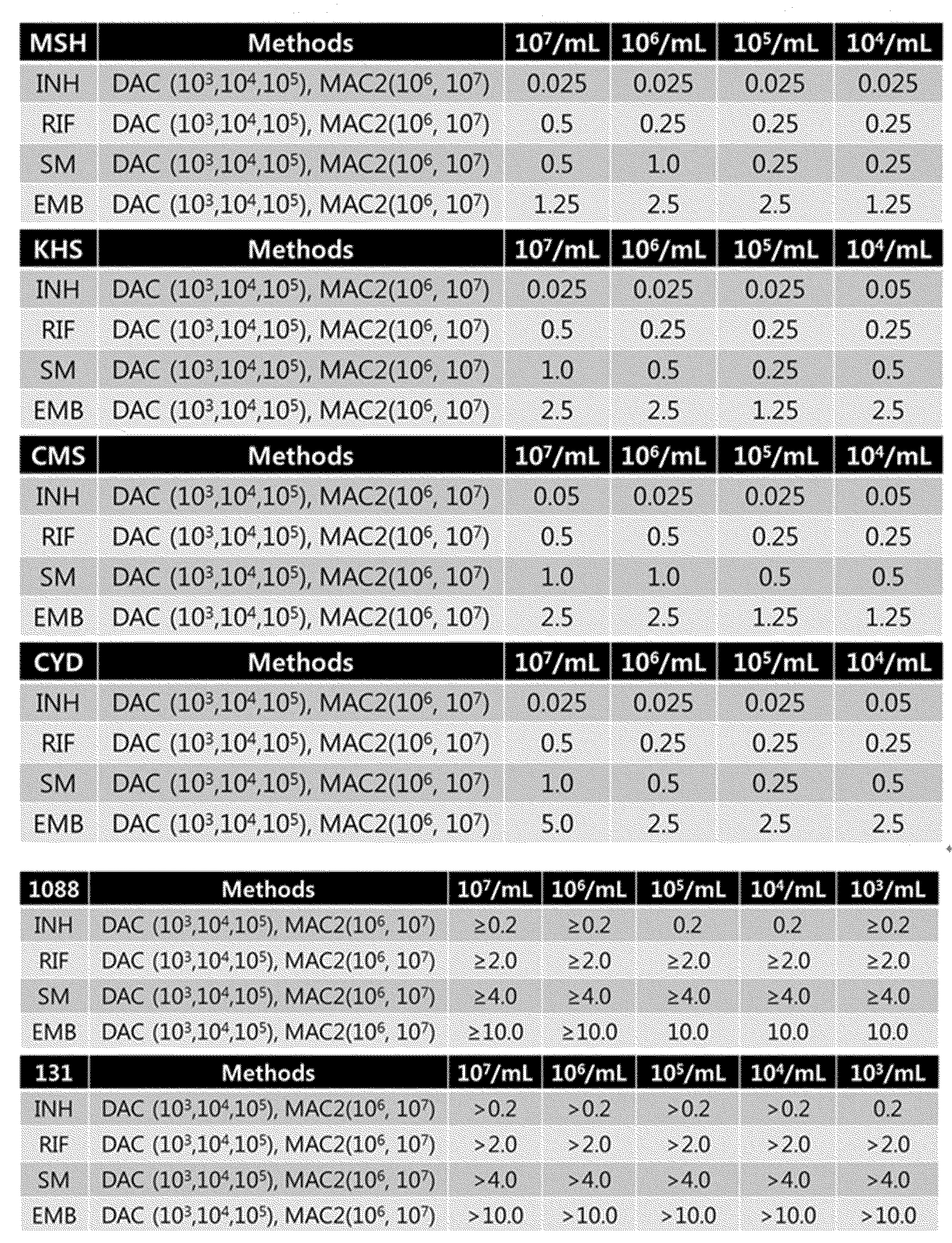
DAC 시스템 상에서 임상 균주에 대한 접종 효과 확인 결과
3.10 DAC를 사용한 접종 크기의 조정이 필요 없는 정확한 DST
LJ (Mkit) 시스템에서의 정확한 DST 및 자동화 액체 배지 기반 DST인 MGIT에서는 정확한 접종량이 필요하다. 따라서 접종 농도를 조절하기 위해 어려운 절차가 수행되어야 한다.
이전의 결과로부터, DAC 시스템에서는 접종량의 조정이 불필요한 것을 증명했다. 본 실시예에서는 이 실험에서 접종 농도를 조정하는 과정은 생략하고, 준비된 원액으로부터 DAC 시스템에 직접 DST를 수행 하였다. 따라서, 접종량은 다양한 범위였다. 도 10에 도시 한 바와 같이 실제의 접종 농도를 7H11 배지에서의 콜로니 개수에 의하여 수득하였다. 접종 농도는 105 내지 108 CFU/ml였다. DAC 시스템에서 DST를 수행 한 후, 결과를 LJ (Mkit) 시스템에서의 DST와 비교하였으며, 에러율을 도출하였다. 테스트 균주의 총 수는 117이고, 테스트 케이스는 총 516개였다.
모두에 감수성을 나타낸, PanS 균주의 경우, 불일치하는 경우는 없었다. MDR의 경우, LJ 시스템에서는 민감한 것으로 확인된 ME (major error)가 8 케이스, LJ 시스템에서는 저항성인 것으로 확인된 VME (very major error)가 4 케이스였다. 516번의 테스트에서 오류률은 단지 6.2% 였다.
표 7 
3.11 아가로오스를 이용함에 따른 박테리아의 랩핑에 의한 접종 효과의 부존재
접종 효과에는 두 가지의 주된 이유가 있다.
첫번째는, 높은 접종량에서 박테리아로부터 발생되는 항생제 분해 단백질이 항생제를 파괴하고 MIC 값이 증가 될 수 있다는 것이다. 다른 이유로는, 더 높은 접종량에서, 박테리아의 단일 세포에 대한 항생제 분자의 수가 감소하며 MIC가 증가 될 수 있다는 것이다. DAC에서 접종 효과가 없음에 대한 이유를 확인하기 위하여, 항생제 분해 단백질을 분비하는 박테리아 균주로 잘 알려진, ESBL 음성 대장균 (ATCC 35218), ESBL 양성 대장균 (ATCC 25922) 및 임상적으로 수득한 대장균 균주를 5*10^7, 5*10^6, 5*10^5 CFU/ml의 접종 농도로 테스트 하였다. 상기 농도는 DAC 에서의 아가로오스 중의 박테리아 농도 및 MDT에서의 액체에서의 박테리아 농도를 의미한다.
접종 효과를 확인하기 위하여 테스트 된 항생제는 세팔로스포린 (cephalosporin) 항생제 4 세대인 세페핌 (cefepime)이었다. ESBL 음성 대장균, ESBL 양성 대장균의 MIC 범위는 0.015에서 0.12 ug/㎖이었다.
표 8 
DAC 시스템에서는, 다양한 크기의 접종량에서 접종효과가 전혀 나타나지 않았다. 그러나, MDT 시스템에서, ESBL + 및 # 5 균주에서 접종 크기에 따라 MIC의 점진적인 증가를 보였다. 이 결과는 접종효과가 MDT 시스템에서 나타난 것과는 다르게 DAC에서는 전혀 효과가 없음을 나타내는 것이다.
본 발명자는 DAC에서 접종효과나 나타나지 않는 이유로서, 박테리아로부터 방출되는 단백질이 아가로 오스에 포획되어 브로스 배양 조건처럼 항생제를 분해할 수 없기 때문인 것으로 가설을 새웠다. 이 효과의 가설을 확인하기 위하여, ESBL 양성 대장균을 사용하여 다른 테스트를 수행하였다. 접종농도는 5*10^7, 5*10^6 및 5*10^5 CFU/ml 였다. 상기 농도는 DAC에서 전체 웰 내의 박테리아 농도 및 액체 내 박테리아 농도를 의미한다. 따라서, 최종 DAC 및 MAC 박테리아의 최종 농도는 동일하였다. 세페핌을 이 실험에 사용하였다. 밤새 배양 한 후에, DAC 및 MDT의 웰 내의 항생제 용액을 추출하고, 순수한 항생제를 얻기 위하여 여과하였다. 그리고 항생제를 사용하여, ESBL 양성 대장균을 이용한 MDT를 수행하였다. 그 결과를 표 9에 나타내었다.
표 9 
항생제 용액에서 밤새 배양에 따른 DAC 및 MDT를 이용한 AST 결과 비교
DAC에서는 다양한 접종량에서 MIC 값에 차이가 없었으며 이는 분해율이 접종량과 유사함을 나타낸다. 그러나, MDT에서는 접종량이 증가함에 따라, MIC 값이 증가하였으며, 이는 항생제의 분해가 증가함을 나타낸다. 이러한 결과는 DAC 시스템에서의 항생제의 분해의 감소가 아가로스에서 박테리아가 포획됨에 기인하는 것을 의미한다.
즉, 본 발명의 DAC 시스템은 세포의 밀도가 증가하는 경우의 박테리아에 대한 약물의 과도한 결합 및 그 결과물에 의하여 발생되는, 분자의 상호 작용 및 세균 박테리아 집단 역동 (bacterial population dynamics)을 모두 제어할 수 있으며, 이는 매트릭스 내에 박테리아 집단이 정착되어 있으며, 각 약물을 포함하는 배지가 챔버내로 구배로써 공급되기 때문이다.
이 장치는 마이코 박테리아에 대한 접종을 엄격하게 제한할 필요 없이, DST를 손쉽게 수행할 수 있는데, 이는 IE가 ML 당 107 마이코박테리아보다 높은 접종에서는 관찰되지 않기 때문이다. 따라서 DAC 시스템은 예상된다 항 결핵 약물에 대한 미생물 학적 효능의 정확한 예측을 가능하게 하며, 이는 기존의 방법을 효과적으로 대체할 수 있을 것으로 기대한다.
실시예 4: 항생제 스크리닝에의 DAC 시스템의 활용
본 발명에서 개발한 DAC 시스템을 항생제 스크리닝에 활용하였다. 구체적으로, 협력사에서 개발중인 결핵용 약물 Q203에 대하여, TDR 임상 균주의 감수성을 확인하기 위하여, 상기 결핵용 약물의 MIC를 측정하였다.
첫번째 임상균주인 TDR 102 환자 균주의 상기 약물에 대한 MIC를 구하기 위하여, 본 발명의 DAC 시스템 상에서 상기 Q203를 각각 3.9, 7.8, 15.6,31.2, 62.5, 125, 250, 500, 1000 ng/ml를 TDR 102 환자 균주에 처리하였다. DAC 시스템에서의 이미지를 도 11에 나타내었으며, 이를 수치화하여 그래프화 한 것을 도 12에 나타내었다.
또한, 두번째 임상균주인 TDR 103 환자 균주의 상기 약물에 대한 MIC를 구하기 위하여 상기와 동일한 방법으로 Q203를 TDR 103 환자 균주에 처리하였다. DAC 시스템에서의 이미지를 도 13에 나타내었으며, 이를 수치화하여 그래프화 한 것을 도 14에 나타내었다.
이하, 첨부한 도면들을 참조하여, 본 발명의 구현예들을 보다 상세하게 설명하고자 한다. 그러나 본 발명은 여기서 설명되는 구현예들에 한정되지 않고 다른 형태로 구체화될 수도 있다. 단지, 여기서 소개되는 구현예들은 개시된 내용이 철저하고 완전해질 수 있도록 그리고 당업자에게 본 발명의 사상이 충분히 전달될 수 있도록 하기 위해 제공되는 것이다. 도면에서 각 장치의 구성요소를 명확하게 표현하기 위하여 상기 구성요소의 폭이나 두께 등의 크기를 다소 확대하여 나타내었다. 전체적으로 도면 설명시 관찰자 시점에서 설명하였고, 일 요소가 다른 요소 위에 위치하는 것으로 언급되는 경우, 이는 상기 일 요소가 다른 요소 위에 바로 위치하거나 또는 그들 요소들 사이에 추가적인 요소가 개재될 수 있다는 의미를 모두 포함한다. 또한, 해당 분야에서 통상의 지식을 가진 자라면 본 발명의 기술적 사상을 벗어나지 않는 범위 내에서 본 발명의 사상을 다양한 다른 형태로 구현할 수 있을 것이다. 그리고, 복수의 도면들 상에서 동일 부호는 실질적으로 서로 동일한 요소를 지칭한다.
플레이트 본체;
상기 플레이트에 하나 이상으로 배열된 배양 유닛 (100); 및
및 고체 박막을 상기 배양 유닛 바닥면에 형성할 수 있는 밑판(110);을 포함하는 생리 활성 검사용 구조물을 제공한다. 도 2a는 본 발명에 따른 검사용 구조물의 제1의 일 구현예의 평면도가, 도 2b는 본 발명에 따른 검사용 구조물의 제1의 일 구현예의 사시도를 나타낸다.
도 2a 및 도 2b을 참조하여 볼 때, 상기 밑판(110)에는 고형화제를 함유한 액상 매질과 생물학적 물질의 혼합 용액을 주입할 수 있는 주입구 (111)를 구비할 수 있다. 또한, 상기 밑판(110)은 하나 이상의 연결부재(113)에 의하여 플레이트의 격벽과 연결되며, 상기 연결부재 사이에는 절개홈(114)이 형성될 수 있으며, 상기 밑판(110)에는 하나 이상의 관통공(112)이 형성될 수 있다. 상기 절개홈 및 관통공으로부터 생물학적 활성물질이 고체 박막에 균일하게 확산될 수 있다.
상기 고체 박막의 두께와 너비는 상기 수용부의 깊이와 너비에 따라 정해진다. 본 명세서에서 "박막"이라 함은 상기 생물학적 물질을 고정하여 단일 세포 단위로 관찰할 수 있을 정도면 두께가 특별히 제한되지 않으며, 예를 들어 1㎛ 내지 5mm, 또는 1㎛ 내지 3mm, 또는 1㎛ 내지 2mm, 또는 1㎛ 내지 1.5mm, 또는 1㎛ 내지 1mm, 또는 1㎛ 내지 800㎛, 또는 1㎛ 내지 500㎛, 1㎛ 내지 100㎛, 또는 10㎛ 내지 3mm, 또는 10㎛ 내지 1mm, 또는 100㎛ 내지 1mm, 또는 200㎛ 내지 1mm, 또는 500㎛ 내지 1mm 중 어느 하나의 범위를 갖는 얇은 층을 의미한다. 여기서 고체 박막의 두께는 관찰하고자 하는 고체 박막 면에 대한 수직방향의 크기에 해당할 수 있다. 고체 박막은 상술한 범위의 두께를 가지므로 내부에 고정된 생화학적 물질을 개별 세포(single cell) 단위로 관찰하거나, 콜로니 형성을 효과적으로 개수할 수 있다.
상기 배양 유닛의 폭 (너비)는 제한이 없으며, 생물학적 물질의 종류 및 특성에 따라서 달라질 수 있으며, 바람직하게는 1 내지 50 mm X 1 내지 50 mm, 1 내지 30 mm X 1 내지 30 mm, 5 내지 50 mm X 5 내지 50 mm, 5 내지 30 mm X 5 내지 30 mm, 10 내지 50 mm X 10 내지 50 mm, 10 내지 30 mm X 10 내지 30 mm, 1 내지 20 mm X 1 내지 20 mm일 수 있다. 가장 바람직하게는 12mm X 12 mm일 수 있다.
또한, 상기 배양 유닛의 높이 역시 제한이 없으며, 생물학적 물질 및 생물활성물질의 종류 및 특성에 따라서 달라질 수 있으며, 예를 들면, 1 내지 50 mm, 1 내지 30 mm, 5 내지 30 mm, 5 내지 15 mm 일 수 있다.
또한, 상기 관통공(112)의 크기는 제한하지 않으며, 일반적으로 1mm 이하의 지름을 가질 수 이다. 상기 관통공의 지름은 예를 들면, 0.99mm 이하, 0.90 mm 이하, 0.85 mm 이하, 0.80 mm 이하, 0.75 mm 이하, 0.70 mm 이하, 0.65 mm 이하, 0.60 mm 이하, 0.55 mm 이하, 0.50 mm 이하, 0.45 mm 이하, 0.40 mm 이하, 0.35 mm 이하, 0.30 mm 이하, 0.25 mm 이하일 수 있다.
본 발명의 일 양상에서, 고형화제를 함유한 액상 매질과 생물학적 물질의 혼합 용액은 밑판 하부에서 고화되어, 상기 생물학적 물질이 고정된 고체 박막을 형성할 수 있다.
액상 매질은 약 80% 이상의 물 또는 완충액을 포함할 수 있으며 고형화제의 존재에 의해 고체화될 수 있다. 고형화제의 예로 한천(agar), 아가로즈(agarose), 젤라틴(gelatin), 알기네이트(alginate), 콜라겐(collagen), 피브린(fibrin) 등을 들 수 있다. 바람직하게는 한천 또는 아가로즈가 사용될 수 있다. 일 예로 액상 매질 내에는 한천이 0.5 내지 5 중량%가 사용될 수 있다. 통상적으로 액상 매질은 영양성분을 필요로 하지 않지만, 몇몇 예에 있어 영양성분을 포함할 수도 있다.
상기 생물학적 물질은 바이러스, 박테리아, 균류, 조류, 원생동물, 기생 병원균, 사람 또는 포유류의 세포 및 바이오필름 등을 포함한다. 상기 생물학적 물질은 액상 또는 고상의 배지 내에서 성장할 수 있고 외부 생리활성 물질의 종류 및 농도에 따라 성장에 영향을 받을 수 있다.
상기 혼합 용액 내에서의 상기 생물학적 물질의 밀도는 제한되지 않는다. 특히, 본 발명의 특징은 생물학적 물질의 밀도와 DST 또는 진단 결과가 무관하다는 것이다. 즉 본 발명에 따른 DAC 시스템은 접종효과 (inoculum effect)가 없다. 따라서, 상기 생물학적 물질의 밀도는 101 내지 1080 cfu/ml의 범위일 수 있으며, 예를 들어, 1070 cfu/ml 이하, 1060 cfu/ml 이하, 1050 cfu/ml 이하, 1040 cfu/ml 이하, 1030 cfu/ml 이하, 1020 cfu/ml 이하, 1010 cfu/ml 이하, 109 cfu/ml 이하, 108 cfu/ml 이하, 107 cfu/ml 이하, 106 cfu/ml 이하, 105 cfu/ml 이하, 또는 104 cfu/ml 이하 일 수 있다.
상기 고형화제를 함유한 액상 매질과 생물학적 물질의 혼합 용액은 상기 배양 유닛의 밑판 상부에서 고화되어 생물학적 물질이 고정된 고체 박막을 형성한다. 높은 온도의 액상 매질의 온도가 내려감에 따라 매질이 고화됨으로써 상기 생물학적 물질의 움직임이 저하된다. 상기 고정에 의하여 운동성이 있는 상기 생물학적 물질을 계속해서 용이하게 관찰할 수 있다.
도 3a는 본 발명에 따른 검사용 구조물의 제2의 일 구현예의 평면도가, 도 3b는 본 발명에 따른 검사용 구조물의 제2의 일 구현예의 사시도를 나타낸다.
도 3a, b을 참조하여 볼 때, 상기 밑판의 상부에는 수용홈(120)이 추가로 구비될 수 있다.
또한, 상기 수용홈(120)의 폭은 제한하지 않으며, 바람직하게는 1 내지 5 mm일 수 있고, 예를 들면, 1 내지 4.5 mm 1, 내지 4.0 mm, 1 내지 3.5 mm일 수 있다. 상기 수용홈의 높이는 바람직하게는 1 내지 3 mm일 수 있으며, 예를 들면, 1 내지 2.5 mm일 수 있다.
상기 배양 유닛 내부에 생리활성물질을 더 포함할 수 있다. 본 명세서에서 상기 생리활성물질은 생물학적 물질에 생리적 영향을 미칠 수 있는 것으로, 항생제, 항암제, 면역억제제와 같은 약물, 영양성분, 세포분비물, 신호전달물질, 바이러스, 세포, micro RNA, 단백질, 항원, 항체, DNA 등 중 어느 하나를 포함할 수 있다.
상기 생리활성물질은 배양 유닛에 액상 매질 또는 액체 배지와 함께 혼합하여 제공될 수 있으며, 상기 수용홈 (120) 내에 제공될 수 있다. 상기 생리활성물질은 특히, 상기 수용홈 (120) 내에 제공되는 경우 동결건조되어 있는 형태 일 수 있다. 동결건조의 경우, 운반, 보존 등을 용이하게 한다.
상기 플레이트 본체, 배양 유닛, 밑판의 전체 또는 일부는 투명 또는 반투명한 재질 일 수 있으며, 바람직하게는 광학 이미징을 위해 투광성 기판일 수 있다. 상기 구조물은 액상 매질이 도포되어 박막이 형성되기에 적합한 표면 특성을 가지면 특별히 제한되지 않는다.
또한, 본 발명의 다른 양상은
고형화제를 함유한 액상 매질과 생물학적 물질의 혼합 용액을 청구항 1 내지 14 중 어느 한 항의 생리활성 검사용 구조물의 바닥면에 제공하는 단계;
상기 혼합 용액을 고화시켜 상기 생물학적 물질이 고정된 고체 박막을 형성하는 단계;
생리활성물질을 상기 고체 박막에 공급하여 상기 생리활성물질이 상기 고체 박막 내부로 확산되도록 하는 단계; 및
상기 생리활성물질에 대한 상기 생물학적 물질의 단일 세포들의 개별적인 반응을 관찰하거나, 상기 생물학적 물질의 콜로니의 형성 여부 또는 콜로니 숫자(colony forming unit, CFU)을 관찰하는 단계를 포함하는, 생리활성 검사 방법을 제공한다.
상기 방법은 상기 생리활성물질에 대한 상기 생물학적 물질의 단일 세포들의 개별적인 반응 또는 콜로니의 형성 여부 또는 콜로니 숫자(colony forming unit, CFU)를 이미징하여 얻은 이미지 분석 결과를 수득하는 단계를 더 포함할 수 있으며, 더욱 바람직하게는 상기 이미지 분석 결과에 기초하여 상기 생리활성물질의 최소억제농도(MIC)를 결정하는 단계를 더 포함할 수 있다.
관찰을 위해 광학 측정장치가 사용될 수 있다. 상기 광학 측정장치는 CCD 또는 CMOS 카메라와 같은 이미지화 시스템을 포함할 수 있다. 상기 광학 측정장치는 또한 초점을 맞추고 빛을 이미지화하는 데 필요한 렌즈, 조명 및 빛 가이드 등을 포함할 수 있다. 또한 상기 광학 측정장치는 카메라에 의해 관찰된 이미지 데이터를 가공 및 분석하기 위한 이미지 처리 시스템을 포함할 수 있다. 상기 광학 측정장치는 검사 과정에 나타난 생물학적 물질의 성장변화를 빠른 속도로 기록하고 분석하여 검사 결과를 얻는다.
상기 생리활성 검사 방법은 다양한 용도로 활용될 수 있다. 박테리아의 항생제 감수성 검사 방법, 항생제 스크리닝 방법, 및 박테리아의 진단 방법에 이용될 수 있다.
즉, 본 발명의 다른 양상은
고형화제를 함유한 액상 매질과 박테리아의 혼합 용액을 청구항 1 내지 14 중 어느 한 항의 생리활성 검사용 구조물의 바닥면에 제공하는 단계;
상기 혼합 용액을 고화시켜 상기 박테리아가 고정된 고체 박막을 형성하는 단계;
임의의 항생제를 상기 고체 박막에 공급하여 상기 항생제가 상기 고체 박막 내부로 확산되도록 하는 단계;
상기 항생제에 대한 상기 박테리아의 단일 세포들의 개별적인 반응을 관찰하거나, 상기 박테리아의 콜로니의 형성 여부 또는 콜로니 숫자(colony forming unit, CFU)을 관찰하는 단계;를 포함하는, 항생제 스크리닝 방법을 제공한다.
상기 방법은 상기 단일 세포들의 개별적인 변화 또는 콜로니의 형성 여부 또는 콜로니 숫자(colony forming unit, CFU)를 이미징하여 얻은 이미지 분석결과에 기초하여 상기 항생제의 최소 억제농도(MIC)를 결정하는 단계를 더 포함할 수 있다.
또한, 본 발명의 일 양상은
고형화제를 함유한 액상 매질과 박테리아의 혼합 용액을 청구항 1 내지 14 중 어느 한 항의 생리활성 검사용 구조물의 바닥면에 제공하는 단계;
상기 혼합 용액을 고화시켜 상기 박테리아가 고정된 고체 박막을 형성하는 단계;
상기 박테리아의 배양 배지를 상기 고체 박막에 공급하여 상기 배양 배지가 상기 고체 박막 내부로 확산되도록 하는 단계; 및
상기 박테리아의 단일 세포들의 개별적인 반응을 관찰하거나, 상기 박테리아의 콜로니의 형성 여부 또는 콜로니 숫자(colony forming unit, CFU)을 관찰하는 단계;를 포함하는 박테리아의 진단 방법을 제공한다.
상기 박테리아의 배양 배지에는 항생제가 추가로 포함될 수 있으며, 상기 진단 방법은 약물 감수성 검사와 동시에 수행될 수 있는 것을 특징으로 할 수 있다.
상기 생리 활성 검사는 약제 감수성, 약제 스크리닝, 또는 세균 배양 진단일 수 있다.
상기 박테리아의 종류는 제한하지 않는다. 가장 바람직하게는 상기 박테리아는 결핵균 또는 비결핵성 항산균 (NTM, Nontuberculous mycobacteria) 균일 수 있으며, 상기 결핵균은 다제내성 (multi-drug-resistant, MDR) 결핵균, 광범위 약제내성 (extensively drug-resistant, XDR) 결핵균, 완전 약제내성 (totally drug-resistant, TDR) 결핵균 등일 수 있다.
또한, 본 발명의 다른 양상에서는
고형화제를 함유한 액상 매질과 생물학적 물질의 혼합 용액을 고화시켜 만든 박막을 바닥면에 구비하며, 생리활성물질을 배양 유닛에 구비한, 청구항 1 내지 14 중 어느 한 항의 생리활성 검사용 구조물;
상기 검사용 구조물을 지지하고 관찰하기 위한 스테이지; 및
상기 생리활성물질의 확산에 의한 전달에 따른 상기 생물학적 물질의 단일 세포들의 개별적인 변화 또는 콜로니의 형성 여부 또는 콜로니 숫자(colony forming unit, CFU)을 이미징하여 얻은 이미지 분석결과에 기초하여 상기 생리활성물질의 최소 억제농도(MIC)를 결정하기 위한 측정장치를 포함하는, 생리활성 검사 시스템을 제공한다.
상기 측정장치는 이미지화 시스템 및 이미지 처리 시스템을 포함할 수 있다. 상기 측정장치는 관찰한 이미지를 생성하는 CCD 또는 CMOS 카메라와 같은 이미지화 시스템을 포함한다. 또한 광학 측정장치는 생성된 이미지를 처리 및 분석하는 이미지 처리 시스템을 포함할 수 있다.
부호의 설명
100: 배양 유닛 120: 수용홈
110: 밑판
111: 주입구
112: 관통공
113: 연결부재
114: 절개홈
Claims (30)
- 플레이트 본체;상기 플레이트에 하나 이상으로 배열된 배양 유닛; 및및 고체 박막을 상기 배양 유닛 바닥면에 형성할 수 있는 밑판;을 포함하는 생리 활성 검사용 구조물.
- 청구항 1에 있어서,상기 생리 활성 검사는 약제 감수성, 약제 스크리닝, 또는 세균 배양 진단인 것을 특징으로 하는, 생리 활성 검사용 구조물.
- 청구항 1에 있어서상기 밑판의 상부에는 수용홈이 추가로 구비된 것을 특징으로 하는, 생리 활성 검사용 구조물.
- 청구항 1에 있어서,상기 밑판에는 고형화제를 함유한 액상 매질과 생물학적 물질의 혼합 용액을 주입할 수 있는 주입구가 형성된 것을 특징으로 하는, 생리 활성 검사용 구조물.
- 청구항 1에 있어서,상기 밑판은 하나 이상의 연결부재에 의하여 플레이트의 격벽과 연결되며,상기 연결부재 사이에는 절개홈이 형성된 것을 특징으로 하는, 생리 활성 검사용 구조물.
- 청구항 1에 있어서,상기 밑판에는 하나 이상의 관통공이 형성된 것을 특징으로 하는, 생리 활성 검사용 구조물.
- 청구항 1에 있어서, 상기 배양 유닛의 폭은 1 내지 50 mm X 1 내지 50 mm이며,상기 배양 유닛의 높이는 1 내지 50 mm인 것을 특징으로 하는, 생리활성 검사용 구조물.
- 청구항 3에 있어서, 상기 수용홈의 폭은 1 내지 5 mm이며,상기 수용홈의 높이는 1 내지 3 mm인 것을 특징으로 하는, 생리활성 검사용 구조물.
- 청구항 4에 있어서,상기 고형화제를 함유한 액상 매질과 생물학적 물질의 혼합 용액은 밑판 하부에서 고화되어, 상기 생물학적 물질이 고정된 고체 박막을 형성하는 것을 특징으로 하는, 생리 활성 검사용 구조물.
- 청구항 1에 있어서, 상기 박막의 두께는 1㎛ 내지 10mm인 것인, 생리활성 검사용 구조물.
- 청구항 4에 있어서,상기 혼합 용액 내에서의 생물학적 물질의 밀도는101 내지 1080 cfu/ml 인 것인, 생리 활성 검사용 구조물.
- 청구항 4에 있어서,상기 고형화제는 한천(agar), 아가로즈(agarose), 젤라틴(gelatin), 알지네이트(alginate), 콜라겐(collagen) 및 피브린(fibrin)으로 이루어진 군 중에서 선택되는 하나 이상의 고형화제인 것인, 생리활성 검사용 구조물.
- 청구항 1에 있어서, 상기 배양 유닛 내부에 생리활성물질을 더 포함하는 것인, 생리활성 검사용 구조물.
- 청구항 3에 있어서, 생리활성물질은 상기 수용홈에 동결 건조된 형태로 포함되어 있는 것을 특징으로 하는, 생리활성 검사용 구조물.
- 청구항 1에 있어서, 상기 플레이트 본체, 배양 유닛 및 밑판은 투명 또는 반투명한 재질로 이루어진 것인, 생리활성 검사용 구조물.
- 고형화제를 함유한 액상 매질과 생물학적 물질의 혼합 용액을 청구항 1 내지 14 중 어느 한 항의 생리활성 검사용 구조물의 바닥면에 제공하는 단계;상기 혼합 용액을 고화시켜 상기 생물학적 물질이 고정된 고체 박막을 형성하는 단계;생리활성물질을 상기 고체 박막에 공급하여 상기 생리활성물질이 상기 고체 박막 내부로 확산되도록 하는 단계; 및상기 생리활성물질에 대한 상기 생물학적 물질의 단일 세포들의 개별적인 반응을 관찰하거나, 상기 생물학적 물질의 콜로니의 형성 여부 또는 콜로니 숫자(colony forming unit, CFU)를 관찰하는 단계를 포함하는, 생리활성 검사 방법.
- 청구항 15에 있어서,상기 생리활성물질에 대한 상기 생물학적 물질의 단일 세포들의 개별적인 반응, 콜로니 형성 여부, 또는 콜로니 숫자 (colony forming unit, CFU)를 이미징하여 얻은 이미지 분석 결과를 수득하는 단계를 더 포함하는 것인, 생리활성 검사 방법.
- 청구항 17에 있어서,상기 이미지 분석 결과에 기초하여 상기 생리활성물질의 최소억제농도(MIC)를 결정하는 단계를 더 포함하는 것인, 생리활성 검사 방법.
- 고형화제를 함유한 액상 매질과 박테리아의 혼합 용액을 청구항 1 내지 14 중 어느 한 항의 생리활성 검사용 구조물의 바닥면에 제공하는 단계;상기 혼합 용액을 고화시켜 상기 박테리아가 고정된 고체 박막을 형성하는 단계;항생제를 상기 고체 박막에 공급하여 상기 항생제가 상기 고체 박막 내부로 확산되도록 하는 단계;상기 항생제에 대한 상기 박테리아의 단일 세포들의 개별적인 반응을 관찰하거나, 상기 박테리아의 콜로니의 형성 여부 또는 콜로니 숫자(colony forming unit, CFU)을 관찰하는 단계;를 포함하는, 박테리아의 항생제 감수성 검사 방법.
- 청구항 19에 있어서,상기 단일 세포들의 개별적인 변화, 콜로니의 형성 여부 또는 콜로니 숫자(colony forming unit, CFU)를 이미징하여 얻은 이미지 분석결과에 기초하여 상기 항생제의 최소 억제농도(MIC)를 결정하는 단계를 포함하는, 박테리아의 항생제 감수성 검사 방법.
- 청구항 19에 있어서, 상기 박테리아는 결핵균 또는 비결핵성 항산균 (NTM, Nontuberculous mycobacteria) 균인 것을 특징으로 하는, 박테리아의 항생제 감수성 검사 방법.
- 청구항 21에 있어서, 상기 결핵균은 다제내성 (multi-drug-resistant, MDR) 결핵균, 광범위 약제내성 (extensively drug-resistant, XDR) 결핵균, 또는 완전 약제내성 (totally drug-resistant, TDR) 결핵균인 것인, 박테리아의 항생제 감수성 검사 방법.
- 청구항 19에 있어서, 상기 박테리아의 항생제 감수성 검사 방법은 접종효과 (inoculum effect)가 없는 것을 특징으로 하는, 박테리아의 항생제 감수성 검사 방법.
- 고형화제를 함유한 액상 매질과 박테리아의 혼합 용액을 청구항 1 내지 14 중 어느 한 항의 생리활성 검사용 구조물의 바닥면에 제공하는 단계;상기 혼합 용액을 고화시켜 상기 박테리아가 고정된 고체 박막을 형성하는 단계;임의의 항생제를 상기 고체 박막에 공급하여 상기 항생제가 상기 고체 박막 내부로 확산되도록 하는 단계;상기 항생제에 대한 상기 박테리아의 단일 세포들의 개별적인 반응을 관찰하거나, 상기 박테리아의 콜로니의 형성 여부 또는 콜로니 숫자(colony forming unit, CFU)을 관찰하는 단계;를 포함하는, 항생제 스크리닝 방법.
- 청구항 24에 있어서,상기 단일 세포들의 개별적인 변화, 콜로니의 형성 여부 또는 콜로니 숫자(colony forming unit, CFU)를 이미징하여 얻은 이미지 분석결과에 기초하여 상기 항생제의 최소 억제농도(MIC)를 결정하는 단계를 더 포함하는, 항생제 스크리닝 방법.
- 고형화제를 함유한 액상 매질과 박테리아의 혼합 용액을 청구항 1 내지 14 중 어느 한 항의 생리활성 검사용 구조물의 바닥면에 제공하는 단계;상기 혼합 용액을 고화시켜 상기 박테리아가 고정된 고체 박막을 형성하는 단계;상기 박테리아의 배양 배지를 상기 고체 박막에 공급하여 상기 배양 배지가 상기 고체 박막 내부로 확산되도록 하는 단계; 및상기 박테리아의 단일 세포들의 개별적인 반응을 관찰하거나, 상기 박테리아의 콜로니의 형성 여부 또는 콜로니 숫자(colony forming unit, CFU)을 관찰하는 단계;를 포함하는 박테리아의 진단 방법.
- 청구항 26에 있어서,상기 박테리아의 배양 배지에 항생제가 추가로 포함된 것을 특징으로 하는, 박테리아의 진단 방법.
- 청구항 26에 있어서,상기 진단 방법은 약물 감수성 검사와 동시에 수행될 수 있는 것을 특징으로 하는, 박테리아의 진단 방법.
- 고형화제를 함유한 액상 매질과 생물학적 물질의 혼합 용액을 고화시켜 만든 박막을 바닥면에 구비하며, 생리활성물질을 배양 유닛에 구비한, 청구항 1 내지 14 중 어느 한 항의 생리활성 검사용 구조물;상기 검사용 구조물을 지지하고 관찰하기 위한 스테이지; 및상기 생리활성물질의 확산에 의한 전달에 따른 상기 생물학적 물질의 단일 세포들의 개별적인 변화, 콜로니의 형성 여부 또는 콜로니 숫자(colony forming unit, CFU)을 이미징하여 얻은 이미지 분석결과에 기초하여 상기 생리활성물질의 최소 억제농도(MIC)를 결정하기 위한 측정장치를 포함하는, 생리활성 검사 시스템.
- 청구항 29에 있어서, 상기 측정장치는 이미지화 시스템 및 이미지 처리 시스템을 포함하는 생리활성 검사 시스템.
Priority Applications (3)
| Application Number | Priority Date | Filing Date | Title |
|---|---|---|---|
| EP14903914.1A EP3202885A4 (en) | 2014-10-16 | 2014-10-16 | Novel biological activity testing structure for tracking single cell, using gelling agents |
| CN201480082659.4A CN107109331B (zh) | 2014-10-16 | 2014-10-16 | 使用胶凝剂跟踪单细胞的新型生物活性测试结构 |
| US15/518,668 US10533207B2 (en) | 2014-10-16 | 2014-10-16 | Bioactivity testing structure for single cell tracking using gelling agents |
Applications Claiming Priority (2)
| Application Number | Priority Date | Filing Date | Title |
|---|---|---|---|
| KR10-2014-0139700 | 2014-10-16 | ||
| KR1020140139700A KR101770610B1 (ko) | 2014-10-16 | 2014-10-16 | 고정화제를 이용한 단일 세포 추적을 위한 신규한 생리 활성 검사용 구조물 |
Publications (1)
| Publication Number | Publication Date |
|---|---|
| WO2016060301A1 true WO2016060301A1 (ko) | 2016-04-21 |
Family
ID=55746830
Family Applications (1)
| Application Number | Title | Priority Date | Filing Date |
|---|---|---|---|
| PCT/KR2014/009755 Ceased WO2016060301A1 (ko) | 2014-10-16 | 2014-10-16 | 고정화제를 이용한 단일 세포 추적을 위한 신규한 생리 활성 검사용 구조물 |
Country Status (5)
| Country | Link |
|---|---|
| US (1) | US10533207B2 (ko) |
| EP (1) | EP3202885A4 (ko) |
| KR (1) | KR101770610B1 (ko) |
| CN (1) | CN107109331B (ko) |
| WO (1) | WO2016060301A1 (ko) |
Cited By (2)
| Publication number | Priority date | Publication date | Assignee | Title |
|---|---|---|---|---|
| CN107130011A (zh) * | 2017-05-09 | 2017-09-05 | 上海理工大学 | 一种食源性致病菌单细胞生长过程的显微观测方法 |
| PL423414A1 (pl) * | 2017-11-10 | 2019-05-20 | Szkola Glowna Gospodarstwa Wiejskiego W Warszawie | Sposób wielostronnej oceny biozgodności materiałów |
Families Citing this family (10)
| Publication number | Priority date | Publication date | Assignee | Title |
|---|---|---|---|---|
| US10071141B2 (en) | 2015-05-08 | 2018-09-11 | Spectral Platforms, Inc. | Albumin-based non-covalent complexes and methods of use thereof |
| WO2018187548A2 (en) * | 2017-04-05 | 2018-10-11 | Arizona Board Of Regents On Behalf Of Arizona State University | Antimicrobial susceptibility testing with large-volume light scattering imaging and deep learning video microscopy |
| JP7076203B2 (ja) * | 2017-12-13 | 2022-05-27 | 株式会社日立ハイテク | 細菌検査用抗菌剤導入プレート、および透明プレート |
| CA3097400A1 (en) * | 2018-04-20 | 2019-10-24 | Advanced Biological Services, Inc. | Systems and methods for treating wastewater and providing class a sludge |
| KR20200044450A (ko) * | 2018-10-19 | 2020-04-29 | 주식회사 이지다이아텍 | 세포의 격리 및 형태학적 분석 방법 |
| AU2020340381A1 (en) * | 2019-08-27 | 2022-04-07 | SeLux Diagnostics, Inc. | Systems and methods for performing antimicrobial susceptibility testing |
| KR102132630B1 (ko) * | 2020-03-16 | 2020-07-10 | 주식회사 퀀타매트릭스 | 섬 구조물을 포함하는 신속한 세포배양검사 장치 |
| EP4168732A4 (en) * | 2020-06-23 | 2025-02-12 | Spectral Platforms, Inc. | SPECTROSCOPIC METHODS, REAGENTS AND SYSTEMS FOR THE DETECTION, IDENTIFICATION AND CHARACTERIZATION OF BACTERIA FOR ANTIMICROBIAL SUSCEPTIBILITY |
| CN117625394B (zh) * | 2023-12-01 | 2024-12-06 | 东莞合安机电有限公司 | 一种荔枝菌菌种培养液、制备方法及培养方法 |
| CN120555166A (zh) * | 2025-04-15 | 2025-08-29 | 科茵(上海)医疗技术有限公司 | 一种生物微流芯片装置及其在微生物运动模式检测中的用途 |
Citations (4)
| Publication number | Priority date | Publication date | Assignee | Title |
|---|---|---|---|---|
| KR20070033685A (ko) * | 2005-09-22 | 2007-03-27 | 한국과학기술원 | 미세유체 기술을 이용한 3차원 세포배양 시스템 |
| JP2009513160A (ja) * | 2005-11-01 | 2009-04-02 | レンセレアー ポリテクニック インスティテュート | 毒物学アッセイのための3次元細胞アレイチップおよびプラットフォーム |
| KR20130089619A (ko) * | 2012-02-02 | 2013-08-12 | 주식회사 퀀타매트릭스 | 고형화제를 이용한 세균 고정, 약물 확산, 세균 단일 세포 추적과 이를 기반으로 한 빠른 약물 검사 시스템 |
| KR101446526B1 (ko) * | 2013-05-02 | 2014-10-08 | 주식회사 퀀타매트릭스 | 마이크로플루이딕 멀티-웰 기반의 세포배양검사 장치 |
Family Cites Families (14)
| Publication number | Priority date | Publication date | Assignee | Title |
|---|---|---|---|---|
| AU658079B2 (en) * | 1992-09-28 | 1995-03-30 | Becton Dickinson & Company | Cell culture insert |
| US6251624B1 (en) * | 1999-03-12 | 2001-06-26 | Akzo Nobel N.V. | Apparatus and method for detecting, quantifying and characterizing microorganisms |
| WO2004055793A1 (en) | 2002-12-18 | 2004-07-01 | Koninklijke Philips Electronics N.V. | Optical pickup apparatus. |
| US7122153B2 (en) * | 2003-01-08 | 2006-10-17 | Ho Winston Z | Self-contained microfluidic biochip and apparatus |
| EP1479760A1 (de) * | 2003-05-19 | 2004-11-24 | ProBioGen AG | Künstliches Immunorgan |
| ES2614425T3 (es) * | 2004-01-08 | 2017-05-31 | Dako Denmark A/S | Aparato y métodos para el procesamiento de muestras biológicas y un depósito para los mismos |
| US8501462B2 (en) * | 2007-02-27 | 2013-08-06 | The Board Of Trustees Of The University Of Illinois | Insert device for multiwell plate |
| WO2009126524A2 (en) * | 2008-04-08 | 2009-10-15 | Massachusetts Institute Of Technology | Three-dimensional microfluidic platforms and methods of use thereof |
| WO2012050981A1 (en) * | 2010-09-29 | 2012-04-19 | Massachusetts Institute Of Technology | Device for high throughput investigations of cellular interactions |
| FR2972117B1 (fr) * | 2011-03-04 | 2013-12-20 | Centre Nat Rech Scient | Systeme microfluidique pour controler un profil de concentration de molecules susceptibles de stimuler une cible |
| US10526572B2 (en) * | 2011-04-01 | 2020-01-07 | EMD Millipore Corporaticn | Cell culture and invasion assay method and system |
| JP5516482B2 (ja) | 2011-04-11 | 2014-06-11 | 東京エレクトロン株式会社 | 基板搬送方法、基板搬送装置、及び塗布現像装置 |
| KR101965737B1 (ko) | 2011-10-19 | 2019-08-27 | 엘지전자 주식회사 | 공기조화기의 전동기 구동장치 |
| KR20140053582A (ko) | 2012-10-26 | 2014-05-08 | 단국대학교 산학협력단 | 절화의 선도유지 및 개화를 지연시키는 절화의 포장방법 |
-
2014
- 2014-10-16 CN CN201480082659.4A patent/CN107109331B/zh active Active
- 2014-10-16 WO PCT/KR2014/009755 patent/WO2016060301A1/ko not_active Ceased
- 2014-10-16 US US15/518,668 patent/US10533207B2/en active Active
- 2014-10-16 EP EP14903914.1A patent/EP3202885A4/en active Pending
- 2014-10-16 KR KR1020140139700A patent/KR101770610B1/ko active Active
Patent Citations (4)
| Publication number | Priority date | Publication date | Assignee | Title |
|---|---|---|---|---|
| KR20070033685A (ko) * | 2005-09-22 | 2007-03-27 | 한국과학기술원 | 미세유체 기술을 이용한 3차원 세포배양 시스템 |
| JP2009513160A (ja) * | 2005-11-01 | 2009-04-02 | レンセレアー ポリテクニック インスティテュート | 毒物学アッセイのための3次元細胞アレイチップおよびプラットフォーム |
| KR20130089619A (ko) * | 2012-02-02 | 2013-08-12 | 주식회사 퀀타매트릭스 | 고형화제를 이용한 세균 고정, 약물 확산, 세균 단일 세포 추적과 이를 기반으로 한 빠른 약물 검사 시스템 |
| KR101446526B1 (ko) * | 2013-05-02 | 2014-10-08 | 주식회사 퀀타매트릭스 | 마이크로플루이딕 멀티-웰 기반의 세포배양검사 장치 |
Non-Patent Citations (1)
| Title |
|---|
| See also references of EP3202885A4 * |
Cited By (3)
| Publication number | Priority date | Publication date | Assignee | Title |
|---|---|---|---|---|
| CN107130011A (zh) * | 2017-05-09 | 2017-09-05 | 上海理工大学 | 一种食源性致病菌单细胞生长过程的显微观测方法 |
| PL423414A1 (pl) * | 2017-11-10 | 2019-05-20 | Szkola Glowna Gospodarstwa Wiejskiego W Warszawie | Sposób wielostronnej oceny biozgodności materiałów |
| PL235845B1 (pl) * | 2017-11-10 | 2020-11-02 | Szkola Glowna Gospodarstwa Wiejskiego W Warszawie | Sposób wielostronnej oceny biozgodności materiałów |
Also Published As
| Publication number | Publication date |
|---|---|
| CN107109331B (zh) | 2025-02-14 |
| KR20160045171A (ko) | 2016-04-27 |
| EP3202885A1 (en) | 2017-08-09 |
| CN107109331A (zh) | 2017-08-29 |
| US20170233786A1 (en) | 2017-08-17 |
| EP3202885A4 (en) | 2018-07-18 |
| KR101770610B1 (ko) | 2017-08-24 |
| US10533207B2 (en) | 2020-01-14 |
Similar Documents
| Publication | Publication Date | Title |
|---|---|---|
| WO2016060301A1 (ko) | 고정화제를 이용한 단일 세포 추적을 위한 신규한 생리 활성 검사용 구조물 | |
| WO2018190656A1 (ko) | In vitro에서 성숙된 인간 장관 오가노이드의 제조 방법 및 이의 용도 | |
| US5347122A (en) | Light transmission system with photon transfer to an optical detector and cell investigation techniques using the light transmission system | |
| WO2013119049A1 (ko) | 생물학적 시료의 자동 분석 장치 및 방법 | |
| WO2017086719A1 (ko) | 혼돈파 센서를 이용한 시료 특성 탐지 장치 | |
| WO2019221557A1 (ko) | 광학 검출 시스템 | |
| WO2014081229A1 (ko) | 안전성 및 항암활성이 증가된 재조합 아데노바이러스 및 이의 용도 | |
| WO2018139788A1 (ko) | 고속 핵산증폭 장치 및 핵산증폭 반응의 온도조절 방법 | |
| WO2022270984A1 (ko) | 개별적으로 동작되는 생물학적 장치들을 이용한 자동화 분석 시스템, 분석 방법 및 저장 매체 | |
| WO2018135902A1 (ko) | 줄기 세포로부터 분화 유도된 연골세포의 제조방법 | |
| WO2023277639A1 (ko) | 자연살해세포 배양용 배지 및 이를 이용한 자연살해세포 대량증식방법 | |
| WO2023191597A1 (ko) | 유도만능줄기세포로부터 생산된 자연살해세포, 이의 제조 방법 및 이의 용도 | |
| WO2015076615A1 (ko) | 슬라이드 분리형 세포 배양 접시 및 이를 이용한 세포 분석 방법 | |
| WO2014175567A1 (ko) | 면역 치료 및 위암 치료제에 대한 효능 및 독성 평가를 위한 마우스 위암 세포주 | |
| WO2022098052A1 (en) | Human intestinal epithelium model and method for preparing same | |
| WO2011037348A2 (ko) | 미생물 분석 장치 및 이를 이용한 분석 방법 | |
| WO2022255841A1 (ko) | 줄기세포 유래 스페로이드형 초자연골세포 분화방법 및 이의 용도 | |
| WO2015034178A1 (ko) | 파라핀블록 제조용 혈중 세포 응집제 및 이를 이용한 파라핀블록 제조방법 | |
| WO2021112635A2 (ko) | Bcl2 계통 단백질 표적 약물에 대한 반응성 예측 방법 | |
| WO2019022299A2 (ko) | 전자동 유전자 판별 통합칩 | |
| WO2017146507A1 (ko) | 배양 패치, 배양 방법, 배양 검사 방법, 배양 검사 장치, 약물 검사 방법 및 약물 검사 장치 | |
| WO2022102865A1 (ko) | Fish 분석을 이용한 미생물을 검출하는 방법 | |
| WO2021091185A1 (ko) | 측분비인자를 포함하는 세포 배양용 조성물 | |
| WO2025034038A1 (ko) | 유전자 분석용 카트리지 및 이를 포함하는 유전자 분석장치 | |
| WO2021145467A1 (ko) | 인간 만능 줄기세포로부터 제작된 3d 오가노이드를 해체하여 세포를 다량 확보하는 분화방법 |
Legal Events
| Date | Code | Title | Description |
|---|---|---|---|
| 121 | Ep: the epo has been informed by wipo that ep was designated in this application |
Ref document number: 14903914 Country of ref document: EP Kind code of ref document: A1 |
|
| WWE | Wipo information: entry into national phase |
Ref document number: 201480082659.4 Country of ref document: CN |
|
| NENP | Non-entry into the national phase |
Ref country code: DE |
|
| REEP | Request for entry into the european phase |
Ref document number: 2014903914 Country of ref document: EP |
|
| WWG | Wipo information: grant in national office |
Ref document number: 201480082659.4 Country of ref document: CN |