WO2016092928A1 - 皮下注射用剤 - Google Patents
皮下注射用剤 Download PDFInfo
- Publication number
- WO2016092928A1 WO2016092928A1 PCT/JP2015/076068 JP2015076068W WO2016092928A1 WO 2016092928 A1 WO2016092928 A1 WO 2016092928A1 JP 2015076068 W JP2015076068 W JP 2015076068W WO 2016092928 A1 WO2016092928 A1 WO 2016092928A1
- Authority
- WO
- WIPO (PCT)
- Prior art keywords
- agent
- hydroxyapatite particles
- particles
- less
- subcutaneous injection
- Prior art date
- Legal status (The legal status is an assumption and is not a legal conclusion. Google has not performed a legal analysis and makes no representation as to the accuracy of the status listed.)
- Ceased
Links
Images


Classifications
-
- A—HUMAN NECESSITIES
- A61—MEDICAL OR VETERINARY SCIENCE; HYGIENE
- A61L—METHODS OR APPARATUS FOR STERILISING MATERIALS OR OBJECTS IN GENERAL; DISINFECTION, STERILISATION OR DEODORISATION OF AIR; CHEMICAL ASPECTS OF BANDAGES, DRESSINGS, ABSORBENT PADS OR SURGICAL ARTICLES; MATERIALS FOR BANDAGES, DRESSINGS, ABSORBENT PADS OR SURGICAL ARTICLES
- A61L27/00—Materials for grafts or prostheses or for coating grafts or prostheses
- A61L27/50—Materials characterised by their function or physical properties, e.g. injectable or lubricating compositions, shape-memory materials, surface modified materials
-
- A—HUMAN NECESSITIES
- A61—MEDICAL OR VETERINARY SCIENCE; HYGIENE
- A61K—PREPARATIONS FOR MEDICAL, DENTAL OR TOILETRY PURPOSES
- A61K33/00—Medicinal preparations containing inorganic active ingredients
- A61K33/42—Phosphorus; Compounds thereof
-
- A—HUMAN NECESSITIES
- A61—MEDICAL OR VETERINARY SCIENCE; HYGIENE
- A61K—PREPARATIONS FOR MEDICAL, DENTAL OR TOILETRY PURPOSES
- A61K47/00—Medicinal preparations characterised by the non-active ingredients used, e.g. carriers or inert additives; Targeting or modifying agents chemically bound to the active ingredient
- A61K47/30—Macromolecular organic or inorganic compounds, e.g. inorganic polyphosphates
- A61K47/36—Polysaccharides; Derivatives thereof, e.g. gums, starch, alginate, dextrin, hyaluronic acid, chitosan, inulin, agar or pectin
- A61K47/38—Cellulose; Derivatives thereof
-
- A—HUMAN NECESSITIES
- A61—MEDICAL OR VETERINARY SCIENCE; HYGIENE
- A61K—PREPARATIONS FOR MEDICAL, DENTAL OR TOILETRY PURPOSES
- A61K9/00—Medicinal preparations characterised by special physical form
- A61K9/0012—Galenical forms characterised by the site of application
- A61K9/0019—Injectable compositions; Intramuscular, intravenous, arterial, subcutaneous administration; Compositions to be administered through the skin in an invasive manner
-
- A—HUMAN NECESSITIES
- A61—MEDICAL OR VETERINARY SCIENCE; HYGIENE
- A61K—PREPARATIONS FOR MEDICAL, DENTAL OR TOILETRY PURPOSES
- A61K9/00—Medicinal preparations characterised by special physical form
- A61K9/08—Solutions
-
- A—HUMAN NECESSITIES
- A61—MEDICAL OR VETERINARY SCIENCE; HYGIENE
- A61L—METHODS OR APPARATUS FOR STERILISING MATERIALS OR OBJECTS IN GENERAL; DISINFECTION, STERILISATION OR DEODORISATION OF AIR; CHEMICAL ASPECTS OF BANDAGES, DRESSINGS, ABSORBENT PADS OR SURGICAL ARTICLES; MATERIALS FOR BANDAGES, DRESSINGS, ABSORBENT PADS OR SURGICAL ARTICLES
- A61L27/00—Materials for grafts or prostheses or for coating grafts or prostheses
-
- A—HUMAN NECESSITIES
- A61—MEDICAL OR VETERINARY SCIENCE; HYGIENE
- A61L—METHODS OR APPARATUS FOR STERILISING MATERIALS OR OBJECTS IN GENERAL; DISINFECTION, STERILISATION OR DEODORISATION OF AIR; CHEMICAL ASPECTS OF BANDAGES, DRESSINGS, ABSORBENT PADS OR SURGICAL ARTICLES; MATERIALS FOR BANDAGES, DRESSINGS, ABSORBENT PADS OR SURGICAL ARTICLES
- A61L27/00—Materials for grafts or prostheses or for coating grafts or prostheses
- A61L27/02—Inorganic materials
- A61L27/12—Phosphorus-containing materials, e.g. apatite
-
- A—HUMAN NECESSITIES
- A61—MEDICAL OR VETERINARY SCIENCE; HYGIENE
- A61L—METHODS OR APPARATUS FOR STERILISING MATERIALS OR OBJECTS IN GENERAL; DISINFECTION, STERILISATION OR DEODORISATION OF AIR; CHEMICAL ASPECTS OF BANDAGES, DRESSINGS, ABSORBENT PADS OR SURGICAL ARTICLES; MATERIALS FOR BANDAGES, DRESSINGS, ABSORBENT PADS OR SURGICAL ARTICLES
- A61L27/00—Materials for grafts or prostheses or for coating grafts or prostheses
- A61L27/14—Macromolecular materials
- A61L27/20—Polysaccharides
-
- A—HUMAN NECESSITIES
- A61—MEDICAL OR VETERINARY SCIENCE; HYGIENE
- A61L—METHODS OR APPARATUS FOR STERILISING MATERIALS OR OBJECTS IN GENERAL; DISINFECTION, STERILISATION OR DEODORISATION OF AIR; CHEMICAL ASPECTS OF BANDAGES, DRESSINGS, ABSORBENT PADS OR SURGICAL ARTICLES; MATERIALS FOR BANDAGES, DRESSINGS, ABSORBENT PADS OR SURGICAL ARTICLES
- A61L27/00—Materials for grafts or prostheses or for coating grafts or prostheses
- A61L27/40—Composite materials, i.e. containing one material dispersed in a matrix of the same or different material
- A61L27/44—Composite materials, i.e. containing one material dispersed in a matrix of the same or different material having a macromolecular matrix
- A61L27/46—Composite materials, i.e. containing one material dispersed in a matrix of the same or different material having a macromolecular matrix with phosphorus-containing inorganic fillers
-
- A—HUMAN NECESSITIES
- A61—MEDICAL OR VETERINARY SCIENCE; HYGIENE
- A61L—METHODS OR APPARATUS FOR STERILISING MATERIALS OR OBJECTS IN GENERAL; DISINFECTION, STERILISATION OR DEODORISATION OF AIR; CHEMICAL ASPECTS OF BANDAGES, DRESSINGS, ABSORBENT PADS OR SURGICAL ARTICLES; MATERIALS FOR BANDAGES, DRESSINGS, ABSORBENT PADS OR SURGICAL ARTICLES
- A61L27/00—Materials for grafts or prostheses or for coating grafts or prostheses
- A61L27/50—Materials characterised by their function or physical properties, e.g. injectable or lubricating compositions, shape-memory materials, surface modified materials
- A61L27/52—Hydrogels or hydrocolloids
-
- A—HUMAN NECESSITIES
- A61—MEDICAL OR VETERINARY SCIENCE; HYGIENE
- A61L—METHODS OR APPARATUS FOR STERILISING MATERIALS OR OBJECTS IN GENERAL; DISINFECTION, STERILISATION OR DEODORISATION OF AIR; CHEMICAL ASPECTS OF BANDAGES, DRESSINGS, ABSORBENT PADS OR SURGICAL ARTICLES; MATERIALS FOR BANDAGES, DRESSINGS, ABSORBENT PADS OR SURGICAL ARTICLES
- A61L2400/00—Materials characterised by their function or physical properties
- A61L2400/06—Flowable or injectable implant compositions
-
- A—HUMAN NECESSITIES
- A61—MEDICAL OR VETERINARY SCIENCE; HYGIENE
- A61L—METHODS OR APPARATUS FOR STERILISING MATERIALS OR OBJECTS IN GENERAL; DISINFECTION, STERILISATION OR DEODORISATION OF AIR; CHEMICAL ASPECTS OF BANDAGES, DRESSINGS, ABSORBENT PADS OR SURGICAL ARTICLES; MATERIALS FOR BANDAGES, DRESSINGS, ABSORBENT PADS OR SURGICAL ARTICLES
- A61L2430/00—Materials or treatment for tissue regeneration
- A61L2430/34—Materials or treatment for tissue regeneration for soft tissue reconstruction
Definitions
- the present invention relates to an agent for subcutaneous injection.
- a filler (filler) containing hydroxyapatite is injected subcutaneously.
- Hydroxyapatite has various effects including biocompatibility, and is a material that is expected to have an excellent beauty improvement effect even in subcutaneous injection.
- Patent Document 1 discloses a biomaterial composition using particles containing calcium hydroxyapatite having a particle size of 80 to 200 ⁇ m.
- Patent Document 1 does not necessarily have good flow characteristics. As a result, it was necessary to select a needle having a large needle diameter (inner diameter of 0.21 mm or more) as the injection needle, and there was a problem such as pain during injection.
- the particle size of the calcium hydroxyapatite particles described in the above-mentioned patent document has a problem such that inflammation occurs because macrophages are recognized as foreign substances.
- the present invention has an object to provide a subcutaneous injection preparation which is excellent in fluidity characteristics and can be used even in a thin injection needle having an inner diameter of 0.20 mm or less and is less prone to pain and inflammation during injection.
- the present invention is as follows.
- the present invention (1) In subcutaneous injection, Having a hydrogel containing calcined hydroxyapatite particles, The content of the calcined hydroxyapatite particles is 1 to 60% by mass based on the total mass of the agent, The fired hydroxyapatite particles have an average particle size of 15 ⁇ m or less.
- the present invention (2) 90% or more of the calcined hydroxyapatite particles have a particle size of 15 ⁇ m or less.
- the present invention (3) 4% or more of the calcined hydroxyapatite particles have a particle size of 5 ⁇ m or more and 30 ⁇ m or less.
- the present invention (4) The agent according to any one of the inventions (1) to (3), wherein the calcined hydroxyapatite particles are used for enhancing collagen production.
- the present invention (5) The agent according to any one of the inventions (1) to (4), wherein the hydrogel is a carboxymethylcellulose gel.
- the present invention (6) The agent according to any one of the inventions (1) to (5), wherein the hydrogel has a viscosity of 50 to 15000 mPa ⁇ s.
- the present invention (7) The agent according to any one of the inventions (1) to (6), wherein the agent passes through a hollow needle having an inner diameter of 0.20 mm or less.
- the present invention In the method for producing a subcutaneous injection, Having a step of mixing calcined hydroxyapatite particles and hydrogel,
- the content of the calcined hydroxyapatite particles is 1 to 60% by mass based on the total mass of the agent,
- the average particle diameter of the calcined hydroxyapatite particles in the agent is 15 ⁇ m or less.
- the present invention 90% or more of the burned hydroxyapatite particles in the agent have a particle size of 15 ⁇ m or less.
- the present invention (10) 4% or more of the calcined hydroxyapatite particles in the agent has a particle size of 5 ⁇ m or more and 30 ⁇ m or less, and the production method according to the invention (8) or (9).
- the present invention (11) The method according to any one of the inventions (8) to (10), wherein the calcined hydroxyapatite particles are used for enhancing collagen production.
- the present invention (12) The method according to any one of the inventions (8) to (11), wherein the hydrogel is a carboxymethylcellulose gel.
- the present invention (13) The method according to any one of the inventions (8) to (12), wherein the hydrogel has a viscosity of 50 to 15000 mPa ⁇ s.
- the present invention (14) The method according to any one of the inventions (8) to (13), wherein the agent passes through a hollow needle having an inner diameter of 0.20 mm or less.
- an agent for subcutaneous injection that has excellent flow characteristics and can be used even with a thin injection needle having an inner diameter of 0.20 mm or less, and is less prone to pain and inflammation during injection. It becomes.
- Drawing 1 is a figure (photograph) which shows a filler before and behind a defoaming process concerning this example.
- FIG. 2 is a diagram showing the result of measuring the particle size distribution of the filler according to this example.
- the subcutaneous injection preparation according to a preferred embodiment of the present invention will be described in the following order.
- Composition of subcutaneous injection (2) Manufacturing method of subcutaneous injection (3) Action of subcutaneous injection (4) Usage of subcutaneous injection (5) Physical properties of subcutaneous injection
- the agent for subcutaneous injection according to the present embodiment contains hydroxyapatite particles and hydrogel, and may further contain other components.
- the hydroxyapatite particle aggregate is an aggregate of hydroxyapatite particles.
- hydroxyapatite particles Hydroaulic apatite particles
- the physical properties (particularly the particle size) of the hydroxyapatite particles shown here indicate the physical properties of the hydroxyapatite particles contained in the subcutaneous injection agent (the physical properties of the hydroxyapatite particles contained in the subcutaneous injection agent)
- the physical properties of the hydroxyapatite particles, which are constituent materials of the subcutaneous injection are not necessarily the same because there may be a change in form depending on the production stage.
- HAp Hydroxyapatite
- Ca 10 (PO 4 ) 6 (OH) 2 a basic calcium phosphate represented by the chemical formula Ca 10 (PO 4 ) 6 (OH) 2 and naturally exists as a main component of teeth and bones and as an ore.
- Hydroxyapatite particles exhibit high biocompatibility.
- hydroxyapatite particles present in the filler as nanoparticles enhance collagen production by stimulating fibroblasts.
- the hydroxyapatite particles present in the filler as microparticles physically function as a spacer.
- hydroxyapatite particles As the hydroxyapatite particles according to the present embodiment, calcined hydroxyapatite particles (hereinafter referred to as "calcined hydroxyapatite particles” when referred to as “hydroxyapatite particles” unless otherwise specified) are used.
- calcined hydroxyapatite particles By firing the hydroxyapatite particles (for example, at 800 ° C. for 1 hour), the crystallinity of the particles is increased, and aggregates of a plurality of primary particles are fused by heat to become stronger and more stable particles. .
- “hydroxyapatite particles” as used in the present invention is a concept including not only calcined hydroxyapatite particles having a primary particle size but also aggregates in which a plurality of primary particles are
- calcined apatite particles By using such calcined apatite particles, it is possible to obtain a highly effective filler that stimulates fibroblasts and enhances collagen production. That is, by making the hydroxyapatite particles into calcined hydroxyapatite, the effect of promoting collagen production is significantly improved as compared with the uncalcined particles. Furthermore, calcined hydroxyapatite has higher crystallinity and lower solubility in the living body than amorphous hydroxyapatite. Therefore, since the biological activity can be maintained for a long time in the living body, the collagen production promoting effect is easily exhibited for a long time.
- Calcinated hydroxyapatite particles can be obtained by firing amorphous hydroxyapatite. Specifically, for example, calcined hydroxyapatite can be obtained by calcining by a dispersion calcining method. Further, it is preferable to use highly crystalline hydroxyapatite particles having high crystallinity of the hydroxyapatite particles.
- the lower limit of the firing temperature is more preferably 500 ° C. or higher. If the firing temperature is lower than 500 ° C., firing may not be sufficient.
- the upper limit of the firing temperature is more preferably 1800 ° C. or less, further preferably 1250 ° C. or less, and particularly preferably 1200 ° C. or less. When the firing temperature is higher than 1800 ° C., hydroxyapatite may decompose. Therefore, by setting the firing temperature within the above range, it is possible to produce hydroxyapatite that is difficult to dissolve in vivo (high crystallinity). Moreover, it does not specifically limit as baking time, What is necessary is just to set suitably. In some cases, the particles are fused to each other by firing. In such a case, the fired particles can be used after being pulverized.
- Whether or not the hydroxyapatite particles are fired can be determined by the degree of crystallinity of the particles.
- the degree of crystallinity of the hydroxyapatite particles can be measured by an X-ray diffraction method (XRD). It can be said that the narrower the half width of the peak indicating each crystal plane is, the higher the crystallinity is.
- apatite nanoparticles that are difficult to aggregate and have high crystallinity can be obtained by firing with an anti-fusing agent.
- the anti-fusing agent is not particularly limited as long as it can prevent fusion between the hydroxyapatite particles, but is preferably non-volatile at the firing temperature. However, it may be non-volatile so long as it remains at 10% or more after the firing step.
- the anti-fusing agent may be one that is chemically decomposed by heat after completion of the firing step.
- the anti-fusing agent is preferably a substance that is soluble in a solvent, particularly an aqueous solvent. This is because the anti-fusing agent can be removed simply by suspending the hydroxyapatite particles mixed with the anti-fusing agent in an aqueous solvent.
- anti-fusing agents examples include calcium chloride, calcium oxide, calcium sulfate, calcium nitrate, calcium carbonate, calcium hydroxide, calcium acetate, calcium citrate and other calcium salts (or complexes), potassium chloride, Potassium oxide, potassium sulfate, potassium nitrate, potassium carbonate, potassium hydroxide, potassium phosphate and other potassium salts, sodium chloride, sodium oxide, sodium sulfate, sodium nitrate, sodium carbonate, sodium hydroxide, sodium phosphate and other sodium salts, etc. Is mentioned.
- a solution method (wet method) is mentioned, for example. This is a method of synthesizing by reacting calcium ions and phosphate ions in a neutral or alkaline aqueous solution, and there are a method by neutralization reaction and a method by which calcium salt and phosphate are reacted. As described above, crystalline apatite particles with little aggregation are obtained by firing in the presence of an anti-fusing agent.
- the particle size (average particle size) of the hydroxyapatite particles according to the present embodiment is 15 ⁇ m or less, preferably 5 ⁇ m or less, more preferably 1 ⁇ m or less, and even more preferably 100 nm or less. is there.
- the particle size of the hydroxyapatite particles is 15 ⁇ m or less, preferably 5 ⁇ m or less, more preferably 1 ⁇ m or less, and even more preferably 100 nm or less. is there.
- the lower limit of the particle size (average particle size) of the hydroxyapatite particles is not particularly limited, but is, for example, 10 nm or more.
- the average particle diameter of the hydroxyapatite particles (including aggregates) in the filler shown here is measured by measuring the particle diameter of each particle (for example, 50 particles) in an image taken with a microscope, and the average diameter thereof. Determined by It is also possible to swell the filler in water or the like to dissolve the hydrogel, measure the particle diameter of each particle (for example, 50 particles) of the extracted hydroxyapatite, and determine the average diameter.
- the average particle size of the hydroxyapatite particles (including aggregates) in the filler shown here is measured using a high-concentration measuring unit of a laser scattering type particle size distribution measuring device (SALD-7500 manufactured by Shimadzu Corporation). And it can also determine by calculating
- the particle size of the hydroxyapatite particles in the agent according to the present embodiment as a practical problem, most of the particles need only be in the desired range, and some of the particles are in the desired range within the range that does not impair the effects of the invention. You may be off. More specifically, preferably 90% or more of the particles are in the desired range (15 ⁇ m or less, preferably 10 ⁇ m or less, more preferably 5 ⁇ m or less, particularly preferably 1 ⁇ m or less), and more Preferably 95% or more of the particles are in the desired range, more preferably 97% or more of the particles are in the desired range, and particularly preferably 99% or more of the particles are in the desired range.
- the particle range shown here is based on the measurement results of the particle diameter of each particle (for example, 100 particles) in an image taken with a microscope, but the particle size obtained by a laser scattering particle size distribution measuring apparatus to be described later. It may be based on the distribution. As will be described later, when considering the reaggregation property of the particles in the subcutaneous injection, a preferable distribution related to the particle size of the particles can be defined from different viewpoints.
- the particle size of the hydroxyapatite particles is nano-order, the viscosity of the subcutaneous injection may be increased. Such an increase in viscosity is advantageous in that the agent for subcutaneous injection remains under the skin, but if the viscosity becomes too high, injection from the injection needle may be difficult.
- the particle size of the hydroxyapatite particles is preferably 15 nm or more, more preferably 20 nm or more, and particularly preferably 100 nm or more.
- the hydroxyapatite particles in the subcutaneous injection according to the present embodiment function as a component having a collagen production promoting action, a viscosity improving component, and the like, and can further serve as an inducing component of macrophages.
- the hydroxyapatite particles preferably have a particle size of 10 nm to 8 ⁇ m.
- the subcutaneous injection preparation containing calcined hydroxyapatite particles has a problem that reaggregation of the hydroxyapatite particles occurs during heat sterilization. When reaggregation occurs, the flow characteristics of the subcutaneous injection may be deteriorated.
- crystalline apatite particles with little aggregation can be obtained by firing in the presence of an anti-fusing agent, but in the particle size distribution of the apatite particles, apatite particles with a large particle size are present at a certain ratio. If not, reaggregation of apatite particles tends to occur during heat sterilization. This is presumably because fine apatite particles reaggregate due to Brownian motion because the viscosity of the filler decreases and liquefies during heat sterilization.
- the flow characteristics of the subcutaneous injection agent deteriorate.
- the gel and apatite particles are separated, and the apatite particles are aggregated, which may increase the force required to pass through the 30G needle.
- 4% or more of the apatite particles preferably have a particle size of 5 ⁇ m or more and 30 ⁇ m or less, and more preferably 5% or more have a particle size of 5 ⁇ m or more and 30 ⁇ m or less.
- 3% or more of the apatite particles preferably have a particle size of 6 ⁇ m or more and 30 ⁇ m or less, and 4% or more of the apatite particles preferably have a particle size of 6 ⁇ m or more and 30 ⁇ m or less.
- the particle size (average particle size) of the hydroxyapatite particles is 15 ⁇ m or less, preferably 10 ⁇ m or less, and more preferably Is 5 ⁇ m or less, more preferably 3 ⁇ m or less, and even more preferably 1 ⁇ m or less.
- the upper limit of the ratio of apatite particles having a large particle size can be determined by the relationship with the aforementioned average particle size.
- the particle size distribution can be measured using the high concentration measurement unit of the laser scattering type particle size distribution measuring device (SALD-7500, manufactured by Shimadzu Corporation). Specifically, it is stretched as much as possible between slide glasses (Matsunami S7213), and a filler is sandwiched between them, and the refractive index is measured as 1.65.
- SALD-7500 laser scattering type particle size distribution measuring device
- the hydrogel is not particularly limited as long as it can be used as an agent, and examples thereof include carboxymethylcellulose gel (CMC), polyvinyl alcohol gel, polyacrylic acid soda gel ⁇ for example, Carbopol (registered trademark), etc. ⁇ . .
- Carboxymethyl cellulose gel is preferred because of the safety and low cost of cellulose as a raw material.
- Carboxymethylcellulose is a cellulose ether in which a carboxymethyl group is introduced into a hydroxyl group of cellulose.
- CMC Carboxymethylcellulose
- Na-CMC sodium carboxymethyl cellulose
- CMC has a degree of etherification in the range of about 0.5 to 1.0, but it is also possible to produce CMC having a degree of etherification of 3 by etherifying all three hydroxyl groups per cellulose unit. Yes, those having a degree of etherification of 1.0 or more are also commercially available.
- the degree of etherification of CMC is obtained by the ash alkali method issued by CMC Industry Association.
- the degree of etherification is preferably 0.5 or more, more preferably 0.8 or more, and further preferably 1.0 or more.
- the degree of etherification generally indicates the average number of hydroxyl groups substituted with a carboxymethyl group or a salt thereof among the hydroxyl groups in glucose residues constituting cellulose.
- the molecular weight of CMC is preferably 50,000 to 500,000. If the molecular weight is less than 50,000, the viscosity becomes low and the thixotropy deteriorates. On the other hand, if the molecular weight exceeds 500,000, the viscosity becomes too high and the amount of injectable apatite is insufficient.
- the molecular weight shown here shows a weight average molecular weight, and can be measured by gel permeation chromatography.
- carboxymethylcellulose gel is not specifically limited, For example, it can manufacture by mix
- Carboxymethylcellulose (CMC) gel is a physical gel exhibiting thixotropic properties. When the shear stress is zero or close to zero, it behaves as an elastic body and exhibits a behavior as a fluid as the shear rate increases. This means that when ejected from the needle of the syringe, it is easily injected as a fluid with a small force, that is, the treatment is easy and the load on the patient due to the treatment is very small. However, after injecting subcutaneously, maintaining the shape as an elastic body has the effect of staying at a specific site under the skin and stretching the wrinkles.
- the viscosity of the hydrogel is preferably 50 to 15000 mPa ⁇ s, more preferably 55 to 10000 mPa ⁇ s, even more preferably 60 to 1000 mPa ⁇ s, and 65 to 500 mPa ⁇ s. Is more preferable, and 70 to 300 mPa ⁇ s is particularly preferable. If the viscosity of the hydrogel is low, the shape cannot be maintained after subcutaneous injection, and the hydrogel cannot remain at a specific site.
- hydroxyapatite particles hydroxyapatite particles having a small diameter according to this embodiment
- a hollow needle having an inner diameter of 0.20 mm or less is used. Can't pass.
- the viscosity can be measured, for example, using a vibration viscometer according to JIS Z 8803, and specifically, is a value measured by Viscomate MODEL VM-10A.
- Hydroxyapatite particles in the particle size range according to the present embodiment have stronger interaction with the skeleton of the hydrogel than particles exceeding the particle size range according to the present embodiment, so that the filler is prevented from dissolving subcutaneously. There is.
- Other components may appropriately include known additives such as viscosity modifiers (for example, glycerin) and dispersants (for example, polyacrylic acid) as long as the effects of this embodiment are not impaired. Good.
- viscosity modifiers for example, glycerin
- dispersants for example, polyacrylic acid
- the content of hydroxyapatite particles is 1% by mass or more, preferably 15% by mass or more, more preferably, based on the total mass of the subcutaneous injection agent. Is 20% by mass or more, and more preferably 30% by mass or more.
- the content of the hydroxyapatite particles is less than 1% by mass (particularly less than 15% by mass), the separation of the apatite particles and the gel due to long-term storage can be remarkable.
- the upper limit of the content of hydroxyapatite particles is not particularly limited, but is 60% by mass or less, preferably 50% by mass or less, and preferably 45% by mass or less, based on the total mass of the subcutaneous injection. 40% by mass or less is particularly suitable. If this upper limit is exceeded, injection from a fine needle having an inner diameter of 0.20 mm or less (and further, an inner diameter of 0.18 or less) becomes difficult, and the load on the patient during the procedure increases.
- the viscosity of the subcutaneous injection may increase.
- the lower limit is not particularly limited, for example, 20 nm
- the blending amount is 40% by mass or less (more preferably 35% by mass or less)
- it becomes easy to optimize the viscosity of the subcutaneous injection (as a result, even in a thinner injection needle). Available).
- the content of hydrogel is not particularly limited, but is preferably 0.5% by mass to 2.0% by mass in terms of solids based on the total mass of the subcutaneous injection. %, More preferably from 0.6% to 1.5% by weight, and even more preferably from 0.7% to 1.0% by weight.
- the content of other components is not particularly limited, but is preferably 5.0% by mass to 10% by mass, more preferably 6.0% by mass, based on the total mass of the subcutaneous injection. Is 9.0 mass%, and more preferably 6.5 mass% to 8.8 mass%.
- the preparation for subcutaneous injection according to this embodiment can be adjusted by appropriately blending and mixing (stirring) the above raw materials, and the production method is not particularly limited.
- the apatite acts passively. Because of the biocompatibility, the surface of the apatite particles is gradually covered with collagen under the skin, and the size is also recognized as a foreign substance, so that the apatite is phagocytosed by macrophages, and the collagen layer covering the surface remains, It is a mechanism that the wrinkle removal effect lasts.
- the method of using the subcutaneous injection according to this embodiment is not limited in any way, but it can be used in subcutaneous injection after filling a syringe of a general syringe.
- it can be set as the injection agent for syringes which has the hollow needle which has an internal diameter of 0.20 mm or less.
- a suitable method for producing a syringe containing the agent for subcutaneous injection according to this embodiment and the application of the agent for subcutaneous injection according to this embodiment The method will be described. These are merely examples, and the method of using the subcutaneous injection according to the present embodiment is not limited to these.
- the manufacturing method of the syringe containing the agent for subcutaneous injection which concerns on this embodiment is a method characterized by including a defoaming process. Each step will be described below.
- the defoaming process First, in the subcutaneous injection agent according to this embodiment, air contained in the subcutaneous injection agent is removed by the defoaming step. From the characteristics of the subcutaneous injection agent according to this embodiment, a method using a centrifuge is preferable as the defoaming step of the subcutaneous injection agent, and a method using a centrifuge under reduced pressure is most preferable. More specifically, when using a filler having thixotropy, such as carboxymethylcellulose (CMC) gel as a main component, shearing force is applied to the filler by centrifugation, thereby reducing the viscosity of the filler and defoaming. The efficiency can be increased. Further, by performing centrifugation under reduced pressure, it is possible to further increase the defoaming efficiency in combination with the defoaming principle.
- CMC carboxymethylcellulose
- air contained in the subcutaneous injection having a hydrogel containing calcined hydroxyapatite particles can be removed, so that inflammation during subcutaneous injection can be suppressed.
- the agent for subcutaneous injection according to the present embodiment may be sterilized and sterilized as necessary.
- Examples of the sterilization / sterilization method include high-pressure steam sterilization.
- the application method of the subcutaneous injection agent and the syringe containing the subcutaneous injection agent according to the present embodiment is the same as that of the conventional subcutaneous injection agent and the syringe containing the subcutaneous injection agent, and is not particularly limited.
- the agent for subcutaneous injection can be used for a thin injection needle having a large inner diameter of 32G (inner diameter 0.18 mm) or less and a thin injection needle having a normal injection needle 30G (inner diameter 0.14 mm) or less. It can also be used with a thin injection needle of a normal injection needle 32G (inner diameter 0.12 mm) or less.
- the agent for subcutaneous injection according to this embodiment preferably has a thixotropy index at 36-37 ° C. (body temperature) of 0.1 to 0.5 in order to further enhance the effects of the invention. It is more preferable that the value be .about.0.3.
- the thixotropy index is a numerical value measured by the following injection characteristics.
- ⁇ Injection characteristics> A method for evaluating the injection characteristics of the filler filled in the syringe from the needle will be described.
- the fluidity quantitative evaluation apparatus according to the present embodiment is used for the injection characteristic evaluation method. With this fluidity quantitative evaluation apparatus, the injection characteristics can be easily evaluated.
- a viscous liquid fluidity quantitative evaluation device for evaluating the injection characteristics when a viscous liquid filled in a syringe barrel is ejected from an injection needle coupled to the syringe barrel, and the viscous liquid is filled
- a load applying unit that applies a constant load to the pusher of the syringe barrel, and a measuring unit that measures time dependence of the load when the viscous liquid is ejected from the injection needle by the load applying unit.
- the fluidity quantitative evaluation apparatus for a viscous liquid wherein a flow curve when the viscous liquid is ejected from the injection needle is obtained from a measurement result by the measuring means. Details of this fluidity quantitative evaluation apparatus will be described below.
- the syringe is fixed, and for example, a load of 3 kg is momentarily applied to the plunger portion.
- the means for applying the load that is, the load applying means for applying a constant load to the pusher of the syringe barrel filled with the viscous liquid is not particularly limited.
- a syringe pump is used.
- measure the indicated value of the weigh scale in which the filler is discharged from the needle and the load applied to the plunger decreases with time as the filler is discharged.
- This is a measuring means for measuring the time dependence of the load when the viscous liquid is ejected from the injection needle by the load applying means.
- the shear rate is obtained from the indication value of the weigh scale, that is, the time derivative of the measurement result by the measuring means.
- a flow curve that can be generally evaluated with a rheometer can be obtained by plotting the indicated value of the weighing scale, that is, the shear stress, with respect to the shear rate. That is, the flow curve when the viscous liquid is ejected from the injection needle can be acquired from the measurement result obtained by the measuring means. From the acquired flow curve, the fluidity of the viscous liquid can be evaluated.
- the equilibrium constants of gel thixotropy and gel network formation are determined from the slope and intercept of the logarithmic plot of logarithm of shear stress and logarithm of shear rate.
- the fluid is a Newtonian fluid (a normal liquid whose resistance increases according to the applied force), and when it is less than 1, the fluid is shaken as a solid when no force is applied. Shows fluid behavior when force is applied), and when it is greater than 1, it exhibits didarancy (fluid behavior when force is not applied, but increases hardness according to force) .
- the filler according to this embodiment exhibits thixotropic properties and is solid in the syringe. However, when a force is applied to the plunger, it is easily discharged from the needle, and after being injected subcutaneously, it solidifies and stays at the injection site. Immediately demonstrates the effect of stretching.
- the present invention may be the following forms (A) to (I).
- This form (A) is a method for producing a subcutaneous injection,
- the agent is An agent having a hydrogel containing calcined hydroxyapatite particles,
- the manufacturing method is It is a manufacturing method which has the defoaming process which removes the air contained in the said agent.
- This form (B) In a method for producing a syringe containing a subcutaneous injection,
- the agent is An agent having a hydrogel containing calcined hydroxyapatite particles,
- the manufacturing method is A defoaming step for removing air contained in the agent; And a step of filling a syringe with the agent after the defoaming step.
- This form (C) is It is a manufacturing method of the said form (A) or (B) whose said defoaming process is a process of defoaming by applying the said agent to a centrifuge under reduced pressure.
- This form (D) is The production method according to any one of the above forms (A) to (C), wherein the hydrogel is a carboxymethylcellulose gel.
- This form (E) is In the production method according to any one of the above forms (A) to (D), the viscosity of the agent is 50 to 15000 mPa ⁇ s.
- This form (F) is The method according to any one of the above embodiments (A) to (E), wherein the calcined hydroxyapatite particles have an average particle size of 15 ⁇ m or less.
- This form (G) is In the production method of the above forms (A) to (H), 90% or more of the calcined hydroxyapatite particles have a particle size of 15 ⁇ m or less.
- This form (H) is In the production method of any one of the above forms (A) to (G), the content of the calcined hydroxyapatite particles is 1 to 60% by mass based on the total mass of the agent.
- This form (I) is In the production method according to any one of the above forms (A) to (H), the agent passes through a hollow needle having an inner diameter of 0.20 mm or less.
- Example A Manufacture of subcutaneous injections ⁇ ⁇ Raw material> ⁇ CMC-Na ⁇ Glycerin ⁇ Calcined hydroxyapatite aggregate ⁇ Sterile water
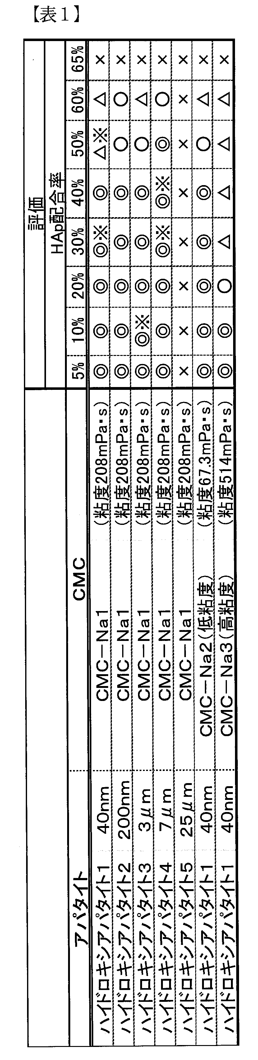
- the calcined hydroxyapatite aggregate to be used is selected from those having a particle size of 40 nm, 200 nm, 3 ⁇ m, 7 ⁇ m, and 25 ⁇ m, and the carboxymethyl cellulose gel has a viscosity of 67.3 mPa ⁇ s, 208 mPa ⁇ s, and 514 mPa ⁇ s.
- Subcutaneous injection preparations were prepared in the same manner as described above except that the selection was made and the HAp content was changed.
- the thixotropy index of the subcutaneous injection agent according to each Example was measured by the method described above, it was confirmed to be 0.1 to 0.3. That is, it can be seen that the agent for subcutaneous injection according to the example can pass through a fine needle and can remain subcutaneously for a sufficient time. In consideration of the property of staying subcutaneously, the gel viscosity was 208 mPa ⁇ s better than that of 67.3 mPa ⁇ s.
- the particle size of 100 calcined hydroxyapatite particles (particles in an image photographed with a microscope) contained in the subcutaneous injection agent according to each example was measured, and all of them had a particle size of 15 ⁇ m or less. It was confirmed.
- apatite with a small particle diameter has a strong tendency to become high thixotropic property, that is, a small thixotropy index with a small amount, especially for particles of less than 100 nm.
- FIG. 1 shows photographs before and after the defoaming step when hydroxyapatite 1, CMC-Na1, and HAp content is 30% ⁇ (a) is before defoaming and (b) is after defoaming. ⁇ , And the range surrounded by a round line is the remaining air.
- Example B ⁇ Manufacture of subcutaneous injections ⁇ ⁇ Raw material> ⁇ CMC-Na ⁇ Glycerin ⁇ Calcined hydroxyapatite aggregate ⁇ Sterile water
- ⁇ Evaluation ⁇ ⁇ Particle size distribution> The particle size distribution of the apatite particles in the subcutaneous injection was measured using the nanoparticle size distribution measuring device SALD-7500 nano (Shimadzu Corp.). The film was stretched as much as possible between the glass slides, and the subcutaneous injection agent was sandwiched, and the refractive index was measured as 1.65. The results are shown in FIG.
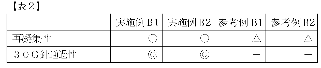
- re-aggregation does not occur in the case of the subcutaneous injection preparation according to Example B (4% or more of the burned hydroxyapatite particles have a particle size of 5 ⁇ m or more and 30 ⁇ m or less).
- the thixotropy index of the subcutaneous injection agent according to each Example was measured by the method described above, it was confirmed to be 0.1 to 0.3. That is, it can be seen that the agent for subcutaneous injection according to the example can pass through a fine needle and can remain subcutaneously for a sufficient time.
Landscapes
- Health & Medical Sciences (AREA)
- Chemical & Material Sciences (AREA)
- Public Health (AREA)
- Medicinal Chemistry (AREA)
- Veterinary Medicine (AREA)
- Epidemiology (AREA)
- Life Sciences & Earth Sciences (AREA)
- Animal Behavior & Ethology (AREA)
- General Health & Medical Sciences (AREA)
- Dermatology (AREA)
- Transplantation (AREA)
- Oral & Maxillofacial Surgery (AREA)
- Inorganic Chemistry (AREA)
- Pharmacology & Pharmacy (AREA)
- Dispersion Chemistry (AREA)
- Engineering & Computer Science (AREA)
- Composite Materials (AREA)
- Materials Engineering (AREA)
- Materials For Medical Uses (AREA)
- Pharmaceuticals Containing Other Organic And Inorganic Compounds (AREA)
- Medicinal Preparation (AREA)
- Infusion, Injection, And Reservoir Apparatuses (AREA)
Abstract
Description
皮下注射用剤において、
焼成ハイドロキシアパタイト粒子を含有するハイドロゲルを有し、
前記焼成ハイドロキシアパタイト粒子の含有率が、前記剤の全質量を基準として、1~60質量%であり、
前記焼成ハイドロキシアパタイト粒子の平均粒径が15μm以下である
ことを特徴とする剤である。
本発明(2)は、
前記焼成ハイドロキシアパタイト粒子の90%以上が、15μm以下の粒径であることを特徴とする、前記発明(1)の剤である。
本発明(3)は、
前記焼成ハイドロキシアパタイト粒子の4%以上が、5μm以上30μm以下の粒径であることを特徴とする、前記発明(1)又は(2)の剤である。
本発明(4)は、
前記焼成ハイドロキシアパタイト粒子が、コラーゲン産生増進用であることを特徴とする、前記発明(1)~(3)のいずれかの剤である。
本発明(5)は、
前記ハイドロゲルが、カルボキシメチルセルロースゲルであることを特徴とする、前記発明(1)~(4)のいずれかの剤である。
本発明(6)は、
前記ハイドロゲルの粘度が、50~15000mPa・sであることを特徴とする、前記発明(1)~(5)のいずれかの剤である。
本発明(7)は、
前記剤が、0.20mm以下の内径を有する中空針を通過することを特徴とする、前記発明(1)~(6)のいずれかの剤である。
本発明(8)は、
皮下注射用剤の製造方法において、
焼成ハイドロキシアパタイト粒子とハイドロゲルとを混合する工程を有し、
前記焼成ハイドロキシアパタイト粒子の含有率が、前記剤の全質量を基準として、1~60質量%であり、
前記剤中の焼成ハイドロキシアパタイト粒子の平均粒径が15μm以下である
ことを特徴とする製造方法である。
本発明(9)は、
前記剤中の焼成ハイドロキシアパタイト粒子の90%以上が、15μm以下の粒径であることを特徴とする、前記発明(8)の製造方法である。
本発明(10)は、
前記剤中の焼成ハイドロキシアパタイト粒子の4%以上が、5μm以上30μm以下の粒径であることを特徴とする、前記発明(8)又は(9)に記載の製造方法である。
本発明(11)は、
前記焼成ハイドロキシアパタイト粒子が、コラーゲン産生増進用であることを特徴とする、前記発明(8)~(10)のいずれかの製造方法である。
本発明(12)は、
前記ハイドロゲルが、カルボキシメチルセルロースゲルであることを特徴とする、前記発明(8)~(11)のいずれかの製造方法である。
本発明(13)は、
前記ハイドロゲルの粘度が、50~15000mPa・sであることを特徴とする、前記発明(8)~(12)のいずれかの製造方法である。
本発明(14)は、
前記剤が、0.20mm以下の内径を有する中空針を通過することを特徴とする、前記発明(8)~(13)のいずれかの製造方法である。
(1)皮下注射用剤の組成
(2)皮下注射用剤の製造方法
(3)皮下注射用剤の作用
(4)皮下注射用剤の使用方法
(5)皮下注射用剤の物性
以下、本実施形態に係る皮下注射用剤の成分及びその配合量に関して詳述する。
本実施形態に係る皮下注射用剤は、ハイドロキシアパタイト粒子とハイドロゲルとを含み、更にその他の成分を含んでいてもよい。尚、ハイドロキシアパタイト粒子凝集体は、ハイドロキシアパタイト粒子の凝集体である。
先ず、本実施形態に係るハイドロキシアパタイト(水酸化燐灰石)粒子に関し、組成、機能及び特性、入手方法、焼成、粒径に関して説明する。尚、ここで示すハイドロキシアパタイト粒子の物性(特に粒径)は、皮下注射用剤中に含まれるハイドロキシアパタイト粒子の物性を示すものである(皮下注射用剤中に含まれるハイドロキシアパタイト粒子の物性と、皮下注射用剤の構成原料であるハイドロキシアパタイト粒子の物性とは、製造段階による形態変化等が生じる場合もあるため、必ずしも同一とは限らない)。
ハイドロキシアパタイト(HAp:Hydroxyapatite)は、化学式Ca10(PO4)6(OH)2で示される塩基性リン酸カルシウムで、天然には歯や骨の主成分として、また鉱石として存在する。
ハイドロキシアパタイト粒子は、高い生体親和性を示す。特に皮下注射用剤として使用した場合には、ナノ粒子としてフィラー中に存在するハイドロキシアパタイト粒子が、繊維芽細胞を刺激することでコラーゲン産生を増進する。また、マイクロ粒子としてフィラー中に存在するハイドロキシアパタイト粒子は、スペーサーとして物理的に機能する。
本実施形態に係るハイドロキシアパタイト粒子としては、焼成ハイドロキシアパタイト粒子(以下、特に断らない限り、「ハイドロキシアパタイト粒子」とある場合には、「焼成ハイドロキシアパタイト粒子」を意味する。)を用いる。ハイドロキシアパタイト粒子を焼成(例えば、800℃で1時間)することにより、粒子の結晶性が高くなり、且つ複数の一次粒子の凝集体が熱により融着して、より強固で安定な粒子となる。特に、分散焼成法により焼成することが好適であり、融着防止剤を用いて焼成することにより凝集しにくくかつ結晶性の高いアパタイトナノ粒子を得ることができる。このように、本発明でいう「ハイドロキシアパタイト粒子」とは、特に断らない限り、一次粒子サイズの焼成ハイドロキシアパタイト粒子のみならず、複数の一次粒子が融着した凝集体も含む概念である。
一般的なハイドロキシアパタイト粒子の製造方法としては、例えば溶液法(湿式法)が挙げられる。これは、中性若しくはアルカリ性の水溶液中でカルシウムイオンとリン酸イオンとを反応させることにより合成する方法であり、中和反応によるものや、カルシウム塩とリン酸塩を反応させるものがある。前述のように、融着防止剤共存下で焼成することにより凝集の少ない結晶性のアパタイト粒子を得る。
本実施形態に係るハイドロキシアパタイト粒子の粒径(平均粒径)は、15μm以下であり、好適には5μm以下であり、より好適には1μm以下であり、更に好適には100nm以下である。ハイドロキシアパタイト粒子の粒径をこのような範囲とすることで、繊維芽細胞を刺激してのコラーゲン産生増進作用を示す。また、このような範囲とすることで、生体内で異物として認識され難いサイズであることから、マクロファージの誘因が起き難いという作用を示す。ハイドロキシアパタイト粒子の粒径(平均粒径)の下限値は特に限定されないが、例えば10nm以上である。尚、ここで示すフィラー中のハイドロキシアパタイト粒子(凝集体を含む)の平均粒径は、顕微鏡により撮影した画像における各粒子(例えば、50個の粒子)の粒子経を測長し、その平均径により決定する。また、フィラーを水等に膨潤させてハイドロゲルを溶解させて、取り出したハイドロキシアパタイトの各粒子(例えば、50個の粒子)の粒子径を測長し、その平均径により決定することもできる。また、ここで示すフィラー中のハイドロキシアパタイト粒子(凝集体を含む)の平均粒径は、レーザー散乱式粒度分布測定装置(島津製作所製SALD-7500)の高濃度測定ユニットを用いて粒度分布を測定し、粒度分布測定結果より50%体積粒径(D50)を求めることによって、決定することもできる。具体的には、スライドガラス(マツナミ S7213)の間に可能な限り延伸してフィラーを挟み込み、屈折率を1.65として測定する。
ハイドロゲルは、剤に用いることができるものであれば、特に限定されないが、例えば、カルボキシメチルセルロースゲル(CMC)、ポリビニルアルコールゲル、ポリアクリル酸ソーダゲル{例えば、Carbopol(登録商標)等}が挙げられる。原料となるセルロースの安全性、安価であることなどから、カルボキシメチルセルロースゲルが好ましい。
カルボキシメチルセルロース(CMC)は、セルロースの水酸基にカルボキシメチル基が導入されたセルロースエーテルである。本形態において、CMCであれば特に限定されないが、CMCのナトリウム塩であるナトリウムカルボキシメチルセルロース(Na-CMC)が好ましい。
その他の成分としては、本形態の効果を阻害しない範囲内で、粘度調整剤(例えば、グリセリン等)や、分散剤(例えば、ポリアクリル酸等)等の公知の添加剤を適宜含んでいてもよい。
本実施形態に係る皮下注射用剤において、ハイドロキシアパタイト粒子の含有率は、皮下注射用剤の全質量を基準として、1質量%以上であり、好適には15質量%以上であり、より好適には20質量%以上であり、更に好適には30質量%以上である。ハイドロキシアパタイト粒子の含有率をこのような範囲とすることにより、高い弾力を示すとともに、フィラーに配合されたアパタイト粒子の沈降による分離を防止できる。また、ハイドロキシアパタイト粒子の含有率が1質量%未満(特には、15質量%未満)であると、長期保管によるアパタイト粒子とゲルの分離が顕著となり得る。
次に、本実施形態に係る皮下注射用剤の製造方法を説明する。
本実施形態に係る皮下注射用剤を皮下注入後、まず、ハイドロゲルが吸収分解される。本形態に係る焼成ハイドロキシアパタイト粒子は粒径が小さいために、焼成ハイドロキシアパタイト粒子が放出される。そして、焼成ハイドロキシアパタイト粒子による繊維芽細胞刺激によってコラーゲン産生が増進される。前述のように、生体内にて異物として認識され難いサイズであることから、マクロファージの誘因が起き難いという作用を示し、結晶性が高いことから、生体内で長期間生体活性を維持することができる。その結果、コラーゲンリッチ組織が皮下に残存する。
本実施形態に係る皮下注射用剤の使用方法は、何ら限定されるものではないが、一般的な注射器のシリンジに充填した後、皮下用の注射において使用することができる。特に、0.20mm以下の内径を有する中空針を有する注射器用の注射用剤とすることができる。ここで、本実施形態に係る皮下注射用剤の具体的な使用方法として、本実施形態に係る皮下注射用剤を含有する注射器の好適な製造方法及び本実施形態に係る皮下注射用剤の適用方法に関して説明する。尚、これらはあくまで一例であり、本実施形態に係る皮下注射用剤の使用方法はこれらには何ら限定されない。
本実施形態に係る皮下注射用剤を含有する注射器の製造方法は、脱泡工程を含むことを特徴とする方法である。以下各々の工程に関して説明する。
まず、本実施形態に係る皮下注射用剤は、脱泡工程によって、皮下注射用剤に含まれるエアが除去される。本形態に係る皮下注射用剤の特性から、遠心分離機にかける方法が皮下注射用剤の脱泡工程として好適であり、減圧下で遠心分離機にかける方法が最も好適である。より具体的には、カルボキシメチルセルロース(CMC)ゲルを主成分とする等、チクソトロピー性を有するフィラーを用いる場合、遠心分離によってフィラーにせん断力が加えられることで、フィラーを低粘度化させ、脱泡効率を上昇させることが可能となる。更に減圧下で遠心分離を行うことにより、当該脱泡原理と合わせ、脱泡効率を更に上昇させることが可能となる。
・レシピ01 高粘度標準脱泡
1.3KPa 9/3 90sec (圧力 公転/自転比 時間)
・レシピ02 金属系フィラー脱泡
1.3KPa 3/9 120sec
1.3KPa 7/5 90sec
1.3KPa 9/2 60sec
・レシピ03 セラミック系フィラー脱泡
1.3KPa 2/5 60sec
1.3KPa 5/5 60sec
次に、脱泡工程後の皮下注射用剤をシリンジに充填し、本実施形態に係る注射器とする。
更に、本実施形態に係る皮下注射用剤は、必要に応じて、殺菌・滅菌を行ってもよい。殺菌・滅菌方法としては、例えば、高圧蒸気滅菌が挙げられる。
本実施形態に係る皮下注射用剤及び皮下注射用剤を含有する注射器の適用方法としては、従来の皮下注射用剤及び皮下注射用剤を含有する注射器等と同様であり、特に限定されない。
本実施形態に係る皮下注射用剤によれば、カルボキシメチルセルロースゲルを主成分として、平均粒径が15μm以下の焼成ハイドロキシアパタイト粒子を用いることで、前記剤の全質量を基準として、1~60質量%以上の焼成ハイドロキシアパタイト粒子を配合しても、内径0.20mm以下であるような細い注射針においても使用可能となる。内径0.20mmとは、内径が大きい注射針30Gに相当する。本実施形態に係る皮下注射用剤は、内径が大きい注射針32G(内径0.18mm)以下の細い注射針や、通常の注射針30G(内径0.14mm)以下の細い注射針においても使用可能であり、通常の注射針32G(内径0.12mm)以下の細い注射針においても使用可能である。
本実施形態に係る皮下注射用剤は、発明の効果をより高めるために、36-37℃(体温)におけるチクソトロピー指数が、0.1~0.5であることが好適であり、0.1~0.3であることがより好適である。チクソトロピー指数は、下記射出特性によって測定された数値である。
シリンジに充填したフィラーの針からの射出特性を評価する方法を述べる。射出特性の評価方法には、本形態に係る流動性定量評価装置を用いる。この流動性定量評価装置によって、簡易に射出特性を評価することができる。
本形態(A)は、皮下注射用剤の製造方法において、
前記剤が、
焼成ハイドロキシアパタイト粒子を含有するハイドロゲルを有する
剤であって、
前記製造方法が、
前記剤に含まれるエアを除去する脱泡工程
を有する製造方法である。
本形態(B)は、
皮下注射用剤を含有する注射器の製造方法において、
前記剤が、
焼成ハイドロキシアパタイト粒子を含有するハイドロゲルを有する
剤であって、
前記製造方法が、
前記剤に含まれるエアを除去する脱泡工程と、
前記脱泡工程後の前記剤をシリンジに充填する工程と
を有する製造方法である。
本形態(C)は、
前記脱泡工程が、減圧下で、前記剤を遠心分離機にかけることによって脱泡を行う工程である、前記形態(A)又は(B)の製造方法である。
本形態(D)は、
前記ハイドロゲルが、カルボキシメチルセルロースゲルである、前記形態(A)~(C)のいずれかの製造方法である。
本形態(E)は、
前記剤の粘度が、50~15000mPa・sである、前記形態(A)~(D)のいずれかの製造方法である。
本形態(F)は、
前記焼成ハイドロキシアパタイト粒子の平均粒径が15μm以下である、前記形態(A)~(E)のいずれかの製造方法である。
本形態(G)は、
前記焼成ハイドロキシアパタイト粒子の90%以上が、15μm以下の粒径である、前記形態(A)~(H)の製造方法である。
本形態(H)は、
前記焼成ハイドロキシアパタイト粒子の含有率が、前記剤の全質量を基準として、1~60質量%である、前記形態(A)~(G)のいずれかの製造方法である。
本形態(I)は、
前記剤が、0.20mm以下の内径を有する中空針を通過する、前記形態(A)~(H)のいずれかの製造方法である。
≪皮下注射用剤の製造≫
<原料>
・CMC-Na
・グリセリン
・焼成ハイドロキシアパタイト凝集体
・滅菌水
使用する器具の滅菌を行った。
次に、グリセリン50gをプロペラシャフト付モーターにて300rpmで撹拌した。2分超撹拌しながら、グリセリン中にCMC-Na5gを配合し、15分撹拌し溶液を得た。
得られた溶液を10分超撹拌し、得られたCMC-Naペーストを、1.5cm角にカットし204mlの滅菌水中に配合した。この滅菌水を120分撹拌し、CMC-Naハイドロゲル(粘度208mPa・s)を得た。
得られたCMC-Naハイドロゲル28.0gに、焼成ハイドロキシアパタイト凝集体12.0gを3回に別けて配合し、10分撹拌し、原料フィラーを得た。
得られた原料フィラー30.0gをプロペラ撹拌し、皮下注射用剤(HAp配合率30質量%)を得た。
次に、上記皮下注射用剤に関して、30Gの針を通過可能かの評価を行った。具体的には、皮下注射用剤を脱泡した後、注射器に充填した。次に、得られた注射器の押し出しを行い、注射器中の皮下注射用剤が30G針通過性を確認した。スムーズに通過する場合を◎、押し出しに少し抵抗がある場合を○、押し出しに強い力が必要な場合を△、押し出しが出来ない場合、又はフィラー自体を作成できない場合を×とした。結果を表1に示す。
≪皮下注射用剤の製造≫
<原料>
・CMC-Na
・グリセリン
・焼成ハイドロキシアパタイト凝集体
・滅菌水
使用する器具の滅菌を行った。
次に、グリセリン50gをプロペラシャフト付モーターにて300rpmで撹拌した。2分超撹拌しながら、グリセリン中にCMC-Na5gを配合し、15分撹拌し溶液を得た。
得られた溶液を10分超撹拌し、得られたCMC-Naペーストを、1.5cm角にカットし204mlの滅菌水中に配合した。この滅菌水を120分撹拌し、CMC-Naハイドロゲル(粘度208mPa・s)を得た。
得られたCMC-Naハイドロゲル28.0gに、焼成ハイドロキシアパタイト凝集体12.0gを3回に別けて配合し、10分撹拌し、原料フィラーを得た。
得られた原料フィラー30.0gをプロペラ撹拌し、皮下注射用剤(HAp配合率30質量%)を得た。
ここで、使用する焼成ハイドロキシアパタイト凝集体については、粒径が40nm、200nm、3μm、7μm、25μmのものを解砕・混合し、実施例B1、実施例B2、参考例B1及び参考例B2の各々に係る皮下注射用剤における焼成ハイドロキシアパタイト凝集体を用意した。
<粒度分布>
前記工程により調整した皮下注射用剤を、ナノ粒子径分布測定装置SALD‐7500nano(島津製作所)を用いて、皮下注射用剤におけるアパタイト粒子の粒度分布を測定した。スライドガラスの間に可能な限り延伸して皮下注射用剤を挟み込み、屈折率を1.65として測定した。結果を図1に示す。
次に、皮下注射用剤を120℃で熱滅菌し、再凝集性を観察した。再凝集が観察される場合を△、再凝集が観察されない場合を○とした。結果を表2に示す。
さらに、再凝集の生じなかった上記皮下注射用剤(即ち、熱滅菌時再凝集性にて評価が○である皮下注射用剤)に関して、30Gの針を通過可能かの評価を行った。具体的には、皮下注射用剤を脱泡した後、注射器に充填した。次に、得られた注射器の押し出しを行い、注射器中の皮下注射用剤が30G針通過性を確認した。スムーズに通過する場合を◎、押し出しに少し抵抗がある場合を○、押し出しに強い力が必要な場合を△、押し出しが出来ない場合を×とした。結果を表2に示す。
Claims (14)
- 皮下注射用剤において、
焼成ハイドロキシアパタイト粒子を含有するハイドロゲルを有し、
前記焼成ハイドロキシアパタイト粒子の含有率が、前記剤の全質量を基準として、1~60質量%であり、
前記焼成ハイドロキシアパタイト粒子の平均粒径が15μm以下である
ことを特徴とする剤。 - 前記焼成ハイドロキシアパタイト粒子の90%以上が、15μm以下の粒径であることを特徴とする、請求項1に記載の剤。
- 前記焼成ハイドロキシアパタイト粒子の4%以上が、5μm以上30μm以下の粒径であることを特徴とする、請求項1又は2に記載の剤。
- 前記焼成ハイドロキシアパタイト粒子が、コラーゲン産生増進用であることを特徴とする、請求項1~3のいずれかに記載の剤。
- 前記ハイドロゲルが、カルボキシメチルセルロースゲルであることを特徴とする、請求項1~4のいずれかに記載の剤。
- 前記ハイドロゲルの粘度が、50~15000mPa・sであることを特徴とする、請求項1~5のいずれかに記載の剤。
- 前記剤が、0.20mm以下の内径を有する中空針を通過することを特徴とする、請求項1~6のいずれかに記載の剤。
- 皮下注射用剤の製造方法において、
焼成ハイドロキシアパタイト粒子とハイドロゲルとを混合する工程を有し、
前記焼成ハイドロキシアパタイト粒子の含有率が、前記剤の全質量を基準として、1~60質量%であり、
前記剤中の焼成ハイドロキシアパタイト粒子の平均粒径が15μm以下である
ことを特徴とする製造方法。 - 前記剤中の焼成ハイドロキシアパタイト粒子の90%以上が、15μm以下の粒径であることを特徴とする、請求項8に記載の製造方法。
- 前記剤中の焼成ハイドロキシアパタイト粒子の4%以上が、5μm以上30μm以下の粒径であることを特徴とする、請求項8又は9に記載の製造方法。
- 前記焼成ハイドロキシアパタイト粒子が、コラーゲン産生増進用であることを特徴とする、請求項8~10のいずれかに記載の製造方法。
- 前記ハイドロゲルが、カルボキシメチルセルロースゲルであることを特徴とする、請求項8~11のいずれかに記載の製造方法。
- 前記ハイドロゲルの粘度が、50~15000mPa・sであることを特徴とする、請求項8~12のいずれかに記載の製造方法。
- 前記剤が、0.20mm以下の内径を有する中空針を通過することを特徴とする、請求項8~13のいずれかに記載の製造方法。
Priority Applications (8)
| Application Number | Priority Date | Filing Date | Title |
|---|---|---|---|
| US15/535,358 US10918766B2 (en) | 2014-12-12 | 2015-09-14 | Agent for hypodermic injection |
| BR112017012289-8A BR112017012289B1 (pt) | 2014-12-12 | 2015-09-14 | Agente para injeção hipodérmica |
| EP15867438.2A EP3231433B1 (en) | 2014-12-12 | 2015-09-14 | Agent for hypodermic injection |
| HK18104485.4A HK1245091B (zh) | 2014-12-12 | 2015-09-14 | 皮下注射用剂 |
| CN201580067195.4A CN107743396B (zh) | 2014-12-12 | 2015-09-14 | 皮下注射用剂 |
| AU2015361574A AU2015361574B2 (en) | 2014-12-12 | 2015-09-14 | Agent for hypodermic injection |
| CA2970592A CA2970592C (en) | 2014-12-12 | 2015-09-14 | Agent for hypodermic injection |
| KR1020177015425A KR102496871B1 (ko) | 2014-12-12 | 2015-09-14 | 피하 주사용제 |
Applications Claiming Priority (4)
| Application Number | Priority Date | Filing Date | Title |
|---|---|---|---|
| JP2014-252327 | 2014-12-12 | ||
| JP2014252327A JP5774189B1 (ja) | 2014-12-12 | 2014-12-12 | 皮下注射用剤 |
| JP2015129289A JP5797864B1 (ja) | 2015-06-26 | 2015-06-26 | 皮下注射用剤 |
| JP2015-129289 | 2015-06-26 |
Publications (1)
| Publication Number | Publication Date |
|---|---|
| WO2016092928A1 true WO2016092928A1 (ja) | 2016-06-16 |
Family
ID=56107124
Family Applications (1)
| Application Number | Title | Priority Date | Filing Date |
|---|---|---|---|
| PCT/JP2015/076068 Ceased WO2016092928A1 (ja) | 2014-12-12 | 2015-09-14 | 皮下注射用剤 |
Country Status (8)
| Country | Link |
|---|---|
| US (1) | US10918766B2 (ja) |
| EP (1) | EP3231433B1 (ja) |
| KR (1) | KR102496871B1 (ja) |
| CN (1) | CN107743396B (ja) |
| AU (1) | AU2015361574B2 (ja) |
| BR (1) | BR112017012289B1 (ja) |
| CA (1) | CA2970592C (ja) |
| WO (1) | WO2016092928A1 (ja) |
Families Citing this family (4)
| Publication number | Priority date | Publication date | Assignee | Title |
|---|---|---|---|---|
| EP4284455A1 (en) * | 2021-01-26 | 2023-12-06 | Merz North America, Inc. | Calcium hydroxyapatite particles and use thereof |
| CN115382011A (zh) * | 2021-07-01 | 2022-11-25 | 意瑞生物科技(苏州)有限公司 | 一种皮肤注射填充剂凝胶及其制备工艺 |
| CN114146020B (zh) * | 2022-02-10 | 2022-04-29 | 中国远大集团有限责任公司 | 一种注射美容产品及其制备方法和应用 |
| KR102673797B1 (ko) * | 2023-03-28 | 2024-06-10 | 주식회사 파마리서치바이오 | 핵산 및 칼슘 하이드록실아파타이트를 포함하는 필러 조성물 |
Citations (10)
| Publication number | Priority date | Publication date | Assignee | Title |
|---|---|---|---|---|
| JPH05186364A (ja) * | 1991-08-01 | 1993-07-27 | Takeda Chem Ind Ltd | 水溶性組成物 |
| JP2003507351A (ja) * | 1999-08-13 | 2003-02-25 | バイオフォーム・インコーポレイテッド | 組織増強物質および方法 |
| WO2005082405A1 (ja) * | 2004-02-26 | 2005-09-09 | Japan Science And Technology Agency | たんぱく性薬物の注射用徐放性微粒子製剤およびその製造法 |
| JP2007001865A (ja) * | 2003-09-16 | 2007-01-11 | Ltt Bio-Pharma Co Ltd | 脂溶性薬物封入微粒子、その製造法およびそれを含有する製剤 |
| JP2007537249A (ja) * | 2004-05-13 | 2007-12-20 | ウニベルジテート ユートレヒト ホールディング ベーフェー | 荷電ポリマーを含むゲル組成物 |
| WO2010137122A1 (ja) * | 2009-05-26 | 2010-12-02 | 株式会社ソフセラ | コラーゲン産生促進剤 |
| JP2012508744A (ja) * | 2008-11-16 | 2012-04-12 | ボード・オブ・リージエンツ,ザ・ユニバーシテイ・オブ・テキサス・システム | 高濃度の低粘度懸濁液 |
| JP2013526545A (ja) * | 2010-05-13 | 2013-06-24 | パシラ ファーマシューティカルズ インコーポレーテッド | 疾患修飾性抗リウマチ薬(dmard)および抗癌剤としてのメトトレキサートの持続性放出製剤 |
| JP5774189B1 (ja) * | 2014-12-12 | 2015-09-09 | ジーンメディカル株式会社 | 皮下注射用剤 |
| JP5797864B1 (ja) * | 2015-06-26 | 2015-10-21 | ジーンメディカル株式会社 | 皮下注射用剤 |
Family Cites Families (7)
| Publication number | Priority date | Publication date | Assignee | Title |
|---|---|---|---|---|
| JPS51133916A (en) | 1975-05-15 | 1976-11-20 | Mitsui Keikinzoku Kako | Connection tool of shape material |
| US5344644A (en) | 1991-08-01 | 1994-09-06 | Takeda Chemical Industries, Ltd. | Water-soluble composition for sustained-release |
| US7968110B2 (en) | 1992-02-11 | 2011-06-28 | Merz Aesthetics, Inc. | Tissue augmentation material and method |
| US7060287B1 (en) | 1992-02-11 | 2006-06-13 | Bioform Inc. | Tissue augmentation material and method |
| FR2715853B1 (fr) | 1994-02-08 | 1996-04-26 | Centre Nat Rech Scient | Composition pour bio-matériau; procédé de préparation. |
| JP5725489B2 (ja) | 2008-06-27 | 2015-05-27 | 公立大学法人大阪市立大学 | 医療用組成物および医療用キット |
| WO2013028551A2 (en) * | 2011-08-19 | 2013-02-28 | Pioneer Surigical Technology | Injectable fillers for aesthetic medical enhancement and for therapeutic applications |
-
2015
- 2015-09-14 US US15/535,358 patent/US10918766B2/en not_active Expired - Fee Related
- 2015-09-14 CA CA2970592A patent/CA2970592C/en active Active
- 2015-09-14 AU AU2015361574A patent/AU2015361574B2/en not_active Ceased
- 2015-09-14 WO PCT/JP2015/076068 patent/WO2016092928A1/ja not_active Ceased
- 2015-09-14 KR KR1020177015425A patent/KR102496871B1/ko active Active
- 2015-09-14 CN CN201580067195.4A patent/CN107743396B/zh not_active Expired - Fee Related
- 2015-09-14 EP EP15867438.2A patent/EP3231433B1/en active Active
- 2015-09-14 BR BR112017012289-8A patent/BR112017012289B1/pt not_active IP Right Cessation
Patent Citations (10)
| Publication number | Priority date | Publication date | Assignee | Title |
|---|---|---|---|---|
| JPH05186364A (ja) * | 1991-08-01 | 1993-07-27 | Takeda Chem Ind Ltd | 水溶性組成物 |
| JP2003507351A (ja) * | 1999-08-13 | 2003-02-25 | バイオフォーム・インコーポレイテッド | 組織増強物質および方法 |
| JP2007001865A (ja) * | 2003-09-16 | 2007-01-11 | Ltt Bio-Pharma Co Ltd | 脂溶性薬物封入微粒子、その製造法およびそれを含有する製剤 |
| WO2005082405A1 (ja) * | 2004-02-26 | 2005-09-09 | Japan Science And Technology Agency | たんぱく性薬物の注射用徐放性微粒子製剤およびその製造法 |
| JP2007537249A (ja) * | 2004-05-13 | 2007-12-20 | ウニベルジテート ユートレヒト ホールディング ベーフェー | 荷電ポリマーを含むゲル組成物 |
| JP2012508744A (ja) * | 2008-11-16 | 2012-04-12 | ボード・オブ・リージエンツ,ザ・ユニバーシテイ・オブ・テキサス・システム | 高濃度の低粘度懸濁液 |
| WO2010137122A1 (ja) * | 2009-05-26 | 2010-12-02 | 株式会社ソフセラ | コラーゲン産生促進剤 |
| JP2013526545A (ja) * | 2010-05-13 | 2013-06-24 | パシラ ファーマシューティカルズ インコーポレーテッド | 疾患修飾性抗リウマチ薬(dmard)および抗癌剤としてのメトトレキサートの持続性放出製剤 |
| JP5774189B1 (ja) * | 2014-12-12 | 2015-09-09 | ジーンメディカル株式会社 | 皮下注射用剤 |
| JP5797864B1 (ja) * | 2015-06-26 | 2015-10-21 | ジーンメディカル株式会社 | 皮下注射用剤 |
Non-Patent Citations (3)
| Title |
|---|
| JI, D. Y. ET AL.: "A novel injectable chitosan/polyglutamate polyelectrolyte complex hydrogel with hydroxyapatite for soft-tissue augmentation", CARBOHYDRATE POLYMERS, vol. 89, no. 4, 2012, pages 1123 - 1130, XP028512034, DOI: doi:10.1016/j.carbpol.2012.03.083 * |
| See also references of EP3231433A4 * |
| THIOLY-BENSOUSSAN, D.: "Non-hyaluronic acid fillers", CLINICS IN DERMATOLOGY, vol. 26, no. 2, 2008, pages 160 - 176, XP022647616, DOI: doi:10.1016/j.clindermatol.2007.09.017 * |
Also Published As
| Publication number | Publication date |
|---|---|
| US10918766B2 (en) | 2021-02-16 |
| CA2970592A1 (en) | 2016-06-16 |
| AU2015361574B2 (en) | 2020-12-17 |
| EP3231433A4 (en) | 2018-08-08 |
| BR112017012289A2 (pt) | 2018-04-24 |
| US20170333600A1 (en) | 2017-11-23 |
| CN107743396A (zh) | 2018-02-27 |
| CA2970592C (en) | 2023-01-31 |
| KR20170093828A (ko) | 2017-08-16 |
| HK1245091A1 (zh) | 2018-08-24 |
| KR102496871B1 (ko) | 2023-02-07 |
| BR112017012289B1 (pt) | 2022-11-16 |
| CN107743396B (zh) | 2021-01-15 |
| EP3231433C0 (en) | 2023-12-27 |
| AU2015361574A1 (en) | 2017-07-13 |
| EP3231433A1 (en) | 2017-10-18 |
| EP3231433B1 (en) | 2023-12-27 |
Similar Documents
| Publication | Publication Date | Title |
|---|---|---|
| JP5797864B1 (ja) | 皮下注射用剤 | |
| JP5774190B1 (ja) | 皮下注射用剤及び皮下注射用剤を含有する注射器の製造方法 | |
| WO2016092928A1 (ja) | 皮下注射用剤 | |
| Wu et al. | Premixed tricalcium silicate/sodium phosphate dibasic cements for root canal filling | |
| JP6113025B2 (ja) | アルジネート硬化性組成物の調製方法 | |
| JP5774189B1 (ja) | 皮下注射用剤 | |
| JP5781681B1 (ja) | 焼成ハイドロキシアパタイト粒子組成物の製造方法 | |
| JP2004244291A (ja) | リン酸水素カルシウム粉体、リン酸カルシウム系ペースト用粉体、リン酸カルシウム系ペースト混練器ユニット及びリン酸水素カルシウム粉体の製造方法 | |
| HK1245091B (zh) | 皮下注射用剂 | |
| HK1241760A1 (en) | Agent for hypodermic injections and production method for syringes containing agent for hypodermic injections | |
| HK1241760B (zh) | 皮下注射用剂以及含有皮下注射用剂的注射器的制造方法 | |
| JP2742814B2 (ja) | 根管充填材 | |
| JP5463392B2 (ja) | リン酸カルシウム組成物の製造方法 | |
| Duarte et al. | Dental cements containing Bi2O3: Physical properties and radiopacity | |
| WO1990008530A1 (fr) | Matiere de remplissage de canal radiculaire comprenant une pate durcissable | |
| JP2002291866A (ja) | リン酸カルシウムセメント粉体及びリン酸カルシウムセメント |
Legal Events
| Date | Code | Title | Description |
|---|---|---|---|
| 121 | Ep: the epo has been informed by wipo that ep was designated in this application |
Ref document number: 15867438 Country of ref document: EP Kind code of ref document: A1 |
|
| ENP | Entry into the national phase |
Ref document number: 20177015425 Country of ref document: KR Kind code of ref document: A |
|
| ENP | Entry into the national phase |
Ref document number: 2970592 Country of ref document: CA |
|
| NENP | Non-entry into the national phase |
Ref country code: DE |
|
| REG | Reference to national code |
Ref country code: BR Ref legal event code: B01A Ref document number: 112017012289 Country of ref document: BR |
|
| REEP | Request for entry into the european phase |
Ref document number: 2015867438 Country of ref document: EP |
|
| ENP | Entry into the national phase |
Ref document number: 2015361574 Country of ref document: AU Date of ref document: 20150914 Kind code of ref document: A |
|
| ENP | Entry into the national phase |
Ref document number: 112017012289 Country of ref document: BR Kind code of ref document: A2 Effective date: 20170609 |