WO2016136850A1 - シグナル増強剤 - Google Patents
シグナル増強剤 Download PDFInfo
- Publication number
- WO2016136850A1 WO2016136850A1 PCT/JP2016/055541 JP2016055541W WO2016136850A1 WO 2016136850 A1 WO2016136850 A1 WO 2016136850A1 JP 2016055541 W JP2016055541 W JP 2016055541W WO 2016136850 A1 WO2016136850 A1 WO 2016136850A1
- Authority
- WO
- WIPO (PCT)
- Prior art keywords
- fucose
- lectin
- urea
- sugar chain
- reaction
- Prior art date
- Legal status (The legal status is an assumption and is not a legal conclusion. Google has not performed a legal analysis and makes no representation as to the accuracy of the status listed.)
- Ceased
Links
Images


Classifications
-
- G—PHYSICS
- G01—MEASURING; TESTING
- G01N—INVESTIGATING OR ANALYSING MATERIALS BY DETERMINING THEIR CHEMICAL OR PHYSICAL PROPERTIES
- G01N33/00—Investigating or analysing materials by specific methods not covered by groups G01N1/00 - G01N31/00
- G01N33/48—Biological material, e.g. blood, urine; Haemocytometers
- G01N33/50—Chemical analysis of biological material, e.g. blood, urine; Testing involving biospecific ligand binding methods; Immunological testing
-
- G—PHYSICS
- G01—MEASURING; TESTING
- G01N—INVESTIGATING OR ANALYSING MATERIALS BY DETERMINING THEIR CHEMICAL OR PHYSICAL PROPERTIES
- G01N33/00—Investigating or analysing materials by specific methods not covered by groups G01N1/00 - G01N31/00
- G01N33/48—Biological material, e.g. blood, urine; Haemocytometers
- G01N33/50—Chemical analysis of biological material, e.g. blood, urine; Testing involving biospecific ligand binding methods; Immunological testing
- G01N33/53—Immunoassay; Biospecific binding assay; Materials therefor
- G01N33/575—Immunoassay; Biospecific binding assay; Materials therefor for cancer
- G01N33/5758—Immunoassay; Biospecific binding assay; Materials therefor for cancer involving compounds serving as markers for tumours, cancers or neoplasias, e.g. cellular determinants, receptors, heat shock/stress proteins, A-protein, oligosaccharides or metabolites
-
- G—PHYSICS
- G01—MEASURING; TESTING
- G01N—INVESTIGATING OR ANALYSING MATERIALS BY DETERMINING THEIR CHEMICAL OR PHYSICAL PROPERTIES
- G01N33/00—Investigating or analysing materials by specific methods not covered by groups G01N1/00 - G01N31/00
- G01N33/48—Biological material, e.g. blood, urine; Haemocytometers
- G01N33/50—Chemical analysis of biological material, e.g. blood, urine; Testing involving biospecific ligand binding methods; Immunological testing
- G01N33/53—Immunoassay; Biospecific binding assay; Materials therefor
- G01N33/575—Immunoassay; Biospecific binding assay; Materials therefor for cancer
- G01N33/57525—Immunoassay; Biospecific binding assay; Materials therefor for cancer of the liver or pancreas
-
- G—PHYSICS
- G01—MEASURING; TESTING
- G01N—INVESTIGATING OR ANALYSING MATERIALS BY DETERMINING THEIR CHEMICAL OR PHYSICAL PROPERTIES
- G01N33/00—Investigating or analysing materials by specific methods not covered by groups G01N1/00 - G01N31/00
- G01N33/48—Biological material, e.g. blood, urine; Haemocytometers
- G01N33/50—Chemical analysis of biological material, e.g. blood, urine; Testing involving biospecific ligand binding methods; Immunological testing
- G01N33/53—Immunoassay; Biospecific binding assay; Materials therefor
- G01N33/575—Immunoassay; Biospecific binding assay; Materials therefor for cancer
- G01N33/57535—Immunoassay; Biospecific binding assay; Materials therefor for cancer of the large intestine, e.g. colon, rectum or anus
-
- G—PHYSICS
- G01—MEASURING; TESTING
- G01N—INVESTIGATING OR ANALYSING MATERIALS BY DETERMINING THEIR CHEMICAL OR PHYSICAL PROPERTIES
- G01N2333/00—Assays involving biological materials from specific organisms or of a specific nature
- G01N2333/435—Assays involving biological materials from specific organisms or of a specific nature from animals; from humans
- G01N2333/46—Assays involving biological materials from specific organisms or of a specific nature from animals; from humans from vertebrates
- G01N2333/47—Assays involving proteins of known structure or function as defined in the subgroups
- G01N2333/4701—Details
- G01N2333/4724—Lectins
-
- G—PHYSICS
- G01—MEASURING; TESTING
- G01N—INVESTIGATING OR ANALYSING MATERIALS BY DETERMINING THEIR CHEMICAL OR PHYSICAL PROPERTIES
- G01N2400/00—Assays, e.g. immunoassays or enzyme assays, involving carbohydrates
-
- G—PHYSICS
- G01—MEASURING; TESTING
- G01N—INVESTIGATING OR ANALYSING MATERIALS BY DETERMINING THEIR CHEMICAL OR PHYSICAL PROPERTIES
- G01N2400/00—Assays, e.g. immunoassays or enzyme assays, involving carbohydrates
- G01N2400/02—Assays, e.g. immunoassays or enzyme assays, involving carbohydrates involving antibodies to sugar part of glycoproteins
Definitions
- the present invention relates to a signal enhancer, and more particularly to a signal enhancer based on a reaction between an ⁇ 1-6 fucose sugar chain and an ⁇ 1-6 fucose-specific lectin.
- ⁇ 1-6 fucose transferase which transfers ⁇ 1-6 fucose (also referred to as core fucose) to N-acetylglucosamine at the reducing end of N-linked sugar chains, is expressed as hepatocytes become cancerous. It is known to do.
- ⁇ 1-6 fucose transferase also referred to as core fucose
- AFP ⁇ -fetoprotein
- AFP-L1, AFP-L2, and AFP-L3 appear from the anode side.
- the non-lectin-binding AFP-L1 band is main, but when hepatocellular carcinoma occurs, the lectin-binding AFP-L3 band increases. If the ratio of AFP-L3 to total AFP (AFP-L3%) is determined and the value exceeds 10%, hepatocellular carcinoma is suspected. If the ratio of AFP in which fucose has metastasized due to ⁇ 1-6 binding can be accurately identified, the accuracy of diagnosis of hepatocellular carcinoma will be further increased.
- ⁇ 1-6 fucose is known to be associated with diseases such as pancreatic cancer and colon cancer in addition to liver cancer.
- AAL and AOL have affinity for ⁇ 1-2 fucose, ⁇ 1-3 fucose, etc.
- LCA, PSA, and VFA have N-linked single-stranded and / or double-chains without fucose ⁇ 1-6.
- lectins such as having affinity for the main chain glycans, are not only ⁇ 1-6 fucose glycans but also glycolipid-based glycans having fucose other than ⁇ 1-6 linkage, and high-capacity without fucose. It also shows affinity for mannose sugar chains.
- Non-patent Document 1 Non-patent Document 1
- SRL salmon bamboo lectin.
- Patent Documents 4 and 5 chemical synthesis and recombinant production of this lectin are also performed.
- the detection of AFP-L3 (Patent Document 1), the determination of the deterioration degree of colorectal cancer (Patent Document 2), and the detection of pancreatic cancer (Patent Document 3) have also been successful.
- Patent 4514163 (fucose ⁇ 1 ⁇ 6 specific lectin)
- Patent 5263379 (Determination method of colorectal cancer)
- Patent 4900903 (Method for detecting pancreatic cancer) JP2011-148736 (peptide) JP2011-148735 (gene)
- an object of the present invention is to provide a signal enhancer that enhances a signal based on a reaction between an ⁇ 1-6 fucose sugar chain and an ⁇ 1-6 fucose-specific lectin that binds to the ⁇ 1-6 fucose sugar chain.
- the present invention is a signal enhancer that enhances a signal based on a reaction between an ⁇ 1-6 fucose sugar chain and an ⁇ 1-6 fucose-specific lectin bound thereto, and is selected from the group consisting of urea and thiourea.
- the signal enhancer is characterized by comprising at least one of the above as an active ingredient.
- ⁇ 1-6 fucose sugar chain means “a structure in which fucose is bonded to N-acetylglucosamine at the reducing end of an N-type sugar chain through an ⁇ 1-6 bond”.
- ⁇ 1-6 fucose-specific lectin is used herein as “(1) a binding constant of 1.0 ⁇ 10 4 M ⁇ 1 or more (at 25 ° C. with respect to ⁇ 1-6 fucose sugar chain). ) And (2) substantially non-binding to a high mannose sugar chain not containing ⁇ 1-6 fucose and / or a glycolipid sugar chain not containing ⁇ 1-6 fucose. "Lectin”.
- JP 2010-145202 discloses a method for enhancing an antigen-antibody reaction, in which an antigen and an antibody are reacted in the presence of polyethylene glycol and urea.
- the reaction to which the signal enhancer of the present invention is applied is a reaction between an ⁇ 1-6 fucose sugar chain and an ⁇ 1-6 fucose-specific lectin that binds to it, and is not an antigen-antibody reaction, and requires polyethylene glycol. This is clearly different from the invention of Japanese Patent Application Laid-Open No. 2010-145202.
- the present invention also relates to a method for enhancing a signal based on a reaction between an ⁇ 1-6 fucose sugar chain and an ⁇ 1-6 fucose-specific lectin bound thereto, which is selected from the group consisting of urea and thiourea.
- the method includes the step of reacting the sugar chain with the lectin in the presence of a signal enhancer containing one kind as an active ingredient.
- the present invention also relates to a method for enhancing a signal based on a reaction between an ⁇ 1-6 fucose sugar chain and an ⁇ 1-6 fucose-specific lectin bound thereto, which is selected from the group consisting of urea and thiourea.
- the method includes the steps of washing the sugar chain with a washing solution containing a signal enhancer containing one kind of active ingredient, and reacting the sugar chain with the lectin.
- the present invention also provides an ⁇ 1-6 fucose sugar chain detection kit comprising at least one selected from the group consisting of urea and thiourea, and a signal enhancer comprising an ⁇ 1-6 fucose-specific lectin as an active ingredient. provide.
- the signal enhancer of the present invention which is essential to contain at least one selected from the group consisting of urea and thiourea (hereinafter referred to as urea and the like), is an ⁇ 1-6 fucose sugar chain as shown in the examples described later. And the ⁇ 1-6 fucose-specific lectin binding thereto can be sensitized and the signal based thereon can be enhanced.
- the urea or the like does not sensitize the reaction between a sugar chain that does not have an ⁇ 1-6 fucose sugar chain (for example, transferrin) and an ⁇ 1-6 fucose-specific lectin, as shown in Comparative Example 1 described later.
- the urea or the like reacts with a sugar chain having an ⁇ 1-6 fucose sugar chain and a lectin (for example, AAL) that has an affinity for ⁇ 1-6 fucose but is not specific. Does not sensitize.
- the urea and the like include sugar chains having an ⁇ 1-6 fucose sugar chain and lectins having no affinity for ⁇ 1-6 fucose (for example, harleen lectin 1 (UEA-1), Miyakogusa lectin ( (Lotus), Tachinama lectin (ConA), etc.) is not sensitized. Therefore, the signal enhancer of the present invention containing urea or the like is directed to a specific reaction between an ⁇ 1-6 fucose sugar chain and an ⁇ 1-6 fucose specific lectin.
- a lectin for example, AAL
- the signal enhancer of the present invention can accurately measure ⁇ 1-6 fucose sugar chains. This greatly contributes to the detection and determination of diseases related to ⁇ 1-6 fucose sugar chains and academic research on ⁇ 1-6 fucose sugar chains.
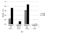
- FIG. 2 is a bar graph showing the results of examining the reaction values (SN) between ⁇ 1-6 fucose-specific lectins and various glycoproteins in the presence and absence of urea.
- the left side of the bar graph shows the absence of urea and the right side shows the presence of 5M urea.
- the reactivity of PhoSL to any of fHP, AFP-L3, and IgG having an ⁇ 1-6 fucose sugar chain is increased in the presence of urea.
- the reactivity to TF that does not belong to the ⁇ 1-6 fucose sugar chain does not change even in the presence of urea.
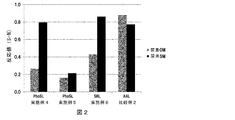
- ⁇ 1-6 fucose specific lectin PhoSL, PhoSL peptide and SRL
- AAL lectin
- ⁇ 1-6 fucose specific but not ⁇ 1-6 fucose specific 3 is a bar graph showing the results of examining the reaction value (SN). The left side of the bar graph shows the absence of urea and the right side shows the presence of 5M urea.
- the reaction value (SN) of all ⁇ 1-6 fucose specific lectins with fHP is increased in the presence of urea.
- the reaction value (SN) of non-specific AAL and fHP although having affinity for ⁇ 1-6 fucose, does not increase even in the presence of urea.
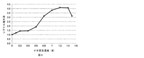
- FIG. 5 shows that the signal is enhanced 1.2 to 4.1 times at a thiourea concentration of 0.1 to 1.5M.
- the signal enhancer of the present invention is a signal enhancer that enhances a signal based on the reaction between an ⁇ 1-6 fucose sugar chain and an ⁇ 1-6 fucose-specific lectin that binds to the ⁇ 1-6 fucose sugar chain, and is selected from the group consisting of urea and thiourea. It includes at least one selected.
- the urea concentration in the signal enhancer may usually be 1 to 9M, preferably 2.5 to 9M, more preferably 3 to 8M, and particularly preferably 3.5 to 7M. Further, the thiourea concentration in the signal enhancer may be usually 0.1 to 1.5M, preferably 0.6 to 1.5M, more preferably 0.8 to 1.5M, particularly preferably 1 to 1.4M.
- the urea or the like in the signal enhancer is preferably used after being dissolved in a solvent.
- solvents include: water; physiological saline such as phosphate buffered saline and Tris buffered saline; buffer solution such as phosphate buffer and Tris buffer; and proteins such as bovine serum albumin dissolved Examples thereof include a buffer solution and an aqueous solution thereof; and a buffer solution containing a surfactant such as Tween-20 and an aqueous solution thereof.
- the pH of the signal enhancer is usually 4 to 10, preferably 5 to 9.
- the ⁇ 1-6 fucose sugar chain means a structure in which fucose is bonded to N-acetylglucosamine at the reducing end of the N-type sugar chain with an ⁇ 1-6 bond.
- a free oligosaccharide is used. Includes all chains, glycopeptides, glycoproteins, cells and the like. Also included are those obtained by chemically modifying an oligosaccharide chain or the like like like a label.
- the N-linked sugar chain may be a high mannose type, a composite type, a hybrid type or the like.
- the sugar chain is obtained by chemically decomposing the sugar chain with acid, hydrazine or the like, sialidase, galactositase, N-acetylhexosaminidase, ⁇ 1-2 fucose, ⁇ 1-3 fucose, ⁇ 1-
- the sugar chain may be partially degraded by using either fucosidase or mannosidase which cuts 4 fucose simultaneously or stepwise.
- a sugar such as glucose, a functional group such as an acetyl group, a sulfate group, or a phosphate group may be added to the sugar chain.
- fucose ⁇ 1-6 sugar chains are shown below. [In the formula, Man means mannose, GlcNAc means N-acetylglucosamine, and Fuc means fucose]
- Particularly preferred specific examples of the ⁇ 1-6 fucose sugar chain are tumor markers such as AFP-L3 and fucosylated haptoglobin.
- the ⁇ 1-6 fucose-specific lectin is characterized by binding to the ⁇ 1-6 fucose sugar chain more specifically than a conventional lectin having an affinity for the ⁇ 1-6 fucose sugar chain (for example, AAL). .
- This feature is characterized by (1) the lower limit of the binding constant for ⁇ 1-6 fucose sugar chain, and (2) the binding to high mannose sugar chain not containing ⁇ 1-6 fucose and / or glycolipid sugar chain not containing ⁇ 1-6 fucose. It can be defined by the upper limit of the constant.
- ⁇ 1-6 fucose-specific lectin is (1) having an affinity represented by a binding constant of 1.0 ⁇ 10 4 M ⁇ 1 or more (at 25 ° C.) for ⁇ 1-6 fucose sugar chain; (2) It is characterized in that it does not substantially bind to a high mannose sugar chain not containing ⁇ 1-6 fucose sugar chain and / or a glycolipid sugar chain not containing ⁇ 1-6 fucose.
- the binding constant means a numerical value measured at an analysis temperature of 25 ° C. using, for example, frontal affinity chromatography (FAC method). Details of the FAC method are described in, for example, Patent Document 1 filed by the present applicant. Patent Document 1 is incorporated herein by reference.
- the binding constant (at 25 ° C.) of (1) ⁇ 1-6 fucose sugar chain of ⁇ 1-6 fucose specific lectin is preferably 5.0 ⁇ 10 4 M ⁇ 1 or more, more preferably 1.0 ⁇ 10. 5 M ⁇ 1 or more, more preferably 1.0 ⁇ 10 6 M ⁇ 1 or more.
- ⁇ 1-6 fucose-specific lectin “(2) does not substantially bind to a high mannose sugar chain not containing ⁇ 1-6 fucose sugar chain and / or a glycolipid sugar chain not containing ⁇ 1-6 fucose sugar chain”
- the binding constant (at 25 ° C.) for a high-mannose sugar chain not containing ⁇ 1-6 fucose sugar chain and / or a glycolipid sugar chain not containing ⁇ 1-6 fucose is usually 1.0. It means not more than ⁇ 10 3 M ⁇ 1 , preferably not more than 1.0 ⁇ 10 2 M ⁇ 1 , particularly preferably 0.
- the molecular weight of ⁇ 1-6 fucose specific lectin by SDS electrophoresis is usually 4,000 to 40,000, preferably 4,000 to 20,000.
- the molecular weight by SDS electrophoresis is measured according to the Laemmi method (Nature, 227, 680, 1976), for example.
- the ⁇ 1-6 fucose-specific lectin may be one in which 2 to 10, preferably 2 to 6, more preferably 2 to 3, subunits are bound.
- the ⁇ 1-6 fucose-specific lectin has a high affinity for ⁇ 1-6 fucose sugar chains having sialic acid at the non-reducing end.
- LCA, NPA and PSA have low affinity for ⁇ 1-6 fucose sugar chains having sialic acid at the non-reducing end.
- the ⁇ 1-6 fucose-specific lectin preferably has a binding constant (at 25 ° C.) for N-linked single-, double-, triple- and / or tetrasaccharide chains to which ⁇ 1-6 fucose is bound.
- a binding constant at 25 ° C.
- ⁇ 1-6 fucose-specific lectin can be extracted and / or purified from natural products. Methods for obtaining a natural product-derived ⁇ 1-6 fucose-specific lectin are described in detail in Patent Document 1 filed by the present applicant and Non-Patent Document 1 filed by the present applicant. In addition, Tsutsugi lectin (PTL) described in Patent Document 1 is read as cedar lectin (PhoSL). The following outlines the ⁇ 1-6 fucose-specific lectin obtained from natural products.
- the above-mentioned natural products are mushrooms such as basidiomycetes and ascomycetes.
- the Moegitake family, the Kishimeji family, the Tachokinae family and the Amanita family belong to the basidiomycete.
- Examples of the Moegitake family include Sugitake, Tsutsugitake, Salmonella, Kuritake, Numerisugimodoki, Numerisugitake and the like.
- Examples of the xyme family include komula shimeji.
- Examples of the octopus kinaceae include white bamboo shoots and tsuchuuchitake.
- Examples of amanita mushrooms include fly agaric.
- Moegiaceae, Asteraceae, or Amanita are particularly preferred from the viewpoint of ⁇ 1-6 fucose sugar chain recognition specificity of lectins and lectin recovery efficiency, and more preferably Sugitake, Tsutsugitake, Numerisugitake Modoki, Salmonella, Kuritake, Komura Saxi-meji or Fly Agaric.
- the ⁇ 1-6 fucose-specific lectin is preferably a basidiomycete-derived lectin, and preferably, Sugitake lectin (PhoSL), Tsutsugitake lectin (PTL), Salmonella lectin (SRL), Kuritatake lectin (NSL), Komura saxi-medge Lectin (LSL) and fly agaric lectin (AML), more preferably PhoSL, PTL, SRL, NSL and AML.
- the ⁇ 1-6 fucose-specific lectin can be isolated, for example, from basidiomycetes or ascomycetes by appropriately combining known extraction methods, separation methods, purification methods, and the like.
- the method includes a step of obtaining an aqueous medium extract of basidiomycetes and / or ascomycetes using an aqueous medium as an extraction solvent.
- the site of use of these basidiomycetes and / or ascomycetes is preferably fruit bodies. From this extract, the molecular weight by SDS electrophoresis is usually 4,000 to 40,000, preferably 4,000 to 20,000, and the binding constant (at 25 ° C.) for ⁇ 1-6 fucose sugar chain.
- it is 1.0 ⁇ 10 4 M ⁇ 1 or more, preferably 5.0 ⁇ 10 4 M ⁇ 1 or more, more preferably 1.0 ⁇ 10 5 M ⁇ 1 or more, and further preferably 1.0 ⁇ 10 6.
- a lectin having an affinity of M- 1 or higher is obtained.
- the ⁇ 1-6 fucose-specific lectin may also be a peptide or protein chemically synthesized based on the amino acid sequence of a naturally-derived lectin, in addition to the extraction from the natural product.
- a chemically synthesized peptide or protein is one in which one or several amino acids in the amino acid sequence of a naturally derived lectin (for example, PhoSL consisting of the amino acid sequence of SEQ ID NO: 1 attached to the present specification) is lysine and / or arginine. It may be a substituted peptide having sugar binding activity (for example, a PhoSL peptide consisting of the amino acid sequence of SEQ ID NO: 2 attached to the present specification).
- the synthesis method is described in detail in Patent Document 4 filed by the present applicant. The specification of Patent Document 4 is incorporated herein by reference.
- the ⁇ 1-6 fucose-specific lectin can be artificially expressed in a known host different from the natural origin using a nucleic acid encoding the amino acid sequence of the natural lectin in addition to the extraction from the natural product.
- Recombinant for example, PhoSL recombinant lectin shown in Example 18
- the recombinant expression method is described in detail in Patent Document 5.
- the specification of Patent Document 5 filed by the present applicant is incorporated herein by reference.
- the ⁇ 1-6 fucose-specific lectin preferably incorporates a labeling means that enables detection.
- the labeling means is not particularly limited, and a known labeling method can be applied, and examples thereof include labeling with a radioisotope and binding of a labeling compound. Examples of the radioisotope include 14 C, 3 H, and 32 P.
- labeling compound examples include enzyme labels (horseradish peroxidase, alkaline phosphatase, etc.), biotin labels, digoxigenin labels, fluorescent labels (fluorescein isothiocyanate, CyDye (registered trademark), ethyl 4-aminobenzoate (ABEE). Aminopyridine, allophycocyanin, phycoerythrin, etc.).
- enzyme labels horseradish peroxidase, alkaline phosphatase, etc.
- biotin labels digoxigenin labels
- fluorescent labels fluorescein isothiocyanate, CyDye (registered trademark)
- ABEE ethyl 4-aminobenzoate
- Aminopyridine allophycocyanin, phycoerythrin, etc.
- biotin labeling is preferable because of its high sensitivity.
- the present invention also relates to a method for enhancing a signal based on a reaction between an ⁇ 1-6 fucose sugar chain and an ⁇ 1-6 fucose-specific lectin bound thereto, which is selected from the group consisting of urea and thiourea.
- the method includes the step of reacting the sugar chain with the lectin in the presence of a gnal enhancer containing one kind as an active ingredient.
- the urea concentration in the reaction step is usually 1 to 9M, preferably 2.5 to 9M, more preferably 3 to 8M, and particularly preferably 3.5 to 7M.
- the thiourea concentration in the reaction step may be usually 0.1 to 1.5M, preferably 0.6 to 1.5M, more preferably 0.8 to 1.5M, particularly preferably 1 ⁇ 1.4M.
- the present invention also relates to a method for enhancing a signal based on a reaction between an ⁇ 1-6 fucose sugar chain and an ⁇ 1-6 fucose-specific lectin bound thereto, which is selected from the group consisting of urea and thiourea.
- the method includes the steps of washing the sugar chain with a washing liquid containing a signal enhancer containing one kind of active ingredient, and reacting the sugar chain with the lectin.
- the urea concentration of the cleaning solution may be usually 1 to 9M, preferably 2.5 to 9M, more preferably 3 to 8M, and particularly preferably 3.5 to 7M.
- the concentration of thiourea in the washing solution may be usually 0.1 to 1.5M, preferably 0.6 to 1.5M, more preferably 0.8 to 1.5M, and particularly preferably 1 to 1.5M. 1.4M.
- the step of reacting the sugar chain with the lectin may be performed in the presence of a gnnal enhancer containing as an active ingredient at least one selected from the group consisting of urea and thiourea.
- the means for detecting ⁇ 1-6 fucose sugar chain reacted with ⁇ 1-6 fucose specific lectin is not particularly limited.
- detection means for example, ELISA (direct adsorption method, sandwich method, and competitive method), lectin affinity chromatography, lectin staining, lectin chip, flow cytometry (FACS) method, aggregation method, surface plasmon resonance method (for example, Biacore) (Registered trademark system), electrophoresis, beads and the like can be used.
- a sample such as blood is added to the plate and immobilized.
- a biotin-labeled lectin is added together with a signal enhancer, and the sugar chain and the lectin are reacted in the presence of the signal enhancer.
- a washing solution containing a signal enhancer may be added first, and a lectin solution containing no signal enhancer may be added after disposal.
- An HRP (horseradish peroxidase) labeled streptavidin solution is added as a secondary labeling compound to react biotin with streptavidin.
- the color developing substrate for HRP is added to cause color development, and the color intensity is measured with an absorptiometer. If a calibration curve is prepared in advance using a standard sample containing a sugar chain having a known concentration, the sugar chain can be quantified.
- an antibody that binds to an antigen having an ⁇ 1-6 fucose sugar chain is added and immobilized on a plate, and then a sample such as serum is added.
- a biotin-labeled lectin is added together with a signal enhancer, and the sugar chain and the lectin are reacted in the presence of the signal enhancer.
- a washing solution containing a signal enhancer may be added first, and a lectin solution containing no signal enhancer may be added after disposal.
- An HRP (horseradish peroxidase) labeled streptavidin solution is added as a secondary labeling compound to react biotin with streptavidin.
- the color developing substrate for HRP is added to cause color development, and the color intensity is measured with an absorptiometer. If a calibration curve is prepared in advance using a standard sample with a known concentration, the ⁇ 1-6 fucose sugar chain can be quantified.
- Lectin affinity chromatography is affinity chromatography that utilizes the property that a lectin immobilized on a carrier specifically binds to a sugar chain. High throughput can be expected by combining with HPLC.
- gel materials such as agarose, dextran, cellulose, starch, polyacrylamide and the like are generally used.
- commercially available products can be used without particular limitation, and examples thereof include Sepharose 4B and Sepharose 6B (both manufactured by GE Healthcare Bioscience).
- Columns used for lectin chromatography include those in which lectins are immobilized on microplates or nanowells.
- the concentration of the lectin to be immobilized is usually 0.001 to 100 mg / mL, preferably 0.01 to 20 mg / mL.
- the carrier is an agarose gel, it is activated with CNBr or the like and then coupled with the lectin.
- a lectin may be immobilized on a gel into which an activation spacer has been introduced. Further, the lectin may be immobilized on a gel into which a formyl group has been introduced and then reduced with NaCNBH 3 .
- a commercially available activated gel such as NHS-Sepharose (manufactured by GE Healthcare Bioscience) may also be used.
- a buffer is run for washing purposes.
- the sample is donated to the column with a signal enhancer added to the buffer.
- An example of the buffer is that the molarity is usually 5 to 500 mM, preferably 10 to 500 mM, the pH is usually 4.0 to 10.0, preferably 6.0 to 9.0, and NaCl.
- elution of sugar chains is performed using a desorbing agent such as sodium chloride or hapten sugar in a neutral non-denaturing buffer solution that can effectively elute sugar chains.
- This buffer may be the same as described above.
- the concentration of the desorbing agent is preferably 1 to 500 mM, particularly preferably 10 to 200 mM.
- the elution buffer preferably does not contain a signal enhancer.
- lectin staining a specimen is sliced out from a tumor tissue, and the slice is attached to a glass plate. Immerse the plate in blocking buffer and mix gently at room temperature. Where appropriate, endogenous peroxidase inactivation is performed. The plate is soaked in a biotin-labeled lectin solution (diluted with 2-5 ⁇ g / mL in blocking buffer) with a signal enhancer and gently stirred for 1 hour at room temperature. Alternatively, a washing solution containing a signal enhancer may be added first, and after discarding the washing solution, a lectin solution containing no signal enhancer may be added. Immerse the plate in HRP labeled avidin solution and gently stir at room temperature.
- cells are suspended in phosphate buffered saline (PBS).
- PBS phosphate buffered saline
- a 2.5 mL size syringe with a 21 G needle is used to loosen the cells about 10 times to loosen the cells, and then the cells are filtered through a 50 ⁇ m mesh.
- a solution of fluorescently labeled lectin is added to the cells thus prepared together with a signal enhancer, and the cells and the lectin are bound in the presence of the signal enhancer.
- a washing solution containing a signal enhancer may be added first, and after discarding the washing solution, a lectin solution containing no signal enhancer may be added. Thereafter, the amount and ratio of lectin-bound cells are analyzed with a flow cytometer (for example, Cytomics FC 500 manufactured by Beckman Coal Tar).
- the specimen used for the above detection means is not particularly limited. Examples thereof include blood, plasma, serum, tears, saliva, body fluid, milk, urine, cell culture supernatant, and secretions from transformed animals.
- the present invention also provides a kit for detecting an ⁇ 1-6 fucose sugar chain, comprising a signal enhancer comprising at least one selected from the group consisting of urea and thiourea as an active ingredient, and an ⁇ 1-6 fucose-specific lectin. To do.
- the detection kit may include a general-purpose detection kit such as various labeling compounds, buffers, plates, beads, and reaction stop solutions as appropriate.
- the detection kit is also a reagent for extracting a glycoprotein having an ⁇ 1-6 fucose sugar chain from a sample obtained from blood of a living body (for example, an antibody such as an anti-haptoglobin antibody or an anti- ⁇ fetoprotein antibody, protein A, protein G). And immunoglobulin binding proteins such as protein L).
- ⁇ 1-6 fucose sugar chain detection kit of the present invention is not particularly limited as long as it is intended to detect ⁇ 1-6 fucose sugar chains. Specific examples thereof will be described below.
- AFP-L3 band ⁇ -fetoprotein band with core fucose metastasis
- ⁇ -fetoprotein band with core fucose metastasis enables early diagnosis of hepatocellular carcinoma complicated with liver cirrhosis clinically, detailed follow-up of hepatocellular carcinoma, accurate determination of therapeutic effect It is useful as an indicator for early detection of fetal tumors, liver regeneration of fulminant hepatitis, and the like.
- ⁇ 5 ⁇ 1 integrin with metastasized core fucose is also expected as an indicator for liver cancer diagnosis.
- ⁇ Fucose in general increases in low-grade colon cancer and primary cancer.
- ⁇ 1-6-linked fucose tends to decrease in colon cancer with high malignancy or metastatic cancer. Therefore, ⁇ 1-6 fucose-specific lectin is allowed to act on the tumor tissue of colorectal cancer of a person diagnosed with colorectal cancer in the primary screening, and the amount of ⁇ 1-6-linked fucose is examined, so that colorectal cancer with high malignancy or metastasis can be obtained. Cancer can be identified.
- fucosylated haptoglobin is produced by adding core fucose to haptoglobin, a glycoprotein.
- This fucosylated haptoglobin increases as the stage of pancreatic cancer progresses and disappears after removal of the tumor site of pancreatic cancer.
- the signal enhancer of the present invention coexist in the reaction of ⁇ 1-6 fucose-specific lectin and fucosylated haptoglobin, the detection sensitivity by ⁇ 1-6 fucose-specific lectin can be increased.
- fucosylated haptoglobin may be washed with a washing solution containing a signal enhancer, and after discarding the washing solution, a lectin solution containing no signal enhancer may be added.
- pancreatic cancer can be detected quickly and easily by an assay using an ⁇ 1-6 fucose-specific lectin and an anti-haptoglobin antibody. Further, pancreatic cancer and pancreatitis can be distinguished by combining an antibody of pancreatic cancer marker CA19-9 and an ⁇ 1-6 fucose-specific lectin. At that time, the determination becomes easier when the signal enhancer of the present invention is allowed to coexist.
- the detection kit of the present invention includes prostate cancer, breast cancer, stomach cancer, small intestine cancer, colorectal cancer, renal cell cancer, small cell lung cancer, non-small cell cancer, uterine cancer, ovarian cancer.
- Thyroid cancer soft tissue sarcoma, osteosarcoma, melanoma, glioblastoma, astrocytoma, medulloblastoma, acute lymphoma, malignant lymphoma, Hodgkin's disease, non-Hodgkin's disease, acute myeloid leukemia, chronic lymphocytic leukemia, etc. It is expected to be used for diagnosis and research of tumors; allergic diseases; autoimmune diseases; and cardiovascular diseases such as emphysema.
- the reagent used in the present invention was prepared by the following procedure.
- PBS Phosphate buffered saline
- Disodium hydrogen phosphate manufactured by Wako Pure Chemical Industries, Ltd.
- potassium dihydrogen phosphate manufactured by Wako Pure Chemical Industries, Ltd.
- potassium chloride manufactured by Wako Pure Chemical Industries, Ltd.
- sodium chloride Wikuhin Co., Ltd.
- PBS was prepared by diluting 10 ⁇ PBS 10 times with water.
- BSA bovine serum albumin
- Sugitake lectin (PhoSL, SEQ ID NO: 1) was purified from Sugitake according to the method described in Non-Patent Document 1. This Sugitake lectin was weighed and dissolved by adding 0.1 M sodium bicarbonate solution (concentration: 5 mg / mL). A biotinylation reagent (manufactured by SIGMA; model number: B2643) was dissolved in dimethyl sulfoxide and added to the lectin solution for reaction. The reaction solution was subjected to solvent replacement with water by ultrafiltration (3K membrane, manufactured by Millipore) and lyophilized to obtain biotin-labeled PhoSL.
- Sugitake lectin (PhoSL, SEQ ID NO: 1) was purified from Sugitake according to the method described in Non-Patent Document 1. This Sugitake lectin was weighed and dissolved by adding 0.1 M sodium bicarbonate solution (concentration: 5 mg / mL). A biotinylation reagent (manufactured
- Biotin-labeled SRL SRL (SEQ ID NO: 3) was extracted according to the method described in Patent Document 2. This lectin was labeled with biotin in the same manner as biotin-labeled PhoSL.
- Biotin-labeled brown tea lectin (biotin-labeled AAL) Biotin-labeled herochawantake lectin (manufactured by J-Oil Mills Co., Ltd.) was prepared.
- Table 1 shows the sequence table of ⁇ 1-6 fucose-specific lectins (before labeling) of (4) to (6).
- the 10th and 17th Xs of SEQ ID NO: 1 may be any amino acid residue, but are preferably Cys, and the 20th, 23rd, 27th, 33th, 35th and 39th Xs are respectively Tyr. / Ser, Phe / Tyr, Arg / Lys / Asn, Asp / Gly / Ser, Asn / Ala, and Thr / Gln.
- SEQ ID NO: 2 is a specific example of SEQ ID NO: 1 (APVPVTKLVC DGDTYKCTAY LDFGDGRWVA QWDTNVFHTG), the first Ala, the 20th Tyr, and the 39th Thr are replaced with Lys, and the 40th Gly is deleted. Yes.
- the 10th and 17th Xs of SEQ ID NO: 3 may be any amino acid residue, but are preferably Cys, and are 4, 7, 9, 13, 20, 27, 29, 33, 34 and 39.
- the th X is Pro / Gly, Glu / Lys, Val / Asp, Asn / Asp / Glu, His / Ser, Lys / His, Val / Ile, Gly / Asn / Ser, Ala / Thr, and Arg, respectively. / Thr.
- fHP fucosylated haptoglobin
- AFP-L3 ⁇ -fetoprotein L3
- FHP mouse immunoglobulin G
- IgG transferrin
- pancreatic cancer cell line obtained from DS Pharma
- PSN-1 pancreatic cancer cell line
- 1000 mL of the culture supernatant was concentrated to 1 mL with an ultrafiltration filter (product name: VIVA SPIN 20-10K, manufactured by Sartorius).
- the concentrated solution was added to 0.5 mL of a gel in which an anti-haptoglobin antibody (manufactured by The Binding Site) was immobilized on NHS-activated Sepharose 4 Fast Flow (manufactured by GE Healthcare).
- the solution containing the gel was added to a 0.45 ⁇ m filter tube (manufactured by Millipore), centrifuged at 400 ⁇ g and a temperature of 4 ° C. for 5 minutes, and the filtrate was discarded. .
- 200 ⁇ L of PBS was added and centrifuged at 400 ⁇ g and a temperature of 4 ° C. for 5 minutes, and the filtrate was discarded. This was repeated twice.
- 200 ⁇ L of Elution Buffer 100 mM glycine, 0.5 M NaCl, pH 3.0
- liver cancer cell line obtained from RIKEN
- RIKEN liver cancer cell line
- 1000 mL of the culture supernatant was concentrated to 1 mL with an ultrafiltration filter (product name: VIVA SPIN 20-10K, manufactured by Sartorius).
- VIVA SPIN 20-10K product name: VIVA SPIN 20-10K, manufactured by Sartorius.
- the concentrated solution was added to 0.5 mL of a gel in which an anti-AFP antibody (manufactured by DAKO) was immobilized on NHS-activated Sepharose 4 Fast Flow (manufactured by GE Healthcare).
- the solution containing the gel was added to a 0.45 ⁇ m filter tube (manufactured by Millipore), centrifuged at 400 ⁇ g and a temperature of 4 ° C. for 5 minutes, and the filtrate was discarded. .
- 200 ⁇ L of PBS was added and centrifuged at 400 ⁇ g and a temperature of 4 ° C. for 5 minutes, and the filtrate was discarded. This was repeated twice.
- 200 ⁇ L of Elution Buffer 100 mM glycine, 0.5 M NaCl, pH 3.0
- IgG and TF solution IgG and TF were respectively dissolved in PBS to prepare a 5000 ng / mL IgG solution and a 5000 ng / mL TF solution.
- Absorbance at wavelengths of 450 nm and 630 nm was measured using a plate reader (product name: POWERSCAN (registered trademark) HT, manufactured by DS PHARMA).
- a value obtained by subtracting the absorbance value at 630 nm from the absorbance value at 450 nm was defined as a detection value (signal: S).
- the detection value (noise: N) of the well not added with glycoprotein was measured, and the value obtained by subtracting the detection value (N) of the well itself from the detection value (S) was defined as the reaction value (SN) urea 5M . did.
- the reaction value (SN) urea 0M when the lectin and glycoprotein were reacted in the absence of urea was also determined.
- reaction values (SN) of PhoSL to various glycoproteins are shown in Table 2 and FIG. (SN) urea 0M : reaction value when reaction is carried out in the absence of urea (SN) (S—N) Urea 5M : Reaction value when reacted in the presence of urea 5M (S—N)
- the signal based on the reaction of fHP, AFP-L3 or IgG with PhoSL in the presence of urea significantly increases both the signal increase amount and the signal increase rate, compared with the case without urea.
- the signal based on the reaction of TF that is not an ⁇ 1-6 fucose sugar chain and PhoSL has a high signal increase rate, but the signal increase amount does not increase significantly.
- Biotin-labeled PhoSL, biotin-labeled PhoSL peptide, and biotin-labeled SRL were used as ⁇ 1-6 fucose-specific lectins.
- biotin-labeled AAL that binds to sugar chains having fucose other than ⁇ 1-6 was used.
- Biotin-labeled PhoSL, biotin-labeled PhoSL peptide, biotin-labeled SRL or biotin-labeled AAL is diluted with 1% BSA / PBS solution, and urea (manufactured by Wako Pure Chemical Industries, Ltd.) is added to each lectin solution.
- urea manufactured by Wako Pure Chemical Industries, Ltd.
- Biotin-labeled lectin final concentration 1 ⁇ g / mL, urea final concentration 5M was prepared. 50 ⁇ L of this lectin solution was added to the wells and allowed to stand at 4 ° C. for 30 minutes, and then the added solution was discarded.
- a lectin solution containing no urea (biotin-labeled lectin final concentration 1 ⁇ g / mL, urea 0 M) was also carried out.
- Washing Each well was added with 250 ⁇ L of 0.05% Tween / PBS, and the added solution was discarded. This operation was repeated a total of 3 times.
- the absorbance at wavelengths of 450 nm and 630 nm was measured in the same manner as in Example 1 using the plate reader, and the reaction value (S—N) urea 5M was determined.
- the reaction value (SN) urea 0M when lectin and glycoprotein were reacted in the absence of urea was also determined.
- Table 3 and FIG. 2 show the reaction values (SN) in the presence and absence of urea, the amount of increase and the rate of increase.
- the signal enhancer of the present invention enhances the signal based on the reaction between the ⁇ 1-6 fucose sugar chain and the ⁇ 1-6 fucose specific lectin bound thereto.
- Urea does not sensitize the reaction between transferrin having no ⁇ 1-6 fucose sugar chain and ⁇ 1-6 fucose-specific lectin (Comparative Example 1).
- Urea does not sensitize the reaction between a sugar chain having an ⁇ 1-6 fucose sugar chain and an AAL having affinity for ⁇ 1-6 fucose but not specific (Comparative Example 2).
- urea did not sensitize the reaction with ⁇ 1-6 fucose sugar chain and UEA-1, Lotus, ConA, etc.
- the signal enhancement by the signal enhancer of the present invention is a phenomenon that occurs only in the reaction between the ⁇ 1-6 fucose sugar chain and the ⁇ 1-6 fucose-specific lectin.
- Example 7 fucose (manufactured by Nacalai Tesque) having a final concentration of 20 mM or 100 mM was allowed to coexist in the reaction between fHP and PhoSL in the presence of 5 M urea.
- Example 8 instead of fucose of Example 7, thyroglobulin (manufactured by SIGMA, hereinafter referred to as “TG”) having a final concentration of 0.01 mg / mL or 0.1 mg / mL was allowed to coexist.
- TG thyroglobulin
- FIG. 3 shows that the reaction value (SN) decreases as the concentration of fucose or TG added increases. From this, it was confirmed that the effect of increasing the signal based on the binding of lectin and glycoprotein in the presence of urea is due to the specific binding of ⁇ 1-6 fucose-specific lectin and ⁇ 1-6 fucose sugar chain.
- Example 9 Change test of urea concentration The preferred range of urea as a signal enhancer was examined by sandwich ELISA. In Example 4, the same procedure as in Example 4 was performed, except that fHP (50 ng / mL) was used and the final urea concentration with respect to the lectin solution was 0 to 9M.
- FIG. 4 shows the signal increase rate compared with the case where urea was not added at each final concentration of urea.
- FIG. 4 shows that the signal is enhanced 1.2 to 2.8 times in the presence of urea having a final concentration of 1 to 9M.
- the final concentration of urea is preferably 3 to 9M, more preferably 3 to 8M, and particularly preferably 3 to 7M.
- Example 10 to 11 Test with thiourea
- a glycoprotein having an ⁇ 1-6 fucose sugar chain (fHP or AFP-L3) and an ⁇ 1-6 fucose-specific lectin (PhoSL) were obtained.
- the reaction was conducted in the presence or absence of thiourea, and the presence or absence of signal enhancement based on the binding reaction between the sugar chain and the lectin was examined.
- the procedure was the same as that of Example 1 except that urea 5M was replaced with thiourea (Wako Pure Chemical Industries) 1.2M in Example 1.
- the reaction value (SN) was determined in the same manner as in Example 1 from the obtained absorbance.
- the reaction value (SN) when lectin and glycoprotein were reacted in the absence of thiourea was also determined.
- Table 4 shows the reaction values (SN) of PhoSL to various glycoproteins.
- SN Thiourea 0M : Reaction value when reaction is carried out in the absence of thiourea (SN)
- SN Thiourea 1.2M : Reaction value when reacted in the presence of 1.2M thiourea (SN)
- the signal based on the reaction of fHP and AFP-L3 with PhoSL in the presence of thiourea significantly increased both the amount of signal increase and the signal increase rate compared to the absence of thiourea.
- Example 12 Change test of thiourea concentration The preferred range of thiourea as a signal enhancer was examined by sandwich ELISA. In Example 4, thiourea was used in place of urea, and the same procedure as in Example 4 was performed, except that the thiourea final concentration with respect to the lectin solution was 0 to 1.5M.
- Example 10 In the same manner as in Example 1, the absorbance at wavelengths of 450 nm and 630 nm was measured using the plate reader, and the reaction value (SN) was further determined. Similarly to Example 10, the signal increase rate at each final concentration of thiourea compared to the absence of thiourea was determined, and the results are shown in FIG.
- FIG. 5 shows that the signal is enhanced 1.2 to 4.1 times in the presence of thiourea having a final concentration of 0.1 to 1.5M.
- the final concentration of thiourea is usually 0.1 to 1.5M, preferably 0.6 to 1.5M, more preferably 0.8 to 1.5M, and particularly preferably 1 to 1.4M. It is.
- Example 4 the final concentration of 5 M urea or final concentration of 1.2 M thiourea was added to the cleaning solution (0.05% Tween / PBS) in “(6) Cleaning”, and “( The procedure was the same as in Example 4 except that urea was not added to the lectin solution in “7) Lectin reaction”.
- the absorbance at wavelengths of 450 nm and 630 nm was measured in the same manner as in Example 1 using the plate reader, and the reaction value (SN) was further determined. Furthermore, the amount of signal increase and the rate of increase when urea or thiourea was added to the cleaning solution compared to the case where urea and thiourea were not added were calculated in the same manner as described above, and are shown in Table 5.
- Examples 15 to 17 In the same manner as Sugitake lectin, Tsutsugitake lectin (PTL), Kuritatake lectin (NSL), and fly agaric lectin (AML) were purified from Tsutsugitake, Kuritatake and Fly Agaric. The obtained lectin was labeled with biotin in the same procedure as biotin-labeled PhoSL to obtain biotin-labeled PTL, biotin-labeled NSL, and biotin-labeled AML.
- PTL Tsutsugitake lectin
- NSL Kuritatake lectin
- AML fly agaric lectin
- Sandwich ELISA was performed in the same manner as in Example 4 except that biotin-labeled PhoSL was replaced with biotin-labeled PTL, biotin-labeled NSL or biotin-labeled AML. The results are shown in Table 6.
- Example 18 Detection method using beads According to the instructions of magnetic beads (manufactured by Invitrogen, 14204), anti-haptoglobin antibodies from which sugar chains were removed were immobilized to prepare antibody-immobilized magnetic beads.
- Detection using beads was performed according to a conventional method. Specifically, fucosylated haptoglobin (fHP) (final concentration: 0 ⁇ g / mL or 0.45 ⁇ g / mL), urea (final concentration 0 M or 5 M), PhoSL recombinant lectin shown in SEQ ID NO: 4 (Patent Document 5: Special) A mixture having DYKDDDDK tag added to SEQ ID NO: 1 of open 2011-148735 (final concentration 5 ⁇ g / mL) was mixed to form a complex.
- fHP fucosylated haptoglobin
- urea final concentration 0 M or 5 M
- PhoSL recombinant lectin shown in SEQ ID NO: 4 Patent Document 5: Special
- the solution containing the complex is diluted 10-fold, antibody-immobilized magnetic beads are added, washed with a magnet, HRP-labeled anti-FLAG antibody (manufactured by SIGMA) is added, and the complex with the detection antibody Formed. Color was developed using the color developing substrate for HRP, and the color development reaction was stopped using 1M phosphoric acid. The absorbance of the obtained solution was measured with a plate reader as described above.
- the value obtained by subtracting the absorbance value at 630 nm from the absorbance value at 450 nm in the presence of fHP was taken as the detection value (signal: S), and similarly the detection value in the absence of fHP (noise: N).
- the value obtained by subtracting the detection value (N) of the well itself from the detection value (S) in the presence of 5M urea was defined as the reaction value (S ⁇ N) urea 5M .
- the reaction value (SN) urea 0M in the absence of urea was determined.
- a value obtained by dividing the detection value S in the presence of 5M urea by the detection value N was defined as a reaction value (S / N) urea 5M .
- the reaction value (S / N) urea 0 M was set in the absence of urea.
- the signal increase amount and the increase rate were calculated. The results are shown in Table 7.
- urea 0M reaction value when reaction is carried out in the absence of urea (SN) (SN)
- Urea 5M Reaction value when reaction is carried out in the presence of urea 5M (SN) (S / N)
- Urea 0M Reaction value when reacted in the absence of urea (S / N) (S / N)
- urea 5M reaction value when reacted in the presence of urea 5M (S / N)
Landscapes
- Health & Medical Sciences (AREA)
- Life Sciences & Earth Sciences (AREA)
- Engineering & Computer Science (AREA)
- Immunology (AREA)
- Urology & Nephrology (AREA)
- Biomedical Technology (AREA)
- Hematology (AREA)
- Chemical & Material Sciences (AREA)
- Molecular Biology (AREA)
- Cell Biology (AREA)
- Biotechnology (AREA)
- Microbiology (AREA)
- Food Science & Technology (AREA)
- Medicinal Chemistry (AREA)
- Physics & Mathematics (AREA)
- Analytical Chemistry (AREA)
- Biochemistry (AREA)
- General Health & Medical Sciences (AREA)
- General Physics & Mathematics (AREA)
- Pathology (AREA)
- Investigating Or Analysing Biological Materials (AREA)
- Peptides Or Proteins (AREA)
- Measuring Or Testing Involving Enzymes Or Micro-Organisms (AREA)
Abstract
【課題】α1-6フコース糖鎖とそれに結合するα1-6フコース特異的レクチンとの反応に基づくシグナルを強化するシグナル増強剤、その使用方法及びその用途を提供する。 【解決手段】α1-6フコース糖鎖とそれに結合するα1-6フコース特異的レクチンとの反応に基づいたシグナルを増強するシグナル増強剤は、尿素及びチオ尿素からなる群から選択される少なくとも一種を有効成分とすることを特徴とする。尿素の終濃度は、好ましくは1~9Mであり、チオ尿素の終濃度は好ましくは0.1~1.5Mである。このシグナル増強剤によれば、α1-6フコース糖鎖を正確に測定できるので、α1-6フコース糖鎖が関係する疾患の検出や判定やα1-6フコース糖鎖の学術研究に大いに役立つ。
Description
本発明は、シグナル増強剤に関し、より詳細には、α1-6フコース糖鎖とα1-6フコース特異的レクチンとの反応に基づくシグナルの増強剤に関する。
N結合型糖鎖の還元末端のN-アセチルグルコサミンにα1-6フコース(コアフコースともいう)を転移させるα1-6フコース転移酵素(α1-6FucT)の遺伝子は、肝細胞の癌化にともなって発現することが知られている。肝細胞癌になると、αフェトプロテイン(AFP)という分子量約70,000の糖タンパク質の糖鎖のN-アセチルグルコサミンにα1-6結合でフコースが転移するようになる。このα1-6結合でフコースが転移したAFPは、現在、肝細胞癌の腫瘍マーカーとして使用されている。被験者の血清を、フコースに親和性をもつレンズマメレクチン(LCA)を用いたレクチン親和電気泳動にかけると、陽極側よりAFP-L1、AFP-L2、AFP-L3と呼ばれる3本のバンドが出現する。肝硬変等の良性肝疾患ではレクチン非結合型のAFP-L1バンドが主であるが、肝細胞癌になるとレクチン結合型のAFP-L3バンドが増加する。総AFPに対するAFP-L3の割合(AFP-L3%)を求め、その値が10%を越えるようであれば、肝細胞癌が疑われる。α1-6結合でフコースが転移したAFPの割合を正確に識別することができれば、肝細胞癌の診断の確度も一層高まる。
α1-6フコースは、肝癌の他にも、膵臓癌、大腸癌等の疾患と関係することが知られている。
従来、α1-6フコースと親和性を有するレクチンとして、レンズマメレクチン(LCA)、エンドウマメレクチン(PSA)、ヒイロチャワンタケレクチン(AAL)、ラッパスイセンレクチン(NPA)、ソラマメレクチン(VFA)、麹菌レクチン(AOL)等が知られていた。しかし、AALやAOLは、α1-2フコース、α1-3フコース等にも親和性を有し、LCA、PSA及びVFAは、フコースα1-6をもたないN結合型一本鎖及び/又は二本鎖糖鎖にも親和性を有するといったように、これらのレクチンは、α1-6フコース糖鎖の他に、α1-6結合以外のフコースを持つ糖脂質系糖鎖や、フコースを持たない高マンノース糖鎖にも親和性を示す。
本発明者等は、従来知られている上記レクチンよりも特異的にα1-6フコース糖鎖を検出するレクチンとして、天然のキノコからスギタケレクチン(PhoSL)(非特許文献1)、サケツバタケレクチン(SRL)(特許文献1)等のレクチンを新規に発見している。さらに、このレクチンの化学合成やリコンビナント作製も行なっている(特許文献4及び5)。そして、このレクチンを用いて、AFP-L3の検出(特許文献1)、大腸癌の悪化度の判別(特許文献2)、及び膵臓癌の検出(特許文献3)にも成功している。
Yuka Kobayashi et al.,"A Novel Core Fucose-specific Lectin from the Mushroom Pholiota squarrosa",J.Biol.Chem,2012,287,p33973-33982
本発明者等が発見したα1-6フコース特異的レクチンによって、α1-6フコース糖鎖の特異的検出が可能になった。α1-6フコース糖鎖とα1-6フコース特異的レクチンとの反応に基づくシグナルを増強することができれば、α1-6フコース糖鎖の正確な検出が可能となる。そこで、本発明の目的は、α1-6フコース糖鎖とそれに結合するα1-6フコース特異的レクチンとの反応に基づくシグナルを強化するシグナル増強剤を提供することにある。
本発明者等は、上記課題を鋭意検討した結果、以下の発明によれば上記課題を解決できることを見出し、本発明を完成させた。すなわち、本発明は、α1-6フコース糖鎖とそれに結合するα1-6フコース特異的レクチンとの反応に基づいたシグナルを増強するシグナル増強剤であって、尿素及びチオ尿素からなる群から選択される少なくとも一種を有効成分とすることを特徴とする前記シグナル増強剤を提供する。前記「α1-6フコース糖鎖」という用語は、本明細書において、「N型糖鎖の還元末端のN-アセチルグルコサミンにα1-6結合でフコースが結合した構造」を意味する。また、前記「α1-6フコース特異的レクチン」という用語は、本明細書において、「(1)α1-6フコース糖鎖に対して結合定数1.0×10⁴M-1以上(25℃において)で示される親和性を有する、かつ(2)α1-6フコースを含まないハイマンノース糖鎖及び/又はα1-6フコースを含まない糖脂質糖鎖に対して実質的に結合しないことを特徴とするレクチン」を意味する。
特開2010-145202には、抗原抗体反応を増強する方法であって、抗原と抗体とをポリエチレングリコール及び尿素の共存下で反応させる方法が開示されている。本発明のシグナル増強剤が適用される反応は、α1-6フコース糖鎖とそれに結合するα1-6フコース特異的レクチンとの反応であって、抗原抗体反応でなく、また、ポリエチレングリコールを必要としない点で、特開2010-145202の発明と明確に相違する。
本発明は、また、α1-6フコース糖鎖とそれに結合するα1-6フコース特異的レクチンとの反応に基づいたシグナルを増強する方法であって、尿素及びチオ尿素からなる群から選択される少なくとも一種を有効成分とするシグナル増強剤の存在下で、前記糖鎖と前記レクチンとを反応させるステップを含む、前記方法を提供する。
本発明は、また、α1-6フコース糖鎖とそれに結合するα1-6フコース特異的レクチンとの反応に基づいたシグナルを増強する方法であって、尿素及びチオ尿素からなる群から選択される少なくとも一種を有効成分とするシグナル増強剤を含む洗浄液で、前記糖鎖を洗浄するステップと、前記糖鎖と前記レクチンとを反応させるステップ、を含む、前記方法を提供する。
本発明は、また、尿素及びチオ尿素からなる群から選択される少なくとも一種、及び、α1-6フコース特異的レクチンを有効成分とするシグナル増強剤を含む、α1-6フコース糖鎖の検出キットを提供する。
尿素及びチオ尿素からなる群から選択される少なくとも一種(以下、尿素等という)を含むことを必須とする本発明のシグナル増強剤は、後述する実施例に示すように、α1-6フコース糖鎖とそれに結合するα1-6フコース特異的レクチンとの反応を増感し、それに基づくシグナルを強化することができる。前記尿素等は、後述の比較例1に示すように、α1-6フコース糖鎖を持たない糖鎖(例えばトランスフェリン)とα1-6フコース特異的レクチンとの反応を増感しない。また、前記尿素等は、比較例2に示すように、α1-6フコース糖鎖を持つ糖鎖と、α1-6フコースに親和性を有するが特異的ではないレクチン(例えばAAL)との反応を増感しない。データを示していないが、前記尿素等は、α1-6フコース糖鎖を持つ糖鎖とα1-6フコースに親和性を有さないレクチン(例えば、ハリエニシダレクチン1(UEA-1)、ミヤコグサレクチン(Lotus)、タチナタマメレクチン(ConA)等)との反応も増感しない。したがって、前記尿素等を含む本発明のシグナル増強剤は、α1-6フコース糖鎖とα1-6フコース特異的レクチンとの特異的反応に向けられる。
本発明のシグナル増強剤によれば、α1-6フコース糖鎖を正確に測定できる。これは、α1-6フコース糖鎖が関係する疾患の検出や判定や、α1-6フコース糖鎖の学術研究に大いに貢献する。
本発明のシグナル増強剤は、α1-6フコース糖鎖とそれに結合するα1-6フコース特異的レクチンとの反応に基づいたシグナルを増強するシグナル増強剤であって、尿素及びチオ尿素からなる群から選択される少なくとも一種を含むことを特徴とする。
シグナル増強剤中の尿素濃度は、通常、1~9Mでよく、好ましくは2.5~9Mであり、さらに好ましくは3~8M、特に好ましくは3.5~7Mである。また、シグナル増強剤中のチオ尿素濃度は、通常、0.1~1.5Mでよく、好ましくは0.6~1.5Mであり、さらに好ましくは0.8~1.5M、特に好ましくは1~1.4Mである。
シグナル増強剤中の前記尿素等は、溶媒に溶解して使用することが好ましい。溶媒の例には、水;リン酸緩衝化生理食塩水やトリス緩衝化生理食塩水等の生理食塩水;リン酸緩衝液やトリス緩衝液等の緩衝液;ウシ血清アルブミン等のタンパク質を溶解した緩衝液やその水溶液;並びにTween-20等の界面活性剤を含む緩衝液やその水溶液が挙げられる。
シグナル増強剤のpHは、通常、4~10であり、好ましくは5~9である。
上記α1-6フコース糖鎖とは、N型糖鎖の還元末端のN-アセチルグルコサミンにα1-6結合でフコースが結合した構造を意味し、N結合型糖鎖である限り、遊離のオリゴ糖鎖、糖ペプチド、糖タンパク質、細胞等のすべてを包含する。また、標識のようにオリゴ糖鎖等を化学修飾したものも含まれる。上記N結合型の糖鎖は、高マンノース型、複合型、混成型等であり得る。さらに、前記糖鎖は、酸、ヒドラジン等で化学的に前記糖鎖を部分的に分解したもの、シアリダーゼ、ガラクシトターゼ、N-アセチルヘキソサミニダーゼ、α1-2フコース・α1-3フコース・α1-4フコースを切断するフコシダーゼ、マンノシダーゼのいずれかの酵素を同時にあるいは段階的に使用して前記糖鎖を部分的に分解したものであってもよい。また、グルコースのような糖、アセチル基、硫酸基、リン酸基のような官能基等を前記糖鎖に付加したものであってもよい。
フコースα1-6糖鎖の代表例を、以下に示す。

〔式中、Manはマンノース、GlcNAcはN-アセチルグルコサミン、Fucはフコースを意味する〕
α1-6フコース糖鎖の特に好ましい具体例は、AFP-L3、フコシル化ハプトグロビン等の腫瘍マーカーである。
α1-6フコース特異的レクチンは、α1-6フコース糖鎖に対して親和性を有する従来のレクチン(例えばAAL)と比べてより特異的にα1-6フコース糖鎖と結合することを特徴とする。この特徴は、(1)α1-6フコース糖鎖に対する結合定数の下限と、(2)α1-6フコースを含まないハイマンノース糖鎖及び/又はα1-6フコースを含まない糖脂質糖鎖に対する結合定数の上限によって定義可能である。すなわち、α1-6フコース特異的レクチンとは、
(1)α1-6フコース糖鎖に対して結合定数1.0×10⁴M-1以上(25℃において)で示される親和性を有する、
(2)α1-6フコース糖鎖を含まないハイマンノース糖鎖及び/又はα1-6フコースを含まない糖脂質糖鎖に対して実質的に結合しない
ことを特徴とする。
(1)α1-6フコース糖鎖に対して結合定数1.0×10⁴M-1以上(25℃において)で示される親和性を有する、
(2)α1-6フコース糖鎖を含まないハイマンノース糖鎖及び/又はα1-6フコースを含まない糖脂質糖鎖に対して実質的に結合しない
ことを特徴とする。
前記結合定数は、本明細書において、例えばフロンタルアフィニティクロマトグラフィー(FAC法)を用い、分析温度25℃において測定される数値を意味する。FAC法の詳細は、例えば、本出願人の出願した特許文献1に記載されている。特許文献1を参照のために本明細書に編入する。
α1-6フコース特異的レクチンの(1)α1-6フコース糖鎖に対して結合定数(25℃において)は、好ましくは5.0×10⁴M-1以上、より好ましくは1.0×10⁵M-1以上、さらに好ましくは1.0×10⁶M-1以上である。
α1-6フコース特異的レクチンの「(2)α1-6フコース糖鎖を含まないハイマンノース糖鎖及び/又はα1-6フコース糖鎖を含まない糖脂質糖鎖に対して実質的に結合しない」とは、本明細書において、α1-6フコース糖鎖を含まないハイマンノース糖鎖及び/又はα1-6フコースを含まない糖脂質糖鎖に対する結合定数(25℃において)が、通常、1.0×103M-1以下であり、好ましくは1.0×102M-1以下、特に好ましくは0であることを意味する。
α1-6フコース特異的レクチンのSDS電気泳動法による分子量は、通常、4,000~40,000であり、好ましくは4,000~20,000である。ここで、SDS電気泳動法による分子量は、例えばLaemmiの方法(Nature,227巻,680頁,1976年)に準じて測定されるものである。α1-6フコース特異的レクチンは、サブユニットが、通常、2~10個、好ましくは2~6個、さらに好ましくは2~3個結合したものでもよい。
α1-6フコース特異的レクチンは、その非還元末端にシアル酸を有するα1-6フコース糖鎖に対しても高い親和性を有する。一方、LCA、NPA及びPSAは、非還元末端にシアル酸を有するα1-6フコース糖鎖に対しては親和性が低い。
α1-6フコース特異的レクチンは、さらにα1-6フコースの結合したN結合型の一本鎖、二本鎖、三本鎖及び/又は四本糖鎖に対する結合定数(25℃において)が、好ましくは、1.0×104M-1以上、より好ましくは、5.0×104M-1以上、さらに好ましくは1.0×105M-1以上で示される親和性を有する。
α1-6フコース特異的レクチンは、天然物から抽出及び/又は精製することができる。天然物由来のα1-6フコース特異的レクチンの入手方法は、本出願人の出願した特許文献1、及び本出願人の投稿した非特許文献1に詳細に記載されている。なお、特許文献1に記載のツチスギタケレクチン(PTL)をスギタケレクチン(PhoSL)と読み替える。以下に天然物から取得するα1-6フコース特異的レクチンを概説する。
上記天然物は、例えば担子菌、子嚢菌等のキノコである。モエギタケ科、キシメジ科、タコウキン科及びテングタケ科は、担子菌に属している。モエギタケ科としては、スギタケ、ツチスギタケ、サケツバタケ、クリタケ、ヌメリスギタケモドキ、ヌメリスギタケ等が挙げられる。キシメジ科としては、コムラサキシメジ等が挙げられる。タコウキン科としては、シロハカワラタケ、ツヤウチワタケ等が挙げられる。テングタケ科としては、ベニテングタケ等が挙げられる。
これらの担子菌又は子嚢菌のうち、レクチンのα1-6フコース糖鎖認識特異性とレクチンの回収効率の観点から、モエギタケ科、キシメジ科又はテングタケ科が特に好ましく、さらに好ましくはスギタケ、ツチスギタケ、ヌメリスギタケモドキ、サケツバタケ、クリタケ、コムラサキシメジ又はベニテングタケである。
α1-6フコース特異的レクチンは、好ましくは担子菌由来のレクチンであり、好ましくはスギタケレクチン(PhoSL)、ツチスギタケレクチン(PTL)、サケツバタケレクチン(SRL)、クリタケレクチン(NSL)、コムラサキシメジレクチン(LSL)、及びベニテングタケレクチン(AML)であり、さらに好ましくはPhoSL、PTL、SRL、NSL及びAMLである。
上記α1-6フコース特異的レクチンは、例えば担子菌や子嚢菌から、公知の抽出方法、分離方法、精製方法等を適宜組み合わせることにより、単離することができる。例えば、水系媒体を抽出溶媒として用いて、担子菌及び/又は子嚢菌の水系媒体抽出物を得る工程を含む。これらの担子菌及び/又は子嚢菌の使用部位は、子実体であることが好ましい。この前記抽出物から、SDS電気泳動法による分子量が、通常、4,000~40,000、好ましくは4,000~20,000、及びα1-6フコース糖鎖に対する結合定数(25℃において)が、通常、1.0×104M-1以上、好ましくは5.0×104M-1以上、より好ましくは1.0×105M-1以上、さらに好ましくは1.0×106M-1以上で示される親和性を有するレクチンを得る。
α1-6フコース特異的レクチンは、また、上記天然物からの抽出の他に、天然由来のレクチンのアミノ酸配列に基づいて化学合成されたペプチドやタンパク質であってもよい。さらに、化学合成のペプチドやタンパク質は、天然由来のレクチン(例えば本明細書に添付した配列番号1のアミノ酸配列からなるPhoSL)のアミノ酸配列中の1又は数個のアミノ酸がリジン及び/又はアルギニンで置換されかつ糖結合活性を有するペプチド(例えば本明細書に添付した配列番号2のアミノ酸配列からなるPhoSLぺプチド)であってもよい。その合成方法は、本出願人の出願した特許文献4に詳細に記載されている。特許文献4の明細書を参照のために本明細書に編入する。
α1-6フコース特異的レクチンは、また、上記天然物からの抽出の他に、天然由来のレクチンのアミノ酸配列をコードする核酸を用いて天然由来とは異なる公知の宿主内で人工的に発現させたリコンビナント(例えば実施例18に示すPhoSL組換えレクチン)であってもよい。リコンビナントの発現方法は、特許文献5に詳細に記載されている。本出願人の出願した特許文献5の明細書を参照のために本明細書に編入する。
α1-6フコース特異的レクチンは、検出可能にする標識手段が組み込まれていることが好ましい。前記標識手段としては、特に制限なく、公知の標識化方法を適用することができ、例えば、放射性同位元素による標識化、標識化合物の結合等を挙げることができる。前記放射性同位元素としては、例えば14C、3H及び32Pが挙げられる。
前記標識化合物としては、例えば酵素標識(西洋ワサビペルオキシダーゼ、アルカリフォスファターゼ等)、ビオチン標識、ジゴキシゲニン標識、蛍光標識(フロオレッセインイソチアシアナート、CyDye(登録商標)、4-アミノ安息香酸エチル(ABEE)、アミノピリジン、アロフィコシアニン、フィコエリスリン等)等を挙げることができる。これらの標識化合物は、常法によりレクチンと結合することができる。特に、ビオチン標識は、感度が高い点で好ましい。
本発明は、また、α1-6フコース糖鎖とそれに結合するα1-6フコース特異的レクチンとの反応に基づいたシグナルを増強する方法であって、尿素及びチオ尿素からなる群から選択される少なくとも一種を有効成分とするグナル増強剤の存在下で、前記糖鎖と前記レクチンとを反応させるステップを含む、前記方法を提供する。
前記反応ステップの尿素濃度は、通常、1~9Mでよく、好ましくは2.5~9Mであり、さらに好ましくは3~8M、特に好ましくは3.5~7Mである。また、前記反応ステップのチオ尿素濃度は、通常、0.1~1.5Mでよく、好ましくは0.6~1.5Mであり、さらに好ましくは0.8~1.5M、特に好ましくは1~1.4Mである。
本発明は、また、α1-6フコース糖鎖とそれに結合するα1-6フコース特異的レクチンとの反応に基づいたシグナルを増強する方法であって、尿素及びチオ尿素からなる群から選択される少なくとも一種を有効成分とするシグナル増強剤を含む洗浄液で、前記糖鎖を洗浄するステップ、及び、前記糖鎖と前記レクチンとを反応させるステップ、を含む、前記方法を提供する。
前記洗浄液の尿素濃度は、通常、1~9Mでよく、好ましくは2.5~9Mであり、さらに好ましくは3~8M、特に好ましくは3.5~7Mである。また、前記洗浄液のチオ尿素濃度は、通常、0.1~1.5Mでよく、好ましくは0.6~1.5Mであり、さらに好ましくは0.8~1.5M、特に好ましくは1~1.4Mである。また、前記糖鎖と前記レクチンとを反応させるステップは、尿素及びチオ尿素からなる群から選択される少なくとも一種を有効成分とするグナル増強剤の存在下で行ってもよい。
上記方法において、α1-6フコース特異的レクチンと反応したα1-6フコース糖鎖を検出する手段は、特に制限されない。検出手段として、例えば、ELISA(直接吸着法、サンドウィッチ法、及び競合法)、レクチンアフィニティクロマトグラフィー、レクチン染色、レクチンチップ、フローサイトメトリー(FACS)法、凝集法、表面プラズモン共鳴法(例えば、Biacore(登録商標)システム)、電気泳動、ビーズ等を使用可能である。いくつかの代表的な検出方法を、以下に概説する。
直接吸着ELISA法では、血液等の検体をプレートに添加して固定化する。次いで、ビオチン標識したレクチンをシグナル増強剤と共に添加して、糖鎖とレクチンとをシグナル増強剤の存在下で反応させる。あるいは、シグナル増強剤を含む洗浄液を先に添加し、廃棄後にシグナル増強剤を含まないレクチン溶液を添加してもよい。2次標識化合物としてHRP(ホースラディッシュパーオキシダーゼ)標識ストレプトアビジン溶液を添加して、ビオチンとストレプトアビジンとを反応させる。次いで、HRP用発色基質を加えて発色させ、発色強度を吸光光度計で測定する。予め、既知の濃度の糖鎖を含む標準試料によって検量線を作成しておけば、糖鎖の定量化も可能である。
サンドイッチELISA法では、α1-6フコース糖鎖を有する抗原に結合する抗体をプレートに添加し固定化してから、血清等の検体を添加する。次いで、ビオチン標識したレクチンをシグナル増強剤と共に添加して、糖鎖とレクチンとをシグナル増強剤の存在下で反応させる。あるいは、シグナル増強剤を含む洗浄液を先に添加し、廃棄後にシグナル増強剤を含まないレクチン溶液を添加してもよい。2次標識化合物としてHRP(ホースラディッシュパーオキシダーゼ)標識ストレプトアビジン溶液を添加して、ビオチンとストレプトアビジンとを反応させる。次いで、HRP用発色基質を加えて発色させ、発色強度を吸光光度計で測定する。予め、既知の濃度の標準試料によって検量線を作成しておけば、α1-6フコース糖鎖の定量化も可能である。
レクチンアフィニティクロマトグラフィーは、担体に固定化されたレクチンが糖鎖と特異的に結合する性質を利用したアフィニティクロマトグラフィーである。HPLCと組み合わせることでハイスループットを期待することができる。
レクチンの固定化担体としては、アガロース、デキストラン、セルロース、スターチ、ポリアクリルアミド等のゲル材が一般的である。これらには、市販のものを特に制限なく使用でき、例えばセファロース4Bやセファロース6B(共にGEヘルスケアバイオサイエンス社製)が挙げられる。レクチンクロマトグラフィーに用いるカラムとしては、マイクロプレートやナノウエルにレクチンを固定化したものも含まれる。
固定化するレクチンの濃度は、通常、0.001~100mg/mL、好ましくは0.01~20mg/mLである。担体がアガロースゲルの場合、それをCNBr等で活性化してからレクチンとカップリングさせる。活性化スペーサーを導入したゲルにレクチンを固定化してもよい。さらには、ホルミル基を導入したゲルにレクチンを固定化してからNaCNBH3で還元してもよい。また、NHS-セファロース(GEヘルスケアバイオサイエンス社製)のような市販の活性化ゲルを使用してもよい。
検体をシグナル増強剤と共にカラムに供与した後、洗浄の目的で緩衝液を流す。あるいは、緩衝液中にシグナル増強剤を添加した状態で検体をカラムに供与する。緩衝液の一例は、モル濃度が、通常、5~500mM、好ましくは10~500mMであり、pHが、通常、4.0~10.0、好ましくは6.0~9.0であり、NaCl含量が、通常、0~0.5M、好ましくは0.1~0.2Mであり、CaCl2、MgCl2、又は、MnCl2含量が、通常、0~10mM、好ましくは0~5mMの緩衝液である。
アフィニティカラムの洗浄後、糖鎖の溶出は、糖鎖を有効に溶出できる中性の非変性緩衝液中で、塩化ナトリウム、ハプテン糖等の脱着剤を用いて行われる。この緩衝液は、上記と同様であってもよい。脱着剤の濃度は、好ましくは、1~500mM、特に好ましくは10~200mM濃度である。溶出緩衝液には、シグナル増強剤を含まないことが好ましい。
レクチン染色では、腫瘍組織から検体を薄く切り出し、その薄片をガラスプレートに貼り付ける。ブロッキングバッファーにプレートを浸し、室温で緩く攪拌する。適宜、内因性ペルオキシダーゼ不活性化を行う。ビオチン標識レクチン溶液(2~5μg/mLにブロッキングバッファーで希釈)をシグナル増強剤と共にプレートを浸し、室温で1時間、緩く撹拌する。あるいは、シグナル増強剤を含む洗浄液を先に添加し、洗浄液を廃棄後にシグナル増強剤を含まないレクチン溶液を添加してもよい。HRP標識アビジン溶液にプレートを浸し、室温で緩く攪拌する。発色液にプレートを浸し、室温で数分間発色させる。発色が十分確認できたら水で洗浄し、反応を停止させる。発色した組織を光学顕微鏡で観察する。レクチン染色には、市販のレクチン染色キットを用いることができる。
FACS法では、細胞(検体)をリン酸緩衝化生理食塩水(PBS)に懸濁する。21Gの針をつけた2.5mLサイズのシリンジで、10回程度強く出し入れして細胞をほぐした後、50μmのメッシュで細胞を濾過する。このように調製した細胞に、蛍光標識したレクチンの溶液をシグナル増強剤と共に加えて、細胞とレクチンとをシグナル増強剤の存在下で結合させる。あるいは、シグナル増強剤を含む洗浄液を先に添加し、洗浄液を廃棄後にシグナル増強剤を含まないレクチン溶液を添加してもよい。その後、フローサイトメーター(例えば、ベックマンコールタール社製Cytomics FC 500)で、レクチンの結合した細胞の量や割合を分析する。
上記の検出手段に用いる検体は、特に限定されない。例えば、血液、血漿、血清、涙、唾液、体液、乳汁、尿、細胞の培養上清、形質転換動物からの分泌物等が挙げられる。
本発明は、また、尿素及びチオ尿素からなる群から選択される少なくとも一種を有効成分とするシグナル増強剤、及びα1-6フコース特異的レクチンを含む、α1-6フコース糖鎖の検出キットを提供する。
検出キットは、適宜、各種の標識化合物、緩衝液、プレート、ビーズ、反応停止液等の検出キットに汎用のものを含んでもよい。検出キットは、また、生体の血液から取得した検体からα1-6フコース糖鎖を有する糖タンパク質を抽出するための試薬(例えば、抗ハプトグロビン抗体、抗αフェトプロテイン抗体等の抗体やプロテインA、プロテインG、プロテインL等の免疫グロブリン結合タンパク質)を含むことが好ましい。
本発明のα1-6フコース糖鎖の検出キットの用途は、α1-6フコース糖鎖の検出を目的とするものである限り、特に限定されない。その具体例を、以下に説明する。
AFP-L3バンド(コアフコースの転移したαフェトプロテインのバンド)の確度の高い検出は、臨床的に肝硬変に合併する肝細胞癌の早期診断、肝細胞癌の緻密な経過観察、治療効果の正確な判定、胎児性腫瘍の早期発見、劇症肝炎の肝再生の指標等として有用である。コアフコースの転移したα5β1インテグリンもまた、肝癌診断の指標として期待される。
大腸癌で悪性度の低いものや原発癌では、コアフコースを含むフコース全般が増加する。一方、大腸癌の悪性度が高いものや転移癌では、α1-6結合フコースが減少する傾向がある。そこで、一次スクリーニングで大腸癌と診断された者の大腸癌の腫瘍組織にα1-6フコース特異的レクチンを作用させ、α1-6結合フコースの量を調べることで、悪性度の高い大腸癌や転移癌の判別が可能となる。
膵臓癌になると、糖タンパク質であるハプトグロビンにコアフコースが付加したフコシル化ハプトグロビンが生成する。このフコシル化ハプトグロビンは、膵臓癌の病期の進展とともに増え、膵臓癌の腫瘍部摘出後には消失する。α1-6フコース特異的レクチンとフコシル化ハプトグロビンとの反応に本発明のシグナル増強剤を共存させることで、α1-6フコース特異的レクチンによる検出感度を高めることができる。また、フコシル化ハプトグロビンを、シグナル増強剤を含む洗浄液で洗浄後、洗浄液を廃棄後にシグナル増強剤を含まないレクチン溶液を添加してもよい。さらに、α1-6フコース特異的レクチンと抗ハプトグロビン抗体を用いたアッセイによれば、膵臓癌を迅速かつ簡便に検出することが可能である。さらに、膵臓癌マーカーCA19-9の抗体とα1-6フコース特異的レクチンとを組み合わせることで膵臓癌と膵炎との判別が可能である。その際、本発明のシグナル増強剤を共存させると、判別が一層容易となる。
本発明の検出キットは、上記の疾患の診断や研究のほかに、前立腺癌、乳癌、胃癌、小腸癌、結腸直腸癌、腎細胞癌、小細胞肺癌、非小細胞癌、子宮癌、卵巣癌、甲状腺癌、軟部肉腫、骨肉腫、メラノーマ、膠芽腫、星状細胞腫、髄芽細胞腫、急性リンパ腫、悪性リンパ腫、ホジキン病、非ホジキン病、急性骨髄性白血病、慢性リンパ性白血病等の腫瘍;アレルギー疾患;自己免疫疾患;並びに肺気腫等の循環器疾患の診断や研究に使用することが期待される。
以下に本発明の実施例を示して、本発明をより詳細に説明する。しかし本発明は以下の実施例に限定されるものではない。
本発明に使用する試薬を、以下の手順で調製した。
(1) リン酸緩衝化生理食塩水(PBS)
リン酸水素二ナトリウム(和光純薬社製)5.75g、リン酸二水素カリウム(和光純薬社製)1.0g、塩化カリウム(和光純薬社製)1.0g、塩化ナトリウム(和光純薬社製)40.0gを秤量し、水を400mL入れ溶解した後、メスシリンダーに移し、水で500mLにメスアップし、10倍濃度のリン酸緩衝化生理食塩水(10×PBS)とした。10×PBSを水で10倍に希釈し、PBSを調製した。
(1) リン酸緩衝化生理食塩水(PBS)
リン酸水素二ナトリウム(和光純薬社製)5.75g、リン酸二水素カリウム(和光純薬社製)1.0g、塩化カリウム(和光純薬社製)1.0g、塩化ナトリウム(和光純薬社製)40.0gを秤量し、水を400mL入れ溶解した後、メスシリンダーに移し、水で500mLにメスアップし、10倍濃度のリン酸緩衝化生理食塩水(10×PBS)とした。10×PBSを水で10倍に希釈し、PBSを調製した。
(2) 1%ウシ血清アルブミン(BSA)/PBS
BSA(和光純薬社製)を1g秤量し、100mLのPBSに溶解して調製した。
BSA(和光純薬社製)を1g秤量し、100mLのPBSに溶解して調製した。
(3) 0.05% Tween/PBS
5LのPBSに対してポリオキシエチレンソルビタンモノラウレート(Tween20 ナカライ社製)を2.5mL添加した。
5LのPBSに対してポリオキシエチレンソルビタンモノラウレート(Tween20 ナカライ社製)を2.5mL添加した。
(4) ビオチン標識PhoSL
α1-6フコース特異的レクチンとして、非特許文献1に記載の方法に従って、スギタケからスギタケレクチン(PhoSL、配列番号1)を精製した。このスギタケレクチンを量り取り、0.1M 炭酸水素ナトリウム溶液を加え溶解した(濃度;5mg/mL)。ビオチン化試薬(SIGMA社製;型番:B2643)をジメチルスルホキシドに溶解し、レクチン溶液に加え反応させた。反応液は限外ろ過(3Kの膜、ミリポア社製)で水に対して溶媒置換を行い、凍結乾燥してビオチン標識PhoSLを得た。
α1-6フコース特異的レクチンとして、非特許文献1に記載の方法に従って、スギタケからスギタケレクチン(PhoSL、配列番号1)を精製した。このスギタケレクチンを量り取り、0.1M 炭酸水素ナトリウム溶液を加え溶解した(濃度;5mg/mL)。ビオチン化試薬(SIGMA社製;型番:B2643)をジメチルスルホキシドに溶解し、レクチン溶液に加え反応させた。反応液は限外ろ過(3Kの膜、ミリポア社製)で水に対して溶媒置換を行い、凍結乾燥してビオチン標識PhoSLを得た。
(5) ビオチン標識PhoSLペプチド
特許文献4の実施例6の記載に基づいて(ただし、特許文献4のPTLをPhoSLと読み替える)、PhoSLペプチド(配列番号2)を合成した。このレクチンをビオチン標識PhoSLと同様の手順でビオチン標識した。
特許文献4の実施例6の記載に基づいて(ただし、特許文献4のPTLをPhoSLと読み替える)、PhoSLペプチド(配列番号2)を合成した。このレクチンをビオチン標識PhoSLと同様の手順でビオチン標識した。
(6) ビオチン標識SRL
特許文献2に記載の方法に従って、SRL(配列番号3)を抽出した。このレクチンをビオチン標識PhoSLと同様の手順でビオチン標識した。
特許文献2に記載の方法に従って、SRL(配列番号3)を抽出した。このレクチンをビオチン標識PhoSLと同様の手順でビオチン標識した。
(7)ビオチン標識ヒイロチャワンタケレクチン(ビオチン標識AAL)
ビオチン標識ヒイロチャワンタケレクチン(株式会社J-オイルミルズ社製)を用意した。
ビオチン標識ヒイロチャワンタケレクチン(株式会社J-オイルミルズ社製)を用意した。
(4)~(6)のα1-6フコース特異的レクチン(標識前)の配列表を、表1に示す。
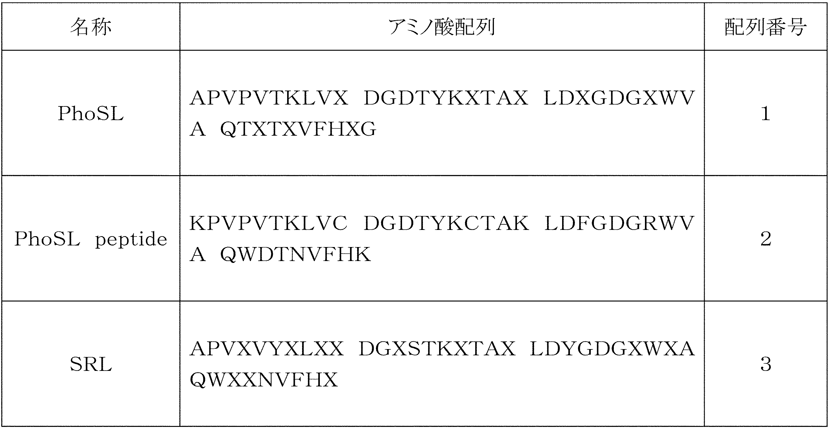
配列番号1の第10及び17番目のXは、任意のアミノ酸残基であってよいが、好ましくはCysであり、第20、23、27、33、35及び39番目のXは、それぞれ、Tyr/Ser、Phe/Tyr、Arg/Lys/Asn、Asp/Gly/Ser、Asn/Ala、及び、Thr/Glnである。
配列番号2は、配列番号1の具体例(APVPVTKLVC DGDTYKCTAY LDFGDGRWVA QWDTNVFHTG)において、1番目のAla、20番目のTyr、及び39番目のThrがLysに置換され、さらに40番目のGlyが欠失している。
配列番号3の第10及び17番目のXは、任意のアミノ酸残基であってよいが、好ましくはCysであり、第4、7、9、13、20、27、29、33、34及び39番目のXは、それぞれ、Pro/Gly、Glu/Lys、Val/Asp、Asn/Asp/Glu、His/Ser、Lys/His、Val/Ile、Gly/Asn/Ser、Ala/Thr、及び、Arg/Thrである。
〔実施例1~3〕シグナル増強試験(1)
直接吸着ELISA法にて、α1-6フコース糖鎖を有する糖タンパク質とα1-6フコース特異的レクチンとを、尿素の存在下又は不存在下で反応させ、糖鎖とレクチンとの結合反応に基づくシグナルの増強の有無を調べた。具体的な手順は、以下のとおりである。
直接吸着ELISA法にて、α1-6フコース糖鎖を有する糖タンパク質とα1-6フコース特異的レクチンとを、尿素の存在下又は不存在下で反応させ、糖鎖とレクチンとの結合反応に基づくシグナルの増強の有無を調べた。具体的な手順は、以下のとおりである。
α1-6フコース糖鎖を有する糖タンパク質として、フコシル化ハプトグロビン(以下、「fHP」という)、α-フェトプロテインL3(以下、「AFP-L3」という)、マウス免疫グロブリンG(SIGMA社製、以下、「IgG」という)を用意した。比較のため、α1-6フコース糖鎖でない糖タンパク質として、トランスフェリン(SIGMA社製、以下、「TF」という)も用意した。fHP及びAFP-L3の調製方法は、以下のとおりである。
(1) fHP溶液の調製
膵臓癌細胞株(PSN-1、DSファーマ社より入手)を常法に従い培養し、培養上清液を得た。培養上清液1000mLを、限外ろ過フィルター(製品名:VIVA SPIN 20‐10K、ザルトリウス社製)で1mLに濃縮した。NHS-activated Sepharose 4 Fast Flow(GEヘルスケア社製)に抗ハプトグロビン抗体(The Binding Site社製)を固定化したゲル0.5mLに上記濃縮液を添加した。室温で10分毎に混和して1時間後、前記ゲルを含む溶液を0.45μmフィルターチューブ(ミリポア社製)に加え、400×g、温度4℃で5分間遠心し、ろ液を廃棄した。次に、PBS 200μLを加えて400×g、温度4℃で5分間遠心して、ろ液を廃棄した。これを2回繰り返した。次いで、Elution Buffer(100mM グリシン、0.5M NaCl、pH3.0)200μLを加えて、400×g、温度4℃で5分間遠心して、ろ液を回収した。これを2回繰り返した。この液を合わせて得た溶液を3NのNaOHで中和した後、PBS 600μLを加え、fHP溶液を得た。このfHP溶液をさらにPBSで希釈して、1000ng/mLのfHP溶液を調製した。
膵臓癌細胞株(PSN-1、DSファーマ社より入手)を常法に従い培養し、培養上清液を得た。培養上清液1000mLを、限外ろ過フィルター(製品名:VIVA SPIN 20‐10K、ザルトリウス社製)で1mLに濃縮した。NHS-activated Sepharose 4 Fast Flow(GEヘルスケア社製)に抗ハプトグロビン抗体(The Binding Site社製)を固定化したゲル0.5mLに上記濃縮液を添加した。室温で10分毎に混和して1時間後、前記ゲルを含む溶液を0.45μmフィルターチューブ(ミリポア社製)に加え、400×g、温度4℃で5分間遠心し、ろ液を廃棄した。次に、PBS 200μLを加えて400×g、温度4℃で5分間遠心して、ろ液を廃棄した。これを2回繰り返した。次いで、Elution Buffer(100mM グリシン、0.5M NaCl、pH3.0)200μLを加えて、400×g、温度4℃で5分間遠心して、ろ液を回収した。これを2回繰り返した。この液を合わせて得た溶液を3NのNaOHで中和した後、PBS 600μLを加え、fHP溶液を得た。このfHP溶液をさらにPBSで希釈して、1000ng/mLのfHP溶液を調製した。
(2) AFP-L3溶液の調製
肝臓癌細胞株(HepG2、理化学研究所より入手)を常法に従い培養し、培養上清液を得た。培養上清液1000mLを、限外ろ過フィルター(製品名:VIVA SPIN 20‐10K、ザルトリウス社製)で1mLに濃縮した。NHS-activated Sepharose 4 Fast Flow(GEヘルスケア社製)に抗AFP抗体(DAKO社製)を固定化したゲル0.5mLに上記濃縮液を添加した。室温で10分毎に混和して1時間後、前記ゲルを含む溶液を0.45μmフィルターチューブ(ミリポア社製)に加え、400×g、温度4℃で5分間遠心し、ろ液を廃棄した。次に、PBS 200μLを加えて400×g、温度4℃で5分間遠心して、ろ液を廃棄した。これを2回繰り返した。次いで、Elution Buffer(100mM グリシン、0.5M NaCl、pH3.0)200μLを加えて、400×g、温度4℃で5分間遠心して、ろ液を回収した。これを2回繰り返した。この液を合わせて得た溶液を3NのNaOHで中和した後、PBS 600μLを加え、AFP-L3溶液を得た。このAFP-L3溶液をさらにPBSで希釈して1000ng/mLのAFP-L3溶液を調製した。
肝臓癌細胞株(HepG2、理化学研究所より入手)を常法に従い培養し、培養上清液を得た。培養上清液1000mLを、限外ろ過フィルター(製品名:VIVA SPIN 20‐10K、ザルトリウス社製)で1mLに濃縮した。NHS-activated Sepharose 4 Fast Flow(GEヘルスケア社製)に抗AFP抗体(DAKO社製)を固定化したゲル0.5mLに上記濃縮液を添加した。室温で10分毎に混和して1時間後、前記ゲルを含む溶液を0.45μmフィルターチューブ(ミリポア社製)に加え、400×g、温度4℃で5分間遠心し、ろ液を廃棄した。次に、PBS 200μLを加えて400×g、温度4℃で5分間遠心して、ろ液を廃棄した。これを2回繰り返した。次いで、Elution Buffer(100mM グリシン、0.5M NaCl、pH3.0)200μLを加えて、400×g、温度4℃で5分間遠心して、ろ液を回収した。これを2回繰り返した。この液を合わせて得た溶液を3NのNaOHで中和した後、PBS 600μLを加え、AFP-L3溶液を得た。このAFP-L3溶液をさらにPBSで希釈して1000ng/mLのAFP-L3溶液を調製した。
(3)IgG及びTF溶液の調製
IgG及びTFをそれぞれ、PBSで溶解して、5000ng/mLのIgG溶液、及び5000ng/mLのTF溶液を調製した。
IgG及びTFをそれぞれ、PBSで溶解して、5000ng/mLのIgG溶液、及び5000ng/mLのTF溶液を調製した。
直接吸着ELISA法の手順を、以下に示す。
(1)抗原固定化
fHP、AFP-L3、IgG又はTFの前記溶液を、マイクロタイタープレート(グライナー社製)のウェルに50μL添加して、4℃で16時間放置後、添加した液を廃棄した。
(2)洗浄
各ウェルに0.05% Tween/PBSを250μL添加し、添加した液を廃棄した。
(3)ブロッキング
各ウェルに1% BSA/PBSを200μL添加して、37℃で1時間放置後、添加した液を廃棄した。
(4)洗浄
各ウェルに0.05% Tween/PBSを250μL添加し、添加した液を廃棄した。この操作を合計3回繰り返した。
(5)レクチン反応
ビオチン標識PhoSLを1%BSA/PBSで希釈し、そして尿素(和光純薬社製)を添加して、レクチン溶液(ビオチン標識PhoSL終濃度1μg/mL、尿素終濃度5M)を調製した。このレクチン溶液50μLをウェルに添加して4℃で30分間放放置後、添加した液を廃棄した。対照として、尿素を添加していないレクチン溶液(ビオチン標識PhoSL終濃度1μg/mL、尿素0M)も、同様の反応を実施した。
(6)洗浄
各ウェルに0.05% Tween/PBSを250μL添加し、添加した液を廃棄した。この操作を合計3回繰り返した。
(7)HRP標識ストレプトアビジン反応
HRP標識ストレプトアビジン溶液(Vector社製、1μg/mL PBSに調製)を、ウェルに50μL添加して、室温で30分間放置後、添加した液を廃棄した。
(8)洗浄
各ウェルに0.05% Tween/PBSを250μL添加し、添加した液を廃棄した。この操作を合計3回繰り返した。
(9)発色反応
HRP用発色基質(製品名:TMB Peroxidase substrate system、KPL社製)を、ウェルに50μL添加し、室温で5分間放置した。
(10)反応停止
1M リン酸を50μL添加し、反応を停止した。
(1)抗原固定化
fHP、AFP-L3、IgG又はTFの前記溶液を、マイクロタイタープレート(グライナー社製)のウェルに50μL添加して、4℃で16時間放置後、添加した液を廃棄した。
(2)洗浄
各ウェルに0.05% Tween/PBSを250μL添加し、添加した液を廃棄した。
(3)ブロッキング
各ウェルに1% BSA/PBSを200μL添加して、37℃で1時間放置後、添加した液を廃棄した。
(4)洗浄
各ウェルに0.05% Tween/PBSを250μL添加し、添加した液を廃棄した。この操作を合計3回繰り返した。
(5)レクチン反応
ビオチン標識PhoSLを1%BSA/PBSで希釈し、そして尿素(和光純薬社製)を添加して、レクチン溶液(ビオチン標識PhoSL終濃度1μg/mL、尿素終濃度5M)を調製した。このレクチン溶液50μLをウェルに添加して4℃で30分間放放置後、添加した液を廃棄した。対照として、尿素を添加していないレクチン溶液(ビオチン標識PhoSL終濃度1μg/mL、尿素0M)も、同様の反応を実施した。
(6)洗浄
各ウェルに0.05% Tween/PBSを250μL添加し、添加した液を廃棄した。この操作を合計3回繰り返した。
(7)HRP標識ストレプトアビジン反応
HRP標識ストレプトアビジン溶液(Vector社製、1μg/mL PBSに調製)を、ウェルに50μL添加して、室温で30分間放置後、添加した液を廃棄した。
(8)洗浄
各ウェルに0.05% Tween/PBSを250μL添加し、添加した液を廃棄した。この操作を合計3回繰り返した。
(9)発色反応
HRP用発色基質(製品名:TMB Peroxidase substrate system、KPL社製)を、ウェルに50μL添加し、室温で5分間放置した。
(10)反応停止
1M リン酸を50μL添加し、反応を停止した。
プレートリーダー(製品名:POWERSCAN(登録商標)HT、DS PHARMA社製)を用いて、波長450nm及び630nmの吸光度を測定した。450nmの吸光度値から630nmの吸光度値を引いた値を検出値(シグナル:S)とした。同様に、糖タンパク質未添加のウェルの検出値(ノイズ:N)を測定し、検出値(S)からウェル自体の検出値(N)を引いた値を反応値(S-N)尿素5Mとした。また、尿素不存在下でレクチンと糖タンパク質とを反応させた場合の反応値(S-N)尿素0Mも求めた。
シグナルの増大量及び増大率を、それぞれ以下の数式:


により求めた。
各種糖タンパク質に対するPhoSLの反応値(S-N)を、表2及び図1に示す。
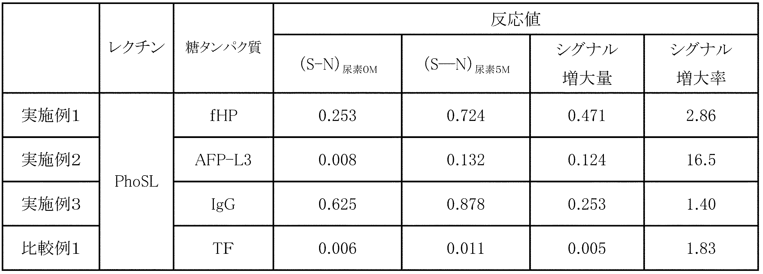
(S-N)尿素0M:尿素不存在下で反応させたときの反応値(S-N)
(S-N)尿素5M:尿素5Mの存在下で反応させたときの反応値(S-N)
(S-N)尿素5M:尿素5Mの存在下で反応させたときの反応値(S-N)
表2及び図1から、尿素存在下でのfHP、AFP-L3又はIgGとPhoSLとの反応に基づくシグナルは、尿素不存在下の場合と比べて、シグナル増大量及びシグナル増大率ともに有意に増大したことがわかる。一方、α1-6フコース糖鎖ではないTFとPhoSLとの反応に基づくシグナルは、シグナル増大率は高いものの、シグナル増大量が有意に増大しなかった。
〔実施例4~6〕シグナル増強試験(2)
サンドイッチELISA法にて、α1-6フコース糖鎖を有する糖タンパク質であるfHPと各種α1-6フコース特異的レクチンとを尿素の存在下又は不存在下で反応させ、糖タンパク質とレクチンとの結合反応に基づくシグナルの増強の有無を調べた。具体的な手順は以下のとおりである。
サンドイッチELISA法にて、α1-6フコース糖鎖を有する糖タンパク質であるfHPと各種α1-6フコース特異的レクチンとを尿素の存在下又は不存在下で反応させ、糖タンパク質とレクチンとの結合反応に基づくシグナルの増強の有無を調べた。具体的な手順は以下のとおりである。
α1-6フコース特異的レクチンとして、ビオチン標識PhoSL、ビオチン標識PhoSLペプチド、及びビオチン標識SRLを用いた。比較のために、α1-6以外のフコースを有する糖鎖にも結合するビオチン標識AALを用いた。
サンドイッチELISA法の手順を以下に示す。
(1)抗体固定化
糖鎖除去した抗ハプトグロビン抗体(日本バイオテスト社製)をPBSで5μg/mLに希釈し、マイクロタイタープレート(グライナー社製)の各ウェルに50μLずつ添加して4℃で16時間放置後、添加した液を廃棄した。
(2)洗浄
各ウェルに0.05% Tween/PBSを250μL添加し、添加した液を廃棄した。
(3)ブロッキング
各ウェルに1% BSA/PBSを200μL添加して、37℃で1時間放置後、添加した液を廃棄した。
(4)洗浄
各ウェルに0.05% Tween/PBSを250μL添加し、添加した液を廃棄した。この操作を合計3回繰り返した。
(5)抗原抗体反応
1%BSA/PBSで希釈した200ng/mL fHPを50μL、各ウェルに添加して、室温で1時間放置後、添加した液を廃棄した。
(6)洗浄
各ウェルに0.05% Tween/PBSを250μL添加し、添加した液を廃棄した。この操作を合計3回繰り返した。
(7)レクチン反応
ビオチン標識PhoSL、ビオチン標識PhoSLペプチド、ビオチン標識SRL又はビオチン標識AALを、1%BSA/PBS溶液で希釈し、そして尿素(和光純薬社製)を添加して、各レクチン溶液(ビオチン標識レクチン終濃度1μg/mL、尿素終濃度5M)を調製した。このレクチン溶液50μLをウェルに添加して、4℃で30分間放置後、添加した液を廃棄した。対照として、尿素を添加していないレクチン溶液(ビオチン標識レクチン終濃度1μg/mL、尿素0M)も実施した。
(8)洗浄
各ウェルに0.05% Tween/PBSを250μL添加し、添加した液を廃棄した。この操作を合計3回繰り返した。
(9)HRP標識ストレプトアビジン反応
HRP標識ストレプトアビジン溶液(1μg/mL)を、ウェルに50μL添加して、室温で30分間放置後、添加した液を廃棄した。
(10)洗浄
各ウェルに0.05% Tween/PBSを250μL添加し、添加した液を廃棄した。この操作を合計3回繰り返した。
(11)発色反応
HRP用発色基質(製品名:TMB Peroxidase substrate system、KPL社製)を、ウェルに50μL添加し、室温で5分間放置した。
(12)反応停止
1M リン酸を50μL添加し、反応を停止した。
(1)抗体固定化
糖鎖除去した抗ハプトグロビン抗体(日本バイオテスト社製)をPBSで5μg/mLに希釈し、マイクロタイタープレート(グライナー社製)の各ウェルに50μLずつ添加して4℃で16時間放置後、添加した液を廃棄した。
(2)洗浄
各ウェルに0.05% Tween/PBSを250μL添加し、添加した液を廃棄した。
(3)ブロッキング
各ウェルに1% BSA/PBSを200μL添加して、37℃で1時間放置後、添加した液を廃棄した。
(4)洗浄
各ウェルに0.05% Tween/PBSを250μL添加し、添加した液を廃棄した。この操作を合計3回繰り返した。
(5)抗原抗体反応
1%BSA/PBSで希釈した200ng/mL fHPを50μL、各ウェルに添加して、室温で1時間放置後、添加した液を廃棄した。
(6)洗浄
各ウェルに0.05% Tween/PBSを250μL添加し、添加した液を廃棄した。この操作を合計3回繰り返した。
(7)レクチン反応
ビオチン標識PhoSL、ビオチン標識PhoSLペプチド、ビオチン標識SRL又はビオチン標識AALを、1%BSA/PBS溶液で希釈し、そして尿素(和光純薬社製)を添加して、各レクチン溶液(ビオチン標識レクチン終濃度1μg/mL、尿素終濃度5M)を調製した。このレクチン溶液50μLをウェルに添加して、4℃で30分間放置後、添加した液を廃棄した。対照として、尿素を添加していないレクチン溶液(ビオチン標識レクチン終濃度1μg/mL、尿素0M)も実施した。
(8)洗浄
各ウェルに0.05% Tween/PBSを250μL添加し、添加した液を廃棄した。この操作を合計3回繰り返した。
(9)HRP標識ストレプトアビジン反応
HRP標識ストレプトアビジン溶液(1μg/mL)を、ウェルに50μL添加して、室温で30分間放置後、添加した液を廃棄した。
(10)洗浄
各ウェルに0.05% Tween/PBSを250μL添加し、添加した液を廃棄した。この操作を合計3回繰り返した。
(11)発色反応
HRP用発色基質(製品名:TMB Peroxidase substrate system、KPL社製)を、ウェルに50μL添加し、室温で5分間放置した。
(12)反応停止
1M リン酸を50μL添加し、反応を停止した。
前記プレートリーダーを用いて波長450nm及び630nmの吸光度を実施例1と同様に測定し、さらに反応値(S-N)尿素5Mを求めた。尿素不存在下でレクチンと糖タンパク質とを反応させた場合の反応値(S-N)尿素0Mも求めた。尿素存在下と尿素不存在下の反応値(S-N)、その増大量及び増大率を、表3及び図2に示す。
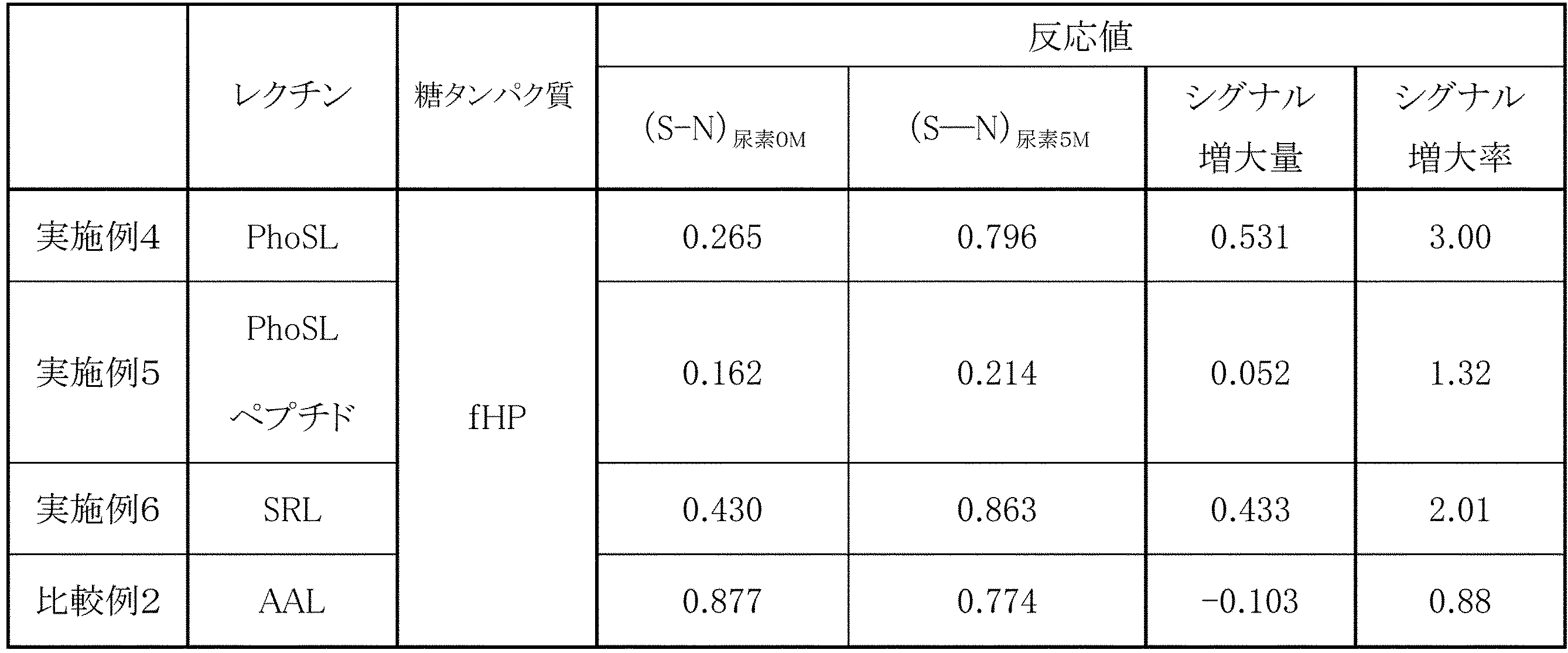
(S-N)尿素5M:尿素5Mの存在下で反応させたときの反応値(S-N)
図2及び表3から、PhoSL、PhoSLペプチド又はSRLとfHPとの5M尿素(終濃度)存在下での反応は、尿素不存在下の場合と比較して、シグナル増大量及びシグナル増大率ともに有意に改善したことがわかる。一方、α1-6フコース以外のフコースにも結合するAALは、反応系に尿素を添加しても、シグナルは増強しなかった。
実施例1~6に示したように、本発明のシグナル増強剤は、α1-6フコース糖鎖とそれに結合するα1-6フコース特異的レクチンとの反応に基づくシグナルを強化する。尿素は、α1-6フコース糖鎖を持たないトランスフェリンとα1-6フコース特異的レクチンとの反応を増感しない(比較例1)。尿素は、α1-6フコース糖鎖を持つ糖鎖と、α1-6フコースに親和性を有するが特異的ではないAALとの反応も増感しない(比較例2)。データを示していないが、尿素は、α1-6フコース糖鎖とα1-6フコースに親和性を有さないUEA-1、Lotus、ConA等との反応を増感しなかった。以上のことから、本発明のシグナル増強剤によるシグナル増強は、α1-6フコース糖鎖とα1-6フコース特異的レクチンとの反応のみに起きる現象であるといえる。
〔実施例7~8〕シグナル増強作用の阻害試験
本発明のシグナル増強剤のシグナル増大効果が、α1-6フコース糖鎖とα1-6フコース特異的レクチンとの特異的結合によるものであるか否かを調べるため、サンドイッチELISA法を用いた阻害試験を行った。
本発明のシグナル増強剤のシグナル増大効果が、α1-6フコース糖鎖とα1-6フコース特異的レクチンとの特異的結合によるものであるか否かを調べるため、サンドイッチELISA法を用いた阻害試験を行った。
実施例7では、5M尿素存在下でのfHPとPhoSLとの反応に、終濃度20mM又は100mMのフコース(ナカライテスク社製)を共存させた。実施例8では、実施例7のフコースに代えて、終濃度0.01mg/mL又は0.1mg/mLのチログロブリン(SIGMA社製、以下、「TG」という)を共存させた。単糖であるフコースを20~100mM程度で共存させると、fHPとPhoSLとの糖鎖特異的結合が阻害されることが知られている。また、α1-6フコース糖鎖を有するTGは、同様に、fHPとPhoSLとの糖鎖特異的結合を阻害することが知られている。
実施例1と同様に、前記プレートリーダーを用いて波長450nm及び630nmの吸光度を測定し、さらに反応値(S-N)を求めた。フコース又はTGを添加したときの検出結果を、図3に示す。
図3から、反応値(S-N)は、フコース又はTGの添加濃度の増大とともに低下することがわかる。これから、レクチンと糖タンパク質の結合に基づくシグナルが尿素存在下で増大する効果は、α1-6フコース特異的レクチンとα1-6フコース糖鎖との特異的結合によるものであることが確認された。
〔実施例9〕尿素濃度の変更試験
サンドイッチELISA法にて、シグナル増強剤としての尿素の好適範囲を調べた。実施例4において、fHP(50ng/mL)を使用したこと、及びレクチン溶液に対する尿素終濃度を0~9Mとしたこと以外は、実施例4と同様の手順で行った。
サンドイッチELISA法にて、シグナル増強剤としての尿素の好適範囲を調べた。実施例4において、fHP(50ng/mL)を使用したこと、及びレクチン溶液に対する尿素終濃度を0~9Mとしたこと以外は、実施例4と同様の手順で行った。
実施例1と同様にして、前記プレートリーダーを用いて波長450nm及び630nmの吸光度を測定し、さらに反応値(S-N)を求めた。尿素の各終濃度における、尿素未添加と比べたシグナル増大率を図4に示す。
図4から、終濃度1~9Mの尿素の存在下において、シグナルが1.2~2.8倍に増強されていることがわかる。尿素の終濃度は、好ましくは3~9Mであり、さらに好ましくは3~8M、特に好ましくは3~7Mである。
〔実施例10~11〕チオ尿素での試験
直接吸着ELISA法にて、α1-6フコース糖鎖を有する糖タンパク質(fHP又はAFP-L3)とα1-6フコース特異的レクチン(PhoSL)とを、チオ尿素の存在下又は不存在下で反応させ、糖鎖とレクチンとの結合反応に基づくシグナルの増強の有無を調べた。手順は、実施例1において、尿素5Mをチオ尿素(和光純薬社製)1.2Mに代えた以外は実施例1と同様とした。
直接吸着ELISA法にて、α1-6フコース糖鎖を有する糖タンパク質(fHP又はAFP-L3)とα1-6フコース特異的レクチン(PhoSL)とを、チオ尿素の存在下又は不存在下で反応させ、糖鎖とレクチンとの結合反応に基づくシグナルの増強の有無を調べた。手順は、実施例1において、尿素5Mをチオ尿素(和光純薬社製)1.2Mに代えた以外は実施例1と同様とした。
得られた吸光度から反応値(S-N)を、実施例1と同様に求めた。また、チオ尿素不存在下でレクチンと糖タンパク質とを反応させた場合の反応値(S-N)も求めた。
シグナルの増大量及び増大率を、それぞれ以下の数式:


により求めた。
各種糖タンパク質に対するPhoSLの反応値(S-N)を、表4に示す。
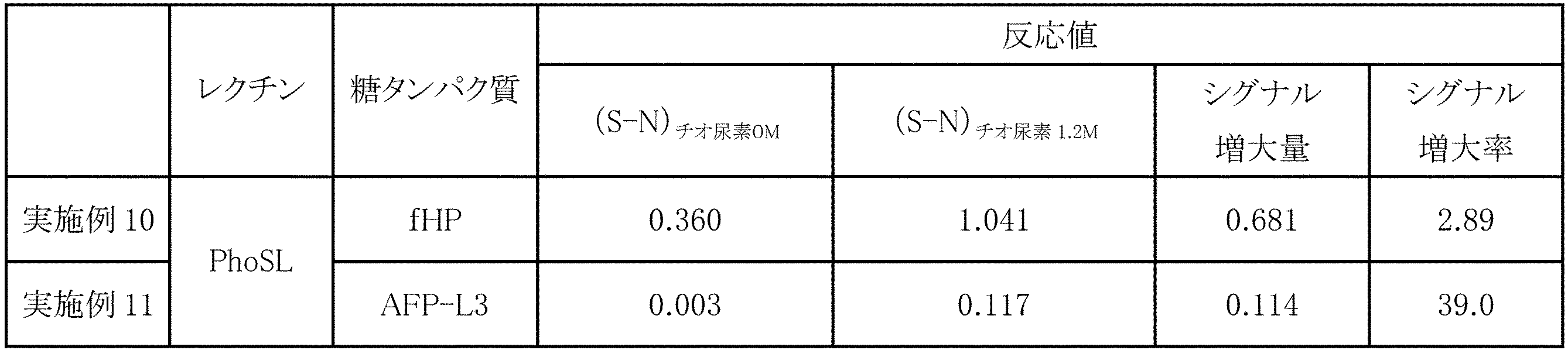
(S-N)チオ尿素0M:チオ尿素不存在下で反応させたときの反応値(S-N)
(S-N)チオ尿素1.2M:チオ尿素1.2Mの存在下で反応させたときの反応値(S-N)
(S-N)チオ尿素1.2M:チオ尿素1.2Mの存在下で反応させたときの反応値(S-N)
fHP及びAFP-L3とPhoSLとのチオ尿素存在下での反応に基づくシグナルは、チオ尿素不存在下の場合と比べて、シグナル増大量及びシグナル増大率ともに有意に増大した。
〔実施例12〕チオ尿素濃度の変更試験
サンドイッチELISA法にて、シグナル増強剤としてのチオ尿素の好適範囲を調べた。実施例4において、尿素に代えてチオ尿素を使用し、レクチン溶液に対するチオ尿素終濃度を0~1.5Mとしたこと以外は、実施例4と同様の手順で行った。
サンドイッチELISA法にて、シグナル増強剤としてのチオ尿素の好適範囲を調べた。実施例4において、尿素に代えてチオ尿素を使用し、レクチン溶液に対するチオ尿素終濃度を0~1.5Mとしたこと以外は、実施例4と同様の手順で行った。
実施例1と同様にして、前記プレートリーダーを用いて波長450nm及び630nmの吸光度を測定し、さらに反応値(S-N)を求めた。実施例10と同様に、チオ尿素の各終濃度における、チオ尿素不存在下と比べたシグナル増大率を求め、その結果を図5に示す。
図5から、終濃度0.1~1.5Mのチオ尿素の存在下において、シグナルが1.2~4.1倍に増強されていることがわかる。チオ尿素の終濃度は、通常、0.1~1.5Mでよく、好ましくは0.6~1.5Mであり、さらに好ましくは0.8~1.5M、特に好ましくは1~1.4Mである。
〔実施例13~14〕洗浄液へのシグナル増強剤の添加試験
サンドイッチELISA法にて、α1-6フコース糖鎖からなる糖タンパク質(fHP)を添加後、尿素又はチオ尿素を含む溶液で洗浄を行い、溶液を廃棄後、レクチン溶液と反応させ、糖鎖とレクチンとの結合反応に基づくシグナルの増強の有無を調べた。
サンドイッチELISA法にて、α1-6フコース糖鎖からなる糖タンパク質(fHP)を添加後、尿素又はチオ尿素を含む溶液で洗浄を行い、溶液を廃棄後、レクチン溶液と反応させ、糖鎖とレクチンとの結合反応に基づくシグナルの増強の有無を調べた。
具体的には、実施例4において、「(6)洗浄」における洗浄液(0.05% Tween/PBS)に対して終濃度5M尿素又は終濃度1.2Mチオ尿素を添加した点と、「(7)レクチン反応」におけるレクチン溶液に尿素を添加しない点を除いて、実施例4と同様の手順で実施した。
前記プレートリーダーを用いて波長450nm及び630nmの吸光度を実施例1と同様に測定し、さらに反応値(S-N)を求めた。さらに、洗浄液中に尿素又はチオ尿素を添加した場合の、尿素及びチオ尿素未添加と比べたシグナル増大量及び増大率を上記と同様に算出し、表5に示す。
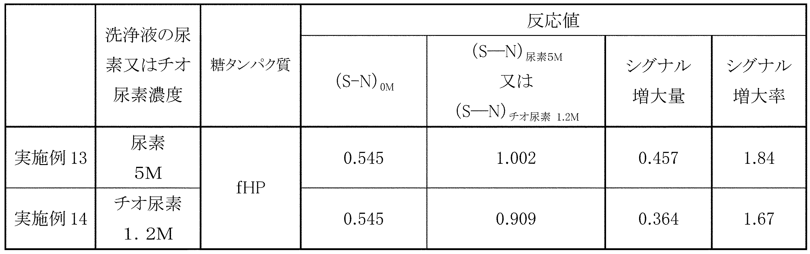
(S-N)尿素5M:尿素5Mの存在下で反応させたときの反応値(S-N)
(S-N)チオ尿素1.2M:チオ尿素1.2Mの存在下で反応させたときの反応値(S-N)
表5から、α1-6フコース糖鎖を有する糖タンパク質を尿素又はチオ尿素を含む洗浄液で洗浄した後、α1-6フコース特異的レクチンを反応させても、糖鎖とレクチンとの結合反応に基づくシグナルの増強が図れることが判明した。
〔実施例15~17〕
スギタケレクチンと同様に、ツチスギタケ、クリタケ及びベニテングタケからツチスギタケレクチン(PTL)、クリタケレクチン(NSL)、及びベニテングタケレクチン(AML)の精製をした。得らえたレクチンをビオチン標識PhoSLと同様の手順でビオチン標識し、ビオチン標識PTL、ビオチン標識NSL及びビオチン標識AMLを得た。
スギタケレクチンと同様に、ツチスギタケ、クリタケ及びベニテングタケからツチスギタケレクチン(PTL)、クリタケレクチン(NSL)、及びベニテングタケレクチン(AML)の精製をした。得らえたレクチンをビオチン標識PhoSLと同様の手順でビオチン標識し、ビオチン標識PTL、ビオチン標識NSL及びビオチン標識AMLを得た。
ビオチン標識PhoSLをビオチン標識PTL、ビオチン標識NSL又はビオチン標識AMLに置き換えた以外は実施例4と同様の方法で、サンドイッチELISAを行った。結果を表6に示す。
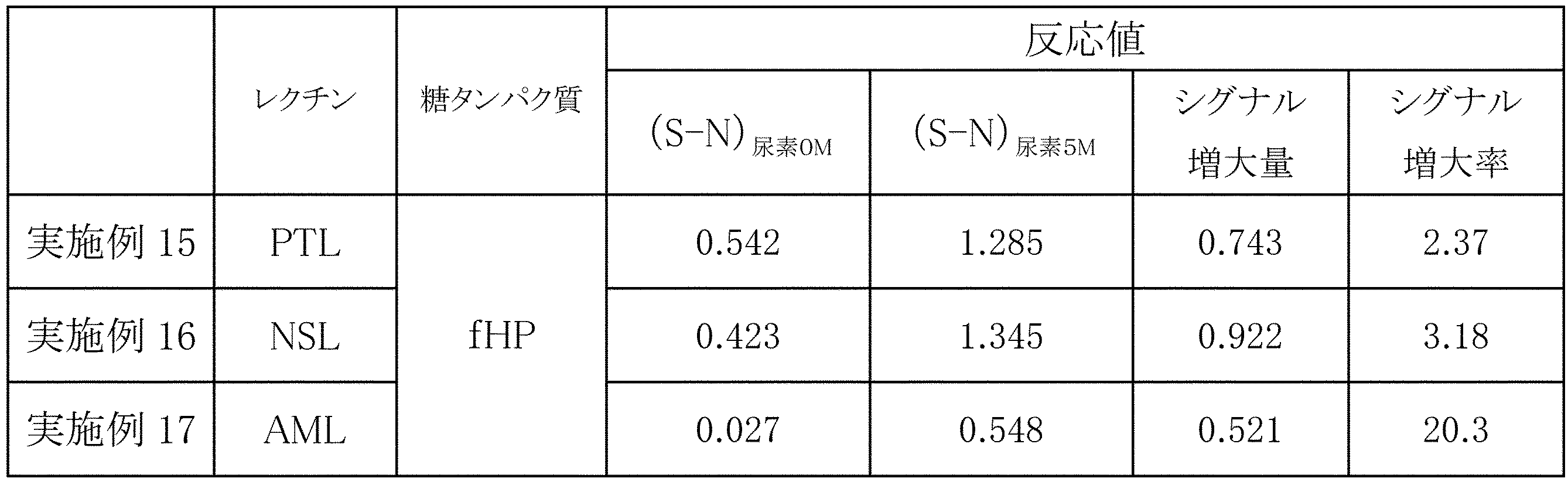
(S-N)尿素5M:尿素5Mの存在下で反応させたときの反応値(S-N)
表6に示すように、いずれのα1-6フコース特異的レクチンも、尿素存在下でシグナルが増強されることがわかった。
〔実施例18〕ビーズを用いた検出方法
磁気ビーズ(インビトロジェン社製、14204)の説明書に従い、糖鎖除去した抗ハプトグロビン抗体を固定化することにより、抗体固定化磁気ビーズを調製した。
磁気ビーズ(インビトロジェン社製、14204)の説明書に従い、糖鎖除去した抗ハプトグロビン抗体を固定化することにより、抗体固定化磁気ビーズを調製した。
ビーズを用いた検出を、常法に従って行った。具体的には、フコシル化ハプトグロビン(fHP)(終濃度:0μg/mL又は0.45μg/mL)、尿素(終濃度0M又は5M)、配列番号4に示すPhoSL組換えレクチン(特許文献5:特開2011-148735の配列番号1にDYKDDDDKタグを付加したもの、終濃度5μg/mL)を混合し、複合体を形成させた。複合体を含む上記溶液を10倍に希釈し、抗体固定化磁気ビーズを添加し、磁石を用いて洗浄した後、HRP標識抗FLAG抗体(SIGMA社製)を添加し、検出抗体との複合体を形成させた。前記HRP用発色基質を用いて、発色させ、1M リン酸を用いて、発色反応を停止した。得られた溶液の吸光度を前記同様プレートリーダーで測定した。
fHP存在下における450nmの吸光度値から630nmの吸光度値を引いた値を検出値(シグナル:S)とし、同様にfHP不存在下の検出値(ノイズ:N)とした。5M尿素存在下の検出値(S)からウェル自体の検出値(N)を引いた値を反応値(S-N)尿素5Mとした。また、同様に尿素不存在下の場合の反応値(S-N)尿素0Mを求めた。また、5M尿素存在下の検出値Sを検出値Nで除した値を反応値(S/N)尿素5Mとした。同様に尿素不存在下の場合を反応値(S/N)尿素0Mとした。実施例1と同様に、シグナルの増大量及び増大率を算出した。その結果を表7に示す。
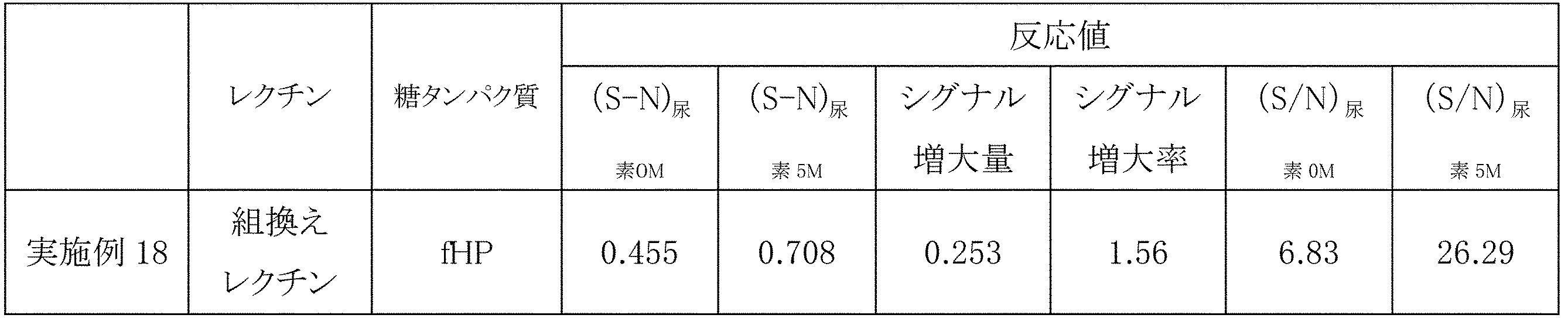
(S-N)尿素5M:尿素5M存在下で反応させたときの反応値(S-N)
(S/N)尿素0M:尿素不存在下で反応させたときの反応値(S/N)
(S/N)尿素5M:尿素5M存在下で反応させたときの反応値(S/N)
表7から、尿素存在下でのfHPと組換えレクチンとの反応に基づくシグナルは、尿素不存在下の場合と比べて、シグナル増大量及びシグナル増大率ともに有意に増大したことがわかる。同様に、反応値(S/N)も、尿素不存在下の場合と比べて顕著に増大する。ビーズを用いた検出方法においても、本発明の効果が得られることが確認された。
Claims (11)
- α1-6フコース糖鎖とそれに結合するα1-6フコース特異的レクチンとの反応に基づいたシグナルを増強するシグナル増強剤であって、尿素及びチオ尿素からなる群から選択される少なくとも一種を有効成分とする前記シグナル増強剤。
- 前記シグナル増強剤中の尿素濃度が、1~9Mである、請求項1に記載の方法。
- 前記シグナル増強剤中のチオ尿素濃度が、0.1~1.5Mである、請求項1に記載の方法。
- α1-6フコース糖鎖とそれに結合するα1-6フコース特異的レクチンとの反応に基づいたシグナルを増強する方法であって、
尿素及びチオ尿素からなる群から選択される少なくとも一種を有効成分とするシグナル増強剤の存在下で、前記糖鎖と前記レクチンとを反応させるステップ
を含む、前記方法。 - 前記尿素の前記反応時の濃度が、1~9Mである、請求項4に記載の方法。
- 前記チオ尿素の前記反応時の濃度が、0.1~1.5Mである、請求項4に記載の方法。
- α1-6フコース糖鎖とそれに結合するα1-6フコース特異的レクチンとの反応に基づいたシグナルを増強する方法であって、
尿素及びチオ尿素からなる群から選択される少なくとも一種を有効成分とするシグナル増強剤を含む洗浄液で、前記糖鎖を洗浄するステップ、及び
前記糖鎖と前記レクチンとを反応させるステップ
を含む、前記方法。 - 前記洗浄ステップを前記反応ステップの直前に行う、請求項7に記載の方法。
- 前記洗浄液の尿素濃度が、1~9Mである、請求項7に記載の方法。
- 前記洗浄液のチオ尿素濃度が、0.1~1.5Mである、請求項7に記載の方法。
- 尿素及びチオ尿素からなる群から選択される少なくとも一種を有効成分とするシグナル増強剤、及び、α1-6フコース特異的レクチンを含む、α1-6フコース糖鎖の検出キット。
Priority Applications (4)
| Application Number | Priority Date | Filing Date | Title |
|---|---|---|---|
| CN201680003626.5A CN107076751A (zh) | 2015-02-27 | 2016-02-25 | 信号增强剂 |
| JP2016536259A JP5996831B1 (ja) | 2015-02-27 | 2016-02-25 | シグナル増強剤 |
| US15/522,484 US20170336415A1 (en) | 2015-02-27 | 2016-02-25 | Signal enhancer |
| EP16755594.5A EP3199950A4 (en) | 2015-02-27 | 2016-02-25 | Signal enhancer |
Applications Claiming Priority (2)
| Application Number | Priority Date | Filing Date | Title |
|---|---|---|---|
| JP2015037717 | 2015-02-27 | ||
| JP2015-037717 | 2015-02-27 |
Publications (1)
| Publication Number | Publication Date |
|---|---|
| WO2016136850A1 true WO2016136850A1 (ja) | 2016-09-01 |
Family
ID=56789608
Family Applications (1)
| Application Number | Title | Priority Date | Filing Date |
|---|---|---|---|
| PCT/JP2016/055541 Ceased WO2016136850A1 (ja) | 2015-02-27 | 2016-02-25 | シグナル増強剤 |
Country Status (5)
| Country | Link |
|---|---|
| US (1) | US20170336415A1 (ja) |
| EP (1) | EP3199950A4 (ja) |
| JP (1) | JP5996831B1 (ja) |
| CN (1) | CN107076751A (ja) |
| WO (1) | WO2016136850A1 (ja) |
Cited By (1)
| Publication number | Priority date | Publication date | Assignee | Title |
|---|---|---|---|---|
| JP2024530509A (ja) * | 2021-08-11 | 2024-08-21 | オスラー ダイアグノスティックス リミテッド | ブロックコポリマーを含む診断アッセイ試薬 |
Families Citing this family (1)
| Publication number | Priority date | Publication date | Assignee | Title |
|---|---|---|---|---|
| CN107209171A (zh) * | 2014-10-21 | 2017-09-26 | 普林斯顿大学理事会 | 用于个人样品分析的系统和方法 |
Citations (3)
| Publication number | Priority date | Publication date | Assignee | Title |
|---|---|---|---|---|
| JP2010145202A (ja) * | 2008-12-18 | 2010-07-01 | Tosoh Corp | ポリエチレングリコールおよび尿素による免疫反応増強方法 |
| WO2011089988A1 (ja) * | 2010-01-21 | 2011-07-28 | 株式会社J-オイルミルズ | 膵臓癌の検出方法 |
| WO2014046248A1 (ja) * | 2012-09-24 | 2014-03-27 | 三菱化学株式会社 | 肝臓癌の検出方法および肝硬変の検出方法 |
Family Cites Families (2)
| Publication number | Priority date | Publication date | Assignee | Title |
|---|---|---|---|---|
| JP4514163B2 (ja) * | 2008-07-22 | 2010-07-28 | 株式会社J−オイルミルズ | フコースα1→6特異的レクチン |
| CN102175839B (zh) * | 2010-12-23 | 2013-08-14 | 中国人民解放军第二军医大学东方肝胆外科医院 | IgG核心岩藻糖基化水平检测方法及其应用 |
-
2016
- 2016-02-25 WO PCT/JP2016/055541 patent/WO2016136850A1/ja not_active Ceased
- 2016-02-25 EP EP16755594.5A patent/EP3199950A4/en not_active Withdrawn
- 2016-02-25 US US15/522,484 patent/US20170336415A1/en not_active Abandoned
- 2016-02-25 JP JP2016536259A patent/JP5996831B1/ja not_active Expired - Fee Related
- 2016-02-25 CN CN201680003626.5A patent/CN107076751A/zh active Pending
Patent Citations (3)
| Publication number | Priority date | Publication date | Assignee | Title |
|---|---|---|---|---|
| JP2010145202A (ja) * | 2008-12-18 | 2010-07-01 | Tosoh Corp | ポリエチレングリコールおよび尿素による免疫反応増強方法 |
| WO2011089988A1 (ja) * | 2010-01-21 | 2011-07-28 | 株式会社J-オイルミルズ | 膵臓癌の検出方法 |
| WO2014046248A1 (ja) * | 2012-09-24 | 2014-03-27 | 三菱化学株式会社 | 肝臓癌の検出方法および肝硬変の検出方法 |
Non-Patent Citations (2)
| Title |
|---|
| See also references of EP3199950A4 * |
| YUKA KOBAYASHI: "Screening for New Lectins and Their Applications", OYO TOSHITSU KAGAKU, vol. 3, no. 3, 2013, pages 200 - 201, XP008185309 * |
Cited By (1)
| Publication number | Priority date | Publication date | Assignee | Title |
|---|---|---|---|---|
| JP2024530509A (ja) * | 2021-08-11 | 2024-08-21 | オスラー ダイアグノスティックス リミテッド | ブロックコポリマーを含む診断アッセイ試薬 |
Also Published As
| Publication number | Publication date |
|---|---|
| EP3199950A4 (en) | 2017-10-25 |
| JP5996831B1 (ja) | 2016-09-21 |
| US20170336415A1 (en) | 2017-11-23 |
| JPWO2016136850A1 (ja) | 2017-04-27 |
| EP3199950A1 (en) | 2017-08-02 |
| CN107076751A (zh) | 2017-08-18 |
Similar Documents
| Publication | Publication Date | Title |
|---|---|---|
| JP4514163B2 (ja) | フコースα1→6特異的レクチン | |
| JP4900983B2 (ja) | 膵臓癌の検出方法 | |
| EP2395357B1 (en) | Method for analyzing PSA, and method for distinguishing between prostate cancer and prostatomegaly employing the analysis method | |
| JP6655248B2 (ja) | 肝細胞がんマーカー | |
| JP5737761B2 (ja) | 肝細胞癌マーカー | |
| JP7240674B2 (ja) | 前立腺癌の検出方法 | |
| CN104714026A (zh) | 一种甲胎蛋白异质体的分离检测组合物、系统及其应用 | |
| CN109061189A (zh) | 一种肌钙蛋白i检测试剂盒及其制备方法 | |
| WO2008031288A1 (en) | Pre-filled centrifugal column for detecting hepatocellular carcinoma-specific alpha-fetoprotein variant and test kit containing the column | |
| EP3410118B1 (en) | Prostate carcinoma determination method | |
| JP5109001B2 (ja) | α1,6フコース糖鎖の検出および分別方法 | |
| JP5996831B1 (ja) | シグナル増強剤 | |
| KR101296511B1 (ko) | 공기 중 나노 입자 노출 여부 진단용 바이오 마커 조성물 및 바이오 마커 검출방법 | |
| Mondal et al. | Alterations of glycan branching and differential expression of sialic acid on alpha fetoprotein among hepatitis patients | |
| Chatterjee et al. | Glycosylation of acute phase proteins: A promising disease biomarker | |
| CN101356437B (zh) | 具有由GnT-V转移的GlcNAc的糖链的检测方法 | |
| KR20210024464A (ko) | 췌장암의 판정용 마커 | |
| Han et al. | A novel, rapid, and sensitive homogeneous sandwich detection method of Glypican-3 as a serum marker for hepatocellular carcinoma | |
| CN106526165A (zh) | 磷脂酰肌醇蛋白聚糖定量测定试剂盒及其检测方法 | |
| JPWO2008096790A1 (ja) | 腫瘍マーカー糖鎖の解析及び利用 | |
| WO2018014709A1 (zh) | 一种检测血清岩藻糖蛋白的免疫渗滤方法及免疫渗滤装置 | |
| KR101100809B1 (ko) | 암 진단용 펩티드 마커 및 이를 이용한 암 진단방법 | |
| Mandal et al. | Identification and analysis of O-acetylated sialoglycoproteins | |
| CN105181971A (zh) | 一种用于肝癌血清学预测的结合珠蛋白试剂盒 | |
| WO2017122613A1 (ja) | 合成スギタケレクチンの親和性を向上させる方法 |
Legal Events
| Date | Code | Title | Description |
|---|---|---|---|
| ENP | Entry into the national phase |
Ref document number: 2016536259 Country of ref document: JP Kind code of ref document: A |
|
| 121 | Ep: the epo has been informed by wipo that ep was designated in this application |
Ref document number: 16755594 Country of ref document: EP Kind code of ref document: A1 |
|
| REEP | Request for entry into the european phase |
Ref document number: 2016755594 Country of ref document: EP |
|
| NENP | Non-entry into the national phase |
Ref country code: DE |