WO2017002934A1 - Gpc3標的治療剤が有効である患者に投与されるgpc3標的治療剤 - Google Patents
Gpc3標的治療剤が有効である患者に投与されるgpc3標的治療剤 Download PDFInfo
- Publication number
- WO2017002934A1 WO2017002934A1 PCT/JP2016/069493 JP2016069493W WO2017002934A1 WO 2017002934 A1 WO2017002934 A1 WO 2017002934A1 JP 2016069493 W JP2016069493 W JP 2016069493W WO 2017002934 A1 WO2017002934 A1 WO 2017002934A1
- Authority
- WO
- WIPO (PCT)
- Prior art keywords
- seq
- gpc3
- therapeutic agent
- antibody
- heavy chain
- Prior art date
- Legal status (The legal status is an assumption and is not a legal conclusion. Google has not performed a legal analysis and makes no representation as to the accuracy of the status listed.)
- Ceased
Links
Images


Classifications
-
- A—HUMAN NECESSITIES
- A61—MEDICAL OR VETERINARY SCIENCE; HYGIENE
- A61K—PREPARATIONS FOR MEDICAL, DENTAL OR TOILETRY PURPOSES
- A61K39/00—Medicinal preparations containing antigens or antibodies
- A61K39/395—Antibodies; Immunoglobulins; Immune serum, e.g. antilymphocytic serum
- A61K39/39533—Antibodies; Immunoglobulins; Immune serum, e.g. antilymphocytic serum against materials from animals
- A61K39/39558—Antibodies; Immunoglobulins; Immune serum, e.g. antilymphocytic serum against materials from animals against tumor tissues, cells, antigens
-
- A—HUMAN NECESSITIES
- A61—MEDICAL OR VETERINARY SCIENCE; HYGIENE
- A61K—PREPARATIONS FOR MEDICAL, DENTAL OR TOILETRY PURPOSES
- A61K31/00—Medicinal preparations containing organic active ingredients
- A61K31/33—Heterocyclic compounds
- A61K31/395—Heterocyclic compounds having nitrogen as a ring hetero atom, e.g. guanethidine or rifamycins
- A61K31/435—Heterocyclic compounds having nitrogen as a ring hetero atom, e.g. guanethidine or rifamycins having six-membered rings with one nitrogen as the only ring hetero atom
- A61K31/44—Non condensed pyridines; Hydrogenated derivatives thereof
-
- A—HUMAN NECESSITIES
- A61—MEDICAL OR VETERINARY SCIENCE; HYGIENE
- A61K—PREPARATIONS FOR MEDICAL, DENTAL OR TOILETRY PURPOSES
- A61K39/00—Medicinal preparations containing antigens or antibodies
- A61K39/395—Antibodies; Immunoglobulins; Immune serum, e.g. antilymphocytic serum
-
- A—HUMAN NECESSITIES
- A61—MEDICAL OR VETERINARY SCIENCE; HYGIENE
- A61K—PREPARATIONS FOR MEDICAL, DENTAL OR TOILETRY PURPOSES
- A61K45/00—Medicinal preparations containing active ingredients not provided for in groups A61K31/00 - A61K41/00
-
- A—HUMAN NECESSITIES
- A61—MEDICAL OR VETERINARY SCIENCE; HYGIENE
- A61K—PREPARATIONS FOR MEDICAL, DENTAL OR TOILETRY PURPOSES
- A61K45/00—Medicinal preparations containing active ingredients not provided for in groups A61K31/00 - A61K41/00
- A61K45/06—Mixtures of active ingredients without chemical characterisation, e.g. antiphlogistics and cardiaca
-
- A—HUMAN NECESSITIES
- A61—MEDICAL OR VETERINARY SCIENCE; HYGIENE
- A61P—SPECIFIC THERAPEUTIC ACTIVITY OF CHEMICAL COMPOUNDS OR MEDICINAL PREPARATIONS
- A61P35/00—Antineoplastic agents
-
- C—CHEMISTRY; METALLURGY
- C12—BIOCHEMISTRY; BEER; SPIRITS; WINE; VINEGAR; MICROBIOLOGY; ENZYMOLOGY; MUTATION OR GENETIC ENGINEERING
- C12Q—MEASURING OR TESTING PROCESSES INVOLVING ENZYMES, NUCLEIC ACIDS OR MICROORGANISMS; COMPOSITIONS OR TEST PAPERS THEREFOR; PROCESSES OF PREPARING SUCH COMPOSITIONS; CONDITION-RESPONSIVE CONTROL IN MICROBIOLOGICAL OR ENZYMOLOGICAL PROCESSES
- C12Q1/00—Measuring or testing processes involving enzymes, nucleic acids or microorganisms; Compositions therefor; Processes of preparing such compositions
- C12Q1/02—Measuring or testing processes involving enzymes, nucleic acids or microorganisms; Compositions therefor; Processes of preparing such compositions involving viable microorganisms
-
- G—PHYSICS
- G01—MEASURING; TESTING
- G01N—INVESTIGATING OR ANALYSING MATERIALS BY DETERMINING THEIR CHEMICAL OR PHYSICAL PROPERTIES
- G01N33/00—Investigating or analysing materials by specific methods not covered by groups G01N1/00 - G01N31/00
- G01N33/48—Biological material, e.g. blood, urine; Haemocytometers
- G01N33/50—Chemical analysis of biological material, e.g. blood, urine; Testing involving biospecific ligand binding methods; Immunological testing
- G01N33/53—Immunoassay; Biospecific binding assay; Materials therefor
- G01N33/575—Immunoassay; Biospecific binding assay; Materials therefor for cancer
-
- G—PHYSICS
- G01—MEASURING; TESTING
- G01N—INVESTIGATING OR ANALYSING MATERIALS BY DETERMINING THEIR CHEMICAL OR PHYSICAL PROPERTIES
- G01N33/00—Investigating or analysing materials by specific methods not covered by groups G01N1/00 - G01N31/00
- G01N33/48—Biological material, e.g. blood, urine; Haemocytometers
- G01N33/50—Chemical analysis of biological material, e.g. blood, urine; Testing involving biospecific ligand binding methods; Immunological testing
- G01N33/53—Immunoassay; Biospecific binding assay; Materials therefor
- G01N33/575—Immunoassay; Biospecific binding assay; Materials therefor for cancer
- G01N33/57525—Immunoassay; Biospecific binding assay; Materials therefor for cancer of the liver or pancreas
-
- G—PHYSICS
- G01—MEASURING; TESTING
- G01N—INVESTIGATING OR ANALYSING MATERIALS BY DETERMINING THEIR CHEMICAL OR PHYSICAL PROPERTIES
- G01N33/00—Investigating or analysing materials by specific methods not covered by groups G01N1/00 - G01N31/00
- G01N33/48—Biological material, e.g. blood, urine; Haemocytometers
- G01N33/50—Chemical analysis of biological material, e.g. blood, urine; Testing involving biospecific ligand binding methods; Immunological testing
- G01N33/68—Chemical analysis of biological material, e.g. blood, urine; Testing involving biospecific ligand binding methods; Immunological testing involving proteins, peptides or amino acids
-
- A—HUMAN NECESSITIES
- A61—MEDICAL OR VETERINARY SCIENCE; HYGIENE
- A61K—PREPARATIONS FOR MEDICAL, DENTAL OR TOILETRY PURPOSES
- A61K39/00—Medicinal preparations containing antigens or antibodies
- A61K2039/57—Medicinal preparations containing antigens or antibodies characterised by the type of response, e.g. Th1, Th2
- A61K2039/572—Medicinal preparations containing antigens or antibodies characterised by the type of response, e.g. Th1, Th2 cytotoxic response
-
- A—HUMAN NECESSITIES
- A61—MEDICAL OR VETERINARY SCIENCE; HYGIENE
- A61K—PREPARATIONS FOR MEDICAL, DENTAL OR TOILETRY PURPOSES
- A61K39/00—Medicinal preparations containing antigens or antibodies
- A61K2039/58—Medicinal preparations containing antigens or antibodies raising an immune response against a target which is not the antigen used for immunisation
- A61K2039/585—Medicinal preparations containing antigens or antibodies raising an immune response against a target which is not the antigen used for immunisation wherein the target is cancer
-
- C—CHEMISTRY; METALLURGY
- C07—ORGANIC CHEMISTRY
- C07K—PEPTIDES
- C07K16/00—Immunoglobulins [IG], e.g. monoclonal or polyclonal antibodies
- C07K16/18—Immunoglobulins [IG], e.g. monoclonal or polyclonal antibodies against material from animals or humans
-
- C—CHEMISTRY; METALLURGY
- C07—ORGANIC CHEMISTRY
- C07K—PEPTIDES
- C07K16/00—Immunoglobulins [IG], e.g. monoclonal or polyclonal antibodies
- C07K16/46—Hybrid immunoglobulins
-
- G—PHYSICS
- G01—MEASURING; TESTING
- G01N—INVESTIGATING OR ANALYSING MATERIALS BY DETERMINING THEIR CHEMICAL OR PHYSICAL PROPERTIES
- G01N2800/00—Detection or diagnosis of diseases
- G01N2800/50—Determining the risk of developing a disease
-
- G—PHYSICS
- G01—MEASURING; TESTING
- G01N—INVESTIGATING OR ANALYSING MATERIALS BY DETERMINING THEIR CHEMICAL OR PHYSICAL PROPERTIES
- G01N2800/00—Detection or diagnosis of diseases
- G01N2800/70—Mechanisms involved in disease identification
- G01N2800/7023—(Hyper)proliferation
- G01N2800/7028—Cancer
Definitions
- the present invention relates to a method for determining the efficacy of a GPC3 targeted therapeutic agent against cancer in a patient or a GPC3 targeted therapeutic agent or formulation for administration to a patient (only) for which the GPC3 targeted therapeutic agent has been determined to be effective. provide.
- Non-patent Document 1 Death due to hepatocellular carcinoma is equivalent to 600,000 throughout the year, and is said to be the fifth among deaths from cancer worldwide.
- Most hepatocellular carcinomas die within 1 year after diagnosis of the disease.
- hepatocellular carcinoma is often diagnosed at a late stage where curable therapies are less successful.
- the effectiveness of medical procedures, including chemotherapy, chemoembolization, ablation and proton beam therapy for these patients remains inadequate.
- Many patients show disease recurrence, which progresses rapidly to an advanced stage with vascular invasion and multi-site intrahepatic metastasis, with a 5-year survival rate of only 7% (Non-Patent Document 2).
- Non-patent Document 3 There is a need in the art for new therapies for such malignant hepatocellular carcinoma.
- Hepatocellular carcinoma is said to account for over 90% of primary liver cancer in Japan.
- a medical treatment method for such hepatocellular carcinoma for example, using a chemotherapeutic agent, an oil-based contrast agent (lipiodol), an anticancer agent, and an embolizing substance (Zelfoam) were mixed into the hepatic artery that is a route for supplying nutrients to the tumor.
- lipiodol an oil-based contrast agent
- Zelfoam embolizing substance
- TAE hepatic artery embolization
- transcutaneous ethanol injection percutaneous microwave coagulation Invasive techniques such as therapy and radiofrequency ablation are used.
- Non-patent Document 4 As chemotherapeutic agents, 5-FU (fluorouracil), UFT (uracil and tegafur), MMC (mitomycin C), DHAD (mitoxantrone), ADR (adriamycin), EPI (epirubicin), CDDP (cisplatin), etc.
- 5-FU fluorouracil
- UFT uracil and tegafur
- MMC mitomycin C
- DHAD mitoxantrone
- ADR adriamycin
- EPI epirubicin
- CDDP cisplatin
- sorafenib (Nexavar, BAY43-9006), which exerts an angiogenic effect and shows an advantageous effect compared with the above-mentioned chemotherapeutic agents, has been approved.
- the efficacy of sorafenib is being investigated in two Phase III multicenter placebo-controlled trials (SHARP trials and trials conducted in the Asia-Pacific region) for advanced hepatocellular carcinoma.
- hepatic dysfunction associated with the progression of liver cancer, loss of appetite, weight loss, general malaise, palpation of right hip mass, right hip pain, abdominal fullness, fever, jaundice, etc. Symptoms are observed.
- chemotherapeutic agents such as sorafenib can cause diarrhea or constipation, anemia, suppression of the immune system to cause (fatal severity) infection and sepsis, bleeding, cardiotoxicity, hepatotoxicity, nephrotoxicity, anorexia, There is a problem to be overcome that side effects inherent to chemotherapeutic agents such as weight loss also occur.
- liver cancer is not initially observed with any special initial symptoms, but with liver cancer progression, liver dysfunction is accompanied, loss of appetite, weight loss, general malaise, right hip mass palpation, right hip pain Symptoms peculiar to liver cancer such as abdominal fullness, fever and jaundice are observed. It is clinically observed that these symptoms are amplified by the use of the chemotherapeutic agents described above. For example, symptoms such as anorexia in a patient in which hepatoma cells are detected and weight loss that accompanies or independently occur are more amplified by administration of a chemotherapeutic agent to the patient compared to non-administration. Sometimes.
- the use of the chemotherapeutic agent may have to be abandoned, and the amplification of the symptom is a factor that hinders treatment with the chemotherapeutic agent. Therefore, establishment of a better treatment method has been demanded from the viewpoint of improvement of therapeutic effect and improvement of QOL of patients receiving treatment.
- GPC3 Glypican 3
- GPC3 is considered to be useful as a target for identification of GPC3 function in liver cancer, a target for treatment of liver cancer, or a target for diagnosis of liver cancer.
- ADCC antibody-dependent cellular cytotoxicity
- CDC complement-dependent cytotoxicity
- a therapeutic agent for liver cancer containing an anti-GPC3 antibody exhibiting activity as an active ingredient has been developed (Patent Document 1).
- a GPC3-targeted therapeutic agent containing a humanized anti-GPC3 antibody having ADCC activity and CDC activity as an active ingredient has been developed (Patent Document 2).
- a GPC3 target comprising GPC3 antibody with ADCC activity and CDC activity and improved plasma kinetics
- Therapeutic agents have been developed.
- the combination therapy of these anti-GPC3 antibodies and chemotherapeutic agents such as sorafenib attenuated the side effects caused by monotherapy with chemotherapeutic agents such as sorafenib, and also found that the synergistic effects of both agents are exhibited.
- chemotherapeutic agents such as sorafenib
- Non-patent Documents 7 and 8 development of a diagnostic method using GPC3 as a target for diagnosis of liver cancer is also underway.
- GPC3 is known to undergo processing by convertase, phospholipase D, Notum, or an unidentified mechanism during or after expression on the cell surface.
- diagnostic agents or diagnostic methods for liver cancer have been developed that contain antibodies that bind to soluble GPC3 epitopes that are processed and secreted into plasma that are present in the patient's plasma. (Patent Document 7).
- a diagnostic or diagnostic method for liver cancer contains an antibody that binds to an epitope of tethered GPC3 that is still present on the cell surface after processing, which is present in tissue preparations isolated from patients.
- Patent Document 8 a diagnostic agent or diagnostic method is a method for detecting the presence of liver cancer in a test patient, and a method for identifying a patient for whom a GPC3 targeted therapeutic agent can be expected to be effective, or a GPC3 targeted therapeutic agent is administered. There was no known method for determining the continuation of GPC3-targeted therapeutics to patients.
- the present invention has been made in view of such circumstances, and an object thereof is to provide a method for determining the effectiveness of a GPC3-targeted therapeutic agent for a liver cancer patient.
- another object is to provide a GPC3-targeted therapeutic agent or preparation for administration to (only) a patient for whom a GPC3-targeted therapeutic agent has been determined to be effective.
- the present inventors measured the expression level of GPC3 in a biological sample isolated from a patient prior to administration of a GPC3-targeted therapeutic agent. Only the expression level of GPC3 per tumor cell is found to be highly correlated with the effectiveness of the patient on the GPC3 target therapeutic agent, and when the GPC3 expression level per tumor cell is a predetermined value Created a way to determine that is effective. In addition, a GPC3-targeted therapeutic agent or formulation has been created for administration to (only) patients whose GPC3-targeted therapeutic agent has been determined to be effective.
- a method for determining the effectiveness of a GPC3-targeted therapeutic agent for liver cancer in a patient comprising measuring the expression level of GPC3 per tumor cell in a biological sample isolated from the patient, Method.
- the method according to [1] or [2], wherein the biological sample is a liver cancer tissue sample.
- the method according to any one of [1] to [3], wherein the expression level of GPC3 per tumor cell is represented by IQD Cell Score.
- [5] The method according to any one of [1] to [4], wherein the GPC3-targeted therapeutic agent is administered so that the blood trough value of the patient is 200 ⁇ g / ml or more.
- the GPC3-targeted therapeutic agent is a therapeutic agent containing an anti-GPC3 antibody as an active ingredient.
- the anti-GPC3 antibody is an antibody having antibody-dependent cytotoxicity (ADCC) activity and / or complement-dependent cytotoxicity (CDC) activity.
- the anti-GPC3 antibody is any of the following (1) to (5): (1) Heavy chain CDR1, heavy chain CDR2, and heavy chain CDR3 represented by SEQ ID NO: 4, SEQ ID NO: 5, and SEQ ID NO: 6, respectively, and SEQ ID NO: 7, SEQ ID NO: 8, and SEQ ID NO: A light chain CDR1, a light chain CDR2, and a light chain CDR3, each represented by 9; (2) heavy chain CDR1, heavy chain CDR2, and heavy chain CDR3 represented by SEQ ID NO: 12, SEQ ID NO: 13, and SEQ ID NO: 14, respectively, and SEQ ID NO: 15, SEQ ID NO: 16, and SEQ ID NO: A light chain CDR1, a light chain CDR2, and a light chain CDR3, each represented by 17; (3) Heavy chain CDR1, heavy chain CDR2 and heavy chain CDR3 represented by SEQ ID NO: 20, SEQ ID NO: 21, and SEQ ID NO: 22, respectively, and SEQ ID NO: 23, SEQ ID NO: 24, and SEQ ID NO: A light chain CDR1,
- Heavy chain CDR3 and light chain CDR1, light chain CDR2 and light chain CDR3 represented by SEQ ID NO: 39, SEQ ID NO: 40, and SEQ ID NO: 41, respectively;
- the anti-GPC3 antibody is any one of the following (1) to (6): (1) Selected from the group of heavy chain variable regions represented by SEQ ID NO: 44, SEQ ID NO: 45, SEQ ID NO: 46, SEQ ID NO: 47, SEQ ID NO: 48, SEQ ID NO: 49, and SEQ ID NO: 50 A heavy chain variable region, and a light chain variable region represented by SEQ ID NO: 51; (2) Selected from the group of heavy chain variable regions represented by SEQ ID NO: 44, SEQ ID NO: 45, SEQ ID NO: 46, SEQ ID NO: 47, SEQ ID NO: 48, SEQ ID NO: 49, and SEQ ID NO: 50 As well as SEQ ID NO: 52, SEQ ID NO: 53, SEQ ID NO: 54, SEQ ID NO: 55, SEQ ID NO: 56, SEQ ID NO: 57, SEQ ID NO: 58, SEQ ID NO: 59, SEQ ID NO: : Light chain variable selected from the group of light chain variable regions represented by: SEQ ID NO: 44
- the patient is a patient whose GPC3 target therapeutic agent is determined to be effective because the expression level of GPC3 per tumor cell in a biological sample isolated from the patient is a predetermined value.
- the drug according to any one of [13] to [15], wherein the expression level of GPC3 per tumor cell is represented by IQD Cell Score.
- the drug according to any one of [13] to [16], wherein the patient is a liver cancer patient.
- the drug according to [17], wherein the liver cancer is liver cancer for which a GPC3-targeted therapeutic agent is effective.
- the liver cancer in which the GPC3-targeted therapeutic agent is effective is characterized by the expression level of GPC3 represented by IQD Cell Score in a biological sample isolated from the patient being a predetermined value.
- the drug according to [18]. [20] The drug according to any one of [13] to [19], wherein the biological sample is a liver cancer tissue sample.
- the GPC3-targeted therapeutic agent is administered so that the blood trough value of the patient is 200 ⁇ g / ml or more.
- the GPC3-targeted therapeutic agent is a therapeutic agent containing an anti-GPC3 antibody as an active ingredient.
- the anti-GPC3 antibody is an antibody having antibody-dependent cytotoxicity (ADCC) activity and / or complement-dependent cytotoxicity (CDC) activity.
- the anti-GPC3 antibody is any one of the following (1) to (5): (1) Heavy chain CDR1, heavy chain CDR2, and heavy chain CDR3 represented by SEQ ID NO: 4, SEQ ID NO: 5, and SEQ ID NO: 6, respectively, and SEQ ID NO: 7, SEQ ID NO: 8, and SEQ ID NO: A light chain CDR1, a light chain CDR2, and a light chain CDR3, each represented by 9; (2) heavy chain CDR1, heavy chain CDR2, and heavy chain CDR3 represented by SEQ ID NO: 12, SEQ ID NO: 13, and SEQ ID NO: 14, respectively, and SEQ ID NO: 15, SEQ ID NO: 16, and SEQ ID NO: A light chain CDR1, a light chain CDR2, and a light chain CDR3, each represented by 17; (3) Heavy chain CDR1, heavy chain CDR2 and heavy chain CDR3 represented by SEQ ID NO: 20, SEQ ID NO: 21, and SEQ ID NO: 22, respectively, and SEQ ID NO: 23, SEQ ID NO: 24, and SEQ ID NO: A light chain CDR
- the anti-GPC3 antibody is any one of the following (1) to (6): (1) Selected from the group of heavy chain variable regions represented by SEQ ID NO: 44, SEQ ID NO: 45, SEQ ID NO: 46, SEQ ID NO: 47, SEQ ID NO: 48, SEQ ID NO: 49, and SEQ ID NO: 50 A heavy chain variable region, and a light chain variable region represented by SEQ ID NO: 51; (2) Selected from the group of heavy chain variable regions represented by SEQ ID NO: 44, SEQ ID NO: 45, SEQ ID NO: 46, SEQ ID NO: 47, SEQ ID NO: 48, SEQ ID NO: 49, and SEQ ID NO: 50 As well as SEQ ID NO: 52, SEQ ID NO: 53, SEQ ID NO: 54, SEQ ID NO: 55, SEQ ID NO: 56, SEQ ID NO: 57, SEQ ID NO: 58, SEQ ID NO: 59, SEQ ID NO: : Light chain variable selected from the group of light chain variable regions represented by: SEQ ID NO: 44
- [27] The drug according to any one of [13] to [26], further comprising one or more anticancer drugs as an active ingredient.
- Kit It further includes an instruction describing that the GPC3 target therapeutic agent is determined to be effective when the expression level of GPC3 per tumor cell in the biological sample is a predetermined value.
- Kit The kit according to [32] or [33], further comprising a GPC3-targeted therapeutic agent.
- kits according to any of [32] to [34], wherein the GPC3-targeted therapeutic agent is a therapeutic agent comprising an anti-GPC3 antibody as an active ingredient.
- the GPC3-targeted therapeutic agent is a therapeutic agent comprising an anti-GPC3 antibody as an active ingredient.
- a GPC3-targeted therapeutic agent and administration of the GPC3-targeted therapeutic agent to a patient having a predetermined expression level of GPC3 per tumor cell in a biological sample isolated from the patient are described. Formulation containing instructions.
- the GPC3-targeted therapeutic agent is a therapeutic agent comprising an anti-GPC3 antibody as an active ingredient.
- a kit for treating liver cancer comprising the following elements: (1) a GPC3-targeted therapeutic agent; and (2) the GPC3-targeted therapeutic agent is effective for a patient whose GPC3 expression level per tumor cell is a predetermined value in a biological sample isolated from the patient. Instructions describing what to decide. [39] The kit according to [38], wherein the GPC3-targeted therapeutic agent is a therapeutic agent containing an anti-GPC3 antibody as an active ingredient.
- the present invention it is possible to determine whether a GPC3-targeted therapeutic agent is effective for a patient easily and accurately. As a result, it is possible to improve the effect of the GPC3-targeted therapeutic agent and improve the QOL of patients undergoing treatment, thereby realizing better cancer treatment.
- FIG. 1 shows the relationship between therapeutic agent administration and tumor diameter in a mouse transplantation model using the human cell line HepG2 that strongly expresses GPC3.
- FIG. 2 shows the abundance of the soluble GPC3 fragment sGPC3-N in the plasma of each administration group at the end of the antitumor effect evaluation.
- FIG. 3 shows the abundance of the soluble GPC3 fragment sGPC3-C in the plasma of each administration group at the end of the antitumor effect evaluation.
- FIG. 4 shows a histogram of cell membrane or intracellular staining intensity (H score) in advanced and / or recurrent hepatocellular carcinoma (HCC) tissues excised before G33 and sorafenib administration and GPC3 tissue immunostained.
- H score cell membrane or intracellular staining intensity
- FIG. 5 shows fluorescence intensity per unit area (IQD Intensity Score) and fluorescence intensity per tumor cell (IQD Cell Score) in HCC tumor tissues excised before the start of administration of GC33 and sorafenib.
- FIG. 6 shows Kaplan-Meier curves for PFS in the low value group and the high value group, respectively, when the cut-off value of IQD Cell Score of GPC3 is changed.
- FIG. 7 shows a Kaplan-Meier curve in each group, in which the GPC3 score is classified into a group in which the tumor cell size of each individual is larger or smaller than the median tumor cell size in the group. .
- FIG. 7A shows a group in which the GPC3-IHC membrane H score is larger than the median value
- FIG. 7B shows a group in which the GPC3-IHC cytoplasmic H score is larger than the median value
- FIG. 7C shows that GPC3-IQD The analysis in the larger group is shown.
- indefinite articles “one (a)” and “one (an)” in the present invention refer to one or more (ie at least one) grammatical objects of the indefinite article.
- “one (a) element” means one element or two or more elements.
- antibody in the present invention is not limited to the whole antibody molecule, and may be a fragment of an antibody or a modified product thereof, and includes both a bivalent antibody and a monovalent antibody.
- antibody fragments include Fab, F (ab ′) 2, Fv, Fab / c having one Fab and complete Fc, or a single chain in which Fv of H chain or L chain is linked by an appropriate linker.
- chain Fv scFv
- the antibody is treated with an enzyme such as papain or pepsin to generate antibody fragments, or a gene encoding these antibody fragments is constructed and introduced into an expression vector, followed by appropriate host cells.
- the H chain V region and the L chain V region are linked via a linker, preferably a peptide linker (Huston, JS et al., Proc. Natl. Acad. Sci. USA (1988) 85, 5879. -5883).
- the H chain V region and L chain V region in scFv may be derived from any of those described as antibodies herein.
- As the peptide linker that links the V regions for example, any single chain peptide consisting of 12 to 19 amino acids is used.
- the scFv-encoding DNA is a DNA encoding the H chain or H chain V region of the antibody, and a DNA encoding the L chain or L chain V region.
- an expression vector containing them and a host transformed with the expression vector can be obtained according to a conventional method. ScFv can be obtained according to the method.
- These antibody fragments can be produced by the host by obtaining and expressing the gene in the same manner as described above.
- the “antibody” in the present invention includes fragments of these antibodies.
- the antibody used in the present invention may be a bispecific antibody. Bispecific antibodies can be prepared by linking HL pairs of two types of antibodies, or by producing hybrid cells producing bispecific antibodies by fusing hybridomas that produce different monoclonal antibodies. it can. Furthermore, bispecific antibodies can be produced by genetic engineering techniques.
- Modification of amino acids For modification of amino acids in the amino acid sequence of an antigen-binding molecule, site-directed mutagenesis (Kunkel et al. (Proc. Natl. Acad. Sci. USA (1985) 82, 488-492)) or Overlap A known method such as extension PCR can be appropriately employed.
- a method for modifying an amino acid to be substituted with an amino acid other than a natural amino acid a plurality of known methods can also be employed (Annu. Rev. Biophys. Biomol. Struct. (2006) 35, 225-249, Proc. Natl Acad. Sci. USA (2003) 100 (11), 6353-6357).
- a cell-free translation system (Clover Direct (Protein Express) in which a tRNA in which a non-natural amino acid is bound to a complementary amber suppressor tRNA of a UAG codon (amber codon) that is one of the stop codons is also suitable. Used.
- the meaning of the term “and / or” used in representing an amino acid modification site includes any combination in which “and” and “or” are appropriately combined.
- “the amino acid at positions 43, 52, and / or 105 is substituted” includes the following amino acid modification variations: (a) position 43, (b) position 52 (C) 105th position, (d) 43rd and 52nd position, (e) 43rd and 105th position, (f) 52th and 105th position, (g) 43rd, 52nd and 105th position.
- EU Numbering and Kabat Numbering According to the method used in the present invention, the amino acid positions assigned to CDRs and FRs of antibodies are defined according to Kabat (Sequences of Proteins of Immunological Interest (National Institute of Health, Bethesda, Md. , 1987 and 1991.
- Kabat Sequences of Proteins of Immunological Interest
- EU index Kabat index
- the present invention is a method for determining the effectiveness of a GPC3 target therapeutic agent for a patient, wherein the expression level of GPC3 per tumor cell in a biological sample isolated from the patient is determined.
- the present invention relates to a method including a measuring step.
- a “biological sample” refers to a tissue or fluid sample isolated from a subject.
- Non-limiting embodiments of such samples include, for example, plasma, serum, spinal fluid, lymph, skin, airways, intestinal tract, and external sections of the genitourinary tract, tears, saliva, sputum, milk, whole blood or any blood fraction Samples of blood components, blood cells, tumors, nerve tissue, organs or any type of tissue, any sample obtained by lavage (eg of the bronchial system), as well as components of cell culture components in vitro.
- isolated means “artificially” changed from its natural state, ie, changed and / or removed from its original environment when it occurs in nature.
- a cell, polynucleotide or polypeptide present in an organism is not isolated, but the same cell, polynucleotide or polypeptide separated from the material present together in the natural state is isolated Means in the present invention.
- the polynucleotide or polypeptide introduced into the organism by transformation, genetic manipulation or any other recombinant method is still present in the organism (which can be alive or non-viable) But it has been isolated.
- a specimen sample is preferably mentioned.
- a preferred subject preparation is a tissue obtained from a subject, more preferably a liver cancer or hepatocellular carcinoma tissue of the subject.
- a biopsy which is a known method is preferably used.
- Liver biopsy is a method in which a thin, long needle is inserted into the liver directly from the surface of the skin and the liver tissue is collected.
- the part where the needle is punctured is the space between the right lower chest.
- the puncture part is sterilized after the safety of the puncture part is confirmed using an ultrasonic examination apparatus before surgery.
- from the skin to the surface of the liver is the target of anesthesia, and the puncture needle is punctured after the skin at the puncture part is cut small.
- tissue preparation Since the tissue preparation is observed with transmitted light under a microscope, the tissue preparation is sliced to such an extent that it sufficiently transmits the light used for the microscope.
- the tissue preparation is fixed as a pre-stage of slicing. That is, by dehydrating or denaturing tissue / cell proteins to coagulate, cells constituting the tissue are quickly killed, and the structure is stabilized and insolubilized.
- a tissue preparation to be fixed is cut out with a knife such as a scalpel as a fragment of a size and shape suitable for making a paraffin-embedded section.
- the fragment is then immersed in a fixative that is a reagent used to perform the fixation.
- formalin more preferably neutral buffered formalin is preferably used.
- the concentration of neutral buffered formalin is appropriately selected according to the characteristics or physical properties of the tissue preparation.
- the concentration may be appropriately changed between 1 to 50%, preferably 5 to 25%, more preferably 10 to 15%.
- the fixative in which the tissue preparation is immersed is appropriately deaerated using a vacuum pump. Fixing is performed by leaving the tissue preparation in the fixing solution for several hours under normal pressure and room temperature conditions.
- the time required for fixation can be appropriately selected within the range of 1 hour to 7 days, preferably 2 hours to 3 days, preferably 3 hours to 24 hours, more preferably 4 hours to 16 hours.
- the sample is further immersed in a phosphate buffer solution for several hours (2 to 48 hours, preferably 3 to 24 hours, more preferably 4 to 16 hours).
- a section can be suitably prepared from the tissue preparation to which the fixation has been applied using the frozen section method or the paraffin section method.
- a preferred example of the frozen section method is a method in which a tissue is placed in O.C.T.compound (Miles. Inc) and frozen, and then sliced using a cryostat (frozen section preparation apparatus).
- a tissue preparation to which fixation is applied is immersed in an embedding agent and hardened, whereby uniform and appropriate hardness is imparted. Paraffin can be suitably used as the embedding agent.
- the tissue preparation to which the fixation has been applied is dehydrated using ethanol.
- the tissue preparation is dehydrated by being sequentially immersed in 70% ethanol, 80% ethanol, and 100% ethanol.
- the time and number of times required for immersion can be appropriately selected within the range of 1 hour to several days and 1 to 3 times.
- the immersion time is long, such as all night.
- the tissue preparation is then embedded in paraffin after the liquid phase is replaced with xylene.
- the time required for replacing the liquid phase with xylene can be appropriately selected within the range of 1 hour to several hours.
- a tissue preparation can be suitably embedded in paraffin by using a paraffin embedding apparatus (EG1160, Leica, etc.) in which the paraffin embedding reaction is automated.
- a “block” is produced by adhering a tissue preparation embedded in paraffin to a rootstock as described above, and this block is sliced to a desired thickness selected from a thickness of 1 to 20 ⁇ m by a microtome. Is done.
- the sliced tissue section is fixed by being placed on a slide glass as a permeable support.
- a slide glass in which 0.01% poly-L-lysine (Sigma) is applied to the slide glass and dried in order to prevent peeling of the tissue section can also be suitably used.
- the fixed tissue sections are air-dried for an appropriate time selected between a few minutes and an hour.
- the reactivity of an antigen whose reactivity with an antibody is reduced by formalin fixation is activated.
- a protease antigen activation method PIER method
- HIER method heat-induced antigen activation method
- the PIER method is applied to one of the “two tissue specimens that can be identified” prepared as shown below, and the other specimen is prepared.
- tissue preparations prepared as shown in the section “Biological Samples” and mounted on a permeable support is provided. It is desirable that the tissue preparation is two tissue preparations that can be regarded as being the same on the organization. “Can be equated” means that the two tissue preparations to be compared are composed of substantially the same cells or tissues in the subject preparation from which the tissue preparations are derived. For example, two tissue preparations prepared as adjacent sections are two tissue preparations that can be viewed. In the present invention, unless otherwise specified, “two tissue preparations that can be equated” refers to two tissue preparations prepared as adjacent sections, but in addition to this, prepared as adjacent sections.
- the structure of the cells or tissues constituting at least two tissue preparations can be identified between the two tissue preparations, it corresponds to “two tissue preparations that can be identified”.
- Examples of cases where the structure of cells or tissues can be identified between the two tissue preparations include, for example, (1) When a section of cells derived from the same cell exists at the same position on a plane coordinate in the tissue section (2) The proportion of the cell slices present at the same position on the plane coordinate is at least 50% or more, preferably 60% or more, more preferably 70% or more, still more preferably 80% or more, more preferably A case where it is 90% or more, particularly preferably 95% or more, is preferably exemplified.
- a microwave heating method As the heat-induced antigen activation method, a microwave heating method, an autoclave heating method, a heating method by boiling treatment, or the like is appropriately used.
- boiling is performed at an output of 780 W so that the liquid temperature is maintained at about 98 ° C.
- the time required for activation of the treatment is appropriately selected from 5 minutes to 60 minutes, for example, 10 minutes.
- the antigen activation treatment can be carried out in a commercially available Target Retrieval Solution (DakoCytomation) or the like in addition to 10 mM sodium citrate buffer. In the example below, Target Retrieval Solution is used.
- any buffer solution An aqueous solution is also preferably used.
- protease used in the protease antigen activation method is not particularly limited, and generally available proteases can be appropriately selected and used.
- proteases used include 0.05% pepsin in 0.01N hydrochloric acid, or 0.1% trypsin further containing 0.01% CaCl 2 in pH 7.6 Tris buffer, 10 mM EDTA and 0.5%
- protease K at a concentration of 1 to 50 ⁇ g / ml in 10 mM Tris hydrochloric acid buffer having a pH of 7.8 containing SDS.
- protease K when protease K is used, the pH of the reaction solution is appropriately selected between 6.5 and 9.5, and an SH reagent, a trypsin inhibitor, or a chymotrypsin inhibitor can also be used as appropriate.
- the protease attached to the Histofine HER2 kit (MONO) (Nichirei Bioscience) is also a specific example of such a suitable protease.
- Protease antigen activation is usually performed at 37 ° C., but the reaction temperature can be appropriately changed within the range of 25 ° C. to 50 ° C.
- the reaction time is appropriately selected, for example, from 1 minute to 5 hours, for example, 15 minutes, 30 minutes, 45 minutes, 1 hour, 2 hours, 3 hours, 4 hours etc.
- the tissue preparation subjected to the process is washed with a washing buffer.
- PBS Phosphate buffer saline
- Tris hydrochloric acid buffer can also be suitably used.
- a cleaning condition a method of performing cleaning for 5 minutes at room temperature three times is adopted, but the cleaning time and temperature may be appropriately changed.
- tissue preparations that have been subjected to antigen activation by the heat-induced antigen activation method and / or tissue preparations that have been subjected to antigen activation by the protease antigen activation method.
- an anti-GPC3 antibody described later is reacted as a primary antibody.
- the reaction is performed under conditions suitable for the anti-GPC3 antibody to recognize an epitope in the antigen and form an antigen-antibody complex.
- the reaction is usually performed overnight at 4 ° C or for 1 hour at 37 ° C, but the reaction conditions are within a range appropriate for the antibody to recognize an epitope in the antigen and form an antigen-antibody complex. It can be changed appropriately.
- the reaction temperature can be varied within the range of 4 ° C.
- the reaction time can be varied between 1 minute and 7 days.
- the reaction at a low temperature it is preferable to carry out the reaction for a long time.
- the tissue preparation is washed with a washing buffer.
- PBS Phosphate buffer saline
- Tris hydrochloric acid buffer can also be suitably used.
- a cleaning condition a method of performing cleaning for 5 minutes at room temperature three times is adopted, but the cleaning time and temperature may be appropriately changed.
- a secondary antibody that recognizes the primary antibody is reacted with the tissue preparation subjected to the primary antibody reaction.
- a secondary antibody pre-labeled with a labeling substance for visualizing the secondary antibody is used.
- Labeling substances include fluorescent dyes such as FITC (fluorescein isothiocyanate), Cy2 (thGEAHealthcare), Alexa488 (Life Technologies), Qdot 655 (Life Technologies), enzymes such as peroxidase and alkaline phosphatase, or gold colloid, etc. are preferable.
- the reaction with the secondary antibody is carried out under conditions suitable for the formation of an antigen-antibody complex between the anti-GPC3 antibody and the secondary antibody that recognizes the anti-GPC3 antibody.
- the reaction is carried out at room temperature or 37 ° C. for 30 minutes to 1 hour, but can be appropriately changed within a suitable range for the anti-GPC3 antibody and the secondary antibody to form an antigen-antibody complex.
- the reaction temperature can be varied within the range of 4 ° C. to 50 ° C.
- the reaction time can be varied between 1 minute and 7 days.
- the tissue preparation is washed with a washing buffer.
- washing buffer PBS (Phosphate buffer saline) is preferably used, and Tris hydrochloric acid buffer can also be preferably used.
- PBS Phosphate buffer saline
- Tris hydrochloric acid buffer can also be preferably used.
- a cleaning condition a method of performing cleaning for 5 minutes at room temperature three times is adopted, but the cleaning time and temperature may be appropriately changed.
- the tissue preparation subjected to the secondary antibody reaction is reacted with a substance for visualizing the labeling substance.
- a substance for visualizing the labeling substance When peroxidase is used as the secondary antibody labeling substance, 0.02% hydrogen peroxide solution and DAB (diaminobenzidine) solution adjusted to 0.1% concentration with 0.1 ⁇ M Tris-HCl buffer solution with pH 7.2 immediately before incubation
- the tissue preparation is incubated with the reaction solution obtained by mixing equal amounts.
- a chromogenic substrate such as DAB-Ni or AEC + (above, DAKO) can be appropriately selected.
- the visualization reaction is stopped by observing the degree of color development from time to time under a microscope and immersing the tissue preparation in PBS at the stage where appropriate color development is confirmed.
- BCIP (5-bromo-4-chloro-3-indolyl phosphate) / NBT (nitroblue tetrazolium) (Zymed) substrate solution (concentration of 10 mM) when alkaline phosphatase is used as the secondary antibody labeling substance
- the tissue preparation is incubated with 50 mM sodium carbonate buffer at pH 9.8 containing MgCl 2 and 28 mM NaCl at a concentration of 0.4 mM NBT and 0.38 mM BCIP.
- Permanent Red, Fast Red, or Fuchsin + (above, DAKO) can be used as appropriate.
- fluorescent dyes such as FITC (fluorescein isothiocyanate), Cy2 (GE Healthcare), Alexa488 (Life Technologies), Qdot 655 (Life Technologies) are used as the secondary antibody labeling substance
- the reaction of the visualization substance A process is unnecessary, and the emitted light can be appropriately detected by irradiating light with an excitation wavelength of the fluorescent substance and using a fluorescence microscope.
- light emitted by irradiating light with an excitation wavelength of the fluorescent substance can be appropriately detected using a virtual slide scanner or the like.
- a virtual slide scanner used for detection of a fluorescent substance Nano Zoomer (Hamamatsu Photonics) etc. are mentioned suitably.
- GPC3 expression level per unit area or GPC3 expression per tumor cell in a tumor tissue in the tissue preparation is detected from GPC3 in the tissue preparation detected by the above method.
- a method for quantifying the expression level of GPC3 by calculating the amount is provided.
- the tumor tissue in the tissue specimen can be appropriately identified by a method known to those skilled in the art.
- the amount of GPC3 expression per unit area in the tumor tissue in the tissue preparation is divided by the total amount of staining intensity or fluorescence intensity detected from the tumor tissue in the tissue specimen by the area of the tumor tissue in the region used for the detection. This can be calculated.
- the expression level of GPC3 per tumor cell in the tissue sample is the number of cells present in the tumor tissue used for the detection, which is the total amount of staining intensity or fluorescence intensity detected from the tumor tissue in the tissue specimen (that is, It can be calculated by dividing by the number of tumor cells).
- the number of cells in the tumor tissue used for the detection can be calculated from the number of cell nuclei present in the tumor tissue. That is, the expression level of GPC3 per tumor cell in the tissue preparation is a value obtained by dividing the abundance of GPC3 in the tumor cells and on the tumor cell membrane by the number of tumor cells (or the number of tumor cell nuclei).
- the positive cell rate (Positive cell rate: PR), the amount of GPC3 expression in the cytoplasm or cell membrane, calculated according to the criteria shown in Table 1, was detected by the detected staining intensity (Staining Intensity of cytoplasm: SI-cp, Staining intensity of cell membrane: SI-cm) and Staining pattern of cell membrane: Sp-cm are added based on the formulas (1) and (2).
- the score obtained in this way is exemplified as a non-limiting GPC3 tissue immunostaining score of the present invention (for convenience, referred to as “composite evaluation score 1”).
- the number of cells in the region used for the detection can be calculated by the number of cell nuclei existing in the region (1986) 46, 4244s-4248s).
- a “GPC3 tissue immunostaining score” for example, these composite evaluation score 1, H score, composite evaluation score 2 and the like can be used alone or in combination.
- a “GPC3 tissue immunostaining score” can be a composite evaluation score of 1.
- a “GPC3 tissue immunostaining score” is a composite evaluation score of 2.
- the “GPC3 tissue immunostaining score” is a cell membrane H score or a cytoplasmic H score in GPC3 tissue immunostaining.
- Tumor cell density and tumor cell size in tissue preparations Tumor cell density can be determined by counting the number of cells per unit area (ie tumor cell number) in the tumor tissue in the tissue preparation. it can. In addition, the size of the tumor cell can be determined by dividing the area of the tumor tissue in the tissue preparation by the number of cells existing in the tumor tissue (that is, the number of tumor cells). The density of tumor cells expressing GPC3 can be determined by counting the number of GPC3-expressing cells per unit area (ie, the number of tumor cells expressing GPC3) in the tumor tissue in the tissue preparation.
- the size of tumor cells that express GPC3 can be determined by dividing the total area of tumor cells in the tissue preparation by the number of GPC3-expressing cells (that is, the number of tumor cells that express GPC3).
- the subject can be a patient who has not been treated with a GPC3-targeted therapeutic agent.
- Immunofluorescent digital slide means an immunohistological image obtained by reacting a fluorescently labeled secondary antibody, which is an image captured by a computer. It is known that a fluorescent image can be stored as a slide (digital slide) that is free from fear of damage or fading by being digitized and stored in a computer. In the digital slide, a non-fluorescent stained tissue image can be stored due to the high storage stability of the stained image and the high quantitativeness of the staining degree. In this specification, a digital slide may be referred to as a “virtual slide”.
- the tissue preparation subjected to the secondary antibody reaction is evaluated by an immunofluorescent digital slide (IQD), and GPC3 per unit area in the tumor tissue in the tissue preparation is evaluated.
- the expression level or the GPC3 expression level per tumor cell is calculated.
- the tissue preparation subjected to the secondary antibody reaction is scanned with the virtual slide scanner to produce a virtual slide from the tissue preparation.
- a region suitable for measuring the expression level of GPC3 is selected from the prepared virtual slide (IQD image), and the fluorescence intensity of the tumor tissue in the region is measured.
- ViwePlus software (Hamamatsu Photonics) or the like is preferably used.
- the expression level of GPC3 measured using IQD can be displayed by, for example, IQD Intensity Score or IQD Cell Score.
- IQD Intensity Score is a score that expresses the amount of GPC3 expression per unit area in the tumor tissue in the tissue preparation. By dividing the total amount of fluorescence intensity of the tumor tissue in the selected region by the area of the tumor tissue Calculated.
- IQD Cell Score is a score representing the GPC3 expression level per tumor cell in the tissue preparation, and the total amount of fluorescence intensity of the tumor tissue in the selected region is the number of cells in the tumor tissue (ie, the number of tumor cells). ) Divided by.
- the number of cells in the tumor tissue can be calculated from the number of cell nuclei present in the tumor tissue.
- the IQD Intensity Score or IQD Cell Score may be a numerical value corrected by the fluorescence intensity of the non-cancerous part or may be a numerical value that is not corrected.
- the number of cell nuclei present in the tumor tissue in the selected region in the tissue preparation is calculated by measuring the number of cell nuclei that have been stained and visualized in advance.
- the method for staining cell nuclei is not particularly limited, and examples thereof include Hematoxilin staining.
- the tissue preparation to be evaluated by IQD Cell Score may, for example, react with an anti-GPC3 antibody after staining the cell nucleus, and simultaneously measure the number of cell nuclei and the expression level of GPC3 using the virtual slide scanner. .
- the GPC3 expression level may be measured by first reacting with an anti-GPC3 antibody and using the virtual slide scanner, then staining the cell nucleus, and again measuring the number of cell nuclei using the virtual slide scanner. Good.
- immunofluorescent digital slides enable long-term storage of data, as well as more GPC3 expression levels, GPC3 expression levels per unit area in tumor tissues, and GPC3 expression levels per tumor cell. Measurement can be easily performed with high quantitativeness.
- the subject patient in addition to the measurement of the expression level of GPC3 per tumor cell in the biological sample detected by the above-described method, the subject patient has Fc ⁇ Also provided is a method comprising the step of determining whether or not a gene polymorphism of the receptor is present.
- the method for confirming whether the subject patient has a gene polymorphism of Fc ⁇ receptor is not particularly limited. For example, it can be confirmed by collecting a biological sample from the subject patient, isolating the genomic gene from the collected sample and determining the base sequence of the gene corresponding to the Fc ⁇ receptor. Specifically, it can be measured according to the method described in Journal of Clinical Oncology, vol. 21, No. 21 (2003) pp3940-3947, for example.
- the biological sample collected here is not particularly limited as long as it is a sample from which a patient-derived genomic gene can be obtained, and examples thereof include peripheral blood and skin slices.
- a GPC3-targeted therapeutic agent in addition to measuring the expression level of GPC3 per tumor cell in the biological sample detected by the above method, is administered. Also provided is a method comprising measuring free GPC3 concentration in a biological sample isolated from a previous patient and / or a patient who has been administered a GPC3-targeted therapeutic agent.
- “Patient before administration of GPC3-targeted therapeutic agent” refers to a patient who has been diagnosed with cancer and has no history of receiving GPC3-targeted therapeutic agent.
- the GPC3 target therapeutic agent may be determined to be effective from the expression level of GPC3 per tumor cell in the tissue.
- the “patient who has been administered a GPC3-targeted therapeutic agent” refers to a patient who has a history of being administered a GPC3-targeted therapeutic agent.
- Free GPC3 refers to GPC3 that is not anchored to cells that express GPC3, and can be easily dissociated from GPC3 that is anchored to cells that express GPC3 under specific conditions in vivo or in vitro. Contains GPC3 fragment.
- a polypeptide located at the amino terminal side from position 358 of GPC3 consisting of the polypeptide defined by SEQ ID NO: 1 and a GPC3 consisting of the polypeptide defined by SEQ ID NO: 1 Examples include polypeptides on the amino-terminal side from position 374, GPC3 polypeptides released by degradation of GPI anchors present at the carboxy terminus, and fragments thereof (WO2004 / 038420).
- free GPC3 In order to specify the structure of free GPC3, those skilled in the art can appropriately select a known technique.
- a method for directly detecting free GPC3 present in the plasma of a patient or a model animal by the method described in Patent Document 7 and the like and analyzing its structure for example, cells cultured in a test tube
- a method of detecting free GPC3 generated by the action of an enzyme that dissociates free GPC3 such as convertase, phospholipase D, or Notum on GPC3 expressed in E. coli and analyzing the structure thereof can be used as appropriate (J. Cell). Biol. (2003) 163 (3), 625-635, etc.).
- a biological sample isolated from a patient prior to administration of a GPC3 targeted therapeutic agent and / or a patient administered a GPC3 targeted therapeutic agent The concentration of free GPC3 in the medium can be measured.
- GPC3-targeted therapeutic refers to causing, inhibiting, inhibiting, or reducing GPC3 biological activity, including GPC3-mediated signal pathways, or expressing GPC3 Any molecule that causes cytotoxicity to cells
- targeted treatment does not imply any specific mechanism of biological action, but a concept that includes all possible pharmacological, physiological, and biochemical interactions of GPC3. It is.
- GPC3-targeted therapeutic agents include: (1) a competitive or non-competitive inhibitor of GPC3 binding to a ligand that binds to GPC3, and thus an agent that interferes with the binding of GPC3 to its ligand, (2) GPC3 and its Instead, it does not interfere with binding to the ligand, but instead, an agent that inhibits or reduces the activity produced by binding to that ligand by GPC3, (3) an agent that decreases GPC3 expression, and (4) a cell that expresses GPC3.
- Non-limiting embodiments of the ligand include wnt (Cancer Res.
- Non-limiting embodiments of such agents include antibodies (including antigen binding domains or fragments thereof), nucleic acid molecules (such as antisense or RNAi molecules), peptides, non-peptide small molecule organics, immune cells, etc. obtain.
- non-peptidic low molecular weight organic substance used as the GPC3-targeted therapeutic agent of the present invention a non-peptidic low molecular quinoline derivative (WO2008 / 046085) acting on a methylation suppressing gene is exemplified.
- HLA-A2 restricted GPC3 peptide 144-152 SEQ ID NO: 2
- HLA-A24 restricted GPC3 peptide 298-306 SEQ ID NO: 3
- GPC3 chimeric antigen receptor (CAR) gene-transferred T cells WO2014 / 180306
- CAR GPC3 chimeric antigen receptor
- the GPC3-targeted therapeutic agent of the present invention comprises one or more anticancer agents administered in the same formulation or separate formulations, simultaneously or separately. You may use together.
- An anticancer agent suitable for use in combination with the GPC3-targeted therapeutic agent of the present invention is a chemotherapeutic agent, preferably a multikinase inhibitor, more preferably sorafenib or sunitinib.
- the anticancer agent used in combination with the anti-GPC3 target therapeutic agent is not bound to the anti-GPC3 target therapeutic agent.
- the anti-GPC3 targeted therapeutic agent and anticancer agent may be provided in the form of a combination containing both, or both may be provided separately and used simultaneously, separately or sequentially . Furthermore, it may be provided as a kit composed of a GPC3-targeted therapeutic agent and an anticancer agent.
- cells can be added to the 1G12 antibody (WO2003 / 100429) (available from BioMosaic under the catalog number B0134R).
- An antibody drug conjugate (Antibody drug conjugate (ADC)) (WO2007 / 137170) or an anti-GPC3 single chain variable fragment (scFv) (CN103833852) or the like can be exemplified.
- a humanized anti-GPC3 antibody described in WO2006 / 006693 or WO2009 / 041062 is exemplified. That is, heavy chain CDR1, heavy chain CDR2, and heavy chain CDR3 represented by SEQ ID NO: 4, SEQ ID NO: 5, and SEQ ID NO: 6, respectively, and SEQ ID NO: 7, SEQ ID NO: 8, and SEQ ID NO: 9
- a humanized anti-GPC3 antibody comprising a light chain CDR1, a light chain CDR2, and a light chain CDR3, respectively, represented by can be used as the GPC3-targeted therapeutic agent of the present invention.
- a heavy chain framework sequence represented by SEQ ID NO: 10 or a human framework sequence having high sequence identity with the light chain framework sequence represented by SEQ ID NO: 11 is appropriately used. Used as a template for humanization upon selection.
- the heavy chain CDR1, heavy chain CDR2, and heavy chain CDR3 represented by SEQ ID NO: 12, SEQ ID NO: 13, and SEQ ID NO: 14, respectively, and SEQ ID NO: 15, SEQ ID NO: 16, and an anti-GPC3 chimeric antibody or humanized anti-GPC3 antibody comprising light chain CDR1, light chain CDR2, and light chain CDR3 represented by SEQ ID NO: 17, respectively, can be used as the GPC3-targeted therapeutic agent of the present invention.
- a heavy chain framework sequence represented by SEQ ID NO: 18 or a human framework sequence having high sequence identity with the light chain framework sequence represented by SEQ ID NO: 19 is appropriately used. Used as a template for humanization upon selection.
- heavy chain CDR1, heavy chain CDR2 and heavy chain CDR3 represented by SEQ ID NO: 20, SEQ ID NO: 21, and SEQ ID NO: 22, respectively, and SEQ ID NO: 23, SEQ ID NO: : 24, and anti-GPC3 chimeric antibody or humanized anti-GPC3 antibody comprising light chain CDR1, light chain CDR2, and light chain CDR3 represented by SEQ ID NO: 25, respectively, can be used as the GPC3-targeted therapeutic agent of the present invention.
- a heavy chain framework sequence represented by SEQ ID NO: 26 or a human framework sequence having high sequence identity with the light chain framework sequence represented by SEQ ID NO: 27 is appropriately used. Used as a template for humanization upon selection.
- heavy chain CDR1, heavy chain CDR2, and heavy chain CDR3 represented by SEQ ID NO: 28, SEQ ID NO: 29, and SEQ ID NO: 30, respectively, and SEQ ID NO: 31, sequence.
- An anti-GPC3 chimeric antibody or a humanized anti-GPC3 antibody comprising light chain CDR1, light chain CDR2, and light chain CDR3 represented by SEQ ID NO: 32 and SEQ ID NO: 33, respectively, can be used as the GPC3-targeted therapeutic agent of the present invention .
- a heavy chain framework sequence represented by SEQ ID NO: 34 or a human framework sequence having high sequence identity with the light chain framework sequence represented by SEQ ID NO: 35 is appropriately used. Used as a template for humanization upon selection.
- the heavy chain CDR1, heavy chain CDR2, and heavy chain CDR3 represented by SEQ ID NO: 36, SEQ ID NO: 37, and SEQ ID NO: 38, respectively, and SEQ ID NO: 39 an anti-GPC3 chimeric antibody or humanized anti-GPC3 antibody comprising light chain CDR1, light chain CDR2, and light chain CDR3 represented by SEQ ID NO: 40 and SEQ ID NO: 41, respectively, is used as the GPC3 target therapeutic agent of the present invention.
- a heavy chain framework sequence represented by SEQ ID NO: 42 or a human framework sequence having high sequence identity with the light chain framework sequence represented by SEQ ID NO: 43 is appropriately used. Used as a template for humanization upon selection.
- SEQ ID NO: 44 SEQ ID NO: 45, SEQ ID NO: 46, SEQ ID NO: 47, SEQ ID NO: 48, SEQ ID NO: 49, and SEQ ID NO: 50.
- a humanized anti-GPC3 antibody comprising a heavy chain variable region selected from the group of heavy chain variable regions and a light chain variable region represented by SEQ ID NO: 51 can be used as the GPC3-targeted therapeutic agent of the present invention. .
- Heavy chain variable region selected from the group of variable regions, as well as SEQ ID NO: 52, SEQ ID NO: 53, SEQ ID NO: 54, SEQ ID NO: 55, SEQ ID NO: 56, SEQ ID NO: 57, SEQ ID NO: 58, sequence SEQ ID NO: 59, SEQ ID NO: 60, SEQ ID NO: 61, SEQ ID NO: 62, SEQ ID NO: 63, SEQ ID NO: 64, SEQ ID NO: 65, and SEQ ID NO: 66.
- a humanized anti-GPC3 antibody comprising a selected light chain variable region can be used as a GPC3-targeted therapeutic agent of the invention.
- a humanized anti-GPC3 antibody comprising a heavy chain variable region represented by SEQ ID NO: 67 and a light chain variable region represented by SEQ ID NO: 68, SEQ ID NO: 69
- a humanized anti-GPC3 antibody comprising a heavy chain variable region represented by SEQ ID NO: 70 and a light chain variable region represented by SEQ ID NO: 71 and a light chain represented by SEQ ID NO: 72
- a humanized anti-GPC3 antibody comprising a chain variable region or a humanized anti-GPC3 antibody comprising a heavy chain variable region represented by SEQ ID NO: 71 and a light chain variable region represented by SEQ ID NO: 73 is also of the present invention. It can be used as a GPC3 targeted therapeutic agent.
- Cytotoxic activity Examples of the anti-GPC3 antibody of the present invention include anti-GPC3 antibodies having cytotoxic activity.
- Non-limiting examples of cytotoxic activity in the present invention include, for example, antibody-dependent cell-mediated cytotoxicity (ADCC) activity, complement-dependent cytotoxicity (CDC) activity.
- cytotoxic activity by T cells In the present invention, CDC activity means cytotoxic activity by the complement system.
- ADCC activity means that immune cells bind to the Fc region of an antigen-binding molecule containing an antigen-binding domain that binds to a membrane-type molecule expressed on the cell membrane of a target cell via the Fc ⁇ receptor expressed on the immune cell. Means that the immune cell damages the target cell.
- Whether the antigen-binding molecule of interest has ADCC activity or CDC activity can be determined by a known method (for example, Current protocols in Immunology, Chapter 7. Immunologic studies in humans, edited by Coligan et al. (1993 )etc).
- effector cells are prepared.
- Spleen cells are isolated in RPMI1640 medium (Invitrogen) from the spleen extracted from CBA / N mice or the like. Effector cells can be prepared by adjusting the concentration of the spleen cells washed with the same medium containing 10% fetal bovine serum (FBS, HyClone) to 5 ⁇ 10 6 / mL.
- FBS fetal bovine serum
- a complement solution can be prepared by diluting Baby Rabbit Complement (CEDARLANE) 10 times with a medium containing 10% FBS (Invitrogen).
- Target cells Antigen-expressing cells were cultured with 0.2 mCi of 51 Cr-sodium chromate (GE Healthcare Bioscience) in DMEM medium containing 10% FBS for 1 hour at 37 ° C.
- Target cells can be radiolabeled. After radiolabeling, the target cells can be prepared by adjusting the concentration of cells washed 3 times with RPMI1640 medium containing 10% FBS to 2 ⁇ 10 5 / mL.
- ADCC activity or CDC activity can be measured by the method described below.
- 50 ⁇ l of each target cell added to a 96-well U-bottom plate (Becton Dickinson) and an antigen-binding molecule are reacted on ice for 15 minutes. Thereafter, the plate to which 100 ⁇ l of effector cells have been added is allowed to stand for 4 hours in a carbon dioxide incubator.
- the final concentration of the antibody (antigen-binding molecule) can be set to a concentration such as 0 or 10 ⁇ g / ml.
- the radioactivity of 100 ⁇ l of the supernatant collected from each well is measured using a gamma counter (COBRAII AUTO-GAMMA, MODEL D5005, Packard Instrument Company).
- the cytotoxic activity (%) can be calculated based on the formula of (A-C) / (B-C) x 100.
- A represents radioactivity (cpm) in each sample
- B represents radioactivity (cpm) in a sample to which 1% NP-40 (nacalaicaltesque) was added
- C represents radioactivity (cpm) of a sample containing only target cells.
- Cytotoxic substances in addition, as a non-limiting embodiment of the anti-GPC3 antibody of the present invention, an anti-GPC3 antibody linked with a cytotoxic substance is also exemplified.
- an anti-GPC3-antibody drug conjugate ADC
- the cytotoxic substance may be a chemotherapeutic agent exemplified below, and disclosed in Alley et al. (Curr. Opin. Chem. Biol. (2010) 14, 529-537) and WO2009 / 140242. These compounds may be bound to the antigen-binding molecule by an appropriate linker or the like.
- Chemotherapeutic agents that are bound to the anti-GPC3 antibodies of the present invention can be exemplified as follows: azaribine, anastrozole, azacytidine, bleomycin, bortezomib, bryostatin- 1 (bryostatin-1), busulfan, camptothecin, 10-hydroxycamptothecin, carmustine, celebrex, chlorambucil, cisplatin, irinotecan ( irinotecan, carboplatin, cladribine, cyclophosphamide, cytarabine, dacarbazine, docetaxel, dactinomycin, daunomyci single clonide n glucuronide, daunorubicin, dexamethasone, diethylstilbestrol, doxorubicin (doxorubicin), doxorubicin burclonide (doxorubicin glucuronide), epirubicest di
- a preferable chemotherapeutic agent is a low-molecular chemotherapeutic agent.
- Small molecule chemotherapeutic agents are less likely to interfere with the function of anti-GPC3 antibodies even after the anti-GPC3-antibody drug conjugates of the invention are bound.
- the low-molecular chemotherapeutic agent usually has a molecular weight of 100 to 2000, preferably 200 to 1000.
- the chemotherapeutic agents exemplified here are all low-molecular chemotherapeutic agents.
- These chemotherapeutic agents in the present invention include a prodrug that is converted into an active chemotherapeutic agent in vivo. Activation of the prodrug can be enzymatic conversion or non-enzymatic conversion.
- the cytotoxic substances linked to the anti-GPC3-antibody drug complex of the present invention include Pseudomonas exotoxin A, saporin-s6, diphtheria toxin, A toxic peptide (toxin) such as Cnidarian toxin, radioiodine, and a photosensitizer can also be exemplified.
- a toxic peptide the following are mentioned suitably, for example. Diphtheria toxin A chain (Langone et al.
- the anti-GPC3-antibody drug conjugate of the present invention 50 ⁇ l each of the target cell and the anti-GPC3-antibody drug conjugate is added to a 96-well flat bottom plate (BectonectDickinson). And the reaction is carried out for 15 minutes on ice. The plate is incubated for 1 to 4 hours in a carbon dioxide incubator. The final concentration of the anti-GPC3-antibody drug conjugate can be appropriately used within the range of 0 to 3 ⁇ g / ml. After culturing, 100 ⁇ l of the supernatant is collected, and the radioactivity of the supernatant is measured with a gamma counter. Cytotoxic activity can be calculated in the same way as measuring ADCC activity.
- the Fc region contained in the constant region contained in the anti-GPC3 antibody of the present invention can be obtained from human IgG, but is not limited to a specific subclass of IgG.
- the Fc region refers to the heavy chain constant region of an antibody including the hinge, CH2, and CH3 domains from the N-terminus of the hinge region at the papain cleavage site at about amino acid 216 represented by EU numbering.
- Preferable examples of the Fc region include Fc regions having binding activity to Fc ⁇ receptors as described later.
- Fc region is represented by human IgG1 (SEQ ID NO: 74), IgG2 (SEQ ID NO: 75), IgG3 (SEQ ID NO: 76), or IgG4 (SEQ ID NO: 77).
- An Fc region included in the constant region is exemplified.
- Fc ⁇ receptor (Fc ⁇ R) Fc ⁇ receptor refers to a receptor that can bind to the Fc region of IgG1, IgG2, IgG3, and IgG4 monoclonal antibodies, and includes virtually any member of the protein family encoded by the Fc ⁇ receptor gene. means. In humans, this family includes Fc ⁇ RI (CD64), including isoforms Fc ⁇ RIa, Fc ⁇ RIb and Fc ⁇ RIc; isoforms Fc ⁇ RIIa (including allotypes H131 and R131.
- Fc ⁇ R may be derived from any organism, including but not limited to humans, mice, rats, rabbits and monkeys.
- Mouse Fc ⁇ Rs include Fc ⁇ RI (CD64), Fc ⁇ RII (CD32), Fc ⁇ RIII (CD16) and Fc ⁇ RIII-2 (Fc ⁇ RIV, CD16-2), as well as any undiscovered mouse Fc ⁇ Rs or Fc ⁇ R isoforms or allotypes However, it is not limited to these.
- Suitable examples of such Fc ⁇ receptors include human Fc ⁇ RI (CD64), Fc ⁇ RIIa (CD32), Fc ⁇ RIIb (CD32), Fc ⁇ RIIIa (CD16) and / or Fc ⁇ RIIIb (CD16).
- the polypeptide sequence of human Fc ⁇ RI is SEQ ID NO: 78 (NP_000557.1), the polypeptide sequence of human Fc ⁇ RIIa (allotype H131) is SEQ ID NO: 79 (AAH20823.1) (allotype R131 is 166th of SEQ ID NO: 79)
- the polypeptide sequence of Fc ⁇ RIIb is SEQ ID NO: 80 (AAI46679.1), the polypeptide sequence of Fc ⁇ RIIIa is SEQ ID NO: 81 (AAH33678.1), and Fc ⁇ RIIIb
- the polypeptide sequence of is described in SEQ ID NO: 82 (AAI28563.1) (the parentheses indicate database registration numbers such as RefSeq).
- Fc ⁇ receptor has binding activity to the Fc region of IgG1, IgG2, IgG3, or IgG4 monoclonal antibodies, FACS and ELISA formats, ALPHA screen (Amplified Luminescent Proximity Homogeneous Assay) and surface plasmon resonance (SPR) phenomenon (Proc. Natl. Acad. Sci. USA (2006) 103 (11), 4005-4010).
- Fc ⁇ RIa including Fc ⁇ RIa, Fc ⁇ RIb and Fc ⁇ RIc (CD64) and isoforms Fc ⁇ RIIIa (including allotypes V158 and F158) and Fc ⁇ RIIIb (including allotypes Fc ⁇ RIIIb-NA1 and Fc ⁇ RIIIb-NA2) and Fc region of IgG and Fc ⁇ RIII (CD16)
- ITAM is contained in its own cytoplasmic domain of isoforms Fc ⁇ RIIa (including allotypes H131 and R131) and Fc ⁇ RIIc (CD32) including Fc ⁇ RIIc.
- Fc ⁇ receptors are expressed in many immune cells such as macrophages, mast cells, and antigen-presenting cells. Activation signals transmitted by the binding of these receptors to the Fc region of IgG promote phagocytic ability of macrophages, production of inflammatory cytokines, degranulation of mast cells, and enhancement of function of antigen-presenting cells.
- An Fc ⁇ receptor having the ability to transmit an activation signal as described above is referred to herein as an active Fc ⁇ receptor.
- the cytoplasmic domain of Fc ⁇ RIIb contains ITIM that transmits inhibitory signals.
- the activation signal from BCR is suppressed by cross-linking of Fc ⁇ RIIb and B cell receptor (BCR), resulting in suppression of BCR antibody production.
- BCR B cell receptor
- macrophages phagocytic ability and ability to produce inflammatory cytokines are suppressed by cross-linking of Fc ⁇ RIII and Fc ⁇ RIIb.
- An Fc ⁇ receptor having the ability to transmit an inhibitory signal as described above is referred to herein as an inhibitory Fc ⁇ receptor.
- the binding activity of the Fc region for Fc [gamma] R include Fc region with binding activity for Fc ⁇ receptor.
- Fc region with binding activity for Fc ⁇ receptor is represented by human IgG1 (SEQ ID NO: 74), IgG2 (SEQ ID NO: 75), IgG3 (SEQ ID NO: 76), or IgG4 (SEQ ID NO: 77).
- An Fc region included in the constant region is exemplified.
- Fc ⁇ receptor has binding activity to the Fc region of IgG1, IgG2, IgG3, or IgG4 monoclonal antibodies, FACS and ELISA formats, ALPHA screen (Amplified Luminescent Proximity Homogeneous Assay) and surface plasmon resonance (SPR) phenomenon (Proc. Natl. Acad. Sci. USA (2006) 103 (11), 4005-4010).
- ALPHA screen is implemented based on the following principle by ALPHA technology using two beads of donor and acceptor.
- a molecule bound to the donor bead interacts biologically with the molecule bound to the acceptor bead, and a luminescent signal is detected only when the two beads are in close proximity.
- a photosensitizer in the donor bead excited by the laser converts ambient oxygen into excited singlet oxygen. Singlet oxygen diffuses around the donor bead, and when it reaches the adjacent acceptor bead, it causes a chemiluminescence reaction in the bead, and finally light is emitted.
- the chemiluminescence reaction does not occur because the singlet oxygen produced by the donor bead does not reach the acceptor bead.
- an anti-GPC3 antibody containing an Fc region labeled with biotin is bound to a donor bead, and an Fc ⁇ receptor tagged with glutathione S transferase (GST) is bound to an acceptor bead.
- GST glutathione S transferase
- the anti-GPC3 antibody having the native Fc region and the Fc ⁇ receptor interact to produce a signal of 520-620 nm.
- An anti-GPC3 antibody comprising an untagged Fc region variant competes with the interaction between an anti-GPC3 antibody having a native Fc region and an Fc ⁇ receptor. Relative binding affinity can be determined by quantifying the decrease in fluorescence that results from competition.
- the Biacore system takes the shift amount, that is, the mass change at the sensor chip surface on the vertical axis, and displays the time change of mass as measurement data (sensorgram).
- Kinetics association rate constant (ka) and dissociation rate constant (kd) are obtained from the sensorgram curve, and affinity (KD) is obtained from the ratio of the constants.
- an inhibition measurement method is also preferably used. Examples of inhibition measurement methods are described in Lazor et al. (Proc.cNatl. Acad. Sci. U.S.A. (2006) 103 (11), 4005-4010).
- Fc ⁇ receptor (Fc ⁇ R) binding modified Fc region As the Fc region included in the anti-GPC3 antibody of the present invention, human IgG1 (SEQ ID NO: 74), IgG2 (SEQ ID NO: 75), IgG3 (SEQ ID NO: 76), or IgG4 (sequence) In addition to the Fc region contained in the constant region represented by No. 77), an Fc ⁇ R-binding modified Fc region, which has higher Fc ⁇ receptor binding activity than the Fc ⁇ receptor binding activity of the native human IgG Fc region, is also used as appropriate. obtain.
- the “Fc region of natural human IgG” refers to EU numbering of Fc regions contained in the constant region of human IgG1, IgG2, IgG3 or IgG4 exemplified by SEQ ID NO: 74, 75, 76 or 77 This means the Fc region where the sugar chain bound at position 297 is a fucose-containing sugar chain.
- Such an Fc ⁇ R-binding modified Fc region can be produced by modifying an amino acid in the Fc region of natural human IgG.
- ALPHA screen Analogously Luminescent Proximity Homogeneous Assay It can be appropriately carried out using a known method such as the BIACORE method using the surface plasmon resonance (SPR) phenomenon.
- “modification of amino acid” or “amino acid modification” of the Fc region includes modification to an amino acid sequence different from the amino acid sequence of the starting Fc region.
- Any Fc region can be used as the starting Fc region so long as the modified variant of the starting Fc region can bind to the human Fc ⁇ receptor in the neutral pH range.
- an Fc region that has been further modified with an Fc region that has already been modified as a starting Fc region can also be suitably used as the Fc region of the present invention.
- the starting Fc region can mean the polypeptide itself, a composition comprising the starting Fc region, or the amino acid sequence encoding the starting Fc region.
- the starting Fc region can include known Fc regions produced recombinantly as outlined in the antibody section.
- the origin of the starting Fc region can be obtained from any organism or person, including but not limited to a non-human animal.
- any organism suitably includes an organism selected from mice, rats, guinea pigs, hamsters, gerbils, cats, rabbits, dogs, goats, sheep, cows, horses, camels, and non-human primates.
- the starting Fc region can also be obtained from a cynomolgus monkey, marmoset, rhesus monkey, chimpanzee, or human.
- the starting Fc region can be obtained from human IgG1, but is not limited to a particular class of IgG.
- the Fc region of any class or subclass of IgG from any of the aforementioned organisms can preferably be used as the starting Fc region. Examples of naturally occurring IgG variants or engineered forms are described in the known literature (Curr. Opin. Biotechnol. (2009) 20 (6), 685-91, Curr. Opin. ), 460-470, Protein Eng. Des. Sel. (2010) 23 (4), 195-202, International Publications WO2009 / 086320, WO2008 / 092117, WO2007 / 041635, and WO2006 / 105338) It is not limited to.
- the modification include one or more mutations, for example, a mutation substituted with an amino acid residue different from the amino acid of the starting Fc region, or insertion of one or more amino acid residues with respect to the amino acid of the starting Fc region or the starting Fc region. Deletion of one or more amino acids from these amino acids.
- the amino acid sequence of the modified Fc region includes an amino acid sequence including at least a part of the Fc region that does not occur in nature.
- Such variants necessarily have less than 100% sequence identity or similarity with the starting Fc region.
- the variant has about 75% to less than 100% amino acid sequence identity or similarity with the amino acid sequence of the starting Fc region, more preferably about 80% to less than 100%, more preferably about 85% to 100%.
- the difference in amino acid between the starting Fc region and the Fc ⁇ R-binding modified Fc region of the present invention can be suitably specified, particularly by the difference in amino acid specified at the amino acid residue position specified by the aforementioned EU numbering.
- a cell-free translation system which includes a tRNA with a non-natural amino acid linked to a complementary amber suppressor tRNA of the UAG codon, which is one of the stop codons, is also suitable. Used.
- the Fc ⁇ R-binding modified Fc region having a higher binding activity to the Fc ⁇ receptor than the binding activity to the Fc ⁇ receptor of the Fc region of natural human IgG contained in the antigen-binding molecule of the present invention can be obtained by any method.
- the Fc ⁇ R-binding modified Fc region can be obtained by modifying the amino acid of the human IgG type immunoglobulin used as the starting Fc region.
- Preferred IgG immunoglobulin Fc regions for modification include, for example, human IgG (IgG1, IgG2, IgG3, or IgG4, and variants thereof) exemplified by SEQ ID NO: 74, 75, 76, or 77.
- An Fc region contained in the constant region can be mentioned.
- the amino acid at any position can be modified.
- the antigen-binding molecule contains the Fc region of human IgG1 as the human Fc region
- the sugar chain bound to EU numbering position 297 is a fucose-containing sugar chain. It is preferable that a modification that brings about an effect of high binding activity to the Fc ⁇ receptor is also included.
- Examples of such amino acid modifications include international publications WO2007 / 024249, WO2007 / 021841, WO2006 / 031370, WO2000 / 042072, WO2004 / 029207, WO2004 / 099249, WO2006 / 105338, WO2007 / 041635, WO2008 / 092117, WO2005 / 070963, It is reported in WO2006 / 020114, WO2006 / 116260, WO2006 / 023403 and the like.
- amino acids that can be modified include, for example, EU positions, 221nd, 222th, 223rd, 224th, 225th, 227th, 228th, 230th, 231st, 232nd, 233th , 234, 235, 236, 237, 238, 239, 240, 241, 243, 244, 245, 246, 247, 249, 250, 251, 254 , 255, 256, 258, 260, 262, 263, 264, 265, 266, 267, 268, 269, 270, 271, 272, 273, 274, 275, 276, 278, 279, 280, 281, 282, 283, 284, 285, 286, 288, 290, 291, 292, 293 , 294, 295, 296, 297, 298, 299, 299, 300, 301, 302, 303, 304, 305, 311, 313, 315, 315, 317, 318 , 320, 322, 323, 324, 325, 326, 327,
- the amino acid at position 315 317 of the amino acid is Glu or Gln
- 318 of the amino acid is His, Leu, Asn, Pro, Gln, Arg, Thr, one of Val or Tyr
- the amino acid at position 320 is Asp, Phe, Gly, His, Ile, Leu, Asn, Pro, Ser, Thr, Val, one of Trp or Tyr
- 322 of the amino acid is Ala, Asp, Phe, Gly, His, Ile, Pro, Ser, Thr, Val, one of Trp or Tyr
- the amino acid at position 323 is Ile
- 324 of the amino acid is Asp, Phe, Gly, His, Ile, Leu, Met, Pro, Arg, Thr, Val, one of Trp or Tyr
- 325 of the amino acid is Ala, Asp, Glu, Phe, Gly, His, Ile, Lys, Leu, Met, Pro, Gln, Arg, Ser, Thr, Val, Trp or Tyr,
- an anti-GPC3 antibody containing an Fc ⁇ R-binding modified Fc region having a higher binding activity to the Fc ⁇ receptor than the binding activity of the Fc region of natural human IgG to the Fc ⁇ receptor is disclosed in WO2007 / 047291.
- the pH conditions for measuring the binding activity between the Fc ⁇ receptor binding domain and the Fc ⁇ receptor contained in the anti-GPC3 antibody of the present invention can be appropriately selected from pH acidic range to pH neutral range.
- the pH acidic range to pH neutral range as conditions for measuring the binding activity between the Fc ⁇ receptor binding domain and the Fc ⁇ receptor contained in the antigen-binding molecule of the present invention usually means pH 5.8 to pH 8.0.
- the binding affinity between the Fc region and the human Fc ⁇ receptor can be evaluated at any temperature between 10 ° C. and 50 ° C.
- ° C. is used to determine the binding affinity between the Fc region and the human Fc ⁇ receptor. More preferably, from 20 ° C. to 35 ° C., such as any one of 20, 21, 22, 23, 24, 25, 26, 27, 28, 29, 30, 31, 32, 33, 34, and 35 ° C. Are also used to determine the binding affinity between the Fc region and the Fc ⁇ receptor.
- a temperature of 25 ° C is a non-limiting example of an embodiment of the present invention.
- the binding activity of the Fc ⁇ R-binding modified Fc region to the Fc ⁇ receptor is higher than the binding activity of the native Fc region to the Fc ⁇ receptor.
- the binding activity to any one of the human Fc ⁇ receptors is higher than the binding activity of the natural Fc region to these human Fc ⁇ receptors.
- the binding activity of an anti-GPC3 antibody containing an Fc ⁇ R-binding modified Fc region is 105% or more compared to the binding activity of an anti-GPC3 antibody containing a natural Fc region of human IgG as a control.
- the natural Fc region a starting Fc region can be used, and a natural Fc region of an antibody of the same subclass can also be used.
- a natural human IgG Fc region in which the sugar chain bonded to the amino acid at position 297 represented by EU numbering is a fucose-containing sugar chain is preferably used as the natural human Fc region of the control human IgG. .
- Whether or not the sugar chain bound to the amino acid at position 297 represented by EU numbering is a fucose-containing sugar chain can be confirmed using a known technique. For example, it is possible to determine whether or not the sugar chain bound to the Fc region of natural human IgG is a fucose-containing sugar chain by the following method.
- an anti-GPC3 antibody containing a natural Fc region of the same subclass antibody as a control an anti-GPC3 antibody having an Fc region of an IgG monoclonal antibody can be used as appropriate.
- an anti-GPC3 antibody having an Fc region of an IgG monoclonal antibody can be used as appropriate.
- SEQ ID NOs: 74 A added to the N end of the database registration number AAC82527.1
- 75 A added to the N end of the database registration number AAB59393.1
- 76 Database registration number CAA27268.1
- And 77 A is added to the N end of database registration number AAB59394.1
- the Fc region contained in the constant region is exemplified.
- an anti-GPC3 antibody of a specific isotype when used as a test substance, the binding activity to the Fc ⁇ receptor by the anti-GPC3 antibody containing the test Fc region is used by using the anti-GPC3 antibody of the specific isotype as a control. The effect of is verified. As described above, an anti-GPC3 antibody containing an Fc region verified to have high binding activity to the Fc ⁇ receptor is appropriately selected.
- active Fc ⁇ receptors include Fc ⁇ RIa, Fc ⁇ RIb and Fc ⁇ RI (CD64) including Fc ⁇ RIc, Fc ⁇ RIIa, and isoform Fc ⁇ RIIIa ( Suitable examples include Fc ⁇ RIII (CD16) including allotypes V158 and F158) and Fc ⁇ RIIIb (including allotypes Fc ⁇ RIIIb-NA1 and Fc ⁇ RIIIb-NA2).
- Fc ⁇ RIIb (including Fc ⁇ RIIb-1 and Fc ⁇ RIIb-2) is a preferred example of an inhibitory Fc ⁇ receptor.
- an anti-GPC3 antibody containing an Fc region whose binding activity to an active Fc ⁇ receptor is higher than the binding activity to an inhibitory Fc ⁇ receptor.
- the binding activity to the active Fc ⁇ receptor is higher than the binding activity to the inhibitory Fc ⁇ receptor.
- the binding activity to the human Fc ⁇ receptor of Fc ⁇ RIa, Fc ⁇ RIIa, Fc ⁇ RIIIa and / or Fc ⁇ RIIIb in the Fc region is to Fc ⁇ RIIb. It means higher than the binding activity.
- the binding activity of the Fc region-containing antigen-binding molecule Fc ⁇ RIa, Fc ⁇ RIIa, Fc ⁇ RIIIa and / or Fc ⁇ RIIIb to the human Fc ⁇ receptor is 105% or more of the binding activity to Fc ⁇ RIIb, preferably 110% or more, 120% or more, 130% or more, 140% or more, particularly preferably 150% or more, 160% or more, 170% or more, 180% or more, 190% or more, 200% or more, 250% or more, 300% More than 350%, 400% or more, 450% or more, 500% or more, 750% or more, 10 times or more, 20 times or more, 30 times or more, 40 times or more, 50 times, 60 times, 70 times, 80 times, It means 90-fold, 100-fold or more binding activity. Since IgG antibodies containing such an Fc region are known to have enhanced ADCC activity, the anti-GPC3 antibody containing the Fc region is useful as a G
- an example of an Fc region that has a higher binding activity to an active Fc ⁇ receptor than a binding activity to an inhibitory Fc ⁇ receptor is described above.
- Table 3 shows examples of Fc regions that have a higher binding activity to the active Fc ⁇ receptor than that to the inhibitory Fc ⁇ receptor (having selective binding activity to the active Fc ⁇ receptor).
- Preferred examples include Fc regions in which a plurality of amino acids described in -1 to 3-3 are modified to amino acids different from the natural Fc region.
- the composition of the sugar chain bound to the Fc region is such that the proportion of the Fc region bound to the fucose-deficient sugar chain is high.
- an Fc region modified so that the proportion of the Fc region to which bisecting N-acetylglucosamine is added may be included. It is known that removal of a fucose residue from N-acetylglucosamine at the N-glycoside-linked complex sugar chain reducing end that binds to the antibody Fc region enhances affinity for Fc ⁇ RIIIa (Sato et al. (Expert Opin. Biol. Ther.
- the antigen-binding molecule containing the Fc region can also be used as an antigen-binding molecule contained in the pharmaceutical composition of the present invention. Useful.
- Examples of an antibody in which a fucose residue is removed from N-acetylglucosamine at the N-glycoside-binding complex type sugar chain reducing end that binds to the antibody Fc region include the following antibodies:-an antibody with a modified glycosylation (International Publication WO1999 / 054342 etc.) -An antibody lacking fucose added to a sugar chain (International Publication WO2000 / 061739, WO2002 / 031140, WO2006 / 067913 etc.).
- composition of the sugar chain bound to the Fc region is modified so that the proportion of the Fc region bound to the fucose-deficient sugar chain is increased, or the proportion of the Fc region added with bisecting N-acetylglucosamine is increased.
- anti-GPC3 antibodies containing Fc regions are described in WO2006 / 046751 and WO2009 / 041062.
- an antibody in which a fucose residue is removed from N-acetylglucosamine at the N-glycoside-binding complex type sugar chain reducing end that binds to the antibody Fc region, the antibody is added to the sugar chain.
- a fucose residue is removed from N-acetylglucosamine at the N-glycoside-binding complex type sugar chain reducing end that binds to the antibody Fc region
- the antibody is added to the sugar chain.
- modification of the activity of forming a sugar chain structure of a polypeptide subjected to sugar chain modification to produce an antibody lacking fucose International Publication WO2000 / 061739, WO2002 / 031140, WO2006 / 067913, etc.
- a host cell with a low ability to add fucose to is produced.
- the antibody lacking fucose in the sugar chain can be recovered from the culture medium of the host cell.
- fucosyltransferase EC 2.4.1.152
- fucose transporter SLC35C1
- GMD GDP-mannose 4,6-dehydratase
- Fx GDP- Selected from the group consisting of keto-6-deoxymannose 3,5-epimerase, 4-reductase (EC 1.1.1.271) and GGFP (GDP- ⁇ -L-fucose pyrophosphorylase) (EC 2.7.7.30)
- Non-limiting preferred examples of the activity of the enzyme or transporter to be mentioned may be mentioned.
- a protein capable of exhibiting these activities is referred to as a functional protein.
- One non-limiting aspect of the method of modifying these activities is the loss of these activities.
- known methods such as a method of disrupting the gene of these functional proteins can be appropriately employed (International Publication WO2000 / 061739, WO2002 / 031140, WO2006 / 067913 etc.).
- Host cells lacking such activity are CHO cells, BHK cells, NS0 cells, SP2 / 0 cells, YO myeloma cells, P3X63 mouse myeloma cells, PER cells, PER.C6 cells, HEK293 cells, or hybridomas.
- CHO cells BHK cells, NS0 cells, SP2 / 0 cells, YO myeloma cells, P3X63 mouse myeloma cells, PER cells, PER.C6 cells, HEK293 cells, or hybridomas.
- Such a functional protein gene endogenous to cells or the like can be produced by a method of dysfunctionally destroying the gene.
- GnTIII ⁇ -1,4-mannosyl-glycoprotein, 4- ⁇ -N-acetylglucosaminyltransferase
- EC ⁇ -1,4-mannosyl-glycoprotein, 4- ⁇ -N-acetylglucosaminyltransferase
- a host cell is produced that expresses a gene encoding a functional protein having activity or GalT ( ⁇ -1,4-galactosyltransferase) (EC (2.4.1.38) activity.
- antibody Fc By transducing an expression vector containing an antibody gene into a host cell having a low ability to add fucose to a sugar chain as described above, and a host cell having an activity to form a sugar chain containing a bisecting GlcNAc structure, antibody Fc An antibody in which a fucose residue is removed from N -acetylglucosamine at the N ⁇ -glycoside-linked complex type sugar chain reducing end that binds to a region, and an antibody having a sugar chain having bisecting GlcNAc can be produced.
- the method for producing these antibodies is such that the composition of the sugar chain bound to the Fc region of the present invention is such that the proportion of the Fc region bound to the fucose-deficient sugar chain is high, or the Fc region added with bisecting N-acetylglucosamine is used.
- the present invention can also be applied to a method for producing an antigen-binding molecule containing a modified Fc region that has been modified to have a high ratio.
- the composition of the sugar chain bound to the Fc region contained in the antigen-binding molecule of the present invention produced by such a production method can be confirmed by the method described in the above “Fc ⁇ receptor (Fc ⁇ R) binding modified Fc region”.
- Anti-GPC3 Antibody with Modified Isoelectric Point As a non-limiting embodiment of the anti-GPC3 antibody used in the present invention, the amino acid residue of the anti-GPC3 antibody is modified so that its isoelectric point (pI) is changed. Also exemplified are anti-GPC3 antibodies. Preferred examples of the “modification of amino acid residue charge” in the anti-GPC3 antibody provided by the present invention include the following. Examples of the modification that increases the pI value include substitution of Q at position 43 represented by Kabat numbering in the heavy chain variable region of the anti-GPC3 antibody represented by SEQ ID NO: 50, and substitution of position D at position 52. At least one substitution selected from substitution to N and substitution of Q at position 105 to R can be performed.
- substitution at E substitution of G at position 61, substitution of E at position 62, substitution of K at position 62, substitution of Q at position 64, substitution of Q at position 65, substitution of D at position 65
- substitution can be made, for example, the amino acid sequence represented by SEQ ID NO: 69 and SEQ ID NO: 71 is modified.
- substitution of R at position 24 represented by Kabat numbering in the light chain variable region of the anti-GPC3 antibody represented by SEQ ID NO: 51 or SEQ ID NO: 66, and E at position 27 Q At least one selected from substitution of K at position 74, substitution of T at position 77, substitution of R at position 77 into S, substitution of K at position 107 to E, for example, SEQ ID NO: 70, SEQ ID NO: 72, and amino acid sequence represented by SEQ ID NO: 73.
- the modification that decreases the pI value is selected from the positions 268, 274, 355, 356, 358, and 419 represented by EU numbering in the heavy chain constant region represented by SEQ ID NO: 74.
- at least one substitution of amino acids to be made At least one substitution of amino acids to be made.
- substitutions include, for example, substitution of the heavy chain constant region represented by SEQ ID NO: 31 with Q at position 268 H represented by EU numbering, substitution at position 274 K with Q, position 355 R
- substitutions include, for example, substitution of the heavy chain constant region represented by SEQ ID NO: 31 with Q at position 268 H represented by EU numbering, substitution at position 274 K with Q, position 355 R
- at least one substitution selected from substitution of Q at position 356, substitution of D at position 356, substitution of L at position 358, substitution of L at position 358, substitution of Q at position 419 with E is given.
- a chimeric body of human IgG1 and IgG4 constant regions is constructed. That is, the substitution can produce an antibody having a desired pI without affecting the immunogenicity of the modified antibody.
- Post-translational modification of a modified polypeptide of an antibody refers to chemical modification to the translated polypeptide in polypeptide biosynthesis. Since the primary structure of the antibody is formed from a polypeptide, the anti-GPC3 antibody of the present invention includes a modified product obtained by post-translational modification of the polypeptide that is the primary structure of the anti-GPC3 antibody.
- Post-translational modifications of polypeptides are largely functional, addition of polypeptides or peptides (ISG, SUMO, ubiquitination), conversion of amino acid chemistry (silylation or deamine, deamid), and It can be classified into structural transformation (disulfation, protease degradation).
- post-translational modification in the present invention includes peptide addition or functional group addition to an amino acid residue which is a polypeptide forming unit.
- modifications include phosphorylation (serine, threonine, tyrosine, aspartic acid, etc.), glucosylation (serine, threonine, aspartic acid, etc.), acylation (lysine), acetylation (lysine), hydroxylation (lysine).
- Proline prenylation (cysteine), palmitoylation (cysteine), alkylation (lysine, arginine), polyglutamylation (glutamic acid), carboxylation (glutamic acid), polyglycylation (glutamic acid), citrullination (arginine), succinimide
- Examples include formation (aspartic acid).
- an anti-GPC3 antibody that has been modified to pyroglutamic acid by pyroglutamylation of N-terminal glutamine is naturally included in the anti-GPC3 antibody of the present invention.
- the thiol group contained in the cysteine of the amino acid can form a disulfide bond or a bridge with the second thiol group.
- the CH1 and CL regions are linked by a disulfide bond, and the two polypeptides that form the heavy chain are linked by a disulfide bond between the cysteine residues at positions 226 and 229 represented by EU numbering. Is done.
- Such a post-translational modification of an anti-GPC3 antibody linked by a disulfide bond is also included in the anti-GPC3 antibody of the present invention.
- Efficacy of GPC3-targeted therapeutics against cancer refer to GPC3-targeted treatment in patients diagnosed with cancer
- An agent refers to producing a desired or beneficial effect. Desired or beneficial effects include (1) inhibition of further growth or spread of cancer cells, (2) death of cancer cells, (3) inhibition of cancer recurrence, (4) cancer related symptoms (pain etc. ) Alleviation, reduction, alleviation, inhibition, or reduction in the frequency of the symptoms, or (5) improvement in patient survival.
- Inhibiting the recurrence of cancer includes inhibiting the growth of the cancer at the primary and surrounding tissues, as well as new distal sites that have been previously treated with radiation, chemotherapy, surgery, or other techniques In the absence of cancer growth at.
- the desired or beneficial effect can be either perceived by the patient or objective.
- the patient is a human
- the human may recognize improved vigor or vitality or reduced pain as a perceived sign of improvement or response to therapy by the patient.
- the clinician may notice a decrease in tumor size or tumor tissue volume based on physical examination, experimental parameters, tumor markers, or radiographic findings.
- test results such as white blood cell count, red blood cell count, platelet count, erythrocyte sedimentation rate, and various enzyme levels.
- the clinician can observe a decrease in detectable tumor markers.
- other tests such as ultrasound diagnostics, nuclear magnetic resonance tests, and positron emission tests can be used to assess objective improvements.
- the cancer targeted by the GPC3-targeted therapeutic agent of the present invention is any cancer as long as the target GPC3 is highly expressed.
- cancers include breast cancer, cervical cancer, colon cancer, endometrial cancer, head and neck cancer, liver cancer, lung cancer, malignant carcinoid, malignant glioma, and malignant lymphoma. lymphoma), malignant melanoma, ovarian cancer, pancreatic cancer, prostate cancer, kidney cancer, skin cancer, gastric cancer, testicular cancer, thyroid cancer, urothelial cancer and the like.
- the GPC3 per tumor cell in a biological sample isolated from a patient prior to administration of a GPC3 targeted therapeutic agent There is provided a method for measuring the expression level and determining that the GPC3-targeted therapeutic agent is effective when the expression level is a predetermined value.
- “Patient before administration of GPC3-targeted therapeutic agent” refers to a patient who has been diagnosed with cancer and has no history of receiving the above-mentioned GPC3-targeted therapeutic agent.
- the GPC3 target therapeutic agent may be determined to be effective from the total expression level or the expression level per unit area of GPC3 in the tumor tissue described above, or per the total expression level or unit area of GPC3 in the tumor tissue described above. It may be determined that the GPC3-targeted therapeutic agent is not effective from the expression level of. In one aspect of the present invention, for example, from a patient who has been determined that a GPC3-targeted therapeutic agent is not effective, the GPC3-targeted therapeutic agent is actually effective by considering the GPC3 expression level per tumor cell together It is possible to extract patients.
- a suitable administration route can be appropriately selected in accordance with the properties of the administered GPC3-targeted therapeutic agent.
- parenteral administration is exemplified as an example of the administration route.
- parenteral administration include, for example, injection administration, nasal administration, pulmonary administration, and transdermal administration.
- parenteral administration include systemic or local administration by intravenous injection administration, intramuscular injection administration, intraperitoneal injection administration, subcutaneous injection administration, and the like.
- the method comprises measuring the expression level of GPC3 per tumor cell expressed in a biological sample isolated from the patient, wherein the expression level is If it is a predetermined value, it is predicted, predicted or determined that the GPC3-targeted therapeutic agent for cancer in the patient is effective.
- the expression level of GPC3 per tumor cell is a predetermined value means that the expression level of GPC3 per tumor cell is high, and the expression level of GPC3 is high. It means that the expression level of GPC3 per tumor cell is higher than a set specific value.
- the predetermined value is a median value in the patient group (50th percentile value), 55th percentile value, 60th percentile value, 65th percentile value, 70th percentile value, 75th or 80th percentile value, or a value higher than that value. can do.
- the predetermined value is, for example, an expression level of GPC3 per tumor cell in a biological sample of a patient group in which a GPC3 target therapeutic agent is administered to a plurality of cancer patients and the effect of the GPC3 target therapeutic agent on cancer cannot be confirmed. If the value is in a range higher than the average value, it can be set to a predetermined value at which the effect of the GPC3-targeted therapeutic agent can be expected. In addition, for example, per tumor cell of a patient group in which a GPC3 targeted therapeutic agent is administered to a plurality of cancer patients and a significant prolongation of PFS (progression-free survival) or a significant prolongation of OS (overall survival) is observed.
- PFS progression-free survival
- OS overall survival
- a predetermined value can be determined based on the average expression level of GPC3. Also, for example, the expression level of GPC3 per tumor cell of multiple cancer patients is measured, and its median (50th percentile value), 55th percentile value, 60th percentile value, 65th percentile value, 70th percentile value, 75th percentile value Or any of the 80th percentile values, or a value higher than that value, a predetermined value for selecting a patient with a significant probability of prolonging PFS or significant OS prolongation by a GPC3 target therapeutic agent. It can be.
- a plurality of cancer patients may be any number that can be calculated as a significant value for a predetermined value for the expression level of GPC3 per tumor cell, which is a standard for determining the effectiveness of a GPC3 target therapeutic agent. 100 or more are preferable, and 150 or more are more preferable.
- the above IQD Cell Score is 10000, 11000, 12000, 13000, 14000, 15000, 16000, 16100, 16200, 16300, 16400, 16500, 16600, 16700, 16800, 16900, 17000, 17100, 17200, 17300, 17400, 17500, 17600, 17700, 17800, 17900, 18000, 18100, 18200, 18300, 18400, 18500, 18600, 18700, 18800, 18900, 19000, 19100, 19200, 19300, 19400 19500, 19600, 19700, 19800, 19900, 20000, 20100, 20200, 20300, 20400, 20500, 20600, 20700, 20800, 20900, 21000, 21100, 21200, 21300, 21400, 21500, 21600, 21700, 21800, 21900, 22000, 22100, 22200, 22300, 22400, 22500, 22600, 22700, 22800, 22900, 23000, etc.
- the specific value can be appropriately selected from a numerical range of 11000 to 22000, for example.
- a preferable numerical range is 13000 to 22000. More preferable numerical ranges include 15000 to 22000, and more preferable numerical ranges include 16000 to 22000, 17000 to 22000, 18000 to 22000, 19000 to 22000, 20000 to 22000, or 21000 to 22000, but are not limited thereto.
- a value higher than a specific value selected from the numerical range can be set as the predetermined value.
- any of the tissue immunostaining scores of GPC3 is low (eg, an IHC total score of less than 7 or a composite assessment score of 2 below 2+, 1+ below or 0), and normally GPC3 Targeting patients who are judged to be ineffective or low in effectiveness of the targeted therapeutic agent, patients whose GPC3 expression level per tumor cell is a predetermined value can be evaluated as effective for the GPC3 targeted therapeutic agent .
- the predetermined value is higher than the average of the patient group in which any of the above tissue immunostaining scores is low, for example, 1 times or more, 1.05 times or more, 1.1 times or more, 1.15 times or more, 1.2 times or more, 1.25 times or more, 1.3 times or more, 1.35 times or more, 1.4 times or more, 1.45 times or more, 1.5 times or more, 1.5 times or more, 1.6 times or more, 1.65 times or more, 1.7 times or more, 1.75 times or more, 1.8 times or more, 1.85 times Above, 1.9 times or more, 1.95 times or more, 2 times or more, 2.1 times or more, 2.2 times or more, 2.3 times or more, 2.4 times or more, 2.5 times or more, 2.6 times or more, 2.7 times or more, 2.8 times or more, 2.9 times or more, 3 times or more, 3.1 times or more, 3.2 times or more, 3.3 times or more, 3.4 times or more, 3.5 times or more, 3.6 times or more, 3.7 times or more, 3.7
- GPC3-IHC membrane H score, GPC3-IHC cytoplasmic H score, GPC3-IQD Intensity Score or IQD Cell Score is a predetermined value (I), Including measuring the size of tumor cells in a patient's tissue preparation, where the GPC3 targeted therapeutic agent for cancer in the patient is effective when the tumor cell size is a predetermined value (II) Is predicted, predicted or determined.
- the given values (I) and (II) are each independently the median (50 percentile value), 55 percentile value, 60 percentile value, 65 percentile value, 70 percentile value, 75 percentile value or 80 percentile in the patient group It can be any value or higher.
- the predetermined value (II) is determined based on, for example, the tumor cell size in a biological sample of a patient group in which the GPC3 target therapeutic agent is administered to a plurality of cancer patients and the effect of the GPC3 target therapeutic agent on cancer cannot be confirmed. can do. Predetermined value (II) is also observed, for example, when GPC3-targeted therapeutic agents are administered to multiple cancer patients and there is a significant prolongation of PFS (progression-free survival) or significant OS (overall survival) This can be determined based on the tumor cell size of the patient group.
- PFS progression-free survival
- OS overall survival
- the predetermined value (II) is also, for example, a tissue standard of a plurality of cancer patients whose GPC3-IHC membrane H score, GPC3-IHC cytoplasmic H score, GPC3-IQD Intensity Score or IQD Cell Score is a predetermined value (I). It can be determined based on the tumor cell size in the article.
- the predetermined value (II) can be used to select a patient with a significant probability of prolonging PFS or significant OS prolonging with an agent with a high probability.
- the predetermined value of the expression level of GPC3 per tumor cell depends on many factors, for example, the assay method used, the type of sample that measures GPC3 per tumor cell, the storage conditions of the sample (such as temperature and duration), It may vary slightly depending on the patient's ethnic identity and the like.
- the expression level of GPC3 per tumor cell is measured in a biological sample isolated from a patient, particularly a liver cancer tissue sample.
- the expression level of GPC3 per tumor cell is a predetermined value as described above, when the GPC3 target therapeutic agent is determined to be effective, the patient is Fc ⁇ receptor type IIA and / or type
- the IIIA gene has a polymorphism in which at least one amino acid residue at position 158 of Fc ⁇ RIIIA is Val and / or a polymorphism in which at least one amino acid residue at position 131 of Fc ⁇ RIIA is His. It is also possible.
- the expression level of GPC3 per tumor cell in the target patient is a predetermined value
- the target patient is a polymorphism in which at least one amino acid residue at position 158 of Fc ⁇ RIIIA is Val and / or position 131 of Fc ⁇ RIIA
- the GPC3-targeted therapeutic agent is effective when it has a polymorphism in which at least one amino acid residue is His.
- having a polymorphism in which at least one amino acid residue at position 158 of Fc ⁇ RIIIA is Val means that in the case of Fc ⁇ RIIIA, the amino acid residue at position 158 is in the case of Fc ⁇ RIIIA. This is the case when the nucleotide sequence encoding the group is confirmed and the nucleotide sequence is homo-type (V / V) or hetero-type (V / F) of Val.
- having a polymorphism in which at least one amino acid residue at position 131 of Fc ⁇ RIIA is His is confirmed by confirming the base sequence encoding the amino acid residue at position 131 of Fc ⁇ RIIA, and homozygous (H / H) or heterozygous (H / R) base sequence.
- the GPC3 target therapeutic agent is effective.
- the concentration of free GPC3 it is also possible to consider the concentration of free GPC3 together. That is, the free GPC3 concentration in a biological sample isolated from a patient prior to administration of the GPC3-targeted therapeutic agent is measured, and the GPC3-targeted therapeutic agent is effective when the free GPC3 concentration is a predetermined value. It is also included to determine that
- the GPC3 target therapeutic agent is administered. It is also possible to consider the free GPC3 concentration when deciding to continue. That is, the concentration of free GPC3 in a biological sample isolated from a patient prior to administration of a GPC3-targeted therapeutic agent and / or a patient administered with a GPC3-targeted therapeutic agent is monitored. It is also included to determine the continuation of administration of the GPC3-targeted therapeutic agent when it is a value.
- the predetermined value for the free GPC3 concentration is 0.1 ng / mL, 0.2 ng / mL, 0.3 ng / mL, 0.4 ng / mL, 0.5 ng / mL, 0.6 ng / mL, 0.7 ng / mL, 0.8 ng / mL, 0.9 ng / mL, 1.0 ng / mL, 2.0 ng / mL, 3.0 ng / mL, 4.0 ng / mL, 5.0 ng / mL, 6.0 ng / mL, 7.0 ng / mL, 8.0 ng / mL, 9.0 ng / mL, 10.0 ng / mL, 15.0 ng / mL, 20.0 ng / mL, 25.0 ng / mL, 30.0 ng / mL, 35.0 ng / / mL, 1
- a numerical range having a specific value arbitrarily selected from the above-described specific value group as an upper limit and a lower limit For example, 0.1 to 100 ng / mL, 0.5 to 80 ng / mL, 1.0 to 60 ng / mL, 2.0 to 55 ng / mL, 3.0 to 50 ng / mL, 4.0 to 45 ng / mL, 5.0 to 40 ng / mL 6.0 to 35 ng / mL, 7.0 to 30 ng / mL, 8.0 to 25 ng / mL, 9.0 to 20 ng / mL, 10 to 20 ng / mL, but are not limited thereto.
- the given value of free GPC3 concentration depends on many factors, for example, the assay method used, the type of biological sample that measures free GPC3, the storage conditions (such as temperature and duration) of the biological sample, the ethnicity of the patient Depending on the identity etc., it can change slightly.
- the method for predicting, predicting or determining the effectiveness as described above, or the method for determining the continuation of administration of the GPC3-targeted therapeutic agent blood, plasma or serum organisms isolated from patients as the concentration of free GPC3 The concentration in the biological sample is measured.
- the free GPC3 concentration can be measured in biological samples isolated after initiation of administration of the GPC3 targeted therapeutic agent, but they are also measured in multiple biological samples taken at predetermined time intervals. obtain.
- the free GPC3 concentration in any one of a plurality of biological samples collected at a predetermined time interval is the above-mentioned predetermined concentration
- the GPC3-targeted therapeutic agent for cancer in the patient is predicted to be effective Predicted or determined, or to continue administration of the GPC3-targeted therapeutic.
- the predetermined time interval is set as appropriate, and as one non-limiting aspect of the interval, 1, 2, 3, 4, 5, 6, 7 (that is, the first administration of the GPC3-targeted therapeutic agent (that is, 1 week), 8 days, 9 days, 10 days, 11 days, 12 days, 13 days, 14 days (ie 2 weeks), 15, 16, 17, 18, 19, 20, 20, 21 days (Ie 3 weeks), 22, 23, 24, 25, 26, 27, 28 (ie 4 weeks), 29, 30, January, 5 weeks, 6 weeks, 7 weeks, GPC3 target, such as 8 weeks, 9 weeks, 10 weeks, February, March, April, May, or June, or after one, two, three, four, or more treatment cycles It can be taken at any time between the start and end of administration of the therapeutic agent.
- the dosing interval or treatment cycle can be set as appropriate. For example, 1 day, 2 days, 3 days, 4 days, 5 days, 6 days, 7 days (ie, 1 week), 8, 9, 10, Day 12, 12, 13, 14 (ie 2 weeks), 15, 16, 17, 18, 19, 20, 20, 21 (ie 3 weeks), 22, 23, 24 , 25, 26, 27, 28 (ie 4 weeks), 29, 30 days, January, 5 weeks, 6 weeks, 7 weeks, 8 weeks, 9 weeks, 10 weeks, February, March April, May, June, etc. are non-limiting examples.
- the monitoring of free GPC3 concentration in a biological sample isolated from the patient is administered the GPC3-targeted therapeutic agent 30 days or 1 month after the start of administration of the GPC3-targeted therapeutic agent.
- the monitoring of free GPC3 concentration in the biological sample isolated from the patient is performed 2 months, 3 months, 4 months after starting administration of the GPC3 targeted therapeutic agent.
- the free GPC3 concentration ranging from 0.1 ng / mL to 100
- the range up to ng / mL is exemplified as a non-limiting embodiment in which the above free GPC3 concentration is a predetermined value.
- the free GPC3 concentration in a biological sample isolated from a patient that has been administered a GPC3-targeted therapeutic agent is isolated from the patient prior to initiating administration of the GPC3-targeted therapeutic agent.
- concentration of free GPC3 measured in biological samples of blood, plasma or serum (“baseline concentration”).
- baseline concentration the concentration of free GPC3 measured in biological samples of blood, plasma or serum.
- the “predetermined value” of the free GPC3 concentration refers to the case where the free GPC3 concentration in a biological sample isolated from a patient who has been administered a GPC3 target therapeutic agent is equal to or higher than the baseline concentration. means.
- the GPC3-targeted therapeutic agent in the same patient has the same or increased free GPC3 concentration before and after the GPC3-targeted therapeutic agent is administered
- the GPC3-targeted therapeutic agent for cancer in that patient is predicted, expected or determined, or The continuation of administration of the GPC3 targeted therapeutic agent is determined.
- the rate at which the free GPC3 concentration is the same or increasing before and after administration of the GPC3-targeted therapeutic agent can be appropriately selected by those skilled in the art and is not limited to a specific value. Such a ratio can be appropriately selected from a numerical range of 1 to 10 6 times.
- the monitoring of free GPC3 concentration in a biological sample isolated from the patient is administered the GPC3-targeted therapeutic agent 30 days or 1 month after the start of administration of the GPC3-targeted therapeutic agent.
- Monitoring the free GPC3 concentration in blood, plasma or serum isolated from a patient who has a free GPC3 concentration above the baseline concentration, if the free GPC3 concentration is a predetermined value It is illustrated as one non-limiting embodiment.
- the monitoring of free GPC3 concentration in the biological sample isolated from the patient is performed 2 months, 3 months, 4 months after starting administration of the GPC3-targeted therapeutic agent Monitoring the free GPC3 concentration in blood, plasma or serum isolated from patients receiving the GPC3-targeted therapeutic agent after 5 months or 6 months, the free GPC3 concentration being one of the baseline concentrations
- the concentration is from 10 times or more to 10 6 times or more is exemplified as a non-limiting embodiment in which the above free GPC3 concentration is a predetermined value.
- the patient selection method is the GPC3 target treatment when the expression level of GPC3 per tumor cell in the biological sample isolated from the patient is a predetermined value. Determining that the agent is an effective patient.
- the above-described method for selecting a patient is further effective when the GPC3-targeted therapeutic agent is effective when the free GPC3 concentration in the biological sample isolated from the patient is a predetermined value. It may also include the step of determining that it is a patient.
- the above patient screening method is further isolated from a patient prior to administration of a GPC3-targeted therapeutic agent and / or a patient administered a GPC3-targeted therapeutic agent. The step of monitoring the free GPC3 concentration in the biological sample and determining the continuation of administration of the GPC3-targeted therapeutic agent when the free GPC3 concentration is a predetermined value can also be included.
- the therapeutic agent usually means a drug for treatment or prevention of a disease, or examination / diagnosis.
- a patient (only) whose expression level of GPC3 per tumor cell in a biological sample isolated from a cancer patient before administration of a GPC3-targeted therapeutic agent is a predetermined value refers to the expression level of GPC3 per tumor cell in a biological sample isolated from a cancer patient before receiving the GPC3-targeted therapeutic agent at a predetermined value.
- a method of treating cancer including administering a GPC3 targeted therapeutic agent to a certain patient (only)”, or “administering a GPC3 targeted therapeutic agent in the manufacture of a medicament for treating cancer”
- the use of a GPC3-targeted therapeutic agent for administration to a patient (only) whose expression level of GPC3 per tumor cell in a biological sample isolated from a previous cancer patient is a predetermined value is possible.
- patient only refers to administration to a patient whose GPC3 expression level per tumor cell in a biological sample is a predetermined value, but not to a patient who is not a predetermined value. Means.
- a patient whose GPC3 expression level per tumor cell is a predetermined value in a biological sample isolated from a cancer patient prior to administration of a GPC3-targeted therapeutic agent.
- the therapeutic agent of the present invention can be formulated using methods known to those skilled in the art.
- it can be used parenterally in the form of a sterile solution with water or other pharmaceutically acceptable liquid, or an injection in suspension.
- a pharmacologically acceptable carrier or medium specifically, sterile water or physiological saline, vegetable oil, emulsifier, suspending agent, surfactant, stabilizer, flavoring agent, excipient, vehicle, preservative Or in combination with binders and the like as appropriate, and can be formulated by mixing in unit dosage forms generally required for accepted pharmaceutical practice.
- the amount of the active ingredient in these preparations is set so as to obtain an appropriate volume within the indicated range.
- a sterile composition for injection can be formulated according to normal pharmaceutical practice using a vehicle such as distilled water for injection.
- a vehicle such as distilled water for injection.
- the aqueous solution for injection include isotonic solutions containing physiological saline, glucose and other adjuvants (for example, D-sorbitol, D-mannose, D-mannitol, sodium chloride).
- Appropriate solubilizers such as alcohol (ethanol, etc.), polyalcohol (propylene glycol, polyethylene glycol, etc.), nonionic surfactants (polysorbate 80 (TM), HCO-50, etc.) can be used in combination.
- oily liquid examples include sesame oil and soybean oil, and benzyl benzoate and / or benzyl alcohol can be used in combination as a solubilizing agent. It can also be formulated with buffers (eg, phosphate buffer and sodium acetate buffer), soothing agents (eg, procaine hydrochloride), stabilizers (eg, benzyl alcohol and phenol), and antioxidants.
- buffers eg, phosphate buffer and sodium acetate buffer
- soothing agents eg, procaine hydrochloride
- stabilizers eg, benzyl alcohol and phenol
- antioxidants antioxidants.
- the prepared injection solution is usually filled into an appropriate ampoule.
- the therapeutic agent of the present invention is preferably administered by parenteral administration.
- a therapeutic agent of an injection type, a nasal administration type, a pulmonary administration type, or a transdermal administration type is administered.
- it can be administered systemically or locally by intravenous injection, intramuscular injection, intraperitoneal injection, subcutaneous injection, and the like.
- the administration method can be appropriately selected depending on the age and symptoms of the patient.
- the dosage of the pharmaceutical preparation containing the therapeutic agent can be set, for example, in the range of 0.0001 mg to 1000 mg per kg of body weight per time. Alternatively, for example, a dose of 0.001 to 100000 mg per patient can be set, but the present invention is not necessarily limited to these values.
- the dose and administration method vary depending on the patient's weight, age, symptoms, etc., but those skilled in the art can set an appropriate dose and administration method in consideration of these conditions. For example, as an example of a preferable dose and administration method of the present invention, administration can be performed so that the blood trough value of a patient is a certain amount or more.
- Preferred blood trough values are, for example, 150 ⁇ g / mL or more, 160 ⁇ g / mL or more, 170 ⁇ g / mL or more, 180 ⁇ g / mL or more, 190 ⁇ g / mL or more, 200 ⁇ g / mL or more, 210 ⁇ g / mL or more, 220 ⁇ g / mL or more, Examples thereof include 230 ⁇ g / mL or more, 240 ⁇ g / mL or more, 250 ⁇ g / mL or more, 260 ⁇ g / mL or more, 270 ⁇ g / mL or more, 280 ⁇ g / mL or more, 290 ⁇ g / mL or more, 300 ⁇ g / mL or more, 400 ⁇ g / mL or more. More preferably, it can be 200 ⁇ g / mL or more.
- the expression level of GPC3 per tumor cell in a biological sample isolated from a cancer patient prior to administration of a GPC3-targeted therapeutic agent is a predetermined value (only )
- the liver cancer treatment kit of the present invention includes GPC3 target therapeutic agent, and expression of GPC3 per tumor cell in a biological sample isolated from a cancer patient before administration of the GPC3 target therapeutic agent
- Instructions for administration to a patient (only) whose amount is a predetermined value are included.
- patient only refers to administration to a patient whose GPC3 expression level per tumor cell in a biological sample is a predetermined value, but not to a patient who is not a predetermined value. Means.
- the instructions include, for example, a predetermined GPC3 expression level per tumor cell in a biological sample isolated from a cancer patient prior to administration of a GPC3-targeted therapeutic agent. It can be used as an instruction to the effect that it is administered to patients who are values, but not to patients who are not prescribed values (for example, the expression level of GPC3 per tumor cell is a constant value) If it is above, it will be administered, but if it is less than a certain value, it may be described that it is not administered).
- the method comprises measuring the expression level of GPC3 per tumor cell in a biological sample isolated from a patient prior to receiving a GPC3-targeted therapeutic agent, When the expression level of GPC3 per unit is a predetermined value, a preparation containing an instruction describing that the GPC3-targeted therapeutic agent is determined to be effective or a kit for treating liver cancer is provided.
- the method comprises measuring the expression level of GPC3 per tumor cell in a biological sample isolated from the patient, wherein the expression level is a predetermined value.
- a formulation or kit for treating liver cancer includes instructions describing that a GPC3-targeted therapeutic agent against cancer in the patient is predicted, expected or determined to be effective.
- the predetermined value described in the instruction is exemplified by the predetermined value described in the above-described method for determining the effectiveness of the GPC3 target therapeutic agent.
- the predetermined value for the expression level of GPC3 per tumor cell depends on many factors, for example, the assay method used, the type of sample that measures the expression level of GPC3 per tumor cell, and the storage conditions of the sample (temperature and duration). Etc.), and may vary slightly depending on the patient's ethnic identity, etc.
- a value in a biological sample isolated from a patient, particularly a liver cancer tissue is measured as a predetermined value of the expression level of GPC3 per tumor cell.
- the instructions describing that the GPC3-targeted therapeutic agent is effective indicate that the patient is an Fc ⁇ receptor type IIA And / or whether the type IIIA gene has a polymorphism in which at least one amino acid residue at position 158 of Fc ⁇ RIIIA is Val and / or a polymorphism in which at least one amino acid residue at position 131 of Fc ⁇ RIIA is His. Can also be described.
- the expression level of GPC3 per tumor cell in the target patient is a predetermined value
- the target patient is a polymorphism in which at least one amino acid residue at position 158 of Fc ⁇ RIIIA is Val and / or position 131 of Fc ⁇ RIIA
- the GPC3-targeted therapeutic agent is determined to be effective if it has a polymorphism in which at least one of the amino acid residues is His.
- having a polymorphism in which at least one amino acid residue at position 158 of Fc ⁇ RIIIA is Val means that in the case of Fc ⁇ RIIIA, the amino acid residue at position 158 is in the case of Fc ⁇ RIIIA. This is the case when the nucleotide sequence encoding the group is confirmed and the nucleotide sequence is homo-type (V / V) or hetero-type (V / F) of Val.
- having a polymorphism in which at least one amino acid residue at position 131 of Fc ⁇ RIIA is His is confirmed by confirming the base sequence encoding the amino acid residue at position 131 of Fc ⁇ RIIA, and homo-type of His ( This is the case in the case of an H / H) or heterozygous (H / R) base sequence.
- the instructions further described from the patient. It can also be described to consider together the concentration of free GPC3 in the separated biological sample. That is, it is also described that the concentration of free GPC3 in a biological sample isolated from a subject patient is measured, and when the free GPC3 concentration is a predetermined value, the GPC3-targeted therapeutic agent is determined to be effective. obtain.
- the GPC3 target therapeutic agent is determined to be effective when the expression level of GPC3 per tumor cell is a predetermined value
- free GPC3 concentration is taken into account when determining continuation of administration of the targeted therapeutic agent. That is, the concentration of free GPC3 in a biological sample isolated from a patient prior to administration of a GPC3-targeted therapeutic agent and / or a patient administered with a GPC3-targeted therapeutic agent is monitored. Determining continuation of administration of the GPC3-targeted therapeutic agent when it is a value can also be described.
- the predetermined value described in the instruction is exemplified by the predetermined value described in the above-described method for determining the effectiveness of the GPC3 target therapeutic agent in consideration of the free GPC3 concentration.
- the diagnostic kit of the present invention includes a reagent for measuring the expression level of GPC3 per tumor cell in a biological sample isolated from a cancer patient before administration of a GPC3-targeted therapeutic agent. It is.
- a reagent for measuring the expression level of GPC3 per tumor cell in a biological sample isolated from a cancer patient prior to administration of a GPC3-targeted therapeutic agent and Provided is a diagnostic kit including instructions describing that the GPC3 target therapeutic agent is determined to be effective when the expression level of GPC3 per tumor cell measured using the reagent is a predetermined value Is done. Further, in one non-limiting embodiment of the present invention, the diagnostic kit further comprises a GPC3-targeted therapeutic agent that is effective because the expression level of GPC3 per tumor cell measured using the reagent is a predetermined value.
- a GPC3-targeted therapeutic agent for administration to a (determined) patient (determined to be) may be included.
- Example 1 GC33 is a recombinant humanized IgG1 monoclonal antibody that binds to human GPC3 with high affinity (WO2006 / 006693).
- sorafenib a kinase inhibitor
- ag-GC33 (asparagine residue at position 297, which is the N-type sugar chain binding site of heavy chain Fc region)
- Ishiguro T et al. Anti-Glypican 3 antibody as a potential antitumor agent for human liver cancer. Cancer Res.
- GC33 or ag-GC33 was administered from the tail vein 3 times a week at 5 mg / kg from the 18th day after HepG2 transplantation, and sorafenib was administered 5 mg / kg from the 18th day after HepG2 transplantation at 5 Twice a week for 3 weeks.
- the transition of the tumor volume in each administration group is shown in FIG. In the GC33 administration group, an antitumor effect was exhibited, and tumor growth was suppressed as compared to the control group.
- ag-GC33 which lost ADCC activity did not show the tumor growth inhibitory effect.
- the tumor growth inhibitory effect was weaker than GC33.
- the tumor growth inhibitory effect was the highest.
- soluble GPC3 fragments were measured by two types of ELISA with different combinations of anti-GPC3 antibodies.
- a mouse monoclonal antibody that binds to the N-terminal subunit of human GPC3 was prepared as described in WO2004 / 022739.
- the obtained antibodies are hereinafter referred to as GT30 and GT607 for convenience.
- a mouse monoclonal antibody that binds to the C-terminal subunit of human GPC3 was prepared.
- the obtained antibodies are hereinafter referred to as GT96 and M3C11 for convenience.
- sGPC3-N is a polypeptide comprising a polypeptide that is not anchored to a GPC3 expressing cell and that is amino-terminal to position 358 of GPC3 comprising the polypeptide defined by SEQ ID NO: 1.
- sGPC3-C is a polypeptide that is a GPC3 anchored to a cell that expresses GPC3, and that includes a polypeptide at the carboxy-terminal side from position 359 of GPC3 comprising the polypeptide defined by SEQ ID NO: 1.
- a polypeptide including a polypeptide known as a polypeptide carboxy terminal to the 375th position of GPC3 consisting of the polypeptide defined by SEQ ID NO: 1 can be detected.
- GT30 antibody diluted with Carbonate-Bicarbonate buffer manufactured by SIGMA
- Carbonate-Bicarbonate buffer manufactured by SIGMA
- Example 2 Phase I clinical trials were conducted at multiple centers to confirm Dose Limiting Toxicity (DLT) in patients with advanced and / or recurrent hepatocellular carcinoma (HCC) when combined with GC33 and sorafenib (GC-002US) .
- DLT Dose Limiting Toxicity
- HCC hepatocellular carcinoma
- GC-002US sorafenib
- GC33 is once a week for HCC patients (2.5 mg / kg body weight, 5 mg / kg body weight, 10 mg / kg body weight or 1,600 mg), or once every two weeks (1,600 mg) administered by intravenous infusion.
- Sorafenib was started on the day after the first administration of GC33, and 400 mg was taken twice daily, and the dose was withdrawn or withdrawn according to the package insert.
- HCC patients considered for treatment had histologically or cytologically confirmed progressive or metastatic HCC unsuitable for surgery and / or curative treatment with no history of sorafenib treatment .
- Eligible patients are at least 18 years of age, are expected to survive more than 3 months, have a US / East Coastal Cancer Clinical Trials Group Performance Status of 0 or 1, and Child-Pugh A or B class (Note (Changed to Child-Pugh A class only during the test). They also had at least one evaluable lesion according to the therapeutic efficacy criteria (RECIST) for solid tumors.
- RECIST therapeutic efficacy criteria
- HCC tumor tissues needle biopsy specimens
- GPC3-IHC GPC3 immunohistochemical staining
- hematopoietic function absolute neutrophil count ⁇ 1500 / ⁇ l, Platelet ⁇ 75,000 / ⁇ l (changed to 100,000 / ⁇ L from the middle of the test)
- appropriate liver function total bilirubin ⁇ normal 3 times (in the middle of test ⁇ 1.5 times normal)
- appropriate renal function serum creatinine ⁇ 2 times normal
- AFP ⁇ -fetoprotein
- PFS progression free survival
- TTP Time-to-progression
- Example 3 The expression of GPC3 protein in HCC tumor tissue was evaluated by GPC3 tissue immunostaining (GPC3-IHC).
- GPC3-IHC GPC3 tissue immunostaining
- HCC tumor tissues Prior to the start of administration of GC33 and sorafenib to HCC patients, HCC tumor tissues were removed from the HCC patients by needle biopsy by each hospital, and a formalin-fixed and paraffin-embedded tumor block was prepared.
- GPC3-IHC of unstained slides of HCC tumor tissue prepared from the tumor block was anti-glypican 3 Mouse GC33 Monoclonal Primary Antibody (Ventana Medical System Inc.) using a BenchMark automatic staining device (Ventana Medical Systgem). Central measurements were made according to the attached instructions.
- the GPC3-IHC score performed in the Ventana Medical System is calculated based on the percentage of cells that show the staining intensity, classifying the staining intensity in the cell membrane or cytoplasm of the tumor cells after staining into 0-3.
- H score (Reference: KS. McCarty Jr. et al., Use of a monoclonal anti-Estrogen receptor antibody in the immunohistochemical evaluation of human tumors. Cancer Res. Suppl. (1986) 46, 4244s-4248s)
- the distribution shown in FIG. 4 was obtained.
- H score 1 x (ratio of cells with weak staining intensity (%)) + 2 x (ratio of cells with medium positive staining intensity (%)) + 3 x (ratio of cells with strong staining intensity (%) %))
- GPC3 expression evaluation by fluorescence quantitative digital slide technology (IQD) was additionally performed using a part of the tumor specimens evaluated above.
- GPC3-IQD uses the mouse GC33 antibody and the method of Hashiguchi et al. (Hashiguchi A. et al., Using immunofluorescent digital slide technology to quantify protein expression in archival paraffin-embedded tissue sections. Pathol. Int. (2010) 60, 720 -725).
- Intensity score (GPC3 IQD Intensity Score) as a combined value of tumor site fluorescence intensity minus non-cancerous site fluorescence intensity, or Cell score (GPC3 IQD) indicating fluorescence intensity per tumor cell Cell Score) was calculated as the average value of each sample, and the distribution shown in FIG. 5 was obtained.
- Table 4 shows the respective GC33 doses and the number of cases evaluated by GPC3-IHC or GPC3-IQD.
- Example 4 The concentration of free GPC3 in the serum of patients receiving GC33 was measured.
- sGPC3-N was measured using a combination of GT30 and GT607
- the concentration of sGPC3-C was measured using a combination of GT96 and M3C11.
- the concentration of free GPC3 was measured, and the combination of GT96 and M3C11 was also released according to the method of Haruyama et al.
- M3C11 and antibody-bound magnetic particles were combined and GT96 and alkaline phosphatase were combined.
- concentration of GPC3 was measured.
- the measured value in patient serum before GC33 and sorafenib is taken as the baseline value, and the change from the measured value of serum collected after administration is calculated.
- the best rate of change that is, the minimum rate of change for sGPC3-N, sGPC3- For C, the maximum rate of change was determined.
- Example 5 As described above, the anti-tumor effects of GC33 and sorafenib administration are related to the expression of GPC3 in tumor tissues using sGPC3-N minimum change rate, sGPC3-C maximum change rate, AFP minimum change rate, TTP and PFS, respectively. It was investigated. GPC3-IHC membrane H score and cytoplasmic H score, GPC3 IQD Cell Score and IQD Intensity Score are used for GPC3 expression evaluation, respectively, and these scores, sGPC3-N minimum change rate, sGPC3-C maximum change rate and AFP minimum The relationship with the rate of change was examined using t-test or Spearman's correlation coefficient for parameter estimates in regression analysis, respectively.
- the IQD Cell Score of GPC3 showed a significant relationship with the sGPC3-C maximum change rate.
- changes in each GPC3 score that is, the membrane H score due to GPC3-IHC, cytoplasmic H score, IQD Intensity Score of GPC3 each time the score changes by 10, each time the GPC3 IQD Cell Score changes by 100,000 Hazard ratios were examined for TTP or PFS.
- each score The median is calculated based on the average value of tumor cell size per unit area ( ⁇ m 2 ) of the tumor tissue after focusing only on cases where the value of is higher than the median value.
- the tumor cell size was divided into two groups: a small group and a large group. In each score, the average value of GPC3 values between the two groups was almost unchanged in the tumor cell size, and no significant difference was observed in any of them (Table 6).
- the present invention contributes to improving the effectiveness of the GPC3 targeted therapeutic agent and improving the QOL of patients undergoing treatment, and is useful for the treatment of cancers such as liver cancer.
Landscapes
- Health & Medical Sciences (AREA)
- Life Sciences & Earth Sciences (AREA)
- Chemical & Material Sciences (AREA)
- Engineering & Computer Science (AREA)
- Medicinal Chemistry (AREA)
- General Health & Medical Sciences (AREA)
- Immunology (AREA)
- Molecular Biology (AREA)
- Animal Behavior & Ethology (AREA)
- Public Health (AREA)
- Veterinary Medicine (AREA)
- Pharmacology & Pharmacy (AREA)
- Biomedical Technology (AREA)
- Microbiology (AREA)
- Urology & Nephrology (AREA)
- Hematology (AREA)
- Epidemiology (AREA)
- Organic Chemistry (AREA)
- Analytical Chemistry (AREA)
- Biotechnology (AREA)
- Biochemistry (AREA)
- Physics & Mathematics (AREA)
- Proteomics, Peptides & Aminoacids (AREA)
- Bioinformatics & Cheminformatics (AREA)
- General Physics & Mathematics (AREA)
- Pathology (AREA)
- Cell Biology (AREA)
- Food Science & Technology (AREA)
- Wood Science & Technology (AREA)
- Zoology (AREA)
- Mycology (AREA)
- Nuclear Medicine, Radiotherapy & Molecular Imaging (AREA)
- Chemical Kinetics & Catalysis (AREA)
- General Chemical & Material Sciences (AREA)
- General Engineering & Computer Science (AREA)
- Biophysics (AREA)
- Genetics & Genomics (AREA)
- Oncology (AREA)
- Medicines Containing Antibodies Or Antigens For Use As Internal Diagnostic Agents (AREA)
- Peptides Or Proteins (AREA)
Abstract
Description
〔1〕患者における肝癌に対するGPC3標的治療剤の有効性を決定する方法であって、当該患者から単離された生物学的試料における1腫瘍細胞あたりのGPC3の発現量を測定することを含む、方法。
〔2〕前記生物学的試料における1腫瘍細胞あたりのGPC3の発現量が所定の値である場合に、GPC3標的治療剤が有効であると決定することをさらに含む、〔1〕に記載の方法。
〔3〕前記生物学的試料が肝癌組織試料である、〔1〕または〔2〕に記載の方法。
〔4〕前記1腫瘍細胞あたりのGPC3の発現量がIQD Cell Scoreで表される、〔1〕から〔3〕のいずれかに記載の方法。
〔5〕前記GPC3標的治療剤が、前記患者の血中トラフ値で200μg/ml以上となるように投与される、〔1〕から〔4〕のいずれかに記載の方法。
〔6〕前記GPC3標的治療剤が、抗GPC3抗体を有効成分として含む治療剤である、〔1〕から〔5〕のいずれかに記載の方法。
〔7〕前記抗GPC3抗体が、抗体依存性細胞傷害(ADCC)活性および/または補体依存性細胞傷害(CDC)活性を有する抗体である、〔6〕に記載の方法。
〔8〕前記抗GPC3抗体が、以下の(1)から(5)のいずれか:
(1) 配列番号:4、配列番号:5、および配列番号:6でそれぞれ表される重鎖CDR1、重鎖CDR2および重鎖CDR3、ならびに配列番号:7、配列番号:8、および配列番号:9でそれぞれ表される軽鎖CDR1、軽鎖CDR2および軽鎖CDR3;
(2) 配列番号:12、配列番号:13、および配列番号:14でそれぞれ表される重鎖CDR1、重鎖CDR2および重鎖CDR3、ならびに配列番号:15、配列番号:16、および配列番号:17でそれぞれ表される軽鎖CDR1、軽鎖CDR2および軽鎖CDR3;
(3) 配列番号:20、配列番号:21、および配列番号:22でそれぞれ表される重鎖CDR1、重鎖CDR2および重鎖CDR3、ならびに配列番号:23、配列番号:24、および配列番号:25でそれぞれ表される軽鎖CDR1、軽鎖CDR2および軽鎖CDR3;
(4) 配列番号:28、配列番号:29、および配列番号:30でそれぞれ表される重鎖CDR1、重鎖CDR2および重鎖CDR3、ならびに配列番号:31、配列番号:32、および配列番号:33でそれぞれ表される軽鎖CDR1、軽鎖CDR2および軽鎖CDR3;もしくは
(5) 配列番号:36、配列番号:37、および配列番号:38でそれぞれ表される重鎖CDR1、重鎖CDR2および重鎖CDR3、ならびに配列番号:39、配列番号:40、および配列番号:41でそれぞれ表される軽鎖CDR1、軽鎖CDR2および軽鎖CDR3;
を含む抗GPC3キメラ抗体またはヒト化抗GPC3抗体である、〔6〕または〔7〕に記載の方法。
〔9〕前記抗GPC3抗体が、以下の(1)から(6)のいずれか:
(1) 配列番号:44、配列番号:45、配列番号:46、配列番号:47、配列番号:48、配列番号:49、および配列番号:50で表される重鎖可変領域の群から選択される重鎖可変領域、ならびに配列番号:51で表される軽鎖可変領域;
(2) 配列番号:44、配列番号:45、配列番号:46、配列番号:47、配列番号:48、配列番号:49、および配列番号:50で表される重鎖可変領域の群から選択される重鎖可変領域、ならびに配列番号:52、配列番号:53、配列番号:54、配列番号:55、配列番号:56、配列番号:57、配列番号:58、配列番号:59、配列番号:60、配列番号:61、配列番号:62、配列番号:63、配列番号:64、配列番号:65、および配列番号:66で表される軽鎖可変領域の群から選択される軽鎖可変領域;
(3) 配列番号:67で表される重鎖可変領域、および配列番号:68で表される軽鎖可変領域;
(4) 配列番号:69で表される重鎖可変領域、および配列番号:70で表される軽鎖可変領域;
(5) 配列番号:71で表される重鎖可変領域、および配列番号:72で表される軽鎖可変領域;もしくは
(6) 配列番号:71で表される重鎖可変領域、および配列番号:73で表される軽鎖可変領域;
を含む抗体である、〔6〕から〔8〕のいずれかに記載の方法。
〔10〕前記抗GPC3抗体が、細胞傷害物質が連結された抗体である、〔6〕に記載の方法。
〔11〕前記GPC3標的治療剤が、1または2以上の抗がん剤と同時または別々に投与される、〔1〕から〔10〕のいずれかに記載の方法。
〔12〕前記抗がん剤がソラフェニブである、〔11〕に記載の方法。
〔13〕GPC3標的治療剤を有効成分として含む薬剤であって、患者から単離された生物学的試料における1腫瘍細胞あたりのGPC3の発現量が所定の値である患者に投与するための薬剤。
〔14〕前記患者が、当該患者から単離された生物学的試料における1腫瘍細胞あたりのGPC3の発現量が所定の値であると決定された患者である、〔13〕に記載の薬剤。
〔15〕前記患者が、当該患者から単離された生物学的試料における1腫瘍細胞あたりのGPC3の発現量が所定の値であることによりGPC3標的治療剤が有効であると決定された患者である、〔13〕に記載の薬剤。
〔16〕前記1腫瘍細胞あたりのGPC3の発現量がIQD Cell Scoreで表される、〔13〕から〔15〕のいずれかに記載の薬剤。
〔17〕前記患者が肝癌患者である、〔13〕から〔16〕のいずれかに記載の薬剤。
〔18〕前記肝癌が、GPC3標的治療剤が有効である肝癌である、〔17〕に記載の薬剤。
〔19〕前記GPC3標的治療剤が有効である肝癌が、前記患者から単離された生物学的試料におけるIQD Cell Scoreで表されるGPC3の発現量が所定の値であることによって特徴付けられる、〔18〕に記載の薬剤。
〔20〕前記生物学的試料が肝癌組織試料である、〔13〕から〔19〕のいずれかに記載の薬剤。
〔21〕前記GPC3標的治療剤が、前記患者の血中トラフ値で200μg/ml以上となるように投与される、〔13〕から〔20〕のいずれかに記載の薬剤。
〔22〕前記GPC3標的治療剤が、抗GPC3抗体を有効成分として含む治療剤である、〔13〕から〔21〕のいずれかに記載の薬剤。
〔23〕前記抗GPC3抗体が、抗体依存性細胞傷害(ADCC)活性および/または補体依存性細胞傷害(CDC)活性を有する抗体である、〔22〕に記載の薬剤。
〔24〕前記抗GPC3抗体が、以下の(1)から(5)のいずれか:
(1) 配列番号:4、配列番号:5、および配列番号:6でそれぞれ表される重鎖CDR1、重鎖CDR2および重鎖CDR3、ならびに配列番号:7、配列番号:8、および配列番号:9でそれぞれ表される軽鎖CDR1、軽鎖CDR2および軽鎖CDR3;
(2) 配列番号:12、配列番号:13、および配列番号:14でそれぞれ表される重鎖CDR1、重鎖CDR2および重鎖CDR3、ならびに配列番号:15、配列番号:16、および配列番号:17でそれぞれ表される軽鎖CDR1、軽鎖CDR2および軽鎖CDR3;
(3) 配列番号:20、配列番号:21、および配列番号:22でそれぞれ表される重鎖CDR1、重鎖CDR2および重鎖CDR3、ならびに配列番号:23、配列番号:24、および配列番号:25でそれぞれ表される軽鎖CDR1、軽鎖CDR2および軽鎖CDR3;
(4) 配列番号:28、配列番号:29、および配列番号:30でそれぞれ表される重鎖CDR1、重鎖CDR2および重鎖CDR3、ならびに配列番号:31、配列番号:32、および配列番号:33でそれぞれ表される軽鎖CDR1、軽鎖CDR2および軽鎖CDR3;もしくは
(5) 配列番号:36、配列番号:37、および配列番号:38でそれぞれ表される重鎖CDR1、重鎖CDR2および重鎖CDR3、ならびに配列番号:39、配列番号:40、および配列番号:41でそれぞれ表される軽鎖CDR1、軽鎖CDR2および軽鎖CDR3;
を含む抗GPC3キメラ抗体またはヒト化抗GPC3抗体である、〔22〕または〔23〕に記載の薬剤。
〔25〕前記抗GPC3抗体が、以下の(1)から(6)のいずれか:
(1) 配列番号:44、配列番号:45、配列番号:46、配列番号:47、配列番号:48、配列番号:49、および配列番号:50で表される重鎖可変領域の群から選択される重鎖可変領域、ならびに配列番号:51で表される軽鎖可変領域;
(2) 配列番号:44、配列番号:45、配列番号:46、配列番号:47、配列番号:48、配列番号:49、および配列番号:50で表される重鎖可変領域の群から選択される重鎖可変領域、ならびに配列番号:52、配列番号:53、配列番号:54、配列番号:55、配列番号:56、配列番号:57、配列番号:58、配列番号:59、配列番号:60、配列番号:61、配列番号:62、配列番号:63、配列番号:64、配列番号:65、および配列番号:66で表される軽鎖可変領域の群から選択される軽鎖可変領域;
(3) 配列番号:67で表される重鎖可変領域、および配列番号:68で表される軽鎖可変領域;
(4) 配列番号:69で表される重鎖可変領域、および配列番号:70で表される軽鎖可変領域;
(5) 配列番号:71で表される重鎖可変領域、および配列番号:72で表される軽鎖可変領域;もしくは
(6) 配列番号:71で表される重鎖可変領域、および配列番号:73で表される軽鎖可変領域;
を含む抗体である、〔22〕から〔24〕のいずれかに記載の薬剤。
〔26〕前記抗GPC3抗体が、細胞傷害物質が連結された抗体である、〔22〕に記載の薬剤。
〔27〕1または2以上の抗がん剤を有効成分としてさらに含む、〔13〕から〔26〕のいずれかに記載の薬剤。
〔28〕1または2以上の抗がん剤と同時にまたは別々に投与される、〔13〕から〔26〕のいずれかに記載の薬剤。
〔29〕前記抗がん剤がソラフェニブである、〔27〕または〔28〕に記載の薬剤。
〔30〕肝癌に罹患した患者群からGPC3標的治療剤が有効な患者 を選別する方法であって、当該患者から単離された生物学的試料における1腫瘍細胞あたりのGPC3の発現量が所定の値である場合に、GPC3標的治療剤が有効な患者であると決定する工程を含む、方法。
〔31〕前記GPC3標的治療剤が、抗GPC3抗体を有効成分として含む治療剤である、〔30〕に記載の方法。
〔32〕患者における肝癌に対するGPC3標的治療剤の有効性を決定するためのキットであって、当該患者から単離された生物学的試料における1腫瘍細胞あたりのGPC3の発現量を測定するための試薬を含むキット。
〔33〕前記生物学的試料における1腫瘍細胞あたりのGPC3の発現量が所定の値である場合に、GPC3標的治療剤が有効であると決定することを記載した指示書をさらに含む、〔32〕に記載のキット。
〔34〕GPC3標的治療剤をさらに含む、〔32〕または〔33〕に記載のキット。
〔35〕前記GPC3標的治療剤が、抗GPC3抗体を有効成分として含む治療剤である、〔32〕から〔34〕のいずれかに記載のキット。
〔36〕GPC3標的治療剤、および患者から単離された生物学的試料における1腫瘍細胞あたりのGPC3の発現量が所定の値である患者に当該GPC3標的治療剤を投与することが記載された指示書を含む製剤。
〔37〕前記GPC3標的治療剤が、抗GPC3抗体を有効成分として含む治療剤である、〔36〕に記載の製剤。
〔38〕次の要素を含む、肝癌の治療用キット:
(1)GPC3標的治療剤;および
(2)患者から単離された生物学的試料における1腫瘍細胞あたりのGPC3の発現量が所定の値である患者に当該GPC3標的治療剤が有効であると決定することを記載した指示書。
〔39〕前記GPC3標的治療剤が、抗GPC3抗体を有効成分として含む治療剤である、〔38〕に記載のキット。
本明細書において別段の定義がない限り、本発明に関連して用いられる化学的用語および技術的用語は、当業者によって一般的に理解されている意味を有するものとする。
「一つの(a)」および「一つの(an)」という不定冠詞は、本発明において、一つのまたは二つ以上の(すなわち少なくとも一つの)その不定冠詞の文法上の対象をいう。例えば「一つの(a)要素」は、一つの要素または二つ以上の要素を意味する。
本発明における「抗体」は、抗体の全体分子に限られず、抗体の断片又はその修飾物であってもよく、二価抗体も一価抗体も含まれる。例えば、抗体の断片としては、Fab、F(ab')2、Fv、1個のFabと完全なFcを有するFab/c、またはH鎖若しくはL鎖のFvを適当なリンカーで連結させたシングルチェインFv(scFv)が挙げられる。具体的には、抗体を酵素、例えばパパイン、ペプシンで処理し抗体断片を生成させるか、または、これら抗体断片をコードする遺伝子を構築し、これを発現ベクターに導入した後、適当な宿主細胞で発現させる(例えば、Co, M. S. et al., J. Immunol. (1994) 152, 2968-2976, Better, M. & Horwitz, A. H. Methods in Enzymology (1989) 178, 476-496, Academic Press, Inc., Plueckthun, A. & Skerra, A. Methods in Enzymology (1989) 178, 476-496, Academic Press, Inc., Lamoyi, E., Methods in Enzymology (1989) 121, 652-663, Rousseaux, J. et al., Methods in Enzymology (1989) 121, 663-669, Bird, R. E. et al., TIBTECH (1991) 9, 132-137 参照)。
scFvは、抗体のH鎖V領域とL鎖V領域とを連結することにより得られる。このscFvにおいて、H鎖V領域とL鎖V領域は、リンカー、好ましくはペプチドリンカーを介して連結される(Huston, J. S. et al., Proc. Natl. Acad. Sci. U.S.A. (1988)85, 5879-5883)。scFvにおけるH鎖V領域およびL鎖V領域は、本明細書に抗体として記載されたもののいずれの由来であってもよい。V領域を連結するペプチドリンカーとしては、例えばアミノ酸12~19残基からなる任意の一本鎖ペプチドが用いられる。
scFvをコードするDNAは、前記抗体のH鎖またはH鎖V領域をコードするDNA、およびL鎖またはL鎖V領域をコードするDNAのうち、それらの配列のうちの全部又は所望のアミノ酸配列をコードするDNA部分を鋳型とし、その両端を規定するプライマー対を用いてPCR法により増幅し、次いで、さらにペプチドリンカー部分をコードするDNA、およびその両端が各々H鎖、L鎖と連結されるように規定するプライマー対を組み合せて増幅することにより得られる。
また、一旦scFvをコードするDNAが作製されると、それらを含有する発現ベクター、および該発現ベクターにより形質転換された宿主を常法に従って得ることができ、また、その宿主を用いることにより、常法に従ってscFvを得ることができる。
これら抗体の断片は、前記と同様にしてその遺伝子を取得し発現させ、宿主により産生させることができる。本発明における「抗体」にはこれらの抗体の断片も包含される。
さらに、本発明で使用される抗体は、二重特異性抗体(bispecific antibody)であってもよい。二重特異性抗体は2種類の抗体のHL対を結合させて作製することもできるし、異なるモノクローナル抗体を産生するハイブリドーマを融合させて二重特異性抗体産生融合細胞を作製し、得ることもできる。さらに、遺伝子工学的手法により二重特異性抗体を作製することも可能である。
本明細書において、たとえば、Ala/A、Leu/L、Arg/R、Lys/K、Asn/N、Met/M、Asp/D、Phe/F、Cys/C、Pro/P、Gln/Q、Ser/S、Glu/E、Thr/T、Gly/G、Trp/W、His/H、Tyr/Y、Ile/I、Val/Vと表されるように、アミノ酸は1文字コードまたは3文字コード、またはその両方で表記されている。
抗原結合分子のアミノ酸配列中のアミノ酸の改変のためには、部位特異的変異誘発法(Kunkelら(Proc. Natl. Acad. Sci. USA (1985) 82, 488-492))やOverlap extension PCR等の公知の方法が適宜採用され得る。また、天然のアミノ酸以外のアミノ酸に置換するアミノ酸の改変方法として、複数の公知の方法もまた採用され得る(Annu. Rev. Biophys. Biomol. Struct. (2006) 35, 225-249、Proc. Natl. Acad. Sci.U.S.A. (2003) 100 (11), 6353-6357)。例えば、終止コドンの1つであるUAGコドン(アンバーコドン)の相補的アンバーサプレッサーtRNAに非天然アミノ酸が結合されたtRNAが含まれる無細胞翻訳系システム(Clover Direct(Protein Express))等も好適に用いられる。
本発明で使用されている方法によると、抗体のCDRとFRに割り当てられるアミノ酸位置はKabatにしたがって規定される(Sequences of Proteins of Immunological Interest(National Institute of Health, Bethesda, Md., 1987年および1991年)。本明細書において、抗原結合分子が抗体または抗原結合断片である場合、可変領域のアミノ酸はKabatナンバリングにしたがって表される。定常領域のアミノ酸は、前出のKabat(Sequences of Proteins of Immunological Interest(National Institute of Health, Bethesda, Md., 1987年および1991年)で報告されている「EUナンバリング」または「EUインデックス」で表される。
本発明は、患者に対するGPC3標的治療剤の有効性を決定する方法であって、当該患者から単離された生物学的試料中の1腫瘍細胞あたりのGPC3の発現量を測定する工程を含む方法に関する。
本発明において「生物学的試料」とは、対象から単離される組織または流体の試料をいう。そのような試料の非限定な一態様として、例えば血漿、血清、髄液、リンパ液、皮膚、気道、腸管、および尿生殖路の外部切片、涙、唾液、痰、乳、全血またはあらゆる血液画分、血液誘導体、血球、腫瘍、神経組織、器官またはあらゆるタイプの組織、洗浄により得られるあらゆる試料(例えば気管支系のもの)、ならびにインビトロでの細胞培養物の構成成分の試料が含まれる。
好ましい態様においては、ホルマリン固定により抗体との反応性が減弱した抗原の反応性を賦活化する。本発明においては、プロテアーゼ抗原賦活化法(PIER法)を適用することも可能であるし、熱誘導抗原賦活化法(HIER法)を適用することも可能である。また、非限定な一態様では、下記で示されるように調製される「同視され得る二つの組織標品」のうち一の標品に対して、PIER法を適用し、他方の標品に対して、HIER法を適用し、抗体と反応させたときの両者間の染色程度の相違を数値化することも可能である。
熱誘導抗原賦活化法による抗原賦活化処理が施された組織標品および/またはプロテアーゼ抗原賦活化法による抗原賦活化処理が施された組織標品に対して、後述される抗GPC3抗体を一次抗体として反応させる。当該反応は抗GPC3抗体が抗原中のエピトープを認識して抗原抗体複合体を形成するのに適切な条件下で実施される。通常、当該反応は4℃にて終夜、または37℃にて1時間実施されるが、反応条件は、抗体が抗原中のエピトープを認識して抗原抗体複合体を形成するのに適切な範囲で適宜変更され得る。例えば、反応温度は4℃から50℃の範囲内で変更され得るし、反応時間は1分から7日の間で変更され得る。低温での反応が実施される場合には長い時間反応させることが好ましい。一次抗体反応が終了した後、組織標品は洗浄用緩衝液によって洗浄される。洗浄用緩衝液はPBS(Phosphate buffer saline)が好適に用いられる他、Tris塩酸緩衝液も好適に使用され得る。通常、洗浄条件としては室温にて5分間の洗浄を3回実施する方法が採用されるが、洗浄の時間および温度は適宜変更され得る。
本発明は、上記の方法によって検出された組織標品中のGPC3から、当該組織標品中の腫瘍組織における、単位面積あたりのGPC3発現量または1腫瘍細胞あたりのGPC3発現量を算出することにより、GPC3発現量を数値化する方法を提供する。組織標本中の腫瘍組織は、当業者に公知の方法で適宜特定することができる。組織標品中の腫瘍組織における単位面積あたりのGPC3発現量は、組織標本中の腫瘍組織から検出された染色強度または蛍光強度等の総量を当該検出に用いた領域中の腫瘍組織の面積で割ることにより算出することができる。組織標品中の1腫瘍細胞あたりのGPC3発現量は、組織標本中の腫瘍組織から検出された染色強度または蛍光強度等の総量を当該検出に用いた腫瘍組織内に存在する細胞の数(すなわち腫瘍細胞の数)で割ることにより算出することができる。上記検出に用いた腫瘍組織内の細胞の数は、当該腫瘍組織内に存在する細胞核の数により算定することができる。すなわち、組織標品中の1腫瘍細胞あたりのGPC3発現量は、腫瘍細胞内および腫瘍細胞膜上のGPC3の存在量を腫瘍細胞数(または腫瘍細胞核数)で除して求められた数値である。
WO2009116659に記載された方法にしたがって、表1に示す基準に従って算出された、陽性細胞率(Positive cell rate: PR)、細胞質または細胞膜におけるGPC3発現量は、検出された染色強度(Staining intensity of cytoplasm: SI-cp, Staining intensity of cell membrane: SI-cm)および細胞膜染色パターン(Staining pattern of cell membrane: Sp-cm)の各々のスコアを式1および式2の計算式にもとづいて加算して得られるスコアが、本発明の非限定なGPC3の組織免疫染色スコア(便宜的に、「複合評価スコア1」と呼ぶ)として例示される。

IHC total = PR + SI-Cp + SI-Cm + Sp-Cm
IHC cm = PR + SI-Cm + Sp-Cm

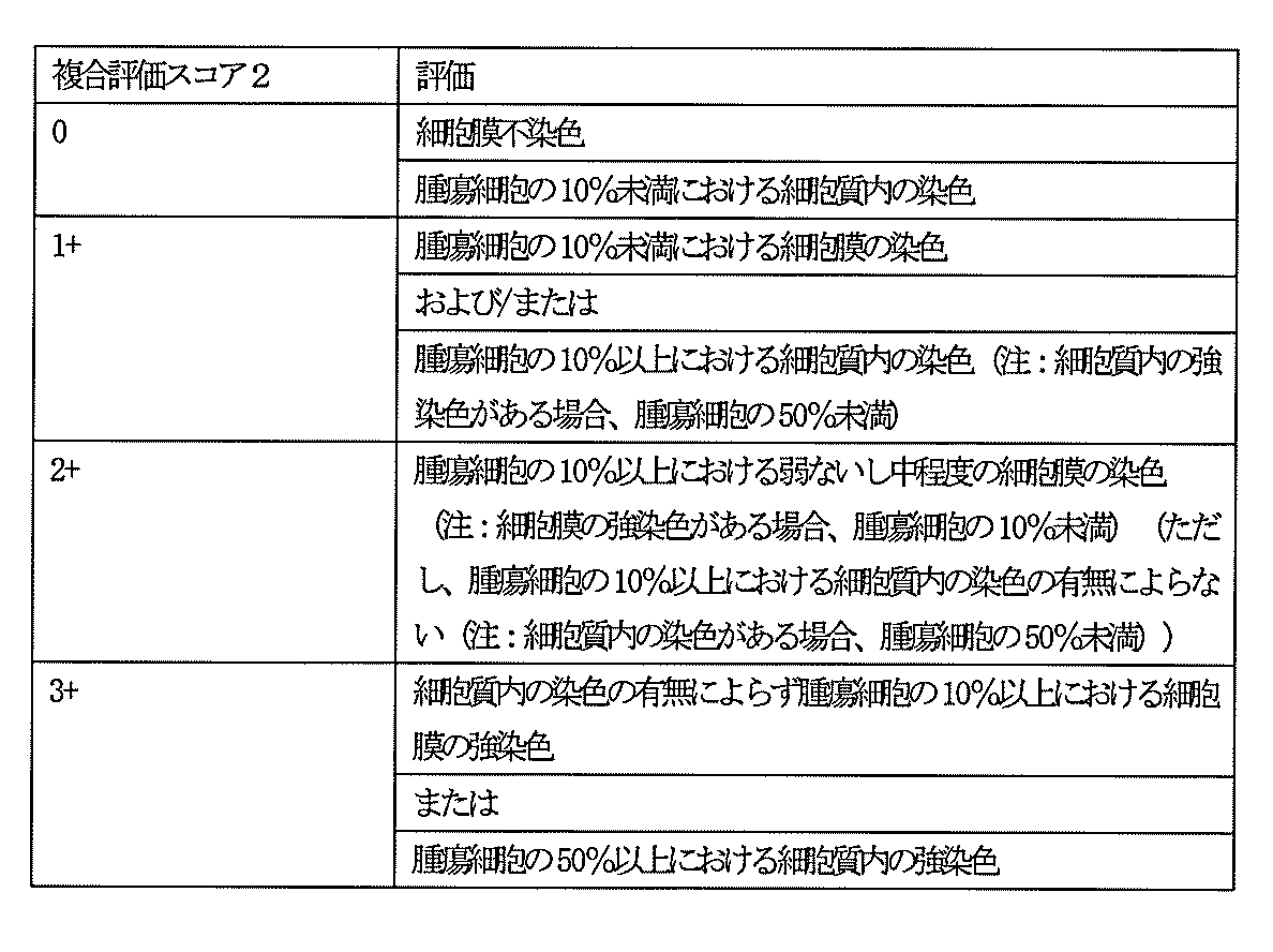
腫瘍細胞の密度は、組織標品中の、腫瘍組織における単位面積当たりの細胞数(すなわち腫瘍細胞数)をカウントすることにより決定することができる。また、腫瘍細胞のサイズは、組織標品中の、腫瘍組織の面積を、当該腫瘍組織内に存する細胞数(すなわち腫瘍細胞数)で除することにより決定することができる。GPC3を発現する腫瘍細胞の密度は、組織標品中の腫瘍組織における単位面積当たりのGPC3発現細胞数(すなわちGPC3を発現する腫瘍細胞数)をカウントすることにより決定することができる。また、GPC3を発現する腫瘍細胞のサイズは、組織標品中の、腫瘍細胞の面積の総和をGPC3発現細胞数(すなわちGPC3を発現する腫瘍細胞数)で除することにより決定することができる。この態様において、対象は、GPC3標的治療剤未治療例の患者とすることができる。
本明細書では、「免疫蛍光デジタルスライド」とは、蛍光標識された二次抗体が反応した免疫組織学像であって、コンピュータに取り込まれた画像を意味する。デジタル化してコンピュータに保存することにより破損や退色などの心配がないスライド(デジタルスライド)として、蛍光像を保存することができることが知られている。デジタルスライドでは、染色像の保存性の高さおよび染色度合いの定量性の高さから、非蛍光の染色組織像を保存することできる。本明細書では、デジタルスライドを「バーチャルスライド」と呼ぶことがある。本発明の好ましい態様においては、二次抗体反応が施された組織標品が免疫蛍光デジタルスライド(Immnoflorescent quantification digital slide、IQD)により評価され、当該組織標品中の腫瘍組織における単位面積あたりのGPC3発現量または1腫瘍細胞あたりのGPC3発現量が算出される。免疫蛍光デジタルスライドによる評価においては、二次抗体反応が施された組織標品を上記バーチャルスライドスキャナでスキャンし、組織標品からバーチャルスライドを作製する。作製されたバーチャルスライド(IQDイメージ)からGPC3の発現量を測定するために適した領域を選択し、当該領域における腫瘍組織の蛍光強度を測定する。蛍光強度の測定に用いるソフトウェアとしては、ViwePlus software(浜松ホトニクス)等が好適に用いられる。
本発明の非限定な一態様では、上記の方法によって検出された生物学的試料における1腫瘍細胞あたりのGPC3の発現量の測定に加えて、対象となる患者がFcγ受容体の遺伝子多型を有するかどうかを確認する工程を含む方法も提供される。本発明において、対象となる患者がFcγ受容体の遺伝子多型を有するかどうか確認する方法は、特に限定されない。例えば、対象となる患者から生物学的試料を採取し、採取した試料からゲノム遺伝子を単離してFcγ受容体に該当する遺伝子の塩基配列を決定することによって確認することができる。具体的には例えば、Journal of Clinical Oncology, vol.21, No.21 (2003) pp3940-3947に記載の方法に従って測定することができる。ここで採取される生物学的試料は、患者由来のゲノム遺伝子を取得することのできる試料であれば特に限定されず、例えば、末梢血、皮膚切片などが挙げられる。
本発明の非限定な一態様では、上記の方法によって検出された生物学的試料における1腫瘍細胞あたりのGPC3の発現量の測定に加えて、GPC3標的治療剤を投与される前の患者および/またはGPC3標的治療剤を投与された患者から単離された生物学的試料中の遊離GPC3濃度を測定する工程を含む方法も提供される。「GPC3標的治療剤を投与される前の患者」とは、癌であると診断された患者であって、GPC3標的治療剤を投与された経歴のない患者をいい、さらに、当該患者は、上述の組織における1腫瘍細胞あたりのGPC3の発現量からGPC3標的治療剤が有効であると決定されていてもよい。また、「GPC3標的治療剤を投与された患者」とは、GPC3標的治療剤を投与された経歴を有する患者をいう。
本発明において「GPC3標的治療剤」という用語は、GPC3により媒介されるシグナル経路を含めた、GPC3の生物学的活性の遮断、抑制、阻害、または低減をもたらす、またはGPC3を発現する細胞に対する細胞傷害をもたらすあらゆる分子をいう。「標的治療」という用語は、生物学的作用のある特定のメカニズムも示唆するものではなく、GPC3の薬理学的、生理学的、および生化学的相互作用のうち可能性のある作用をすべて含む概念である。GPC3標的治療剤の例として、(1)GPC3に結合するリガンドに対するGPC3の結合の拮抗阻害剤または非拮抗阻害剤、よってGPC3とそのリガンドとの結合に干渉する作用物質、(2)GPC3とそのリガンドとの結合には干渉しないが、その代わり、GPC3によるそのリガンドに対する結合によって生じる活性を阻害または減少させる作用物質、(3)GPC3の発現を減少させる作用物質、(4)GPC3を発現する細胞に対して細胞傷害活性を誘起することが可能な作用物質、が含まれる。リガンドの非限定な一態様として、wnt(Cancer Res. (2005) 65, 6245-6254)、IGF-II(Carcinogenesis (2008) 29 (7), 1319-1326)またはFibroblast Growth Factor 2(Int. J. Cancer (2003) 103 (4), 455-465)等が例示され得る。このような作用物質の非限定な一態様として、抗体(その抗原結合ドメインまたは断片を含む)、核酸分子(アンチセンスまたはRNAi分子等)、ペプチド、非ペプチド低分子有機物、免疫細胞等が含まれ得る。
本発明のGPC3標的治療剤は、1または2以上の抗がん剤を、同一の製剤または別々の製剤で、同時または別々に投与して併用してもよい。本発明のGPC3標的治療剤と併用するのに適当な抗がん剤は化学療法剤であり、好ましくはマルチキナーゼ阻害剤であり、さらに好ましくは、ソラフェニブまたはスニチニブである。抗GPC3標的治療剤と併用される抗がん剤は当該抗GPC3標的治療剤とは結合されていない。抗GPC3標的治療剤と抗がん剤は、両者がともに含有される配合剤の形で提供されてもよいし、両者が別々に提供され、同時に、別々に、または順次に使用されてもよい。さらに、GPC3標的治療剤と抗がん剤から構成されるキットとして提供されてもよい。
本発明のGPC3標的治療剤として使用される抗GPC3抗体の非限定な一態様として、(BioMosaic社よりカタログ番号B0134Rのもとに販売されている)1G12抗体(WO2003/100429)に細胞傷害性物質が連結された抗体薬物複合体(Antibody drug conjugate(ADC))(WO2007/137170)、または抗GPC3一本鎖抗体(single chain variable fragment; scFv)(CN103833852)等が例示され得る。
本発明の抗GPC3抗体として、細胞傷害活性を有する抗GPC3抗体が例示される。本発明において細胞傷害活性の非限定な例として、例えば抗体依存性細胞介在性細胞傷害(antibody-dependent cell-mediated cytotoxicity:ADCC)活性、補体依存性細胞傷害(complement-dependent cytotoxicity:CDC)活性およびT細胞による細胞傷害活性等が挙げられる。本発明において、CDC活性とは補体系による細胞傷害活性を意味する。一方ADCC活性とは、標的細胞の細胞膜に発現された膜型分子に結合する抗原結合ドメインを含む抗原結合分子のFc領域に、免疫細胞等が当該免疫細胞に発現したFcγレセプターを介して結合し、当該免疫細胞が標的細胞に傷害を与える活性を意味する。目的の抗原結合分子がADCC活性を有するか否か、又はCDC活性を有するか否かは公知の方法により測定され得る(例えば、Current protocols in Immunology, Chapter7. Immunologic studies in humans、Coliganら編(1993)等)。
(1)エフェクター細胞の調製
CBA/Nマウスなどから摘出された脾臓から、RPMI1640培地(Invitrogen)中で脾臓細胞が分離される。10%ウシ胎児血清(FBS、HyClone)を含む同培地で洗浄された当該脾臓細胞の濃度を5×106/mLに調製することによって、エフェクター細胞が調製され得る。
10% FBS含有培地(Invitrogen)によってBaby Rabbit Complement(CEDARLANE)を10倍に希釈することによって、補体溶液が調製され得る。
抗原を発現する細胞を0.2 mCiの51Cr-クロム酸ナトリウム(GEヘルスケアバイオサイエンス)とともに、10% FBS含有DMEM培地中で37℃にて1時間培養することにより該標的細胞が放射性標識され得る。放射性標識後、10% FBS含有RPMI1640培地にて3回洗浄された細胞の濃度を2×10⁵/mLに調製することによって、当該標的細胞が調製され得る。
また、本発明の抗GPC3抗体の非限定な一態様として、細胞傷害性物質が連結された抗GPC3抗体も例示される。そのような抗GPC3-抗体薬物複合体(Antibody drug conjugate(ADC))はWO2007/137170等に具体的に開示されているがこれに限定されない。すなわち、細胞傷害性物質としては、以下に例示される化学療法剤であってもよく、またAlleyら(Curr. Opin. Chem. Biol. (2010) 14, 529-537)やWO2009/140242に開示されている化合物であってもよく、これらの化合物が適切なリンカー等で抗原結合分子に結合される。
シュードモナスエキソトキシン(Pseudomonas Exotoxin)(Nature Medicine (1996) 2, 350-353);
リシンA鎖(Ricin A Chain)(Fultonら(J. Biol. Chem. (1986) 261, 5314-5319)、Sivamら(Cancer Res. (1987) 47, 3169-3173)、Cumberら、(J. Immunol. Methods (1990) 135,15-24、Wawrzynczakら(Cancer Res. (1990) 50, 7519-7562)、およびGheeiteら(J. Immunol.Methods (1991) 142,223-230));
無糖鎖リシンA鎖(Deglycosylated Ricin A Chain)(Thorpeら(Cancer Res. (1987) 47, 5924-5931));
アブリンA鎖(Abrin A Chain)(Wawrzynczakら(Br. J. Cancer (1992) 66, 361-366)、Wawrzynczakら(Cancer Res. (1990) 50, 7519-7562)、Sivamら(Cancer Res. (1987) 47, 3169-3173)、およびThorpeら(Cancer Res. (1987) 47, 5924-5931));
ゲロニン(Gelonin)(Sivamら(Cancer Res. (1987) 47, 3169-3173)、Cumberら(J. Immunol. Methods (1990) 135, 15-24)、Wawrzynczakら(Cancer Res., (1990) 50, 7519-7562)、およびBolognesiら(Clin. exp. Immunol. (1992) 89, 341-346));
ポークウイード抗ウィルス蛋白(PAP-s; Pokeweed anti-viral protein fromseeds)(Bolognesiら(Clin. exp. Immunol. (1992) 89, 341-346));
ブリオジン(Bryodin)(Bolognesiら(Clin. exp. Immunol. (1992) 89, 341-346));
サポリン(Saporin)(Bolognesiら(Clin. exp. Immunol. (1992) 89, 341-346));
モモルジン(Momordin)(Cumberら(J. Immunol. Methods (1990) 135, 15-24);Wawrzynczakら(Cancer Res. (1990) 50, 7519-7562)、およびBolognesiら(Clin. exp. Immunol. (1992) 89, 341-346));
モモルコキン(Momorcochin)(Bolognesiら(Clin. exp. Immunol. (1992) 89, 341-346));
ジアンシン32(Dianthin 32)(Bolognesiら(Clin. exp. Immunol. (1992) 89, 341-346));
ジアンシン30(Dianthin 30)(Stirpe F., Barbieri L.(FEBS letter (1986) 195, 1-8));
モデッシン(Modeccin)(Stirpe F., Barbieri L.(FEBS letter (1986) 195, 1-8));
ビスカミン(Viscumin)(Stirpe F., Barbieri L.(FEBS letter (1986) 195, 1-8));
ボルケシン(Volkensin)(Stirpe F., Barbieri L.(FEBS letter (1986) 195, 1-8));
ドデカンドリン(Dodecandrin)(Stirpe F., Barbieri L.(FEBS letter (1986) 195, 1-8));
トリチン(Tritin)(Stirpe F., Barbieri L.(FEBS letter (1986) 195, 1-8));
ルフィン(Luffin)(Stirpe F., Barbieri L.(FEBS letter (1986) 195, 1-8));および
トリコキリン(Trichokirin)(Casellasら(Eur. J. Biochem. (1988) 176, 581-588)、およびBolognesiら(Clin. exp. Immunol., (1992) 89, 341-346))。
本発明の抗GPC3抗体に含まれる定常領域に含まれるFc領域は、ヒトIgGから取得され得るが、IgGの特定のサブクラスに限定されるものでもない。Fc領域とは、EUナンバリングで表されるおよそ216位のアミノ酸における、パパイン切断部位のヒンジ領域のN末端から、当該ヒンジ、CH2およびCH3ドメインを含める抗体の重鎖定常領域をいう。当該Fc領域の好適な例として、後述されるようにFcγレセプターに対する結合活性を有するFc領域が挙げられる。そのようなFc領域の非限定な一態様として、ヒトIgG1(配列番号:74)、IgG2(配列番号:75)、IgG3(配列番号:76)、またはIgG4(配列番号:77)で表される定常領域に含まれるFc領域が例示される。
Fcγレセプター(FcγRとも記載される)とは、IgG1、IgG2、IgG3、IgG4モノクローナル抗体のFc領域に結合し得るレセプターをいい、実質的にFcγレセプター遺伝子にコードされるタンパク質のファミリーのいかなるメンバーをも意味する。ヒトでは、このファミリーには、アイソフォームFcγRIa、FcγRIbおよびFcγRIcを含むFcγRI(CD64);アイソフォームFcγRIIa(アロタイプH131およびR131を含む。 即ち、FcγRIIa (H)およびFcγRIIa (R))、FcγRIIb(FcγRIIb-1およびFcγRIIb-2を含む)およびFcγRIIcを含むFcγRII(CD32);およびアイソフォームFcγRIIIa(アロタイプV158およびF158を含む。即ち、FcγRIIIa (V)およびFcγRIIIa (F))およびFcγRIIIb(アロタイプFcγRIIIb-NA1およびFcγRIIIb-NA2を含む)を含むFcγRIII(CD16)、ならびにいかなる未発見のヒトFcγR類またはFcγRアイソフォームまたはアロタイプも含まれるが、これらに限定されるものではない。FcγRは、ヒト、マウス、ラット、ウサギおよびサルを含むが、これらに限定されるものではない、いかなる生物由来でもよい。マウスFcγR類には、FcγRI(CD64)、FcγRII(CD32)、FcγRIII(CD16)およびFcγRIII-2(FcγRIV、CD16-2)、並びにいかなる未発見のマウスFcγR類またはFcγRアイソフォームまたはアロタイプも含まれるが、これらに限定されない。こうしたFcγレセプターの好適な例としてはヒトFcγRI(CD64)、FcγRIIa(CD32)、FcγRIIb(CD32)、FcγRIIIa(CD16)及び/又はFcγRIIIb(CD16)が挙げられる。ヒトFcγRIのポリペプチド配列は配列番号:78(NP_000557.1)に、ヒトFcγRIIa(アロタイプH131)のポリペプチド配列は配列番号:79(AAH20823.1)に(アロタイプR131は配列番号:79の166番目のアミノ酸がArgに置換されている配列である)、FcγRIIbのポリペプチド配列は配列番号:80(AAI46679.1)に、FcγRIIIaのポリペプチド配列は配列番号:81(AAH33678.1)に、およびFcγRIIIbのポリペプチド配列は、配列番号:82(AAI28563.1)に記載されている(カッコ内はRefSeq等のデータベース登録番号を示す)。Fcγレセプターが、IgG1、IgG2、IgG3、IgG4モノクローナル抗体のFc領域に結合活性を有するか否かは、FACSやELISAフォーマットのほか、ALPHAスクリーン(Amplified Luminescent Proximity Homogeneous Assay)や表面プラズモン共鳴(SPR)現象を利用したBIACORE法等の公知の方法によって確認され得る(Proc. Natl. Acad. Sci. U.S.A. (2006) 103 (11), 4005-4010)。
前述されるように、本発明の抗GPC3抗体に含まれるFc領域として、Fcγレセプターに対する結合活性を有するFc領域が挙げられる。そのようなFc領域の非限定な一態様として、ヒトIgG1(配列番号:74)、IgG2(配列番号:75)、IgG3(配列番号:76)、またはIgG4(配列番号:77)で表される定常領域に含まれるFc領域が例示される。Fcγレセプターが、IgG1、IgG2、IgG3、IgG4モノクローナル抗体のFc領域に結合活性を有するか否かは、FACSやELISAフォーマットのほか、ALPHAスクリーン(Amplified Luminescent Proximity Homogeneous Assay)や表面プラズモン共鳴(SPR)現象を利用したBIACORE法等の公知の方法によって確認され得る(Proc. Natl. Acad. Sci. U.S.A. (2006) 103 (11), 4005-4010)。
本発明の抗GPC3抗体が含むFc領域として、ヒトIgG1(配列番号:74)、IgG2(配列番号:75)、IgG3(配列番号:76)、またはIgG4(配列番号:77)で表される定常領域に含まれるFc領域のほかに、天然型ヒトIgGのFc領域のFcγレセプターに対する結合活性よりもFcγレセプターに対する結合活性が高いFcγR結合改変Fc領域も適宜使用され得る。本明細書において、「天然型ヒトIgGのFc領域」とは、配列番号:74、75、76または77で例示されるヒトIgG1、IgG2、IgG3またはIgG4の定常領域に含まれるFc領域のEUナンバリング297位に結合した糖鎖がフコース含有糖鎖であるFc領域を意味する。そのようなFcγR結合改変Fc領域は、天然型ヒトIgGのFc領域のアミノ酸を改変することによって作製され得る。FcγR結合改変Fc領域のFcγRに対する結合活性が、天然型ヒトIgGのFc領域のFcγRに対する結合活性より高いか否かは、前記のFACSやELISAフォーマットのほか、ALPHAスクリーン(Amplified Luminescent Proximity Homogeneous Assay)や表面プラズモン共鳴(SPR)現象を利用したBIACORE法等の公知の方法を用いて適宜実施され得る。
221位のアミノ酸がLysまたはTyrのいずれか、
222位のアミノ酸がPhe、Trp、GluまたはTyrのいずれか、
223位のアミノ酸がPhe、Trp、GluまたはLysのいずれか、
224位のアミノ酸がPhe、Trp、GluまたはTyrのいずれか、
225位のアミノ酸がGlu、LysまたはTrpのいずれか、
227位のアミノ酸がGlu、Gly、LysまたはTyrのいずれか、
228位のアミノ酸がGlu、Gly、LysまたはTyrのいずれか、
230位のアミノ酸がAla、Glu、GlyまたはTyrのいずれか、
231位のアミノ酸がGlu、Gly、Lys、ProまたはTyrのいずれか、
232位のアミノ酸がGlu、Gly、LysまたはTyrのいずれか、
233位のアミノ酸がAla、Asp、Phe、Gly、His、Ile、Lys、Leu、Met、Asn、Gln、Arg、Ser、Thr、Val、TrpまたはTyrのいずれか、
234位のアミノ酸がAla、Asp、Glu、Phe、Gly、His、Ile、Lys、Met、Asn、Pro、Gln、Arg、Ser、Thr、Val、TrpまたはTyrのいずれか、
235位のアミノ酸がAla、Asp、Glu、Phe、Gly、His、Ile、Lys、Met、Asn、Pro、Gln、Arg、Ser、Thr、Val、TrpまたはTyrのいずれか、
236位のアミノ酸がAla、Asp、Glu、Phe、His、Ile、Lys、Leu、Met、Asn、Pro、Gln、Arg、Ser、Thr、Val、TrpまたはTyrのいずれか、
237位のアミノ酸がAsp、Glu、Phe、His、Ile、Lys、Leu、Met、Asn、Pro、Gln、Arg、Ser、Thr、Val、TrpまたはTyrのいずれか、
238位のアミノ酸がAsp、Glu、Phe、Gly、His、Ile、Lys、Leu、Met、Asn、Gln、Arg、Ser、Thr、Val、TrpまたはTyrのいずれか、
239位のアミノ酸がAsp、Glu、Phe、Gly、His、Ile、Lys、Leu、Met、Asn、Pro、Gln、Arg、Thr、Val、TrpまたはTyrのいずれか、
240位のアミノ酸がAla、Ile、MetまたはThrのいずれか、
241位のアミノ酸がAsp、Glu、Leu、Arg、TrpまたはTyrのいずれか、
243位のアミノ酸がLeu、Glu、Leu、Gln、Arg、TrpまたはTyrのいずれか、
244位のアミノ酸がHis、
245位のアミノ酸がAla、
246位のアミノ酸がAsp、Glu、HisまたはTyrのいずれか、
247位のアミノ酸がAla、Phe、Gly、His、Ile、Leu、Met、Thr、ValまたはTyrのいずれか、
249位のアミノ酸がGlu、His、GlnまたはTyrのいずれか、
250位のアミノ酸がGluまたはGlnのいずれか、
251位のアミノ酸がPhe、
254位のアミノ酸がPhe、MetまたはTyrのいずれか、
255位のアミノ酸がGlu、LeuまたはTyrのいずれか、
256位のアミノ酸がAla、MetまたはProのいずれか、
258位のアミノ酸がAsp、Glu、His、SerまたはTyrのいずれか、
260位のアミノ酸がAsp、Glu、HisまたはTyrのいずれか、
262位のアミノ酸がAla、Glu、Phe、IleまたはThrのいずれか、
263位のアミノ酸がAla、Ile、MetまたはThrのいずれか、
264位のアミノ酸がAsp、Glu、Phe、Gly、His、Ile、Lys、Leu、Met、Asn、Pro、Gln、Arg、Ser、Thr、TrpまたはTyrのいずれか、
265位のアミノ酸がAla、Leu、Phe、Gly、His、Ile、Lys、Leu、Met、Asn、Pro、Gln、Arg、Ser、Thr、Val、TrpまたはTyrのいずれか、
266位のアミノ酸がAla、Ile、MetまたはThrのいずれか、
267位のアミノ酸がAsp、Glu、Phe、His、Ile、Lys、Leu、Met、Asn、Pro、Gln、Arg、Thr、Val、TrpまたはTyrのいずれか、
268位のアミノ酸がAsp、Glu、Phe、Gly、Ile、Lys、Leu、Met、Pro、Gln、Arg、Thr、ValまたはTrpのいずれか、
269位のアミノ酸がPhe、Gly、His、Ile、Lys、Leu、Met、Asn、Pro、Arg、Ser、Thr、Val、TrpまたはTyrのいずれか、
270位のアミノ酸がGlu、Phe、Gly、His、Ile、Leu、Met、Pro、Gln、Arg、Ser、Thr、TrpまたはTyrのいずれか、
271位のアミノ酸がAla、Asp、Glu、Phe、Gly、His、Ile、Lys、Leu、Met、Asn、Gln、Arg、Ser、Thr、Val、TrpまたはTyrのいずれか、
272位のアミノ酸がAsp、Phe、Gly、His、Ile、Lys、Leu、Met、Pro、Arg、Ser、Thr、Val、TrpまたはTyrのいずれか、
273位のアミノ酸がPheまたはIleのいずれか、
274位のアミノ酸がAsp、Glu、Phe、Gly、His、Ile、Leu、Met、Asn、Pro、Arg、Ser、Thr、Val、TrpまたはTyrのいずれか、
275位のアミノ酸がLeuまたはTrpのいずれか、
276位のアミノ酸が、Asp、Glu、Phe、Gly、His、Ile、Leu、Met、Pro、Arg、Ser、Thr、Val、TrpまたはTyrのいずれか、
278位のアミノ酸がAsp、Glu、Gly、His、Ile、Lys、Leu、Met、Asn、Pro、Gln、Arg、Ser、Thr、ValまたはTrpのいずれか、
279位のアミノ酸がAla、
280位のアミノ酸がAla、Gly、His、Lys、Leu、Pro、Gln、TrpまたはTyrのいずれか、
281位のアミノ酸がAsp、Lys、ProまたはTyrのいずれか、
282位のアミノ酸がGlu、Gly、Lys、ProまたはTyrのいずれか、
283位のアミノ酸がAla、Gly、His、Ile、Lys、Leu、Met、Pro、ArgまたはTyrのいずれか、
284位のアミノ酸がAsp、Glu、Leu、Asn、ThrまたはTyrのいずれか、
285位のアミノ酸がAsp、Glu、Lys、Gln、TrpまたはTyrのいずれか、
286位のアミノ酸がGlu、Gly、ProまたはTyrのいずれか、
288位のアミノ酸がAsn、Asp、GluまたはTyrのいずれか、
290位のアミノ酸がAsp、Gly、His、Leu、Asn、Ser、Thr、TrpまたはTyrのいずれか、
291位のアミノ酸がAsp、Glu、Gly、His、Ile、GlnまたはThrのいずれか、
292位のアミノ酸がAla、Asp、Glu、Pro、ThrまたはTyrのいずれか、
293位のアミノ酸がPhe、Gly、His、Ile、Leu、Met、Asn、Pro、Arg、Ser、Thr、Val、TrpまたはTyrのいずれか、
294位のアミノ酸がPhe、Gly、His、Ile、Lys、Leu、Met、Asn、Pro、Arg、Ser、Thr、Val、TrpまたはTyrのいずれか、
295位のアミノ酸がAsp、Glu、Phe、Gly、His、Ile、Lys、Met、Asn、Pro、Arg、Ser、Thr、Val、TrpまたはTyrのいずれか、
296位のアミノ酸がAla、Asp、Glu、Gly、His、Ile、Lys、Leu、Met、Asn、Gln、Arg、Ser、ThrまたはValのいずれか、
297位のアミノ酸がAsp、Glu、Phe、Gly、His、Ile、Lys、Leu、Met、Pro、Gln、Arg、Ser、Thr、Val、TrpまたはTyrのいずれか、
298位のアミノ酸がAla、Asp、Glu、Phe、His、Ile、Lys、Met、Asn、Gln、Arg、Thr、Val、TrpまたはTyrのいずれか、
299位のアミノ酸がAla、Asp、Glu、Phe、Gly、His、Ile、Lys、Leu、Met、Asn、Pro、Gln、Arg、Ser、Val、TrpまたはTyrのいずれか、
300位のアミノ酸がAla、Asp、Glu、Gly、His、Ile、Lys、Leu、Met、Asn、Pro、Gln、Arg、Ser、Thr、ValまたはTrpのいずれか、
301位のアミノ酸がAsp、Glu、HisまたはTyrのいずれか、
302位のアミノ酸がIle、
303位のアミノ酸がAsp、GlyまたはTyrのいずれか、
304位のアミノ酸がAsp、His、Leu、AsnまたはThrのいずれか、
305位のアミノ酸がGlu、Ile、ThrまたはTyrのいずれか、
311位のアミノ酸がAla、Asp、Asn、Thr、ValまたはTyrのいずれか、
313位のアミノ酸がPhe、
315位のアミノ酸がLeu、
317位のアミノ酸がGluまたはGln、
318位のアミノ酸がHis、Leu、Asn、Pro、Gln、Arg、Thr、ValまたはTyrのいずれか、
320位のアミノ酸がAsp、Phe、Gly、His、Ile、Leu、Asn、Pro、Ser、Thr、Val、TrpまたはTyrのいずれか、
322位のアミノ酸がAla、Asp、Phe、Gly、His、Ile、Pro、Ser、Thr、Val、TrpまたはTyrのいずれか、
323位のアミノ酸がIle、
324位のアミノ酸がAsp、Phe、Gly、His、Ile、Leu、Met、Pro、Arg、Thr、Val、TrpまたはTyrのいずれか、
325位のアミノ酸がAla、Asp、Glu、Phe、Gly、His、Ile、Lys、Leu、Met、Pro、Gln、Arg、Ser、Thr、Val、TrpまたはTyrのいずれか、
326位のアミノ酸がAla、Asp、Glu、Gly、Ile、Leu、Met、Asn、Pro、Gln、Ser、Thr、Val、TrpまたはTyrのいずれか、
327位のアミノ酸がAla、Asp、Glu、Phe、Gly、His、Ile、Lys、Leu、Met、Asn、Pro、Arg、Thr、Val、TrpまたはTyrのいずれか、
328位のアミノ酸がAla、Asp、Glu、Phe、Gly、His、Ile、Lys、Met、Asn、Pro、Gln、Arg、Ser、Thr、Val、TrpまたはTyrのいずれか、
329位のアミノ酸がAsp、Glu、Phe、Gly、His、Ile、Lys、Leu、Met、Asn、Gln、Arg、Ser、Thr、Val、TrpまたはTyrのいずれか、
330位のアミノ酸がCys、Glu、Phe、Gly、His、Ile、Lys、Leu、Met、Asn、Pro、Arg、Ser、Thr、Val、TrpまたはTyrのいずれか、
331位のアミノ酸がAsp、Phe、His、Ile、Leu、Met、Gln、Arg、Thr、Val、TrpまたはTyrのいずれか、
332位のアミノ酸がAla、Asp、Glu、Phe、Gly、His、Lys、Leu、Met、Asn、Pro、Gln、Arg、Ser、Thr、Val、TrpまたはTyrのいずれか、
333位のアミノ酸がAla、Asp、Glu、Phe、Gly、His、Ile、Leu、Met、Pro、Ser、Thr、ValまたはTyrのいずれか、
334位のアミノ酸がAla、Glu、Phe、Ile、Leu、ProまたはThrのいずれか、
335位のアミノ酸がAsp、Phe、Gly、His、Ile、Leu、Met、Asn、Pro、Arg、Ser、Val、TrpまたはTyrのいずれか、
336位のアミノ酸がGlu、LysまたはTyrのいずれか、
337位のアミノ酸がGlu、HisまたはAsnのいずれか、
339位のアミノ酸がAsp、Phe、Gly、Ile、Lys、Met、Asn、Gln、Arg、SerまたはThrのいずれか、
376位のアミノ酸がAlaまたはValのいずれか、
377位のアミノ酸がGlyまたはLysのいずれか、
378位のアミノ酸がAsp、
379位のアミノ酸がAsn、
380位のアミノ酸がAla、AsnまたはSerのいずれか、
382位のアミノ酸がAlaまたはIleのいずれか、
385位のアミノ酸がGlu、
392位のアミノ酸がThr、
396位のアミノ酸がLeu、
421位のアミノ酸がLys、
427位のアミノ酸がAsn、
428位のアミノ酸がPheまたはLeuのいずれか、
429位のアミノ酸がMet、
434位のアミノ酸がTrp、
436位のアミノ酸がIle、もしくは
440位のアミノ酸がGly、His、Ile、LeuまたはTyrのいずれか、
の群から選択される少なくともひとつ一つ以上のアミノ酸の改変が挙げられる。また、改変されるアミノ酸の数は特に限定されず、一箇所のみのアミノ酸が改変され得るし、二箇所以上のアミノ酸が改変され得る。二箇所以上のアミノ酸の改変の組合せとしては、例えば表3(表3-1~表3-3)に記載されるような組合せが挙げられる。また、天然型ヒトIgGのFc領域のFcγレセプターに対する結合活性よりもFcγレセプターに対する結合活性が高いFcγR結合改変Fc領域(FcγR結合改変Fc領域)を含む抗GPC3抗体の具体的な例が、WO2007/047291に記載されている。


前記したように、活性型Fcγレセプターとしては、FcγRIa、FcγRIbおよびFcγRIcを含むFcγRI(CD64)、 FcγRIIaならびにアイソフォームFcγRIIIa(アロタイプV158およびF158を含む)およびFcγRIIIb(アロタイプFcγRIIIb-NA1およびFcγRIIIb-NA2を含む)を含むFcγRIII(CD16)が好適に挙げられる。また、FcγRIIb(FcγRIIb-1およびFcγRIIb-2を含む)が抑制型Fcγレセプターの好適な例として挙げられる。
本発明が提供する抗GPC3抗体に含まれるFc領域として、Fc領域に結合した糖鎖の組成がフコース欠損糖鎖を結合したFc領域の割合が高くなるように、またはバイセクティングN-アセチルグルコサミンが付加したFc領域の割合が高くなるように修飾されたFc領域も含まれ得る。抗体Fc領域に結合するN -グリコシド結合複合型糖鎖還元末端のN -アセチルグルコサミンからフコース残基を除去すると、FcγRIIIaに対する親和性が増強されることが知られている(Satoら(Expert Opin. Biol. Ther. (2006) 6 (11), 1161-1173))。こうしたFc領域を含むIgG1抗体は、上記ADCC活性が増強されていることが知られていることから、当該Fc領域を含む抗原結合分子は、本発明の医薬組成物に含まれる抗原結合分子としても有用である。抗体Fc領域に結合するN -グリコシド結合複合型糖鎖還元末端のN -アセチルグルコサミンからフコース残基が除去された抗体としては、例えば、次のような抗体;-グリコシル化が修飾された抗体(国際公開WO1999/054342等)、-糖鎖に付加するフコースが欠損した抗体(国際公開WO2000/061739、WO2002/031140、WO2006/067913等)、が挙げられる。また、Fc領域に結合した糖鎖の組成がフコース欠損糖鎖を結合したFc領域の割合が高くなるように、またはバイセクティングN-アセチルグルコサミンが付加したFc領域の割合が高くなるように修飾されたFc領域を含む抗GPC3抗体の具体的な例が、WO2006/046751およびWO2009/ 041062に記載されている。
本発明で使用される抗GPC3抗体の非限定な一態様として、その等電点(pI)が変化するように、抗GPC3抗体のアミノ酸残基が改変されている抗GPC3抗体も例示される。本発明が提供する抗GPC3抗体における「アミノ酸残基の電荷の改変」の好適な例としては以下のものが挙げられる。pI値を増加させる改変としては、例えば、配列番号:50で表される抗GPC3抗体の重鎖可変領域中のKabatナンバリングで表される43位のQのKへの置換、52位のDのNへの置換、105位のQのRへの置換から選択される少なくとも一つの置換を行うことができ、例えば配列番号:67で表されるアミノ酸配列に改変される。また、例えば、配列番号:51または配列番号:66で表される抗GPC3抗体の軽鎖可変領域中のKabatナンバリングで表される17位のEのQへの置換、27位のQのRへの置換、105位のQのRへの置換から選択される少なくとも一つの置換を行うことができ、例えば配列番号:68で表されるアミノ酸配列に改変される。一方、pI値を減少させる改変としては、配列番号:50で表される抗GPC3抗体の重鎖可変領域中のKabatナンバリングで表される19位のKのTへの置換、43位のQのEへの置換、61位のGのEへの置換、62位のKのSへの置換、64位のKのQへの置換、65位のGのDへの置換から選択される少なくとも一つの置換を行うことができ、例えば配列番号:69、配列番号:71で表されるアミノ酸配列に改変される。また、例えば、配列番号:51または配列番号:66で表される抗GPC3抗体の軽鎖可変領域中のKabatナンバリングで表される24位のRのQへの置換、27位のQのEへの置換、74位のKのTへの置換、77位のRのSへの置換、107位のKのEへの置換から選択される少なくとも一つの置換を行うことができ、例えば配列番号:70、配列番号:72、配列番号:73で表されるアミノ酸配列に改変される。さらに、pI値を減少させる改変としては、配列番号:74で表される重鎖定常領域中においてEUナンバリングで表される268位、274位、355位、356位、358位、419位から選択されるアミノ酸の少なくとも一つの置換が挙げられる。これらの置換は、例えば、配列番号31で表される重鎖定常領域の、EUナンバリングで表される268位のHのQへの置換、274位のKのQへの置換、355位のRのQへの置換、356位のDのEへの置換、358位のLのMへの置換、419位のQのEへの置換、から選択される少なくとも一つの置換が好適な例として挙げられ得る。これらの置換の結果、ヒト抗体のIgG1の定常領域とIgG4の定常領域とのキメラ体が構築される。すなわち、当該置換によって、改変された抗体の免疫原性に影響を及ぼすことなく、所望のpIを有する抗体の作製が可能となる。
配列番号:74、75、76または77で表されるIgGの定常領域におけるEUナンバリングで表される446位のGlyおよび447位のLysが欠損したIgG定常領域も本発明の抗GPC3抗体に含まれる定常領域として使用され得る。これらのアミノ酸を両方欠損させることにより、抗体の重鎖定常領域の末端に由来するヘテロジェニティーを低減することが可能である。
ポリペプチドの翻訳後修飾とは、ポリペプチドの生合成において翻訳されたポリペプチドに対する化学修飾をいう。抗体の一次構造はポリペプチドから形成されるため、本発明の抗GPC3抗体には、抗GPC3抗体の一次構造であるポリペプチドの翻訳後修飾を受けた修飾物も含まれる。ポリペプチドの翻訳後修飾には大きく、官能基付加、ポリペプチドまたはペプチドの付加(ISG化、SUMO化、ユビキチン化)、アミノ酸の化学的性質の変換(シリル化または脱アミン、脱アミド)、および構造変換(ジスルフィド化、プロテアーゼ分解)に分類することができる。本発明における翻訳後修飾の非限定な一態様として、ポリペプチドの形成単位であるアミノ酸残基に対するペプチド付加または官能基付加が挙げられる。かかる修飾として具体的には、リン酸化(セリン、スレオニン、チロシン、アスパラギン酸等)、グルコシル化(セリン、スレオニン、アスパラギン酸等)、アシル化(リシン)、アセチル化(リシン)、ヒドロキシル化(リシン、プロリン)、プレニル化(システイン)、パルミトイル化(システイン)、アルキル化(リシン、アルギニン)、ポリグルタミル化(グルタミン酸)、カルボキシル化(グルタミン酸)、ポリグリシル化(グルタミン酸)、シトルリン化(アルギニン)、スクシンイミド形成(アスパラギン酸)等を挙げることができる。例えば、N末端のグルタミンのピログルタミル化によるピログルタミン酸への修飾を受けた抗GPC3抗体も当然のことながら本発明の抗GPC3抗体に含まれる。また、例えば、二個の硫黄原子の間に形成される共有結合を意味する「ジスルフィド結合」によって重鎖と軽鎖、または重鎖同士が連結された抗GPC3抗体の翻訳後修飾物も、本発明の抗GPC3抗体に含まれる。アミノ酸のシステインに含まれるチオール基は、第二のチオール基とジスルフィド結合または架橋を形成することができる。一般的にIgG分子では、CH1およびCL領域がジスルフィド結合によって連結され、重鎖を形成する二つのポリペプチドは、EUナンバリングで表される226位および229位のシステイン残基間のジスルフィド結合によって連結される。こうしたジスルフィド結合によって連結された抗GPC3抗体の翻訳後修飾物も、本発明の抗GPC3抗体に含まれる。
「癌に対するGPC3標的治療剤の有効性」または「癌に対するGPC3標的治療剤が有効である」という用語は、癌であると診断された患者において、GPC3標的治療剤が、所望のまたは有益な効果を生じさせることをいう。所望のまたは有益な効果には、(1)癌細胞のさらなる成長または拡散の阻害、(2)癌細胞の死滅、(3)癌の再発の阻害、(4)癌に関連する症候(痛みなど)の緩和、低減、軽減、阻害、もしくは前記症候の頻度の低減、または(5)患者の生存率の改善が含まれ得る。癌の再発の阻害には、放射線、化学療法、外科手術、または他の技術によりこれまでに治療されている、癌の原発部位および周辺組織での癌の成長の阻害、ならびに新たな遠位部位での癌の成長の不存在が含まれる。所望のまたは有益な効果は、患者により知覚されるものまたは他覚的なもののいずれかであり得る。例えば、患者がヒトである場合、ヒトは、改善または療法に対する応答の、患者により知覚される兆候として、気力もしくは活力の改善または痛みの減少を認識し得る。あるいは、臨床医は、身体的検査、実験的パラメーター、腫瘍マーカー、またはX線撮影による所見に基づいて、腫瘍サイズまたは腫瘍組織量の減少に気付き得る。治療に対する応答について臨床医が観察し得るいくつかの実験的兆候には、白血球数、赤血球数、血小板数、赤血球沈降速度、および様々な酵素レベルなどの、試験結果の正常化が含まれる。さらに、臨床医は、検出可能な腫瘍マーカーの減少を観察し得る。あるいは、他覚的な改善を評価するために、超音波診断、核磁気共鳴試験、および陽電子放出試験などの他の試験を用いることができる。
本発明の非限定な一態様では、GPC3標的治療剤を投与される前の患者から単離された生物学的試料中の1腫瘍細胞あたりのGPC3の発現量を測定し、当該発現量が所定の値である場合に当該GPC3標的治療剤が有効であると決定する方法が提供される。「GPC3標的治療剤を投与される前の患者」とは、癌であると診断された患者であって、上述のGPC3標的治療剤を投与された経歴のない患者をいい、さらに、当該患者は、上述の腫瘍組織におけるGPC3の総発現量または単位面積あたりの発現量からGPC3標的治療剤が有効であると決定されていてもよいし、上述の腫瘍組織におけるGPC3の総発現量または単位面積あたりの発現量からGPC3標的治療剤が有効でないと決定されていてもよい。本発明のある態様では、例えば、GPC3標的治療剤が有効でないと決定された患者から、1腫瘍細胞あたりのGPC3発現量を併せて考慮することにより、現実にはGPC3標的治療剤が有効である患者を抽出することが可能である。GPC3標的治療剤の投与経路は、投与されるGPC3標的治療剤の性状等に併せて好適な投与経路が適宜選択され得る。例えば、投与経路の例として非経口投与が例示される。非経口投与のさらなる例として、例えば、注射投与、経鼻投与、経肺投与、経皮投与が例示される。例えば、注射投与のさらなる例として、静脈内注射投与、筋肉内注射投与、腹腔内注射投与、皮下注射投与などによる全身または局部的な投与が例示される。
上記の、当該1腫瘍細胞あたりのGPC3の発現量が所定の値である場合に、当該GPC3標的治療剤が有効であると決定する際に、さらに、遊離GPC3濃度を併せて考慮することも可能である。すなわち、GPC3標的治療剤を投与される前の患者から単離された生物学的試料中の遊離GPC3濃度を測定し、当該遊離GPC3濃度が所定の値である場合に当該GPC3標的治療剤が有効であると決定することも含まれる。
本発明における患者の選別方法は、上述の方法により、患者から単離された生物学的試料中の1腫瘍細胞あたりのGPC3の発現量が所定の値である場合にGPC3標的治療剤が有効な患者であると決定する工程を含む。本発明の非限定な一態様において、上記の患者の選別方法は、さらに、患者から単離された生物学的試料中の遊離GPC3濃度が所定の値である場合にGPC3標的治療剤が有効な患者であると決定する工程を含むこともできる。また、別の異なる非限定な一態様において、上記の患者の選別方法は、さらに、GPC3標的治療剤を投与される前の患者および/またはGPC3標的治療剤を投与された患者から単離された生物学的試料中の遊離GPC3濃度をモニタリングし、当該遊離GPC3濃度が所定の値である場合に当該GPC3標的治療剤の投与の継続を決定する工程を含むこともできる。
本発明において治療剤とは、通常、疾患の治療もしくは予防、または検査・診断のための薬剤をいう。また、本発明において、「GPC3標的治療剤を投与される前の癌患者から単離された生物学的試料中の1腫瘍細胞あたりのGPC3の発現量が所定の値である患者(のみ)に投与するためのGPC3標的治療剤」との用語は、「GPC3標的治療剤を受ける前の癌患者から単離された生物学的試料中の1腫瘍細胞あたりのGPC3の発現量が所定の値である患者(のみ)にGPC3標的治療剤を投与することを含む癌の治療方法」と言い換えることも可能であるし、「癌を治療するための医薬の製造における、GPC3標的治療剤を投与される前の癌患者から単離された生物学的試料中の1腫瘍細胞あたりのGPC3の発現量が所定の値である患者(のみ)に投与するためのGPC3標的治療剤の使用」と言い換えることも可能である。この文脈において、用語「患者のみ」とは、生物学的試料中の1腫瘍細胞あたりのGPC3の発現量が所定の値である患者には投与するが、所定の値でない患者には投与しないことを意味する。本発明の好ましいある特定の態様では、例えば、GPC3標的治療剤を投与される前の癌患者から単離された生物学的試料中の1腫瘍細胞あたりのGPC3発現量が所定の値である患者にはGPC3標的治療剤を投与するが、所定の値ではない患者に対しては投与しないことができる(例えば、生物学的試料中の1腫瘍細胞あたりのGPC3発現量が一定値以上であれば投与するが、一定値未満の場合には投与しない)。
本発明の製剤には、GPC3標的治療剤を投与される前の癌患者から単離された生物学的試料中の1腫瘍細胞あたりのGPC3の発現量が所定の値である患者(のみ)に投与する旨の指示書が含まれる。また、本発明における肝癌の治療用キットには、GPC3標的治療剤、およびGPC3標的治療剤を投与される前の癌患者から単離された生物学的試料中の1腫瘍細胞あたりのGPC3の発現量が所定の値である患者(のみ)に投与する旨の指示書が含まれる。この文脈において、用語「患者のみ」とは、生物学的試料中の1腫瘍細胞あたりのGPC3の発現量が所定の値である患者には投与するが、所定の値でない患者には投与しないことを意味する。本発明の好ましいある特定の態様では、指示書は、例えば、GPC3標的治療剤を投与される前の癌患者から単離された生物学的試料中の1腫瘍細胞あたりのGPC3発現量が所定の値である患者には投与するが、所定の値ではない患者に対しては投与しないという趣旨の記載がなされた指示書とすることができる(例えば、1腫瘍細胞あたりのGPC3発現量が一定値以上であれば投与するが、一定値未満の場合には投与しないという趣旨の記載としてもよい)。
本発明の診断キットには、GPC3標的治療剤を投与される前の癌患者から単離された生物学的試料中の1腫瘍細胞あたりのGPC3の発現量を測定するための試薬が含まれる。
GC33は、ヒトGPC3に高い親和性をもって結合する遺伝子組換えヒト化IgG1モノクローナル抗体である(WO2006/006693)。また、GC33とキナーゼ阻害剤であるソラフェニブとの併用効果がヒト肝癌細胞株を用いた非臨床試験において見出されている(Ishiguro T et al., Anti-Glypican 3 antibody as a potential antitumor agent for human liver cancer. Cancer Res. (2008) 68, 9832-9838. またはWO2009/122667)。
Ishiguroらに従い、GPC3を強発現するヒト細胞株HepG2を用いたマウス移植モデルにおいてGC33、ag-GC33(重鎖Fc領域のN型糖鎖結合部位である297位のアスパラギン残基をアラニン残基に置換し、糖鎖を結合しなくすることでADCC活性をなくしたGC33の改変体:Ishiguro T et al., Anti-Glypican 3 antibody as a potential antitumor agent for human liver cancer. Cancer Res. (2008) 68, 9832-9838)もしくはソラフェニブ単剤、またはGC33およびag-GC33とソラフェニブとの併用における抗腫瘍効果に加え、マウス血漿中の可溶型GPC3の変化を検討した。
GC33またはag-GC33は、HepG2移植後18日目より5 mg/kgで週1回、計3回尾静脈より投与され、ソラフェニブは、同じくHepG2移植後18日目より5 mg/kgで週5回、3週間経口投与された。各投与群での腫瘍体積の推移を図1に示す。
GC33投与群においては抗腫瘍効果が発揮され、対照群に比べ腫瘍の増大が抑制された。一方で、ADCC活性をなくしたag-GC33では腫瘍増大抑制効果は見られなかった。ソラフェニブ投与群では、腫瘍増大抑制効果がGC33より弱かった。GC33及びソラフェニブ併用群においては、腫瘍増大抑制効果が最も高かった。
ELISAにおけるsGPC3-Nの検出およびsGPC3-Cの検出は、具体的には、以下のように行った。まず、96ウェルマイクロプレート(Meso Scale Discovery社製、Multi-array 96-well plate)に1 μg/mLとなるようにCarbonate-Bicarbonateバッファー(SIGMA社製)にて希釈したGT30抗体、または128 μg/mLとなるように同様に調製したM3C11抗体を50 μLずつ加え、室温で1時間撹拌した。次いで、マイクロプレートを洗浄液(SIGMA社製、PBS Buffered Saline with Tween 20, pH 7.4)で5回洗浄後、ブロッキング溶液(Roche社製Bovine Serum Albumin Fraction Vを2.5または5.0%ふくむPBS Buffered Saline with Tween 20, pH 7.4)を100 μLずつ加え、室温で1時間撹拌した。さらに、マイクロプレートを洗浄液で3回洗浄後、検量線用標準品を希釈液(Bovine Serum Albumin Fraction Vを0.05または1.0%ふくむPBS Buffered Saline with Tween 20, pH 7.4)にて段階希釈した後、さらに希釈液で5倍希釈したコントロールマウス血漿を等用量で混合した検量線用標準品溶液または希釈液で10倍希釈したマウス血漿を50 μLずつ加え、室温で1時間撹拌した。洗浄液で5回洗浄後、検出抗体溶液としてビオチン標識したGT607抗体(1 μg/mL)、またはビオチン標識したGT96抗体(8 μg/mL)を50μLずつ加え、室温で1時間撹拌した。洗浄液で5回洗浄後、希釈液にて500倍希釈したSULFO-TAG Streptavidin(Meso Scale Discovery社製)を25μLずつ加え、室温で1時間撹拌した。洗浄液で5回洗浄後、基質溶液(MSD社製、MSD Read Buffer T (4x) with Surfactant)を100μLずつ加え、電気化学発光免疫測定器(ECL:Meso Scale Discovery社製、SECTOR imager 2400)にてシグナルを測定した。測定結果をもとに、検量線よりマウス血漿中のsGPC3-NまたはsGPC3-C濃度が算出された。なお、検量線用標準品としては、ヘパラン硫酸糖鎖が結合しないように495位ならびに509位のセリン残基がアラニン残基に置換されたリコンビナントGPC3(Hippoら(Cancer Res.(2004) 64, 2418-2423))が用いられた。
各マウスにおけるsGPC3-Nの測定結果のプロット図を図2に、sGPC3-Cの測定結果のプロット図を図3に示す。
GC33投与群においては、対照群に比べsGPC3-Nが少ないことが見出された。一方で、ADCC活性をなくしたag-GC33では、sGPC3-Nは対照群と同程度であった。ソラフェニブ投与群では、sGPC3-N値はGC33と同程度であった。
sGPC3-Cに関しては、sGPC3-Nと異なり、GC33投与群で対照群、ag-GC33ならびにソラフェニブ群に比べ高値であり、GC33のADCC活性による抗腫瘍効果がsGPC-3の上昇をもたらすことが示唆された。
GC33およびソラフェニブ併用時の、進行および/または再発性肝細胞癌(HCC)患者におけるDose Limiting Toxicity(DLT)を確認するため、多施設において第I相臨床試験が実施された(GC-002US試験)。進行および/または再発性HCC患者における安全性および/または忍容性の確認、GC33の薬物動態プロファイル、抗腫瘍効果、およびバイオマーカー探索を目的とする本試験では、HCC患者にGC33が週1回(2.5 mg/kg体重、5 mg/kg体重、10 mg/kg体重もしくは1,600 mg)、または2週に1回(1,600 mg)、点滴を用いた静脈注射によって投与された。ソラフェニブは最初のGC33投与の次の日から投薬が開始され、400 mgを1日2回、毎日服薬され、添付文書に従い減薬または休薬された。
投与が検討されたHCC患者は、ソラフェニブ治療歴のない組織学的または細胞学的に確認された、外科手術および/または治癒的治療に適していない進行性または転移性のHCCを有していた。適格患者は少なくとも18歳であり、3ヶ月以上の生存が見込まれ、米国東海岸癌臨床試験グループ・パフォーマンスステータスが0または1で、かつChild-Pugh(チャイルドピュー指数) AまたはBクラス(なお、試験途中よりChild-Pugh Aクラスのみに変更)であった。また、固形腫瘍の治療効果基準(RECIST)に従い少なくとも1つの評価可能な病変を有していた。他の基準としては、GPC3免疫組織染色(GPC3-IHC)に用いられるHCC腫瘍組織(針生検標本)が提供可能であることのほか、適切な造血機能(絶対好中球数≧1500/μl、血小板≧75,000 /μl(なお、試験途中より100,000/μLに変更))、適切な肝機能(総ビリルビン≦正常の3 倍(なお、試験途中より≦正常の1.5倍)、アスパルギン酸アミノトランスフェラーゼおよびアラニンアミノトランスフェラーゼ≦正常の5倍、PT-INR≦2.0)、および適切な腎機能(血清クレアチニン≦正常の2倍)が定められた。経口服薬困難な患者、妊婦、授乳婦または妊娠検査陽性(妊娠検査は登録日から12カ月以内に月経のあった女性を対象とする)の患者、適切な避妊法を用いる意思のない患者、HIV抗体陽性の患者、HBVまたはHCVを除く治療を要する活動性の感染症を有する患者、無病期間が5年未満の他の活動性悪性腫瘍を有する患者、移植の既往歴を有する患者、管理不能な併存疾患を有する患者、症状を伴う脳転移が認められる患者、同意または治験実施の理解に支障をきたす中枢神経系障害または他の精神障害を有する患者、管理不能な高血圧症を有する患者、癌に関連のない血栓塞栓症、GPC3標的治療剤投与前の4週間以内に重度の肺出血や生命を脅かす他の出血の既往がある患者、重度な治癒していない創傷または潰瘍、骨折、針生検による影響が残る患者を有する患者、GPC3標的治療剤投与前の2週間以内に抗凝固薬や血栓溶解剤、全身用抗ウイルス薬、輸血を受けた患者、他の抗体医薬品またはCHO細胞によって製造された医薬品に対する既知の過敏性を示した患者、CYP3A4を誘導する薬剤の治療を受けている患者は、登録の除外対象となった。また、GPC3標的治療剤投与前の4週間以内に主要な外科手術、放射線療法、その他の化学療法を含めた治療を受けた患者は、GPC標的治療剤の登録の除外対象となる所定期間のwash-out以降にGPC3標的治療がなされた。プロトコルは、医薬品の臨床試験の実施の基準のガイドラインに従って実施され、それぞれの参加する治験倫理審査委員会により認可された。全ての患者は、登録の前に、書面のインフォームドコンセントに署名した。患者へのGC33投与は、週1回投与の場合は4回投与を1サイクル、2週に1回投与の場合は2回投与を1サイクルとし、疾患の進行または許容不可能な毒性が発現しない限り、GC33およびソラフェニブの投与が継続された。腫瘍の評価はベースラインで実施し、疾患が進行するまで2サイクルごとに反復して評価された。疾患の状態は治験責任医師によって評価された。
主目的として安全性・忍容性の評価が、副次目的として、PK解析、効果評価、バイオマーカー探索、第II相臨床試験に向けた至適用量の検討がなされた。効果評価としては、肝細胞癌マーカーとして知られるαフェトプロテイン(AFP)のベースラインからの変化、ならびに無増悪生存期間(PFS:Progression free survival)、増悪までの期間(TTP:Time-to-progression)が求められた。
HCC腫瘍組織におけるGPC3タンパク質の発現はGPC3組織免疫染色(GPC3-IHC)によって評価された。HCC患者に対するGC33およびソラフェニブの投与開始前に、各病院によって針生検により当該HCC患者からHCC腫瘍組織が摘出され、フォルマリン固定およびパラフィン法埋された腫瘍ブロックが作製された。当該腫瘍ブロックより調製されたHCC腫瘍組織の未染色スライドのGPC3-IHCは、BenchMark自動染色装置(Ventana Medical Systgem社製)を用い、anti-glypican 3 Mouse GC33 Monoclonal Primary Antibody(Ventana Medical System Inc.)添付の指示書に従って中央測定がなされた。Ventana Medical Systemにて実施されたGPC3-IHCのスコアは、染色後の腫瘍細胞の細胞膜、または細胞質内の染色強度を0~3に分類し、それぞれの染色強度を示す細胞の割合を基に算出するHスコア(文献:KS. McCarty Jr. et al., Use of a monoclonal anti-Estrogen receptor antibody inthe immunohistochemical evaluation of human tumors. Cancer Res. Suppl. (1986) 46, 4244s-4248s)を用い下記計算式により算出され、図4に示す分布が得られた。
Hスコア = 1×(染色強度が弱陽性の細胞の割合(%)) + 2×(染色強度が中陽性の細胞の割合(%)) + 3×(染色強度が強陽性の細胞の割合(%))
IQD法によるスコアとして、非癌部の蛍光強度を差し引いた腫瘍部位の蛍光強度の合算値としてのintensityスコア(GPC3のIQD Intensity Score)、または腫瘍細胞あたりの蛍光強度を示すCellスコア(GPC3のIQD Cell Score)がそれぞれ各サンプルの平均値として算出され、図5に示す分布が得られた。
それぞれのGC33投与量ならびにGPC3-IHCまたはGPC3-IQDにて評価された症例数を表4に示す。
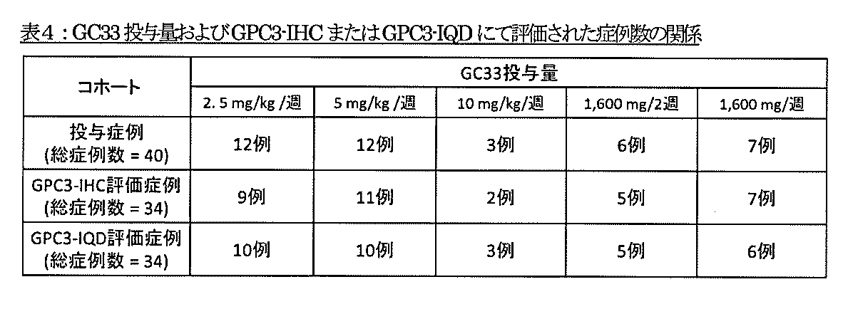
GC33が投与された患者の血清中の遊離GPC3の濃度が測定された。実施例1と同様にGT30とGT607の組合せを用いてsGPC3-Nを測定し、GT96とM3C11の組合せを用いてsGPC3-Cの濃度を測定した。GT30とGT607の組合せに関しては、Haruyamaらの方法(Haruyama Y et al., High preoperative levels of serum glypican-3 containing N-terminal subunit are associated with poor prognosis in patients with hepatocellular carcinoma after partial hepatectomy. (2015) in press)に従い遊離GPC3の濃度が測定され、GT96とM3C11の組合せに関しても、M3C11と抗体結合磁性粒子を結合させ、GT96とアルカリフォスファターゼを結合させたものを用いたほかは、Haruyamaらの方法に従い遊離GPC3の濃度が測定された。
GC33およびソラフェニブ投与前の患者血清中の測定値をベースライン値とし、投与後に採血された血清の測定値との変化が算出され、最良変化率、すなわちsGPC3-Nについては最小変化率、sGPC3-Cについては最大変化率が求められた。
GC33およびソラフェニブ投与による抗腫瘍効果として、前述の通りsGPC3-N最小変化率、sGPC3-C最大変化率、AFP最小変化率、TTPならびにPFSをそれぞれ用い、腫瘍組織におけるGPC3の発現との関連性が調べられた。GPC3の発現評価としてGPC3-IHCによる膜Hスコアおよび細胞質Hスコア、GPC3のIQD Cell ScoreおよびIQD Intensity Scoreをそれぞれ用い、これらのスコアとsGPC3-N最小変化率、sGPC3-C最大変化率およびAFP最小変化率との関連性を、回帰分析のパラメーター推定量についてのt検定またはSpearmanの相関係数を用いてそれぞれ検討した。その結果、表5に示すように、GPC3のIQD Cell Scoreにおいて、sGPC3-C最大変化率との有意な関係性が示された。また、各GPC3スコアの変化、すなわちGPC3-IHCによる膜Hスコア、細胞質Hスコア、GPC3のIQD Intensity Scoreについてはそれぞれのスコアが10変化する毎、GPC3のIQD Cell Scoreについてはスコアが100,000変化する毎でのTTPまたはPFSについてハザード比を検討した。その結果、表5に示すように、従来の手法(例えば、GPC3-IHCや単位面積あたりの蛍光強度IQD Intensity Score)ではPFSとの高い相関は見られなかったが、意外にも、GPC3の1腫瘍細胞あたりの存在量(IQD Cell Score)においてのみPFSと予想外の高い相関が見られた。

続いて、今回の評価においてPFSとの高い相関がみられなかった各種GPC3評価法、すなわちGPC3-IHC膜Hスコア、又は細胞質Hスコア、若しくはGPC3-IQD Intensity Scoreを用いた評価法において、各スコアの値がそれぞれの中央値より高い値を示す症例のみに絞った上で、腫瘍組織の単位面積(μm2)あたりの腫瘍細胞サイズの平均値をもとに中央値を算出し、中央値よりも腫瘍細胞サイズが小さい群と、大きい群の2群に分類した。それぞれのスコアにおいて、腫瘍細胞サイズで2群間のGPC3値の平均値はほぼ変わりはなく、いずれにおいても有意差が見られなかった(表6)。


Claims (39)
- 患者における肝癌に対するGPC3標的治療剤の有効性を決定する方法であって、当該患者から単離された生物学的試料における1腫瘍細胞あたりのGPC3の発現量を測定することを含む、方法。
- 前記生物学的試料における1腫瘍細胞あたりのGPC3の発現量が所定の値である場合に、GPC3標的治療剤が有効であると決定することをさらに含む、請求項1に記載の方法。
- 前記生物学的試料が肝癌組織試料である、請求項1または請求項2に記載の方法。
- 前記1腫瘍細胞あたりのGPC3の発現量がIQD Cell Scoreで表される、請求項1から請求項3のいずれかに記載の方法。
- 前記GPC3標的治療剤が、前記患者の血中トラフ値で200μg/ml以上となるように投与される、請求項1から請求項4のいずれかに記載の方法。
- 前記GPC3標的治療剤が、抗GPC3抗体を有効成分として含む治療剤である、請求項1から請求項5のいずれかに記載の方法。
- 前記抗GPC3抗体が、抗体依存性細胞傷害(ADCC)活性および/または補体依存性細胞傷害(CDC)活性を有する抗体である、請求項6に記載の方法。
- 前記抗GPC3抗体が、以下の(1)から(5)のいずれか:
(1) 配列番号:4、配列番号:5、および配列番号:6でそれぞれ表される重鎖CDR1、重鎖CDR2および重鎖CDR3、ならびに配列番号:7、配列番号:8、および配列番号:9でそれぞれ表される軽鎖CDR1、軽鎖CDR2および軽鎖CDR3;
(2) 配列番号:12、配列番号:13、および配列番号:14でそれぞれ表される重鎖CDR1、重鎖CDR2および重鎖CDR3、ならびに配列番号:15、配列番号:16、および配列番号:17でそれぞれ表される軽鎖CDR1、軽鎖CDR2および軽鎖CDR3;
(3) 配列番号:20、配列番号:21、および配列番号:22でそれぞれ表される重鎖CDR1、重鎖CDR2および重鎖CDR3、ならびに配列番号:23、配列番号:24、および配列番号:25でそれぞれ表される軽鎖CDR1、軽鎖CDR2および軽鎖CDR3;
(4) 配列番号:28、配列番号:29、および配列番号:30でそれぞれ表される重鎖CDR1、重鎖CDR2および重鎖CDR3、ならびに配列番号:31、配列番号:32、および配列番号:33でそれぞれ表される軽鎖CDR1、軽鎖CDR2および軽鎖CDR3;もしくは
(5) 配列番号:36、配列番号:37、および配列番号:38でそれぞれ表される重鎖CDR1、重鎖CDR2および重鎖CDR3、ならびに配列番号:39、配列番号:40、および配列番号:41でそれぞれ表される軽鎖CDR1、軽鎖CDR2および軽鎖CDR3;
を含む抗GPC3キメラ抗体またはヒト化抗GPC3抗体である、請求項6または請求項7に記載の方法。 - 前記抗GPC3抗体が、以下の(1)から(6)のいずれか:
(1) 配列番号:44、配列番号:45、配列番号:46、配列番号:47、配列番号:48、配列番号:49、および配列番号:50で表される重鎖可変領域の群から選択される重鎖可変領域、ならびに配列番号:51で表される軽鎖可変領域;
(2) 配列番号:44、配列番号:45、配列番号:46、配列番号:47、配列番号:48、配列番号:49、および配列番号:50で表される重鎖可変領域の群から選択される重鎖可変領域、ならびに配列番号:52、配列番号:53、配列番号:54、配列番号:55、配列番号:56、配列番号:57、配列番号:58、配列番号:59、配列番号:60、配列番号:61、配列番号:62、配列番号:63、配列番号:64、配列番号:65、および配列番号:66で表される軽鎖可変領域の群から選択される軽鎖可変領域;
(3) 配列番号:67で表される重鎖可変領域、および配列番号:68で表される軽鎖可変領域;
(4) 配列番号:69で表される重鎖可変領域、および配列番号:70で表される軽鎖可変領域;
(5) 配列番号:71で表される重鎖可変領域、および配列番号:72で表される軽鎖可変領域;もしくは
(6) 配列番号:71で表される重鎖可変領域、および配列番号:73で表される軽鎖可変領域;
を含む抗体である、請求項6から請求項8のいずれかに記載の方法。 - 前記抗GPC3抗体が、細胞傷害物質が連結された抗体である、請求項6に記載の方法。
- 前記GPC3標的治療剤が、1または2以上の抗がん剤と同時または別々に投与される、請求項1から請求項10のいずれかに記載の方法。
- 前記抗がん剤がソラフェニブである、請求項11に記載の方法。
- GPC3標的治療剤を有効成分として含む薬剤であって、患者から単離された生物学的試料における1腫瘍細胞あたりのGPC3の発現量が所定の値である患者に投与するための薬剤。
- 前記患者が、当該患者から単離された生物学的試料における1腫瘍細胞あたりのGPC3の発現量が所定の値であると決定された患者である、請求項13に記載の薬剤。
- 前記患者が、当該患者から単離された生物学的試料における1腫瘍細胞あたりのGPC3の発現量が所定の値であることによりGPC3標的治療剤が有効であると決定された患者である、請求項13に記載の薬剤。
- 前記1腫瘍細胞あたりのGPC3の発現量がIQD Cell Scoreで表される、請求項13から請求項15のいずれかに記載の薬剤。
- 前記患者が肝癌患者である、請求項13から請求項16のいずれかに記載の薬剤。
- 前記肝癌が、GPC3標的治療剤が有効である肝癌である、請求項17に記載の薬剤。
- 前記GPC3標的治療剤が有効である肝癌が、前記患者から単離された生物学的試料におけるIQD Cell Scoreで表されるGPC3の発現量が所定の値であることによって特徴付けられる、請求項18に記載の薬剤。
- 前記生物学的試料が肝癌組織試料である、請求項13から請求項19のいずれかに記載の薬剤。
- 前記GPC3標的治療剤が、前記患者の血中トラフ値で200μg/ml以上となるように投与される、請求項13から請求項20のいずれかに記載の薬剤。
- 前記GPC3標的治療剤が、抗GPC3抗体を有効成分として含む治療剤である、請求項13から請求項21のいずれかに記載の薬剤。
- 前記抗GPC3抗体が、抗体依存性細胞傷害(ADCC)活性および/または補体依存性細胞傷害(CDC)活性を有する抗体である、請求項22に記載の薬剤。
- 前記抗GPC3抗体が、以下の(1)から(5)のいずれか:
(1) 配列番号:4、配列番号:5、および配列番号:6でそれぞれ表される重鎖CDR1、重鎖CDR2および重鎖CDR3、ならびに配列番号:7、配列番号:8、および配列番号:9でそれぞれ表される軽鎖CDR1、軽鎖CDR2および軽鎖CDR3;
(2) 配列番号:12、配列番号:13、および配列番号:14でそれぞれ表される重鎖CDR1、重鎖CDR2および重鎖CDR3、ならびに配列番号:15、配列番号:16、および配列番号:17でそれぞれ表される軽鎖CDR1、軽鎖CDR2および軽鎖CDR3;
(3) 配列番号:20、配列番号:21、および配列番号:22でそれぞれ表される重鎖CDR1、重鎖CDR2および重鎖CDR3、ならびに配列番号:23、配列番号:24、および配列番号:25でそれぞれ表される軽鎖CDR1、軽鎖CDR2および軽鎖CDR3;
(4) 配列番号:28、配列番号:29、および配列番号:30でそれぞれ表される重鎖CDR1、重鎖CDR2および重鎖CDR3、ならびに配列番号:31、配列番号:32、および配列番号:33でそれぞれ表される軽鎖CDR1、軽鎖CDR2および軽鎖CDR3;もしくは
(5) 配列番号:36、配列番号:37、および配列番号:38でそれぞれ表される重鎖CDR1、重鎖CDR2および重鎖CDR3、ならびに配列番号:39、配列番号:40、および配列番号:41でそれぞれ表される軽鎖CDR1、軽鎖CDR2および軽鎖CDR3;
を含む抗GPC3キメラ抗体またはヒト化抗GPC3抗体である、請求項22または請求項23に記載の薬剤。 - 前記抗GPC3抗体が、以下の(1)から(6)のいずれか:
(1) 配列番号:44、配列番号:45、配列番号:46、配列番号:47、配列番号:48、配列番号:49、および配列番号:50で表される重鎖可変領域の群から選択される重鎖可変領域、ならびに配列番号:51で表される軽鎖可変領域;
(2) 配列番号:44、配列番号:45、配列番号:46、配列番号:47、配列番号:48、配列番号:49、および配列番号:50で表される重鎖可変領域の群から選択される重鎖可変領域、ならびに配列番号:52、配列番号:53、配列番号:54、配列番号:55、配列番号:56、配列番号:57、配列番号:58、配列番号:59、配列番号:60、配列番号:61、配列番号:62、配列番号:63、配列番号:64、配列番号:65、および配列番号:66で表される軽鎖可変領域の群から選択される軽鎖可変領域;
(3) 配列番号:67で表される重鎖可変領域、および配列番号:68で表される軽鎖可変領域;
(4) 配列番号:69で表される重鎖可変領域、および配列番号:70で表される軽鎖可変領域;
(5) 配列番号:71で表される重鎖可変領域、および配列番号:72で表される軽鎖可変領域;もしくは
(6) 配列番号:71で表される重鎖可変領域、および配列番号:73で表される軽鎖可変領域;
を含む抗体である、請求項22から請求項24のいずれかに記載の薬剤。 - 前記抗GPC3抗体が、細胞傷害物質が連結された抗体である、請求項22に記載の薬剤。
- 1または2以上の抗がん剤を有効成分としてさらに含む、請求項13から請求項26のいずれかに記載の薬剤。
- 1または2以上の抗がん剤と同時にまたは別々に投与される、請求項13から請求項26のいずれかに記載の薬剤。
- 前記抗がん剤がソラフェニブである、請求項27または請求項28に記載の薬剤。
- 肝癌に罹患した患者群からGPC3標的治療剤が有効な患者を選別する方法であって、当該患者から単離された生物学的試料における1腫瘍細胞あたりのGPC3の発現量が所定の値である場合に、GPC3標的治療剤が有効な患者であると決定する工程を含む、方法。
- 前記GPC3標的治療剤が、抗GPC3抗体を有効成分として含む治療剤である、請求項30に記載の方法。
- 患者における肝癌に対するGPC3標的治療剤の有効性を決定するためのキットであって、当該患者から単離された生物学的試料における1腫瘍細胞あたりのGPC3の発現量を測定するための試薬を含むキット。
- 前記生物学的試料における1腫瘍細胞あたりのGPC3の発現量が所定の値である場合に、GPC3標的治療剤が有効であると決定することを記載した指示書をさらに含む、請求項32に記載のキット。
- GPC3標的治療剤をさらに含む、請求項32または請求項33に記載のキット。
- 前記GPC3標的治療剤が、抗GPC3抗体を有効成分として含む治療剤である、請求項32から請求項34のいずれかに記載のキット。
- GPC3標的治療剤、および患者から単離された生物学的試料における1腫瘍細胞あたりのGPC3の発現量が所定の値である患者に当該GPC3標的治療剤を投与することが記載された指示書を含む製剤。
- 前記GPC3標的治療剤が、抗GPC3抗体を有効成分として含む治療剤である、請求項36に記載の製剤。
- 次の要素を含む、肝癌の治療用キット:
(1)GPC3標的治療剤;および
(2)患者から単離された生物学的試料における1腫瘍細胞あたりのGPC3の発現量が所定の値である患者に当該GPC3標的治療剤が有効であると決定することを記載した指示書。 - 前記GPC3標的治療剤が、抗GPC3抗体を有効成分として含む治療剤である、請求項38に記載のキット。
Priority Applications (3)
| Application Number | Priority Date | Filing Date | Title |
|---|---|---|---|
| US15/741,219 US11376326B2 (en) | 2015-07-01 | 2016-06-30 | GPC3-targeting therapeutic agent which is administered to patient for whom the GPC3-targeting therapeutic agent is effective |
| EP16818042.0A EP3318879B1 (en) | 2015-07-01 | 2016-06-30 | Gpc3-targeting therapeutic agent which is administered to patient for whom the gpc3-targeting therapeutic agent is effective |
| JP2017526441A JP7096667B2 (ja) | 2015-07-01 | 2016-06-30 | Gpc3標的治療剤が有効である患者に投与されるgpc3標的治療剤 |
Applications Claiming Priority (2)
| Application Number | Priority Date | Filing Date | Title |
|---|---|---|---|
| JP2015-133076 | 2015-07-01 | ||
| JP2015133076 | 2015-07-01 |
Publications (1)
| Publication Number | Publication Date |
|---|---|
| WO2017002934A1 true WO2017002934A1 (ja) | 2017-01-05 |
Family
ID=57608235
Family Applications (1)
| Application Number | Title | Priority Date | Filing Date |
|---|---|---|---|
| PCT/JP2016/069493 Ceased WO2017002934A1 (ja) | 2015-07-01 | 2016-06-30 | Gpc3標的治療剤が有効である患者に投与されるgpc3標的治療剤 |
Country Status (4)
| Country | Link |
|---|---|
| US (1) | US11376326B2 (ja) |
| EP (1) | EP3318879B1 (ja) |
| JP (1) | JP7096667B2 (ja) |
| WO (1) | WO2017002934A1 (ja) |
Cited By (6)
| Publication number | Priority date | Publication date | Assignee | Title |
|---|---|---|---|---|
| CN109385403A (zh) * | 2017-08-04 | 2019-02-26 | 科济生物医药(上海)有限公司 | 靶向gpc3的car nk细胞 |
| JP2021512875A (ja) * | 2018-02-02 | 2021-05-20 | カースゲン セラピューティクス カンパニー リミテッドCarsgen Therapeutics Co., Ltd. | 細胞免疫療法の組み合わせ |
| JPWO2020066043A1 (ja) * | 2018-09-28 | 2021-08-30 | オリンパス株式会社 | 顕微鏡システム、投影ユニット、及び、画像投影方法 |
| JP2022512958A (ja) * | 2018-11-07 | 2022-02-07 | ソティオ,リミティド ライアビリティ カンパニー | トランス共刺激分子と組み合わされた抗gpc3キメラ抗原受容体(car)及びその治療的用途 |
| JP2023518931A (ja) * | 2020-03-18 | 2023-05-09 | ユーティレックス カンパニー リミテッド | Il-18を分泌するgpc3 car-t細胞、並びにそれを作製する方法及び使用する方法 |
| JP2023518930A (ja) * | 2020-03-18 | 2023-05-09 | ユーティレックス カンパニー リミテッド | Gpc3 car-t細胞組成物、並びにそれを作製する方法及び使用する方法 |
Families Citing this family (4)
| Publication number | Priority date | Publication date | Assignee | Title |
|---|---|---|---|---|
| CN112175085B (zh) * | 2019-06-14 | 2022-11-04 | 厦门大学 | 抗gpc3抗体及其用途 |
| US10980836B1 (en) | 2019-12-11 | 2021-04-20 | Myeloid Therapeutics, Inc. | Therapeutic cell compositions and methods of manufacturing and use thereof |
| EP4247433A1 (en) * | 2020-11-19 | 2023-09-27 | Ardeagen Corporation | Gpc3 binding agents, conjugates thereof and methods of using the same |
| KR20240013094A (ko) | 2021-03-17 | 2024-01-30 | 마이얼로이드 테라퓨틱스, 인크. | 조작된 키메라 융합 단백질 조성물 및 그의 사용 방법 |
Citations (3)
| Publication number | Priority date | Publication date | Assignee | Title |
|---|---|---|---|---|
| WO2009116659A1 (ja) * | 2008-03-17 | 2009-09-24 | 国立大学法人宮崎大学 | 抗グリピカン3抗体を用いる肝癌細胞の検出法 |
| WO2013118858A1 (ja) * | 2012-02-09 | 2013-08-15 | 中外製薬株式会社 | 抗体のFc領域改変体 |
| JP2015511702A (ja) * | 2012-03-02 | 2015-04-20 | ロシュ グリクアート アーゲー | Adcc増強抗体を用いた癌治療のための予測バイオマーカー |
Family Cites Families (50)
| Publication number | Priority date | Publication date | Assignee | Title |
|---|---|---|---|---|
| US5322678A (en) | 1988-02-17 | 1994-06-21 | Neorx Corporation | Alteration of pharmacokinetics of proteins by charge modification |
| US5530101A (en) | 1988-12-28 | 1996-06-25 | Protein Design Labs, Inc. | Humanized immunoglobulins |
| AU3272695A (en) | 1994-08-12 | 1996-03-07 | Immunomedics Inc. | Immunoconjugates and humanized antibodies specific for b-cell lymphoma and leukemia cells |
| US6617156B1 (en) | 1997-08-15 | 2003-09-09 | Lynn A. Doucette-Stamm | Nucleic acid and amino acid sequences relating to Enterococcus faecalis for diagnostics and therapeutics |
| US6528624B1 (en) | 1998-04-02 | 2003-03-04 | Genentech, Inc. | Polypeptide variants |
| US6737056B1 (en) | 1999-01-15 | 2004-05-18 | Genentech, Inc. | Polypeptide variants with altered effector function |
| EP1156823B1 (en) | 1999-02-12 | 2008-10-29 | The Scripps Research Institute | Methods for treatment of tumors and metastases using a combination of anti-angiogenic and immuno therapies |
| DK2223922T3 (en) | 2000-04-25 | 2016-06-06 | Icos Corp | Inhibitors of HUMANPHOSPHATIDYL-inositol 3-KINASEDELTA |
| NZ505538A (en) | 2000-07-03 | 2004-12-24 | Malaghan Inst Of Medical Res | A vaccine comprising immunogenic acyl glyceryl phosphatidylinositol manno-oligosaccharide (PIM); and the use of PIM for inducing an immune response in a patient effective in the prevention or treatment of Th2-mediated diseases or disorders such as asthma |
| US6436411B1 (en) | 2000-10-23 | 2002-08-20 | The Center For The Improvement Of Human Functioning, Int'l., Inc. | Cancer treatment method |
| US7521054B2 (en) | 2000-11-17 | 2009-04-21 | The United States Of America As Represented By The Department Of Health And Human Services | Reduction of the nonspecific animal toxicity of immunotoxins by mutating the framework regions of the Fv to lower the isoelectric point |
| EP1411118B1 (en) | 2001-06-22 | 2008-09-03 | Chugai Seiyaku Kabushiki Kaisha | Cell proliferation inhibitors containing anti-glypican-3 antibody |
| WO2003057881A1 (en) | 2001-12-28 | 2003-07-17 | Chugai Seiyaku Kabushiki Kaisha | Method of stabilizing protein |
| US20040132101A1 (en) | 2002-09-27 | 2004-07-08 | Xencor | Optimized Fc variants and methods for their generation |
| US7317091B2 (en) | 2002-03-01 | 2008-01-08 | Xencor, Inc. | Optimized Fc variants |
| AU2003217912A1 (en) | 2002-03-01 | 2003-09-16 | Xencor | Antibody optimization |
| WO2004022597A1 (ja) | 2002-09-04 | 2004-03-18 | Chugai Seiyaku Kabushiki Kaisha | Gpc3の血中可溶化n端ペプチドに対する抗体 |
| BRPI0311233B8 (pt) | 2002-05-23 | 2021-07-27 | Sunnybrook And Womens College Health Sciences Centre | métodos in vitro para triagem de um paciente para carcinoma hepatocelular (hcc), e para diagnosticar hcc em um paciente, bem como método e kit para detectar ou determinar o nível de gpc3 em uma amostra |
| WO2004022595A1 (ja) | 2002-09-04 | 2004-03-18 | Chugai Seiyaku Kabushiki Kaisha | MRL/lprマウスを用いた抗体の作製 |
| AU2002330482A1 (en) | 2002-09-04 | 2004-03-29 | Perseus Proteomics Inc. | Method of diagnosing cancer by detecting gpc3 |
| WO2004029207A2 (en) | 2002-09-27 | 2004-04-08 | Xencor Inc. | Optimized fc variants and methods for their generation |
| US20090010920A1 (en) | 2003-03-03 | 2009-01-08 | Xencor, Inc. | Fc Variants Having Decreased Affinity for FcyRIIb |
| EP1671645A4 (en) | 2003-09-04 | 2007-07-25 | Chugai Pharmaceutical Co Ltd | REMEDIES AND MEDIUM FOR DETECTING GALLENGIFT CANCER |
| JP4658926B2 (ja) | 2004-04-28 | 2011-03-23 | 株式会社ペルセウスプロテオミクス | 肝癌治療後の再発予測判定薬 |
| JP4331227B2 (ja) | 2004-07-09 | 2009-09-16 | 中外製薬株式会社 | 抗グリピカン3抗体 |
| CA2544692C (en) | 2004-07-09 | 2014-04-01 | Chugai Seiyaku Kabushiki Kaisha | Anti-glypican 3 antibody |
| AU2005277641A1 (en) | 2004-08-16 | 2006-03-02 | Medimmune, Llc | Eph receptor Fc variants with enhanced antibody dependent cell-mediated cytotoxicity activity |
| KR101300546B1 (ko) | 2004-08-24 | 2013-09-02 | 추가이 세이야쿠 가부시키가이샤 | 항 글리피칸 3 항체를 이용한 어쥬번트 요법 |
| EP1813944A4 (en) | 2004-10-05 | 2009-11-11 | Perseus Proteomics Inc | DRUG TO MONITOR THE INJURY OF HEPATITIS |
| AU2005297772B2 (en) | 2004-10-26 | 2011-06-23 | Chugai Seiyaku Kabushiki Kaisha | Anti-glypican 3 antibody having modified sugar chain |
| JPWO2006067847A1 (ja) | 2004-12-22 | 2008-06-12 | 中外製薬株式会社 | フコーストランスポーターの機能が阻害された細胞を用いた抗体の作製方法 |
| WO2007005612A2 (en) | 2005-07-01 | 2007-01-11 | Medimmune, Inc. | An integrated approach for generating multidomain protein therapeutics |
| JP2007093274A (ja) | 2005-09-27 | 2007-04-12 | Perseus Proteomics Inc | 癌の診断方法および治療薬 |
| US20070087005A1 (en) | 2005-10-14 | 2007-04-19 | Lazar Gregory A | Anti-glypican-3 antibody |
| US10155816B2 (en) | 2005-11-28 | 2018-12-18 | Genmab A/S | Recombinant monovalent antibodies and methods for production thereof |
| EP1998799B8 (en) | 2006-03-06 | 2014-03-05 | Medlmmune, LLC | Humanized anti-cd22 antibodies and their use in treatment of oncology, transplantation and autoimmune disease |
| WO2007137170A2 (en) | 2006-05-20 | 2007-11-29 | Seattle Genetics, Inc. | Anti-glypican-3 antibody drug conjugates |
| ATE501436T1 (de) | 2006-09-13 | 2011-03-15 | Glycode | Verfahren zur untersuchung der reaktion auf eine behandlung mit einem monoklonalen antikörper |
| CN101874042B9 (zh) | 2007-09-26 | 2019-01-01 | 中外制药株式会社 | 利用cdr的氨基酸取代来改变抗体等电点的方法 |
| EP2196541B1 (en) | 2007-09-28 | 2012-11-07 | Chugai Seiyaku Kabushiki Kaisha | Anti-glypican-3 antibody having improved kinetics in plasma |
| CL2009000647A1 (es) | 2008-04-04 | 2010-06-04 | Chugai Pharmaceutical Co Ltd | Composicion farmaceutica para tratar o prevenir cancer hepatico que comprende una combinacion de un agente quimioterapeutico y un anticuerpo anti-glipicano 3; agente para atenuar un efecto secundario que comprende dicho anticuerpo; metodo para tratar o prevenir un cancer hepatico de un sujeto. |
| CN101377506A (zh) | 2008-04-16 | 2009-03-04 | 北京科美东雅生物技术有限公司 | 磷脂酰肌醇蛋白聚糖-3化学发光免疫分析测定试剂盒及其制备方法 |
| CN102276721B (zh) | 2010-06-12 | 2013-12-25 | 上海市肿瘤研究所 | 一种用于肝癌血清学诊断的单克隆抗体及其用途 |
| TWI654204B (zh) | 2010-11-30 | 2019-03-21 | Chugai Seiyaku Kabushiki Kaisha | 具有鈣依存性的抗原結合能力之抗體 |
| WO2012145469A1 (en) | 2011-04-19 | 2012-10-26 | The United States Of America, As Represented By The Secretary, Department Of Health & Human Services | Human monoclonal antibodies specific for glypican-3 and use thereof |
| US9409994B2 (en) | 2012-06-01 | 2016-08-09 | The United States Of America, As Represented By The Secretary, Department Of Health And Human Services | High-affinity monoclonal antibodies to glypican-3 and use thereof |
| CA2876916A1 (en) | 2012-06-18 | 2013-12-27 | National Cancer Center | Kit for diagnosing malignant melanoma |
| TWI693073B (zh) | 2012-12-21 | 2020-05-11 | 日商中外製藥股份有限公司 | 對gpc3標的治療劑療法為有效之患者投與的gpc3標的治療劑 |
| CN107460201A (zh) | 2013-05-08 | 2017-12-12 | 科济生物医药(上海)有限公司 | 编码gpc‑3嵌合抗原受体蛋白的核酸及表达gpc‑3嵌合抗原受体蛋白的t淋巴细胞 |
| TWI735410B (zh) | 2014-05-08 | 2021-08-11 | 日商中外製藥股份有限公司 | 對gpc3標的治療劑療法為有效之患者投予的gpc3標的治療劑 |
-
2016
- 2016-06-30 WO PCT/JP2016/069493 patent/WO2017002934A1/ja not_active Ceased
- 2016-06-30 JP JP2017526441A patent/JP7096667B2/ja active Active
- 2016-06-30 EP EP16818042.0A patent/EP3318879B1/en not_active Not-in-force
- 2016-06-30 US US15/741,219 patent/US11376326B2/en active Active
Patent Citations (3)
| Publication number | Priority date | Publication date | Assignee | Title |
|---|---|---|---|---|
| WO2009116659A1 (ja) * | 2008-03-17 | 2009-09-24 | 国立大学法人宮崎大学 | 抗グリピカン3抗体を用いる肝癌細胞の検出法 |
| WO2013118858A1 (ja) * | 2012-02-09 | 2013-08-15 | 中外製薬株式会社 | 抗体のFc領域改変体 |
| JP2015511702A (ja) * | 2012-03-02 | 2015-04-20 | ロシュ グリクアート アーゲー | Adcc増強抗体を用いた癌治療のための予測バイオマーカー |
Non-Patent Citations (6)
| Title |
|---|
| HASHIGUCHI A ET AL.: "Using immunofluorescent digital slide technology to quantify protein expression in archival paraffin-embedded tissue sections", PATHOL INT, vol. 60, no. 11, November 2010 (2010-11-01), pages 720 - 725, XP055169347, DOI: doi:10.1111/j.1440-1827.2010.02590.x * |
| IKEDA M ET AL.: "Japanese phase I study of GC33, a humanized antibody against glypican-3 for advanced hepatocellular carcinoma", CANCER SCI., vol. 105, no. 4, April 2014 (2014-04-01), pages 455 - 462, XP055360433 * |
| MIHO KAWAIDA ET AL.: "Kan Saibogan ni Okeru Glypican-3 no Saibonai Kyokuzai Narabini Hatsugenryo to Rinsho Byorigakuteki Kento", NIPPON BYORI GAKKAI KAISHI, vol. 104, no. 1, 23 March 2015 (2015-03-23), pages 324, XP009508009 * |
| MIKA ENDO: "A novel molecular targeted therapy, humanized anti-glypican 3 antibody (GC33), for the treatment of unresectable hepatocellular cancer", MED. SCI. DIGEST, vol. 39, no. 9, 25 August 2013 (2013-08-25), pages 440 - 443, XP008185599 * |
| See also references of EP3318879A4 * |
| ZHU ANDREW X ET AL.: "First-in-Man Phase I Study of GC33, a Novel Recombinant Humanized Antibody Against Glypican-3, in Patients with Advanced Hepatocellular Carcinoma", CLIN. CANCER RES., vol. 19, no. 4, 2013, pages 920 - 928, XP055207838, DOI: doi:10.1158/1078-0432.CCR-12-2616 * |
Cited By (8)
| Publication number | Priority date | Publication date | Assignee | Title |
|---|---|---|---|---|
| CN109385403A (zh) * | 2017-08-04 | 2019-02-26 | 科济生物医药(上海)有限公司 | 靶向gpc3的car nk细胞 |
| CN109385403B (zh) * | 2017-08-04 | 2024-03-08 | 恺兴生命科技(上海)有限公司 | 靶向gpc3的car nk细胞 |
| JP2021512875A (ja) * | 2018-02-02 | 2021-05-20 | カースゲン セラピューティクス カンパニー リミテッドCarsgen Therapeutics Co., Ltd. | 細胞免疫療法の組み合わせ |
| JPWO2020066043A1 (ja) * | 2018-09-28 | 2021-08-30 | オリンパス株式会社 | 顕微鏡システム、投影ユニット、及び、画像投影方法 |
| JP2022512958A (ja) * | 2018-11-07 | 2022-02-07 | ソティオ,リミティド ライアビリティ カンパニー | トランス共刺激分子と組み合わされた抗gpc3キメラ抗原受容体(car)及びその治療的用途 |
| JP2023518931A (ja) * | 2020-03-18 | 2023-05-09 | ユーティレックス カンパニー リミテッド | Il-18を分泌するgpc3 car-t細胞、並びにそれを作製する方法及び使用する方法 |
| JP2023518930A (ja) * | 2020-03-18 | 2023-05-09 | ユーティレックス カンパニー リミテッド | Gpc3 car-t細胞組成物、並びにそれを作製する方法及び使用する方法 |
| JP7778081B2 (ja) | 2020-03-18 | 2025-12-01 | ユーティレックス カンパニー リミテッド | Il-18を分泌するgpc3 car-t細胞、並びにそれを作製する方法及び使用する方法 |
Also Published As
| Publication number | Publication date |
|---|---|
| US11376326B2 (en) | 2022-07-05 |
| US20180169237A1 (en) | 2018-06-21 |
| EP3318879A4 (en) | 2018-11-21 |
| EP3318879A1 (en) | 2018-05-09 |
| EP3318879B1 (en) | 2020-10-21 |
| JPWO2017002934A1 (ja) | 2018-06-07 |
| JP7096667B2 (ja) | 2022-07-06 |
Similar Documents
| Publication | Publication Date | Title |
|---|---|---|
| JP6359461B2 (ja) | Gpc3標的治療剤療法が有効である患者に投与されるgpc3標的治療剤 | |
| JP6827319B2 (ja) | Gpc3標的治療剤療法が有効である患者に投与されるgpc3標的治療剤 | |
| JP7096667B2 (ja) | Gpc3標的治療剤が有効である患者に投与されるgpc3標的治療剤 | |
| JP2017043622A (ja) | ヒト化抗Epiregulin抗体および当該抗体を有効成分として含む癌治療剤 | |
| JP6890581B2 (ja) | Cd37の検出のための抗体およびアッセイ | |
| JP5801557B2 (ja) | 抗lgr7抗体を用いる癌の診断および治療 | |
| JPWO2014208482A1 (ja) | ヒト化抗Epiregulin抗体を有効成分として含む腺癌以外の非小細胞肺癌の治療剤 | |
| WO2012124334A1 (ja) | 抗体、乳がんの治療に用いられる医薬組成物、腫瘍検査方法、及び、腫瘍検査用試薬 | |
| EP3557260A1 (en) | Gpc3-targeting drug which is administered to patient responsive to gpc3-targeting drug therapy | |
| HK40059872A (en) | Gpc3-targeted therapeutic agent for administration to patients for whom gpc3-targeted therapeutic agent therapy is effective | |
| NZ707774B2 (en) | Gpc3-targeting drug which is administered to patient responsive to gpc3-targeting drug therapy | |
| HK1208910B (zh) | 用於向gpc3靶向治疗剂疗法有效的患者施予的gpc3靶向治疗剂 | |
| HK40019410A (en) | Gpc3-targeted therapeutic agent for administration to patients for whom gpc3-targeted therapeutic agent therapy is effective | |
| HK1165445A (en) | Diagnosis and treatment of cancer using anti-lgr7 antibody |
Legal Events
| Date | Code | Title | Description |
|---|---|---|---|
| 121 | Ep: the epo has been informed by wipo that ep was designated in this application |
Ref document number: 16818042 Country of ref document: EP Kind code of ref document: A1 |
|
| ENP | Entry into the national phase |
Ref document number: 2017526441 Country of ref document: JP Kind code of ref document: A |
|
| WWE | Wipo information: entry into national phase |
Ref document number: 15741219 Country of ref document: US |
|
| NENP | Non-entry into the national phase |
Ref country code: DE |
|
| WWE | Wipo information: entry into national phase |
Ref document number: 2016818042 Country of ref document: EP |