WO2017006962A1 - Kit d'examen sanguin, et procédé d'analyse sanguine - Google Patents
Kit d'examen sanguin, et procédé d'analyse sanguine Download PDFInfo
- Publication number
- WO2017006962A1 WO2017006962A1 PCT/JP2016/070007 JP2016070007W WO2017006962A1 WO 2017006962 A1 WO2017006962 A1 WO 2017006962A1 JP 2016070007 W JP2016070007 W JP 2016070007W WO 2017006962 A1 WO2017006962 A1 WO 2017006962A1
- Authority
- WO
- WIPO (PCT)
- Prior art keywords
- blood
- component
- test kit
- sample
- diluent
- Prior art date
- Legal status (The legal status is an assumption and is not a legal conclusion. Google has not performed a legal analysis and makes no representation as to the accuracy of the status listed.)
- Ceased
Links
Images


Classifications
-
- G—PHYSICS
- G01—MEASURING; TESTING
- G01N—INVESTIGATING OR ANALYSING MATERIALS BY DETERMINING THEIR CHEMICAL OR PHYSICAL PROPERTIES
- G01N1/00—Sampling; Preparing specimens for investigation
- G01N1/02—Devices for withdrawing samples
- G01N1/10—Devices for withdrawing samples in the liquid or fluent state
-
- G—PHYSICS
- G01—MEASURING; TESTING
- G01N—INVESTIGATING OR ANALYSING MATERIALS BY DETERMINING THEIR CHEMICAL OR PHYSICAL PROPERTIES
- G01N33/00—Investigating or analysing materials by specific methods not covered by groups G01N1/00 - G01N31/00
- G01N33/48—Biological material, e.g. blood, urine; Haemocytometers
- G01N33/50—Chemical analysis of biological material, e.g. blood, urine; Testing involving biospecific ligand binding methods; Immunological testing
- G01N33/96—Chemical analysis of biological material, e.g. blood, urine; Testing involving biospecific ligand binding methods; Immunological testing involving blood or serum control standard
Definitions
- the present invention relates to a blood test kit and a blood analysis method for analyzing a target component in a blood sample.
- a general qualified blood sample is collected by a doctor or other qualified person using a syringe to collect blood from the vein, and the subject is self-collected by inserting a blood collection needle into his / her finger or the like. There is blood sampling.
- Blood collected by general blood collection is transported to a medical institution or inspection in a state of being sealed in a collection container, where it is inspected.
- a test is performed after blood is separated into blood cells and plasma by a centrifuge at a medical institution or inspection institution.
- the collected blood is separated into blood cells and plasma by a separation membrane and transported to the examination site in this separated state, where the examination is performed.
- Patent Document 1 the amount of a component to be analyzed in a sample is measured, and further, the amount of a standard component originally present in the sample other than the above is measured.
- a quantitative analysis method is described in which the amount of a sample is determined from the known concentrations of the standard components, and the concentration of the analysis target component in the sample is determined from the sample amount and the analysis target component amount.
- Patent Document 2 describes a method for examining a blood sample collected by self blood collection. Specifically, 1) Prepare a sample for quantification consisting of an unknown volume of a biological sample containing a component to be quantified collected without quantifying the volume and a fixed amount of an aqueous solution containing a fixed amount of an indicator substance.
- Patent Document 3 describes that a small amount of blood is collected from a human or animal using a blood dilution quantification instrument and is supplied as it is or after dilution to supply a constant amount to another device, container or reagent.
- Patent Document 4 describes a method of quantifying the concentration of a component to be quantified in a biological sample using the absorbance of an indicator substance in an aqueous solution for dilution.
- JP 2001-330603 A JP 2003-161729 A JP 2009-128202 A JP 2009-109196 A
- blood collection is an invasive action that damages the skin, and it may be uncomfortable to stare at the red color of the blood. It is common to want to stop the blood. For these reasons, the amount of blood collected is not always constant and often varies. If the variation in the amount of blood collected is large, the accuracy of the dilution rate will be reduced, so visualization and stabilization of the amount of blood collected is desired.
- the problem to be solved by the present invention is to provide a blood test kit and a blood analysis method capable of performing a blood test with a small amount of blood with high accuracy by visualizing and stabilizing the blood collection volume.
- the present inventors include a diluent for diluting the components of the blood sample and at least one transparent container for containing the components of the blood sample and the diluent.
- the present invention has found that the above-mentioned problems can be solved by providing a scale for measuring the components of the blood sample and the amount of the diluted solution to at least one component included in the blood test kit. It came to complete. That is, according to the present invention, the following inventions are provided.
- a diluent for diluting the components of the blood sample At least one transparent container for containing a blood sample component and a diluent;
- a blood test kit comprising: A blood test kit in which a scale for measuring the components of a blood sample and the amount of a diluent is added to at least one component included in the blood test kit.
- the scale includes at least a scale for measuring the amount of a diluted solution without a blood sample component.
- the constituent element is at least one transparent container for containing a blood sample component and a diluent.
- the blood test kit is a blood test kit for analyzing the concentration of a target component in a blood sample component using a standard component that is constantly present in the blood, and the diluent contains the standard component
- the blood test kit according to any one of (1) to (6).
- the blood test kit is a blood test kit for analyzing the concentration of a target component in a component of a blood sample using a standard component that is constantly present in blood and verifying the analysis, The blood test kit according to any one of (1) to (6), wherein the diluent does not contain the standard component.
- the blood test kit according to (7) or (8), wherein the standard component is a component selected from the group consisting of sodium ion, chloride ion, total protein, and albumin.
- a blood test kit for analyzing a concentration of a target component in a component of a blood sample using a standard component that is not present in blood includes a standard component that is not present in blood
- the blood test kit according to (10), wherein the standard component not present in blood is glycerol triphosphate or lithium.
- an amino alcohol compound wherein the diluent is selected from the group consisting of 2-amino-2-methyl-1-propanol, 2-ethylaminoethanol, N-methyl-D-glucamine, diethanolamine, and triethanolamine; 2- [4- (2-hydroxyethyl-1-piperazinyl) ethanesulfonic acid), also referred to as HEPES, N-tris (hydroxymethyl) methyl-2-aminoethanesulfonic acid, also referred to as TES, 3-morpholinopropane, also referred to as MOPS (1) to (12), which is a diluent containing a buffer selected from the group consisting of sulfonic acid and BES (N, N-bis (2-hydroxyethyl) -2-aminoethanesulfonic acid)
- a buffer selected from the group consisting of sulfonic acid and BES (N, N-bis (2-hydroxyethyl) -2-aminoethane
- a blood analysis method using a blood test kit comprising a diluent for diluting a component of a blood sample and at least one transparent container for containing the component of the blood sample and the diluent, A step of selecting a component-containing sample of a blood sample of a predetermined amount or more as a sample to be analyzed, and excluding a component-containing sample of a blood sample of less than a predetermined amount from the sample to be analyzed, and a component-containing sample of the selected blood sample
- a blood analysis method comprising a step of analyzing blood using
- the blood test kit is the blood test kit according to any one of (1) to (13), and using a scale, a component-containing sample of a blood sample of a predetermined amount or more determined in advance is selected.
- (17) The dilution rate of the blood sample component is calculated by the following equation 1, and the concentration of the analysis target component in the diluted solution is multiplied by the dilution rate to analyze the concentration of the target component in the blood sample component.
- Formula 1 (A + C) / (B + D)
- A represents the measured absorbance of the diluted solution containing the internal standard substance
- B represents the absorbance obtained by subtracting the absorbance of the diluted solution obtained by diluting the blood sample component from A
- C represents sodium ion as a homeostatic substance.
- the measured absorbance at a concentration of 142 mmol / L is shown
- D is the absorbance of sodium ions in a diluted solution obtained by diluting a blood sample component.
- the blood analysis method according to any one of (14) to (17), wherein analysis is performed using blood of 10 ⁇ L or more and 70 ⁇ L or less.
- a blood test with a very small amount of blood can be performed with high accuracy by visualizing and stabilizing the amount of blood collected.
- FIG. 1 shows an example of the configuration of a container for containing a plasma component collected from a diluted blood sample.
- FIG. 2 shows a conceptual diagram of the bottle and cylinder scales during plasma separation and sealing.
- FIG. 3 shows an example of the shape of the container and the position of the scale.
- FIG. 4 shows an example of the shape of the container and the position of the scale.
- FIG. 5 shows an example of the shape of the container and the position of the scale.
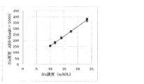
- FIG. 6 shows the linearity of the sodium enzymatic assay.
- a standard component that is constantly present in blood is called an external standard substance or external standard.
- a standard component that does not exist in blood is called an internal standard substance or internal standard.
- blood from patients or examiners is collected from veins and examined at medical institutions or examination sites.
- it is necessary to wait for a long time, and it may be a burden for a child to collect blood from a vein.
- including movement for examination requires more than half a day, which may be inconvenient for the patient or examiner.
- a patient or an examiner collects a small amount of blood at home or in a laboratory, and stably stores and transports it.
- Patent Document 1 has a problem in that the amount of collected blood varies, and when the collected blood has a high dilution rate, there is a problem in that the analysis accuracy decreases and results vary, and the blood is agglomerated. There is also a problem that the stability of the analysis target component is not guaranteed due to solidification. In addition, it is necessary to add NaOH, NaCl or HCl to the buffer solution for extracting the analysis target component from the dried sample in order to adjust the pH or stabilize the analysis target component. There is a problem that the concentration of sodium ions and chloride ions, which are homeostatic and have little difference between individuals, cannot be used as an external standard for correcting the concentration of the target component.
- Patent Document 2 discloses a method for diluting a collected trace blood with an internal standard-containing buffer and quantifying the amount of components present in diluted plasma from the dilution factor of the internal standard substance.
- N- (2-hydroxy-3-sulfopropyl) -3,5-dimethoxyaniline sodium salt (HSDA) or Acid Blue 9 (Brilliant Blue FCF) is used as an internal standard substance.
- HSDA N- (2-hydroxy-3-sulfopropyl) -3,5-dimethoxyaniline sodium salt
- Acid Blue 9 Brilliant Blue FCF
- a method for determining the dilution factor from the absorbance of the HEPES buffer is disclosed.
- buffering agents and preservatives for stably holding blood are used.
- Such a formulation has achieved the stability of its components by not coagulating blood, but if the blood collection volume varies and the collection volume is small, the dilution rate of the internal standard substance after dilution The problem is that the reliability of the test accuracy is lowered due to the decrease in the amount of blood and the blood component amount itself.
- the method of diluting with a buffer solution is a biological component stored in a buffer solution with physiological conditions of pH 7.4 and is excellent in stability during transportation. There was a problem that measurement errors were likely to occur when the dilution rate was small and a small amount of sample was added.
- Patent Document 1 there is a description of an external standard, but there is no description regarding visualization and stabilization of a blood collection amount.
- Patent Document 2 describes the internal standard, but does not describe the combined use with the external standard.
- Patent Document 2 there is no description about removing a highly constant substance such as sodium or potassium, which is preferable as an external standard substance, from a buffer solution used as a diluting solution, and a specific means for achieving this is not described. There is no description.
- Patent Documents 1 and 2 do not disclose means for quantitatively collecting a very small amount of blood with self-collection with high accuracy.
- the blood test kit and the blood analysis method of the present invention even when the amount of blood collected is not always constant, the amount of blood collected can be grasped in a container in which blood is diluted and plasma is collected.
- the accuracy can be further improved by using a method for obtaining a dilution ratio using an internal standard.
- high-accuracy measurement can be performed by determining the dilution factor and analyzing the measurement target component in a state in which the blood collection amount is within a certain range and the variation in the blood collection amount is small. It was completely unexpected that the structure of the present invention enabled highly accurate blood analysis.
- the blood test kit and the blood analysis method of the present invention when a patient collects blood by himself / herself, the amount of blood collected from the container in which the blood is diluted and separated into plasma even if the amount of blood collected is not always constant. By knowing, it becomes possible for blood collectors and analysts to predict the result of the dilution ratio within a narrow range, to realize a dilution rate with good reproducibility, and to accurately analyze the measurement target component.
- the collection of micro blood using the blood test kit of the present invention is not limited in time and place, so it can be applied to cases that do not have time to go to medical institutions, disasters, telemedicine, health care, etc. Because it can be detected early, it can contribute to the reduction of medical costs.
- test data can be used in a system for daily health management or early detection of disease by, for example, converting it to electronic data and transmitting it to a smartphone.
- many tests such as 13 biochemical tests, tumor markers, and hepatitis tests can be performed using a very small amount of blood (65 ⁇ L).
- the blood test kit of the present invention includes a diluent for diluting the components of the blood sample and at least one transparent container for containing the components of the blood sample and the diluent.
- the scale for measuring the components of the blood sample and the amount of the diluted solution is attached to at least one component included in the blood test kit.
- the blood test kit of the present invention is for diluting blood collected by a patient and transporting it to a medical institution or testing institution to analyze a component to be measured. Therefore, there is a possibility of being left for a long time from blood collection to analysis. During. It is preferable to prevent decomposition and denaturation of the target component in the diluted blood.
- the pH of blood is usually kept constant at about pH 7.30 to 7.40 in healthy individuals.
- the diluent is preferably pH 6.5 to pH 8.0, more preferably pH 7.0 to pH 7.5, and still more preferably pH 7.3 to A buffer solution having a pH of 7.4 and containing a buffer component that suppresses fluctuations in pH is preferable.
- the types of buffer include acetate buffer (Na), phosphate buffer (Na), citrate buffer (Na), borate buffer (Na), tartrate buffer (Na), Tris (Tris (hydroxy Methyl) aminoethane) buffer (Cl), HEPES ([2- [4- (2-hydroxyethyl) -1-piperazinyl] ethanesulfonic acid]) buffer, phosphate buffered saline (Na), etc. are known ing.
- phosphate buffers, Tris buffers, and HEPES buffers are representative examples of buffers around pH 7.0 to pH 8.0.
- the phosphate buffer contains a sodium salt of phosphate.
- the Tris buffer Since the Tris buffer has a dissociated pKa (Ka is an acid dissociation constant) of 8.08, it is usually used in combination with hydrochloric acid in order to have a buffering ability in the vicinity of pH 7.0 to pH 8.0.
- the pKa of sulfonic acid dissociation of HEPES is 7.55, and a mixture of sodium hydroxide, sodium chloride and HEPES is usually used to prepare a buffer solution with a constant ionic strength.
- these are useful as buffers having the action of keeping the pH constant, they contain sodium ions or chloride ions, which are preferably used as external standard substances in the present invention. It is not preferable. Therefore, the present inventors have intensively studied and found a new buffer solution that does not contain sodium ions or chloride ions.
- the diluent containing no sodium ion and chloride ion that can be used in the present invention is preferably 2-amino-2-methyl-1-propanol (AMP), 2-ethylaminoethanol, N-methyl-D-glucamine. , Diethanolamine, and at least one amino alcohol compound selected from the group consisting of triethanolamine, and Good's buffer (Good buffer), which is also referred to as HEPES, which is a buffer having a pKa of around 7.4.
- AMP 2-amino-2-methyl-1-propanol
- 2-ethylaminoethanol N-methyl-D-glucamine
- Diethanolamine and at least one amino alcohol compound selected from the group consisting of triethanolamine
- Good buffer Good buffer
- HEPES Good buffer having a pKa of around 7.4.
- the concentration ratio of amino alcohol and Good's buffer solution is 1: 2 to 2: 1, preferably 1: 1.5 to 1.5: 1, more preferably 1: 1.
- the concentration of the buffer is not limited, but the concentration of amino alcohol or Good's buffer is 0.1 to 1000 mM / L, preferably 1 to 500 mM / L, and more preferably 10 to 100 mM / L.
- chelating agents In the buffer solution, chelating agents, surfactants, antibacterial agents, preservatives, coenzymes, saccharides and the like may be contained for the purpose of keeping the components to be analyzed stable.
- chelating agents include ethylenediaminetetraacetate (EDTA), citrate, and oxalate.
- surfactant include a cationic surfactant, an anionic surfactant, an amphoteric surfactant, and a nonionic surfactant.
- the preservative include sodium azide and antibiotics.
- coenzyme include pyridoxal phosphate, magnesium, zinc and the like.
- saccharide of the erythrocyte stabilizer examples include mannitol, dextrose, oligosaccharide and the like.
- by adding antibiotics it is possible to suppress the growth of bacteria partially mixed from the finger surface at the time of hand blood collection, to suppress the degradation of the biological components by bacteria, and to stabilize the biological components.
- Components diluted with these buffer solutions do not interfere with measurement even when using various biochemical / immunoanalytical analyzers. Furthermore, blood cells do not undergo hemolysis and biological components are as stable as possible even at 37 ° C. It is preferable that it can be stored.
- the osmotic pressure of the buffer solution is equivalent to that of blood (285 mOsm / kg (mOsm / kg: 1 kg of solution water has By setting the osmotic pressure to the number of millimoles of ions)) or more, hemolysis of blood cells can be prevented.
- the osmotic pressure can be adjusted to be isotonic with salts, sugars, buffers, etc. that do not affect the measurement of the target component and the measurement of the standard component that is constantly present in the blood.
- a diluent that does not contain a substance that is constantly present in blood (hereinafter also referred to as a homeostatic substance) used when determining the dilution factor is used. is there.
- a homeostatic substance a substance that is constantly present in blood
- does not contain means “does not contain substantially”.
- substantially free means that the homeostatic substance used when determining the dilution factor is not included at all, or even if it is included, the homeostatic substance in the diluted solution after the blood sample is diluted It means the case where it is contained at a very small concentration that does not affect the measurement of the above.
- sodium ions or chloride ions are used as the homeostatic substance
- a diluent that does not substantially contain sodium ions or chloride ions is used as the diluent.
- the blood test kit of the present invention is a blood test kit for analyzing the concentration of a target component in a blood sample using a standard component that is constantly present in blood, the diluent is the standard component. It is a diluted solution that does not contain.
- a second example of a diluent for diluting a blood sample component is a diluent containing an internal standard substance.
- the internal standard substance can be added to a diluent used for diluting a biological sample so as to have a predetermined concentration.
- the internal standard substance a substance which is not contained in the blood sample at all or is contained in a trace amount even if it is contained can be used.
- Internal standards include substances that do not interfere with the measurement of target components in blood samples, substances that do not decompose under the action of biological enzymes in blood samples, substances that are stable in dilute solutions, and do not permeate blood cell membranes. It is preferable to use a substance that is not contained in blood cells, a substance that is not adsorbed in the storage container for the diluted solution, or a substance that can use a detection system that can measure with high accuracy.
- the internal standard substance a substance that is stable even if stored for a long time in a state of being added to a diluent that is a buffer solution is preferable.
- Examples of internal standards include glycerol triphosphate, Li, Rb, Cs, or Fr as alkali metals, and Sr, Ba, or Ra as alkaline earth metals, among which glycerol triphosphate Or Li is preferable.
- These internal standard samples are colored by adding a second reagent at the time of concentration measurement after blood dilution, and the concentration in diluted blood can be determined from the color density.
- the measurement of the internal standard substance of lithium added to the buffer solution uses a chelate colorimetric method (halogenated porphyrin chelate method: perfluoro-5,10,15,20-tetraphenyl-21H, 23H-porphyrin).
- a biochemical automatic analyzer a lot of samples can be easily measured.
- the blood test kit of the present invention is a blood test kit for analyzing the concentration of a target component in a blood sample using a standard component that is not present in blood, the diluent is present in the blood. This is a diluted solution containing standard components that do not.
- a third example of a diluent for diluting a blood sample is a diluent that does not contain a standard component that is constantly present in the blood used when determining the dilution factor, and that contains an internal standard substance. .
- Transparent container for storing blood sample components and diluent and other components The shape and size of the transparent container for storing blood sample components and diluent are not particularly limited.
- the term “transparent” as used in the present invention is not limited as long as the observer can confirm the amount of liquid inside, and is a concept including translucency.
- the material of the container is preferably a synthetic resin from the viewpoint of resistance to breakage, hygiene, and price.
- polyethylene polypropylene, polyvinyl chloride, polyvinylidene chloride, polystyrene, polyvinyl acetate, polyurethane, polyethylene terephthalate, polylactic acid, acrylonitrile butadiene styrene resin (ABS resin), acrylonitrile styrene resin (AS resin), acrylic resin (PMMA) , Polycarbonate, silicone resin, silicone rubber and the like.
- ABS resin acrylonitrile butadiene styrene resin
- AS resin acrylonitrile styrene resin
- PMMA acrylic resin
- Polycarbonate silicone resin, silicone rubber and the like.
- a diluent for diluting a component of a blood sample a first storage device containing the diluent, and plasma for separating and collecting plasma from a blood sample diluted with the diluent Separation device as a separation means, a holding device for holding the separation device, a second storage device for storing the collected plasma, and a seal for maintaining the stored plasma in the second storage device Instruments, needles that hurt the skin and allow blood to bleed out of the skin, lancets, adhesive bandages or disinfecting materials (eg, non-woven fabric impregnated with isopropanol (70% by mass isopropanol) or ethanol), handling Instructions etc. can be provided.
- a separation means for recovering plasma components from a diluted blood sample an embodiment that is a separation membrane is preferable, and a filter having pores that can separate blood cell components is more preferable.
- an embodiment in which a plasma component separated from a blood sample by a separation means is used as the blood sample component is preferable.
- the first storage device and the second storage device may be used as a first storage device and a second storage device, or may be provided with separate devices.
- the first storage device and the second storage device The instrument is preferably made of a transparent material.
- the holding device for holding the separation device is a gasket
- the sealing device when the storage device is a tube-shaped device, a cap that can cover the opening, a cover having a spiral groove, or a rubber plug is used. can do.
- the blood component stability and the fluctuation of the component due to hemolysis from the blood cell are reduced by providing the blood and diluent mixture container with the function of immediately separating plasma blood cells after diluting the blood with the diluent.
- the influence can be eliminated and the stability of the sample after blood collection can be imparted.
- a first storage device containing a diluent containing a blood sample diluted with the diluent
- a holding device for holding the separation device for holding the separation device
- a first device for containing the collected plasma are described, for example, in FIGS. 1 to 13 of Japanese Patent No. 3597827. Can be used.
- FIG. 1 of Japanese Patent No. 3597827 is incorporated as FIG. 1 of the present application.
- the blood separation instrument 1 includes a blood collection container 2 (first accommodation instrument in which a diluent is accommodated), a cylinder 3 (second accommodation instrument for accommodating the collected plasma) that can be inserted into the blood collection container 2, and
- the cap piston 4 that can be attached to the cylindrical body 3 and a sealing lid 5 (sealing device) provided at the lower end of the cap piston 4, and before use, as shown in FIG.
- the upper end opening is sealed with a cap 6 via a packing 7.
- the container for storing the diluted blood sample in the present invention corresponds to the combination of the blood collection container 2 and the cylinder 3 in the configuration of FIG. That is, the container for storing the diluted blood sample may be one or a combination of two or more.
- the blood collection container 2 is made of a transparent material and has a cylindrical shape.
- a screw portion 8 is formed on the outer surface of the blood collection container 2 and an engaging portion 9 is projected on the inner surface.
- an inverted conical bottom portion 10 is formed at the lower end portion of the blood collection container 2, and a cylindrical leg portion 11 is formed around the bottom portion 10.
- the legs 11 have the same outer diameter as the sample cup used at the time of blood analysis and testing, and preferably, slit grooves 12 are formed in the vertical direction at positions opposite to the lower ends thereof. Further, as shown in FIG. 1, a required amount, for example, a diluted solution 13 of 500 mm 3 may be placed in the blood collection container 2 in advance.
- the cylindrical body 3 is made of a transparent material and has a cylindrical shape, and an enlarged diameter portion 14 is formed at an upper end portion thereof.
- the enlarged diameter portion 14 is connected to the main body portion 16 through a thin portion 15.
- a reduced diameter portion 18 is formed at the lower end of the cylindrical body 3, and a locking projection 19 is formed on the inner surface of the reduced diameter portion 18.
- the outer flange portion 20 (holding device) is formed at the lower end portion of the reduced diameter portion 18, the lower end opening portion of the outer flange portion 20 is covered with a filtration membrane 21 (separation device), and the filtration membrane 21 is in the blood. It allows passage of plasma and prevents passage of blood cells.
- a silicon rubber cover 22 is attached to the outer periphery of the reduced diameter portion 18 (FIG. 1).
- the cap piston 4 includes a substantially cylindrical knob 26 and a mandrel 27 that is concentric with the knob 26 and extends downward.
- a cylindrical space 28 into which the enlarged diameter portion 14 of the cylindrical body 3 can be fitted is formed at the inner upper end portion of the knob portion 26, and the lower portion thereof is screwed and can be screwed into the screw.
- the lower end portion 29 of the mandrel portion 27 is formed in a pin shape, and the sealing lid 5 is detachably provided on the lower end portion 29 (see FIG. 1).
- the sealing lid 5 is made of silicon rubber.
- a scale for measuring the components of the blood sample and the amount of the diluted solution is given to at least one component included in the blood test kit. From the scale, the blood sampler or the examiner can confirm that the blood collection amount is a certain amount or more.
- the number of scales may be one, or two or more. For example, when a single scale indicating a certain minimum amount is provided, when two scales indicating a lower limit amount and an upper limit amount are provided, and a lower limit amount, an upper limit amount, and an intermediate standard amount therebetween. For example, it is possible to provide three scales indicating the above. Furthermore, the scales can be given meaning by distinguishing the colors.
- the scale may include at least a scale for measuring the volume of the diluted solution without the blood sample component.
- a suitable blood volume and dilution liquid volume can be designed with good reproducibility with respect to the dilution liquid component design.
- the scale can be applied to any component of the blood test kit as long as the blood sampler or the examiner can confirm that the blood collection amount is a certain amount or more, but the scale is preferably blood. It can be applied to at least one transparent container for containing the components of the specimen and the diluent. When a scale is attached to the transparent container, the scale can be attached so as to sandwich the liquid, and the amount of the liquid can be easily grasped, which is preferable.
- Examples of the above-described constituent elements in the case where a scale is given to the constituent elements other than the transparent container for storing the blood sample components and the diluent include a mandrel portion of a sealing cap for preventing backflow.
- the blood test kit stores a bottle (container) containing a diluent for diluting blood, a separation membrane that separates blood into blood cell components and plasma components, a gasket that holds the separation membrane, and a plasma component after separation.
- a bottle container
- a configuration having a cylinder will be described. In this configuration, the cylinder is inserted into the bottle without blood sampling and separated, and the increase in liquid volume from the liquid surface is measured with the sealing cap mandrel inserted to prevent backflow of the separated liquid.
- the scale may be on either.
- the cylinder when the cylinder is inserted into the bottle, the cylinder will be pushed in until it comes into contact with the bottom of the bottle, but the reference line is marked on both the bottle and the cylinder, and when the cylinder is fully pushed in, each reference By making the line positions coincide, it is possible to prevent the amount of plasma component from being accurately measured due to insufficient push-in.
- the inner diameter of the transparent container for containing the blood sample components and the diluent is such that the amount of diluted plasma liquid separated after the dilution coincides with the scale position.
- the inner diameter of the container is preferably 5 mm or less and more preferably about 3 mm in order to increase detection sensitivity.
- a difference of ⁇ about 14 ⁇ L can be detected if the position of the scale ⁇ 0.5 mm can be visually recognized in the case of 3 mm, and a difference of ⁇ about 40 ⁇ L can be detected in the case of 5 mm.
- the position accuracy of the scale is ⁇ 1. Even at 0.0 mm, the difference in liquid volume can be detected with an accuracy of about ⁇ 12 ⁇ L due to the volume reduction effect in the cylinder by the mandrel. In such a case, by inserting three or more scale lines with a target liquid volume of ⁇ 1.0 mm, the degree of insufficient liquid volume can be reduced to 12 ⁇ L or less. This is shown in FIG.
- Fig. 2 shows a conceptual diagram of the bottle and cylinder scales during plasma separation and sealing.
- one scale 109 is provided for the transparent bottle and three for the transparent cylinder.
- the scale of the transparent bottle and the middle scale of the transparent cylinder indicate standard amounts.
- the scale below and the scale above the transparent cylinder indicate the lower limit and the upper limit, respectively.
- the scale 110 of a dilution liquid shows the scale in the case of only the dilution liquid which is not diluted with plasma. The volume volume increased from this scale corresponds to the volume of the plasma component before dilution.
- the transparent container When a scale is applied to at least one transparent container for containing a blood sample component and a mixture of diluents, the transparent container has a portion whose internal cross-sectional area is smaller than other portions, and the internal cross-sectional area is the other. It is preferable that a scale is applied to the part smaller than the part. It is more preferable that a scale is given to a portion where the internal cross-sectional area of the transparent container is the smallest.
- the internal cross-sectional area means a cross-sectional area when the space inside the transparent container is cut along a horizontal plane.
- the portion having the scale is a portion for accurately measuring the amount of liquid, as described above, the measurement can be performed with high accuracy by providing the scale with the portion having a small internal cross-sectional area.
- a portion having a small internal cross-sectional area can be provided by providing a portion having a small inner diameter.
- a part of the cylinder is narrowed and the plasma component amount can be accurately measured by giving a scale to the narrowed part.
- the constricted part of the cylinder is thick enough to allow the mandrel part to be inserted, and the capacity of the mandrel part is also taken into consideration.
- it is preferable that a scale is added to the constricted portion so that the plasma component amount can be accurately measured.
- the inner cross-sectional area is smaller than other parts, and the inner diameter of the thinner part is less than half of the inner diameter that is not thinner. Is preferably provided.
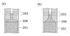
- FIGS. 3 and 4 A form in which a part of the cylinder is narrowed is shown in FIGS.
- a scale 202 is given to the thin portion at the top of the cylinder 201.
- a scale 206 of dilution liquid is provided.
- FIG. 3 shows a form in which the thickness of the container is smaller than other parts (the thickness of the outer wall of the container is the same)
- FIG. 3 (a) shows a form in which the thickness of the container changes at a certain part.
- 3 (b) shows a form in which the thickness of the container changes continuously.
- FIG. 4 shows a form in which the inner diameter of the container is smaller than other parts (the thickness of the container is the same)
- FIG. 4 (a) shows a form in which the inner diameter of the container changes at a certain part
- FIG. ) Shows a form in which the inner diameter of the container continuously changes.
- a portion having a small internal cross-sectional area can be provided by providing a thick portion in the thickness of the mandrel (FIG. 5).
- a scale 202 is provided on the upper portion of the cylinder 201.
- a scale 206 of dilution liquid is provided.
- the mandrel portion having the backflow prevention valve 203 has a thin portion 204 and a thick portion 205, and a scale is provided at a position corresponding to the thick portion 205.
- the number of each element included in the blood test kit of the present invention is not particularly limited, and may be one or two or more.
- the blood test kit of the present invention comprises a diluent for diluting the components of the blood sample, at least one transparent container for containing the components of the blood sample and the diluent, and optionally other components. It can provide as a form accommodated in the storage container which accommodates.
- the blood analysis method of the present invention includes a diluent for diluting a component of a blood sample, and at least one transparent container for containing the component of the blood sample and the diluent.
- a blood analysis method using a kit the step of selecting a component-containing sample of a blood sample of a predetermined amount or more determined in advance as a sample to be analyzed and excluding a component-containing sample of a blood sample less than a predetermined amount from the sample to be analyzed And a step of analyzing blood using a component-containing sample of the selected blood sample.
- the configuration of the blood test kit used in the blood analysis method of the present invention is as described in the above [1] of the present specification, except that the scale for measuring the components of the blood sample and the liquid volume of the diluent is as follows: It may be given to at least one component included in the blood test kit, or may not be given. Preferably, a blood test kit in which a scale is attached to at least one component included in the blood test kit can be used.
- a blood test kit in which a scale is attached to at least one component included in the blood test kit can be used.
- the blood analysis method of the present invention may be performed by self-collection in which the subject himself collects blood, or may be performed in general blood collection in which a qualified person such as a doctor collects blood using a syringe. Good.
- the biological sample to be analyzed is blood
- blood is a concept including serum or plasma.
- plasma or serum obtained by collecting a small amount of blood from a subject, diluting with a diluent, and then separating blood cells by a filter or centrifugation can be used.
- the component of the blood sample is preferably a plasma component separated from the blood sample by the separation means.
- the origin of the blood sample is not limited to humans, but may be animals other than humans (mammals, birds, fishes, etc.). Examples of animals other than humans include horses, cows, pigs, sheep, goats, dogs, cats, mice, bears, pandas, and the like.
- the source of the biological sample is human.
- the patient himself / herself collects the blood that has come out of the skin by damaging a fingertip or the like using an instrument with a knife such as a lancet.
- the amount of blood collected from the patient used in the analysis method of the present invention is preferably 100 ⁇ L or less, more preferably 10 ⁇ L or more and 70 ⁇ L or less. Even in such a small amount of blood collection region, the present invention allows the blood sampler to recognize that the amount of blood collected is small and compensate / compensate. Even if the amount of blood collected is small, it is possible to know the shortage of blood collected from the blood-diluted and plasma-separated container, and blood can be collected again. Therefore, it is possible to provide a method for measuring an analysis object with high measurement accuracy.
- a component that is constantly contained in a blood sample is used as a standard component.
- Specific examples include sodium ion (Na +), chloride ion (Cl ⁇ ), potassium ion (K +), magnesium ion (Mg2 +), calcium ion (Ca2 +), total protein (“TP”), albumin and the like.
- the concentration of these standard components contained in the blood sample is such that the Na concentration is 134 to 146 mmol / liter (average value: 142 mmol / liter), the Cl concentration is 97 to 107 mmol / liter (average value: 102 mmol / liter), K concentration is 3.2 to 4.8 mmol / liter (average value: 4.0 mmol / liter), Mg concentration is 0.75 to 1.0 mmol / liter (average value: 0.9 mmol / liter), Ca concentration Is 4.2 to 5.1 mmol / liter (average value: 4.65 mmol / liter), the total protein concentration is 6.7 to 8.3 g / 100 mL (average value: 7.5 g / 100 mL), and the albumin concentration is 4.1 to 5.1 g / 100 mL (average value: 4.6 g / 100 mL).
- a standard present in a high concentration in blood It is preferable to measure the substance, and the standard substance present at a high concentration in the blood is resistant to the influence of contamination even if an unintended component other than blood is mixed into the diluted solution. It is high and it is thought that the fall of inspection accuracy can be controlled.
- a standard component sodium ion or chloride ion is preferable, and among the standard components that are constantly present in blood, sodium ion having the highest amount in blood is most preferable. Na ion accounts for 90% or more of the total cation in plasma at a standard value (normal value) of 142 mmol / liter.
- plasma is collected from a blood sample using a blood test kit containing a diluent that does not contain standard components that are constantly present in blood, and the collected plasma is diluted. It is possible to analyze the concentration of the target component in the blood sample by diluting with a liquid, determining the dilution ratio of plasma using the standard component in the diluted plasma that is constantly present in the blood.
- Sodium ion concentration and chloride ion concentration can be measured, for example, by flame photometry, atomic absorption, glass electrode, titration, ion selective electrode, enzyme activity, and the like.
- a small amount of blood collected from a finger and diluted with a diluent is only about 150 ⁇ L, and more than 10 items of biochemical components and immunological test items are measured. It is preferable that it can be measured by. In addition, since it is necessary to analyze a large number of samples, it is preferable to be able to adapt to a commercially available biochemical / immunological automatic analyzer.
- the preferred standard substance is sodium ion.
- concentration X concentration of sodium ion in the diluted solution after blood dilution and the known concentration value (concentration Y; 142 mmol / liter), the dilution rate (Y / X) of the blood sample is calculated.
- concentration Z concentration of the analyte in the diluted blood sample
- the dilution ratio is independently determined from different standard components that are constantly contained in two or more plasmas. It is preferable to confirm that the values match.
- the coincidence means that in two measured values (a, b), the ratio of their difference to their average value, that is,
- the analysis of the concentration of the target component in the blood sample using the dilution factor determined from the measured value of the diluted solution of sodium ion and the known concentration value in plasma (142 mmol / liter) is sodium
- the standard component used for the analysis of the concentration of the target component is preferably selected from sodium ions or chloride ions
- the standard component used for verifying the analysis is selected from total protein or albumin. Preferably, it is selected from total proteins.
- Methods for measuring total protein include known methods such as the Burette method, the ultraviolet absorption method, the Breadford method, the Raleigh method, the bicinchoninic acid (BCA) method, and the fluorescence method.
- a method to be used as appropriate can be selected according to the amount and the like.
- the dilution concentration is determined using standard components that are not present in the blood.
- a blood test kit containing a diluent containing a standard component that is not present in blood plasma is collected from the blood sample, the collected plasma is diluted with the diluent, and the diluted plasma is diluted with blood.
- the dilution factor of plasma is determined using the standard component that is not present, and the concentration of the target component in the blood sample can be analyzed.
- plasma is collected from a blood sample using a blood test kit containing a diluent containing standard components not present in blood, and the collected plasma is diluted with the diluent.
- the above-mentioned standard component that is constantly present in the blood and the standard component that is not present in the blood are used to determine the dilution ratio of the plasma and analyze the concentration of the target component in the blood sample. be able to.
- the dilution rate of the blood sample component is calculated by any one of the following formulas 1 to 4, and the concentration of the target component in the blood sample component is calculated by multiplying the concentration of the analysis target component in the diluted solution by the dilution rate. Is preferably analyzed.
- A, B, C, D, B ′ and X are defined as follows.
- the blood sample component is calculated by calculating with Equation 5 using the root-mean-square method, and multiplying the concentration of the analysis target component in the diluted solution by the dilution rate calculated with Equation 5.
- An embodiment in which the concentration of the target component is analyzed is also preferable.
- ALT aminotransferase
- AST aminotransferase
- ⁇ -GTP ⁇ glutamyl transpeptidase
- ALP alkaline phosphatase
- total bilirubin The concentration in the blood of several or more substances such as total protein and albumin is measured.
- the amount of diluted blood is required to some extent in consideration of the possibility of remeasurement. Therefore, it is important to secure a certain amount of the diluent for diluting the collected blood.
- the amount of collected blood is 100 ⁇ L or less is preferable.
- the dilution rate of plasma, which is a component of the blood sample is about 7 times or more.
- the present invention relates to a blood test kit for collecting blood by a patient, transporting the collected blood to a medical institution or testing institution, and performing a test, and a blood analysis method using the blood test kit. Therefore, there is a possibility of being left in a diluted state for a long time after blood collection until the test. If, for example, hemolysis of erythrocytes occurs during that time, substances or enzymes with high concentrations in the blood cells will become plasma or serum. If the sample is eluted and affects the test result, or the analysis target component is measured by color tone, hemoglobin may affect the test. Therefore, it is necessary to prevent hemolysis, blood coagulation, and the like during transportation. In the present invention, it is preferable to include a step of separating blood cells from blood after diluting the blood collected by the patient with a diluent.
- plasma separation after blood collection is preferably performed immediately after the blood is diluted with a diluent.
- the blood is separated into blood cells and plasma components by centrifugation, and transported in a separated state, or the blood components are pressurized and passed through a separation membrane such as a filtration membrane.
- a separation membrane such as a filtration membrane.
- a method of separating a blood cell component from blood by capturing the blood cell component on a separation membrane is used.
- backflow prevention described in JP-A-2003-270239 is preferred. It is preferable to use a biological sample separation instrument having means.
- analyzing the concentration of the target component in the components of the blood sample determines the concentration of the target component (that is, quantifies the target component), or the concentration of the target component is equal to or higher than a predetermined reference value.
- the form of analysis is not particularly limited, including determining whether it is present or less than a predetermined reference value, performing qualitative detection that includes a certain level of concentration, and the like.
- the analysis target component is not limited, and any substance contained in a biological sample is targeted. Examples include biochemical test items in blood used for clinical diagnosis, markers for various diseases such as tumor markers and hepatitis markers, and include proteins, sugars, lipids, low molecular weight compounds, and the like. Further, the measurement includes not only the substance concentration but also the activity of substances having an activity such as an enzyme. Each target component can be measured by a known method.
- a method for efficiently analyzing a large number of samples with a commercially available biochemical / immunological autoanalyzer for quantitative determination of enzyme components of diluted plasma biological sample of unknown concentration in collected blood and enzyme activity It is.
- Lithium is used as an external standard that uses plasma sodium, etc. that maintain a constant concentration in the biological sample, and as an internal standard that does not pass through the blood cell membrane. Prepare and add to buffer.
- an organic compound is used as an internal standard, depending on the type of organic compound, it may be affected by the action of biological enzymes and affected by storage stability. When this lithium is used as an internal standard substance, it is stable for a long time in a diluted solution and can be easily quantified.
- sodium which is the external standard of the measurement sample, is stable because it is an element.
- a flame photometer for measuring plasma sodium diluted with a diluent as an external standard.
- a small amount of blood is collected from a finger or sputum, diluted with a diluent, and the plasma sample after separation of blood plasma and blood cells is only about 300 ⁇ L, and the biochemical components and immunological test items are measured from 10 items or more.
- standard sodium measurement it is necessary to measure with a few microliters.
- biochemical / immunological automatic analyzer since it is necessary to analyze a large amount of sample, it is necessary to be adaptable to a commercially available biochemical / immunological automatic analyzer.
- a sample of very low concentration sodium (24 mmol / L or less) diluted with a diluent is used by utilizing the enzyme activity of the enzyme galactosidase activated by sodium ions. It is preferred to use an enzymatic assay that can be measured in a few ⁇ L. This method is a particularly preferable measurement method because it can be applied to a biochemical / immunological automatic analyzer and is highly efficient and economical in that it does not require a separate measuring instrument for sodium measurement.
- Lithium internal standard substance added to the diluent can be measured by using a chelate colorimetric method (halogenated porphyrin chelate method: perfluoro-5,10,15,20-tetraphenyl-21H, 23H -porphyrin) Using a biochemical automatic analyzer, a lot of samples can be easily measured.
- a chelate colorimetric method halogenated porphyrin chelate method: perfluoro-5,10,15,20-tetraphenyl-21H, 23H -porphyrin
- the diluted solution does not contain substances similar to these components or similar alkali metals. It is preferable that the pH can be adjusted to around 7pH7.4. For this reason, 2-amino-2-methyl-1-propanol, 2-ethylaminoethanol, N-methyl-D-glucamine, diethanolamine, triethanol are compounds that show alkalinity without containing alkali metals (such as NaOH). It is preferable to use an amino alcohol compound such as an amine.
- HEPES [4- (2-hydroxyethyl) -1-piperazinyl] ethanesulfonic acid
- TES N-tris (hydroxymethyl) methyl-2-aminoethanesulfonic acid
- MOPS 3-morpholinopropanesulfonic acid
- BES N, N-bis (2-hydroxyethyl) -2-aminoethanesulfonic acid
- diluents do not contain alkali metals or alkaline metal earths such as external standard sodium or internal standard lithium, and do not interfere with the measurement system for sodium or lithium to be measured. It is preferable to use it.
- components diluted with these diluents do not interfere with various measurement methods using biochemical / immunoimmunological analyzers, and blood cells do not undergo hemolysis, and biological components can be stored stably even at 37 ° C. This is a preferred embodiment as a diluent.
- the component in the diluent does not denature the biological component in plasma or substantially affect the stability.
- a diluted solution with buffer performance adjusted to pH 7.4 by mixing 2-amino-2-methyl-1-propanol (AMP) and HEPES does not denature biological components and dilutes plasma components
- AMP 2-amino-2-methyl-1-propanol
- HEPES 2-amino-2-methyl-1-propanol
- a blood cell component in blood is filtered by filtration after the collected blood sample is diluted with a diluent
- the osmotic pressure of the diluent is equivalent to that of blood (285 mOsm / kg (mOsm / kg: solution). It is possible to prevent hemolysis of blood cells by setting the osmotic pressure of 1 kg of water to the number of millimoles of ions)) or more.
- mannitol or pyridoxal phosphate is added to the diluted solution in order to stabilize the blood cell membrane and the enzyme.
- the measurement of trace sodium in diluted plasma is based on the fact that ⁇ -galactosidase is activated by sodium, and is performed by an enzymatic measurement method utilizing the proportional relationship between sodium concentration and galactosidase activity in the sample diluted with the diluent. Is preferred.
- Example 1 Except for the cylinder and the sponge-like chip for collecting blood, blood collection, plasma separation, and blood analysis were performed using a Demecal (registered trademark) blood test kit (Regier Co., Ltd.). A comparison was made between the case where a scale serving as a guide for the amount of liquid after blood separation was placed in the cylinder (the present invention) and the case where no scale was placed (a comparative example).
- Scaled cylinder Specification 1: The thickness of the inner wall and the width of the scale were adjusted so that the difference in the amount of the separated blood award could be detected by 30 ⁇ L.
- the dilution volume set for the scale application was 300 ⁇ L, and the blood collection volume was 65 ⁇ L.
- Method 1 When a cylinder without a scale is used, it is assumed that the blood volume is 65 ⁇ L, the hematocrit value (a numerical value indicating the volume ratio of blood cells in the blood) is constant at 40%, and the diluent is 300 ⁇ L. The dilution factor was calculated. When a cylinder with a scale was used, the amount of plasma was estimated from the scale, and the dilution rate was determined using that value.
- Non-Patent Document 1 The diluent described in Non-Patent Document 1 was used except that the diluent was designed noting that a substance containing Na including impurities was used.
- OSMOATAT OM-6040 manufactured by ARKRAY, Inc.
- Example 2 A diluted plasma solution was prepared in the same manner as in Example 1. In addition to Method 1, the dilution rate was determined according to the following Method 2, Method 3, and Method 4.
- Method 2 Prepare a mixed solution of the collected blood plasma component and the diluted solution, measure the sodium ion concentration in the mixed solution, and with respect to the sodium ion concentration 142 mmol / L normally evaluated as a homeostatic substance
- the dilution factor was determined from the concentration value. Specifically, a part of the mixture of the plasma component and the diluted solution was diluted 5-fold with purified water, and 3 ⁇ L was weighed. On the other hand, the first sodium ion measuring reagent shown in Table 3 below was weighed. 52 ⁇ L of the reagent was added, and kept at 27 ° C. for 5 minutes.
- FIG. 6 shows a calibration curve showing the sodium concentration and the amount of change in absorbance. Linearity passing through the origin up to 24 mmol / L was obtained, confirming the quantitativeness of sodium.
- the diluted solution shown in Table 1 and the first reagent and the second reagent of the sodium measurement reagent shown in Table 3 below were prepared noting that they contained sodium ions.
- Non-patent Document 1 Absorbance serving as an index of the concentration of glycerol triphosphate added to the diluent according to the method described in “Clinical Pathology Vol. 56, No. 7 (July 2008), Supplement 577-583” (Non-patent Document 1) was used to determine the blood dilution rate according to the method described in (Non-patent Document 1).
- the reagent used for color development of glycerol triphosphate was designed in consideration of the fact that it does not contain sodium ions from the solution described in Non-Patent Document 1, as in Method 2.
- Method 4 Using the values obtained using Method 2 and Method 3, the dilution ratio of plasma with a diluent was determined by the following equation (1).
- X (A + C) / (B + D) (1)
- a C Sodium ion concentration of 142 mmol / L as a homeostatic substance
- the measured absorbance D of the solution is: Absorbance of sodium ion concentration of the diluted solution obtained by diluting the plasma component
- X Plasma dilution rate
- the AST present in plasma was determined by multiplying the concentration of AST in diluted plasma determined using JCA-BM6050 manufactured by JEOL Ltd. by the dilution factor determined as described above. The same measurement was repeated 10 times, and the AST measurement maximum value (U / L), the AST measurement minimum value (U / L), and the difference between the measurement maximum value and the measurement minimum value (U / L) were determined.
- U / L represents the amount of enzyme capable of changing 1 ⁇ mol of substrate per minute at a temperature of 30 ° C. in 1 L of sample under optimum conditions. The results are shown in Table 2.
- Example 3 The mark (scale), which is a measure of the liquid volume after blood separation, is omitted from the cylinder used in the specification-2 of Example 1. Instead, the specification- The same effect as in Example 1 was confirmed by performing measurement in the same manner as in Example 1 except that a scale that can measure the increase in the liquid amount was added, as in 2.
- Example 4 In addition to the measurement of sodium ion concentration, the total protein concentration was measured for the mixed solution with the diluted plasma component solution collected in Method 2 of Example 2 by the following method.
- a blood sample obtained from a measured value (concentration X) of sodium ion concentration in a diluted solution after blood dilution and a known concentration value (concentration Y; 142 mmol / L) of sodium ion as a standard component in the blood sample For the dilution ratio (Y / X) of the blood, the measured value of total protein in the diluted solution after blood dilution, and the known concentration value (concentration; 7.5 g / 100 mL) of the total protein as the standard component in the blood sample ) And the dilution ratio determined from the results were the same. Thereby, it turned out that it can verify that the measurement of the dilution rate calculated
- Example 5 In the same manner as in Example 1, the features of the present invention with little variation were confirmed for the following blood test items.
- ⁇ Inspection item> Total protein, albumin, ⁇ -glutamyltransferase, total cholesterol, neutral fat, good cholesterol, urea nitrogen, creatine, uric acid, blood glucose, glycohemoglobin, p53 antibody (tumor marker), CEA (carcinoembryonic antigen), CA125 ( Antigen 125: ovarian cancer tumor marker), pepsinogen, AFP ( ⁇ -fetoprotein: liver cancer tumor marker), CA19-9 (carbohydrate antigen 19-9; pancreatic / gallbladder cancer marker), H. pylori, HIV (human immunodeficiency virus) antibody , HBs antigen (protein outside hepatitis B virus), HCV (hepatitis C virus) antigen.
- HIV human immunodeficiency virus
- HBs antigen protein outside hepatitis B virus
- HCV hepatit
- Example 6 In the composition shown in Table 1, an internal standard addition dilution solution containing no sodium ion in which glucerol triphosphate was changed to lithium chloride 1 mmol / L was prepared. The internal standard substance of lithium added to the buffer was measured using chelate colorimetry (halogenated porphyrin chelate method: perfluoro-5,10,15,20-tetraphenyl-21H, 23H-porphyrin). It can be obtained by measuring.
- chelate colorimetry halogenated porphyrin chelate method: perfluoro-5,10,15,20-tetraphenyl-21H, 23H-porphyrin
- EDTA was added to blood collected from multiple patients to prevent blood clotting.
- a small amount of blood collected using the cylinder of specification-2 and sponge tip used in Example 1 was diluted with a diluent.
- biochemical components in diluted plasma obtained by separating blood cells with a separation membrane were measured.
- A is the absorbance of the lithium buffer
- B is the amount of change in the absorbance of lithium after mixing the plasma and the diluent
- the rest of the plasma is the same as in Method 4 of Example 1.
- the value (A) of the biochemical component present in the original plasma was obtained by determining the dilution factor and multiplying it by the separately determined biochemical component value in the diluted plasma.
- the value (B) of the biochemical component of undiluted plasma obtained by separating blood cells by centrifugation of the same blood sample collected by adding EDTA was obtained.
- the correlation coefficient between the two values obtained from each patient was calculated using the following formula:
- Correlation coefficient (covariance of value (A) and value (B)) / ⁇ (standard deviation of value (A)) ⁇ (standard deviation of value (B)) ⁇ (2)
Landscapes
- Life Sciences & Earth Sciences (AREA)
- Health & Medical Sciences (AREA)
- Engineering & Computer Science (AREA)
- Chemical & Material Sciences (AREA)
- Hematology (AREA)
- Immunology (AREA)
- Molecular Biology (AREA)
- Biomedical Technology (AREA)
- Urology & Nephrology (AREA)
- Analytical Chemistry (AREA)
- Physics & Mathematics (AREA)
- Biochemistry (AREA)
- General Health & Medical Sciences (AREA)
- General Physics & Mathematics (AREA)
- Pathology (AREA)
- Food Science & Technology (AREA)
- Medicinal Chemistry (AREA)
- Microbiology (AREA)
- Cell Biology (AREA)
- Biotechnology (AREA)
- Hydrology & Water Resources (AREA)
- Investigating Or Analysing Biological Materials (AREA)
Abstract
L'invention a pour objet de fournir un kit d'examen sanguin et un procédé d'analyse sanguine qui permettent d'effectuer selon une haute précision un examen sanguin avec une très petite quantité de sang en rendant visible et constante une quantité de sang prélevé. Plus précisément, l'invention fournit un kit d'examen sanguin qui contient une solution de dilution destinée à diluer un composant d'un échantillon de sang, et au moins un réceptacle transparent destiné à admettre le composant de l'échantillon de sang et la solution de dilution, et dans lequel une graduation destinée à mesurer la quantité en liquide du composant de l'échantillon de sang et de la solution de dilution, est appliquée à au moins un élément constitutif contenu dans le kit.
Priority Applications (4)
| Application Number | Priority Date | Filing Date | Title |
|---|---|---|---|
| EP16821431.0A EP3321676B1 (fr) | 2015-07-06 | 2016-07-06 | Kit d'examen sanguin, et procédé d'analyse sanguine |
| KR1020187000468A KR102080417B1 (ko) | 2015-07-06 | 2016-07-06 | 혈액 검사 키트 및 혈액 분석 방법 |
| CN201680039611.4A CN107923902A (zh) | 2015-07-06 | 2016-07-06 | 血液检查试剂盒及血液分析方法 |
| US15/862,700 US10823745B2 (en) | 2015-07-06 | 2018-01-05 | Blood test kit and blood analysis method |
Applications Claiming Priority (4)
| Application Number | Priority Date | Filing Date | Title |
|---|---|---|---|
| JP2015-135067 | 2015-07-06 | ||
| JP2015135067 | 2015-07-06 | ||
| JP2016133961A JP6518629B2 (ja) | 2015-07-06 | 2016-07-06 | 血液検査キット及び血液分析方法 |
| JP2016-133961 | 2016-07-06 |
Related Child Applications (1)
| Application Number | Title | Priority Date | Filing Date |
|---|---|---|---|
| US15/862,700 Continuation US10823745B2 (en) | 2015-07-06 | 2018-01-05 | Blood test kit and blood analysis method |
Publications (1)
| Publication Number | Publication Date |
|---|---|
| WO2017006962A1 true WO2017006962A1 (fr) | 2017-01-12 |
Family
ID=57685518
Family Applications (1)
| Application Number | Title | Priority Date | Filing Date |
|---|---|---|---|
| PCT/JP2016/070007 Ceased WO2017006962A1 (fr) | 2015-07-06 | 2016-07-06 | Kit d'examen sanguin, et procédé d'analyse sanguine |
Country Status (1)
| Country | Link |
|---|---|
| WO (1) | WO2017006962A1 (fr) |
Cited By (5)
| Publication number | Priority date | Publication date | Assignee | Title |
|---|---|---|---|---|
| WO2019122158A1 (fr) | 2017-12-22 | 2019-06-27 | L'oreal | Composition sous la forme d'une émulsion comportant une résine de silicone et une huile silicone aminée et procédé mettant en oeuvre ladite composition |
| WO2019188798A1 (fr) * | 2018-03-27 | 2019-10-03 | 富士フイルム株式会社 | Instrument de prélèvement de sang et kit pour analyse de sang |
| CN110595823A (zh) * | 2019-10-25 | 2019-12-20 | 扬州大学 | 一种水稻体内乙烯检测的水下取样装置及其使用方法 |
| US20210010998A1 (en) * | 2018-03-28 | 2021-01-14 | Fujifilm Corporation | Sample acquisition information management device, sample acquisition information management system, and sample acquisition information management method |
| US20210387180A1 (en) * | 2019-03-27 | 2021-12-16 | Fujifilm Corporation | Blood examination kit and method of separating plasma or serum |
Citations (5)
| Publication number | Priority date | Publication date | Assignee | Title |
|---|---|---|---|---|
| JPS5257688U (fr) * | 1975-10-23 | 1977-04-26 | ||
| JP2000046825A (ja) * | 1998-05-26 | 2000-02-18 | Sekisui Chem Co Ltd | 血液検査用容器 |
| JP2001330603A (ja) * | 2000-05-18 | 2001-11-30 | Arkray Inc | 定量分析法 |
| JP2002538458A (ja) * | 1999-03-02 | 2002-11-12 | クオリジエン・インコーポレイテツド | 生物学的流体の分離のための装置を用いる方法 |
| WO2015189961A1 (fr) * | 2014-06-12 | 2015-12-17 | 株式会社 リージャー | Procédé de séparation de plasma dilué à l'aide de récipient de dilution de sang et stockage contenant un agent gélifiant pour la séparation du plasma |
-
2016
- 2016-07-06 WO PCT/JP2016/070007 patent/WO2017006962A1/fr not_active Ceased
Patent Citations (5)
| Publication number | Priority date | Publication date | Assignee | Title |
|---|---|---|---|---|
| JPS5257688U (fr) * | 1975-10-23 | 1977-04-26 | ||
| JP2000046825A (ja) * | 1998-05-26 | 2000-02-18 | Sekisui Chem Co Ltd | 血液検査用容器 |
| JP2002538458A (ja) * | 1999-03-02 | 2002-11-12 | クオリジエン・インコーポレイテツド | 生物学的流体の分離のための装置を用いる方法 |
| JP2001330603A (ja) * | 2000-05-18 | 2001-11-30 | Arkray Inc | 定量分析法 |
| WO2015189961A1 (fr) * | 2014-06-12 | 2015-12-17 | 株式会社 リージャー | Procédé de séparation de plasma dilué à l'aide de récipient de dilution de sang et stockage contenant un agent gélifiant pour la séparation du plasma |
Non-Patent Citations (2)
| Title |
|---|
| See also references of EP3321676A4 * |
| SUSUMU OSAWA ET AL.: "Revolution of medical services at home using a small amount of blood collected from the fingertip", CLINICAL TESTING, vol. 59, no. 5, 15 May 2015 (2015-05-15), pages 397 - 404, XP009503398 * |
Cited By (8)
| Publication number | Priority date | Publication date | Assignee | Title |
|---|---|---|---|---|
| WO2019122158A1 (fr) | 2017-12-22 | 2019-06-27 | L'oreal | Composition sous la forme d'une émulsion comportant une résine de silicone et une huile silicone aminée et procédé mettant en oeuvre ladite composition |
| WO2019188798A1 (fr) * | 2018-03-27 | 2019-10-03 | 富士フイルム株式会社 | Instrument de prélèvement de sang et kit pour analyse de sang |
| JPWO2019188798A1 (ja) * | 2018-03-27 | 2021-04-08 | 富士フイルム株式会社 | 血液採取器具、及び血液検査キット |
| JP7083889B2 (ja) | 2018-03-27 | 2022-06-13 | 富士フイルム株式会社 | 血液採取器具、及び血液検査キット |
| US20210010998A1 (en) * | 2018-03-28 | 2021-01-14 | Fujifilm Corporation | Sample acquisition information management device, sample acquisition information management system, and sample acquisition information management method |
| US12254608B2 (en) * | 2018-03-28 | 2025-03-18 | Fujifilm Corporation | Sample acquisition information management device, sample acquisition information management system, and sample acquisition information management method |
| US20210387180A1 (en) * | 2019-03-27 | 2021-12-16 | Fujifilm Corporation | Blood examination kit and method of separating plasma or serum |
| CN110595823A (zh) * | 2019-10-25 | 2019-12-20 | 扬州大学 | 一种水稻体内乙烯检测的水下取样装置及其使用方法 |
Similar Documents
| Publication | Publication Date | Title |
|---|---|---|
| JP6518629B2 (ja) | 血液検査キット及び血液分析方法 | |
| JP6522556B2 (ja) | 血液検査キット、及びそれを用いた分析方法 | |
| WO2017006962A1 (fr) | Kit d'examen sanguin, et procédé d'analyse sanguine | |
| US10739361B2 (en) | Blood analysis method and blood test kit | |
| US10634661B2 (en) | Blood analysis method and blood test kit | |
| WO2017006963A1 (fr) | Kit de test sanguin et procédé d'analyse l'utilisant | |
| US10788478B2 (en) | Blood test kit, member thereof, and method for manufacturing the same | |
| JP6789109B2 (ja) | 血液分析方法及び血液検査キット | |
| JP6789106B2 (ja) | 血液分析方法及び血液検査キット | |
| WO2017006965A1 (fr) | Procédé d'analyse du sang, et kit d'examen du sang | |
| WO2017006964A1 (fr) | Trousse d'analyse sanguine, éléments associés, et son procédé de production |
Legal Events
| Date | Code | Title | Description |
|---|---|---|---|
| 121 | Ep: the epo has been informed by wipo that ep was designated in this application |
Ref document number: 16821431 Country of ref document: EP Kind code of ref document: A1 |
|
| DPE1 | Request for preliminary examination filed after expiration of 19th month from priority date (pct application filed from 20040101) | ||
| ENP | Entry into the national phase |
Ref document number: 20187000468 Country of ref document: KR Kind code of ref document: A |
|
| NENP | Non-entry into the national phase |
Ref country code: DE |
|
| WWE | Wipo information: entry into national phase |
Ref document number: 2016821431 Country of ref document: EP |