WO2017136504A1 - Phosphor-containing drug activator, suspension thereof, system containing the suspension, and methods for use - Google Patents
Phosphor-containing drug activator, suspension thereof, system containing the suspension, and methods for use Download PDFInfo
- Publication number
- WO2017136504A1 WO2017136504A1 PCT/US2017/016138 US2017016138W WO2017136504A1 WO 2017136504 A1 WO2017136504 A1 WO 2017136504A1 US 2017016138 W US2017016138 W US 2017016138W WO 2017136504 A1 WO2017136504 A1 WO 2017136504A1
- Authority
- WO
- WIPO (PCT)
- Prior art keywords
- phosphor
- suspension
- phosphors
- kvp
- rays
- Prior art date
- Legal status (The legal status is an assumption and is not a legal conclusion. Google has not performed a legal analysis and makes no representation as to the accuracy of the status listed.)
- Ceased
Links
Classifications
-
- A—HUMAN NECESSITIES
- A61—MEDICAL OR VETERINARY SCIENCE; HYGIENE
- A61K—PREPARATIONS FOR MEDICAL, DENTAL OR TOILETRY PURPOSES
- A61K41/00—Medicinal preparations obtained by treating materials with wave energy or particle radiation ; Therapies using these preparations
-
- A—HUMAN NECESSITIES
- A61—MEDICAL OR VETERINARY SCIENCE; HYGIENE
- A61N—ELECTROTHERAPY; MAGNETOTHERAPY; RADIATION THERAPY; ULTRASOUND THERAPY
- A61N5/00—Radiation therapy
- A61N5/10—X-ray therapy; Gamma-ray therapy; Particle-irradiation therapy
-
- A—HUMAN NECESSITIES
- A61—MEDICAL OR VETERINARY SCIENCE; HYGIENE
- A61K—PREPARATIONS FOR MEDICAL, DENTAL OR TOILETRY PURPOSES
- A61K31/00—Medicinal preparations containing organic active ingredients
- A61K31/33—Heterocyclic compounds
- A61K31/335—Heterocyclic compounds having oxygen as the only ring hetero atom, e.g. fungichromin
- A61K31/365—Lactones
- A61K31/366—Lactones having six-membered rings, e.g. delta-lactones
- A61K31/37—Coumarins, e.g. psoralen
-
- A—HUMAN NECESSITIES
- A61—MEDICAL OR VETERINARY SCIENCE; HYGIENE
- A61K—PREPARATIONS FOR MEDICAL, DENTAL OR TOILETRY PURPOSES
- A61K41/00—Medicinal preparations obtained by treating materials with wave energy or particle radiation ; Therapies using these preparations
- A61K41/0057—Photodynamic therapy with a photosensitizer, i.e. agent able to produce reactive oxygen species upon exposure to light or radiation, e.g. UV or visible light; photocleavage of nucleic acids with an agent
- A61K41/0066—Psoralene-activated UV-A photochemotherapy (PUVA-therapy), e.g. for treatment of psoriasis or eczema, extracorporeal photopheresis with psoralens or fucocoumarins
-
- A—HUMAN NECESSITIES
- A61—MEDICAL OR VETERINARY SCIENCE; HYGIENE
- A61K—PREPARATIONS FOR MEDICAL, DENTAL OR TOILETRY PURPOSES
- A61K41/00—Medicinal preparations obtained by treating materials with wave energy or particle radiation ; Therapies using these preparations
- A61K41/0057—Photodynamic therapy with a photosensitizer, i.e. agent able to produce reactive oxygen species upon exposure to light or radiation, e.g. UV or visible light; photocleavage of nucleic acids with an agent
- A61K41/008—Two-Photon or Multi-Photon PDT, e.g. with upconverting dyes or photosensitisers
-
- A—HUMAN NECESSITIES
- A61—MEDICAL OR VETERINARY SCIENCE; HYGIENE
- A61K—PREPARATIONS FOR MEDICAL, DENTAL OR TOILETRY PURPOSES
- A61K45/00—Medicinal preparations containing active ingredients not provided for in groups A61K31/00 - A61K41/00
- A61K45/06—Mixtures of active ingredients without chemical characterisation, e.g. antiphlogistics and cardiaca
-
- A—HUMAN NECESSITIES
- A61—MEDICAL OR VETERINARY SCIENCE; HYGIENE
- A61K—PREPARATIONS FOR MEDICAL, DENTAL OR TOILETRY PURPOSES
- A61K9/00—Medicinal preparations characterised by special physical form
- A61K9/10—Dispersions; Emulsions
-
- A—HUMAN NECESSITIES
- A61—MEDICAL OR VETERINARY SCIENCE; HYGIENE
- A61N—ELECTROTHERAPY; MAGNETOTHERAPY; RADIATION THERAPY; ULTRASOUND THERAPY
- A61N5/00—Radiation therapy
- A61N5/06—Radiation therapy using light
- A61N5/0613—Apparatus adapted for a specific treatment
- A61N5/062—Photodynamic therapy, i.e. excitation of an agent
-
- A—HUMAN NECESSITIES
- A61—MEDICAL OR VETERINARY SCIENCE; HYGIENE
- A61P—SPECIFIC THERAPEUTIC ACTIVITY OF CHEMICAL COMPOUNDS OR MEDICINAL PREPARATIONS
- A61P31/00—Antiinfectives, i.e. antibiotics, antiseptics, chemotherapeutics
- A61P31/04—Antibacterial agents
-
- A—HUMAN NECESSITIES
- A61—MEDICAL OR VETERINARY SCIENCE; HYGIENE
- A61P—SPECIFIC THERAPEUTIC ACTIVITY OF CHEMICAL COMPOUNDS OR MEDICINAL PREPARATIONS
- A61P35/00—Antineoplastic agents
-
- A—HUMAN NECESSITIES
- A61—MEDICAL OR VETERINARY SCIENCE; HYGIENE
- A61P—SPECIFIC THERAPEUTIC ACTIVITY OF CHEMICAL COMPOUNDS OR MEDICINAL PREPARATIONS
- A61P37/00—Drugs for immunological or allergic disorders
- A61P37/02—Immunomodulators
-
- A—HUMAN NECESSITIES
- A61—MEDICAL OR VETERINARY SCIENCE; HYGIENE
- A61P—SPECIFIC THERAPEUTIC ACTIVITY OF CHEMICAL COMPOUNDS OR MEDICINAL PREPARATIONS
- A61P43/00—Drugs for specific purposes, not provided for in groups A61P1/00-A61P41/00
-
- A—HUMAN NECESSITIES
- A61—MEDICAL OR VETERINARY SCIENCE; HYGIENE
- A61N—ELECTROTHERAPY; MAGNETOTHERAPY; RADIATION THERAPY; ULTRASOUND THERAPY
- A61N5/00—Radiation therapy
- A61N5/06—Radiation therapy using light
- A61N2005/0658—Radiation therapy using light characterised by the wavelength of light used
- A61N2005/0661—Radiation therapy using light characterised by the wavelength of light used ultraviolet
-
- A—HUMAN NECESSITIES
- A61—MEDICAL OR VETERINARY SCIENCE; HYGIENE
- A61N—ELECTROTHERAPY; MAGNETOTHERAPY; RADIATION THERAPY; ULTRASOUND THERAPY
- A61N5/00—Radiation therapy
- A61N5/06—Radiation therapy using light
- A61N2005/0658—Radiation therapy using light characterised by the wavelength of light used
- A61N2005/0662—Visible light
-
- A—HUMAN NECESSITIES
- A61—MEDICAL OR VETERINARY SCIENCE; HYGIENE
- A61N—ELECTROTHERAPY; MAGNETOTHERAPY; RADIATION THERAPY; ULTRASOUND THERAPY
- A61N5/00—Radiation therapy
- A61N5/10—X-ray therapy; Gamma-ray therapy; Particle-irradiation therapy
- A61N2005/1092—Details
- A61N2005/1098—Enhancing the effect of the particle by an injected agent or implanted device
Definitions
- PROLIFERATION DISORDERS USING PLASMONICS ENHANCED PHOTOSPECTRAL THERAPY (PEPST) AND EXCITON-PLASMON ENHANCED PHOTOTHERAPY (EPEP)," the entire contents of which are hereby incorporated herein by reference.
- PEPST PLASMONICS ENHANCED PHOTOSPECTRAL THERAPY
- EPEP EXCITON-PLASMON ENHANCED PHOTOTHERAPY
- PROLIFERATION DISORDERS the contents of each of which are hereby incorporated by reference in their entireties.
- This application is related to provisional Serial Number 61/035,559, filed March 11, 2008, entitled “SYSTEMS AND METHODS FOR INTERIOR ENERGY-ACTIVATION FROM AN EXTERIOR SOURCE,” the entire contents of which are hereby incorporated herein by reference.
- This application is also related to provisional Serial Number 61/792,125, filed March 15, 2013, entitled “INTERIOR ENERGY- ACTIVATION OF PHOTO-REACTIVE SPECIES INSIDE A MEDIUM OR BODY,” the entire contents of which are hereby incorporated herein by reference.
- This application is further related to provisional Serial Number 61/505,849 filed July 8, 2011, and US
- the invention relates to methods and systems for treating cell proliferation disorders, that provide better distinction between normal, healthy cells and those cells suffering a cell proliferation and preferably that can be performed using non-invasive or minimally invasive techniques.
- Light modulation from a deeply penetrating radiation like X-ray opens the possibility for activating bio-therapeutic agents of various kinds within mammalian bodies.
- the binding of psoralen to DNA through the formation of monoadducts is well known to engender an immune response if done properly.
- Psoralen under the correct light activation gains the aptitude to bind to DNA.
- Psoralen has been reported to react to other sites that have a suitable reactivity including and not limited to cell walls. If this reaction is of the correct kind, as is the case for psoralen-DNA monoadducts formation, the binding leads to a programmable cell death referred to as Apoptosis.
- Apoptosis programmable cell death
- Such programmable cell death if accomplished over a cell population, can signal the body to mount an immune response permitting target specific cell kill throughout the body.
- Such immune response is of importance for various medical treatments including cancer treatment.
- Psoralens are naturally occurring compounds found in plants (furocoumarin family) with anti-cancer and immunogenic properties. They freely penetrate the phospholipid cellular bilayer membranes and intercalate into DNA between nucleic acid pyrimidines, where they are biologically inert (unless photo-activated) and ultimately excreted within 24 hours. However psoralens are photo-reactive, acquiring potent cytotoxicity after 'activation' by ultra-violet (UVA) light. When photo-activated, psoralens form mono-adducts and di- adducts with DNA leading to marked tumor cytotoxicity and apoptosis.
- UVA ultra-violet
- Cell signaling events in response to DNA damage include up-regulation of p21 waf,ci P and p53 activation, with mitochondrial induced cytochrome c release and consequent cell death.
- Photo-activated psoralen can also induce apoptosis by blocking oncogenic receptor tyrosine kinase signaling, and can affect immunogenicity and photochemical modification of a range of cellular proteins in treated cells.
- psoralen can promote a strong long-term clinical response, as observed in the treatment of cutaneous T Cell Lymphoma utilizing extracorporeal photopheresis (ECP).
- ECP extracorporeal photopheresis
- UVA ultraviolet A
- complete long term responses over many decades have been observed in a sub-set of patients, even though only a small fraction of malignant cells were treated.
- psoralens have also found wide clinical application through PUVA treatment of proliferative skin disorders and cancer including psoriasis, vitiligo, mycosis fungoides, and melanoma.
- cytotoxic and immunogenic effects of psoralen are often attributed to psoralen mediated photoadduct DNA damage.
- a principle mechanism underlying the long-term immunogenic clinical response likely derives from psoralen induced tumor cell cytotoxicity and uptake of the apoptotic cells by immature dendritic cells, in the presence of inflammatory cytokines.
- photochemical modification of proteins and other cellular components can also impact the antigenicity and potential immunogenicity of treated cells.
- the diversity and potency of psoralen application is further illustrated by recent success using psoralen in the development of virus vaccines.
- the present invention provides a phosphor-containing drug activator comprising an admixture of two or more phosphors, which include
- each of the two phosphors have at least one coating selected from the group consisting of an ethylene cellulose coating and a diamond-like carbon coating.
- the admixture is preferably in dry solid/powder form.
- a suspension of the phosphor-containing drug activator at least includes two or more phosphors capable of emitting ultraviolet and visible light upon interaction with x-rays.
- the two or more phosphors include Zn 2 Si0 4 :Mn 2+ and (3Ca 3 (P0 4 ) 2 Ca(F, Cl) 2 : Sb 3+ , Mn 2+ ) at a ratio from 1:10 to 10:1, and each of the two phosphors have at least one coating selected from the group consisting of an ethylene cellulose coating and a diamond-like carbon coating.
- the suspension further includes a pharmaceutically acceptable carrier.
- a system for treating a disease in a subject in need thereof includes a) the above-noted suspension, b) a photoactivatable drug comprising 8 MOP or UVADEX untethered from the two or more phosphors, c) one or more devices which infuse the photoactivatable drug and the suspension including the
- an x-ray source which is controlled to deliver a dose of x-rays to the subject for production of the ultraviolet and visible light inside the subject to activate the photoactivatable drug and induce a persistent therapeutic response, said dose comprising a pulsed sequence of x-rays delivering from 0.5-2 Gy to the tumor.
- One method includes a) infusing the photoactivatable drug and the suspension including the pharmaceutically acceptable carrier into a diseased site in the subject, and b) delivering a dose of x-rays to the subject for production of the ultraviolet and visible light inside the subject to activate the photoactivatable drug and induce a persistent therapeutic response, said dose comprising a pulsed sequence of x-rays delivering from 0.5-2 Gy to the tumor.
- a further method includes a) hydrating the dry admixture of the phosphor- containing drug activator, b) combining the hydrated form of the phosphor-containing drug activator with the photoactivatable drug, with the combining either being subsequent to the hydrating or simultaneously with the hydrating, and c) delivering a dose of x-rays to the subject for production of the ultraviolet and visible light inside the subject to activate the photoactivatable drug and induce a persistent therapeutic response, said dose comprising a pulsed sequence of x-rays delivering from 0.5-2 Gy to the tumor.
- FIG. 1A illustrates a system according to one exemplary embodiment of the present invention
- FIG. IB is a flow diagram for one process of the invention for manufacturing the phosphor-containing device
- FIG. 2 is a depiction of cathodoluminescence data for Zn 2 Si0 4 :Mn 2+ measured between 100-400 nm;
- FIG. 3 is a depiction of cathodoluminescence data for Zn 2 Si0 4 :Mn 2+ measured between 450-700 nm;
- FIG. 4 is a depiction of cathodoluminescence data for (3Ca3(P0 4 ) 2 .Ca(F, Cl) 2 : Sb + , Mn 2+ ) measured between 100-400 nm;
- FIG. 5 is a depiction of cathodoluminescence data for (3Ca3(P0 4 ) 2 .Ca(F, Cl) 2 : Sb 3+ , Mn 2+ ) measured between 450-700 nm;
- FIG. 6 is an illustration of a combination phosphor device having a dual coating
- FIG. 7 is an illustration of a combination phosphor device having a 2: 1 ratio with one part of Zn 2 Si0 4 :Mn 2+ for every two parts of (3Ca 3 (P0 4 ) 2 .Ca(F, Cl) 2 : Sb 3+ ;
- FIG. 8 is a photographic depiction of a packaged device kit according to one embodiment of the invention.
- FIGS. 9A, 9B, 9C, 9D, and 9E show graphs showing tumor volume as a function of days after treatment for an in- vivo treatment of BALBC mice with syngeneic 4T1-HER2 tumors, as well as photographs of tumors being treated during the course of treatment;
- FIG. 10 is a plot summarizing the fractional cell kills as a function of kVp for a fixed amperage of 200 mA;
- FIG. 11 is a photographic depiction showing of methylene blue staining for cell viability post treatment with x-rays, phosphors, and UVADEX;
- FIG. 12 is a plot summarizing the fractional cell kills under different x-ray exposure cycles
- FIGS. 13A, 13B, 13C, and 13D illustrate the efficacy of a treatment in-vitro against 4T1-HER2 cells
- FIGS. 14A and 14B are illustrations of the relative effectiveness of UV activated psoralen on three independent cell lines;
- FIGS. 15A and 15B are illustrations of the anti-tumor effects of the x-ray psoralen activated cancer therapy (XPACT) treatment and individual components on 4T1-HER2 cells;
- XPACT x-ray psoralen activated cancer therapy
- FIG. 16 is a comparison of the phosphor-containing drug activator at two different x- ray energies (80 and 100 kVp) for 4T1-HER2 cells treated with 8-MOP;
- FIGS. 17A and 17B are photographic depictions showing the efficacy of the phosphor-containing drug activator during a canine study pre-treatment and post-treatment on Subject #1, respectively;
- FIGS. 18A and 18B are further photographic depictions showing the efficacy of the phosphor-containing drug activator during the canine study pre-treatment and post-treatment on Subject #2, respectively.
- the present invention sets forth a novel method of treating cell proliferation disorders that is effective, specific, and has few side-effects.
- cell proliferation disorder refers to any condition where the growth rate of a population of cells is less than or greater than a desired rate under a given physiological state and conditions. Although, preferably, the proliferation rate that would be of interest for treatment purposes is faster than a desired rate, slower than desired rate conditions may also be treated by methods of the invention.
- Exemplary cell proliferation disorders may include, but are not limited to, cancer, bacterial infection, immune rejection response of organ transplant, solid tumors, viral infection, autoimmune disorders (such as arthritis, lupus, inflammatory bowel disease, Sjogrens syndrome, multiple sclerosis) or a combination thereof, as well as aplastic conditions wherein cell proliferation is low relative to healthy cells, such as aplastic anemia.
- Particularly preferred cell proliferation disorders for treatment using the present methods are cancer, staphylococcus aureus (particularly antibiotic resistant strains such as methicillin resistant staphylococcus aureus or MRSA), and autoimmune disorders.
- a treatment for cell proliferation disorders is capable of chemically binding cellular nucleic acids, including but not limited to, the DNA or mitochondrial DNA or RNA of the target cells.
- a photoactivatable agent such as a psoralen or a psoralen derivative
- an energy source e.g., x-rays
- energy modulation agents e.g., phosphors
- the terms “at” or “about,” as used herein when referring to a measurable value or metric is meant to encompass variations of 20%, 10%, 5%, 1%, 0.5%, or even 0.1% of the specified amount, for example a specified ratio, a specified thickness, a specified phosphor size, or a specified water contact angle.
- the terms “comprises” and/or “comprising,” when used in this specification, specify the presence of stated features, integers, steps, operations, elements, and/or components, but do not preclude the presence or addition of one or more other features, integers, steps, operations, elements, components, and/or groups thereof.
- the present invention utilizes x-ray driven activation of 8MOP (or UVADEX) to induce a persistent anti-tumor response and a resulting arrest of tumor growth or regression.
- a persistent antitumor response is a response which slows or stops the tumor growth from that of a control or blind subject receiving only a placebo.
- the present invention demonstrates that x-ray driven activation of a photoactivatable drug (e.g., 8MOP) slows tumor growth in some cases and in other cases arrests growth of the tumor leading to signs of complete remission for the subject.
- the present invention utilizes a novel phosphor-containing drug activator for causing a change in activity in a subject that is effective, specific, and able to produce a change to the medium or body.
- the phosphor-containing drug activator comprises a mixture of two different phosphors, which upon x-ray excitation, each have emissions in the UV and visible spectrum.
- the mixture of phosphors results in superior performance compared to either phosphor alone.
- the mixture of phosphors preferably includes a mixture of two or more phosphors, namely NP-200 and GTP- 4300, that are purchased from Nichia and Global Tungsten and Powders, respectively.
- these phosphors absorb penetrating forms of energy (e.g., low dose x-rays) and emit light in wavelengths that activate the 8M0P (or UVADEX) in-situ.
- the phosphors in the novel phosphor-containing drug activator are coated with a biocompatible Ethyl Cellulose coating and/or coated with a Diamond Like Carbon (DLC) coatings. The coatings are described below.
- FIG. 1A illustrates a system according to one exemplary embodiment of the invention.
- an exemplary system according to one embodiment of the invention may have an initiation energy source 1 directed at the subject 4.
- An activatable pharmaceutical agent 2 and the above-noted phosphor-containing drug activator 3 can be administered to the subject 4 by way of a sterile suspension of two or more of the above- noted phosphors.
- the initiation energy source may additionally be controlled by a computer system 5 that is capable of directing the delivery of the initiation energy (e.g., X-rays).
- dose calculation and robotic manipulation devices may be included in the system to adjust the distance between the initiation energy source 1 and the subject 4 and/or to adjust the energy and/or dose (e.g., kVp or filtering) of the initiation energy source such that the x-rays incident on the target site are within a prescribed energy band. Further refinements in the x-ray energy and dose can be had by adjusting the distance to the subject 4 or the intervening materials between the target site and the initiation energy source 1.
- the initiation energy source 1 i.e., an X-ray source
- the initiation energy source 1 may be a linear accelerator equipped with at least kV image guided computer-control capability to deliver a precisely calibrated beam of radiation to a pre-selected coordinate.
- linear accelerators include the SMARTBEAMTM IMRT (intensity modulated radiation therapy) system (from Varian Medical Systems, Inc., Palo Alto, California) or Varian OBI technology (OBI stands for "On-board Imaging", and is found on many commercial models of Varian machines).
- the initiation energy source 1 may be commercially available components of X-ray machines or non-medical X-ray machines. X-ray machines that produce from 10 to 150 keV X-rays are readily available in the marketplace.
- the General Electric DEFINIUM series or the Siemens MULTIX series are two non- limiting examples of typical X-ray machines designed for the medical industry, while the EAGLE PACK series from Smith Detection is an example of a non-medical X-ray machine.
- Another suitable commercially available device is the SIEMENS DEFINITION FLASH, (a CT system), by Siemens Medical Solutions. As such, the invention is capable of performing its desired function when used in conjunction with commercial X-ray equipment.
- the initiation energy source 1 is a source of low energy x-rays, of 300 kVp or lower, e.g., at or below 300 kVp, at or below 200 kVp, at or below 120 kVp, at or below 105 kVp, at or below 80 kVp, at or below 70 kVp, at or below 60 kVp, at or below 50 kVp, at or below 40 kVp, at or below 30 kVp, at or below 20 kVp, at or below 10 kVp, or at or below 5 kVp.
- the initiation energy source provides low energy x-rays which are converted by the phosphor-containing drug activator 3 in situ to an energy capable of activating 8MOP (or UVADEX).
- the phosphors in the phosphor-containing drug activator are first coated with a biocompatible Ethyl Cellulose coating, and then overcoated with a second coating of Diamond Like Carbon (DLC).
- DLC Diamond Like Carbon
- Ethyl Cellulose is widely used in biomedical applications today, including artificial kidney membranes, coating materials for drugs, blood coagulants, additives of pharmaceutical products, blood compatible materials. EC and its derivatives have been widely used in various, personal care, food, biomedical and drug related applications. EC is not a skin sensitizer, it is not an irritant to the skin, and it is not mutagenic. EC is generally regarded as safe (GRAS), and widely used for example in food applications such flavor encapsulation, inks for making fruits and vegetables, paper and paperboard in contact with aqueous and fatty foods.
- GRAS safe
- EC is also widely used for controlled release of active ingredients. The enhanced lipophilic and hydrophobic properties make it a material of choice for water resistant applications.
- EC is soluble in various organic solvents and can form a film on surfaces and around particles (such as phosphors).
- ethyl cellulose is used to encapsulate the phosphors particles of the phosphor-containing drug activator to ensure that an added degree of protection is in place on the surface of the phosphors.
- EC polymers with high molecular weight for permanent encapsulation and long term biocompatibility are used to encapsulate the phosphors particles of the phosphor-containing drug activator.
- the EC polymer can be any commercially available pharmaceutical grade ethyl cellulose polymer having sufficient molecular weight to form a coating on the phosphor surface.
- Suitable EC polymers include, but are not limited to, the ETHOCEL brand of ethyl cellulose polymers available from Dow Chemical, preferably ETHOCEL FP grade products, most preferably ETHOCEL FP 100.
- Diamond Like Carbon (DLC) films are in general dense, mechanically hard, smooth, impervious, abrasion resistant, chemically inert, and resistant to attack by both acids and bases; they have a low coefficient of friction, low wear rate, are biocompatible and thromboresistant. Tissues adhere well to carbon coated implants and sustain a durable interface. In presence of blood, a protein layer is formed which prevents the formation of blood clots at the carbon surface. For medical prostheses that contact blood (heart valves, anathomic sheets, stents, blood vessels, etc.), DLC coatings have been used.
- DLC has emerged over the past decade as a versatile and useful biomaterial. It is harder than most ceramics, bio-inert, and has a low friction coefficient. DLC is one of the best materials for implantable applications. Studies of the biocompatibility of DLC demonstrate that there is no cytotoxicity and cell growth is normal on a DLC-coated surface. (DLC coatings on stainless steel have performed very well in in vitro studies of
- Figure IB is a flow diagram for one process of the invention for manufacturing the novel phosphor-containing drug activator using the raw materials noted in Table 1 below. (The present invention is not limited to the various steps described below in the illustrative manufacturing process. The steps merely provide specific ways that these steps can occur.)
- manufacturing of the novel phosphor-containing drug activator of the invention starts with quality control of the raw materials.
- the raw materials utilized in the novel phosphor-containing drug activator are characterized with one or more of the following suite of tests:
- ICP Inductively Coupled Plasma
- X-ray diffraction is nondestructive technique for characterizing crystalline materials. It provides information on structures, phases, preferred crystal orientations (texture), and other structural parameters, such as average grain size, crystallinity, strain, and crystal defects.
- the x-ray diffraction pattern is a fingerprint of periodic atomic arrangements in a given material. A comparison of an observed diffraction pattern to a known reference material allows confirmation of the crystal lattice of the solid material.
- x-ray diffraction peaks matching known references form one acceptance criterion of the invention for further processing.
- the Zn 2 Si0 4 :Mn 2+ phosphor has cathodoluminescent emission peaks at least at 160 nm, 360 nm, and 525 nm, while preferably the (3Ca 3 (P04) 2 Ca(F, Cl) 2 : Sb + , Mn 2+ ) phosphor has a cathodoluminescent emission edge at least at 400 nm and a cathodoluminescent emission peak at least at 570 nm.
- Spectroscopy for Chemical Analysis is used to determine quantitative atomic composition and chemistry. It is a surface analysis technique with a sampling volume that extends from the surface to a depth of approximately 50-70 Angstroms.
- XPS analysis can be utilized to characterize thin films by quantifying matrix-level elements as a function of depth.
- XPS is an elemental analysis technique that is unique in providing chemical state information of the detected elements, such as distinguishing between sulfate and sulfide forms of the element sulfur. The process works by irradiating a sample with monochromatic x-rays, resulting in the emission of photoelectrons whose energies are characteristic of the elements within the sampling volume.
- XPS is another acceptance criterion of the invention for further processing in which both the position (energy) of the emitted photoelectrons and their relative intensity patterns should match the reference patterns on file for each inorganic phosphor being used (e.g. NP200 and GTP430).
- this analytical method is used to determine the surface elemental composition of the raw material(s) and subsequent changes in atomic % of carbon to confirm that both the EC and DLC coating processes are within acceptable tolerances (e.g. up to a 25-75% increase in C content for the final EC/DLC autoclave product).
- acceptable tolerances e.g. up to a 25-75% increase in C content for the final EC/DLC autoclave product.
- ICP Inductively Coupled Plasma analytical techniques can quantitatively measure the elemental content of a material from the ppt to the wt% range.
- solid samples are dissolved or digested in a liquid, usually an acidic aqueous solution.
- the sample solution is then sprayed into the core of an inductively coupled argon plasma, which can reach temperatures of approximately 8000°C.
- analyte species are atomized, ionized and thermally excited.
- the analyte species is then detected and quantified with a mass spectrometer (MS).
- MS mass spectrometer
- XPS is another acceptance criterion of the invention in which both the mass number and intensity (relative quantity) should match reference patterns on file for each inorganic phosphor used (e.g. NP200 and GTP430).
- SEM Scanning Electron Microscopy
- EDS auxiliary Energy Dispersive X-ray Spectroscopy
- SEM/EDS screens raw and final materials for gross size and morphological particle analysis as well as a confirmation of elemental surface analysis of both our raw and processed materials.
- SEM and/or EDS is another acceptance criterion of the invention in which the range of crystal sizes and/or elemental constituency is confirmed.
- Cathodoluminescence is a technique that detects light emissions based on the specific chemistry of a crystalline lattice structure. Cathodoluminescence accelerates and collimates an electron beam toward a material (e.g., a phosphorous material). When the incident beam impacts the material, it causes the creation of secondary electrons and hole formation, the recombination of which leads to the emission of photons which are detected by a photospectrometer placed in close proximity to the material.
- a material e.g., a phosphorous material
- a representative phosphor contained in the novel phosphor-containing drug activator would be tested by placing 1 Omg inside a high vacuum chamber.
- the electron beam would be accelerated using a bias voltage of 1000V to 1500V.
- Obtaining at least 5000 counts (au) ensures that the material is emitting properly, and forms another acceptance criterion of the invention.
- Reference cathodoluminescence data for raw material phosphors are illustrated in Figures 2-5.
- Figure 2 is a depiction of
- cathodoluminescence data for Zn 2 Si0 4 :Mn 2+ measured between 100-400 nm.
- Figure 3 is a depiction of cathodoluminescence data for Zn 2 Si0 4 :Mn 2+ measured between 450-700 nm.
- Figure 4 is a depiction of cathodoluminescence data for (3Ca3(P0 4 ) 2 .Ca(F, Cl) 2 : Sb 3+ , Mn 2+ ) measured between 100-400 nm.
- Figure 5 is a depiction of cathodoluminescence data for (3Ca 3 (P0 4 ) 2 .Ca(F, Cl) 2 : Sb 3+ , Mn 2+ ) measured between 450-700 nm.
- the cathodoluminescence emission wavelength of UV and visible light emitted form an acceptance criterion of the invention.
- composition ICP composition ICP
- manufacturing of the novel phosphor-containing drug activator starts processing of the qualified raw phosphor materials by washing of the phosphor materials. More specifically, in one example, the phosphor materials are individually weighed with one gram (lg) of phosphor placed in 50mL plastic test tubes. Six mL of acetone are added and vortexed to thoroughly mix with the phosphors. The phosphors are pelletized via a low speed centrifuge, after which the excess acetone is removed. This cycle is repeated an additional two times for each of the two phosphors.
- manufacturing of the novel phosphor-containing drug activator then coats each of the two phosphors first with ethyl cellulose, followed by a second coating consisting of diamond like carbon.
- Each of the phosphors constituting the phosphor containing device in one embodiment of the invention is independently doubly coated, before mixing the two phosphors together.
- Figure 6 is an illustration of both phosphors (NP-200: Zn 2 Si0 4 :Mn 2+ and GTP-4300: (3Ca 3 (P0 4 ) 2 .Ca(F, Cl) 2 : Sb 3+ , Mn 2+ ) coated with a first coating (Ethyl-Cellulose) and a second coating (Diamond-Like-Carbon).
- the phosphor particles are encapsulated based on the parameters provided in Table 4.
- manufacturing of the novel phosphor-containing drug activator in one embodiment of the invention then coats each of the two phosphors with a secondary coat of DLC by Physical Vapor Deposition to further encapsulate the phosphors and to further enhance their biocompatibility.
- a preferred thickness is 100 nm +/- 3 nm, and a preferred Elastic Modulus is 45-55 Gpa, most preferably 50-53 Gpa.
- the PVD coating machine is equipped with various process control sensors and interlocks to ensure reproducibility.
- the contact angle of non-coated glass and non-coated silicon are 19 degrees and 65 degrees respectively. After the coating process, the contact angles are preferably 100° +/- 10%.
- the contact angle (for a water droplet) of both substrates is targeted to be between 90 and 110°.
- the water droplet contact angle provides another acceptance criterion of the invention.
- manufacturing of the novel phosphor-containing drug activator in one embodiment of the invention continues by mixing the two types of coated phosphors.
- the phosphor-containing drug activator as noted above is made of a combination of two phosphors. Specifically, NP-200 ( Zn 2 Si04:Mn 2+ ) is mixed with GTP-4300
- Figure 7 is a representative illustration of the mixture of phosphors constituting the phosphor-containing device. (The efficacy of this mixture has been determined in vitro by assessing the cell kill brought about by the addition of the drug alone, mixture of phosphors alone, and then the mixture of drug and phosphors under X-Ray energy.)
- manufacturing of the novel phosphor-containing drug activator in one embodiment of the invention continues by packaging of the combination phosphor-containing device.
- the phosphor-containing drug activator is aseptically pre-weighed and packaged in sterile, nonpyrogenic lOmL borosilicate amber glass vials. These vials come equipped with a 20 mm crimp neck, fitted with a 20 mm butyl rubber stopper and finally crimp sealed with 20 mm flip-top aluminum seals.
- the final amount of device per sterile container is specified by a kit number.
- multiple treatment kits can be prepared to accommodate different tumor sizes, with each vial designed for example to deliver 0.6 mg of phosphors per cubic centimeter of tumor volume.
- container closure system Specifics of the container closure system are listed below in , although other sterile enclosure systems or enclosure systems that can be sterilized are suitable for this invention.
- manufacturing of the novel phosphor-containing drug activator in one embodiment of the invention continues by sterilizing the vials. Specifically,
- FIG. 8 is a photographic depiction of on example of final, packaged device kit according to one embodiment of the invention in which the device kit includes the novel phosphor-containing devices described above.
- manufacturing of the novel phosphor-containing drug activator in one embodiment of the invention continues by device storage.
- the sterile materials of the novel phosphor-containing drug activator should be kept at room temperature (20-30 °C) in a humidity controlled environment. Dark storage is preferred but not required.
- USP United States Pharmacopeia
- preparation of the vials will be performed under USP 797 guidelines for compounding sterile preparations. Specifically, using a sterile syringe and 18-20 Ga needle, the novel phosphor-containing drug activator will be hydrated with a specified volume of sterile UVADEX (psoralen). The contents of the vial will be vortexed for a minimum of 3 minutes to ensure proper phosphor dispersion, after which the contents of the vial will be transferred into a standard syringe.
- the treatment administration syringe will be labeled, at a minimum, with the following information: Subject name, subject number, device name, eIRB #, dose due date and time, pharmacist initials. Immediately following preparation, the device preparation will be delivered to the treatment area for administration to the subject.
- multiple treatment kits can be prepared to accommodate different tumor sizes, with each vial designed to deliver a consistent mass of phosphors per cubic centimeter of tumor volume.
- five (5) treatment kits can be prepared in accordance with Table 9 below.
- Administration in one embodiment of the invention is preferably by intratumoral injection immediately prior to irradiation, at a total volume 0.033-0.067 mL per cm tumor, including 0.33 to 0.667 mg phosphor per cm ⁇ tumor.
- the phosphor-containing drug activator including the UVADEX will be administered in multiple injections across the tumor.
- the phosphor- containing drug activator will be activated with a low dose X-ray from an on-board imaging (OBI) system of the treatment linear accelerator.
- OBI on-board imaging
- the prescribed dose is 0.6 to 1.0 Gy per fraction.
- the radiation delivery is set such that 1 Gy of radiation is delivered per fraction using 80 kVp X-rays from the OBI on the linac CT.
- the region of interest immediately following intratumoral injection, the region of interest will be exposed to a low dose kilovoltage radiation, by acquiring a cone beam CT (CBCT). At least one rotational kilovoltage CBCT can be utilized such that images can be stored for future evaluation. Subsequent CBCT's can be shared if there has been a significant reduction in tumor volume such that RT re-planning is necessary to avoid overdosing normal tissues adjacent to the tumor.
- CBCT cone beam CT
- activation of the phosphor-containing drug activator can be performed using 1.0 Gy of 80 - 100 kVp of x-ray energy delivered from a CT device.
- the in vivo phosphor-containing drug activator in one embodiment absorbs low energy x- rays from commercially available, FDA- cleared CT scanners and re- emits that energy in wavelengths that overlap with the absorption spectra of UVADEX, an FDA approved drug that promotes apoptosis of tumors cells by for example forming photoadducts with DNA, resulting in inhibition of DNA synthesis and cell division.
- mice subcutaneously to the right thigh of each mouse. There were 6-8 mice per arm, and the study was repeated a second time, yielding effective sample sizes of 12-16.
- the toxicity of the treatment was evaluated by the monitoring of the average body weight for different arms of the treatment, as shown in Figure 9A. There was no significant loss in body weight for any of the arms. Meanwhile, the data in Figures 9B and 9C show the suppression of tumor growth as compared to a saline injection.
- the overall saline controls are indicated by line (1).
- the two other component control arms correspond to 5 ⁇ psoralen (AMT) only, and 10( ⁇ g of phosphor only and are shown as lines (2) and (3), respectively.
- a consistent x-ray irradiation technique was used for all arms (except saline control) which was 0.36 Gy delivered at 75 kVp by 30 5 mA in 3 minutes. (The full treatment, consisting of the device, drug and X-ray, is depicted as x-ray psoralen activated cancer therapy X-PACT, indicated by line (4).)
- the initiation energy that is used to activate the phosphor device is delivered through a series of x-ray pulses consisting of a programmable kV, a set distance from the source, an amperage, and a time.
- the preferred setting for x-ray pulsing that activates UVADEX in the presence of phosphors consists of a distance of 50 cm, 10 80 kV, 200 mA and 800 ms. Each of these pulses is repeated a number of times to achieve the desired dose. To obtain a dose of 1 Gy, twenty one (21) such pulses are needed.
- the time between these programmable pulses is optimized at 10 sec. It was found that the process is stable and that small variations in any of the settings do not lead to drastic changes in the results.
- Figure 10 is a summary of the fractional cell kill. Figure 10 illustrates that the best results were obtained at 80 kV, 800 ms for a fixed amperage of 200mA.
- Methylene blue staining of viable 4T1-HER2 cells confirms that the device works well according to the target parameters identified above.
- a plate having six (6) wells is subjected to treatment.
- Figure 11 is a photographic depiction showing of Methylene Blue 0 stain for cell viability post treatment with X-ray, phosphors and UVADEX.
- One well (#1) is the control.
- One well (#2) has phosphor coated with EC and DLC (H100).
- One well (#3) has phosphor coated with EC (but no DLC).
- One well ( #4) has drug UVADEX but no phosphors.
- One well (#5) has drug UVADEX and phosphors coated with EC and DLC.
- One well (#6) has drug UVADEX and phosphors coated with EC and no DLC.
- the elapsed time between the various x-ray pulses was considered as a variable.
- the x- ray pulses were delivered using 5.3 seconds cycles between pulses. These tests were compared to cycles of 10 sec and 20 seconds between cycles.
- Figure 12 is a plot showing the optimum cycle time between pulses. The cycle time that best optimizes the fractional cell kill is 10 sec between pulses. So, in effect, a dose of lGy is delivered using twenty one (21) X- Ray pulses spaced apart by 10 seconds; and, each x-ray pulse consists of the following settings: 80 kV, 800 ms, 200 mA. These were the settings used in the follow on canine in- vivo studies.
- Guava Annexin V flow cell cytometry was used to quantify cytotoxicity in 3 murine tumor cell lines (mammary -4T1 ; 4T1-HER2, 4T1 stably transfected with the human HER2 oncogene; glioma-CT2A; sarcoma KP-B).
- the mouse breast cancer cell line 4T1 was purchased from ATCC.
- 4T1-HER2 was provided by Dr. Michael Kershaw (Cancer
- Tumors between 250 and 300 cm 3 were digested using a mixture of
- collagenase/dispase/trypsin for 1 hour passed through a 70-micron filter, and cultured 5 to 8 passages before being used for experiments.
- Cells were cultured in DMEM medium supplemented with 10% FBS and incubated at 37°C with 5% C0 2 in a humidified cell- culture incubator.
- the plates were then irradiated with x-rays by placing the plate at a known distance from the x-ray source (50 cm). After irradiation, the cells were incubated on the plate for 48 hours prior to performing flow cytometry. For compatibility with 96-well Guava Nexin® assay, the remaining cells were again trypsinized (after the 48 hour incubation) and plated onto the 96-well plate.
- kVp x-ray energy
- CBCT cone-beam computed tomography
- the primary kV x-ray source utilized in the in vitro studies was a Varian on-board-imaging x-ray source commonly found on Varian medical linear accelerators.
- the x-ray dose delivered for the in-vitro irradiations studied here ranged from 0.2-2 Gy, with main emphasis on lower doses of 0.5-1 Gy.
- the well plates were positioned at a set distance (typically 50 cm) from the x-ray source on a solid water phantom and the position of the well plates within the x-ray beam was verified by low dose kV imaging.
- Irradiations were typically delivered in a "radiograph" mode; where multiple pulses of a set mA (typically 200) and ms (typically 800) and pulses were delivered every 5-15 seconds.
- the radiation was also delivered in a "pulsed fluoroscopy mode" (10Hz) at the maximum mA setting.
- Results of statistical analyses are classified in four categories: weakly significant, moderately significant, significant, and very significant.
- a single asterisk indicates weakly significant statistics (*), where the p-value is in the range 0.01 ⁇ p ⁇ 0.05.
- Double asterisks indicate moderately significant statistics (**), where 0.001 ⁇ p ⁇ 0.01.
- Triple asterisks indicate significant statistics (***), where 0.0001 ⁇ p ⁇ 0.001.
- Quadruple asterisks indicate very significant statistics (****), where p ⁇ 0.0001. This convention will be used throughout the Results and Discussion section.
- Figures 13A-13D illustrates the efficacy of treatment in-vitro in 4T1-HER2 cells, utilizing a regimen of 1/10-diluted UVADEX (with equivalent of lOuM 8-MOP), 50 ⁇ g/mL phosphor lGy of 80 kVp x-rays.
- Figure 13A presents the cell viability data for three treatment conditions: UVADEX alone, phosphors alone, and the combination of UVADEX and phosphors. These data were compiled from experiments performed on 5 different days (within 1 month), including 15 separate experimental and 10 control plate irradiations.
- Figure 13B presents the Annexin V (+) signal for the same 3 conditions as Figure 13 A.
- Figures 13C and 13D show corresponding images of viable cell populations revealed by methylene blue staining. Two results from two separate plates are shown, each with identical preparations to investigate reproducibility. Three concentrations of phosphor (25, 50, & 100 ⁇ g/mL) were tested with the UVADEX concentration fixed at 1/10 dilution (lOuM 8- MOP). The anti-tumor effect is evident from this data. In Figures 13A-13D, the anti-tumor effects of the treatment and its individual components on 4T1-HER2 cells.
- FIG 13 A cell viability after treatment (10 ⁇ 8- MOP equivalent dilution of UVADEX, 50 ⁇ g/mL phosphor, 1 Gy of 80 kVp radiation) as determined by Guava flow cytome cytometry is depicted. N is the number of independent measurements (different days), and error bars indicate one standard deviation.
- Figure 13B the Annexin V (+) fraction of viable cells shown in 13A.
- Figures 13C and 13D cell viability illustrated by methyl blue staining for identical plates each receiving 1 Gy of 80 kVp x-rays is depicted. Each plate contained wells including no additives (control), three concentrations of phosphor only (25, 50, & 100 ⁇ g/mL with DLC), UVADEX only (10 uM 8-MOP equivalent dilution), and three combination treatment regimes
- Figure 14A shows comparable sensitivity of CT2A (murine malignant glioma), 4T1 and KP-B (sarcoma) cell lines to psoralen activated by the phosphor device.
- Figure 14B presents data on CT2A malignant glioma cells, for a range of treatment parameters including variable x-ray dose (0, 0.67 and 1 Gy), phosphor concentration (50 or 100 ⁇ g) and psoralen concentration (8-MOP) at 10, 20 and 40 ⁇ respectively.
- the treatment cytotoxicity increases with X-ray dose (0, 0.67 and 1.00 Gy respectively), concentration of 8-MOP psoralen (10, 20 and 40 ⁇ respectively), and phosphor (50 and 100 ⁇ g/ml) respectively (p values shown thereon).
- the primary endpoint was device safety, with secondary endpoints to include treatment feasibility and tumor response.
- Each of six dogs was treated three times a week for three consecutive weeks.
- the treatment consisted of anesthetizing the dog, administering the phosphor-containing drug activator in a slurry of UVADEX and delivering 0.6 to 1 Gy of 80 kVp x-ray energy from a cone beam CT system. Dogs were followed for one year post treatment.
- each session typically included: tumor measurements, toxicity scoring, labwork (collected -at treatments #2, 3, 6 and 9), intratumoral injections of drug and energy modulator substances (preferably while anesthetized), and radiation treatment (RT) with for example radiation of 1 Gy via 80 kVp X-rays.
- RT radiation treatment
- the follow-up weekly evaluations a) evaluated acute local and systemic toxicity via physical examination and routine labwork, and b) estimated the tumor volume.
- Following the nine sessions there were follow-up monthly evaluations at 3, 6, 9 and 12 months after completing the last RT.
- the follow-up monthly evaluations a) evaluated delayed local toxicity via physical examination, and b) described duration of local tumor in enrolled cases.
- Treatment and Imaging As noted above, subjects in the protocol were anesthetized nine (9) times over 3 weeks.
- the treatment included intratumoral injections of a slurry containing the novel phosphor-containing drug activator described above.
- the tumor is imaged preferably using a cone-beam CT technology. The imaging may provide an indication of the localization of phosphors and there distribution throughout the volume of the tumor.
- Tumor volume will be estimated by multiplying the product of 3 orthogonal diameters by ⁇ /6.
- the fur/hair was clipped to improve visibility of the tumor.
- the tumor skin overlying the tumor was prepared via three (3) alternating scrubs of alcohol (or sterile saline) and chlorohexidine (or iodine).
- a grid (e.g., of 1 cm squares) can optionally be used to ensure distribution of the phosphor injections over the course of multiple treatments.
- the grid can serve as a template for free-hand injection of the psoralen/phosphor slurry.
- the grid can be rotated (in the same plane, pivoting about the center) 0.25 cm per day.
- a 20-25 gauge sterile hypodermic needle was used to make free-hand injections in multiple injection sites across the tumor, or at the comer of each square on the grid (if used). (Changing the size of the needle or syringe can be used to optimize the mjection distribution.) The total volume to be injected was divided evenly. Injections were preferably made into palpable tumor, but not adjacent normal tissues. The plunger was depressed as the needle was withdrawn from the tumor, to maximize the distribution of phosphors and UVADEX.
- tumors on or near the surface can be palpated to facilitate delivery of the phosphors.
- multiple injections are made to help distribute the phosphors throughout the tumor mass.
- ultrasound guidance can be employed. Additionally, ultrasound can be used to assist in the dispersion of the UVADEX after the phosphors were delivered to the treatment site.
- UVADEX (8-methoxypsoralen) as the activatable pharmaceutical agent (using concentrations in the range of 10 ⁇ g/mL to 50 ⁇ g/ml), and used HI 00 (diamond coating formed in the presence of 40 atomic% hydrogen) and EC (ethyl cellulose coating) with the combination phosphor being a 1:2 mixture of NP200:GTP-4300.
- 0.6-lGy of radiation was delivered per treatment session using 80-100 kVp X-rays from the on board imaging (OBI) device of a Novalis Tx radiosurgery platform.
- OBI on board imaging
- this platform includes three imaging modalities for pinpointing a tumor and positioning the patient with high precision.
- the OBI may be programmed to provide continual imaging during treatment to detect movement and support robotic adjustments in patient positioning in six dimensions (although image quality during treatment will not be optimum).
- the patient disposed on the Novalis Tx platform is positioned above the concentric imaging position of the x-ray source at a distance of 50 to 70 cm from the x-ray anode.
- Subjects can be positioned on a linear accelerator's treatment couch (with the gantry at zero degrees) with the tumor centered at the isocenter of the linear accelerator (centering accomplished using visual inspection and lasers from the linear accelerator); the subject can then be vertically raised to a position with a source to surface distance SSD of 70-90 cm, per the optical distance indicator. This corresponds to a source to surface distance of 50-70 cm when the kilovoltage X-ray source (in the on-board imaging system) is moved to zero degrees for irradiation.
- Subjects with small body size are elevated on a riser which sits atop the 1 linear accelerator's couch, to facilitate a terminal SSD of 50-70 cm; the goal is always to make the terminal SSD (from the kV source) as close to 50 cm as possible, to minimize treatment times.
- alignment radiation from the x-ray source confirmed that the source was properly positioned to deliver x- rays to the tumor site by imaging of fiducial markers around the tumor. Then, within several or 5 minutes of the final injection, x-rays from the 80 kVp source pulsing for 800
- microsecond pulses was delivered to the target site.
- the flux of x-rays was interrupted periodically and restarted until a dose of 0.5 to 1.0 Gy has been delivered in total.
- multiple pulses can be used with each pulse is set for 80 kV, 200 mA, 800 milliseconds.
- the total dose (in Gy) delivered was determined by the number of pulses delivered.
- the number of pulses delivered to achieve the therapeutic dose was a function of the depth and location of the tumor. Bone mass in the exposure region should be accounted for.
- a radiation therapy typically was designed for a maximum estimated fractional bone dose of 3 Gy per fraction.
- this therapeutic radiation treatment (preferably less than 30 minutes, more preferably less than 20 minutes)
- the region of interest was typically exposed to the kilovoltage radiation using the Varian Novalis OBI (on bard imaging system).
- At least one rotational kilovoltage CBCT is typically scheduled such that images can be stored for evaluation. Additional beam angles collimated per the recommendations can be used.
- Pharmacokinetic samples were frozen and stored. The pharmacokinetic study determined whether enough psoralen is absorbed systemically to create a concern regarding systemic exposure and toxicity.
- Blood and urine samples for elemental analysis were frozen and stored. Additional plasma samples are collected and stored.
- the preceding treatment in one patient was further supplemented with a "booster” treatment, that is, the initial treatment considered a “priming treatment, with an additional treatment used to "boost” the initial treatment response.
- a “booster treatment” in one embodiment could involve re-injecting the tumor with psoralen (or other photoactivatable drug) and radiating the tumor site again.
- a “booster treatment” in another embodiment could involve re-injecting the tumor with psoralen (or other photoactivatable drug) and an energy modulation agent and radiating the tumor site again.
- a “booster treatment” in another embodiment could involve radiating the tumor site again, but at a radiation level considered to be at either a palliative or therapeutic level. The purpose of these "booster” treatments is to activate the immune response initially or originally generated within the patient during the initial treatments.
- the phosphor concentration was increased to 20mg/mL, the amount of UVADEX was increased 2-4 times, and the treatment frequency was increased to five (5) treatments in five (5) consecutive days. Furthermore, the timing between the prime (initial treatment sessions such as the nine treatments described above) and the booster treatment was set to allow for an initial humoral or cellular immune response, followed by a period of homeostasis, most typically weeks or months after the initial priming treatment.
- MCHC minimum mean cell hemoglobin concentration
- Tumor response was evaluated in accordance with the Response Evaluation Criteria In Solid Tumors (RECIST) criteria, below:
- Table 13 Tumor response by RECIST.
- Figures 17A and 17B demonstrate a dramatic and complete response in one subject.
- the depicted pretreatment photograph (Fig. 17 A) is directed to a rostral maxillary tumor with a histopathologic diagnosis of a round cell tumor.
- the post treatment photograph (Fig. 17B) was taken three weeks after the completion of treatment. This dog remains in complete response one year after treatment.
- This subject had a maxillary plasma cell tumor with disease progression after melphalan chemotherapy.
- This dog was treated with the phosphor-containing drug activator and 8-MOP and had stable disease thereafter.
- An additional "booster" treatment (consisting of 5 treatments in 5 consecutive days) was added, after which the intra-oral part of the tumor completely resolved.
- the infra-oral component has remained stable for several months.
- an intervening treatment between the prime and boost stages can be provided to stunt the growth of the tumor while the immune system develops a response.
- the intervening treatment can take the form of palliative radiation, or other treatments known to those skilled in the art.
- the invention can utilize one or more booster treatments in a manner similar to that described by David L. Woodland in their paper in TRENDS in Immunology Vol.25 No.2 February 2004, entitled “Jump-Starting the Immune System: Prime-Boosting Comes of Age” (the entire contents of which are incorporated herein by reference).
- the basic prime-boost strategy involves priming the immune system to a target antigen, or a plurality of antigens created by the drug and/or radiation induced cell kill, and then selectively boosting this immunity by re-exposing the antigen or plurality of antigens in the boost treatment.
- One key strength of this strategy in the present invention is that greater levels of immunity are established by heterologous prime-boost than can be attained by a single vaccine
- the initial priming events elicited by a first exposure to an antigen or a plurality of antigens appear to be imprinted on the immune system.
- This phenomenon is particularly strong in T cells and is exploited in prime-boost strategies to selectively increase the numbers of memory T cells specific for a shared antigen in the prime and boost vaccines.
- the initial treatment protocol develops antibodies or cellular immune responses to the psoralen-modified or X-ray modified cancer cells. These "initial" responses can then be stimulated by the occurrence of a large number of newly created psoralen- modified or X-ray modified cancer cells. As such, the patient's immune system would mount a more robust response against the cancer than would be realized in a single treatment series.
- the treatments for the nonadherent or liquid tumors can be given once, or periodically (such as 3 to 5 times a week), or intermittently, such as 3 to 5 times a week, followed by a period of no treatment, typically one to two weeks, followed by another treatment period of 3 to 5 times a week.
- a prime-boost strategy can be employed, such as is described herein for the treatment of solid tumors.
- the prime phase can be a single treatment, periodic treatment or intermittent treatment, followed by a period of no treatment, typically 6 - 12 weeks, followed by a booster treatment.
- the booster treatment can be the same duration and frequency as the prime treatment, or can be accelerated or shortened.
- the immune system of the subject could be further stimulated by injection of a more conventional vaccine such as for example a tetanus vaccine.
- a more conventional vaccine such as for example a tetanus vaccine.
- a tetanus booster Prior work by others has shown the efficacy of a tetanus booster to bolster the immune system's attack on the tumor by helping cancer vaccines present in the subject migrate to the lymph nodes, activating an immune response.
- the autovaccines generated internally from the treatments described above could also benefit from this effect.
- the invention also has utility in treating non-adherent (liquid) tumors, such as lymphoma.
- non-adherent (liquid) tumors such as lymphoma.
- the phosphor and drug combination can be injected into a lymph node, preferably the draining lymph node distal to a lymphoma tumor, or any lymph node with disease involvement.
- treating any area with a lymphoma infiltration is acceptable.
- Debris from dead and dying tumor cells would be transported to regional lymph nodes where immune activation would occur and tumor specific immune cells would then recirculate and begin to destroy tumor cells at multiple sites. This killing of tumor cells in the lymph or any organ with a lymphoma infiltrate creates more immune stimuli for activation in the regional lymph nodes and further re-circulation, making repeat treatments beneficial.
- intervening treatments to control the growth or spread of the lymphoma while the immune system activates can also be added.
- treatments can include palliative x-ray, enzyme treatments such as asparginase,
- the typical tube voltage for radiography is typically in the range of 60-120 kV.
- the x-ray beam is then passed through filtration achieved by interposing various metal filters in the x-ray path.
- the metals that can be used include Aluminum (Al) and Copper (Cu).
- the filtration of the beam eliminates noise and results in a cleaner output beam, preferentially removing softer photons. This leads to a cleaner spectrum and systems from different vendors would result in having substantially the same output spectrum.
- the beam is passed through a collimator.
- X-ray radiation can be collimated into a fan-shaped beam.
- the beam is passed through an adjustable aperture.
- Lead (Pb) plates of about 2 mm in thickness can be used to block the beam and limit the exposure of x-ray to the tumor area.
- the 60-120 kV beam can be sufficient to activate the bio-therapeutic agent via the phosphors described in this invention.
- a method in accordance with the present invention utilizes the principle of energy transfer to and among different agents to control delivery and activation of cellular changes by irradiation such that delivery of the desired effect is more intensified, precise, and effective than the conventional techniques.
- the phosphors noted above represent but one energy modulation agent of the present invention.
- at least one energy modulation agent can be administered to the subject which adsorbs, intensifies or modifies said initiation energy into an energy that effects a predetermined cellular change in said target structure.
- the energy modulation agent may be located around, on, or in said target structure. Further, the energy modulation agent can transform a photonic initiation energy into a photonic energy that effects a predetermined change in said target structure.
- the energy modulation agent decreases the wavelength of the photonic initiation energy (down convert). In another embodiment, the energy modulation agent can increase the wavelength of the photonic initiation energy (up convert).
- the modulation agent is one or more members selected from a biocompatible fluorescing metal nanoparticle, fluorescing metal oxide nanoparticle, fluorescing dye molecule, gold nanoparticle, silver nanoparticle, gold-coated silver nanoparticle, a water soluble quantum dot encapsulated by polyamidoamine dendrimers, a luciferase, a biocompatible phosphorescent molecule, a combined electromagnetic energy harvester molecule, and a lanthanide chelate exhibiting intense luminescence.
- a downconverting energy modulation agent can comprise inorganic particulates selected from the group consisting of: metal oxides; metal sulfides; doped metal oxides; and mixed metal chalcogenides.
- the downconverting material can comprise at least one of Y 2 0 3 , Y 2 0 2 S, NaYF 4 , NaYbF 4 , YAG, YAP, Nd 2 0 3 , LaF 3 , LaCl 3 , La 2 0 3 , Ti0 2 , LuP0 4> YV0 4 , YbF 3 , YF 3 , Na-doped YbF 3 , ZnS; ZnSe; MgS; CaS and alkali lead silicate including compositions of Si0 2 , B 2 0 3 , Na 2 0, K 2 0, PbO, MgO, or Ag, and combinations or alloys or layers thereof.
- the downconverting material can include a dopant including at least one of Er, Eu, Yb, Tm, Nd, Mn Tb, Ce, Y, U, Pr, La, Gd and other rare-earth species or a combination thereof.
- the dopant can be included at a concentration of 0.01%-50% by mol concentration.
- the downconverting energy modulation agent can comprise materials such as ZnSeS:Cu, Ag, Ce, Tb; CaS: Ce,Sm; La 2 0 2 S:Tb; Y 2 0 2 S:Tb; Gd 2 0 2 S:Pr, Ce, F; LaP0 4 .
- the downconverting material can comprise phosphors such as ZnS: Ag and ZnS:Cu, Pb.
- the downconverting material can be alloys of the ZnSeS family doped with other metals.
- suitable materials include ZnSe x S y :Cu, Ag, Ce, Tb, where the following x, y values and intermediate values are acceptable: x:y; respectively 0:1; 0.1 :0.9; 0.2:0.8; 0.3:0.7; 0.4:0.6; 0.5:0.5; 0.6:0.4; 0.7:0.3; 0.8:0.2; 0.9:0.1; and 1.0:0.0.
- the downconverting energy modulation agent can be materials such as sodium yttrium fluoride (NaYF 4 ), lanthanum fluoride (LaF 3 ), lanthanum oxysulfide (La 2 0 2 S), yttrium oxysulfide (Y 2 0 2 S), yttrium fluoride (YF 3 ), yttrium gallate, yttrium aluminum garnet (YAG), gadolinium fluoride (GdF 3 ), barium yttrium fluoride (BaYF 5 , BaY 2 F 8 ), gadolinium oxysulfide (Gd 2 0 2 S), calcium tungstate (CaW0 4 ), yttrium oxide:terbium (Yt 2 0 3 Tb), gadolinium oxysulphide:europium (Gd 2 0 2 S:Eu), lanthanum oxysulphide:europium (La 2 0 2 S)
- the downconverting energy modulation agent can be near-infrared (NIR) downconversion (DC) phosphors such as KSrP0 4 :Eu 2+ , Pr 3+ , or NaGdF 4 :Eu or Zn 2 Si0 4 :Tb 3+ ,Yb 3+ or p-NaGdF 4 co-doped with Ce 3+ and Tb 3+ ions or Gd 2 0 2 S:Tm or BaYF 5 :Eu + or other down converters which emit NIR from visible or UV light exposure (as in a cascade from x-ray to UV to NIR) or which emit NIR directly after x- ray or e-beam exposure.
- NIR near-infrared
- DC downconversion
- an up converting energy modulation agent can be at least one of Y 2 0 3 , Y 2 0 2 S, NaYF 4 , NaYbF 4 , YAG, YAP, Nd 2 0 3 , LaF 3 , LaCl 3 , La 2 0 3 , Ti0 2 , LuP0 4 , YV0 4 , YbF 3 , YF 3 , Na-doped YbF 3 , or Si0 2 or alloys or layers thereof.
- the energy modulation agents can be used singly or in combination with other down converting or up converting materials.
- plastic scintillators plastic scintillator fibers and related materials are made of polyvinyltoluene or styrene and fluors. These materials could be used in the present invention especially if encapsulated or otherwise chemically isolated from the target structure so not as to be dissolved or otherwise deteriorated by the fluids of the target structure.
- These and other formulations are commercially available, such as from Saint Gobain Crystals, as BC-414, BC-420, BC-422, or BCF-10.
- Table 17 shows a wide variety of energy modulation agents which can be used in this invention.
- the present invention permits one to provide in a vicinity of or within a target structure one or more light emitters capable of emitting different wavelengths corresponding to respective biological responses, and permits the activation of one or more biological responses in the target structure depending on at least one or more different wavelengths of light generated internally or provided internally within the subject, wherein the different wavelengths activate the respective biological responses (i.e., selective activation).
- Another embodiment to deliver the energy modulation agent-PA drugs involves the use of ferritin and apoferritin compounds.
- Ferritin the principal iron storage molecule in a wide variety of organisms, can also be used as a vehicle for targeted drug delivery. It contains a hollow protein shell, apoferritin, which can contain up to its own weight of hydrous ferric oxide-phosphate as a microcrystalline micelle.
- the 24 subunits of ferritin assemble automatically to form a hollow protein cage with internal and external diameters of 8 and 12 nm, respectively.
- ZnSe NPs Zinc selenide nanoparticles
- Some of the phosphors used for psoralen activation have a high atomic mass with a high probability of interaction with the X-Ray photons. As a result, the phosphors are also X- Ray contrasting agents. An image can be derived through X-Ray imaging and can be used to pin-point the location of the tumor.
- methods in accordance with the present invention may further include adding an additive to alleviate treatment side-effects.
- additives may include, but are not limited to, antioxidants, adjuvant, or combinations thereof.
- psoralen is used as the activatable pharmaceutical agent
- UV-A is used as the activating energy
- antioxidants are added to reduce the unwanted side-effects of irradiation.
- the activatable pharmaceutical agent and derivatives thereof as well as the energy modulation agent can be incorporated into pharmaceutical compositions suitable for administration.
- Such compositions typically comprise the activatable pharmaceutical agent and a pharmaceutically acceptable carrier.
- the pharmaceutical composition also comprises at least one additive having a complementary therapeutic or diagnostic effect, wherein the additive is one selected from an antioxidant, an adjuvant, or a combination thereof.
- pharmaceutically acceptable carrier is intended to include any and all solvents, dispersion media, coatings, antibacterial and antifungal agents, isotonic and absorption delaying agents, and the like, compatible with pharmaceutical administration.
- the use of such media and agents for pharmaceutically active substances is well known in the art. Except insofar as any conventional media or agent is incompatible with the active compound, use thereof in the compositions is contemplated.
- Supplementary active compounds can also be incorporated into the compositions. Modifications can be made to the compound of the present invention to affect solubility or clearance of the compound. These molecules may also be synthesized with D-amino acids to increase resistance to enzymatic degradation. If necessary, the activatable pharmaceutical agent can be co-administered with a solubilizing agent, such as cyclodextran.
- a pharmaceutical composition of the invention is formulated to be compatible with its intended route of administration.
- routes of administration include parenteral, e.g., intravenous, intradermal, subcutaneous, oral (e.g., inhalation), transdermal (topical), transmucosal, rectal administration, and direct injection into the affected area, such as direct injection into a tumor.
- Solutions or suspensions used for parenteral, intradermal, or subcutaneous application can include the following components: a sterile diluent such as water for injection, saline solution, fixed oils, polyethylene glycols, glycerin, propylene glycol or other synthetic solvents; antibacterial agents such as benzyl alcohol or methyl parabens; antioxidants such as ascorbic acid or sodium bisulfite; chelating agents such as ethylenediaminetetraacetic acid; buffers such as acetates, citrates or phosphates, and agents for the adjustment of tonicity such as sodium chloride or dextrose.
- the pH can be adjusted with acids or bases, such as hydrochloric acid or sodium hydroxide.
- the parenteral preparation can be enclosed in ampoules, disposable syringes or multiple dose vials made of glass or plastic.
- compositions suitable here for injectable use include sterile aqueous solutions (where water soluble) or dispersions and sterile powders for the extemporaneous preparation of sterile injectable solutions or dispersion.
- suitable carriers include physiological saline, bacteriostatic water, or phosphate buffered saline (PBS).
- PBS phosphate buffered saline
- the composition must be sterile and should be fluid to the extent that easy syringability exists. It must be stable under the conditions of manufacture and storage and must be preserved against the contaminating action of microorganisms such as bacteria and fungi.
- the carrier can be a solvent or dispersion medium containing, for example, water, ethanol, polyol (for example, glycerol, propylene glycol, and liquid polyethylene glycol, and the like), and suitable mixtures thereof.
- the proper fluidity can be maintained, for example, by the use of a coating such as lecithin, by the maintenance of the required particle size in the case of dispersion and by the use of surfactants.
- Prevention of the action of microorganisms can be achieved by various antibacterial and antifungal agents, for example, parabens, chlorobutanol, phenol, ascorbic acid, thimerosal, and the like.
- isotonic agents for example, sugars, polyalcohols such as manitol, sorbitol, sodium chloride in the composition.
- Prolonged absorption of the injectable compositions can be brought about by including in the composition an agent which delays absorption, for example, aluminum monostearate and gelatin.
- Sterile injectable solutions can be prepared by incorporating the active compound in the required amount in an appropriate solvent with one or a combination of ingredients enumerated above, as required, followed by filtered sterilization.
- dispersions are prepared by incorporating the active compound into a sterile vehicle that contains a basic dispersion medium and the required other ingredients from those enumerated above.
- methods of preparation are vacuum drying and freeze-drying that yields a powder of the active ingredient plus any additional desired ingredient from a previously sterile-filtered solution thereof.
- the active compounds are prepared with carriers that will protect the compound against rapid elimination from the body, such as a controlled release formulation, including implants and microencapsulated delivery systems.
- a controlled release formulation including implants and microencapsulated delivery systems.
- Biodegradable, biocompatible polymers can be used, such as ethylene vinyl acetate, polyanhydrides, polyglycolic acid, collagen, polyorthoesters, and polylactic acid. Methods for preparation of such formulations will be apparent to those skilled in the art. The materials can also be obtained commercially. Liposomal suspensions (including liposomes targeted to infected cells with monoclonal antibodies to viral antigens) can also be used as
- the pharmaceutical compositions can be included in a container, pack, or dispenser together with instructions for administration.
- the instructions could be in any desired form, including but not limited to, printed on a kit insert, printed on one or more containers, as well as electronically stored instructions provided on an electronic storage medium, such as a computer readable storage medium.
- a software package on a computer readable storage medium that permits the user to integrate the information and calculate a control dose, to calculate and control intensity of the irradiation source.
- the order of administering the different agents is not particularly limited.
- the activatable pharmaceutical agent may be administered before the phosphors comprising the novel phosphor-containing drug activator, while in other embodiments the phosphors may be administered prior to the activatable pharmaceutical agent.
- different combinations of ordering may be advantageously employed depending on factors such as the absorption rate of the agents, the localization and molecular trafficking properties of the agents, and other pharmacokinetics or pharmacodynamics considerations.
- a phosphor-containing drug activator comprising:
- said two or more phosphors comprising Zn 2 Si0 4 :Mn 2+ and (3Ca 3 (P0 4 ) 2 Ca(F, Cl) 2 :
- each of said two phosphors having at least one coating selected from the group consisting of an ethylene cellulose coating and a diamond-like carbon coating; and, optionally in the case of the suspension, a pharmaceutically acceptable carrier.
- a pharmaceutically acceptable carrier such as sodium EDTA
- each of said two or more phosphors has a first coating comprising said ethylene cellulose coating on the phosphor, and a second outer coating comprising said diamond-like carbon coating on said first coating.
- each of said two or more phosphors has an outer coating of said ethylene cellulose coating.
- each of said two or more phosphors has an outer coating of said diamond-like carbon coating.
- a system for treating a disease in a subject in need thereof comprising:
- a photoactivatable drug comprising 8 MOP
- one or more devices which infuse the photoactivatable drug and the activator including the pharmaceutically acceptable carrier into a diseased site in the subject; and an x-ray source which is controlled to deliver a dose of x-rays to the subject for production of the ultraviolet and visible light inside the subject to activate the
- photoactivatable drug and induce a persistent therapeutic response, said dose comprising a pulsed sequence of x-rays delivering from 0.5-2 Gy to the tumor.
- an amount of the phosphors in the pharmaceutical carrier ranges from 0.1 to 0.66 milligrams of phosphor per cm 3 of the volume of the diseased site, and
- a concentration of the photoactivatable drug in the pharmaceutical carrier ranges from 10 ⁇ g/mL to 50 ⁇ g/mL.
- the x-ray source is configured to generate x- rays from a peak applied cathode voltage at or below 300 kVp, at or below 200 kVp, at or below 120 kVp, at or below 105 kVp, at or below 80 kVp, at or below 70 kVp, at or below 60 kVp, at or below 50 kVp, at or below 40 kVp, at or below 30 kVp, at or below 20 kVp, at or below 10 kVp, or at or below 5 kVp.
- the x-ray source directs x-rays to at least one of a eukaryotic cell, a prokaryotic cell, a subcellular structure, an extracellular structure, a virus or prion, a cellular tissue, a cell membrane, a nuclear membrane, cell nucleus, nucleic acid, mitochondria, ribosome, or other cellular organelle.
- the x-ray source directs x-rays to a diseased site in a pulsed manner having an on and off time.
- said predetermined change comprises at least one of 1) affects a prion, viral, bacterial, fungal, or parasitic infection, 2) comprises at least one of one of tissue regeneration, inflammation relief, pain relief, immune system fortification, or 3) comprises at least changes in cell membrane permeability, up-regulation and down-regulation of adenosine triphosphate and nitric oxide.
- a method for treating a disease in a subject in need thereof using the system of statement 31 comprising:
- a dose of x-rays to the subject for production of the ultraviolet and visible light inside the subject to activate the photoactivatable drug and induce a persistent therapeutic response, said dose comprising a pulsed sequence of x-rays delivering from 0.5-2 Gy to the tumor.
- infusing comprises administering the photoactivatable drug in accordance with a volume of the diseased site.
- an amount of the phosphors in the pharmaceutical carrier ranges from 0.1 to 0.66 5 milligrams of phosphor per cm 3 of the volume of the diseased site, and
- a concentration of the photoactivatable drug in the pharmaceutical carrier ranges from 10 g/mL to 50 ⁇ g/mL.
- delivering comprises generating x-rays from a peak applied cathode voltage at or below 300 kVp, at or below 200 kVp, at or below
- delivering comprises providing a dose of x- rays in an amount to cause an auto-vaccine effect in the human or animal body.
- delivering comprises providing a booster treatment which repeats on a periodic basis a treatment of the diseased site.
- delivering comprises directing x-rays to at least one of a tumor or a malignancy.
- delivering comprises directing x-rays to at 0 least one of a eukaryotic cell, a prokaryotic cell, a subcellular structure, an extracellular structure, a virus or prion, a cellular tissue, a cell membrane, a nuclear membrane, cell nucleus, nucleic acid, mitochondria, ribosome, or other cellular organelle.
- delivering comprises directing x-rays to a diseased site in a pulsed manner having an on and off time.
- delivering comprises directing x-rays to the diseased site such that the on time activates the phosphor and the off time is long enough for decay of phosphor light emission.
- delivering comprises directing x-rays to a tumor or a malignancy in a pulsed manner having an on and off time.
- delivering comprises directing x-rays to the tumor or the malignancy such that the on time activates the phosphor and the off time is long enough for decay of phosphor light emission.
- delivering comprises directing x-rays to the diseased site according to a predetermined radiation protocol such that a predetermined change occurs in the diseased site.
- said predetermined change comprises at least one of 1) affects a prion, viral, bacterial, fungal, or parasitic infection, 2) comprises at least one of one of tissue regeneration, inflammation relief, pain relief, immune system fortification, or 3) comprises at least changes in cell membrane permeability, up-regulation and down-regulation of adenosine triphosphate and nitric oxide.
- delivering comprises providing a dose of about lGy using twenty one x-ray pulses spaced apart by 10 seconds; and, each x-ray pulse of 800 ms is delivered from an x-ray source set at a voltage of 80 kV and an amperage of 200 mA.
Landscapes
- Health & Medical Sciences (AREA)
- Life Sciences & Earth Sciences (AREA)
- General Health & Medical Sciences (AREA)
- Animal Behavior & Ethology (AREA)
- Veterinary Medicine (AREA)
- Public Health (AREA)
- Chemical & Material Sciences (AREA)
- Medicinal Chemistry (AREA)
- Pharmacology & Pharmacy (AREA)
- Engineering & Computer Science (AREA)
- Nuclear Medicine, Radiotherapy & Molecular Imaging (AREA)
- Biomedical Technology (AREA)
- Epidemiology (AREA)
- Organic Chemistry (AREA)
- General Chemical & Material Sciences (AREA)
- Chemical Kinetics & Catalysis (AREA)
- Radiology & Medical Imaging (AREA)
- Pathology (AREA)
- Biochemistry (AREA)
- Molecular Biology (AREA)
- Bioinformatics & Cheminformatics (AREA)
- Biophysics (AREA)
- Dermatology (AREA)
- Immunology (AREA)
- Oncology (AREA)
- Dispersion Chemistry (AREA)
- Communicable Diseases (AREA)
- Medicines That Contain Protein Lipid Enzymes And Other Medicines (AREA)
- Medicinal Preparation (AREA)
- Pharmaceuticals Containing Other Organic And Inorganic Compounds (AREA)
- Medicines Containing Antibodies Or Antigens For Use As Internal Diagnostic Agents (AREA)
- Agricultural Chemicals And Associated Chemicals (AREA)
- Radiation-Therapy Devices (AREA)
- Luminescent Compositions (AREA)
Abstract
Description


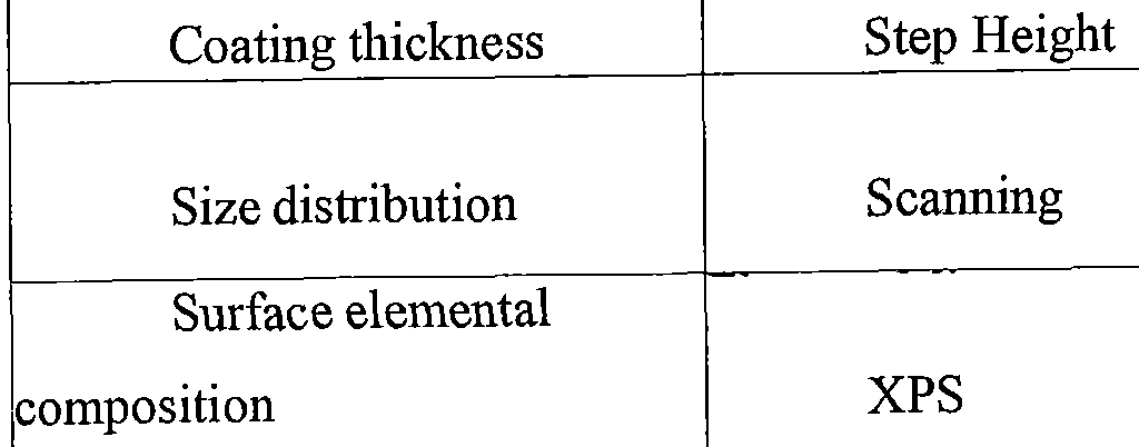


Claims
Priority Applications (15)
| Application Number | Priority Date | Filing Date | Title |
|---|---|---|---|
| CN202310836000.4A CN116898966A (en) | 2016-02-02 | 2017-02-02 | Phosphorescent pharmaceutical activators, suspensions thereof, systems containing suspensions and methods of use |
| IL260849A IL260849B (en) | 2016-02-02 | 2017-02-02 | Phosphor-containing drug activator, suspension thereof, system containing the suspension, and methods for use |
| AU2017213801A AU2017213801B2 (en) | 2016-02-02 | 2017-02-02 | Phosphor-containing drug activator, suspension thereof, system containing the suspension, and methods for use |
| US16/074,707 US11260129B2 (en) | 2016-02-02 | 2017-02-02 | Phosphor-containing drug activator, suspension thereof, system containing the suspension, and methods for use |
| EP17748119.9A EP3411119A4 (en) | 2016-02-02 | 2017-02-02 | LUMINOPHORE CONTAINING MEDICINAL ACTIVATOR, SUSPENSION THEREOF, SUSPENSION CONTAINING SYSTEM, AND METHODS OF USE |
| CN201780014860.2A CN108778417A (en) | 2016-02-02 | 2017-02-02 | Phosphor-containing drug activators, suspensions thereof, systems comprising suspensions and methods of use |
| KR1020187025292A KR102774788B1 (en) | 2016-02-02 | 2017-02-02 | Phosphor-containing drug activator, suspension thereof, system containing the suspension, and method of use |
| JP2018559681A JP6966478B2 (en) | 2016-02-02 | 2017-02-02 | Phosphor-containing drug activator, suspension thereof, system containing suspension, and method of use |
| BR112018015749A BR112018015749A2 (en) | 2016-02-02 | 2017-02-02 | phosphorus-containing drug activator, suspension of a phosphorus-containing drug activator, and system and method for treating a disease. |
| KR1020257006041A KR20250029299A (en) | 2016-02-02 | 2017-02-02 | Phosphor-containing drug activator, suspension thereof, system containing the suspension, and methods for use |
| CA3013335A CA3013335A1 (en) | 2016-02-02 | 2017-02-02 | Phosphor-containing drug activator, suspension thereof, system containing the suspension, and methods for use |
| SA518392126A SA518392126B1 (en) | 2016-02-02 | 2018-07-31 | A drug activator containing a phosphorous compound, a suspension thereof, a system containing the suspension, and methods of use |
| JP2021171729A JP7206350B2 (en) | 2016-02-02 | 2021-10-20 | Phosphor-Containing Drug Activators, Suspensions Thereof, Systems Containing Suspensions, and Methods of Use |
| US17/538,770 US12226483B2 (en) | 2016-02-02 | 2021-11-30 | Phosphor-containing drug activator, suspension thereof, system containing the suspension, and methods for use |
| US18/751,743 US20240342287A1 (en) | 2016-02-02 | 2024-06-24 | Phosphor-containing drug activator kit |
Applications Claiming Priority (4)
| Application Number | Priority Date | Filing Date | Title |
|---|---|---|---|
| US201662290203P | 2016-02-02 | 2016-02-02 | |
| US62/290,203 | 2016-02-02 | ||
| US201662304525P | 2016-03-07 | 2016-03-07 | |
| US62/304,525 | 2016-03-07 |
Related Child Applications (2)
| Application Number | Title | Priority Date | Filing Date |
|---|---|---|---|
| US16/074,707 A-371-Of-International US11260129B2 (en) | 2016-02-02 | 2017-02-02 | Phosphor-containing drug activator, suspension thereof, system containing the suspension, and methods for use |
| US17/538,770 Continuation US12226483B2 (en) | 2016-02-02 | 2021-11-30 | Phosphor-containing drug activator, suspension thereof, system containing the suspension, and methods for use |
Publications (1)
| Publication Number | Publication Date |
|---|---|
| WO2017136504A1 true WO2017136504A1 (en) | 2017-08-10 |
Family
ID=59501058
Family Applications (1)
| Application Number | Title | Priority Date | Filing Date |
|---|---|---|---|
| PCT/US2017/016138 Ceased WO2017136504A1 (en) | 2016-02-02 | 2017-02-02 | Phosphor-containing drug activator, suspension thereof, system containing the suspension, and methods for use |
Country Status (11)
| Country | Link |
|---|---|
| US (3) | US11260129B2 (en) |
| EP (1) | EP3411119A4 (en) |
| JP (2) | JP6966478B2 (en) |
| KR (2) | KR20250029299A (en) |
| CN (2) | CN108778417A (en) |
| AU (1) | AU2017213801B2 (en) |
| BR (1) | BR112018015749A2 (en) |
| CA (1) | CA3013335A1 (en) |
| IL (1) | IL260849B (en) |
| SA (1) | SA518392126B1 (en) |
| WO (1) | WO2017136504A1 (en) |
Cited By (1)
| Publication number | Priority date | Publication date | Assignee | Title |
|---|---|---|---|---|
| JP2020142062A (en) * | 2019-03-05 | 2020-09-10 | グリフォルス・ワールドワイド・オペレーションズ・リミテッド | How to prepare a blood product container |
Families Citing this family (5)
| Publication number | Priority date | Publication date | Assignee | Title |
|---|---|---|---|---|
| CA2946386C (en) * | 2014-04-22 | 2024-01-02 | Immunolight, Llc | Tumor imaging using photon-emitting phosphors having therapeutic properties |
| CA3131865A1 (en) | 2019-03-04 | 2020-09-10 | Immunolight, Llc. | Energy augment structures for use with energy emitters and collectors |
| EP3880379A4 (en) * | 2019-03-22 | 2022-09-28 | Komatsuseiki Kosakusho Co., Ltd. | METALLIC MATERIAL AND ARTICLES MADE THEREOF HAVING BIOLOGICAL PROPERTIES |
| KR102355947B1 (en) * | 2021-03-23 | 2022-02-08 | 어썸레이 주식회사 | Treatment apparatus and method for treating, inhibiting and preventing inflammatory disease by using electromagnetic wave |
| US20260035587A1 (en) * | 2022-08-29 | 2026-02-05 | Immunolight, Llc | Methods of improving emission output of coated energy modulation agents and compositions produced thereby |
Citations (5)
| Publication number | Priority date | Publication date | Assignee | Title |
|---|---|---|---|---|
| US5820872A (en) * | 1992-11-18 | 1998-10-13 | Yale University | Methods and compositions for improving the effectiveness of X-irradiation therapy for the treatment of an internal solid tumor |
| WO2000047402A1 (en) * | 1998-12-02 | 2000-08-17 | Advanced Refractory Technologies, Inc. | Fluorine-doped diamond-like coatings |
| US20130189803A1 (en) * | 2012-01-19 | 2013-07-25 | Nanoco Technologies, Ltd. | Molded Nanoparticle Phosphor For Light Emitting Applications |
| WO2014143826A2 (en) * | 2013-03-15 | 2014-09-18 | Bourke Frederic Avery | Interior energy-activation of photo-reactive species inside a medium or body |
| WO2015164485A1 (en) * | 2014-04-22 | 2015-10-29 | Immunolight, Llc. | Tumor imaging using photon-emitting phosphorus having therapeutic properties |
Family Cites Families (9)
| Publication number | Priority date | Publication date | Assignee | Title |
|---|---|---|---|---|
| KR100571307B1 (en) | 1999-02-09 | 2006-04-17 | 소니 가부시끼 가이샤 | Coding Systems and Methods, Coding Devices and Methods, Decoding Devices and Methods, Recording Devices and Methods, and Reproducing Devices and Methods |
| CA3008270A1 (en) * | 2005-09-21 | 2007-03-29 | The Regents Of The University Of California | Systems, compositions, and methods for local imaging and treatment of pain |
| US9358292B2 (en) | 2007-04-08 | 2016-06-07 | Immunolight, Llc | Methods and systems for treating cell proliferation disorders |
| US8383836B2 (en) | 2009-04-21 | 2013-02-26 | Duke University | Methods and systems for treating cell proliferation disorders with psoralen derivatives |
| US20090104130A1 (en) | 2007-04-16 | 2009-04-23 | Bernstein Eric F | Compositions comprising organic and inorganic phosphors for converting electromagnetic radiation and methods for using the same |
| US9907976B2 (en) | 2011-07-08 | 2018-03-06 | Immunolight, Llc | Phosphors and scintillators for light stimulation within a medium |
| WO2010123993A1 (en) | 2009-04-21 | 2010-10-28 | Tuan Vo-Dinh | Non-invasive energy upconversion methods and systems for in-situ photobiomodulation |
| PT3213738T (en) * | 2010-08-27 | 2020-03-31 | Sebacia Inc | Compositions and methods for targeted thermomodulation |
| EP3244928A4 (en) | 2015-01-14 | 2018-08-01 | Immunolight, Llc. | Non-invasive systems and methods for treatment of a host carrying a virus with photoactivatable drugs |
-
2017
- 2017-02-02 JP JP2018559681A patent/JP6966478B2/en active Active
- 2017-02-02 BR BR112018015749A patent/BR112018015749A2/en not_active Application Discontinuation
- 2017-02-02 WO PCT/US2017/016138 patent/WO2017136504A1/en not_active Ceased
- 2017-02-02 CN CN201780014860.2A patent/CN108778417A/en active Pending
- 2017-02-02 IL IL260849A patent/IL260849B/en unknown
- 2017-02-02 AU AU2017213801A patent/AU2017213801B2/en active Active
- 2017-02-02 US US16/074,707 patent/US11260129B2/en active Active
- 2017-02-02 CN CN202310836000.4A patent/CN116898966A/en active Pending
- 2017-02-02 EP EP17748119.9A patent/EP3411119A4/en active Pending
- 2017-02-02 KR KR1020257006041A patent/KR20250029299A/en active Pending
- 2017-02-02 CA CA3013335A patent/CA3013335A1/en active Pending
- 2017-02-02 KR KR1020187025292A patent/KR102774788B1/en active Active
-
2018
- 2018-07-31 SA SA518392126A patent/SA518392126B1/en unknown
-
2021
- 2021-10-20 JP JP2021171729A patent/JP7206350B2/en active Active
- 2021-11-30 US US17/538,770 patent/US12226483B2/en active Active
-
2024
- 2024-06-24 US US18/751,743 patent/US20240342287A1/en not_active Abandoned
Patent Citations (5)
| Publication number | Priority date | Publication date | Assignee | Title |
|---|---|---|---|---|
| US5820872A (en) * | 1992-11-18 | 1998-10-13 | Yale University | Methods and compositions for improving the effectiveness of X-irradiation therapy for the treatment of an internal solid tumor |
| WO2000047402A1 (en) * | 1998-12-02 | 2000-08-17 | Advanced Refractory Technologies, Inc. | Fluorine-doped diamond-like coatings |
| US20130189803A1 (en) * | 2012-01-19 | 2013-07-25 | Nanoco Technologies, Ltd. | Molded Nanoparticle Phosphor For Light Emitting Applications |
| WO2014143826A2 (en) * | 2013-03-15 | 2014-09-18 | Bourke Frederic Avery | Interior energy-activation of photo-reactive species inside a medium or body |
| WO2015164485A1 (en) * | 2014-04-22 | 2015-10-29 | Immunolight, Llc. | Tumor imaging using photon-emitting phosphorus having therapeutic properties |
Cited By (3)
| Publication number | Priority date | Publication date | Assignee | Title |
|---|---|---|---|---|
| JP2020142062A (en) * | 2019-03-05 | 2020-09-10 | グリフォルス・ワールドワイド・オペレーションズ・リミテッド | How to prepare a blood product container |
| JP2024092018A (en) * | 2019-03-05 | 2024-07-05 | グリフォルス・ワールドワイド・オペレーションズ・リミテッド | Method for preparing a blood product container |
| JP7747431B2 (en) | 2019-03-05 | 2025-10-01 | グリフォルス・ワールドワイド・オペレーションズ・リミテッド | Method for preparing a blood product container |
Also Published As
| Publication number | Publication date |
|---|---|
| EP3411119A4 (en) | 2020-01-01 |
| JP7206350B2 (en) | 2023-01-17 |
| BR112018015749A2 (en) | 2019-01-08 |
| JP2019510074A (en) | 2019-04-11 |
| US20220080045A1 (en) | 2022-03-17 |
| CA3013335A1 (en) | 2017-08-10 |
| IL260849A (en) | 2018-09-20 |
| US11260129B2 (en) | 2022-03-01 |
| AU2017213801B2 (en) | 2022-04-14 |
| JP6966478B2 (en) | 2021-11-17 |
| CN116898966A (en) | 2023-10-20 |
| SA518392126B1 (en) | 2023-06-01 |
| US20240342287A1 (en) | 2024-10-17 |
| AU2017213801A1 (en) | 2018-08-16 |
| US12226483B2 (en) | 2025-02-18 |
| JP2022020680A (en) | 2022-02-01 |
| EP3411119A1 (en) | 2018-12-12 |
| KR20250029299A (en) | 2025-03-04 |
| IL260849B (en) | 2022-07-01 |
| KR102774788B1 (en) | 2025-02-27 |
| CN108778417A (en) | 2018-11-09 |
| US20200282056A1 (en) | 2020-09-10 |
| KR20220022907A (en) | 2022-03-02 |
Similar Documents
| Publication | Publication Date | Title |
|---|---|---|
| US12226483B2 (en) | Phosphor-containing drug activator, suspension thereof, system containing the suspension, and methods for use | |
| US11865359B2 (en) | Tumor imaging with x-rays and other high energy sources using as contrast agents photon-emitting phosphors having therapeutic properties | |
| US10940329B2 (en) | Phosphor-containing drug activator activatable by a Monte Carlo derived x-ray exposure, system containing the activator, and methods for use | |
| US11992697B2 (en) | X-ray psoralen activated cancer therapy (X-PACT) | |
| US10441810B2 (en) | X-ray psoralen activated cancer therapy (X-PACT) | |
| EP3630283B1 (en) | X-ray psoralen activated cancer therapy (x-pact) with associated treatments | |
| US11577092B2 (en) | Phosphor-containing drug activator activatable by a monte carlo derived X-ray exposure, system containing the activator, and methods for use | |
| US20230338539A1 (en) | Method of treating difficult to access tumors with photoactivated cancer therapy | |
| BR122024007197A2 (en) | SYSTEM AND USE OF A PHOTOACTIVABLE DRUG |
Legal Events
| Date | Code | Title | Description |
|---|---|---|---|
| 121 | Ep: the epo has been informed by wipo that ep was designated in this application |
Ref document number: 17748119 Country of ref document: EP Kind code of ref document: A1 |
|
| WWE | Wipo information: entry into national phase |
Ref document number: 260849 Country of ref document: IL |
|
| WWE | Wipo information: entry into national phase |
Ref document number: 3013335 Country of ref document: CA |
|
| ENP | Entry into the national phase |
Ref document number: 2018559681 Country of ref document: JP Kind code of ref document: A |
|
| NENP | Non-entry into the national phase |
Ref country code: DE |
|
| REG | Reference to national code |
Ref country code: BR Ref legal event code: B01A Ref document number: 112018015749 Country of ref document: BR |
|
| ENP | Entry into the national phase |
Ref document number: 2017213801 Country of ref document: AU Date of ref document: 20170202 Kind code of ref document: A |
|
| WWE | Wipo information: entry into national phase |
Ref document number: 2017748119 Country of ref document: EP |
|
| ENP | Entry into the national phase |
Ref document number: 2017748119 Country of ref document: EP Effective date: 20180903 |
|
| ENP | Entry into the national phase |
Ref document number: 112018015749 Country of ref document: BR Kind code of ref document: A2 Effective date: 20180801 |
|
| WWD | Wipo information: divisional of initial pct application |
Ref document number: 1020257006041 Country of ref document: KR |
|
| WWP | Wipo information: published in national office |
Ref document number: 1020257006041 Country of ref document: KR |