WO2017138497A1 - 被検物質の検出方法および被検物質の検出用試薬キット - Google Patents
被検物質の検出方法および被検物質の検出用試薬キット Download PDFInfo
- Publication number
- WO2017138497A1 WO2017138497A1 PCT/JP2017/004271 JP2017004271W WO2017138497A1 WO 2017138497 A1 WO2017138497 A1 WO 2017138497A1 JP 2017004271 W JP2017004271 W JP 2017004271W WO 2017138497 A1 WO2017138497 A1 WO 2017138497A1
- Authority
- WO
- WIPO (PCT)
- Prior art keywords
- solid phase
- complex
- antibody
- binding
- capture antibody
- Prior art date
- Legal status (The legal status is an assumption and is not a legal conclusion. Google has not performed a legal analysis and makes no representation as to the accuracy of the status listed.)
- Ceased
Links
Images


Classifications
-
- G—PHYSICS
- G01—MEASURING; TESTING
- G01N—INVESTIGATING OR ANALYSING MATERIALS BY DETERMINING THEIR CHEMICAL OR PHYSICAL PROPERTIES
- G01N33/00—Investigating or analysing materials by specific methods not covered by groups G01N1/00 - G01N31/00
- G01N33/48—Biological material, e.g. blood, urine; Haemocytometers
- G01N33/50—Chemical analysis of biological material, e.g. blood, urine; Testing involving biospecific ligand binding methods; Immunological testing
- G01N33/53—Immunoassay; Biospecific binding assay; Materials therefor
- G01N33/543—Immunoassay; Biospecific binding assay; Materials therefor with an insoluble carrier for immobilising immunochemicals
- G01N33/54306—Solid-phase reaction mechanisms
-
- G—PHYSICS
- G01—MEASURING; TESTING
- G01N—INVESTIGATING OR ANALYSING MATERIALS BY DETERMINING THEIR CHEMICAL OR PHYSICAL PROPERTIES
- G01N33/00—Investigating or analysing materials by specific methods not covered by groups G01N1/00 - G01N31/00
- G01N33/48—Biological material, e.g. blood, urine; Haemocytometers
- G01N33/50—Chemical analysis of biological material, e.g. blood, urine; Testing involving biospecific ligand binding methods; Immunological testing
- G01N33/53—Immunoassay; Biospecific binding assay; Materials therefor
- G01N33/543—Immunoassay; Biospecific binding assay; Materials therefor with an insoluble carrier for immobilising immunochemicals
- G01N33/54313—Immunoassay; Biospecific binding assay; Materials therefor with an insoluble carrier for immobilising immunochemicals the carrier being characterised by its particulate form
-
- G—PHYSICS
- G01—MEASURING; TESTING
- G01N—INVESTIGATING OR ANALYSING MATERIALS BY DETERMINING THEIR CHEMICAL OR PHYSICAL PROPERTIES
- G01N2333/00—Assays involving biological materials from specific organisms or of a specific nature
- G01N2333/435—Assays involving biological materials from specific organisms or of a specific nature from animals; from humans
- G01N2333/46—Assays involving biological materials from specific organisms or of a specific nature from animals; from humans from vertebrates
- G01N2333/47—Assays involving proteins of known structure or function as defined in the subgroups
- G01N2333/4701—Details
- G01N2333/4709—Amyloid plaque core protein
-
- G—PHYSICS
- G01—MEASURING; TESTING
- G01N—INVESTIGATING OR ANALYSING MATERIALS BY DETERMINING THEIR CHEMICAL OR PHYSICAL PROPERTIES
- G01N2800/00—Detection or diagnosis of diseases
- G01N2800/28—Neurological disorders
- G01N2800/2814—Dementia; Cognitive disorders
- G01N2800/2821—Alzheimer
Definitions
- the present invention relates to a test substance detection method and a test substance detection reagent kit.
- a sandwich immunoassay widely used as an immunological assay is a method in which a labeled antibody and a capture antibody are bound to an antigen in a sample and the antigen is detected based on the label.
- the sandwich immunoassay when the epitope of the labeled antibody and the epitope of the capture antibody overlap, only one of them binds to the antigen due to steric hindrance and cannot be detected, and thus two types of antibodies with different epitopes are usually used.
- the antigen is a multimer, a plurality of identical epitopes exist in the antigen.
- two types of antibodies with overlapping epitopes are used as a labeled antibody and a capture antibody, no monomer is detected and a multimer is detected.
- Patent Document 1 and Non-Patent Document 1 describe a sandwich immunoassay using a detection antibody and a capture antibody.
- the epitope of the detection antibody and the epitope of the capture antibody are the same or overlap, multimers of amyloid ⁇ can be detected.
- Non-Patent Documents 2 to 4 describe sandwich immunoassays using a detection antibody and two types of capture antibodies. In these documents, since the epitope of the detection antibody and the epitope of the two types of capture antibodies are all the same or overlap, the multimer of amyloid ⁇ can be detected.
- An object of the present invention is to provide a novel method for immunologically measuring an antigen.
- the present invention provides a method for detecting a test substance.
- This method includes a test substance, a labeled antibody that binds to the test substance, a first capture antibody that binds to the substance, a second capture antibody that binds to the test substance, and a first solid phase that binds to the first capture antibody.
- a complex containing a test substance, a labeled antibody, a first capture antibody and a second capture antibody on the first solid phase and dissociating the binding between the first capture antibody and the first solid phase Releasing the complex, bringing the complex into contact with the second solid phase that binds to the second capture antibody, and transferring the complex onto the second solid phase, and the complex on the second solid phase Measuring the label contained in the substance and detecting the substance.
- the test substance is a multimer, and the epitope of the labeled antibody, the epitope of the first capture antibody, and the second capture antibody epitope overlap.
- the present invention also provides a method for detecting a test substance in a different mode.
- a test substance, a labeled antibody that binds to the test substance, a capture antibody that binds to the test substance, a first solid phase that binds to the capture antibody are contacted, and the test substance, the labeled antibody, and the capture antibody are brought into contact with each other.
- Forming a complex on the first solid phase releasing the complex by dissociating the binding between the capture antibody and the first solid phase, and binding the capture antibody with the second solid phase and the complex.
- the test substance is a multimer, and the epitope of the labeled antibody and the epitope of the capture antibody overlap.
- the present invention also provides a method for detecting a test substance in a different mode.
- a test substance, a labeled antibody that binds to the test substance, a capture antibody that binds to the test substance, and a first solid phase that binds to the capture antibody are brought into contact with each other.
- the test substance is amyloid ⁇ or tau protein.
- the measurement mechanism of a 1st embodiment is shown.
- the measurement mechanism of 2nd Embodiment is shown.
- the measurement mechanism of 3rd Embodiment is shown.
- An example of a reagent kit is shown.
- An example of a reagent kit is shown.
- 6 is a graph showing the results of Example 1 and Comparative Example 1.
- 10 is a graph showing the results of Example 2.
- 10 is a graph showing the results of Example 3. It is a graph which shows the result of Example 4.
- 10 is a graph showing the results of Example 5.
- the test substance is detected by an immune complex transfer method (hereinafter also referred to as “ICT-EIA”) including the steps described below.
- ICT-EIA immune complex transfer method
- a multimer is detected as a test substance.
- a “multimer” is also called a polymer, and is formed by polymerizing or agglomerating a plurality of monomer molecules physically and chemically.
- monomer molecules include polypeptides, nucleic acids, sugar chains and the like.
- Specific examples of the polypeptide multimer include amyloid ⁇ oligomer obtained by polymerizing amyloid ⁇ monomer, tau oligomer obtained by polymerizing tau protein, and the like.
- serum-amyloid-A-protein-aggregate, IgG-light chain-aggregate, AapoAI-aggregate, AapoAII-aggregate, ATTR-aggregate, DISC1-aggregate, FUS-aggregate, IAPP- Examples include aggregates, SOD1-aggregates, ⁇ -synuclein-aggregates, TDP-43-aggregates, huntingtin-aggregates, lysozyme-aggregates and the like.
- the “multimer” only needs to contain a plurality of monomer molecules, and may contain other molecules. The “multimer” does not necessarily require that monomer molecules are firmly bonded to each other by a covalent bond or the like, and includes aggregates aggregated and aggregated by looser bonds.
- Amyloid ⁇ is a polypeptide usually consisting of about 40 amino acids and has the property of aggregating to form insoluble amyloid fibrils.
- Tau protein also called “tau”, is a protein having a molecular weight of about 50,000 and has functions such as binding between minute cells in the cell to stabilize the cytoskeleton. Tau protein exists in six isoforms due to alternative splicing of exon 2, exon 3 and exon 10 in humans.
- tau protein or “tau” includes any isoform unless otherwise specified.
- the test substance is included in biological samples.
- the sample include a sample collected from a living body or a dead body, cultured cells, and the like. Specific examples include brain tissue, blood (whole blood), plasma, serum, cerebrospinal fluid and the like.
- the biological species is not particularly limited, and can be collected from a human or a non-human mammal.
- the test substance, the labeled antibody that binds to the test substance, the first capture antibody that binds to the test substance, the second capture antibody that binds to the test substance, and the first capture antibody bind to
- the first solid phase is brought into contact with each other to form a complex containing the test substance, the labeled antibody, the first capture antibody, and the second capture antibody on the first solid phase.
- the “antibody” includes an antigen-binding antibody fragment such as Fab, F (ab ′) 2 or a derivative thereof.
- the forming step is usually performed in solution.
- the epitope of the labeled antibody, the epitope of the first capture antibody, and the epitope of the second capture antibody overlap.
- “Overlapping epitopes” means that the epitopes of these antibodies are identical or partially matched. When the epitopes are identical or partially matched, only one antibody binds to the epitope of the monomer molecule, and other antibodies are inhibited from binding due to steric hindrance.
- a multimer containing a plurality of epitopes can be detected.
- detection of monomer molecules and detection of multimers (dimers) containing two monomers are suppressed, and multimers containing three or more molecules of monomers are preferably detected.
- the labeled antibody is not particularly limited as long as it is an antibody that can specifically bind to a test substance and is labeled with a known labeling substance commonly used in immunological techniques.
- a labeled antibody can be prepared by binding or linking an antibody and a labeling substance by a known method using an appropriate cross-linking agent or a commercially available labeling kit.
- the labeling substance is not particularly limited as long as it can emit a measurable signal, and examples thereof include enzymes, fluorescent substances, and radioisotopes.
- the enzyme include alkaline phosphatase, ⁇ -galactosidase, peroxidase, glucose oxidase, tyrosinase, acid phosphatase, and luciferase.
- the fluorescent substance include fluorescent dyes such as fluorescein isothiocyanate (FITC) and fluorescent proteins such as green fluorescent protein (GFP).
- FITC fluorescein isothiocyanate
- GFP green fluorescent protein
- radioactive isotopes include 125 I, 14 C, and 32 P. Among them, an enzyme is particularly preferable as the labeling substance.
- the first solid phase can be selected from known solid phases commonly used in immunological techniques.
- a solid phase material include latex, rubber, polyethylene, polypropylene, polystyrene, styrene-butadiene copolymer, polyvinyl chloride, polyvinyl acetate, polyacrylamide, polymethacrylate, styrene-methacrylate copolymer, Examples thereof include polyglycidyl methacrylate, acrolein-ethylene glycol dimethacrylate copolymer, polyvinylidene difluoride (PVDF), silicone, agarose, gelatin, erythrocyte, silica gel, glass, inert alumina, and a magnetic substance.
- PVDF polyvinylidene difluoride
- Examples of the shape of the solid phase include a microtiter plate, a test tube, and particles.
- the particles may have magnetism.
- Magnetic particles are known in the art and include particles containing Fe 2 O 3 and / or Fe 3 O 4 , cobalt, nickel, phyllite, magnetite, etc. as a substrate.
- the first capture antibody in the complex binds to the first solid phase, thereby immobilizing the complex on the first solid phase.
- the binding mode between the first capture antibody and the first solid phase is not particularly limited as long as it is a dissociable bond, and examples thereof include physical adsorption and ionic bond. Moreover, both can also be combined using the substance which intervenes between the 1st capture antibody and the 1st solid phase. As such a substance, a combination of two substances that specifically bind to each other and can be dissociated is preferable (the two substances are referred to as “binding substance” and “binding partner”, respectively).
- binding substances and binding partners are known in the art.
- antigens excluding test substances
- their antibodies ligands and their receptors
- oligonucleotides and their complementary strands biotins (biotin, desthiobiotin) And avidin (including avidin analogs such as avidin and streptavidin), nickel and histidine tags, glutathione and glutathione-S-transferase.
- avidin including avidin analogs such as avidin and streptavidin
- nickel and histidine tags glutathione and glutathione-S-transferase.
- a hapten and an anti-hapten antibody As a combination of an antigen and its antibody, a hapten and an anti-hapten antibody, and biotin (or desthiobiotin) and an anti-biotin antibody (or anti-desthiobiotin antibody) are preferable.
- biotin or desthiobiotin
- anti-biotin antibody or anti-desthiobiotin antibody
- 2, 4-dinitrophenol (DNP) and an anti-DNP antibody are particularly preferable.
- the binding substance and which is the binding partner is not particularly limited.
- the hapten may be used as a binding substance
- the anti-hapten antibody may be used as a binding partner
- the anti-hapten antibody may be used as a binding substance and the hapten may be used as a binding partner. Since the binding substance is bound to the first capture antibody, it is preferable to use a hapten as a binding substance and an anti-hapten antibody as a binding partner.
- Methods for binding a binding agent or binding partner to an antibody and a first solid phase are known in the art.
- a method using a crosslinking agent that reacts with an amino group or a thiol group in the antibody for example, maleimide, N-hydroxysuccinimide, etc.
- a method for binding the binding partner to the first solid phase a physical adsorption method, a covalent bond method, an ionic bond method, or the like is known.
- the test substance, the labeled antibody, the first capture antibody, and the second capture antibody are bound by an antigen-antibody reaction, and “labeled antibody-test substance—first capture antibody and second capture antibody” Is formed. Then, the first capture antibody contained in the complex is captured on the first solid phase.
- the addition amount of the labeled antibody, the first capture antibody, the second capture antibody and the first solid phase is not particularly limited, but is usually added to the reaction system in excess of the expected amount of the test substance.
- the order of contact with the sample containing the labeled antibody, the first capture antibody, the second capture antibody, the first solid phase and the test substance is not particularly limited.
- the labeled antibody, the first capture antibody, the second capture antibody, and the test substance are contacted to form a complex, and then the first solid phase is contacted.
- the first capture antibody and the first solid phase are first contacted.
- reaction temperature and reaction time in the formation step are not particularly limited, but it is usually sufficient to stand at a temperature of 20 to 45 ° C. for 15 to 30 minutes or gently agitate.
- the reaction system in addition to the first solid phase in which the complex is formed, there are components of the pretreatment liquid, impurities contained in the sample, and unreacted components such as excess antibodies.
- unreacted component refers to a component other than the complex bound on the first solid phase and a free component not bound to the first solid phase.
- the recovery step is generally referred to as B / F separation, and the first solid phase is separated and recovered from unreacted components. Therefore, the unreacted component which has a bad influence on the measurement process mentioned later can be removed by this collection process. In the recovery step, it is not necessary to completely remove unreacted components, and they may remain as long as they do not adversely affect the measurement.
- the method itself for recovering the first solid phase in the sample is known in the art, and can be appropriately determined according to the type of the first solid phase.
- the first solid phase in the sample can be recovered by magnetic separation. Specifically, the magnetic particles are recovered by bringing the magnet close to the wall of the container containing the sample obtained in the forming process, fixing the magnetic particles in the sample to the wall of the container, and removing the liquid phase by suction. can do.
- gelatin particles or latex particles are used, the particles can be collected by centrifugation and then recovered by sucking and removing the liquid phase.
- the recovery step may further include a step of washing the recovered first solid phase.
- the washing of the first solid phase can be performed, for example, by adding a washing solution to the collected first solid phase and then removing the washing solution from the first solid phase.
- the washing solution is preferably a buffer solution that does not damage the complex formed on the first solid phase.
- a buffer solution containing a surfactant is particularly preferable, and examples thereof include TBS-T (0.05% Tween 20-containing Tris buffered saline) and PBST (0.05% Tween 20-containing phosphate buffered saline).
- TBS-T 0.05% Tween 20-containing Tris buffered saline
- PBST 0.05% Tween 20-containing phosphate buffered saline
- a commercially available cleaning solution such as an HISCL cleaning solution (manufactured by Sysmex Corporation) can also be used.
- a step of releasing the complex on the first solid phase and transferring it to a second solid phase different from the first solid phase is performed. Release is usually done in solution.
- the method itself for releasing the complex on the solid phase is known in the art.
- a method using a substance capable of dissociating the binding between the first capture antibody in the complex obtained in the forming step and the first solid phase (hereinafter referred to as “release agent”) can be mentioned.
- the release agent is known in the art, and can be appropriately selected depending on the binding mode between the first capture antibody and the first solid phase.
- the complex when the first capture antibody in the complex and the first solid phase are bound by physical adsorption, the complex is liberated by using a solution containing a surfactant as the liberating agent. be able to. In the case of ionic bonding, the complex can be liberated by using a solution containing ions.
- the first capture antibody and the first solid phase are bound using the affinity between the binding substance and the binding partner, by using a release agent that dissociates the binding between the binding substance and the binding partner.
- the complex can be released.
- release agents are also known in the art and can be appropriately selected depending on the combination of the binding substance and the binding partner.
- the hapten or a derivative thereof can be used as a release agent.
- biotin or desthiobiotin
- avidin or streptavidin
- the treatment temperature and treatment time can be appropriately set depending on the type of release agent. Usually, it is allowed to stand for 3 to 8 minutes at a temperature of 20 to 45 ° C. or gently stirred.
- the complex released from the first solid phase as described above is brought into contact with and bound to the second solid phase different from the first solid phase, whereby the complex is To be transferred.
- the “second solid phase different from the first solid phase” means a second solid phase different from the first solid phase that is present when added in the formation step. (Hereinafter also simply referred to as “second solid phase”). That is, in this transfer step, it is not intended that the released complex binds again to the first solid phase present from when it was added in the formation step.
- the complex released from the first solid phase is recovered and brought into contact with a newly prepared second solid phase.
- the complex when particles are used as the first solid phase, the complex is released from the first solid phase, and then the first solid phase is separated from the wall surface or bottom of the container by magnetic force or centrifugation in the same manner as in the above recovery step. To fix. Then, the liquid phase containing the complex can be recovered, and the liquid phase can be brought into contact with the second solid phase. Contact between the complex and the second solid phase is usually performed in solution.
- the material and shape of the second solid phase are the same as those described for the first solid phase.
- the materials and shapes of the first solid phase and the second solid phase may be different from or different from those of the first solid phase.
- the second capture antibody contained in the released complex binds to the second solid phase, so that the complex is fixed to the second solid phase.
- the binding mode of the second solid phase and the complex is not particularly limited, but it is preferable to bind both using a substance that intervenes between the second capture antibody in the complex and the second solid phase.
- Such materials include combinations of the binding materials and binding partners described above. For example, by binding the binding substance to the second capture antibody in advance as a “binding site for the second solid phase” and binding the binding partner to the second solid phase, the affinity between the binding substance and the binding partner can be increased. Utilizing this, the second capture antibody in the complex can be bound to the second solid phase.
- biotins and avidins are preferable.
- the combination of the binding substance and the binding partner for binding the second capture antibody and the second solid phase is a combination of the binding substance and the binding partner for binding the first capture antibody and the first solid phase.
- a first capture antibody bound with DNP a first solid phase immobilized with an anti-DNP antibody, a second capture antibody bound with biotin, and a second solid phase immobilized with streptavidin can be used.
- the treatment temperature and treatment time for transferring the complex to the second solid phase are not particularly limited.
- the complex is allowed to stand at a temperature of 20 to 45 ° C. for 1 to 30 minutes or gently stirred.
- the shape of the second solid phase is particles, it may be allowed to stand for 1 to 5 minutes or gently stirred.
- the second solid phase recovery step can be performed by the same method as the first solid phase recovery step.
- the label contained in the complex on the second solid phase is measured.
- the method of measuring the label itself is known in the art.
- an appropriate measurement method can be selected according to the type of signal derived from the labeling substance.
- the labeling substance is an enzyme
- it can be performed by measuring a signal such as light and color generated by reacting a substrate for the enzyme with a known apparatus.
- Examples of such a measuring device include a spectrophotometer and a luminometer.
- the substrate of the enzyme can be appropriately selected from known substrates depending on the type of enzyme.
- the substrate is CDP-Star (registered trademark) (4-chloro-3- (methoxyspiro [1,2-dioxetane-3, 2 '-(5'-chloro) trixiro). [3. 3. 1. 1 3, 7 ] decane] -4-yl) phenyl phosphate disodium), CSPD® (3- (4-methoxyspiro [1,2-dioxetane-3,2- Chemiluminescent substrates such as (5'-chloro) tricyclo [3. 3. 1.
- the labeling substance is a radioisotope
- the radiation as a signal can be measured using a known device such as a scintillation counter.
- the labeling substance is a fluorescent substance
- the fluorescence as a signal can be measured using a known device such as a fluorescent microplate reader.
- test substance is detected based on the above measurement result.
- detect includes qualitative detection, quantification, and semi-quantification of existence.
- semi-quantitative means that the content of the test substance is determined stepwise such as “negative”, “weak positive”, “strong positive”, and the like.
- the threshold value includes a sample that does not contain a plurality of test substances or a trace amount (for example, a biological sample obtained from a healthy person), and a sample that contains a plurality of test substances (for example, neurological diseases such as Alzheimer's dementia)
- a test substance concentration is measured in advance using a biological sample obtained from a patient, and a value that can most accurately classify a negative group and a positive group can be used as a threshold value.
- those skilled in the art can appropriately set the threshold according to the purpose of measurement in consideration of sensitivity, specificity, positive predictive value, negative predictive value, and the like.
- This specific example is a system that suitably detects oligomers of trimer or higher of amyloid ⁇ (denoted as A ⁇ in the figure). This is merely an example and does not limit the present invention.
- the labeled antibody is labeled with an enzyme.
- DNP is added to the first capture antibody.
- Biotin is added to the second capture antibody.
- the first solid phase is a particle on which an anti-DNP antibody is immobilized.
- the labeled antibody, the first capture antibody and the second capture antibody are the same monoclonal antibody that recognizes a specific epitope of amyloid ⁇ . That is, the epitopes of these antibodies are the same.
- FIG. 1E After B / F separation, an enzyme substrate is added, and luminescence generated by the enzyme reaction is measured.
- the first capture antibody is bound to the monomer, but there may be a monomer to which the labeled antibody or the second capture antibody is bound instead of the first capture antibody. No signal is detected from this complex when a labeled antibody or a second capture antibody is bound.
- the first capture antibody and the labeled antibody are bound to the dimer.
- the first capture antibody, the second capture antibody, and the labeled antibody two molecules of the same or different types of antibodies can bind. . In either case, no signal is detected from this complex.
- the first capture antibody, the second capture antibody, and the labeled antibody are bound to the trimer.
- the same kind of antibody may be bound to at least two of the three epitopes. It is done. In this case, no signal is detected from this complex.
- a test substance having a high degree of polymerization has a high probability of binding one or more molecules of the first capture antibody, the second capture antibody, and the labeled antibody.
- the lower the degree of polymerization the higher the probability that any one type of antibody will not bind.
- a multimer is detected as a test substance.
- the “multimer” and the “sample” including the test substance are the same as those described in the first embodiment.
- a test substance, a labeled antibody that binds to the test substance, a capture antibody that binds to the test substance, and a first solid phase that binds to the capture antibody are brought into contact with each other.
- a step of forming a complex containing an antibody and a capture antibody on the first solid phase is performed.
- the “antibody” is the same as described in the first embodiment.
- epitope of the labeled antibody and the epitope of the capture antibody overlap. “Epitope overlaps” is the same as described in the first embodiment. In this embodiment, since two types of antibodies are used, detection of monomer molecules is suppressed, and multimers are suitably detected.
- the labeled antibody is the same as that described in the first embodiment.
- the first solid phase is the same as that described in the first embodiment.
- the first binding substance and the second binding substance are bound to the capture antibody, A first binding partner that specifically binds to the first binding substance is immobilized on the first solid phase, and a second binding partner that specifically binds to the second binding substance is immobilized on the second solid phase.
- the binding mode between the capture antibody and the first solid phase is the same as the binding mode between the first capture antibody and the first solid phase described in the description of the first embodiment.
- the binding mode between the capture antibody and the second solid phase is the same as the binding mode between the first capture antibody and the second solid phase described in the description of the first embodiment.
- the test substance, the labeled antibody and the capture antibody are combined by an antigen-antibody reaction to form a sandwich complex including “labeled antibody-test substance-capture antibody”. Then, the capture antibody contained in the complex is captured on the first solid phase.
- the addition amount of the labeled antibody, the capture antibody and the first solid phase is the same as that described in the first embodiment.
- the order of contact with the sample containing the labeled antibody, the capture antibody, the first solid phase, and the test substance is the same as that described in the first embodiment.
- the recovery process is the same as that described in the first embodiment.
- the capture antibody includes both the first binding substance and the second binding substance.
- the second binding substance of the capture antibody contained in the released complex binds to the second binding partner on the second solid phase, the released complex is fixed to the second solid phase.
- the second solid phase recovery step can be performed by the same method as the first solid phase recovery step.
- a specific example of the measurement mechanism of the present embodiment will be described with reference to FIG.
- a specific example is a system that detects oligomers of dimer or higher of amyloid ⁇ (denoted as A ⁇ in the figure). This is merely an example and does not limit the present invention.
- the labeled antibody is labeled with an enzyme.
- DNP indicated by a circle in the figure
- biotin indicated by a square in the figure
- the first solid phase is a particle on which an anti-DNP antibody is immobilized.
- the labeled antibody and the capture antibody are the same monoclonal antibody that recognizes a specific epitope of amyloid ⁇ . That is, the epitopes of these antibodies are the same.
- the capture antibody is bound to the monomer, but there may also be a monomer to which the labeled antibody is bound instead of the capture antibody.
- the labeled antibody is bound, no signal is detected from this complex.
- a capture antibody and a labeled antibody are bound to the dimer, but two molecules of the same or different types of antibodies can be bound to the capture antibody and the labeled antibody. No signal is detected when two molecules of capture antibody are bound or when two molecules of labeled antibody are bound.
- the capture antibody and two labeled antibody molecules are bound to the trimer, but no signal is detected when three capture antibody molecules are bound or three labeled antibody molecules are bound.
- a test substance having a high degree of polymerization has a high probability of binding one or more molecules of the capture antibody and the labeled antibody.
- the lower the degree of polymerization the higher the probability that any one type of antibody will not bind.
- amyloid ⁇ and tau protein are detected as test substances.
- the epitope of the labeled antibody and the epitope of the capture antibody are different, and the labeled antibody and the capture antibody can bind to different sites of the amyloid ⁇ or tau protein that is the test substance of this embodiment. Thereby, not only multimers but also monomer molecules can be detected.
- the other points are the same as in the second embodiment.
- a specific example of the measurement mechanism of this embodiment will be described with reference to FIG.
- a specific example of this is a system that detects amyloid ⁇ (denoted as A ⁇ in the figure). This is merely an example and does not limit the present invention.
- FIG. 3A A sample containing a monomer and a dimer of amyloid ⁇ , a labeled antibody, a capture antibody, and a first solid phase are mixed.
- the labeled antibody is labeled with an enzyme.
- DNP indicated by a circle in the figure
- biotin indicated by a square in the figure
- the first solid phase is a particle on which an anti-DNP antibody is immobilized.
- the labeled antibody and the capture antibody have different epitopes and bind to different positions in the amino acid sequence of amyloid ⁇ .
- the sample contains a monomer and a dimer.
- the capture antibody and the labeled antibody can bind to the multimer more than the trimer in the same manner to detect the signal. it can.
- a test substance having a high degree of polymerization has a high probability of binding one or more molecules of the capture antibody and the labeled antibody.
- the lower the degree of polymerization the higher the probability that any one type of antibody will not bind.
- the reagent kit includes a labeled antibody, a first capture antibody, and a second capture antibody.
- a first solid phase and a second solid phase may further be included.
- the label is an enzyme, it may further contain a substrate for the enzyme. Further, it may further contain a release agent for separating the complex from the first solid phase.
- Some of these reagents may be housed in the same container, but each reagent may be housed in a separate container in order to suppress non-specific reactions in the container. In the case of an enzyme, the labeled antibody and the substrate need to be stored in separate containers.
- the release agent is added to the reaction system after forming a complex on the first solid phase, it is preferably accommodated in a container different from other reagents.
- These reagents are provided to the user in a dry state or in a liquid state (dissolved or dispersed in the liquid).
- the chemical reaction in the above detection method is preferably performed in a solution, the reagent is in a liquid state. It is preferable. The details of these reagents are the same as those described in the first embodiment.
- the reagent kit may further contain other reagents.
- a buffer solution is included.
- a buffer solution is not particularly limited as long as it has a buffering action at pH 6.5 to 8, for example, phosphate buffer solution (PBS), imidazole buffer solution, triethanolamine hydrochloride buffer solution (TEA).
- PBS phosphate buffer solution
- TAA triethanolamine hydrochloride buffer solution
- Good buffer solution include MES, Bis-Tris, ADA, PIPES, Bis-Tris-Propane, ACES, MOPS, MOPSO, BES, TES, HEPES, HEPPS, Tricine, Tris, Bicine, TAPS, etc. .
- the buffer may be a known additive such as a protein stabilizer (such as BSA), a preservative (such as sodium azide or phenylmethanesulfonyl fluoride), or an inorganic salt (such as magnesium chloride or zinc chloride), if necessary. May be included.
- a protein stabilizer such as BSA
- a preservative such as sodium azide or phenylmethanesulfonyl fluoride
- an inorganic salt such as magnesium chloride or zinc chloride
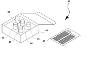
- Fig. 4 shows an example of a reagent kit.
- the reagent kit 40 of this embodiment includes a first container 41 containing a first capture antibody, a second container 42 containing a second capture antibody, a third container 43 containing particles as a first solid phase, A fourth container 44 containing particles as a second solid phase and a fifth container 45 containing labeled antibodies are included. These containers are accommodated in a packing box 48 together with the package insert 47.
- the present invention includes use of a labeled antibody, a first capture antibody, and a second capture antibody for producing a reagent kit used in the detection method of the first embodiment.
- the reagent kit includes a labeled antibody and a capture antibody. Unlike the reagent kit used in the first embodiment, only one type of capture antibody may be used. Other configurations are the same as those described for the reagent kit used in the first embodiment.
- Fig. 5 shows an example of a reagent kit.
- the reagent kit 30 of this embodiment includes a first container 31 containing a capture antibody, a second container 32 containing particles as a first solid phase, a third container 33 containing particles as a second solid phase, A fourth container 34 containing the labeled antibody is included. These containers are accommodated in the packing box 38 together with the package insert 37.
- the present invention includes the use of a labeled antibody and a capture antibody for producing a reagent kit used in the detection method of the second embodiment.
- the configuration of the reagent kit is basically the same as that of the reagent kit used in the second embodiment.
- the epitopes of the capture antibody and the labeled antibody contained in this reagent kit are different.
- the present invention includes the use of a labeled antibody and a capture antibody for producing a reagent kit used in the detection method of the third embodiment.
- particle size is a diameter
- BSA bovine serum albumin
- DNP 2,4-dinitrophenyl group
- Bio biotinyl group
- OPDM N, N '-(1,2-phenylene) dimaleimide
- SATA N-succinimide
- EMCS N- (6- Maleimidocaproyloxy) succinimide
- SH thiol group mal: maleimide group
- BSA-Bio-DNP BSA modified with biotin and DNP (PEG) 8 -BSA-Bio-DNP: BSA-Bio-DNP with added PEG linker
- 4-MUG 4-Methylumbelliferyl- ⁇ -D-galactopyranoside
- DMF N, N-dimethylformamide
- PEG Polyethylene glycol chain (PEG) n: Polyethylene glycol in which the number of added oxyethylene groups is n Chain
- Gal ⁇ -galactosidase
- ALP alkaline phosphate
- Example 1 Detection of amyloid ⁇ 42 by ICT-EIA Preparation of test substance solution
- a standard substance of amyloid ⁇ 42 ELISA Kit (manufactured by Covance, trade name: BetaMark) was prepared by using buffer A (0.4 M sodium chloride, 0.1 mass% BSA and 0.1 M sodium phosphate buffer). (Including pH 7.0) to prepare 100 ⁇ L of a test substance solution containing 0 pg, 0.003 pg, 0.01 pg, 0.1 pg, 0.3 pg, 1 pg, 3 pg, or 10 pg of amyloid ⁇ 42.
- an antibody labeled with Gal was prepared as follows.
- anti-amyloid ⁇ 42 rabbit monoclonal antibody (IgG) (manufactured by Life Technologies, clone name: H31L21) was fragmented with pepsin to obtain F (ab ′) 2 fragment.
- the F (ab ′) 2 fragment was reduced to obtain Fab′-SH.
- Gal was reacted with OPDM to obtain Gal-mal.
- Fab′-SH and Gal-mal By reacting Fab′-SH and Gal-mal, a labeled antibody solution containing Fab′-Gal was obtained.
- a capture antibody conjugated with biotin and DNP was prepared as follows.
- anti-amyloid ⁇ 42 mouse monoclonal antibody IgG
- pepsin E10
- F (ab ′) 2 fragment was fragmented with pepsin to obtain an F (ab ′) 2 fragment.
- the obtained F (ab ′) 2 fragment was reduced to obtain Fab′-SH.
- BSA-Bio-DNP- (PEG) 8 -mal was obtained by reacting BSA-Bio-DNP with a linker (manufactured by Life Technologies, trade name: SM (PEG) 8).
- a capture antibody solution containing Fab ′-(PEG) 8 -BSA-Bio-DNP was obtained by reacting Fab′-SH with BSA-Bio-DNP- (PEG) 8 -mal.
- a labeled antibody solution, a capture antibody solution, and a buffer A were mixed so that the content of the labeled antibody was 10 fmol and the capture antibody was 100 fmol to prepare 100 ⁇ L of an antibody solution.
- Anti-DNP antibody (rabbit polyclonal antibody prepared by a conventional method) is immobilized on polystyrene beads having a particle size of 6.35 mm (trade name: Immunobead 6.35 ⁇ , manufactured by Immunochemical Co., Ltd.) by a conventional method.
- an anti-DNP antibody solid phase was prepared.
- streptavidin (trade name: Streptavidin, manufactured by Wako Pure Chemical Industries, Ltd.) by a conventional method on polystyrene beads having a particle size of 6.35 mm (trade name: Immunobead 6.35 ⁇ , manufactured by Immunochemical Co., Ltd.)
- STA solid phase streptavidin solid phase
- Example 1 and Comparative Example 1 The fluorescence intensities calculated in Example 1 and Comparative Example 1 are shown in FIG.
- the detection limit of the method of Example 1 was about 0.1 pg / mL
- the detection limit of the method of Comparative Example 1 was about 1 pg / mL. From the above, according to the method of Example 1, it was possible to detect with about 10 times higher sensitivity than Comparative Example 1.
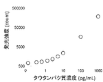
- Tau protein (manufactured by Millipore, trade name: HUMAN TAU-381) is serially diluted with buffer A, and tau protein is 0 pg / mL, 0.3 pg / mL, 0.6 pg / mL, 70 ⁇ L of a test substance solution was prepared so as to have a concentration of 1.25 pg / mL, 2.5 pg / mL, 5 pg / mL, 10 pg / mL, 100 pg / mL, or 1000 pg / mL.
- An antibody labeled with ALP was prepared as follows.
- an anti-tau mouse monoclonal antibody (IgG) (Bio Legend, clone name: TAU12) was fragmented with pepsin to obtain an F (ab ′) 2 fragment.
- the obtained F (ab ′) 2 fragment was reduced to obtain Fab′-SH.
- EMCS was reacted with ALP to obtain ALP-mal.
- ALP-mal By reacting Fab′-SH and ALP-mal, a labeled antibody solution containing Fab′-ALP was obtained. The concentration of the labeled antibody was adjusted to 50 fmol / 20 ⁇ L.
- a capture antibody conjugated with biotin and DNP was prepared as follows.
- an anti-tau mouse monoclonal antibody IgG (manufactured by Covance, clone name: TAU5) was fragmented with pepsin to obtain an F (ab ′) 2 fragment.
- the obtained F (ab ′) 2 fragment was reduced to obtain Fab′-SH.
- BSA-Bio-DNP- (PEG) 8 -mal was obtained by reacting BSA-Bio-DNP with a linker (manufactured by Life Technologies, trade name: SM (PEG) 8).
- a capture antibody solution containing Fab ′-(PEG) 8 -BSA-Bio-DNP was obtained by reacting Fab′-SH with BSA-Bio-DNP- (PEG) 8 -mal.
- the concentration of the capture antibody was adjusted to 200 fmol / 20 ⁇ l.
- Anti-DNP antibody solid phase was prepared by immobilizing anti-DNP antibody (manufactured by Sysmex) on magnetic particles (JSR, trade name: MAG2201) having a particle size of 2.2 ⁇ m by a conventional method.
- JSR magnetic particles
- MAG2201 magnetic particles having a particle size of 2.2 ⁇ m by a conventional method.
- streptavidin solid phase hereinafter referred to as “STA solid phase”
- HISCL R2 reagent manufactured by Sysmex Corporation
- Example 2 The fluorescence intensity measured in Example 2 is shown in FIG.
- the detection limit of the method of Example 2 is about 0.3 pg / mL, and it was found that this method can be detected with high sensitivity.
- Example 3 Detection of amyloid ⁇ by ICT-EIA using capture antibodies and labeled antibodies with overlapping epitopes
- a monomer sample containing 1000 pg / mL of amyloid ⁇ (1-11) monomer (manufactured by Anaspec) was prepared.
- a dimer sample containing 1000 pg / mL of amyloid ⁇ (1-16) dimer (manufactured by IBL) was prepared.
- Amyloid ⁇ 42 (manufactured by IBL) was polymerized to prepare an oligomer, and an oligomer sample containing 1000 pg / mL thereof was prepared.
- the main component of the oligomer sample was a pentameric oligomer.
- antibody solution 1 Using the anti-amyloid ⁇ mouse monoclonal antibody (IgG) (IBL, clone name: 82E1). In the same manner, a labeled antibody was prepared. An anti-amyloid ⁇ mouse monoclonal antibody (IgG) (manufactured by IBL, clone name: 82E1) was used as described in 2. of Example 1. In the same manner, a capture antibody was prepared. The labeled antibody solution, the capture antibody solution, and the buffer A were mixed so that the content of the labeled antibody was 100 fmol and the capture antibody was 100 fmol to prepare 80 ⁇ L of the antibody solution.
- IgG anti-amyloid ⁇ mouse monoclonal antibody
- IgG anti-amyloid ⁇ mouse monoclonal antibody
- a capture antibody was prepared. The labeled antibody solution, the capture antibody solution, and the buffer A were mixed so that the content of the labeled antibody was 100 fmol and the capture antibody was 100 fmol to prepare
- Example 3 The fluorescence intensity measured in Example 3 is shown in FIG. Oligomers and dimers of amyloid ⁇ (denoted as A ⁇ in the figure) were detected, but no monomer was detected. According to this method, it was found that amyloid ⁇ of dimer or higher was detected and the detection of monomer was suppressed.
- Example 4 Detection of amyloid ⁇ by ICT-EIA using a first capture antibody, a second capture antibody and a labeled antibody with overlapping epitopes Preparation of test substance solution Prepared in the same manner as in Example 3.
- antibody solution 1. Using the anti-amyloid ⁇ mouse monoclonal antibody (IgG) (IBL, clone name: 82E1). In the same manner, a labeled antibody was prepared. A thiol group was introduced into an anti-amyloid ⁇ mouse monoclonal antibody (IgG) (manufactured by IBL, clone name: 82E1) using SATA (manufactured by Thermo Fisher Scientific). DNP-Lys (manufactured by Tokyo Chemical Industry Co., Ltd.) was maleimidized using EMCS. A first capture antibody was prepared by mixing and reacting a thiol group-introduced antibody with maleimidated DNP-Lys.
- IgG anti-amyloid ⁇ mouse monoclonal antibody
- SATA manufactured by Thermo Fisher Scientific
- DNP-Lys manufactured by Tokyo Chemical Industry Co., Ltd.
- a first capture antibody was prepared by mixing and reacting a thiol group-intr
- a thiol group was introduced into an anti-amyloid ⁇ mouse monoclonal antibody (IgG) (manufactured by IBL, clone name: 82E1) using SATA (manufactured by Thermo Fisher Scientific). Mix thiol group-introduced antibody and Biotin-PEAC5-maleimide (6- ⁇ N '-[2- (N-maleimido) ethyl] -N-piperazinylamide ⁇ hexyl D-biotinamide hydrochloride) And a second capture antibody was prepared.
- IgG anti-amyloid ⁇ mouse monoclonal antibody
- SATA manufactured by Thermo Fisher Scientific
- a labeled antibody solution, a first capture antibody solution, a second capture antibody solution, and a buffer A were mixed so that the content of the labeled antibody was 100 fmol, the first capture antibody was 50 fmol, and the second capture antibody was 50 fmol to prepare 80 ⁇ L of an antibody solution. .
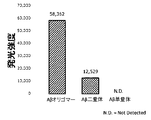
- Example 4 The fluorescence intensity measured in Example 4 is shown in FIG. Strong luminescence was detected from an oligomer sample of amyloid ⁇ (denoted as A ⁇ in the figure). Slight luminescence was detected from the dimer sample. No luminescence was detected from the monomer sample. According to the method, it was found that pentamer or higher amyloid ⁇ was detected, and detection of dimer and monomer was suppressed.
- Example 5 Detection of tau protein by ICT-EIA using capture antibody and labeled antibody with overlapping epitopes
- a monomer sample containing 1000 pg / mL of tau protein monomer (manufactured by Nipro Corporation) was prepared.
- An oligomer was prepared by polymerizing tau protein monomer (manufactured by Wako), and an oligomer sample containing 1000 pg / mL thereof was prepared.
- the main component of the oligomer sample was a 10-mer oligomer.
Landscapes
- Health & Medical Sciences (AREA)
- Immunology (AREA)
- Life Sciences & Earth Sciences (AREA)
- Engineering & Computer Science (AREA)
- Chemical & Material Sciences (AREA)
- Molecular Biology (AREA)
- Biomedical Technology (AREA)
- Hematology (AREA)
- Urology & Nephrology (AREA)
- Biotechnology (AREA)
- Biochemistry (AREA)
- Cell Biology (AREA)
- Food Science & Technology (AREA)
- Medicinal Chemistry (AREA)
- Physics & Mathematics (AREA)
- Analytical Chemistry (AREA)
- Microbiology (AREA)
- General Health & Medical Sciences (AREA)
- General Physics & Mathematics (AREA)
- Pathology (AREA)
- Chemical Kinetics & Catalysis (AREA)
- Peptides Or Proteins (AREA)
- Investigating Or Analysing Biological Materials (AREA)
Abstract
Description
以下、本発明の実施形態について説明する。
本実施形態では、被検物質として多量体が検出される。「多量体」は重合体とも呼ばれ、複数のモノマー分子が物理学的・化学的に重合あるいは凝集して形成される。モノマー分子としては、ポリペプチド、核酸、糖鎖などが例示される。ポリペプチドの多量体としては、具体的には、アミロイドβモノマーが重合したアミロイドβオリゴマー、タウタンパク質が重合したタウオリゴマーなどが例示される。その他にも、血清-アミロイド-A-タンパク質-凝集体、IgG-軽鎖-凝集体、AapoAI-凝集体、AapoAII-凝集体、ATTR-凝集体、DISC1-凝集体、FUS-凝集体、IAPP-凝集体、SOD1-凝集体、α-シヌクレイン-凝集体、TDP-43-凝集体、ハンチンチン-凝集体、リゾチーム-凝集体などが例示される。「多量体」は、モノマー分子が複数含まれていればよく、その他の分子が含まれていてもよい。「多量体」は、必ずしもモノマー分子同士が共有結合などによって強固に結合している必要はなく、より緩やかな結合により凝集して集合した凝集体も含む。
本実施形態の検出方法では、被検物質、被検物質と結合する標識抗体、被検物質と結合する第1捕捉抗体、被検物質と結合する第2捕捉抗体、および第1捕捉抗体と結合する第1固相とを接触させ、被検物質、標識抗体、第1捕捉抗体および第2捕捉抗体を含む複合体を前記第1固相上に形成する工程を行う。本明細書において、「抗体」は、Fab、F(ab')2などの抗原結合性抗体フラグメントまたはその誘導体をも含む。形成工程は、通常は溶液中で行われる。
次に、第1固相上の複合体を遊離させて、該第1固相とは異なる第2固相に転移させる工程を行う。遊離は、通常は溶液中で行われる。ここで、固相上の複合体を遊離させる方法自体は、当該技術において公知である。例えば、形成工程で得られた複合体中の第1捕捉抗体と、第1固相との結合を解離させることができる物質(以下、「遊離剤」という)を用いる方法が挙げられる。遊離剤は当該技術において公知であり、第1捕捉抗体と第1固相との結合様式に応じて適宜選択することができる。例えば、複合体中の第1捕捉抗体と、第1固相とが物理的吸着により結合している場合は、遊離剤として、界面活性剤を含む溶液を用いることで、該複合体を遊離させることができる。また、イオン結合の場合は、イオンを含む溶液を用いることで該複合体を遊離させることができる。
測定工程では、第2固相上の複合体に含まれる標識が測定される。ここで、標識を測定する方法自体は当該技術において公知である。本発明の実施形態においては、上記の標識物質に由来するシグナルの種類に応じた適切な測定方法を選択することができる。例えば、該標識物質が酵素である場合、該酵素に対する基質を反応させることによって発生する光、色などのシグナルを、公知の装置を用いて測定することにより行うことができる。そのような測定装置としては、分光光度計、ルミノメータなどが挙げられる。
図1(B):単量体に第1捕捉抗体が結合する。二量体には、第1捕捉抗体および標識抗体が結合する。三量体には、第1捕捉抗体、第2捕捉抗体および標識抗体が結合する。
図1(C):B/F分離の後、遊離剤としてDNP-Lys(DNP-リジン)を添加し、第1固相からアミロイドβと抗体との複合体を遊離する。
図1(D):(C)工程で得られた複合体を含む液相画分にストレプトアビジンを固定化した第2固相を添加する。これにより、アミロイドβ三量体を含む複合体と第2固相が結合する。
図1(E):B/F分離の後、酵素基質を添加し、酵素反応によって生じる発光を測定する。
この例では、二量体には第1捕捉抗体および標識抗体が結合しているが、第1捕捉抗体、第2捕捉抗体および標識抗体のうち、同一又は異なる種類の抗体が2分子結合し得る。何れの場合も、この複合体からシグナルは検出されない。
この例では、三量体には第1捕捉抗体、第2捕捉抗体および標識抗体が結合しているが、たとえば3カ所あるエピトープのうち少なくとも2カ所に同一の種類の抗体が結合することも考えられる。この場合は、この複合体からシグナルは検出されない。
本実施形態では、被検物質として多量体が検出される。「多量体」および被検物質を含む「試料」は、第1の実施形態において説明したことと同様である。
本実施形態の検出方法では、被検物質、被検物質と結合する標識抗体、被検物質と結合する捕捉抗体、および捕捉抗体と結合する第1固相とを接触させ、被検物質、標識抗体および捕捉抗体を含む複合体を前記第1固相上に形成する工程を行う。「抗体」は、第1の実施形態において説明したことと同様である。
第1固相に第1結合物質と特異的に結合する第1結合パートナーが固定されており、第2固相に第2結合物質と特異的に結合する第2結合パートナーが固定されている。捕捉抗体と第1固相との結合様式は、第1の実施形態の説明において説明した、第1捕捉抗体と第1固相との結合様式と同様である。捕捉抗体と第2固相との結合様式は、第1の実施形態の説明において説明した、第1捕捉抗体と第2固相との結合様式と同様である。
第1固相からの複合体の遊離は、第1の実施形態において説明したことと同様である。
標識の測定及び被検物質の検出については、第1の実施形態において説明したことと同様である。
図2(B):単量体には、捕捉抗体が結合する。二量体には、捕捉抗体および標識抗体が結合する。三量体には、捕捉抗体と標識抗体2分子が結合する。
図2(C):B/F分離の後、遊離剤としてDNP-Lys(DNP-リジン)を添加し、第1固相からアミロイドβと抗体との複合体を遊離する。
図2(D):(C)工程で得られた複合体を含む液相画分にストレプトアビジンを固定化した第2固相を添加する。これにより、複合体と第2固相が結合する。
図2(E):B/F分離の後、酵素基質を添加し、酵素反応によって生じる発光を測定する。
この例では、二量体には捕捉抗体および標識抗体が結合しているが、捕捉抗体および標識抗体のうち、同一又は異なる種類の抗体が2分子結合し得る。捕捉抗体が2分子結合した場合または標識抗体が2分子結合した場合は、シグナルは検出されない。
この例では、三量体には捕捉抗体および標識抗体2分子が結合しているが、捕捉抗体3分子が結合した場合または標識抗体が3分子結合した場合は、シグナルは検出されない。
本実施形態では、被検物質としてアミロイドβおよびタウタンパク質が検出される。本実施形態では、標識抗体のエピトープおよび捕捉抗体のエピトープは相違しており、標識抗体および捕捉抗体は本実施形態の被検物質であるアミロイドβまたはタウタンパク質の異なる部位に結合し得る。これによって、多量体だけでなくモノマー分子も検出することができる。その他の点については、第2の実施形態と同様である。
図3(B):単量体および二量体には、それぞれ捕捉抗体および標識抗体が結合する。
図3(C):B/F分離の後、遊離剤としてDNP-Lys(DNP-リジン)を添加し、第1固相から複合体を遊離する。
図3(D):(C)工程で得られた複合体を含む液相画分にストレプトアビジンを固定化した第2固相を添加する。これにより、複合体と第2固相が結合する。
図3(E):B/F分離の後、酵素基質を添加し、酵素反応によって生じる発光を測定する。
試薬キットは、標識抗体、第1捕捉抗体および第2捕捉抗体を含む。第1固相および第2固相をさらに含んでいてもよい。標識が酵素である場合は、さらに当該酵素の基質を含んでいてもよい。また、複合体を第1固相から分離するための遊離剤をさらに含んでいてもよい。これらの試薬のうちの一部が同一の容器に収容されていてもよいが、容器内での非特異的な反応を抑制するため各試薬は別々の容器に収容されていてもよい。酵素である場合は、標識抗体と基質とは別々の容器に収容される必要がある。遊離剤は、第1固相上に複合体を形成した後に反応系に添加されるため、他の試薬とは異なる容器に収容されることが好ましい。これらの試薬は乾燥状態または液状(液中に溶解もしくは分散させた状態)でユーザに提供されるが、上述の検出方法における化学反応は溶液中で行われることが好ましいため、試薬は液状であることが好ましい。なお、これらの試薬の詳細は、第1の実施形態において説明したことと同様である。
試薬キットは、標識抗体および捕捉抗体を含む。第1の実施形態に用いられる試薬キットと異なり、捕捉抗体は1種類でよい。その他の構成については、第1の実施形態に用いられる試薬キットについて説明したことと同様である。
試薬キットの構成は第2の実施形態に用いられる試薬キットと基本的に同様である。この試薬キットに含まれる捕捉抗体と標識抗体のエピトープは異なっている。
BSA: ウシ血清アルブミン
DNP: 2,4-ジニトロフェニル基
Bio: ビオチニル基
OPDM: N,N'-(1,2-フェニレン)ジマレイミド
SATA:N-スクシンイミド S-アセチルチオアセテート
EMCS: N-(6-マレイミドカプロイルオキシ)スクシンイミド
SH: チオール基
mal: マレイミド基
BSA-Bio-DNP: ビオチンとDNPとによって修飾されたBSA
(PEG)8-BSA-Bio-DNP: PEGリンカーが付加されたBSA-Bio-DNP
4-MUG: 4-メチルウンベリフェリル-β-D-ガラクトピラノシド
DMF: N,N-ジメチルホルムアミド
PEG: ポリエチレングリコール鎖
(PEG)n: オキシエチレン基の付加モル数がnであるポリエチレングリコール鎖
Gal: β-ガラクトシダーゼ
ALP: アルカリフォスファターゼ
1.被検物質溶液の調製
アミロイドβ42 ELISA Kit(コバンス社製、商品名:BetaMark)の標準物質を、緩衝液A(0.4M塩化ナトリウム、0.1質量%BSAおよび0.1Mリン酸ナトリウム緩衝液(pH7.0)を含む)で段階希釈し、アミロイドβ42が0pg、0.003pg、0.01pg、0.1pg、0.3pg、1pg、3pgまたは10pg含まれる被検物質溶液100μLを調製した。
Galで標識した抗体を以下のようにして作製した。慣用の手法により、抗アミロイドβ42ウサギモノクローナル抗体(IgG)(ライフ・テクノロジーズ社製、クローン名:H31L21)をペプシンで断片化してF(ab’)2断片を得た。F(ab’)2断片を還元してFab’-SHを得た。GalにOPDMを反応させてGal-malを得た。Fab’-SHとGal-malを反応させることにより、Fab’-Galを含む標識抗体溶液を得た。
粒径6.35mmのポリスチレンビーズ(イムノケミカル社製、商品名:Immuno bead 6.35φ)に慣用の手法により抗DNP抗体(慣用の手法により作成したウサギポリクローナル抗体)を固定化することにより、抗DNP抗体固相を調製した。
粒径6.35mmのポリスチレンビーズ(イムノケミカル社製、商品名:Immuno bead 6.35φ)に慣用の手法によりストレプトアビジン(和光純薬工業社製、商品名ストレプトアビジン)を固定化することにより、ストレプトアビジン固相(以下、「STA固相」という)を調製した。
(1)複合体形成
測定試料100μLと抗体溶液100μLとを混合した。4℃で12時間インキュベートし、標識抗体-被検物質-捕捉抗体のサンドイッチ複合体を形成させた。
複合体を含む溶液200μLに、抗DNP抗体固相1個を添加した。25℃で30分間インキュベートし、固相上に複合体を固定した。
液相を除去し、2mLの洗浄液1(0.1M塩化ナトリウム、0.1質量%BSAおよび0.1Mリン酸ナトリウム緩衝液(pH7.0)を含む)を用いて2回洗浄した。
複合体を固定化した固相に溶出液1(12mM DNPを含む)を150μL添加した。25℃で30分間インキュベートすることにより、抗DNP抗体固相と複合体との間の結合を切断して複合体を遊離させた。
複合体を含む上清を回収し、ここにSTA固相1個を添加した。25℃で30分間インキュベートし、固相上に複合体を固定した。液相を除去し、2mLの緩衝液B(0.1M塩化ナトリウム、0.1質量%BSAおよび0.1Mリン酸ナトリウム緩衝液(pH7.0)を含む)を用いて3回洗浄した。
複合体を固定化した固相に200μLの緩衝液Bと基質溶液(4-MUGを0.2mM含む)とを添加し、30℃で20時間インキュベートした。反応液に波長360nmの励起光を照射し、波長450nmの蛍光の強度を測定した。複合体を固定化した固相を含まず、200μLの緩衝液Bと基質溶液とを混合した溶液の蛍光強度を上記と同様にして測定し、バックグラウンドの値とした。複合体を固定化した固相を用いて測定した蛍光強度からバックグラウンドの値を控除した。
比較として、複合体の転移を行わないサンドイッチELISAでアミロイドβ42の測定を行った。実施例1(1)および(2)と同様にして抗DNP抗体固相に複合体を固定化、洗浄し、ここに実施例1(5)と同様にして、基質溶液を添加し、30℃で2時間インキュベートした後、蛍光強度の測定を行った。比較例1では、実施例1(3)および(4)の工程は行わなかった。
実施例1および比較例1で算出した蛍光強度を図6示す。実施例1の方法の検出限界は約0.1pg/mLであり、比較例1の方法の検出限界は約1pg/mLであった。以上より、実施例1の方法によれば、比較例1に比べて約10倍高感度に検出することができた。
1.被検物質溶液の調製
タウタンパク質(ミリポア社製、商品名:HUMAN TAU-381)を、緩衝液Aで段階希釈し、タウタンパク質が0pg/mL、0.3pg/mL、0.6pg/mL、1.25pg/mL、2.5pg/mL、5pg/mL、10pg/mL、100pg/mLまたは1000pg/mLの濃度となるよう被検物質溶液70μLを調製した。
ALPで標識した抗体を以下のようにして作製した。慣用の手法により、抗タウマウスモノクローナル抗体(IgG)(バイオレジェンド社製、クローン名:TAU12)をペプシンで断片化してF(ab’)2断片を得た。得られたF(ab’)2断片を還元してFab’-SHを得た。ALPにEMCSを反応させてALP-malを得た。Fab’-SHとALP-malを反応させることにより、Fab’-ALPを含む標識抗体溶液を得た。標識抗体の濃度が50fmol/20μLとなるよう調整した。
粒径2.2μmの磁性粒子(JSR社製、商品名:MAG2201)に慣用の手法により抗DNP抗体(シスメックス社製)を固定化することにより、抗DNP抗体固相を調製した。
ストレプトアビジン固相(以下、「STA固相」という)はHISCL R2試薬(シスメックス社製)を使用した。
(1)複合体形成
被検物質溶液70μLと捕捉抗体溶液20μLとを混合し、42℃で216秒間反応させた。ここに標識抗体溶液20μL添加し、42℃で584秒間反応させ、標識抗体-被検物質-捕捉抗体のサンドイッチ複合体を形成させた。
抗DNP抗体固相の懸濁液(粒子濃度2.5%)20μLを添加した。42℃で720秒間インキュベートし、固相上に複合体を固定した。液相を除去し、300μLのHISCL洗浄液(シスメックス社製)を用いて4回洗浄した。
複合体を固定化した固相に溶出液2(5mM DNP-Lys、DMSO、0.1M MES、2%casein Na、NaOH、NaN3を含む)を41μL添加した。42℃で144秒間インキュベートすることにより、抗DNP固相と複合体との間の結合を切断して複合体を遊離させた。
複合体を含む上清30μLを回収し、ここにHISCL R2試薬(シスメックス社製)30μLを添加した。42℃で288秒間インキュベートし、固相上に複合体を固定した。液相を除去し、300μLのHISCL洗浄液(シスメックス社製)を用いて4回洗浄した。
複合体を固定化した固相に、HISCL発光基質セットR4試薬(シスメックス社製)50μLと、HISCL発光基質セットR5試薬(シスメックス社製)100μLとを混合し、42℃で300秒間反応させ、発光強度を測定した。
実施例2で測定した蛍光強度を図7に示す。実施例2の方法の検出限界は約0.3pg/mLであり、当該方法によると高感度に検出可能であることがわかった。
1.被検物質溶液の調製
アミロイドβ(1-11)単量体(Anaspec社製)1000pg/mL含む単量体試料を調製した。
アミロイドβ(1-16)二量体(IBL社製)1000pg/mL含む二量体試料を調製した。
アミロイドβ42(IBL社製)を重合させてオリゴマーを調製し、これを1000pg/mL含むオリゴマー試料を調製した。ゲル濾過クロマトグラフィーにより常法に従ってオリゴマーの分子量を分析した結果、オリゴマー試料の主な構成成分は5量体オリゴマーであった。
抗アミロイドβマウスモノクローナル抗体(IgG)(IBL社製、クローン名:82E1)を用い、実施例1の2.と同様にして標識抗体を調製した。
抗アミロイドβマウスモノクローナル抗体(IgG)(IBL社製、クローン名:82E1)を用い、実施例1の2.と同様にして捕捉抗体を調製した。
標識抗体100fmol、捕捉抗体100fmolの含有量となるよう標識抗体溶液、捕捉抗体溶液および緩衝液Aを混合し、抗体溶液80μLを調製した。
実施例2の3.と同様にして、抗DNP抗体固相およびSTA固相を調製した。
(1)複合体形成
被検物質溶液70μLと抗体溶液80μLとを混合し、42℃で92分間反応させ、標識抗体-被検物質-捕捉抗体のサンドイッチ複合体を形成させた。
抗DNP抗体固相の懸濁液(粒子濃度2.5%)20μLを添加した。42℃で11分間インキュベートし、固相上に複合体を固定した。液相を除去し、300μLのHISCL洗浄液(シスメックス社製)を用いて4回洗浄した。
複合体を固定化した固相に溶出液2(2.5mM DNP-Lys、DMSO、0.1M MES、2%casein Na、NaOH、NaN3を含む)を110μL添加した。42℃で5分間インキュベートすることにより、抗DNP固相と複合体との間の結合を切断して複合体を遊離させた。
複合体を含む上清80μLを回収し、ここにHISCL R2試薬(シスメックス社製)30μLを添加した。42℃で5分間インキュベートし、固相上に複合体を固定した。液相を除去し、300μLのHISCL洗浄液(シスメックス社製)を用いて4回洗浄した。
複合体を固定化した固相に、HISCL発光基質セットR4試薬(シスメックス社製)50μLと、HISCL発光基質セットR5試薬(シスメックス社製)100μLとを混合し、42℃で5分間反応させ、発光強度を測定した。
実施例3で測定した蛍光強度を図8に示す。アミロイドβ(図では、Aβと表記)のオリゴマーおよび二量体は検出されたが、単量体は検出されなかった。当該方法によると、二量体以上のアミロイドβが検出され、単量体の検出は抑制されることがわかった。
1.被検物質溶液の調製
実施例3と同様にして調製した。
抗アミロイドβマウスモノクローナル抗体(IgG)(IBL社製、クローン名:82E1)を用い、実施例1の2.と同様にして標識抗体を調製した。
抗アミロイドβマウスモノクローナル抗体(IgG)(IBL社製、クローン名:82E1)に、SATA(Thermo Fischer Scientific社製)を用いてチオール基を導入した。DNP-Lys(東京化成工業社製)を、EMCSを用いてマレイミド化した。チオール基を導入した抗体とマレイミド化したDNP-Lysとを混合して反応させ、第1捕捉抗体を調製した。
抗アミロイドβマウスモノクローナル抗体(IgG)(IBL社製、クローン名:82E1)に、SATA(Thermo Fischer Scientific社製)を用いてチオール基を導入した。チオール基を導入した抗体と、Biotin-PEAC5-maleimide(6-{N'-[2-(N-マレイミド)エチル]-N-ピペラジニルアミド}ヘキシルD-ビオチンアミド塩酸塩)とを混合して反応させ、第2捕捉抗体を調製した。
標識抗体100fmol、第1捕捉抗体50fmol、第2捕捉抗体50fmolの含有量となるよう標識抗体溶液、第1捕捉抗体溶液、第2捕捉抗体溶液および緩衝液Aを混合し、抗体溶液80μLを調製した。
実施例2の3.と同様にして、抗DNP抗体固相およびSTA固相を調製した。
実施例3と同様にして発光強度を測定した。
実施例4で測定した蛍光強度を図9に示す。アミロイドβ(図では、Aβと表記)のオリゴマー試料からは強い発光が検出された。二量体試料からは僅かに発光が検出された。単量体試料からは発光は検出されなかった。当該方法によると、5量体以上のアミロイドβが検出され、二量体および単量体の検出は抑制されることがわかった。
1.被検物質溶液の調製
タウタンパク質単量体(ニプロ社製)1000pg/mL含む単量体試料を調製した。
タウタンパク質単量体(Wako社製)を重合させてオリゴマーを調製し、これを1000pg/mL含むオリゴマー試料を調製した。ゲル濾過クロマトグラフィーにより常法に従ってオリゴマーの分子量を分析した結果、オリゴマー試料の主な構成成分は10量体オリゴマーであった。
実施例2の2.と同様にして標識抗体を調製した。
抗タウタンパク質マウスモノクローナル抗体(バイオレジェンド社製、IgG、クローン名:Tau12)を用い、実施例1の2.と同様にして捕捉抗体を調製した。
標識抗体100fmol、捕捉抗体100fmolの含有量となるよう標識抗体溶液、捕捉抗体溶液および緩衝液Aを混合し、抗体溶液80μLを調製した。
実施例2の3.と同様にして、抗DNP抗体固相およびSTA固相を調製した。
(1)複合体形成
被検物質溶液70μLと抗体溶液80μLとを混合し、37℃で27分間反応させ、標識抗体-被検物質-捕捉抗体のサンドイッチ複合体を形成させた。
抗DNP抗体固相の懸濁液(粒子濃度2.5%)20μLを添加した。37℃で11分間インキュベートし、固相上に複合体を固定した。液相を除去し、300μLのHISCL洗浄液(シスメックス社製)を用いて4回洗浄した。
複合体を固定化した固相に溶出液2(2.5mM DNP-Lys、DMSO、0.1M MES、2%casein Na、NaOH、NaN3を含む)を110μL添加した。37℃で5分間インキュベートすることにより、抗DNP固相と複合体との間の結合を切断して複合体を遊離させた。
複合体を含む上清80μLを回収し、ここにSTA固相の懸濁液(粒子濃度2.5%)30μLを添加した。37℃で5分間インキュベートし、固相上に複合体を固定した。液相を除去し、300μLのHISCL洗浄液(シスメックス社製)を用いて4回洗浄した。
複合体を固定化した固相に、HISCL発光基質セットR4試薬(シスメックス社製)50μLと、HISCL発光基質セットR5試薬(シスメックス社製)100μLとを混合し、42℃で5分間反応させ、発光強度を測定した。
実施例5で測定した蛍光強度を図10に示す。タウタンパク質のオリゴマーは検出されたが、単量体は検出されなかった。当該方法によると、タウタンパク質オリゴマーが検出され、単量体の検出は抑制されることがわかった。
31 第1容器
32 第2容器
33 第3容器
34 第4容器
37 添付文書
38 梱包箱
40 試薬キット
41 第1容器
42 第2容器
43 第3容器
44 第4容器
45 第5容器
47 添付文書
48 梱包箱
Claims (24)
- 被検物質、前記被検物質と結合する標識抗体、前記被検物質と結合する第1捕捉抗体、前記被検物質と結合する第2捕捉抗体、および前記第1捕捉抗体と結合する第1固相とを接触させ、前記被検物質、前記標識抗体、前記第1捕捉抗体および前記第2捕捉抗体を含む複合体を前記第1固相上に形成する工程と、
前記第1捕捉抗体と前記第1固相との結合を解離することにより前記複合体を遊離させ、前記第2捕捉抗体と結合する第2固相と前記複合体とを接触させ、前記複合体を前記第2固相上に転移する工程と、
前記第2固相上の複合体に含まれる標識を測定し、前記被検物質を検出する工程と、
を含み、
前記被検物質が多量体であり、
前記標識抗体のエピトープ、前記第1捕捉抗体のエピトープおよび前記第2捕捉抗体エピトープが重複する、
被検物質の検出方法。 - 前記被検物質が、ポリペプチドを含む、請求項1記載の方法。
- 前記被検物質が、三量体以上の多量体である、請求項1または2記載の方法。
- 前記被検物質が、アミロイドβまたはタウタンパク質である、請求項1~3のいずれかに記載の方法。
- 前記第1捕捉抗体が第1結合物質を含み、前記第1固相が前記第1結合物質と特異的に結合する第1結合パートナーを含み、
前記形成工程において、前記第1結合パートナーと前記第1結合物質との特異的結合により、前記複合体が前記第1固相上に結合し、
前記第2捕捉抗体が第2結合物質を含み、前記第2固相が前記第2結合物質と特異的に結合する第2結合パートナーを含み、
前記転移工程において、前記第2結合パートナーと前記第2結合物質との特異的結合により、遊離した前記複合体が前記第2固相に結合する、
請求項1~4のいずれかに記載の方法。 - 請求項1~5記載のいずれかに記載の方法に用いられ、前記標識抗体、前記第1捕捉抗体、および前記第2捕捉抗体を含む、被検物質の検出試薬キット。
- 被検物質、前記被検物質と結合する標識抗体、前記被検物質と結合する捕捉抗体、前記捕捉抗体と結合する第1固相を接触させ、前記被検物質、前記標識抗体、および前記捕捉抗体を含む複合体を前記第1固相上に形成する工程と、
前記捕捉抗体と前記第1固相との結合を解離することにより前記複合体を遊離させ、前記捕捉抗体と結合する第2固相と前記複合体とを接触させ、前記複合体を前記第2固相上に転移する工程と、
前記第2固相上の複合体に含まれる標識を測定し、前記被検物質を検出する工程と、
を含み、
前記被検物質が多量体であり、
前記標識抗体のエピトープおよび前記捕捉抗体のエピトープが重複する、
被検物質の検出方法。 - 前記被検物質が、ポリペプチドを含む、請求項7記載の方法。
- 前記被検物質が、二量体以上の多量体である、請求項7または8記載の方法。
- 前記被検物質が、アミロイドβまたはタウタンパク質である、請求項7~9のいずれかに記載の方法。
- 前記捕捉抗体が第1結合物質および第2結合物質を含み、前記第1固相が前記第1結合物質と特異的に結合する第1結合パートナーを含み、前記第2固相が前記第2結合物質と特異的に結合する第2結合パートナーを含み、
前記形成工程において、前記第1結合パートナーと前記第1結合物質との特異的結合により、前記複合体が前記第1固相上に結合し、
前記転移工程において、前記第2結合パートナーと前記第2結合物質との特異的結合により、遊離した前記複合体が前記第2固相に結合する、
請求項7~10のいずれかに記載の方法。 - 被検物質、前記被検物質と結合する標識抗体、前記被検物質と結合する捕捉抗体、および前記捕捉抗体と結合する第1固相を接触させ、前記被検物質、前記標識抗体および前記捕捉抗体を含む複合体を前記第1固相上に形成させる工程と、
前記捕捉抗体と前記第1固相との結合を解離することにより前記第1固相上の複合体を遊離させ、前記捕捉抗体と結合する第2固相に転移する工程と、
前記第2固相上の複合体に含まれる標識を測定し、前記被検物質を検出する工程と
を含み、
前記被検物質がアミロイドβまたはタウタンパク質である、
被検物質の検出方法。 - 前記捕捉抗体が第1結合物質および第2結合物質を含み、前記第1固相が前記第1結合物質と特異的に結合する第1結合パートナーを含み、前記第2固相が前記第2結合物質と特異的に結合する第2結合パートナーを含み、
前記形成工程において、前記第1結合パートナーと前記第1結合物質との特異的結合により、前記複合体が前記第1固相上に結合し、
前記転移工程において、前記第2結合パートナーと前記第2結合物質との特異的結合により、遊離した前記複合体が前記第2固相に結合する、
請求項12記載の方法。 - 前記第1固相が粒子であり、
前記形成工程の後、前記転移工程の前に、未反応成分の除去および前記第1固相の回収を行う工程を含む、
請求項1~13のいずれかに記載の方法。 - 前記転移工程において、遊離剤により前記第1固相上の複合体を前記第1固相から遊離する、請求項1~14のいずれかに記載の方法。
- 前記転移工程の後、前記検出工程の前に、未反応成分の除去および前記第2固相の回収を行う工程を含む、
請求項1~15のいずれかに記載の方法。 - 前記第1結合パートナーが抗ハプテン抗体であり、前記第1結合物質がハプテンであり、前記第2結合パートナーがアビジン類であり、前記第2結合物質がビオチン類である、請求項5,11または13記載の方法。
- 前記ハプテンがジニトロフェニル基であり、
前記転移工程において、ジニトロフェニルアミノ酸を用いて前記第1固相上の複合体を前記第1固相から遊離する、
請求項17記載の方法。 - 前記標識が、酵素または蛍光物質である、請求項1~18のいずれかに記載の方法。
- 前記標識が、βガラクトシダーゼまたはアルカリフォスファターゼであり、
前記測定工程において、前記酵素の基質と前記酵素とを反応させ、前記酵素反応により生じた反応産物から生じる標識を測定する、
請求項1~19のいずれかに記載の方法。 - 前記標識が、蛍光物質であり、
前記測定工程において、前記第2固相上の複合体に励起光を照射し、前記複合体の標識から生じる蛍光を測定する、
請求項1~19のいずれかに記載の方法。 - 請求項7~21記載の方法に用いられ、前記標識抗体および前記捕捉抗体を含む、被検物質の検出試薬キット。
- 前記第1固相をさらに含む、請求項6または22記載のキット。
- 前記第2固相をさらに含む、請求項6、22または23記載のキット。
Priority Applications (4)
| Application Number | Priority Date | Filing Date | Title |
|---|---|---|---|
| EP17750220.0A EP3415912B1 (en) | 2016-02-08 | 2017-02-06 | Analyte detection method and reagent kit for detecting analyte |
| JP2017554615A JP6320651B2 (ja) | 2016-02-08 | 2017-02-06 | 被検物質の検出方法および被検物質の検出用試薬キット |
| CN201780010831.9A CN108603880B (zh) | 2016-02-08 | 2017-02-06 | 受试物质的检测方法及受试物质的检测用试剂盒 |
| US16/056,853 US11169148B2 (en) | 2016-02-08 | 2018-08-07 | Method for detecting test substance and reagent kit for detecting test substance |
Applications Claiming Priority (2)
| Application Number | Priority Date | Filing Date | Title |
|---|---|---|---|
| JP2016021610 | 2016-02-08 | ||
| JP2016-021610 | 2016-02-08 |
Related Child Applications (1)
| Application Number | Title | Priority Date | Filing Date |
|---|---|---|---|
| US16/056,853 Continuation US11169148B2 (en) | 2016-02-08 | 2018-08-07 | Method for detecting test substance and reagent kit for detecting test substance |
Publications (1)
| Publication Number | Publication Date |
|---|---|
| WO2017138497A1 true WO2017138497A1 (ja) | 2017-08-17 |
Family
ID=59563878
Family Applications (1)
| Application Number | Title | Priority Date | Filing Date |
|---|---|---|---|
| PCT/JP2017/004271 Ceased WO2017138497A1 (ja) | 2016-02-08 | 2017-02-06 | 被検物質の検出方法および被検物質の検出用試薬キット |
Country Status (5)
| Country | Link |
|---|---|
| US (1) | US11169148B2 (ja) |
| EP (1) | EP3415912B1 (ja) |
| JP (1) | JP6320651B2 (ja) |
| CN (1) | CN108603880B (ja) |
| WO (1) | WO2017138497A1 (ja) |
Cited By (6)
| Publication number | Priority date | Publication date | Assignee | Title |
|---|---|---|---|---|
| EP3699593A1 (en) | 2019-02-20 | 2020-08-26 | Sysmex Corporation | Method for acquiring information on analyte |
| WO2021065306A1 (ja) * | 2019-09-30 | 2021-04-08 | ニプロ株式会社 | 血液試料を検体とするタウタンパク質検出方法 |
| JP7450995B1 (ja) | 2023-04-13 | 2024-03-18 | 国立研究開発法人量子科学技術研究開発機構 | 被験者の脳のタウタンパク質の蓄積量を推定する方法及び装置 |
| WO2024214588A1 (ja) * | 2023-04-13 | 2024-10-17 | 国立研究開発法人量子科学技術研究開発機構 | 被験者の脳のタウタンパク質の蓄積量を推定する方法及び装置 |
| WO2025142873A1 (ja) * | 2023-12-27 | 2025-07-03 | 富士レビオ株式会社 | 被検物質の検出方法、並びに、それに用いる標識用バッファー及びキット |
| WO2026071006A1 (ja) * | 2024-09-30 | 2026-04-02 | 富士フイルム株式会社 | アミロイドβ前駆体タンパク質由来の被検物質を検出する方法、キレート剤の影響を低減する方法、緩衝液試薬及び試薬キット |
Families Citing this family (3)
| Publication number | Priority date | Publication date | Assignee | Title |
|---|---|---|---|---|
| JP6765232B2 (ja) * | 2016-06-30 | 2020-10-07 | シスメックス株式会社 | 免疫複合体転移法により被検物質を検出するための抗体試薬及びその製造方法、並びにその抗体試薬の利用 |
| JP7478040B2 (ja) * | 2020-06-25 | 2024-05-02 | シスメックス株式会社 | Aβペプチドの測定方法及びその方法に用いられる試薬組成物 |
| JP7629278B2 (ja) * | 2020-06-25 | 2025-02-13 | シスメックス株式会社 | Aβペプチドを測定するための抗体セット、Aβペプチドの測定方法及び試薬キット |
Citations (10)
| Publication number | Priority date | Publication date | Assignee | Title |
|---|---|---|---|---|
| JP2681370B2 (ja) * | 1988-06-30 | 1997-11-26 | 塩野義製薬株式会社 | 免疫測定法 |
| JP2001116571A (ja) * | 1999-09-29 | 2001-04-27 | Internatl Business Mach Corp <Ibm> | 道路データの処理方法、地図データ処理装置、地図データ処理システム、道路データを処理するためのソフトウエア・プロダクトを格納した記憶媒体 |
| JP2001305137A (ja) * | 2000-04-20 | 2001-10-31 | Eiji Ishikawa | 特異的複合体の固定化方法および測定方法 |
| JP2008530578A (ja) * | 2005-02-19 | 2008-08-07 | ピープルバイオ アイ エヌ シー | 多量体形成ポリペプチドの単量体型から多量体型を分別検出する方法 |
| WO2009011500A2 (en) * | 2007-07-16 | 2009-01-22 | Peoplebio, Inc. | Methods for decreasing false signals in immunoassay to detect a multimeric form from a monomeric form of multimer-forming polypeptides |
| JP2009085685A (ja) * | 2007-09-28 | 2009-04-23 | Igaku Seibutsugaku Kenkyusho:Kk | インスリン受容体αサブユニットの測定試薬 |
| JP2009517652A (ja) * | 2005-11-25 | 2009-04-30 | コーニンクレッカ フィリップス エレクトロニクス エヌ ヴィ | 強結合対の構築による高感度磁気捕獲分析 |
| JP5164971B2 (ja) * | 2006-04-21 | 2013-03-21 | ピープルバイオ,アイ エヌ シー | 三次元的相互作用を用いてマルチマー形成ポリペプチドのモノマーからマルチマーを分別検出する方法 |
| WO2014123131A1 (ja) * | 2013-02-05 | 2014-08-14 | Hashida Seiichi | 1型糖尿病の早期診断マーカーであるgad抗体の高感度測定方法 |
| JP2015502551A (ja) * | 2011-12-23 | 2015-01-22 | フォルシュングスツェントルム ユーリッヒ ゲゼルシャフ | Aβ凝集体の選択的定量化方法 |
Family Cites Families (13)
| Publication number | Priority date | Publication date | Assignee | Title |
|---|---|---|---|---|
| US5312730A (en) * | 1992-05-27 | 1994-05-17 | Ciba Corning Diagnostics Corp. | Immune complex transfer with lypophilic bridge |
| JPH08178926A (ja) * | 1994-10-25 | 1996-07-12 | Sumitomo Pharmaceut Co Ltd | イムノアッセイプレートおよびその用途 |
| KR100987639B1 (ko) * | 2005-02-19 | 2010-10-13 | 주식회사 피플바이오 | 멀티머-형성 폴리펩타이드의 모노머로부터 멀티머를 분별검출하는 방법 |
| CN101427132A (zh) * | 2006-04-21 | 2009-05-06 | 人民生物公司 | 通过三维相互作用从多聚体形成多肽的单体形式中示差检测多聚体形式的方法 |
| CN102089659A (zh) | 2008-05-08 | 2011-06-08 | 武田药品工业株式会社 | Aβ寡聚物的测定方法 |
| EP2478365A1 (en) | 2009-09-18 | 2012-07-25 | Probiodrug AG | Novel assay for the detection of amyloid beta peptides |
| CN102081018A (zh) * | 2009-11-30 | 2011-06-01 | 希森美康株式会社 | 样品的预处理方法及hcv的免疫测定方法 |
| WO2012113718A1 (en) * | 2011-02-21 | 2012-08-30 | Roche Diagnostics Gmbh | A diagnostic method for type ii diabetes |
| AU2012282825B2 (en) | 2011-07-13 | 2016-05-26 | Merck Sharp & Dohme Corp. | Method for detection of amyloid beta oligomers in a fluid sample and uses thereof |
| CN104737020B (zh) * | 2012-08-30 | 2016-08-17 | 希森美康株式会社 | 早期肾障碍的评价指标和其测定方法 |
| AU2013342190B2 (en) * | 2012-11-09 | 2018-11-08 | Ansh Labs Llc | Antibody compositions and immunoassay methods to detect isoforms of Anti-Mullerian Hormone |
| EP2950099B1 (en) * | 2013-01-28 | 2018-09-19 | Sysmex Corporation | Pretreatment method for sample for detecting hbs antigen, and use therefor |
| JP6675165B2 (ja) * | 2015-07-31 | 2020-04-01 | シスメックス株式会社 | 被検物質の検出方法、検出用試薬キットおよび検出用試薬 |
-
2017
- 2017-02-06 WO PCT/JP2017/004271 patent/WO2017138497A1/ja not_active Ceased
- 2017-02-06 CN CN201780010831.9A patent/CN108603880B/zh active Active
- 2017-02-06 JP JP2017554615A patent/JP6320651B2/ja active Active
- 2017-02-06 EP EP17750220.0A patent/EP3415912B1/en active Active
-
2018
- 2018-08-07 US US16/056,853 patent/US11169148B2/en active Active
Patent Citations (10)
| Publication number | Priority date | Publication date | Assignee | Title |
|---|---|---|---|---|
| JP2681370B2 (ja) * | 1988-06-30 | 1997-11-26 | 塩野義製薬株式会社 | 免疫測定法 |
| JP2001116571A (ja) * | 1999-09-29 | 2001-04-27 | Internatl Business Mach Corp <Ibm> | 道路データの処理方法、地図データ処理装置、地図データ処理システム、道路データを処理するためのソフトウエア・プロダクトを格納した記憶媒体 |
| JP2001305137A (ja) * | 2000-04-20 | 2001-10-31 | Eiji Ishikawa | 特異的複合体の固定化方法および測定方法 |
| JP2008530578A (ja) * | 2005-02-19 | 2008-08-07 | ピープルバイオ アイ エヌ シー | 多量体形成ポリペプチドの単量体型から多量体型を分別検出する方法 |
| JP2009517652A (ja) * | 2005-11-25 | 2009-04-30 | コーニンクレッカ フィリップス エレクトロニクス エヌ ヴィ | 強結合対の構築による高感度磁気捕獲分析 |
| JP5164971B2 (ja) * | 2006-04-21 | 2013-03-21 | ピープルバイオ,アイ エヌ シー | 三次元的相互作用を用いてマルチマー形成ポリペプチドのモノマーからマルチマーを分別検出する方法 |
| WO2009011500A2 (en) * | 2007-07-16 | 2009-01-22 | Peoplebio, Inc. | Methods for decreasing false signals in immunoassay to detect a multimeric form from a monomeric form of multimer-forming polypeptides |
| JP2009085685A (ja) * | 2007-09-28 | 2009-04-23 | Igaku Seibutsugaku Kenkyusho:Kk | インスリン受容体αサブユニットの測定試薬 |
| JP2015502551A (ja) * | 2011-12-23 | 2015-01-22 | フォルシュングスツェントルム ユーリッヒ ゲゼルシャフ | Aβ凝集体の選択的定量化方法 |
| WO2014123131A1 (ja) * | 2013-02-05 | 2014-08-14 | Hashida Seiichi | 1型糖尿病の早期診断マーカーであるgad抗体の高感度測定方法 |
Non-Patent Citations (2)
| Title |
|---|
| LEVINE H III: "Alzheimer's beta-peptide oligomer formation at physiologic concentrations", ANAL BIOCHEM, vol. 335, no. 1, 1 December 2004 (2004-12-01), pages 81 - 90, XP004616778 * |
| See also references of EP3415912A4 * |
Cited By (12)
| Publication number | Priority date | Publication date | Assignee | Title |
|---|---|---|---|---|
| EP3699593A1 (en) | 2019-02-20 | 2020-08-26 | Sysmex Corporation | Method for acquiring information on analyte |
| JP2020134329A (ja) * | 2019-02-20 | 2020-08-31 | シスメックス株式会社 | 被検物質の情報の取得方法及び被検物質の捕捉方法 |
| JP7373286B2 (ja) | 2019-02-20 | 2023-11-02 | シスメックス株式会社 | 被検物質の情報の取得方法及び被検物質の捕捉方法 |
| WO2021065306A1 (ja) * | 2019-09-30 | 2021-04-08 | ニプロ株式会社 | 血液試料を検体とするタウタンパク質検出方法 |
| JPWO2021065306A1 (ja) * | 2019-09-30 | 2021-04-08 | ||
| EP4040153A4 (en) * | 2019-09-30 | 2023-07-26 | Nipro Corporation | PROCEDURE FOR DETECTING TAU PROTEIN USING A BLOOD SAMPLE AS SAMPLE |
| JP7718270B2 (ja) | 2019-09-30 | 2025-08-05 | ニプロ株式会社 | 血液試料を検体とするタウタンパク質検出方法 |
| JP7450995B1 (ja) | 2023-04-13 | 2024-03-18 | 国立研究開発法人量子科学技術研究開発機構 | 被験者の脳のタウタンパク質の蓄積量を推定する方法及び装置 |
| WO2024214588A1 (ja) * | 2023-04-13 | 2024-10-17 | 国立研究開発法人量子科学技術研究開発機構 | 被験者の脳のタウタンパク質の蓄積量を推定する方法及び装置 |
| JP2024152000A (ja) * | 2023-04-13 | 2024-10-25 | 国立研究開発法人量子科学技術研究開発機構 | 被験者の脳のタウタンパク質の蓄積量を推定する方法及び装置 |
| WO2025142873A1 (ja) * | 2023-12-27 | 2025-07-03 | 富士レビオ株式会社 | 被検物質の検出方法、並びに、それに用いる標識用バッファー及びキット |
| WO2026071006A1 (ja) * | 2024-09-30 | 2026-04-02 | 富士フイルム株式会社 | アミロイドβ前駆体タンパク質由来の被検物質を検出する方法、キレート剤の影響を低減する方法、緩衝液試薬及び試薬キット |
Also Published As
| Publication number | Publication date |
|---|---|
| CN108603880A (zh) | 2018-09-28 |
| EP3415912A1 (en) | 2018-12-19 |
| EP3415912A4 (en) | 2019-08-21 |
| US20180348212A1 (en) | 2018-12-06 |
| JPWO2017138497A1 (ja) | 2018-02-22 |
| JP6320651B2 (ja) | 2018-05-09 |
| CN108603880B (zh) | 2021-01-05 |
| US11169148B2 (en) | 2021-11-09 |
| EP3415912B1 (en) | 2023-03-29 |
Similar Documents
| Publication | Publication Date | Title |
|---|---|---|
| JP6320651B2 (ja) | 被検物質の検出方法および被検物質の検出用試薬キット | |
| US9476874B2 (en) | Analyte detection | |
| CN103380377B (zh) | 心肌肌钙蛋白的测定方法 | |
| US20090087869A1 (en) | Sandwich immunoassay and method of detecting an antigen by using the same | |
| TWI224674B (en) | Immunoassay using insoluble magnetic support particles | |
| US11268956B2 (en) | Method for producing antibody reagent | |
| US20170138937A1 (en) | Detection of analytes | |
| JP2019191187A (ja) | 標的物質の検出用試薬、検出方法および標的物質を検出するために用いられる担体ならびにその製造方法 | |
| JP2010107363A (ja) | トロポニンiの測定方法及び測定用試薬キット | |
| CN101910843A (zh) | 检测hiv-1抗原用试剂及其检测方法 | |
| US20080227111A1 (en) | Reagent kit and method for measuring hcv antibody | |
| JPH11295313A (ja) | 抗体または抗体断片の重合体とその利用 | |
| JP5137880B2 (ja) | 結合性物質を固定化した乾燥粒子の製造方法 | |
| JPH08327629A (ja) | 検体前処理方法 | |
| JP5143046B2 (ja) | 被験物質の測定方法及び該測定方法を実施するためのキット | |
| CN113474655A (zh) | 目标物质的检测方法、用于目标物质的检测的试剂、及用于目标物质的检测的试剂盒 |
Legal Events
| Date | Code | Title | Description |
|---|---|---|---|
| 121 | Ep: the epo has been informed by wipo that ep was designated in this application |
Ref document number: 17750220 Country of ref document: EP Kind code of ref document: A1 |
|
| ENP | Entry into the national phase |
Ref document number: 2017554615 Country of ref document: JP Kind code of ref document: A |
|
| NENP | Non-entry into the national phase |
Ref country code: DE |
|
| WWE | Wipo information: entry into national phase |
Ref document number: 2017750220 Country of ref document: EP |
|
| ENP | Entry into the national phase |
Ref document number: 2017750220 Country of ref document: EP Effective date: 20180910 |