WO2017141604A1 - 膜型ムチン様タンパク質の認識とその医療応用 - Google Patents
膜型ムチン様タンパク質の認識とその医療応用 Download PDFInfo
- Publication number
- WO2017141604A1 WO2017141604A1 PCT/JP2017/001250 JP2017001250W WO2017141604A1 WO 2017141604 A1 WO2017141604 A1 WO 2017141604A1 JP 2017001250 W JP2017001250 W JP 2017001250W WO 2017141604 A1 WO2017141604 A1 WO 2017141604A1
- Authority
- WO
- WIPO (PCT)
- Prior art keywords
- antibody
- mesothelioma
- protein
- heg1
- seq
- Prior art date
- Legal status (The legal status is an assumption and is not a legal conclusion. Google has not performed a legal analysis and makes no representation as to the accuracy of the status listed.)
- Ceased
Links
Images





Classifications
-
- G—PHYSICS
- G01—MEASURING; TESTING
- G01N—INVESTIGATING OR ANALYSING MATERIALS BY DETERMINING THEIR CHEMICAL OR PHYSICAL PROPERTIES
- G01N33/00—Investigating or analysing materials by specific methods not covered by groups G01N1/00 - G01N31/00
- G01N33/48—Biological material, e.g. blood, urine; Haemocytometers
- G01N33/50—Chemical analysis of biological material, e.g. blood, urine; Testing involving biospecific ligand binding methods; Immunological testing
- G01N33/53—Immunoassay; Biospecific binding assay; Materials therefor
- G01N33/575—Immunoassay; Biospecific binding assay; Materials therefor for cancer
-
- A—HUMAN NECESSITIES
- A61—MEDICAL OR VETERINARY SCIENCE; HYGIENE
- A61K—PREPARATIONS FOR MEDICAL, DENTAL OR TOILETRY PURPOSES
- A61K39/00—Medicinal preparations containing antigens or antibodies
- A61K39/395—Antibodies; Immunoglobulins; Immune serum, e.g. antilymphocytic serum
-
- A—HUMAN NECESSITIES
- A61—MEDICAL OR VETERINARY SCIENCE; HYGIENE
- A61K—PREPARATIONS FOR MEDICAL, DENTAL OR TOILETRY PURPOSES
- A61K49/00—Preparations for testing in vivo
-
- A—HUMAN NECESSITIES
- A61—MEDICAL OR VETERINARY SCIENCE; HYGIENE
- A61K—PREPARATIONS FOR MEDICAL, DENTAL OR TOILETRY PURPOSES
- A61K49/00—Preparations for testing in vivo
- A61K49/001—Preparation for luminescence or biological staining
- A61K49/0013—Luminescence
- A61K49/0017—Fluorescence in vivo
- A61K49/005—Fluorescence in vivo characterised by the carrier molecule carrying the fluorescent agent
- A61K49/0058—Antibodies
-
- A—HUMAN NECESSITIES
- A61—MEDICAL OR VETERINARY SCIENCE; HYGIENE
- A61K—PREPARATIONS FOR MEDICAL, DENTAL OR TOILETRY PURPOSES
- A61K49/00—Preparations for testing in vivo
- A61K49/06—Nuclear magnetic resonance [NMR] contrast preparations; Magnetic resonance imaging [MRI] contrast preparations
- A61K49/08—Nuclear magnetic resonance [NMR] contrast preparations; Magnetic resonance imaging [MRI] contrast preparations characterised by the carrier
- A61K49/10—Organic compounds
- A61K49/14—Peptides, e.g. proteins
- A61K49/16—Antibodies; Immunoglobulins; Fragments thereof
-
- A—HUMAN NECESSITIES
- A61—MEDICAL OR VETERINARY SCIENCE; HYGIENE
- A61P—SPECIFIC THERAPEUTIC ACTIVITY OF CHEMICAL COMPOUNDS OR MEDICINAL PREPARATIONS
- A61P35/00—Antineoplastic agents
-
- C—CHEMISTRY; METALLURGY
- C07—ORGANIC CHEMISTRY
- C07K—PEPTIDES
- C07K14/00—Peptides having more than 20 amino acids; Gastrins; Somatostatins; Melanotropins; Derivatives thereof
- C07K14/435—Peptides having more than 20 amino acids; Gastrins; Somatostatins; Melanotropins; Derivatives thereof from animals; from humans
- C07K14/705—Receptors; Cell surface antigens; Cell surface determinants
-
- C—CHEMISTRY; METALLURGY
- C07—ORGANIC CHEMISTRY
- C07K—PEPTIDES
- C07K16/00—Immunoglobulins [IG], e.g. monoclonal or polyclonal antibodies
- C07K16/18—Immunoglobulins [IG], e.g. monoclonal or polyclonal antibodies against material from animals or humans
- C07K16/28—Immunoglobulins [IG], e.g. monoclonal or polyclonal antibodies against material from animals or humans against receptors, cell surface antigens or cell surface determinants
- C07K16/30—Immunoglobulins [IG], e.g. monoclonal or polyclonal antibodies against material from animals or humans against receptors, cell surface antigens or cell surface determinants from tumour cells
-
- C—CHEMISTRY; METALLURGY
- C07—ORGANIC CHEMISTRY
- C07K—PEPTIDES
- C07K16/00—Immunoglobulins [IG], e.g. monoclonal or polyclonal antibodies
- C07K16/18—Immunoglobulins [IG], e.g. monoclonal or polyclonal antibodies against material from animals or humans
- C07K16/28—Immunoglobulins [IG], e.g. monoclonal or polyclonal antibodies against material from animals or humans against receptors, cell surface antigens or cell surface determinants
- C07K16/30—Immunoglobulins [IG], e.g. monoclonal or polyclonal antibodies against material from animals or humans against receptors, cell surface antigens or cell surface determinants from tumour cells
- C07K16/3076—Immunoglobulins [IG], e.g. monoclonal or polyclonal antibodies against material from animals or humans against receptors, cell surface antigens or cell surface determinants from tumour cells against structure-related tumour-associated moieties
- C07K16/3092—Immunoglobulins [IG], e.g. monoclonal or polyclonal antibodies against material from animals or humans against receptors, cell surface antigens or cell surface determinants from tumour cells against structure-related tumour-associated moieties against tumour-associated mucins
-
- C—CHEMISTRY; METALLURGY
- C07—ORGANIC CHEMISTRY
- C07K—PEPTIDES
- C07K16/00—Immunoglobulins [IG], e.g. monoclonal or polyclonal antibodies
- C07K16/46—Hybrid immunoglobulins
-
- C—CHEMISTRY; METALLURGY
- C12—BIOCHEMISTRY; BEER; SPIRITS; WINE; VINEGAR; MICROBIOLOGY; ENZYMOLOGY; MUTATION OR GENETIC ENGINEERING
- C12N—MICROORGANISMS OR ENZYMES; COMPOSITIONS THEREOF; PROPAGATING, PRESERVING, OR MAINTAINING MICROORGANISMS; MUTATION OR GENETIC ENGINEERING; CULTURE MEDIA
- C12N15/00—Mutation or genetic engineering; DNA or RNA concerning genetic engineering, vectors, e.g. plasmids, or their isolation, preparation or purification; Use of hosts therefor
- C12N15/09—Recombinant DNA-technology
-
- C—CHEMISTRY; METALLURGY
- C12—BIOCHEMISTRY; BEER; SPIRITS; WINE; VINEGAR; MICROBIOLOGY; ENZYMOLOGY; MUTATION OR GENETIC ENGINEERING
- C12N—MICROORGANISMS OR ENZYMES; COMPOSITIONS THEREOF; PROPAGATING, PRESERVING, OR MAINTAINING MICROORGANISMS; MUTATION OR GENETIC ENGINEERING; CULTURE MEDIA
- C12N15/00—Mutation or genetic engineering; DNA or RNA concerning genetic engineering, vectors, e.g. plasmids, or their isolation, preparation or purification; Use of hosts therefor
- C12N15/09—Recombinant DNA-technology
- C12N15/11—DNA or RNA fragments; Modified forms thereof; Non-coding nucleic acids having a biological activity
- C12N15/62—DNA sequences coding for fusion proteins
-
- G—PHYSICS
- G01—MEASURING; TESTING
- G01N—INVESTIGATING OR ANALYSING MATERIALS BY DETERMINING THEIR CHEMICAL OR PHYSICAL PROPERTIES
- G01N33/00—Investigating or analysing materials by specific methods not covered by groups G01N1/00 - G01N31/00
- G01N33/48—Biological material, e.g. blood, urine; Haemocytometers
- G01N33/50—Chemical analysis of biological material, e.g. blood, urine; Testing involving biospecific ligand binding methods; Immunological testing
- G01N33/53—Immunoassay; Biospecific binding assay; Materials therefor
- G01N33/575—Immunoassay; Biospecific binding assay; Materials therefor for cancer
- G01N33/5752—Immunoassay; Biospecific binding assay; Materials therefor for cancer of the lungs
-
- G—PHYSICS
- G01—MEASURING; TESTING
- G01N—INVESTIGATING OR ANALYSING MATERIALS BY DETERMINING THEIR CHEMICAL OR PHYSICAL PROPERTIES
- G01N33/00—Investigating or analysing materials by specific methods not covered by groups G01N1/00 - G01N31/00
- G01N33/48—Biological material, e.g. blood, urine; Haemocytometers
- G01N33/50—Chemical analysis of biological material, e.g. blood, urine; Testing involving biospecific ligand binding methods; Immunological testing
- G01N33/68—Chemical analysis of biological material, e.g. blood, urine; Testing involving biospecific ligand binding methods; Immunological testing involving proteins, peptides or amino acids
Definitions
- the present invention relates to recognition of membrane-type mucin-like protein and its medical application. More specifically, the present invention relates to antibodies that detect mesothelioma and antibodies that have high specificity and high sensitivity to mesothelioma. The present invention also relates to a method for diagnosing mesothelioma using the antibody, a mesothelioma diagnostic reagent containing the antibody, and a mesothelioma diagnostic kit. The invention further relates to a diagnostic marker for mesothelioma. The present invention further relates to a pharmaceutical composition for use in treating mesothelioma comprising the antibody of the present invention or an antigen-binding fragment thereof.
- Malignant mesothelioma is a serious social problem as a malignant tumor caused mainly by asbestos exposure. Early detection is difficult, and it is positioned as one of malignant tumors with poor prognosis. Malignant mesothelioma may be pathologically similar to metastatic adenocarcinoma, sarcoma, or benign proliferative reactive mesothelial cells, and is often associated with pathological differentiation. In addition, it is not uncommon to have a variety of tissue types such as epithelial type and sarcoma type and have difficulty in diagnosis.
- markers for pathological diagnosis of malignant mesothelioma calretinin, cytokeratin 5/6 (CK5 / 6), mesothelin, podoplanin, Wilms tumor gene product-1 (WT-1), etc. are frequently used. These markers are generally capable of detecting epithelial malignant mesothelioma with high sensitivity (about 80-90%). However, its specificity is not necessarily excellent, and it may not be used for differentiation between lung cancer and epithelial malignant mesothelioma, or for detection of sarcoma-type malignant mesothelioma.
- the nuclear translocation of calretinin and WT-1 in malignant mesothelioma is an important element in the differentiation of malignant mesothelioma, but these molecules are expressed in the cytoplasm in various healthy tissues and cancer cells. Therefore, it is necessary to determine whether the expression is in the nucleus or the cytoplasm, and it has a problem in visibility as a pathological diagnosis marker.
- Non-Patent Document 1 analysis of genes expressed as mRNA in malignant mesothelioma has been made, but it is concluded that an increase in the analyzed mRNA level is not a statistically significant phenomenon and can be used for diagnosis The specific factors have not been identified.
- the present invention provides an antibody for detecting mesothelioma and an antibody having high specificity and high sensitivity to mesothelioma.
- the present invention also provides a method for diagnosing mesothelioma using the antibody, a mesothelioma diagnostic reagent containing the antibody, and a mesothelioma diagnostic kit.
- the present invention further provides a diagnostic marker for mesothelioma.
- the present invention further provides a pharmaceutical composition for use in treating mesothelioma comprising the antibody of the present invention or an antigen-binding fragment thereof.
- the present inventors have found that the SKM9-2 antibody obtained by immunizing mice with human mesothelioma cells can recognize mesothelioma with high sensitivity and high specificity.
- the inventors have also found that the antigen of the SKM9-2 antibody is HEG1 protein. Based on this result, the present inventors have found that the HEG1 protein can be used as a marker for mesothelioma.
- the present inventors have further found that the SKM9-2 antibody binds to the HEG1 protein in a sugar chain modification-dependent manner.
- the present inventors have found that HEG1 protein glycosylated in mesothelioma can also be used as a marker for mesothelioma. The present invention has been made based on such findings.
- an antibody that binds to the HEG1 protein on the cell membrane of mesothelioma [4] The antibody according to claim 3, (1) an antibody comprising a heavy chain variable region comprising CDR1 having the amino acid sequence of SEQ ID NO: 6, CDR2 having the amino acid sequence of SEQ ID NO: 8, and CDR3 having the amino acid sequence of SEQ ID NO: 10, (2) an antibody comprising CDR1 having the amino acid sequence of SEQ ID NO: 12, CDR2 having the amino acid sequence of SEQ ID NO: 14, and a light chain variable region comprising CDR3 having the amino acid sequence of SEQ ID NO: 16, (3) having a heavy chain variable region comprising CDR1 having the amino acid sequence of SEQ ID NO: 6, CDR2 having the amino acid sequence of SEQ ID NO: 8, CDR3 having the amino acid sequence of SEQ ID NO: 10, and the amino acid sequence of SEQ ID NO: 12 An antibody comprising CDR1, CDR2 having the amino acid sequence of SEQ ID NO: 14, and a light chain variable region comprising C
- An antibody that binds to HEG1 protein, the antibody according to any one of [1] to [6] above or an antigen-binding fragment thereof, or the protein complex according to [7] above, and an imaging probe A kit for in-vivo diagnosis of mesothelioma, including a conjugate.
- a mesothelioma comprising an antibody that binds to the HEG1 protein, the antibody according to any one of [1] to [6] or an antigen-binding fragment thereof, or the protein complex according to [7] above
- a pharmaceutical composition for use in treating [13]
- a method for detecting mesothelioma comprising detecting HEG1 protein in a sample separated from a living body.
- a method for detecting mesothelioma comprising detecting HEG1 protein in a sample.
- [16 ′] A HEG1 protein having an O-type sugar chain modification obtained from mesothelioma, or a fragment thereof comprising the amino acid sequence from 799th to 809th in the amino acid sequence of SEQ ID NO: 35.
- a diagnostic marker for mesothelioma comprising the HEG1 protein or the fragment thereof according to [16] above.
- a diagnostic marker for mesothelioma comprising the HEG1 protein according to [16 ′] or the fragment thereof.
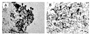
- FIG. 1 shows immunohistochemically stained images using the SKM9-2 antibody against a representative mesothelioma subtype.
- FIG. 1A shows an immunohistological staining image of epithelial mesothelioma
- FIG. 1B shows an immunohistological staining image of sarcoma mesothelioma. The arrowheads in the figure indicate stained portions.
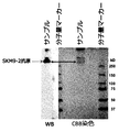
- FIG. 2 shows the results of Western blot and SDS-PAGE of purified SKM9-2 antigen.
- FIG. 3 shows the results of Western blotting of a cell extract obtained by knocking down the HEG1 protein and a recombinant soluble HEG1 protein expressed in a mesothelioma cell line using the SKM9-2 antibody.
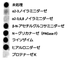
- FIG. 4 is a graph showing that antigen recognition of the SKM9-2 antibody is weakened by treatment with desialylase.
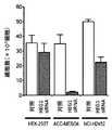
- FIG. 5 shows that the antigen of the SKM9-2 antibody is expressed on the cell surface of mesothelioma.
- FIG. 6 shows the base sequence and amino acid sequence of the heavy chain and light chain of the antibody gene of the hybridoma producing the SKM9-2 antibody, respectively.
- the V H region and the VL region are shown as regions sandwiched by arrows.
- FIG. 7 is a view showing that the SKM10-2 antibody can also detect HEG1 protein in a sugar chain modification-dependent manner.
- FIG. 8 is a Intelectin (ITLN) and V H region and CH1 region of the fused SKM9-2 antibody, V L region and a C L region (i.e., the light chain of SKM9-2 antibody) protein complex with, It is a figure which shows that HEG1 protein can be detected similarly to SKM9-2 antibody.
- FIG. 9 is a diagram showing that inectin binds strongly to beads having a diol bond.
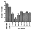
- FIG. 10 is a diagram showing the time-dependent effect of knockdown of the HEG1 gene on cell proliferation of a mesothelioma cell line.
- FIG. 11 is a diagram showing the effect of knockdown of the HEG1 gene on cell proliferation of a mesothelioma cell line.
- FIG. 12 shows the effect of knockdown of the HEG1 gene on the cell proliferation of other mesothelioma cell lines.
- FIG. 13 is a graph showing the positive (+) and negative ( ⁇ ) binding between the SKM9-2 antibody and each human HEG1 fragment. In FIG. 13, the position of each human HEG1 fragment in HEG1 is shown. The result of alanine scanning is shown at the bottom of FIG. In FIG. 13, unmodified amino acids are indicated by the symbol “-”.
- FIG. 14A shows the results of Western blotting confirming the reactivity of HEG1 full-length, 3 kb fragment, 2 kb fragment and 1 kb fragment with the SKM9-2 antibody.
- FIG. 14A shows the results of Western blotting confirming the reactivity of HEG1 full-length, 3 kb fragment, 2 kb fragment and 1 kb fragment with the SKM9-2 antibody.
- FIG. 14B shows the results of Western blotting confirming the reactivity between each HEG1 fragment and the SKM9-2 antibody. In FIG. 14B, positive signals are confirmed in the 7.6 and 7.62 fragments.
- FIG. 14C shows the results of Western blot confirming the reactivity between each HEG1 fragment and the SKM9-2 antibody. In FIG. 14C, positive signals are confirmed in the 7.623 fragment and the 7.6231 fragment.
- FIG. 14D shows the results of alanine scanning. S799A in the figure indicates a fragment in which the 799th serine in the amino acid sequence of SEQ ID NO: 35 is substituted with alanine.
- FIG. 14E shows that the neuraminidase-treated 7.6231 fragment does not react with the SKM9-2 antibody.
- the “subject” may be a mammal, preferably a human.
- a subject can be a subject suffering from or at risk for mesothelioma or other tumor or cancer.
- antibody means an immunoglobulin and includes a polyclonal antibody and a monoclonal antibody.
- a preferred antibody is a monoclonal antibody.
- the origin of the antibody is not particularly limited, and examples thereof include a non-human animal antibody, a non-human mammal antibody, and a human antibody.
- the antibody may also be a chimeric antibody, a humanized antibody, or a human antibody.
- the antibody may also be a bispecific antibody.
- the term “antigen-binding fragment” means a part of an antibody that maintains the binding to an antigen.
- the antigen-binding fragment can comprise the heavy chain variable region or the light chain variable region of the antibody of the present invention, or both.
- the antigen-binding fragment may be chimerized or humanized.
- Examples of the antigen-binding fragment include Fab, Fab ′, F (ab ′) 2 , Fv, scFv (single chain Fv), diabody, and sc (Fv) 2 (single chain (Fv) 2 ).
- Such antibody fragments are not particularly limited, and can be obtained, for example, by treating an antibody with an enzyme.
- Fab can be obtained by digesting an antibody with papain.
- F (ab ′) 2 can be obtained by digesting the antibody with pepsin
- Fab ′ can be obtained by further reducing this.
- such an antigen-binding fragment of an antibody can be used.
- mesothelioma means a mesothelial cell-derived tumor.
- mesothelioma pleural mesothelioma, peritoneal mesothelioma, pericardial mesothelioma, and testicular sheath mesothelioma are known from the site of their occurrence.
- mesothelioma means benign mesothelioma and / or malignant mesothelioma.
- Mesothelioma is broadly divided into histological types, epithelial mesothelioma, sarcoma mesothelioma, biphasic mesothelioma, and other mesothelioma (such as fibrosis).
- the HEG1 protein is a protein whose expression and function are hardly understood, and is estimated to be a membrane protein from gene ontology analysis.
- the human HEG1 protein is a protein (SEQ ID NO: 30) encoded by a gene having the sequence of SEQ ID NO: 29 registered as NM_020733.1 in the National Center for Biotechnology Information (NCBI), and has the amino acid sequence of SEQ ID NO: 31 Protein, a protein encoded by the gene having the sequence of SEQ ID NO: 36 registered as XM — 005247666 (SEQ ID NO: 37), a gene encoded protein having the sequence of SEQ ID NO: 32 (SEQ ID NO: 33), and the sequence of SEQ ID NO: 34 And a protein (SEQ ID NO: 35) encoded by a gene having Natural variants of HEG1 protein are not particularly limited.
- variants of HEG1 protein having amino acid mutations corresponding to mutations such as Q145R, S305P, F602S, V980L, and M1039T in the amino acid sequence of SEQ ID NO: 30 are known. Yes.
- natural variants of HEG1 protein are also included in HEG1 protein.
- the 1359th serine in the amino acid sequence of SEQ ID NO: 30 may be phosphoserine.
- a HEG1 protein in which the serine corresponding to the 1359th serine in the amino acid sequence of SEQ ID NO: 30 is phosphoserine is also included in the HEG1 protein.
- the signal peptide portion is a domain corresponding to positions 1 to 29 of the amino acid sequence of SEQ ID NO: 30, and the extracellular domain is positions 30 to 1248 of the amino acid sequence of SEQ ID NO: 30.
- a transmembrane domain is a domain corresponding to positions 1249 to 1269 of the amino acid sequence of SEQ ID NO: 30, and an intracellular domain is a domain corresponding to positions 1270 to 1381 of the amino acid sequence of SEQ ID NO: 30 Is expected.
- the HEG1 protein has a DNA sequence represented by SEQ ID NO: 29, 32, 34 or 36 or a complementary strand of DNA having a DNA sequence encoding the amino acid sequence of SEQ ID NO: 30, 31, 33, 35 or 37.
- Proteins encoded by DNA sequences that can hybridize under stringent conditions can also be included. Examples of stringent conditions include, for example, washing conditions after hybridization at 65 ° C. with 1 ⁇ SSC (an aqueous solution containing 0.15 M NaCl and 15 mM trisodium citrate).
- the HEG1 protein can also include a protein having an amino acid sequence that is 90% or more, 95% or more, 98% or more, or 99% or more homologous to the amino acid sequence represented by SEQ ID NO: 30.
- the HEG1 protein may contain one or more amino acid substitutions, insertions, additions and / or deletions in the amino acid sequence represented by SEQ ID NO: 30.
- HEG1 protein is mainly expressed in mesothelioma. Further, according to the present inventors, mesothelioma can be diagnosed based on the expression level of HEG1 protein. Therefore, according to the present invention, mesothelioma can be detected using an antibody that binds to the HEG1 protein. Also according to the present invention, HEG1 protein is expressed on the cell membrane surface of mesothelioma and has an extracellular domain. Accordingly, the antibodies of the present invention bind to mesothelioma cell membranes, membrane proteins on mesothelioma cell membranes, or HEG1 protein on mesothelioma cell membranes.
- the antibody of the present invention can be an antibody against the extracellular domain of HEG1 protein or an antigen-binding fragment thereof.
- HEG1 protein is also expressed in well-differentiated papillary mesothelioma (WDPM) and may be used to detect these mesothelioma.
- WDPM papillary mesothelioma
- the extracellular domain of HEG1 protein is subjected to sialylated sugar chain modification in mesothelioma. Therefore, in one aspect, the antibody of the present invention or an antigen-binding fragment thereof binds to a HEG1 protein that has undergone glycosylation.
- the apparent molecular weight can be detected as a broad band in the range of 300 kDa to 500 kDa. More specifically, the apparent molecular weight by SDS-PAGE can be about 400 kDa.
- the sugar chain modification is a sugar chain modification received by the HEG1 protein expressed on the cell membrane of mesothelioma.
- the antibody or antigen-binding fragment thereof of the present invention binds to a HEG1 protein having a sugar chain modification found in mesothelioma cells, but the binding is dependent on the sugar chain modification. That is, in this aspect, the antibody of the present invention or the antigen-binding fragment thereof does not bind to a HEG1 protein that does not have a sugar chain modification (for example, a HEG1 protein obtained by decomposing a sugar chain by glycolysis), Binding is weaker (less affinity) than for HEG1 protein with glycosylation.
- the antibody of the present invention or an antigen-binding fragment thereof binds to a sugar chain portion of HEG1 protein having a sugar chain modification found in mesothelioma cells.
- the antibody of the present invention can bind to a HEG1 protein that has been subjected to sugar chain modification in a sugar chain modification-dependent manner.
- the sugar chain modification is caused by HEG1 protein present on the cell membrane of mesothelioma. It may have.
- the antibodies of the invention bind to a HEG1 protein having a glycosylation found in denatured mesothelioma cells.
- the antibody of the present invention is a denatured fragment of HEG1 protein having a sugar chain modification found in mesothelioma cells, and comprises the amino acid sequence from 799th to 809th in the amino acid sequence of SEQ ID NO: 35 It binds to fragments having the amino acid sequence.
- the antibody of the invention or antigen-binding fragment thereof binds to an epitope of the extracellular domain of human HEG1 protein expressed in human mesothelioma cells, but is ⁇ 2-3 neuraminidase or ⁇ 2-3, 6, 8.
- the extracellular domain of human HEG1 protein expressed in human mesothelioma cells has undergone sialylated glycosylation.
- HEG1 protein having a sugar chain modification obtained from mesothelioma may be a HEG1 protein having a sugar chain modification characteristic of mesothelioma, for example, a sialylated O-type sugar chain It can be a modification.
- an antibody that binds to a mesothelioma cell membrane, a membrane protein on a mesothelioma cell membrane, or a HEG1 protein on a mesothelioma cell membrane comprising: Heavy chain CDR1 having the amino acid sequence of SEQ ID NO: 6, heavy chain CDR2 having the amino acid sequence of SEQ ID NO: 8, heavy chain CDR3 having the amino acid sequence of SEQ ID NO: 10, light chain CDR1 having the amino acid sequence of SEQ ID NO: 12, SEQ ID NO: Provided is an antibody comprising any one, two, three, four, five or six of a light chain CDR2 having 14 amino acid sequences and a light chain CDR3 having an amino acid sequence of SEQ ID NO: 16 .
- An antibody comprising a light chain variable region comprising any one, two, or three is provided.
- an antibody that binds to a mesothelioma cell membrane, a membrane protein on a mesothelioma cell membrane, or a HEG1 protein on a mesothelioma cell membrane comprising: (1) an antibody comprising a heavy chain variable region comprising CDR1 having the amino acid sequence of SEQ ID NO: 6, CDR2 having the amino acid sequence of SEQ ID NO: 8, and CDR3 having the amino acid sequence of SEQ ID NO: 10, (2) an antibody comprising CDR1 having the amino acid sequence of SEQ ID NO: 12, CDR2 having the amino acid sequence of SEQ ID NO: 14, and a light chain variable region comprising CDR3 having the amino acid sequence of SEQ ID NO: 16, (3) having a heavy chain variable region comprising CDR1 having the amino acid sequence of SEQ ID NO: 6, CDR2 having the amino acid sequence of SEQ ID NO: 8, CDR3 having the amino acid sequence of SEQ ID NO: 10, and the amino acid sequence of SEQ ID NO: 12
- the antibody of the present invention is an antibody that binds to a peptide having the sequence of SEQ ID NO: 53. In certain embodiments, an antibody of the invention is an antibody that binds to a peptide having the sequence of SEQ ID NO: 53 expressed in a mesothelioma cell line. In one embodiment, the antibody of the present invention is an antibody that binds to a peptide having the sequence of SEQ ID NO: 53 expressed in a mesothelioma cell line in a glycosylation-dependent manner.
- an antibody of the invention is an antibody that binds to a mesothelioma cell membrane, a membrane protein on a mesothelioma cell membrane, or a HEG1 protein on a mesothelioma cell membrane, wherein the sequence of SEQ ID NO: 53 is It is an antibody that binds to the peptide it has.
- an antibody of the invention is an antibody that binds to a mesothelioma cell membrane, a membrane protein on a mesothelioma cell membrane, or a HEG1 protein on a mesothelioma cell membrane, wherein the antibody is a mesothelioma cell line.
- an antibody that binds to the expressed peptide having the sequence of SEQ ID NO: 53 is an antibody that binds to a mesothelioma cell membrane, a membrane protein on a mesothelioma cell membrane, or a HEG1 protein on a mesothelioma cell membrane, wherein the antibody is a mesothelioma cell line.
- Sequence homology can be determined with default parameters by, for example, the FASTA program of Pearson and Lipman, PNAS, 85: 2444-2448, 1988.
- the antibodies of the invention have a heavy chain having the amino acid sequence of SEQ ID NO: 2.
- an antibody of the invention has a light chain having the amino acid sequence of SEQ ID NO: 4.
- the antibody of the present invention has a heavy chain having the amino acid sequence of SEQ ID NO: 2 and a light chain having the amino acid sequence of SEQ ID NO: 4.
- the antibody of the present invention has the amino acid sequence of No. 20 to No. 132 of SEQ ID NO: 2 as the heavy chain variable region (V H region).
- the antibody of the present invention has the amino acid sequence of No. 20 to No. 132 of SEQ ID NO: 4 as the light chain variable region ( VL region).
- the antibody of the present invention has a heavy chain variable region having amino acid sequences 20 to 132 of SEQ ID NO: 2 and a light chain variable region having amino acid sequences 20 to 132 of SEQ ID NO: 4. .
- the antibody of the present invention competes with the above-mentioned antibody for binding to a glycosylated HEG1 protein.
- the present invention also provides a chimeric antigen receptor comprising a heavy chain variable region such as scFv and a light chain variable region.
- a chimeric antigen receptor comprising a heavy chain variable region such as scFv and a light chain variable region.
- the chimeric antigen receptor for example, the first generation in which scFv and the T cell receptor ⁇ chain are linked via a spacer, the second generation in which CD28 or 4-1BB is linked, CD28 and 4-1BB or OX40
- Various chimeric antigen receptors such as third generation linked via can be used.
- a conjugate (conjugate) of the antibody of the present invention or an antigen-binding fragment thereof and a cytotoxic agent is provided.
- a pharmaceutical composition for use in treating mesothelioma comprising a conjugate (conjugate) of the antibody of the present invention or an antigen-binding fragment thereof and an imaging probe.
- the cytotoxic agent used in the present invention include radioisotopes for cancer treatment (for example, P 32 , Y 90 , I 131 , I 125 , Sm 153 , Re 186 , Re 188 , At 211 , Bi 212 , Pb 212 and Lu).
- the cytotoxic agent used in the present invention include an anticancer agent.
- a pharmaceutical composition for use in treating mesothelioma comprising siRNA, shRNA, or antisense oligo for the HEG1 gene.
- the siRNA, shRNA or antisense oligo may be a modified nucleic acid.
- the modified nucleic acid include a fluorescent dye-modified nucleic acid, a biotinylated nucleic acid, and a nucleic acid into which a cholesteryl group has been introduced.
- RNA may be 2′-O-methyl modified, 2′-fluoro modified, or 2′-methoxyethyl (MOE) modified to the base to enhance stability, thereby phosphodiester bonds in the nucleic acid backbone.
- MOE 2′-methoxyethyl
- Artificial nucleic acids include 2'-position oxygen atom and 4'-position carbon atom via a methylene bridged DNA (locked nucleic acid (LNA), N- (2-aminoethyl) instead of deoxyribose or ribose Peptide nucleic acid (PNA) in which a polymer in which a glycine amide bond is a main chain is used.
- LNA locked nucleic acid
- PNA ribose Peptide nucleic acid
- siRNA, shRNA, or antisense oligo may be encapsulated in vesicles such as micelles and liposomes. Vesicles such as micelles and liposomes can have a particle size of 20 to 100 nm, for example.
- vesicles such as micelles and liposomes can be efficiently accumulated in tumor tissue by the EPR effect (Enhanced permeability and retention effect), and the contents can be delivered to the tumor tissue.
- Such vesicles may be modified with a surface modifier such as polyethylene glycol.
- Certain aspects of the invention also provide the use of siRNA, shRNA, or antisense oligos against the HEG1 gene in the manufacture of a pharmaceutical composition for use in treating mesothelioma.
- Certain embodiments of the invention further provide a method of treating mesothelioma, comprising reducing HEG1 protein expression in a subject in need thereof.
- a method of treating mesothelioma comprising administering an expression inhibitor (eg, siRNA, shRNA, or antisense oligo) to the HEG1 gene in a subject in need thereof.
- an expression inhibitor eg, siRNA, shRNA, or antisense oligo
- the antibody of the present invention may have cytotoxic activity such as antibody-dependent cytotoxic activity (ADCC activity) or complement-dependent cytotoxic activity (CDC activity).
- ADCC activity antibody-dependent cytotoxic activity
- CDC activity complement-dependent cytotoxic activity
- the antibody of the present invention binds to the HEG1 protein expressed on the surface of the mesothelioma membrane and can exhibit cytotoxic activity against mesothelioma cells.
- a pharmaceutical composition or a mesothelioma therapeutic agent for use in treating mesothelioma comprising the antibody of the present invention or an antigen-binding fragment thereof as an active ingredient is provided.
- “treatment” is used in the meaning including therapy.
- the pharmaceutical composition or drug containing the antibody of the present invention as an active ingredient can be formulated by a known pharmaceutical method.
- the pharmaceutical composition or medicament of the present invention may contain a pharmaceutically acceptable excipient.
- the excipient can be an excipient that can be appropriately administered to give an effective amount of the antibody of the present invention, which is an active ingredient, to the subject.
- the pharmaceutical composition or medicament of the present invention can be an injection, and the injectable excipient is a sterile aqueous solution, such as a pharmaceutical such as Ringer's solution, Hanks' solution or saline. And an isotonic solution containing glucose, glucose and other adjuvants.
- the adjuvant examples include alcohols such as ethanol, polyalcohols such as polyethylene glycol, and nonionic surfactants such as polysorbate 80, which can be added during formulation. Sesame oil, coconut oil and soybean oil can be used as an oily liquid for injection, and benzyl benzoate or benzyl alcohol can be used as an auxiliary agent.
- the pharmaceutical composition or medicament of the present invention can be administered parenterally (for example, intravenously or intrathoracically) in the form of an injection.
- ADCC activity is achieved, for example, by incubating mesothelioma cells and effector cells expressing Fc receptors (eg, NK cells and monocytes) under physiological conditions in the presence of the antibody of the present invention. It can be determined by counting the number of live and / or dead cells.
- CDC activity can be determined by, for example, incubating mesothelioma cells with a solution containing complement (eg, human serum) in the presence of antibodies under physiological conditions to determine the number of live and / or dead mesothelioma cells. It can be determined by counting.
- Cytotoxic activity can be enhanced by various methods well known to those skilled in the art.
- antibodies lacking fucose in the Fc region sugar chain, bisecting N-acetylglucosamine (GlcNAc) bound to the sugar chain, amino acid substitution in the Fc region enhances the binding between the Fc receptor of the effector cell and the antibody
- GlcNAc N-acetylglucosamine
- the antibody can be a genetically modified antibody such as a chimeric antibody, a humanized antibody or a human antibody by a method well known to those skilled in the art for the purpose of reducing the antigenicity of the antibody itself in humans.
- the antibody of the present invention may be a chimeric antibody, a humanized antibody or a human antibody.
- the antibody may also be a bispecific antibody.
- a conjugate (conjugate) of the antibody of the present invention or an antigen-binding fragment thereof and an imaging probe is provided.
- the conjugate (conjugate) of the antibody or antigen-binding fragment thereof of the present invention and an imaging probe can be used for in vitro diagnosis or in vivo diagnosis of mesothelioma. Therefore, according to the present invention, there is provided a diagnostic agent or diagnostic kit for mesothelioma comprising a conjugate (conjugate) of the antibody of the present invention or an antigen-binding fragment thereof and an imaging probe.
- the present invention also provides an in-vivo diagnostic agent or in-vivo diagnostic kit for mesothelioma comprising a conjugate (conjugate) of the antibody of the present invention or an antigen-binding fragment thereof and an imaging probe.
- Imaging probes that can be used for in-vivo diagnosis of mesothelioma include, for example, fluorescent imaging probes, enhancement agents such as contrast agents for nuclear magnetic resonance imaging (MRI) (eg paramagnetic ions), and PET molecular imaging probes Examples include radionuclides for imaging.
- MRI nuclear magnetic resonance imaging
- PET molecular imaging probes examples include radionuclides for imaging.
- a HEG1 protein having a sugar chain modification (eg, O-type sugar chain modification) obtained from mesothelioma or a fragment thereof (particularly the amino acid sequence from 799th to 809th in the amino acid sequence of SEQ ID NO: 35) And fragments thereof) are provided.
- HEG1 protein having a sugar chain modification (for example, O-type sugar chain modification) obtained from mesothelioma or a fragment thereof is used as a marker for detecting mesothelioma with high sensitivity and high specificity. Can be used.
- a diagnostic marker for mesothelioma comprising a HEG1 protein having a sugar chain modification (eg, O-type sugar chain modification) obtained from mesothelioma or a fragment thereof.
- the O-type sugar chain modification of the HEG1 protein obtained from mesothelioma or the fragment thereof may be a sialylated O-type sugar chain modification.
- Such HEG1 protein has, for example, 80% or more, 90% or more, 95% or more, or 98% or more of sequence homology with any one amino acid sequence selected from SEQ ID NOs: 30, 31, 33, 35 and 37 It may be a HEG1 protein obtained from mesothelioma having a sex amino acid sequence, or a variant obtained from these mesothelioma.
- the HEG1 protein having a sugar chain modification (for example, O-type sugar chain modification) obtained from mesothelioma or the fragment thereof is obtained from the antibody of the present invention (glycan modification obtained from the mesothelioma of the present invention) from mesothelioma cells.
- HEG1 protein having an O-type sugar chain modification For example, an antibody that binds to a HEG1 protein having an O-type sugar chain modification.
- HEG1 protein or fragment thereof has sialic acid and GlcNAc, HEG1 against a lectin that specifically binds to them (eg, WGA that is sialic acid specific or DSA that is GlcNAc specific or a combination thereof). It is also conceivable to purify the HEG1 protein using the protein affinity.
- HEG1 protein having a sugar chain modification for example, O-type sugar chain modification obtained from mesothelioma or the fragment thereof (particularly, the fragment containing the amino acid sequence of 799 to 809 in the amino acid sequence of SEQ ID NO: 35), It may be detained.
- the fragment (particularly the denatured fragment) is an amino acid sequence of the 799th to 809th amino acids in the amino acid sequence of SEQ ID NO: 35 in the HEG1 protein having a sugar chain modification (eg, O-type sugar chain modification) obtained from mesothelioma.
- the peripheral area may be included.
- the present invention provides fusion proteins of intelctin and Fab or scFv fragments of the antibodies of the present invention.
- a heavy chain of an Fab fragment or an scFv fragment and inlectin exist as a fusion form.
- a complex of a fusion protein of inlectin with the VH region and linker (eg, CH1 region) of the antibody of the present invention and the L chain of the antibody of the present invention is provided.
- Such a fusion protein or protein complex binds to a mesothelioma cell membrane, a membrane protein on the cell membrane, or a HEG1 protein on the cell membrane.
- the present invention also provides a fusion protein of the scFv fragment of the antibody of the present invention and inlectin.
- a fusion protein or protein complex contains inlectin, which binds specifically and strongly to the diol structure, so it is treated with a column (eg, treated with an epoxide crosslinking agent) that uses a resin that displays the diol structure. It can be easily purified with a gel containing polysaccharide (dextran or agarose) or a gel filtration column such as silica gel hydrophilized by diol modification.
- the fusion protein or protein complex can be purified using a column using diol sepharose beads shown in Table 5. After purifying the complex, a fusion protein may be further generated on an affinity column to which an antigen is linked.
- a fusion protein of a heavy chain variable region comprising CDR1 having the amino acid sequence of SEQ ID NO: 6, CDR2 having the amino acid sequence of SEQ ID NO: 8, and CDR3 having the amino acid sequence of SEQ ID NO: 10; 2) including the fusion protein of (1) above, CDR1 having the amino acid sequence of SEQ ID NO: 12, CDR2 having the amino acid sequence of SEQ ID NO: 14, and the light chain variable region comprising CDR3 having the amino acid sequence of SEQ ID NO: 16
- a protein complex is provided.
- the protein complex binds to a mesothelioma cell membrane, a membrane protein on a mesothelioma cell membrane, or a HEG1 protein on a mesothelioma cell membrane.
- the heavy chain variable region can form a fusion protein with intelectin via or without the CH1 region, preferably the heavy chain variable region is intelectin and fusion protein via the CH1 region. Can be formed.
- This fusion protein can bind to each other through physiological intactin moieties under physiological conditions to form multimers (mainly trimers). In the present specification, monomers and multimers are included in the fusion protein.
- the above-mentioned inlectin fusion protein and complex of the present invention can bind to a HEG1 protein having a sugar chain modification obtained from mesothelioma. Therefore, the above-mentioned inlectin fusion protein or complex of the present invention can be used for detection of mesothelioma. Therefore, according to the present invention, there is provided a diagnostic agent for mesothelioma comprising the above-mentioned inlectin fusion protein or complex of the present invention.
- the conjugate of the above-mentioned interectin fusion protein or complex of the present invention and an imaging probe can be used for in-vivo imaging of mesothelioma.
- an in-vivo diagnostic agent for mesothelioma comprising the conjugate of the above-mentioned intellctin fusion protein or complex of the present invention and an imaging probe.
- the conjugate of the above-described inlectin fusion protein and complex of the present invention and the cytotoxic agent as defined above can be used for treating mesothelioma.
- a pharmaceutical composition for use in treating mesothelioma comprising a conjugate of the above-mentioned intellctin fusion protein and complex of the present invention and a cytotoxic agent as defined above. Is done.
- Antibodies can be produced by methods well known to those skilled in the art. That is, a polyclonal antibody can be obtained by immunizing an animal with an antigen and an adjuvant and obtaining plasma of the immunized animal. Alternatively, an animal may be immunized with an antigen and an adjuvant, B lymphocytes may be obtained from the immunized animal, cell hybridized with myeloma to form a hybridoma, and a hybridoma that produces a desired antibody may be cloned. In the immunization step, HEG1 protein may be forcibly expressed in a mesothelioma cell line (eg, ACC-MESO4 cells), and the cells may be immunized.
- a mesothelioma cell line eg, ACC-MESO4 cells
- the immunized animal can produce antibodies against the HEG1 protein.
- the HEG1 protein may be purified from a cell that expresses the HEG1 protein, preferably from a mesothelioma cell line, to immunize the animal.
- a solubilized form of HEG1 protein eg, extracellular domain
- a mesothelioma cell line eg, ACC-MESO4 cells
- a solubilized form of HEG1 protein obtained from the culture supernatant is used. May immunize animals.
- such solubilized HEG1 protein is also subjected to sugar chain modification, and this sugar chain modification is similar to the modification of HEG1 protein on the mesothelioma cell membrane.
- Chimeric antibodies can be prepared by methods well known in the art. For example, it can be prepared by replacing the constant region of an antibody with the constant region of a human antibody.
- Humanized antibodies include, for example, complementarity determining regions (CDRs) derived from animals other than humans, framework regions derived from human antibodies, and constant regions derived from human antibodies.
- CDRs complementarity determining regions
- a humanized antibody can be obtained, for example, by transplanting the above CDR into a human antibody.
- Human antibodies can be obtained, for example, by immunizing genetically modified mice that produce human antibodies with antigens.
- Bispecific antibodies are antibodies that can bind to two different epitopes or antigens and can be prepared by methods well known to those skilled in the art.
- Bispecific antibodies are, for example, a method of further hybridizing cells that produce two different antibodies to produce a hybrid hybridoma, or a pair of V H region and VL region cannot be formed between the two regions.
- An antibody that competes in the binding between an antibody and an antigen can be obtained by a competitive assay well known to those skilled in the art.
- An antibody that competes for binding to the same antigen if it can block the binding of the desired antibody in a competition assay, for example, at least 20%, preferably at least 20-50%, more preferably at least 50%. Can do.
- Competing antibodies can be confirmed by cross-blocking assays, preferably by competitive ELISA assays.
- the antigen is coated on, for example, a microtiter plate, where the presence of a candidate competitive antibody is added and incubated to form a bond between the antigen and the candidate antibody. Then, after labeling the desired antibody, it is further added to the well, incubated, washed, and the binding amount of the desired antibody is quantified to determine whether or not the antibody has competed. If competing, there should be less label remaining in the well.
- a method for diagnosing mesothelioma in a patient suffering from or at risk of suffering from mesothelioma using an antibody or antigen-binding fragment thereof that binds to the HEG1 protein is provided. Is done.
- the present invention also provides a preliminary method for diagnosing mesothelioma in a patient suffering from or at risk of suffering from mesothelioma using an antibody or antigen-binding fragment thereof that binds to the HEG1 protein (E.g., industrial methods).
- the present invention further provides a method of detecting mesothelioma cells in a patient suffering from or at risk of suffering from mesothelioma using an antibody or antigen-binding fragment thereof that binds to the HEG1 protein.
- the diagnosis of mesothelioma is performed by a doctor, and the present invention provides the doctor with basic information of diagnosis as a method for diagnosis by the doctor.
- a diagnostic agent for diagnosing mesothelioma in a patient suffering from or at risk of suffering from mesothelioma comprising an antibody or antigen-binding fragment thereof that binds to the HEG1 protein
- a diagnostic kit may comprise instructions for detecting mesothelioma using an antibody or antigen-binding fragment thereof that binds to the HEG1 protein.
- the diagnostic agent or kit may contain any of the antibodies of the present invention or an antigen-binding fragment thereof.
- the diagnostic agent and diagnostic kit of the present invention can be used to detect mesothelioma as described below.
- a method for diagnosing mesothelioma comprising contacting a sample with the antibody of the present invention or an antigen-binding fragment thereof. And a method for detecting mesothelioma to assist a doctor in diagnosing mesothelioma.
- the detection of mesothelioma according to the present invention can be performed by bringing the antibody of the present invention or an antigen-binding fragment thereof into contact with a sample.
- a sample separated from a living body such as a cell, tissue, or tissue section
- the antibody reaction can be detected by a method well known to those skilled in the art (for example, immunohistological staining method).
- an antibody reaction is observed (for example, when it is positive), it is highly likely that the subject from which the sample is derived suffers from mesothelioma. Whether or not the antibody reaction is positive can be determined based on the Allred Score method (see Allred DC et al., Mod Pathol, 11: 155-168, 1998).
- Total score (TS) staining rate score (PS) + staining intensity score (IS)
- PS and IS staining intensity score
- the detection of mesothelioma is Contacting the antibody of the invention or antigen-binding fragment thereof with a sample obtained from a subject suspected of being mesothelioma; Detecting the presence of the HEG1 protein (preferably the extracellular domain of the HEG1 protein) present in the sample using the antibody of the present invention.
- the HEG1 protein to be detected is HEG1 that has undergone glycosylation, more preferably HEG1 protein having O-type glycosylation, and even more preferably mesothelioma. Is a HEG1 protein having a sugar chain modification.
- the detection of mesothelioma may further include determining whether or not mesothelioma has been detected using the staining rate and / or staining intensity as an index.
- the method may include determining whether mesothelioma has been detected based on the Allred Score method. In one embodiment, it may include determining that mesothelioma has been detected when the total score (TS) is 2 or more, preferably 3 or more, based on the Allred Score method.
- HEG1 having a sugar chain modification obtained from mesothelioma using an antibody that binds to the HEG1 protein having a sugar chain modification obtained from mesothelioma in a sugar chain modification-dependent manner.
- the presence of the protein can be detected in the sample.
- the presence of HEG1 protein having sugar chain modification obtained from mesothelioma is detected in the sample using an antibody that binds to HEG1 protein having sugar chain modification obtained from mesothelioma. It is good also as what detects by.
- the antigen protein or a part thereof can be detected by immunohistological staining using the antibody of the present invention.
- the mesothelioma diagnosis method, mesothelioma detection method, mesothelioma detection method of the present invention, or mesothelioma detection method for assisting doctors in diagnosing mesothelioma Can be determined that mesothelioma has been detected from the subject.
- the mesothelioma to be detected in the present invention is at least one mesothelioma selected from the group consisting of epithelial type, biphasic type, sarcoma type, and fibrogenic type.
- the mesothelioma to be detected in the present invention may be a sarcoma type mesothelioma.
- mesothelioma in a subject suspected of having mesothelioma (especially epithelial mesothelioma) or lung adenocarcinoma, It is possible to identify whether or not the patient has adenocarcinoma. Accordingly, detection of mesothelioma according to the present invention can be performed on a subject suspected of having mesothelioma (particularly epithelial mesothelioma) or lung adenocarcinoma. Further, according to the present invention, mesothelioma can be differentiated from other cancers. Thus, the detection of mesothelioma according to the present invention can be performed on a subject suspected of having mesothelioma or other cancer.
- the detection of mesothelioma is performed by using a conjugate of an imaging probe and the antibody of the present invention or an antigen-binding fragment thereof instead of the antibody of the present invention or an antigen-binding fragment thereof, or the present invention as defined above.
- a fusion protein or complex of the antibody fragment and inlectin may be used.
- the fusion protein or complex of the antibody fragment of the present invention and inlectin defined above may be in the form of a conjugate with an imaging probe.
- the cell precipitate that had been stored frozen was suspended in 100 ⁇ L of PBS and mixed with 100 ⁇ L of AddaVax (INVIVOGEN). The whole amount was administered intraperitoneally to a 6-week-old Balb / c mouse (female), and the cell precipitate that had been stored frozen every two weeks was repeatedly administered in the same manner. After a total of three immunizations, a drop of blood was collected from the tail vein to obtain serum. Immunostaining was performed using thin slices prepared from ACC-MESO1 and ACC-MESO4 cell pellets fixed with AMeX (Sato Y et al. Am J Pathol 125: 431-435 (1986)), and mesothelioma cells It was confirmed whether the antibody against was produced. Mice with high antibody titers were immunized again using cell precipitation as described above, and the spleen was aseptically removed one week later.
- the spleen was ground on a stainless mesh, and lymphocytes were dispersed in 10 mL of RPMI 1640. After loosening a large cell mass by pipetting, it was transferred to a 15 mL centrifuge tube and allowed to stand for 3 minutes. The cell suspension was transferred to a new centrifuge tube so as not to suck a large aggregate, and the cells were washed twice with RPMI 1640, and the number of cells was counted.
- Lymphocytes (6.75 ⁇ 10 7 cells) and myeloma cells (PAI) (5.25 ⁇ 10 6 cells) that had been cultured in advance were mixed, washed twice with RPMI 1640, and then cell fusion was performed.
- Cell fusion was performed using PEG1500 (Roche Diagnostics Inc.) according to the protocol attached to the product. After the fusion, the cells were suspended in RPMI 1640 (76 mL) containing 15% FCS, 10% BM Condimed H1 (Roche Diagnostics), 1 ⁇ HAT (Thermo Fisher Scientific), and 200 ⁇ L per well (about 1.9 ⁇ 10 5 cells / well) were placed in four 96-well plates. After culturing at 37 ° C.
- Screening was performed by immunostaining using sliced sections prepared from cell precipitates of ACC-MESO1 and ACC-MESO4 fixed with AMeX. The sliced sections were blocked with 0.5% casein for 10 minutes, and 100 ⁇ L of the hybridoma culture supernatant was reacted as a primary antibody for 2 hours. After washing with PBS, the EnVision + kits (Dako Japan Godo Kaisha) was reacted for 30 minutes as a secondary antibody, and coloring was performed using DAB. 384 wells were screened and 93 clones were obtained as immunostaining positive clones.
- Example 2 Detection of mesothelioma by the obtained monoclonal antibody Various types of mesothelioma were stained using the monoclonal antibody produced from SKM9-2 obtained in the above screening.
- a cell pellet fixed with AMeX or a tissue fixed with formalin was embedded in paraffin to prepare a sliced slice.
- the sections were fixed on a slide glass, deparaffinized and dehydrated with xylene and ethanol, and antigens were activated under various conditions.
- the sections were treated with 3% hydrogen peroxide for 5 minutes to inactivate endogenous peroxidase, washed with PBS, and blocked with 0.5% casein for 10 minutes.
- cytokeratin 5/6 CK5 / 6
- podoplanin cytokeratin 5/6
- WT-1 Wilms tumor gene product 1
- the activation solution was calretinin, CK5 / 6, podoplanin 1 mM ethylenediaminetetraacetic acid (EDTA) 10 mM Tris buffer (pH 9.0), WT-1, mesothelin, SKM9-2 antigen 0.1% Tween-20. It was a 10 mM citrate buffer solution (pH 6.0).
- the primary antibodies used were as follows: calretinin, rabbit anti-calretinin polyclonal antibody (PAD: DC8) (Life Technologies Japan Ltd., Tokyo, Japan); CK5 / 6, mouse anti-cytokeratin 5,6 monoclonal antibody (D5) / 16B4) (Nichirei Biosciences); mesothelin, mouse anti-human mesothelin monoclonal antibody (5B2) (Leica Micro- systems, Inc., Bannockburn, IL); podoplanin, mouse anti-podoplanin monoclonal antibody (D2-40) (Roche Diagnostics Inc.); WT-1, mouse anti-human WT-1 monoclonal antibody (6F-H2) (Roche Diagnostics Inc.).
- FIGS. 1A and B Representative staining images of mesothelioma were as shown in FIGS. 1A and B.
- the SKM9-2 antibody was able to satisfactorily stain each of the epithelial type (FIG. 1A) and sarcoma type (FIG. 1B). Further, as shown in Table 1, the SKM9-2 antibody was able to detect mesothelioma with higher sensitivity than any of the other antibodies.
- the positive determination was made positive when the ratio of cells that were 1 or more in the determination based on Allred score to the cells was 10% or more.
- the SKM9-2 antibody can detect each type of mesothelioma with high sensitivity.
- well-differentiated papillary mesothelioma which is a benign or low-grade mesothelioma, could be stained well with the SKM9-2 antibody (1 case in 1 case).
- other cancers are gastric cancers, colon cancers, breast cancers, ovarian cancers, renal cell cancers, urothelial cancers, which tend to be a problem in differential diagnosis from mesothelioma, and “soft tissue sarcomas” are cancers.
- the SKM9-2 antibody has high specificity for mesothelioma and can be differentiated from other types of tumors or cancers with high accuracy. In addition, the SKM9-2 antibody hardly recognized any healthy tissue. Only three cases of SKM9-2 antibody were positive: urothelial carcinoma (1/10), leiomyosarcoma (1/10), and epithelioid vascular endothelial species (1/6). .
- Calretinin antibody, cytokeratin 5/6 antibody, mesothelin antibody and WT-1 antibody known as conventional mesothelioma diagnostic antibodies can each detect epithelial mesothelioma with high sensitivity.
- these antibodies do not necessarily have excellent specificity, and it is difficult to distinguish lung adenocarcinoma from epithelial mesothelioma, or to detect sarcoma mesothelioma. It was known that there was.
- calretinin and WT-1 are expressed in the cytoplasm in various healthy and cancerous tissues, and there is a risk of misidentifying as a nuclear translocation image characteristic of mesothelioma. It was not an easy marker.
- the SKM9-2 antibody can easily distinguish mesothelioma depending on whether it is positive or negative, and can detect both epithelial mesothelioma and sarcoma mesothelioma. It was better than the antibody. It was also possible to differentiate between mesothelioma and other cancers.
- the antigen recognized by the SKM9-2 antibody can be a very useful marker in mesothelioma diagnosis.
- Example 3 Analysis of the obtained hybridoma In Examples 1 and 2, it was revealed that a monoclonal antibody having high specificity for mesothelioma cells was obtained. Thus, identification of the antigen of SKM9-2 antibody and antibody Further analysis of the antigen recognition properties of SKM9-2 antibody.
- the precipitate was extracted with 20% Tris buffer (ph 7.2) containing 0.1% Tween 20, 150 mM NaCl, and the supernatant after centrifugation was concentrated using Amicon Ultra-15,100 kDa (Merck Millipore) and 6M
- the solution was exchanged with 20 mM Tris buffer (pH 8.0) containing guanidine hydrochloride.
- Tris (2-carboxyethyl) phosphine hydrochloride having a final concentration of 10 mM was added to the concentrated solution and heated at 60 ° C. for 30 minutes, and then 40 mM of iodoacetamide having a final concentration was added and reacted at room temperature for 3 hours.
- the reaction solution was subjected to gel filtration chromatography (Superose 6 Increase 10 / 300GL (GE Healthcare)) in 20 mM Tris buffer (pH 8.0) containing 6 M guanidine hydrochloride and 0.05% Tween 20, and positive by dot blot. The fractions to be collected were collected. The sample was dialyzed overnight at room temperature against 25 mM phosphate buffer (pH 7.0) containing 4 M urea and 0.05% Tween 20, and applied to Mono Q 5 / 50GL (GE Healthcare).
- the dot blot was performed as follows. A PVDF membrane (Immobilon-P Merck Millipore) was immersed in methanol, and then washed and immersed in pure water. A hydrophilic PVDF membrane was placed on a filter paper soaked with water so as not to sandwich air, and after removing excess water, the sample was impregnated into dots with a micropipette of 1 to 5 ⁇ L. After the membrane was dried overnight at room temperature, it was hydrophilized again with methanol and pure water, and with 20 mM Tris buffer (pH 7.4) (blocking buffer) containing 5% skim milk, 0.1% Tween 20, 150 mM NaCl at room temperature. Blocking was performed for 1 hour.
- Tris buffer pH 7.4
- the mouse ascites of SKM9-2 hybridoma diluted 1,000 times with blocking buffer was reacted as a primary antibody for 1 hour at room temperature, and then 20 mM Tris buffer (pH 7.4) containing 0.1% Tween 20, 150 mM NaCl (pH 7.4) ( The membrane was washed by TBST).
- Horseradish peroxidase-labeled goat anti-mouse IgG (Jackson ImmunoResearch) diluted 30,000 times in blocking buffer was allowed to react at room temperature for 1 hour, washed with TBST, then developed and detected with ECL prime (GE Healthcare). Went.
- the purified antigen was confirmed by Western blotting. Samples were separated with 6% SDS-PAGE or 4-15% Mini-PROTEAN TGX precast gel (Bio-Rad), and submarine type using 10 mM CAPS buffer (pH 10.5) containing 0.01% SDS. The sample was transferred to the PVDF membrane by a transfer device. The transferred membrane was subjected to blocking and antibody reaction in the same manner as the dot blot, and color development and detection were performed with ECL prime (GE Healthcare).
- the membrane was washed with 20 mM Tris buffer (pH 8.0) containing 6M guanidine hydrochloride, 0.1% Tween 20, 1% 2-mercaptoethanol for 15 minutes at room temperature, washed thoroughly purely, and then blocked again. went. Detection was performed in the same manner using a mouse anti- ⁇ -actin monoclonal antibody (Sigma-Aldrich) diluted 5,000 times as a control antibody.
- the molecular weight of the antigen of the SKM9-2 antibody by SDS-PAGE was estimated to be about 400 kDa, which was inconsistent with the estimated molecular weight of the HEG1 protein (about 150 kDa).
- the SKM9-2 antibody could recognize the HEG1 protein.
- H1097 sense strand GAUCUUUGACGGUCAGUCUGG (SEQ ID NO: 23)
- H1097 antisense strand AGACUGACCGUCAAAGAUCGC (SEQ ID NO: 24)
- H2674 sense strand CCUAUAGCCGUACAGACUACA (SEQ ID NO: 25)
- H2674 antisense strand UAGUCUGUACGGCUAUAGGGC (SEQ ID NO: 26)
- H3671 antisense strand UGAAAGUAUCCCGACUUGCAC (SEQ ID NO: 28)
- As a negative control Mission Negative control SIC-001, confidential sequences (manufactured by Sigma) was used.
- siRNAs were each transfected into ACC-MESO4 using Lipofectamine 2000 (Thermo Fisher Scientific). After culturing for 72 hours, the cells were washed with PBS, 25 mM / cm 2 of 20 mM Tris buffer (pH 8.0) containing 1% SDS, 125 mU / mL Benzonase nuclease (Merck Millipore) was added, and the mixture was allowed to stand on ice for 5 minutes. And the lysate was collected. The supernatant after centrifugation was used as a sample, separated by 6% SDS-PAGE, and then Western blotted.
- expression suppression was also performed using a lentivirus that expresses shRNA against human HEG1.
- Lentiviral particles sc-78365-V
- ACC-MESO4 ACC-MESO4 according to the product protocol.
- drug selection was started using 10 ⁇ g / mL puromycin, and cells that survived and proliferated until 10 days were used as shRNA stably expressing cells.
- the operation after solubilization was performed in the same manner as in the siRNA treatment.
- cop GFP control lentiviral particles sc-108084, manufactured by Santa Cruz
- HEG1 cDNA (SEQ ID NOs: 32 and 34) cloned from ACC-MESO4, a FLAG tag and His are added to the base sequence (# 1 to 4059 of SEQ ID NO: 32) corresponding to the extracellular region of the base sequence shown in SEQ ID NO: 32.
- the gene to which the tag was connected (SEQ ID NO: 17) was inserted between the XhoI site and NotI site of pcDNA3.1 (-) (Thermo Fisher Fisher Scientific) to prepare a recombinant soluble HEG1 expression plasmid.
- This plasmid was transfected into ACC-MESO4 using Lipofectamine LTX Reagent with PLUS Reagent (Thermo Fisher Scientific), and drug selection was performed with 750 ⁇ g / mL geneticin (Thermo Fisher Scientific). Two weeks later, the surviving and proliferating cells were collected and used as recombinant soluble HEG1 stably expressing cells. 40 mL of the cell culture supernatant was added to HisTrap excel (1 mL) (GE Healthcare), and the column was washed with 25 mM phosphate buffer (pH 7.2) containing 10 mM imidazole and 0.5 M NaCl. Elution was performed in solution with a 10-500 mM imidazole gradient.
- the SKM9-2 antibody expresses post-translational modification HEG1 expressed in mesothelioma. It was suggested that they were aware.
- the post-translational modification of HEG1 contained in the epitope The apparent molecular weight of SDS-PAGE of HEG1 protein was about 400 kDa, which was considerably larger than the expected molecular weight of 148 kDa.
- the primary amino acid sequence of the HEG1 protein was analyzed, and it was predicted that HEG1 was subjected to many O-type and N-type sugar chain modifications. Therefore, the purified antigen was treated with a sugar chain cleaving enzyme, and the reactivity of the SKM9-2 antibody was analyzed.
- the reactivity of the SKM9-2 antibody was confirmed after the HEG1 protein purified from the ACC-MESO4 cells was untreated or treated with various enzymes. The reactivity was confirmed by the dot blot method as described above.
- Example 1 a polyclonal antibody was obtained from the serum of a mouse immunized with a human malignant pleural mesothelioma-derived cell line, and the HEG1 protein was treated with neuraminidase, followed by dot plot. As a result, recognition of the HEG1 antibody was recognized by neuraminidase treatment. Weakened. From this, it is considered that the obtained antibody contains many antibodies that recognize the HEG1 protein in a sugar chain modification-dependent manner.
- N-glycanase F N-glycanase F treatment did not eliminate the reactivity of the antibody, so the sugar chain contained in the epitope was considered to be an O-type sugar chain. . Furthermore, since the reactivity of the antibody disappeared by the proteinase K treatment, it was predicted that the peptide region was also included in the epitope. From these results, it was suggested that the SKM9-2 antibody is an antibody that recognizes the sugar chain portion and the peptide portion of the HEG1 protein.
- anti-HEG1 antibodies are antibodies against peptide antigens, such as anti-HEG1 antibodies (HEG1 (N-13): sc-102592), which are goat polyclonal antibodies from Santa Cruz Biotechnology, and rabbits from Bioss Inc.
- Anti-HEG1 antibody which is a polyclonal antibody, did not react with HEG1 protein purified from mesothelioma.
- ACC-MESO4 was cultured, and the cells adhered to the dish were detached from the culture dish with a scraper. The detached cells were reacted with the SKM9-2 antibody, then treated with FITC-labeled anti-mouse IgG, and analyzed with a flow cytometer. As a negative control, 2D2 antibody that does not bind to cells was used. If the SKM9-2 antibody binds to the extracellular region of the HEG1 protein, the cells will be FITC labeled and fluorescence should be observed by flow cytometry. The result was as shown in FIG.
- Example 4 SKM9-2 antibody sequencing
- the DNA and amino acid sequences of the SKM9-2 antibody were determined.
- Example 5 Reactivity of SKM10-2 antibody to HEG1 protein The reaction specificity of the SKM10-2 antibody obtained as an antibody specifically recognizing mesothelioma in Example 1 was confirmed.
- Recombinant soluble HEG1 (see Example 3 (2)) and ACC-MESO4 cell lysate were separated by 6% SDS-PAGE, and Western blotting was performed using the culture supernatant of SKM10-2. .
- the result was as shown in FIG.
- the SKM10-2 antibody recognized the HEG1 protein.
- the SKM10-2 antibody is an antibody that binds to the HEG1 protein in a sugar chain-dependent manner like the SKM9-2 antibody. It became clear.
- the SKM10-2 antibody can also compete for binding between the SKM9-2 antibody and the HEG1 protein.
- Example 6 Preparation of intactin fusion Fab A fusion protein of Fab fragment of SKM9-2 antibody and intellactin was prepared, and it was confirmed whether this intactin fusion antibody could recognize HEG1 protein.
- a protein (SEQ ID NO: 21) in which the V H region and CH1 region of the H chain of the SKM9-2 antibody and the 19th to 313th amino acids of human intellectin (SEQ ID NO: 20) are fused is added to the L of the SKM9-2 antibody.
- the intactin fusion antibody was obtained by co-expressing in chains and cells.
- the inventors have shown that inlectin has a diol structure and very high specificity and high affinity and can be highly purified by a column packed with a diol-modified gel. Therefore, the culture supernatant containing the intellactin fusion antibody was added to a column packed with diol sepharose and eluted with 1,2-propanediol to obtain a purified intellectin fusion antibody.
- Diol Sepharose was prepared by alkaline hydrolysis of the epoxy group of Sepharose beads (epoxy-activated Sepharose 6B (GE Healthcare Bioscience)) into which 1,4-bis (2,3-epoxypropyl) butane was introduced. It was confirmed by Western blotting whether the lectin fusion antibody can recognize the HEG1 protein by using an anti-intellectin antibody labeled with horseradish peroxidase as the secondary antibody. The results were as shown in Fig. 8, and the intellactin fusion antibody recognized the sugar chain-modified HEG1 protein in the same manner as the SKM9-2 antibody.
- the present inventors have clarified that inlectin binds to a diol structure. That is, as shown in Table 4, the diol structure was added to 500 ⁇ L of the culture supernatant of RK-13 cells forcibly expressing human inlectin-1 (MEM medium containing recombinant human inlectin-1 and 5% FCS). With or without beads were added and stirred at 25 ° C. for 18 hours. Thereafter, the beads were collected by centrifugation, and the collected beads were washed once with 20 mM Tris buffer (pH 7.2) containing 0.1% Tween 20 and 150 mM NaCl, and eluted with a buffer containing 15% glycerol. .
- MEM medium containing recombinant human inlectin-1 and 5% FCS
- the eluted sample was separated by SDS-PAGE and stained with CBB.
- diol beads beads obtained by binding 3-amino-1-propanediol via amino groups to the surface of polystyrene beads are used, and as diol sepharose beads, 1,4-bis (2,3- Epoxy-activated Sepharose B 6B (GE Healthcare Biosciences), which had an epoxy hydrolyzed epoxy group, was used.
- the inlectin binds to the diol structure of the compound, it can be purified using the diol sepharose beads or the like.
- the fusion protein of the antibody Fab fragment and inlectin could be purified with diol sepharose beads.
- an intectin fusion protein can be purified by using a column or bead on which a compound shown in Table 5 is immobilized. Since a column having a diol structure can be procured at a low cost, the intectin fusion protein is useful in the detection of mesothelioma from the viewpoint of production cost.
- Example 7A Suppressive effect of mesothelioma proliferation by knockdown of HEG1
- HEG1 was knocked down in a mesothelioma cell line using siRNA against HEG1, and the effect on mesothelioma cell proliferation was examined. It was.
- the ACC-MESO-4 cell line was used as the mesothelioma cell.
- Cells were seeded in 96-well plates at 5 ⁇ 10 3 cells / well and cultured in 100 ⁇ L of medium for 24 hours. The cells were washed with PBS and 15 ⁇ Lopti-MEM (Thermo Fisher Scientific Inc.) containing 7.5 pmol siRNA, 0.15 ⁇ L Lipofectamine 2000 or Lipofectamine RNAiMAX (Thermo Fisher Scientific Inc.) was added to each well. The cells were cultured for 24 hours, 48 hours, or 72 hours, and the number of viable cells was measured using CellTiter 96 AQueous One Solution Cell Proliferation Reagent (Promega KK, Tokyo, Japan).
- siRNA a 1: 1 mixture of the above H1097 and H2674 was used as siRNA1, and commercially available sc-78365 (Santa Cruz Biothechnology, Inc.) was used as siRNA2.
- H3059 sense strand 5'-GCGAAUGCGUCGCAGACAACA-3 '(SEQ ID NO: 38)
- H3059 antisense strand 5'-UUGUCUGCGACGCAUUCGCCA-3 '(SEQ ID NO: 39)
- H9106 sense strand 5'-CUGGCGUUCUAGUCAGUAAAA-3 '(SEQ ID NO: 40)
- H9106 antisense strand 5'-UUACUGACUAGAACGCCAGAC-3 '(SEQ ID NO: 41) was used.
- MISSION siRNA Universal Negative Control SIC-001
- HEG1 knockdown was as shown in FIG. As shown in FIG. 10, it was revealed that the cell proliferation ability was lowered in both cases where HEG1 was knocked down (HEG1, siRNA1, and siRNA2) as compared to the control and untreated mesothelioma cell lines. In addition, in HEG1 siRNA1, the number of cells was decreased compared with that before the start of culture (FIG. 10). Some of the mesothelioma cells treated with HEG1 siRNA1 induced cell death at 48 hours.
- siRNA was introduced into cells as described above. The result was as shown in FIG. As shown in FIG. 11, even when HEG1 was knocked down using any siRNA, mesothelioma cells had reduced cell proliferation ability.
- HEG1 is strongly involved in cell proliferation of mesothelioma. It was also revealed that cell growth of mesothelioma can be suppressed by suppressing the expression of HEG1. This suggests that treatment of cancers (eg, mesothelioma) expressing HEG1 becomes possible by inhibiting the expression of HEG1 and the cell growth promoting function of HEG1.
- cancers eg, mesothelioma
- the HEG1 protein has three EGF domains in the extracellular region.
- a protein called MUC4 has an EGF domain outside the cell, and is known to be involved in carcinogenesis by binding to ErbB2 (or HER2 / neu). This suggests that the EGF domain of HEG1 protein may also be involved in carcinogenesis.
- HEG1 knockdown suppressed the growth of tumor cells. That is, in this example, the results showed that HEG1 protein is required for tumor cell growth. From this, it was considered that the EGF domain may be involved in the cell growth promoting function of the HEG1 protein.
- HEG1 when the intracellular expression site of HEG1 is confirmed, the conventional non-glycan chain-modified HEG1 is expressed at a site of tight junction between epithelial cells, whereas it has a sugar chain modification characteristic of mesothelioma. HEG1 is hydrophilic due to sugar chain modification and is expressed on the apical surface of epithelial cells. This also suggests that HEG1 may be involved in intercellular signals or signal transduction involved in cell proliferation.
- Example 8A Determination of Epitope Region
- endogenous HEG1 of mesothelioma cell line ACC-MESO-1 was knocked down, human HEG1 fragment was expressed, and the reactivity with SKM9-2 antibody was determined. confirmed.
- the epitope region was then tested for reactivity with the antibody by alanine scanning to determine the amino acids important for antibody binding.
- HEG1 siRNA H9106
- human HEG1 were introduced into mesothelioma cell line ACC-MESO-1, and the reactivity of SKM9-2 was determined by Western blotting. confirmed.
- Human HEG1 used was a full length (HEG1 full length) or a fragment (HEG1 3 kb, HEG1 2 kb, or HEG1 1 kb).
- the full length of human HEG1 is a protein having the amino acid sequence of SEQ ID NO: 35, and a plasmid obtained by inserting the base sequence of SEQ ID NO: 34 into pFLAG-CMV1 (Sigma) was transformed into the above mesothelioma.
- HEG1 3 kb fragment was expressed in the above mesothelioma cell line by inserting the 285th leucine to 1387th phenylalanine fragment of SEQ ID NO: 35 into pFLAG-CMV1 (Sigma) so as to be in-frame.
- HEG1 2 kb and HEG1 1 kb were respectively expressed in the above mesothelioma cell line in the same manner as the 677th leucine to 1387th phenylalanine fragment and 992nd valine to 1387th phenylalanine fragment of SEQ ID NO: 35. It was.
- Cell lysates were analyzed by Western blot using SKM9-2 antibody. The results were as shown in FIGS.
- HEG1 full length including HEG1 exon 7, HEG1 3 kb, and HEG1 2 kb were positive (+).
- HEG1 1 kb was negative ( ⁇ ) for binding to the antibody (FIGS. 13 and 14A). This revealed that the SKM9-2 antibody binds to the exon 7 region of HEG1.
- the fusion protein of the partial sequence of exon 7 of HEG1 and SLURPgpi used was as follows.
- a fragment (7.6) obtained by binding a fragment from aspartic acid at amino acid number 783 to serine at position 991 to SLURPgpi;
- a fragment (7.7) obtained by binding a fragment from glutamine of amino acid number 832 to serine at position 991 to SLURPgpi;
- a fragment (7.8) obtained by binding a fragment from glutamine of amino acid number 886 to serine at position 991 to SLURPgpi;
- a fragment (7.9) obtained by binding a fragment of amino acid number 941 from alanine to serine at position 991 to SLURPgpi;
- FIGS. 13 and 14B The results were as shown in FIGS. 13 and 14B. As shown in FIGS. 13 and 14B, the fusion proteins (7.6 and 7.62) were positive. From this, it was revealed that the SKM9-2 antibody binds to a region containing E793 to T812 of HEG1. Moreover, as shown in FIG. 13 and FIG. 14C, the fusion proteins (7.623 and 7.6231) became positive. From this, it was revealed that the SKM9-2 antibody binds to a region containing S799 to E810 of HEG1.
- SEQ ID NO: 1 SKM9-2 antibody heavy chain nucleic acid sequence
- SEQ ID NO: 2 SKM9-2 antibody heavy chain amino acid sequence
- SEQ ID NO: 3 SKM9-2 antibody light chain nucleic acid sequence
- SEQ ID NO: 4 SKM9-2 antibody Light chain amino acid sequence
- SEQ ID NO: 5 nucleic acid sequence of heavy chain CDR1 of SKM9-2 antibody
- SEQ ID NO: 6 amino acid sequence of heavy chain CDR1 of SKM9-2 antibody
- SEQ ID NO: 7 nucleic acid sequence sequence sequence of heavy chain CDR2 of SKM9-2 antibody No.
- SEQ ID NO: 28 Nucleic acid sequence of antisense strand of H3671
- SEQ ID NO: 29 Nucleic acid sequence of HEG1 gene registered as NM — 0207333.1
- SEQ ID NO: 30 Amino acid sequence of the protein encoded by SEQ ID NO: 29: Natural of HEG1 protein Soluble variant of SEQ ID NO: 32: Nucleic acid sequence of nucleic acid encoding a natural variant of HEG1 protein
- SEQ ID NO: 33 Amino acid sequence of the protein encoded by SEQ ID NO: 32: SEQ ID NO: 34 encodes a natural variant of HEG1 protein Nucleic acid arrangement SEQ ID NO: 35: Amino acid sequence of the protein encoded by SEQ ID NO: 34
- SEQ ID NO: 36 Nucleic acid sequence of a nucleic acid encoding a natural variant of the HEG1 protein
- SEQ ID NO: 37 Amino acid sequence SEQ ID NO: 38 of the protein encoded by SEQ ID NO: 36 : Nu
- Amino acid sequence of L807A mutant peptide of FIG. 13 SEQ ID NO: 63: Amino acid sequence of P808A mutant peptide of FIG. 13 SEQ ID NO: 64: Amino acid sequence of T809A mutant peptide of FIG. 13 SEQ ID NO: 65: E810A mutant peptide of FIG. Amino acid sequence
Landscapes
- Health & Medical Sciences (AREA)
- Life Sciences & Earth Sciences (AREA)
- Chemical & Material Sciences (AREA)
- Engineering & Computer Science (AREA)
- Immunology (AREA)
- Organic Chemistry (AREA)
- General Health & Medical Sciences (AREA)
- Genetics & Genomics (AREA)
- Molecular Biology (AREA)
- Biomedical Technology (AREA)
- Medicinal Chemistry (AREA)
- Biochemistry (AREA)
- Biotechnology (AREA)
- Biophysics (AREA)
- Microbiology (AREA)
- Zoology (AREA)
- Bioinformatics & Cheminformatics (AREA)
- Cell Biology (AREA)
- Urology & Nephrology (AREA)
- Hematology (AREA)
- Proteomics, Peptides & Aminoacids (AREA)
- Physics & Mathematics (AREA)
- Animal Behavior & Ethology (AREA)
- Public Health (AREA)
- Veterinary Medicine (AREA)
- General Engineering & Computer Science (AREA)
- Wood Science & Technology (AREA)
- Epidemiology (AREA)
- Food Science & Technology (AREA)
- General Physics & Mathematics (AREA)
- Analytical Chemistry (AREA)
- Pathology (AREA)
- Pharmacology & Pharmacy (AREA)
- Plant Pathology (AREA)
- Nuclear Medicine, Radiotherapy & Molecular Imaging (AREA)
- Gastroenterology & Hepatology (AREA)
- Toxicology (AREA)
- General Chemical & Material Sciences (AREA)
- Chemical Kinetics & Catalysis (AREA)
- Mycology (AREA)
Abstract
Description
[1]中皮腫から得られる糖鎖修飾を有するHEG1タンパク質に結合する抗体、またはその抗原結合性断片。
[2]中皮腫から得られる糖鎖修飾を有するHEG1タンパク質に糖鎖修飾依存的に結合する、上記[1]に記載の抗体、またはその抗原結合性断片。
[2’]中皮腫から得られる糖鎖修飾を有するHEG1タンパク質の糖鎖部分およびペプチド部分の両方に結合する、上記[1]または[2]に記載の抗体、またはその抗原結合性断片。
[3]中皮腫の細胞膜上のHEG1タンパク質に結合する抗体。
[4]請求項3記載の抗体であって、
(1)配列番号6のアミノ酸配列を有するCDR1と、配列番号8のアミノ酸配列を有するCDR2と、配列番号10のアミノ酸配列を有するCDR3を含む重鎖可変領域を含む、抗体、
(2)配列番号12のアミノ酸配列を有するCDR1と、配列番号14のアミノ酸配列を有するCDR2と、配列番号16のアミノ酸配列を有するCDR3を含む軽鎖可変領域とを含む、抗体、
(3)配列番号6のアミノ酸配列を有するCDR1と、配列番号8のアミノ酸配列を有するCDR2と、配列番号10のアミノ酸配列を有するCDR3を含む重鎖可変領域と、配列番号12のアミノ酸配列を有するCDR1と、配列番号14のアミノ酸配列を有するCDR2と、配列番号16のアミノ酸配列を有するCDR3を含む軽鎖可変領域とを含む、抗体、
(4)上記(1)~(3)のいずれかの抗体と80%以上(例えば90%以上または95%以上)のアミノ酸配列の配列相同性を有する抗体、
(5)上記(1)~(4)のいずれかの抗体と結合において競合する抗体、若しくは
(6)上記(1)~(4)のいずれかの抗体と同じエピトープに結合する抗体
またはこれらの抗原結合性断片。
[5]中皮腫細胞株で発現された配列番号53のアミノ酸配列を有するペプチドに結合する抗体。
[6]抗原が、中皮腫から得られる糖鎖修飾を有するHEG1タンパク質である、上記[4]に記載の抗体、またはその抗原結合性断片。
[7]上記[1]~[6]のいずれか一項に記載の抗体のVH領域またはVH領域およびCH1領域とヒトインテレクチンタンパク質との融合タンパク質と、該抗体の軽鎖とのタンパク質複合体。
[8]HEG1タンパク質に結合する抗体、上記[1]~[6]のいずれか一項に記載の抗体若しくはこれらの抗原結合性断片、または上記[7]に記載のタンパク質複合体を含む、中皮腫診断薬。
[9]HEG1タンパク質に結合する抗体、上記[1]~[6]のいずれかに記載の抗体若しくはこれらの抗原結合性断片、または上記[7]に記載のタンパク質複合体と、イメージングプローブとの結合体を含む、中皮腫の体内診断薬。
[10]HEG1タンパク質に結合する抗体、上記[1]~[6]のいずれか一項に記載の抗体若しくはこれらの抗原結合性断片、または上記[7]に記載のタンパク質複合体を含む、中皮腫診断用キット。
[11]HEG1タンパク質に結合する抗体、上記[1]~[6]のいずれかに記載の抗体若しくはこれらの抗原結合性断片、または上記[7]に記載のタンパク質複合体と、イメージングプローブとの結合体を含む、中皮腫の体内診断用キット。
[12]HEG1タンパク質に結合する抗体、上記[1]~[6]のいずれかに記載の抗体若しくはこれらの抗原結合性断片、または上記[7]に記載のタンパク質複合体を含む、中皮腫を処置することに用いるための医薬組成物。
[13]HEG1に対する発現抑制剤を含む、中皮腫を処置することに用いるための医薬組成物。
[14]生体から分離された試料において、HEG1タンパク質を検出することを含む、中皮腫の検出方法。
[15]HEG1タンパク質に結合する抗体、上記[1]~[6]のいずれかに記載の抗体若しくはこれらの抗原結合性断片、または上記[7]に記載のタンパク質複合体により、生体から分離された試料中のHEG1タンパク質を検出することを含む、中皮腫の検出方法。
[16]中皮腫から得られる糖鎖修飾を有するHEG1タンパク質または配列番号35のアミノ酸配列において799番目~809番目のアミノ酸配列を含むその断片。
[16’]中皮腫から得られるO型糖鎖修飾を有するHEG1タンパク質または配列番号35のアミノ酸配列において799番目~809番目のアミノ酸配列を含むその断片。
[17]上記[16]に記載のHEG1タンパク質または前記その断片を含む、中皮腫の診断マーカー。
[17’]上記[16’]に記載のHEG1タンパク質または前記その断片を含む、中皮腫の診断マーカー。
配列番号6のアミノ酸配列を有する重鎖CDR1、配列番号8のアミノ酸配列を有する重鎖CDR2、配列番号10のアミノ酸配列を有する重鎖CDR3、配列番号12のアミノ酸配列を有する軽鎖CDR1、配列番号14のアミノ酸配列を有する軽鎖CDR2、および、配列番号16のアミノ酸配列を有する軽鎖CDR3のいずれか1つ、2つ、3つ、4つ、5つまたは6つを含む抗体が提供される。ある態様では、配列番号6のアミノ酸配列を有する重鎖CDR1、配列番号8のアミノ酸配列を有する重鎖CDR2、および、配列番号10のアミノ酸配列を有する重鎖CDR3のいずれか1つ、2つ、または3つを含む重鎖可変領域と、配列番号12のアミノ酸配列を有する軽鎖CDR1、配列番号14のアミノ酸配列を有する軽鎖CDR2、および、配列番号16のアミノ酸配列を有する軽鎖CDR3のいずれか1つ、2つ、または3つを含む軽鎖可変領域とを含む抗体が提供される。
(1)配列番号6のアミノ酸配列を有するCDR1と、配列番号8のアミノ酸配列を有するCDR2と、配列番号10のアミノ酸配列を有するCDR3を含む重鎖可変領域を含む、抗体、
(2)配列番号12のアミノ酸配列を有するCDR1と、配列番号14のアミノ酸配列を有するCDR2と、配列番号16のアミノ酸配列を有するCDR3を含む軽鎖可変領域とを含む、抗体、
(3)配列番号6のアミノ酸配列を有するCDR1と、配列番号8のアミノ酸配列を有するCDR2と、配列番号10のアミノ酸配列を有するCDR3を含む重鎖可変領域と、配列番号12のアミノ酸配列を有するCDR1と、配列番号14のアミノ酸配列を有するCDR2と、配列番号16のアミノ酸配列を有するCDR3を含む軽鎖可変領域とを含む、抗体、
(4)上記(1)~(3)のいずれかの抗体と80%以上(例えば90%以上または95%以上)のアミノ酸配列の配列相同性を有する抗体、
(5)上記(1)~(4)のいずれかの抗体と結合において競合する抗体、若しくは
(6)上記(1)~(4)のいずれかの抗体と同じエピトープに結合する抗体
またはこれらの抗原結合性断片が提供される。
本発明では、インテレクチンと本発明の抗体のFab断片またはscFv断片との融合タンパク質が提供される。この融合タンパク質では、例えば、Fab断片の重鎖またはscFv断片とインテレクチンとが融合形態として存在する。例えば、インテレクチンと本発明の抗体のVH領域およびリンカー(例えば、CH1領域)との融合タンパク質と本発明の抗体のL鎖との複合体が提供される。このような融合タンパク質またはタンパク質複合体は、中皮腫の細胞膜、細胞膜上の膜タンパク質、または細胞膜上のHEG1タンパク質に結合する。本発明ではまた、本発明の抗体のscFv断片とインテレクチンとの融合タンパク質が提供される。このような融合タンパク質またはタンパク質複合体は、インテレクチンを含み、インテレクチンはジオール構造に特異的かつ強く結合するので、ジオール構造を表出する樹脂を用いたカラム(例えば、エポキシド架橋剤で処理された多糖(デキストランやアガロース)を含むゲルやジオール修飾により親水化されたシリカゲルなどのゲル濾過カラム)で簡単に精製することができる。例えば、表5に示すジオールセファロースビーズを用いたカラムを用いて融合タンパク質またはタンパク質複合体を精製することができる。複合体を精製した後には、抗原を連結したアフィニティーカラムで融合タンパク質をさらに生成してもよい。
(1)配列番号6のアミノ酸配列を有するCDR1と、配列番号8のアミノ酸配列を有するCDR2と、配列番号10のアミノ酸配列を有するCDR3を含む重鎖可変領域とインテレクチンとの融合タンパク質、または
(2)上記(1)の融合タンパク質と配列番号12のアミノ酸配列を有するCDR1と、配列番号14のアミノ酸配列を有するCDR2と、配列番号16のアミノ酸配列を有するCDR3を含む軽鎖可変領域とを含む、タンパク質複合体
が提供される。上記タンパク質複合体は、中皮腫の細胞膜、中皮腫の細胞膜上の膜タンパク質、または中皮腫の細胞膜上のHEG1タンパク質に結合する。この態様では、例えば、重鎖可変領域は、CH1領域を介して、または介さずにインテレクチンと融合タンパク質を形成し得、好ましくは、重鎖可変領域はCH1領域を介してインテレクチンと融合タンパク質を形成し得る。この融合タンパク質は、生理的条件下で互いにインテレクチン部分を介して結合して多量体(主に3量体)を形成し得る。本明細書においては、単量体および多量体が、融合タンパク質に含まれる。
抗体は、当業者に周知の方法により作成することができる。すなわち、抗原とアジュバントとを動物に免疫することと、免疫した動物の血漿を得ることによりポリクローナル抗体を得ることができる。あるいは、抗原とアジュバントとを動物に免疫し、免疫した動物からBリンパ球を得て、ミエローマと細胞融合させてハイブリドーマを形成させ、所望の抗体を産生するハイブリドーマをクローニングして得てもよい。免疫の工程は、HEG1タンパク質を中皮腫細胞株(例えば、ACC-MESO4細胞)に強制発現させ、細胞を動物に免疫してもよい。この場合、HEG1タンパク質は細胞表面に暴露するので免役された動物はHEG1タンパク質に対する抗体を産生しうる。または、HEG1タンパク質を発現する細胞、好ましくは、中皮腫細胞株からHEG1タンパク質を精製して動物に免疫してもよい。あるいは、免疫の工程は、HEG1タンパク質の可溶化型(例えば、細胞外ドメイン)を中皮腫細胞株(例えば、ACC-MESO4細胞)に強制発現させ、培養上清から得られる可溶化型HEG1タンパク質を動物に免疫してもよい。本発明によれば、このような可溶化型HEG1タンパク質も糖鎖修飾を受けており、この糖鎖修飾は中皮腫細胞膜上でのHEG1タンパク質の修飾と類似している。
本発明では、本発明の抗体またはその抗原結合性断片と試料とを接触させることを含む、中皮腫の診断方法、中皮腫の検出方法、中皮腫の検出のための方法、および医師による中皮腫の診断を補助するための中皮腫の検出方法が提供される。
合計スコア(TS)=染色率スコア(PS)+染色強度スコア(IS)
により合計スコアが3以上である場合に、反応が陽性であると判定することができる。PSおよびISは、以下のスコア表により求めることができる。
染色率スコアPS
0:染色性なし
1:染色率1%未満
2:染色率1%以上10%未満
3:染色率10%以上1/3未満
4:染色率1/3以上2/3未満
5:染色率2/3以上
染色強度スコアIS
0:陰性
1:弱染色性
2:中間染色性
3:強染色性
本発明の抗体またはその抗原結合性断片と、中皮腫と疑われる対象から得られた試料とを接触させることと、
試料中に存在するHEG1タンパク質(好ましくは、HEG1タンパク質の細胞外ドメイン)の存在を、本発明の抗体を用いて検出することと
を含む。好ましい態様では、中皮腫の検出において、検出するHEG1タンパク質は、糖鎖修飾を受けたHEG1であり、より好ましくは、O型糖鎖修飾を有するHEG1タンパク質であり、さらに好ましくは、中皮腫から得られる糖鎖修飾を有するHEG1タンパク質である。中皮腫の検出はさらに、染色率および/または染色強度を指標として中皮腫が検出されたか否かを判定することを含んでいてもよい。ある態様では、Allred Score法に基づいて中皮腫が検出されたか否かを判定することを含んでいてもよい。ある態様では、Allred Score法に基づいて合計スコア(TS)が、2以上であるとき、好ましくは3以上である時に中皮腫が検出されたと判定することを含んでいてもよい。
本発明における中皮腫の検出においては、中皮腫から得られる糖鎖修飾を有するHEG1タンパク質に糖鎖修飾依存的に結合する抗体を用いて、中皮腫から得られる糖鎖修飾を有するHEG1タンパク質の存在を試料中で検出するものとすることができる。本発明における中皮腫の検出においては、中皮腫から得られる糖鎖修飾を有するHEG1タンパク質に結合する抗体を用いて、中皮腫から得られる糖鎖修飾を有するHEG1タンパク質の存在を試料中で検出するものとしてもよい。
本発明のある態様では、試料が組織または組織切片である場合には、抗原タンパク質またはその一部は、本発明の抗体を用いた免疫組織学染色によって検出することができる。
本発明の中皮腫の診断方法、中皮腫の検出方法、中皮腫の検出のための方法、または医師による中皮腫の診断を補助するための中皮腫の検出方法では、本発明の抗体が認識する抗原が試料中に含まれる場合には、対象から中皮腫が検出されたと決定することができる。
上述のスクリーニングで得られたSKM9-2から産生されるモノクローナル抗体を用いて、様々なタイプの中皮腫を染色した。


実施例1および2において、中皮腫細胞に特異性の高いモノクローナル抗体が得られたことが明らかとなったので、SKM9-2抗体の抗原の同定および抗体の抗原認識特性の更なる解析を行った。
ACC-MESO4を大量に培養し、細胞溶解液より抗原の精製を行った。10cmディッシュ240枚分の細胞を1% Triton X-100, 1% CHAPS, 1 mM EDTA, 50 mM NaCl, プロテアーゼ阻害剤(Complete ロシュ・ライフサイエンス社)を含む50mM Tris緩衝液(pH8.0)で可溶化し、次いで遠心した。得られた上清を20mM酢酸緩衝液(pH5.0)に4℃で18時間透析して酸性沈殿物を得た。沈殿を0.1% Tween20、150mM NaClを含む20mM Tris緩衝液(ph7.2)で抽出し、遠心後の上清をアミコン ウルトラ-15,100kDa(メルク・ミリポア社)を用いて濃縮、および6M塩酸グアニジンを含む20mM Tris緩衝液(pH8.0)に溶液交換した。濃縮液に終濃度10mMのTris(2-カルボキシエチル)ホスフィン塩酸塩を加え60℃、30分間加熱した後、終濃度40mMのヨードアセトアミドを加え、室温で3時間反応させた。反応液を6M塩酸グアニジン、0.05% Tween20を含む20mM Tris緩衝液(pH8.0)中でゲル濾過クロマトグラフィ(Superose 6 Increase 10/300GL (GE ヘルスケア社))を行い、ドットブロットにて陽性となる画分を集めた。サンプルを4M 尿素、0.05% Tween20を含む25mM リン酸緩衝液(pH 7.0)に対し室温で一晩透析し、Mono Q 5/50GL(GEヘルスケア社)にアプライした。4M 尿素、0.05% Tween20を含む25mM リン酸緩衝液(pH7.0)でカラムを洗浄後、0~1MのNaCl塩濃度勾配により溶出を行った。ドットブロットで強陽性となる画分を集め、150mM NaCl、0.05% Tween20を含む25mM リン酸緩衝液(pH7.2)に対し、室温で一晩透析し、WGA-Sepharose(Jオイルミルズ社)1mLにアプライした。緩衝液で洗浄後、0.2M N-アセチルグルコサミン、150mM NaCl、0.05% Tween20を含む25mM リン酸緩衝液(pH7.2)で溶出し、溶出液をCentricon(YM-30)(ミリポア社)を用いて濃縮した。3M GdnHCl、0.5% CHAPSを含む25mM リン酸緩衝液(pH7.2)中でゲル濾過クロマトグラフィ(Superose 6 Increase 10/300GL (GEヘルスケア社))を行い、ドットブロットで強陽性となる画分を0.5% CHAPSを含む20mM NH4HCO3に対して室温で一晩透析し、精製抗原を得た。
CBB染色で約400kDaを示した上記のバンドを切りだし、ペプチダーゼ処理後に質量分析(nano-LC MS/MS)を行い、Mascot searchにより候補蛋白質を同定した。その結果、候補タンパク質としてProtein HEG homolog 1(HEG1)が得られた。
H1097センス鎖:GAUCUUUGACGGUCAGUCUGG(配列番号23)
H1097アンチセンス鎖:AGACUGACCGUCAAAGAUCGC(配列番号24)
H2674センス鎖:CCUAUAGCCGUACAGACUACA(配列番号25)
H2674アンチセンス鎖:UAGUCUGUACGGCUAUAGGGC(配列番号26)
H3671センス鎖:GCAAGUCGGGAUACUUUCAGU(配列番号27)
H3671アンチセンス鎖:UGAAAGUAUCCCGACUUGCAC(配列番号28)
陰性対照としては、Mission Negative control SIC-001, confidential sequences (Sigma社製)を用いた。
具体的には、これらのsiRNAをそれぞれLipofectamine 2000(Thermo Fisher Scientific 社)を用いてACC-MESO4にトランスフェクションした。72時間培養後、細胞をPBSで洗浄し、1% SDS、125mU/mL Benzonase nuclease(メルク・ミリポア社)を含む20mM Tris緩衝液(pH8.0)を25μL/cm2加え、氷上で5分間静置し、可溶化液を回収した。遠心後の上清をサンプルとし、6% SDS-PAGEで分離後、ウェスタンブロットを行った。また、ヒトHEG1に対するshRNAを発現するレンチウイルスを用いての発現抑制も行った。レンチウイルス粒子(sc-78365-V)はサンタクルズ社より入手したものを用い、製品プロトコールに従いACC-MESO4に感染させた。48時間後から10μg/mLのピューロマイシンを用いて薬剤選択を開始し、10日後まで生存し増殖している細胞をshRNA安定発現細胞として用いた。可溶化以降の操作はsiRNA処理の場合と同様に行った。陰性対照としては、cop GFP対照レンチウイルス粒子(sc-108084、サンタクルズ社製)を用いた。
HEG1タンパク質のSDS-PAGEによる見かけ上の分子量は約400kDaであり、予想されるタンパク質の分子量である148kDaと比較してかなり大きなものであった。この原因を探るため、HEG1タンパク質のアミノ酸一次配列を解析したところ、HEG1は多数のO型、N型糖鎖修飾を受けることが予想された。そこで、精製抗原を糖鎖切断酵素により処理し、SKM9-2抗体の反応性を解析した。
HEG1タンパク質が免疫組織学染色により細胞膜上に局在することから、SKM9-2抗体がHEG1タンパク質の細胞外領域に結合するのか、細胞内領域に結合するのかを調べた。
本実施例では、SKM9-2抗体のDNA配列およびアミノ酸配列を決定した。
実施例1において中皮腫を特異的に認識する抗体として得られたSKM10-2抗体について、その反応特異性を確認した。
SKM9-2抗体のFab断片とインテレクチンとの融合タンパク質を作製して、このインテレクチン融合抗体がHEG1タンパク質を認識できるかどうかを確認した。


本実施例では、HEG1に対するsiRNAを用いて中皮腫細胞株でHEG1をノックダウンし、中皮腫の細胞増殖に与える影響を調べた。
siRNAとしては、siRNA1として上記H1097およびH2674の1:1混合物、siRNA2として市販のsc-78365 (Santa Cruz Biothechnology, Inc.)を用い、これらに加えて、S3816, SASI_Hs02_00353816; S3817, SASI_Hs02_00353817; S3818, SASI_Hs02_00353818 (Sigma-Aldrich Japan K.K.)を用いた。
また、H3059として、
H3059センス鎖:5'-GCGAAUGCGUCGCAGACAACA-3' (配列番号38)
H3059アンチセンス鎖:5'-UUGUCUGCGACGCAUUCGCCA-3' (配列番号39)
を用い、H9106として、
H9106センス鎖:5'-CUGGCGUUCUAGUCAGUAAAA-3' (配列番号40)
H9106アンチセンス鎖:5'-UUACUGACUAGAACGCCAGAC-3' (配列番号41)
を用いた。
また、対照として、MISSION siRNA Universal Negative Control (SIC-001) (Sigma-Aldrich Japan K.K., Tokyo, Japan)を用いた。
図12に示されるように、他の中皮腫細胞株においても、HEG1 siRNAによりHEG1をノックダウンすると細胞増殖が抑制された。一方で、HEG1を発現しないHEK-293T細胞はsiRNAの影響を受けなかった。
また、HEG1の細胞内発現部位を確認すると、従来の非糖鎖修飾型HEG1は、上皮細胞間のタイトジャンクションの部位に発現するのに対して、中皮腫に特徴的な糖鎖修飾を有するHEG1は、糖鎖修飾故に親水性であり、上皮細胞の頂上部表面(apical surface)に発現する。このことも、HEG1が細胞増殖に関わる細胞間シグナルまたはシグナル伝達に関与している可能性を示唆するものである。
本実施例では、中皮腫細胞株ACC-MESO-1の内在性HEG1をノックダウンした上で、ヒトHEG1断片を発現させ、SKM9-2抗体との反応性を確認した。次いで、エピトープ領域についてアラニンスキャニングにより抗体との反応性を試験し、抗体との結合に重要なアミノ酸を決定した。
中皮腫細胞株ACC-MESO-1に、HEG1 siRNA (H9106)(配列番号40、41) とヒトHEG1を遺伝子導入し、SKM9-2の反応性をウェスタンブロット法で確認した。ヒトHEG1は全長(HEG1 full length)または断片(HEG1 3kb, HEG1 2kb, またはHEG1 1kb)を用いた。ここで、ヒトHEG1全長は、配列番号35のアミノ酸配列を有するタンパク質であり、配列番号34の塩基配列をpFLAG-CMV1(シグマ社)に挿入して得たプラスミドを形質転換して上記中皮腫細胞株に発現させた。HEG1 3kb断片は、配列番号35の285番目のロイシンから1387番目のフェニルアラニンまでの断片をインフレームとなるようpFLAG-CMV1(シグマ社)に挿入して上記中皮腫細胞株に発現させた。HEG1 2kbおよびHEG1 1kbはそれぞれ、配列番号35の677番目のロイシンから1387番目のフェニルアラニンまでの断片および992番目のバリンから1387番目のフェニルアラニンまでの断片を同様にして上記中皮腫細胞株に発現させた。細胞溶解物をSKM9-2抗体を用いたウェスタンブロットにより解析を行った。結果は、図13および図14Aに示される通りであった。
図13および図14Aに示されるように、HEG1のエクソン7を含むHEG1 full length、HEG1 3kb、HEG1 2kbが陽性(+)となった。一方で、HEG1 1kbは抗体との結合が陰性(-)であった(図13および図14A)。
このことから、SKM9-2抗体は、HEG1のエクソン7の領域に結合することが明らかとなった。
さらにHEG1のエクソン7の領域を詳細に解析した。ヒトSLURP1にGPIアンカーシグナルを接続したタンパク質(配列番号42;以下、「SLURPgpi」と呼ぶ)のN末端側にHEG1のエクソン7の一部のアミノ酸配列を接続した融合タンパク質を用いて、SKM9-2の反応性をウェスタンブロット法で確認した。融合タンパク質には、シグナル配列(配列番号43)をN末端に付加して発現させた。
アミノ酸番号783のアスパラギン酸から991番目のセリンまでの断片をSLURPgpiに結合した断片(7.6);
アミノ酸番号832のグルタミンから991番目のセリンまでの断片をSLURPgpiに結合した断片(7.7);
アミノ酸番号886のグルタミンから991番目のセリンまでの断片をSLURPgpiに結合した断片(7.8);
アミノ酸番号941のアラニンから991番目のセリンまでの断片をSLURPgpiに結合した断片(7.9);
配列番号44をSLURPgpiに結合した断片(7.61);
配列番号45をSLURPgpiに結合した断片(7.62);
配列番号46をSLURPgpiに結合した断片(7.63);
配列番号47をSLURPgpiに結合した断片(7.64);
配列番号48をSLURPgpiに結合した断片(7.623);
配列番号49をSLURPgpiに結合した断片(7.6231);
配列番号50をSLURPgpiに結合した断片(7.6232);
配列番号51をSLURPgpiに結合した断片(7.6241);
配列番号52をSLURPgpiに結合した断片(7.6242)。
図13および図14Bに示されるように、融合タンパク質(7.6および7.62)が陽性となった。このことから、SKM9-2抗体は、HEG1のE793~T812を含む領域に結合することが明らかとなった。
また、図13および図14Cに示されるように、融合タンパク質(7.623および7.6231)が陽性となった。このことから、SKM9-2抗体は、HEG1のS799~E810を含む領域に結合することが明らかとなった。
次に、S799~E810断片のアミノ酸を1アミノ酸ずつアラニンに改変した断片を作成し、上記と同様にSLURPgpiに連結させて、SKM9-2抗体との反応性をウェスタンブロット法で確認した。
すると、図13および図14Dに示されるように、S799~T809(SKSPSLVSLPT;配列番号53)がSKM9-2抗体のエピトープ(線形エピトープ)であることが分かった。
配列番号1 :SKM9-2抗体重鎖核酸配列
配列番号2 :SKM9-2抗体重鎖アミノ酸配列
配列番号3 :SKM9-2抗体軽鎖核酸配列
配列番号4 :SKM9-2抗体軽鎖アミノ酸配列
配列番号5 :SKM9-2抗体の重鎖CDR1の核酸配列
配列番号6 :SKM9-2抗体の重鎖CDR1のアミノ酸配列
配列番号7 :SKM9-2抗体の重鎖CDR2の核酸配列
配列番号8 :SKM9-2抗体の重鎖CDR2のアミノ酸配列
配列番号9 :SKM9-2抗体の重鎖CDR3の核酸配列
配列番号10:SKM9-2抗体の重鎖CDR3のアミノ酸配列
配列番号11:SKM9-2抗体の軽鎖CDR1の核酸配列
配列番号12:SKM9-2抗体の軽鎖CDR1のアミノ酸配列
配列番号13:SKM9-2抗体の軽鎖CDR2の核酸配列
配列番号14:SKM9-2抗体の軽鎖CDR2のアミノ酸配列
配列番号15:SKM9-2抗体の軽鎖CDR3の核酸配列
配列番号16:SKM9-2抗体の軽鎖CDR3のアミノ酸配列
配列番号17:FLAGタグおよびHisタグを有する可溶型HEG1 cDNAの核酸配列
配列番号18:FLAGタグおよびHisタグを有する可溶型HEG1 cDNAによりコードされるアミノ酸配列
配列番号19:ヒトインテレクチン-1遺伝子の核酸配列
配列番号20:ヒトインテレクチン-1タンパク質のアミノ酸配列
配列番号21:SKM9-2抗体重鎖とインテレクチンとの融合遺伝子の核酸配列
配列番号22:SKM9-2抗体重鎖とインテレクチンとの融合タンパク質のアミノ酸配列
配列番号23:H1097のセンス鎖の核酸配列
配列番号24:H1097のアンチセンス鎖の核酸配列
配列番号25:H2674のセンス鎖の核酸配列
配列番号26:H2674のアンチセンス鎖の核酸配列
配列番号27:H3671のセンス鎖の核酸配列
配列番号28:H3671のアンチセンス鎖の核酸配列
配列番号29:NM_020733.1として登録されたHEG1遺伝子の核酸配列
配列番号30:配列番号29によりコードされるタンパク質のアミノ酸配列
配列番号31:HEG1タンパク質の天然の可溶型バリアント
配列番号32:HEG1タンパク質の天然のバリアントをコードする核酸の核酸配列
配列番号33:配列番号32によりコードされるタンパク質のアミノ酸配列
配列番号34:HEG1タンパク質の天然のバリアントをコードする核酸の核酸配列
配列番号35:配列番号34によりコードされるタンパク質のアミノ酸配列
配列番号36:HEG1タンパク質の天然のバリアントをコードする核酸の核酸配列
配列番号37:配列番号36によりコードされるタンパク質のアミノ酸配列
配列番号38:H3059のセンス鎖の核酸配列
配列番号39:H3059のアンチセンス鎖の核酸配列
配列番号40:H9106のセンス鎖の核酸配列
配列番号41:H9106のアンチセンス鎖の核酸配列
配列番号42:SLURPgpi部分のアミノ酸配列
配列番号43:SLURPgpi融合蛋白質のシグナル配列(アミノ酸配列)
配列番号44:SLURPgpi (7.61)断片中のHEG1部分のアミノ酸配列
配列番号45:SLURPgpi (7.62)断片中のHEG1部分のアミノ酸配列
配列番号46:SLURPgpi (7.63)断片中のHEG1部分のアミノ酸配列
配列番号47:SLURPgpi (7.64)断片中のHEG1部分のアミノ酸配列
配列番号48:SLURPgpi (7.623)断片中のHEG1部分のアミノ酸配列
配列番号49:SLURPgpi (7.6231)断片中のHEG1部分のアミノ酸配列
配列番号50:SLURPgpi (7.6232)断片中のHEG1部分のアミノ酸配列
配列番号51:SLURPgpi (7.6241)断片中のHEG1部分のアミノ酸配列
配列番号52:SLURPgpi (7.6242)断片中のHEG1部分のアミノ酸配列
配列番号53:HEG1タンパク質のS799からT809までのアミノ酸配列
配列番号54:図13のS799A変異体ペプチドのアミノ酸配列
配列番号55:図13のK800A変異体ペプチドのアミノ酸配列
配列番号56:図13のS801A変異体ペプチドのアミノ酸配列
配列番号57:図13のP802A変異体ペプチドのアミノ酸配列
配列番号58:図13のS803A変異体ペプチドのアミノ酸配列
配列番号59:図13のL804A変異体ペプチドのアミノ酸配列
配列番号60:図13のV805A変異体ペプチドのアミノ酸配列
配列番号61:図13のS806A変異体ペプチドのアミノ酸配列
配列番号62:図13のL807A変異体ペプチドのアミノ酸配列
配列番号63:図13のP808A変異体ペプチドのアミノ酸配列
配列番号64:図13のT809A変異体ペプチドのアミノ酸配列
配列番号65:図13のE810A変異体ペプチドのアミノ酸配列
Claims (17)
- 中皮腫から得られる糖鎖修飾を有するHEG1タンパク質に結合する抗体、またはその抗原結合性断片。
- 中皮腫から得られる糖鎖修飾を有するHEG1タンパク質に糖鎖修飾依存的に結合する、請求項1に記載の抗体、またはその抗原結合性断片。
- 中皮腫の細胞膜上のHEG1タンパク質に結合する抗体。
- 請求項3記載の抗体であって、
(1)配列番号6のアミノ酸配列を有するCDR1と、配列番号8のアミノ酸配列を有するCDR2と、配列番号10のアミノ酸配列を有するCDR3を含む重鎖可変領域を含む、抗体、
(2)配列番号12のアミノ酸配列を有するCDR1と、配列番号14のアミノ酸配列を有するCDR2と、配列番号16のアミノ酸配列を有するCDR3を含む軽鎖可変領域とを含む、抗体、
(3)配列番号6のアミノ酸配列を有するCDR1と、配列番号8のアミノ酸配列を有するCDR2と、配列番号10のアミノ酸配列を有するCDR3を含む重鎖可変領域と、配列番号12のアミノ酸配列を有するCDR1と、配列番号14のアミノ酸配列を有するCDR2と、配列番号16のアミノ酸配列を有するCDR3を含む軽鎖可変領域とを含む、抗体、
(4)上記(1)~(3)のいずれかの抗体と80%以上のアミノ酸配列の配列相同性を有する抗体、
(5)上記(1)~(4)のいずれかの抗体と結合において競合する抗体、若しくは
(6)上記(1)~(4)のいずれかの抗体と同じエピトープに結合する抗体
またはこれらの抗原結合性断片。 - 中皮腫細胞株で発現された配列番号53のアミノ酸配列を有するペプチドに結合する抗体。
- 抗原が、中皮腫から得られる糖鎖修飾を有するHEG1タンパク質である、請求項4に記載の抗体、またはその抗原結合性断片。
- 請求項1~6のいずれか一項に記載の抗体のVH領域またはVH領域およびCH1領域とヒトインテレクチンタンパク質との融合タンパク質と、該抗体の軽鎖とのタンパク質複合体。
- HEG1タンパク質に結合する抗体、請求項1~6のいずれか一項に記載の抗体若しくはこれらの抗原結合性断片、または請求項7に記載のタンパク質複合体を含む、中皮腫診断薬。
- HEG1タンパク質に結合する抗体、請求項1~6のいずれか一項に記載の抗体若しくはこれらの抗原結合性断片、または請求項7に記載のタンパク質複合体と、イメージングプローブとの結合体を含む、中皮腫の体内診断薬。
- HEG1タンパク質に結合する抗体、請求項1~6のいずれか一項に記載の抗体若しくはこれらの抗原結合性断片、または請求項7に記載のタンパク質複合体を含む、中皮腫診断用キット。
- HEG1タンパク質に結合する抗体、請求項1~6のいずれか一項に記載の抗体若しくはこれらの抗原結合性断片、または請求項7に記載のタンパク質複合体と、イメージングプローブとの結合体を含む、中皮腫の体内診断用キット。
- HEG1タンパク質に結合する抗体、請求項1~6のいずれか一項に記載の抗体若しくはこれらの抗原結合性断片、または請求項7に記載のタンパク質複合体を含む、中皮腫を処置することに用いるための医薬組成物。
- HEG1に対する発現抑制剤を含む、中皮腫を処置することに用いるための医薬組成物。
- 生体から分離された試料において、HEG1タンパク質を検出することを含む、中皮腫の検出方法。
- HEG1タンパク質に結合する抗体、請求項1~6のいずれか一項に記載の抗体若しくはこれらの抗原結合性断片、または請求項7に記載のタンパク質複合体により、生体から分離された試料中のHEG1タンパク質を検出することを含む、中皮腫の検出方法。
- 中皮腫から得られる糖鎖修飾を有するHEG1タンパク質または配列番号35のアミノ酸配列において799番目~809番目のアミノ酸配列を含むその断片。
- 請求項16に記載のHEG1タンパク質または前記その断片を含む、中皮腫の診断マーカー。
Priority Applications (7)
| Application Number | Priority Date | Filing Date | Title |
|---|---|---|---|
| EP17752868.4A EP3418304B1 (en) | 2016-02-15 | 2017-01-16 | Recognition of membrane type mucin-like protein and clinical application thereof |
| CN201780011527.6A CN108699159B (zh) | 2016-02-15 | 2017-01-16 | 膜型粘蛋白样蛋白质的识别及其医疗应用 |
| DK17752868.4T DK3418304T3 (da) | 2016-02-15 | 2017-01-16 | Genkendelse af mucin-lignende protein af membrantypen og klinisk anvendelse deraf |
| US16/077,911 US12123876B2 (en) | 2016-02-15 | 2017-01-16 | Antibody binding to HEG1 and use of the antibody for detection and treatment of mesothelioma |
| ES17752868T ES2888024T3 (es) | 2016-02-15 | 2017-01-16 | Reconocimiento de proteína similar a mucina de tipo membrana y aplicación clínica de la misma |
| PL17752868T PL3418304T3 (pl) | 2016-02-15 | 2017-01-16 | Rozpoznawanie białka typu błonowego podobnego do mucyny i jego zastosowanie kliniczne |
| JP2017567991A JP6859498B2 (ja) | 2016-02-15 | 2017-01-16 | 膜型ムチン様タンパク質の認識とその医療応用 |
Applications Claiming Priority (2)
| Application Number | Priority Date | Filing Date | Title |
|---|---|---|---|
| JP2016-025689 | 2016-02-15 | ||
| JP2016025689 | 2016-02-15 |
Publications (1)
| Publication Number | Publication Date |
|---|---|
| WO2017141604A1 true WO2017141604A1 (ja) | 2017-08-24 |
Family
ID=59624936
Family Applications (1)
| Application Number | Title | Priority Date | Filing Date |
|---|---|---|---|
| PCT/JP2017/001250 Ceased WO2017141604A1 (ja) | 2016-02-15 | 2017-01-16 | 膜型ムチン様タンパク質の認識とその医療応用 |
Country Status (8)
| Country | Link |
|---|---|
| US (1) | US12123876B2 (ja) |
| EP (1) | EP3418304B1 (ja) |
| JP (2) | JP6859498B2 (ja) |
| CN (1) | CN108699159B (ja) |
| DK (1) | DK3418304T3 (ja) |
| ES (1) | ES2888024T3 (ja) |
| PL (1) | PL3418304T3 (ja) |
| WO (1) | WO2017141604A1 (ja) |
Cited By (5)
| Publication number | Priority date | Publication date | Assignee | Title |
|---|---|---|---|---|
| JPWO2022102695A1 (ja) * | 2020-11-12 | 2022-05-19 | ||
| JPWO2023277113A1 (ja) * | 2021-06-30 | 2023-01-05 | ||
| WO2023277144A1 (ja) * | 2021-06-30 | 2023-01-05 | 日本メジフィジックス株式会社 | Heg1タンパク質に結合するヒト化抗体および当該抗体と放射性核種との複合体 |
| WO2023033022A1 (ja) | 2021-08-31 | 2023-03-09 | 日本メジフィジックス株式会社 | 脱グリコシル化抗体の放射性複合体、及び、放射性医薬 |
| WO2024181576A1 (ja) | 2023-03-02 | 2024-09-06 | 日本メジフィジックス株式会社 | 放射性金属標識抗体、放射性医薬、及び化合物 |
Families Citing this family (2)
| Publication number | Priority date | Publication date | Assignee | Title |
|---|---|---|---|---|
| CN108699159B (zh) | 2016-02-15 | 2022-07-29 | 地方独立行政法人神奈川县立病院机构 | 膜型粘蛋白样蛋白质的识别及其医疗应用 |
| CN118852420B (zh) * | 2024-08-16 | 2025-11-11 | 东北农业大学 | 一种猪δ冠状病毒中和性单克隆抗体及其应用 |
Citations (2)
| Publication number | Priority date | Publication date | Assignee | Title |
|---|---|---|---|---|
| JP2004528828A (ja) * | 2001-02-07 | 2004-09-24 | ザ ジェネラル ホスピタル コーポレーション | 心疾患の診断および治療の方法 |
| WO2016140344A1 (ja) * | 2015-03-05 | 2016-09-09 | 国立大学法人東京大学 | 結合体及びその使用 |
Family Cites Families (5)
| Publication number | Priority date | Publication date | Assignee | Title |
|---|---|---|---|---|
| MX2010005603A (es) | 2007-11-26 | 2010-08-02 | Bayer Schering Pharma Ag | Anticuerpos antimesotelina y usos de los mismos. |
| EP2287202A1 (en) * | 2009-08-07 | 2011-02-23 | Dublin City University | Anti-sialic acid antibody molecules |
| US9588121B2 (en) | 2009-11-06 | 2017-03-07 | Go Therapeutics, Inc. | Method for early detection of cancer |
| WO2015121454A1 (en) * | 2014-02-14 | 2015-08-20 | Cellectis | Cells for immunotherapy engineered for targeting antigen present both on immune cells and pathological cells |
| CN108699159B (zh) | 2016-02-15 | 2022-07-29 | 地方独立行政法人神奈川县立病院机构 | 膜型粘蛋白样蛋白质的识别及其医疗应用 |
-
2017
- 2017-01-16 CN CN201780011527.6A patent/CN108699159B/zh active Active
- 2017-01-16 JP JP2017567991A patent/JP6859498B2/ja active Active
- 2017-01-16 EP EP17752868.4A patent/EP3418304B1/en active Active
- 2017-01-16 PL PL17752868T patent/PL3418304T3/pl unknown
- 2017-01-16 DK DK17752868.4T patent/DK3418304T3/da active
- 2017-01-16 ES ES17752868T patent/ES2888024T3/es active Active
- 2017-01-16 US US16/077,911 patent/US12123876B2/en active Active
- 2017-01-16 WO PCT/JP2017/001250 patent/WO2017141604A1/ja not_active Ceased
-
2020
- 2020-08-11 JP JP2020135551A patent/JP2020196730A/ja active Pending
Patent Citations (2)
| Publication number | Priority date | Publication date | Assignee | Title |
|---|---|---|---|---|
| JP2004528828A (ja) * | 2001-02-07 | 2004-09-24 | ザ ジェネラル ホスピタル コーポレーション | 心疾患の診断および治療の方法 |
| WO2016140344A1 (ja) * | 2015-03-05 | 2016-09-09 | 国立大学法人東京大学 | 結合体及びその使用 |
Non-Patent Citations (11)
| Title |
|---|
| "National Center for Biotechnology Information (NCBI", Database accession no. NM 020733.1 |
| ALLRED DC ET AL., MOD PATHOL, vol. 11, 1998, pages 155 - 168 |
| KAZUYA HIDARI ET AL.: "Siaalpha2-3 Gan'yu Tosa no Men'ekigakuteki Kenshutsu = [Immunological detection of Sialpha 2-3 containing sugar chains]", WAKO JUN'YAKU JIHO, vol. 80, no. l, 2012, JP, pages 2 - 4, XP009513184, ISSN: 1347-4804 * |
| KOTA WASHIMI ET AL.: "Akusei Kyomaku Chuhishu ni Taisuru Shinki Byori Shindan Marker no Kento", JAPANESE SOCIETY OF PATHOLOGY SOKAI, vol. 102, 2013, pages 345, XP009514570 * |
| MELAIU 0. ET AL., MUTATION RESEARCH, vol. 771, 2015, pages 6 - 12 |
| MELAIU O. ET AL.: "Expression status of candidate genes in mesothelioma tissues and cell lines", MUTATION RESEARCH/FUNDAMENTAL AND MOLECULAR MECHANISMS OF MUTAGENESIS, vol. 771, 2015, pages 6 - 12, XP055574983, ISSN: 0027-5107, DOI: 10.1016/j.mrfmmm.2014.11.002 * |
| PEARSON; LIPMAN, PNAS, vol. 85, 1988, pages 2444 - 2448 |
| REIJI KANNAGI ET AL.: "Carbohydrate Antigen in Cancer Cells", KAGAKU TO SEIBUTSU, vol. 26, no. 4, 1 January 1988 (1988-01-01), pages 220 - 234, XP055574954, DOI: 10.1271/kagakutoseibutsu1962.26.220 * |
| SATO Y ET AL., AM J PATHOL, vol. 125, 1986, pages 431 - 435 |
| SHOTARO TSUJI ET AL.: "Kiwamete Sugureta Tokuido to Kando o Shimesu Shinki Chuhishu Marker Bunshi no Dotei", JAPAN SOCIETY FOR MOLECULAR TUMOR MARKER RESEARCH PROGRAM . KOEN SHOROKUSHU, vol. 36, September 2016 (2016-09-01), pages 38 - 39, XP9515245 * |
| SHOTARO TSUJI ET AL.: "New clinical application of intelectin, a biomarker of malignant pleural mesothelioma", JAPAN SOCIETY FOR MOLECULAR TUMOR MARKER RESEARCH PROGRAM . KOEN SHOROKUSHU, vol. 31, 2015, pages 48 - 49, XP055574998, DOI: 10.11241/jsmtmr.31.48 * |
Cited By (9)
| Publication number | Priority date | Publication date | Assignee | Title |
|---|---|---|---|---|
| JPWO2022102695A1 (ja) * | 2020-11-12 | 2022-05-19 | ||
| WO2022102695A1 (ja) * | 2020-11-12 | 2022-05-19 | 第一三共株式会社 | 抗b7-h3抗体-薬物コンジュゲート投与による中皮腫の治療 |
| JPWO2023277113A1 (ja) * | 2021-06-30 | 2023-01-05 | ||
| WO2023277144A1 (ja) * | 2021-06-30 | 2023-01-05 | 日本メジフィジックス株式会社 | Heg1タンパク質に結合するヒト化抗体および当該抗体と放射性核種との複合体 |
| JPWO2023277144A1 (ja) * | 2021-06-30 | 2023-01-05 | ||
| WO2023277113A1 (ja) * | 2021-06-30 | 2023-01-05 | 地方独立行政法人神奈川県立病院機構 | Heg1タンパク質に結合するヒト化抗体 |
| WO2023033022A1 (ja) | 2021-08-31 | 2023-03-09 | 日本メジフィジックス株式会社 | 脱グリコシル化抗体の放射性複合体、及び、放射性医薬 |
| WO2024181576A1 (ja) | 2023-03-02 | 2024-09-06 | 日本メジフィジックス株式会社 | 放射性金属標識抗体、放射性医薬、及び化合物 |
| EP4674439A1 (en) | 2023-03-02 | 2026-01-07 | Nihon Medi-Physics Co., Ltd. | Radioactive metal labeled antibody, radiopharmaceutical, and compound |
Also Published As
| Publication number | Publication date |
|---|---|
| CN108699159A (zh) | 2018-10-23 |
| US12123876B2 (en) | 2024-10-22 |
| US20190071517A1 (en) | 2019-03-07 |
| PL3418304T3 (pl) | 2021-12-20 |
| JP2020196730A (ja) | 2020-12-10 |
| EP3418304A4 (en) | 2019-08-07 |
| EP3418304A1 (en) | 2018-12-26 |
| ES2888024T3 (es) | 2021-12-30 |
| JPWO2017141604A1 (ja) | 2019-03-07 |
| JP6859498B2 (ja) | 2021-04-14 |
| DK3418304T3 (da) | 2021-09-20 |
| CN108699159B (zh) | 2022-07-29 |
| EP3418304B1 (en) | 2021-08-04 |
Similar Documents
| Publication | Publication Date | Title |
|---|---|---|
| JP6859498B2 (ja) | 膜型ムチン様タンパク質の認識とその医療応用 | |
| JP6936784B2 (ja) | グリコシル化pd−l1に特異的な抗体およびその使用方法 | |
| CA3027417C (en) | Anti-human trop-2 antibody having an antitumor activity in vivo | |
| CN103073641B (zh) | 在体内具有抗肿瘤活性的抗人Dlk-1抗体 | |
| EA031043B1 (ru) | Антитело против trop-2 человека, обладающее противоопухолевой активностью in vivo | |
| WO2016171242A1 (ja) | Epha2の検出 | |
| AU2017254775A1 (en) | Method of treating or preventing liver conditions | |
| JP7377185B2 (ja) | 抗ヒトtlr7抗体 | |
| EP3527225A1 (en) | Composition containing anti-robo4 antibody and other agents | |
| JP2015515490A (ja) | 抗robo4抗体 | |
| Aguilar et al. | Structural basis for multivalent MUC16 recognition and robust Anti-pancreatic cancer activity of humanized antibody AR9. 6 | |
| JP7082112B2 (ja) | 抗gpr20抗体 | |
| JP5956424B2 (ja) | 抗体、乳がんの治療に用いられる医薬組成物、腫瘍検査方法、及び、腫瘍検査用試薬 | |
| JP7209284B2 (ja) | Tmem132aに結合する抗体、抗がん剤、およびがんの検査方法 | |
| WO2025125557A1 (en) | Composition comprising an antibody which binds to human igf2bp1 present on the cell surface of a target cell | |
| KR20260028757A (ko) | 항-cldn18.2 항체 및 이의 진단적 용도 | |
| Hsu et al. | Differential immunoscreening identifies a glycosylation variant of the epidermal growth factor receptor in ME-180 cervical carcinoma cells | |
| NZ623464B2 (en) | Anti-human trop-2 antibody having an antitumor activity in vivo |
Legal Events
| Date | Code | Title | Description |
|---|---|---|---|
| 121 | Ep: the epo has been informed by wipo that ep was designated in this application |
Ref document number: 17752868 Country of ref document: EP Kind code of ref document: A1 |
|
| WWE | Wipo information: entry into national phase |
Ref document number: 2017567991 Country of ref document: JP |
|
| NENP | Non-entry into the national phase |
Ref country code: DE |
|
| WWE | Wipo information: entry into national phase |
Ref document number: 2017752868 Country of ref document: EP |
|
| ENP | Entry into the national phase |
Ref document number: 2017752868 Country of ref document: EP Effective date: 20180917 |