WO2017155167A1 - 세포 리프로그래밍 장치 - Google Patents
세포 리프로그래밍 장치 Download PDFInfo
- Publication number
- WO2017155167A1 WO2017155167A1 PCT/KR2016/008755 KR2016008755W WO2017155167A1 WO 2017155167 A1 WO2017155167 A1 WO 2017155167A1 KR 2016008755 W KR2016008755 W KR 2016008755W WO 2017155167 A1 WO2017155167 A1 WO 2017155167A1
- Authority
- WO
- WIPO (PCT)
- Prior art keywords
- culture
- cells
- cell reprogramming
- cell
- ultrasonic
- Prior art date
- Legal status (The legal status is an assumption and is not a legal conclusion. Google has not performed a legal analysis and makes no representation as to the accuracy of the status listed.)
- Ceased
Links
Images



Classifications
-
- C—CHEMISTRY; METALLURGY
- C12—BIOCHEMISTRY; BEER; SPIRITS; WINE; VINEGAR; MICROBIOLOGY; ENZYMOLOGY; MUTATION OR GENETIC ENGINEERING
- C12M—APPARATUS FOR ENZYMOLOGY OR MICROBIOLOGY; APPARATUS FOR CULTURING MICROORGANISMS FOR PRODUCING BIOMASS, FOR GROWING CELLS OR FOR OBTAINING FERMENTATION OR METABOLIC PRODUCTS, i.e. BIOREACTORS OR FERMENTERS
- C12M1/00—Apparatus for enzymology or microbiology
- C12M1/36—Apparatus for enzymology or microbiology including condition or time responsive control, e.g. automatically controlled fermentors
-
- C—CHEMISTRY; METALLURGY
- C12—BIOCHEMISTRY; BEER; SPIRITS; WINE; VINEGAR; MICROBIOLOGY; ENZYMOLOGY; MUTATION OR GENETIC ENGINEERING
- C12M—APPARATUS FOR ENZYMOLOGY OR MICROBIOLOGY; APPARATUS FOR CULTURING MICROORGANISMS FOR PRODUCING BIOMASS, FOR GROWING CELLS OR FOR OBTAINING FERMENTATION OR METABOLIC PRODUCTS, i.e. BIOREACTORS OR FERMENTERS
- C12M1/00—Apparatus for enzymology or microbiology
- C12M1/42—Apparatus for the treatment of microorganisms or enzymes with electrical or wave energy, e.g. magnetism, sonic waves
-
- C—CHEMISTRY; METALLURGY
- C12—BIOCHEMISTRY; BEER; SPIRITS; WINE; VINEGAR; MICROBIOLOGY; ENZYMOLOGY; MUTATION OR GENETIC ENGINEERING
- C12N—MICROORGANISMS OR ENZYMES; COMPOSITIONS THEREOF; PROPAGATING, PRESERVING, OR MAINTAINING MICROORGANISMS; MUTATION OR GENETIC ENGINEERING; CULTURE MEDIA
- C12N13/00—Treatment of microorganisms or enzymes with electrical or wave energy, e.g. magnetism, sonic waves
Definitions
- the present invention relates to a cell reprogramming device and method that can facilitate the influx of the environment through ultrasound, laser or heat treatment.
- the national research and development project supporting the present invention is a high-tech medical technology development project of the Ministry of Health and Welfare, the project identification number HI14C3297, the development of stem cell distribution and neurodifferentiation monitoring method in vivo using micro RNA tracking system in ischemic brain injury model. It was supported by the clergy University of Kwandong University.
- the national research and development project supporting the present invention is a support project for the intermediate researcher of the Ministry of Science, ICT and Future Planning, a project identification number 2013R1A2A2A01068140, a microRNA-based stem cell differentiation tracking radiation biomolecule imaging method, and the clergymanong University Industry-Academic Cooperation Group Supported.
- the medium component contains various substances and ions, and the intracellular inflow of such an environment may be a breakthrough method for promoting cellular change.
- the cells are not easily delivered by the cell membrane composed of phospholipids.
- ultrasonic stimulation has a positive effect on cell development. Ultrasound also induces ATP, and it has been reported that this ATP reacts with receptors on cell membranes to cause mass transport.
- the present inventors devised a method of delivering a variety of substances into the cell by using a physical stimulation such as ultrasound to temporarily damage the somatic cell membrane and at the same time using the cavitation effect of the medium caused by the ultrasonic wave.
- a cell reprogramming method using a flowing environment, also known as "Physical-mediated stimulation of permeation En vironmental ransition guided t c e r llular eprogramming, ENTER cell", thereby completing the present invention.
- the present invention is to solve the problem to provide a cell reprogramming device that can promote the influx of the environment to the differentiated cells through ultrasound, laser or heat treatment.
- a cell reprogramming device that includes one or more instruments arranged to provide energy.
- the apparatus may include at least one of an ultrasonic generator for irradiating ultrasonic waves, a laser irradiation apparatus for irradiating a laser, and a temperature control device.
- the cell reprogramming device may further include one or more of a humidity control device for adjusting the humidity in the culture chamber and a gas control device for controlling the amount of carbon dioxide in the culture chamber.
- the cell reprogramming device may further include a display unit having an input unit for adjusting one or more of the temperature, humidity, carbon dioxide amount in the culture chamber.
- the apparatus may include an ultrasonic generator, and the display unit may be provided to set an ultrasonic frequency and time.
- a sample tube for receiving cells is disposed in the culture chamber, and the ultrasonic wave generator may be provided to be able to lift and lower toward the sample tube.
- the culture chamber may be provided with a sample tube holder for supporting the sample tube.
- sample tube may have a double tube structure.
- the ultrasonic generator may be provided to replace the ultrasonic transducer.
- the temperature control device may include a heating wire provided around the culture chamber.
- a cell reprogramming device comprising an autosampler provided to be equipped with a plurality of sample tubes and an ultrasonic wave generator configured to generate ultrasonic waves with any one of the sample tubes mounted on the autosampler.
- the auto sampler may be rotatably provided, and the ultrasonic wave generator may be provided to be liftable.
- the ultrasonic wave generator may be in an elevated state.
- the cell reprogramming apparatus may further include a culture medium tank in which an ultrasonicated culture medium is stored, one or more culture flasks supplied with the culture medium in the culture medium tank, and an injection tube for fluidly connecting the culture flask and the sample tube. It may further comprise.
- the cells in the sample tube to which the ultrasound is applied may be arranged to be transferred to the culture flask by the injection tube.
- the cell reprogramming device may further comprise a transfer belt for transferring the plurality of culture flasks.
- the cell reprogramming apparatus supplies the culture medium to the incubator in which the culture flask is accommodated, the recovery tank in which the culture medium of the culture flask in the incubator is recovered, and the culture medium in the incubator, and recovers the culture medium in the culture flask to the recovery tank. It may further include a circulator for.
- the culture flask may be provided with flow passages on each side.
- both flow passages of the culture flask may be located at different heights.
- Each flow passage may also be equipped with a stopper.
- At least one stopper may be provided so that an injector for culture medium flow is mounted.
- the incubator may be provided with a shaker for shaking the seated culture flask.
- the cell reprogramming apparatus has the following effects.
- the application of energy, such as ultrasound, laser or heat treatment, to differentiated cells has the effect of inducing reprogramming to new types of pluripotent cells with pluripotent properties or any differentiated cells that differ in phenotype from the differentiated cells.
- FIG. 1 and 2 are conceptual diagrams illustrating a cell reprogramming apparatus according to a first embodiment of the present invention.
- FIG. 3 is a conceptual diagram illustrating various embodiments of an ultrasonic transducer constituting the cell reprogramming apparatus shown in FIG. 1.
- FIG. 4 is a conceptual diagram illustrating a sample tube constituting the cell reprogramming apparatus shown in FIG. 1.
- 5 to 7 are conceptual diagrams illustrating a cell reprogramming apparatus according to a second embodiment of the present invention.
- FIG. 8 is a view showing a culture flask constituting the cell reprogramming apparatus shown in FIG.
- FIG 9 shows an injector mounted on a culture flask.
- FIG. 10 is a view showing a state in which the injector is mounted in the culture flask.
- each component member may be exaggerated or reduced. Can be.
- the present invention provides a physical stimulus capable of promoting environmental influx in a mixture of differentiated cells and culture medium, and incubating the mixture provided with the physical stimulus for a period of time to obtain reprogrammed cells. To reprogram the cells.
- the present invention provides pluripotency by culturing differentiated cells in any medium capable of inducing the desired reprogrammed cells while providing physical stimulation to promote the influx of the environment such as ultrasound, laser or heat treatment to the differentiated cells. ) cell; Or the separation of cells into any differentiated cells that differ in phenotype from the differentiated cells, such as hepatocytes, osteoblasts, adipocytes, muscle cells, nerve cells, astrocytes, keratinocytes, hair root cells, pancreatic beta cells or cardiomyocytes. It is characterized by being able to induce programming.
- the differentiated cells when reprogrammed cells are intended for pluripotent cells, the differentiated cells may be reprogrammed into pluripotent cells by mixing differentiated cells with stem cell culture medium and providing a physical stimulus to culture for a period of time. .
- the differentiated cells and the differentiated induction medium of the desired differentiated cells are mixed and cultured for a period of time by providing a physical stimulus.
- differentiated cells can be reprogrammed to any differentiated cell with a different phenotype.
- the reprogramming method of cells of the present invention appears to induce reprogramming of differentiated cells in response to extracellular environmental influx through physical stimulation to the differentiated cells.
- influx may include genetic material, chemicals, small molecules or exosomes released from differentiated cells that have been provided with physical stimuli; Or influx into neighboring differentiated cells, such as culture medium components.
- environmental influx into differentiated cells is characterized by reprogramming pluripotency into pluripotent cells stably expressing pluripotency markers and trioderm markers, and to differentiated cells with different phenotypes. Seems to be able to determine.
- the reprogramming directionality seems to be determined by the type of culture medium.
- reprogramming from differentiated cells to pluripotent cells can be induced when physical stimulation is provided to the mixture of differentiated cells and stem cell culture media, and any differentiation with different phenotypes in differentiated cells.
- Reprogramming into cells can be induced when a physical stimulus is provided to a mixture of differentiated cells and differentiation-inducing media of any differentiated cell.
- the inventors considered cell membrane damage and cell secreting substances (exosomes) in particular by physical stimulation. That is, ultrasonic waves, lasers, or heat treatments induce temperature rise due to energy, vibration of microbubbles generated by ultrasonic waves, and induction of flow of liquids, that is, microstream generation along the cell membranes.
- the Ca 2 + concentration rapidly increases immediately after sonication and gradually decreases so as not to process ultrasonic waves. By decreasing to the level of the control group it can be seen that after the damage of the cell membrane is induced.
- the generation and increase of ATP due to ultrasound is known to induce endocytosis in response to various cellular stresses and in response to intracellular membrane ATP receptors.
- exosomes are known to contain genetic information (DNA, mRNA, microRNA, protein) inside the exosomes through the process of exosomes that escaped out of the cell membrane through the damage to the cell membrane reenters other cells around the inside Genetic information material present in can be delivered. Therefore, the pluripotency induced or promoted the expression of pluripotent markers that were under-expressed or maintained in the suppressed state of the cell due to the stimulation by the ultrasonic treatment and damage to the cell membrane.
- the exosomes present inside the cells containing the markers are discharged to the outside and delivered to the surrounding cells. Since the surrounding cells are also partially damaged, the cell membrane fluidity is increased and the efficiency of entering the exosomes into the cells is higher than that of the normal state.
- the culture medium is recovered during the process of pluripotent cell induction, and the exosomes in the medium are extracted to confirm whether a pluripotent cell-related pluripotency marker is present inside the exosome. It is confirmed that the expression level is shown to support the hypothesis of the present inventors. In addition, such ultrasonic, laser, or heat treatments were found to be normal without karyotyping.
- somatic cells including dermal fibroblasts, dermal fibroblasts and the like derived from mammals; Cancer cells including uterine cancer cells (HeLa), liver cancer cells (Hep3B), and the like; Or organ tissue cells, including lung epithelial cells (L132 cell) can be used.
- the term "somatic cell” refers to a cell that constitutes an adult and is limited in the capacity for differentiation and self-production.
- the somatic cells may be somatic cells constituting the skin, hair, fat of the mammal, preferably fibroblasts derived from mammals, but is not limited thereto.
- pluripotent cells refers to cells that have acquired pluripotency after ultrasound, laser, or heat treatment in a physical stimulus, strict sense.
- the pluripotency means a state of stably expressing a pluripotency marker expressed in stem cells. In addition, it means a state expressing three kinds of endoderm, ectoderm and mesoderm three germ layers markers.
- the pluripotent cells are "E mbryonic s tem cell-based media En vironmental ransition t-guided c e llular r eprogramming (es / ENTER) cells can be used.
- the pluripotent cells according to the present invention are well differentiated from the induced pluripotent stem cells in that differentiation is well induced according to the external environment, and that the properties of the progenitor cells are stronger than the properties of the stem cells.
- a preparatory step of undergoing a differentiation process is required, which includes a risk factor that can turn into cancer, and a virus for introducing a differentiation inducer.
- pluripotent cells of the present invention are induced without introducing a differentiation-inducing substance such as a back-differentiation inducer or a chemical for genetic variation, so co-culture with other types of cells There is no problem of cell contamination (problem of mixing with other cells) because there is no need for culturing through it.
- the pluripotent cells of the present invention have the advantage that the induction process is simple and short, so that time can be dramatically reduced until transplantation by treating autologous cells.
- the pluripotent cells are pluripotent markers or mesoderm of any one of OCT3 / 4, SOX2, NANOG, c-MYC, KLF4, TDGF1, SSEA4, TRA-1-60, PAX6, Nestin, Brachyury, SMA, GATA4, or AFP. And it is characterized by stably expressing the three germ seed marker gene consisting of endoderm.
- differentiated cells may be reprogrammed into pluripotent cells or any differentiated cells of interest that differ in phenotype from differentiated cells upon receiving a physical stimulus capable of promoting environmental influx.
- the differentiated cells are, for example, neurons expressing any one of PAX6, Nestin, MAP2, TuJ1, GFAP or O4 (referred to as " n euronal stem cell media-based ENTER, n / ENTER”); Myocytes expressing any of Desmin, Actinin, SMA, GATA4 or NKX2-5 (called “ m uscle differentiation media-based ENTER, m / ENTER”); Hepatocytes expressing any of AFP, HNF4a, CK18 or ALB (called “ h epatocyte differentiation media-based ENTER, h / ENTER”); Or adipocytes expressing any one of Pparc2, C / ebpa, aP2 or Fabp4 (referred to as “ a dipocyte differentiation media-based ENTER, a / ENTER”), but are not limited thereto.
- culture medium is a medium used for in vitro cell culture in a comprehensive sense, and means a stem cell culture medium or differentiation induction medium in the present invention, the stem cell culture medium more specifically embryonic stem cell culture Mean badge.
- differentiation induction medium is a medium used for induction of normal stem cells into differentiated cells, for example, hepatocyte differentiation induction medium, bone formation differentiation induction medium, adipocyte differentiation induction medium, muscle cell differentiation induction medium.
- Stellate cell differentiation induction medium, neuronal cell differentiation induction medium, keratinocyte differentiation induction medium, pancreatic beta cell differentiation induction medium or cardiomyocyte differentiation induction medium and the like can be used, but is not limited thereto.
- the culture medium and the differentiated cells are mixed and the mixture is provided with physical stimulation.
- a physical stimulus Prior to providing a physical stimulus to the mixture of differentiated cells, a physical stimulus may be provided to the culture medium to increase the reprogramming efficiency of the cells.
- the physical stimulus may be any one of ultrasonic wave, laser or heat treatment.
- Ultrasonic treatment of the culture medium has an output intensity of 1W / cm 2 1 to 20 minutes to the ultrasound of 20W / cm 2, specifically, the output strength of 2W / cm 2 To 10 W / cm 2 of ultrasound for 5 to 15 minutes, more specifically, output intensity 3 W / cm 2 To 7 W / cm 2 may be performed for 7 to 13 minutes.
- Laser treatment of the culture medium is performed for 1 minute to 20 minutes for the pulsed laser beam in the 300 to 900 nm wavelength band, more specifically for 3 minutes to 15 minutes for the pulsed laser beam in the wavelength band, more specifically for the wavelength.
- the pulsed laser beam of the band may be irradiated for 5 to 10 minutes.
- the wavelength band may use a wavelength of 400 nm, 808 nm, and 880 nm.
- Heat treatment of the culture medium may be carried out for 5 to 20 minutes at a temperature condition of 40 to 50 °C.
- the sonication of the mixture of culture medium and differentiated cells is performed for 1 to 5 seconds at a power intensity of 0.5 W / cm 2 to 3 W / cm 2 , more specifically, from 0.7 W / cm 2 to 2 W / cm 2.
- the output strength may be performed for 1 to 5 seconds at 0.8W / cm 2 to 1.5W / cm 2 .
- Laser treatment of the mixture of the culture medium and the differentiated cells can be performed from 1 second to 20 seconds for a pulsed laser beam in the 300 to 900 nm wavelength band, more specifically 3 seconds to 10 seconds for a pulsed laser beam in the wavelength band, More specifically, the pulsed laser beam of the wavelength band may be irradiated for 4 seconds to 6 seconds.
- the wavelength band may use a wavelength of 400 nm, 808 nm, and 880 nm.
- the heat treatment of the mixture of the culture medium and the differentiated cells may be performed by exposing for 1 minute to 10 minutes at a temperature condition of 40 to 50 ° C. and then exposing for 5 to 10 seconds at a temperature condition of 0 ° C. to 4 ° C. .
- the mixture provided with the physical stimulus is incubated for a period of time to obtain reprogrammed cells.
- Incubation of the mixture provided with the physical stimulus is carried out during the period of 2 to 10 days in which spheroids stably express pluripotency markers or differentiation markers are formed through a suspended culture or monolayer culture. It may be performed, but is not particularly limited thereto.
- the floating culture exhibits higher spheroid formation efficiency than the adherent culture.
- the floating culture has a larger number and size of spheroids than the attached culture, and shows a constant size distribution.
- the reprogramming starts from this time by increasing or stabilizing the expression of the pluripotency marker or the differentiation marker from about 3 days in the suspension culture of the human skin fibroblasts treated with ultrasound or laser.
- the expression of pluripotency markers increased or stabilized from about 8 days, and reprogramming started from this time.
- pluripotency markers for example, OCT3 / 4, SOX2, NANOG, c-MYC, KLF4, TDGF1, SSEA4, TRA-1-60, it can be confirmed that spheroids have pluripotency.
- Identification of pluripotency markers may be analyzed by RT-PCR or immunocytochemistry, but is not particularly limited thereto.
- the pluripotent cells of the present invention are characterized by expressing high levels of trioderm markers, ie ectoderm (PAX6, Nestin), mesoderm (Brachyury, SMA), endoderm (GATA4, AFP) markers.
- trioderm markers ie ectoderm (PAX6, Nestin), mesoderm (Brachyury, SMA), endoderm (GATA4, AFP) markers.
- spheroids when providing physical stimulation to skin fibroblasts in differentiation-inducing media, may form between about 2 and 6 days after culture.
- Differentiation markers may be one or more of PAX6, Nestin, MAP2, TuJ1, GFAP or O4 when reprogrammed into neurons.
- reprogrammed into muscle cells it may be one or more of Desmin, Actinin, SMA, GATA4 or NKX2-5.
- telomeres When reprogrammed into hepatocytes, it may be one or more of AFP, HNF4a, CK18 or ALB.
- oil red O staining may be any one of Pparc2, C / ebpa, aP2 or Fabp4.
- the pluripotent cells of the present invention are characterized by having proliferative capacity by expressing the proliferation marker protein Ki-67.
- the co-culture of the reprogrammed pluripotent cells with feeder cells can increase the proliferation of pluripotent cells.
- the reprogramming method of cells of the present invention may further comprise the step of culturing the pluripotent cells in differentiation induction medium.
- Pluripotent cells may be differentiated into desired differentiated cells according to the type of differentiation-inducing medium.
- differentiation induction medium hepatocyte differentiation induction medium, bone formation differentiation induction medium, adipocyte differentiation induction medium, muscle cell differentiation induction medium, astrocyte differentiation induction medium, neuronal cell differentiation induction medium, keratinocyte differentiation induction medium, pancreatic beta cell Differentiation-inducing medium or cardiomyocyte differentiation-inducing medium may be used, but is not particularly limited thereto.
- the physical stimulus applied to the cells may be any of ultrasonic waves, lasers, or heat treatment.
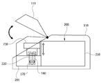
- FIG. 1 and 2 are conceptual diagrams illustrating a cell reprogramming apparatus 100 according to a first embodiment of the present invention
- FIG. 3 is an ultrasonic transducer 220-1 constituting the cell reprogramming apparatus shown in FIG. 220-2 and 220-3 are conceptual views illustrating various embodiments of the present invention
- FIG. 4 is a conceptual diagram illustrating a sample tube 160 constituting the cell reprogramming apparatus illustrated in FIG. 1.
- the cell reprogramming apparatus 100 includes a culture chamber 110 provided to receive cells and culture medium, and cells and culture medium in the culture chamber 110. And one or more instruments arranged to provide energy by applying physical stimulation of at least one of ultrasonic waves, lasers, and heat.
- the apparatus may include one or more of the ultrasonic generator 200 for irradiating ultrasonic waves, the laser irradiation apparatus for irradiating a laser, and the temperature control device 120.
- the cell reprogramming device 100 is one or more of the humidity control device 130 for adjusting the humidity in the culture chamber 110 and the gas control device 140 for controlling the amount of carbon dioxide in the culture chamber 110. It may further comprise.
- the cell reprogramming apparatus 100 may further include a display unit 150 having an input unit for adjusting at least one of temperature, humidity, and carbon dioxide amount in the culture chamber 110.
- the display unit 150 may be provided to set the ultrasonic frequency, and time.
- the present invention is disposed on one side of the culture chamber 110 and the culture chamber 110 that can accommodate the cells and culture medium, it is possible to promote environmental influx to the cells and culture medium (environmental influx) It includes a device capable of providing energy.
- the reprogrammed cells provide a mixture of differentiated cells and culture medium with energy to promote environmental influx and incubated for a period of time to induce differentiated cells.
- a device for inducing cell reprogrammed in a cell is provided.
- the culture chamber 110 may mean a chamber that can be used for normal cell culture.
- the culture chamber 110 may be a chamber in which temperature, humidity, and / or gas may be adjusted to allow cell culture.
- the culture chamber 110 includes a temperature control unit and a carbon dioxide control unit, and the cell culture conditions in the culture chamber 110 may be appropriately adjusted at a person skilled in the art according to the purpose and the type of cells.
- the culture chamber 110 may use a cell suspension culture or a monolayer culture method, it is preferable that such a culture is possible.
- it may be a culture chamber equipped with a stirrer for suspension culture.
- the device capable of providing energy to facilitate the inflow of the environment may include an ultrasonic generator 200 capable of irradiating ultrasonic waves, a laser generator capable of irradiating a laser (not shown), or a temperature controller 120. It may include. In addition, the function of the temperature control device 120 may be implemented through the sample tube holder 170 to be described later.
- the ultrasonic generator 200 may be used without limitation as long as it is a known ultrasonic apparatus that generates ultrasonic waves having a frequency of 10 kHz to 100 MHz.
- the ultrasonic generator may include an ultrasonic generator 210 and an ultrasonic transducer 220: 220-1, 220-2, 220-3.
- the ultrasonic wave generator 200 may be provided to replace the ultrasonic transducer 220.
- the ultrasonic generator 200 may be provided in the ultrasonic chamber 201.
- the laser generator generates a pulsed laser beam having a wavelength range of 300 to 900 nm, and may use a laser device having a pulse duration of 1 ms to 900 ms and a frequency of 1 to 100 Hz with a 1 to 15 W output. It is not limiting.
- the temperature control device 120 may use a known temperature control device capable of temperature control in the range of -40 ° C to 99.9 ° C, but is not particularly limited thereto.
- the temperature control device 120 may be a hot wire provided around the culture chamber.
- Induction apparatus of reprogrammed cells in the differentiated cells of the present invention is a mixture of the culture medium and the differentiated cells by ultrasonic, laser, or heat treatment using a ultrasonic generator, a laser generator, or a temperature control device for a predetermined time Culture can be induced to reprogram to pluripotent or differentiated cells.
- the culture medium may be subjected to ultrasonic, laser, or heat treatment in advance before mixing the culture medium with the differentiated cells.
- the cell reprogramming device 100 may include a humidity control device 130 for controlling the humidity of the culture chamber.
- a humidity control device 130 for controlling the humidity of the culture chamber.
- gas control device 140 carbon dioxide control unit
- the cell reprogramming apparatus 100 may include a display unit 150 for adjusting the above-described temperature, humidity, and carbon dioxide amount.
- the user can adjust the temperature, humidity, and carbon dioxide amount in the culture chamber 110 by manipulating the display unit.
- the display unit 150 may include an input unit of a touch input method or a button input method.
- a sample tube 160 for accommodating cells is disposed in the ultrasonic chamber 201, and the ultrasonic generator 200 may be provided to be lifted and raised toward the sample tube 160.
- the ultrasonic chamber 201 may be provided with a sample tube holder 170 for supporting the sample tube 160.
- the sample tube 160 may have a double tube structure.
- the sample tube holder 170 may have a temperature control function, for example, may be provided as a hit block.
- the culture chamber 110 may include a sample tube 160 for receiving cells to which ultrasound is applied.
- the ultrasonic wave generator 200 is arranged to generate ultrasonic waves into the sample tube 160, and thus the ultrasonic chamber 201 has a sample tube holder (sample tube holder) for supporting the sample tube 160 ( 170) is provided.
- the ultrasonic wave generator 200 in particular, the ultrasonic transducer 220 may be provided to be liftable to correspond to the entry / exit of the sample tube 160 and the sample tube 160 having various sizes.
- the ultrasonic generator 200 in particular, the ultrasonic transducer 220 may be raised or lowered.
- the transfer unit 230 is mounted so that the ultrasonic transducer 220 can be lowered, it may be provided to block the inlet of the sample tube 160 when lowered.
- a lid 111 surrounding the ultrasonic generator 200 and the sample tube 160 may be provided.
- the lid 111 may be mounted to be opened and closed, and may be formed of a transparent material such as glass or transparent resin.
- the ultrasonic generator 200 may be interchangeably mounted with various types of ultrasonic transducers 220-1, 220-2, and 220-3 according to the size of the sample tube and the radiation characteristics of the ultrasonic waves. have.
- the sample tube 160 may have a double tube 161 and 162 structure.
- the sample tube 160 may be inserted into the sample tube holder 170.
- the sample tube 160 may be formed of a material (eg, a resin material) having excellent fire resistance and heat resistance, and may implement a sound insulation effect as the sample tube holder 170 surrounds the sample holder.
- the sample tube holder 170 is preferably provided as a closed space for detecting cell contamination, and preferably includes a soundproof function that can block noise due to ultrasonic waves.
- the sample tube holder 170 may be provided to perform a function of a temperature control device (for example, a hot wire).
- FIGS. 8 and 10 show the culture flask 180 constituting the cell reprogramming apparatus 300. Drawing.
- the cell reprogramming apparatus 100 described with reference to FIGS. 1 to 4 may be applied to a large automated facility.
- the cell reprogramming apparatus 300 includes an auto sampler 500 and an auto sampler 500 provided to mount a plurality of sample tubes 160. It includes an ultrasonic generator 400 provided to generate ultrasonic waves to any one of the sample tube mounted on.
- the auto sampler 500 is provided to be rotatable.
- the ultrasonic generator 400 includes the ultrasonic generator 410, the ultrasonic transducer 420 and the transfer unit 430, as in the first embodiment.
- the ultrasonic transducer 420 may be provided to be elevated.
- the ultrasonic wave generator 400 may be in an elevated state.
- the cell reprogramming apparatus 300 includes a culture medium tank 330 in which a culture medium sonicated is stored, one or more culture flasks 180 to which the culture medium in the culture medium tank 330 is supplied, and a culture flask ( It may further include an injection tube (312) for fluidly connecting the 180 and the sample tube (160).
- the cells in the sample tube 160 to which the ultrasound is applied may be provided to be moved to the culture flask 180 by the injection tube (312).
- the cell reprogramming apparatus 300 may further include a transfer belt 320 for transferring the plurality of culture flasks 180.
- the cell reprogramming apparatus 300 includes an incubator 600 in which the culture flask 180 is accommodated, a recovery tank 610 in which the culture medium of the culture flask 180 in the incubator 600 is recovered, and an incubator 600. Supplying the culture medium to the culture flask 180 in the), and may further include a circulator 620 for recovering the culture medium in the culture flask 180 to the recovery tank 610.
- the culture flask 180 may be provided with flow passages on both sides, and both flow passages of the culture flask may be located at different heights. Additionally, each flow passage may be equipped with stoppers 181 and 182. In addition, the at least one stopper 182 may be provided to be equipped with an injector (191) for culture medium flow.
- the automatic cell reprogramming apparatus 300 includes an auto sampler on which a plurality of sample tubes can be mounted.
- the auto sampler is rotatably provided.
- the ultrasonic generator 400 described above is provided.
- the ultrasonic generator 400 is disposed to generate ultrasonic waves inside any one of the sample tubes mounted on the auto sampler 500.
- the position of the ultrasonic generator 400 may be fixed.
- the ultrasonic generator 400 may be provided to be raised and lowered along the height direction through the transfer unit 430.
- the ultrasonic generator 400 (specifically, the ultrasonic transducer) may be in an elevated state, and generates ultrasonic waves to the sample tube 160 located therein. In the process, the ultrasonic generator 400 (specifically, the ultrasonic transducer) may be in a lowered state.
- the plurality of sample tubes 160 is moved toward the ultrasonic transducer 420, and the ultrasonic transducer 420 generates ultrasonic waves to the sample tube 160 located. That is, when a user mounts a plurality of sample tubes on the auto sampler 500, each sample tube 160 is moved by the auto sampler 500 to the ultrasonic transducer 420, in turn, and the sample tube 160. Ultrasound may be applied to the cells in the stomach.
- the ultrasonically applied cells may be transferred to the culture flask 180 through an injection tube 312 (also referred to as a 'second injection tube'). That is, during the rotation of the auto sampler 500, an ultrasonic wave may be applied to any one of the sample tubes 160, and the cells in the sample tube 160 to which the ultrasonic waves are applied are injected into the culture flask 180 by the injection tube 312. Can be moved.
- an injection tube 312 also referred to as a 'second injection tube'
- the internal temperature, gas, and humidity may be controlled, and may be implemented as an externally sealed device (sterile facility).
- the ultrasonic transducer 420 may also be repurposed into a heat stick that can be subjected to a laser or heat shock.
- the automatic cell reprogramming device 300 is provided with a culture medium tank 330 (media tank) in which the cultured medium sonicated is stored.
- the culture medium in the culture medium tank 330 is supplied to the culture flask 180 through the first injection tube (311). Meanwhile, the plurality of culture flasks 180 are transferred from the auto sampler 500 side to the incubator 600 side by a transfer unit such as a moving belt 320.
- one or more ultrasonic transducers 420 are provided in the culture medium tank 330.
- the automatic cell reprogramming apparatus 300 may be further provided with a recovery tank 610 (waste tank) in which the culture medium in the culture flask 180 is recovered.
- a recovery tank 610 waste tank
- an ultrasonic treatment device 400 is provided in the culture medium tank 330 and generates ultrasonic waves to the culture medium in the culture medium tank 330 through one or more ultrasonic transducers 420.
- a display unit 150 (refer to FIG. 1) for setting and adjusting the intensity and time of the ultrasonic wave may be provided.
- a timer may be provided to count the time for which the ultrasound is applied.
- the culture medium is supplied to the culture flask 180 in the incubator 600, when the medium exchange time, the circulator 620 for recovering the culture medium in the culture flask 180 to the recovery tank 610 (Circulator) ) May be provided.
- the circulator 620 is provided to recover the culture medium in the culture flask 180 to the recovery tank 610 when the medium exchange time set in the timer 621 is elapsed.
- Ultrasonic treated culture medium was provided to supply the circulator 620 (Circulator) from the culture medium tank 330 to each culture flask 180.
- the incubator 600 may be provided with a shaker 601 for shaking the seated culture flask.
- the shaker 601 may be provided to shake the culture flask like a seesaw.
- the incubator 600 like the culture chamber 110 described in the first embodiment, the internal temperature, gas, humidity can be set and adjusted, it can be implemented as a device (sterile facility) sealed to the outside. .
- the culture flask 180 may have a dockable structure on each side so that the medium can be automatically injected and recovered from the injection tube side and the incubator 600 side.
- flow passages may be provided at both sides (eg, front / rear) of the culture flask 180, respectively.
- Each flow passage may be provided with rubber or resin stoppers 181 and 182.
- the portion to be docked may be provided so that the culture medium does not come out as it is formed of a rubber material.
- the height of the docked portion is adjusted to recover only the proper amount during the medium recovery. For example, in the medium wastewater, the medium up to the height of the rear flow passage can be recovered.
- at least one stopper may be provided to engage the injector 191 for flow of the culture medium.
- a new type of pluripotent cells having pluripotent properties by applying energy such as ultrasound, laser or heat treatment to differentiated cells or any different from the phenotype of the differentiated cells Has the effect of inducing reprogramming into differentiated cells.
Landscapes
- Life Sciences & Earth Sciences (AREA)
- Chemical & Material Sciences (AREA)
- Engineering & Computer Science (AREA)
- Bioinformatics & Cheminformatics (AREA)
- Health & Medical Sciences (AREA)
- Wood Science & Technology (AREA)
- Organic Chemistry (AREA)
- Zoology (AREA)
- Biotechnology (AREA)
- Genetics & Genomics (AREA)
- Microbiology (AREA)
- Biochemistry (AREA)
- General Engineering & Computer Science (AREA)
- General Health & Medical Sciences (AREA)
- Biomedical Technology (AREA)
- Sustainable Development (AREA)
- Medicinal Chemistry (AREA)
- Apparatus Associated With Microorganisms And Enzymes (AREA)
- Micro-Organisms Or Cultivation Processes Thereof (AREA)
Abstract
Description
Claims (20)
- 세포 및 배양배지를 수용하도록 마련된 배양챔버; 및배양챔버 내의 세포 및 배양배지에 초음파, 레이저 및 열 중 적어도 하나의 물리적 자극을 가함으로써 에너지를 제공할 수 있도록 마련된 하나 이상의 기구를 포함하는 세포 리프로그래밍 장치.
- 제 1 항에 있어서,상기 기구는, 초음파를 조사하는 초음파 발생장치, 레이저를 조사하는 레이저 조사장치 및 온도조절장치 중 하나 이상을 포함하는 세포 리프로그래밍 장치.
- 제 2 항에 있어서,배양챔버 내의 습도를 조절하기 위한 습도조절장치 및 배양챔버 내의 이산화탄소량을 조절하기 위한 가스조절장치 중 하나 이상을 추가로 포함하는 세포 리프로그래밍 장치.
- 제 3 항에 있어서,배양챔버 내의 온도, 습도, 이산화탄소량 중 하나 이상을 조절하기 위한 입력부를 갖는 디스플레이부를 추가로 포함하는 세포 리프로그래밍 장치.
- 제 4 항에 있어서,기구가 초음파 발생장치를 포함하고,디스플레이부는 초음파 주파수, 및 시간을 설정할 수 있도록 마련된 세포 리프로그래밍 장치.
- 제 2 항에 있어서,배양 챔버에는 세포를 수용하는 샘플 튜브가 배치되며,초음파 발생장치는 샘플 튜브를 향하여 승강 가능하게 마련된 세포 리프로그래밍 장치.
- 제 6 항에 있어서,배양 챔버에는 샘플 튜브를 지지하기 위한 샘플 튜브 홀더가 마련된 세포 리프로그래밍 장치.
- 제 6 항에 있어서,샘플 튜브는 이중 튜브 구조를 갖는 세포 리프로그래밍 장치.
- 제 2 항에 있어서,초음파 발생장치는 초음파 트랜스듀서가 교체 가능하게 마련된 세포 리프로그래밍 장치.
- 제 2 항에 있어서,온도 조절 장치는 배양 챔버 주변으로 마련된 열선을 포함하는 세포 리프로그래밍 장치.
- 복수 개의 샘플 튜브가 장착될 수 있도록 마련된 오토 샘플러; 및오토 샘플러에 탑재된 어느 한 샘플 튜브로 초음파를 발생시키도록 마련된 초음파 발생장치를 포함하는 세포 리프로그래밍 장치.
- 제 11 항에 있어서,오토 샘플러는 회전 가능하게 마련되고,초음파 발생장치는 승강 가능하게 마련된 세포 리프로그래밍 장치.
- 제 12 항에 있어서,오토 샘플러가 회전하는 경우, 초음파 발생장치는 상승된 상태인 세포 리프로그래밍 장치.
- 제 11 항에 있어서,초음파 처리된 배양배지가 저장된 배양배지 탱크;상기 배양배지 탱크 내의 배양배지가 공급되는 하나 이상의 배양 플라스크; 및배양 플라스크와 샘플 튜브를 유체 이동 가능하게 연결하는 인젝션 튜브를 추가로 포함하는 세포 리프로그래밍 장치.
- 제 14 항에 있어서,초음파가 가해진 샘플 튜브 내의 세포는 인젝션 튜브에 의해 배양 플라스크로 이동되도록 마련된 세포 리프로그래밍 장치.
- 제 14 항에 있어서,복수 개의 배양 플라스크를 이송시키기 위한 이송 벨트를 추가로 포함하는 세포 리프로그래밍 장치.
- 제 15 항에 있어서,배양 플라스크가 수용되는 인큐베이터;인큐베이터 내의 배양 플라스크의 배양 배지가 회수되는 회수 탱크; 및인큐베이터 내의 배양 플라스크로 배양 배지를 공급하고, 배양 플라스크 내의 배양 배지를 회수 탱크로 회수하기 위한 서큘레이터를 추가로 포함하는 세포 리프로그래밍 장치.
- 제 14 항에 있어서,배양 플라스크는 양 측에 유동 통로가 각각 마련된 세포 리프로그래밍 장치.
- 제 18 항에 있어서,배양 플라스크의 양 측 유동 통로는 서로 다른 높이에 위치하는 세포 리프로그래밍 장치.
- 제 17 항에 있어서,인큐베이터에는 안착된 배양 플라스크를 흔들기 위한 셰이커(shaker)가 마련된 세포 리프로그래밍 장치.
Priority Applications (4)
| Application Number | Priority Date | Filing Date | Title |
|---|---|---|---|
| CN201680084455.3A CN109089424A (zh) | 2016-03-11 | 2016-08-09 | 细胞重编程装置 |
| EP16893681.3A EP3428276A4 (en) | 2016-03-11 | 2016-08-09 | CELL REPROGRAMMING DEVICE |
| JP2018567551A JP2019508069A (ja) | 2016-03-11 | 2016-08-09 | 細胞のリプログラミング装置 |
| US16/082,928 US20190106669A1 (en) | 2016-03-11 | 2016-08-09 | Cell reprogramming apparatus |
Applications Claiming Priority (4)
| Application Number | Priority Date | Filing Date | Title |
|---|---|---|---|
| KR10-2016-0029707 | 2016-03-11 | ||
| KR20160029707 | 2016-03-11 | ||
| KR10-2016-0098754 | 2016-08-03 | ||
| KR1020160098754A KR102050852B1 (ko) | 2016-03-11 | 2016-08-03 | 세포 리프로그래밍 장치 |
Publications (1)
| Publication Number | Publication Date |
|---|---|
| WO2017155167A1 true WO2017155167A1 (ko) | 2017-09-14 |
Family
ID=59789509
Family Applications (1)
| Application Number | Title | Priority Date | Filing Date |
|---|---|---|---|
| PCT/KR2016/008755 Ceased WO2017155167A1 (ko) | 2016-03-11 | 2016-08-09 | 세포 리프로그래밍 장치 |
Country Status (1)
| Country | Link |
|---|---|
| WO (1) | WO2017155167A1 (ko) |
Citations (4)
| Publication number | Priority date | Publication date | Assignee | Title |
|---|---|---|---|---|
| US7052720B1 (en) * | 1999-06-17 | 2006-05-30 | University Of Wales College Of Medicine | Spheroid preparation |
| KR20090080390A (ko) * | 2008-01-21 | 2009-07-24 | 전남대학교산학협력단 | 다중 세포배양기를 구비한 인큐베이터 |
| US20140038257A1 (en) * | 2012-08-01 | 2014-02-06 | Anuradha Subramanian | Methods of using ultrasound in tissue culture and tissue engineering |
| KR20150045935A (ko) * | 2012-04-24 | 2015-04-29 | 더 브리검 앤드 우먼즈 하스피털, 인크. | 다능성 세포를 다시 생성하는 방법 |
-
2016
- 2016-08-09 WO PCT/KR2016/008755 patent/WO2017155167A1/ko not_active Ceased
Patent Citations (4)
| Publication number | Priority date | Publication date | Assignee | Title |
|---|---|---|---|---|
| US7052720B1 (en) * | 1999-06-17 | 2006-05-30 | University Of Wales College Of Medicine | Spheroid preparation |
| KR20090080390A (ko) * | 2008-01-21 | 2009-07-24 | 전남대학교산학협력단 | 다중 세포배양기를 구비한 인큐베이터 |
| KR20150045935A (ko) * | 2012-04-24 | 2015-04-29 | 더 브리검 앤드 우먼즈 하스피털, 인크. | 다능성 세포를 다시 생성하는 방법 |
| US20140038257A1 (en) * | 2012-08-01 | 2014-02-06 | Anuradha Subramanian | Methods of using ultrasound in tissue culture and tissue engineering |
Non-Patent Citations (2)
| Title |
|---|
| LV , YONGGANG ET AL.: "Effects of Low-intensity Pulsed Ultrasound on Cell Viability, Proliferation and Neural Differentiation of Induced Pluripotent Stem Cells-derived Neural Crest Stem Cells", BIOTECHNOLOGY LETTERS, vol. 35, no. 12, 28 September 2013 (2013-09-28), pages 2201 - 2212, XP055551630 * |
| See also references of EP3428276A4 * |
Similar Documents
| Publication | Publication Date | Title |
|---|---|---|
| KR102050852B1 (ko) | 세포 리프로그래밍 장치 | |
| US20240101998A1 (en) | Cell reprogramming method using imposition of physical stimulation-mediated environmental transition | |
| Sheng et al. | Direct reprogramming of Sertoli cells into multipotent neural stem cells by defined factors | |
| Dell’Anno et al. | Remote control of induced dopaminergic neurons in parkinsonian rats | |
| JP6986968B2 (ja) | エネルギーを利用した多能性細胞誘導装置及び方法 | |
| Ofenbauer et al. | Strategies for in vivo reprogramming | |
| Mohamed et al. | Generation of human midbrain organoids from induced pluripotent stem cells [version 2; peer review: 3 approved, 1 approved with reservations] | |
| Kirwan et al. | Generation and characterization of functional human hypothalamic neurons | |
| WO2021112638A1 (ko) | 신경에 대한 효능 및 독성평가를 위한 생체모방 신경칩 및 이의 용도 | |
| WO2017155166A1 (ko) | 물리적 자극에 의한 환경유입을 이용한 세포 리프로그래밍 방법 | |
| CN108368486A (zh) | 鉴定神经肌肉接头活动的调节剂的体外方法 | |
| WO2021086063A1 (ko) | 3차원 뇌 오가노이드를 이용한 뇌신경계질환 치료용 줄기세포 스크리닝 방법 | |
| Matsumura et al. | Establishment of integration-free induced pluripotent stem cells from human recessive dystrophic epidermolysis bullosa keratinocytes | |
| Leal et al. | Modeling hypothalamic pathophysiology in vitro for metabolic, circadian, and sleep disorders | |
| Ullah et al. | Generating and characterizing human telencephalic brain organoids from stem cell-derived single neural rosettes | |
| Elder et al. | Diseased, differentiated and difficult: Strategies for improved engineering of in vitro neurological systems | |
| US20200248139A1 (en) | Methods for generating and using organoids and cells thereof | |
| WO2017155167A1 (ko) | 세포 리프로그래밍 장치 | |
| WO2025037879A1 (ko) | 다중세포 배양 플랫폼으로서 역-미세둑 구조가 포함된 다중장기모사칩 및 이의 용도 | |
| Göksu | A review article on the development of dopaminergic neurons and establishment of dopaminergic neuron–based in vitro models by using immortal cell lines or stem cells to study and treat Parkinson's disease | |
| Xie et al. | Reprogramming of adult human neural stem cells into induced pluripotent stem cells | |
| Pruszak et al. | Molecular and cellular determinants for generating ES-cell derived dopamine neurons for cell therapy | |
| Boström et al. | Neural network and" ganglion" formations in vitro: a video microscopy and scanning electron microscopy study on adult cultured spiral ganglion cells | |
| WO2016089178A1 (ko) | 에너지를 이용한 다능성 세포 유도 장치 및 방법 | |
| Wenker et al. | Cell reprogramming and neuronal differentiation applied to neurodegenerative diseases: Focus on Parkinson’s disease |
Legal Events
| Date | Code | Title | Description |
|---|---|---|---|
| ENP | Entry into the national phase |
Ref document number: 2018567551 Country of ref document: JP Kind code of ref document: A |
|
| NENP | Non-entry into the national phase |
Ref country code: DE |
|
| WWE | Wipo information: entry into national phase |
Ref document number: 2016893681 Country of ref document: EP |
|
| ENP | Entry into the national phase |
Ref document number: 2016893681 Country of ref document: EP Effective date: 20181011 |
|
| 121 | Ep: the epo has been informed by wipo that ep was designated in this application |
Ref document number: 16893681 Country of ref document: EP Kind code of ref document: A1 |