WO2017179425A1 - 圧脈波検出装置及び生体情報測定装置 - Google Patents
圧脈波検出装置及び生体情報測定装置 Download PDFInfo
- Publication number
- WO2017179425A1 WO2017179425A1 PCT/JP2017/012940 JP2017012940W WO2017179425A1 WO 2017179425 A1 WO2017179425 A1 WO 2017179425A1 JP 2017012940 W JP2017012940 W JP 2017012940W WO 2017179425 A1 WO2017179425 A1 WO 2017179425A1
- Authority
- WO
- WIPO (PCT)
- Prior art keywords
- pulse wave
- pressure
- pressure pulse
- pressing
- wave detection
- Prior art date
- Legal status (The legal status is an assumption and is not a legal conclusion. Google has not performed a legal analysis and makes no representation as to the accuracy of the status listed.)
- Ceased
Links
Images





Classifications
-
- A—HUMAN NECESSITIES
- A61—MEDICAL OR VETERINARY SCIENCE; HYGIENE
- A61B—DIAGNOSIS; SURGERY; IDENTIFICATION
- A61B5/00—Measuring for diagnostic purposes; Identification of persons
- A61B5/02—Detecting, measuring or recording for evaluating the cardiovascular system, e.g. pulse, heart rate, blood pressure or blood flow
- A61B5/021—Measuring pressure in heart or blood vessels
- A61B5/022—Measuring pressure in heart or blood vessels by applying pressure to close blood vessels, e.g. against the skin; Ophthalmodynamometers
- A61B5/0225—Measuring pressure in heart or blood vessels by applying pressure to close blood vessels, e.g. against the skin; Ophthalmodynamometers the pressure being controlled by electric signals, e.g. derived from Korotkoff sounds
-
- A—HUMAN NECESSITIES
- A61—MEDICAL OR VETERINARY SCIENCE; HYGIENE
- A61B—DIAGNOSIS; SURGERY; IDENTIFICATION
- A61B5/00—Measuring for diagnostic purposes; Identification of persons
- A61B5/02—Detecting, measuring or recording for evaluating the cardiovascular system, e.g. pulse, heart rate, blood pressure or blood flow
- A61B5/021—Measuring pressure in heart or blood vessels
- A61B5/02108—Measuring pressure in heart or blood vessels from analysis of pulse wave characteristics
- A61B5/02116—Measuring pressure in heart or blood vessels from analysis of pulse wave characteristics of pulse wave amplitude
-
- A—HUMAN NECESSITIES
- A61—MEDICAL OR VETERINARY SCIENCE; HYGIENE
- A61B—DIAGNOSIS; SURGERY; IDENTIFICATION
- A61B5/00—Measuring for diagnostic purposes; Identification of persons
- A61B5/02—Detecting, measuring or recording for evaluating the cardiovascular system, e.g. pulse, heart rate, blood pressure or blood flow
- A61B5/021—Measuring pressure in heart or blood vessels
- A61B5/02141—Details of apparatus construction, e.g. pump units or housings therefor, cuff pressurising systems, arrangements of fluid conduits or circuits
-
- A—HUMAN NECESSITIES
- A61—MEDICAL OR VETERINARY SCIENCE; HYGIENE
- A61B—DIAGNOSIS; SURGERY; IDENTIFICATION
- A61B5/00—Measuring for diagnostic purposes; Identification of persons
- A61B5/103—Measuring devices for testing the shape, pattern, colour, size or movement of the body or parts thereof, for diagnostic purposes
- A61B5/11—Measuring movement of the entire body or parts thereof, e.g. head or hand tremor or mobility of a limb
-
- A—HUMAN NECESSITIES
- A61—MEDICAL OR VETERINARY SCIENCE; HYGIENE
- A61B—DIAGNOSIS; SURGERY; IDENTIFICATION
- A61B5/00—Measuring for diagnostic purposes; Identification of persons
- A61B5/68—Arrangements of detecting, measuring or recording means, e.g. sensors, in relation to patient
- A61B5/6801—Arrangements of detecting, measuring or recording means, e.g. sensors, in relation to patient specially adapted to be attached to or worn on the body surface
- A61B5/6813—Specially adapted to be attached to a specific body part
- A61B5/6824—Arm or wrist
-
- A—HUMAN NECESSITIES
- A61—MEDICAL OR VETERINARY SCIENCE; HYGIENE
- A61B—DIAGNOSIS; SURGERY; IDENTIFICATION
- A61B5/00—Measuring for diagnostic purposes; Identification of persons
- A61B5/68—Arrangements of detecting, measuring or recording means, e.g. sensors, in relation to patient
- A61B5/6801—Arrangements of detecting, measuring or recording means, e.g. sensors, in relation to patient specially adapted to be attached to or worn on the body surface
- A61B5/6843—Monitoring or controlling sensor contact pressure
-
- A—HUMAN NECESSITIES
- A61—MEDICAL OR VETERINARY SCIENCE; HYGIENE
- A61B—DIAGNOSIS; SURGERY; IDENTIFICATION
- A61B5/00—Measuring for diagnostic purposes; Identification of persons
- A61B5/72—Signal processing specially adapted for physiological signals or for diagnostic purposes
- A61B5/7235—Details of waveform analysis
-
- A—HUMAN NECESSITIES
- A61—MEDICAL OR VETERINARY SCIENCE; HYGIENE
- A61B—DIAGNOSIS; SURGERY; IDENTIFICATION
- A61B5/00—Measuring for diagnostic purposes; Identification of persons
- A61B5/72—Signal processing specially adapted for physiological signals or for diagnostic purposes
- A61B5/7271—Specific aspects of physiological measurement analysis
- A61B5/7275—Determining trends in physiological measurement data; Predicting development of a medical condition based on physiological measurements, e.g. determining a risk factor
-
- A—HUMAN NECESSITIES
- A61—MEDICAL OR VETERINARY SCIENCE; HYGIENE
- A61B—DIAGNOSIS; SURGERY; IDENTIFICATION
- A61B5/00—Measuring for diagnostic purposes; Identification of persons
- A61B5/74—Details of notification to user or communication with user or patient; User input means
- A61B5/746—Alarms related to a physiological condition, e.g. details of setting alarm thresholds or avoiding false alarms
-
- A—HUMAN NECESSITIES
- A61—MEDICAL OR VETERINARY SCIENCE; HYGIENE
- A61B—DIAGNOSIS; SURGERY; IDENTIFICATION
- A61B5/00—Measuring for diagnostic purposes; Identification of persons
- A61B5/02—Detecting, measuring or recording for evaluating the cardiovascular system, e.g. pulse, heart rate, blood pressure or blood flow
- A61B5/024—Measuring pulse rate or heart rate
- A61B5/0245—Measuring pulse rate or heart rate by using sensing means generating electric signals, i.e. ECG signals
Definitions
- the present invention relates to a pressure pulse wave detection device and a biological information measurement device.
- a biological information measuring apparatus capable of performing the above is known.
- the positional relationship between the pressure sensor and the artery affects the detection accuracy of the pressure pulse wave. Therefore, as shown in the following Patent Documents 1 to 5, a configuration for adjusting the position of the living body part and the pressure sensor has been proposed.
- Patent Document 3 discloses a biological information measuring apparatus that includes a pressure sensor that is brought into contact with a living body part and has a drive unit that moves the pressure sensor in a direction intersecting the artery.
- Patent Document 4 discloses a biological information measuring apparatus having a pressure sensor array that is brought into contact with a living body part and having a drive unit that rotates the pressure sensor array in a plane that intersects the pressing direction of the pressure sensor array.
- Patent Document 5 has a pressing surface in which a plurality of pressure sensor rows to be brought into contact with a living body part are arranged, and the pressing surface is rotated around an axis extending in a direction orthogonal to the arrangement direction of the plurality of pressure sensor rows.
- a biological information measuring device having a driving unit to be disclosed is disclosed.
- Patent Documents 1 and 2 can change the contact state of the sensor group to the living body part, the contact state changes following the shape of the wrist of the user, or the contact state changes manually. It is something to let you. For this reason, it is not possible to position the sensor with sufficient consideration of pulse wave detection accuracy.
- the present invention has been made in view of the above circumstances, and is a pressure that can improve the pressure pulse wave detection accuracy by flexibly changing the contact state of the pressure detection element that is used in contact with the living body part. It is an object of the present invention to provide a pulse wave detection device and a biological information measurement device including the same.
- a pressure pulse wave detection device includes a pressing member having a pressing surface in which a plurality of element arrays made of a plurality of pressure detecting elements arranged in one direction are arranged in a direction intersecting the one direction, and the one direction is a living body.
- a pressing mechanism that presses the pressing surface against the body surface in a state intersecting with the direction in which the artery below the body surface extends, and two axes that are orthogonal to the pressing direction of the pressing surface by the pressing mechanism.
- a rotation driving mechanism for rotating the pressing surface around each of a first axis extending in a direction and a second axis orthogonal to the one direction, the pressing mechanism, the rotating driving mechanism, and the pressing member A support member that supports the support member, a housing that houses the support member, and a moving mechanism that moves the support member in the one direction within the housing.
- the biological information measuring device of the present invention includes the pressure pulse wave detection device, a biological information calculation unit that calculates biological information based on a pressure pulse wave detected by a pressure detection element included in the pressure pulse wave detection device, Is provided.
- a pressure pulse wave detection device capable of flexibly changing the contact state of a pressure detection element used in contact with a living body part and improving the pressure pulse wave detection accuracy, and a living body including the pressure pulse wave detection apparatus.
- An information measuring device can be provided.
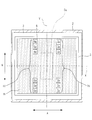
- FIG. 1 It is a side view showing a schematic structure of living body information measuring device 200 for explaining one embodiment of the present invention. It is the figure which looked at the state with which biological information measuring device 200 shown in Drawing 1 was equipped with a wrist from the palm side. It is the figure which looked at the state with which living body information measuring device 200 shown in Drawing 1 was equipped with the wrist from the back side of the hand. It is a cross-sectional schematic diagram which shows schematic structure of the pressure pulse wave detection part 100 in the state with which the biological information measuring apparatus 200 shown in FIG. 1 was mounted
- FIG. 9 is a schematic sectional view taken along line IX-IX shown in FIG. 8. It is a figure which shows the block configuration of the biological information measuring device 200 shown in FIG. It is a flowchart for demonstrating operation
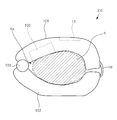
- FIG. 1 is a side view showing a schematic configuration of a biological information measuring apparatus 200 for explaining an embodiment of the present invention.
- a biological information measuring device 200 shown in FIG. 1 is a wrist-worn biological information measuring device that is used by being worn on the wrist of a measurement subject who has a radial artery.
- the biological information measuring device 200 is not limited to the wrist-worn type, as long as it is used by being attached to a biological part where an artery is present below the body surface.
- the biological information measuring apparatus 200 includes a main body unit including a housing 101 and a housing 102, a hinge unit 103 that connects the housing 101 and the housing 102, and a belt 106 for fixing the main body unit to a wrist. .
- the housing 102 is rotatably connected to the housing 101 by the hinge portion 103.
- the inner peripheral surfaces of the casings 101 and 102 are shaped along the outer shape of the wrist H.
- the wrist H of the person to be measured can be inserted between the housing 101 and the housing 102 with the end opposite to the hinge portion 103 side of the housing 101 closest to the housing 102.
- a space Ka is formed.
- one end of the casing 101 and one end of the casing 102 are fixed by the belt 106, whereby the biological information measuring device 200 is fixed (attached) to the wrist H.
- the housing 101 and the housing 102 may be integrated without using the hinge portion 103.
- the casing 101 includes a pressure pulse wave detection unit 100 provided at a position facing the wrist H in a state where the biological information measuring device 200 is attached to the wrist H, and a display unit 13 provided on the outer peripheral surface in the state. Prepare.
- the casing 101 and the pressure pulse wave detection unit 100 constitute a pressure pulse wave detection device.
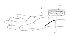
- FIG. 2 is a diagram of a state in which the biological information measuring apparatus 200 is worn on the left wrist of the measurement subject as viewed from the palm side.
- the display unit 13 is provided on the outer peripheral surface of the housing 101, and the display unit 13 can be visually recognized from the palm side.
- FIG. 3 is a diagram of a state in which the biological information measuring device 200 is mounted on the left wrist of the measurement subject as viewed from the back side of the hand.
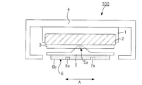
- FIG. 4 is a schematic cross-sectional view showing the configuration of the pressure pulse wave detecting unit 100 in the wrist wearing state of the biological information measuring device 200 shown in FIGS. In FIG. 4, only the pressure pulse wave detection unit 100 of the biological information measurement apparatus 200 is illustrated.
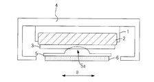
- FIG. 5 is an enlarged view of the pressure pulse wave detection unit 100 shown in FIG. 6 is a schematic cross-sectional view of the pressure pulse wave detection unit 100 in the wearing state shown in FIG. 4 as viewed from the fingertip side of the person to be measured.
- FIG. 7 is a view of the pressure pulse wave detection unit 100 in the wearing state shown in FIG. 4 as seen from the side of contact with the wrist. 4 to 7 schematically show the pressure pulse wave detection unit 100, and do not limit the dimensions and arrangement of each part.
- the pressure pulse wave detection unit 100 includes a housing 1 containing the air bag 2, a flat plate portion 3 that is a flat plate member fixed to the air bag 2, and a two-axis rotation mechanism 5 a for the flat plate portion 3.
- a rotating unit 5 supported rotatably about each of the two shafts, a sensor unit 6 provided on a plane opposite to the flat plate unit 3 side of the rotating unit 5, a housing 1, and an air bag 2.
- the casing 1, the air bag 2, the flat plate portion 3, and the rotating portion 5 are configured so that the pressing surface 6 b of the sensor unit 6 is placed on the body surface of the living body part (wrist) while the biological information measuring device 200 is attached to the wrist.
- a pressing mechanism that presses against is configured.
- the pressing mechanism may be anything as long as it can press the pressing surface 6b of the sensor unit 6 against the body surface, and is not limited to one using an air bag.
- the air bag 2 has a direction in which the flat plate portion 3 fixed to the air bag 2 is perpendicular to the surface of the flat plate portion 3 (a plane on the rotating portion 5 side) by controlling the amount of air inside by a pump (not shown). Move to.
- the pressing surface 6b of the sensor unit 6 included in the pressure pulse wave detection unit 100 contacts the skin of the wrist of the measurement subject.
- the internal pressure of the air bag 2 increases, and the sensor unit 6 is pressed toward the radial artery T below the wrist.
- the pressing force applied to the radial artery T by the sensor unit 6 is equivalent to the internal pressure of the air bag 2.
- the sensor unit 6 includes a plurality of sensors arranged in a direction B intersecting (orthogonal in the example of FIG. 7) with the extending direction A of the radial artery T present at the mounting site in the mounted state shown in FIG.
- An element array 60 including the pressure detection elements 6a and an element array 70 including a plurality of pressure detection elements 7a arranged in the direction B are provided.
- the element row 60 and the element row 70 are arranged in the direction A.
- Each pressure detection element 6a and the pressure detection element 7a having the same position in the direction B with the pressure detection element 6a constitute a pair, and the sensor unit 6 has a configuration in which a plurality of pairs are arranged in the direction B. ing.
- the pressure detection element 6a and the pressure detection element 7a for example, an element such as a strain gauge resistance type, a semiconductor piezoresistance type, or a capacitance type is used.
- the pressure detection elements included in the element row 60 and the element row 70 are formed on the same plane, and this plane is protected by a protective member such as a resin.
- the plane on which each pressure detection element is formed and the surface of the protective member that protects the plane are parallel to each other, and the surface of the protective member constitutes the pressing surface 6b.
- the pressure detection element 6a (7a) is a pressure generated from the radial artery T and transmitted to the skin by being pressed against the radial artery T so that the arrangement direction intersects (substantially orthogonal) the radial artery T.
- a vibration wave that is, a pressure pulse wave is detected.
- the pressure signal output from the pressure detection element 6a (7a) includes a direct current component generated by contact with an object and an alternating current component generated by a pressure vibration wave, and the alternating current component is a pressure pulse wave signal.
- the intervals in the arrangement direction of the pressure detection elements 6a (7a) are sufficiently small so that a necessary and sufficient number is arranged on the radial artery T.
- the lengths of the element rows 560 and 70 are necessary and sufficiently larger than the radial dimension of the radial artery T.
- the sensor unit 6 configured in this way constitutes a pressing member.
- the biaxial rotating mechanism 5 a is a mechanism for rotating the rotating unit 5 around each of two rotating axes X and Y orthogonal to the pressing direction of the flat plate part 3 by the air bag 2. is there.
- the biaxial rotation mechanism 5a and the rotation unit 5 constitute a rotation drive mechanism.
- the biaxial rotation mechanism 5a has two rotation axes X and Y set on the surface of the flat plate portion 3 and orthogonal to each other, and the rotation axes X and Y are respectively driven to rotate by a rotation drive unit 10 described later.
- the rotation axis Y is a first axis extending in the arrangement direction of the plurality of pressure detection elements 6a (7a) formed on the pressing surface 6b.
- the rotation axis Y is set between the element array 60 and the element array 70 in the plan view of FIG. 7 (intermediate in the example of FIG. 7).
- the rotation axis X is a second axis extending in a direction orthogonal to the arrangement direction of the plurality of pressure detection elements 6a (7a) formed on the pressing surface 6b.
- the rotation axis X is set on a straight line that equally divides the element array 60 and the element array 70 into two.
- the rotation surface 5 rotates about the rotation axis X, so that the pressing surface 6b rotates around the rotation axis X.
- the rotation surface 5 rotates around the rotation axis Y, so that the pressing surface 6 b rotates around the rotation axis Y.
- the accommodating portion 4 of the pressure pulse wave detection unit 100 is built in the housing 101 and is supported by the housing 101 so as to be movable in the direction B.
- the accommodating part 4 is comprised by the cup-shaped member, for example, The housing
- the accommodating part 4 supports the housing 1 in the hollow part.
- the housing 1 and the air bag 2, the flat plate portion 3, the biaxial rotation mechanism 5 a, the rotation portion 5, and the sensor portion 6 that are fixed to the housing 1 move with the movement of the housing portion 4.
- the accommodating part 4 moves in the direction B so that the pressing mechanism constituted by the casing 1, the air bag 2, the flat plate part 3 and the rotating part 5, the biaxial rotating mechanism 5 a and the rotating part 5.
- the rotation drive mechanism constituted by the sensor unit 6 and the sensor unit 6 can move together in the direction B, any support member that supports the pressing mechanism, the rotation drive mechanism, and the sensor unit 6 may be used.
- the housing portion 4 may be configured to integrally support the pressing mechanism, the rotation drive mechanism, and the sensor portion 6 with a flat plate-like member that supports the housing 1.
- FIG. 8 is a diagram schematically showing a developed view of a portion in which the housing portion 4 of the housing 101 shown in FIG. 1 is built.
- FIG. 8A is a side view of the housing 101 as viewed from the peripheral side (fingertip side) of the measurement subject with the biological information measuring device 200 attached to the wrist.
- FIG. 8B is a top view of the housing 101 as viewed from the pressing direction with the biological information measuring device 200 attached to the wrist.
- FIG. 8C is a side view of the housing 101 viewed from the central side (elbow side) of the person to be measured with the biological information measuring device 200 attached to the wrist.
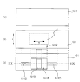
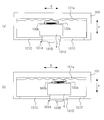
- 9 is a schematic cross-sectional view taken along the line IX-IX shown in FIG.
- the end surface on the central side (elbow side) of the measurement subject with the biological information measuring device 200 attached to the wrist (hereinafter referred to as the central side end surface). ) Is provided with a fixing member 101A fixed to the accommodating portion 4. A convex portion 101B is provided on the surface of the fixing member 101A.
- a slit 101 ⁇ / b> C extending in the direction B is formed on the central end surface of the housing 101.
- a convex portion 101 ⁇ / b> D that penetrates the slit 101 ⁇ / b> C is formed on the back surface of the fixing member 101 ⁇ / b> A, and the convex portion 101 ⁇ / b> D is fixed to the accommodating portion 4. Accordingly, the fixing member 101A is movable in the direction B through the slit 101C.
- the leaf spring 100 b is fixed to the end face on the distal side of the measurement subject with the biological information measuring device 200 attached to the wrist among the end faces in the direction A of the housing portion 4. Yes.
- a slide member 100a is fixed to the leaf spring 100b.
- the slide member 100a is a member having a triangular prism-shaped convex portion that is long in the pressing direction (direction orthogonal to the direction A and the direction B).
- the housing 101 is formed with a slide rail 101a having seven recesses in the example of FIG. 9 in which the protrusions of the slide member 100a can be engaged.
- the slide member 100a is urged toward the slide rail 101a by a leaf spring 100b.
- FIG. 9A illustrates a state where there is a gap between the slide member 100a and the slide rail 101a for the sake of explanation.
- the distance between two recesses adjacent to the slide rail 101a (the distance between the bottoms of the recesses) is designed to be about 1 mm, for example.
- the accommodating portion 4 can be moved 3 mm in total in one direction in the direction B from the reference position by using the position shown in FIG. 9A as the reference position. A total of 3 mm can be moved by 1 mm in the direction.
- 1 mm is an example as a minimum unit when moving the accommodating part 4, and it is not limited to this.
- the distance which can move the accommodating part 4 is 6 mm in said example, this numerical value is also an example and is not limited to this.
- the slide rail 101a, the slide member 100a, the leaf spring 100b, the fixing member 101A, and the convex portion 101B constitute a moving mechanism for moving the accommodating portion 4 in the direction B within the housing 101.
- casing 101 not only what was shown in FIG.8 and FIG.9 but a well-known mechanism is employable.
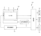
- FIG. 10 is a diagram showing a block configuration of the biological information measuring apparatus 200 shown in FIG.
- the biological information measuring device 200 includes a pressure pulse wave detection unit 100, a rotation drive unit 10, an air bag drive unit 11, a control unit 12 that performs overall control of the entire device, a display unit 13, an operation unit 14, and a memory. 15.
- the rotation drive unit 10 is an actuator connected to each of the rotation axes X and Y of the biaxial rotation mechanism 5a of the pressure pulse wave detection unit 100.
- the rotation drive unit 10 rotates the rotation axes X and Y according to instructions from the control unit 12 to rotate the pressing surface 6b around the rotation axis X, or the pressing surface 6b around the rotation axis Y. Or rotate it.
- the air bag drive unit 11 controls the amount of air injected into the air bag 2 (internal pressure of the air bag 2) under the instruction of the control unit 12.
- the display unit 13 is for displaying various information such as the measured blood pressure value, and is configured by, for example, a liquid crystal display element, an organic electroluminescence display element, or electronic paper.
- the operation unit 14 is an interface for inputting an instruction signal to the control unit 12, and includes a button for instructing the start of various operations including blood pressure measurement.
- the memory 15 is a ROM (Read Only Memory) that stores a program for causing the control unit 12 to perform a predetermined operation and various data, a RAM (Random Access Memory) as a work memory, and various information such as measured blood pressure data. Including a flash memory or the like.
- the control unit 12 functions as a press control unit, a biological information calculation unit, a rotation control unit, a calibration data generation unit, and a display control unit by executing a program stored in the ROM of the memory 15.
- the pressing control unit controls the pressing force applied to the wrist by the pressing surface 6b by controlling the air bag driving unit 11 and adjusting the amount of air in the air bag 2.
- the living body information calculation unit presses the first pressing surface 6b in the radial artery T based on the pressure pulse wave detected by the pressure detection elements 6a and 7a formed on the pressing surface 6b in a state where the pressing surface 6b is pressed against the radial artery T.
- the blood pressure value is calculated.
- the biological information calculation unit detects the pressure pulse wave detected by the pressure detection elements 6a and 7a in the process in which the pressing force to the radial artery T is changed (increased or decreased) by the air bag driving unit 11. Based on the above, the first blood pressure value in the radial artery T is calculated.
- the calibration data generation unit generates calibration data using the first blood pressure value calculated by the biological information calculation unit.
- the rotation control unit is a pressing surface by the rotation driving unit 10 based on the pressure pulse wave detected by the pressure detection elements 6a and 7a in the process in which the pressing force to the radial artery T is increased by the air bag driving unit 11. The necessity of rotation of 6b is determined. When the rotation control unit determines that rotation is necessary, the rotation control unit controls the rotation driving unit 10 to rotate the pressing surface 6b.
- the biological information calculation unit is detected for each beat by the pressure detection elements 6a and 7a in a state where the pressing surface 6b is pressed against the radial artery T with an optimal pressing force for deforming a part of the radial artery T flatly.
- the second blood pressure value in the radial artery T is calculated every beat by calibrating the pressure pulse wave.
- the display control unit performs display control of the display unit 13.
- the biological information measuring apparatus 200 of the present embodiment measures and flashes blood pressure values (SBP (Systemic Blood pressure), so-called systolic blood pressure and DBP (Diastrotic Blood pressure)), so-called diastolic blood pressure, for each beat of the heart. It has a continuous blood pressure measurement mode in which it is stored in the memory and the measured blood pressure is displayed on the display unit 13.
- SBP Systemic Blood pressure
- DBP Diastrotic Blood pressure
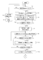
- 11 and 12 are flowcharts for explaining the operation up to the generation of calibration data in the continuous blood pressure measurement mode of the biological information measuring apparatus 200.
- the rotation unit 5 of the pressure pulse wave detection unit 100 has a rotation amount set to, for example, zero, and the pressing surface 6b is parallel to the flat plate unit 3. It shall be.
- step S1 When the measurement start button included in the operation unit 14 is pressed to instruct the start of blood pressure measurement (step S1: YES), the control unit 12 controls the air bag driving unit 11 to air into the air bag 2. Injection is started, and the pressing force on the wrist by the pressing surface 6b is increased (step S2). When the blood pressure measurement start instruction is not given, the control unit 12 waits for the blood pressure measurement start instruction.
- control unit 12 controls the air bag driving unit 11 to stop injecting air into the air bag 2 when a sufficient time has elapsed to start the closure of the radial artery T. .
- the control unit 12 It is determined whether or not the position of the radial artery T with respect to any one of the columns 70 is optimal (step S3).
- the optimal position of the radial artery T with respect to an arbitrary element row means that the radial artery T exists below the vicinity of the center in the direction B of the element row.
- the radial artery T is optimally positioned with respect to the element row.
- control unit 12 determines a pressure detection element in which the amplitude value of the AC component in the pressure signal is equal to or greater than a threshold value as a pressure detection element located above the radial artery T. And the control part 12 is the vicinity of the center where the position of the pressure detection element 6a determined to be located above the radial artery T among the pressure detection elements 6a included in the element array 60 is preset with respect to the element array 60 When all of them fall within the range, the position of the radial artery T with respect to the element row 60 is determined to be optimal.
- control unit 12 determines that the position of the pressure detection element 7a that is determined to be located above the radial artery T among the pressure detection elements 7a included in the element array 70 is a center set in advance with respect to the element array 70. When all of them fall within the vicinity, it is determined that the position of the radial artery T with respect to the element row 70 is optimal.
- step S4 NO
- the control unit 12 determines that the position of the radial artery T with respect to each of the element row 60 and the element row 70 is not optimal (step S4: NO)
- the control unit 12 controls the air bag driving unit 11 to control the inside of the air bag 2
- the air is discharged to return to the initial state (step S5).
- the control part 12 displays the information which instruct
- the control part 12 returns a process to step S1 after step S6.
- step S6 for example, a message such as “The artery cannot be detected accurately. Please move the sensor unit and then press the measurement start button again” is displayed on the display unit 13. The person to be measured who has seen this display moves the convex portion 101B and the fixing member 101A shown in FIG. 8C to the left or right by the minimum unit (1 mm in the above example).
- step S4 determines in step S4 that the position of the radial artery T with respect to either the element row 60 or the element row 70 is optimal (step S4: YES)
- the control unit 12 controls the air bag drive unit 11. After the air in the air bag 2 is exhausted, the injection of air into the air bag 2 is started again, and the pressing force on the wrist by the pressing surface 6b is increased (step S7).
- the control unit 12 determines that at an arbitrary timing (for example, a periodic timing) after a sufficient time has elapsed for the occlusion of the radial artery T to start.
- an arbitrary timing for example, a periodic timing
- a plurality of pressure pulse wave information I1 is acquired in order of detection time.
- the control unit 12 detects the time among the pressure pulse wave groups (referred to as pressure pulse wave information I2) detected by each pressure detection element 7a and stored in the memory 15 at the above arbitrary timing.
- a plurality of pieces of pressure pulse wave information I2 are acquired in order of newness (step S8).
- the control unit 12 calculates, for example, an average value Ave1 of the amplitude of the pressure pulse wave included in the pressure pulse wave information I1 detected at the time t1 among the plurality of pressure pulse wave information I1 acquired in step S8, and the time t1 An average value Ave2 of the amplitude of the pressure pulse wave included in the pressure pulse wave information I1 detected at a later time t2 is calculated. Further, the control unit 12 calculates an average value Ave3 of the amplitude of the pressure pulse wave included in the pressure pulse wave information I2 detected at time t1 among the plurality of pressure pulse wave information I2 acquired in step S8, An average value Ave4 of the amplitude of the pressure pulse wave included in the pressure pulse wave information I2 detected at t2 is calculated. And the control part 12 calculates ratio ((Ave1 / Ave3) and (Ave2 / Ave4)) of the average value calculated with respect to the same time.
- the control unit 12 determines whether or not the rotation unit 5 should be rotated by the rotation drive unit 10 based on the change in the ratio calculated for a plurality of timings. That is, the control unit 12 determines whether or not to rotate the rotating unit 5 based on the pressure pulse waves detected by the pressure detection elements 6a and 7a at a plurality of timings in the increasing process of the pressing force (step S9). .
- the control part 12 determines with rotation of the rotation part 5 being required.
- the control part 12 determines with rotation of the rotation part 5 being required.
- the control part 12 determines with the rotation of the rotation part 5 being unnecessary.
- the element rows 60 and 70 can sufficiently press the radial artery T, or only one of the element rows can sufficiently press the radial artery T. Cannot determine if it is not. For this reason, the control part 12 determines with the rotation of the rotation part 5 being unnecessary.
- control unit 12 determines the necessity of rotation based on the variation in the ratio calculated for a plurality of timings. Instead of this ratio, a difference between the average value Ave1 (Ave2) and the average value Ave3 (Ave4) (a value considering the sign) may be used.
- FIG. 13A is a diagram illustrating an example of a state in which the radial artery T is blocked by the element row 70 but the radial artery T is not blocked by the element row 60.
- FIG. 13A In the state of FIG. 13A, the distance between the element row 60 and the radial artery T is larger than the distance between the element row 70 and the radial artery T.
- step S9 when it is determined in step S9 that the rotation of the rotation unit 5 around the rotation axis Y is necessary, the control unit 12 determines the rotation axis Y of the rotation unit 5 according to the value of (6A / 7A) at the latest time.
- the surrounding rotation is controlled (step S10).
- control unit 12 is a data table indicating the relationship between the value of (6A / 7A) and the rotation amount of the rotation unit 5 (obtained experimentally before product shipment and stored in the memory 15). , The rotation amount corresponding to the value of (6A / 7A) is read, and the read rotation amount is set.
- control unit 12 determines which one of the average value 6A and the average value 7A is larger, and when the average value 6A is large, in order to reduce the distance between the element array 60 and the radial artery T,
- the rotation direction of the rotation unit 5 is set counterclockwise in FIG.
- control unit 12 sets the rotation direction of the rotation unit 5 around the rotation axis Y in the clockwise direction in FIG. 13 in order to reduce the distance between the element row 70 and the radial artery T.
- the control unit 12 rotates the rotating unit 5 according to the rotation direction and the rotation amount set as described above.
- the pressing surface 6b and the radial artery T can be made parallel, and the radial artery T can be uniformly crushed over a wide range.
- step S11 The control part 12 transfers a process to step S11 after step S10 and when it determines with rotation of the rotation part 5 being unnecessary in step S9.
- step S11 the control unit 12 determines whether or not the pressing force has reached a pressure (necessary pressing force) sufficient to block the radial artery T.
- step S11 YES
- the control unit 12 controls the air bag driving unit 11 to stop the injection of air into the air bag 2 (step S12).
- step S12 When the pressing force does not reach the required pressing force, the control unit 12 returns the process to step S8.
- step S12 the control unit 12 determines the amplitude of the pressure pulse wave detected by each pressure detection element 6a at the same time between step S7 and step S12, and the position of each pressure detection element 6a on the pressing surface 6b.
- a so-called tonogram is obtained which shows an amplitude distribution curve showing Moreover, the control part 12 calculates
- the control unit 12 converts the tonogram generated for the element array 60 into the identification information of the element array 60, the detection time of the pressure pulse wave, and the pressing force in the pressing direction by the air bag 2 at this detection time (air bag 2
- the internal pressure is stored in the memory 15.
- control unit 12 uses the tonogram generated for the element array 70 as the identification information of the element array 70, the pressure pulse wave detection time, and the pressing force in the pressing direction by the air bag 2 at the detection time.
- the data are stored in the memory 15 in association with each other.
- control part 12 calculates the moving amount
- FIG. 14A and 14B show an example of the amplitude value of the pressure pulse wave detected by each pressure detection element 6a of the sensor unit 6 when the pressure applied to the wrist by the sensor unit 6 is changed.
- the horizontal axis indicates the position of each pressure detection element 6a in the direction B, and the vertical axis indicates the pressing force.
- the amplitude of the pressure pulse wave detected by the pressure detecting element 6a at each position is color-coded according to the magnitude.
- Symbol A1 is a portion where the amplitude is greater than or equal to the threshold value TH1.
- Symbol A2 is a portion where the amplitude is greater than or equal to threshold TH2 and less than threshold TH1.
- Symbol A3 is a portion where the amplitude is greater than or equal to threshold TH3 and less than threshold TH2.
- Symbol A4 is a portion where the amplitude is greater than or equal to threshold TH4 and less than threshold TH3.
- Symbol A5 is a portion where the amplitude is less than the threshold value TH4. Note that threshold TH1> threshold TH2> threshold TH3> threshold TH4.
- FIG. 14 (a) shows an example in which the position of the pressure detecting element 6a that detects a pressure pulse wave with an amplitude greater than or equal to the threshold TH1 does not substantially change in the process of increasing the pressing force.
- FIG. 14B shows an example in which the position of the pressure detecting element 6a that detects a pressure pulse wave having an amplitude greater than or equal to the threshold TH1 is shifted to the left in the process of increasing the pressing force. ing.
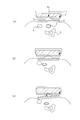
- FIG. 15 is a diagram showing a state in which the pressure pulse wave detection unit 100 is applied to the wrist and the sensor unit 6 is pressed toward the wrist by the air bag 2.
- the symbol TB indicates a rib
- the symbol K indicates a tendon.
- the radial artery T may move in the direction B as shown in FIG. 15 (b).
- the distribution of the amplitude value of the pressure pulse wave during pressing becomes as shown in FIG. 14B. That is, the position of the pressure detecting element 6a that detects the amplitude value in the pressing force in which the amplitude value equal to or greater than the threshold value TH1 is detected for the first time, and the amplitude value in the pressing force in which the amplitude value that is equal to or greater than the threshold value TH1 is detected last. There will be a large deviation from the position of the pressure detecting element 6a.
- the position of the pressure detection element 6a that detected the amplitude value for the first time when the amplitude value equal to or greater than the threshold value TH1 was detected, and the pressure value that the amplitude value equal to or greater than the threshold value TH1 was detected last. There is no significant deviation from the position of the pressure detecting element 6a that detects the amplitude value in the pressure. That is, it can be seen that the radial artery T is closed without substantially moving in the direction B in the process of increasing the pressing force.
- the change in the position of the radial artery T in the direction B can be detected by looking at the change in the tonogram in the process of changing the pressing force. If the radial artery T is occluded by increasing the pressing force in the state shown in FIG. 15B, there is a possibility that an accurate tonogram cannot be obtained due to the influence of the living tissue such as the tendon K.
- the control unit 12 determines the position of the pressure detection element 6a that has detected the amplitude value in the pressing force in which the amplitude value equal to or greater than the threshold value TH1 is detected for the first time from the data in FIG. 14 showing the relationship between the pressing force and the tonogram,
- the difference that is, the amount of movement of the radial artery T in the direction B
- the control unit 12 determines the position of the pressure detection element 6a that has detected the amplitude value in the pressing force in which the amplitude value equal to or greater than the threshold value TH1 is detected for the first time from the data in FIG. 14 showing the relationship between the pressing force and the tonogram.
- the difference that is, the amount of movement of the radial artery T in the direction B
- It is determined whether or not the calculated difference is greater than or equal to a threshold value THa (step S14).
- the control unit 12 calculates the amount of movement of the radial artery T in the direction B based on the data indicating the relationship between the tonogram generated for the element array 70 and the pressing force, and uses the calculated amount of movement as the threshold THa. You may compare.
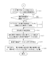
- step S14 If the amount of movement is equal to or greater than the threshold THa (step S14: YES), the control unit 12 obtains the vector indicated by the arrow in FIG. 14B in step S15. If the difference between the two positions is less than the threshold THa (step S14: NO), the process of step S16 is performed.
- the direction and magnitude of the vector shown in FIG. 14 and the information indicating in what direction and how much the rotating unit 5 should be rotated around the rotation axis X are experimentally obtained in advance and associated with each other. And remember.
- control unit 12 acquires information about the rotation direction and the rotation amount corresponding to the obtained vector size and direction from the memory 15 and transmits the acquired information to the rotation drive unit 10. And the rotation drive part 10 rotates the rotation part 5 as shown in FIG.15 (c) according to the received information (step S15).
- step S16 the control unit 12 controls the air bag drive unit 11 to discharge the air in the air bag 2, and starts to reduce the pressing force on the radial artery T.
- the control unit 12 starts decreasing the pressing force in step S16, and after decreasing the pressing force to the minimum value, determines the optimum pressure detecting element from all the pressure detecting elements 6a and 7a. For example, the control unit 12 determines the pressure detection element that has detected the pressure pulse wave having the maximum amplitude in the process of decreasing the pressing force as the optimum pressure detection element.
- the pressure pulse wave detected by the pressure detection element located directly above the portion where the radial artery T is flat is not affected by the tension of the wall of the radial artery T and has the largest amplitude.
- This pressure pulse wave has the highest correlation with the blood pressure value in the radial artery T. For this reason, the pressure detection element that detects the pressure pulse wave having the maximum amplitude is determined as the optimum pressure detection element.
- the plurality of pressure detection elements may be treated as optimum pressure detection elements and detected by each of the plurality of pressure detection elements.
- an average of the generated pressure pulse waves may be handled as a pressure pulse wave detected by the optimum pressure detection element.
- control unit 12 generates pulse wave envelope data from the pressure pulse wave detected by the optimum pressure detecting element in the process of decreasing the pressing force (step S17).
- the pulse wave envelope data refers to a pressing force applied to the radial artery T by the sensor unit 6 (internal pressure of the air bag 2), and an optimal pressure detecting element in a state where the optimal pressure detecting element is pressed against the radial artery T by the pressing force.
- the data correlates with the amplitude of the pressure pulse wave detected by.
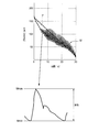
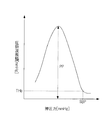
- FIG. 16 is a diagram showing an example of changes in the pressing force to the radial artery T and changes in the pressure pulse wave detected by the optimum pressure detection element.
- a straight line indicated by a symbol P indicates a pressing force
- a waveform indicated by a symbol M indicates a pressure pulse wave.
- an enlarged view of one pressure pulse wave is shown.
- the pressure at the rising point is referred to as the minimum value Mmin
- the pressure at the falling point is referred to as the maximum value Mmax.
- the amplitude of the pressure pulse wave is a value obtained by subtracting the minimum value Mmin from the maximum value Mmax.
- the maximum value Mmax and the minimum value Mmin are each information that specifies the shape of the pressure pulse wave.
- step S17 the control unit 12 generates pulse wave envelope data as shown in FIG. 17 from the relationship between the pressing force and the pressure pulse wave shown in FIG.
- control unit 12 calculates SBP and DBP from the generated pulse wave envelope data (step S18).
- the control unit 12 starts to decrease the pressing force when the pressure pulse wave amplitude starts to increase sharply after the pressing force starts decreasing, that is, the pressing force starts decreasing. Thereafter, the pressing force at the time when the pressure pulse wave amplitude detected by the optimum pressure detecting element exceeds the threshold value THb for which it can be determined that the artery is not occluded for the first time is determined as SBP.
- the control unit 12 calculates a difference between two adjacent amplitude values in the pulse wave envelope data, and determines the pressing force when the difference exceeds a threshold value as SBP.
- control unit 12 determines the maximum value Mmax of any one of the pressure pulse waves (for example, the pressure pulse wave having the maximum amplitude) detected by the optimum pressure detection element determined in the pressure reduction process after step S16. Using the minimum value Mmin and the SBP and DBP calculated in step S18, calibration data used at the time of continuous blood pressure measurement described later is generated and stored in the memory 15 (step S19).
- the control unit 12 adds the SBP and DBP obtained in step S18 to the equations (1) and (2), and the maximum value Mmax and the minimum value of the pressure pulse wave with the maximum amplitude in the pulse wave envelope of FIG. Substituting Mmin, slope a and intercept b are calculated. The calculated coefficients a and b and equations (1) and (2) are stored in the memory 15 as calibration data.
- FIG. 18 is a flowchart for explaining the continuous blood pressure measurement operation in the continuous blood pressure measurement mode of the biological information measuring apparatus 200 of the present embodiment.
- step S20 When the continuous blood pressure measurement mode is set and the start of blood pressure measurement is instructed, first, the same processing as steps S1 to S16 shown in FIGS. 11 and 12 is performed (step S20). Thereafter, when the pressing force reaches the minimum value, the control unit 12 controls the air bag driving unit 11 to increase the internal pressure of the air bag 2 and increase the pressing force on the wrist by the pressing surface 6b (step S21). ).
- control unit 12 determines, as the optimum pressure detecting element, the pressure detecting element that detects the pressure pulse wave having the maximum amplitude in the process of increasing the pressing force among the pressure detecting elements 6a and 7a. Moreover, the control part 12 determines the internal pressure of the air bag 2 at the time of detecting the pressure pulse wave of this maximum amplitude as an optimal pressing force (step S22).
- control unit 12 releases the internal pressure of the air bag 2 and returns it to the initial state (step S23), and then increases the internal pressure of the air bag 2 to the optimum pressing force determined in step S22. The pressure is maintained (step S24).
- control unit 12 acquires the pressure pulse wave detected by the optimum pressure detecting element determined in step S22 in a state where the pressing surface 6b is pressed against the wrist with the optimum pressing force (step S25).
- control unit 12 calibrates the acquired one pressure pulse wave using the calibration data generated in step S19 in FIG. 12, and calculates and calculates the second blood pressure values SBP and DBP.
- SBP and DBP are displayed on the display unit 13 (step S26).
- control unit 12 calculates the SBP by substituting the maximum value Mmax of the pressure pulse wave acquired in step S25 and the coefficients a and b calculated in step S19 into the above-described equation (1).
- the DBP is calculated by substituting the minimum value Mmin of the pressure pulse wave acquired in S25 and the coefficients a and b calculated in step S19 into the above-described equation (2). If there is an instruction to end continuous blood pressure measurement (step S27: YES), the control unit 12 ends the process. If there is no instruction to end (step S27: NO), the control unit 12 returns the process to step S25.
- the pressing surface 6b is configured to be rotatable around each of the rotation axis X and the rotation axis Y.
- the detection accuracy of the pressure pulse wave can be improved by changing flexibly.
- the processing after step S7 in FIG. 11 can be performed in a state where the position of the radial artery T with respect to the element row 60 or the element row 70 is optimal.
- the process after step S8 can be performed in a state in which a pressure pulse wave can be detected from the radial artery T by many pressure detection elements, and the necessity of rotation in step S9 in FIG.
- Each of rotation control around the rotation axis Y of the rotation unit 5 in step S10, calculation of the amount of arterial movement in step S13 in FIG. 12, and rotation control around the rotation axis X of the rotation unit 5 in step S15 in FIG. Processing can be performed with high accuracy. As a result, accurate calibration data and blood pressure information can be calculated.
- the fixing member 101A is arranged on the central side end face of the measurement subject of the housing 101 with the biological information measuring apparatus 200 mounted on the wrist. Therefore, the measurement subject can easily apply force to the fixing member 101A with the thumb while holding the both end surfaces in the direction A of the casing 101 with the right hand not wearing the biological information measurement device 200. The operability when moving the accommodating portion 4 is improved.
- a slit 101C shown in FIG. 8C is formed on the upper surface of the casing 101 (the surface shown in FIG. 8B), and a fixing member that penetrates the slit 101C and is fixed to the housing portion 4 is provided.
- the structure provided in this upper surface may be sufficient. According to this structure, visual observation of a fixing member becomes easy and the operativity at the time of moving the accommodating part 4 improves.
- the biological information measuring apparatus 200 calculates blood pressure information including SBP and DBP based on the pressure pulse wave detected by the pressure detection element of the sensor unit 6, but instead of this blood pressure information, the pulse rate Alternatively, biological information such as heart rate may be calculated and stored.
- the moving mechanism for moving the accommodating portion 4 is a mechanism for moving the accommodating portion 4 in the direction B by a manual operation for moving the fixing member 101A by hand (a mechanism for moving the accommodating portion 4 without using a drive source). There is, but is not limited to this.
- a configuration may be adopted in which an actuator that drives the housing unit 4 is added to the inside of the housing 101, and the control unit 12 moves the housing unit 4 in the direction B via the actuator.
- the mechanism of the biological information measuring device 200 can be simplified, and the manufacturing cost of the biological information measuring device 200 can be reduced.
- the pressure pulse wave detection unit 100 has a configuration in which the element array 60 and the element array 70 are formed on one pressing surface, but the pressing surface is divided and an element array is formed on each dividing surface. Good.
- the rotation axis Y is set between the element array 60 and the element array 70, but is not limited thereto.
- the rotation axis Y may be set outside the element array 60 and the element array 70.
- the rotation axis Y may be on the left side of the element row 60 in FIG.
- the rotation axis Y may be on the right side of the element row 70 in FIG.
- the rotation axis X is at a position that divides each of the two element rows in half, but this is not restrictive.
- the rotation axis X may be at an arbitrary position on each element row.
- two element rows are formed on the pressing surface 6b, but three or more element rows may be formed on the pressing surface 6b.
- three or more element arrays it is possible to determine the necessity of rotation around each of the rotation axis X and the rotation axis Y of the pressing surface 6b and the amount of rotation thereof with higher accuracy, and biometric information. Can be calculated with high accuracy.
- the determination method of necessity of rotation around each of the rotation axis X and the rotation axis Y of the pressing surface 6b described in FIG. 11 and FIG. 12 and the determination method of the rotation amount and the rotation direction when rotating are examples. You may employ
- the moving mechanism shown in FIGS. 8 and 9 has a configuration in which the accommodating portion 4 moves in the direction B by sliding the fixing member 101A in the direction B.
- a mechanism that has a rotatable member on the outer peripheral surface of the housing 101 and rotates the member 4 in the direction B by rotating the member may be adopted.
- a dial DY that is rotatable about the direction B as a rotation axis is provided. When the dial DY is rotated, the accommodating portion 4 moves in the direction B according to the amount of rotation.
- the operability can be improved by moving the accommodating portion 4 by the dial operation.
- the dial DY is provided on the end face 101T that is easy to operate with the right hand opposite to the hand on which the biological information measuring device of the measurement subject is attached, thereby improving operability. be able to.
- the disclosed pressure pulse wave detection device includes a pressing member having a pressing surface in which a plurality of element arrays each including a plurality of pressure detecting elements arranged in one direction are arranged in a direction intersecting the one direction, and the one direction is a living body.
- a pressing mechanism that presses the pressing surface against the body surface in a state intersecting with the direction in which the artery below the body surface extends, and two axes that are orthogonal to the pressing direction of the pressing surface by the pressing mechanism.
- a rotation driving mechanism for rotating the pressing surface around each of a first axis extending in a direction and a second axis orthogonal to the one direction, the pressing mechanism, the rotating driving mechanism, and the pressing member A support member that supports the support member, a housing that houses the support member, and a moving mechanism that moves the support member in the one direction within the housing.
- the moving mechanism moves the support member in the one direction by a manual operation.
- the disclosed pressure pulse wave detection device is used with the casing mounted on the wrist of the measurement subject, and the moving mechanism includes a fixing member fixed to the support member, and the fixing member is Of the end faces of the casing in the direction orthogonal to the one direction and the pressing direction, the casing is attached to the wrist and can be moved in the one direction to the end face on the central side of the person to be measured. It is provided.
- the moving mechanism includes a rotatable member, and moves the support member in the one direction as the member rotates.
- the disclosed pressure pulse wave detection device is used with the casing attached to the wrist of a person to be measured, and the rotatable member is rotatable about the one direction as a rotation axis.
- the body is provided on the end face close to the hand on the opposite side of the hand on which the housing is mounted.
- the disclosed biological information measurement device includes the pressure pulse wave detection device, a biological information calculation unit that calculates biological information based on a pressure pulse wave detected by a pressure detection element included in the pressure pulse wave detection device, Is provided.
- the present invention is particularly convenient and effective when applied to a blood pressure monitor or the like.
Landscapes
- Health & Medical Sciences (AREA)
- Life Sciences & Earth Sciences (AREA)
- Engineering & Computer Science (AREA)
- Molecular Biology (AREA)
- Animal Behavior & Ethology (AREA)
- Veterinary Medicine (AREA)
- Biophysics (AREA)
- Pathology (AREA)
- Public Health (AREA)
- Biomedical Technology (AREA)
- Heart & Thoracic Surgery (AREA)
- Medical Informatics (AREA)
- General Health & Medical Sciences (AREA)
- Surgery (AREA)
- Physics & Mathematics (AREA)
- Cardiology (AREA)
- Physiology (AREA)
- Vascular Medicine (AREA)
- Ophthalmology & Optometry (AREA)
- Artificial Intelligence (AREA)
- Computer Vision & Pattern Recognition (AREA)
- Psychiatry (AREA)
- Signal Processing (AREA)
- Dentistry (AREA)
- Oral & Maxillofacial Surgery (AREA)
- Measuring Pulse, Heart Rate, Blood Pressure Or Blood Flow (AREA)
Abstract
生体部位に接触させて用いる圧力検出素子の生体部位との接触状態を柔軟に変更して圧脈波検出精度を向上させることのできる圧脈波検出装置とこれを備える生体情報測定装置を提供する。生体情報測定装置200は、方向Bに並ぶ複数の圧力検出素子からなる素子列60及び素子列70が方向Bと直交する方向Aに配列された押圧面6bを有するセンサ部6、方向Bが橈骨動脈Tの伸びる方向と交差する状態で押圧面6bを手首Hに対し押圧する押圧機構、及び、押圧機構による押圧面6bの押圧方向に直交する2つの軸であって方向Aに伸びる回転軸Xと方向Bに伸びる回転軸Yの各々の周りに押圧面6bを回転するための回転駆動機構を収容する収容部4と、収容部4を内蔵する筐体101と、筐体101内で収容部4を方向Bに移動させるための移動機構と、を備える。
Description
本発明は、圧脈波検出装置及び生体情報測定装置に関する。
手首の橈骨動脈等の動脈が通る生体部位に圧力センサを直接接触させた状態で、この圧力センサにより検出される情報を用いて血圧、脈拍数、又は、心拍数等の生体情報を測定することのできる生体情報測定装置が知られている。この生体情報測定装置では、圧力センサと動脈との位置関係が圧脈波の検出精度に影響する。そこで、下記特許文献1~5に示すように、生体部位と圧力センサとの位置調整を行うための構成が提案されている。
特許文献1に記載の生体情報測定装置は、生体部位に接触させる6×7=42個のセンサ群を有し、センサ群の各センサの出力が良好になるように、センサ群の動脈方向の傾きを手動で調整できる機構を有するものとなっている。
特許文献2に記載の生体情報測定装置は、生体部位に接触させる6×7=42個のセンサ群を有し、センサ群の接触を手の動きに追従させるべく、センサ群が4分割され、各分割エリアの高さを調整可能な機構を有するものとなっている。
特許文献3には、生体部位に接触させる圧力センサを有し、圧力センサを動脈と交差する方向に移動させる駆動部を有する生体情報測定装置が開示されている。
特許文献4には、生体部位に接触させる圧力センサ列を有し、圧力センサ列を圧力センサ列の押圧方向と交差する面内で回転させる駆動部を有する生体情報測定装置が開示されている。
特許文献5には、生体部位に接触させる圧力センサ列が複数並べて形成された押圧面を有し、この押圧面を、複数の圧力センサ列の配列方向と直交する方向に伸びる軸の周りに回転させる駆動部を有する生体情報測定装置が開示されている。
特許文献1,2に記載の装置は、センサ群の生体部位への接触状態を変えることはできるものの、利用者の手首の形に追従して接触状態が変化したり、手動で接触状態を変化させたりするものである。このため、脈波の検出精度を十分に考慮したセンサの位置決めを行うことはできない。
特許文献3,4,5に記載の装置は、圧力センサの出力が良好となるように圧力センサの位置を駆動するものであるため、脈波の検出精度を考慮した圧力センサの位置決めが可能である。しかし、圧力センサを生体部位に対して押圧し、その状態で圧力センサから出力される情報を用いて生体情報を測定する場合には、押圧力によって動脈の位置が変化することが想定される。特許文献3,4,5の装置では、このような位置変化に十分に追従することは難しい。
本発明は、上記事情に鑑みてなされたものであり、生体部位に接触させて用いる圧力検出素子の生体部位との接触状態を柔軟に変更して圧脈波検出精度を向上させることのできる圧脈波検出装置とこれを備える生体情報測定装置を提供することを目的とする。
本発明の圧脈波検出装置は、一方向に並ぶ複数の圧力検出素子からなる素子列が上記一方向と交差する方向に複数配列された押圧面を有する押圧部材と、上記一方向が生体の体表面下の動脈の伸びる方向と交差する状態で、上記押圧面を上記体表面に対し押圧する押圧機構と、上記押圧機構による上記押圧面の押圧方向に直交する2つの軸であって上記一方向に伸びる第一の軸と上記一方向と直交する第二の軸の各々の周りに上記押圧面を回転するための回転駆動機構と、上記押圧機構、上記回転駆動機構、及び、上記押圧部材を支持する支持部材と、上記支持部材を内蔵する筐体と、上記筐体内で上記支持部材を上記一方向に移動させるための移動機構と、を備えるものである。
本発明の生体情報測定装置は、上記圧脈波検出装置と、上記圧脈波検出装置に含まれる圧力検出素子により検出される圧脈波に基づいて生体情報を算出する生体情報算出部と、を備えるものである。
本発明によれば、生体部位に接触させて用いる圧力検出素子の生体部位との接触状態を柔軟に変更して圧脈波検出精度を向上させることのできる圧脈波検出装置とこれを備える生体情報測定装置を提供することができる。
以下、本発明の実施形態について図面を参照して説明する。
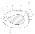
図1は、本発明の一実施形態を説明するための生体情報測定装置200の概略構成を示す側面図である。図1に示す生体情報測定装置200は、橈骨動脈が存在する被測定者の手首に装着して用いられる手首装着型の生体情報測定装置である。生体情報測定装置200は、動脈が体表面下に存在する生体部位に装着して用いるものであればよく、手首装着型に限定されるものではない。
生体情報測定装置200は、筐体101及び筐体102と、筐体101と筐体102を連結するヒンジ部103とから構成される本体部と、本体部を手首に固定するためのベルト106と、を備える。ヒンジ部103により、筐体102は筐体101に対して回動自在に連結されている。
筐体101,102の内周面は手首Hの外形に沿った形状となっている。筐体101のヒンジ部103側とは反対側の端部が、筐体102に最も近付けられた状態で、筐体101と筐体102の間には、被測定者の手首Hが挿入可能な空間Kaが形成される。
空間Kaに手首Hが挿入された状態で、筐体101の一端と筐体102の一端がベルト106によって固定されることにより、生体情報測定装置200は手首Hに固定(装着)される。なお、筐体101と筐体102はヒンジ部103を介さずに一体化されたものであってもよい。
筐体101は、生体情報測定装置200が手首Hに装着された状態で手首Hと対向する位置に設けられる圧脈波検出部100と、該状態において外周面に設けられる表示部13と、を備える。筐体101と圧脈波検出部100とにより、圧脈波検出装置が構成される。
図2は、生体情報測定装置200が被測定者の左手首に装着された状態を掌側から見た図である。図2に示すように、筐体101の外周面には表示部13が設けられており、掌側から表示部13を視認可能である。図3は、生体情報測定装置200が被測定者の左手首に装着された状態を手の甲側から見た図である。
図4は、図2,3に示す生体情報測定装置200の手首装着状態における圧脈波検出部100の構成を示す断面模式図である。図4では、生体情報測定装置200のうち圧脈波検出部100のみを図示している。
図5は、図4に示す圧脈波検出部100の拡大図である。図6は、図4に示す装着状態での圧脈波検出部100を被測定者の指先側から見た状態での断面模式図である。図7は、図4に示す装着状態での圧脈波検出部100を手首との接触部位側から見た図である。図4~図7は、圧脈波検出部100を模式的に示したものであり、各部の寸法や配置等を限定するものではない。
圧脈波検出部100は、空気袋2を内蔵する筐体1と、空気袋2に固定された平板状の部材である平板部3と、平板部3に対して2軸回転機構5aにより2つの軸の各々を中心に回転可能に支持された回動部5と、回動部5の平板部3側とは反対側の平面に設けられたセンサ部6と、筐体1、空気袋2、平板部3、2軸回転機構5a、回動部5、及び、センサ部6を収容する収容部4と、備える。
筐体1、空気袋2、平板部3、及び、回動部5は、生体情報測定装置200が手首に装着された状態で、センサ部6の押圧面6bを生体部位(手首)の体表面に対して押圧する押圧機構を構成する。押圧機構は、センサ部6の押圧面6bを体表面に対して押圧できる機構であれば何でもよく、空気袋を用いたものには限定されない。
空気袋2は、図示しないポンプにより、内部の空気量が制御されることで、空気袋2に固定された平板部3を平板部3の表面(回動部5側の平面)に垂直な方向に移動させる。
図4に示す装着状態では、圧脈波検出部100に含まれるセンサ部6の押圧面6bが被測定者の手首の皮膚に接触する。この状態で空気袋2に注入される空気量が増えることで、空気袋2の内圧が増加し、センサ部6は手首下の橈骨動脈Tに向けて押圧される。以下、センサ部6による橈骨動脈Tへの押圧力は、空気袋2の内圧と等価であるとして説明する。
図7に示すように、センサ部6は、図4に示す装着状態において、装着部位に存在する橈骨動脈Tの伸びる方向Aと交差(図7の例では直交)する方向Bに並ぶ複数個の圧力検出素子6aからなる素子列60と、方向Bに並ぶ複数個の圧力検出素子7aからなる素子列70とを有する。素子列60と素子列70は方向Aに並べられている。
各圧力検出素子6aと、この圧力検出素子6aと方向Bでの位置が同じ圧力検出素子7aとがペアを構成し、センサ部6には、このペアが方向Bに複数配列された構成となっている。圧力検出素子6aと圧力検出素子7aは、それぞれ、例えば、歪ゲージ抵抗式、半導体ピエゾ抵抗式、又は、静電容量式等の素子が用いられる。
素子列60と素子列70に含まれる各圧力検出素子は同一平面上に形成されており、この平面が樹脂等の保護部材によって保護されている。各圧力検出素子が形成された平面と、この平面を保護する保護部材の表面とは平行になっており、この保護部材の表面が押圧面6bを構成する。
圧力検出素子6a(7a)は、その配列方向が橈骨動脈Tと交差(略直交)するように橈骨動脈Tに対して押圧されることにより、橈骨動脈Tから発生して皮膚に伝達される圧力振動波、すなわち圧脈波を検出する。圧力検出素子6a(7a)から出力される圧力信号は、物との接触によって生じる直流成分と、圧力振動波によって生じる交流成分を含み、このうちの交流成分が圧脈波の信号となる。
各圧力検出素子6a(7a)の配列方向の間隔は、橈骨動脈T上に必要かつ充分な数が配置されるように充分小さくされている。各素子列560,70の長さは、橈骨動脈Tの径寸法より必要かつ充分に大きくされている。このように構成されたセンサ部6は、押圧部材を構成する。
図7に示すように、2軸回転機構5aは、空気袋2による平板部3の押圧方向に直交する2つの回転軸X,Yの各々を中心に回動部5を回転させるための機構である。2軸回転機構5a及び回動部5は、回転駆動機構を構成する。
2軸回転機構5aは、平板部3の表面上に設定された互いに直交する2つの回転軸X,Yを有し、回転軸X,Yは、それぞれ、後述する回転駆動部10によって回転駆動される。
回転軸Yは、押圧面6bに形成された複数の圧力検出素子6a(7a)の配列方向に伸びる第一の軸である。回転軸Yは、図7の平面視において、素子列60と素子列70との間(図7の例では中間)に設定されている。
回転軸Xは、押圧面6bに形成された複数の圧力検出素子6a(7a)の配列方向と直交する方向に伸びる第二の軸である。回転軸Xは、図7の例では、素子列60と素子列70をそれぞれ均等に2分割する直線上に設定されている。
回動部5が回転軸Xを中心に回転することで、押圧面6bは、回転軸Xの軸周りに回転する。また、回動部5が回転軸Yを中心に回転することで、押圧面6bは、回転軸Yの軸周りに回転する。
圧脈波検出部100の収容部4は、筐体101に内蔵されており、筐体101によって方向Bに移動可能に支持されている。収容部4は、例えばコップ状の部材で構成され、この部材の中空部に、筐体1、空気袋2、平板部3、2軸回転機構5a、回動部5、及び、センサ部6が収容される。具体的には、収容部4は、中空部において筐体1を支持している。これにより、収容部4の移動に伴って、筐体1とこれに固定された空気袋2、平板部3、2軸回転機構5a、回動部5、及び、センサ部6が移動する。
収容部4は、これが方向Bに移動することで、筐体1、空気袋2、平板部3、及び、回動部5により構成される押圧機構と、2軸回転機構5a及び回動部5により構成される回転駆動機構と、センサ部6と、が一体となって方向Bに移動できるように、押圧機構、回転駆動機構、及び、センサ部6を支持する支持部材であればよい。例えば、収容部4は、筐体1を支持する平板状の部材によって、押圧機構、回転駆動機構、及び、センサ部6を一体的に支持する構成であってもよい。
図8は、図1に示す筐体101の収容部4が内蔵される部分の展開図を模式的に示した図である。
図8(a)は、生体情報測定装置200を手首に装着した状態で筐体101を被測定者の抹消側(指先側)から見た側面図である。図8(b)は、生体情報測定装置200を手首に装着した状態で筐体101を押圧方向から見た上面図である。図8(c)は、生体情報測定装置200を手首に装着した状態で筐体101を被測定者の中枢側(肘側)から見た側面図である。図9は、図8に示すIX-IX線断面模式図である。
図8に示すように、筐体101の方向Aにおける端面のうち、生体情報測定装置200を手首に装着した状態で被測定者の中枢側(肘側)にある端面(以下、中枢側端面という)には、収容部4に固定された固定部材101Aが設けられている。固定部材101Aの表面には、凸部101Bが設けられている。
筐体101の中枢側端面には、方向Bに伸びるスリット101Cが形成されている。固定部材101Aの裏面には、図8(b)に示すように、スリット101Cを貫通する凸部101Dが形成されており、この凸部101Dが収容部4に固定されている。これにより、固定部材101Aは、スリット101Cを介して、方向Bに移動自在となっている。
図9に示すように、収容部4の方向Aにおける端面のうち、生体情報測定装置200が手首に装着された状態で被測定者の末梢側にある端面には、板バネ100bが固定されている。板バネ100bにはスライド部材100aが固定されている。スライド部材100aは、押圧方向(方向A及び方向Bに直交する方向)に長手の三角柱状の凸部を有する部材である。
筐体101には、スライド部材100aの凸部が係合可能な凹部を図9の例では7つ有するスライドレール101aが形成されている。スライド部材100aは、板バネ100bによってスライドレール101aに向けて付勢されている。なお、図9(a)では、説明のために、スライド部材100aとスライドレール101aの間に隙間がある状態を図示している。
図9(a)に示す状態から、被測定者が凸部101Bに指をかけ、固定部材101Aに対して図9中の右方向に力を加えると、スライド部材100aの凸部は、スライドレール101aの凹部の斜面に沿って右方向に移動し、隣の凹部と係合する。この状態では、スライド部材100aの凸部は、板バネ100bによってスライドレール101aの凹部に付勢されて、図9(b)に示す状態となる。
スライドレール101aの隣接する2つの凹部の間隔(凹部の底部同士の距離)は例えば1mm程度に設計される。これにより、収容部4は、図9(a)に示す位置を基準位置として、この基準位置から方向Bの一方の方向に1mmずつ計3mm動かすことができ、この基準位置から方向Bの他方の方向に1mmずつ計3mm動かすことができる。
なお、収容部4を移動させる際の最小単位として1mmは一例であり、これに限定されるものではない。また、収容部4を移動させることのできる距離は上記の例では6mmであるが、この数値も一例であり、これに限定されるものではない。
このように、スライドレール101a、スライド部材100a、板バネ100b、固定部材101A、及び、凸部101Bによって、収容部4を筐体101内で方向Bに移動するための移動機構が構成される。なお、筐体101内で収容部4を方向Bに移動させる機構としては、図8及び図9に示したものに限らず、周知の機構を採用することができる。
図10は、図1に示す生体情報測定装置200のブロック構成を示す図である。
生体情報測定装置200は、圧脈波検出部100と、回転駆動部10と、空気袋駆動部11と、装置全体を統括制御する制御部12と、表示部13と、操作部14と、メモリ15と、を備える。
回転駆動部10は、圧脈波検出部100の2軸回転機構5aの各回転軸X,Yに接続されたアクチュエータである。回転駆動部10は、制御部12の指示にしたがい各回転軸X,Yを回転駆動して、押圧面6bを回転軸Xの軸周りに回転させたり、押圧面6bを回転軸Yの軸周りに回転させたりする。
空気袋駆動部11は、制御部12の指示のもと、空気袋2に注入する空気量(空気袋2の内圧)を制御する。
表示部13は、測定された血圧値等の各種情報を表示するためのものであり、例えば液晶表示素子、有機電界発光表示素子、又は、電子ペーパ等により構成される。
操作部14は、制御部12に対する指示信号を入力するためのインターフェースであり、血圧測定を含む各種動作の開始を指示するためのボタン等により構成される。
メモリ15は、制御部12に所定の動作をさせるためのプログラム及び各種データを記憶するROM(Read Only Memory)、ワークメモリとしてのRAM(Random Access Memory)、及び、測定した血圧データ等の各種情報を記憶するフラッシュメモリ等を含む。
制御部12は、メモリ15のROMに記憶されたプログラムを実行することにより、押圧制御部、生体情報算出部、回転制御部、校正用データ生成部、及び、表示制御部として機能する。
押圧制御部は、空気袋駆動部11を制御して空気袋2内の空気量を調整することで、押圧面6bによる手首への押圧力を制御する。
生体情報算出部は、押圧面6bを橈骨動脈Tに押圧した状態で、押圧面6bに形成された圧力検出素子6a,7aにより検出される圧脈波に基づいて、橈骨動脈T内の第一の血圧値を算出する。
具体的には、生体情報算出部は、空気袋駆動部11によって橈骨動脈Tへの押圧力が変化(増加又は減少)させられていく過程で圧力検出素子6a,7aにより検出される圧脈波に基づいて、橈骨動脈T内の第一の血圧値を算出する。
校正データ生成部は、生体情報算出部により算出された第一の血圧値を用いて校正用データを生成する。
回転制御部は、空気袋駆動部11によって橈骨動脈Tへの押圧力が増加させられていく過程で圧力検出素子6a,7aにより検出された圧脈波に基づいて、回転駆動部10による押圧面6bの回転の要否を判定する。そして、回転制御部は、回転が必要と判定したときに、回転駆動部10を制御して押圧面6bを回転させる。
生体情報算出部は、橈骨動脈Tの一部を平坦に変形させるための最適押圧力で押圧面6bが橈骨動脈Tに押圧された状態で、圧力検出素子6a,7aにより1拍毎に検出される圧脈波を校正用データによって校正することで、1拍毎に橈骨動脈T内の第二の血圧値を算出する。
表示制御部は、表示部13の表示制御を行う。
以下、本実施形態の生体情報測定装置200の動作について説明する。本実施形態の生体情報測定装置200は、心臓の拍動の1拍毎に血圧値(SBP(Systolic Blood pressure)、いわゆる最高血圧と、DBP(Diastolic Blood pressure)、いわゆる最低血圧を測定してフラッシュメモリに記憶し、測定した血圧を表示部13に表示する連続血圧測定モードを有する。
図11及び図12は、生体情報測定装置200の連続血圧測定モードにおける校正用データ生成までの動作を説明するためのフローチャートである。
なお、圧脈波検出部100の回動部5は、血圧測定の開始指示がなされる前の初期状態では、回転量が例えばゼロに設定され、押圧面6bが平板部3と平行になっているものとする。
操作部14に含まれる測定開始ボタンが押されて、血圧測定の開始指示がなされると(ステップS1:YES)、制御部12は、空気袋駆動部11を制御して空気袋2への空気の注入を開始し、押圧面6bによる手首への押圧力を増加させる(ステップS2)。血圧測定の開始指示がなされていない場合には、制御部12は、血圧測定の開始指示がなされるのを待機する。
ステップS2の処理の後、制御部12は、橈骨動脈Tの閉塞が開始されるのに十分な時間が経過すると、空気袋駆動部11を制御して空気袋2への空気の注入を停止する。制御部12は、この状態で素子列60の各圧力検出素子6aにより検出された圧力信号と、素子列70の各圧力検出素子7aにより検出された圧力信号とに基づいて、素子列60と素子列70のいずれか一方に対する橈骨動脈Tの位置が最適であるか否かを判定する(ステップS3)。
任意の素子列に対する橈骨動脈Tの位置が最適であるとは、この素子列の方向Bの中央付近の下方に橈骨動脈Tが存在することを言う。
例えば、圧力検出素子が方向Bに46個配列された素子列を考える。この場合は、46個の圧力検出素子のうち、両端から数えてそれぞれ9番目の2つの圧力検出素子の間にある28個の圧力検出素子(素子列の中央付近の範囲内にある圧力検出素子)の下方に橈骨動脈Tが存在する場合に、この素子列に対する橈骨動脈Tの位置が最適であるとする。
具体的には、制御部12は、圧力信号のうちの交流成分の振幅値が閾値以上となる圧力検出素子を、橈骨動脈T上方に位置する圧力検出素子として判定する。そして、制御部12は、素子列60に含まれる圧力検出素子6aのうち、橈骨動脈T上方に位置すると判定した圧力検出素子6aの位置が、素子列60に対して予め設定されている中央付近の範囲内に全て入る場合に、素子列60に対する橈骨動脈Tの位置が最適であると判定する。同様に、制御部12は、素子列70に含まれる圧力検出素子7aのうち、橈骨動脈T上方に位置すると判定した圧力検出素子7aの位置が、素子列70に対して予め設定されている中央付近の範囲内に全て入る場合に、素子列70に対する橈骨動脈Tの位置が最適であると判定する。
制御部12は、素子列60と素子列70の各々に対する橈骨動脈Tの位置が最適ではないと判定した場合(ステップS4:NO)には、空気袋駆動部11を制御して空気袋2内の空気を排出させ、初期状態に戻す(ステップS5)。そして、制御部12は、収容部4を方向Bに移動させることを指示する情報を表示部13に表示させて、収容部4の移動を被測定者に対して指示する(ステップS6)。制御部12は、ステップS6の後はステップS1に処理を戻す。
ステップS6の処理により、例えば「動脈を正確に検出できません。センサユニットを移動させてから、再度、測定開始ボタンを押してください」といったメッセージが表示部13に表示される。この表示を見た被測定者は、図8(c)に示した凸部101B及び固定部材101Aを左右のどちらかに最小単位(上記の例では1mm)分、移動させる。
その後、被測定者が測定開始ボタンを押してステップS1の判定がYESになると、ステップS2~ステップS4の処理が再び行われる。制御部12は、ステップS4において、素子列60と素子列70のいずれか一方に対する橈骨動脈Tの位置が最適であると判定した場合(ステップS4:YES)には、空気袋駆動部11を制御して空気袋2の空気を排気した後、再び空気袋2への空気の注入を開始し、押圧面6bによる手首への押圧力を増加させる(ステップS7)。
ステップS7で開始された押圧力の増加過程において、制御部12は、橈骨動脈Tの閉塞が開始されるのに十分な時間が経過した後の任意のタイミング(例えば周期的なタイミング)で、それまでに各圧力検出素子6aにより検出されてメモリ15に記憶された圧脈波群(圧脈波情報I1とする)のうち、検出時刻が新しい順に複数の圧脈波情報I1を取得する。また、制御部12は、上記任意のタイミングで、それまでに各圧力検出素子7aにより検出されてメモリ15に記憶された圧脈波群(圧脈波情報I2とする)のうち、検出時刻が新しい順に複数の圧脈波情報I2を取得する(ステップS8)。
制御部12は、ステップS8で取得した複数の圧脈波情報I1のうち、時刻t1に検出された圧脈波情報I1に含まれる圧脈波の例えば振幅の平均値Ave1を算出し、時刻t1よりも後の時刻t2に検出された圧脈波情報I1に含まれる圧脈波の振幅の平均値Ave2を算出する。また、制御部12は、ステップS8で取得した複数の圧脈波情報I2のうち、時刻t1に検出された圧脈波情報I2に含まれる圧脈波の振幅の平均値Ave3を算出し、時刻t2に検出された圧脈波情報I2に含まれる圧脈波の振幅の平均値Ave4を算出する。そして、制御部12は、同じ時刻に対して算出した平均値の比((Ave1/Ave3)と(Ave2/Ave4))を算出する。
制御部12は、複数のタイミングについて算出した比の変化に基づいて、回転駆動部10による回動部5の回転を行うべきか否かを判定する。つまり、制御部12は、押圧力の増加過程における複数タイミングで圧力検出素子6a,7aにより検出された圧脈波に基づいて、回動部5を回転させるか否かを判定する(ステップS9)。
例えば、複数のタイミングについて算出した比が単調増加している場合には、素子列70は橈骨動脈Tを閉塞する方向に向かっているが、素子列60は橈骨動脈Tを閉塞する方向に向かっていないと判定できる。このため、制御部12は、回動部5の回転は必要と判定する。
また、複数のタイミングについて算出した比が単調減少している場合には、素子列60は橈骨動脈Tを閉塞する方向に向かっているが、素子列70は橈骨動脈Tを閉塞する方向に向かっていないと判定できる。このため、制御部12は、回動部5の回転は必要と判定する。
また、複数のタイミングについて算出した比がほとんど変化していない場合には、素子列60,70が同じように橈骨動脈Tから高精度に圧脈波を検出していると判定できる。このため、制御部12は、回動部5の回転は不要と判定する。
また、複数のタイミングについて算出した比が増減を繰り返している場合には、素子列60,70が橈骨動脈Tを十分に押圧できているか、一方の素子列だけが橈骨動脈Tを十分に押圧できていないのかの判定ができない。このため、制御部12は、回動部5の回転は不要と判定する。
このように、制御部12は、複数のタイミングについて算出した比の変動に基づいて回転の要否を判定する。なお、この比の代わりに、平均値Ave1(Ave2)と平均値Ave3(Ave4)の差分(符号を考慮した値)を用いてもよい。
図13(a)は、素子列70によって橈骨動脈Tが閉塞されているが、素子列60によっては橈骨動脈Tが閉塞されていない状態の例を示す図である。図13(a)の状態では、素子列60と橈骨動脈Tの距離が、素子列70と橈骨動脈Tの距離よりも大きくなっている。
各圧力検出素子6aにより検出された圧脈波の振幅平均値を6Aとし、各圧力検出素子7aにより検出された圧脈波の振幅平均値を7Aとすると、図13の状態では、6Aと7Aの比である(6A/7A)は“1”よりも大きくなる。この状態では、素子列60を橈骨動脈Tに近づければ、(6A/7A)は1に近づく。
そこで、制御部12は、ステップS9において回動部5の回転軸Y周りの回転が必要と判定したときは、最新時刻における(6A/7A)の値に応じて回動部5の回転軸Y周りの回転制御を行う(ステップS10)。
具体的には、制御部12は、(6A/7A)の値と回動部5の回転量との関係を示すデータテーブル(製品出荷前に実験的に求めてメモリ15に記憶しておく)を参照し、(6A/7A)の値に対応する回転量を読みだして、読みだした回転量を設定する。
また、制御部12は、平均値6Aと平均値7Aのどちらが大きいかを判定し、平均値6Aが大きい場合には、素子列60と橈骨動脈Tとの距離を縮めるべく、回転軸Y周りの回動部5の回転方向を図13において反時計回りに設定する。
制御部12は、平均値7Aが大きい場合には、素子列70と橈骨動脈Tとの距離を縮めるべく、回転軸Y周りの回動部5の回転方向を図13において時計回りに設定する。
制御部12は、このようにして設定した回転方向及び回転量によって回動部5を回転させる。これにより、図13(b)のように、押圧面6bと橈骨動脈Tとを平行にすることができ、橈骨動脈Tを広い範囲にわたって均等に圧扁した状態を得ることができる。
制御部12は、ステップS10の後と、ステップS9において回動部5の回転が不要と判定した場合には、ステップS11に処理を移行する。ステップS11において、制御部12は、橈骨動脈Tが閉塞されるのに十分な圧力(必要押圧力)に押圧力が到達したか否かを判定する。制御部12は、押圧力が必要押圧力に到達した場合(ステップS11:YES)に、空気袋駆動部11を制御して空気袋2への空気の注入を停止させる(ステップS12)。制御部12は、押圧力が必要押圧力に達していない場合はステップS8に処理を戻す。
ステップS12の後、制御部12は、ステップS7~ステップS12の間において各圧力検出素子6aにより同時刻に検出された圧脈波の振幅と、その各圧力検出素子6aの押圧面6b上における位置との関係を示す振幅分布曲線、いわゆるトノグラムを求める。また、制御部12は、各圧力検出素子7aにより同時刻に検出された圧脈波の振幅と、その各圧力検出素子7aの押圧面6b上における位置との関係を示すトノグラムを求める。
制御部12は、素子列60に対して生成したトノグラムを、この素子列60の識別情報、圧脈波の検出時刻、及びこの検出時刻における空気袋2による押圧方向への押圧力(空気袋2の内圧)と対応付けてメモリ15に記憶する。
同様に、制御部12は、素子列70に対して生成したトノグラムを、この素子列70の識別情報、圧脈波の検出時刻、及びこの検出時刻における空気袋2による押圧方向への押圧力と対応付けてメモリ15に記憶する。
そして、制御部12は、メモリ15に記憶したトノグラムのデータを用いて、押圧面6bの手首への押圧中における橈骨動脈Tの方向Bへの移動量を算出する(ステップS13)。
図14(a),(b)は、センサ部6による手首への押圧力を変化させていったときに、センサ部6の各圧力検出素子6aにより検出される圧脈波の振幅値の一例を示した図である。図14(a),(b)において、横軸は各圧力検出素子6aの方向Bでの位置を示し、縦軸は押圧力を示す。
図14(a),(b)では、各位置にある圧力検出素子6aにより検出された圧脈波の振幅を、その大きさによって色分けしている。
符号A1は、振幅が閾値TH1以上となっている部分である。符号A2は、振幅が閾値TH2以上閾値TH1未満となっている部分である。符号A3は、振幅が閾値TH3以上閾値TH2未満となっている部分である。符号A4は、振幅が閾値TH4以上閾値TH3未満となっている部分である。符号A5は、振幅が閾値TH4未満となっている部分である。なお、閾値TH1>閾値TH2>閾値TH3>閾値TH4である。
図14(a)は、押圧力が増加していく過程で、閾値TH1以上の振幅の圧脈波を検出している圧力検出素子6aの位置がほぼ変化しない例を示している。これに対し、図14(b)は、押圧力が増加していく過程で、閾値TH1以上の振幅の圧脈波を検出している圧力検出素子6aの位置が左にずれていく例を示している。
図15は、圧脈波検出部100を手首に当てて、空気袋2によりセンサ部6を手首に向けて押圧していく状態を示す図である。図15において、符号TBは橈骨を示し、符号Kは腱を示す。
図15(a)に示すようにセンサ部6を手首に押圧していくと、図15(b)に示すように、橈骨動脈Tが方向Bに移動してしまうことがある。
図15(b)のように、押圧中に橈骨動脈Tが方向Bに移動してしまうと、押圧中の圧脈波の振幅値の分布は図14(b)のようになる。つまり、閾値TH1以上の振幅値が初めて検出された押圧力における当該振幅値を検出した圧力検出素子6aの位置と、閾値TH1以上の振幅値が最後に検出された押圧力における当該振幅値を検出した圧力検出素子6aの位置とには大きなずれが生じることになる。
図14(a)の例では、閾値TH1以上の振幅値が初めて検出された押圧力における当該振幅値を検出した圧力検出素子6aの位置と、閾値TH1以上の振幅値が最後に検出された押圧力における当該振幅値を検出した圧力検出素子6aの位置とには大きなずれは生じていない。つまり、押圧力を増加させていく過程において、橈骨動脈Tが方向Bにほぼ移動することなく閉塞されていっていることが分かる。
このように、押圧力が変化する過程におけるトノグラムの変化をみることで、橈骨動脈Tについて、方向Bでの位置変化を検出することができる。図15(b)に示す状態のまま、押圧力を増加させて橈骨動脈Tを閉塞すると、腱K等の生体組織の影響を受けて、正確なトノグラムを取得できない可能性が生じる。
そこで、制御部12は、押圧力とトノグラムの関係を示す図14のデータから、閾値TH1以上の振幅値が初めて検出された押圧力における当該振幅値を検出した圧力検出素子6aの位置と、閾値TH1以上の振幅値が最後に検出された押圧力における当該振幅値を検出した圧力検出素子6aの位置との差(つまり橈骨動脈Tの方向Bへの移動量)をステップS13にて算出し、算出した差が閾値THa以上か否かを判定する(ステップS14)。
なお、制御部12は、素子列70に対して生成したトノグラムと押圧力の関係を示すデータに基づいて、橈骨動脈Tの方向Bへの移動量を算出し、算出した移動量を閾値THaと比較してもよい。
上記の移動量が閾値THa以上であれば(ステップS14:YES)、制御部12は、ステップS15において図14(b)の矢印で示したベクトルを求める。2つの位置の差が閾値THa未満であれば(ステップS14:NO)、ステップS16の処理が行われる。
メモリ15には、図14に示したベクトルの向き及び大きさと、回動部5を回転軸X周りにどの方向にどの程度回転させるべきかを示す情報とを、予め実験的に求めて対応付けて記憶しておく。
そして、制御部12は、求めたベクトルの大きさ及び向きに対応する回転方向及び回転量の情報をメモリ15から取得し、取得した情報を回転駆動部10に送信する。そして、回転駆動部10は、受信した情報にしたがって、図15(c)に示したように回動部5を回転させる(ステップS15)。
ステップS15に続くステップS16では、制御部12が、空気袋駆動部11を制御して、空気袋2内の空気を排出させ、橈骨動脈Tへの押圧力の減少を開始する。
制御部12は、ステップS16で押圧力の減少を開始し、押圧力を最小値まで減少させた後、全ての圧力検出素子6a,7aの中から最適圧力検出素子を決定する。制御部12は、例えば、押圧力の減少過程において最大振幅の圧脈波を検出した圧力検出素子を最適圧力検出素子として決定する。
橈骨動脈Tが平坦になった部分の真上に位置する圧力検出素子によって検出される圧脈波は、橈骨動脈Tの壁の張力の影響がなく、最も振幅が大きくなる。また、この圧脈波は橈骨動脈T内の血圧値との相関が最も高い。このような理由により、最大振幅の圧脈波を検出した圧力検出素子を最適圧力検出素子として決定する。
なお、最大振幅の圧脈波を検出した圧力検出素子が複数ある場合もあり、その場合には、この複数の圧力検出素子を最適圧力検出素子として扱い、この複数の圧力検出素子の各々で検出された圧脈波の例えば平均を、この最適圧力検出素子により検出される圧脈波として扱えばよい。
そして、制御部12は、押圧力の減少過程でその最適圧力検出素子により検出された圧脈波から脈波包絡線データを生成する(ステップS17)。
脈波包絡線データとは、センサ部6による橈骨動脈Tへの押圧力(空気袋2の内圧)と、その押圧力で最適圧力検出素子が橈骨動脈Tに押圧された状態で最適圧力検出素子により検出される圧脈波の振幅とを対応付けたデータである。
図16は、橈骨動脈Tへの押圧力の変化と、最適圧力検出素子により検出される圧脈波の変化の一例を示した図である。図16において、符号Pで示す直線が押圧力を示し、符号Mで示す波形が圧脈波を示している。図16の下段には、1つの圧脈波の拡大図を図示している。
図16に示したように、圧脈波において、立ち上がり点での圧力を最小値Mminといい、立ち下がり点での圧力を最大値Mmaxという。圧脈波の振幅は、最大値Mmaxから最小値Mminを引いた値を言う。最大値Mmaxと最小値Mminは、それぞれ、圧脈波の形状を特定する情報の1つである。
図16に示したように、押圧力が減少を開始して橈骨動脈Tの閉塞状態が解除されると、最適圧力検出素子により検出される圧脈波は振幅が急激に大きくなり、その後、押圧力の減少に伴って図に示したように変化していく。制御部12は、ステップS17において、図16に示す押圧力と圧脈波の関係から、図17に示すような脈波包絡線データを生成する。
制御部12は、図17に示す脈波包絡線データを生成すると、生成した脈波包絡線データからSBPとDBPを算出する(ステップS18)。
例えば、制御部12は、図17に示す脈波包絡線において、押圧力が減少を開始してから圧脈波振幅が急激に上昇を開始したときの押圧力、すなわち、押圧力が減少を開始してから最適圧力検出素子により検出される圧脈波振幅が動脈閉塞状態ではなくなったと判断できる閾値THbを初めて超えた時点での押圧力をSBPとして決定する。または、制御部12は、脈波包絡線データにおいて隣接する2つの振幅値の差分を算出し、この差分が閾値を超えた時点での押圧力をSBPとして決定する。
更に、制御部12は、図17に示す脈波包絡線において、圧脈波振幅の最大値を脈圧(PP)とし、求めたSBP及びPPと、SBP-DBP=PPの関係式により、DBPを算出する。
ステップS18の後、制御部12は、ステップS16以降の減圧過程で決定した最適圧力検出素子により検出された各圧脈波のいずれか(例えば、最大振幅となった圧脈波)の最大値Mmax及び最小値Mminと、ステップS18で算出したSBP及びDBPと、を用いて、後述する連続血圧測定時に用いる校正用データを生成してメモリ15に記憶する(ステップS19)。
aを一次関数の傾き、bを一次関数の切片とすると、
SBP=a×Mmax+b ・・・(1)
DBP=a×Mmin+b ・・・(2)
の関係が成り立つ。
SBP=a×Mmax+b ・・・(1)
DBP=a×Mmin+b ・・・(2)
の関係が成り立つ。
制御部12は、式(1)と式(2)に、ステップS18で求めたSBP及びDBPと、図17の脈波包絡線における振幅が最大となった圧脈波の最大値Mmax及び最小値Mminとを代入して、傾きaと切片bを算出する。そして、算出した係数a,bと、式(1),(2)とを校正用データとしてメモリ15に記憶する。
図18は、本実施形態の生体情報測定装置200の連続血圧測定モードにおける連続血圧測定動作を説明するためのフローチャートである。
連続血圧測定モードが設定され、血圧測定の開始指示がされると、まず、図11及び図12に示したステップS1~ステップS16と同じ処理が行われる(ステップS20)。その後、押圧力が最小値に達すると、制御部12は、空気袋駆動部11を制御し、空気袋2の内圧を上昇させて、押圧面6bによる手首への押圧力を増加させる(ステップS21)。
次に、制御部12は、各圧力検出素子6a,7aのうち、押圧力の増加過程において最大振幅の圧脈波を検出した圧力検出素子を最適圧力検出素子として決定する。また、制御部12は、この最大振幅の圧脈波が検出された時点での空気袋2の内圧を最適押圧力として決定する(ステップS22)。
次に、制御部12は、空気袋2の内圧を解放して初期状態に戻し(ステップS23)、その後、空気袋2の内圧をステップS22で決定した最適押圧力まで上昇させて、この最適押圧力を保持する(ステップS24)。
次に、制御部12は、最適押圧力で押圧面6bが手首に押圧された状態で、ステップS22で決定した最適圧力検出素子により検出される圧脈波を取得する(ステップS25)。
そして、制御部12は、取得した1つの圧脈波を、図12のステップS19で生成した校正用データを用いて校正して、第二の血圧値であるSBP及びDBPを算出し、算出したSBP及びDBPを表示部13に表示させる(ステップS26)。
具体的には、制御部12は、ステップS25で取得した圧脈波の最大値Mmaxと、ステップS19で算出した係数a,bを上述した式(1)に代入してSBPを算出し、ステップS25で取得した圧脈波の最小値Mminと、ステップS19で算出した係数a,bを上述した式(2)に代入してDBPを算出する。制御部12は、連続血圧測定の終了指示があれば(ステップS27:YES)処理を終了し、終了指示がなければ(ステップS27:NO)、ステップS25に処理を戻す。
以上のように、生体情報測定装置200によれば、押圧面6bが、回転軸Xと回転軸Yの各々周りに回転可能に構成されているため、センサ部6の生体部位との接触状態を柔軟に変更して圧脈波の検出精度を向上させることができる。
また、生体情報測定装置200によれば、素子列60又は素子列70に対する橈骨動脈Tの位置が最適な状態で、図11のステップS7以降の処理を行うことができる。このため、多くの圧力検出素子によって橈骨動脈Tから圧脈波を検出することができる状態でステップS8以降の処理を行うことができ、図11のステップS9における回転の要否判定、図11のステップS10における回動部5の回転軸Y周りの回転制御、図12のステップS13における動脈移動量の算出、及び、図12のステップS15における回動部5の回転軸X周りの回転制御の各処理を高い精度で行うことができる。この結果、正確な校正用データ及び血圧情報の算出が可能となる。
また、生体情報測定装置200によれば、生体情報測定装置200を手首に装着した状態で、筐体101の被測定者の中枢側端面に固定部材101Aが配置される。このため、被測定者は、生体情報測定装置200を装着していない右手によって筐体101の方向Aの両端面を把持しながら、親指によって、この固定部材101Aに容易に力を加えることができ、収容部4を移動させる際の操作性が向上する。
なお、図8(c)に示したスリット101Cが筐体101の上面(図8(b)に示した面)に形成され、このスリット101Cを貫通して収容部4と固定される固定部材が該上面に設けられた構成であってもよい。この構成によれば、固定部材の目視が容易となり、収容部4を移動させる際の操作性が向上する。
なお、生体情報測定装置200は、センサ部6の圧力検出素子により検出される圧脈波に基づいてSBP及びDBPを含む血圧情報を算出するものとしたが、この血圧情報の代わりに、脈拍数又は心拍数等の生体情報を算出し、記憶してもよい。
また、収容部4を移動させる移動機構は、固定部材101Aを手で移動させる手動操作によって収容部4を方向Bに移動させるもの(収容部4を、駆動源を用いることなく移動させる機構)であるが、これに限らない。
例えば、筐体101内部に収容部4を駆動するアクチュエータを追加して、制御部12がこのアクチュエータを介して、収容部4を方向Bに移動させる構成であってもよい。収容部4を手動で移動させる構成によれば、生体情報測定装置200の機構を簡素化することができ、生体情報測定装置200の製造コストを削減することができる。
圧脈波検出部100は、1つの押圧面に素子列60と素子列70が形成される構成としたが、押圧面を分割し、各分割面に素子列が形成された構成であってもよい。
押圧面を分割する構成によれば、圧脈波検出部100の設計自由度が上がるため、押圧面の皮膚への接触状態を良好にするための構造設計等が容易となり、装着性向上等が期待できる。一方で、図7の構成では、押圧力を均等に動脈に伝えやすくなり、圧脈波の測定精度向上が期待できる。
図7の例では、回転軸Yが、素子列60と素子列70との間に設定されているものとしたが、これに限らない。例えば、回転軸Yが、素子列60と素子列70よりも外側に設定されていてもよい。
具体的には、図7において、素子列60よりも左側に回転軸Yがあってもよい。または、図7において、素子列70よりも右側に回転軸Yがあってもよい。
同様に、図7の例では、回転軸Xが、2つの素子列の各々を半分に分割する位置にあるが、これに限らない。例えば、回転軸Xは、各素子列上の任意の位置にあればよい。また、各素子列と交差しない位置(センサ部6の上側又は下側)に設定されていてもよい。
以上の説明では、押圧面6bに形成される素子列が2つであるものとしたが、押圧面6bに形成される素子列は3つ以上であってもよい。3つ以上の素子列を用いることで、押圧面6bの回転軸X及び回転軸Yの各々の周りの回転の要否と、その回転量の決定をより高精度に行うことができ、生体情報を高精度に算出することができる。
図11及び図12で説明した押圧面6bの回転軸X及び回転軸Yの各々の周りの回転の要否の判定方法と、回転させる際の回転量及び回転方向の決定方法は一例であり、上述した方法以外の方法を採用してもよい。
図8及び図9に示した移動機構は、固定部材101Aを方向Bにスライドさせることで、収容部4が方向Bに移動する構成である。
この移動機構の変形例として、筐体101の外周面に回転自在の部材を有し、この部材を回転させることで、収容部4を方向Bに移動させる機構を採用してもよい。
例えば、図19に示すように、筐体101の方向Bの端面のうちの尺骨側の端面101T(被測定者の生体情報測定装置が装着された手とは反対側の手に近い端面)に、方向Bを回転軸として回転自在のダイヤルDYが設けられる。そして、ダイヤルDYが回転されると、この回転量に応じて、収容部4が方向Bに移動する。
このように、ダイヤル操作によって収容部4を移動させられることで、操作性を向上させることができる。また、図19に示す構成のように、被測定者の生体情報測定装置が装着された手とは反対側の右手で操作がしやすい端面101TにダイヤルDYがあることで、操作性を向上させることができる。
今回開示された実施形態はすべての点で例示であって制限的なものではないと考えられるべきである。本発明の範囲は上記した説明ではなくて特許請求の範囲によって示され、特許請求の範囲と均等の意味及び範囲内でのすべての変更が含まれることが意図される。
以上説明してきたように、本明細書には以下の事項が開示されている。
開示された圧脈波検出装置は、一方向に並ぶ複数の圧力検出素子からなる素子列が上記一方向と交差する方向に複数配列された押圧面を有する押圧部材と、上記一方向が生体の体表面下の動脈の伸びる方向と交差する状態で、上記押圧面を上記体表面に対し押圧する押圧機構と、上記押圧機構による上記押圧面の押圧方向に直交する2つの軸であって上記一方向に伸びる第一の軸と上記一方向と直交する第二の軸の各々の周りに上記押圧面を回転するための回転駆動機構と、上記押圧機構、上記回転駆動機構、及び、上記押圧部材を支持する支持部材と、上記支持部材を内蔵する筐体と、上記筐体内で上記支持部材を上記一方向に移動させるための移動機構と、を備えるものである。
開示された圧脈波検出装置は、上記移動機構は、手動操作によって上記支持部材を上記一方向に移動させるものである。
開示された圧脈波検出装置は、被測定者の手首に上記筐体が装着されて用いられるものであり、上記移動機構は、上記支持部材に固定された固定部材を備え、上記固定部材は、上記筐体の上記一方向及び上記押圧方向と直交する方向の端面のうち、上記筐体が手首に装着された状態で被測定者の中枢側にある端面に、上記一方向に移動自在に設けられているものである。
開示された圧脈波検出装置は、前記移動機構は、回転自在の部材を含み、前記部材の回転に伴って前記支持部材を前記一方向に移動させるものである。
開示された圧脈波検出装置は、被測定者の手首に前記筐体が装着されて用いられるものであり、前記回転自在の部材は、前記一方向を回転軸として回転自在であり、前記筐体の前記一方向の端面のうちの前記筐体が装着されている手の反対側の手に近い端面に設けられているものである。
開示された生体情報測定装置は、上記圧脈波検出装置と、上記圧脈波検出装置に含まれる圧力検出素子により検出される圧脈波に基づいて生体情報を算出する生体情報算出部と、を備えるものである。
本発明は、特に血圧計等に適用して利便性が高く、有効である。
以上、本発明を特定の実施形態によって説明したが、本発明はこの実施形態に限定されるものではなく、開示された発明の技術思想を逸脱しない範囲で種々の変更が可能である。
本出願は、2016年4月14日出願の日本特許出願(特願2016-081392)に基づくものであり、その内容はここに取り込まれる。
本出願は、2016年4月14日出願の日本特許出願(特願2016-081392)に基づくものであり、その内容はここに取り込まれる。
200 生体情報測定装置
100 圧脈波検出部
1、101、102 筐体
103 ヒンジ部
13 表示部
106 ベルト
Ka 空間
H 手首
T 橈骨動脈
2 空気袋
3 平板部
4 収容部
5 回動部
5a 2軸回転機構
6 センサ部
6a,7a 圧力検出素子
6b 押圧面
60,70 素子列
X、Y 回転軸
101A 固定部材
101B、101D 凸部
101C スリット
100a スライド部材
100b 板バネ
101a スライドレール
10 回転駆動部
11 空気袋駆動部
12 制御部
14 操作部
15 メモリ
TB 橈骨
K 腱
100 圧脈波検出部
1、101、102 筐体
103 ヒンジ部
13 表示部
106 ベルト
Ka 空間
H 手首
T 橈骨動脈
2 空気袋
3 平板部
4 収容部
5 回動部
5a 2軸回転機構
6 センサ部
6a,7a 圧力検出素子
6b 押圧面
60,70 素子列
X、Y 回転軸
101A 固定部材
101B、101D 凸部
101C スリット
100a スライド部材
100b 板バネ
101a スライドレール
10 回転駆動部
11 空気袋駆動部
12 制御部
14 操作部
15 メモリ
TB 橈骨
K 腱
Claims (6)
- 一方向に並ぶ複数の圧力検出素子からなる素子列が前記一方向と交差する方向に複数配列された押圧面を有する押圧部材と、
前記一方向が生体の体表面下の動脈の伸びる方向と交差する状態で、前記押圧面を前記体表面に対し押圧する押圧機構と、
前記押圧機構による前記押圧面の押圧方向に直交する2つの軸であって前記一方向に伸びる第一の軸と前記一方向と直交する第二の軸の各々の周りに前記押圧面を回転するための回転駆動機構と、
前記押圧機構、前記回転駆動機構、及び、前記押圧部材を支持する支持部材と、
前記支持部材を内蔵する筐体と、
前記筐体内で前記支持部材を前記一方向に移動させるための移動機構と、を備える圧脈波検出装置。 - 請求項1記載の圧脈波検出装置であって、
前記移動機構は、手動操作によって前記支持部材を前記一方向に移動させるものである圧脈波検出装置。 - 請求項2記載の圧脈波検出装置であって、
被測定者の手首に前記筐体が装着されて用いられるものであり、
前記移動機構は、前記支持部材に固定された固定部材を備え、
前記固定部材は、前記筐体の前記一方向及び前記押圧方向と直交する方向の端面のうち、前記筐体が手首に装着された状態で被測定者の中枢側にある端面に、前記一方向に移動自在に設けられている圧脈波検出装置。 - 請求項2記載の圧脈波検出装置であって、
前記移動機構は、回転自在の部材を含み、前記部材の回転に伴って前記支持部材を前記一方向に移動させるものである圧脈波検出装置。 - 請求項4記載の圧脈波検出装置であって、
被測定者の手首に前記筐体が装着されて用いられるものであり、
前記回転自在の部材は、前記一方向を回転軸として回転自在であり、前記筐体の前記一方向の端面のうちの前記筐体が装着されている手の反対側の手に近い端面に設けられている圧脈波検出装置。 - 請求項1~5のいずれか1項記載の圧脈波検出装置と、
前記圧脈波検出装置に含まれる圧力検出素子により検出される圧脈波に基づいて生体情報を算出する生体情報算出部と、を備える生体情報測定装置。
Priority Applications (3)
| Application Number | Priority Date | Filing Date | Title |
|---|---|---|---|
| EP17782243.4A EP3427646B1 (en) | 2016-04-14 | 2017-03-29 | Pulse wave detection device and biometric information measurement device |
| CN201780023406.3A CN109069021B (zh) | 2016-04-14 | 2017-03-29 | 压力脉搏波检测装置和生物信息测量装置 |
| US16/158,465 US11134852B2 (en) | 2016-04-14 | 2018-10-12 | Pressure pulse wave detector and biometric information measurement device |
Applications Claiming Priority (2)
| Application Number | Priority Date | Filing Date | Title |
|---|---|---|---|
| JP2016-081392 | 2016-04-14 | ||
| JP2016081392A JP6662164B2 (ja) | 2016-04-14 | 2016-04-14 | 圧脈波検出装置及び生体情報測定装置 |
Related Child Applications (1)
| Application Number | Title | Priority Date | Filing Date |
|---|---|---|---|
| US16/158,465 Continuation US11134852B2 (en) | 2016-04-14 | 2018-10-12 | Pressure pulse wave detector and biometric information measurement device |
Publications (1)
| Publication Number | Publication Date |
|---|---|
| WO2017179425A1 true WO2017179425A1 (ja) | 2017-10-19 |
Family
ID=60041528
Family Applications (1)
| Application Number | Title | Priority Date | Filing Date |
|---|---|---|---|
| PCT/JP2017/012940 Ceased WO2017179425A1 (ja) | 2016-04-14 | 2017-03-29 | 圧脈波検出装置及び生体情報測定装置 |
Country Status (5)
| Country | Link |
|---|---|
| US (1) | US11134852B2 (ja) |
| EP (1) | EP3427646B1 (ja) |
| JP (1) | JP6662164B2 (ja) |
| CN (1) | CN109069021B (ja) |
| WO (1) | WO2017179425A1 (ja) |
Cited By (1)
| Publication number | Priority date | Publication date | Assignee | Title |
|---|---|---|---|---|
| CN112105292A (zh) * | 2018-05-24 | 2020-12-18 | 欧姆龙健康医疗事业株式会社 | 血压测定装置 |
Families Citing this family (9)
| Publication number | Priority date | Publication date | Assignee | Title |
|---|---|---|---|---|
| US11766198B2 (en) * | 2018-02-02 | 2023-09-26 | Cercacor Laboratories, Inc. | Limb-worn patient monitoring device |
| JP7053990B2 (ja) * | 2018-02-21 | 2022-04-13 | ニプロ株式会社 | センサモジュール固定器具 |
| JP7136585B2 (ja) * | 2018-05-01 | 2022-09-13 | 京セラ株式会社 | 電子機器 |
| CN110292368B (zh) * | 2019-06-25 | 2024-02-13 | 浙江大学 | 具备测量点位置容错性能的血压柔性传感器 |
| CA3169672A1 (en) | 2020-02-12 | 2021-08-19 | Juno Therapeutics, Inc. | Cd19-directed chimeric antigen receptor t cell compositions and methods and uses thereof |
| CN115243611A (zh) * | 2020-03-16 | 2022-10-25 | 亿酷科株式会社 | 人体运动传感器、程序、信息提示系统 |
| CN112656376B (zh) * | 2020-12-23 | 2023-09-15 | 上海掌门科技有限公司 | 一种利用诊脉设备诊脉的方法与设备 |
| CN112674733B (zh) * | 2020-12-23 | 2023-08-22 | 上海掌门科技有限公司 | 一种诊脉装置 |
| KR20250036284A (ko) * | 2023-09-05 | 2025-03-14 | 삼성디스플레이 주식회사 | 표시 장치 |
Citations (7)
| Publication number | Priority date | Publication date | Assignee | Title |
|---|---|---|---|---|
| JPH01288228A (ja) * | 1988-05-16 | 1989-11-20 | Koorin Denshi Kk | 脈波検出装置 |
| JPH0520709U (ja) * | 1991-09-03 | 1993-03-19 | コーリン電子株式会社 | 脈波検出装置 |
| JPH05184548A (ja) * | 1992-01-08 | 1993-07-27 | Nippon Colin Co Ltd | 脈拍数測定装置 |
| JP2004215752A (ja) * | 2003-01-10 | 2004-08-05 | Omron Healthcare Co Ltd | 脈波測定装置 |
| JP2004305268A (ja) * | 2003-04-02 | 2004-11-04 | Colin Medical Technology Corp | 心音検出装置 |
| JP2008012161A (ja) * | 2006-07-07 | 2008-01-24 | Omron Healthcare Co Ltd | 脈波検出装置およびプログラム |
| US20100185104A1 (en) * | 2009-01-19 | 2010-07-22 | Samsung Electronics Co., Ltd. | Pressurizing apparatus |
Family Cites Families (14)
| Publication number | Priority date | Publication date | Assignee | Title |
|---|---|---|---|---|
| US4211289A (en) * | 1977-12-06 | 1980-07-08 | Bayer Aktiengesellschaft | Sleeve with armband |
| US4830017A (en) | 1988-02-25 | 1989-05-16 | Nippon Colin Co., Ltd. | Automatic positioning system for continuous blood pressure monitor transducer |
| JP2524278Y2 (ja) * | 1990-09-10 | 1997-01-29 | コーリン電子株式会社 | 脈波検出装置 |
| JPH0520709A (ja) | 1991-07-16 | 1993-01-29 | Nec Corp | 光学式信号記録再生装置および光学式信号記録方法 |
| WO2001017425A2 (en) * | 1999-09-03 | 2001-03-15 | Tensys Medical, Inc. | Smart physiologic parameter sensor and method |
| US6676600B1 (en) * | 1999-09-03 | 2004-01-13 | Tensys Medical, Inc. | Smart physiologic parameter sensor and method |
| JP3772691B2 (ja) | 2001-05-09 | 2006-05-10 | オムロンヘルスケア株式会社 | 脈波検出装置 |
| JP4487639B2 (ja) * | 2004-05-31 | 2010-06-23 | オムロンヘルスケア株式会社 | 脈波検出装置および脈波検出方法 |
| CN100586365C (zh) * | 2006-12-08 | 2010-02-03 | 上海中医药大学 | 多点式、三部位、自动加压式中医脉搏检测装置 |
| JP2009072407A (ja) * | 2007-09-21 | 2009-04-09 | Omron Healthcare Co Ltd | 脈波測定装置 |
| JP2010220949A (ja) | 2009-03-25 | 2010-10-07 | Citizen Holdings Co Ltd | 生体測定装置 |
| JP5395484B2 (ja) | 2009-03-25 | 2014-01-22 | シチズンホールディングス株式会社 | 装着装置 |
| CN201572087U (zh) * | 2010-01-19 | 2010-09-08 | 中国医学科学院阜外心血管病医院 | 踝动脉脉搏波测量传感器固定装置 |
| JP6228557B2 (ja) * | 2015-02-27 | 2017-11-08 | オムロンヘルスケア株式会社 | 生体情報測定装置 |
-
2016
- 2016-04-14 JP JP2016081392A patent/JP6662164B2/ja active Active
-
2017
- 2017-03-29 WO PCT/JP2017/012940 patent/WO2017179425A1/ja not_active Ceased
- 2017-03-29 EP EP17782243.4A patent/EP3427646B1/en active Active
- 2017-03-29 CN CN201780023406.3A patent/CN109069021B/zh active Active
-
2018
- 2018-10-12 US US16/158,465 patent/US11134852B2/en active Active
Patent Citations (7)
| Publication number | Priority date | Publication date | Assignee | Title |
|---|---|---|---|---|
| JPH01288228A (ja) * | 1988-05-16 | 1989-11-20 | Koorin Denshi Kk | 脈波検出装置 |
| JPH0520709U (ja) * | 1991-09-03 | 1993-03-19 | コーリン電子株式会社 | 脈波検出装置 |
| JPH05184548A (ja) * | 1992-01-08 | 1993-07-27 | Nippon Colin Co Ltd | 脈拍数測定装置 |
| JP2004215752A (ja) * | 2003-01-10 | 2004-08-05 | Omron Healthcare Co Ltd | 脈波測定装置 |
| JP2004305268A (ja) * | 2003-04-02 | 2004-11-04 | Colin Medical Technology Corp | 心音検出装置 |
| JP2008012161A (ja) * | 2006-07-07 | 2008-01-24 | Omron Healthcare Co Ltd | 脈波検出装置およびプログラム |
| US20100185104A1 (en) * | 2009-01-19 | 2010-07-22 | Samsung Electronics Co., Ltd. | Pressurizing apparatus |
Cited By (3)
| Publication number | Priority date | Publication date | Assignee | Title |
|---|---|---|---|---|
| CN112105292A (zh) * | 2018-05-24 | 2020-12-18 | 欧姆龙健康医疗事业株式会社 | 血压测定装置 |
| CN112105292B (zh) * | 2018-05-24 | 2024-04-02 | 欧姆龙健康医疗事业株式会社 | 血压测定装置 |
| US11963747B2 (en) | 2018-05-24 | 2024-04-23 | Omron Healthcare Co., Ltd. | Blood pressure measurement device |
Also Published As
| Publication number | Publication date |
|---|---|
| EP3427646A1 (en) | 2019-01-16 |
| CN109069021A (zh) | 2018-12-21 |
| JP6662164B2 (ja) | 2020-03-11 |
| CN109069021B (zh) | 2021-05-25 |
| EP3427646B1 (en) | 2021-02-24 |
| JP2017189467A (ja) | 2017-10-19 |
| US11134852B2 (en) | 2021-10-05 |
| US20190046048A1 (en) | 2019-02-14 |
| EP3427646A4 (en) | 2019-10-23 |
Similar Documents
| Publication | Publication Date | Title |
|---|---|---|
| JP6662164B2 (ja) | 圧脈波検出装置及び生体情報測定装置 | |
| JP6228557B2 (ja) | 生体情報測定装置 | |
| EP3430990B1 (en) | Pulse wave detection device and biological information measurement device | |
| CN107148240B (zh) | 血压测量装置 | |
| JP6366463B2 (ja) | 血圧測定装置 | |
| JP6506726B2 (ja) | 生体情報取得装置 | |
| CN106999070B (zh) | 血压测量装置 | |
| Battaglia et al. | ThimbleSense: an individual-digit wearable tactile sensor for experimental grasp studies | |
| US11000199B2 (en) | Pressure pulse wave measurement apparatus and bodily information measurement apparatus | |
| WO2017179558A1 (ja) | 脈波検出装置、生体情報測定装置、脈波検出装置の制御方法、及び、脈波検出装置の制御プログラム | |
| US11000196B2 (en) | Pressure pulse wave measurement apparatus and bodily information measurement apparatus | |
| WO2016136865A1 (ja) | 血圧測定装置及び血圧表示制御方法 | |
| WO2017179561A1 (ja) | 脈波検出装置及び生体情報測定装置 | |
| WO2016204281A1 (ja) | 生体情報取得装置 | |
| JP2018134400A (ja) | 生体情報取得装置 |
Legal Events
| Date | Code | Title | Description |
|---|---|---|---|
| WWE | Wipo information: entry into national phase |
Ref document number: 2017782243 Country of ref document: EP |
|
| NENP | Non-entry into the national phase |
Ref country code: DE |
|
| ENP | Entry into the national phase |
Ref document number: 2017782243 Country of ref document: EP Effective date: 20181012 |
|
| 121 | Ep: the epo has been informed by wipo that ep was designated in this application |
Ref document number: 17782243 Country of ref document: EP Kind code of ref document: A1 |