WO2018043714A1 - 多能性幹細胞からミクログリアを得る方法 - Google Patents
多能性幹細胞からミクログリアを得る方法 Download PDFInfo
- Publication number
- WO2018043714A1 WO2018043714A1 PCT/JP2017/031635 JP2017031635W WO2018043714A1 WO 2018043714 A1 WO2018043714 A1 WO 2018043714A1 JP 2017031635 W JP2017031635 W JP 2017031635W WO 2018043714 A1 WO2018043714 A1 WO 2018043714A1
- Authority
- WO
- WIPO (PCT)
- Prior art keywords
- cells
- microglia
- cell
- positive cells
- disease
- Prior art date
- Legal status (The legal status is an assumption and is not a legal conclusion. Google has not performed a legal analysis and makes no representation as to the accuracy of the status listed.)
- Ceased
Links
Images

Classifications
-
- C—CHEMISTRY; METALLURGY
- C12—BIOCHEMISTRY; BEER; SPIRITS; WINE; VINEGAR; MICROBIOLOGY; ENZYMOLOGY; MUTATION OR GENETIC ENGINEERING
- C12N—MICROORGANISMS OR ENZYMES; COMPOSITIONS THEREOF; PROPAGATING, PRESERVING, OR MAINTAINING MICROORGANISMS; MUTATION OR GENETIC ENGINEERING; CULTURE MEDIA
- C12N5/00—Undifferentiated human, animal or plant cells, e.g. cell lines; Tissues; Cultivation or maintenance thereof; Culture media therefor
- C12N5/06—Animal cells or tissues; Human cells or tissues
- C12N5/0602—Vertebrate cells
- C12N5/0618—Cells of the nervous system
- C12N5/0622—Glial cells, e.g. astrocytes, oligodendrocytes; Schwann cells
-
- A—HUMAN NECESSITIES
- A61—MEDICAL OR VETERINARY SCIENCE; HYGIENE
- A61K—PREPARATIONS FOR MEDICAL, DENTAL OR TOILETRY PURPOSES
- A61K35/00—Medicinal preparations containing materials or reaction products thereof with undetermined constitution
- A61K35/12—Materials from mammals; Compositions comprising non-specified tissues or cells; Compositions comprising non-embryonic stem cells; Genetically modified cells
- A61K35/30—Nerves; Brain; Eyes; Corneal cells; Cerebrospinal fluid; Neuronal stem cells; Neuronal precursor cells; Glial cells; Oligodendrocytes; Schwann cells; Astroglia; Astrocytes; Choroid plexus; Spinal cord tissue
-
- A—HUMAN NECESSITIES
- A61—MEDICAL OR VETERINARY SCIENCE; HYGIENE
- A61P—SPECIFIC THERAPEUTIC ACTIVITY OF CHEMICAL COMPOUNDS OR MEDICINAL PREPARATIONS
- A61P21/00—Drugs for disorders of the muscular or neuromuscular system
- A61P21/02—Muscle relaxants, e.g. for tetanus or cramps
-
- A—HUMAN NECESSITIES
- A61—MEDICAL OR VETERINARY SCIENCE; HYGIENE
- A61P—SPECIFIC THERAPEUTIC ACTIVITY OF CHEMICAL COMPOUNDS OR MEDICINAL PREPARATIONS
- A61P25/00—Drugs for disorders of the nervous system
- A61P25/14—Drugs for disorders of the nervous system for treating abnormal movements, e.g. chorea, dyskinesia
- A61P25/16—Anti-Parkinson drugs
-
- A—HUMAN NECESSITIES
- A61—MEDICAL OR VETERINARY SCIENCE; HYGIENE
- A61P—SPECIFIC THERAPEUTIC ACTIVITY OF CHEMICAL COMPOUNDS OR MEDICINAL PREPARATIONS
- A61P25/00—Drugs for disorders of the nervous system
- A61P25/28—Drugs for disorders of the nervous system for treating neurodegenerative disorders of the central nervous system, e.g. nootropic agents, cognition enhancers, drugs for treating Alzheimer's disease or other forms of dementia
-
- C—CHEMISTRY; METALLURGY
- C12—BIOCHEMISTRY; BEER; SPIRITS; WINE; VINEGAR; MICROBIOLOGY; ENZYMOLOGY; MUTATION OR GENETIC ENGINEERING
- C12N—MICROORGANISMS OR ENZYMES; COMPOSITIONS THEREOF; PROPAGATING, PRESERVING, OR MAINTAINING MICROORGANISMS; MUTATION OR GENETIC ENGINEERING; CULTURE MEDIA
- C12N5/00—Undifferentiated human, animal or plant cells, e.g. cell lines; Tissues; Cultivation or maintenance thereof; Culture media therefor
- C12N5/06—Animal cells or tissues; Human cells or tissues
- C12N5/0602—Vertebrate cells
- C12N5/0634—Cells from the blood or the immune system
- C12N5/0645—Macrophages, e.g. Kuepfer cells in the liver; Monocytes
-
- G—PHYSICS
- G01—MEASURING; TESTING
- G01N—INVESTIGATING OR ANALYSING MATERIALS BY DETERMINING THEIR CHEMICAL OR PHYSICAL PROPERTIES
- G01N33/00—Investigating or analysing materials by specific methods not covered by groups G01N1/00 - G01N31/00
- G01N33/48—Biological material, e.g. blood, urine; Haemocytometers
- G01N33/50—Chemical analysis of biological material, e.g. blood, urine; Testing involving biospecific ligand binding methods; Immunological testing
- G01N33/5005—Chemical analysis of biological material, e.g. blood, urine; Testing involving biospecific ligand binding methods; Immunological testing involving human or animal cells
- G01N33/5008—Chemical analysis of biological material, e.g. blood, urine; Testing involving biospecific ligand binding methods; Immunological testing involving human or animal cells for testing or evaluating the effect of chemical or biological compounds, e.g. drugs, cosmetics
- G01N33/5044—Chemical analysis of biological material, e.g. blood, urine; Testing involving biospecific ligand binding methods; Immunological testing involving human or animal cells for testing or evaluating the effect of chemical or biological compounds, e.g. drugs, cosmetics involving specific cell types
- G01N33/5058—Neurological cells
-
- C—CHEMISTRY; METALLURGY
- C12—BIOCHEMISTRY; BEER; SPIRITS; WINE; VINEGAR; MICROBIOLOGY; ENZYMOLOGY; MUTATION OR GENETIC ENGINEERING
- C12N—MICROORGANISMS OR ENZYMES; COMPOSITIONS THEREOF; PROPAGATING, PRESERVING, OR MAINTAINING MICROORGANISMS; MUTATION OR GENETIC ENGINEERING; CULTURE MEDIA
- C12N2501/00—Active agents used in cell culture processes, e.g. differentation
- C12N2501/10—Growth factors
- C12N2501/165—Vascular endothelial growth factor [VEGF]
-
- C—CHEMISTRY; METALLURGY
- C12—BIOCHEMISTRY; BEER; SPIRITS; WINE; VINEGAR; MICROBIOLOGY; ENZYMOLOGY; MUTATION OR GENETIC ENGINEERING
- C12N—MICROORGANISMS OR ENZYMES; COMPOSITIONS THEREOF; PROPAGATING, PRESERVING, OR MAINTAINING MICROORGANISMS; MUTATION OR GENETIC ENGINEERING; CULTURE MEDIA
- C12N2501/00—Active agents used in cell culture processes, e.g. differentation
- C12N2501/20—Cytokines; Chemokines
- C12N2501/22—Colony stimulating factors (G-CSF, GM-CSF)
-
- C—CHEMISTRY; METALLURGY
- C12—BIOCHEMISTRY; BEER; SPIRITS; WINE; VINEGAR; MICROBIOLOGY; ENZYMOLOGY; MUTATION OR GENETIC ENGINEERING
- C12N—MICROORGANISMS OR ENZYMES; COMPOSITIONS THEREOF; PROPAGATING, PRESERVING, OR MAINTAINING MICROORGANISMS; MUTATION OR GENETIC ENGINEERING; CULTURE MEDIA
- C12N2501/00—Active agents used in cell culture processes, e.g. differentation
- C12N2501/20—Cytokines; Chemokines
- C12N2501/23—Interleukins [IL]
-
- C—CHEMISTRY; METALLURGY
- C12—BIOCHEMISTRY; BEER; SPIRITS; WINE; VINEGAR; MICROBIOLOGY; ENZYMOLOGY; MUTATION OR GENETIC ENGINEERING
- C12N—MICROORGANISMS OR ENZYMES; COMPOSITIONS THEREOF; PROPAGATING, PRESERVING, OR MAINTAINING MICROORGANISMS; MUTATION OR GENETIC ENGINEERING; CULTURE MEDIA
- C12N2501/00—Active agents used in cell culture processes, e.g. differentation
- C12N2501/20—Cytokines; Chemokines
- C12N2501/23—Interleukins [IL]
- C12N2501/2303—Interleukin-3 (IL-3)
-
- C—CHEMISTRY; METALLURGY
- C12—BIOCHEMISTRY; BEER; SPIRITS; WINE; VINEGAR; MICROBIOLOGY; ENZYMOLOGY; MUTATION OR GENETIC ENGINEERING
- C12N—MICROORGANISMS OR ENZYMES; COMPOSITIONS THEREOF; PROPAGATING, PRESERVING, OR MAINTAINING MICROORGANISMS; MUTATION OR GENETIC ENGINEERING; CULTURE MEDIA
- C12N2501/00—Active agents used in cell culture processes, e.g. differentation
- C12N2501/20—Cytokines; Chemokines
- C12N2501/23—Interleukins [IL]
- C12N2501/2334—Interleukin-34 (IL-34)
-
- C—CHEMISTRY; METALLURGY
- C12—BIOCHEMISTRY; BEER; SPIRITS; WINE; VINEGAR; MICROBIOLOGY; ENZYMOLOGY; MUTATION OR GENETIC ENGINEERING
- C12N—MICROORGANISMS OR ENZYMES; COMPOSITIONS THEREOF; PROPAGATING, PRESERVING, OR MAINTAINING MICROORGANISMS; MUTATION OR GENETIC ENGINEERING; CULTURE MEDIA
- C12N2502/00—Coculture with; Conditioned medium produced by
- C12N2502/08—Coculture with; Conditioned medium produced by cells of the nervous system
- C12N2502/086—Coculture with; Conditioned medium produced by cells of the nervous system glial cells
-
- C—CHEMISTRY; METALLURGY
- C12—BIOCHEMISTRY; BEER; SPIRITS; WINE; VINEGAR; MICROBIOLOGY; ENZYMOLOGY; MUTATION OR GENETIC ENGINEERING
- C12N—MICROORGANISMS OR ENZYMES; COMPOSITIONS THEREOF; PROPAGATING, PRESERVING, OR MAINTAINING MICROORGANISMS; MUTATION OR GENETIC ENGINEERING; CULTURE MEDIA
- C12N2502/00—Coculture with; Conditioned medium produced by
- C12N2502/11—Coculture with; Conditioned medium produced by blood or immune system cells
- C12N2502/1157—Monocytes, macrophages
-
- C—CHEMISTRY; METALLURGY
- C12—BIOCHEMISTRY; BEER; SPIRITS; WINE; VINEGAR; MICROBIOLOGY; ENZYMOLOGY; MUTATION OR GENETIC ENGINEERING
- C12N—MICROORGANISMS OR ENZYMES; COMPOSITIONS THEREOF; PROPAGATING, PRESERVING, OR MAINTAINING MICROORGANISMS; MUTATION OR GENETIC ENGINEERING; CULTURE MEDIA
- C12N2502/00—Coculture with; Conditioned medium produced by
- C12N2502/13—Coculture with; Conditioned medium produced by connective tissue cells; generic mesenchyme cells, e.g. so-called "embryonic fibroblasts"
-
- C—CHEMISTRY; METALLURGY
- C12—BIOCHEMISTRY; BEER; SPIRITS; WINE; VINEGAR; MICROBIOLOGY; ENZYMOLOGY; MUTATION OR GENETIC ENGINEERING
- C12N—MICROORGANISMS OR ENZYMES; COMPOSITIONS THEREOF; PROPAGATING, PRESERVING, OR MAINTAINING MICROORGANISMS; MUTATION OR GENETIC ENGINEERING; CULTURE MEDIA
- C12N2502/00—Coculture with; Conditioned medium produced by
- C12N2502/45—Artificially induced pluripotent stem cells
-
- C—CHEMISTRY; METALLURGY
- C12—BIOCHEMISTRY; BEER; SPIRITS; WINE; VINEGAR; MICROBIOLOGY; ENZYMOLOGY; MUTATION OR GENETIC ENGINEERING
- C12N—MICROORGANISMS OR ENZYMES; COMPOSITIONS THEREOF; PROPAGATING, PRESERVING, OR MAINTAINING MICROORGANISMS; MUTATION OR GENETIC ENGINEERING; CULTURE MEDIA
- C12N2506/00—Differentiation of animal cells from one lineage to another; Differentiation of pluripotent cells
- C12N2506/45—Differentiation of animal cells from one lineage to another; Differentiation of pluripotent cells from artificially induced pluripotent stem cells
Definitions
- the present invention relates to a method for obtaining microglia from pluripotent stem cells.
- Microglia are glial cells present in the cerebrospinal cord and are also called Hortega cells. It is derived from mesoderm and is activated by the invasion and trauma of a foreign substance such as a virus into the brain, and is known to have various physiological functions such as sterilization and tissue repair as well as antitumor action. Microglia also removes dead cells such as apoptosis and damage and waste products such as amyloid ⁇ by phagocytosis.
- microglia In relation to the disease, microglia is involved in various diseases such as chronic neurodegenerative diseases with neurodegeneration such as Alzheimer's disease and Parkinson's disease, cerebral infarction, etc., and waste accumulated in the brain represented by amyloid ⁇ It has been reported that the reactivity changes depending on the product (Non-patent Document 1). Therefore, it is necessary to use microglia for the development and evaluation of therapeutic agents for diseases associated with these microglia. Therefore, various methods have been tried to prepare microglia.
- Non-Patent Document 2 describes a method for obtaining microglia from mouse ES cells.
- Patent Document 1 describes a method for obtaining microglia progenitor cells from human iPS cells. As described in Example 1, cells having immunoreactivity to CD45 obtained by differentiation (microglia progenitor cells). ) Is only 2-10%. As described in Non-Patent Document 3, in spite of attempts to differentiate iPS cells into microglia in many laboratories, at present, an efficient method for obtaining microglia from human iPS cells is unknown.
- An object of the present invention is to efficiently produce microglia from pluripotent stem cells.
- step (a) blood progenitor cells are prepared on feeder cells from human-derived iPS cells (step (a)), monocytes are prepared from blood progenitor cells by embryonic hematopoiesis (step (b)), and astrocytes Were co-cultured (step (c)) to prepare microglia.
- step (a) blood progenitor cells are prepared on feeder cells from human-derived iPS cells
- monocytes are prepared from blood progenitor cells by embryonic hematopoiesis
- step (c) astrocytes Were co-cultured
- step (c) all embryonic monocytes (100%) differentiated into microglia. That is, as described in Non-Patent Document 3, an efficient method for obtaining microglia is not known, and in the present invention, a cell surface marker (Iba1) peculiar to microglia and macrophages is expressed, and macrophages In other words, cells having protrusions in the shape of microglia were established very efficiently.
- a cell surface marker (Iba1) peculiar to microglia and macrophages is expressed, and macrophages
- macrophages cells having protrusions in the shape of microglia were established very efficiently.
- the present inventor has a culture period of step (a) of 7 days or more (particularly 13 days or more), and 25 ng of IL-3 in step (b).
- / Ml or more or 50-ng / ml or more of GM-CSF addition of 25-ng / ml or more of M-CSF in step (c) is particularly preferable, and the culture period in step (b) has no significant effect. I found out.
- a method for producing microglia from pluripotent stem cells comprising the following steps: (A) co-culturing pluripotent stem cells with feeder cells for 7 days or more to obtain blood precursor cells, (B) co-culturing the blood progenitor cells obtained in step (a) with feeder cells in the presence of IL-3 and / or GM-CSF to obtain embryonic monocytes; and (c) step (b) The step of culturing the embryonic monocytes obtained in (1) in the presence of M-CSF with astrocytes or using an astrocyte supernatant.
- a screening method for a preventive or therapeutic agent for a disease associated with microglia or an agent for improving memory or learning ability characterized by using the microglia produced by the method of (1) to (8).
- Diseases involving microglia are spinal cord trauma, neuropathy due to stroke, epilepsy, neuropathic pain, vaso-occlusive eye disease, demyelinating disease, mental illness, cerebral infarction, Nasu hakora disease or neurodegenerative disease
- the screening method according to (10), wherein the neurodegenerative disease is Alzheimer's disease, Parkinson's disease, or amyotrophic lateral sclerosis.
- microglia can be efficiently produced from pluripotent stem cells.
- the obtained microglia can be used for various basic researches such as research on microglia itself and research on diseases related to microglia.
- screening for therapeutic agents for diseases involving microglia, characterized by using microglia, or selecting optimal therapeutic agents for individuals using microglia prepared from pluripotent stem cells established from individual patients It is extremely useful in selecting so-called custom-made therapeutic agents.
- utilization for cell therapy is also expected.
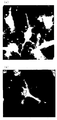
- Iba1 immunostaining image of cells after differentiation induction by the method of the present invention (A) and (b) both enlarged images of cells after differentiation induction by the method of the present invention
- the method for inducing differentiation of pluripotent stem cells into microglia is a method comprising the following steps.
- pluripotent stem cells are cultured under conditions suitable for promoting differentiation into blood precursor cells.
- “Conditions suitable for promoting differentiation into blood precursor cells” means co-culturing pluripotent stem cells and feeder cells.
- a pluripotent stem cell refers to a cell that maintains undifferentiated and pluripotent properties, typified by embryonic stem cells (ES cells) and induced pluripotent stem cells (iPS cells).
- the ES cell may be an ES cell produced by nuclear reprogramming from a somatic cell.
- Embryonic Germ Cell EG cell
- mGS cell multipotent germline stem cell isolated from testis
- multipotent adult progenitor cell isolated from bone marrow, etc.
- these pluripotent stem cells are derived from humans.
- the pluripotent stem cell is preferably an iPS cell, particularly preferably a human iPS (hiPS) cell.
- Pluripotent stem cells can be prepared by known methods. For example, it is specifically described below.
- Human iPS cell lines are available, for example, from the University of Tokyo Stem Cell Bank for TkDA3-4 and 201B7 from the Institute for iPS Cell Research at Kyoto University.
- Human ES cell lines, for example, WA01 (H1) and WA09 (H9) are available from the WiCell Research Institute, and KhES-1, KhES-2 and KhES-3 are available from the Institute of Regenerative Medicine, Kyoto University.
- ES cells or iPS cells are used as pluripotent stem cells, for example, as the medium, IMDM supplemented with FBS at a final concentration of 15% is used, and other growth factors and supplements are appropriately added even in the case of serum-free. Can be used.
- VEGF may be added, and when culturing in a hypoxic environment, the same effect as when VEGF is added can be obtained without adding VEGF.
- the concentration of VEGF is not particularly limited as long as blood precursor cells can be obtained, but may be 5 ng / ml to 50 ng / ml, and preferably 20 ng / ml or more.
- “Hypoxic condition” means that the oxygen concentration in the atmosphere when cells are cultured is significantly lower than that in the air. Specifically, generally the conditions of low oxygen concentration than the oxygen concentration in the atmosphere of 5 ⁇ 10% CO 2/95 ⁇ 90% air used are exemplified by conventional cell culture, for example, oxygen in the atmosphere The condition where the concentration is 18% or less is applicable. Preferably, the oxygen concentration in the atmosphere is 15% or less (eg, 14% or less, 13% or less, 12% or less, 11% or less, etc.), 10% or less (eg, 9% or less, 8% or less, 7% or less) 6% or less), or 5% or less (eg, 4% or less, 3% or less, 2% or less, etc.).
- the oxygen concentration in the atmosphere is preferably 0.1% or more (eg, 0.2% or more, 0.3% or more, 0.4% or more, etc.), 0.5% or more (eg, 0.6 % Or more, 0.7% or more, 0.8% or more, 0.95 or more, etc.), or 1% or more (eg, 1.1% or more, 1.2% or more, 1.3% or more, 1.4 % Or more).
- a method for creating a hypoxic state in the cell environment is not particularly limited, but a method of culturing the cells in a CO 2 incubator in which the oxygen concentration can be adjusted is the easiest and is a preferable example.
- CO 2 incubators with adjustable oxygen concentrations are available from various equipment manufacturers (for example, CO for low oxygen culture manufactured by Thermo scientific, Ikemoto Riken, Toji Field, Waken Pharmaceutical, etc.) 2 incubators can be used).
- any cell that contributes to differentiation induction of pluripotent stem cells can be used.
- mouse embryo fibroblasts preferably 10T1 / 2 cell line, OP9 cells, etc. are used. be able to.
- feeder cells it is preferable to suppress the proliferation of the cells, for example, by irradiating with radiation.
- the culture period is not particularly limited as long as the pluripotent stem cells have sufficient days to “differentiate into blood precursor cells”. For example, a period of 7 days or more, 10 days or more, 13 days or more can be mentioned, but it is not limited thereto. Those skilled in the art can appropriately adjust the culture period according to the conditions used.
- Other culture conditions include, for example, 5% CO 2 , 36 to 38 ° C., preferably 37 ° C., but are not limited thereto. Culturing under the above conditions can be performed using, for example, a known CO 2 incubator.
- “Blood progenitor cells” are CD34 negative cells, CD34 positive cells, Lin negative cells (CD2 negative cells, CD3 negative cells, CD4 negative cells, CD7 negative cells, CD8 negative cells, CD10 negative cells, CD14 negative cells, CD16 negative cells.
- Cell CD19 negative cell, CD20 negative cell, CD24 negative cell, CD41 negative cell, CD45 negative cell, CD56 negative cell, CD66b negative cell or CD235a negative cell), CD38 negative cell, CD90 positive cell, CD49f positive cell, VEGFR2 Hematopoietic system characterized by a single cell or a combination of a plurality of markers among positive cells, CD31 positive cells, CD43 positive cells, CD34 positive, CD45 positive cells, Rhodamine weak positive cells, or Hoechst negative / weak positive cells Cells A.
- step (b) the blood progenitor cells obtained in step (a) are cultured under conditions suitable for promoting differentiation into embryonic monocytes (embryonic hematopoiesis occurs).
- “Conditions suitable for promoting differentiation into embryonic monocytes” means co-culturing blood precursor cells and feeder cells. More specifically, for example, blood precursor cells and feeder cells are co-cultured in the presence of IL-3 and / or GM-CSF.
- the concentrations of IL-3 and GM-CSF are not particularly limited as long as embryonic monocytes are obtained.
- IL-3 is 1 ng / ml to 200 ng / ml, 20 ng / ml to 150 ng / ml, 25 ng / ml to 100 ng / ml.
- GM-CSF is 1 ng / ml to 200 ng / ml, 20 ng / ml to 150 ng / ml, 25 ng / ml to 100 ng / ml.
- IL-3 is particularly preferably 25 ng / ml or more
- GM-CSF is particularly preferably 50 ng / ml or more.
- the culture period is not particularly limited as long as it is a sufficient number of days to “differentiate into monocytes” and is capable of embryonic hematopoiesis. Those skilled in the art can appropriately adjust the culture period according to the conditions used.
- “Monocytes” means CD11b positive cells, CD14 positive cells, CD15 positive cells, CD4 positive cells, CD163 positive cells, CD9 positive cells, CD11c positive cells, CDw12 positive cells, CD13 positive, CD17 positive cells, CD31 positive cells, CD32 positive cells, CD33 positive cells, CD35 positive cells, CD36 positive cells, CD38 positive cells, CD40 positive cells, CD43 positive cells, CD45RO positive cells, CD45RA positive cells, CD45RB positive cells, CD49b positive cells, CD49e positive cells, CD49f positive Cells, CD63 positive cells, CD64 positive cells, CD65s positive cells, CD68 positive cells, CD74 positive cells, CD84 positive cells, CD85 positive cells, CD86 positive cells, CD87 positive cells, CD89 positive cells, CD91 positive cells, D92 positive cells, CD93 positive cells, CD98 positive cells, CD101 positive cells, CD102 positive cells, CD111 positive cells, CD112 positive cells, CD115 positive cells, CD116 positive cells, CD119 positive cells, CD121b positive cells, CD123 positive cells,
- Embryonic hematopoiesis is essentially hematopoiesis that occurs transiently in the yolk sac in the early embryonic period. In the present specification, it can be confirmed, for example, by the method described in Step 2 of Example 1 (5) that the hematopoietic stage is “embryo type”.
- step (c) the embryonic monocytes obtained in step (b) are cultured under conditions suitable for promoting differentiation into microglia.
- “Conditions suitable for promoting differentiation into microglia” means co-culturing embryonic monocytes and astrocytes, or culturing embryonic monocytes using an astrocyte supernatant. . More specifically, for example, embryonic monocytes and astrocytes are co-cultured or cultured using an astrocyte supernatant in the presence of M-CSF.
- IL-34 may be present.
- the concentration of IL-34 and / or M-CSF is not particularly limited as long as microglia can be obtained.
- IL-34 is 1 ng / ml to 200 ng / ml, 20 ng / ml to 150 ng / ml, 25 ng / ml to 100 ng / ml.
- M-CSF is 1 ng / ml to 200 ng / ml, 20 ng / ml to 150 ng / ml, 25 ng / ml to 100 ng / ml.
- M-CSF has a great influence on the production of efficient microglia and is particularly preferably 25 ng / ml or more.
- “Astrocyte supernatant” is obtained by culturing astrocytes collected from nerve tissue in a nutrient medium. For example, it can be prepared with reference to the method described in International Publication No. 2006/028049.
- the culture period is not particularly limited as long as it is a sufficient number of days for “differentiation into microglia”. For example, a period of 3 days or more, 5 days or more, 7 days or more, 8 days or more can be mentioned, but it is not limited thereto. Particularly preferably, it is 7 days or more. Those skilled in the art can appropriately adjust the culture period according to the conditions used.
- microglia means Iba1 positive cells, CD11b positive cells, P2Y12 positive cells, P2X7 positive cells, P2X4 positive cells, IL-1 ⁇ positive cells, CX3CR1 positive cells, CCR2 positive cells, CCR7 positive cells, CD80 positive cells, CD209 positive cells, CD23 positive cells, CD163 positive cells, TREM2 positive cells, CD45 positive cells, P2X2 positive cells, CCL21 positive cells, IRF8 positive cells, IRF5 positive cells, TLR-4 positive cells, OX42 positive cells, CD14 positive cells, Among CD16-positive cells, integrin- ⁇ 4-positive cells, integrin- ⁇ 1-positive cells, and CD68-positive cells, the cells are characterized by one or a combination of a plurality of markers.
- Iba1 positive cells, CD11b positive cells and the like are expressed not only in microglia but also in macrophages.
- the microglia is not a macrophage.
- the medium may be prepared as a basic medium.
- the basic medium include IMDM medium, 199 medium, Eagle minimum essential medium (EMEM), alpha-MEM medium, Dulbecco's modified Eagle medium (DMEM), Ham F12 medium, RPMI 1640 medium, Fisher medium, Glasgow MEM, and the like Of the mixture.
- the basic medium may contain serum or cytokines.
- the surface of the culture dish is coated with collagen I, collagen IV, gelatin, poly-L-lysine, poly-D-lysine, laminin, fibronectin, Matrigel TM ( It may be coated with a cell support substrate such as Becton, Dickinson and Company.
- cytokines such as VEGF, IL-3, IL-34, M-CSF, and GM-CSF used in the present invention
- natural cytokines or recombinant cytokines prepared by genetic engineering may be used.
- a partial protein or peptide including a region related to binding to the receptor may be used.
- it may be a protein or peptide in which the amino acid sequence or the three-dimensional structure is modified to such an extent that the binding force with the receptor is not lost.
- proteins, peptides, drugs and the like that can function as agonists for these cytokine receptors may be used.
- Screening method (screening method for therapeutic drugs for diseases involving microglia activation)
- the present invention provides a screening method for a substance that acts on microglia by bringing the microglia obtained by I above into contact with a test substance.
- test substance may be any known compound or novel compound, such as a nucleic acid, carbohydrate, lipid, protein, peptide, low molecular weight organic compound, compound library prepared using combinatorial chemistry techniques, Examples include random peptide libraries prepared by phase synthesis or phage display methods, or natural components derived from microorganisms, animals and plants, marine organisms, and the like.
- the microglia obtained by the above I is brought into contact with the test substance, and the degree of influence on the microglia (for example, morphological change, cytokine production / release, phagocytosis, etc.) is measured. Then, compare the degree with the degree of microglia that does not come into contact with the test substance, and select the test substance that significantly changes the degree of influence as compared with the case without contact with the test substance as an effective component. To do.
- the degree of influence on the microglia for example, morphological change, cytokine production / release, phagocytosis, etc.
- a method known in this field can be used. For example, morphological changes can be captured by imaging and processing microglia, the concentration of cytokines released into the culture supernatant can be measured by ELISA, and the state of phagocytosing beads can be digitized by image processing. .
- Test substances screened in this way include spinal cord trauma, neuropathy due to stroke, epilepsy, neuropathic pain, vaso-occlusive eye disease, demyelinating diseases (multiple sclerosis, Guillain-Barre syndrome, etc.), Mental illness (depression, schizophrenia, autism, developmental disorder, addiction, etc.), cerebral infarction, Nasu hakora disease, neurodegenerative disease (especially Alzheimer's disease, Parkinson's disease, amyotrophic lateral sclerosis, etc.) It can be used as an agent for preventing or treating or improving memory or learning ability.
- customized therapeutic agent means an optimal therapeutic agent that suits the individuality of an individual patient.
- induced differentiation of induced pluripotent stem cells produced from somatic cells of a subject suffering from spinal cord trauma, neuropathy due to stroke, epilepsy, neuropathic pain, vaso-occlusive eye disease or neurodegenerative disease The present invention provides a method for screening a therapeutic agent that acts on microglia by bringing the microglia thus obtained into contact with an existing therapeutic agent.
- the screened therapeutic agent can be an optimal therapeutic agent for a subject for which induced pluripotent stem cells have been established.
- Examples of existing therapeutic agents in the present invention include spinal cord trauma therapeutic agents such as baclofen and tizanidine, phenytoin, phenobarbital, carbamazepine, valproic acid, ethosuximide, zonisamide, gabapentin, topiramate, lamotrigine, levetiracetam and other epilepsy drugs, pregabalin, etc. And other therapeutic agents for Alzheimer's disease such as donepezil, rivastigmine, galantamine, memantine and the like.
- Example 1 Production of microglia (1) Feeder cell (Mouse Embryonic Fibroblast; MEF) Mouse (strain; ICR) fetuses (E13.5) were dissected under a stereomicroscope, and after blood removal, the head, limbs and internal organs were removed with tweezers. The remaining part was finely chopped with scissors, and 10 ml of digestive fluid (0.05% trypsin-ethylenediaminetetraacetic acid (EDTA) (Invitrogen) +1/1000 recombinant DNase (Takara)) was added.
- EDTA trypsin-ethylenediaminetetraacetic acid
- the pellets were resuspended in medium A and counted, and then 4 ⁇ 10 6 in a flask (150 cm 2 ; TPP) coated with 0.1% gelatin (Sigma) / phosphate buffered saline (PBS) (Wako) for 30 minutes. Seeds per piece / flask. After culturing for 3 days, the cells were washed with PBS and 0.05% trypsin-EDTA was added to detach the cells. An equal amount of medium A was added to stop and collect the enzyme reaction, and the centrifuged pellet was suspended in Cell Banker 1 (Nippon Zenyaku Kogyo Co., Ltd.) and stored frozen.
- PBS phosphate buffered saline
- cryopreserved cells were awakened and seeded at 2 ⁇ 10 6 cells / flask in a gelatin-coated flask under the same conditions as described above. After culturing for 3 days, the cells were detached by the same method as described above, and seeded at 2 ⁇ 10 6 cells / flask in a newly gelatin-coated flask under the same conditions as described above. After culturing for 3 days, the medium in the flask was replaced with medium A supplemented with 10 ⁇ g / ml mitomycin C (MMC; Sigma) to stop growth, and after 90 minutes of incubation, washed with PBS, Cells were detached by the same method. The obtained cells were stored frozen by the same method as described above. Cryopreserved MMC-treated MEFs were awakened the day before use and seeded at 5 ⁇ 10 5 cells / dish in gelatin-coated 10 cm cell culture dishes.
- MMC mitomycin C
- Stromal cells (10T1 / 2) It was obtained from RIKEN BioResource Center.
- the medium is BME (Invitrogen) + 10% fetal calf serum (FCS) (Hyclone) + 1 / 100GlutaMAX1 (Invitrogen) +1/100 penicillin / streptomycin (Invitrogen)), and the cell detachment solution is 0.05% trypsin-EDTA (Invitrogen). Invitrogen) was used.
- the passage was maintained in a 150 cm 2 flask, the passage was performed twice a week, and the cell density was about 1/8 times (1 flask of cells was passaged to about 8 flasks) each time (5 ⁇ 10 5 to 5 ⁇ 10 6 cells) (Maintained within the range of the flask) .
- Those up to passage number p30 were used after growth inhibition by MMC.10T1 / 2 treated with MMC was seeded at 1 ⁇ 10 6 cells / dish on a gelatin-coated 10 cm cell culture dish. did.
- the obtained cells were resuspended in medium B, and after counting, seeded in poly-D-lysine (PDL) coated flasks (75 cm 2 ; BD) at 5 ⁇ 10 5 cells / flask.
- the medium B was exchanged 2 days and 5 days after sowing (after shaking for 3 hours with a shaker) and cultured for 7 days.
- the cells were shaken with a shaker for 3 hours before cell collection, and after the medium B was replaced with PBS, the cells were further shaken for 5 hours.
- 0.25% trypsin-EDTA Nacalai Tesque
- the enzyme reaction was stopped with an equal volume of medium B and recovered.
- the obtained cells were centrifuged (300G for 3 minutes), and the pellet was stored frozen in a cell banker.
- Cryopreserved astrocytes are generated and seeded at 2.5 ⁇ 10 5 cells / dish in a gelatin-coated 10 cm cell culture dish, medium B is replaced after 4 days, and medium C for coculture (DMEM (Invitrogen) on day 7) + 10% FCS (Hyclone) + 1 / 100GlutaMAX1 (Invitrogen) +1/100 Sodium pyruvate (Invitrogen) +1/100 HEPES (Invitrogen) +1/100 penicillin-streptomycin (Invitrogen)) Used after.
- the peeled MEF was sucked with an aspirator and further washed with PBS, and then differentiation medium D (IMDM (Invitrogen) + 15% fetal calf serum (FCS) (EquitechLab) +1/100 GlutaMAX1) (Invitrogen) +1/100 ITS- X (Invitrogen) +0.5 mmol / l monothioglycerol (Wako) +50 ⁇ g / ml L-ascorbic acid phosphate magnesium salt n hydrate (Wako) +1/100 penicillin-streptomycin (Invitrogen)) Then, the hiPS cells were scraped with a cell scraper.
- IMDM Invitrogen
- FCS fetal calf serum
- ITS- X Invitrogen
- Wako monothioglycerol
- Wako monothioglycerol
- Wako monothioglycerol
- Wako monothioglyce
- the obtained hiPS cells were passaged by the method described in (3) at a density of 1/40 on 10T1 / 2 (1 dish of cells was seeded in about 40 dishes) at a density of 20 ng / ml VEGF (PEPROTECH; non According to Patent Document 4: Blood 2008 111: 5298-5306, it may not be added) and seeded.
- Embryonic monocytes obtained in step 2 were coculture medium F (DMEM (Invitrogen) + 10% FCS (EquitechLab) +1/100 GlutaMAX1 (Invitrogen) +1/100 sodium pyruvate (Invitrogen) +1/100 HEPES (Invitrogen) +1/100 Penicillin-Streptomycin (Invitrogen)), 50 ng / ml M-CSF (PEPROTECH) and 50 ng / ml IL-34 (PEPROTECH) are added, and (4) described Seeded at a density of 1/2 on primary cultured rat astrocytes.
- DMEM Invitrogen
- FCS EquitechLab
- Iba1 immunostaining Cells collected by the method described in (5) were seeded on a PDL / laminin-coated plate (Biocoat; Corning), and the next day, adherent cells were treated with 4% paraformaldehyde (PFA) / PBS. Fix for 1 hour at ° C. The fixed cells were washed 3 times with PBS, treated with 0.1% Triton X-100 / PBS for 5 minutes, washed 3 times with PBS, and then treated with blocking solution (1.5% donkey serum / PBS) for 30 minutes. Next, 1/50 anti-Iba1 antibody (Abcam) / blocking solution was reacted at 4 ° C.
- PFA paraformaldehyde
- Example 2 Production of microglia using feeder-free iPS cells
- TkDA3-4 strain purchased from Stem Cell Bank, University of Tokyo was used.
- the coating is iMatrix-511 (Nippi)
- the stripping solution is 0.5x Triple select (Triple TM select CTS (Invitrogen), 0.5 mol / l EDTA pH 8.0 (Nacalai Tesque), PBS (Wako)).
- / Ml VEGF (PEPROTECH; it may not be added according to Non-Patent Document 4) was added and seeded. After seeding 3, 6, 8, 10 and 12 days, the differentiation medium E was replaced with 20 ng / ml VEGF (PEPROTECH, which may not be added according to Non-Patent Document 4), and on day 13 It was collected. While removing Sac with a cell scraper, it was peeled off from the cell culture dish and collected through a cell strainer (40 ⁇ ). The centrifuged pellet (120 G (confirmed that there was no effect on the rotation rate and purity at 100 to 150 G) for 10 minutes without brake) was resuspended in differentiation medium E.
- Process 2 IL-3 (50 ng / ml; PEPROTECH) and GM-CSF (50 ng / ml; PEPROTECH) are added to all the cells obtained in step 1 and one resuspension for 5 cell culture dishes is added. Seeded on 10T1 / 2 in a cell culture dish. Three days later, 5 ml of differentiation medium E containing IL-3 and GM-CSF was added, and after 6 days, it was collected by pipetting. Process 3 The embryonic monocytes obtained in step 2 were resuspended in co-culture medium F, and 50 ng / ml M-CSF (PEPROTECH) and 50 ng / ml IL-34 (PEPROTECH) were added.
- Iba1 immunostaining Cells collected in step 3 were seeded on a PDL / laminin-coated plate (Biocoat; Corning), and the next day, adherent cells were fixed with 4% paraformaldehyde (PFA) / PBS for 1 hour at 4 ° C. did.
- the fixed cells were washed 3 times with PBS, treated with 0.1% Triton X-100 / PBS for 5 minutes, washed 3 times with PBS, and then treated with blocking solution (1.5% donkey serum / PBS) for 30 minutes.
- blocking solution (1.5% donkey serum / PBS) for 30 minutes.
- 1/50 anti-Iba1 antibody (Abcam) / blocking solution was reacted at 4 ° C.
- Example 3 Examination of differentiation induction period in blood progenitor cell preparation stage (1) Differentiation induction step 1 The 10 cm cell culture dish to which the hiPS cells passaged by the method described in Example 1 (2) were adhered was washed with PBS, 1.5 ml of Dissolution Solution (ReproCELL) was added, and MEF was peeled off while shaking. The peeled MEF was sucked up with an aspirator, further washed with PBS, 10 ml of differentiation medium D was added, and hiPS cells were scraped off with a cell scraper.
- Dissolution Solution ReproCELL
- the obtained hiPS cells were subcultured by the method described in Example 1 (3) on 10T1 / 2 at a density of 1/40 (1 dish of cells was seeded in about 40 dishes) at a density of 20 ng / ml VEGF ( According to Non-Patent Document 4, it was not necessary to add PEPROTECH) and seeded. After the seeding 3, 6, 8, 10, 12 days, the culture medium was replaced with differentiation medium E supplemented with 20 ng / ml VEGF (PEPROTECH, which may not be added according to Non-Patent Document 4), and cultured for any number of days. After recovery. While removing Sac with a cell scraper, it was peeled off from the cell culture dish and collected through a cell strainer (40 ⁇ ).
- Step 2 to Step 3 The same method as in Step 2 to Step 3 of Example 2 was performed.
- Example 2 Iba1 immunostaining This was carried out in the same manner as in Example 2 (3).
- the blood progenitor cell production period was set to 6, 13, 20, and 27 days, differentiation into microglia was carried out. Microglia production was possible under all conditions, but microglial differentiation induction efficiency was particularly high in 13 to 27 days. Was good.
- Example 4 Examination of differentiation induction period in embryonic monocyte production stage (1) Differentiation induction step 1 The 10 cm cell culture dish to which the hiPS cells passaged by the method described in Example 1 (2) were adhered was washed with PBS, 1.5 ml of Dissolution Solution (ReproCELL) was added, and MEF was peeled off while shaking. The peeled MEF was sucked up with an aspirator, further washed with PBS, 10 ml of differentiation medium D was added, and hiPS cells were scraped off with a cell scraper.
- Dissolution Solution ReproCELL
- the obtained hiPS cells were subcultured by the method described in Example 1 (3) on 10T1 / 2 at a density of 1/40 (1 dish of cells was seeded in about 40 dishes) at a density of 20 ng / ml VEGF ( According to Non-patent Document 4, it was not necessary to add PEPROTECH) and seeded.
- the differentiation medium E was replaced with 20 ng / ml VEGF (PEPROTECH, which may not be added according to Non-Patent Document 4), and on day 13 It was collected. While removing Sac with a cell scraper, it was peeled off from the cell culture dish and collected through a cell strainer (40 ⁇ ). Centrifuged (120G (confirmed that there was no effect on recovery and purity at 100-150G) 10 minutes without brake) pellets were resuspended in differentiation medium E.
- IL-3 50 ng / ml; PEPROTECH
- GM-CSF 50 ng / ml; PEPROTECH
- Process 2 IL-3 (50 ng / ml; PEPROTECH) and GM-CSF (50 ng / ml; PEPROTECH) are added to all the cells obtained in step 1 and one resuspension for 5 cell culture dishes is added. Seeded on 10T1 / 2 in a cell culture dish. Three days later, 5 ml of differentiation medium E containing IL-3 and GM-CSF was added, and collected by pipetting after culturing for an arbitrary period. The collected cell suspension was centrifuged (300 G for 3 minutes), and the pellet cells were subjected to the next step.
- Monocytes obtained in step 2 were resuspended in co-culture medium F, and M-CSF (50 ng / ml; PEPROTECH) and IL-34 (50 ng / ml; PEPROTECH) were added. Inoculated at a density of 1/2 on the primary cultured rat astrocytes described in (4). Two days after sowing, 5 ml of medium supplemented with M-CSF and IL-34 was added to medium F. On days 4 and 7, 7 ml of medium was collected and centrifuged (300 G for 3 minutes).
- the pellet was resuspended in 8 ml of medium supplemented with TGF- ⁇ 1 (50 ng / ml; PEPROTECH; confirmed to have no effect on differentiation rate) in medium supplemented with CSF and IL-34 . Cells were collected by pipetting 9 days after seeding.
- Example 2 Iba1 immunostaining This was carried out in the same manner as in Example 2 (3).
- step 2 was skipped for convenience and expressed as 0 days for convenience
- microglia could be efficiently produced in all induction days. It was.
- Example 5 Examination of cytokine concentration (1) Differentiation induction step 1 Blood progenitor cells were prepared in the same manner as in Step 1 of Example 4. Process 2 IL-3 (PEPROTECH) and GM-CSF (PEPROTECH) are added to all cells obtained in step 1 at an arbitrary concentration, and a resuspension for 5 cell culture dishes is added to one cell culture. Seeded on 10T1 / 2 in dish. Three days later, 5 ml of differentiation medium E containing IL-3 and GM-CSF at the same concentrations as those at the time of cell seeding was added, and the cells were collected by pipetting after 6 days of culture. The collected cell suspension was centrifuged (300 G for 3 minutes), and the pellet cells were subjected to the next step.
- Process 2 IL-3 (PEPROTECH) and GM-CSF (PEPROTECH) are added to all cells obtained in step 1 at an arbitrary concentration, and a resuspension for 5 cell culture dishes is added to one cell culture. Seeded on 10T
- Process 3 The monocytes obtained in step 2 were resuspended in co-culture medium F, and M-CSF (PEPROTECH) and IL-34 (PEPROTECH) were added at arbitrary concentrations, respectively. ) The seeded primary cultured rat astrocytes were seeded at a density of 1/2. Two days after seeding, 5 ml of medium with M-CSF and IL-34 added to medium F at the same concentration as at the time of cell seeding was added, and 7 ml of medium was collected on days 4 and 7 and centrifuged (300 G for 3 minutes).
- M-CSF PEPROTECH
- IL-34 PEPROTECH
- the pellet was resuspended with 8 ml of medium supplemented with TGF- ⁇ 1 (PEPROTECH) in medium F with M-CSF and IL-34 added at the same concentration as at the time of cell seeding. Cells were collected by pipetting 9 days after seeding.
- Example 2 Iba1 immunostaining This was carried out in the same manner as in Example 2 (3).
- the results are summarized in Table 1.
- the equivalent differentiation induction efficiency was taken as ⁇ , and the differentiation induction efficiency below 1/10 was taken as ⁇ .
- Experiments were conducted at respective cytokine concentrations of 0, 25, 50, and 100 ng / ml, and microglia could be produced under all conditions.
- step 2 differentiation induction efficiency was particularly good when IL-3 was added at 25 ng / ml or more, or GM-CSF was added at 50 ng / ml or more.
- the efficiency was particularly good when M-CSF was added at 25 ng / ml or more.
- microglia can be efficiently produced from pluripotent stem cells.
- the obtained microglia can be used for various basic researches such as research on microglia itself and research on diseases related to microglia.
- a method for screening a therapeutic agent for a disease involving microglia, characterized by using microglia, or a so-called custom-made therapeutic agent that selects optimal therapeutic agents for individuals using microglia established from individual patients It is extremely useful in the selection of Furthermore, utilization for cell therapy is also expected.
Landscapes
- Health & Medical Sciences (AREA)
- Engineering & Computer Science (AREA)
- Biomedical Technology (AREA)
- Life Sciences & Earth Sciences (AREA)
- Bioinformatics & Cheminformatics (AREA)
- Chemical & Material Sciences (AREA)
- Neurology (AREA)
- Organic Chemistry (AREA)
- Neurosurgery (AREA)
- General Health & Medical Sciences (AREA)
- Immunology (AREA)
- Biotechnology (AREA)
- Cell Biology (AREA)
- Medicinal Chemistry (AREA)
- Zoology (AREA)
- Genetics & Genomics (AREA)
- Wood Science & Technology (AREA)
- Pharmacology & Pharmacy (AREA)
- Animal Behavior & Ethology (AREA)
- Public Health (AREA)
- Veterinary Medicine (AREA)
- Chemical Kinetics & Catalysis (AREA)
- Hematology (AREA)
- General Chemical & Material Sciences (AREA)
- Nuclear Medicine, Radiotherapy & Molecular Imaging (AREA)
- Biochemistry (AREA)
- Microbiology (AREA)
- Urology & Nephrology (AREA)
- Molecular Biology (AREA)
- General Engineering & Computer Science (AREA)
- Tropical Medicine & Parasitology (AREA)
- Food Science & Technology (AREA)
- Pathology (AREA)
- General Physics & Mathematics (AREA)
- Analytical Chemistry (AREA)
- Toxicology (AREA)
- Physics & Mathematics (AREA)
- Physical Education & Sports Medicine (AREA)
- Orthopedic Medicine & Surgery (AREA)
- Pain & Pain Management (AREA)
Abstract
Description
したがって、これらのミクログリアに関連する疾患の治療薬の開発と評価のためにミクログリアを用いることが必要である。そこで、ミクログリアを調製するために様々な方法が試みられている。
特許文献1には、ヒトiPS細胞からミクログリア前駆細胞を得る方法が記載されているが、実施例1に記載の通り、分化して得られるCD45に対して免疫反応性を持つ細胞(ミクログリア前駆細胞)はわずか2~10%である。
非特許文献3にも記載されているように、数多くの研究室においてiPS細胞からミクログリアへの分化が試みられているにも関わらず、現時点において、ヒトiPS細胞からミクログリアを得る効率的な方法は知られていない。
(1)以下の工程を含む、多能性幹細胞からミクログリアを製造する方法:
(a)多能性幹細胞を7日間以上フィーダー細胞と共培養し、血液前駆細胞を得る工程、
(b)工程(a)で得られた血液前駆細胞をIL-3及び/又はGM-CSFの存在下でフィーダー細胞と共培養し、胚型単球を得る工程、及び
(c)工程(b)で得られた胚型単球をM-CSFの存在下でアストロサイトと共培養又はアストロサイト上清を用いて培養する工程。
(2)工程(a)の培養期間が、13日以上である、(1)記載の方法。
(3)工程(c)において、IL-34の存在下で培養することを特徴とする(1)又は(2)記載の方法。
(4)工程(b)において、IL-3及びGM-CSFの存在下で培養することを特徴とする、(1)~(3)いずれかに記載の方法。
(5)フィーダー細胞が、10T1/2細胞又はOP9細胞である、(1)~(4)いずれかに記載の方法。
(6)工程(a)において、VEGFの存在下で培養することを特徴とする、(1)~(5)いずれかに記載の方法。
(7)多能性幹細胞が、iPS細胞である、(1)~(6)いずれかに記載の方法。
(8)iPS細胞が、ヒト又はマウス由来である、(7)記載の方法。
(9)(1)~(8)の方法で製造されたミクログリアを用いることを特徴とする、ミクログリアが関与する疾患の予防若しくは治療剤又は記憶若しくは学習能力改善剤のスクリーニング方法。
(10)ミクログリアが関与する疾患が、脊髄の外傷、脳卒中による神経障害、てんかん、神経障害性疼痛、血管閉塞性の眼疾患、脱髄疾患、精神疾患、脳梗塞、那須ハコラ病又は神経変性疾患である、(9)記載のスクリーニング方法。
(11)神経変性疾患が、アルツハイマー病、パーキンソン病又は筋萎縮性側索硬化症である、(10)記載のスクリーニング方法。
本発明の多能性幹細胞のミクログリアへの分化誘導方法は、以下の工程を含む方法である。
(a)多能性幹細胞を7日間以上フィーダー細胞と共培養し、血液前駆細胞を得る工程、
(b)工程(a)で得られた血液前駆細胞をIL-3及び/又はGM-CSFの存在下でフィーダー細胞と共培養し、胚型単球を得る工程、及び
(c)工程(b)で得られた胚型単球をM-CSFの存在下でアストロサイトと共培養又はアストロサイト上清を用いて培養する工程。
また、ヒトiPS細胞株は、例えば、TkDA3-4は東京大学ステムセルバンクから、201B7は京都大学iPS細胞研究所から入手可能である。
ヒトES細胞株は、例えばWA01(H1)及びWA09(H9)は、WiCell Reserch Instituteから、KhES-1、KhES-2及びKhES-3は、京都大学再生医科学研究所から入手可能である。
上記マーカーのうち、例えば、Iba1陽性細胞、CD11b陽性細胞等は、ミクログリアだけでなく、マクロファージでも発現している。例えば、以下のいずれかの特徴と組み合わせることによって、マクロファージではなく、ミクログリアであると、判断することが可能である。
- ミクログリアに特徴的な形態(突起)
- プリン作動性受容体であるP2Y12の発現
(ミクログリアの活性化が関与する疾患の治療薬のスクリーニング方法)
本発明は、上記Iにより得られたミクログリアと試験物質とを接触させ、ミクログリアに対して作用する物質のスクリーニング方法を提供する。
本発明において、「オーダーメード治療薬」とは、ある患者個人の個性にかなった最適な治療薬を意味する。
本発明では、脊髄の外傷、脳卒中による神経障害、てんかん、神経障害性疼痛、血管閉塞性の眼疾患又は神経変性疾患を罹患する対象の体細胞から製造された人工多能性幹細胞を分化誘導させることにより得られた前記ミクログリアと既存の治療薬を接触させ、ミクログリアに対して作用する治療薬のスクリーニング方法を提供する。このように、スクリーニングされた治療薬は、人工多能性幹細胞を樹立された対象にとって最適な治療薬と成りうる。
(1)フィーダー細胞(Mouse Embryonic Fibroblast; MEF)
マウス(系統;ICR)の胎仔(E13.5)を実体顕微鏡下で解剖し、脱血後ピンセットで頭部、手足、内臓を取り除いた。残った部位をハサミで細かく刻み、消化液(0.05%トリプシン-エチレンジアミン四酢酸(EDTA)(Invitrogen社)+1/1000 recombinant DNase(Takara社))を胎仔1体あたり10ml加えた。室温で約1時間、スターラーで攪拌しながら分散し、等量の培地A(D-MEM(Sigma社)+10%ウシ胎児血清(FBS)(Equitech-Bio社)+1/100ペニシリン-ストレプトマイシン(Invitrogen社))を加えて酵素反応を停止させた。細胞分散液をセルストレーナー(70φ;BD Falcon社)に通して回収し、遠心分離(300G, 3分)後上清を除いた。ペレットを培地Aに再懸濁して計数後、30分間0.1%ゼラチン(Sigma社)/リン酸緩衝生理食塩水(PBS)(wako社)コートしたフラスコ(150cm2; TPP社)に4x10⁶個/フラスコで播種した。3日間の培養後、PBSで洗浄後0.05%トリプシン-EDTAを加えて細胞を剥離させた。等量の培地Aを加えて酵素反応を停止して回収し、遠心分離後のペレットをセルバンカー1(日本全薬工業株式会社)に懸濁して凍結保存した。次に凍結保存した細胞を起こし、上記と同様の条件でゼラチンコートしたフラスコに2x106個/フラスコで播種した。3日間培養後、上記と同様の手法にて細胞を剥離させ、新しく上記同様の条件でゼラチンコートしたフラスコに2x106個/フラスコで播種した。3日間培養後、フラスコ内の培地を培地Aに10μg/mlのマイトマイシンC(MMC;Sigma社)を添加したものに交換して増殖を停止させ、90分培養後にPBSで洗浄した後に、上記と同様の手法にて細胞を剥離させた。得られた細胞は上記と同様の手法にて凍結保存した。凍結保存したMMC処理済みMEFは使用する前日に起こし、ゼラチンコートした10cm細胞培養ディッシュに5x105個/ディッシュで播種した。
東京大学ステムセルバンクより購入したTkDA3-4株と京都大学iPS細胞研究所より購入した201B7株を使用した。
フィーダー細胞は(1)に記載の方法で作製したMEFを用い、培地はDMEM(Invitrogen社)+20%KnockOut Serum Replacement(KSR)(Gibco社)+1/100ペニシリン/ストレプトマイシン(Invitrogen社)とし、細胞剥離液はDissociation Solution(ReproCELL社)を使用した。
独立行政法人理化学研究所バイオリソースセンターから入手した。
培地はBME(Invitrogen社)+10%ウシ胎児血清(FCS)(Hyclone社)+1/100GlutaMAX1(Invitrogen社)+1/100ペニシリン/ストレプトマイシン(Invitrogen社))、細胞剥離液は0.05%トリプシン-EDTA(Invitrogen社)を使用した。(150cm2フラスコで継代維持し、週2回の頻度で継代し毎回約1/8倍(1フラスコの細胞を約8フラスコに継代)の細胞密度にした(5x105~5x106個/フラスコの範囲内で維持)。継代数p30までの物をMMCによる増殖阻害をした上で使用した。MMC処理済み10T1/2はゼラチンコートした10cm細胞培養ディッシュ上に1x106個/ディッシュで播種した。
新生仔(P5-7)から大脳を分離して、実体顕微鏡下でピンセットを用いて髄膜を剥がした。培地B(AdDMEM/F12(Invitrogen社)+10%ウシ胎児血清(FCS)(Hyclone社)+1/100GlutaMAX1(Invitrogen社)+1/100ペニシリン/ストレプトマイシン(Invitrogen社))を添加してピペッティングすることにより、機械的に脳を破壊して分散させた。静置した際の上清を回収し、セルストレーナー(100Φ; BD Falcon社)に通して遠心分離(200G 4分)し、ペレットを回収した。得られた細胞を培地Bに再懸濁し、計数後ポリ-D-リシン(PDL)コートフラスコ(75cm2; BD社)に5x10⁵個/フラスコで播種した。播種2日及び5日後に培地Bを交換(シェイカーで3時間振とう後)し、7日間培養したものを使用した。細胞回収前にシェイカーで3時間振とうし、培地BをPBSに置換した後にさらに5時間振とうした。PBSを除去後に0.25%トリプシン-EDTA(ナカライテスク株式会社)を添加し、シェイカーで3分振とう後に等量の培地Bで酵素反応を停止後回収した。得られた細胞は遠心分離(300G 3分)し、ペレットをセルバンカーで凍結保存した。凍結保存したアストロサイトを起こし、ゼラチンコートした10cm細胞培養ディッシュに2.5x10⁵個/ディッシュで播種し、4日後に培地Bを交換、7日目に共培養用培地C(DMEM(Invitrogen社)+10%FCS(Hyclone社)+1/100GlutaMAX1(Invitrogen社)+1/100ピルビン酸ナトリウム(Invitrogen社)+1/100 HEPES(Invitrogen社)+1/100ペニシリン-ストレプトマイシン(Invitrogen社))に交換後3時間培養してから使用した。
工程1
(2)記載の方法で継代したhiPS細胞が接着した10cm細胞培養ディッシュをPBSで洗浄し、Dissociation Solution(ReproCELL社)を1.5ml入れ、ゆすりながらMEFを剥がした。剥がれたMEFをアスピレーターで吸い取って、さらにPBSで洗浄後、分化培地D(IMDM(Invitrogen社)+15% ウシ胎児血清(FCS)(EquitechLab社)+1/100 GlutaMAX1)(Invitrogen社)+1/100 ITS-X(Invitrogen社)+0.5mmol/l モノチオグリセロール(wako社)+50μg/ml L-アスコルビン酸リン酸エステルマグネシウム塩n水和物(wako社)+1/100 ペニシリン-ストレプトマイシン(Invitrogen社))を10ml添加し、セルスクレーパーでhiPS細胞をかきとった。得られたhiPS細胞を(3)に記載の方法で継代した10T1/2上に1/40(1ディッシュの細胞を約40ディッシュに播種)の密度で、20ng/ml VEGF(PEPROTECH社;非特許文献4:Blood 2008 111:5298-5306によれば添加しなくても良い)を加えて播種した。播種3、6、8、10、12日後に分化培地E(IMDM(Invitrogen社)+15% ウシ胎児血清(FCS)(EquitechLab社)+1/100 GlutaMAX1)(Invitrogen社)+1/100 ITS-G(Invitrogen社)+0.5mmol/l モノチオグリセロール(wako社)+50μg/ml L-アスコルビン酸リン酸エステルマグネシウム塩n水和物(wako社)+1/100 ペニシリン-ストレプトマイシン(Invitrogen社))に20ng/ml VEGF(PEPROTECH社;非特許文献4によれば添加しなくても良い)を加えたものに交換し、13日目に回収した。セルスクレーパーでSacを破壊しながら細胞培養ディッシュから剥がして、セルストレーナー(40Φ)を通して回収した。遠心分離(120G(100~150Gで回収率と純度に影響がないことを確認済)10分ブレーキ無し)したペレットを分化培地Eに再懸濁した。得られた細胞は約50%がCD34陽性であることから、血液前駆細胞を作製できたと判断した。
工程2
工程1で得られた血液前駆細胞を、50ng/ml エリスロポエチン(PEPROTECH社)を添加して10T1/2上で培養して得られた赤血球のヘモグロビンの型が6日目まで全て胎児型であることから、血液前駆細胞からの分化誘導6日目までは全てが胚型造血であると判断した。
工程1で得られた全細胞に50ng/ml IL-3(PEPROTECH社)と50ng/ml GM-CSF(PEPROTECH社)を添加して細胞培養ディッシュ5枚分の再懸濁液を1枚の細胞培養ディッシュ内の10T1/2上に播種した。3日後に50ng/ml IL-3と50ng/ml GM-CSFを含む分化培地Eを5ml追加し、6日後にピペッティングによって回収した。回収した細胞懸濁液を遠心分離(300G 3分)し、ペレットの細胞が全て(100%)CD14陽性であることから、6日目までは胚型造血であることと合わせて判断し、これらを胚型単球とした。
工程3
工程2で得られた胚型単球は共培養用培地F(DMEM(Invitrogen社)+10%FCS(EquitechLab社)+1/100GlutaMAX1(Invitrogen社)+1/100ピルビン酸ナトリウム(Invitrogen社)+1/100 HEPES(Invitrogen社)+1/100ペニシリン-ストレプトマイシン(Invitrogen社))で再懸濁し、50ng/ml M-CSF(PEPROTECH社)と50ng/ml IL-34(PEPROTECH社)を添加して、(4)記載の初代培養ラットアストロサイト上に1/2の密度で播種した。播種2日後に培地Fに50ng/ml M-CSFと50ng/ml IL-34を加えた培地を5ml追加し、4日及び7日目には培地を7ml回収して遠心分離(300G 3分)し、上記の培地FにM-CSFとIL-34を加えた培地に50ng/ml TGF-β1(PEPROTECH社;入れなくても分化率に影響がない)を加えた培地8mlでペレットを再懸濁して戻した。播種9日後(25日後までは同様の結果が得られた)にピペッティングによって細胞を回収した。
(5)に記載の方法で回収した細胞をPDL/ラミニンコートプレート(Biocoat; Corning社)に播種し、翌日接着細胞を4%パラホルムアルデヒド(PFA)/PBSを用いて4℃で1時間固定した。固定した細胞はPBSで3回洗浄後、0.1% TritonX-100/PBSで5分処置し、PBSで3回洗浄後にブロッキング溶液(1.5% ロバ血清/PBS)で30分処置した。次に1/50 抗Iba1抗体(Abcam社)/ブロッキング溶液を4℃で一晩反応させ、PBSで3回洗浄後に1/500 AlexaFluor 488 donkey anti-goat IgG(H+L)+1/10,000 Hoechst33342(Invitrogen社)/ブロッキング溶液を加えて90分反応させた。最後にPBSで3回洗浄し、蛍光顕微鏡観察した。
蛍光顕微鏡で撮影した像を図1に示す。回収した細胞の全て(100%)がIba1陽性であり、突起を伸長していた(図2)。これは本方法によって、hiPS細胞より分化した細胞が、ミクログリアであることを示している。
(1)フィーダーフリーiPS細胞の維持培養
東京大学ステムセルバンクより購入したTkDA3-4株を使用した。コーティングにはiMatrix-511(nippi社)を、剥離液には0.5xTriple select(Triple TM select CTS(Invitrogen社)と0.5mol/l EDTA pH8.0(NacalaiTesque社)、PBS(Wako社)を2:1:1で混ぜ合わせたもの)、培地はAK03N(Ajinomoto社)を使用した。
工程1
(1)に記載の方法で継代したiPS細胞が接着した6穴細胞接着プレートをPBSで洗浄し、ReLeSR(STEM CELL Technologies社)を1ml/well入れ、37℃で10分間処置した。再度PBSで洗浄後、分化培地Dを10ml添加し、セルスクレーパーでhiPS細胞をかきとった。得られたhiPS細胞を実施例1の(3)に記載の方法で継代した10T1/2上に約1/10(6wellプレート1well分の細胞を10cmディッシュ2ディッシュに播種)の密度で、20ng/ml VEGF(PEPROTECH社;非特許文献4によれば添加しなくても良い)を加えて播種した。播種3、6、8、10、12日後に分化培地Eに20ng/ml VEGF(PEPROTECH社;非特許文献4によれば添加しなくても良い )を加えたものに交換し、13日目に回収した。セルスクレーパーでSacを破壊しながら細胞培養ディッシュから剥がして、セルストレーナー(40Φ)を通して回収した。遠心分離(120G(100~150Gで回転率と純度に影響がないことを確認済)10分ブレーキ無し)したペレットを分化培地Eに再懸濁した。
工程2
工程1で得られた全細胞にIL-3(50ng/ml; PEPROTECH社)とGM-CSF(50ng/ml; PEPROTECH社)を添加して細胞培養ディッシュ5枚分の再懸濁液を1枚の細胞培養ディッシュ内の10T1/2上に播種した。3日後にIL-3とGM-CSFを含む分化培地Eを5ml追加し、6日後にピペッティングによって回収した。
工程3
工程2で得られた胚型単球は共培養用培地Fで再懸濁し、50ng/ml M-CSF(PEPROTECH社)と50ng/ml IL-34(PEPROTECH社)を添加して、実施例1の(4)記載の初代培養ラットアストロサイト上に1/2の密度で播種した。播種2日後に血球分化培地FにM-CSFとIL-34を加えた培地を5ml追加し、4日及び7日目には培地を7ml回収して遠心分離(300G 3分)し、上記の血球分化培地FにM-CSFとIL-34を加えた培地に50ng/ml TGF-β1(PEPROTECH社;入れなくても分化率に影響がないことを確認済み)を加えた培地8mlでペレットを再懸濁して戻した。播種9日後にピペッティングによって細胞を回収した。
工程3で回収した細胞をPDL/ラミニンコートプレート(Biocoat; Corning社)に播種し、翌日接着細胞を4%パラホルムアルデヒド(PFA)/PBSを用いて4℃で1時間固定した。固定した細胞はPBSで3回洗浄後、0.1% TritonX-100/PBSで5分処置し、PBSで3回洗浄後にブロッキング溶液(1.5% ロバ血清/PBS)で30分処置した。次に1/50 抗Iba1抗体(Abcam社)/ブロッキング溶液を4℃で一晩反応させ、PBSで3回洗浄後に1/500 AlexaFluor 488 donkey anti-goat IgG(H+L)+1/10,000 Hoechst33342(Invitrogen社)/ブロッキング溶液を加えて90分反応させた。最後にPBSで3回洗浄し、蛍光顕微鏡観察した。その結果、回収した細胞の全て(100%)がIba1陽性であり、突起を伸長していた。これは本方法によって、フィーダーフリーhiPS細胞より分化した細胞が、ミクログリアであることを示している。
(1)分化誘導
工程1
実施例1の(2)記載の方法で継代したhiPS細胞が接着した10cm細胞培養ディッシュをPBSで洗浄し、Dissociation Solution(ReproCELL社)を1.5ml入れ、ゆすりながらMEFを剥がした。剥がれたMEFをアスピレーターで吸い取って、さらにPBSで洗浄後、分化培地Dを10ml添加し、セルスクレーパーでhiPS細胞をかきとった。得られたhiPS細胞を実施例1の(3)に記載の方法で継代した10T1/2上に1/40(1ディッシュの細胞を約40ディッシュに播種)の密度で、20ng/ml VEGF(PEPROTECH社;非特許文献4によれば添加しなくても良い)を加えて播種した。播種3、6、8、10、12日後に分化培地Eに20ng/ml VEGF(PEPROTECH社;非特許文献4によれば添加しなくても良い )を加えたものに交換し、任意の日数培養後回収した。セルスクレーパーでSacを破壊しながら細胞培養ディッシュから剥がして、セルストレーナー(40Φ)を通して回収した。遠心分離(120G(100~150Gで回収率と純度に影響がないことを確認済)10分ブレーキ無し)したペレットを分化培地Eに再懸濁した。
工程2~工程3
実施例2の工程2~工程3と同じ方法で実施した。
実施例2の(3)と同じ方法で実施した。
血液前駆細胞作製期間を6、13、20、27日間に設定してミクログリアへの分化誘導を実施したところ、全ての条件でミクログリア作製可能であるが、特に13~27日間においてミクログリア分化誘導効率が良かった。
(1)分化誘導
工程1
実施例1の(2)記載の方法で継代したhiPS細胞が接着した10cm細胞培養ディッシュをPBSで洗浄し、Dissociation Solution(ReproCELL社)を1.5ml入れ、ゆすりながらMEFを剥がした。剥がれたMEFをアスピレーターで吸い取って、さらにPBSで洗浄後、分化培地Dを10ml添加し、セルスクレーパーでhiPS細胞をかきとった。得られたhiPS細胞を実施例1の(3)に記載の方法で継代した10T1/2上に1/40(1ディッシュの細胞を約40ディッシュに播種)の密度で、20ng/ml VEGF(PEPROTECH社;非特許文献4によれば添加しなくても良い )を加えて播種した。播種3、6、8、10、12日後に分化培地Eに20ng/ml VEGF(PEPROTECH社;非特許文献4によれば添加しなくても良い )を加えたものに交換し、13日目に回収した。セルスクレーパーでSacを破壊しながら細胞培養ディッシュから剥がして、セルストレーナー(40Φ)を通して回収した。遠心分離(120G(100~150Gで回収率と純度に影響がないことを確認済)10分ブレーキ無し)したペレットを分化培地Eに再懸濁した。
工程1で得られた全細胞にIL-3(50ng/ml; PEPROTECH社)とGM-CSF(50ng/ml; PEPROTECH社)を添加して細胞培養ディッシュ5枚分の再懸濁液を1枚の細胞培養ディッシュ内の10T1/2上に播種した。3日後にIL-3とGM-CSFを含む分化培地Eを5ml追加し、任意の期間培養後にピペッティングによって回収した。回収した細胞懸濁液を遠心分離(300G 3分)し、ペレットの細胞を次の工程に供した。
工程2で得られた単球は共培養用培地Fで再懸濁し、M-CSF(50ng/ml; PEPROTECH社)とIL-34(50ng/ml; PEPROTECH社)を添加して、実施例1の(4)記載の初代培養ラットアストロサイト上に1/2の密度で播種
した。播種2日後に培地FにM-CSFとIL-34を加えた培地を5ml追加し、4日及び7日目には培地を7ml回収して遠心分離(300G 3分)し、培地FにM-CSFとIL-34を加えた培地にTGF-β1(50ng/ml; PEPROTECH社;なくても分化率に影響がないことを確認済み)を加えた培地8mlでペレットを再懸濁して戻した。播種9日後にピペッティングによって細胞を回収した。
実施例2の(3)と同じ方法で実施した。
単球誘導日数を0(工程2をスキップしたものを便宜上0日と表記した)、1、6、10、13、17、20日間で実験したところ、全ての誘導日数においてミクログリアを効率良く作製できた。
(1)分化誘導
工程1
実施例4の工程1と同様の方法で血液前駆細胞を作製した。
工程2
工程1で得られた全細胞にそれぞれ任意の濃度でIL-3(PEPROTECH社)とGM-CSF(PEPROTECH社)を添加して細胞培養ディッシュ5枚分の再懸濁液を1枚の細胞培養ディッシュ内の10T1/2上に播種した。3日後に細胞播種時と同濃度のIL-3とGM-CSFを含む分化培地Eを5ml追加し、6日間培養後にピペッティングによって回収した。回収した細胞懸濁液を遠心分離(300G 3分)し、ペレットの細胞を次の工程に供した。
工程2で得られた単球は共培養用培地Fで再懸濁し、それぞれ任意の濃度でM-CSF(PEPROTECH社)とIL-34(PEPROTECH社)を添加して、実施例1の(4)記載の初代培養ラットアストロサイト上に1/2の密度で播種した。播種2日後に培地FにM-CSFとIL-34を細胞播種時と同じ濃度で加えた培地を5ml追加し、4日及び7日目には培地を7ml回収して遠心分離(300G 3分)し、上記の培地FにM-CSFとIL-34を細胞播種時と同じ濃度で加えた培地にTGF-β1(PEPROTECH社)を加えた培地8mlでペレットを再懸濁して戻した。播種9日後にピペッティングによって細胞を回収した。
実施例2の(3)と同じ方法で実施した。結果を表1にまとめる。実施例1の結果を基準とし、同等の分化誘導効率を○、1/10以下の分化誘導効率を△とした。
各サイトカイン濃度はそれぞれ0、25、50、100ng/mlで実験を行い、全ての条件でミクログリアを作製可能であった。工程2ではIL-3を25ng/ml以上、もしくはGM-CSFを50ng/ml以上添加すると、特に分化誘導効率が良かった。工程3ではM-CSFを25ng/ml以上で添加すると特に効率が良かった。

Claims (11)
- 以下の工程を含む、多能性幹細胞からミクログリアを製造する方法:
(a)多能性幹細胞を7日間以上フィーダー細胞と共培養し、血液前駆細胞を得る工程、
(b)工程(a)で得られた血液前駆細胞をIL-3及び/又はGM-CSFの存在下でフィーダー細胞と共培養し、胚型単球を得る工程、及び
(c)工程(b)で得られた胚型単球をM-CSFの存在下でアストロサイトと共培養又はアストロサイト上清を用いて培養する工程。 - 工程(a)の培養期間が、13日以上である、請求項1記載の方法。
- 工程(c)において、IL-34の存在下で培養することを特徴とする請求項1又は2記載の方法。
- 工程(b)において、IL-3及びGM-CSFの存在下で培養することを特徴とする、請求項1~3いずれかに記載の方法。
- フィーダー細胞が、10T1/2細胞又はOP9細胞である、請求項1~4いずれかに記載の方法。
- 工程(a)において、VEGFの存在下で培養することを特徴とする、請求項1~5いずれかに記載の方法。
- 多能性幹細胞が、iPS細胞である、請求項1~6いずれかに記載の方法。
- iPS細胞が、ヒト又はマウス由来である、請求項7記載の方法。
- 請求項1~8の方法で製造されたミクログリアを用いることを特徴とする、ミクログリアが関与する疾患の予防若しくは治療剤又は記憶若しくは学習能力改善剤のスクリーニング方法。
- ミクログリアが関与する疾患が、脊髄の外傷、脳卒中による神経障害、てんかん、神経障害性疼痛、血管閉塞性の眼疾患、脱髄疾患、精神疾患、脳梗塞、那須ハコラ病又は神経変性疾患である、請求項9記載のスクリーニング方法。
- 神経変性疾患が、アルツハイマー病、パーキンソン病又は筋萎縮性側索硬化症である、請求項10記載のスクリーニング方法。
Priority Applications (5)
| Application Number | Priority Date | Filing Date | Title |
|---|---|---|---|
| JP2018537566A JP6963555B2 (ja) | 2016-09-02 | 2017-09-01 | 多能性幹細胞からミクログリアを得る方法 |
| US16/328,396 US11473057B2 (en) | 2016-09-02 | 2017-09-01 | Method for obtaining microglia from pluripotent stem cells |
| KR1020197007583A KR102288953B1 (ko) | 2016-09-02 | 2017-09-01 | 다능성 줄기세포로부터 마이크로글리아를 얻는 방법 |
| EP17846717.1A EP3508569B1 (en) | 2016-09-02 | 2017-09-01 | Method for obtaining microglia from pluripotent stem cells |
| CN201780066830.6A CN109963940A (zh) | 2016-09-02 | 2017-09-01 | 从多能干细胞获得小胶质细胞的方法 |
Applications Claiming Priority (2)
| Application Number | Priority Date | Filing Date | Title |
|---|---|---|---|
| JP2016-171690 | 2016-09-02 | ||
| JP2016171690 | 2016-09-02 |
Publications (1)
| Publication Number | Publication Date |
|---|---|
| WO2018043714A1 true WO2018043714A1 (ja) | 2018-03-08 |
Family
ID=61301256
Family Applications (1)
| Application Number | Title | Priority Date | Filing Date |
|---|---|---|---|
| PCT/JP2017/031635 Ceased WO2018043714A1 (ja) | 2016-09-02 | 2017-09-01 | 多能性幹細胞からミクログリアを得る方法 |
Country Status (6)
| Country | Link |
|---|---|
| US (1) | US11473057B2 (ja) |
| EP (1) | EP3508569B1 (ja) |
| JP (1) | JP6963555B2 (ja) |
| KR (1) | KR102288953B1 (ja) |
| CN (1) | CN109963940A (ja) |
| WO (1) | WO2018043714A1 (ja) |
Cited By (1)
| Publication number | Priority date | Publication date | Assignee | Title |
|---|---|---|---|---|
| JP2023511004A (ja) * | 2019-12-17 | 2023-03-16 | コアステム カンパニー リミテッド | ヒト多能性幹細胞由来の3dオルガノイドを使用して大量のミクログリアを確保する、ミクログリアの分化のための方法 |
Families Citing this family (3)
| Publication number | Priority date | Publication date | Assignee | Title |
|---|---|---|---|---|
| CN109609453B (zh) * | 2019-02-15 | 2022-07-19 | 中国医学科学院医学生物学研究所 | 树鼩小胶质细胞培养基与其体外分离培养及纯化的方法 |
| CN114426951B (zh) * | 2020-10-29 | 2024-05-14 | 北京赛尔湃腾科技咨询合伙企业(有限合伙) | 利用多能干细胞制备小胶质细胞样细胞的方法 |
| CA3215311A1 (en) | 2021-04-01 | 2022-10-06 | The Regents Of The University Of California | Genetic modification of mammalian cells to confer resistance to csf1r antagonists |
Citations (9)
| Publication number | Priority date | Publication date | Assignee | Title |
|---|---|---|---|---|
| WO2006028049A1 (ja) | 2004-09-06 | 2006-03-16 | Tanabe Seiyaku Co., Ltd. | アストロサイト様細胞馴化培地の製造方法 |
| WO2007069666A1 (ja) | 2005-12-13 | 2007-06-21 | Kyoto University | 核初期化因子 |
| WO2010068955A2 (en) | 2008-12-13 | 2010-06-17 | Dna Microarray | MICROENVIRONMENT NICHE ASSAY FOR CiPS SCREENING |
| WO2010125110A1 (en) | 2009-04-28 | 2010-11-04 | Life & Brain Gmbh | Method for obtaining human microglial precursor cells from pluripotent stem cells |
| WO2011030915A1 (en) | 2009-09-08 | 2011-03-17 | Kyoto University | Method for producing mast cells from pluripotent stem cells |
| WO2013094771A1 (en) | 2011-12-19 | 2013-06-27 | Kyoto University | Method for inducing differentiation of human pluripotent stem cells into intermediate mesoderm cells |
| WO2014014119A1 (en) | 2012-07-17 | 2014-01-23 | Kyoto University | Novel cardiomyocyte marker |
| WO2014065435A1 (en) | 2012-10-23 | 2014-05-01 | Kyoto University | Method of efficiently establishing induced pluripotent stem cells |
| US20160186137A1 (en) * | 2014-12-31 | 2016-06-30 | Wisconsin Alumni Research Foundation | Derivation of human microglia from pluripotent stem cells |
Family Cites Families (9)
| Publication number | Priority date | Publication date | Assignee | Title |
|---|---|---|---|---|
| US6267955B1 (en) | 1995-09-15 | 2001-07-31 | Yeda Research And Development Co. Ltd. | Mononuclear phagocytes and their use to promote axonal regeneration |
| US20040254152A1 (en) | 2003-04-17 | 2004-12-16 | Monje Michelle L. | Prevention of deficits in neurogenesis with anti-inflammatory agents |
| KR20080094431A (ko) * | 2007-04-20 | 2008-10-23 | 노재규 | 말초 혈액 유래 단핵 세포로부터 신경 전구 세포를 분리,배양 및 분화하는 방법 |
| CA2718438C (en) * | 2008-03-27 | 2021-06-29 | Geron Corporation | Differentiation of primate pluripotent stem cells to hematopoietic lineage cells |
| EP2324109B1 (en) * | 2008-09-03 | 2016-10-26 | Mesoblast, Inc. | Expansion of haemopoietic precursors |
| KR101720961B1 (ko) * | 2009-02-27 | 2017-03-29 | 셀룰러 다이내믹스 인터내셔널, 인코포레이티드 | 다능성 세포의 분화 |
| KR102031386B1 (ko) * | 2011-10-03 | 2019-10-11 | 닛산 가가쿠 가부시키가이샤 | 다능성 간세포로부터의 거핵구 및/또는 혈소판의 제조 방법 |
| WO2014017513A1 (ja) * | 2012-07-24 | 2014-01-30 | 日産化学工業株式会社 | 培地組成物及び当該組成物を用いた細胞又は組織の培養方法 |
| EP3000483A1 (en) | 2014-09-29 | 2016-03-30 | Charité - Universitätsmedizin Berlin | Means and methods for the diagnosis and treatment of neurodegenerative diseases |
-
2017
- 2017-09-01 JP JP2018537566A patent/JP6963555B2/ja not_active Expired - Fee Related
- 2017-09-01 EP EP17846717.1A patent/EP3508569B1/en active Active
- 2017-09-01 WO PCT/JP2017/031635 patent/WO2018043714A1/ja not_active Ceased
- 2017-09-01 KR KR1020197007583A patent/KR102288953B1/ko not_active Expired - Fee Related
- 2017-09-01 US US16/328,396 patent/US11473057B2/en active Active
- 2017-09-01 CN CN201780066830.6A patent/CN109963940A/zh active Pending
Patent Citations (9)
| Publication number | Priority date | Publication date | Assignee | Title |
|---|---|---|---|---|
| WO2006028049A1 (ja) | 2004-09-06 | 2006-03-16 | Tanabe Seiyaku Co., Ltd. | アストロサイト様細胞馴化培地の製造方法 |
| WO2007069666A1 (ja) | 2005-12-13 | 2007-06-21 | Kyoto University | 核初期化因子 |
| WO2010068955A2 (en) | 2008-12-13 | 2010-06-17 | Dna Microarray | MICROENVIRONMENT NICHE ASSAY FOR CiPS SCREENING |
| WO2010125110A1 (en) | 2009-04-28 | 2010-11-04 | Life & Brain Gmbh | Method for obtaining human microglial precursor cells from pluripotent stem cells |
| WO2011030915A1 (en) | 2009-09-08 | 2011-03-17 | Kyoto University | Method for producing mast cells from pluripotent stem cells |
| WO2013094771A1 (en) | 2011-12-19 | 2013-06-27 | Kyoto University | Method for inducing differentiation of human pluripotent stem cells into intermediate mesoderm cells |
| WO2014014119A1 (en) | 2012-07-17 | 2014-01-23 | Kyoto University | Novel cardiomyocyte marker |
| WO2014065435A1 (en) | 2012-10-23 | 2014-05-01 | Kyoto University | Method of efficiently establishing induced pluripotent stem cells |
| US20160186137A1 (en) * | 2014-12-31 | 2016-06-30 | Wisconsin Alumni Research Foundation | Derivation of human microglia from pluripotent stem cells |
Non-Patent Citations (8)
| Title |
|---|
| BLOOD, vol. 111, 2008, pages 5298 - 5306 |
| BRAIN RESEARCH, vol. 1656, 2017, pages 98 - 106 |
| J. NEUROINFLAMMATION, vol. 1, 2004, pages 14 |
| NATURE PROTOCOLS, vol. 5, 2010, pages 1481 - 1494 |
| NATURE REVIEW IMMUNOLOGY, vol. 11, 2011, pages 775 - 787 |
| OHGIDANI,M. ET AL.: "Introducing directly induced microglia-like(iMG) cells from fresh human monocytes: a novel translational researcj tool for psychiatric disorders", FRONTIERS IN CELLULAR NEUROSCIENCE, vol. 9, 2015, XP055377654 * |
| PARK,J.K. ET AL.: "Differentiation of induced pluripotent stem cells to microglia dor treatment of CNS diseases", JOURNAL OF NEUROSURGERY, vol. 119, 2013, pages A544, ISSN: 0022-3085 * |
| See also references of EP3508569A4 |
Cited By (2)
| Publication number | Priority date | Publication date | Assignee | Title |
|---|---|---|---|---|
| JP2023511004A (ja) * | 2019-12-17 | 2023-03-16 | コアステム カンパニー リミテッド | ヒト多能性幹細胞由来の3dオルガノイドを使用して大量のミクログリアを確保する、ミクログリアの分化のための方法 |
| JP7535582B2 (ja) | 2019-12-17 | 2024-08-16 | コアステムケモン インコーポレイテッド | ヒト多能性幹細胞由来の3dオルガノイドを使用して大量のミクログリアを確保する、ミクログリアの分化のための方法 |
Also Published As
| Publication number | Publication date |
|---|---|
| KR102288953B1 (ko) | 2021-08-11 |
| EP3508569B1 (en) | 2023-04-19 |
| US20190194613A1 (en) | 2019-06-27 |
| EP3508569A1 (en) | 2019-07-10 |
| JP6963555B2 (ja) | 2021-11-10 |
| KR20190043148A (ko) | 2019-04-25 |
| CN109963940A (zh) | 2019-07-02 |
| JPWO2018043714A1 (ja) | 2019-06-24 |
| US11473057B2 (en) | 2022-10-18 |
| EP3508569A4 (en) | 2020-03-04 |
Similar Documents
| Publication | Publication Date | Title |
|---|---|---|
| US9453204B2 (en) | Production of pluripotent cells through inhibition of bright/ARID3a function | |
| JP7049261B2 (ja) | 幹細胞および/または前駆細胞からt前駆細胞を作製する方法ならびに該t前駆細胞の使用 | |
| JP4146802B2 (ja) | 単球を起源に持つ、脱分化したプログラム可能な幹細胞およびそれらの製造と使用 | |
| JP7048977B2 (ja) | コロニー形成培地及びその使用 | |
| JP6916746B2 (ja) | 制御性t細胞を得る方法およびその使用 | |
| Hansen et al. | Efficient production of erythroid, megakaryocytic and myeloid cells, using single cell-derived iPSC colony differentiation | |
| JP6963555B2 (ja) | 多能性幹細胞からミクログリアを得る方法 | |
| WO2018135646A1 (ja) | CD8α+β+細胞傷害性T細胞の製造方法 | |
| Al-Drees et al. | Making blood: the haematopoietic niche throughout ontogeny | |
| AU2012248333A1 (en) | Pluripotent stem cell-derived brown adipocytes, pluripotent stem cell-derived cell aggregate, method for producing same, and cell therapy and medical therapy therefor | |
| Zheng et al. | Generating hematopoietic cells from human pluripotent stem cells: approaches, progress and challenges | |
| JP2022534249A (ja) | 単球前駆細胞を生成するための方法 | |
| Balint et al. | The role of SOX2 and SOX9 transcription factors in the reactivation-related functional properties of NT2/D1-derived astrocytes | |
| WO2022004864A1 (ja) | ヒト長期造血幹細胞マーカー | |
| EP2454362A2 (en) | Hematopoietic stromal progenitor cells and uses thereof | |
| JP2007089432A (ja) | 幹細胞由来血小板産生増加法 | |
| US12018276B2 (en) | Lymphocyte production method | |
| JP2017063669A (ja) | 合成ペプチドを用いた神経幹細胞の生産方法 | |
| KR101627907B1 (ko) | Znf281을 발현하는 제대혈 유래 줄기세포의 대량 배양방법 | |
| Elbadry et al. | Induced pluripotent stem cell technology: a window for studying the pathogenesis of acquired aplastic anemia and possible applications | |
| KR101664473B1 (ko) | TGF-β1 단백질의 생산율 증가 방법 | |
| JPWO2005056778A1 (ja) | 造血幹細胞の分化抑制又は増殖方法 | |
| Hansen et al. | Efficient production of erythroid, megakaryoid and myeloid cells, using single cell-derived iPSC colony differentiation | |
| LI et al. | Pluripotent Stem Cells | |
| Batista | A novel process for human hematopoietic stem cell selection |
Legal Events
| Date | Code | Title | Description |
|---|---|---|---|
| 121 | Ep: the epo has been informed by wipo that ep was designated in this application |
Ref document number: 17846717 Country of ref document: EP Kind code of ref document: A1 |
|
| ENP | Entry into the national phase |
Ref document number: 2018537566 Country of ref document: JP Kind code of ref document: A |
|
| NENP | Non-entry into the national phase |
Ref country code: DE |
|
| ENP | Entry into the national phase |
Ref document number: 20197007583 Country of ref document: KR Kind code of ref document: A |
|
| ENP | Entry into the national phase |
Ref document number: 2017846717 Country of ref document: EP Effective date: 20190402 |