WO2018139548A1 - 幹細胞の中胚葉系細胞への分化誘導用培地および中胚葉系細胞の製造方法 - Google Patents
幹細胞の中胚葉系細胞への分化誘導用培地および中胚葉系細胞の製造方法 Download PDFInfo
- Publication number
- WO2018139548A1 WO2018139548A1 PCT/JP2018/002321 JP2018002321W WO2018139548A1 WO 2018139548 A1 WO2018139548 A1 WO 2018139548A1 JP 2018002321 W JP2018002321 W JP 2018002321W WO 2018139548 A1 WO2018139548 A1 WO 2018139548A1
- Authority
- WO
- WIPO (PCT)
- Prior art keywords
- cells
- stem cells
- medium
- cell
- fgf
- Prior art date
- Legal status (The legal status is an assumption and is not a legal conclusion. Google has not performed a legal analysis and makes no representation as to the accuracy of the status listed.)
- Ceased
Links
Images


Classifications
-
- C—CHEMISTRY; METALLURGY
- C12—BIOCHEMISTRY; BEER; SPIRITS; WINE; VINEGAR; MICROBIOLOGY; ENZYMOLOGY; MUTATION OR GENETIC ENGINEERING
- C12N—MICROORGANISMS OR ENZYMES; COMPOSITIONS THEREOF; PROPAGATING, PRESERVING, OR MAINTAINING MICROORGANISMS; MUTATION OR GENETIC ENGINEERING; CULTURE MEDIA
- C12N5/00—Undifferentiated human, animal or plant cells, e.g. cell lines; Tissues; Cultivation or maintenance thereof; Culture media therefor
- C12N5/06—Animal cells or tissues; Human cells or tissues
- C12N5/0602—Vertebrate cells
- C12N5/0652—Cells of skeletal and connective tissues; Mesenchyme
- C12N5/0657—Cardiomyocytes; Heart cells
-
- C—CHEMISTRY; METALLURGY
- C12—BIOCHEMISTRY; BEER; SPIRITS; WINE; VINEGAR; MICROBIOLOGY; ENZYMOLOGY; MUTATION OR GENETIC ENGINEERING
- C12N—MICROORGANISMS OR ENZYMES; COMPOSITIONS THEREOF; PROPAGATING, PRESERVING, OR MAINTAINING MICROORGANISMS; MUTATION OR GENETIC ENGINEERING; CULTURE MEDIA
- C12N2501/00—Active agents used in cell culture processes, e.g. differentation
- C12N2501/10—Growth factors
- C12N2501/115—Basic fibroblast growth factor (bFGF, FGF-2)
-
- C—CHEMISTRY; METALLURGY
- C12—BIOCHEMISTRY; BEER; SPIRITS; WINE; VINEGAR; MICROBIOLOGY; ENZYMOLOGY; MUTATION OR GENETIC ENGINEERING
- C12N—MICROORGANISMS OR ENZYMES; COMPOSITIONS THEREOF; PROPAGATING, PRESERVING, OR MAINTAINING MICROORGANISMS; MUTATION OR GENETIC ENGINEERING; CULTURE MEDIA
- C12N2501/00—Active agents used in cell culture processes, e.g. differentation
- C12N2501/10—Growth factors
- C12N2501/155—Bone morphogenic proteins [BMP]; Osteogenins; Osteogenic factor; Bone inducing factor
-
- C—CHEMISTRY; METALLURGY
- C12—BIOCHEMISTRY; BEER; SPIRITS; WINE; VINEGAR; MICROBIOLOGY; ENZYMOLOGY; MUTATION OR GENETIC ENGINEERING
- C12N—MICROORGANISMS OR ENZYMES; COMPOSITIONS THEREOF; PROPAGATING, PRESERVING, OR MAINTAINING MICROORGANISMS; MUTATION OR GENETIC ENGINEERING; CULTURE MEDIA
- C12N2501/00—Active agents used in cell culture processes, e.g. differentation
- C12N2501/10—Growth factors
- C12N2501/16—Activin; Inhibin; Mullerian inhibiting substance
-
- C—CHEMISTRY; METALLURGY
- C12—BIOCHEMISTRY; BEER; SPIRITS; WINE; VINEGAR; MICROBIOLOGY; ENZYMOLOGY; MUTATION OR GENETIC ENGINEERING
- C12N—MICROORGANISMS OR ENZYMES; COMPOSITIONS THEREOF; PROPAGATING, PRESERVING, OR MAINTAINING MICROORGANISMS; MUTATION OR GENETIC ENGINEERING; CULTURE MEDIA
- C12N2501/00—Active agents used in cell culture processes, e.g. differentation
- C12N2501/10—Growth factors
- C12N2501/165—Vascular endothelial growth factor [VEGF]
-
- C—CHEMISTRY; METALLURGY
- C12—BIOCHEMISTRY; BEER; SPIRITS; WINE; VINEGAR; MICROBIOLOGY; ENZYMOLOGY; MUTATION OR GENETIC ENGINEERING
- C12N—MICROORGANISMS OR ENZYMES; COMPOSITIONS THEREOF; PROPAGATING, PRESERVING, OR MAINTAINING MICROORGANISMS; MUTATION OR GENETIC ENGINEERING; CULTURE MEDIA
- C12N2501/00—Active agents used in cell culture processes, e.g. differentation
- C12N2501/70—Enzymes
- C12N2501/72—Transferases [EC 2.]
- C12N2501/727—Kinases (EC 2.7.)
-
- C—CHEMISTRY; METALLURGY
- C12—BIOCHEMISTRY; BEER; SPIRITS; WINE; VINEGAR; MICROBIOLOGY; ENZYMOLOGY; MUTATION OR GENETIC ENGINEERING
- C12N—MICROORGANISMS OR ENZYMES; COMPOSITIONS THEREOF; PROPAGATING, PRESERVING, OR MAINTAINING MICROORGANISMS; MUTATION OR GENETIC ENGINEERING; CULTURE MEDIA
- C12N2506/00—Differentiation of animal cells from one lineage to another; Differentiation of pluripotent cells
- C12N2506/45—Differentiation of animal cells from one lineage to another; Differentiation of pluripotent cells from artificially induced pluripotent stem cells
Definitions
- the present disclosure relates to a medium for inducing differentiation of stem cells into mesodermal cells, and a method for producing mesodermal cells from stem cells using the medium.
- Non-Patent Documents 1 to 7 Yamanaka et al. Have developed a method for efficiently producing cardiomyocytes from stem cells by once forming EBs, dissociating and reaggregating the obtained EBs (Patent Document 1).
- stem cells In inducing differentiation of stem cells into target cells, it is desirable to differentiate into target cells as efficiently as possible.
- the conventional methods have a problem that it is necessary to examine the optimal EB formation period in advance and that it takes time necessary for EB formation. Therefore, there is a need for a rapid and simple method for inducing stem cell differentiation.
- Rho-associated kinase Rho-associated coiled-coil forming kinase: ROCK
- BMP bone morphogenetic factor
- FGF fibroblast growth factor
- activin activin
- the present invention provides the following: [1] A medium for inducing differentiation into stem mesodermal cells, comprising a ROCK inhibitor, bone morphogenetic factor (BMP), fibroblast growth factor (FGF), and activin; [2] The medium according to [1], wherein the ROCK inhibitor is Y-27632.
- BMP bone morphogenetic factor
- FGF fibroblast growth factor
- stem cells can be induced to differentiate into mesodermal cells with high efficiency. Furthermore, when suspension culture is used, a large amount of stem cells can be induced to differentiate into mesodermal cells without passing through the EB formation period.
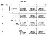
- FIG. 1 shows the culture conditions of Example 1 (Conditions 1 to 4).
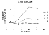
- FIG. 2 shows the transition of the viable cell density under conditions 1 to 4.
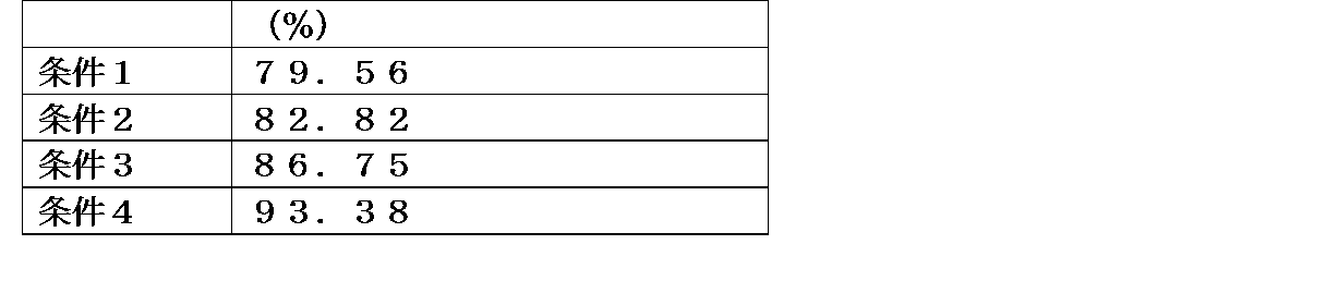
- FIG. 3 shows the ratio of cTnT positive cells under conditions 1 to 4.
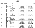
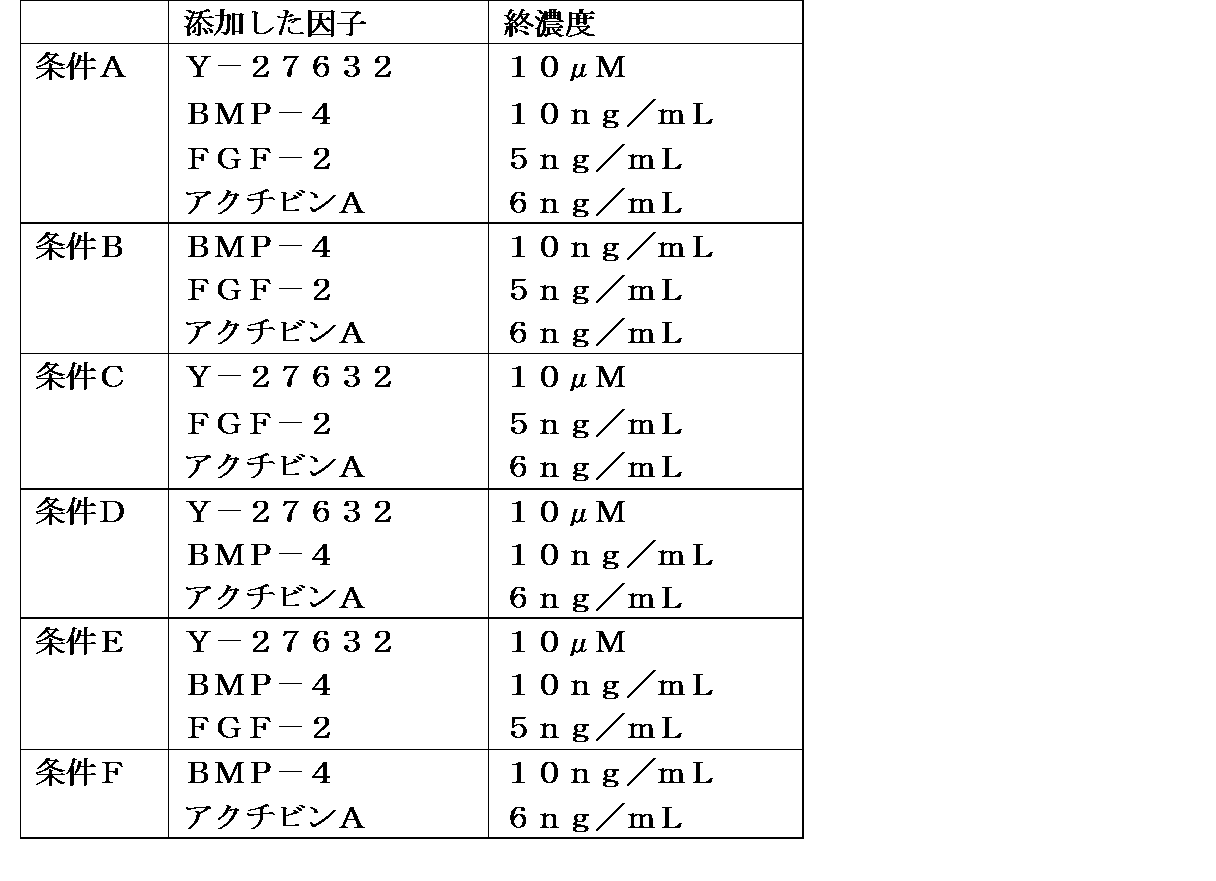
- FIG. 4 shows the culture conditions of Example 2 (Conditions A to F).
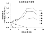
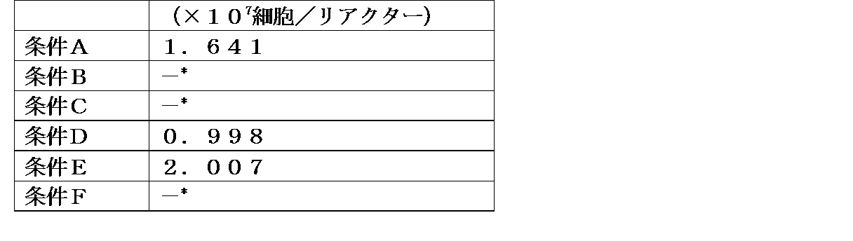
- FIG. 5 shows changes in the density of living cells under conditions A to F.
- FIG. 6 shows the ratio of cTnT positive cells under conditions A, D and E.
- the present invention is a medium for producing mesodermal cells from stem cells, comprising a ROCK inhibitor, BMP, activin, and FGF, a medium for inducing differentiation from stem cells to mesodermal cells (hereinafter referred to as “this”).
- Inventive medium a method of producing mesodermal cells by culturing stem cells in the medium (hereinafter also referred to as“ method of the present invention ”), induction of differentiation of stem cells into mesodermal cells.
- a composition for assisting hereinafter also referred to as “the composition of the present invention”
- a cell group obtained by the method of the present invention hereinafter also referred to as “the cell group of the present invention”.
- the medium of the present invention is a medium for inducing differentiation into stem cell mesodermal cells containing a ROCK inhibitor, BMP, FGF, and activin.
- the medium of the present invention is a medium obtained by adding a component containing a ROCK inhibitor, BMP, FGF, and activin to a basal medium for stem cells.
- stem cell refers to an immature cell having self-replication ability and differentiation / proliferation ability.
- Stem cells have hierarchies, and upper undifferentiated stem cells have high self-replicating ability and high pluripotency capable of differentiating into various cell lineages. It is known that it can only differentiate into cell lineages.
- the stem cell-derived species to which the medium of the present invention can be applied is not particularly limited, and examples thereof include rodents such as rats, mice, hamsters and guinea pigs, rabbits such as rabbits, and hoofs such as pigs, cows, goats and sheep. Eyes, cats such as dogs and cats, and primates such as humans, monkeys, rhesus monkeys, marmosets, orangutans and chimpanzees.
- Stem cells include pluripotent stem cells, multipotent stem cells, unipotent stem cells, etc., depending on their differentiation ability.
- pluripotent stem cell means a stem cell that can be differentiated into all cells that can exist in a living body (that is, has pluripotency) and has proliferative ability.
- pluripotent stem cells include induced pluripotent stem cells (iPS cells), embryonic stem cells (ES cells), cloned embryo-derived embryonic stem cells (ntES cells) obtained by nuclear transfer, sperm stem cells (GS cells) , Embryonic germ cells (EG cells), cultured fibroblasts, bone marrow stem cell-derived pluripotent cells (Muse cells), and the like, but are not limited thereto.
- the pluripotent stem cell used may be prepared by a conventionally known method or may be a cell line that is generally available. Examples of ES cells include, but are not limited to, KhES1, KhES3, and the like.
- the pluripotent stem cell used in the present invention is an iPS cell.
- An iPS cell is a somatic cell-derived artificial stem cell having pluripotency and proliferative ability, which is produced by introducing a specific reprogramming factor into a somatic cell in the form of a nucleic acid or protein.
- genes included in the reprogramming factor include Oct3 / 4, Sox2, Sox1, Sox3, Sox15, Sox17, Klf4, Klf2, c-Myc, N-Myc, L-Myc, Nanog, Lin28, Fbx15, Eras, Examples include ECAT15-2, Tcl1, beta-catenin, Lin28b, Sall1, Sall4, Esrrb, Nr5a2, Tbx3, and Glis1. These initialization factors may be used alone or in combination.
- the iPS cells used in the present invention may be prepared by a conventionally known method or may be a cell line that is generally available.
- Exemplary strains of human iPS cells include 253G1 (RIKEN Cell Bank No. HPS0002), 201B7 (RIKEN Cell Bank No. HPS0063), 409B2 (RIKEN Cell Bank No. HPS0076), 454E2 (RIKEN Cell Bank No. HPS0077), 606A1 (RIKEN) Cell Bank No. HPS0328), 610B1 (RIKEN Cell Bank No. HPS0331), 648A1 (RIKEN Cell Bank No.
- HPS0360 MYH (Patent Literature 1), 427F1 (Patent Literature 1), 457C1 (Patent Literature 1), 604A1 (Patent Literature 1) ), HiPS-RIKEN-1A (RIKEN Cell Bank No. HPS0003), HiPS-RIKEN-2A (RIKEN Cell Bank No. HPS0009), HiPS-RIKEN-12A (RIKEN Cell Bank N) .HPS0029), Nips-B2 (RIKEN Cell Bank No.HPS0223) or the like include, but are not limited to.
- Exemplary strains of iPS cells derived from non-human animals include iPS-MEF-Ng-20D-17, iPS-MEF-Ng-178B-5, iPS-MEF-Fb / Ng-440A-3, iPS-MEF -Ng-492B-4, iPS-Stm-FB / gfp-99-1, iPS-Stm-FB / gfp-99-3, iPS-Hep-FB / Ng / gfp-103C-1, iPS-L1, iPS -S1 and the like can be mentioned, but not limited thereto. Alternatively, disease specific iPS cells may be used.
- diseases include blood system diseases, immune system diseases, endocrine diseases, metabolic diseases, visual diseases, cardiovascular diseases, respiratory diseases, skin / connective tissue diseases, bone / joint diseases, kidneys -Include, but are not limited to, urinary diseases, syndromes with changes in chromosomes or genes (see http://cell.brc.riken.jp/ja/hps/hps_dislistist_index).
- examples of the above cell lines include the RIKEN BioResource Center (see http://cell.brc.riken.jp/en/) and the JCRB cell bank (http://cellbank.nibiohn.go.jp/). available from English /).
- the iPS cells used in the present invention may be obtained by culturing on feeder cells. Or you may obtain by culture
- multipotent stem cells examples include somatic stem cells such as mesenchymal stem cells, hematopoietic stem cells, neural stem cells, bone marrow stem cells, and reproductive stem cells.
- the multipotent stem cell is preferably a mesenchymal stem cell.
- Mesenchymal stem cells are undifferentiated cells (somatic stem cells) present in adult bone marrow, adipose tissue, placental tissue, umbilical cord blood, dental pulp, etc., and proliferative and multipotent (especially bone cells, chondrocytes,
- a broad term means a population of stem cells or progenitor cells having the ability to differentiate into muscle cells, tendon cells, fat cells, etc.).
- mesenchymal stem cells include mesenchymal stem cells derived from human bone marrow (hMSC-BM, manufactured by Takara), mesenchymal stem cells derived from human umbilical cord matrix (hMSC-UC, manufactured by Takara), or mesenchymal cells derived from human adipose tissue.
- System stem cells hMSC-AT, manufactured by Takara
- any stem cell can be suitably used, but preferably a pluripotent stem cell or a mesenchymal stem cell, more preferably a pluripotent stem cell can be used.
- the pluripotent stem cell is preferably an ES cell or iPS cell, more preferably an iPS cell, and particularly preferably a human iPS cell.
- the “mesoderm cell” refers to a cell constituting a tissue derived from mesoderm.
- Tissues derived from mesoderm include bone, cartilage, spleen, bone marrow, dentin, peritoneal epithelium, kidney, ureter, pleural epithelium, adrenal cortex, muscle (other than pupil sphincter and dilated pupil), ovary, uterus, testis , And connective tissue.
- mesoderm cells include, but are not limited to, neuroglial cells, myocardial progenitor cells, cardiomyocytes, vascular endothelial progenitor cells, vascular endothelial cells, blood cells, and mesenchymal stem cells.
- the “basal medium for stem cells” is not particularly limited as long as it can induce differentiation of stem cells into mesodermal cells by adding a ROCK inhibitor, BMP, FGF and activin.
- the stem cell basal medium comprises one or more sugar (s), one or more inorganic salts (s), one or more amino acids (s), one or more vitamins (s), and one or more traces. Preferably it contains the component (s).
- antibiotics such as kanamycin can be included as appropriate for use in drug sensitivity tests.
- sugar (s) examples include monosaccharides such as glucose, lactose, mannose, fructose and galactose, and disaccharides such as sucrose, maltose and lactose.
- monosaccharides such as glucose, lactose, mannose, fructose and galactose
- disaccharides such as sucrose, maltose and lactose.
- glucose is particularly preferable.
- These saccharides can be added alone or in combination.
- Examples of the inorganic salt (s) include calcium chloride, calcium nitrate, copper sulfate pentahydrate, iron (III) nitrate nonahydrate, iron (II) sulfate heptahydrate, magnesium chloride hexahydrate.
- Any inorganic salt or a combination thereof can be used as long as it is a component that advantageously acts on differentiation induction of stem cells into mesodermal cells.
- amino acid (s) examples include alanine, arginine, asparagine, aspartic acid, cystine, cysteine, glutamine, glycine, histidine, glutamic acid, hydroxyproline, isoleucine, leucine, lysine, methionine, phenylalanine, proline, serine, threonine, Examples include tryptophan, tyrosine and valine, and L-amino acids are preferred.
- the amino acid (s) may include derivatives such as derivatives, salts and hydrates thereof.
- Examples of arginine derivatives include L-arginine hydrochloride and L-arginine monohydrochloride.
- Examples of derivatives of aspartic acid include L-aspartic acid sodium salt monohydrate, L-aspartic acid monohydrate, potassium L-aspartate, and magnesium L-aspartate.
- Examples of cysteine derivatives include L-cysteine dihydrochloride and L-cysteine hydrochloride monohydrate.
- Examples of lysine derivatives include L-lysine hydrochloride.
- Examples of the derivative of glutamic acid include L-glutamic acid monosodium salt and the like.
- As a derivative of asparagine for example, L-asparagine monohydrate and the like can be mentioned.
- Examples of the derivative of tyrosine include L-tyrosine disodium dihydrate and the like.
- examples of histidine derivatives include histidine hydrochloride and histidine hydrochloride monohydrate.
- Examples of lysine derivatives include L-lysine hydrochloride.
- vitamin (s) examples include ascorbic acid, biotin, choline, folic acid, inositol, niacin, pantothenic acid, pyridoxine, riboflavin, thiamine, vitamin B12, and paraaminobenzoic acid (PABA).
- ascorbic acid is preferably added.
- the vitamin (s) can include derivatives such as derivatives, salts and hydrates thereof.
- Examples of derivatives of ascorbic acid include ascorbic acid 2-phosphate ester (Ascorbic acid 2-phosphate), magnesium ascorbate phosphate, sodium ascorbate sulfate, ascorbyl aminopropyl phosphate, and sodium ascorbate phosphate. It is done.
- Examples of the derivative of choline include choline chloride.
- Examples of niacin derivatives include nicotinic acid, nicotinamide and nicotinic alcohol.
- Examples of the derivative of pantothenic acid include calcium pantothenate, sodium pantothenate, panthenol and the like.
- Examples of the derivative of pyridoxine include pyridoxine hydrochloride, pyridoxal hydrochloride, pyridoxal phosphate, and pyridoxamine.
- Derivatives of thiamine include, for example, thiamine hydrochloride, thiamine nitrate, bisthiamine nitrate, thiamine dicetyl sulfate ester salt, fursultiamine hydrochloride, octothiamine and benfotiamine.
- the minor component (s) is preferably a component that advantageously acts on differentiation induction of stem cells into mesodermal cells.
- the trace component (s) include components used as usual medium components such as glutathione, hypoxanthine, lipoic acid, linolenic acid, phenol red, putrescine, pyruvic acid, thymidine, and NaHCO 3 .
- the trace component (s) can include derivatives such as derivatives, salts and hydrates thereof. Examples of the derivative include putrescine dihydrochloride.
- basal medium for stem cells a basal medium known to those skilled in the art can be used.
- commercially available culture media such as StemPro (registered trademark) -34 (Thermo Fisher Scientific), mESF Basal Medium (Wako Pure Chemicals), etc.
- MEM Minimum Essential Medium
- BME Basic Medium Eagle
- DMEM Dulbecco's Modified Eagle Medium
- EMEM Eagle's minimalEsdM (Glas-gow's MEM)
- F12 Ham's F12 Mediu
- DMEM / F12 medium in which DMEM and F12 medium are mixed at 1: 1)
- RPMI1640 RD
- BMC-3 Brinster's BMOC-3 Medium
- CMRL-1066 L-15 medium (Leibovitz's L -15 medium)
- McCoy's 5A Media 199, MEM ⁇ Media
- MCDB105 MCDB131
- MCDB153 MCDB201
- Williams' medium E, and ESF Williams' medium E, and ESF.
- the basal medium for stem cells may be KnockOut (trademark) Serum Replacement (KSR) (Thermo Fisher Scientific), StemSure Serum Replacement (SSR) (Wako Pure Chemicals, etc.) Serum-free supplements such as B27 Replacement (Thermo Fisher Scientific) may be added.
- KSR Serum Replacement
- SSR StemSure Serum Replacement
- B27 Replacement Thermo Fisher Scientific
- Examples of such a medium include, for example, a medium in which KSR is added to MEM, BME, DMEM, EMEM, IMDM, DMEM / F12, and RPMI1640, and B27 Replacement to MEM, BME, DMEM, EMEM, IMDM, DMEM / F12, and RPMI1640.
- StemPro (registered trademark) -34 preferably, MEM, BME, DMEM, EMEM, IMDM, DMEM / F12 and RPMI1640 to which B27 Replacement is added, and StemPro (registered trademark) -34, More preferably, a medium in which B27 Replacement is added to RPMI1640, and StemPro (registered trademark) -34, most preferably StemPro (registered trademark) -34 are listed. Rukoto can.
- additives usually used for cell culture may be further added.
- additives include, but are not limited to, L-ascorbic acid 2-trisodium phosphate, L-glutamine, 1-thioglycerol and the like.
- the type and amount of the medium used can be appropriately selected by those skilled in the art according to the type of cells to be cultured.
- ROCK inhibitors used in the present invention include (R)-(+)-trans-N- (4-pyrylyl) -4- (1-aminoethyl) -cyclohexanecarbamide, 2HCl, H 2 O (Y-27632 ), 1- (5-isoquinolinesulfonyl) piperazine hydrochloride (HA100), 1- (5-isoquinolinesulfonyl) homopiperazine dihydrochloride (Fasudil / HA-1077), (S)-(+)-2-methyl- 4-glycyl-1- (4-methylisoquinolinyl-5-sulfonyl) homopiperidine dihydrochloride (H-1152), 1- (5-isoquinolinesulfonyl) -2-methylpiperazine (H-7), 1 -(5-isoquinolinesulfonyl) -3-methylpiperazine (isoH-7), N-2- (methyla C) ethyl-5-isoquinolinesulf
- the concentration of the ROCK inhibitor used in the present invention is about 0.2 to about 100 ⁇ M, for example, about 1 to about 75 ⁇ M, about 2 to about 50 ⁇ M, or about 5 to about 20 ⁇ M. In an exemplary embodiment, the concentration of ROCK inhibitor used is about 10 ⁇ M.
- BMP used in the present invention examples include, but are not limited to, BMP2, BMP4, BMP6, and BMP8.
- the BMP used is BMP4.
- the concentration of BMP used in the present invention is about 0.5 to about 500 ng / mL, such as about 1 to about 100 ng / mL, about 2 to about 50 ng / mL, or about 5 to about 20 ng / mL. In an exemplary embodiment, the concentration of BMP used is about 10 ng / mL.
- FGF used in the present invention examples include FGF-1, FGF-2, FGF-3, FGF-4, FGF-5, FGF-6, FGF-7, FGF-8, and FGFv9. It is not limited to.
- the FGF used is FGF-2.
- the concentration of FGF used in the present invention is about 0.1 to about 250 ng / mL, such as about 0.5 to about 50 ng / mL, about 1 to about 25 ng / mL, or about 2.5 to about 10 ng / mL. It is. In an exemplary embodiment, the concentration of FGF used is about 5 ng / mL.
- activins used in the present invention include, but are not limited to, activin A, activin B, and activin AB.
- the activin used is activin A.
- the concentration of activin used in the present invention is about 0.12 to about 300 ng / mL, such as about 0.6 to about 60 ng / mL, about 1.2 to about 30 ng / mL, or about 3 to about 12 ng / mL. It is. In an exemplary embodiment, the concentration of activin used is about 6 ng / mL.
- the method of the present invention is a method for producing mesodermal cells from stem cells, comprising culturing the stem cells in the medium of the present invention described in 1 above.
- the culture conditions such as culture temperature and culture time in the method of the present invention can be appropriately selected by those skilled in the art depending on the type of cells to be cultured.
- the pluripotent stem cells are cultured in the medium of the present invention at 34 to 40 ° C./2 to 8% CO 2 for 12 hours to 7 days. Incubate.
- the cells are cultured at 35 to 39 ° C./3 to 7% CO 2 for 1 to 4 days.
- the medium of the present invention containing the above four factors (ROCK inhibitor, BMP, FGF, and activin) at 36 to 38 ° C./4 to 6% CO 2 for 6 to 42 hours, preferably 12 After culturing for a time to 36 hours, more preferably 18 to 30 hours, in a medium containing 3 factors (BMP, FGF, and activin) excluding the ROCK inhibitor at 36 to 38 ° C./4 to 6% CO 2
- the culture is performed for 12 hours to 4 days, preferably 1 to 3 days, more preferably 36 to 60 hours, and most preferably 42 to 54 hours.
- the pluripotent stem cells are cultured in the medium of the present invention at 34 to 40 ° C./2 to 8% CO 2 for 12 hours to 7 days. To do. Preferably, the cells are cultured at 35 to 39 ° C./3 to 7% CO 2 for 1 to 4 days, more preferably 2 to 4 days.
- the types, concentrations, etc. of the ROCK inhibitor, BMP, FGF, and activin contained in the medium are as described above.
- the ROCK inhibitor used is Y-27632
- BMP is BMP4
- FGF is FGF-2
- activin activin A.
- adhesion culture or suspension culture can be used, and as a preferred form, suspension culture can be used.
- Suspension culture can be performed using a means, method, or apparatus that can keep cells floating.
- incubators with stirring blades such as single-use bioreactors (Biot Co., Ltd.), single-use bioreactors (Thermo Fisher), single-use bioreactors (Sartorius Stedium), single-use bioreactors (GE Healthcare Life Sciences) It can be implemented by using.
- the type of the incubator used and the stirring speed can be appropriately selected by those skilled in the art depending on the type of cells to be cultured. Examples of the stirring speed include, but are not limited to, 0 to 100 rpm, 20 to 80 rpm, or 45 to 65 rpm.
- the mesoderm cells are as described above.
- inducing differentiation of stem cells into mesodermal cells a method well known to those skilled in the art is used to induce differentiation of mesoderm-derived tissue from mesoderm after culturing the stem cells in the medium of the present invention. be able to.
- differentiation medium suitable for differentiation into a desired mesoderm-derived tissue differentiation into a desired mesoderm cell can be induced (Nathan J Palpant. Al. Nature Protocols 2016 Dec; 12 (1): 15-31, Cythia A. Batchelder et.al.2009 Jul; 78 (1): 45-56 etc.).
- the stem cells may be induced to differentiate into mesenchymal stem cells, and the obtained mesenchymal stem cells may be differentiated into mesodermal cells.
- Methods for inducing differentiation of mesenchymal stem cells into mesodermal cells are well known to those skilled in the art.
- mesenchymal stem cells can be induced to differentiate into mesodermal cells by culturing mesenchymal stem cells in a differentiation medium suitable for differentiation into desired cells (Nishiyama et al. 2007 Aug; 25 (8): 2017-2024 etc.).
- the present invention provides a method for producing myocardial progenitor cells or cardiomyocytes from stem cells.
- the method is a method comprising (1) culturing stem cells in the above-described medium of the present invention, and (2) culturing in a medium containing a Wnt inhibitor. Step (2) may be followed by (3) culturing in a medium containing vascular endothelial growth factor (VEGF) and FGF.
- VEGF vascular endothelial growth factor
- the above-mentioned step (2) is a step of culturing in a medium containing IWR-1 and IWP-2, and the above-mentioned step (3) is cultivated in a medium containing VEGF and FGF-2. It is a stage.
- a Wnt inhibitor refers to a substance that inhibits the Wnt signal transduction pathway.
- IWR-1 (4-[(3aR, 4S, 7R, 7aS) -1,3,3a, 4,7,7a-hexahydro-1,3-dioxo-4,7-methano-2H -Isoindol-2-yl] -N-8-quinolinyl-benzamide
- IWP-2 N- (6-Methyl-2-benzothiozyl) -2-[(3,4,6,7-tetrahydr-4--4-oxo) -3-phenylthieno [3,2-d] pyrimidin-2-yl) thio] -acetamide
- WntC59 (4- (2-methy1-4-pyridinyl) -N- (4- (3-pyridinyl) phenyl)- benzeneacetamide
- IWP4 N- (6-met yl-2-benzothiazolyl) -2-[[3,4,6,7-tetrahydro-3
- the concentration of IWR-1 used is, for example, about 0.4 to about 40 ⁇ M, about 0.8 to about 20 ⁇ M, or about 2 to about 8 ⁇ M. In an exemplary embodiment, the concentration of IWR-1 used is about 4 ⁇ M.
- the concentration of IWP-2 used is, for example, from about 1 to about 100 ⁇ M, from about 2 to about 50 ⁇ M, or from about 5 to about 20 ⁇ M. In an exemplary embodiment, the concentration of IWP-2 used is about 10 ⁇ M.
- the concentration of VEGF used is about 0.1 to about 100 ng / mL, such as about 0.5 to about 50 ng / mL, about 1 to about 25 ng / mL, or about 2.5 to about 10 ng / mL. In an exemplary embodiment, the concentration of VEGF used is about 5 ng / mL.
- the concentration of FGF-2 used is about 0.1 to about 250 ng / mL, such as about 1 to about 100 ng / mL, about 2 to about 50 ng / mL, or about 5 to About 20 ng / mL. Specific examples of FGF include FGF 1 described above. In an exemplary embodiment, FGF-2 is used as FGF, and the concentration of FGF-2 used is about 10 ng / mL.
- step (2) may be culturing for 1 to 7 days at 35 to 39 ° C./3 to 7% CO 2 .
- the step (3) may be culturing for 1 to 20 days at 35 to 42 ° C./3 to 7% CO 2 . More preferably, step (2) is culturing for 2 to 4 days at 36-38 ° C./4-6% CO 2 and step (3) is 7 for 36-40 ° C./4-6% CO 2. Incubation for ⁇ 15 days.
- composition of the present invention is a composition for assisting inducing differentiation of stem cells into mesodermal cells, comprising a ROCK inhibitor, BMP, activin, and FGF.
- the types, concentrations, and the like of the ROCK inhibitor, BMP, activin, and FGF contained in the above composition are as described above.
- the ROCK inhibitor used is Y-27632
- BMP is BMP4
- activin is activin A
- FGF is FGF-2.
- the composition may be a liquid composition or a powdered composition such as a lyophilized product.
- the cell group of the present invention is a cell group obtained by the above-described method of the present invention, and about 90% or more of mesodermal cells obtained by differentiation induction. In the cell population, about 90% or more, such as about 91%, about 92%, about 93%, about 94%, about 95%, about 96%, about 97%, about 98%, about 99%, or more Are mesodermal cells.
- iPS cell line 253G1 which is a pluripotent stem cell
- MEF cells CF-1 MEF
- Maintenance culture was performed in a medium for iPS cells.
- KnockOut TM DMEM / F12 was added to KnockOut TM Serum Replacement (KSR) (final concentration 20%), 0.1 mM MEM non-essential amino acid solution, 1 mM L-glutamine, 0.1 mM ⁇ -Mercaptoethanol with 4 ng / mL FGF-2 added was used.
- KSR KnockOut TM Serum Replacement
- Viable cell counting Cells were detached using Accutase or AccuMax (Innovative Cell Technologies). After staining with trypan blue, viable cells were counted using a hemocytometer.
- Alexa Floor 488 goat anti-mouse IgG1 (1: 100) was then added as a secondary antibody.
- Cells were washed twice with Perm / Wash TM buffer. The cells were washed once with 2% FBS in PBS, resuspended, and samples were measured and analyzed using Cell Sorter SH800 (Sony).
- Example 1 Effect on the induction of cardiomyocyte differentiation by the presence or absence of EB formation StemPro (registered trademark) -34 SFM (Thermo Fisher Scientific) to L-ascorbic acid 2-trisodium phosphate (final concentration 50 ⁇ g / mL), 2 mM L A medium supplemented with glutamine and 400 ⁇ M 1-thioglycerol was used (hereinafter referred to as “basal medium”). According to the culture conditions outlined in FIG. 1 (conditions 1 to 4), iPS cells were induced to differentiate into cardiomyocytes.
- the iPS cells cultured in advance were detached and collected by Accutase treatment, and then centrifuged to remove the supernatant.
- the collected iPS cells were suspended in a medium.
- iPS cells were suspended in a basal medium containing 10 ⁇ M Y-27632.
- iPS cells were suspended in a basal medium containing 10 ⁇ M Y-27632, 10 ng / mL BMP-4, 5 ng / mL FGF-2, and 6 ng / mL activin A.
- Suspended iPS cells were seeded into Single use bio-reactor for 30 mL (Biot Co., Ltd.) at 4.0 ⁇ 10 6 cells / reactor.
- the culture was performed at a stirring speed of 55 rpm and 37 ° C./5% CO 2 .
- Conditions 1 to 3 were cultured in a basal medium containing 10 ⁇ M Y-27632 for one day (condition 1: Day-3 to -2, condition 2: Day-2 to -1, condition 3: Day-1 to 0). . After culturing, conditions 1 and 2 were further cultured in a basal medium for 2 days and 1 day, respectively (condition 1: Day-2 to 0, condition 2: Day-1 to 0).
- the cells were cultured for 3 days in a basal medium containing 10 ng / mL BMP-4, 5 ng / mL FGF-2, and 6 ng / mL activin A (conditions 1-3: Day 0 to 3).
- condition 4 After stirring for 1 day in basal medium containing 10 ⁇ M Y-27632, 10 ng / mL BMP-4, 5 ng / mL FGF-2, and 6 ng / mL activin A (condition 4: Day 0 to 1) The cells were cultured for 2 days in a basal medium containing 10 ng / mL BMP-4, 5 ng / mL FGF-2, and 6 ng / mL activin A (condition 4: Day 1 to 3). After culturing under each condition, the cell mass was recovered from the reactor and transferred to a centrifuge tube.
- the cell mass was washed with a basal medium, then resuspended in a basal medium containing 4 ⁇ M IWR-1 and 10 ⁇ M IWP-2, and cultured for 3 days (conditions 1 to 4: Day 3 to 6). Thereafter, the medium was replaced with a basal medium containing 5 ng / mL VEGF and 10 ng / mL FGF-2 and cultured for a maximum of 10 days (Condition 1 to 4: Day 6 to). In culture with these media, the media was changed every two days.
- the maximum value of the viable cell density under each condition was as shown in the following table (FIG. 2).
- the maximum value of the cardiomyocyte marker (Cardiac Troponin T: cTnT) in each condition was as shown in the following table (FIG. 3).
- condition 4 without EB formation period had more viable cells and more cTnT positive cells than conditions 1 to 3 with EB formation period.
- Example 2 Effect on differentiation induction efficiency due to difference in factors added to basal medium Factors shown in the following table were added to the basal medium.
- the iPS cells cultured in advance were detached and collected by Accutase treatment, and then centrifuged to remove the supernatant.
- the collected iPS cells were suspended in each of the media under conditions A to F, and then seeded into 30 mL of single use bio-reactor for at 5.07 ⁇ 10 6 cells / reactor.
- the culture was performed at a stirring speed of 55 rpm and 37 ° C./5% CO 2 .
- the culture procedure for each condition is outlined in FIG. For conditions A and C to E, after culturing for one day in the medium of each condition (conditions A and C to E: Day 0 to 1), each medium was replaced with a medium except 10 ⁇ M Y-27632.
- condition A and C to E Day 1 to 3
- condition B and F after culturing for 1 day under each condition (conditions B and F: Day 0 to 1), the medium was changed and further cultured for 2 days under the same conditions (conditions B and F: Day 1 to 3).
- the cell mass was recovered from the reactor and transferred to a centrifuge tube. After washing the cell mass with basal medium, the cells were resuspended in basal medium containing 4 ⁇ M IWR-1 and 10 ⁇ M IWP-2, and cultured for 3 days (conditions AF: Day 3-6). Thereafter, the medium was replaced with a basal medium containing 5 ng / mL VEGF and 10 ng / mL FGF-2 and cultured for a maximum of 10 days (conditions A to F: Day 6 to).
- the maximum value of the viable cell density under each condition is shown in the following table (FIG. 5). * : The cell died during differentiation induction. In addition, the maximum value of the cardiomyocyte marker (Cardiac Troponin T: cTnT) under each condition in which the cells survived was as shown in the following table (FIG. 6).
- Example 3 Measurement of cell pulsation A culture solution (2 ml) containing a myocardial cell mass obtained by inducing differentiation under the condition A in Example 2 was transferred to a 12 well plate MICROPPLATE with Lid (IWAKI), and Leica It observed with DMi1 inverted microscope (Leica). As a result, it was confirmed that each myocardial cell mass was autonomously beating at a constant cycle.
- Example 4 Measurement of Ca + imaging Using Calcium Kit-Fluo 4 (Dojindo Laboratories), Ca + imaging was performed on cell masses and cells adhered on the substrate. Differentiation was induced for 16 days under the condition A of Example 2 to obtain a cell mass. The obtained cell mass was washed with PBS, suspended in Loading Buffer, and transferred to MATUNAMI GLASS BOTTOM DISH Hydro 35mm dish (Matsunami Glass Industrial Co., Ltd.). Then, it incubated at 37 degreeC for 1 hour. After the incubation, the loading buffer was removed and washed with PBS. The obtained cell mass was transferred to Recording Medium, and fluorescence observation was performed with ECLIPSE Ts2 (Nikon).
- a cell mass obtained by performing differentiation induction for 16 days under the condition A in Example 2 was treated with AccuMax (Innovation cell technologies), and the cell mass was detached.
- the detached cell mass was suspended in DMEM + 10% FBS, and the suspension was passed through a 40 ⁇ m strainer (FALCON).
- Ca + imaging of the cells adhered on the substrate was performed in the same manner as described above. As a result, it was possible to confirm that the Ca + concentration increased in synchronism with the pulsation of the cell mass and the adhered cells.
- Example 5 Expression of myocardial marker A cell mass obtained by inducing differentiation for 16 days under the condition A in Example 2 was treated with AccuMax (Innovation cell technologies), and the cell mass was detached. The suspension was suspended in DMEM + 10% FBS, and the suspension was passed through a 40 ⁇ m strainer (FALCON). Cells diluted with DMEM + 10% FBS so as to be 4 ⁇ 10 4 cells / well are seeded in 24 well plate MICROPLATE with Lid (IWAKI) previously coated with fibronectin (5 ⁇ g / cm 2 , Sigma), and in DMEM + 10% FBS For 6 days.
- the culture supernatant was removed, and after washing 3 times with PBS, cells were fixed with Fixation / Permeabilization solution (4 ° C., 60 minutes or overnight). After washing 3 times with PBS, blocking with PBS + 3% FBS was performed at room temperature for 30 minutes. The solution was discarded and replaced with PBS + 1% FBS containing various antibodies (4 ° C., 60 minutes or overnight). The solution was discarded and replaced with 0.1% FBS / PBST, and left to stand for 5 minutes for washing (stationary washing). This washing operation was performed three times. After removing the washing solution, various secondary antibodies were added (room temperature, 60 minutes). After performing static washing with PBS for 5 minutes three times, it was treated with DAPI (Life technologies) (room temperature, 15 minutes).
- Example 6 Measurement of extracellular potential A cell mass obtained by inducing differentiation for 14 days under the condition A in Example 2 was treated with AccuMax (Innovation cell technologies), and the cell mass was detached. The suspension was suspended in DMEM + 10% FBS, and the suspension was passed through a 40 ⁇ m strainer (FALCON). In advance, a frame was made of silicon so as to surround the electrode of multi-electrode dish MED-P530A (Alphamed Scientific Co., Ltd.), and then the inside was coated with fibronectin (about 5 ⁇ g / cm 2 , Sigma). 1 ⁇ 10 5 cells were seeded in a frame and allowed to stand for 1 hour in a 37 ° C., 5% CO 2 incubator.
- FALCON 40 ⁇ m strainer
- the medium was replenished and cultured for 4 days.
- the extracellular potential was measured with MED64 (Alpha Med Scientific Co., Ltd.). As a result, it was confirmed that depolarization and repolarization occurred autonomously in the induced cardiomyocytes.
- iPS cells were suspended in culture using a medium supplemented with the above four factors (ROCK inhibitor, BMP4, FGF-2, and activin A), and without undergoing preparation steps such as EB formation. It was shown that mesoderm cells can be induced and the obtained mesoderm cells can be induced into cardiomyocytes. It was also demonstrated that cardiomyocytes can be induced with high survival rate and high efficiency. Furthermore, it was confirmed that the induced cardiomyocytes beat autonomously at a constant cycle. Furthermore, it was confirmed that depolarization and repolarization occur autonomously in induced cardiomyocytes. That is, it was shown that cardiomyocytes can be induced quickly and easily with high efficiency by using the method of the present invention. This method is also suitable for mass culture of cells when suspension culture is used.
- Example 7 Cardiomyocyte differentiation using feeder-free cultured iPS cells Stem Cell and Culture Method
- iPS cell line 253G1 which is a pluripotent stem cell
- the feeder-free iPS cell culture was performed under the following conditions. Incubator was coated with iMatrix® 511 at 0.5 ng / cm 2 . StemFit (registered trademark) AK02N was used as a medium for iPS cells.
- the cultured cells were washed with PBS and then detached with 0.5 ⁇ TrypLE (trademark). Thereafter, the supernatant was removed by centrifugation.
- the collected iPS cells were suspended in the medium of Condition A of Example 2 and then seeded into 30 mL of Single use bio-reactor for 5.0 ⁇ 10 6 cells / reactor.
- the culture was performed at a stirring speed of 55 rpm and 37 ° C./5% CO 2 .
- Culturing was performed in the medium of Condition A of Example 2 for 3 days. Thereafter, the cell mass was recovered from the reactor and transferred to a centrifuge tube.
- the supernatant was removed, the cell mass was washed once with basal medium, resuspended in basal medium containing 4 ⁇ M IWR-1 and 10 ⁇ M IWP-2, and cultured again in the reactor for 3 days. After the culture, the medium was replaced with a basal medium containing 5 ng / mL VEGF and 10 ng / mL FGF-2, and the cells were cultured for a maximum of 15 days. During the culture, the medium was changed every two days.
- the cardiomyocyte marker (Cardiac Troponin T: cTnT) in each differentiation induction period was as shown in the following table.
- mesodermal cells can be induced from iPS cells cultured in a feeder-free state using the medium and method of the present invention without undergoing preparatory steps such as EB formation. It was shown that the obtained mesodermal cells can be induced into cardiomyocytes with high efficiency.
- the present invention it is possible to induce differentiation of stem cells into mesodermal cells such as cardiomyocytes quickly and easily with high efficiency.
- This method is also suitable for mass culture of mesodermal cells when suspension culture is used. Therefore, the present invention is useful in the field of regenerative medicine. The present invention is also useful in pharmaceutical development.
Landscapes
- Health & Medical Sciences (AREA)
- Engineering & Computer Science (AREA)
- Biomedical Technology (AREA)
- Life Sciences & Earth Sciences (AREA)
- Bioinformatics & Cheminformatics (AREA)
- Wood Science & Technology (AREA)
- Biotechnology (AREA)
- Organic Chemistry (AREA)
- Chemical & Material Sciences (AREA)
- Genetics & Genomics (AREA)
- Zoology (AREA)
- Cardiology (AREA)
- Microbiology (AREA)
- Rheumatology (AREA)
- Biochemistry (AREA)
- General Engineering & Computer Science (AREA)
- General Health & Medical Sciences (AREA)
- Cell Biology (AREA)
- Micro-Organisms Or Cultivation Processes Thereof (AREA)
Abstract
Description
[1]ROCK阻害剤、骨形成因子(BMP)、繊維芽細胞増殖因子(FGF)、およびアクチビンを含む、幹細胞の中胚葉系細胞への分化誘導用培地;
[2]ROCK阻害剤が、Y-27632である、[1]に記載の培地;
[3]BMPが、BMP4である、[1]または[2]に記載の培地;
[4]FGFが、FGF-2である、[1]~[3]のいずれか一つに記載の培地;
[5]アクチビンが、アクチビンAである、[1]~[4]のいずれか一つに記載の培地;
[6]中胚葉系細胞が、心筋前駆細胞または心筋細胞である、[1]~[5]のいずれか一つに記載の培地;
[7]幹細胞が、多能性幹細胞または間葉系幹細胞である、[1]~[6]のいずれか一つに記載の培地;
[8]幹細胞から中胚葉系細胞を製造する方法であって、幹細胞を[1]~[7]のいずれか一つに記載の培地で培養する段階を含む、方法;
[9]幹細胞から心筋前駆細胞または心筋細胞を製造する方法であって、
(1)幹細胞を[1]~[7]のいずれか一つに記載の培地で培養する段階、次いで
(2)Wnt阻害剤を含む培地で培養する段階を含む、方法;
[10]Wnt阻害剤が、IWR-1およびIWP-2である、[9]に記載の方法;
[11]培養が、浮遊培養である、[8]~[10]のいずれか一つに記載の方法;
[12]幹細胞が、多能性幹細胞または間葉系幹細胞である、[8]~[11]のいずれか一つに記載の方法;
[13]ROCK阻害剤、BMP、FGF、およびアクチビンを含む、幹細胞の中胚葉系細胞への分化誘導を補助するための組成物;ならびに
[14][8]~[12]のいずれか一つに記載の方法によって得られる細胞群であって、90%以上が中胚葉系細胞である、細胞群。
本発明の培地は、ROCK阻害剤、BMP、FGF、およびアクチビンを含む、幹細胞の中胚葉系細胞への分化誘導用培地である。本発明の培地は、幹細胞用基礎培地に、ROCK阻害剤、BMP、FGF、およびアクチビンを含む成分を添加した培地である。
本発明の方法は、幹細胞から中胚葉系細胞を製造する方法であって、幹細胞を前記1の本発明の培地で培養する段階を含む、方法である。
本発明の組成物は、ROCK阻害剤、BMP、アクチビン、およびFGFを含む、幹細胞の中胚葉系細胞への分化誘導を補助するための組成物である。上記の組成物に含まれるROCK阻害剤、BMP、アクチビン、およびFGFの種類、濃度等については、前記1のとおりである。例示的な実施形態では、用いられるROCK阻害剤はY-27632、BMPはBMP4、アクチビンはアクチビンA、FGFはFGF-2である。上記の組成物は、液体組成物であってもよく、凍結乾燥品などの粉末状組成物であってもよい。
本発明の細胞群は、上述した本発明の方法によって得られる細胞群であって、約90%以上が分化誘導により得られた中胚葉系細胞である、細胞群である。当該細胞群では、約90%以上、例えば約91%、約92%、約93%、約94%、約95%、約96%、約97%、約98%、約99%、またはそれ以上が、中胚葉系細胞である。
幹細胞として、多能性幹細胞であるiPS細胞株253G1を用いた(Nakagawa, M. et. al. Nature Biotechnology 26: 101-106 (2008))。MEF細胞(CF-1 MEF)をフィーダー細胞として用いた。iPS細胞用の培地にて維持培養を行った。iPS細胞用の培地として、KnockOut(商標) DMEM/F12に、KnockOut(商標) Serum Replacement(KSR)(終濃度20%)、0.1mM MEM非必須アミノ酸溶液、1mM L-グルタミン、0.1mM β-メルカプトエタノール、4ng/mL FGF-2を添加したものを用いた。培養したiPS細胞を1mg/mlディスパーゼとセルスクレーパーで剥離した後に継代を行った(Split Ratio=1:5~1:6)。
AccutaseまたはAccuMax(Innovative Cell Technologies)を用いて細胞を剥離した。トリパンブルーで染色後、血球算定盤を用いて生細胞を計数した。
AccuMaxを用いて細胞塊を剥離した後、0.5~2×10⁶個の細胞をFixation/Permeabilization solutionで懸濁し、静置した(4℃、30分または一晩)。Perm/Wash(商標)バッファーで細胞を2回洗浄した。次いで、Anti-Troponin, Cardiac Isoform Mouse-Mono(13-11), Ab-1(1:100)、またはPurified Mouse IgG1, k Isotype Ctrlを一次抗体として添加し、静置した(4℃、30分または一晩)。Perm/Wash(商標)バッファーで細胞を2回洗浄した。次いで、Alexa Flour 488 goat anti-mouse IgG1(1:100)を二次抗体として添加した。Perm/Wash(商標)バッファーで細胞を2回洗浄した。2% FBS in PBSで細胞を1回洗浄後、再懸濁し、Cell Sorter SH800(Sony)を用いてサンプルの測定と解析を行った。
StemPro(登録商標)-34 SFM(Thermo Fisher Scientific)にL-アスコルビン酸2-リン酸三ナトリウム(終濃度50μg/mL)、2mM L-グルタミン、および400μM 1-チオグリセロールを添加した培地を用いた(以下、「基礎培地」という)。図1に概説される培養条件(条件1~4)に従って、iPS細胞を心筋細胞へと分化誘導させた。
条件1~3については、10μM Y-27632を含む基礎培地で1日間培養した(条件1:Day-3~-2、条件2:Day-2~-1、条件3:Day-1~0)。培養後、条件1、2については、さらに、基礎培地でそれぞれ2日間、1日間培養した(条件1:Day-2~0、条件2:Day-1~0)。その後、10ng/mL BMP-4、5ng/mL FGF-2、および6ng/mL アクチビンAを含む基礎培地で3日間培養した(条件1~3:Day0~3)。
条件4については、10μM Y-27632、10ng/mL BMP-4、5ng/mL FGF-2、および6ng/mL アクチビンAを含む基礎培地で1日間の撹拌培養の後(条件4:Day0~1)、10ng/mL BMP-4、5ng/mL FGF-2、および6ng/mL アクチビンAを含有する基礎培地で2日間培養した(条件4:Day1~3)。
それぞれの条件で培養後、リアクターから細胞塊を回収して遠心管に移した。基礎培地で細胞塊を洗浄した後、4μM IWR-1および10μM IWP-2を含有する基礎培地で再懸濁し、3日間培養した(条件1~4:Day3~6)。その後、培地を5ng/mL VEGFおよび10ng/mL FGF-2を含有する基礎培地に置換して、最大10日間培養した(条件1~4:Day6~)。これらの培地での培養では、2日毎に培地交換を行った。

基礎培地に以下の表に示す因子を添加した。
条件AおよびC~Eについては、各条件の培地で1日間の培養の後(条件AおよびC~E:Day0~1)、各培地を10μM Y-27632を除いた培地に置換して、2日間培養した(条件AおよびC~E:Day1~3)。条件BおよびFについては、各条件で1日間培養した後(条件BおよびF:Day0~1)、培地を交換し、さらに同じ条件で2日間培養した(条件BおよびF:Day1~3)。
それぞれの条件で培養後、リアクターから細胞塊を回収して遠心管に移した。基礎培地で細胞塊を洗浄した後、4μM IWR-1および10μM IWP-2を含有する基礎培地で再懸濁し、3日間培養した(条件A~F:Day3~6)。その後、培地を5ng/mL VEGFおよび10ng/mL FGF-2を含有する基礎培地に置換して、最大10日間培養した(条件A~F:Day6~)。
また、細胞が生存した各条件における心筋細胞マーカー(Cardiac Troponin T:cTnT)の最大値は以下の表のとおりであった(図6)。

実施例2の条件Aの条件下で分化誘導して得られた心筋細胞塊を含む培養液(2ml)を12 well plate MICROPLATE with Lid(IWAKI)へ移し、ライカ DMi1 倒立顕微鏡(Leica)にて観察した。その結果、各々の心筋細胞塊が自律的に一定の周期で拍動していることを確認した。
Calcium Kit-Fluo 4(同仁化学研究所)を用いて、細胞塊、ならびに基板上に接着させた細胞のCa+イメージングを行った。実施例2の条件Aの条件下で16日間分化誘導を行って細胞塊を得た。得られた細胞塊をPBSで洗浄した後にLoading Bufferに懸濁し、MATUNAMI GLASS BOTTOM DISH Hydro 35mm dish(松浪硝子工業株式会社)に移した。その後、37℃で1時間インキュベートを行った。インキュベート後に、Loading Bufferを除去しPBSで洗浄を行った。得られた細胞塊をRecording Mediumに移し替え、ECLIPSE Ts2(Nikon)にて、蛍光観察を行った。同様に、実施例2の条件Aの条件下で16日間分化誘導を行って得た細胞塊をAccuMax(Innovation cell technologies)で処理し、細胞塊を剥離した。剥離した細胞塊をDMEM+10%FBSに懸濁し、40μm ストレーナー(FALCON)に懸濁液を通した。予めヒト組換えラミニン-221(0.5μg/cm2、ベリタス)でコートしたMATUNAMI GLASS BOTTOM DISH Hydro 35mm dish上に、1×10⁶細胞/wellとなるよう、DMEM+10%FBSで希釈した細胞を播種し、DMEM+10%FBS中で7日間培養した。その後、上記と同様の操作で、基板上に接着させた細胞のCa+イメージングを行った。その結果、細胞塊、ならびに接着させた細胞について、拍動と同調して、Ca+濃度が上昇する様子を確認することができた。
実施例2の条件Aの条件下で16日間分化誘導を行って得た細胞塊をAccuMax(Innovation cell technologies)で処理し、細胞塊を剥離した。DMEM+10%FBSに懸濁し、40μm ストレーナー(FALCON)に懸濁液を通した。予めフィブロネクチン(5μg/cm2、Sigma)でコートした24 well plate MICROPLATE with Lid(IWAKI)に、4×10⁴細胞/wellとなるよう、DMEM+10%FBSで希釈した細胞を播種し、DMEM+10%FBS中で6日間培養した。培養上清を除き、PBSで3回洗浄後、Fixation/Permeabilization solutionで細胞の固定を行った(4℃、60分または一晩)。PBSで3回洗浄後、PBS+3%FBSでのブロッキングを室温にて、30分間行った。液を捨て、各種抗体を含むPBS+1%FBSに置換した(4℃、60分または一晩)。液を捨て、0.1%FBS/PBSTに置換し、5分間静置して洗浄した(静置洗浄)。この洗浄操作を3回行った。洗浄液を除去後、各種二次抗体を添加した(室温、60分間)。PBSで5分間の静置洗浄を3回行った後、DAPI(Life technologies)で処理した(室温、15分間)。PBSで3回洗浄後、EVOS FL Auto(Life technologies)で、蛍光測定を行った。使用した抗体とその濃度を以下に示す。
[使用した抗体とその濃度]
Anti-Troponin Cardiac Isoform Mouse-Mono(13-11),Ab-1(Thermo Fisher Scientific): 2μg/ml(100倍希釈)
Anti-Sarcomeric Alpha Actinin ab9465(abacam):2.5μg/ml(50倍希釈)
Purified Mouse IgG1,k Isotype Control(Biolegend):2μg/mlまたは2.5μg/ml
Myosin Heavy Chain(MHC) Antibody(R&D systems):2.5μg/ml(200倍希釈)
Negative Control Mouse IgG2b(Dako):2.5μg/ml
Alexa Fluor 488 A21121およびAlexa Fluor 488 A11001(Thermo Fisher Scientific):20μg/ml(100倍希釈)
その結果、心筋マーカーであるcTnT、α-Actinin、およびMHCが、分化誘導させたiPS由来心筋細胞において発現していることを確認した。
実施例2の条件Aの条件下で14日間分化誘導を行って得た細胞塊をAccuMax(Innovation cell technologies)で処理し、細胞塊を剥離した。DMEM+10%FBSに懸濁し、40μm ストレーナー(FALCON)に懸濁液を通した。予め、multi-electrode dish MED-P530A(アルファメッドサイエンティフィック株式会社)の電極を囲う様にシリコンで枠を作成した後、内部をフィブロネクチン(約5μg/cm2、Sigma)でコートした。枠内に1×105個の細胞を播種し、37℃、5%CO2インキュベーター内で1時間静置させた。細胞が沈降、接着した事を確認した後に、培地を補充して4日間培養を行った。細胞外電位の測定はMED64(アルファメッドサイエンティフィック株式会社)で行った。その結果、誘導した心筋細胞で脱分極と再分極が自律的に発生することを確認した。
幹細胞および培養方法
幹細胞として、多能性幹細胞であるiPS細胞株253G1を用いた(Nakagawa, M. et. al. Nature Biotechnology 26: 101-106 (2008))。フィーダーフリーでのiPS細胞の培養は以下の条件で行った。iMatrix(登録商標)511で0.5ng/cm2で培養器をコーティングした。 iPS細胞用の培地として、StemFit(登録商標)AK02Nを使用した。0.5×TrypLE(商標)にて細胞を剥離した後、StemFit(登録商標)AK02NにY―276314を10μM添加した培地にて、4-8×10⁴細胞/T-25フラスコ(Corning)に継代を行った。培養1日以降はStemFit(登録商標)AK02Nで培地交換を行い、維持培養した。

Claims (14)
- ROCK阻害剤、骨形成因子(BMP)、繊維芽細胞増殖因子(FGF)、およびアクチビンを含む、幹細胞の中胚葉系細胞への分化誘導用培地。
- ROCK阻害剤が、Y-27632である、請求項1に記載の培地。
- BMPが、BMP4である、請求項1または2に記載の培地。
- FGFが、FGF-2である、請求項1~3のいずれか一項に記載の培地。
- アクチビンが、アクチビンAである、請求項1~4のいずれか一項に記載の培地。
- 中胚葉系細胞が、心筋前駆細胞または心筋細胞である、請求項1~5のいずれか一項に記載の培地。
- 幹細胞が、多能性幹細胞または間葉系幹細胞である、請求項1~6のいずれか一項に記載の培地。
- 幹細胞から中胚葉系細胞を製造する方法であって、幹細胞を請求項1~7のいずれか一項に記載の培地で培養する段階を含む、方法。
- 幹細胞から心筋前駆細胞または心筋細胞を製造する方法であって、
(1)幹細胞を請求項1~7のいずれか一項に記載の培地で培養する段階、次いで
(2)Wnt阻害剤を含む培地で培養する段階を含む、方法。 - Wnt阻害剤が、IWR-1およびIWP-2である、請求項9に記載の方法。
- 培養が、浮遊培養である、請求項8~10のいずれか一項に記載の方法。
- 幹細胞が、多能性幹細胞または間葉系幹細胞である、請求項8~11のいずれか一項に記載の方法。
- ROCK阻害剤、BMP、FGF、およびアクチビンを含む、幹細胞の中胚葉系細胞への分化誘導を補助するための組成物。
- 請求項8~12のいずれか一項に記載の方法によって得られる細胞群であって、90%以上が中胚葉系細胞である、細胞群。
Priority Applications (3)
| Application Number | Priority Date | Filing Date | Title |
|---|---|---|---|
| EP18745322.0A EP3575392A4 (en) | 2017-01-26 | 2018-01-25 | MEDIUM INTENDED TO INDUCE DIFFERENTIATION OF STEM CELLS INTO MESODERMAL CELLS AND PROCESS INTENDED TO PRODUCE MESODERMAL CELLS |
| US16/480,670 US20210130785A1 (en) | 2017-01-26 | 2018-01-25 | Medium for inducing differentiation of stem cells into mesodermal cells and method for producing mesodermal cells |
| JP2018564632A JP7162537B2 (ja) | 2017-01-26 | 2018-01-25 | 幹細胞の中胚葉系細胞への分化誘導用培地および中胚葉系細胞の製造方法 |
Applications Claiming Priority (4)
| Application Number | Priority Date | Filing Date | Title |
|---|---|---|---|
| JP2017-012333 | 2017-01-26 | ||
| JP2017012333 | 2017-01-26 | ||
| JP2017033322 | 2017-02-24 | ||
| JP2017-033322 | 2017-02-24 |
Publications (1)
| Publication Number | Publication Date |
|---|---|
| WO2018139548A1 true WO2018139548A1 (ja) | 2018-08-02 |
Family
ID=62978577
Family Applications (1)
| Application Number | Title | Priority Date | Filing Date |
|---|---|---|---|
| PCT/JP2018/002321 Ceased WO2018139548A1 (ja) | 2017-01-26 | 2018-01-25 | 幹細胞の中胚葉系細胞への分化誘導用培地および中胚葉系細胞の製造方法 |
Country Status (4)
| Country | Link |
|---|---|
| US (1) | US20210130785A1 (ja) |
| EP (1) | EP3575392A4 (ja) |
| JP (1) | JP7162537B2 (ja) |
| WO (1) | WO2018139548A1 (ja) |
Cited By (3)
| Publication number | Priority date | Publication date | Assignee | Title |
|---|---|---|---|---|
| JPWO2020203532A1 (ja) * | 2019-03-29 | 2020-10-08 | ||
| WO2022181776A1 (ja) * | 2021-02-25 | 2022-09-01 | 学校法人関西医科大学 | 多能性幹細胞から腹膜中皮細胞を分化誘導する方法 |
| WO2023210661A1 (ja) * | 2022-04-25 | 2023-11-02 | 国立大学法人金沢大学 | CAR-T細胞活性化、造血幹細胞増殖、iPS細胞分化を制御する組成物およびその用途 |
Families Citing this family (3)
| Publication number | Priority date | Publication date | Assignee | Title |
|---|---|---|---|---|
| CN115003791A (zh) * | 2020-01-14 | 2022-09-02 | 味之素株式会社 | 细胞培养用培养基组合物 |
| US11459372B2 (en) | 2020-11-30 | 2022-10-04 | Crispr Therapeutics Ag | Gene-edited natural killer cells |
| WO2022144632A1 (en) | 2020-12-30 | 2022-07-07 | Crispr Therapeutics Ag | Compositions and methods for differentiating stem cells into nk cells |
Citations (36)
| Publication number | Priority date | Publication date | Assignee | Title |
|---|---|---|---|---|
| WO2007069666A1 (ja) | 2005-12-13 | 2007-06-21 | Kyoto University | 核初期化因子 |
| WO2008118820A2 (en) | 2007-03-23 | 2008-10-02 | Wisconsin Alumni Research Foundation | Somatic cell reprogramming |
| WO2009007852A2 (en) | 2007-06-15 | 2009-01-15 | Izumi Bio, Inc | Multipotent/pluripotent cells and methods |
| WO2009032194A1 (en) | 2007-08-31 | 2009-03-12 | Whitehead Institute For Biomedical Research | Wnt pathway stimulation in reprogramming somatic cells |
| WO2009058413A1 (en) | 2007-10-29 | 2009-05-07 | Shi-Lung Lin | Generation of human embryonic stem-like cells using intronic rna |
| WO2009057831A1 (ja) | 2007-10-31 | 2009-05-07 | Kyoto University | 核初期化方法 |
| WO2009075119A1 (ja) | 2007-12-10 | 2009-06-18 | Kyoto University | 効率的な核初期化方法 |
| WO2009079007A1 (en) | 2007-12-17 | 2009-06-25 | Gliamed, Inc. | Stem-like cells and method for reprogramming adult mammalian somatic cells |
| WO2009091659A2 (en) | 2008-01-16 | 2009-07-23 | Shi-Lung Lin | Generation of tumor-free embryonic stem-like pluripotent cells using inducible recombinant rna agents |
| WO2009101084A1 (en) | 2008-02-13 | 2009-08-20 | Fondazione Telethon | Method for reprogramming differentiated cells |
| WO2009101407A2 (en) | 2008-02-11 | 2009-08-20 | Cambridge Enterprise Limited | Improved reprogramming of mammalian cells, and the cells obtained |
| WO2009102983A2 (en) | 2008-02-15 | 2009-08-20 | President And Fellows Of Harvard College | Efficient induction of pluripotent stem cells using small molecule compounds |
| WO2009114949A1 (en) | 2008-03-20 | 2009-09-24 | UNIVERSITé LAVAL | Methods for deprogramming somatic cells and uses thereof |
| WO2009117439A2 (en) | 2008-03-17 | 2009-09-24 | The Scripps Research Institute | Combined chemical and genetic approaches for generation of induced pluripotent stem cells |
| WO2009126251A2 (en) | 2008-04-07 | 2009-10-15 | Nupotential, Inc. | Reprogramming a cell by inducing a pluripotent gene through use of an hdac modulator |
| WO2009157593A1 (en) | 2008-06-27 | 2009-12-30 | Kyoto University | Method of efficiently establishing induced pluripotent stem cells |
| WO2010009015A2 (en) | 2008-07-14 | 2010-01-21 | Oklahoma Medical Research Foundation | Production of pluripotent cells through inhibition of bright/arid3a function |
| WO2010033920A2 (en) | 2008-09-19 | 2010-03-25 | Whitehead Institute For Biomedical Research | Compositions and methods for enhancing cell reprogramming |
| WO2010033906A2 (en) | 2008-09-19 | 2010-03-25 | President And Fellows Of Harvard College | Efficient induction of pluripotent stem cells using small molecule compounds |
| WO2010042800A1 (en) | 2008-10-10 | 2010-04-15 | Nevada Cancer Institute | Methods of reprogramming somatic cells and methods of use for such cells |
| WO2010050626A1 (en) | 2008-10-30 | 2010-05-06 | Kyoto University | Method for producing induced pluripotent stem cells |
| WO2010056831A2 (en) | 2008-11-12 | 2010-05-20 | Nupotential, Inc. | Reprogramming a cell by inducing a pluripotent gene through use of an hdac modulator |
| WO2010068955A2 (en) | 2008-12-13 | 2010-06-17 | Dna Microarray | MICROENVIRONMENT NICHE ASSAY FOR CiPS SCREENING |
| WO2010098419A1 (en) | 2009-02-27 | 2010-09-02 | Kyoto University | Novel nuclear reprogramming substance |
| WO2010102267A2 (en) | 2009-03-06 | 2010-09-10 | Ipierian, Inc. | Tgf-beta pathway inhibitors for enhancement of cellular reprogramming of human cells |
| WO2010111409A2 (en) | 2009-03-25 | 2010-09-30 | The Salk Institute For Biological Studies | Pluripotent stem cells |
| WO2010111422A2 (en) | 2009-03-25 | 2010-09-30 | The Salk Institute For Biological Studies | Induced pluripotent stem cell generation using two factors and p53 inactivation |
| WO2010115050A2 (en) | 2009-04-01 | 2010-10-07 | The Regents Of The University Of California | Embryonic stem cell specific micrornas promote induced pluripotency |
| WO2010124290A2 (en) | 2009-04-24 | 2010-10-28 | Whitehead Institute For Biomedical Research | Compositions and methods for deriving or culturing pluripotent cells |
| WO2010147395A2 (en) | 2009-06-16 | 2010-12-23 | Korea Research Institute Of Bioscience And Biotechnology | Medium composition comprising neuropeptide y for the generation, maintenance, prologned undifferentiated growth of pluripotent stem cells and method of culturing pluripotent stem cell using the same |
| WO2010147612A1 (en) | 2009-06-18 | 2010-12-23 | Lixte Biotechnology, Inc. | Methods of modulating cell regulation by inhibiting p53 |
| JP2013507936A (ja) * | 2009-10-19 | 2013-03-07 | セルラー ダイナミクス インターナショナル, インコーポレイテッド | 心筋細胞の生成 |
| WO2014136519A1 (ja) * | 2013-03-08 | 2014-09-12 | 国立大学法人京都大学 | Egf受容体阻害剤を含む多能性幹細胞の心筋分化促進剤 |
| WO2014185358A1 (ja) | 2013-05-14 | 2014-11-20 | 国立大学法人京都大学 | 効率的な心筋細胞の誘導方法 |
| WO2015058117A1 (en) * | 2013-10-18 | 2015-04-23 | Icahn School Of Medicine At Mount Sinai | Directed cardiomyocyte differentiation and ventricular specification of stem cells |
| WO2016104614A1 (ja) * | 2014-12-25 | 2016-06-30 | 国立大学法人京都大学 | 新規成熟心筋細胞マーカー |
Family Cites Families (4)
| Publication number | Priority date | Publication date | Assignee | Title |
|---|---|---|---|---|
| EP2784149A1 (en) * | 2013-03-25 | 2014-10-01 | Technische Universität Hamburg-Harburg | Method and apparatus for measuring contraction characteristics of engineered heart tissue constructs |
| JP6681825B2 (ja) | 2013-09-20 | 2020-04-15 | レパイロン・ゲーエムベーハーRepairon Gmbh | 多能性幹細胞から機能的心筋へと直接分化させる方法 |
| CN108064274A (zh) * | 2014-07-30 | 2018-05-22 | 耶达研究及发展有限公司 | 用于培养多能干细胞的培养基 |
| EP3464562B1 (en) * | 2016-06-01 | 2021-09-15 | Miltenyi Biotec B.V. & Co. KG | Process for generation, identification and isolation of human pluripotent stem cell-derived cardiomyocytes and cardiomyocyte subpopulations |
-
2018
- 2018-01-25 JP JP2018564632A patent/JP7162537B2/ja active Active
- 2018-01-25 WO PCT/JP2018/002321 patent/WO2018139548A1/ja not_active Ceased
- 2018-01-25 EP EP18745322.0A patent/EP3575392A4/en active Pending
- 2018-01-25 US US16/480,670 patent/US20210130785A1/en not_active Abandoned
Patent Citations (38)
| Publication number | Priority date | Publication date | Assignee | Title |
|---|---|---|---|---|
| WO2007069666A1 (ja) | 2005-12-13 | 2007-06-21 | Kyoto University | 核初期化因子 |
| WO2008118820A2 (en) | 2007-03-23 | 2008-10-02 | Wisconsin Alumni Research Foundation | Somatic cell reprogramming |
| WO2009007852A2 (en) | 2007-06-15 | 2009-01-15 | Izumi Bio, Inc | Multipotent/pluripotent cells and methods |
| WO2009032194A1 (en) | 2007-08-31 | 2009-03-12 | Whitehead Institute For Biomedical Research | Wnt pathway stimulation in reprogramming somatic cells |
| WO2009058413A1 (en) | 2007-10-29 | 2009-05-07 | Shi-Lung Lin | Generation of human embryonic stem-like cells using intronic rna |
| WO2009057831A1 (ja) | 2007-10-31 | 2009-05-07 | Kyoto University | 核初期化方法 |
| WO2009075119A1 (ja) | 2007-12-10 | 2009-06-18 | Kyoto University | 効率的な核初期化方法 |
| WO2009079007A1 (en) | 2007-12-17 | 2009-06-25 | Gliamed, Inc. | Stem-like cells and method for reprogramming adult mammalian somatic cells |
| WO2009091659A2 (en) | 2008-01-16 | 2009-07-23 | Shi-Lung Lin | Generation of tumor-free embryonic stem-like pluripotent cells using inducible recombinant rna agents |
| WO2009101407A2 (en) | 2008-02-11 | 2009-08-20 | Cambridge Enterprise Limited | Improved reprogramming of mammalian cells, and the cells obtained |
| WO2009101084A1 (en) | 2008-02-13 | 2009-08-20 | Fondazione Telethon | Method for reprogramming differentiated cells |
| WO2009102983A2 (en) | 2008-02-15 | 2009-08-20 | President And Fellows Of Harvard College | Efficient induction of pluripotent stem cells using small molecule compounds |
| WO2009117439A2 (en) | 2008-03-17 | 2009-09-24 | The Scripps Research Institute | Combined chemical and genetic approaches for generation of induced pluripotent stem cells |
| WO2009114949A1 (en) | 2008-03-20 | 2009-09-24 | UNIVERSITé LAVAL | Methods for deprogramming somatic cells and uses thereof |
| WO2009126251A2 (en) | 2008-04-07 | 2009-10-15 | Nupotential, Inc. | Reprogramming a cell by inducing a pluripotent gene through use of an hdac modulator |
| WO2009126250A2 (en) | 2008-04-07 | 2009-10-15 | Nupotential, Inc. | Reprogramming a cell by inducing a pluripotent gene through rna interference |
| WO2009126655A2 (en) | 2008-04-07 | 2009-10-15 | Nupotential, Inc. | Reprogramming a cell by inducing a pluripotent gene through use of a small molecule modulator |
| WO2009157593A1 (en) | 2008-06-27 | 2009-12-30 | Kyoto University | Method of efficiently establishing induced pluripotent stem cells |
| WO2010009015A2 (en) | 2008-07-14 | 2010-01-21 | Oklahoma Medical Research Foundation | Production of pluripotent cells through inhibition of bright/arid3a function |
| WO2010033920A2 (en) | 2008-09-19 | 2010-03-25 | Whitehead Institute For Biomedical Research | Compositions and methods for enhancing cell reprogramming |
| WO2010033906A2 (en) | 2008-09-19 | 2010-03-25 | President And Fellows Of Harvard College | Efficient induction of pluripotent stem cells using small molecule compounds |
| WO2010042800A1 (en) | 2008-10-10 | 2010-04-15 | Nevada Cancer Institute | Methods of reprogramming somatic cells and methods of use for such cells |
| WO2010050626A1 (en) | 2008-10-30 | 2010-05-06 | Kyoto University | Method for producing induced pluripotent stem cells |
| WO2010056831A2 (en) | 2008-11-12 | 2010-05-20 | Nupotential, Inc. | Reprogramming a cell by inducing a pluripotent gene through use of an hdac modulator |
| WO2010068955A2 (en) | 2008-12-13 | 2010-06-17 | Dna Microarray | MICROENVIRONMENT NICHE ASSAY FOR CiPS SCREENING |
| WO2010098419A1 (en) | 2009-02-27 | 2010-09-02 | Kyoto University | Novel nuclear reprogramming substance |
| WO2010102267A2 (en) | 2009-03-06 | 2010-09-10 | Ipierian, Inc. | Tgf-beta pathway inhibitors for enhancement of cellular reprogramming of human cells |
| WO2010111409A2 (en) | 2009-03-25 | 2010-09-30 | The Salk Institute For Biological Studies | Pluripotent stem cells |
| WO2010111422A2 (en) | 2009-03-25 | 2010-09-30 | The Salk Institute For Biological Studies | Induced pluripotent stem cell generation using two factors and p53 inactivation |
| WO2010115050A2 (en) | 2009-04-01 | 2010-10-07 | The Regents Of The University Of California | Embryonic stem cell specific micrornas promote induced pluripotency |
| WO2010124290A2 (en) | 2009-04-24 | 2010-10-28 | Whitehead Institute For Biomedical Research | Compositions and methods for deriving or culturing pluripotent cells |
| WO2010147395A2 (en) | 2009-06-16 | 2010-12-23 | Korea Research Institute Of Bioscience And Biotechnology | Medium composition comprising neuropeptide y for the generation, maintenance, prologned undifferentiated growth of pluripotent stem cells and method of culturing pluripotent stem cell using the same |
| WO2010147612A1 (en) | 2009-06-18 | 2010-12-23 | Lixte Biotechnology, Inc. | Methods of modulating cell regulation by inhibiting p53 |
| JP2013507936A (ja) * | 2009-10-19 | 2013-03-07 | セルラー ダイナミクス インターナショナル, インコーポレイテッド | 心筋細胞の生成 |
| WO2014136519A1 (ja) * | 2013-03-08 | 2014-09-12 | 国立大学法人京都大学 | Egf受容体阻害剤を含む多能性幹細胞の心筋分化促進剤 |
| WO2014185358A1 (ja) | 2013-05-14 | 2014-11-20 | 国立大学法人京都大学 | 効率的な心筋細胞の誘導方法 |
| WO2015058117A1 (en) * | 2013-10-18 | 2015-04-23 | Icahn School Of Medicine At Mount Sinai | Directed cardiomyocyte differentiation and ventricular specification of stem cells |
| WO2016104614A1 (ja) * | 2014-12-25 | 2016-06-30 | 国立大学法人京都大学 | 新規成熟心筋細胞マーカー |
Non-Patent Citations (23)
| Title |
|---|
| BYUNG SUN YOON ET AL., DIFFERENTIATION, vol. 74, no. 4, April 2006 (2006-04-01), pages 149 - 59 |
| CHRISTINE MUMMERY ET AL., CIRCULATION, vol. 107, no. 21, 3 June 2003 (2003-06-03), pages 2733 - 40 |
| EMINLI S ET AL., STEM CELLS, vol. 26, 2008, pages 2467 - 2474 |
| FENG B ET AL., NAT CELL BIOL., vol. 11, 2009, pages 197 - 203 |
| HAN J ET AL., NATURE, vol. 463, 2010, pages 1096 - 100 |
| HENG JC ET AL., CELL STEM CELL, vol. 6, 2010, pages 167 - 74 |
| HENNING KEMPH ET AL., STEM CELL REPORTS, vol. 3, no. 6, 9 December 2014 (2014-12-09), pages 1132 - 46 |
| HUANGFU D ET AL., NAT BIOTECHNOL., vol. 26, 2008, pages 1269 - 1275 |
| HUANGFU D ET AL., NAT. BIOTECHNOL., vol. 26, 2008, pages 795 - 797 |
| ICHIDA JK ET AL., CELL STEM CELL, vol. 5, 2009, pages 491 - 503 |
| IZHAK KEHAT ET AL., J CLIN INVEST., vol. 108, no. 3, August 2001 (2001-08-01), pages 407 - 14 |
| KATSUHISA MATSUMURA ET AL., BIOCHEM BIOPHYS RES COMMUN., vol. 462, no. 1, 19 June 2015 (2015-06-19), pages 52 - 7 |
| KIM JB ET AL., NATURE, vol. 461, 2009, pages 649 - 643 |
| LYSSIOTIS CA ET AL., PROC NATL ACAD SCI U SA., vol. 106, 2009, pages 8912 - 8917 |
| MAEKAWA M ET AL., NATURE, vol. 474, 2011, pages 225 - 9 |
| MALI P ET AL., STEM CELLS, vol. 28, 2010, pages 713 - 720 |
| MICHAEL A LAFLAMME ET AL., NAT BIOTECHNOL., vol. 25, no. 9, September 2007 (2007-09-01), pages 1015 - 24 |
| NAKAGAWA, M. ET AL., NATURE BIOTECHNOLOGY, vol. 26, 2008, pages 101 - 106 |
| NATHAN J PALPANT ET AL., NATURE PROTOCOLS, vol. 12, no. 1, December 2016 (2016-12-01), pages 15 - 31 |
| R.L. JUDSON ET AL., NAT. BIOTECH., vol. 27, 2009, pages 459 - 461 |
| See also references of EP3575392A4 |
| SHOGO TOHYAMA ET AL., CELL METAB., vol. 23, no. 4, 12 April 2016 (2016-04-12), pages 663 - 74 |
| ZHAO Y ET AL., CELL STEM CELL, vol. 3, 2008, pages 132 - 135 |
Cited By (7)
| Publication number | Priority date | Publication date | Assignee | Title |
|---|---|---|---|---|
| JPWO2020203532A1 (ja) * | 2019-03-29 | 2020-10-08 | ||
| WO2020203532A1 (ja) * | 2019-03-29 | 2020-10-08 | 株式会社カネカ | 多能性幹細胞の製造方法 |
| JP7520813B2 (ja) | 2019-03-29 | 2024-07-23 | 株式会社カネカ | 多能性幹細胞の製造方法 |
| US12534705B2 (en) | 2019-03-29 | 2026-01-27 | Kaneka Corporation | Method for producing pluripotent stem cells |
| WO2022181776A1 (ja) * | 2021-02-25 | 2022-09-01 | 学校法人関西医科大学 | 多能性幹細胞から腹膜中皮細胞を分化誘導する方法 |
| JPWO2022181776A1 (ja) * | 2021-02-25 | 2022-09-01 | ||
| WO2023210661A1 (ja) * | 2022-04-25 | 2023-11-02 | 国立大学法人金沢大学 | CAR-T細胞活性化、造血幹細胞増殖、iPS細胞分化を制御する組成物およびその用途 |
Also Published As
| Publication number | Publication date |
|---|---|
| JPWO2018139548A1 (ja) | 2019-11-14 |
| EP3575392A1 (en) | 2019-12-04 |
| EP3575392A4 (en) | 2020-08-26 |
| US20210130785A1 (en) | 2021-05-06 |
| JP7162537B2 (ja) | 2022-10-28 |
Similar Documents
| Publication | Publication Date | Title |
|---|---|---|
| JP7162537B2 (ja) | 幹細胞の中胚葉系細胞への分化誘導用培地および中胚葉系細胞の製造方法 | |
| JPWO2018164228A1 (ja) | Ror1陽性の間葉系幹細胞を含有する、線維症を伴う疾患の予防又は処置のための医薬組成物、及びその調製方法、並びにror1陽性の間葉系幹細胞を用いる線維症を伴う疾患の予防又は処置方法 | |
| EP3565887B1 (en) | Methods for generating skeletal muscle progenitor cells | |
| EP2671945A1 (en) | Method for culturing human pluripotent stem cells | |
| US10196609B2 (en) | Composition for promoting cardiac differentiation of pluripotent stem cell comprising EGFR inhibitor | |
| EP3511410A1 (en) | Medium for culturing stem cells, method for culturing stem cells, stem cell growth promoter, and cell composition and method for producing same | |
| JPWO2017038784A1 (ja) | Ror1陽性の間葉系幹細胞及びその調製方法、ror1陽性の間葉系幹細胞を含む医薬組成物及びその調製方法、並びにror1陽性の間葉系幹細胞を用いる疾患の予防又は治療方法 | |
| EP3138906A1 (en) | Cd82-positive cardiac progenitor cells | |
| JP7758347B2 (ja) | 心筋細胞の精製方法 | |
| JP2023078353A (ja) | 多能性幹細胞由来の間葉系幹細胞直接分化用培地、それを利用して間葉系幹細胞を製造する方法、及びそれによって製造された間葉系幹細胞 | |
| JP6487654B2 (ja) | 多能性幹細胞から心筋細胞を分化誘導する方法、並びに該方法に好適な培地添加剤、分化誘導調節剤、培地、培地作製用キット、及び多能性幹細胞から心筋細胞を分化誘導するためのキット | |
| JP7778376B2 (ja) | 多能性幹細胞由来の血管平滑筋細胞を製造する方法、使用およびそれに関連する組成物 | |
| WO2020235319A1 (ja) | 軟骨又は骨の前駆細胞の拡大培養方法 | |
| WO2017170925A1 (ja) | Egfr及びmic-abからなる群より選択される少なくとも1種の細胞表面マーカーを高発現する間葉系幹細胞及びその調製方法、並びに上記間葉系幹細胞を含む医薬組成物及びその調製方法 | |
| JP2017108705A (ja) | 心筋細胞の製造方法 | |
| KR20240056604A (ko) | 수임 심장 전구세포의 제조 방법 | |
| JPWO2017188082A1 (ja) | 培地添加剤 | |
| RU2813532C1 (ru) | Способ очистки кардиомиоцитов | |
| JP2016073323A (ja) | ヒト多能性幹細胞の培養方法 | |
| JP2022540579A (ja) | cP1Pまたはその薬学的に許容可能な塩を有効成分として含む幹細胞増殖促進用組成物 | |
| CN114269900A (zh) | 心肌细胞的富集方法 | |
| JP2020129970A (ja) | 多能性幹細胞を培養するための組成物及び方法 | |
| HK40082387A (en) | Cardiomyocyte purification method |
Legal Events
| Date | Code | Title | Description |
|---|---|---|---|
| 121 | Ep: the epo has been informed by wipo that ep was designated in this application |
Ref document number: 18745322 Country of ref document: EP Kind code of ref document: A1 |
|
| ENP | Entry into the national phase |
Ref document number: 2018564632 Country of ref document: JP Kind code of ref document: A |
|
| NENP | Non-entry into the national phase |
Ref country code: DE |
|
| ENP | Entry into the national phase |
Ref document number: 2018745322 Country of ref document: EP Effective date: 20190826 |
|
| WWW | Wipo information: withdrawn in national office |
Ref document number: 2018745322 Country of ref document: EP |