WO2019094360A1 - Targeting lilrb4 with car-t or car-nk cells in the treatment of cancer - Google Patents
Targeting lilrb4 with car-t or car-nk cells in the treatment of cancer Download PDFInfo
- Publication number
- WO2019094360A1 WO2019094360A1 PCT/US2018/059362 US2018059362W WO2019094360A1 WO 2019094360 A1 WO2019094360 A1 WO 2019094360A1 US 2018059362 W US2018059362 W US 2018059362W WO 2019094360 A1 WO2019094360 A1 WO 2019094360A1
- Authority
- WO
- WIPO (PCT)
- Prior art keywords
- cells
- cell
- cancer
- car
- lilrb4
- Prior art date
- Legal status (The legal status is an assumption and is not a legal conclusion. Google has not performed a legal analysis and makes no representation as to the accuracy of the status listed.)
- Ceased
Links
Classifications
-
- A—HUMAN NECESSITIES
- A61—MEDICAL OR VETERINARY SCIENCE; HYGIENE
- A61P—SPECIFIC THERAPEUTIC ACTIVITY OF CHEMICAL COMPOUNDS OR MEDICINAL PREPARATIONS
- A61P35/00—Antineoplastic agents
- A61P35/02—Antineoplastic agents specific for leukemia
-
- C—CHEMISTRY; METALLURGY
- C07—ORGANIC CHEMISTRY
- C07K—PEPTIDES
- C07K14/00—Peptides having more than 20 amino acids; Gastrins; Somatostatins; Melanotropins; Derivatives thereof
- C07K14/435—Peptides having more than 20 amino acids; Gastrins; Somatostatins; Melanotropins; Derivatives thereof from animals; from humans
- C07K14/705—Receptors; Cell surface antigens; Cell surface determinants
- C07K14/70503—Immunoglobulin superfamily
-
- A—HUMAN NECESSITIES
- A61—MEDICAL OR VETERINARY SCIENCE; HYGIENE
- A61K—PREPARATIONS FOR MEDICAL, DENTAL OR TOILETRY PURPOSES
- A61K40/00—Cellular immunotherapy
- A61K40/10—Cellular immunotherapy characterised by the cell type used
- A61K40/11—T-cells, e.g. tumour infiltrating lymphocytes [TIL] or regulatory T [Treg] cells; Lymphokine-activated killer [LAK] cells
-
- A—HUMAN NECESSITIES
- A61—MEDICAL OR VETERINARY SCIENCE; HYGIENE
- A61K—PREPARATIONS FOR MEDICAL, DENTAL OR TOILETRY PURPOSES
- A61K40/00—Cellular immunotherapy
- A61K40/10—Cellular immunotherapy characterised by the cell type used
- A61K40/15—Natural-killer [NK] cells; Natural-killer T [NKT] cells
-
- A—HUMAN NECESSITIES
- A61—MEDICAL OR VETERINARY SCIENCE; HYGIENE
- A61K—PREPARATIONS FOR MEDICAL, DENTAL OR TOILETRY PURPOSES
- A61K40/00—Cellular immunotherapy
- A61K40/30—Cellular immunotherapy characterised by the recombinant expression of specific molecules in the cells of the immune system
- A61K40/31—Chimeric antigen receptors [CAR]
-
- A—HUMAN NECESSITIES
- A61—MEDICAL OR VETERINARY SCIENCE; HYGIENE
- A61K—PREPARATIONS FOR MEDICAL, DENTAL OR TOILETRY PURPOSES
- A61K40/00—Cellular immunotherapy
- A61K40/40—Cellular immunotherapy characterised by antigens that are targeted or presented by cells of the immune system
- A61K40/41—Vertebrate antigens
- A61K40/42—Cancer antigens
- A61K40/4202—Receptors, cell surface antigens or cell surface determinants
- A61K40/421—Immunoglobulin superfamily
-
- A—HUMAN NECESSITIES
- A61—MEDICAL OR VETERINARY SCIENCE; HYGIENE
- A61K—PREPARATIONS FOR MEDICAL, DENTAL OR TOILETRY PURPOSES
- A61K45/00—Medicinal preparations containing active ingredients not provided for in groups A61K31/00 - A61K41/00
- A61K45/06—Mixtures of active ingredients without chemical characterisation, e.g. antiphlogistics and cardiaca
-
- C—CHEMISTRY; METALLURGY
- C07—ORGANIC CHEMISTRY
- C07K—PEPTIDES
- C07K14/00—Peptides having more than 20 amino acids; Gastrins; Somatostatins; Melanotropins; Derivatives thereof
- C07K14/435—Peptides having more than 20 amino acids; Gastrins; Somatostatins; Melanotropins; Derivatives thereof from animals; from humans
- C07K14/705—Receptors; Cell surface antigens; Cell surface determinants
- C07K14/70503—Immunoglobulin superfamily
- C07K14/7051—T-cell receptor (TcR)-CD3 complex
-
- C—CHEMISTRY; METALLURGY
- C07—ORGANIC CHEMISTRY
- C07K—PEPTIDES
- C07K16/00—Immunoglobulins [IG], e.g. monoclonal or polyclonal antibodies
- C07K16/18—Immunoglobulins [IG], e.g. monoclonal or polyclonal antibodies against material from animals or humans
- C07K16/28—Immunoglobulins [IG], e.g. monoclonal or polyclonal antibodies against material from animals or humans against receptors, cell surface antigens or cell surface determinants
- C07K16/2803—Immunoglobulins [IG], e.g. monoclonal or polyclonal antibodies against material from animals or humans against receptors, cell surface antigens or cell surface determinants against the immunoglobulin superfamily
-
- C—CHEMISTRY; METALLURGY
- C12—BIOCHEMISTRY; BEER; SPIRITS; WINE; VINEGAR; MICROBIOLOGY; ENZYMOLOGY; MUTATION OR GENETIC ENGINEERING
- C12N—MICROORGANISMS OR ENZYMES; COMPOSITIONS THEREOF; PROPAGATING, PRESERVING, OR MAINTAINING MICROORGANISMS; MUTATION OR GENETIC ENGINEERING; CULTURE MEDIA
- C12N15/00—Mutation or genetic engineering; DNA or RNA concerning genetic engineering, vectors, e.g. plasmids, or their isolation, preparation or purification; Use of hosts therefor
-
- C—CHEMISTRY; METALLURGY
- C12—BIOCHEMISTRY; BEER; SPIRITS; WINE; VINEGAR; MICROBIOLOGY; ENZYMOLOGY; MUTATION OR GENETIC ENGINEERING
- C12N—MICROORGANISMS OR ENZYMES; COMPOSITIONS THEREOF; PROPAGATING, PRESERVING, OR MAINTAINING MICROORGANISMS; MUTATION OR GENETIC ENGINEERING; CULTURE MEDIA
- C12N5/00—Undifferentiated human, animal or plant cells, e.g. cell lines; Tissues; Cultivation or maintenance thereof; Culture media therefor
- C12N5/06—Animal cells or tissues; Human cells or tissues
- C12N5/0602—Vertebrate cells
- C12N5/0634—Cells from the blood or the immune system
- C12N5/0636—T lymphocytes
-
- C—CHEMISTRY; METALLURGY
- C12—BIOCHEMISTRY; BEER; SPIRITS; WINE; VINEGAR; MICROBIOLOGY; ENZYMOLOGY; MUTATION OR GENETIC ENGINEERING
- C12N—MICROORGANISMS OR ENZYMES; COMPOSITIONS THEREOF; PROPAGATING, PRESERVING, OR MAINTAINING MICROORGANISMS; MUTATION OR GENETIC ENGINEERING; CULTURE MEDIA
- C12N5/00—Undifferentiated human, animal or plant cells, e.g. cell lines; Tissues; Cultivation or maintenance thereof; Culture media therefor
- C12N5/06—Animal cells or tissues; Human cells or tissues
- C12N5/0602—Vertebrate cells
- C12N5/0634—Cells from the blood or the immune system
- C12N5/0646—Natural killers cells [NK], NKT cells
-
- A—HUMAN NECESSITIES
- A61—MEDICAL OR VETERINARY SCIENCE; HYGIENE
- A61K—PREPARATIONS FOR MEDICAL, DENTAL OR TOILETRY PURPOSES
- A61K39/00—Medicinal preparations containing antigens or antibodies
- A61K2039/505—Medicinal preparations containing antigens or antibodies comprising antibodies
-
- A—HUMAN NECESSITIES
- A61—MEDICAL OR VETERINARY SCIENCE; HYGIENE
- A61K—PREPARATIONS FOR MEDICAL, DENTAL OR TOILETRY PURPOSES
- A61K39/00—Medicinal preparations containing antigens or antibodies
- A61K2039/58—Medicinal preparations containing antigens or antibodies raising an immune response against a target which is not the antigen used for immunisation
- A61K2039/585—Medicinal preparations containing antigens or antibodies raising an immune response against a target which is not the antigen used for immunisation wherein the target is cancer
-
- A—HUMAN NECESSITIES
- A61—MEDICAL OR VETERINARY SCIENCE; HYGIENE
- A61K—PREPARATIONS FOR MEDICAL, DENTAL OR TOILETRY PURPOSES
- A61K2239/00—Indexing codes associated with cellular immunotherapy of group A61K40/00
- A61K2239/31—Indexing codes associated with cellular immunotherapy of group A61K40/00 characterized by the route of administration
-
- A—HUMAN NECESSITIES
- A61—MEDICAL OR VETERINARY SCIENCE; HYGIENE
- A61K—PREPARATIONS FOR MEDICAL, DENTAL OR TOILETRY PURPOSES
- A61K2239/00—Indexing codes associated with cellular immunotherapy of group A61K40/00
- A61K2239/38—Indexing codes associated with cellular immunotherapy of group A61K40/00 characterised by the dose, timing or administration schedule
-
- A—HUMAN NECESSITIES
- A61—MEDICAL OR VETERINARY SCIENCE; HYGIENE
- A61K—PREPARATIONS FOR MEDICAL, DENTAL OR TOILETRY PURPOSES
- A61K38/00—Medicinal preparations containing peptides
-
- A—HUMAN NECESSITIES
- A61—MEDICAL OR VETERINARY SCIENCE; HYGIENE
- A61K—PREPARATIONS FOR MEDICAL, DENTAL OR TOILETRY PURPOSES
- A61K40/00—Cellular immunotherapy
- A61K40/50—Cellular immunotherapy characterised by the use of allogeneic cells
-
- C—CHEMISTRY; METALLURGY
- C07—ORGANIC CHEMISTRY
- C07K—PEPTIDES
- C07K2317/00—Immunoglobulins specific features
- C07K2317/20—Immunoglobulins specific features characterized by taxonomic origin
- C07K2317/24—Immunoglobulins specific features characterized by taxonomic origin containing regions, domains or residues from different species, e.g. chimeric, humanized or veneered
-
- C—CHEMISTRY; METALLURGY
- C07—ORGANIC CHEMISTRY
- C07K—PEPTIDES
- C07K2317/00—Immunoglobulins specific features
- C07K2317/60—Immunoglobulins specific features characterized by non-natural combinations of immunoglobulin fragments
- C07K2317/62—Immunoglobulins specific features characterized by non-natural combinations of immunoglobulin fragments comprising only variable region components
- C07K2317/622—Single chain antibody (scFv)
-
- C—CHEMISTRY; METALLURGY
- C07—ORGANIC CHEMISTRY
- C07K—PEPTIDES
- C07K2317/00—Immunoglobulins specific features
- C07K2317/70—Immunoglobulins specific features characterized by effect upon binding to a cell or to an antigen
- C07K2317/76—Antagonist effect on antigen, e.g. neutralization or inhibition of binding
-
- C—CHEMISTRY; METALLURGY
- C07—ORGANIC CHEMISTRY
- C07K—PEPTIDES
- C07K2319/00—Fusion polypeptide
- C07K2319/01—Fusion polypeptide containing a localisation/targetting motif
- C07K2319/03—Fusion polypeptide containing a localisation/targetting motif containing a transmembrane segment
-
- C—CHEMISTRY; METALLURGY
- C07—ORGANIC CHEMISTRY
- C07K—PEPTIDES
- C07K2319/00—Fusion polypeptide
- C07K2319/33—Fusion polypeptide fusions for targeting to specific cell types, e.g. tissue specific targeting, targeting of a bacterial subspecies
-
- C—CHEMISTRY; METALLURGY
- C12—BIOCHEMISTRY; BEER; SPIRITS; WINE; VINEGAR; MICROBIOLOGY; ENZYMOLOGY; MUTATION OR GENETIC ENGINEERING
- C12Y—ENZYMES
- C12Y207/00—Transferases transferring phosphorus-containing groups (2.7)
- C12Y207/07—Nucleotidyltransferases (2.7.7)
Definitions
- the present disclosure relates generally to the fields of medicine, immunology, cell biology, and molecular biology.
- the field of the disclosure concerns immunotherapy. More particularly, it concerns chimeric antigen receptor (CAR) T cells and NK cells, and therapeutic methods of using such cells.
- CAR chimeric antigen receptor
- T cells can be transduced with genetic material encoding a single chain variable fragment (scFv) of an antibody, fused to a transmembrane domain and intracellular domains containing signaling molecules or modules, to specifically recognize a cell surface antigen on a target cell type of choice in a non-MHC restricted manner.
- scFv single chain variable fragment
- Such chimeric antigen receptor (CAR)-T cells targeting tumor-associated antigens have shown promise in the treatment of some malignancies, most notably Pre-B acute lymphoblastic leukemia.
- CAR-T cell therapy for the treatment of other tumors is the potential for on- target/off-tumor elimination of normal cells.
- an antigen with high specificity for tumor cells or tumor microenvironment cells must be identified and targeted.
- NK cells Natural killer (NK) cells represent an important part of innate immunity. Unlike T cells, NK cells can initiate anti-tumor cytotoxicity without prior sensitization and may potentially have fewer complications due to cytokine release syndrome, and on-target/off- tumor effects (Hermanson and Kaufman, 2015). Because of shared signaling activation mechanisms in T-cells and NK-cells, the CAR construct containing CD3 ⁇ activation domain can also activate NK cells (Schonfeld et al , 2015). SUMMARY
- a chimeric antigen receptor (CAR) protein wherein the CAR protein binds LILRB4.
- the CAR protein may have a binding affinity to LILRB4 (EC50 as measured by ELISA) below 1 nM, but greater than zero, such as 0.05-0.99 nM, 0.05-0.9 nM, 0.05-0.8 nM, 0.05- 0.7 nM, 0.05-0.6 nM, 0.05-0.5 nM, 0.05-0.4 nM, 0.05- 0.3 nM, 0.05-0.2 nM, or 0.05-0.1 nM.
- the CAR protein may comprise (i) VH CDRs 1-3 of SEQ ID NOS: 1-3 and VL CDRs 1-3 of SEQ ID NOS: 4-6, or (ii) VH CDRs 1-3 of SEQ ID NOS: 11-13 and VL CDRs 1-3 of SEQ ID NOS: 14-16.
- the CAR protein may comprise an VH amino acid sequence at least 90% identical to SEQ ID NO: 7 and a VL amino acid sequence at least 85%, 90%, 95% or 99% identical to SEQ ID NO: 9.
- the CAR protein may comprise an VH amino acid sequence at least 90% identical to SEQ ID NO: 7 and a VL amino acid sequence identical to SEQ ID NO: 9.
- the CAR protein may comprise an VH amino acid sequence at least 90% identical to SEQ ID NO: 17 and a VL amino acid sequence at least 85%, 90%, 95% or 99% identical to SEQ ID NO: 19.
- the CAR protein may comprise an VH amino acid sequence at least 90% identical to SEQ ID NO: 17 and a VL amino acid sequence identical to SEQ ID NO: 19.
- the CAR protein may comprise an amino acid sequence at least 85%, 90%, 95% or 99% identical to SEQ ID NOS: 21-23, 31-33, or 40-41.
- the CAR protein may comprise an amino acid sequence identical to SEQ ID NOS: 21-23, 31-33 or 40-41.
- a polynucleotide molecule encoding a CAR protein as described above.
- the polynucleotide molecule may further comprise a promoter active in eukaryotic cells.
- the polynucleotide may be further defined as an expression vector.
- an engineered cell comprising a polynucleotide molecule encoding a chimeric antigen receptor (CAR) that binds LILRB4.
- the polynucleotide molecule may encode a CAR as defined above.
- the cell may be a T cell or an NK Cell.
- the cell may further comprise a transposase.
- a method of treating cancer in a human subject in need thereof comprising administering to the subject an effective amount of an engineered cell as defined above.
- the method may further comprise administering to said human subject a second cancer therapy, such as chemotherapy, immunotherapy, radiotherapy, hormone therapy or surgery.
- the second cancer therapy may be administered at the same time as the cell therapy, or administered before or after the cell therapy.
- the method may further comprise administering to said human subject a second administration of an effective amount of an engineered cell as defined above.
- the cancer may be a de novo, metastatic, recurrent, refractory or drug-resistant cancer.
- the cell therapy may be administered local to cancer site, region to a cancer site, or systemically.
- the cancer may be acute myeloid leukemia (AML).
- AML acute myeloid leukemia
- the cancer may be a hematologic malignancy such as Pre-B acute lymphoblastic leukemia (Pre-B ALL), chronic lympocytic leukemia (CLL), multiple myeloma (MM), and blastic plasmacytoid dendritic cell neoplasm (BPDCN).
- Pre-B ALL Pre-B acute lymphoblastic leukemia
- CLL chronic lympocytic leukemia
- MM multiple myeloma
- BPDCN blastic plasmacytoid dendritic cell neoplasm
- the cancer may be solid cancer including breast cancer, lung cancer, or prostate cancer.
- FIGS. 1A-H LILRB4 is a specific marker for monocytic AML, with normal expression restricted to cells of monocytic lineage and displays no expression on normal CD34 + hematopoietic stem cells.
- FIG. 1A Flow cytometry plot of representative patient samples for myelomonocytic AML (M4, Red) and monocytic AML (M5, Blue), demonstrates that LILRB4 is expressed by leukemia blasts in monocytic AML.
- M4 myelomonocytic AML
- M5 monocytic AML
- LILRB4 Quantified flow cytometry analysis of LILRB4 expression in 105 patients with AML demonstrates that LILRB4 is expressed on greater than 98% (SD 2.78%) of leukemia cells in patients with monocytic AML (M5).
- FIGS. 1C-D Expression of LILRB4 in normal tissue at the mRNA and protein level was assessed by gene expression analysis and mass-spectrometry proteomic analysis, respectively, showing that LILRB4 displays restricted expression on cells of the monocyte lineage.
- FIGS. 1E-F LILRB4 surface expression, evaluated by flow cytometry, is significantly increased on monocytic AML blasts (Red) as compared to paired normal monocytes (Blue).
- FIG. 1G Representative flow cytometry plot demonstrates LILRB4 is not co-expressed with CD34 on healthy human bone marrow cells. However, it does mark a sub-population of LILRB47CD34 + AML-M5 leukemia cells.
- FIG. 1H Flow cytometry analysis of LILRB4 expression (Red) on AML cell lines THP-1 and MV4-11 (Blue, Isotype control).
- FIG. 2 Schematic representation of LILRB4 CAR constructs. 2 nd
- scFv derived from anti-LILRB4 monoclonal antibody: Humanized #128-3 (scFv Hul28) and Humanized #8 (scFv Hu8).
- FIGS. 3A-B Efficient generation of LILRB4 CAR-T cells.
- Human primary T-cells were transduced with lentivirus encoding LILRB4 CAR (scFv Hul28).
- FIG. 3A Following transduction, cells were selected by Puromycin treatment and expanded in culture for 2-3 weeks.
- LILRB4 CAR-T cells were identified by binding to LILBR4-Fc fusion protein.
- FIG. 3B Following transduction, GFP positive LILRB4 CAR-T cells display binding to LILRB4-Fc fusion protein. GFP positive cells were then sorted by flow cytometry and expanded in culture for 2-3 weeks.
- FIGS. 4A-C Anti-LILRB4 CAR-T cells demonstrate potent in vitro cytotoxicity and specific cytokine release when stimulated by LILRB4 + AML cells.
- Anti-LILRB4 CAR-T cells display efficient cytotoxicity against multiple LILRB4 + AML cell lines.
- AML cell lines were co-cultured with control-T cells (blue) or anti-LILRB4 CAR-T cells (red) for 4 hours at E:T ranging from 1: 1 to 10: 1. Cytotoxicity was determined using a flow cytometry-based assay.
- B Anti-LILRB4 CAR-T cells display efficient cytotoxicity against LILRB4 + primary AML samples and LILRB4 + normal monocytes.
- FIGS. 5A-E LILRB4 CAR-T cells significantly reduce leukemia burden in MV4-11 AML xenograft mouse model.
- Immunocompromised NSG mice were irradiated and injected with lxlO 6 MV4-11 luciferase-expressing AML cells the following day (Day 0). Mice were treated on Day 4 with PBS, control-T cells (2x10 6 cells/200 ⁇ CN-T) or LILRB4 CAR-T cells (2x10 6 cells/200 ⁇ CAR-T).
- FIG. 5 A Weekly bioluminescence imaging (BLI) of control-T cell and LILRB4 CAR-T cell treated mice.
- FIG. 5 A Weekly bioluminescence imaging (BLI) of control-T cell and LILRB4 CAR-T cell treated mice.
- FIG. 5B Summary BLI data (Total flux (p/s)) demonstrates LILRB4 CAR-T cell treated mice show significantly decreased leukemia burden as compared to control-T cell treated mice.
- FIG. 5C-D Percent human leukemia blasts in peripheral blood (FIG.5C) and bone marrow (FIG. 5D) at Day 28.
- LILRB4 CAR-T cell treated mice show significantly decreased circulating leukemia blasts in peripheral blood and bone marrow compared to PBS and control-T cell treated mice.
- FIG. 5E Survival analysis of MV4-11 mouse xenograft. LILRB4 CAR-T cell treated mice show significantly improved survival compared to PBS or control-T cell treated mice. *p ⁇ 0.05, **p ⁇ 0.01.
- FIGS. 6A-D LILRB4 is not expressed on human HSCs & anti-LILRB4 CAR-T cells have no toxicity against human HSPCs in vitro or in vivo.
- FIG. 6A Flow cytometry analysis of LILRB4 expression on human HSCs and MPPs obtained from normal- healthy adult bone marrow. Cells were gated from Low SSC/Low FSC/CD45-Dim.
- FIG. 6B UCB-CD34 cells were co-cultured with control T cells or anti-LILRB4 CAR-T cells at E:T 10: 1 for 4 hours.
- Total cell culture was resuspended in Methocult Classic (Stemcell), plated, and colonies were counted after 10 days. No significant difference in erythroid burst forming units (BFU-E), granulocyte- colony forming unit (CFU-G), monocyte CFU-(M), CFU-GM or CFU-GEMM colony numbers, in cells treated with PBS, control (untransduced)-T cells, or anti-LILRB4 CAR-T cells.
- BFU-E erythroid burst forming units
- CFU-G granulocyte- colony forming unit
- monocyte CFU-(M) CFU-GM or CFU-GEMM colony numbers
- FIGS. 6C-D 8 x 10 4 umbilical cord blood CD34 + (UCB-CD34) cells were transplanted into NSG mice to generate a humanized- hematopoietic reconstituted mouse model.
- FIGS. 7A-E LILRB4 CAR-NK shows specific cytotoxicity against AML cell lines in vitro.
- FIG. 7A LILRB4 CAR-NKL (128-41BB NKL) or control NKL (NKL) cells were co-cultured with MV4-11 cells (left panel) or THP-1 cells (right panel) at varying E:T ratio (3: 1 - 6: 1). Cytotoxicity was determined by flow cytometry as shown in representative flow plots.
- FIG. 7B Quantification of cytotoxicity of flow cytometry based assay.
- FIGS. 8A-B Cytokines release by LILRB4 CAR-NKL after stimulation with leukemia cells.
- CAR-NKL (#8-CD28) cells were stimulated with MV4-11 cell, MOLM13 or MOLM 13 -LILRB4 KO cells at 1 : 1 E:T ratio for 10 h. Release of IFN- ⁇ was detected in the culture supernatants by ELISA kit.
- CAR-NKL (128-41BB) cells were stimulated with MV4-11 cell or MOLM13 at 1 : 1 E:T ratio for 10 h. Release of IFN- ⁇ was detected in the culture supernatants by ELISA kit. *** pO.001.
- FIGS. 9A-D LILRB4 CAR-NKL cells decreases leukemia engraftment in MV4-11 AML mouse xenograft model.
- FIG. 9A Schematic of in vivo xenograft experiment.
- FIG. 9B Summary BLI data, total flux (p/s).
- LILRB4 CAR-NKL (128-41BB CAR NKL) shows significantly decreased leukemia burden vs PBS and control NKL cell treated mice.
- FIG. 9C Percent NKL cells in peripheral blood. LILRB4 CAR NKL expanded more than control NKL cells in NSG mice engrafted with MV4-11 cells. * p ⁇ 0.05.
- FIG. 9D Bioluminescence imaging of control NKL (NKL) vs LILRB4 CAR-NKL (CAR-NKL) cell treated mice at Day 37.
- FIGS. 10A-D LILRB4 CAR-NKL cells decreases leukemia burden in MV4-11 AML mouse xenograft model.
- FIG. 10A Schematic of in vivo xenograft experiment.
- FIG. 10B Bioluminescence imaging of CN-NKL vs LILRB4 CAR-NKL (#128- 41BB) cell treated mice at Day 24.
- FIG. IOC Percent human leukemia blasts and NKL cells in peripheral blood at Day 29.
- LILRB4 CAR-NKL (#128-4 IBB) cell treated mice show significantly decreased circulating leukemia blasts (hCD45 + CD4 + as the surface phenotype of MV4-11 cells) and increased NK cells (hCD45 + CD4 " as the surface phenotype of NKL cells) in peripheral blood compared to CN-NKL cell treated mice.
- LILRB4 CAR-NKL (128-4 IBB) cells showed significant more expansion than control NKL cells in PB in MV4- 11 mouse xenograft model.
- FIGS. 11 A-E LILRB4 is a specific marker for multiple myeloma and recoganized by CAR-T cells.
- FIG. 11 A LILRB4 is expressed on primary multiple myeloma cells (CD38+, left panel) and myeloma cell line OPM2 and KMS26 (right panel).
- FIG. 11B LILRB4 CAR-T or control T cells were co-cultured with OPM2 cells 4 hrs (left panel) or 24 hrs (right panel). Cytotoxicity was determined by flow cytometry.
- FIG. 11 A LILRB4 is expressed on primary multiple myeloma cells (CD38+, left panel) and myeloma cell line OPM2 and KMS26 (right panel).
- FIG. 11B LILRB4 CAR-T or control T cells were co-cultured with OPM2 cells 4 hrs (left panel) or 24 hrs (right panel). Cytotoxicity was determined by flow cytometry.
- mice were treated on Day 5 with PBS, control-T cells (1 x 10 6 cells, CN-T) or LILRB4 CAR- T cells (1 x 10 6 cells, CAR-T). Bioluminescence imaging (BLI) were conducted one month later. (FIG. HE) Survival analysis of OPM2 mouse xenograft. LILRB4 CAR-T cell treated mice show significantly improved survival compared to PBS and control T cell treated mice.
- FIGS. 12 A-F LILRB4 is a specific marker for Pre-B ALL and recognized by CAR cells. LILRB4 is expressed on primary MLL Pre-B ALL patient samples (FIG. 12A), as well as B leukemia cell line RS4;11, KOPN8 and RCH-ACV (FIG. 12B). (FIG. 12C) LILRB4 CAR-NKL or NKL cells were co-cultured with RS4;11 cells for 4 hrs. Cytotoxicity was determined by flow cytometry. (FIG.
- LILRB4 CAR-NKL or NKL cells were co- cultured with KOPN8 cells (left panel) and RCH-ACV WT or RCH-ACV LILRB4 KO cells (right panel). Cytotoxicity was determined by flow cytometry 4 hrs after co-culture.
- FIG. 12E Supernatant was collected after 24-hour co-culture of LILRB4 CAR-NKL or NKL cells with pre-B ALL cells (at E:T ratio of 1 : 1) and assayed for IFN- ⁇ release by ELISA.
- FIG. 12F LILRB4 CAR-T or control T cells were co-cultured with RS4;11 cells for 4 hrs. Cytotoxicity was determined by flow cytometry. ** p ⁇ 0.01, *** pO.001.
- AML Acute myeloid leukemia
- Conventional treatment with intensive multi-agent chemotherapy and stem cell transplant has failed to improve outcomes; therefore, novel therapeutic strategies are needed.
- Chimeric antigen receptor-T (CAR-T) cells directed against CD 19 has proved successful in achieving and maintaining durable disease remission in Pre-B ALL; however, similar targets specifically expressed on AML leukemia cells and not on normal hematopoietic stem and progenitor cells (HSPC) have not been identified to support CAR-T treatment of AML.
- CAR-T Chimeric antigen receptor-T
- LILRB4 Leukocyte immunoglobulin like receptor-B4
- FAB M4, M5 acute monocytic leukemia
- the inventors sought to determine if LILRB4 is a good target for AML-directed CAR-T cell therapy, which could eradicate AML and its leukemia stem cells, while maintaining normal hematopoiesis.
- LILRB4-CAR utilizing a single chain variable fragment (scFv) derived from a set of rabbit monoclonal antibodies that are subsequently humanized, with high affinity and specificity for LILRB4.
- This scFV was fused to either the CD28 or 4- IBB costimulatory domain, followed by CDS- ⁇ activation domain.
- This construct was expressed in primary human-T cells or the NKL cell line by lentiviral transduction. They demonstrate that LILRB4 CAR-T cells were able to specifically bind LILRB4, either on cell membrane or as LILRB4-Fc fusion protein in suspension.
- LILRB4 CAR-T cells were co-culture of LILRB4 CAR-T cells with leukemia cell line K562 stably expressing LILRB4, or THP-1 or MV4-11, either of which is monocytic AML cell line with endogenous LILRB4 expression.
- LILRB4 CAR-T cells displayed strong cytotoxic effect against all cell lines compared to control untransduced T cells, while sparing target cells negative for expression of LILRB4.
- the efficacy of LILRB4 CAR-T cells was tested in vivo in an AML-mouse xenograft model.
- LILRB4 CAR-T (128-41BB) treated mice showed significantly decreased leukemia burden following treatment, compared to mice in control conditions (PBS-transduced or untransduced T cells). Additionally, CAR-T treated mice showed prolonged survival compared to mice in control conditions.
- CFU-GM/GEMM Gramulocyte-Monocyte Colony Forming Unit
- BFU-E Erythroid Burst Forming Unit
- the inventors demonstrate, for the first time, the construction of a novel LILRB4 CAR-T cell which specifically targets the AML tumor associated antigen, LILRB4, leading to efficient leukemia cell killing in both in vitro and in vivo xenograft models.
- This work offers a new treatment strategy to improve outcomes for monocytic AML, with the potential for elimination of leukemic disease while minimizing the risk of on-target/off-tumor toxicity against normal HSPCs.
- the term "about,” when used in conjunction with a percentage or other numerical amount, means plus or minus 10% of that percentage or other numerical amount. For example, the term “about 80%,” would encompass 80% plus or minus 8%.
- the terms “disease”, “disorder” or “condition” refer to a state of being or health status of a patient or subject capable of being treated with a compound, pharmaceutical composition, or method provided herein.
- the disease is cancer (e.g. pancreatic cancer, colon cancer, gastric cancer, lung cancer, ovarian cancer, osteosarcoma, bladder cancer, cervical cancer, liver cancer, kidney cancer, skin cancer (e.g. , Merkel cell carcinoma), testicular cancer, leukemia, lymphoma, head and neck cancer, colorectal cancer, prostate cancer, pancreatic cancer, melanoma, breast cancer, neuroblastoma, gastric cancer).
- cancer e.g. pancreatic cancer, colon cancer, gastric cancer, lung cancer, ovarian cancer, osteosarcoma, bladder cancer, cervical cancer, liver cancer, kidney cancer, skin cancer (e.g. , Merkel cell carcinoma), testicular cancer, leukemia, lymphoma, head and neck cancer, colorectal cancer, prostate cancer, pancreatic
- the terms “treating”, or “treatment” refers to any indicia of success in the treatment or amelioration of an injury, disease, pathology or condition, including any objective or subjective parameter such as abatement; remission; diminishing of symptoms or making the injury, pathology or condition more tolerable to the patient; slowing in the rate of degeneration or decline; making the final point of degeneration less debilitating; improving a patient's physical or mental well-being.
- the treatment or amelioration of symptoms can be based on objective or subjective parameters; including the results of a physical examination, neuropsychiatric exams, and/or a psychiatric evaluation.
- the term “treating” and conjugations thereof, include prevention of an injury, pathology, condition, or disease. In some embodiments, “treating” refers to the treatment of cancer.
- prevent contemplate an action that occurs before a patient begins to suffer from a disorder that involves cancer that delays the onset of, and/or inhibits or reduces the severity of cancer.
- the terms “manage,” “managing,” and “management” encompass preventing, delaying, or reducing the severity of a recurrence of a disorder such as cancer in a patient who has already suffered from such a disease, disorder or condition.
- the terms encompass modulating the threshold, development, and/or duration of the disorder that involves cancer or changing how a patient responds to the disorder that involves cancer.
- a "therapeutically effective amount" of a compound is an amount sufficient to provide any therapeutic benefit in the treatment or management of a disorder that involves electrically active cells, such as but not limited to neuronal dysfunction, a neuron mediated disorder, ocular disorder or cardiac disorder, or to delay or minimize one or more symptoms associated with a disorder that involves electrically active cells, such as but not limited to neuronal dysfunction, a neuron mediated disorder, ocular disorder or cardiac disorder.
- a therapeutically effective amount of a compound means an amount of the compound, alone or in combination with one or more other therapies and/or therapeutic agents that provide any therapeutic benefit in the treatment or management of a disorder that involves electrically active cells, such as but not limited to neuronal dysfunction, a neuron mediated disorder, ocular disorder or cardiac disorder.
- an "effective amount” is an amount sufficient for a compound to accomplish a stated purpose relative to the absence of the compound (e.g. achieve the effect for which it is administered, treat a disease, reduce enzyme activity, increase enzyme activity, reduce a signaling pathway, or reduce one or more symptoms of a disease or condition).
- An example of a “therapeutically effective amount” is an amount sufficient to contribute to the treatment, prevention, or reduction of a symptom or symptoms of a disease, which could also be referred to as a “therapeutically effective amount.”
- a “reduction” of a symptom or symptoms means decreasing the severity or frequency of the symptom(s), or elimination of the symptom(s).
- a prophylactically effective amount of a compound is an amount sufficient to prevent or delay the onset of cancer or one or more symptoms associated with cancer, or prevent or delay its recurrence.
- a prophylactically effective amount of a compound means an amount of the compound, alone or in combination with one or more other treatment and/or prophylactic agent that provides a prophylactic benefit in the prevention of a disorder such as cancer.
- the term “prophylactically effective amount” can encompass an amount that prevents a disorder such as cancer, improves overall prophylaxis, or enhances the prophylactic efficacy of another prophylactic agent.
- the "prophylactically effective amount” can be prescribed prior to, for example, the development of a disorder such as cancer.
- patient or “subject in need thereof refers to a living organism suffering from or prone to a disease or condition that can be treated by administration of a composition or pharmaceutical composition as provided herein.
- Non-limiting examples include humans, primates, companion animals (dogs, cats, etc. ), other mammals, such as but not limited to, bovines, rats, mice, monkeys, goat, sheep, cows, deer, as well as other non-mammalian animals.
- a patient is human.
- the term "conservative substitution” generally refers to amino acid replacements that preserve the structure and functional properties of a protein or polypeptide.
- Such functionally equivalent (conservative substitution) peptide amino acid sequences include, but are not limited to, additions or substitutions of amino acid residues within the amino acid sequences encoded by a nucleotide sequence that result in a silent change, thus producing a functionally equivalent gene product.
- Conservative amino acid substitutions may be made on the basis of similarity in polarity, charge, solubility, hydrophobicity, hydrophilicity, and/or the amphipathic nature of the residues involved.
- nonpolar (hydrophobic) amino acids include alanine, leucine, isoleucine, valine, proline, phenylalanine, tryptophan, and methionine;
- polar neutral amino acids include glycine, serine, threonine, cysteine, tyrosine, asparagine, and glutamine;
- positively charged (basic) amino acids include arginine, lysine, and histidine;
- negatively charged (acidic) amino acids include aspartic acid and glutamic acid.
- Bio sample refers to materials obtained from or derived from a subject or patient.
- a biological sample includes sections of tissues such as biopsy and autopsy samples, and frozen sections taken for histological purposes.
- Such samples include bodily fluids such as blood and blood fractions or products (e.g. , serum, plasma, platelets, red blood cells, and the like), sputum, tissue, cultured cells (e.g., primary cultures, explants, and transformed cells) stool, urine, synovial fluid, joint tissue, synovial tissue, synoviocytes, fibroblast-like synoviocytes, macrophage-like synoviocytes, immune cells, hematopoietic cells, fibroblasts, macrophages, T cells, etc.
- bodily fluids such as blood and blood fractions or products (e.g. , serum, plasma, platelets, red blood cells, and the like), sputum, tissue, cultured cells (e.g., primary cultures, explants, and transformed cells) stool, urine, synovial fluid,
- a biological sample is typically obtained from a eukaryotic organism, such as a mammal such as a primate e.g., chimpanzee or human; cow; dog; cat; a rodent, e.g. , guinea pig, rat, mouse; rabbit; or a bird; reptile; or fish.
- a mammal such as a primate e.g., chimpanzee or human
- cow chimpanzee or human
- dog cat
- a rodent e.g. , guinea pig, rat, mouse
- rabbit or a bird; reptile; or fish.
- a "cell” as used herein, refers to a cell carrying out metabolic or other functions sufficient to preserve or replicate its genomic DNA.
- a cell can be identified by well-known methods in the art including, for example, the presence of an intact membrane, staining by a particular dye, ability to produce progeny or, in the case of a gamete, ability to combine with a second gamete to produce a viable offspring.
- Cells may include prokaryotic and eukaryotic cells.
- Prokaryotic cells include but are not limited to bacteria.
- Eukaryotic cells include but are not limited to yeast cells and cells derived from plants and animals, for example, mammalian, insect (e.g., Spodoptera) and human cells. Cells may be useful when they are naturally nonadherent or have been treated not to adhere to surfaces, for example by trypsinization.
- polypeptide refers to a polymer of amino acid residues, wherein the polymer may optionally be conjugated to a moiety that does not consist of amino acids.
- the terms apply to amino acid polymers in which one or more amino acid residue is an artificial chemical mimetic of a corresponding naturally occurring amino acid, as well as to naturally occurring amino acid polymers and non-naturally occurring amino acid polymers.
- a “fusion protein” refers to a chimeric protein encoding two or more separate protein sequences that are recombinantly expressed as a single moiety.
- Nucleic acid refers to deoxyribonucleotides or ribonucleotides and polymers thereof in either single- or double-stranded form, and complements thereof.
- polynucleotide refers to a linear sequence of nucleotides.
- nucleotide typically refers to a single unit of a polynucleotide, i.e. , a monomer. Nucleotides can be ribonucleotides, deoxyribonucleotides, or modified versions thereof.
- nucleic acid as used herein also refers to nucleic acids that have the same basic chemical structure as a naturally occurring nucleic acid. Such analogs have modified sugars and/or modified ring substituents, but retain the same basic chemical structure as the naturally occurring nucleic acid.
- a nucleic acid mimetic refers to chemical compounds that have a structure that is different the general chemical structure of a nucleic acid, but that functions in a manner similar to a naturally occurring nucleic acid.
- Percentage of sequence identity is determined by comparing two optimally aligned sequences over a comparison window, wherein the portion of the polynucleotide or polypeptide sequence in the comparison window may comprise additions or deletions (i.e. , gaps) as compared to the reference sequence (which does not comprise additions or deletions) for optimal alignment of the two sequences.
- the percentage is calculated by determining the number of positions at which the identical nucleic acid base or amino acid residue occurs in both sequences to yield the number of matched positions, dividing the number of matched positions by the total number of positions in the window of comparison and multiplying the result by 100 to yield the percentage of sequence identity.
- nucleic acids or polypeptide sequences refer to two or more sequences or subsequences that are the same or have a specified percentage of amino acid residues or nucleotides that are the same ⁇ i.e. , 60% identity, optionally 65%, 70%, 75%, 80%, 85%, 90%, 95%, 98%, or 99% identity over a specified region, e.g. , of the entire polypeptide sequences of the disclosure or individual domains of the polypeptides of the disclosure), when compared and aligned for maximum correspondence over a comparison window, or designated region as measured using one of the following sequence comparison algorithms or by manual alignment and visual inspection.
- sequences are then said to be “substantially identical.”
- This definition also refers to the complement of a test sequence.
- the identity exists over a region that is at least about 50 nucleotides in length, or more preferably over a region that is 100 to 500 or 1000 or more nucleotides in length.
- the present disclosure includes polypeptides that are substantially identical to any identified herein [0049]
- the word "expression” or “expressed” as used herein in reference to a gene means the transcriptional and/or translational product of that gene.
- the level of expression of a DNA molecule in a cell may be determined on the basis of either the amount of the corresponding mRNA that is present within the cell or the amount of protein encoded by that DNA produced by the cell.
- non-coding nucleic acid molecules e.g. , siRNA
- the level of expression of non-coding nucleic acid molecules may be detected by standard PCR or Northern blot methods well known in the art. See, Sambrook et al, 1989 MOLECULAR CLONING: A LABORATORY MANUAL, 18.1-18.88.
- Expression of a transfected gene can occur transiently or stably in a cell. During “transient expression" the transfected gene is not transferred to the daughter cell during cell division. Since its expression is restricted to the transfected cell, expression of the gene is lost over time. In contrast, stable expression of a transfected gene can occur when the gene is co-transfected with another gene that confers a selective advantage to the transfected cell.
- Such a selective advantage may be a resistance towards a certain toxin that is presented to the cell.
- Expression of a transfected gene can further be accomplished by transposon-mediated insertion into to the host genome. During transposon-mediated insertion, the gene is positioned in a predictable manner between two transposon linker sequences that allow insertion into the host genome as well as subsequent excision. Stable expression of a transfected gene can further be accomplished by infecting a cell with a lentiviral vector, which after infection forms part of (integrates into) the cellular genome thereby resulting in stable expression of the gene.
- plasmid refers to a nucleic acid molecule that encodes for genes and/or regulatory elements necessary for the expression of genes. Expression of a gene from a plasmid can occur in cis or in trans. If a gene is expressed in cis, the gene and the regulatory elements are encoded by the same plasmid. Expression in trans refers to the instance where the gene and the regulatory elements are encoded by separate plasmids.
- transfection can be used interchangeably and are defined as a process of introducing a nucleic acid molecule or a protein to a cell.
- Nucleic acids are introduced into a cell using non- viral or viral-based methods.
- the nucleic acid molecules may be gene sequences encoding complete proteins or functional portions thereof.
- Non-viral methods of transfection include any appropriate transfection method that does not use viral DNA or viral particles as a delivery system to introduce the nucleic acid molecule into the cell.
- Exemplary non-viral transfection methods include calcium phosphate transfection, liposomal transfection, nucleofection, sonoporation, transfection through heat shock, magnetization and electroporation.
- the nucleic acid molecules are introduced into a cell using electroporation following standard procedures are well known in the art.
- any useful viral vector may be used in the methods described herein.
- viral vectors include, but are not limited to retroviral, adenoviral, lentiviral and adeno-associated viral vectors.
- the nucleic acid molecules are introduced into a cell using a retroviral vector following standard procedures well known in the art.
- the terms "transfection” or "transduction” also refer to introducing proteins into a cell from the external environment. Typically, transduction or transfection of a protein relies on attachment of a peptide or protein capable of crossing the cell membrane to the protein of interest. See, e.g. , Ford et al. (2001) and Prochiantz (2007).
- Antibody refers to a polypeptide comprising a framework region from an immunoglobulin gene or fragments thereof that specifically binds and recognizes an antigen.
- the recognized immunoglobulin genes include the kappa, lambda, alpha, gamma, delta, epsilon, and mu constant region genes, as well as the myriad immunoglobulin variable region genes.
- Light chains are classified as either kappa or lambda.
- Heavy chains are classified as gamma, mu, alpha, delta, or epsilon, which in turn define the immunoglobulin classes, IgG, IgM, IgA, IgD, and IgE, respectively.
- antibodies or fragments of antibodies may be derived from different organisms, including humans, mice, rats, hamsters, camels, etc.
- Antibodies may include antibodies that have been modified or mutated at one or more amino acid positions to improve or modulate a desired function of the antibody (e.g. glycosylation, expression, antigen recognition, effector functions, antigen binding, specificity, etc.).
- the specified antibodies bind to a particular protein at least two times the background and more typically more than 10 to 100 times background.
- Specific binding to an antibody under such conditions typically requires an antibody that is selected for its specificity for a particular protein.
- polyclonal antibodies can be selected to obtain only a subset of antibodies that are specifically immunoreactive with the selected antigen and not with other proteins.
- This selection may be achieved by subtracting out antibodies that cross-react with other molecules.
- a variety of immunoassay formats may be used to select antibodies specifically immunoreactive with a particular protein.
- solid-phase ELISA immunoassays are routinely used to select antibodies specifically immunoreactive with a protein (see, e.g. , Harlow & Lane, Using Antibodies, A Laboratory Manual (1998) for a description of immunoassay formats and conditions that can be used to determine specific immunoreactivity).
- nucleic acid or protein when applied to a nucleic acid or protein, denotes that the nucleic acid or protein is essentially free of other cellular components with which it is associated in the natural state. It can be, for example, in a homogeneous state and may be in either a dry or aqueous solution. Purity and homogeneity are typically determined using analytical chemistry techniques such as polyacrylamide gel electrophoresis or high- performance liquid chromatography. A protein that is the predominant species present in a preparation is substantially purified.
- a "control" sample or value refers to a sample that serves as a reference, usually a known reference, for comparison to a test sample.
- a test sample can be taken from a test condition, e.g., in the presence of a test compound, and compared to samples from known conditions, e.g., in the absence of the test compound (negative control), or in the presence of a known compound (positive control).
- a control can also represent an average value gathered from a number of tests or results.
- controls can be designed for assessment of any number of parameters. For example, a control can be devised to compare therapeutic benefit based on pharmacological data (e.g., half-life) or therapeutic measures (e.g.
- the terms "metastasis,” “metastatic,” and “metastatic cancer” can be used interchangeably and refer to the spread of a proliferative disease or disorder, e.g. , cancer, from one organ or another non-adjacent organ or body part. Cancer occurs at an originating site, e.g. , breast, which site is referred to as a primary tumor, e.g., primary breast cancer. Some cancer cells in the primary tumor or originating site acquire the ability to penetrate and infiltrate surrounding normal tissue in the local area and/or the ability to penetrate the walls of the lymphatic system or vascular system circulating through the system to other sites and tissues in the body.
- a second clinically detectable tumor formed from cancer cells of a primary tumor is referred to as a metastatic or secondary tumor.
- the metastatic tumor and its cells are presumed to be similar to those of the original tumor.
- the secondary tumor in the breast is referred to a metastatic lung cancer.
- metastatic cancer refers to a disease in which a subject has or had a primary tumor and has one or more secondary tumors.
- non-metastatic cancer or subjects with cancer that is not metastatic refers to diseases in which subjects have a primary tumor but not one or more secondary tumors.
- metastatic lung cancer refers to a disease in a subject with or with a history of a primary lung tumor and with one or more secondary tumors at a second location or multiple locations, e.g. , in the breast.
- Anti-cancer agent is used in accordance with its plain ordinary meaning and refers to a composition (e.g. compound, drug, antagonist, inhibitor, modulator) having antineoplastic properties or the ability to inhibit the growth or proliferation of cells.
- an anti-cancer agent is a chemotherapeutic.
- an anticancer agent is an agent identified herein having utility in methods of treating cancer.
- an anti-cancer agent is an agent approved by the FDA or similar regulatory agency of a country other than the USA, for treating cancer. II. Acute Myeloid Leukemia
- Acute myeloid leukemia is a cancer of the myeloid line of blood cells, characterized by the rapid growth of abnormal white blood cells that build up in the bone marrow and interfere with the production of normal blood cells.
- AML is the most common acute leukemia affecting adults, and its incidence increases with age.
- AML is a relatively rare disease, accounting for roughly 1.2% of cancer deaths in the United States, or 3.7 persons per 100,000 of the population, the number of cases is expected to increase as the population ages.
- AML also comprises approximately 15-20% of pediatric acute leukemia cases.
- AML AML acute myelogenous leukemia
- leukemic cells which causes a drop in red blood cells, platelets, and normal white blood cells.
- These symptoms include fatigue, shortness of breath, easy bruising and bleeding, and increased risk of infection.
- risk factors and molecular and chromosomal abnormalities have been identified, but the specific cause is not clear.
- AML progresses rapidly and is typically fatal within weeks or months if left untreated.
- AML has several subtypes; treatment and prognosis vary among subtypes.
- AML is cured in 35-40% of people under 60 years old and 5-15% over 60 years old. Older people who are not able to withstand intensive chemotherapy have an average survival of 5- 10 months.
- AML is treated initially with chemotherapy aimed at inducing remission; people may go on to receive additional chemotherapy or a hematopoietic stem cell transplant.
- chemotherapy aimed at inducing remission; people may go on to receive additional chemotherapy or a hematopoietic stem cell transplant.
- CML chronic myelogenous leukemia
- AML involves higher percentages of dedifferentiated and undifferentiated cells, including more blasts (myeloblasts, monoblasts, and megakaryoblasts).
- AML A lack of normal white blood cell production makes people more susceptible to infections; while the leukemic cells themselves are derived from white blood cell precursors, they have no infection-fighting capacity.
- a drop in red blood cell count (anemia) can cause fatigue, paleness, and shortness of breath, and severe or life threatening anemia may result as disease progresses.
- a lack of platelets can lead to easy bruising or bleeding with minor trauma and may lead to severe bleeding manifestations.
- Enlargement of the spleen may occur in AML, but it is typically mild and asymptomatic. Lymph node swelling is rare in AML, in contrast to acute lymphoblastic leukemia. The skin is involved about 10% of the time in the form of leukemia cutis. Rarely, Sweet's syndrome, a paraneoplastic inflammation of the skin, can occur with AML.
- a number of risk factors for developing AML have been identified, including: other blood disorders, chemical exposures, ionizing radiation, and genetics.
- the first clue to a diagnosis of AML is typically an abnormal result on a complete blood count. While an excess of abnormal white blood cells (leukocytosis) is a common finding with the leukemia, and leukemic blasts are sometimes seen, AML can also present with isolated decreases in platelets, red blood cells, or even with a low white blood cell count (leukopenia). While a presumptive diagnosis of AML can be made by examination of the peripheral blood smear when there are circulating leukemic blasts, a definitive diagnosis usually requires an adequate bone marrow aspiration and biopsy as well as ruling out pernicious anemia (Vitamin B12 deficiency), folic acid deficiency and copper deficiency.
- Marrow or blood is examined under light microscopy, as well as flow cytometry, to diagnose the presence of leukemia, to differentiate AML from other types of leukemia (e.g. , acute lymphoblastic leukemia - ALL), and to classify the subtype of disease.
- a sample of marrow or blood is typically also tested for chromosomal abnormalities by routine cytogenetics or fluorescent in situ hybridization. Genetic studies may also be performed to look for specific mutations in genes such as FLT3, nucleophosmin, and KIT, which may influence the outcome of the disease.
- Cytochemical stains on blood and bone marrow smears are helpful in the distinction of AML from ALL, and in sub-classification of AML.
- the combination of a myeloperoxidase or Sudan black stain and a nonspecific esterase stain will provide the desired information in most cases.
- the myeloperoxidase or Sudan black reactions are most useful in establishing the identity of AML and distinguishing it from ALL.
- the nonspecific esterase stain is used to identify a monocytic component in AMLs and to distinguish a poorly differentiated monoblastic leukemia from ALL.
- AML AML-derived hematopathologist
- diagnosis can be challenging, and should be performed by a qualified hematopathologist or hematologist.
- certain morphologic features such as Auer rods
- specific flow cytometry results can distinguish AML from other leukemias; however, in the absence of such features, diagnosis may be more difficult.
- AML AML-American-British
- WHO World Health Organization
- the French- American-British (FAB) classification is a bit more stringent, requiring a blast percentage of at least 30% in bone marrow (BM) or peripheral blood (PB) for the diagnosis of AML.
- AML must be carefully differentiated from "preleukemic" conditions such as myelodysplastic or myeloproliferative syndromes, which are treated differently.
- APL acute promyelocytic leukemia
- First-line treatment of AML consists primarily of chemotherapy, and is divided into two phases: induction and post-remission (or consolidation) therapy.
- induction therapy is to achieve a complete remission by reducing the number of leukemic cells to an undetectable level;
- consolidation therapy is to eliminate any residual undetectable disease and achieve a cure.
- Hematopoietic stem cell transplantation is usually considered if induction chemotherapy fails or after a person relapses, although transplantation is also sometimes used as front-line therapy for people with high-risk disease. Efforts to use tyrosine kinase inhibitors in AML continue.
- FAB subtypes except M3 are usually given induction chemotherapy with cytarabine (ara-C) and an anthracycline (most often daunorubicin).
- This induction chemotherapy regimen is known as "7+3" (or “3+7"), because the cytarabine is given as a continuous IV infusion for seven consecutive days while the anthracycline is given for three consecutive days as an IV push. Up to 70% of people with AML will achieve a remission with this protocol.
- Other alternative induction regimens including high-dose cytarabine alone, FLAG-like regimens or investigational agents, may also be used.
- AML is treated similarly with a common regimen consisting of a backbone of cytarabine and anthracylcine containing chemotherapy in addition to etoposide.
- APL acute promyelocytic leukemia
- ATRA drug all-frafts-retinoic acid
- APL is eminently curable, with well-documented treatment protocols.
- the goal of the induction phase is to reach a complete remission.
- Complete remission does not mean the disease has been cured; rather, it signifies no disease can be detected with available diagnostic methods.
- Complete remission is obtained in about 50%-75% of newly diagnosed adults, although this may vary based on the prognostic factors described above.
- the length of remission depends on the prognostic features of the original leukemia. In general, all remissions will fail without additional consolidation therapy.
- the specific type of post-remission therapy is individualized based on a person's prognostic factors (see above) and general health.
- prognostic factors i.e. , inv(16), t(8;21), and t(15;17)
- people will typically undergo an additional three to five courses of intensive chemotherapy, known as consolidation chemotherapy.
- intensive chemotherapy e.g. , those with high-risk cytogenetics, underlying MDS, or therapy-related AML
- allogeneic stem cell transplantation is usually recommended if the person is able to tolerate a transplant and has a suitable donor.
- arsenic tri oxide is approved by the US FDA. Like ATRA, arsenic trioxide does not work with other subtypes of AML.
- CARs Chimeric antigen receptors
- a CAR protein comprises an extracellular domain that introduces the desired specificity, a transmembrane domain and an intracellular domain that transmits a signal to the immune effector cells when the immune effector cells bind to the antigen.
- the extracellular domain comprises a leader peptide, an antigen recognition region and a spacer region.
- the antigen recognition region is derived from an antibody that specifically binds to the antigen.
- the antigen recognition region is a single-chain variable fragment (scFv) derived from the antibody.
- the single-chain variable fragment (scFv) is derived from a humanized antibody (HuCAR scFv).
- the single-chain variable fragment comprises a heavy chain variable region fused to a light chain variable region through a flexible linker.
- leader peptide as referred to herein is used according to its ordinary meaning in the art and refers to a peptide having a length of about 5-30 amino acids.
- a leader peptide is present at the N-terminus of newly synthesized proteins that form part of the secretory pathway. Proteins of the secretory pathway include, but are not limited to proteins that reside either inside certain organelles (the endoplasmic reticulum, Golgi or endosomes), are secreted from the cell, or are inserted into a cellular membrane.
- the leader peptide forms part of the transmembrane domain of a protein.
- the present disclosure provides a CAR protein that binds LILRB4 (LILRB4 CAR protein).
- LILRB4 is the antigen
- the chimeric antigen receptor (or CAR protein) is an antibody against LILRB4, or a binding fragment that recognizes LILRB4, in the context of other membrane and intracellular components.
- the anti- LILRB4 antibody or LILRB4-binding fragment is humanized, and the CAR protein comprising such humanized antibody or fragment may be referred to as "LILRB4 HuCAR".
- the LILRB4 CAR protein includes from the N-terminus to the C-terminus: a leader peptide, an anti-LILRB4 heavy chain variable domain, a linker domain, an anti-LILRB4 light chain variable domain, a CD8a hinge region, a CD8a transmembrane domain (or a CD28 transmembrane domain), a 4-1BB intracellular co-stimulatory signaling domain (or a CD28 intracellular co-stimulatory signaling domain, or a CD28 intracellular co-stimulatory signaling domain followed by a 4-1BB intracellular co-stimulatory signaling domain) and a CDS- ⁇ intracellular T cell signaling domain in one of two isoforms (CD3zIsol or CD3zIso3).
- the protein includes from the N-terminus to the C- terminus: a CD8a leader peptide, a LILRB4 HuCAR scFv, a human CD8a hinge domain, a CD28 transmembrane domain and intracellular co-stimulatory signaling domain, and the zeta ( ⁇ ) chain of the human CD3 complex T-cell signaling domain.
- the protein includes from the N-terminus to the C- terminus: a CD8a leader peptide, a LILRB4 HuCAR scFv, a human CD8a hinge domain and transmembrane domain, a CD28 intracellular co-stimulatory signaling domain, and the zeta ( ⁇ ) chain of the human CD3 complex T-cell signaling domain.
- the protein includes from the N-terminus to the C- terminus: a CD8a leader peptide, a LILRB4 HuCAR scFv, a human CD8a hinge domain and transmembrane domain, a 4- IBB intracellular co-stimulatory signaling domain, and the zeta ( ⁇ ) chain of the human CD3 complex T-cell signaling domain.
- the protein includes from the N-terminus to the C-terminus: a CD8a leader peptide, a LILRB4 HuCAR scFv, a human CD8a hinge domain and transmembrane domain, a CD28 intracellular co-stimulatory signaling domain, a 4-1BB intracellular co-stimulatory signaling domain, and the zeta ( ⁇ ) chain of the human CD3 complex T-cell signaling domain.
- the protein includes from the N-terminus to the C- terminus: a leader peptide, an anti-LILRB4 heavy chain variable domain, a linker domain, an anti-LILRB4 light chain variable domain, a human IgGl-CH2-CH3 domain, a spacer region, a CD28 transmembrane domain, a 4- IBB intracellular co-stimulatory signaling and the zeta ( ⁇ ) chain of the human CD3 complex T-cell signaling domain.
- the construct comprises the nucleic acid sequence shown within the vector pLVX-EFlalpha-IRES-ZsGreen from Clontech, or pSIN-EFlalpha-IRES- Puromycin or pSIN-EFlalpha (with IRES-Puromycin removed), and designed CAR-128- CD28 (SEQ ID NO: 21, SEQ ID NO: 40), CAR-128-41BB (SEQ ID NO: 22), CAR-8-CD28 (SEQ ID NO: 23, SEQ ID NO: 41), CAR-8-41BB (SEQ ID NO: 31), CAR-128-CD28-41BB (SEQ ID NO: 32), CAR-8-CD28-41BB (SEQ ID NO: 33) (see Table 2).
- the nucleic acid encodes the antibody heavy chain variable domain and the antibody light chain variable domain from an antibody that binds LILRB4.
- an expression vector including a nucleic acid provided herein including embodiments thereof is provided.
- a T lymphocyte including the expression vector provided herein including embodiments thereof is provided.
- a mammalian cell including the expression vector provided herein including embodiments thereof is provided.
- a recombinant protein is provided.
- the recombinant protein includes (i) an antibody region including a central cavity formed by a heavy chain variable (VH) region and a light chain variable (VL) region, wherein the central cavity forms a peptide binding site including framework region amino acid residues; and (ii) a transmembrane domain.
- VH heavy chain variable
- VL light chain variable
- a recombinant protein in another aspect, includes a first portion including an antibody heavy chain variable domain and a second portion including an antibody light chain variable domain and an antibody light chain constant domain, wherein the first portion further includes a transmembrane domain, and wherein the antibody heavy chain variable domain, the antibody light chain variable domain and the antibody light chain constant domain together form an antibody region.
- a recombinant protein in another aspect, includes a first portion including an antibody heavy chain variable domain and a second portion including an antibody light chain variable domain, wherein the first portion further includes a transmembrane domain, and wherein the antibody heavy chain variable domain and the antibody light chain variable domain together form an antibody region.
- a recombinant protein in another aspect, includes a first portion including an antibody heavy chain variable domain and an antibody heavy chain constant domain, and a second portion including an antibody light chain variable domain, wherein the first portion further includes a transmembrane domain, and wherein the antibody heavy chain variable domain, the antibody heavy chain constant domain and the antibody light chain variable domain together form an antibody region.
- a recombinant protein is provided.
- the recombinant protein includes a first portion including an antibody heavy chain variable domain and a second portion including an antibody light chain variable domain and an antibody light chain constant domain, wherein the second portion further includes a transmembrane domain, and wherein the antibody heavy chain variable domain, the antibody light chain variable domain and the antibody light chain constant domain together form an antibody region.
- a recombinant protein in another aspect, includes a first portion including an antibody heavy chain variable domain and a second portion including an antibody light chain variable domain, wherein the second portion further includes a transmembrane domain, and wherein the antibody heavy chain variable domain and the antibody light chain variable domain together form an antibody region.
- a mammalian cell including the recombinant protein provided herein including embodiments thereof is provided, wherein the transmembrane domain is within the cell membrane of the mammalian cell.
- the transmembrane domain is a CD8a transmembrane domain.
- CD 8a transmembrane domain includes any of the recombinant or naturally-occurring forms of the transmembrane domain of CD8a.
- the variants or homologs have at least 90%, 95%, 96%, 97%, 98%, 99% or 100% amino acid sequence identity across the whole sequence or a portion of the sequence compared to a naturally occurring CD8a transmembrane domain polypeptide.
- the CD8a transmembrane domain has the polypeptide sequence of IYIWAPLAGTCGVLLLSLVIT (SEQ ID NO: 34).
- the CD8a transmembrane domain is the protein encoded by the nucleic acid sequence of ATCTACATCTGGGCTCCACTGGCAGGAACCTGTGGCGTGCTGCTGCTGTCCCTGG TCATCACA (SEQ ID NO: 35).
- the transmembrane domain is a CD28 transmembrane domain.
- CD28 transmembrane domain includes any of the recombinant or naturally-occurring forms of the transmembrane domain of CD28, or variants or homologs thereof that maintain CD28 transmembrane domain activity.
- the variants or homologs have at least 90%, 95%, 96%, 97%, 98%, 99% or 100% amino acid sequence identity across the whole sequence or a portion of the sequence compared to a naturally occurring CD28 transmembrane domain polypeptide.
- the CD28 transmembrane domain has the polypeptide sequence of FWVLVVVGGVLACYSLLVTVAFIIFWV (SEQ ID NO: 36). In some embodiments, the CD28 transmembrane domain is the protein encoded by the nucleic acid sequence of TTTTGGGTGCTGGTGGTGGTTGGTGGAGTCCTGGCTTGCTATAGCTTGCTAGTAA CAGTGGCCTTTATTATTTTCTGGGTG (SEQ ID NO: 37).
- the intracellular T cell signaling domain is a
- the intracellular T cell signaling domain includes the signaling domain of the zeta ( ⁇ ) chain of the human CD3 complex. In some embodiments, the intracellular T cell signaling domain is a 0 ⁇ 3- ⁇ intracellular T cell signaling domain. In some embodiments, the intracellular T cell signaling domain is the protein CD3zIsol with the amino acid sequence of RVKFSRSADAPAYQQGQNQLYNELNLGRREEYDVLDKRRGRDPEMGGKPQRRKNP QEGLYNELQKDKMAEAYSEIGMKGERRRGKGHDGLYQGLSTATKDTYDALHMQA LPPR (SEQ ID No: 42).
- the intracellular T cell signaling domain is the protein CD3zIso3 with the amino acid sequence of RVKFSRSADAPAYQQGQNQLYNELNLGRREEYDVLDKRRGRDPEMGGKPRRKNPQ EGLYNELQKDKMAEAYSEIGMKGERRRGKGHDGLYQGLSTATKDTYDALHMQAL PPR (SEQ ID No: 43), encoded by the nucleic acid sequence of AGAGTGAAGTTCAGCAGGAGCGCAGACGCCCCCGCGTACCAGCAGGGCCAGAA CCAGCTATAACGAGCTCAATCTAGGACGAAGAGAGGAGTACGATGTTTTGGA CAAGACGTGGCCGGGACCCTGAGATGGGGGGAAAGCCGAGAAGGAAGAACC CTCAGGAAGGCCTGTACAATGAACTGCAGAAAGATAAGATGGCGGAGGCCTACA GTGAGATTGGGATGAAAGGCGAGCCGGAGGGGCAAGGGGCACGATGGCCTT TACCAG
- the isolated nucleic acid provided herein includes an intracellular co-stimulatory signaling sequence encoding an intracellular co-stimulatory signaling domain.
- An "intracellular co-stimulatory signaling domain" as provided herein includes amino acid sequences capable of providing co-stimulatory signaling in response to binding of an antigen to the antibody region provided herein including embodiments thereof.
- the signaling of the co-stimulatory signaling domain results in the production of cytokines and proliferation of the T cell expressing the same.
- the intracellular co-stimulatory signaling domain is a CD28 intracellular co- stimulatory signaling domain, a 4-lBB intracellular co-stimulatory signaling domain.
- the intracellular co-stimulatory signaling domain includes a CD28 intracellular co-stimulatory signaling domain, a 4-1BB intracellular co-stimulatory signaling domain, an ICOS intracellular co-stimulatory signaling domain, an OX-40 intracellular co-stimulatory signaling domain or any combination thereof.
- the CD28 co-stimulating domain has the polypeptide sequence of
- the CD28 intracellular co-stimulatory signaling domain is the protein encoded by the nucleic acid sequence of
- the 4-1BB intracellular co- stimulatory signaling domain has the polypeptide sequence of KRGRKKLLYIFKQPFMRPVQTTQEEDGCSCRFPEEEEGGCEL (SEQ ID NO: 39).
- the 4- IBB intracellular co-stimulatory signaling domain is the protein encoded by the nucleic acid sequence of
- the isolated nucleic acid provided herein includes a spacer sequence encoding a spacer region.
- a "spacer region” as provided herein is a polypeptide connecting the antibody region with the transmembrane domain, or connecting various components of the antibody region. In some embodiments, the spacer region is between the antibody region and the transmembrane domain. In some embodiments, the spacer region connects the heavy chain variable region with the transmembrane domain. In some embodiments, the spacer region connects the heavy chain constant region with the transmembrane domain. In some embodiments, the spacer region connects the light chain variable region with the transmembrane domain.
- the spacer region connects the light chain constant region with the transmembrane domain.
- the binding affinity of the antibody region to an antigen is increased compared to the absence of the spacer region.
- the steric hindrance between an antibody region and an antigen is decreased in the presence of the spacer region.
- the spacer region includes a hinge region.
- the hinge region is a CD8a hinge region.
- the hinge region is a CD28 hinge region.
- the spacer region includes a Fc region. Examples of spacer regions contemplated for the compositions and methods provided herein include without limitation, immunoglobulin molecules or fragments thereof (e.g. , IgGl, IgG2, IgG3, IgG4) and immunoglobulin molecules or fragments thereof (e.g. , IgGl, IgG2, IgG3, IgG4) including mutations affecting Fc receptor binding.
- the spacer region is a fragment of an IgG (e.g. , IgG4), wherein said fragment includes a deletion of the CH2 domain.
- the spacer region may be a peptide linker.
- the nucleic acid does not include a spacer sequence encoding a spacer region.
- the spacer region connects various components of the antibody region. In some embodiments, the spacer region connects the heavy chain variable region with the light chain variable region.
- the isolated nucleic acid provided herein includes a linker sequence encoding a linker domain.
- the linker domain is inserted between the VH and VL of the scFv.
- the linker domain is between the transmembrane domain and the intracellular T cell signaling domain.
- the linker domain is between the intracellular T cell signaling domain and the intracellular co- stimulatory signaling domain.
- the linker domain comprises the sequence GGGGSGGGGSGGGGS (SEQ ID NO: 25).
- the isolated nucleic acid provided herein does not include a linker sequence encoding a linker domain.
- the nucleic acid includes (i) a heavy chain sequence encoding a heavy chain domain of the protein, the heavy chain domain includes a variable heavy chain domain and the transmembrane domain; and (ii) a light chain sequence encoding a light chain domain of the protein, the light chain domain includes a variable light chain domain, wherein the variable heavy chain domain and the variable light chain domain together form at least a portion of the antibody region.
- the nucleic acid includes (i) a heavy chain sequence encoding a heavy chain domain of the protein, the heavy chain domain includes a variable heavy chain domain; and (ii) a light chain sequence encoding a light chain domain of the protein, the light chain domain includes a variable light chain domain and a transmembrane domain, wherein the variable heavy chain domain and the variable light chain domain together form at least a portion of the antibody region.
- a “heavy chain sequence” as provided herein refers to the nucleic acid sequence encoding for a heavy chain domain provided herein.
- a heavy chain domain provided herein may include heavy chain variable (VH) region and/or a heavy chain constant region (CH).
- a “light chain sequence” as provided herein refers to the nucleic acid sequence encoding for a light chain domain provided herein.
- a light chain domain provided herein may include a light chain variable (VL) region and/or a light chain constant region (CL).
- VH heavy chain variable
- CL light chain constant region
- the term “heavy chain domain” as referred to herein is used according to its ordinary meaning in the art and refers to a polypeptide including a heavy chain variable (VH) region and a heavy chain constant region (CH).
- light chain domain refers to a polypeptide including a light chain variable (VL) region and a light chain constant region (CL).
- VL light chain variable
- CL light chain constant region
- the antibody heavy chain variable domain and the antibody light chain variable domain are humanized.
- the protein or antibody region provided herein including embodiments thereof competes for antigen binding with, specifically binds to the same antigen or epitope as, and/or contains one, more, or all CDRs (or CDRs comprising at least at or about 75, 80, 85, 90, 91, 92, 93, 94, 95, 96, 97, 98, or 99% identity to the CDRs), e.g., including a heavy chain CDR 1, 2, and/or 3 and/or a light chain CDRl, 2, and/or 3, of antibody that bind LILRB4.
- the nucleic acid encodes the antibody heavy chain variable domain and the antibody light chain variable domain from an antibody that binds LILRB4.
- the nucleic acid encoding the antibody heavy chain variable domain is identified by SEQ ID NO: 8 or SEQ ID NO: 18 (see Table 1).
- the nucleic acid encoding the antibody light chain variable domain is identified by SEQ ID NO: 10 or SEQ ID NO: 20 (see Table 1).
- the protein includes an intracellular co- stimulatory signaling domain and a CD3 ⁇ intracellular T cell signaling domain.
- the protein includes from the amino terminus to the carboxyl terminus: a heavy chain variable domain, a light chain variable domain, a transmembrane domain, an intracellular co-stimulatory signaling domain and a CD3 ⁇ intracellular T cell signaling domain.
- the protein includes an intracellular co- stimulatory signaling domain and a CD3 ⁇ intracellular T cell signaling domain.
- the protein includes from the amino terminus to the carboxyl terminus: a light chain variable domain, a heavy chain variable domain, a transmembrane domain, an intracellular co-stimulatory signaling domain and a CD3 ⁇ intracellular T cell signaling domain.
- the recombinant protein includes a first portion including an antibody heavy chain variable domain and a second portion including an antibody light chain variable domain.
- the first portion includes an intracellular co-stimulatory signaling domain and a CD3 ⁇ intracellular T cell signaling domain.
- the first portion includes from the amino terminus to the carboxyl terminus: a heavy chain variable domain, a transmembrane domain, an intracellular co-stimulatory signaling domain and a CD3 ⁇ intracellular T cell signaling domain.
- the recombinant protein includes a first portion including an antibody heavy chain variable domain and a heavy chain constant domain, and a second portion including an antibody light chain variable domain.
- the first portion includes an intracellular co-stimulatory signaling domain and a CD3- intracellular T cell signaling domain.
- the first portion includes from the amino terminus to the carboxyl terminus: the heavy chain variable domain, a heavy chain constant domain, a transmembrane domain, an intracellular co-stimulatory signaling domain and a CD3- ⁇ intracellular T cell signaling domain.
- the protein includes a CO3-C, intracellular T cell signaling domain and intracellular co-stimulatory signaling domain.
- the protein includes from the amino terminus to the carboxyl terminus: a heavy chain variable domain, a light chain variable domain, a transmembrane domain, a D3-C, intracellular T cell signaling domain and an intracellular co-stimulatory signaling domain.
- the recombinant protein includes a first portion including an antibody heavy chain variable domain and a second portion including an antibody light chain variable domain.
- the first portion includes a D3-C, intracellular T cell signaling domain and intracellular co-stimulatory signaling domain.
- the first portion includes from the amino terminus to the carboxyl terminus: a heavy chain variable domain, a transmembrane domain, a CO3-C, intracellular T cell signaling domain and an intracellular co-stimulatory signaling domain.
- the recombinant protein includes a first portion including an antibody heavy chain variable domain and a heavy chain constant domain, and a second portion including an antibody light chain variable domain.
- the first portion includes a CO3-C, intracellular T cell signaling domain and intracellular co- stimulatory signaling domain.
- the first portion includes from the amino terminus to the carboxyl terminus: a heavy chain variable domain, a heavy chain constant domain, a transmembrane domain, a CO3-C, intracellular T cell signaling domain and an intracellular co-stimulatory signaling domain.
- the isolated nucleic acid encodes a protein from the N-terminus to the C-terminus: a leader peptide, an anti-LILRB4 heavy chain variable domain, a linker domain, an anti-LILRB4 light chain variable domain, a human IgGl-CH2- CH3 domain, a spacer region, a CD28 transmembrane domain, a 4-1BB intracellular co- stimulatory signaling domain and a D3-C, intracellular T cell signaling domain.
- the isolated nucleic acid encodes a protein from the N-terminus to the C-terminus: a leader peptide, an anti-LILRB4 heavy chain variable domain, a linker domain, an anti-LILRB4 light chain variable domain, a spacer region, a CD28 transmembrane domain, a 4- IBB intracellular co-stimulatory signaling domain and a D3-C, intracellular T cell signaling domain.
- the isolated nucleic acid encodes a protein from the N-terminus to the C-terminus: a leader peptide, an anti-LILRB4 heavy chain variable domain, a linker domain, an anti-LILRB4 light chain variable domain, a spacer region, a CD28 transmembrane and co-stimulatory domain, and a CDS- ⁇ intracellular T cell signaling domain.
- the isolated nucleic acid encodes a protein from the N-terminus to the C-terminus: a leader peptide, an anti-LILRB4 heavy chain variable domain, a linker domain, an anti-LILRB4 light chain variable domain, a spacer region, a CD8a transmembrane domain (or a CD28 transmembrane domain), a 4- IBB intracellular co- stimulatory signaling domain (or a CD28 intracellular co-stimulatory signaling domain) and a CDS- ⁇ intracellular T cell signaling domain.
- the protein includes from the N-terminus to the
- C-terminus a leader peptide encoded by the nucleic acid of ATGGCCTTACCAGTGACCGCCTTGCTCCTGCCGCTGGCCTTGCTGCTCCACGCCG CCAGGCCG (SEQ ID NO: 24), an anti-LILRB4 heavy chain variable domain encoded by the nucleic acid of SEQ ID NO: 8 or SEQ ID NO: 18 (see Table 1), a linker domain encoded by the nucleic acid of
- GGTGGAGGCGGTTCAGGTGGCGGCGGTTCGGGCGGTGGCGGCTCT (SEQ ID NO: 30), an anti-LILRB4 light chain variable domain encoded by the nucleic acid of SEQ ID NO: 10 or SEQ ID NO: 20 (see Table 1), a hinge region encoded by the nucleic acid of ACCACGACGCCAGCGCCGCGACCACCAACACCGGCGCCCACCATCGCGTCGCAG CCCCTGTCCCTGCGCCCAGAGGCGTGCCGGCCAGCGGCGGGGGGCGCAGTGCAC AC GAGGGGGCTGGACTTC GC CTGTGAT (SEQ ID NO: 26), a CD28 intracellular co- stimulatory signaling domain encoded by the nucleic acid of SEQ ID NO: 27; a 4-1BB intracellular co-stimulatory signaling domain encoded by the nucleic acid of SEQ ID NO: 28 and a CD3 ⁇ intracellular T cell signaling domain encoded by the nucleic acid of SEQ ID NO: 29.
- an isolated nucleic acid molecule comprises SEQ ID NO: 24 which encodes a leader peptide, SEQ ID NO: 8 or SEQ ID NO: 18 (see Table 1) which encodes an anti-LILRB4 heavy chain variable domain, SEQ ID NO: 30 which encodes a linker domain, SEQ ID NO: 10 or SEQ ID NO: 20 (see Table 1) which encodes an anti-LILRB4 light chain variable domain, SEQ ID NO: 26 which encodes a hinge region, SEQ ID NO: 27 which encodes a CD28 intracellular co-stimulatory signaling domain, SEQ ID NO: 28 which encodes a 4-1BB intracellular co-stimulatory signaling domain, and SEQ ID NO: 29 which encodes a CD3 ⁇ intracellular T cell signaling domain.
- the LILRB4 CAR protein provided herein demonstrates a high affinity to LILRB4.
- the CAR protein provided herein has a binding affinity to LILRB4 (EC50 as measured by ELISA) of less than 1 nM, 0.9 nM, 0.8 nM, 0.7 nM, 0.6 nM, 0.5 nM, 0.4 nM, 0.3 nM, 0.2 nM, 0.1 nM, 0.09 nM, 0.08 nM, 0.07 nM, 0.06 nM or 0.05 nM.
- ELISA EC50 values may be determined as follows.
- LILRB-4 extracellular domain protein (with 6 HIS tag at the C- terminus) was produced recombinantly in HEK293 cells and coated onto a high binding 96- well clear plate (Corning-Costar, Fisher Scientific) at 1 ⁇ g/ml concentration (100 ⁇ /well) at 4°C for 14 to 16 hours.
- the coated plates were washed with PBS, pH 7.4, briefly and blocked with 200 ⁇ /well of 5% non-fat milk in PBS for 2 hour at 37°C.
- Detection was conducted by adding HRP substrate, TMB (ThermoFisher) for 10 minutes, and stopped by adding 50 ⁇ /well of 2N H2SO4. The plates were read for absorbance at 450 nm using a plate reader (SpectraMax M4, Molecular Devices). Data were collected and graphed using a 4-parameter fitting curve with GrapPad Prism 7 software for EC50 calculation.
- a T lymphocyte including the recombinant protein provided herein including embodiments thereof is provided, wherein the transmembrane domain is within the cell membrane of the T lymphocyte.
- the method includes administering to a subject in need thereof an effective amount of a mammalian cell provided herein including embodiments thereof, wherein the antibody region is an anti-cancer antibody region.
- a method of treating cancer is provided. The method includes administering to a subject in need thereof an effective amount of the T-lymphocyte provided herein including embodiments thereof, wherein the antibody region is an anti-cancer antibody region.
- a method of reprogramming a T lymphocyte is provided. The method includes contacting a T lymphocyte with the expression vector provided herein including embodiments thereof.
- a method of detecting a cancer is provided.
- the method includes (i) administering to a cancer patient an effective amount of a T lymphocyte including the recombinant protein provided herein including embodiments thereof and a compound including a peptidyl moiety capable of binding to the peptide binding site, wherein the compound further includes a detectable label, and wherein the antibody region is an anti-cancer antibody region.
- the method includes (ii) allowing the compound to bind to the peptide binding site thereby forming a recombinant protein-compound complex. And (iii) the recombinant protein-compound complex is detected within the cancer patient thereby detecting the cancer.
- immune cells which express a chimeric antigen receptor (CAR).
- the immune cells may be T cells (e.g., regulatory T cells, CD4+ T cells, CD8+ T cells, or gamma-delta T cells), Natural Killer (NK) cells, invariant NK cells, or NKT cells.
- T cells e.g., regulatory T cells, CD4+ T cells, CD8+ T cells, or gamma-delta T cells
- NK Natural Killer
- NKT cells invariant NK cells
- the immune cells may be used as immunotherapy, such as to target cancer cells.
- the immune cells may be isolated from subjects, particularly human subjects.
- the immune cells can be obtained from a subject of interest, such as a subject suspected of having a particular disease or condition, a subject suspected of having a predisposition to a particular disease or condition, a subject who is undergoing therapy for a particular disease or condition, a subject who is a healthy volunteer or healthy donor, or from blood bank.
- Immune cells can be collected from any location in which they reside in the subject including, but not limited to, blood, cord blood, spleen, thymus, lymph nodes, and bone marrow.
- the isolated immune cells may be used directly, or they can be stored for a period of time, such as by freezing.
- the immune cells may be enriched/purified from any tissue where they reside including, but not limited to, blood (including blood collected by blood banks or cord blood banks), spleen, bone marrow, tissues removed and/or exposed during surgical procedures, and tissues obtained via biopsy procedures. Tissues/organs from which the immune cells are enriched, isolated, and/or purified may be isolated from both living and non-living subjects, wherein the non-living subjects are organ donors.
- the immune cells are isolated from blood, such as peripheral blood or cord blood.
- immune cells isolated from cord blood have enhanced immunomodulation capacity, such as measured by CD4- or CD8-positive T cell suppression.
- the immune cells are isolated from pooled blood, particularly pooled cord blood, for enhanced immunomodulation capacity.
- the pooled blood may be from 2 or more sources, such as 3, 4, 5, 6, 7, 8, 9, 10 or more sources (e.g., donor subjects).
- the population of immune cells can be obtained from a subject in need of therapy or suffering from a disease associated with reduced immune cell activity. Thus, the cells will be autologous to the subject in need of therapy.
- the population of immune cells can be obtained from a donor, preferably a histocompatibility matched donor.
- the immune cell population can be harvested from the peripheral blood, cord blood, bone marrow, spleen, or any other organ/tissue in which immune cells reside in said subj ect or donor.
- the immune cells can be isolated from a pool of subjects and/or donors, such as from pooled cord blood.
- the donor is preferably allogeneic, provided the cells obtained are subject- compatible in that they can be introduced into the subject.
- Allogeneic donor cells may or may not be human-leukocyte-antigen (HLA)-compatible.
- HLA human-leukocyte-antigen
- allogeneic cells can be treated to reduce immunogenicity.
- the immune cells are T cells.
- TILs tumor- infiltrating lymphocytes
- APCs artificial antigen-presenting cells
- beads coated with T cell ligands and activating antibodies or cells isolated by virtue of capturing target cell membrane
- allogeneic cells naturally expressing anti-host tumor T cell receptor (TCR)
- non-tumor-specific autologous or allogeneic cells genetically reprogrammed or "redirected" to express tumor-reactive TCR or chimeric TCR molecules displaying antibody-like tumor recognition capacity known as "T- bodies”.
- the T cells are derived from the blood, bone marrow, lymph, umbilical cord, or lymphoid organs.
- the cells are human cells.
- the cells typically are primary cells, such as those isolated directly from a subject and/or isolated from a subject and frozen.
- the cells include one or more subsets of T cells or other cell types, such as whole T cell populations, CD4 + cells, CD8 + cells, and subpopulations thereof, such as those defined by function, activation state, maturity, potential for differentiation, expansion, recirculation, localization, and/or persistence capacities, antigen- specificity, type of antigen receptor, presence in a particular organ or compartment, marker or cytokine secretion profile, and/or degree of differentiation.
- the cells may be allogeneic and/or autologous.
- the cells are pluripotent and/or multipotent, such as stem cells, such as induced pluripotent stem cells (iPSCs).
- the methods include isolating cells from the subject, preparing, processing, culturing, and/or engineering them, as described herein, and re-introducing them into the same patient, before or after cryopreservation.
- T cells e.g., CD4 + and/or CD8 + T cells
- TN naive T
- TEFF effector T cells
- memory T cells and sub-types thereof such as stem cell memory T (TSCM), central memory T (TCM), effector memory T (TEM), or terminally differentiated effector memory T cells, tumor-infiltrating lymphocytes (TIL), immature T cells, mature T cells, helper T cells, cytotoxic T cells, mucosa-associated invariant T (MAIT) cells, naturally occurring and adaptive regulatory T (Treg) cells, helper T cells, such as TH1 cells, TH2 cells, TH3 cells, TH17 cells, TH9 cells, TH22 cells, follicular helper T cells, alpha/beta T cells, and delta/gamma T cells.
- TIL tumor-infiltrating lymphocytes
- MAIT mucosa-associated invariant T
- Reg adaptive regulatory T
- helper T cells such as TH
- one or more of the T cell populations is enriched for or depleted of cells that are positive for a specific marker, such as surface markers, or that are negative for a specific marker.
- a specific marker such as surface markers
- such markers are those that are absent or expressed at relatively low levels on certain populations of T cells (e.g., non-memory cells) but are present or expressed at relatively higher levels on certain other populations of T cells (e.g., memory cells).
- T cells are separated from a PBMC sample by negative selection of markers expressed on non-T cells, such as B cells, monocytes, or other white blood cells, such as CD 14.
- a CD4 + or CD8 + selection step is used to separate CD4 + helper and CD8 + cytotoxic T cells.
- Such CD4 + and CD8 + populations can be further sorted into sub-populations by positive or negative selection for markers expressed or expressed to a relatively higher degree on one or more naive, memory, and/or effector T cell subpopulations.
- CD8 + T cells are further enriched for or depleted of naive, central memory, effector memory, and/or central memory stem cells, such as by positive or negative selection based on surface antigens associated with the respective subpopulation.
- enrichment for central memory T (TCM) cells is carried out to increase efficacy, such as to improve long-term survival, expansion, and/or engraftment following administration, which in some aspects is particularly robust in such sub-populations. See Terakura et al. (2012); Wang et al. (2012).
- the T cells are autologous T cells.
- tumor samples are obtained from patients and a single cell suspension is obtained.
- the single cell suspension can be obtained in any suitable manner, e.g., mechanically (disaggregating the tumor using, e.g., a gentleMACSTM Dissociator, Miltenyi Biotec, Auburn, Calif.) or enzymatically (e.g., collagenase or DNase).
- Single-cell suspensions of tumor enzymatic digests are cultured in interleukin-2 (IL-2).
- the cells are cultured until confluence (e.g., about 2x l0 6 lymphocytes), e.g., from about 5 to about 21 days, preferably from about 10 to about 14 days.
- the cells may be cultured from 5 days, 5.5 days, or 5.8 days to 21 days, 21.5 days, or 21.8 days, such as from 10 days, 10.5 days, or 10.8 days to 14 days, 14.5 days, or 14.8 days.
- the cultured T cells can be pooled and rapidly expanded. Rapid expansion provides an increase in the number of antigen-specific T-cells of at least about 50- fold (e.g., 50-, 60-, 70-, 80-, 90-, or 100-fold, or greater) over a period of about 10 to about 14 days. More preferably, rapid expansion provides an increase of at least about 200-fold (e.g., 200-, 300-, 400-, 500-, 600-, 700-, 800-, 900-, or greater) over a period of about 10 to about 14 days.
- Expansion can be accomplished by any of a number of methods as are known in the art.
- T cells can be rapidly expanded using non-specific T-cell receptor stimulation in the presence of feeder lymphocytes and either interleukin-2 (IL-2) or interleukin- 15 (IL-15), with IL-2 being preferred.
- the non-specific T-cell receptor stimulus can include around 30 ng/ml of OKT3, a mouse monoclonal anti-CD3 antibody (available from Ortho-McNeil®, Raritan, N.J.).
- T cells can be rapidly expanded by stimulation of peripheral blood mononuclear cells (PBMC) in vitro with one or more antigens (including antigenic portions thereof, such as epitope(s), or a cell) of the cancer, which can be optionally expressed from a vector, such as a human leukocyte antigen A2 (HLA-A2) binding peptide, in the presence of a T-cell growth factor, such as 300 IU/ml IL-2 or IL-15, with IL-2 being preferred.
- HLA-A2 human leukocyte antigen A2
- T-cell growth factor such as 300 IU/ml IL-2 or IL-15, with IL-2 being preferred.
- the in vzYroinduced T-cells are rapidly expanded by re-stimulation with the same antigen(s) of the cancer pulsed onto HLA-A2-expressing antigen-presenting cells.
- the T-cells can be re-stimulated with irradiated, autologous lymphocytes or with irradi
- the autologous T-cells can be modified to express a T-cell growth factor that promotes the growth and activation of the autologous T-cells.
- Suitable T-cell growth factors include, for example, interleukin (IL)-2, IL-7, IL-15, and IL-12.
- IL interleukin
- Suitable methods of modification are known in the art. See, for instance, Sambrook et al., MOLECULAR CLONING: A LABORATORY MANUAL, 3 rd ed., Cold Spring Harbor Press, Cold Spring Harbor, N.Y. 2001; and Ausubel et al, CURRENT PROTOCOLS IN MOLECULAR BIOLOGY, Greene Publishing Associates and John Wiley & Sons, NY, 1994.
- modified autologous T- cells express the T-cell growth factor at high levels.
- T-cell growth factor coding sequences such as that of IL-12, are readily available in the art, as are promoters, the operable linkage of which to a T-cell growth factor coding sequence promote high-level expression.
- the immune cells are natural killer (NK) cells.
- Natural killer (NK) cells are a subpopulation of lymphocytes that have spontaneous cytotoxicity against a variety of tumor cells, virus -infected cells, and some normal cells in the bone marrow and thymus. NK cells are critical effectors of the early innate immune response toward transformed and virus-infected cells. NK cells constitute about 10% of the lymphocytes in human peripheral blood. When lymphocytes are cultured in the presence of interleukin 2 (IL-2), strong cytotoxic reactivity develops. NK cells are effector cells known as large granular lymphocytes because of their larger size and the presence of characteristic azurophilic granules in their cytoplasm (Herberman, 1986).
- IL-2 interleukin 2
- NK cells differentiate and mature in the bone marrow, lymph nodes, spleen, tonsils, and thymus. NK cells can be detected by specific surface markers, such as CD16, CD56, and CD8 in humans. NK cells do not express T-cell antigen receptors, the pan T marker CD3, or surface immunoglobulin B cell receptors.
- Stimulation of NK cells is achieved through a cross-talk of signals derived from cell surface activating and inhibitory receptors. The activation status of NK cells is regulated by a balance of intracellular signals received from an array of germ-line-encoded activating and inhibitory receptors (Campbell, 2006).
- NK cells When NK cells encounter an abnormal cell (e.g., tumor or virus -infected cell) and activating signals predominate, the NK cells can rapidly induce apoptosis of the target cell through directed secretion of cytolytic granules containing perforin and granzymes or engagement of death domain-containing receptors.
- Activated NK cells can also secrete type I cytokines, such as interferon- ⁇ , tumor necrosis factor-a and granulocyte-macrophage colony-stimulating factor (GM-CSF), which activate both innate and adaptive immune cells as well as other cytokines and. Production of these soluble factors by NK cells in early innate immune responses significantly influences the recruitment and function of other hematopoietic cells.
- GM-CSF granulocyte-macrophage colony-stimulating factor
- Production of these soluble factors by NK cells in early innate immune responses significantly influences the recruitment and function of other hematopoietic cells
- NK cells are derived from human peripheral blood mononuclear cells (PBMC), unstimulated leukapheresis products (PBSC), human embryonic stem cells (hESCs), induced pluripotent stem cells (iPSCs), bone marrow, or umbilical cord blood by methods well known in the art. Particularly, umbilical CB is used to derive NK cells.
- PBMC peripheral blood mononuclear cells
- hESCs human embryonic stem cells
- iPSCs induced pluripotent stem cells
- bone marrow or umbilical cord blood
- umbilical CB is used to derive NK cells.
- the NK cells are isolated and expanded by the previously described method of ex vivo expansion of NK cells (Spanholtz et al, 2011; Shah et al, 2013).
- CB mononuclear cells are isolated by ficoll density gradient centrifugation and cultured in a bioreactor with IL-2 and artificial antigen presenting cells (aAPCs). After 7 days, the cell culture is depleted of any cells expressing CD3 and re-cultured for an additional 7 days. The cells are again CD3 -depleted and characterized to determine the percentage of CD56 + /CD3 " cells or NK cells.
- umbilical CB is used to derive NK cells by the isolation of CD34 + cells and differentiation into CD56 + /CD3 " cells by culturing in medium contain SCF, IL-7, IL-15, and IL-2.
- the immune cells e.g., autologous or allogeneic T cells (e.g., regulatory T cells)
- the immune cells e.g., autologous or allogeneic T cells (e.g., regulatory
- T cells CD4+ T cells, CD8+ T cells, or gamma-delta T cells
- NK cells invariant NK cells
- NKT cells can be genetically engineered to express antigen receptors such as engineered TCRs and/or chimeric antigen receptors (CARs).
- the host cells e.g, autologous or allogeneic T-cells
- TCR T cell receptor
- NK cells are engineered to express a TCR.
- the NK cells may be further engineered to express a CAR.
- Multiple CARs and/or TCRs, such as to different antigens may be added to a single cell type, such as T cells or NK cells.
- the cells may be transduced to express a T cell receptor (TCR) having antigenic specificity for a cancer antigen using transduction techniques described in Heemskerk et al. (2008) and Johnson et al. (2009).
- TCR T cell receptor
- the cells comprise one or more nucleic acids introduced via genetic engineering that encode one or more antigen receptors, and genetically engineered products of such nucleic acids.
- the nucleic acids are heterologous, i.e., normally not present in a cell or sample obtained from the cell, such as one obtained from another organism or cell, which for example, is not ordinarily found in the cell being engineered and/or an organism from which such cell is derived.
- the nucleic acids are not naturally occurring, such as a nucleic acid not found in nature (e.g., chimeric).
- the present disclosure provides methods for immunotherapy comprising administering an effective amount of the immune cells of the present disclosure.
- a medical disease or disorder is treated by transfer of an immune cell population that elicits an immune response.
- cancer or infection is treated by transfer of an immune cell population that elicits an immune response.
- Provided herein are methods for treating or delaying progression of cancer in an individual comprising administering to the individual an effective amount an antigen-specific cell therapy. The present methods may be applied for the treatment of immune disorders, solid cancers, hematologic cancers, and viral infections.
- Tumors for which the present treatment methods are useful include any malignant cell type, such as those found in a solid tumor or a hematological tumor.
- Exemplary solid tumors can include, but are not limited to, a tumor of an organ selected from the group consisting of pancreas, colon, cecum, stomach, brain, head, neck, ovary, kidney, larynx, sarcoma, lung, bladder, melanoma, prostate, and breast.
- Exemplary hematological tumors include tumors of the bone marrow, T or B cell malignancies, leukemias, lymphomas, blastomas, myelomas, and the like.
- cancers that may be treated using the methods provided herein include, but are not limited to, lung cancer (including small-cell lung cancer, non-small cell lung cancer, adenocarcinoma of the lung, and squamous carcinoma of the lung), cancer of the peritoneum, gastric or stomach cancer (including gastrointestinal cancer and gastrointestinal stromal cancer), pancreatic cancer, cervical cancer, ovarian cancer, liver cancer, bladder cancer, breast cancer, colon cancer, colorectal cancer, endometrial or uterine carcinoma, salivary gland carcinoma, kidney or renal cancer, prostate cancer, vulval cancer, thyroid cancer, various types of head and neck cancer, and melanoma.
- lung cancer including small-cell lung cancer, non-small cell lung cancer, adenocarcinoma of the lung, and squamous carcinoma of the lung
- cancer of the peritoneum gastric or stomach cancer (including gastrointestinal cancer and gastrointestinal stromal cancer)
- pancreatic cancer cervical cancer, ovarian cancer, liver cancer, bladder cancer, breast cancer, colon
- the cancer may specifically be of the following histological type, though it is not limited to these: neoplasm, malignant; carcinoma; carcinoma, undifferentiated; giant and spindle cell carcinoma; small cell carcinoma; papillary carcinoma; squamous cell carcinoma; lymphoepithelial carcinoma; basal cell carcinoma; pilomatrix carcinoma; transitional cell carcinoma; papillary transitional cell carcinoma; adenocarcinoma; gastrinoma, malignant; cholangiocarcinoma; hepatocellular carcinoma; combined hepatocellular carcinoma and cholangiocarcinoma; trabecular adenocarcinoma; adenoid cystic carcinoma; adenocarcinoma in adenomatous polyp; adenocarcinoma, familial polyposis coli; solid carcinoma; carcinoid tumor, malignant; branchiolo-alveolar adenocarcinoma; papillary adenocarcinoma; chromophobe carcinoma;
- Leukemia is a cancer of the blood or bone marrow and is characterized by an abnormal proliferation (production by multiplication) of blood cells, usually white blood cells (leukocytes). It is part of the broad group of diseases called hematological neoplasms. Leukemia is a broad term covering a spectrum of diseases. Leukemia is clinically and pathologically split into its acute and chronic forms.
- immune cells are delivered to an individual in need thereof, such as an individual that has cancer.
- the cells then enhance the individual's immune system to attack the respective cancer cells.
- the individual is provided with one or more doses of the immune cells.
- the duration between the administrations should be sufficient to allow time for propagation in the individual, and in specific embodiments the duration between doses is 1, 2, 3, 4, 5, 6, 7, or more days.
- the subject can be administered nonmyeloablative lymphodepleting chemotherapy prior to the immune cell therapy.
- the nonmyeloablative lymphodepleting chemotherapy can be any suitable such therapy, which can be administered by any suitable route.
- the nonmyeloablative lymphodepleting chemotherapy can comprise, for example, the administration of cyclophosphamide and fludarabine, particularly if the cancer is melanoma, which can be metastatic.
- An exemplary route of administering cyclophosphamide and fludarabine is intravenously.
- any suitable dose of cyclophosphamide and fludarabine can be administered.
- a growth factor that promotes the growth and activation of the immune cells is administered to the subject either concomitantly with the immune cells or subsequently to the immune cells.
- the immune cell growth factor can be any suitable growth factor that promotes the growth and activation of the immune cells.
- Suitable immune cell growth factors include interleukin (IL)-2, IL-7, IL-15, and IL-12, which can be used alone or in various combinations, such as IL-2 and IL-7, IL-2 and IL-15, IL-7 and IL-15, IL-2, IL-7 and IL-15, IL-12 and IL-7, IL-12 and IL-15, or IL-12 and IL2.
- IL-2 and IL-7 interleukin
- IL-7 and IL-15 IL-2 and IL-15
- IL-7 and IL-15 IL-2, IL-7 and IL-15, IL-12 and IL-7, IL-12 and IL-15, or IL-12 and IL2.
- Therapeutically effective amounts of immune cells can be administered by a number of routes, including parenteral administration, for example, intravenous, intraperitoneal, intramuscular, intrasternal, or intraarticular injection, or infusion.
- parenteral administration for example, intravenous, intraperitoneal, intramuscular, intrasternal, or intraarticular injection, or infusion.
- the immune cell population can be administered in treatment regimens consistent with the disease, for example a single or a few doses over one to several days to ameliorate a disease state or periodic doses over an extended time to inhibit disease progression and prevent disease recurrence.
- the precise dose to be employed in the formulation will also depend on the route of administration, and the seriousness of the disease or disorder, and should be decided according to the judgment of the practitioner and each patient's circumstances.
- the therapeutically effective number of immune cells will be dependent on the subject being treated, the severity and type of the affliction, and the manner of administration.
- doses that could be used in the treatment of human subjects range from at least 3.8 x lO 4 , at least 3.8x l0 5 , at least 3.8x l0 6 , at least 3.8x l0 7 , at least 3.8x l0 8 , at least 3.8x l0 9 , or at least 3.8x l0 10 immune cells/m 2 .
- the dose used in the treatment of human subjects ranges from about 3.8x l0 9 to about 3.8x l0 10 immune cells/m 2 .
- a therapeutically effective number of immune cells can vary from about 5 x l0 6 cells per kg body weight to about 7.5 x lO 8 cells per kg body weight, such as about 2x l0 7 cells to about 5x l0 8 cells per kg body weight, or about 5 x l0 7 cells to about 2x l0 8 cells per kg body weight.
- the exact number of immune cells is readily determined by one of skill in the art based on the age, weight, sex, and physiological condition of the subject. Effective doses can be extrapolated from dose-response curves derived from in vitro or animal model test systems.
- compositions and formulations comprising immune cells (e.g., T cells or NK cells) and a pharmaceutically acceptable carrier.
- immune cells e.g., T cells or NK cells
- a pharmaceutically acceptable carrier e.g., T cells or NK cells
- Pharmaceutical compositions and formulations as described herein can be prepared by mixing the active ingredients (such as an antibody or a polypeptide) having the desired degree of purity with one or more optional pharmaceutically acceptable carriers (Remington's Pharmaceutical Sciences 22 nd edition, 2012), in the form of lyophilized formulations or aqueous solutions.
- Pharmaceutically acceptable carriers are generally nontoxic to recipients at the dosages and concentrations employed, and include, but are not limited to: buffers such as phosphate, citrate, and other organic acids; antioxidants including ascorbic acid and methionine; preservatives (such as octadecyldimethylbenzyl ammonium chloride; hexamethonium chloride; benzalkonium chloride; benzethonium chloride; phenol, butyl or benzyl alcohol; alkyl parabens such as methyl or propyl paraben; catechol; resorcinol; cyclohexanol; 3-pentanol; and m-cresol); low molecular weight (less than about 10 residues) polypeptides; proteins, such as serum albumin, gelatin, or immunoglobulins; hydrophilic polymers such as polyvinylpyrrolidone; amino acids such as glycine, glutamine, asparagine, histidine, arg
- sHASEGP soluble neutral-active hyaluronidase glycoproteins
- rHuPH20 HYLENEX ® , Baxter International, Inc.
- Certain exemplary sHASEGPs and methods of use, including rHuPH20, are described in U.S. Patent Publication Nos. 2005/0260186 and 2006/0104968.
- a sHASEGP is combined with one or more additional glycosaminoglycanases such as chondroitinases.
- the compositions and methods of the present embodiments involve an immune cell population in combination with at least one additional therapy.
- the additional therapy may be radiation therapy, surgery (e.g. , lumpectomy and a mastectomy), chemotherapy, gene therapy, DNA therapy, viral therapy, RNA therapy, immunotherapy, bone marrow transplantation, nanotherapy, monoclonal antibody therapy, or a combination of the foregoing.
- the additional therapy may be in the form of adjuvant or neoadjuvant therapy.
- the additional therapy is the administration of small molecule enzymatic inhibitor or anti-metastatic agent.
- the additional therapy is the administration of side- effect limiting agents (e.g.
- the additional therapy is radiation therapy.
- the additional therapy is surgery.
- the additional therapy is a combination of radiation therapy and surgery.
- the additional therapy is gamma irradiation.
- the additional therapy is therapy targeting PBK/AKT/mTOR pathway, HSP90 inhibitor, tubulin inhibitor, apoptosis inhibitor, and/or chemopreventative agent.
- the additional therapy may be one or more of the chemotherapeutic agents known in the art.
- An immune cell therapy may be administered before, during, after, or in various combinations relative to an additional cancer therapy, such as immune checkpoint therapy.
- the administrations may be in intervals ranging from concurrently to minutes to days to weeks.
- the immune cell therapy is provided to a patient separately from an additional therapeutic agent, one would generally ensure that a significant period of time did not expire between the time of each delivery, such that the two compounds would still be able to exert an advantageously combined effect on the patient.
- chemotherapeutic agents may be used in accordance with the present embodiments.
- the term "chemotherapy” refers to the use of drugs to treat cancer.
- a "chemotherapeutic agent” is used to connote a compound or composition that is administered in the treatment of cancer.
- agents or drugs are categorized by their mode of activity within a cell, for example, whether and at what stage they affect the cell cycle.
- an agent may be characterized based on its ability to directly cross-link DNA, to intercalate into DNA, or to induce chromosomal and mitotic aberrations by affecting nucleic acid synthesis.
- chemotherapeutic agents include alkylating agents, such as thiotepa and cyclosphosphamide; alkyl sulfonates, such as busulfan, improsulfan, and piposulfan; aziridines, such as benzodopa, carboquone, meturedopa, and uredopa; ethylenimines and methylamelamines, including altretamine, triethylenemelamine, trietylenephosphoramide, triethiylenethiophosphoramide, and trimethylolomelamine; acetogenins (especially bullatacin and bullatacinone); a camptothecin (including the synthetic analogue topotecan); bryostatin; callystatin; CC-1065 (including its adozelesin, carzelesin and bizelesin synthetic analogues); cryptophycins (particularly cryptophycin 1 and cryptophycin 8); do
- DNA damaging factors include what are commonly known as ⁇ -rays, X-rays, and/or the directed delivery of radioisotopes to tumor cells.
- Other forms of DNA damaging factors are also contemplated, such as microwaves, proton beam irradiation (U.S. Patents 5,760,395 and 4,870,287), and UV- irradiation. It is most likely that all of these factors affect a broad range of damage on DNA, on the precursors of DNA, on the replication and repair of DNA, and on the assembly and maintenance of chromosomes.
- Dosage ranges for X-rays range from daily doses of 50 to 200 roentgens for prolonged periods of time (3 to 4 wk), to single doses of 2000 to 6000 roentgens.
- Dosage ranges for radioisotopes vary widely, and depend on the half-life of the isotope, the strength and type of radiation emitted, and the uptake by the neoplastic cells.
- immunotherapeutics generally, rely on the use of immune effector cells and molecules to target and destroy cancer cells.
- Rituximab (RITUXAN®) is such an example.
- the immune effector may be, for example, an antibody specific for some marker on the surface of a tumor cell.
- the antibody alone may serve as an effector of therapy or it may recruit other cells to actually affect cell killing.
- the antibody also may be conjugated to a drug or toxin (chemotherapeutic, radionuclide, ricin A chain, cholera toxin, pertussis toxin, etc.) and serve as a targeting agent.
- the effector may be a lymphocyte carrying a surface molecule that interacts, either directly or indirectly, with a tumor cell target.
- Various effector cells include cytotoxic T cells and NK cells [00170]
- ADCs Antibody-drug conjugates
- MAbs monoclonal antibodies
- This approach combines the high specificity of MAbs against their antigen targets with highly potent cytotoxic drugs, resulting in "armed" MAbs that deliver the payload (drug) to tumor cells with enriched levels of the antigen.
- ADCETRIS® (brentuximab vedotin) in 2011
- KADCYLA® tacuzumab emtansine or T-DM1
- T-DM1 tumor necrosis factor 1
- the tumor cell must bear some marker that is amenable to targeting, i.e., is not present on the majority of other cells.
- Common tumor markers include CD20, carcinoembryonic antigen, tyrosinase (p97), gp68, TAG-72, HMFG, Sialyl Lewis Antigen, MucA, MucB, PLAP, laminin receptor, erb B, and pi 55.
- An alternative aspect of immunotherapy is to combine anticancer effects with immune stimulatory effects.
- Immune stimulating molecules also exist including: cytokines, such as IL-2, IL-4, IL-12, GM-CSF, gamma-IFN, chemokines, such as MIP-1, MCP-1, IL-8, and growth factors, such as FLT3 ligand.
- cytokines such as IL-2, IL-4, IL-12, GM-CSF, gamma-IFN
- chemokines such as MIP-1, MCP-1, IL-8
- growth factors such as FLT3 ligand.
- immunotherapies currently under investigation or in use are immune adjuvants, e.g., Mycobacterium bovis, Plasmodium falciparum, dinitrochlorobenzene, and aromatic compounds (U.S. Patent Nos.
- cytokine therapy e.g., interferons a, ⁇ , and ⁇ , IL-1, GM-CSF, and TNF (Bukowski et al, 1998; Davidson et al, 1998; Hellstrand et al, 1998); gene therapy, e.g., TNF, IL-1, IL-2, and p53 (Qin et al, 1998; Austin-Ward and Villaseca, 1998; U.S.
- Patents 5,830,880 and 5,846,945) ; and monoclonal antibodies, e.g., anti- CD20, anti-ganglioside GM2, and anti-pl85 (Hollander, 2012; Hanibuchi et al, 1998; U.S. Patent 5,824,311). It is contemplated that one or more anti-cancer therapies may be employed with the antibody therapies described herein.
- the immunotherapy may be an immune checkpoint inhibitor.
- Immune checkpoints either turn up a signal (e.g., co-stimulatory molecules) or turn down a signal.
- Inhibitory immune checkpoints that may be targeted by immune checkpoint blockade include adenosine A2A receptor (A2AR), B7-H3 (also known as CD276), B and T lymphocyte attenuator (BTLA), cytotoxic T-lymphocyte-associated protein 4 (CTLA-4, also known as CD152), indoleamine 2,3-dioxygenase (IDO), killer-cell immunoglobulin (KIR), lymphocyte activation gene-3 (LAG3), programmed death 1 (PD-1), T-cell immunoglobulin domain and mucin domain 3 (TIM-3) and V-domain Ig suppressor of T cell activation (VISTA).
- the immune checkpoint inhibitors target the PD-1 axis and/or CTLA-4.
- the immune checkpoint inhibitors may be drugs such as small molecules, recombinant forms of ligand or receptors, or, in particular, are antibodies, such as human antibodies (e.g., International Patent Publication WO2015016718; Pardoll, Nat Rev Cancer, 12(4): 252-64, 2012; both incorporated herein by reference).
- Known inhibitors of the immune checkpoint proteins or analogs thereof may be used, in particular chimerized, humanized or human forms of antibodies may be used.
- alternative and/or equivalent names may be in use for certain antibodies mentioned in the present disclosure. Such alternative and/or equivalent names are interchangeable in the context of the present disclosure. For example, it is known that lambrolizumab is also known under the alternative and equivalent names MK-3475 and pembrolizumab.
- the PD-1 binding antagonist is a molecule that inhibits the binding of PD-1 to its ligand binding partners.
- the PD-1 ligand binding partners are PDLl and/or PDL2.
- a PDLl binding antagonist is a molecule that inhibits the binding of PDLl to its binding partners.
- PDLl binding partners are PD-1 and/or B7-1.
- the PDL2 binding antagonist is a molecule that inhibits the binding of PDL2 to its binding partners.
- a PDL2 binding partner is PD-1.
- the antagonist may be an antibody, an antigen binding fragment thereof, an immunoadhesin, a fusion protein, or oligopeptide.
- Exemplary antibodies are described in U.S. Patent Nos. 8,735,553, 8,354,509, and 8,008,449, all incorporated herein by reference.
- Other PD-1 axis antagonists for use in the methods provided herein are known in the art such as described in U.S. Patent Publication Nos. 20140294898, 2014022021, and 20110008369, all incorporated herein by reference.
- the PD-1 binding antagonist is an anti-PD-1 antibody (e.g. , a human antibody, a humanized antibody, or a chimeric antibody).
- the anti-PD-1 antibody is selected from the group consisting of nivolumab, pembrolizumab, and CT-011.
- the PD-1 binding antagonist is an immunoadhesin (e.g., an immunoadhesin comprising an extracellular or PD-1 binding portion of PDLl or PDL2 fused to a constant region (e.g. , an Fc region of an immunoglobulin sequence).
- the PD-1 binding antagonist is AMP- 224.
- Nivolumab also known as MDX-1106-04, MDX-1106, ONO-4538, BMS-936558, and OPDIVO ® , is an anti- PD-1 antibody described in WO2006/121168.
- Pembrolizumab also known as MK-3475, Merck 3475, lambrolizumab, KEYTRUDA ® , and SCH-900475, is an anti-PD-1 antibody described in WO2009/114335.
- CT-011 also known as hBAT or hBAT-1, is an anti-PD-1 antibody described in WO2009/101611.
- AMP-224 also known as B7-DCIg, is a PDL2-Fc fusion soluble receptor described in WO2010/027827 and WO2011/066342.
- CTLA-4 cytotoxic T-lymphocyte-associated protein 4
- CD 152 cytotoxic T-lymphocyte-associated protein 4
- the complete cDNA sequence of human CTLA-4 has the Genbank accession number LI 5006.
- CTLA-4 is found on the surface of T cells and acts as an "off switch when bound to CD80 or CD86 on the surface of antigen-presenting cells.
- CTLA4 is a member of the immunoglobulin superfamily that is expressed on the surface of Helper T cells and transmits an inhibitory signal to T cells.
- CTLA4 is similar to the T-cell co-stimulatory protein, CD28, and both molecules bind to CD80 and CD86, also called B7-1 and B7-2 respectively, on antigen-presenting cells.
- CTLA4 transmits an inhibitory signal to T cells, whereas CD28 transmits a stimulatory signal.
- Intracellular CTLA4 is also found in regulatory T cells and may be important to their function. T cell activation through the T cell receptor and CD28 leads to increased expression of CTLA-4, an inhibitory receptor for B7 molecules.
- the immune checkpoint inhibitor is an anti-
- CTLA-4 antibody e.g. , a human antibody, a humanized antibody, or a chimeric antibody
- an antigen binding fragment thereof an immunoadhesin, a fusion protein, or oligopeptide.
- Anti-human-CTLA-4 antibodies (or VH and/or VL domains derived therefrom) suitable for use in the present methods can be generated using methods well known in the art.
- art recognized anti-CTLA-4 antibodies can be used.
- the anti-CTLA-4 antibodies disclosed in: US Patent No. 8,119,129, WO 01/14424, WO 98/42752; WO 00/37504 (CP675,206, also known as tremelimumab; formerly ticilimumab), U.S. Patent No. 6,207,156; Hurwitz et al. (1998) Proc Natl Acad Sci USA 95(17): 10067-10071; Camacho et al.
- An exemplary anti-CTLA-4 antibody is ipilimumab (also known as
- the antibody comprises the heavy and light chain CDRs or VRs of ipilimumab. Accordingly, in one embodiment, the antibody comprises the CDR1, CDR2, and CDR3 domains of the VH region of ipilimumab, and the CDR1, CDR2 and CDR3 domains of the VL region of ipilimumab. In another embodiment, the antibody competes for binding with and/or binds to the same epitope on CTLA-4 as the above- mentioned antibodies.
- the antibody has at least about 90% variable region amino acid sequence identity with the above-mentioned antibodies (e.g. , at least about 90%, 95%, or 99% variable region identity with ipilimumab).
- Other molecules for modulating CTLA-4 include CTLA-4 ligands and receptors such as described in U.S. Patent Nos. 5844905, 5885796 and International Patent Application Nos. WO 1995001994 and WO 1998042752; all incorporated herein by reference, and immunoadhesins such as described in U.S. Patent No. 8329867, incorporated herein by reference. 4. Surgery
- Curative surgery includes resection in which all or part of cancerous tissue is physically removed, excised, and/or destroyed and may be used in conjunction with other therapies, such as the treatment of the present embodiments, chemotherapy, radiotherapy, hormonal therapy, gene therapy, immunotherapy, and/or alternative therapies.
- Tumor resection refers to physical removal of at least part of a tumor.
- treatment by surgery includes laser surgery, cryosurgery, electrosurgery, and microscopically-controlled surgery (Mohs' surgery).
- a cavity may be formed in the body.
- Treatment may be accomplished by perfusion, direct injection, or local application of the area with an additional anti-cancer therapy. Such treatment may be repeated, for example, every 1, 2, 3, 4, 5, 6, or 7 days, or every 1, 2, 3, 4, and 5 weeks or every 1, 2, 3, 4, 5, 6, 7, 8, 9, 10, 11, or 12 months. These treatments may be of varying dosages as well.
- agents may be used in combination with certain aspects of the present embodiments to improve the therapeutic efficacy of treatment.
- additional agents include agents that affect the upregulation of cell surface receptors and GAP junctions, cytostatic and differentiation agents, inhibitors of cell adhesion, agents that increase the sensitivity of the hyperproliferative cells to apoptotic inducers, or other biological agents. Increases in intercellular signaling by elevating the number of GAP junctions would increase the anti-hyperproliferative effects on the neighboring hyperproliferative cell population.
- cytostatic or differentiation agents can be used in combination with certain aspects of the present embodiments to improve the anti- hyperproliferative efficacy of the treatments.
- Inhibitors of cell adhesion are contemplated to improve the efficacy of the present embodiments.
- Examples of cell adhesion inhibitors are focal adhesion kinase (FAKs) inhibitors and Lovastatin. It is further contemplated that other agents that increase the sensitivity of a hyperproliferative cell to apoptosis, such as the antibody c225, could be used in combination with certain aspects of the present embodiments to improve the treatment efficacy .
- An article of manufacture or a kit comprising immune cells is also provided herein.
- the article of manufacture or kit can further comprise a package insert comprising instructions for using the immune cells to treat or delay progression of cancer in an individual or to enhance immune function of an individual having cancer.
- Any of the antigen- specific immune cells described herein may be included in the article of manufacture or kits.
- Suitable containers include, for example, bottles, vials, bags and syringes.
- the container may be formed from a variety of materials such as glass, plastic (such as polyvinyl chloride or polyolefin), or metal alloy (such as stainless steel or hastelloy).
- the container holds the formulation and the label on, or associated with, the container may indicate directions for use.
- the article of manufacture or kit may further include other materials desirable from a commercial and user standpoint, including other buffers, diluents, filters, needles, syringes, and package inserts with instructions for use.
- the article of manufacture further includes one or more of another agent (e.g., a chemotherapeutic agent, and anti-neoplastic agent).
- Suitable containers for the one or more agent include, for example, bottles, vials, bags and syringes.
- KMS26, RS4-11, KOPN8 and RCH-ACV cell lines were maintained in RPMI 1640 supplemented with 10% heat-inactivated FBS (Sigma Aldrich) +1% penicillin and streptomycin.
- Primary human T-cells from healthy donors (CD3 + , frozen) were purchased from All Cells.
- T-cells were maintained in Immunocult- XF T cell expansion media (Stemcell) supplemented with 300u/ml of recombinant human-IL2.
- NKL cell line was maintained in RPMI-1640 medium supplemented with 10% heat-inactivated FBS (Sigma Aldrich) +1% penicillin and of streptomycin and 150 unit/ml human IL2 (Peprotech).
- Lentivirus production The indicated lentivirus construct was mixed with psPAX2 and pMD2.G (Addgene) at a ratio of 4:3:1 and transfected into 293T cells using Polyjet transfection reagent (Signagen). Virus-containing supernatant was collected 48-72 hrs post-transfection and concentrated over 20% sucrose cushion by ultracentrifugation at 25k RPM for 90 min. The concentrated virus was resuspended in 500 ⁇ of T cell medium (Immunocult with 100-300 U/ml human IL-2) with rotation for 2-4 hr. [00189] CAR-T cell transduction.
- T cells were thawed in 1.5 ml T cell medium, upright in a 25-ml flask, stimulated with anti-CD3/CD28 magnetic beads or Tetrameric antibody according to manufacturer's instruction.
- 500 ⁇ of concentrated lentivirus was added to T cells in upright flask.
- cell culture was monitored until the medium color changes to orange.
- Medium was added to a total volume of 4 ml, after which the flask was laid flat.
- the virus containing medium and beads were removed and the cells were resuspended at ⁇ 1 x 10 6 cells/ml in fresh T cell medium.
- pSIN-Puro CAR-T construct For pSIN-Puro CAR-T construct, at days 5-8, Puromycin was added at 0.5 ⁇ g/ml for 48 hr. Dead cells were removed by separation over Ficoll (centrifugation at lg, with no brake). This was followed by 1 ⁇ g/ml Puromycin selection for 24 hrs, followed by dead cell removal. The percentage of CART- LILRB4 was determined by flow cytometry-based LILRB4 binding assay. At days 9-21, T cells were activated with anti-CD3/CD28 beads for 3 days, followed by replacing with fresh medium to expand the culture. For pLVX-GFP CAR-T construct, at days 5-7, ⁇ 5 x 10 6 GFP + cells were sorted for further expansion.
- Retronectin (Takara) coated plates were prepared per manufacturers 's instruction. Concentrated lentivirus was bound to Retronectin (Retronectin bound virus [RBV]) by plate centrifuguation per manufacturer's direction.
- activated T-cells were added to RBV plates and cultured for total 7-10 days in T-cell media with 100 U/ml recombinant human IL2, maintaining cell density ⁇ 1 x 10 6 cells/ml. Magnetic beads were removed and media changed on day 5 of culture. The percentage of CART-LILRB4 was determined by flow cytometry-based LILRB4 binding assay. Cells were then viably frozen for downstream use.
- NK cell transduction NKL were resuspended in viral supernatants of pLVX-GFP CAR-T construct (1 x 10 6 cells/ml) with 8 g ml polybrene, centrifuged at 1800 rpm for 120 min and incubated for another 4 hrs. Then viral supernatants were replaced by RPMI-1640 medium supplemented with 10% heat-inactivated FBS (Sigma Aldrich) +1% penicillin and of streptomycin and 150 unit/ml human IL-2 (Peprotech). GFP + cells were sorted as CAR NKL cells.
- CAR construct were cloned into retrovirus backbone XZ201 and transfected into phenix-ampho 297T cells.
- the retrovirus supertanant were collected 48 hrs and 72 hrs after transfect and bounded to retronectin coated plate.
- Primary NK cells were isolated from Umbilical Cord Blood (UCB) by depleing CD3 + cell and CD14 + cells using autoMACS.
- NK cells were cocultured with K562-4-1BBL feeder cells (K562 cells tranfected with the cDNA of TNFSF9 gene by lentivirus) in RPMI-1640 medium supplemented with 10% heat-inactivated FBS (Sigma Aldrich) +1% penicillin and of streptomycin and 150 U/ml human IL-2 (Peprotech). 5 days later, the expanded NK cells were added to Retronectin Bound Virus [RBV] plates and cultured for another 5 to 9 days. During the culture of NK cells, the culture medium with IL2 were changed every two days.
- T cell flow-based killing assay T-cells (CAR-T or Control) were co- cultured with DDAO-SE-labeled target AML cells for 4-6 hours in RPMI in 96 well U-bottom plates at the indicated E:T ratios. Following this, each sample was mixed with PI (1 : 1000 total volume) and flow cytometry counting beads were added ( ⁇ 10K/sample).
- NK cell flow-based killing assay Flow-based killing assays were performed by co-culculturing untransformed NKL cells or CAR NK cells with carboxyfluorescein diacetate succinimidyl ester (CFSE)-labeled leukemia cells in U- bottom 96-well plates for 4-6 hrs. Following this, each sample was mixed with PI and analyzed by FACS Caliburl (BD bioscience). Cell lysis was calculated as the percentage of PI positive leukemia cells among total leukemia cells. Spontaneous cell death (no effector cells) was subtracted from total killing (in the presence of effector cells).
- CFSE carboxyfluorescein diacetate succinimidyl ester
- mice Four days later, 2 x 10 6 CAR-T cells resuspended in 200 ⁇ PBS were injected into each mouse via tail-vein injection. Weight, peripheral blood, and BLI were monitored and analyzed weekly. For survival curve experiments, the death of mice was recorded when the moribund animals were euthanized.
- mice 6-8 week-old NSG mice were sub-lethally irradiated with 200cGy X-ray. One day later (defined as day 0), each mouse was given 3 x 10 5 MV4-11-luci resuspended in 200 ⁇ PBS via tail-vein injection.
- day 0 time 0
- mice was given 3 x 10 5 MV4-11-luci resuspended in 200 ⁇ PBS via tail-vein injection.
- 5 x 10 6 untransformed NKL or C AR-NKL cells were transplanted into each mouse via tail-vein injection on day 0, followed by 4 additional injections of 5 x 10 6 untransformed NKL or C AR-NKL cells every 3 days.
- NKL or CAR-NKL cells were transplanted into each mouse via tail-vein injection on day 7, followed by 3 additional injections of 5 x 10 6 untransformed NKL or CAR-NKL cells every 3 days.
- Human IL2 10000 IU was administration to each mouse via I. P . inj ection together when NKL cells were injected each time. Weight, peripheral blood, and BLI were monitored weekly.
- CFU Assay Human cord blood (1 x 10 3 ) CD34+ cells were co-cultured with 1 x 10 4 indicated types of T cells or NKL cells for 4 hrs then resuspended in MethoCult Optimum (STEMCELL Technologies) for 10 days followed by CFUs counting. The colonies were then solubilized in RPMI overnight and a portion of cells were used for flow cytometry analysis.
- LILRBs and a related ITIM-containing receptor, LAIR1 have tumor-promoting functions in various hematopoietic and solid cancer cells (Zheng et al , 2012; Kang et al , 2015; Kang et al , 2016; Chen et al , 2015). They systematically analysed the surface expression of LILRBs on 105 AML patient samples from the University of Texas Soiled Medical Center (UTSW). LILRB4 was only detected on monocytic AML cells (FAB M4, M5) but not on other AML subtypes (FIG. 1 A-B).
- LILRB4 on human monocytic AML cells is more sensitive and specific than that of the mature monocytic AML cell marker, CD14 (FIG. 1A).
- LILRB4 levels were higher on monocytic AML cells compared to those on normal monocytes (FIG. 1E-F).
- the inventors observed the expression pattern of LILRB4 can be expressed on CD34 + AML progenitors that may enrich for AML stem cell activity (FIG. 1G, right panel).
- LILRB4 is only expressed on normal monocytic cells and immune-suppressive tolerogenic dendritic cells but does not show expression on other cells including hematopoietic stem cells (HSC) (FIG.
- HSC hematopoietic stem cells
- LILRB4 is also expressed on tumor protective immune cells, such as tolerogenic dendritic cells, myeloid derived suppressor cells, and tumor associated macrophages (Kang et al , 2016). These results demonstrate that LILRB4, as a marker of monocytic AML, may represent an ideal target for treating this subtype of AML.
- the inventors developed several humanized anti-LILRB4 antibodies that have been demonstrated to decrease disease burden and prolong survival in various xenograft mouse models of AML. Based on this, the inventors generated several variations of a novel LILRB4 CAR-T cell (FIG. 2).
- the antigen recognition domain - single chain variable fragment (scFv) was derived from anti-LILRB4 antibodies: #128-3 (humanized ab) and #8 (humanized ab). This was cloned the CD8a hinge and transmembrane domain, followed by either a CD28 or 4-1BB co-stimulatory domain, both terminating with the D3-C, activation domain.
- Some CAR constructs were codon-optimized for expression in human cells (Genescript).
- LILRB4 CAR-T cells Primary human T cells were able to be efficiently transduced by lentivirus encoding LILRB4 CAR (FIGS. 3A-B). These LILRB4 CAR-T cells also show specific binding to target protein LILRB4 (FIGS. 3A-B). Importantly, these results demonstrate these cells efficiently lysed LILRB4 + target AML cells in co-culture experiment (FIGS. 4A-C) and decreased leukemia burden in AML xenograft experiments (FIGS. 5A-D). In contrast, control- T cells did not have specific cytotoxic activity (FIGS. 4A-C, FIGS. 5A-D). Significantly, LILRB4 CAR-T cells do not show toxicity against normal human cord blood CD34 + HSCs in a CFU assay (FIG. 6).
- NK cells Natural killer (NK) cells represent an important part of innate immunity. Unlike T cells, NK cells can initiate anti-tumor cytotoxicity without prior sensitization and may potentially have fewer complications due to cytokine release syndrome, and on-target/off- tumor effects (Hermanson and Kaufman, 2015). Because of shared signaling activation mechanisms in T-cells and NK-cells, the CAR construct containing CDS- ⁇ activation domain can also activate NK cells (Schonfeld et al , 2015). The inventors showed that the introduction of the LILRB4 CAR into the human NKL cell line to generate LILRB4 CAR-NKL cells, can specifically target monocytic AML cells both in vitro (FIGS. 7A-D, FIGS.
- LILRB4 CAR-NK cells may provide an alternative universal "off-the shelf CAR product with safe and controllable properties.
- LILRB4 CAR-T and CAR-NKL cells may effectively target monocytic AML, and may be useful in some cases LILRB4 + CLL, multiple myeloma, Hodgkin's Lymphoma, and blastic plasmacytoid dendritic cell neoplasm (BPDCN), while having minimal toxicity against normal cells.
- LILRB4 CAR may also eliminate myeloid derived suppressor cells (MDSCs), tumor associated macrophages (TAMs), and tolerogenic dendritic cells in solid cancer.
- MDSCs myeloid derived suppressor cells
- TAMs tumor associated macrophages
- LILRB4 CAR may potentially reduce the risk of this life-threatening adverse effect by targeting IL-6 producing monocytic APCs while eliminating monocytic AML.
- CAR-8-CD28-41BB (SEQ ID NO: 33) CD8a H, CD8a TM, CD28 cyto, 41BB cyto, CD3zIsol
- CAR-8-CD28 (SEQ ID NO: 41) CD8a H, CD28 TM, CD28 cyto, CD3zIso3
- CD3zIsol (SEQ ID: 42) RVKFSRSADAPAYQQGQNQLYNELNLGRR
- CD3zIso3 (SEQ ID: 43) RVKFSRSADAPAYQQGQNQLYNELNLGRR
Landscapes
- Health & Medical Sciences (AREA)
- Life Sciences & Earth Sciences (AREA)
- Chemical & Material Sciences (AREA)
- Engineering & Computer Science (AREA)
- General Health & Medical Sciences (AREA)
- Organic Chemistry (AREA)
- Biomedical Technology (AREA)
- Immunology (AREA)
- Genetics & Genomics (AREA)
- Zoology (AREA)
- Animal Behavior & Ethology (AREA)
- Veterinary Medicine (AREA)
- Public Health (AREA)
- Biotechnology (AREA)
- Bioinformatics & Cheminformatics (AREA)
- Wood Science & Technology (AREA)
- Epidemiology (AREA)
- Biochemistry (AREA)
- Hematology (AREA)
- Medicinal Chemistry (AREA)
- Cell Biology (AREA)
- General Engineering & Computer Science (AREA)
- Microbiology (AREA)
- Biophysics (AREA)
- Molecular Biology (AREA)
- Pharmacology & Pharmacy (AREA)
- Proteomics, Peptides & Aminoacids (AREA)
- Nuclear Medicine, Radiotherapy & Molecular Imaging (AREA)
- Oncology (AREA)
- Chemical Kinetics & Catalysis (AREA)
- General Chemical & Material Sciences (AREA)
- Toxicology (AREA)
- Gastroenterology & Hepatology (AREA)
- Physics & Mathematics (AREA)
- Plant Pathology (AREA)
- Medicines That Contain Protein Lipid Enzymes And Other Medicines (AREA)
- Medicines Containing Material From Animals Or Micro-Organisms (AREA)
- Peptides Or Proteins (AREA)
- Micro-Organisms Or Cultivation Processes Thereof (AREA)
- Medicines Containing Antibodies Or Antigens For Use As Internal Diagnostic Agents (AREA)
Abstract
Description
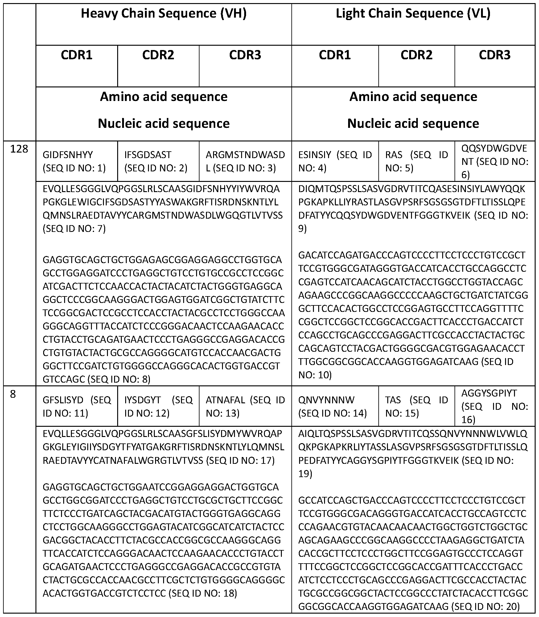

Claims
Priority Applications (7)
| Application Number | Priority Date | Filing Date | Title |
|---|---|---|---|
| US16/762,273 US12570710B2 (en) | 2017-11-07 | 2018-11-06 | Targeting LILRB4 with CAR-T or CAR-NK cells in the treatment of cancer |
| EP18876416.1A EP3707165A4 (en) | 2017-11-07 | 2018-11-06 | TARGETING LILRB4 WITH CAR-T OR CAR-NK CELLS IN CANCER TREATMENT |
| KR1020207014347A KR20200085780A (en) | 2017-11-07 | 2018-11-06 | LILRB4 targeting with CAR-T or CAR-NK cells in the treatment of cancer |
| CA3079999A CA3079999A1 (en) | 2017-11-07 | 2018-11-06 | Targeting lilrb4 with car-t or car-nk cells in the treatment of cancer |
| AU2018365880A AU2018365880A1 (en) | 2017-11-07 | 2018-11-06 | Targeting LILRB4 with car-T or car-NK cells in the treatment of cancer |
| CN201880085571.6A CN111601823B (en) | 2017-11-07 | 2018-11-06 | Targeting LILRB4 in cancer therapy with CAR-T or CAR-NK cells |
| JP2020524547A JP7339944B2 (en) | 2017-11-07 | 2018-11-06 | Methods for targeting LILRB4 using CAR-T cells or CAR-NK cells in cancer treatment |
Applications Claiming Priority (6)
| Application Number | Priority Date | Filing Date | Title |
|---|---|---|---|
| US201762582769P | 2017-11-07 | 2017-11-07 | |
| US62/582,769 | 2017-11-07 | ||
| US201762583825P | 2017-11-09 | 2017-11-09 | |
| US62/583,825 | 2017-11-09 | ||
| US201762584770P | 2017-11-11 | 2017-11-11 | |
| US62/584,770 | 2017-11-11 |
Publications (1)
| Publication Number | Publication Date |
|---|---|
| WO2019094360A1 true WO2019094360A1 (en) | 2019-05-16 |
Family
ID=66438981
Family Applications (1)
| Application Number | Title | Priority Date | Filing Date |
|---|---|---|---|
| PCT/US2018/059362 Ceased WO2019094360A1 (en) | 2017-11-07 | 2018-11-06 | Targeting lilrb4 with car-t or car-nk cells in the treatment of cancer |
Country Status (8)
| Country | Link |
|---|---|
| US (1) | US12570710B2 (en) |
| EP (1) | EP3707165A4 (en) |
| JP (1) | JP7339944B2 (en) |
| KR (1) | KR20200085780A (en) |
| CN (1) | CN111601823B (en) |
| AU (1) | AU2018365880A1 (en) |
| CA (1) | CA3079999A1 (en) |
| WO (1) | WO2019094360A1 (en) |
Cited By (9)
| Publication number | Priority date | Publication date | Assignee | Title |
|---|---|---|---|---|
| US10704021B2 (en) | 2012-03-15 | 2020-07-07 | Flodesign Sonics, Inc. | Acoustic perfusion devices |
| WO2020234632A1 (en) | 2019-05-23 | 2020-11-26 | Ichorlabs, D.O.O | Method for removal of nucleic acids impurities from liquid composition comprising genetically engineered particles or proteins |
| CN112552414A (en) * | 2020-12-29 | 2021-03-26 | 浙江康佰裕生物科技有限公司 | LILRB4 and B7-H3 double-targeting chimeric antigen receptor and application thereof |
| US10975368B2 (en) | 2014-01-08 | 2021-04-13 | Flodesign Sonics, Inc. | Acoustophoresis device with dual acoustophoretic chamber |
| WO2021195536A1 (en) * | 2020-03-27 | 2021-09-30 | The Trustees Of Indiana University | Immunotherapeutic targets in multiple myeloma and methods for their identification |
| US11377651B2 (en) | 2016-10-19 | 2022-07-05 | Flodesign Sonics, Inc. | Cell therapy processes utilizing acoustophoresis |
| US20230065434A1 (en) * | 2019-12-30 | 2023-03-02 | Beijing Dcty Biotech Co., Ltd. | Preparation method of trophoblasts with limited generations, culture method of snk cells and method for treating tumor |
| US11708572B2 (en) | 2015-04-29 | 2023-07-25 | Flodesign Sonics, Inc. | Acoustic cell separation techniques and processes |
| US12202895B2 (en) | 2018-09-13 | 2025-01-21 | The Board Of Regents Of The University Of Texas System | LILRB4 antibodies and uses thereof |
Families Citing this family (6)
| Publication number | Priority date | Publication date | Assignee | Title |
|---|---|---|---|---|
| US12440066B2 (en) | 2018-04-05 | 2025-10-14 | Vinglace Llc | Insulated food and beverage container |
| CN113527507A (en) * | 2020-04-16 | 2021-10-22 | 上海赛比曼生物科技有限公司 | Chimeric antigen receptor targeting CD22 and its preparation and application |
| CN111892661B (en) * | 2020-08-12 | 2021-05-14 | 浙江康佰裕生物科技有限公司 | Chimeric antigen receptor and application thereof in preparation of products for treating tumors |
| EP4376885A4 (en) * | 2021-07-28 | 2025-07-02 | Merck Sharp & Dohme Llc | Method for treating acute myelogenous leukemia with anti-ILT3 antibodies |
| CN115707485A (en) * | 2021-08-19 | 2023-02-21 | 杭州乾合细胞生物科技有限公司 | Method for applying targeted Her-2CAR-NK to tumor treatment |
| CN116712550A (en) * | 2023-07-11 | 2023-09-08 | 中国医学科学院血液病医院(中国医学科学院血液学研究所) | Compositions for the treatment of multiple myeloma with high proliferative activity and multidrug resistance and applications thereof |
Citations (33)
| Publication number | Priority date | Publication date | Assignee | Title |
|---|---|---|---|---|
| US4870287A (en) | 1988-03-03 | 1989-09-26 | Loma Linda University Medical Center | Multi-station proton beam therapy system |
| WO1995001994A1 (en) | 1993-07-09 | 1995-01-19 | Synergen, Inc. | Recombinant ctla4 polypeptides and methods for making the same |
| US5739169A (en) | 1996-05-31 | 1998-04-14 | Procept, Incorporated | Aromatic compounds for inhibiting immune response |
| US5760395A (en) | 1996-04-18 | 1998-06-02 | Universities Research Assoc., Inc. | Method and apparatus for laser-controlled proton beam radiology |
| US5801005A (en) | 1993-03-17 | 1998-09-01 | University Of Washington | Immune reactivity to HER-2/neu protein for diagnosis of malignancies in which the HER-2/neu oncogene is associated |
| WO1998042752A1 (en) | 1997-03-21 | 1998-10-01 | Brigham And Women's Hospital Inc. | Immunotherapeutic ctla-4 binding peptides |
| US5824311A (en) | 1987-11-30 | 1998-10-20 | Trustees Of The University Of Pennsylvania | Treatment of tumors with monoclonal antibodies against oncogene antigens |
| US5830880A (en) | 1994-08-26 | 1998-11-03 | Hoechst Aktiengesellschaft | Gene therapy of tumors with an endothelial cell-specific, cell cycle-dependent active compound |
| US5844905A (en) | 1996-07-09 | 1998-12-01 | International Business Machines Corporation | Extensions to distributed MAC protocols with collision avoidance using RTS/CTS exchange |
| US5846945A (en) | 1993-02-16 | 1998-12-08 | Onyx Pharmaceuticals, Inc. | Cytopathic viruses for therapy and prophylaxis of neoplasia |
| US5885796A (en) | 1991-06-27 | 1999-03-23 | Bristol-Myers Squibb Company | CTLA4 receptor and uses thereof |
| WO2000037504A2 (en) | 1998-12-23 | 2000-06-29 | Pfizer Inc. | Human monoclonal antibodies to ctla-4 |
| WO2001014424A2 (en) | 1999-08-24 | 2001-03-01 | Medarex, Inc. | Human ctla-4 antibodies and their uses |
| US20050260186A1 (en) | 2003-03-05 | 2005-11-24 | Halozyme, Inc. | Soluble glycosaminoglycanases and methods of preparing and using soluble glycosaminoglycanases |
| US20060104968A1 (en) | 2003-03-05 | 2006-05-18 | Halozyme, Inc. | Soluble glycosaminoglycanases and methods of preparing and using soluble glycosaminogly ycanases |
| WO2006121168A1 (en) | 2005-05-09 | 2006-11-16 | Ono Pharmaceutical Co., Ltd. | Human monoclonal antibodies to programmed death 1(pd-1) and methods for treating cancer using anti-pd-1 antibodies alone or in combination with other immunotherapeutics |
| WO2009101611A1 (en) | 2008-02-11 | 2009-08-20 | Curetech Ltd. | Monoclonal antibodies for tumor treatment |
| WO2009114335A2 (en) | 2008-03-12 | 2009-09-17 | Merck & Co., Inc. | Pd-1 binding proteins |
| WO2009155723A2 (en) * | 2008-06-25 | 2009-12-30 | Esbatech, An Alcon Biomedical Research Unit Llc | STABLE AND SOLUBLE ANTIBODIES INHIBITING TNFα |
| WO2010027827A2 (en) | 2008-08-25 | 2010-03-11 | Amplimmune, Inc. | Targeted costimulatory polypeptides and methods of use to treat cancer |
| WO2011066342A2 (en) | 2009-11-24 | 2011-06-03 | Amplimmune, Inc. | Simultaneous inhibition of pd-l1/pd-l2 |
| US8017114B2 (en) | 1999-08-24 | 2011-09-13 | Medarex, Inc. | Human CTLA-4 antibodies and their uses |
| US8119129B2 (en) | 2008-08-01 | 2012-02-21 | Bristol-Myers Squibb Company | Combination of anti-CTLA4 antibody with dasatinib for the treatment of proliferative diseases |
| US8329867B2 (en) | 2010-02-19 | 2012-12-11 | Xencor, Inc. | CTLA4-Ig immunoadhesins |
| US8354509B2 (en) | 2007-06-18 | 2013-01-15 | Msd Oss B.V. | Antibodies to human programmed death receptor PD-1 |
| US20140022021A1 (en) | 2012-07-17 | 2014-01-23 | Murata Manufacturing Co | Power amplifier |
| US8735553B1 (en) | 2013-09-13 | 2014-05-27 | Beigene, Ltd. | Anti-PD1 antibodies and their use as therapeutics and diagnostics |
| US20140271582A1 (en) * | 2013-03-15 | 2014-09-18 | City Of Hope | Cd123-specific chimeric antigen receptor redirected t cells and methods of their use |
| US20140294898A1 (en) | 2013-03-15 | 2014-10-02 | Bristol-Myers Squibb Company | Macrocyclic inhibitors of the pd-1/pd-l1 and cd80(b7-1)/pd-l1 protein/protein interactions |
| WO2016130726A1 (en) * | 2015-02-10 | 2016-08-18 | Minerva Biotechnologies Corporation | Humanized anti-muc1* antibodies |
| WO2016144728A2 (en) * | 2015-03-06 | 2016-09-15 | The Board Of Regents Of The University Of Texas System | Anti-lilrb antibodies and their use in detecting and treating cancer |
| WO2016207240A1 (en) * | 2015-06-24 | 2016-12-29 | F. Hoffmann-La Roche Ag | Anti-transferrin receptor antibodies with tailored affinity |
| US20170095531A1 (en) * | 2015-10-01 | 2017-04-06 | Heat Biologics, Inc. | Compositions and methods for adjoining type i and type ii extracellular domains as heterologous chimeric proteins |
Family Cites Families (6)
| Publication number | Priority date | Publication date | Assignee | Title |
|---|---|---|---|---|
| PH12013501201A1 (en) | 2010-12-09 | 2013-07-29 | Univ Pennsylvania | Use of chimeric antigen receptor-modified t cells to treat cancer |
| EP3783098A1 (en) * | 2013-05-14 | 2021-02-24 | Board Of Regents, The University Of Texas System | Human application of engineered chimeric antigen receptor (car) t-cells |
| CA2945388A1 (en) | 2014-04-23 | 2015-10-29 | Board Of Regents, The University Of Texas System | Chimeric antigen receptors (car) for use in therapy and methods for making the same |
| CA2961950A1 (en) | 2014-09-28 | 2016-03-31 | The Regents Of The University Of California | Modulation of stimulatory and non-stimulatory myeloid cells |
| JP6849600B6 (en) | 2015-01-29 | 2021-06-30 | リージェンツ オブ ザ ユニバーシティ オブ ミネソタ | Chimeric antigen receptor, composition and method |
| US10244644B2 (en) | 2015-12-04 | 2019-03-26 | Continental Automotive Systems, Inc. | Automotive electronic device having a cover with fins to reduce gel vibration |
-
2018
- 2018-11-06 WO PCT/US2018/059362 patent/WO2019094360A1/en not_active Ceased
- 2018-11-06 CN CN201880085571.6A patent/CN111601823B/en active Active
- 2018-11-06 CA CA3079999A patent/CA3079999A1/en active Pending
- 2018-11-06 JP JP2020524547A patent/JP7339944B2/en active Active
- 2018-11-06 AU AU2018365880A patent/AU2018365880A1/en not_active Abandoned
- 2018-11-06 EP EP18876416.1A patent/EP3707165A4/en not_active Withdrawn
- 2018-11-06 KR KR1020207014347A patent/KR20200085780A/en active Pending
- 2018-11-06 US US16/762,273 patent/US12570710B2/en active Active
Patent Citations (36)
| Publication number | Priority date | Publication date | Assignee | Title |
|---|---|---|---|---|
| US5824311A (en) | 1987-11-30 | 1998-10-20 | Trustees Of The University Of Pennsylvania | Treatment of tumors with monoclonal antibodies against oncogene antigens |
| US4870287A (en) | 1988-03-03 | 1989-09-26 | Loma Linda University Medical Center | Multi-station proton beam therapy system |
| US5885796A (en) | 1991-06-27 | 1999-03-23 | Bristol-Myers Squibb Company | CTLA4 receptor and uses thereof |
| US5846945A (en) | 1993-02-16 | 1998-12-08 | Onyx Pharmaceuticals, Inc. | Cytopathic viruses for therapy and prophylaxis of neoplasia |
| US5801005A (en) | 1993-03-17 | 1998-09-01 | University Of Washington | Immune reactivity to HER-2/neu protein for diagnosis of malignancies in which the HER-2/neu oncogene is associated |
| WO1995001994A1 (en) | 1993-07-09 | 1995-01-19 | Synergen, Inc. | Recombinant ctla4 polypeptides and methods for making the same |
| US5830880A (en) | 1994-08-26 | 1998-11-03 | Hoechst Aktiengesellschaft | Gene therapy of tumors with an endothelial cell-specific, cell cycle-dependent active compound |
| US5760395A (en) | 1996-04-18 | 1998-06-02 | Universities Research Assoc., Inc. | Method and apparatus for laser-controlled proton beam radiology |
| US5739169A (en) | 1996-05-31 | 1998-04-14 | Procept, Incorporated | Aromatic compounds for inhibiting immune response |
| US5844905A (en) | 1996-07-09 | 1998-12-01 | International Business Machines Corporation | Extensions to distributed MAC protocols with collision avoidance using RTS/CTS exchange |
| US6207156B1 (en) | 1997-03-21 | 2001-03-27 | Brigham And Women's Hospital, Inc. | Specific antibodies and antibody fragments |
| WO1998042752A1 (en) | 1997-03-21 | 1998-10-01 | Brigham And Women's Hospital Inc. | Immunotherapeutic ctla-4 binding peptides |
| WO2000037504A2 (en) | 1998-12-23 | 2000-06-29 | Pfizer Inc. | Human monoclonal antibodies to ctla-4 |
| WO2001014424A2 (en) | 1999-08-24 | 2001-03-01 | Medarex, Inc. | Human ctla-4 antibodies and their uses |
| US8017114B2 (en) | 1999-08-24 | 2011-09-13 | Medarex, Inc. | Human CTLA-4 antibodies and their uses |
| US20050260186A1 (en) | 2003-03-05 | 2005-11-24 | Halozyme, Inc. | Soluble glycosaminoglycanases and methods of preparing and using soluble glycosaminoglycanases |
| US20060104968A1 (en) | 2003-03-05 | 2006-05-18 | Halozyme, Inc. | Soluble glycosaminoglycanases and methods of preparing and using soluble glycosaminogly ycanases |
| US8008449B2 (en) | 2005-05-09 | 2011-08-30 | Medarex, Inc. | Human monoclonal antibodies to programmed death 1 (PD-1) and methods for treating cancer using anti-PD-1 antibodies alone or in combination with other immunotherapeutics |
| WO2006121168A1 (en) | 2005-05-09 | 2006-11-16 | Ono Pharmaceutical Co., Ltd. | Human monoclonal antibodies to programmed death 1(pd-1) and methods for treating cancer using anti-pd-1 antibodies alone or in combination with other immunotherapeutics |
| US8354509B2 (en) | 2007-06-18 | 2013-01-15 | Msd Oss B.V. | Antibodies to human programmed death receptor PD-1 |
| WO2009101611A1 (en) | 2008-02-11 | 2009-08-20 | Curetech Ltd. | Monoclonal antibodies for tumor treatment |
| US20110008369A1 (en) | 2008-03-12 | 2011-01-13 | Finnefrock Adam C | Pd-1 binding proteins |
| WO2009114335A2 (en) | 2008-03-12 | 2009-09-17 | Merck & Co., Inc. | Pd-1 binding proteins |
| WO2009155723A2 (en) * | 2008-06-25 | 2009-12-30 | Esbatech, An Alcon Biomedical Research Unit Llc | STABLE AND SOLUBLE ANTIBODIES INHIBITING TNFα |
| US8119129B2 (en) | 2008-08-01 | 2012-02-21 | Bristol-Myers Squibb Company | Combination of anti-CTLA4 antibody with dasatinib for the treatment of proliferative diseases |
| WO2010027827A2 (en) | 2008-08-25 | 2010-03-11 | Amplimmune, Inc. | Targeted costimulatory polypeptides and methods of use to treat cancer |
| WO2011066342A2 (en) | 2009-11-24 | 2011-06-03 | Amplimmune, Inc. | Simultaneous inhibition of pd-l1/pd-l2 |
| US8329867B2 (en) | 2010-02-19 | 2012-12-11 | Xencor, Inc. | CTLA4-Ig immunoadhesins |
| US20140022021A1 (en) | 2012-07-17 | 2014-01-23 | Murata Manufacturing Co | Power amplifier |
| US20140271582A1 (en) * | 2013-03-15 | 2014-09-18 | City Of Hope | Cd123-specific chimeric antigen receptor redirected t cells and methods of their use |
| US20140294898A1 (en) | 2013-03-15 | 2014-10-02 | Bristol-Myers Squibb Company | Macrocyclic inhibitors of the pd-1/pd-l1 and cd80(b7-1)/pd-l1 protein/protein interactions |
| US8735553B1 (en) | 2013-09-13 | 2014-05-27 | Beigene, Ltd. | Anti-PD1 antibodies and their use as therapeutics and diagnostics |
| WO2016130726A1 (en) * | 2015-02-10 | 2016-08-18 | Minerva Biotechnologies Corporation | Humanized anti-muc1* antibodies |
| WO2016144728A2 (en) * | 2015-03-06 | 2016-09-15 | The Board Of Regents Of The University Of Texas System | Anti-lilrb antibodies and their use in detecting and treating cancer |
| WO2016207240A1 (en) * | 2015-06-24 | 2016-12-29 | F. Hoffmann-La Roche Ag | Anti-transferrin receptor antibodies with tailored affinity |
| US20170095531A1 (en) * | 2015-10-01 | 2017-04-06 | Heat Biologics, Inc. | Compositions and methods for adjoining type i and type ii extracellular domains as heterologous chimeric proteins |
Non-Patent Citations (37)
| Title |
|---|
| "Genbank", Database accession no. L15006 |
| "Remington: The Science and Practice of Pharmacy", 2003, LIPPINCOTT, WILLIAMS & WILKINS |
| AUSTIN-WARDVILLASECA, REVISTA MEDICA DE CHILE, vol. 126, no. 7, 1998, pages 838 - 845 |
| BARRETT ET AL.: "Interleukin 6 Is Not Made By Chimeric Antigen Receptor T Cells and Does Not Impact Their Function", BLOOD, vol. 128, 2016, pages 654 |
| BUKOWSKI ET AL., CLINICAL CANCER RES., vol. 4, no. 10, 1998, pages 2337 - 2347 |
| CAMACHO ET AL., J CLIN ONCOLOGY, vol. 22, no. 145, 2004 |
| CHEN ET AL.: "Signalling thresholds and negative B-cell selection in acute lymphoblastic leukaemia", NATURE, vol. 521, 2015, pages 357 - 361 |
| CHRISTODOULIDES ET AL., MICROBIOLOGY, vol. 144, 1998, pages 3027 - 3037 |
| DAVIDSON ET AL., J. IMMUNOTHER., vol. 21, no. 5, 1998, pages 389 - 398 |
| FORD ET AL., GENE THERAPY, vol. 8, 2001, pages 1 - 88 |
| HANIBUCHI ET AL., INT. J. CANCER, vol. 78, no. 4, 1998, pages 480 - 485 |
| HEEMSKERK ET AL., HUM GENE THER., vol. 19, 2008, pages 496 - 510 |
| HELLSTRAND ET AL., ACTA ONCOLOGICA, vol. 37, no. 4, 1998, pages 347 - 353 |
| HERMANSON, D.L.KAUFMAN, D.S.: "Utilizing chimeric antigen receptors to direct natural killer cell activity", FRONT IMMUNOL, vol. 6, 2015, pages 195 |
| HOLLANDER, FRONT. IMMUN., vol. 3, pages 3 |
| HUIHASHIMOTO, INFECTION IMMUN., vol. 66, no. 11, 1998, pages 5329 - 5336 |
| HURWITZ ET AL., PROC NATL ACAD SCI USA, vol. 95, no. 17, 1998, pages 10067 - 10071 |
| JOHN ET AL., MOL. THER., vol. 26, no. 10, 2018, pages 2487 - 2495 |
| JOHNSON ET AL., BLOOD, vol. 114, 2009, pages 535 - 46 |
| KANG ET AL.: "Inhibitory leukocyte immunoglobulin-like receptors: Immune checkpoint proteins and tumor sustaining factors", CELL CYCLE,, vol. 15, 2016, pages 25 - 40, XP055439503, DOI: 10.1080/15384101.2015.1121324 |
| KANG ET AL.: "The ITIM-containing receptor LAIR1 is essential for acute myeloid leukaemia development", NAT CELL BIOL, vol. 17, 2015, pages 665 - 677, XP055471891, DOI: 10.1038/ncb3158 |
| LIEBERMAN, PHARMACEUTICAL DOSAGE FORMS, vol. 1-3, 1992 |
| LLOYD, THE ART, SCIENCE AND TECHNOLOGY OF PHARMACEUTICAL COMPOUNDING, 1999 |
| MOKYR ET AL., CANCER RES, vol. 58, 1998, pages 5301 - 5304 |
| PARDOLL, NAT REV CANCER, vol. 12, no. 4, 2012, pages 252 - 64 |
| PICKAR, DOSAGE CALCULATIONS, 1999 |
| PROCHIANTZ, NAT. METHODS, vol. 4, 2007, pages 119 - 20 |
| QIN ET AL., PROC. NATL. ACAD. SCI. USA, vol. 95, no. 24, 1998, pages 14411 - 14416 |
| SAMBROOK ET AL.: "MOLECULAR CLONING, A LABORATORY MANUAL", 1989, COLD SPRINGS HARBOR |
| SCHONFELD ET AL.: "Selective inhibition of tumor growth by clonal NK cells expressing an ErbB2/HER2-specific chimeric antigen receptor", MOL THER, vol. 23, 2015, pages 330 - 338, XP055265087, DOI: 10.1038/mt.2014.219 |
| See also references of EP3707165A4 |
| SHAH ET AL., PLOS ONE, vol. 8, 2013, pages e776781 |
| SINGLETON ET AL.: "DICTIONARY OF MICROBIOLOGY AND MOLECULAR BIOLOGY", 1994, GREENE PUBLISHING ASSOCIATES AND JOHN WILEY & SONS |
| SPANHOLTZ ET AL., PLOS ONE, vol. 6, no. 6, 2011, pages e20740 |
| TERAKURA ET AL., BLOOD, vol. 1, 2012, pages 72 - 82 |
| WANG ET AL., J IMMUNOTHER., vol. 35, no. 9, 2012, pages 689 - 701 |
| ZHENG ET AL.: "Inhibitory receptors bind ANGPTLs and support blood stem cells and leukaemia development", NATURE, vol. 485, 2012, pages 656 - 660, XP055204718, DOI: 10.1038/nature11095 |
Cited By (10)
| Publication number | Priority date | Publication date | Assignee | Title |
|---|---|---|---|---|
| US10704021B2 (en) | 2012-03-15 | 2020-07-07 | Flodesign Sonics, Inc. | Acoustic perfusion devices |
| US10975368B2 (en) | 2014-01-08 | 2021-04-13 | Flodesign Sonics, Inc. | Acoustophoresis device with dual acoustophoretic chamber |
| US11708572B2 (en) | 2015-04-29 | 2023-07-25 | Flodesign Sonics, Inc. | Acoustic cell separation techniques and processes |
| US11377651B2 (en) | 2016-10-19 | 2022-07-05 | Flodesign Sonics, Inc. | Cell therapy processes utilizing acoustophoresis |
| US12202895B2 (en) | 2018-09-13 | 2025-01-21 | The Board Of Regents Of The University Of Texas System | LILRB4 antibodies and uses thereof |
| WO2020234632A1 (en) | 2019-05-23 | 2020-11-26 | Ichorlabs, D.O.O | Method for removal of nucleic acids impurities from liquid composition comprising genetically engineered particles or proteins |
| US20230065434A1 (en) * | 2019-12-30 | 2023-03-02 | Beijing Dcty Biotech Co., Ltd. | Preparation method of trophoblasts with limited generations, culture method of snk cells and method for treating tumor |
| WO2021195536A1 (en) * | 2020-03-27 | 2021-09-30 | The Trustees Of Indiana University | Immunotherapeutic targets in multiple myeloma and methods for their identification |
| CN112552414A (en) * | 2020-12-29 | 2021-03-26 | 浙江康佰裕生物科技有限公司 | LILRB4 and B7-H3 double-targeting chimeric antigen receptor and application thereof |
| CN112552414B (en) * | 2020-12-29 | 2022-05-27 | 浙江康佰裕生物科技有限公司 | LILRB4 and B7-H3 double-targeting chimeric antigen receptor and application thereof |
Also Published As
| Publication number | Publication date |
|---|---|
| AU2018365880A1 (en) | 2020-05-28 |
| CN111601823A (en) | 2020-08-28 |
| KR20200085780A (en) | 2020-07-15 |
| US12570710B2 (en) | 2026-03-10 |
| EP3707165A4 (en) | 2021-09-08 |
| CA3079999A1 (en) | 2019-05-16 |
| JP2021502808A (en) | 2021-02-04 |
| CN111601823B (en) | 2024-09-24 |
| JP7339944B2 (en) | 2023-09-06 |
| US20210179687A1 (en) | 2021-06-17 |
| EP3707165A1 (en) | 2020-09-16 |
Similar Documents
| Publication | Publication Date | Title |
|---|---|---|
| US12570710B2 (en) | Targeting LILRB4 with CAR-T or CAR-NK cells in the treatment of cancer | |
| US11730761B2 (en) | Combination immune therapy and cytokine control therapy for cancer treatment | |
| US11963980B2 (en) | Activated CD26-high immune cells and CD26-negative immune cells and uses thereof | |
| WO2021053667A2 (en) | Combination cancer therapy and cytokine control therapy for cancer treatment | |
| US12180277B2 (en) | LILRB4-binding antibody and methods of use thereof | |
| JP2024537176A (en) | Natural killer cells and methods of use thereof | |
| US10821134B2 (en) | BK virus specific T cells | |
| US20210401887A1 (en) | T cells from lymphatic fluid for diagnostic and therapeutic use | |
| EP4031655A2 (en) | Combination cancer therapy and cytokine control therapy for cancer treatment | |
| US20190255111A1 (en) | Cd38-nad+ regulated metabolic axis in anti-tumor immunotherapy | |
| JP2025540600A (en) | Anti-ZP4 antibodies and chimeric antigen receptors and methods of using them | |
| KR20260016499A (en) | Compositions comprising dendritic cell exosomes and methods of using the same | |
| HK40129647A (en) | Anti-zp4 antibodies and chimeric antigen receptors and methods of use thereof | |
| HK40110020A (en) | Natural killer cells and methods of use thereof |
Legal Events
| Date | Code | Title | Description |
|---|---|---|---|
| 121 | Ep: the epo has been informed by wipo that ep was designated in this application |
Ref document number: 18876416 Country of ref document: EP Kind code of ref document: A1 |
|
| ENP | Entry into the national phase |
Ref document number: 3079999 Country of ref document: CA |
|
| ENP | Entry into the national phase |
Ref document number: 2020524547 Country of ref document: JP Kind code of ref document: A |
|
| NENP | Non-entry into the national phase |
Ref country code: DE |
|
| ENP | Entry into the national phase |
Ref document number: 20207014347 Country of ref document: KR Kind code of ref document: A |
|
| ENP | Entry into the national phase |
Ref document number: 2018365880 Country of ref document: AU Date of ref document: 20181106 Kind code of ref document: A |
|
| ENP | Entry into the national phase |
Ref document number: 2018876416 Country of ref document: EP Effective date: 20200608 |
|
| WWW | Wipo information: withdrawn in national office |
Ref document number: 2018876416 Country of ref document: EP |
|
| WWG | Wipo information: grant in national office |
Ref document number: 16762273 Country of ref document: US |