WO2019146738A1 - ストレス状態の検出方法、及び、ストレス検出装置 - Google Patents
ストレス状態の検出方法、及び、ストレス検出装置 Download PDFInfo
- Publication number
- WO2019146738A1 WO2019146738A1 PCT/JP2019/002394 JP2019002394W WO2019146738A1 WO 2019146738 A1 WO2019146738 A1 WO 2019146738A1 JP 2019002394 W JP2019002394 W JP 2019002394W WO 2019146738 A1 WO2019146738 A1 WO 2019146738A1
- Authority
- WO
- WIPO (PCT)
- Prior art keywords
- stress
- choroid
- stress state
- choroidal
- detecting
- Prior art date
- Legal status (The legal status is an assumption and is not a legal conclusion. Google has not performed a legal analysis and makes no representation as to the accuracy of the status listed.)
- Ceased
Links
Images




Classifications
-
- A—HUMAN NECESSITIES
- A61—MEDICAL OR VETERINARY SCIENCE; HYGIENE
- A61B—DIAGNOSIS; SURGERY; IDENTIFICATION
- A61B3/00—Apparatus for testing the eyes; Instruments for examining the eyes
- A61B3/10—Objective types, i.e. instruments for examining the eyes independent of the patients' perceptions or reactions
- A61B3/14—Arrangements specially adapted for eye photography
-
- A—HUMAN NECESSITIES
- A61—MEDICAL OR VETERINARY SCIENCE; HYGIENE
- A61B—DIAGNOSIS; SURGERY; IDENTIFICATION
- A61B5/00—Measuring for diagnostic purposes; Identification of persons
- A61B5/16—Devices for psychotechnics; Testing reaction times ; Devices for evaluating the psychological state
- A61B5/165—Evaluating the state of mind, e.g. depression, anxiety
-
- A—HUMAN NECESSITIES
- A61—MEDICAL OR VETERINARY SCIENCE; HYGIENE
- A61B—DIAGNOSIS; SURGERY; IDENTIFICATION
- A61B3/00—Apparatus for testing the eyes; Instruments for examining the eyes
- A61B3/10—Objective types, i.e. instruments for examining the eyes independent of the patients' perceptions or reactions
- A61B3/102—Objective types, i.e. instruments for examining the eyes independent of the patients' perceptions or reactions for optical coherence tomography [OCT]
-
- A—HUMAN NECESSITIES
- A61—MEDICAL OR VETERINARY SCIENCE; HYGIENE
- A61B—DIAGNOSIS; SURGERY; IDENTIFICATION
- A61B3/00—Apparatus for testing the eyes; Instruments for examining the eyes
- A61B3/10—Objective types, i.e. instruments for examining the eyes independent of the patients' perceptions or reactions
- A61B3/12—Objective types, i.e. instruments for examining the eyes independent of the patients' perceptions or reactions for looking at the eye fundus, e.g. ophthalmoscopes
- A61B3/1225—Objective types, i.e. instruments for examining the eyes independent of the patients' perceptions or reactions for looking at the eye fundus, e.g. ophthalmoscopes using coherent radiation
-
- A—HUMAN NECESSITIES
- A61—MEDICAL OR VETERINARY SCIENCE; HYGIENE
- A61B—DIAGNOSIS; SURGERY; IDENTIFICATION
- A61B5/00—Measuring for diagnostic purposes; Identification of persons
- A61B5/0059—Measuring for diagnostic purposes; Identification of persons using light, e.g. diagnosis by transillumination, diascopy, fluorescence
- A61B5/0073—Measuring for diagnostic purposes; Identification of persons using light, e.g. diagnosis by transillumination, diascopy, fluorescence by tomography, i.e. reconstruction of 3D images from 2D projections
-
- A—HUMAN NECESSITIES
- A61—MEDICAL OR VETERINARY SCIENCE; HYGIENE
- A61B—DIAGNOSIS; SURGERY; IDENTIFICATION
- A61B5/00—Measuring for diagnostic purposes; Identification of persons
- A61B5/0059—Measuring for diagnostic purposes; Identification of persons using light, e.g. diagnosis by transillumination, diascopy, fluorescence
- A61B5/0062—Arrangements for scanning
- A61B5/0066—Optical coherence imaging
Definitions
- the present invention relates to a stress state detection method and a stress detection device.
- stress is the cause of all diseases
- stress adversely affects the mind and body and causes various diseases.
- Stress means a state in which the mind and body are distorted due to mental and physical stress caused by various external stimuli.
- animals such as humans sense external stress such as temporary mental and physical stress, hormones such as cortisol and adrenaline are secreted from the adrenal gland and a moderate stimulus is given to the body.
- the body is strengthened by increasing the resistance to external stress through an increase in heart rate, dilation of the heart and blood vessels, an increase in blood sugar levels, bronchial relaxation and the like.
- the Ministry of Health, Labor and Welfare has introduced a stress check system that defines examinations for grasping the degree of psychological burden on workers and implementation of interview guidance by doctors based on examination results.
- workers respond to questions regarding factors that are considered to be occupational stress, physical and mental responses caused by stress, and other factors that affect stress responses, and the answers to each question are scored Do.
- These results are tabulated and analyzed for each group of a certain size, and highly stressed persons are selected by score based on numerical criteria.
- the stress check is a subjective test in which the workers themselves answer the question items, it is possible to manipulate the answers intentionally so that there is no stress, so it is possible to appropriately select a high stressed person. It may not be possible. Therefore, it is important to select an actual highly stressed person by combining subjective examination and objective examination.
- Patent Document 3 Furthermore, as a method of evaluating chronic stress, a method of using daily time-course changes of cortisol in saliva as an index has been reported (see Patent Document 3). According to the method described in Patent Document 3, a standard cortisol concentration change range obtained from saliva cortisol concentration over a predetermined time range measured in a large number of healthy persons, and in saliva measured in a specific subject It compares with the cortisol concentration change of and evaluates those who deviated from the standard cortisol change range as having the possibility of having chronic stress.
- the stress marker due to the above-mentioned physiological chemical substance is usually decomposed and eliminated in a short time in a living body even if it appears in response to stress.
- the half-life in blood of catecholamines such as adrenalin is as short as 1 to 2 minutes. Therefore, the measured value may not reflect the stress state of the living body, and it is difficult to accurately evaluate the stress state of the subject.
- the in-vivo concentration of the physiological chemical has a large physiological diurnal fluctuation, and also has a large individual difference due to differences in sleep awakening rhythm among individuals. For example, cortisol exhibits daily fluctuation of morning high and low at night, and is less than half of the early morning value after the evening.
- the present inventors have found that the stress state of a subject can be evaluated using a choroid, which is a part of ocular tissue, as an index.
- a choroid which is a part of ocular tissue, as an index.
- the thickness or volume of the choroid accurately reflects the stress state of the subject.
- Such choroidal changes can be objectively detected, and the stress state of the object can be accurately detected.
- optical coherence tomography it can be detected noninvasively and in a short time and easily.
- the present inventors have completed the present invention based on these findings.
- the present invention provides the inventions shown in the following [1] to [7] in order to solve the above-mentioned problems.
- a method of detecting a stress state comprising the step of detecting a stress state of the object based on a tomographic image of a choroid of the object.
- the configuration of the above [1] it is possible to provide a method of detecting a stress state in which the stress state of the object is detected by using the choroid as an index based on the tomographic image of the choroid which is a part of eye tissue of the object.
- the stress state of the object can be objectively detected, and the choroid can accurately reflect the stress state of the living body, so that the stress state of the object can be accurately detected.
- the tomographic image can be acquired by optical coherence tomography in which the eye is irradiated with near-infrared light, and the stress state can be detected non-contactingly, non-invasively, and in a short time and conveniently. And physical burden can be reduced. Therefore, the stress load upon detection can be eliminated, and the stress state of the object can be accurately detected. Furthermore, there is also an advantage that it is not susceptible to eye movement.
- the stress state detection method of this configuration it is possible to detect the object in a stress state with high reliability. For subjects detected as under stress, consultation with a suitable medical institution and counseling can be recommended.
- the method of detecting a stress state of the present configuration it is possible to detect a latent stress state in which stress is not manifested as a specific symptom, disease or the like. Therefore, a stress state can be detected early, which can lead to prevention of symptoms and diseases developed by stress. Therefore, the method of detecting stress conditions according to the present invention can be suitably used for regular health check etc. of workers and can contribute to the physical and mental health management of workers. As a result, it leads to the improvement of the work environment, the work efficiency rises, and it can contribute to the improvement of labor productivity.
- the stress state of the subject is detected using the thickness of the choroid as an index. It can provide a detection method. Since the choroidal thickness which can be clearly and easily calculated from the tomographic image of the choroid is used as the index of the stress state, the stress state of the object can be detected more simply and accurately. In particular, according to the configuration of the above [3], the stress state of the object can be detected based on the clear reference value, and further improvement of simplicity and accuracy in detection of the stress state can be expected.
- the stress state detecting step calculates the volume of the choroid from the tomographic image, and detects the stress state of the subject based on the calculated volume of the choroid. Detection method.
- the configuration of the above [4] it is possible to provide a method of detecting a stress state in which the stress state of the object is detected using the volume of the choroid as an index based on the tomographic image of the choroid which is a part of the eye tissue of the object . Since the volume of the choroid that can be clearly and easily calculated from the tomographic image of the choroid such as a three-dimensional image is used as an index, the stress state of the object can be detected more simply and accurately.
- the step of detecting the stress state includes selecting the calculated choroidal thickness as at least one of a detection time, an age of the subject, a gender, a current medical history, and an index obtained from an eye structure.
- the calculated choroidal thickness of the subject is detected at the detection time that affects the choroidal thickness, the age, sex, current medical history of the subject, and axial length and equivalent sphere area etc.
- the step of detecting the stress state includes selecting at least one of the calculated volume of the choroid from the detection time, the age of the subject, the gender, the current medical history, and the index obtained from the eye structure.
- the calculated volume of the choroid of the subject is detected at the detection time affecting the volume of the choroid, the age, sex, current medical history of the subject, and the axial length and equivalent sphere area
- the correction method using at least one factor selected from the indices obtained from the eye structure and the like can provide a detection method of a stress state that detects the stress state of the object more accurately.
- An image acquisition unit for acquiring a tomographic image of the choroid of the target object, A calculation unit that calculates a choroidal thickness or a choroidal volume from the tomographic image; And a detection unit configured to detect a stress state of the subject based on the choroidal thickness or the choroidal volume.
- a stress detection device for detecting the stress state of the object using the choroidal thickness or the choroidal volume as an index based on the tomographic image of the choroid which is a part of the eye tissue of the object.
- the stress detection device of this configuration uses the choroidal thickness or the choroidal volume that accurately reflects the stress state of the living body as an indicator of the stress state of the object, so it can detect the stress state of the object with high reliability.
- the stress state of the subject can be detected in a non-contact, non-invasive manner, in a short time and easily, so that the mental and physical body of the subject Burden can be reduced. Therefore, the stress load upon detection can be eliminated, and the stress state of the object can be accurately detected.
- the stress detection device of the present configuration can be suitably used for regular health checkup and the like of workers, it can contribute to mental and physical health management of workers through installation in each health checkup center and medical facility.
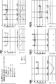
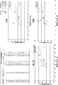
- Example 1 which examined about the relation between choroidal thickness and stress and anxiety is shown, and this figure is a graph showing the result of verifying the relation between choroidal thickness and subjective stress.
- the result of Example 1 which examined about the relation between choroidal thickness and stress and anxiety is shown, and this figure is a graph showing the result of verifying the relation between choroidal thickness and subjective stress.
- the result of Example 1 which examined about the relation between choroidal thickness and stress and anxiety is shown, and this figure is a graph showing the result of verifying the relation between choroidal thickness and subjective stress.
- the result of Example 1 which examined about the relation between choroidal thickness and stress and anxiety is shown, and this figure is a graph showing the result of verifying the relation between choroidal thickness and subjective stress.
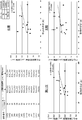
- Example 1 The results of Example 1 in which the relationship between choroidal thickness and stress and anxiety were examined are shown, and this figure shows the occupation in the verification of the relationship between choroidal thickness and the result of the occupational stress simple questionnaire (57 items) The results of the sexual stress simple questionnaire (57 items) are shown.
- the results of Example 1 in which the relationship between choroidal thickness and stress and anxiety were examined are shown, and this figure shows the physical and mental responses caused by the stress of choroidal thickness and the occupational stress simple questionnaire (57 items) (region B) It is a graph which shows the verification result of relevancy with.
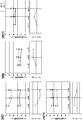
- Example 1 The result of Example 1 in which the relationship between choroidal thickness and stress and anxiety was examined is shown, and this figure shows the physical and mental caused by the stress of choroidal thickness / axial length and the occupational stress simple questionnaire (57 items) It is a graph which shows the verification result of relevance with reaction (B area
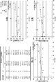
- the results of Example 1 in which the relationship between choroidal thickness and stress and anxiety were examined are shown, and this figure is a verification of the relationship between choroidal thickness and the results of the Japanese version of the mood / anxiety disorder questionnaire (K6).
- K6 The results of the Japanese version of the Mood / Anxiety Disorder Questionnaire (K6) and the correlation with the mind-body response (area B) caused by stress on the Occupational Stress Simple Questionnaire (57 items) are shown.
- Example 1 The results of Example 1 in which the relationship between choroidal thickness and stress and anxiety were examined are shown, and this figure is a verification of the relationship between choroidal thickness and the results of the Japanese version of the mood / anxiety disorder questionnaire (K6). It is a graph which shows a result.
- the results of Example 1 in which the relationship between choroidal thickness and stress and anxiety was examined are shown, and this figure shows the results of choroidal thickness / axial length and the Japanese version of the mood / anxiety disorder questionnaire (K6). It is a graph which shows the verification result of relevance.
- the method of detecting a stress state detects a stress state of a target object by using the choroid of the target body as an index.
- the detection method uses the choroid, which is a part of eye tissue that has not been recognized as a stress marker in the past, as an indicator of the stress state of the subject.
- Subjects in the method for detecting a stress state according to the present embodiment include humans, non-human primates, rabbits, rats, guinea pigs, mice, dogs, cats, cats, horses, cows, pigs, sheep, goats, chickens, etc. Although an animal is exemplified, a human is particularly preferable.
- the stress state in the method of detecting a stress state means a tension state generated by exerting a physical and mental load by various stimuli.
- the stimuli that induce stress are called stressors, and there are stressors due to environmental factors, physical factors, psychological factors, social factors, and the like.
- Environmental factors include, for example, temperature, humidity, light, weather, noise, vibration, exposure to harmful substances, air pollution and the like.
- Physical factors include, for example, trauma, illness, excessive exercise, lack of exercise, physical fatigue, lack of sleep, lack of nutrition, aging, unhealthy lifestyle, obesity and the like.
- reactive oxygen species and the like generated in excess can be included as physical factors.
- Psychological factors include, for example, anxiety, anxiety, frustration, anger, fear, disappointment, conflict and the like.
- Social factors include human relations, employment, career change, unemployment, admission, going to school, poor grades, bullying and so on. However, it is not limited to these.
- the stress state to be detected in the method of detecting a stress state according to the present embodiment is not particularly limited as long as it is a state induced by a stressor, and is any of a short-term acute stress state and a long-term chronic stress state. May be
- the stress state includes stress-related symptoms and diseases (hereinafter, may be abbreviated as “stress symptoms” and “stress diseases”, respectively), and may be a latent condition in which symptoms and diseases do not manifest in the mind and body. Stress conditions can also be targeted for detection. When exposed to stimuli such as stressors, the living body maintains its homeostasis through the interaction of the autonomic nervous system, endocrine system, immune system and the like.
- stress conditions can preferably include sympathetic dominant states, and symptoms and diseases induced by such sympathetic dominant states.
- Stress symptoms include, for example, physical symptoms such as stiff neck, palpitations, dizziness, insomnia, headache, weight fluctuation, loss of appetite, eating disorder, abdominal pain, addiction, etc., psychological symptoms such as anxiety, anger, nervousness, apathy, etc. It includes social symptoms such as overeating, alcohol consumption and withdrawal.
- stress diseases include mental and neurological diseases such as depression, neurosis and autonomic imbalance, cardiovascular diseases such as hypertension, arteriosclerosis, angina, myocardial infarction, obesity, diabetes, hyperlipidemia, Endocrine and metabolic disorders such as hyperthyroidism, respiratory diseases such as secretory bronchitis and hyperpnea syndrome, irritable bowel syndrome, gastric and duodenal ulcers, digestive tract diseases such as ulcerative colitis and psychogenic vomiting, Skin diseases such as atopic dermatitis, psoriasis, alopecia areata, central serous choroidal retinopathy (CSC), eye strain, eyelid spasms, eye diseases such as dry eye, otolaryngologic diseases such as Meniere's disease, enuresis Urinary and genital diseases such as cancer, inflammatory diseases such as cancer and chronic inflammation can be included.
- cardiovascular diseases such as hypertension, arteriosclerosis, angina, myocardial infarction, obesity, diabetes, hyperlipidemia, En
- mental and neurological diseases are considered, for example, anxiety which is a disease that suffers from excessive anxiety and fear caused by excessive anxiety and fear represented by panic disorder, social anxiety disorder, general anxiety disorder, phobia etc.
- Disorders and mood disorders which are disorders that cause an abnormality such as bipolar disorder, depression, and dysthymic disorder, are included.
- Stress conditions include factors such as various symptoms and exacerbation of diseases, so stress symptoms and diseases include not only onset of various symptoms and diseases but also exacerbation.
- the choroid which is an index for detecting a stress state, is a tissue rich in pigment cells and blood vessels between the retina and the sclera, and histologically, from the retina side It can be distinguished into four layers: basal plate (Bruch's membrane), choriocapillaris plate, blood vessel plate, and choroidal plate.
- basal plate Bruch's membrane
- choriocapillaris plate choriocapillaris plate
- blood vessel plate choroidal plate.
- the choroid is a tissue composed mainly of blood vessels, and it has been reported that the choroidal blood flow reaches 85% of the total eye blood flow (see, for example, Experimental Eye Research, 15 (1), 1973, p15-29) about). This leads to strong influence of physiological chemicals.
- choroidal blood vessels unlike retinal blood vessels, have been reported to have low self-regulatory ability, and it has been reported that blood flow through choroidal blood vessels fluctuates due to various physiological stimuli such as blood pressure (for example, Arch Ophthalmol, 83 (1) , 1970, p 95-99), choroidal vessels are strongly affected by changes in the general state of the body. Furthermore, it is known that the choroidal blood vessels express receptors that affect vascular tone, and in particular, it is known that choroidal blood flow is changed in pregnancy and hypertension.
- the method of detecting a stress state uses the choroid, which is a part of ocular tissue as described above, as an index of the stress state.
- the choroid is considered to be able to reflect changes in blood vessels throughout the body, and is considered to properly reflect the stress state of the living body.
- conventionally there has been no knowledge on the relationship between a stress state and ocular tissues including choroid.
- the method of detecting a stress state is preferably to detect a stress state of the object based on a tomographic image of the choroid of the object. From the tomographic image of the choroid, the state including the form of the choroid such as the choroidal thickness and the volume of the choroid can be grasped in detail.
- the tomographic image of the choroid is a tomographic image of an eye including the choroid, preferably a tomographic image of a fundus including the choroid. It is preferable that the boundary between the choroid and the retina and the boundary between the choroid and the sclera can be extracted in order to calculate the characteristic value of the choroid such as the thickness and volume of the choroid. Therefore, it is preferable that the tomographic image of the choroid includes at least a part of the retina and a part of the sclera in addition to the choroid.
- the tomographic image of the choroid may be, for example, a tomographic image of a region including the subfoveal region of the retina or a region including a position separated by a predetermined distance in a predetermined direction from the fovea relative to the fovea of the retina. it can. Further, a tomographic image of a region including the optic papilla, and a region including a position separated by a predetermined distance in a predetermined direction from the optic nerve papilla with respect to the optic nerve papilla can be used. Furthermore, it may be a tomographic image including a plurality of regions.
- the tomographic image of the choroid may be any of a one-dimensional image, a two-dimensional image, and a three-dimensional image.
- Tomographic images of the choroid can be obtained by tomographic imaging of the choroid using techniques known in the art.
- Tomographic imaging of the choroid can be performed preferably using an apparatus based on optical coherence tomography (hereinafter referred to as “OCT”) (hereinafter referred to as “OCT apparatus”).
- OCT apparatus is an interference optical apparatus that branches a light beam emitted from a light source and causes the reflected or scattered light of measurement light incident on a predetermined position of an object to interfere with reference light reflected from a reference object. This is a technology that uses interference light to image the structure of an object in the depth direction of the position where measurement light is incident, and can obtain a tomographic image of the object with high resolution.
- the OCT apparatus can acquire a tomographic image of the fundus including the choroid noninvasively and with high resolution by irradiating the fundus with the measuring light.
- the OCT apparatus is an apparatus capable of objectively inspecting the choroid, and since it is a short-time non-invasive inspection apparatus generally performed daily in ophthalmologic examination, the burden on the object is also small.
- the OCT apparatus is a time domain OCT (hereinafter referred to as "TD-OCT") that performs light wave interference in the time domain, and a Fourier domain OCT (frequency-domain OCT: below) that performs light wave interference in the frequency domain or wavelength domain. Or the like can be used, and preferably FD-OCT.
- TD-OCT time domain OCT
- frequency-domain OCT frequency-domain OCT: below
- FD-OCT Spectral domain OCT
- SD-OCT Spectral-domain OCT
- SS And the like, etc. are exemplified.
- it is SS-OCT.
- EDI-OCT based on enhanced intensity imaging (hereinafter referred to as “EDI”) or high penetration (hereinafter referred to as "HP-OCT”) using a long wavelength light source
- EDI enhanced intensity imaging
- HP-OCT high penetration
- a light source a light source emitting light with a wavelength of 1 ⁇ m band, typically about 950 nm to about 1100 nm, particularly 1050 nm, is preferable.
- 1 ⁇ m band light source it is less susceptible to absorption of light in the retinal pigment epithelium or the like and scattering in the case where there is turbidity in the intermediate translucent material, etc. Images can be acquired.
- a one-dimensional tomographic image (z image) of the fundus including the choroid in the depth direction (z direction) of the irradiation position by irradiating the fundus of the subject with the measurement light, it is possible to acquire a one-dimensional tomographic image (z image) of the fundus including the choroid in the depth direction (z direction) of the irradiation position. Furthermore, a two-dimensional tomographic image (xz image) of the fundus including the choroid can be acquired by one-dimensionally scanning the irradiation position in the direction (x direction) perpendicular to the depth direction with respect to the fundus of the object. One-dimensional scanning can be performed along a predetermined straight line or curve. Thereby, it is possible to obtain a two-dimensional tomographic image in the depth direction along the scanning direction of the measurement light.
- a three-dimensional image (xyz image) including the choroid can be acquired by two-dimensionally scanning the irradiation position in the direction (x, y directions) perpendicular to the depth direction with respect to the fundus. At this time, two-dimensional scanning can be performed within a predetermined area. That is, a three-dimensional tomographic image can be constructed from a plurality of two-dimensional images acquired by repeatedly scanning the measurement light while shifting the position with respect to the fundus of the object in a predetermined area. Thus, the structure of the fundus including the choroid can be captured in three dimensions.
- SS-OCT can scan a wide range of about 3 to 12 mm in the x direction and y direction in a few seconds and acquire an image in the deep direction, so a clear three-dimensional tomographic image of the fundus including the choroid can be constructed. It becomes possible.
- the time required for acquiring a tomographic image of the choroid by the OCT apparatus is about 3 seconds, and there is also an advantage of being less susceptible to eye movement.
- a tomographic image of the choroid it is preferable to acquire a tomographic image of the choroid by avoiding during or immediately after exercise.
- choroidal thickness it has been reported that the choroid has become significantly thickened for at least 5 minutes during exercise with low load and moderate intensity (for example, Sayin N et al., Indian J Ophthalmol. 2015, 63). (5), 445-450).
- acquisition of a tomographic image of the choroid is generally performed in the sitting position, and may be a tomographic image of the choroid of either the left or right eye, or may be a tomographic image of the choroid of both eyes.
- the step of detecting a stress state may include calculating a choroidal thickness from the tomographic image and detecting a stress state of the object based on the calculated choroidal thickness.
- the choroidal thickness is the thickness of the choroid, and can be calculated from the tomographic image of the choroid as, for example, the vertical distance from the border between the retinal pigment epithelium located at the outermost side of the retina and the choroid to the border between the choroid and sclera.
- the localization of the retinal pigment epithelium-choroid interface and the localization of the choroid-sclera interface can be identified using image processing techniques known in the art. For example, the positions of these boundaries can be identified from changes in pixel values (for example, luminance values) in the tomographic image.
- the choroidal thickness may be calculated, for example, by using the calculation function of a computer.
- the calculation of the choroidal thickness is preferably performed at a predetermined position of the choroid.
- the position may be a position of the fovea of the retina or a position separated from the fovea by a predetermined distance in a predetermined direction (for example, upper, lower, nasal side, ear side) with reference to the fovea of the retina .
- the position may be measured at the position of the optic nerve head or at a position away from the optic disk by a predetermined distance in a predetermined direction with reference to the optic disk.
- the choroidal thickness may be measured at a plurality of positions. Particularly preferred is the choroidal thickness below the fovea of the retina.
- the calculation of choroidal thickness may be performed on the choroid of either the left or right eye, or may be performed on the choroid of both eyes. Also, after calculating the choroidal thickness of both eyes, only the thin or thick choroid may be used for detecting the stress state, but the thin one with high correlation of the stress state is used for detection Is preferred.
- the subject in a stressed state exhibits a significantly higher value of choroidal thickness as compared to a healthy non-stressed normal control group.
- choroidal blood vessels dilate and choroids become thick due to overstimulation of the autonomic nerve.
- detection of an object in a stress state is a reference value calculated in advance from a choroidal thickness calculated from a tomogram of the choroid of the object and, for example, a tomogram of a choroid in a healthy object not in a stress state.
- the object is detected as being in a stress state, and if the value is lower than the reference value, the object can be detected not in a stress state.
- selection of healthy body which is not in the stress state used for calculation of standard value such as occupational stress simple questionnaire (57 items) which Japanese Ministry of Health, Labor and Welfare recommends, Japanese version mood / anxiety disorder questionnaire (K6) And other known methods of detecting stress such as a stress marker, and a plurality of methods of detecting stress are preferably performed.
- the stress state can be detected with the reference value.
- a reference value for example, in the case of an adult, it can be set to any value of 250 ⁇ m, 300 ⁇ m or less under the fovea, and preferably can be set to any value from 200 to 300 ⁇ m under the fovea.
- the calculated choroidal thickness of the object is obtained from the detection time, the age, sex, medical history of the object, and an index obtained from the eye structure such as axial length and equivalent sphere area Correction may be included using at least one factor selected from For example, choroidal thickness is negatively correlated with age, and it has been reported that choroidal thinning with age (Wakatsuki Y et al., PLoS One. 2015 Dec 3; 10 (12): e0144156).
- the choroidal thickness is diurnal, and that the choroid is thick in the early morning but becomes gradually thinner in the daytime, and there is no difference in the fluctuation width of the choroidal thickness (for example, See Tan CS et al., Invest Opthalmol Vis Sci, 2012; 53 (1): 261-266 etc.).
- the choroidal layer thickness is affected by some eye diseases and systemic pathologies, and, for example, in central serous chorioretinosis or Harada disease, the choroidal layer thickness is significantly thickened, conversely, strong myopia, age-related macular degeneration It has been reported that glaucoma, diabetic retinopathy, etc. are thinning.
- the eye axis is short, or It has been reported that the thicker the equivalent sphere area, the thicker the choroid. Furthermore, flatter cornea, it has been reported that the choroid is thicker as the correction vision is better.
- the choroidal film thickness calculated from the group with the above factor at the standard value and not in the stress state is used as the standard choroidal film thickness. Determined as Then, when the above factor is not a standard value, a ratio of a standard choroidal film thickness to a choroidal film thickness calculated from a group not in a stress state (standard choroidal film thickness / calculated choroidal film thickness) is used as a correction coefficient. By multiplying the correction film thickness by the correction coefficient, it is possible to correct the correction film thickness calculated from an object having no corresponding standard factor.
- the correction can be made based on the correlation.
- the factor is gender
- the choroidal thickness of the other sex can be corrected based on the choroidal thickness of one sex.
- the choroidal thickness calculated from the group not having any of the above factors and not in the stress state is used as a standard.
- the ratio of standard choroidal film thickness to choroidal film thickness calculated from a group not in a stressed state having any one of the above factors (standard choroidal film thickness / calculated choroidal film thickness) as choroidal film thickness is used as a correction coefficient Based on this, it is possible to correct the choroidal film thickness calculated from the object having the corresponding factor.
- the correction may be performed, for example, by using the calculation function of a computer, as in the calculation of the choroidal film thickness.
- the step of detecting the stress state may include calculating a choroidal volume from the tomographic image and detecting a stress state of the subject based on the calculated volume of the choroid.
- the volume of the choroid can be a volume of a portion surrounded by the retinal pigment epithelium located at the outermost side of the retina and the border of the choroid and the border between the choroid and the sclera, and can be calculated based on the choroidal thickness described above.
- the calculation of the choroidal volume may be performed, for example, by using the computing function of a computer.
- the calculation of the choroidal volume is preferably performed in a predetermined area of the choroid.
- the position of the fovea of the retina, or a direction (x direction) perpendicular to a predetermined direction (deep direction (z direction) from the fovea relative to the fovea of the retina, eg, upper, lower, nasal side It can be calculated as the volume of the choroid in a region whose center point is a position separated by a predetermined distance on the ear side).
- the volume of the choroid may be calculated as the central point of the position of the optic nerve head or the position at a predetermined distance from the optic nerve head in a predetermined direction with reference to the position of the optic nerve head.
- the volume of the choroid in a plurality of regions may be calculated. Particularly preferred is the volume of the choroid in a region centered on the position of the fovea of the retina.
- the size of the area for calculating the volume of the choroid is not particularly limited, but can be, for example, 3 mm ⁇ 3 mm, 6 mm ⁇ 6 mm, 10 mm ⁇ 10 mm.
- the calculation of the choroidal volume may be performed on the choroid of either the left or right eye, or may be performed on the choroid of both eyes. In addition, after calculating the choroidal thickness of both eyes, only one with a large or small choroidal volume may be used for detecting a stress state.
- the choroidal volume shows a significantly higher value as compared to a healthy non-stressed normal control group.
- the stress state of the subject can be detected by comparing the volume of the choroid calculated from the tomogram of the choroid of the subject with, for example, a reference value calculated in advance from the tomogram of the choroid of a healthy subject not in a stress state. If the value of the choroidal volume of the subject is higher than the reference value, the subject is detected as being in a stressed state, and if the value is lower, it can be detected that the subject is not in a stressed state.
- a reference value for example, in the case of an adult, it is set to an arbitrary value of 24.3 mm 3 or less, preferably 20.3 mm 3 under an area of 9 mm ⁇ 9 mm (12 mm ⁇ 9 mm at maximum) centered on the fovea it can.
- the method of detecting a stress state can be configured to measure the stress intensity of the subject based on the volume of the choroid calculated from the tomographic image of the choroid of the object.
- the step of detecting the stress state includes selecting at least one of the calculated choroidal volume of the subject from the time of detection, the age of the subject, the gender, the current medical history, the index obtained from the eye structure, etc.
- a step of correcting with a factor can be included.
- the index obtained from the eye structure the axial length, the equivalent sphere area and the like are exemplified, but it is not limited thereto.
- the choroidal volume is also affected by the factors described above, as well as the choroidal thickness described above. Therefore, by correcting the influence of the factors described above on the choroidal volume, it is possible to detect a more accurate stress state of the object.
- the correction is performed when the volume of the choroid changes due to the numerical value fluctuation of the factor such as the detection time or the age, the volume of the choroid calculated from the group in which the factor is at the standard value and is not in the stress state Determined as a standard volume. Then, when the above factor is not a standard value, the ratio of the standard volume to the choroidal volume calculated from the group not in a stress state (standard volume / calculated choroidal volume) is used as a correction coefficient. By multiplying the calculated volume of the choroid by the correction factor, it is possible to correct the volume of the choroid calculated from the object for which the corresponding factor is not in the standard value.
- correction can be made based on the correlation. For example, if the factor is gender, then the volume of one sex choroid can be corrected based on the volume of the other sex choroid. Also, for example, when the volume of the choroid changes due to the presence or absence of the factor such as the present disease history, the choroid that is calculated from the group that does not have any of the factors described above and is not in a stress state Assuming that the volume is a standard volume, the ratio of the standard volume to the choroidal volume (standard volume / calculated choroidal volume) calculated from the group not in a stressed state having any one of the factors described above is a correction factor. Based on the correction factor, it is possible to correct the choroidal volume calculated from the object having the corresponding above mentioned factor. The correction may be performed, for example, by using the calculation function of a computer, as in the calculation of the choroidal volume and the like.
- the stress state of the object can be objectively detected, and the choroid can accurately reflect the stress state of the living body, so that the stress state of the object can be accurately detected. . Therefore, there is no room for non-detection of the stress state due to the intentional response change of the target object, which is a problem in the conventional stress detection method which is the subjective stress detection method, etc., and highly stressed persons can be selected with high reliability. .
- the stress state can be detected in a non-contact, non-invasive manner, in a short time and easily, and therefore the mental and physical burden on the object can be reduced. Therefore, the stress load upon detection can be eliminated, and the stress state of the object can be accurately detected. Furthermore, there is also an advantage that it is not susceptible to eye movement.
- conventional biomarkers are degraded and disappear in a short time even if they appear in the body in response to stress, and there is a problem that it is difficult to grasp the stress state. According to such a stress state detection method, such a problem does not occur.
- the stress state detection method according to the present embodiment may be configured to automatically detect a stress state by an evaluation unit using a stress detection device described later, a computer, or the like.
- a subject in a stress state can be detected with high reliability.
- consultation with a suitable medical institution and counseling can be recommended.
- the method of detecting a stress state according to the present embodiment it is possible to detect a potential stress state in which stress is not manifested as a specific symptom, disease or the like. Therefore, a stress state can be detected early, which can lead to prevention of symptoms and diseases developed by stress. Therefore, the method of detecting a stress state according to the present embodiment can be suitably used for regular health check and the like of workers, and can contribute to the physical and mental health management and the like of workers. As a result, it leads to the improvement of the work environment, the work efficiency rises, and it can contribute to the improvement of labor productivity.
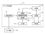
- the stress detection apparatus X can include, for example, an image acquisition unit 11, an input unit 12, an operation unit 13, a detection unit 14, a storage unit 15, a display unit 16 and the like as shown in FIG. .
- the stress detection device X in the present embodiment is configured of a desktop personal computer, a notebook personal computer, a tablet terminal or the like, and can communicate with the OCT device 10 wirelessly or by wire using the image acquisition unit 11 as a communication interface.
- the arithmetic unit 13 and the detection unit 14 are configured by hardware or software or both with the CPU as a core member
- the storage unit 15 is configured by a ROM, a RAM, and the like
- the display unit 16 is a liquid crystal screen or the like. It comprises a known display.
- the stress detection device X may be provided integrally with the OCT device 10, or part of the functions of the calculation unit 13 and the like may be incorporated in the OCT device 10, and is not particularly limited.
- the image acquisition unit 11 acquires tomographic image information of the fundus including the choroid of the object from the OCT apparatus 10 described above.
- the OCT apparatus 10 branches a light source, a measurement optical system that branches a light flux emitted from the light source and irradiates the inside of the eye to be examined and guides measurement light from the fundus of the subject, and branches the light flux emitted from the light source.
- the reference optical system that irradiates the reference object and guides the reference light that is the reflected light, and the interference light that combines the measurement light guided by the measurement optical system and the reference light guided by the reference optical system
- a light receiving element that converts the light into a signal and outputs the signal.
- the input unit 12 receives an operation input by the operator, and outputs an operation input signal corresponding to the operation input to the operation unit 13.
- the input unit 12 is configured of a button, a switch, and a keyboard.
- the input unit 12 can input, for example, information of an object (age, gender, current medical history, axial length, etc.), examination date, and the like.
- the calculation unit 13 processes the tomographic image signal acquired by the image acquisition unit 11.
- the calculation unit 13 includes, for example, an image signal processing unit 13a and a calculation unit 13b.
- the image signal processing unit 13 a performs various types of image processing and the like based on the output signal of the image acquisition unit 11 to generate a tomographic image of the fundus including the choroid.
- the tomographic image generated by the image signal processing unit 13 a is configured to be stored in the storage unit 15 and to be output to the display unit 16.
- the calculating unit 13 b calculates a feature value of the choroid based on the tomographic image of the fundus including the choroid generated by the image signal processing unit 13 a.
- the characteristic value of the choroid can be exemplified by the choroidal thickness of the object, the volume of the choroid, and the like.
- the definition of choroidal thickness and choroidal volume is as described in the section of (Method of detecting stress state) above.
- the choroidal thickness can be calculated by specifying the boundary between the retinal pigment epithelium and the choroid and the boundary between the choroid and the sclera, and measuring the vertical distance between the two boundaries.
- the boundary between the retinal pigment epithelium and the choroid, and the boundary between the choroid and the sclera can be specified by applying known image processing techniques, and automatically performed according to a preset algorithm. You can also.
- a boundary value can be extracted by searching for a pixel value (for example, a luminance value or the like) in the depth direction and detecting a change in the pixel value.
- a boundary line may be specified by
- the measurement of the choroidal thickness is performed by measuring the distance between the two boundaries identified above at a defined measurement position.
- the measurement can be performed, for example, by counting the number of pixels aligned in the depth direction between both boundaries in the tomographic image.
- the specification of the position of the fovea centralis or the optic disc of the retina which is the basis of calculation of the choroidal value, can be set to be automatically performed by applying a known image processing technique in the tomographic image of the choroid. Also, the operator may specify it manually.
- the volume of the choroid can be calculated by adding the thickness information calculated above in the three-dimensional tomographic image of the choroid, and preferably, the volume in the preset specific region is calculated.
- the calculator 13b calculates the characteristic value of the choroid calculated from the tomographic image of the choroid by the calculator 13b, for example, the detection time input by the input unit 12, the age of the object, the sex, the present medical history, the eye axis length, Corrections can be made to eliminate the effects of factors that affect choroidal feature values, such as indices obtained from eye structures such as equivalent sphere areas.
- the correction of the characteristic value of the choroid is the same as the correction content described above, so the description will be omitted.
- the detection unit 14 detects the stress state of the object based on the feature value of the choroid calculated from the tomographic image of the choroid by the calculation unit 13 b.
- the detection unit 14 determines whether the feature value of the choroid calculated by the calculation unit 13 b is equal to or greater than a reference value calculated in advance from the tomographic image of the choroid of a healthy subject not in a stress state stored in the storage unit 15. To detect.
- the comparison between the characteristic value of the choroid of the subject and the reference value can be made based on known statistical methods.
- the detection unit 14 stores the detection result in the storage unit 15 and outputs the detection result to the display unit 16 as a detection result of the stress state.
- the storage unit 15 is calculated by the calculation unit 13 b, the tomographic image of the choroid formed by the image signal processing unit 13 a of the calculation unit 13, the input data such as the information of the object and the examination date and time input by the operator from the input unit 12. Information such as the characteristic value of the choroid of the object and the stress state detected by the detection unit 14 is stored.
- the storage unit 15 also stores a calculation program for calculating the feature value of the choroid, a detection program for detecting a stress state, a reference value used in detection, and the like.
- the storage unit 15 affects the characteristic values of the choroid such as the detection time, the age of the subject, the sex, the current medical history, and the index obtained from the eye structure such as axial length and equivalent sphere area.
- the correction program etc. for correcting the influence of the factor to be stored are stored.
- the display unit 16 displays the information of the object input by the input unit, the examination date, the tomographic image of the choroid formed by the image signal processing unit, and the characteristic value of the choroid calculated from the tomographic image of the choroid by the calculation unit.
- the detection unit displays the detection result of the stress state detected based on the characteristic value of the choroid by comparison with the reference value.
- the display unit 16 can display a warning that it is necessary to have a medical examination or counseling when it is detected that the display device 16 is in a stress state according to the detection result.
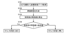
- Step S101 The tomographic image signal (tomographic image data) of the eye of the subject taken by the OCT apparatus 10 is acquired by the image acquisition unit 11.
- Step S102 Then, based on the tomographic image signal acquired by the image signal processing unit 13a, the image signal processing unit 13a performs various types of image processing and the like to generate a tomographic image of the fundus including the choroid.
- the tomographic image of the fundus including the choroid generated by the image signal processing unit 13a may be stored in the storage unit 15, and the tomographic image of the fundus including the choroid may be displayed on the display unit 16.
- the operation of the OCT apparatus 10 the arrangement of the eye of the object, etc. are appropriately corrected, and step S101 is performed again. It may be configured to execute.
- the calculation unit 13b follows the calculation program for calculating the feature value of the choroid stored in the storage unit 15 based on the tomographic image of the fundus including the choroid generated by the image signal processing unit 13a. Alternatively, control is performed to calculate the volume and to output information on the calculated characteristic value of the choroid to the display unit 16. At this time, the calculation unit 13 b may correct the feature value of the choroid according to a correction program for correcting the feature value of the choroid stored in the storage unit 15 as necessary. In addition, the display unit 16 may display an image of the feature value and the correction content of the choroid based on the input information.
- Step S104 The detection unit 14 determines whether the object is in a stress state based on the information on the feature value of the choroid calculated by the calculation unit 13 b and the reference value of the feature value of the choroid stored in the storage unit 15. To detect. Specifically, the detection unit 14 detects that the object is in a stress state when the characteristic value of the choroid is equal to or greater than the reference value, and the object is detected when the characteristic value of the choroid is less than the reference value. The body detects that it is not in stress.
- the detection result of the detection unit 14 is output to the display unit 16, and the display unit 16 displays the stress state of the object on the screen based on the input information.
- a warning screen or the like may be displayed on the assumption that consultation or counseling is necessary.
- the detection unit 14 may detect the stress state in multiple stages, and may display characters such as “high stress”, “medium stress”, “low stress” or the like on the display unit 16 or even by numerical display in multiple stages. Good.
- Example 1 Verification of Relationship between Choroidal Thickness and Stress and Anxiety
- the relationship between stress and anxiety and the choroidal thickness which is a part of eye tissue was verified.
- the relationship with the detection result by the subjective stress detection method was evaluated.
- Method 1-1 Implementation period 1-1-1. Date of implementation: 1-3-2 consecutive days from July to October 2017 1-1-2. Implementation time Evening to night
- the subject profiles are summarized in Table 1 below.
- 1-3 Verification method The stress state of the object was verified by the following three methods. 1-3-1. Verification by Choroidal Film Thickness The choroidal film thickness was measured by SS-OCT (TOPCON (registered trademark) SS-OCT, manufactured by TOPON) on the subject. The choroid was measured by measuring the foveal choroidal thickness. Specifically, a tomographic image of the fundus is acquired so that the fovea retina is as vertical as possible with respect to the tomographic screen, and a ruled line from immediately below the retinal pigment epithelium in the fovea to a position considered to be the lower edge of the choroid. was vertically measured, and the distance was measured as the foveal choroidal thickness.
- the position of the fixation point was finely adjusted so that the fovea was at the center of the tomographic image. Further, the axial length of the subject was measured by IOLmaster (manufactured by ZEISS), and measurement of blood pressure and pulse, and BUT test were performed. At the time of measurement, subjective stress felt by the subject was scored by self-evaluation. Subjective stress was rated on a scale of 5 (5: very, 4: somewhat, 3: slightly, 2: hardly, 1: not at all).
- the method of selecting highly stressed persons using the raw score conversion table was based on the manual for stress check system implementation based on the Industrial Safety and Health Law (http://www.mhlw.go.jp/bunya/roudoukijun/anzeneisei 12 / pdf / See 150803-1.pdf).
- K6 Verification by Japanese version mood / anxiety disorder questionnaire (K6)
- K6 Questionnaire by Japanese version mood / anxiety disorder questionnaire (K6) (http://www.city.noshiro.akita.jp/upload/ (See download / 118779download.pdf), and the questionnaire results were collected.
- K6 is a six-item, five-measure scale that measures mood and anxiety disorders developed by Kessler et al. (See Kessler RC et al., Psychological Medicine, 2002, 32, 959-976). It has been reported that it can be adapted (Sakurai K et al., Psychiatry Clin Neurosci., 2011; 65 (5): 434-441, etc.).
- FIG. Fig. 7 shows the verification results of the relationship between the choroidal thickness and the mind-body response (area B) caused by the stress in the questionnaire for occupational stress (57 items), and Fig. 8 shows the choroidal thickness / axial length and occupationality.
- FIG. Fig. 7 shows the verification results of the relationship between the choroidal thickness and the mind-body response (area B) caused by the stress in the questionnaire for occupational stress (57 items)
- Fig. 8 shows the choroidal thickness / axial length and occupationality.
- the choroidal thickness is the average foveal thickness for 3 days or 4 days.
- FIG. 10 shows the result of examining the relationship between the choroidal thickness and the results of the Japanese version of the mood / anxiety disorder questionnaire (K6), and FIG.
- FIG. 11 shows the choroidal thickness / axial length and the Japanese version of the mood / anxiety disorder questionnaire.
- the verification result of relevance with the result of (K6) is shown.
- the choroidal thickness is the average foveal thickness for 3 days or 4 days.
- the choroid was found to be a stress marker.
- the Japanese version of the Mood Anxiety Disorder Questionnaire (K6) is used worldwide. Therefore, it can be understood that the thickness of the choroid is a stress marker as a matter of course from the data of this research, and it can be understood that if the choroid is thick, the object can be detected with high reliability if it is in a stress state. .
- Tables 3 to 5 show the measurement time of the choroidal thickness and the measurement result of the foveal choroidal thickness (left and right).
- Table 4 shows the measurement results of age and mean foveal choroidal thickness (left and right), and it can be understood that positive correlation is recognized between age and choroidal thickness.
- Table 5 shows the measurement results of gender and mean foveal choroidal thickness (left and right), and it was recognized that the female tended to be thicker in the choroid than the male. Therefore, it can be understood that the stress state of the object can be detected more accurately based on the choroidal film thickness by correcting the measured choroidal film thickness with respect to factors affecting the choroidal film thickness such as age and gender.
- the present invention can be used in all technical fields requiring detection of a stress condition, such as early detection of a stress condition or prevention of a disease or condition induced by a stress condition, and in particular, health management of workers. It can be used for etc.
- OCT apparatus 11 Image acquisition unit 12: Input unit 13: Calculation unit 13a: Image signal processing unit 13b: Calculation unit 14: Detection unit 15: Storage unit 16: Display unit X: Stress detection device
Landscapes
- Health & Medical Sciences (AREA)
- Life Sciences & Earth Sciences (AREA)
- Engineering & Computer Science (AREA)
- Veterinary Medicine (AREA)
- Heart & Thoracic Surgery (AREA)
- Public Health (AREA)
- General Health & Medical Sciences (AREA)
- Animal Behavior & Ethology (AREA)
- Surgery (AREA)
- Physics & Mathematics (AREA)
- Molecular Biology (AREA)
- Biophysics (AREA)
- Medical Informatics (AREA)
- Biomedical Technology (AREA)
- Ophthalmology & Optometry (AREA)
- Psychiatry (AREA)
- Radiology & Medical Imaging (AREA)
- Nuclear Medicine, Radiotherapy & Molecular Imaging (AREA)
- Pathology (AREA)
- Social Psychology (AREA)
- Psychology (AREA)
- Hospice & Palliative Care (AREA)
- Educational Technology (AREA)
- Child & Adolescent Psychology (AREA)
- Developmental Disabilities (AREA)
- Investigating Or Analysing Biological Materials (AREA)
- Measurement Of The Respiration, Hearing Ability, Form, And Blood Characteristics Of Living Organisms (AREA)
- Eye Examination Apparatus (AREA)
Abstract
対象体の脈絡膜の断層画像に基づいて対象体のストレス状態を検出する工程を有するストレス状態の検出方法、及び、対象体の脈絡膜の断層画像を取得する画像取得部(11)と、断層画像から脈絡膜厚又は脈絡膜の容積を算出する算出部(13b)と、脈絡膜厚又は脈絡膜の容積に基づいて対象体のストレス状態を検出する検出部(14)とを備えたストレス検出装置(X)。
Description
本発明は、ストレス状態の検出方法、及び、ストレス検出装置に関する。
昨今、「ストレスは万病のもと」、すなわち、ストレスは心身に悪影響を与え、種々の疾患を誘発するとの認識が広まってきている。ストレスは、外部からの様々な刺激により精神的及び肉体的に負担がかかることで、心身に歪みが生じる状態を意味する。本来、ヒトをはじめとする動物が一時期的な精神的・肉体的ストレス等の外的ストレスを感じると、副腎からコルチゾールやアドレナリン等のホルモンが分泌され適度な刺激が身体に与えられる。心拍数の増加、心・血管の拡張、血糖値の増加、気管支弛緩等を介して、外的ストレスからの抵抗力を増強させることで身体は強化される。しかしながら、過度又は継続的に外的ストレスにさらされると、ホルモンの過剰分泌により身体の防御能力が低下し、結果として精神的又は肉体的異常が出現すると考えられている。ストレスが心身に与える影響には個人差があり、特にストレス感受性が高い者(高ストレス者)は、不登校や欠勤等の様々な問題が顕在化し日常生活に支障を来たすことがある。したがって、ストレス状態を正確に評価することは、健康管理、疾患の予防及び治療の上で極めて重要である。
そこで、2015年に、厚生労働省は、労働者の心理的な負担の程度を把握するための検査や、検査結果に基づく医師による面接指導の実施等を定めたストレスチェック制度を導入している。ストレスチェックでは、職業性のストレスと考えられる因子、ストレスによって起こる心身の反応、及び、ストレス反応に影響を与える他の因子に関する質問項目に労働者が回答し、各質問項目への回答を点数化する。かかる結果を一定規模の集団毎に集計・分析を行い、数値基準に基づいて点数により高ストレス者を選定する。しかしながら、ストレスチェックは労働者自身が質問項目に回答する自覚的検査であるため、意図的にストレスが無いように回答を操作することが可能であるため、高ストレス者を適切に選定することができない可能性がある。したがって、自覚的な検査と他覚的な検査を組み合わせて、実際の高ストレス者を選定することが重要になる。
現在までに、種々の他覚的なストレス検査が試みられている。急性ストレスを評価する方法として、コルチゾール等の副腎皮質系因子、テストステロン等の性腺系因子、アミラーゼ等の青斑核/ノルアドレナリン系因子、アドレナリンやノルアドレナリン等のカテコールアミン系因子等のストレスマーカを指標とする方法が報告されている(特許文献1参照)。特許文献1に記載の方法では、検体として汎用される血液、唾液、又は、尿ではなく、対象体の皮膚に窄刺具等により形成した微細孔から採取した組織液中のストレスマーカを測定するものである。また、対象体から採取した血清中のアポリポタンパク質A-II、ハプトグロブリン、ビタミンD結合タンパク質プリカーサ等を指標としてストレスを評価する方法が報告されている(特許文献2参照)。
更に、慢性ストレスを評価する方法として、唾液中のコルチゾールの一日の経時変化を指標とする方法が報告されている(特許文献3参照)。特許文献3に記載の方法は、多くの健常人において測定された一日の所定時間範囲にわたる唾液中のコルチゾール濃度から得られた標準コルチゾール濃度変化範囲と、特定の対象体において測定された唾液中のコルチゾール濃度変化とを比較し、標準コルチゾール変化範囲を逸脱した者を、慢性ストレスを有する可能性あると評価するものである。
しかしながら、特許文献2に記載の方法のように血液を検体とする場合に、血液採取血行為には痛み等の精神的かつ肉体的な負担を伴い、血液採取に伴うストレス負荷によりストレス状態を正確に評価できない可能性がある。更に、血液採取行為は、医療行為であるとの制約があると共に、感染症を誘発する恐れがあるという問題がある。特許文献1に記載の方法は、上記負担の軽減を目的とするものであるが、依然として侵襲性があり、上記負担を完全に解消するものではない。特許文献3に記載の方法に唾液を検体とする場合には、上記負担は軽減されるものの、特許文献1に記載されるように唾液は食事や喫煙等の外的要因によって変動する可能性があり、しかも唾液の分泌量や組成等の個人差が大きい。したがって、検体を採取する際のタイミング等の条件を詳細に設定する必要があり、唾液を採取する操作が煩雑になるという問題点がある。
上記した生理化学物質によるストレスマーカは、ストレスに応答し出現したとしても生体内で通常短時間で分解され消失してしまう。例えば、アドレナリン等のカテコールアミン系因子の血液中での半減期は1~2分と非常に短い。したがって、測定値は、生体のストレス状態を反映していない可能性があり、対象体のストレス状態を正確に評価することは困難である。また、生理化学物質の生体内濃度は、生理的日内変動が大きく、また、個人間の睡眠覚醒リズムの相違等に起因した個人差が大きい。例えば、コルチゾールは、早朝高値、夜間低値の日内変動を示し、夕方以降は早朝値の半分以下となる。そのため、上記した生理化学物質の生体内濃度に一定の基準値を設けてストレス状態を簡便に評価することは困難である。更に、自律神経系の慢性ストレス評価においても、特許文献3に記載の方法のような日内変動や個人差の大きいコルチゾール等のホルモンをストレスマーカとすることができず、確立された評価方法が存在しないのが現状である。
したがって、生体のストレス状態を的確に反映可能なストレスマーカにより他覚的に対象体のストレス状態を評価できる技術の構築が依然として求められている。特に、非侵襲的手段により評価でき、日内変動や個人差等の少ないストレスマーカにより簡便かつ正確に対象体のストレス状態を評価できる技術の構築が求められている。
本発明者らは、上記課題を解決すべく研究を重ねた結果、対象体のストレス状態を、眼組織の一部である脈絡膜を指標として評価できることを見出した。特に、脈絡膜の厚さ又は容積が、対象体のストレス状態を的確に反映していることを見出した。かかる脈絡膜の変化は他覚的に検出することができ、正確に対象体のストレス状態を検出できる。また、光干渉断層法を利用することにより、非侵襲的に、しかも短時間かつ簡便に検出できる。
本発明者らは、これらの知見に基づき本発明を完成するに至った。
本発明者らは、これらの知見に基づき本発明を完成するに至った。
即ち、本発明は、上記課題を解決するため、以下の〔1〕~〔7〕に示す発明を提供する。
〔1〕対象体の脈絡膜の断層画像に基づいて前記対象体のストレス状態を検出する工程を有するストレス状態の検出方法。
上記〔1〕の構成によれば、対象体の眼組織の一部である脈絡膜の断層画像に基づき、当該脈絡膜を指標として対象体のストレス状態を検出するストレス状態の検出方法を提供できる。本構成によれば、対象体のストレス状態を他覚的に検出できると共に、脈絡膜は生体のストレス状態を的確に反映できることから対象体のストレス状態を正確に検出できる。
更に、断層画像は眼に近赤外線を照射する光干渉断層法により取得することが可能であり、非接触かつ非侵襲的に、しかも短時間かつ簡便にストレス状態を検出できることから、対象体の精神的かつ肉体的負担を軽減できる。したがって、検出に際してのストレス負荷を排除でき、対象体のストレス状態を正確に検出できる。更に、眼球運動の影響も受け難いという利点もある。
したがって、本構成のストレス状態の検出方法によれば、ストレス状態にある対象体を信頼性高く検出できる。ストレス状態にあると検出された対象体に対しては、適切な医療機関のへの受診やカウンセリングを推奨できる。特に、本構成のストレス状態の検出方法によれば、ストレスが特定の症状や疾患等として顕在化していない潜在的なストレス状態をも検出できる。そのため、早期にストレス状態を検出することができ、ストレスによって発症する症状や疾患の予防に繋げることができる。したがって、本構成のストレス状態の検出方法は労働者の定期的な健康診断等にも好適に利用することができ、労働者の心身の健康管理に貢献できる。ひいては、職場環境の改善にもつながり、仕事能率が上昇し労働生産性の向上にも寄与できる。
〔2〕前記ストレス状態を検出する工程は、前記断層画像から脈絡膜厚を算出し、算出された前記脈絡膜厚に基づいて前記対象体のストレス状態を検出する上記〔1〕のストレス状態の検出方法。
〔3〕算出された前記脈絡膜厚が、中心窩下で300μm以上であるときにストレス状態にあると検出する上記〔2〕のストレス状態の検出方法。
〔3〕算出された前記脈絡膜厚が、中心窩下で300μm以上であるときにストレス状態にあると検出する上記〔2〕のストレス状態の検出方法。
上記〔2〕及び〔3〕の構成によれば、対象体の眼組織の一部である脈絡膜の断層画像に基づき、当該脈絡膜の厚さを指標として対象体のストレス状態を検出するストレス状態の検出方法を提供できる。脈絡膜の断層画像から明確かつ簡単に算出できる脈絡膜厚をストレス状態の指標とするため、より簡便かつ正確に対象体のストレス状態を検出できる。
特に、上記〔3〕の構成によれば、明確な基準値に基づいて対象体のストレス状態を検出することができ、ストレス状態の検出における簡便性及び正確性の更なる向上を期待できる。
特に、上記〔3〕の構成によれば、明確な基準値に基づいて対象体のストレス状態を検出することができ、ストレス状態の検出における簡便性及び正確性の更なる向上を期待できる。
〔4〕前記ストレス状態を検出する工程は、前記断層画像から脈絡膜の容積を算出し、算出された前記脈絡膜の容積に基づいて前記対象体のストレス状態を検出する上記〔1〕のストレス状態の検出方法。
上記〔4〕の構成によれば、対象体の眼組織の一部である脈絡膜の断層画像に基づき、当該脈絡膜の容積を指標として対象体のストレス状態を検出するストレス状態の検出方法を提供できる。三次元画像等の脈絡膜の断層画像から明確かつ簡単に算出できる脈絡膜の容積を指標とするため、より簡便かつ正確に対象体のストレス状態を検出できる。
〔5〕前記ストレス状態を検出する工程は、算出された前記脈絡膜厚を、検出時刻、前記対象体の年齢、性別、現病歴、及び、眼構造から得られる指標から選択される少なくともの1つの因子を用いて補正する工程を含む上記〔2〕又は〔3〕のストレス状態の検出方法。
上記〔5〕の構成によれば、算出された対象体の脈絡膜厚を、脈絡膜厚に影響を与える検出時刻、対象体の年齢、性別、現病歴、並びに、眼軸長及び等価球面積等の眼構造から得られる指標から選択される少なくともの1つの因子を用いて補正することにより、更に正確に対象体のストレス状態を検出するストレス状態の検出方法を提供できる。
〔6〕前記ストレス状態を検出する工程は、算出された前記脈絡膜の容積を、検出時刻、前記対象体の年齢、性別、現病歴、及び、眼構造から得られる指標から選択される少なくともの1つの因子を用いて補正する工程を含む上記〔4〕のストレス状態の検出方法。
上記〔6〕の構成によれば、算出された対象体の脈絡膜の容積を、脈絡膜の容積に影響を与える検出時刻、対象体の年齢、性別、現病歴、並びに、眼軸長及び等価球面積等の眼構造から得られる指標から選択される少なくともの1つの因子を用いて補正することにより、更に正確に対象体のストレス状態を検出するストレス状態の検出方法を提供できる。
〔7〕対象体の脈絡膜の断層画像を取得する画像取得部と、
前記断層画像から脈絡膜厚又は脈絡膜の容積を算出する算出部と、
前記脈絡膜厚又は脈絡膜の容積に基づいて前記対象体のストレス状態を検出する検出部とを備えたストレス検出装置。
前記断層画像から脈絡膜厚又は脈絡膜の容積を算出する算出部と、
前記脈絡膜厚又は脈絡膜の容積に基づいて前記対象体のストレス状態を検出する検出部とを備えたストレス検出装置。
上記〔7〕の構成によれば、対象体の眼組織の一部である脈絡膜の断層画像に基づき、脈絡膜厚又は脈絡膜の容積を指標として対象体のストレス状態を検出するストレス検出装置を提供できる。本構成のストレス検出装置は、生体のストレス状態を的確に反映する脈絡膜厚又は脈絡膜の容積を対象体のストレス状態の指標とするものであるから、信頼性高く対象体のストレス状態を検出できる。更に、脈絡膜の断層画像の取得に際して光干渉断層法を利用することにより、非接触かつ非侵襲的に、しかも短時間かつ簡便に被検体のストレス状態を検出できることから、対象体の精神的かつ肉体的負担を軽減できる。したがって、検出に際してのストレス負荷を排除でき、対象体のストレス状態を正確に検出できる。また、本構成のストレス検出装置は労働者の定期的な健康診断等にも好適に利用できることから、各健診センターや医療施設への設置等を通して労働者の心身の健康管理に貢献できる。
以下、本発明の実施形態に係るストレス状態の検出方法及びストレス検出装置について詳細に説明する。ただし、本発明は、後述する実施形態に限定されるものではない。
(ストレス状態の検出方法)
本実施形態に係るストレス状態の検出方法は、対象体の脈絡膜を指標として、対象体のストレス状態を検出するものである。当該検出方法は、従来においてはストレスマーカとしては認識されていなかった眼組織の一部である脈絡膜を対象体のストレス状態の指標とするものである。
本実施形態に係るストレス状態の検出方法は、対象体の脈絡膜を指標として、対象体のストレス状態を検出するものである。当該検出方法は、従来においてはストレスマーカとしては認識されていなかった眼組織の一部である脈絡膜を対象体のストレス状態の指標とするものである。
本実施形態に係るストレス状態の検出方法における対象体としては、ヒト、非ヒト霊長類、ウサギ、ラット、モルモット、マウス、イヌ、ネコ、ネコ、ウマ、ウシ、ブタ、ヒツジ、ヤギ、ニワトリ等の動物が例示されるが、特に好ましくはヒトである。
本実施形態に係るストレス状態の検出方法におけるストレス状態とは、様々な刺激によって心身に負荷がかかることで生じる緊張状態を意味する。ストレスを誘発する刺激はストレッサーと称され、ストレッサーとしては、環境的要因、身体的要因、心理的要因、及び、社会的要因等によるものがある。環境的要因としては、例えば、温度、湿度、光、天候、騒音、振動、有害物質の曝露、大気汚染等が挙げられる。身体的要因としては、例えば、外傷、病気、過度な運動、運動不足、肉体疲労、睡眠不足、栄養不足、加齢、不健康な生活習慣、肥満等が挙げられる。更に、生体内において活性酸素による酸化作用と、抗酸化物質等の抗酸化作用との均衡が崩れた結果、過剰に生成される活性酸素種等も身体的要因に含むことができる。心理的要因としては、例えば、不安、心配、欲求不満、怒り、恐怖、失望、葛藤等が挙げられる。社会的要因としては、人間関係、就職、転職、失業、入学、進学、成績不振、いじめ等が挙げられる。しかしながら、これらに限定されるものではない。
本実施形態に係るストレス状態の検出方法における検出対象となるストレス状態としては、ストレッサーにより誘発される状態である限り特に限定されず、短期的な急性ストレス状態及び長期にわたる慢性ストレス状態の何れであってもよい。ストレス状態にはストレスに関連する症状及び疾患(以下、それぞれ「ストレス症状」及び「ストレス疾患」と略する場合がある)が含まれ、また、心身に症状や疾患が顕在化していない潜在的なストレス状態をも検出の対象とすることができる。生体は、ストレッサー等の刺激に曝されると、自律神経系、内分泌系、免疫系等が相互作用し合うことでその恒常性を保持している。しかしながら、過度・継続的にストレッサーに曝されると自律神経系のバランスが崩れ、副交感神経がうまく機能せずに、交感神経の活動が亢進し続ける。このような交感神経が優位な状態が続くと、段階的にストレッサーに対する生体の抵抗力が衰え、自律神経系、内分泌系、及び、免疫系等の機能低下により様々な症状や疾患が誘発される。したがって、ストレス状態には、好ましくは、交感神経優位な状態、及び、かかる交感神経優位な状態により誘発される症状及び疾患を含むことができる。
ストレス症状としては、例えば、肩こり、動悸、眩暈、不眠、頭痛、体重変動、食欲低下、摂食障害、腹痛、依存症等の身体的症状、不安、怒り、焦燥、無気力等の心理的症状、過食、飲酒、引きこもり等の社会的症状等が含まれる。ストレス疾患としては、うつ病、神経症、自律神経失調症等の精神・神経疾患、高血圧症、動脈硬化、狭心症、心筋梗塞等の循環器疾患、肥満症、糖尿病、高脂血症、甲状腺機能亢進症等の内分泌・代謝異常疾患、分泌気管支炎、過呼吸症候群等の呼吸器疾患、過敏性腸症候群、胃・十二指腸潰瘍、潰瘍性大腸炎、心因性嘔吐等の消化器疾患、アトピー性皮膚炎、乾癬、円形脱毛症等の皮膚疾患、中心性漿液性脈絡膜網膜症(CSC)、眼精疲労、眼瞼痙攣、ドライアイ等の眼科疾患、メニエール病等の耳鼻咽喉科疾患、夜尿症等の泌尿・生殖器疾患、癌、慢性炎症等の炎症性疾患等を含めることができる。特には、精神・神経疾患が想定され、例えば、パニック障害、社会不安障害、全般性不安障害、恐怖症等に代表される過剰な不安・恐怖によって苦しみ生活に支障をきたすような疾患である不安障害、及び、双極性障害、うつ病、気分変調性障害等に代表される気分に異常をきたす状態になってしまう疾患である気分障害等が含まれる。ストレス状態は、各種症状及び疾患の増悪等の要因となることから、ストレス症状及び疾患には、各種症状及び疾患の発症だけでなく、増悪も含まれる。
本実施形態に係るストレス状態の検出方法において、ストレス状態の検出の指標となる脈絡膜は、網膜と強膜の間にある色素細胞と血管に富む組織であり、組織学的には、網膜側から基底板(ブルッフ膜)、脈絡毛細管板、血管板、脈絡上板の4層に区別できる。脈絡膜は、ほぼ血管により構成される組織であり、脈絡膜血流は全眼血流の85%に達するという報告がある(例えば、Experimental Eye Research, 15(1), 1973, p15-29を参照のこと)。そのため、生理化学物質の影響を強く受けることが導かれる。また、脈絡膜血管は、網膜血管とは異なり自己調節能が低く、血圧等の種々の生理的刺激により脈絡膜血管内を流れる血流量が変動するとの報告があり(例えば、Arch Ophthalmol, 83(1), 1970, p95-99を参照のこと)、脈絡膜血管は生体の全身状態の変化の影響を強く受けることになる。更には、脈絡膜血管には、血管の緊張に影響する受容体が発現されていることが知られており、特に妊娠高血圧症候群において脈絡膜血流が変化することが知られている。
本実施形態に係るストレス状態の検出方法は、上記した通り眼組織の一部である脈絡膜をストレス状態の指標とするものである。脈絡膜は全身の血管の変化を反映できるものであることが考えられ、生体のストレス状態を的確に反映しているものと考えられる。一方で、従来においてはストレス状態と脈絡膜を含めた眼組織との関連性に関する知見は存在しなかった。
本実施形態に係るストレス状態の検出方法は、好ましくは、対象体の脈絡膜の断層画像に基づいて前記対象体のストレス状態を検出するものである。脈絡膜の断層画像からは、脈絡膜厚や脈絡膜の容積等の脈絡膜の形態を含めた状態を詳細に把握できる。
脈絡膜の断層画像は、脈絡膜を含む眼の断層画像であり、好ましくは、脈絡膜を含む眼底の断層画像である。脈絡膜の厚さや容積等の脈絡膜の特徴値の算出のため、脈絡膜と網膜との境界、及び、脈絡膜と強膜との境界を抽出できることが好ましい。したがって、脈絡膜の断層画像は、脈絡膜に加え、少なくとも網膜の一部及び強膜の一部を含むことが好ましい。脈絡膜の断層画像は、例えば、網膜の中心窩下を含む領域、又は、網膜の中心窩を基準として中心窩から所定の方向に所定距離だけ離れた位置を含んだ領域の断層画像とすることができる。また、視神経乳頭を含む領域、視神経乳頭を基準として視神経乳頭から所定の方向に所定の距離だけ離れた位置を含む領域の断層画像とすることができる。更には、複数の領域を含んだ断層画像であってもよい。特に好ましくは、断層画像の取得の容易性の観点から、網膜の中心窩下を含む領域の断層画像である。また、脈絡膜の断層画像は、一次元画像、二次元画像、三次元画像の何れであってもよい。
脈絡膜の断層画像は、当該技術分野で公知の技術を用いる脈絡膜の断層画像化によって取得できる。脈絡膜の断層画像化は、好ましくは、光干渉断層法(Optical coherence tomography;以下、「OCT」と略する)に基づく装置(以下、「OCT装置」と称する)を用いて行うことができる。OCT装置は、光源から出射された光束を分岐させ、対象物の所定位置に入射させた測定光の反射又は散乱光と、参照物体から反射した参照光とを干渉させる干渉光学装置である。干渉光を利用して、測定光を入射させた位置の深度方向の対象物の構造を画像化する技術であり、対象物の断層画像を高分解能に取得できる。つまり、OCT装置は眼底に測定光を照射することにより、脈絡膜を含む眼底の断層画像を非侵襲的かつ高解像度で取得できる。しかも、OCT装置は他覚的に脈絡膜を検査できる装置であり、一般に眼科検診で日常的に行われる短時間型非侵襲的検査装置であることから対象体に対する負担も少ない。
OCT装置は、光波の干渉を時間領域で行うタイムドメインOCT(以下、「TD-OCT」と略する)、光波の干渉を周波数領域又は波長領域で行うフーリエドメインOCT(frequency-domain OCT:以下、「FD-OCT」と略する。)等を利用することができ、好ましくは、FD-OCTである。FD-OCTとしては、広帯域光源を用いるスペクトラルドメインOCT(Spectral-domain OCT:以下、「SD-OCT」と略する)、波長掃引レーザを用いる波長掃引型OCT(Swept-source OCT:以下、「SS-OCT」と略する)等が例示される。好ましくは、SS-OCTである。更に、深部強調画像(Enhanced depth imaging:以下、「EDI」と略する)によるEDI-OCTや、長波長光源を用いた高侵達(high-penetration-OCT、以下、「HP-OCT」と略する)等を利用できる。光源としては、1μm帯域、典型的には950 nm~1100 nm程度、特には1050 nmの波長の光を射出する光源が好ましい。1μm帯の光源を用いると、網膜色素上皮等での光の吸収や中間透光体に混濁がある場合での散乱の影響を受けにくいこと等から、脈絡膜を含む眼底等の深部組織のより鮮明な画像を取得できる。
詳細には、測定光を対象体の眼底に照射することにより、照射位置の深度方向(z方向)の脈絡膜を含む眼底の一次元断層画像(z画像)を取得できる。更に、対象体の眼底に対して、照射位置を深度方向に垂直な方向(x方向)に一次元走査することにより脈絡膜を含む眼底の二次元断層画像(xz画像)を取得できる。一次元走査は、予め定められた直線又は曲線に沿って行うことができる。これにより、測定光の走査方向に沿った深度方向の二次元の断層画像を得ることができる。また、眼底に対して、照射位置を深度方向に垂直な方向(x、y方向)に二次元走査することにより脈絡膜を含む三次元画像(xyz画像)を取得できる。このとき、二次元走査は予め定められた領域内で行うことができる。つまり、予め定められた領域内を、対象体の眼底に対して位置をずらしながら測定光を繰り返し走査することにより取得した複数の二次元画像から三次元の断層画像を構築できる。これにより、脈絡膜を含めた眼底の構造を立体的に捉えることができる。特に、SS-OCTでは、数秒でx方向、y方向の3~12mm程度の広い範囲を走査し深部方向の画像を取得できることから、脈絡膜を含めた眼底の鮮明な三次元の断層画像の構築が可能となる。
また、OCT装置による脈絡膜の断層画像の取得に要する時間は3秒程であり、眼球運動の影響を受け難いという利点もある。
脈絡膜の断層画像の取得は、運動時や運動直後を避けて取得することが好ましい。例えば、脈絡膜厚に関して、負荷の少ない中程度の強度の運動時において、少なくとも5分間は脈絡膜が顕著に厚くなったことが報告されている(例えば、Sayin N他著、Indian J Ophthalmol. 2015, 63(5), 445-450を参照のこと)。一方で、15分後には脈絡膜厚はベースラインと比較して有意な差が認められなかったことも報告されている。したがって、対象体が日常的な活動よりも運動強度の高い運動を行った場合には、運動終了時から所定時間(例えば、15分間、又は、30分間)経過後に脈絡膜の断層画像を取得することが好ましい。また、脈絡膜の断層画像の取得は、一般的には座位で行われ、左右何れかの眼の脈絡膜の断層画像であってもよいし、両眼の脈絡膜の断層画像であってもよい。
ストレス状態を検出する工程は、前記断層画像から脈絡膜厚を算出し、算出された前記脈絡膜厚に基づいて前記対象体のストレス状態を検出することを含むことができる。脈絡膜厚は脈絡膜の厚さであり、脈絡膜の断層画像から、例えば、網膜の最外側に位置する網膜色素上皮と脈絡膜の境界線から脈絡膜と強膜の境界線までの垂直距離として算出できる。網膜色素上皮と脈絡膜の境界の位置の特定、及び、脈絡膜と強膜の境界の位置の特定は、当該技術分野で公知の画像処理技術を利用して特定できる。例えば、断層画像内の画素値(例えば、輝度値)の変化から、これらの境界の位置を特定できる。また、脈絡膜厚の算出は、例えば、コンピュータの演算機能を利用することにより行ってもよい。
脈絡膜厚の算出は、脈絡膜の予め定められた位置で行うことが好ましい。例えば、網膜の中心窩の位置、又は、網膜の中心窩を基準として中心窩から所定の方向(例えば、上方、下方、鼻側、耳側)に所定距離だけ離れた位置等とすることができる。また、視神経乳頭の位置、又は、視神経乳頭を基準として視神経乳頭から所定の方向に所定の距離だけ離れた位置で測定してよい。更には、複数の位置で脈絡膜厚を測定してもよい。特に好ましくは、網膜の中心窩下の脈絡膜厚である。脈絡膜厚の算出は、左右何れかの眼の脈絡膜に対して行ってもよいし、両眼の脈絡膜に対して行ってもよい。また、両眼の脈絡膜厚を算出した後、脈絡膜が薄い方、若しくは厚い方のみをストレス状態の検出のために用いてもよいが、ストレス状態の相関関係が高い薄い方を検出のために用いることが好ましい。
ストレス状態にある対象体では、健康でストレス状態にない正常対照群に比較して、脈絡膜厚が有意に高い値を示す。例えば、高ストレス者においては、自律神経の過剰刺激により脈絡膜血管が拡張し脈絡膜が厚くなることが考えられる。具体的には、ストレス状態にある対象体の検出は、対象体の脈絡膜の断層画像から算出された脈絡膜厚と、例えば、ストレス状態にない健常体の脈絡膜の断層画像から予め算出された基準値との比較により実施できる。当該基準値よりも、対象体の脈絡膜厚の値が高ければ当該対象体はストレス状態にあるとして検出され、当該基準値よりも値が低ければ当該対象体はストレス状態にないと検出できる。なお、基準値の算出に用いるストレス状態にない健常体の選定は、厚生労働省が推奨している職業性ストレス簡易調査票(57項目)や日本語版気分・不安障害調査票(K6)等の、他の公知のストレスマーカ等のストレス検出方法により実施することができ、好ましくは複数のストレス検出方法を実施する。また、基準値は複数の健常体の平均値として算出することが好ましい。一旦基準値が定まれば、当該基準値をもってストレス状態を検出することができる。基準値としては、例えば、成人の場合、中心窩下で250μm、300μm、若しくは、それ以下の任意の値に設定でき、好ましくは、中心窩下で200~300μmの任意の値に設定できる。
前記ストレス状態を検出する工程は、算出された対象体の脈絡膜厚を、検出時刻、前記対象体の年齢、性別、現病歴、並びに、眼軸長及び等価球面積等の眼構造から得られる指標から選択される少なくとも1つの因子を用いて補正する工程を含むことができる。例えば、脈絡膜厚は年齢と負の相関が認められ、年齢と共に脈絡膜が菲薄化することが報告されている(Wakatsuki Y他著、PLoS One. 2015 Dec 3;10(12):e0144156)。脈絡膜厚には日内変動が認められ、脈絡膜は早朝には厚いが日中には段階的に薄くなること、かかる脈絡膜厚の変動幅に左右の差はないこと等が報告されている(例えば、Tan CS他著、InvestOphthalmol Vis Sci, 2012;53(1):261-266等を参照のこと)。更に、一部の眼疾患及び全身性病態により脈絡膜厚に影響を及ぼし、例えば、中心漿液性網脈絡膜症や原田病などでは脈絡膜厚が有意に肥厚化し、逆に、強度近視眼、加齢黄斑変性、緑内障、及び、糖尿病網膜症等では菲薄化していることが報告されている。また、年齢に加え、眼軸、等価球面積との関係についても報告されており(例えば、上記したTan CS他著、Invest Ophthalmol Vis Sci, 2012;53(1):261-266、Ikuno Y他著、Invest Ophthalmol Vis Sci, 2010;51(4):2173-2176、Wen Bin Wei他著、Ophthalmology, 2013;120(1):175-180等を参照のこと)、眼軸が短い、又は、等価球面積が大きいほど、脈絡膜が厚いこと等が報告されている。更には、flatter cornea、矯正視力がよいほど脈絡膜は厚いことが報告されている。一方で、血圧、眼灌流圧、眼圧、喫煙、飲酒量、血中の脂質・グルコース濃度、糖尿病、動脈性高血圧等と脈絡膜厚とは特に相関はないことも報告されている(上記したWen Bin Wei他著、Ophthalmology, 2013;120(1):175-180等を参照のこと)。したがって、上記因子が脈絡膜厚に与える影響を補正することにより、より正確な対象体のストレス状態を検出できる。
例えば、上記因子が検出時刻や年齢等のように因子の数値変動により脈絡膜厚が変動する場合には、上記因子が標準値にあってストレス状態にない群から算出された脈絡膜厚を標準脈絡膜厚として定める。そして、上記因子が標準値ではない場合にストレス状態にない群から算出された脈絡膜厚に対する標準脈絡膜厚の比(標準脈絡膜厚/算出された脈絡膜厚)を補正係数とする。かかる補正係数を算出された脈絡膜厚に対して乗算することにより、対応する上記因子が標準値にない対象体から算出された脈絡膜厚を補正できる。また、上記因子の数値変動と脈絡膜厚の変動との相関関係が得られる場合には、かかる相関関係に基づいて補正できる。例えば、上記因子が性別の場合には、一方の性の脈絡膜厚に基づき、他方の性の脈絡膜厚を補正できる。また、例えば、上記因子が現病歴のように因子の有無により脈絡膜厚が変動する場合には、上記因子の何れをも有さず、かつ、ストレス状態にない群から算出された脈絡膜厚を標準脈絡膜厚として、上記因子の何れか一つを有するストレス状態にない群から算出された脈絡膜厚に対する標準脈絡膜厚の比(標準脈絡膜厚/算出された脈絡膜厚)を補正係数として、かかる補正係数に基づいて、対応する上記因子を有する対象体から算出された脈絡膜厚を補正できる。補正は、脈絡膜厚の算出と同様に、例えば、コンピュータの演算機能を利用することにより行ってもよい。
ストレス状態を検出する工程は、前記断層画像から脈絡膜の容積を算出し、算出された前記脈絡膜の容積に基づいて前記対象体のストレス状態を検出することを含むことができる。脈絡膜の容積は、網膜の最外側に位置する網膜色素上皮と脈絡膜の境界線と脈絡膜と強膜の境界線により囲まれる部分の容積とすることができ、上記した脈絡膜厚に基づいて算出できる。脈絡膜の容積の算出は、例えば、コンピュータの演算機能を利用することにより行ってもよい。
脈絡膜の容積の算出は、脈絡膜の予め定められた領域で行うことが好ましい。例えば、網膜の中心窩の位置、又は、網膜の中心窩を基準として中心窩から所定の方向(深部方向(z方向)とは垂直な方向(x方向)、例えば、上方、下方、鼻側、耳側)に所定距離だけ離れた位置等を中心点とした領域の脈絡膜の容積として算出できる。また、視神経乳頭の位置、又は、視神経乳頭の位置を基準として視神経乳頭から所定の方向に所定の距離だけ離れた位置を中心点とした領域の脈絡膜の容積として算出してもよい。更には、複数の領域の脈絡膜の容積を算出してもよい。特に好ましくは、網膜の中心窩の位置を中心とした領域の脈絡膜の容積である。脈絡膜の容積の算出する領域の大きさは特に制限はないが、例えば3mm×3mm、6mm×6mm、10mm×10mmとすることができる。脈絡膜の容積の算出は、左右何れかの眼の脈絡膜に対して行ってもよいし、両眼の脈絡膜に対して行ってもよい。また、両眼の脈絡膜厚を算出した後、脈絡膜の容積が大きい方、若しくは小さい方のみをストレス状態の検出のために用いてもよい。
ストレス状態にある対象体では、健康でストレス状態にない正常対照群に比較して、脈絡膜の容積が有意に高い値を示す。対象体の脈絡膜の断層画像から算出された脈絡膜の容積と、例えば、ストレス状態にない健常体の脈絡膜の断層画像から予め算出された基準値との比較により前記対象体のストレス状態を検出できる。当該基準値よりも、対象体の脈絡膜の容積の値が高ければ当該対象体はストレス状態にあるとして検出され、値が低ければ当該対象体はストレス状態にないと検出できる。基準値としては、例えば、成人の場合、中心窩と中心とした9 mm×9 mm(最大で12 mm×9 mm)の領域下で、24.3 mm3以下の任意の値、好ましくは20.3 mm3に設定できる。
更に、本実施形態に係るストレス状態の検出方法は、対象体の脈絡膜の断層画像から算出された脈絡膜の容積に基づいて、被検体のストレス強度を測定するように構成することもできる。
前記ストレス状態を検出する工程は、算出された対象体の脈絡膜の容積を、検出時刻、対象体の年齢、性別、現病歴、及び、眼構造から得られる指標等等から選択される少なくとも1つの因子を用いて補正する工程を含むことができる。ここで、眼構造から得られる指標としては、眼軸長及び等価球面積等が例示されるが、これらに限定するものではない。脈絡膜の容積についても、上記した脈絡膜厚と同様に上記した因子によって影響を受ける。したがって、上記した因子が脈絡膜の容積に与える影響を補正することにより、より正確な対象体のストレス状態を検出できる。
補正は、上記因子が検出時刻や年齢等のように因子の数値変動により脈絡膜の容積が変動する場合には、上記因子が標準値にあってストレス状態にない群から算出された脈絡膜の容積を標準容積として定める。そして、上記因子が標準値ではない場合にストレス状態にない群から算出された脈絡膜の容積に対する標準容積の比(標準容積/算出された脈絡膜の容積)を補正係数とする。かかる補正係数を算出された脈絡膜の容積に対して乗算することにより、対応する上記因子が標準値にない対象体から算出された脈絡膜の容積を補正できる。上記因子の数値変動と脈絡膜の容積の変動との相関関係が得られる場合には、かかる相関関係に基づいて補正できる。例えば、上記因子が性別の場合には、一方の性の脈絡膜の容積に基づき、他方の性の脈絡膜の容積を補正できる。また、例えば、上記因子が現病歴のように因子の有無により脈絡膜の容積が変動する場合には、上記した因子の何れをも有さず、かつ、ストレス状態にない群から算出された脈絡膜の容積を標準容積として、上記した因子の何れか一つを有するストレス状態にない群から算出された脈絡膜の容積に対する標準容積の比(標準容積/算出された脈絡膜の容積)を補正係数として、かかる補正係数に基づいて、対応する上記した因子を有する対象体から算出された脈絡膜の容積を補正できる。補正は、脈絡膜の容積の算出等と同様に、例えば、コンピュータの演算機能を利用することにより行ってもよい。
本実施形態に係るストレス状態の検出方法によれば、対象体のストレス状態を他覚的に検出できると共に、脈絡膜は生体のストレス状態を的確に反映できることから対象体のストレス状態を正確に検出できる。したがって、従来の自覚的なストレス検出方法であるストレスチェック等で問題となる対象体の意図的な回答の変更によるストレス状態の不検出等が生じる余地がなく、高ストレス者を信頼性高く選定できる。
更に、光干渉断層法を利用することにより、非接触かつ非侵襲的に、しかも短時間かつ簡便にストレス状態を検出できることから、対象体の精神的かつ肉体的負担を軽減できる。したがって、検出に際してのストレス負荷を排除でき、対象体のストレス状態を正確に検出できる。更に、眼球運動の影響も受け難いという利点もある。また、従来のバイオマーカはストレスに応答し生体内に出現しても短時間で分解され消失してしまうことからストレス状態を把握が困難であるとの問題点があったが、本実施形態に係るストレス状態の検出方法によればそのような問題も生じない。また、本実施形態に係るストレス状態の検出方法は、後述のストレス検出装置等を用いた評価手段やコンピュータ等により自動的にストレス状態を検出するように構成してもよい。
したがって、本実施形態に係るストレス状態の検出方法によれば、ストレス状態にある対象体を信頼性高く検出できる。ストレス状態にあると検出された対象体に対しては、適切な医療機関のへの受診やカウンセリングを推奨できる。特に、本実施形態に係るストレス状態の検出方法によれば、ストレスが特定の症状や疾患等として顕在化していない潜在的なストレス状態をも検出できる。そのため、早期にストレス状態を検出することができ、ストレスによって発症する症状や疾患の予防に繋げることができる。したがって、本実施形態に係るストレス状態の検出方法は労働者の定期的な健康診断等にも好適に利用することができ、労働者の心身の健康管理等に貢献できる。ひいては、職場環境の改善にもつながり、仕事能率が上昇し労働生産性の向上にも寄与できる。
(ストレス検出装置)
本実施形態に係るストレス検出装置Xは、例えば、図1に示すように、画像取得部11、入力部12、演算部13、検出部14、記憶部15、表示部16等を備えることができる。本実施形態におけるストレス検出装置Xは、デスクトップ型パソコン、ノート型パソコン又はタブレット端末等で構成されており、画像取得部11を通信インターフェースとして、OCT装置10と無線又は有線により通信可能となっている。演算部13および検出部14は、CPUを中核部材としてハードウェア又はソフトウェア或いはその両方で構成されており、記憶部15は、ROMやRAM等で構成されており、表示部16は液晶画面等の公知のディスプレイで構成されている。なお、ストレス検出装置XをOCT装置10と一体で設けてもよいし、演算部13などの一部の機能をOCT装置10に組み込んでも良く、特に限定されない。
本実施形態に係るストレス検出装置Xは、例えば、図1に示すように、画像取得部11、入力部12、演算部13、検出部14、記憶部15、表示部16等を備えることができる。本実施形態におけるストレス検出装置Xは、デスクトップ型パソコン、ノート型パソコン又はタブレット端末等で構成されており、画像取得部11を通信インターフェースとして、OCT装置10と無線又は有線により通信可能となっている。演算部13および検出部14は、CPUを中核部材としてハードウェア又はソフトウェア或いはその両方で構成されており、記憶部15は、ROMやRAM等で構成されており、表示部16は液晶画面等の公知のディスプレイで構成されている。なお、ストレス検出装置XをOCT装置10と一体で設けてもよいし、演算部13などの一部の機能をOCT装置10に組み込んでも良く、特に限定されない。
画像取得部11では、上記したOCT装置10から対象体の脈絡膜を含む眼底の断層画像情報を取得する。OCT装置10は、光源と、光源から出射された光束を分岐して被検眼の内部に照射すると共に対象体の眼底からの測定光を導く測定光学系と、光源から出射された光束を分岐して参照物体に照射すると共にその反射光である参照光を導く参照光学系と、測定光学系により導かれた測定光と参照光学系により導かれた参照光とを合成した干渉光を受光し電気信号に変換して出力する受光素子と、を含むことができる。
入力部12は、オペレータによる操作入力を受け付け、操作入力に応じた操作入力信号を演算部13に出力する。入力部12は、ボタンやスイッチ、キーボードによって構成されている。入力部12では、例えば、対象体の情報(年齢、性別、現病歴、眼軸長等)や検査日時等を入力できる。
演算部13は、画像取得部11にて取得した断層画像信号を処理する。演算部13は、例えば、画像信号処理部13aと算出部13bとを有している。
画像信号処理部13aは、画像取得部11の出力信号に基づいて、各種の画像処理等を施すことで脈絡膜を含む眼底の断層画像を生成する。画像信号処理部13aで生成された断層画像は記憶部15に格納されると共に、表示部16に出力するように構成されている。
算出部13bでは、画像信号処理部13aで生成された脈絡膜を含む眼底の断層画像に基づいて、脈絡膜の特徴値を算出する。脈絡膜の特徴値としては、対象体の脈絡膜厚、脈絡膜の容積等が例示できる。脈絡膜厚及び脈絡膜の容積の定義については、上記の(ストレス状態の検出方法)の項で説明した通りである。
脈絡膜厚は、網膜色素上皮と脈絡膜との境界線、及び、脈絡膜と強膜との境界線を特定し、両境界線間の垂直距離を測定することにより算出できる。網膜色素上皮と脈絡膜との境界線、及び、脈絡膜と強膜との境界線の特定は、公知の画像処理技術を適用することにより行うことができ、予め設定されたアルゴリズムに従って自動的に行うこともできる。
例えば、脈絡膜の断層画像において、深度方向に画素値(例えば、輝度値等)を検索し、画素値の変化を検出することにより、境界線を抽出できる。画素値の変化が乏しく境界線を自動で抽出することが困難である場合等には、オペレータが表示部に表示された断層画像を目視し、当該断層画像内に境界を手動で書き込む等の方法により境界線を特定してもよい。
例えば、脈絡膜の断層画像において、深度方向に画素値(例えば、輝度値等)を検索し、画素値の変化を検出することにより、境界線を抽出できる。画素値の変化が乏しく境界線を自動で抽出することが困難である場合等には、オペレータが表示部に表示された断層画像を目視し、当該断層画像内に境界を手動で書き込む等の方法により境界線を特定してもよい。
脈絡膜厚の測定は、定められた測定位置で、上記で特定された両境界線間の距離を測定することで行う。測定は、例えば、断層画像において、両境界間の深度方向に並ぶ画素数をカウントすることにより行うことができる。
脈絡膜値の算出の基準となる網膜の中心窩や視神経乳頭の位置の特定は、脈絡膜の断層画像において、公知の画像処理技術を適用することにより自動的に行うように設定できる。また、オペレータが手動により特定してもよい。
脈絡膜の容積は、脈絡膜の三次元の断層画像において、上記で算出した厚み情報を付加することにより算出することができ、好ましくは、予め設定した特定領域内における容積を算出する。
算出部13bでは、算出部13bで脈絡膜の断層画像から算出された脈絡膜の特徴値を、例えば入力部12で入力された検出時刻、対象体の年齢、性別、現病歴、並びに、眼軸長及び等価球面積等の眼構造から得られる指標等の脈絡膜の特徴値に影響を与える因子の影響を排除するために補正を行うことができる。脈絡膜の特徴値の補正は、上述した補正内容と同様であるので説明を省略する。
検出部14では、算出部13bで脈絡膜の断層画像から算出された脈絡膜の特徴値に基づいて、対象体のストレス状態を検出する。検出部14では、算出部13bで算出された脈絡膜の特徴値が、記憶部15に記憶されたストレス状態にない健常体の脈絡膜の断層画像から予め算出された基準値以上であるか否かを検出する。かかる、対象体の脈絡膜の特徴値と、基準値との比較は公知の統計学的手法に基づいて行うことができる。検出部14は、検出結果を、ストレス状態の検出結果として、記憶部15に記憶すると共に表示部16に出力する。
記憶部15は、オペレータが入力部12より入力した対象体の情報や検査日時等の入力データ、演算部13の画像信号処理部13aで形成された脈絡膜の断層画像、算出部13bで算出された対象体の脈絡膜の特徴値、及び、検出部14で検出されたストレス状態等の情報を格納している。また、記憶部15は、脈絡膜の特徴値を算出するための算出プログラムや、ストレス状態を検出するための検出プログラム及び検出の際に用いる基準値等をも格納している。更に、記憶部15は、検出時刻、対象体の年齢、性別、現病歴、並びに、眼軸長及び等価球面積等の眼構造から得られる指標等の脈絡膜の特徴値に影響を与えることが知られる因子の影響を補正するための補正プログラム等を格納している。
表示部16は、入力部で入力された対象体の情報や検査日時、画像信号処理部で形成された脈絡膜の断層画像、及び、算出部で脈絡膜の断層画像から算出された脈絡膜の特徴値、検出部で基準値との比較により脈絡膜の特徴値に基づいて検出されたストレス状態の検出結果等を表示する。また、表示部16は、検出結果によりストレス状態にあると検出された場合には受診やカウンセリングが必要であるとして警告を表示することなどができる。
本実施形態に係るストレス検出装置Xの動作の一例について、図2に示すフローチャートを参照して説明する。
(ステップS101)
OCT装置10によって撮影された対象体の眼の断層画像信号(断層画像データ)を画像取得部11により取得する。
OCT装置10によって撮影された対象体の眼の断層画像信号(断層画像データ)を画像取得部11により取得する。
(ステップS102)
次いで、画像信号処理部13aにより取得された断層画像信号に基づいて、画像信号処理部13aが各種の画像処理等を施すことで脈絡膜を含む眼底の断層画像を生成する。このとき、画像信号処理部13aで生成した脈絡膜を含む眼底の断層画像を記憶部15に格納すると共に、脈絡膜を含む眼底の断層画像を表示部16に表示してもよい。また、脈絡膜の断層画像が、記憶部に格納された脈絡膜の断層画像の正常範囲を逸脱する場合には、OCT装置10の作動や対象体の眼の配置等を適宜補正し、再度ステップS101を実行するように構成してもよい。
次いで、画像信号処理部13aにより取得された断層画像信号に基づいて、画像信号処理部13aが各種の画像処理等を施すことで脈絡膜を含む眼底の断層画像を生成する。このとき、画像信号処理部13aで生成した脈絡膜を含む眼底の断層画像を記憶部15に格納すると共に、脈絡膜を含む眼底の断層画像を表示部16に表示してもよい。また、脈絡膜の断層画像が、記憶部に格納された脈絡膜の断層画像の正常範囲を逸脱する場合には、OCT装置10の作動や対象体の眼の配置等を適宜補正し、再度ステップS101を実行するように構成してもよい。
(ステップS103)
算出部13bは、画像信号処理部13aで生成した脈絡膜を含む眼底の断層画像に基づき、記憶部15に格納された脈絡膜の特徴値の算出に関する算出プログラムに従い、脈絡膜の特徴値(脈絡膜の厚さ又は容積)を算出し、算出した脈絡膜の特徴値に関する情報を表示部16に出力するように制御する。このとき、算出部13bは、必要に応じて、記憶部15に格納された脈絡膜の特徴値の補正に関する補正プログラムに従い、脈絡膜の特徴値の補正を行ってもよい。また、表示部16は、入力された情報に基づいて脈絡膜の特徴値や補正内容を画像表示してもよい。
算出部13bは、画像信号処理部13aで生成した脈絡膜を含む眼底の断層画像に基づき、記憶部15に格納された脈絡膜の特徴値の算出に関する算出プログラムに従い、脈絡膜の特徴値(脈絡膜の厚さ又は容積)を算出し、算出した脈絡膜の特徴値に関する情報を表示部16に出力するように制御する。このとき、算出部13bは、必要に応じて、記憶部15に格納された脈絡膜の特徴値の補正に関する補正プログラムに従い、脈絡膜の特徴値の補正を行ってもよい。また、表示部16は、入力された情報に基づいて脈絡膜の特徴値や補正内容を画像表示してもよい。
(ステップS104)
検出部14は、算出部13bで算出された脈絡膜の特徴値に関する情報と、記憶部15に格納されている脈絡膜の特徴値の基準値とに基づき、対象体はストレス状態にあるか否かを検出する。具体的には、検出部14では、脈絡膜の特徴値が基準値以上である場合には、対象体はストレス状態にあると検出し、脈絡膜の特徴値が基準値未満である場合には、対象体はストレス状態に無いと検出する。
検出部14は、算出部13bで算出された脈絡膜の特徴値に関する情報と、記憶部15に格納されている脈絡膜の特徴値の基準値とに基づき、対象体はストレス状態にあるか否かを検出する。具体的には、検出部14では、脈絡膜の特徴値が基準値以上である場合には、対象体はストレス状態にあると検出し、脈絡膜の特徴値が基準値未満である場合には、対象体はストレス状態に無いと検出する。
次いで、検出部14での検出結果を表示部16に出力し、表示部16は入力された情報に基づいて対象体のストレス状態を画面表示する。このとき、検出部14の検出結果によりストレス状態にあると検出された場合には受診やカウンセリングが必要であるとして警告画面等を表示するように構成してもよい。なお、検出部14は、ストレス状態を多段階で検出して、表示部16で「高ストレス」「中ストレス」「低ストレス」など文字表示してもよいし、多段階で数値表示してもよい。
以下、実施例により本発明を具体的に説明する。ただし、本発明は、これらの実施例に限定されるものではない。
実施例1.脈絡膜厚とストレスや不安との関連性についての検証
本実施例では、ストレスや不安と、眼組織の一部である脈絡膜厚との関連性を検証した。ここでは、自覚的なストレス検出方法による検出結果との関連性について評価した。
本実施例では、ストレスや不安と、眼組織の一部である脈絡膜厚との関連性を検証した。ここでは、自覚的なストレス検出方法による検出結果との関連性について評価した。
1.方法1-1.実施期間1-1-1.実施日
2017年7月~10月の間の連続する3日~4日間1-1-2.実施時間
夕方~夜
2017年7月~10月の間の連続する3日~4日間1-1-2.実施時間
夕方~夜
1-2.対象体
大阪大学医学部付属病院の職員からランダムに選出した成人男女14人(内訳:男性6人、女性8人)。対象体のプロファイルを下記表1に要約する。

大阪大学医学部付属病院の職員からランダムに選出した成人男女14人(内訳:男性6人、女性8人)。対象体のプロファイルを下記表1に要約する。
1-3.検証手法
以下の3種類の方法で、対象体のストレス状態を検証した。
1-3-1.脈絡膜厚による検証
対象体に対して、SS-OCT(TOPCON(登録商標)SS-OCT、TOPON社製)による脈絡膜厚の測定を行った。脈絡膜の測定は、中心窩脈絡膜厚の測定により行った。具体的には、中心窩網膜が断層画面に対して出来るだけ垂直になるように眼底の断層画像を取得し、中心窩の網膜色素上皮の直下から、脈絡膜の下縁と思われる位置までの罫線を垂直に下ろし、その距離を中心窩脈絡膜厚として測定した。断層画像の取得に際しては、固視点の位置を微調整して、中心窩が断層画像の中心に来るように微調整した。また、対象体に対してIOLmaster(ZEISS社製)により眼軸長を測定すると共に、血圧及び脈拍の測定、並びに、BUT検査を実施した。また、測定時に、対象体が感じている自覚的ストレスを自己評価で点数化した。自覚的ストレスは、5段階(5:とてもある、4:ややある、3:少しだけある、2:ほとんどない、1:全くない)で評価した。
以下の3種類の方法で、対象体のストレス状態を検証した。
1-3-1.脈絡膜厚による検証
対象体に対して、SS-OCT(TOPCON(登録商標)SS-OCT、TOPON社製)による脈絡膜厚の測定を行った。脈絡膜の測定は、中心窩脈絡膜厚の測定により行った。具体的には、中心窩網膜が断層画面に対して出来るだけ垂直になるように眼底の断層画像を取得し、中心窩の網膜色素上皮の直下から、脈絡膜の下縁と思われる位置までの罫線を垂直に下ろし、その距離を中心窩脈絡膜厚として測定した。断層画像の取得に際しては、固視点の位置を微調整して、中心窩が断層画像の中心に来るように微調整した。また、対象体に対してIOLmaster(ZEISS社製)により眼軸長を測定すると共に、血圧及び脈拍の測定、並びに、BUT検査を実施した。また、測定時に、対象体が感じている自覚的ストレスを自己評価で点数化した。自覚的ストレスは、5段階(5:とてもある、4:ややある、3:少しだけある、2:ほとんどない、1:全くない)で評価した。
1-3-2.職業性ストレス簡易調査票による検証
対象体に対して、厚生労働省が推奨している職業性ストレス簡易調査票(57項目)(http://www.mhlw.go.jp/bunya/roudoukijun/anzeneisei12/dl/stress-check_j.pdfを参照のこと)によるアンケートを実施し、アンケートの結果を素点換算表により採点した。職業性ストレス簡易調査票(57項目)は、A領域(ストレスの原因と考えられる因子)、B領域(ストレスによって起こる心身の反応)、C領域(ストレス反応に影響を与える他の因子)の3つの領域に分かれている。素点換算表による高ストレス者の選定方法は、労働安全衛生法に基づくストレスチェック制度実施マニュアルに基づいて行った(http://www.mhlw.go.jp/bunya/roudoukijun/anzeneisei12/pdf/150803-1.pdfを参照のこと)。
対象体に対して、厚生労働省が推奨している職業性ストレス簡易調査票(57項目)(http://www.mhlw.go.jp/bunya/roudoukijun/anzeneisei12/dl/stress-check_j.pdfを参照のこと)によるアンケートを実施し、アンケートの結果を素点換算表により採点した。職業性ストレス簡易調査票(57項目)は、A領域(ストレスの原因と考えられる因子)、B領域(ストレスによって起こる心身の反応)、C領域(ストレス反応に影響を与える他の因子)の3つの領域に分かれている。素点換算表による高ストレス者の選定方法は、労働安全衛生法に基づくストレスチェック制度実施マニュアルに基づいて行った(http://www.mhlw.go.jp/bunya/roudoukijun/anzeneisei12/pdf/150803-1.pdfを参照のこと)。
1-3-3.日本語版気分・不安障害調査票(K6)による検証
対象体に対して日本語版気分・不安障害調査票(K6)によるアンケート(http://www.city.noshiro.akita.jp/upload/download/118779download.pdfを参照のこと)を実施し、アンケート結果を回収した。K6は Kessler他(Kessler RC他著、Psychological Medicine, 2002, 32, 959-976等を参照のこと)によって開発された気分・不安障害を測定する6項目、5件法の尺度であり、日本でも適応することができることが報告されている(Sakurai K他著、Psychiatry Clin Neurosci., 2011;65(5):434-441等を参照のこと)。カットオフ値については、上記したSakurai K他著、Psychiatry Clin Neurosci., 2011;65(5):434-441、Kessler RC他著、ARCH GEN PSYCHIATRY 2003;60:184-189、Bert LR Cornelius他著, BMC Public Health,2013;13:128、南部泰士他著、厚生の指標、2014年5月、第61巻、第5号、第23~30頁、及び、川上憲人他著、平成16年度厚生労働科学研究費補助金(こころの健康科学研究事業)「自殺の実態に基づく予防対策の推進に関する研究」 分担研究報告書「成人期における自殺予防対策のあり方に関する精神保健的研究」等の報告に基づいて決定した。
対象体に対して日本語版気分・不安障害調査票(K6)によるアンケート(http://www.city.noshiro.akita.jp/upload/download/118779download.pdfを参照のこと)を実施し、アンケート結果を回収した。K6は Kessler他(Kessler RC他著、Psychological Medicine, 2002, 32, 959-976等を参照のこと)によって開発された気分・不安障害を測定する6項目、5件法の尺度であり、日本でも適応することができることが報告されている(Sakurai K他著、Psychiatry Clin Neurosci., 2011;65(5):434-441等を参照のこと)。カットオフ値については、上記したSakurai K他著、Psychiatry Clin Neurosci., 2011;65(5):434-441、Kessler RC他著、ARCH GEN PSYCHIATRY 2003;60:184-189、Bert LR Cornelius他著, BMC Public Health,2013;13:128、南部泰士他著、厚生の指標、2014年5月、第61巻、第5号、第23~30頁、及び、川上憲人他著、平成16年度厚生労働科学研究費補助金(こころの健康科学研究事業)「自殺の実態に基づく予防対策の推進に関する研究」 分担研究報告書「成人期における自殺予防対策のあり方に関する精神保健的研究」等の報告に基づいて決定した。
2.結果2-1.各測定項目
対象体の平均中心窩脈絡膜厚、眼軸長、血圧、脈拍、BUT検査の測定結果を下記表2に要約する。

対象体の平均中心窩脈絡膜厚、眼軸長、血圧、脈拍、BUT検査の測定結果を下記表2に要約する。
2-2.脈絡膜厚と自覚的ストレスとの関連性
上記1-3-1.で測定した脈絡膜厚と自覚的ストレスとの関連性について検証した。
その結果を図3~図5に示す。その結果、自覚的ストレスが高い場合には脈絡膜が厚くなり、自覚的ストレスが低い場合には脈絡膜が薄くなる一定の傾向は認められたが、自覚的ストレスが低いにも関わらず、脈絡膜厚の値が大きい対象体も認められた。ここで言う自覚的ストレスはアンケートによる自己申告のものであるため、ストレスがあったとしても自己で調整が出来る。一方脈絡膜の測定は他覚的検査であり、対象体の身体所見を直接反映している可能性があるため、真のストレスを反映している可能性がある。
上記1-3-1.で測定した脈絡膜厚と自覚的ストレスとの関連性について検証した。
その結果を図3~図5に示す。その結果、自覚的ストレスが高い場合には脈絡膜が厚くなり、自覚的ストレスが低い場合には脈絡膜が薄くなる一定の傾向は認められたが、自覚的ストレスが低いにも関わらず、脈絡膜厚の値が大きい対象体も認められた。ここで言う自覚的ストレスはアンケートによる自己申告のものであるため、ストレスがあったとしても自己で調整が出来る。一方脈絡膜の測定は他覚的検査であり、対象体の身体所見を直接反映している可能性があるため、真のストレスを反映している可能性がある。
2-3.脈絡膜厚と職業性ストレス簡易調査票(57項目)の結果との関連性
上記1-3-1.で測定した脈絡膜厚と上記1-3-2.で実施した職業性ストレス簡易調査票(57項目)の結果との関連性を検証した。図6に職業性ストレス簡易調査票(57項目)の結果を示す。図7に脈絡膜厚と職業性ストレス簡易調査票(57項目)のストレスによって起こる心身の反応(B領域)との関連性の検証結果を示すと共に、図8に脈絡膜厚/眼軸長と職業性ストレス簡易調査票(57項目)のストレスによって起こる心身の反応(B領域)との関連性の検証結果を示す。なお、脈絡膜厚は3日間又は4日間の平均中心窩脈絡膜厚である。その結果、職業性ストレス簡易調査票(57項目)のストレスによって起こる心身の反応(B領域)と脈絡膜厚は正の相関が認められ、ストレスによって起こる心身の反応が高い場合には脈絡膜が厚くなり、心身の反応が低い場合には脈絡膜が薄くなることが理解できる。特に、脈絡膜が薄い方での比較では、かなりの相関が認められた(Pearsonの相関係数=0.490879(図7))。これらの結果より、脈絡膜がストレスマーカであることが判明した。職業性ストレス簡易調査票(57項目)の調査票は回答を計算値に当てはめて算出するため、より精度の高いストレス調査票と考える。そのため本項目で得られたデータは強く脈絡膜がストレスと関連して厚みが変化すると理解できる。
上記1-3-1.で測定した脈絡膜厚と上記1-3-2.で実施した職業性ストレス簡易調査票(57項目)の結果との関連性を検証した。図6に職業性ストレス簡易調査票(57項目)の結果を示す。図7に脈絡膜厚と職業性ストレス簡易調査票(57項目)のストレスによって起こる心身の反応(B領域)との関連性の検証結果を示すと共に、図8に脈絡膜厚/眼軸長と職業性ストレス簡易調査票(57項目)のストレスによって起こる心身の反応(B領域)との関連性の検証結果を示す。なお、脈絡膜厚は3日間又は4日間の平均中心窩脈絡膜厚である。その結果、職業性ストレス簡易調査票(57項目)のストレスによって起こる心身の反応(B領域)と脈絡膜厚は正の相関が認められ、ストレスによって起こる心身の反応が高い場合には脈絡膜が厚くなり、心身の反応が低い場合には脈絡膜が薄くなることが理解できる。特に、脈絡膜が薄い方での比較では、かなりの相関が認められた(Pearsonの相関係数=0.490879(図7))。これらの結果より、脈絡膜がストレスマーカであることが判明した。職業性ストレス簡易調査票(57項目)の調査票は回答を計算値に当てはめて算出するため、より精度の高いストレス調査票と考える。そのため本項目で得られたデータは強く脈絡膜がストレスと関連して厚みが変化すると理解できる。
2-4.脈絡膜厚と日本語版気分・不安障害調査票(K6)の結果との関連性
上記1-3-1.で測定した脈絡膜厚と上記1-3-3.で実施した日本語版気分・不安障害調査票(K6)の結果との関連性を検証した。図9に日本語版気分・不安障害調査票(K6)の結果を示すと共に、職業性ストレス簡易調査票(57項目)のストレスによって起こる心身の反応(B領域)との相関を示す。その結果、両者の間には相関が認められた。
図10に脈絡膜厚と日本語版気分・不安障害調査票(K6)の結果との関連性の検証結果を示すと共に、図11に脈絡膜厚/眼軸長と日本語版気分・不安障害調査票(K6)の結果との関連性の検証結果を示す。なお、脈絡膜厚は3日間又は4日間の平均中心窩脈絡膜厚である。その結果、日本語版気分・不安障害調査票(K6)の結果と脈絡膜厚は正の相関が認められ、K6の点数が高い場合には脈絡膜が厚くなり、K6の点数が低い場合には脈絡膜が薄くなることが理解できる。特に、脈絡膜が薄い方での比較では、かなりの相関が認められた(Pearsonの相関係数=0.453832(図10))これらの結果より、2-3.で得られた結果と同様に、脈絡膜がストレスマーカであることが判明した。日本語版気分・不安障害調査票(K6)は世界的にも使用されているものである。したがって、本研究のデータより世界的にも脈絡膜の厚みがストレスマーカであることが当然言えると理解でき、脈絡膜が厚ければ対象体はストレス状態にあると信頼性高く検出されることが理解できる。
上記1-3-1.で測定した脈絡膜厚と上記1-3-3.で実施した日本語版気分・不安障害調査票(K6)の結果との関連性を検証した。図9に日本語版気分・不安障害調査票(K6)の結果を示すと共に、職業性ストレス簡易調査票(57項目)のストレスによって起こる心身の反応(B領域)との相関を示す。その結果、両者の間には相関が認められた。
図10に脈絡膜厚と日本語版気分・不安障害調査票(K6)の結果との関連性の検証結果を示すと共に、図11に脈絡膜厚/眼軸長と日本語版気分・不安障害調査票(K6)の結果との関連性の検証結果を示す。なお、脈絡膜厚は3日間又は4日間の平均中心窩脈絡膜厚である。その結果、日本語版気分・不安障害調査票(K6)の結果と脈絡膜厚は正の相関が認められ、K6の点数が高い場合には脈絡膜が厚くなり、K6の点数が低い場合には脈絡膜が薄くなることが理解できる。特に、脈絡膜が薄い方での比較では、かなりの相関が認められた(Pearsonの相関係数=0.453832(図10))これらの結果より、2-3.で得られた結果と同様に、脈絡膜がストレスマーカであることが判明した。日本語版気分・不安障害調査票(K6)は世界的にも使用されているものである。したがって、本研究のデータより世界的にも脈絡膜の厚みがストレスマーカであることが当然言えると理解でき、脈絡膜が厚ければ対象体はストレス状態にあると信頼性高く検出されることが理解できる。
2-5.その他の解析結果
脈絡膜厚に影響を与えることが報告されている因子について、検討した結果を下記表3~表5に要約する。表3は、脈絡膜厚の測定時刻と中心窩脈絡膜厚(左右)の測定結果を示す。表4は、年齢と平均中心窩脈絡膜厚(左右)の測定結果を示し、年齢と脈絡膜厚には正の相関が認められることが理解できる。表5は、性別と平均中心窩脈絡膜厚(左右)の測定結果を示し、女性の方が男性よりも脈絡膜が厚い傾向があることが認められた。したがって、測定された脈絡膜厚に対して、年齢や性別等の脈絡膜厚に影響を与える因子に関して補正を行うことにより、より正確に脈絡膜厚に基づいて対象体のストレス状態を検出できることが理解できる。
脈絡膜厚に影響を与えることが報告されている因子について、検討した結果を下記表3~表5に要約する。表3は、脈絡膜厚の測定時刻と中心窩脈絡膜厚(左右)の測定結果を示す。表4は、年齢と平均中心窩脈絡膜厚(左右)の測定結果を示し、年齢と脈絡膜厚には正の相関が認められることが理解できる。表5は、性別と平均中心窩脈絡膜厚(左右)の測定結果を示し、女性の方が男性よりも脈絡膜が厚い傾向があることが認められた。したがって、測定された脈絡膜厚に対して、年齢や性別等の脈絡膜厚に影響を与える因子に関して補正を行うことにより、より正確に脈絡膜厚に基づいて対象体のストレス状態を検出できることが理解できる。
測定時刻と中心窩脈絡膜厚
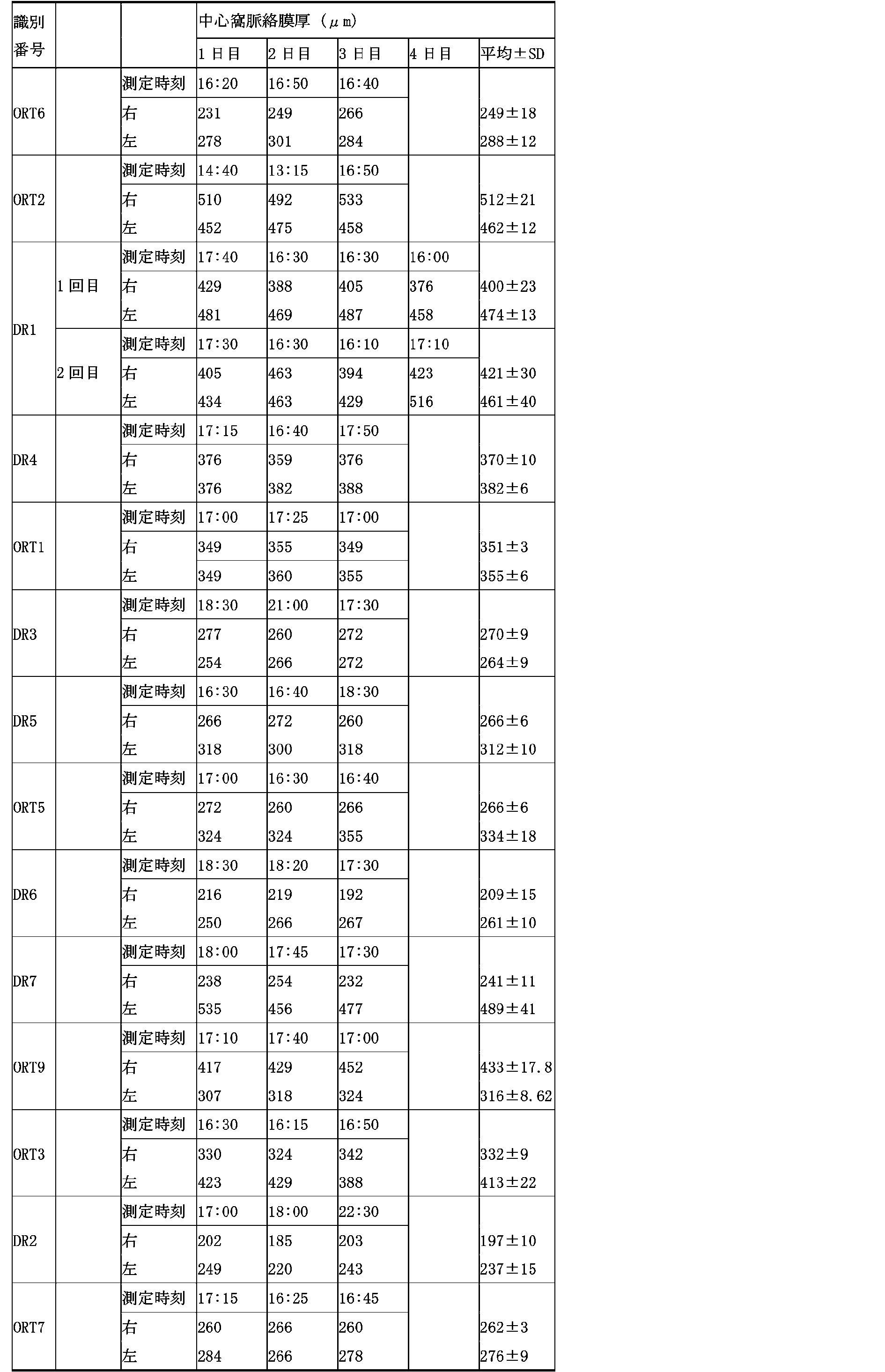

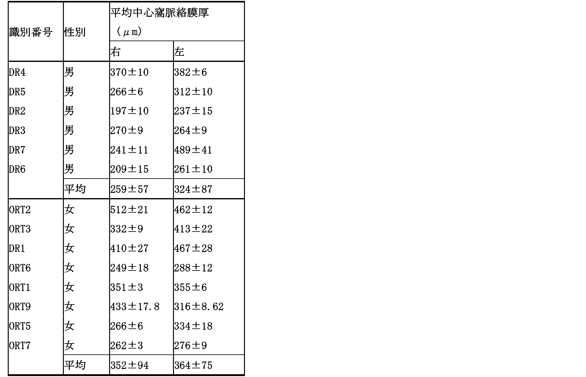
本発明は、ストレス状態の検出を必要とする全ての技術分野、例えば、ストレス状態の早期発見やストレス状態により誘発される疾患や症状の予防等に利用することができ、特に労働者の健康管理等に利用できる。
10 :OCT装置
11 :画像取得部
12 :入力部
13 :演算部
13a :画像信号処理部
13b :算出部
14 :検出部
15 :記憶部
16 :表示部
X :ストレス検出装置
11 :画像取得部
12 :入力部
13 :演算部
13a :画像信号処理部
13b :算出部
14 :検出部
15 :記憶部
16 :表示部
X :ストレス検出装置
Claims (7)
- 対象体の脈絡膜の断層画像に基づいて前記対象体のストレス状態を検出する工程を有するストレス状態の検出方法。
- 前記ストレス状態を検出する工程は、前記断層画像から脈絡膜厚を算出し、算出された前記脈絡膜厚に基づいて前記対象体のストレス状態を検出する請求項1に記載のストレス状態の検出方法。
- 算出された前記脈絡膜厚が、中心窩下で300μm以上であるときにストレス状態にあると検出する請求項2に記載のストレス状態の検出方法。
- 前記ストレス状態を検出する工程は、前記断層画像から脈絡膜の容積を算出し、算出された前記脈絡膜の容積に基づいて前記対象体のストレス状態を検出する請求項1に記載のストレス状態の検出方法。
- 前記ストレス状態を検出する工程は、算出された前記脈絡膜厚を、検出時刻、前記対象体の年齢、性別、現病歴、及び、眼構造から得られる指標から選択される少なくともの1つの因子を用いて補正する工程を含む請求項2又は3に記載のストレス状態の検出方法。
- 前記ストレス状態を検出する工程は、算出された前記脈絡膜の容積を、検出時刻、前記対象体の年齢、性別、現病歴、眼軸長、等価球面積、及び、及び、眼構造から得られる指標から選択される少なくともの1つの因子を用いて補正する工程を含む請求項4に記載のストレス状態の検出方法。
- 対象体の脈絡膜の断層画像を取得する画像取得部と、
前記断層画像から脈絡膜厚又は脈絡膜の容積を算出する算出部と、
前記脈絡膜厚又は脈絡膜の容積に基づいて前記対象体のストレス状態を検出する検出部とを備えたストレス検出装置。
Priority Applications (4)
| Application Number | Priority Date | Filing Date | Title |
|---|---|---|---|
| JP2019567175A JP6930763B2 (ja) | 2018-01-25 | 2019-01-25 | ストレス状態の検出方法、及び、ストレス検出装置 |
| EP19743747.8A EP3744254A4 (en) | 2018-01-25 | 2019-01-25 | STRESS CONDITION DETECTION METHOD AND STRESS DETECTION DEVICE |
| US16/964,029 US12035973B2 (en) | 2018-01-25 | 2019-01-25 | Method for detecting stressed state and stress detection apparatus |
| CN201980010385.0A CN111655151B (zh) | 2018-01-25 | 2019-01-25 | 压力状态的检测方法以及压力检测装置 |
Applications Claiming Priority (2)
| Application Number | Priority Date | Filing Date | Title |
|---|---|---|---|
| JP2018-010822 | 2018-01-25 | ||
| JP2018010822 | 2018-01-25 |
Publications (1)
| Publication Number | Publication Date |
|---|---|
| WO2019146738A1 true WO2019146738A1 (ja) | 2019-08-01 |
Family
ID=67396059
Family Applications (1)
| Application Number | Title | Priority Date | Filing Date |
|---|---|---|---|
| PCT/JP2019/002394 Ceased WO2019146738A1 (ja) | 2018-01-25 | 2019-01-25 | ストレス状態の検出方法、及び、ストレス検出装置 |
Country Status (5)
| Country | Link |
|---|---|
| US (1) | US12035973B2 (ja) |
| EP (1) | EP3744254A4 (ja) |
| JP (1) | JP6930763B2 (ja) |
| CN (1) | CN111655151B (ja) |
| WO (1) | WO2019146738A1 (ja) |
Cited By (2)
| Publication number | Priority date | Publication date | Assignee | Title |
|---|---|---|---|---|
| JPWO2022208873A1 (ja) * | 2021-04-02 | 2022-10-06 | ||
| WO2026018634A1 (ja) * | 2024-07-19 | 2026-01-22 | 株式会社ニコン | 画像処理装置、画像処理方法、及びプログラム |
Families Citing this family (3)
| Publication number | Priority date | Publication date | Assignee | Title |
|---|---|---|---|---|
| CN111954485B (zh) * | 2018-04-18 | 2024-08-23 | 株式会社尼康 | 图像处理方法、计算机可读存储介质及眼科系统 |
| JP7277188B2 (ja) * | 2019-03-14 | 2023-05-18 | 株式会社日立製作所 | 作業場の管理支援システムおよび管理支援方法 |
| CN112826442B (zh) * | 2020-12-31 | 2024-06-25 | 上海鹰瞳医疗科技有限公司 | 基于眼底图像的心理状态识别方法及设备 |
Citations (7)
| Publication number | Priority date | Publication date | Assignee | Title |
|---|---|---|---|---|
| JP2000275248A (ja) | 1999-03-24 | 2000-10-06 | Agency Of Ind Science & Technol | 慢性ストレス判定方法及びその装置、記録媒体、判定シート |
| JP2002544159A (ja) * | 1999-05-07 | 2002-12-24 | ジョンズ ホプキンス ユニヴァーシティ スクール オヴ メディシン | 眼障害の処置におけるプロテインチロシンキナーゼ経路インヒビターの使用 |
| JP2007225606A (ja) | 2006-01-27 | 2007-09-06 | Prima Meat Packers Ltd | 新規ストレスバイオマーカー及びその用途 |
| JP2012251857A (ja) | 2011-06-02 | 2012-12-20 | Nagaoka Univ Of Technology | ストレスの評価方法 |
| JP2013066702A (ja) * | 2011-09-06 | 2013-04-18 | Canon Inc | 画像処理装置および画像処理方法 |
| JP2014104275A (ja) * | 2012-11-29 | 2014-06-09 | Osaka Univ | 眼科装置 |
| JP2017074273A (ja) * | 2015-10-16 | 2017-04-20 | 国立大学法人 鹿児島大学 | 画像処理装置、画像処理方法、診断システム及びプログラム |
Family Cites Families (18)
| Publication number | Priority date | Publication date | Assignee | Title |
|---|---|---|---|---|
| US7575321B2 (en) * | 2003-10-30 | 2009-08-18 | Welch Allyn, Inc. | Apparatus and method of diagnosis of optically identifiable ophthalmic conditions |
| JP5006070B2 (ja) * | 2007-02-28 | 2012-08-22 | 森永製菓株式会社 | ストレス強度演算方法、及びストレス強度演算装置 |
| US7942820B2 (en) * | 2007-08-26 | 2011-05-17 | Philip Chidi Njemanze | Method and system for evaluation of the hemodynamic model in depression for diagnosis and treatment |
| JP5192250B2 (ja) * | 2008-02-04 | 2013-05-08 | 株式会社トプコン | 眼底観察装置 |
| JP2010246787A (ja) | 2009-04-17 | 2010-11-04 | Sanyo Electric Co Ltd | マッサージ機 |
| AU2010286335B2 (en) * | 2009-08-26 | 2015-07-30 | The Bionics Institute Of Australia | Apparatus for stimulating and/or monitoring activity in tissue |
| CN103108583B (zh) * | 2010-06-25 | 2015-09-09 | 安尼迪斯保健系统有限公司 | 用于对脉络膜成像的方法及装置 |
| JP5836634B2 (ja) * | 2011-05-10 | 2015-12-24 | キヤノン株式会社 | 画像処理装置及び方法 |
| JP6146952B2 (ja) * | 2012-01-27 | 2017-06-14 | キヤノン株式会社 | 画像処理装置、画像処理方法及びプログラム。 |
| JP6278295B2 (ja) * | 2013-06-13 | 2018-02-14 | 国立大学法人 筑波大学 | 脈絡膜の血管網を選択的に可視化し解析する光干渉断層計装置及びその画像処理プログラム |
| JP6322042B2 (ja) * | 2014-04-28 | 2018-05-09 | キヤノン株式会社 | 眼科撮影装置、その制御方法、およびプログラム |
| WO2015188279A1 (en) * | 2014-06-12 | 2015-12-17 | Rsem, Limited Partnership | Non-invasive measurement of choroidal volume change and ocular rigidity through oct imaging |
| NZ773822A (en) * | 2015-03-16 | 2022-07-29 | Magic Leap Inc | Methods and systems for diagnosing and treating health ailments |
| WO2017136696A1 (en) * | 2016-02-05 | 2017-08-10 | Murali Archana | Methods and apparatus for processing opthalmic biomarkers |
| JP2017169974A (ja) * | 2016-03-25 | 2017-09-28 | パナソニックIpマネジメント株式会社 | 生体情報計測装置 |
| US10332315B2 (en) * | 2016-06-20 | 2019-06-25 | Magic Leap, Inc. | Augmented reality display system for evaluation and modification of neurological conditions, including visual processing and perception conditions |
| JP6165935B2 (ja) * | 2016-07-08 | 2017-07-19 | 株式会社トプコン | 光画像計測装置 |
| JP6427224B2 (ja) * | 2017-04-27 | 2018-11-21 | 株式会社トプコン | 眼科撮影装置 |
-
2019
- 2019-01-25 WO PCT/JP2019/002394 patent/WO2019146738A1/ja not_active Ceased
- 2019-01-25 CN CN201980010385.0A patent/CN111655151B/zh active Active
- 2019-01-25 JP JP2019567175A patent/JP6930763B2/ja active Active
- 2019-01-25 US US16/964,029 patent/US12035973B2/en active Active
- 2019-01-25 EP EP19743747.8A patent/EP3744254A4/en active Pending
Patent Citations (7)
| Publication number | Priority date | Publication date | Assignee | Title |
|---|---|---|---|---|
| JP2000275248A (ja) | 1999-03-24 | 2000-10-06 | Agency Of Ind Science & Technol | 慢性ストレス判定方法及びその装置、記録媒体、判定シート |
| JP2002544159A (ja) * | 1999-05-07 | 2002-12-24 | ジョンズ ホプキンス ユニヴァーシティ スクール オヴ メディシン | 眼障害の処置におけるプロテインチロシンキナーゼ経路インヒビターの使用 |
| JP2007225606A (ja) | 2006-01-27 | 2007-09-06 | Prima Meat Packers Ltd | 新規ストレスバイオマーカー及びその用途 |
| JP2012251857A (ja) | 2011-06-02 | 2012-12-20 | Nagaoka Univ Of Technology | ストレスの評価方法 |
| JP2013066702A (ja) * | 2011-09-06 | 2013-04-18 | Canon Inc | 画像処理装置および画像処理方法 |
| JP2014104275A (ja) * | 2012-11-29 | 2014-06-09 | Osaka Univ | 眼科装置 |
| JP2017074273A (ja) * | 2015-10-16 | 2017-04-20 | 国立大学法人 鹿児島大学 | 画像処理装置、画像処理方法、診断システム及びプログラム |
Non-Patent Citations (14)
| Title |
|---|
| ARCH OPHTHALMOL, vol. 83, no. 1, 1970, pages 95 - 99 |
| BERT LR CORNELIUS ET AL., BMC PUBLIC HEALTH, vol. 13, 2013, pages 128 |
| EXPERIMENTAL EYE RESEARCH, vol. 15, no. 1, 1973, pages 15 - 29 |
| HIROHITO NANBU ET AL., KOUSEI NO SHIHYO, vol. 61, no. 5, May 2014 (2014-05-01), pages 23 - 30 |
| IKUNO Y ET AL., INVEST OPHTHALMOL VIS SCI, vol. 51, no. 4, 2010, pages 2173 - 2176 |
| KESSLER RC ET AL., ARCH GEN PSYCHIATRY, vol. 60, 2003, pages 184 - 189 |
| KESSLER RC ET AL., PSYCHOLOGICAL MEDICINE, vol. 32, 2002, pages 959 - 976 |
| NORITO KAWAKAMI ET AL., HEALTH AND LABOR SCIENCES RESEARCH GRANT, 2014 |
| SAKURAI K ET AL., PSYCHIATRY CLIN NEUROSCI., vol. 65, no. 5, 2011, pages 434 - 441 |
| SAYIN N ET AL., INDIAN J OPHTHALMOL., vol. 63, no. 5, 2015, pages 445 - 450 |
| See also references of EP3744254A4 |
| TAN CS ET AL., INVEST OPHTHALMOL VIS SCI, vol. 53, no. 1, 2012, pages 261 - 266 |
| WAKATSUKI Y ET AL., PLOS ONE, vol. 10, no. 12, 3 December 2015 (2015-12-03), pages e0144156 |
| WEN BIN WEI ET AL., OPHTHALMOLOGY, vol. 120, no. 1, 2013, pages 175 - 180 |
Cited By (3)
| Publication number | Priority date | Publication date | Assignee | Title |
|---|---|---|---|---|
| JPWO2022208873A1 (ja) * | 2021-04-02 | 2022-10-06 | ||
| WO2022208873A1 (ja) * | 2021-04-02 | 2022-10-06 | 日本電気株式会社 | ストレス推定装置、ストレス推定方法及び記憶媒体 |
| WO2026018634A1 (ja) * | 2024-07-19 | 2026-01-22 | 株式会社ニコン | 画像処理装置、画像処理方法、及びプログラム |
Also Published As
| Publication number | Publication date |
|---|---|
| CN111655151A (zh) | 2020-09-11 |
| US12035973B2 (en) | 2024-07-16 |
| CN111655151B (zh) | 2024-03-22 |
| US20210038077A1 (en) | 2021-02-11 |
| EP3744254A1 (en) | 2020-12-02 |
| JP6930763B2 (ja) | 2021-09-01 |
| JPWO2019146738A1 (ja) | 2020-12-03 |
| EP3744254A4 (en) | 2021-11-03 |
Similar Documents
| Publication | Publication Date | Title |
|---|---|---|
| JP6930763B2 (ja) | ストレス状態の検出方法、及び、ストレス検出装置 | |
| Ferguson et al. | Psychophysiological associations with gastrointestinal symptomatology in autism spectrum disorder | |
| Wong et al. | Does refractive error influence the association of blood pressure and retinal vessel diameters? The Blue Mountains Eye Study | |
| Wu et al. | Intrasession test–retest variability of microperimetry in age-related macular degeneration | |
| Sinclair et al. | Low energy diet and intracranial pressure in women with idiopathic intracranial hypertension: prospective cohort study | |
| Pritchard et al. | Longitudinal assessment of neuropathy in type 1 diabetes using novel ophthalmic markers (LANDMark): study design and baseline characteristics | |
| Simons et al. | The Fear of Pain Questionnaire (FOPQ): assessment of pain-related fear among children and adolescents with chronic pain | |
| Kurysheva et al. | Value of structural and hemodynamic parameters for the early detection of primary open-angle glaucoma | |
| Salvi et al. | Abnormalities of retinal ganglion cell complex at optical coherence tomography in patients with type 2 diabetes: a sign of diabetic polyneuropathy, not retinopathy | |
| Realini et al. | Diurnal intraocular pressure patterns are not repeatable in the short term in healthy individuals | |
| Almasri et al. | The impact of diabetes mellitus on health-related quality of life in Saudi Arabia | |
| Pokharel et al. | Macular thickness and macular volume measurements using spectral domain optical coherence tomography in normal Nepalese eyes | |
| Ziolkowski et al. | An evaluation of life satisfaction and health–Quality of life of senior citizens | |
| Granström et al. | Visual functioning and health-related quality of life in diabetic patients about to undergo anti-vascular endothelial growth factor treatment for sight-threatening macular edema | |
| Vigil et al. | Gender expression, sexual orientation and pain sensitivity in women | |
| Yuki et al. | Evaluation of fear of falling in patients with primary open-angle glaucoma and the importance of inferior visual field damage | |
| Takeshima et al. | Central serous chorioretinopathy and heart rate variability analysis with a smartphone application | |
| Kusbeci et al. | The evaluation of anatomic and functional changes in unilateral moderate amblyopic eyes using optical coherence tomography and pupil cycle time | |
| Shah et al. | Validation of the Pediatric Vision Scanner in a normal preschool population | |
| US20240226081A1 (en) | Attention evaluation and methods for medicating | |
| Wooten | Clinical features and electrodiagnosis of diabetic peripheral neuropathy in the dysvascular patient | |
| EP3513739B1 (en) | System for diagnosing chronic pain-related diseases and diseases requiring differentiation therefrom | |
| HK40033339A (en) | Stress state detection method and stress detection device | |
| Murphy et al. | The influence of fixation stability on balance in central vision loss | |
| Duran et al. | Comparative assessment of choroidal thickness and choroidal vascularity index in patients with restless legs syndrome |
Legal Events
| Date | Code | Title | Description |
|---|---|---|---|
| 121 | Ep: the epo has been informed by wipo that ep was designated in this application |
Ref document number: 19743747 Country of ref document: EP Kind code of ref document: A1 |
|
| ENP | Entry into the national phase |
Ref document number: 2019567175 Country of ref document: JP Kind code of ref document: A |
|
| NENP | Non-entry into the national phase |
Ref country code: DE |
|
| ENP | Entry into the national phase |
Ref document number: 2019743747 Country of ref document: EP Effective date: 20200825 |