WO2019198704A1 - 運動強度推定方法、運動強度推定装置およびプログラム - Google Patents
運動強度推定方法、運動強度推定装置およびプログラム Download PDFInfo
- Publication number
- WO2019198704A1 WO2019198704A1 PCT/JP2019/015430 JP2019015430W WO2019198704A1 WO 2019198704 A1 WO2019198704 A1 WO 2019198704A1 JP 2019015430 W JP2019015430 W JP 2019015430W WO 2019198704 A1 WO2019198704 A1 WO 2019198704A1
- Authority
- WO
- WIPO (PCT)
- Prior art keywords
- wave
- exercise intensity
- amplitude
- index
- subject
- Prior art date
- Legal status (The legal status is an assumption and is not a legal conclusion. Google has not performed a legal analysis and makes no representation as to the accuracy of the status listed.)
- Ceased
Links
Images


Classifications
-
- A—HUMAN NECESSITIES
- A61—MEDICAL OR VETERINARY SCIENCE; HYGIENE
- A61B—DIAGNOSIS; SURGERY; IDENTIFICATION
- A61B5/00—Measuring for diagnostic purposes; Identification of persons
- A61B5/103—Measuring devices for testing the shape, pattern, colour, size or movement of the body or parts thereof, for diagnostic purposes
- A61B5/11—Measuring movement of the entire body or parts thereof, e.g. head or hand tremor or mobility of a limb
- A61B5/1118—Determining activity level
-
- A—HUMAN NECESSITIES
- A61—MEDICAL OR VETERINARY SCIENCE; HYGIENE
- A61B—DIAGNOSIS; SURGERY; IDENTIFICATION
- A61B5/00—Measuring for diagnostic purposes; Identification of persons
- A61B5/24—Detecting, measuring or recording bioelectric or biomagnetic signals of the body or parts thereof
- A61B5/316—Modalities, i.e. specific diagnostic methods
- A61B5/318—Heart-related electrical modalities, e.g. electrocardiography [ECG]
- A61B5/346—Analysis of electrocardiograms
- A61B5/349—Detecting specific parameters of the electrocardiograph cycle
- A61B5/352—Detecting R peaks, e.g. for synchronising diagnostic apparatus; Estimating R-R interval
-
- A—HUMAN NECESSITIES
- A61—MEDICAL OR VETERINARY SCIENCE; HYGIENE
- A61B—DIAGNOSIS; SURGERY; IDENTIFICATION
- A61B5/00—Measuring for diagnostic purposes; Identification of persons
- A61B5/24—Detecting, measuring or recording bioelectric or biomagnetic signals of the body or parts thereof
- A61B5/316—Modalities, i.e. specific diagnostic methods
- A61B5/318—Heart-related electrical modalities, e.g. electrocardiography [ECG]
- A61B5/329—Load diagnosis, e.g. cardiac stress tests
-
- A—HUMAN NECESSITIES
- A61—MEDICAL OR VETERINARY SCIENCE; HYGIENE
- A61B—DIAGNOSIS; SURGERY; IDENTIFICATION
- A61B5/00—Measuring for diagnostic purposes; Identification of persons
- A61B5/24—Detecting, measuring or recording bioelectric or biomagnetic signals of the body or parts thereof
- A61B5/316—Modalities, i.e. specific diagnostic methods
- A61B5/318—Heart-related electrical modalities, e.g. electrocardiography [ECG]
- A61B5/346—Analysis of electrocardiograms
- A61B5/349—Detecting specific parameters of the electrocardiograph cycle
- A61B5/355—Detecting T-waves
-
- A—HUMAN NECESSITIES
- A61—MEDICAL OR VETERINARY SCIENCE; HYGIENE
- A61B—DIAGNOSIS; SURGERY; IDENTIFICATION
- A61B5/00—Measuring for diagnostic purposes; Identification of persons
- A61B5/02—Detecting, measuring or recording for evaluating the cardiovascular system, e.g. pulse, heart rate, blood pressure or blood flow
- A61B5/024—Measuring pulse rate or heart rate
- A61B5/0245—Measuring pulse rate or heart rate by using sensing means generating electric signals, i.e. ECG signals
-
- A—HUMAN NECESSITIES
- A61—MEDICAL OR VETERINARY SCIENCE; HYGIENE
- A61B—DIAGNOSIS; SURGERY; IDENTIFICATION
- A61B5/00—Measuring for diagnostic purposes; Identification of persons
- A61B5/24—Detecting, measuring or recording bioelectric or biomagnetic signals of the body or parts thereof
- A61B5/316—Modalities, i.e. specific diagnostic methods
- A61B5/318—Heart-related electrical modalities, e.g. electrocardiography [ECG]
- A61B5/332—Portable devices specially adapted therefor
-
- A—HUMAN NECESSITIES
- A61—MEDICAL OR VETERINARY SCIENCE; HYGIENE
- A61B—DIAGNOSIS; SURGERY; IDENTIFICATION
- A61B5/00—Measuring for diagnostic purposes; Identification of persons
- A61B5/68—Arrangements of detecting, measuring or recording means, e.g. sensors, in relation to patient
- A61B5/6801—Arrangements of detecting, measuring or recording means, e.g. sensors, in relation to patient specially adapted to be attached to or worn on the body surface
- A61B5/6802—Sensor mounted on worn items
- A61B5/6804—Garments; Clothes
Definitions
- the present invention relates to an exercise intensity estimation method, an exercise intensity estimation device, and a program for estimating exercise intensity from a human electrocardiogram waveform.
- the ECG waveform can be measured, various biological information can be acquired. For example, in sports training, when the exercise intensity exceeds a certain level, the heart needs to increase blood flow to the body, so the heart increases the heart rate and increases the end-diastolic volume of the ventricle (front Load reserve). In such a case, the ECG waveform is considered to change the T wave profile corresponding to the ventricular diastole. Therefore, analysis of the T wave can be utilized for estimation of exercise intensity and the like.
- Patent Document 1 discloses a configuration in which exercise intensity is evaluated based on the relaxation time of the heart, that is, the change in the length of the T wave.
- the configuration disclosed in Patent Document 1 does not take into account fluctuations in the amplitude level of the ECG waveform.
- the present invention has been made in view of the above problems, and an exercise intensity estimation method and an exercise intensity estimation apparatus capable of obtaining an appropriate index indicating exercise intensity even when the amplitude of an ECG waveform varies. And to provide a program.
- the exercise intensity estimation method of the present invention calculates either the RS amplitude from the R wave peak value to the S wave peak value of the subject's ECG waveform, the R wave height, or the S wave depth.
- one configuration example of the exercise intensity estimation method of the present invention further includes a fourth step of calculating a heart rate from the electrocardiogram waveform, and a value obtained by multiplying the first index by the heart rate.
- the exercise intensity estimation device of the present invention is any one of the RS amplitude from the peak value of the R wave to the peak value of the S wave, the height of the R wave, or the depth of the S wave of the subject's electrocardiogram waveform.
- a first calculation unit configured to calculate the T wave
- a second calculation unit configured to calculate either the amplitude or height of the T wave of the electrocardiogram waveform, and the amplitude of the T wave or
- a value obtained by normalizing the height using any one of the RS amplitude, the height of the R wave, or the depth of the S wave is calculated as a first index indicating the exercise intensity of the subject.
- a third calculator configured as described above.
- the exercise intensity estimation program of the present invention is any one of the RS amplitude from the R wave peak value to the S wave peak value of the subject's electrocardiogram waveform, the R wave height, or the S wave depth.
- the change of the T wave can be accurately grasped, and an appropriate index indicating the exercise intensity of the subject can be obtained even when the amplitude of the electrocardiogram waveform of the subject fluctuates.
- an appropriate index can be obtained even when a wearable waveform measuring device is used.
- FIG. 1 is a diagram illustrating an example of an ECG waveform.
- FIG. 2 is a diagram illustrating an example of a heart rate.
- FIG. 3 is a diagram in which the amplitude of the T wave of the ECG waveform is plotted.
- FIG. 4 is a diagram in which the amplitude of the T wave normalized by the RS amplitude of the ECG waveform is plotted.
- FIG. 5 is a diagram showing a value obtained by multiplying the amplitude of the T wave of the ECG waveform by the heart rate.
- FIG. 6 is a diagram illustrating a value obtained by multiplying the normalized T-wave amplitude of the ECG waveform by the heart rate.
- FIG. 1 is a diagram illustrating an example of an ECG waveform.
- FIG. 2 is a diagram illustrating an example of a heart rate.
- FIG. 3 is a diagram in which the amplitude of the T wave of the ECG waveform is plotted.
- FIG. 4 is a
- FIG. 7 is a block diagram showing the configuration of the exercise intensity estimation apparatus according to the embodiment of the present invention.
- FIG. 8 is a flowchart for explaining the operation of the exercise intensity estimation apparatus according to the embodiment of the present invention.
- FIG. 9 is a diagram showing an example of P waves, Q waves, R waves, S waves, and T waves of an ECG waveform.
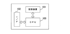
- FIG. 10 is a block diagram illustrating a configuration example of a computer that realizes the exercise intensity estimation apparatus according to the embodiment of the present invention.
- FIG. 1 is a diagram showing a subject's ECG waveform acquired using a wearable device when the subject is running.
- the amplitude level of the ECG waveform decreases for some reason in the interval of about 220 to 440 seconds on the horizontal axis.
- FIG. 1B is an enlarged view of the vicinity of 150 seconds in the graph of FIG. 1A
- FIG. 1C is an enlarged view of the vicinity of 350 seconds of the graph of FIG. is there.
- the ⁇ marks indicate the positions of the T wave peaks. According to (b) and (c) of FIG. 1, it can be seen that the amplitude level of the R wave is reduced in the vicinity of 350 seconds as compared with that in the vicinity of 150 seconds. Therefore, it is suggested that the relative relationship with the amplitude of the R wave should be considered in order to evaluate the height of the T wave.
- FIG. 2 is a diagram showing the heart rate of the subject during the same measurement period as in FIG. According to FIG. 2, the heart rate in the section of 220 to 440 seconds is high, and there is a peak around 330 seconds. It can be estimated that the exercise intensity is the highest in this vicinity.
- the height of the T wave corresponds to the end-diastolic volume of the ventricle, and reflects the load on the heart, that is, the intensity of exercise applied to the subject, from a different aspect than the heart rate. It is thought that it is an index to do. However, it is necessary to appropriately evaluate the fluctuation of the amplitude level of the ECG waveform.
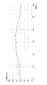
- FIG. 3 is a diagram plotting the amplitude of the T wave in the ECG waveform of FIG.
- the amplitude of the T wave for example, an R wave is detected from an ECG waveform, a maximum value and a minimum value in a certain time width following the R wave are detected, and a difference between the maximum value and the minimum value is taken. Can be obtained.
- FIG. 4 is a diagram in which the value obtained by dividing the amplitude of the T wave of FIG. 3 by the RS amplitude of the same heartbeat as this T wave, that is, the amplitude of the T wave normalized by the RS amplitude is plotted.
- each of the amplitude of the T wave and the RS amplitude is resampled at intervals of 0.5 seconds, and a moving average for 30 seconds is taken. .
- the amplitude of the T wave is about 25% larger than that of other sections in the section of 220 to 440 seconds.
- the amplitude of the normalized T wave is about 50% larger than that of the other sections in the 240 to 440 second interval, and the activation of the preload reserve of the heart and the exercise intensity are further increased. The probability of accurately representing it is high.
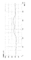
- FIG. 5 shows a value obtained by multiplying the amplitude of the T wave of FIG. 3 by the heart rate of FIG. 2
- FIG. 6 shows a value obtained by multiplying the normalized amplitude of the T wave of FIG. 4 by the heart rate of FIG. ing. Similar to FIGS. 3 and 4, there are differences in FIGS. 5 and 6. It can be considered that FIG. 6 more accurately evaluates the blood volume ejected by the heart.
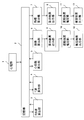
- FIG. 7 is a block diagram showing the configuration of the exercise intensity estimation apparatus according to the embodiment of the present invention.
- the exercise intensity estimation apparatus includes an electrocardiograph 1 that outputs an ECG waveform sampling data sequence, a storage unit 2 that stores an ECG waveform sampling data sequence and sampling time information, and an ECG waveform R wave peak value.
- An RS wave calculation unit 3 (first calculation unit) that calculates the RS amplitude up to the peak value of the S wave
- a T wave calculation unit 4 (second calculation unit) that calculates the amplitude of the T wave of the ECG waveform
- a heart rate calculation unit 5 (fourth calculation unit) that calculates a heart rate from the ECG waveform
- an index calculation that calculates the amplitude of the T wave normalized by the RS amplitude as a first index indicating the exercise intensity of the subject.
- a unit 6 (third calculation unit), and an index calculation unit 7 (fifth calculation unit) that calculates a value obtained by multiplying the first index by the heart rate as a second index indicating the exercise intensity of the subject.
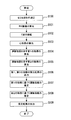
- FIG. 8 is a flowchart for explaining the operation of the exercise intensity estimation apparatus
- FIG. 9 is a diagram showing examples of P waves, Q waves, R waves, S waves, and T waves of ECG waveforms.
- the electrocardiograph 1 measures the ECG waveform of the subject of exercise intensity estimation and outputs a sampling data string of the ECG waveform (step S100 in FIG. 8). At this time, the electrocardiograph 1 adds the sampling time information to each sampling data and outputs it. Since a specific method for measuring an ECG waveform is a well-known technique, detailed description thereof is omitted.
- the storage unit 2 stores a sampling data string of the ECG waveform output from the electrocardiograph 1 and information on the sampling time.
- the RS wave calculation unit 3 calculates, for each heartbeat, an RS amplitude (A1 in FIG. 9) that is an amplitude from the peak value of the R wave of the ECG waveform stored in the storage unit 2 to the peak value of the immediately following S wave (A1 in FIG. 9).
- FIG. 8 step S101 A method for detecting the R wave and the S wave is disclosed in, for example, Japanese Patent Application Laid-Open No. 2015-156936.
- the T wave calculation unit 4 calculates the amplitude of the T wave of the ECG waveform stored in the storage unit 2 for each heartbeat (step S102 in FIG. 8). Specifically, the T-wave calculation unit 4 has a maximum value (T-wave peak value) and a minimum value (T-wave peak value) in a detection period of a certain time width after a predetermined standby time has elapsed from the time of the R-wave peak value. T-wave bottom value) is detected, and the amplitude from the maximum value to the minimum value (A2 in FIG. 9) is calculated as the amplitude of the T-wave.
- the standby time is set in advance so that the S wave immediately after the R wave is removed. Note that the standby time may be determined starting from the time of the peak value of the S wave instead of the R wave.
- the time width of the detection period is set in advance so as to include the peak value of the T wave and the subsequent bottom value.
- the heart rate calculation unit 5 calculates a heart rate HR (instantaneous heart rate) for each heart rate from the ECG waveform stored in the storage unit 2 (step S103 in FIG. 8).
- the heart rate calculation unit 5 detects the RR interval (I in FIG. 9), which is the time interval between the R wave and the previous R wave, for each heart beat, and calculates the heart rate HR for each heart beat using the following equation. .
- HR [bpm] 60000 / I [ms] (1)
- the index calculation unit 6 calculates the amplitude A2 ′ of the T wave normalized by the RS amplitude A1 for each heartbeat as the first index E1 indicating exercise intensity (step S104 in FIG. 8). Specifically, the index calculation unit 6 divides the value obtained by dividing the amplitude A2 of the T wave by the RS amplitude A1 of the same heartbeat as the T wave (that is, the RS amplitude A1 immediately before the T wave) as in the following equation. Calculated as the first index E1.
- the amplitude A2 of the T wave used in the equation (2) is the same as the amplitude of the T wave in the heartbeat for which the index E1 is to be calculated and a certain time (for example, past the heartbeat). It is good also as an average value with the amplitude of T wave of 30 seconds.
- the RS amplitude A1 used in the equation (2) may be an average value of the RS amplitude in the heartbeat for which the index E1 is to be calculated and the RS amplitude for a certain past time (for example, 30 seconds) from the heartbeat. .
- the index calculation unit 7 uses a value obtained by multiplying the first index E1 by the heart rate HR calculated from the same heart rate as the first index E1 (T wave) as a second index E2 indicating exercise intensity. It is calculated every time (step S105 in FIG. 8).
- E2 E1 ⁇ HR (3)
- the calculation result output units 8 and 9 output the calculation results of the index calculation units 6 and 7, respectively (step S106 in FIG. 8).
- the output method at this time includes, for example, a graph display of the calculation result, transmission of the calculation result to an external device, and the like.
- the exercise intensity estimation unit 10 compares the first index E1 calculated by the index calculation unit 6 with a predetermined threshold TH1, and estimates the exercise intensity of the subject (step S107 in FIG. 8). Specifically, the exercise intensity estimation unit 10 estimates that the exercise intensity of the subject is low when the first index E1 is less than or equal to the threshold TH1, and if the first index E1 exceeds the threshold TH1, Estimated that exercise intensity is high. In the example of FIG. 4, for example, 0.25 [a. u. ] May be set to the threshold value TH1. Note that a plurality of threshold values TH1 may be provided to evaluate the exercise intensity of the subject in multiple stages.
- the exercise intensity estimation unit 11 compares the second index E2 calculated by the index calculation unit 7 with a predetermined threshold TH2 to estimate the exercise intensity of the subject (step S108 in FIG. 8). Specifically, the exercise intensity estimating unit 11 estimates that the exercise intensity of the subject is low when the second index E2 is equal to or less than the threshold TH2, and if the second index E2 exceeds the threshold TH2, the exercise intensity of the subject Estimated that exercise intensity is high. In the example of FIG. 6, for example, 40 [a. u. ] May be set to the threshold value TH2. Similarly to the above, a plurality of threshold values TH2 may be provided to evaluate the exercise intensity of the subject in multiple stages.
- the estimation result output units 12 and 13 output the estimation results of the exercise intensity estimation units 10 and 11, respectively (step S109 in FIG. 8).
- the output method at this time includes, for example, display of an estimation result, audio output of the estimation result, and transmission of the estimation result to an external device.
- the present invention is suitable for an ECG waveform measurement device (wearable device) that acquires an ECG waveform by placing an electrode on the inner surface of clothes such as a shirt and bringing the electrode into contact with the body surface of the subject. is there.
- the application target of the present invention is not limited to such an ECG waveform measurement device.
- the storage unit 2, the RS wave calculation unit 3, the T wave calculation unit 4, the heart rate calculation unit 5, and the index calculation units 6 and 7 of the exercise intensity estimation device described in this embodiment are a CPU (Central Processing Unit) and storage. It can be realized by a computer having a device and an interface and a program for controlling these hardware resources. An example of the configuration of this computer is shown in FIG.
- the computer includes a CPU 100, a storage device 101, and an interface device (hereinafter abbreviated as I / F) 102.
- the electrocardiograph 1 and the hardware of the calculation result output units 8 and 9 are connected to the I / F 102.
- an exercise intensity estimation program for realizing the exercise intensity estimation method of the present invention is stored in the storage device 101.
- the CPU 100 executes the processing described in this embodiment according to the exercise intensity estimation program stored in the storage device 101.
- the height of the R wave from the baseline of the ECG waveform to the peak value of the R wave may be used instead of the RS amplitude A1.
- an R wave calculation unit (first calculation unit) that calculates the height H1 of the R wave for each heartbeat is provided.
- the RS amplitude A1 the depth of the S wave from the baseline of the ECG waveform to the peak value of the S wave (D in FIG. 9) may be used.
- an S wave calculation unit (first calculation unit) that calculates the depth D of the S wave for each heartbeat is provided instead of the RS wave calculation unit 3.
- the height of the T wave from the baseline of the ECG waveform to the peak value of the T wave (H2 in FIG. 9) may be used.
- a T wave calculation unit (second calculation unit) that calculates the height H2 of the T wave for each heartbeat is provided.
- the present invention can be applied to a technique for estimating the exercise intensity of a person.
Landscapes
- Health & Medical Sciences (AREA)
- Life Sciences & Earth Sciences (AREA)
- Cardiology (AREA)
- Engineering & Computer Science (AREA)
- Medical Informatics (AREA)
- Animal Behavior & Ethology (AREA)
- Biophysics (AREA)
- Pathology (AREA)
- Biomedical Technology (AREA)
- Heart & Thoracic Surgery (AREA)
- Veterinary Medicine (AREA)
- Molecular Biology (AREA)
- Surgery (AREA)
- Physics & Mathematics (AREA)
- General Health & Medical Sciences (AREA)
- Public Health (AREA)
- Physiology (AREA)
- Signal Processing (AREA)
- Dentistry (AREA)
- Oral & Maxillofacial Surgery (AREA)
- Power Engineering (AREA)
- Measurement And Recording Of Electrical Phenomena And Electrical Characteristics Of The Living Body (AREA)
- Measuring Pulse, Heart Rate, Blood Pressure Or Blood Flow (AREA)
Abstract
Description
特許文献1には、心臓の弛緩時間、すなわちT波の長さの変化に基づいて、運動強度を評価する構成が開示されている。しかし、特許文献1に開示された構成では、ECG波形の振幅レベルの変動については考慮されていない。
また、本発明の運動強度推定方法の1構成例は、さらに、前記心電図波形から心拍数を算出する第4のステップと、前記第1の指標に前記心拍数を乗じた値を、前記対象者の運動強度を示す第2の指標として算出する第5のステップとを含むことを特徴とするものである。
[発明の原理]
図1は、被験者がランニングをしているときに、ウェアラブルデバイスを用いて取得した、被験者のECG波形を示す図である。図1の(a)のグラフ(600秒分)において、横軸のおよそ220~440秒の区間では、ECG波形の振幅レベルが何らかの理由によって低下している。
図5は、図3のT波の振幅に図2の心拍数を乗じた値を示し、図6は、図4の規格化したT波の振幅に図2の心拍数を乗じた値を示している。図3、図4と同様に、図5、図6には差異が見られる。図6の方が、心臓が駆出している血液量に関して、より的確に評価していると考えられる。
以下、本発明の実施例について図面を参照して説明する。図7は本発明の実施例に係る運動強度推定装置の構成を示すブロック図である。運動強度推定装置は、ECG波形のサンプリングデータ列を出力する心電計1と、ECG波形のサンプリングデータ列とサンプリング時刻の情報とを記憶する記憶部2と、ECG波形のR波のピーク値からS波のピーク値までのRS振幅を算出するRS波算出部3(第1の算出部)と、ECG波形のT波の振幅を算出するT波算出部4(第2の算出部)と、ECG波形から心拍数を算出する心拍数算出部5(第4の算出部)と、RS振幅で規格化したT波の振幅を、対象者の運動強度を示す第1の指標として算出する指標算出部6(第3の算出部)と、第1の指標に心拍数を乗じた値を、対象者の運動強度を示す第2の指標として算出する指標算出部7(第5の算出部)と、指標算出部6の算出結果を出力する算出結果出力部8と、指標算出部7の算出結果を出力する算出結果出力部9と、第1の指標に基づいて対象者の運動強度を推定する運動強度推定部10と、第2の指標に基づいて対象者の運動強度を推定する運動強度推定部11と、運動強度推定部10の推定結果を出力する推定結果出力部12と、運動強度推定部11の推定結果を出力する推定結果出力部13とを備えている。
HR[bpm]=60000/I[ms] ・・・(1)
E1=A2’=A2/A1 ・・・(2)
E2=E1×HR ・・・(3)
Claims (8)
- 対象者の心電図波形のR波のピーク値からS波のピーク値までのRS振幅、前記R波の高さ、または前記S波の深さのいずれかを算出する第1のステップと、
前記心電図波形のT波の振幅または高さのいずれかを算出する第2のステップと、
前記T波の振幅または高さを、前記RS振幅、前記R波の高さ、または前記S波の深さのいずれかを用いて規格化した値を、前記対象者の運動強度を示す第1の指標として算出する第3のステップとを含むことを特徴とする運動強度推定方法。 - 請求項1記載の運動強度推定方法において、
さらに、前記心電図波形から心拍数を算出する第4のステップと、
前記第1の指標に前記心拍数を乗じた値を、前記対象者の運動強度を示す第2の指標として算出する第5のステップとを含むことを特徴とする運動強度推定方法。 - 請求項1記載の運動強度推定方法において、
さらに、前記第1の指標に基づいて前記対象者の運動強度を推定する第6のステップを含むことを特徴とする運動強度推定方法。 - 請求項2記載の運動強度推定方法において、
さらに、前記第2の指標に基づいて前記対象者の運動強度を推定する第7のステップを含むことを特徴とする運動強度推定方法。 - 対象者の心電図波形のR波のピーク値からS波のピーク値までのRS振幅、前記R波の高さ、または前記S波の深さのいずれかを算出するように構成された第1の算出部と、
前記心電図波形のT波の振幅または高さのいずれかを算出するように構成された第2の算出部と、
前記T波の振幅または高さを、前記RS振幅、前記R波の高さ、または前記S波の深さのいずれかを用いて規格化した値を、前記対象者の運動強度を示す第1の指標として算出するように構成された第3の算出部とを備えることを特徴とする運動強度推定装置。 - 請求項5記載の運動強度推定装置において、
さらに、前記心電図波形から心拍数を算出するように構成された第4の算出部と、
前記第1の指標に前記心拍数を乗じた値を、前記対象者の運動強度を示す第2の指標として算出するように構成された第5の算出部とを備えることを特徴とする運動強度推定装置。 - 対象者の心電図波形のR波のピーク値からS波のピーク値までのRS振幅、前記R波の高さ、または前記S波の深さのいずれかを算出する第1のステップと、
前記心電図波形のT波の振幅または高さのいずれかを算出する第2のステップと、
前記T波の振幅または高さを、前記RS振幅、前記R波の高さ、または前記S波の深さのいずれかを用いて規格化した値を、前記対象者の運動強度を示す第1の指標として算出する第3のステップとを、コンピュータに実行させることを特徴とする運動強度推定プログラム。 - 請求項7記載の運動強度推定プログラムにおいて、
さらに、前記心電図波形から心拍数を算出する第4のステップと、
前記第1の指標に前記心拍数を乗じた値を、前記対象者の運動強度を示す第2の指標として算出する第5のステップとを、コンピュータに実行させることを特徴とする運動強度推定プログラム。
Priority Applications (6)
| Application Number | Priority Date | Filing Date | Title |
|---|---|---|---|
| CN201980024887.9A CN111989037B (zh) | 2018-04-10 | 2019-04-09 | 运动强度估计方法、运动强度估计装置和程序 |
| ES19785140T ES2929851T3 (es) | 2018-04-10 | 2019-04-09 | Método de estimación de intensidad de ejercicio, dispositivo de estimación de intensidad de ejercicio y programa |
| US17/046,061 US11363969B2 (en) | 2018-04-10 | 2019-04-09 | Exercise intensity estimation method, exercise intensity estimation device, and program |
| JP2020513405A JP6859489B2 (ja) | 2018-04-10 | 2019-04-09 | 運動強度推定方法、運動強度推定装置およびプログラム |
| EP19785140.5A EP3777673B1 (en) | 2018-04-10 | 2019-04-09 | Exercise intensity estimation method, exercise intensity estimation device, and program |
| AU2019251933A AU2019251933B2 (en) | 2018-04-10 | 2019-04-09 | Exercise intensity estimation method, exercise intensity estimation device, and program |
Applications Claiming Priority (2)
| Application Number | Priority Date | Filing Date | Title |
|---|---|---|---|
| JP2018075230 | 2018-04-10 | ||
| JP2018-075230 | 2018-04-10 |
Publications (1)
| Publication Number | Publication Date |
|---|---|
| WO2019198704A1 true WO2019198704A1 (ja) | 2019-10-17 |
Family
ID=68164091
Family Applications (1)
| Application Number | Title | Priority Date | Filing Date |
|---|---|---|---|
| PCT/JP2019/015430 Ceased WO2019198704A1 (ja) | 2018-04-10 | 2019-04-09 | 運動強度推定方法、運動強度推定装置およびプログラム |
Country Status (7)
| Country | Link |
|---|---|
| US (1) | US11363969B2 (ja) |
| EP (1) | EP3777673B1 (ja) |
| JP (1) | JP6859489B2 (ja) |
| CN (1) | CN111989037B (ja) |
| AU (1) | AU2019251933B2 (ja) |
| ES (1) | ES2929851T3 (ja) |
| WO (1) | WO2019198704A1 (ja) |
Citations (4)
| Publication number | Priority date | Publication date | Assignee | Title |
|---|---|---|---|---|
| JP2004275281A (ja) | 2003-03-13 | 2004-10-07 | Seiko Epson Corp | 運動負荷強度評価装置及び運動機器 |
| JP2015156936A (ja) | 2014-02-24 | 2015-09-03 | 日本電信電話株式会社 | 心拍検出方法および心拍検出装置 |
| JP2017144132A (ja) * | 2016-02-19 | 2017-08-24 | 株式会社豊田中央研究所 | 個人識別装置、個人識別方法、及び個人識別プログラム |
| JP2017169885A (ja) * | 2016-03-24 | 2017-09-28 | トヨタ自動車株式会社 | 心電波形のt波波高を用いた緊張状態推定装置 |
Family Cites Families (14)
| Publication number | Priority date | Publication date | Assignee | Title |
|---|---|---|---|---|
| US4622980A (en) * | 1984-11-01 | 1986-11-18 | Horst E. Kunig | Method and apparatus for determining of stress condition of a subject |
| US7072708B1 (en) * | 2002-12-02 | 2006-07-04 | Inovise Medical, Inc. | Differentiating acute myocardial infarction from other ECG abnormalities |
| US7643873B2 (en) * | 2006-04-28 | 2010-01-05 | Idt Technology Limited | Exercise data apparatus |
| RU2009133362A (ru) * | 2007-02-06 | 2011-03-20 | Грайнер Био-Уан Интернэшнл Аг (At) | Способ определения интенсивности тренировки |
| JP4649429B2 (ja) * | 2007-03-12 | 2011-03-09 | 株式会社大成 | 心拍測定システム及び方法 |
| US7909764B1 (en) | 2007-11-30 | 2011-03-22 | Pacesetter, Inc. | Methods and systems to monitor venous blood oxygen saturation |
| EP2676604B1 (en) * | 2012-06-19 | 2016-08-10 | Texas Instruments France | Real time QRS duration measurement in electrocardiogram |
| US20160143552A1 (en) * | 2013-08-07 | 2016-05-26 | National Cheng Kung University | Electrocardiography signal extraction method |
| JP6173905B2 (ja) * | 2013-12-16 | 2017-08-02 | 大名 魏 | Twa測定装置及びtwa測定装置の作動方法 |
| JP6305161B2 (ja) * | 2014-03-31 | 2018-04-04 | 株式会社リアルデザイン | 終末期の終末前予測システム |
| US9468385B2 (en) * | 2014-08-22 | 2016-10-18 | Medtronic, Inc. | Visual representation of a cardiac signal sensing test |
| JP6655794B2 (ja) * | 2015-03-17 | 2020-02-26 | パナソニックIpマネジメント株式会社 | 個人認証装置、個人認証方法、およびプログラム |
| EP3282939B1 (en) * | 2015-04-14 | 2021-03-03 | LifeWatch Technologies, Ltd. | Alignment of a detachable monitoring device |
| WO2017121729A1 (en) * | 2016-01-14 | 2017-07-20 | Koninklijke Philips N.V. | Automatic classification/intepretation of ecg waves for non-athletes/athletes |
-
2019
- 2019-04-09 WO PCT/JP2019/015430 patent/WO2019198704A1/ja not_active Ceased
- 2019-04-09 ES ES19785140T patent/ES2929851T3/es active Active
- 2019-04-09 JP JP2020513405A patent/JP6859489B2/ja active Active
- 2019-04-09 AU AU2019251933A patent/AU2019251933B2/en active Active
- 2019-04-09 US US17/046,061 patent/US11363969B2/en active Active
- 2019-04-09 CN CN201980024887.9A patent/CN111989037B/zh active Active
- 2019-04-09 EP EP19785140.5A patent/EP3777673B1/en active Active
Patent Citations (4)
| Publication number | Priority date | Publication date | Assignee | Title |
|---|---|---|---|---|
| JP2004275281A (ja) | 2003-03-13 | 2004-10-07 | Seiko Epson Corp | 運動負荷強度評価装置及び運動機器 |
| JP2015156936A (ja) | 2014-02-24 | 2015-09-03 | 日本電信電話株式会社 | 心拍検出方法および心拍検出装置 |
| JP2017144132A (ja) * | 2016-02-19 | 2017-08-24 | 株式会社豊田中央研究所 | 個人識別装置、個人識別方法、及び個人識別プログラム |
| JP2017169885A (ja) * | 2016-03-24 | 2017-09-28 | トヨタ自動車株式会社 | 心電波形のt波波高を用いた緊張状態推定装置 |
Non-Patent Citations (1)
| Title |
|---|
| See also references of EP3777673A4 |
Also Published As
| Publication number | Publication date |
|---|---|
| CN111989037B (zh) | 2024-03-19 |
| AU2019251933A1 (en) | 2020-11-05 |
| JP6859489B2 (ja) | 2021-04-14 |
| US20210030311A1 (en) | 2021-02-04 |
| CN111989037A (zh) | 2020-11-24 |
| ES2929851T3 (es) | 2022-12-02 |
| EP3777673B1 (en) | 2022-10-12 |
| EP3777673A1 (en) | 2021-02-17 |
| JPWO2019198704A1 (ja) | 2020-12-03 |
| US11363969B2 (en) | 2022-06-21 |
| AU2019251933B2 (en) | 2021-05-20 |
| EP3777673A4 (en) | 2021-11-03 |
Similar Documents
| Publication | Publication Date | Title |
|---|---|---|
| CN107920758B (zh) | 用于确定血压值的方法、装置和计算机程序 | |
| KR102154652B1 (ko) | 순환신경망을 이용한 구간혈압 추정 방법 및 그 방법을 구현하기 위한 구간 혈압 추정 장치 | |
| KR102202029B1 (ko) | 순환신경망을 이용한 구간혈압 추정 방법 및 그 방법을 구현하기 위한 구간 혈압 추정 장치 | |
| RU2712844C2 (ru) | Обрабатывающее устройство, система и способ обработки сигналов акселерометра для использования при мониторинге жизненных показателей субъекта | |
| JP6645926B2 (ja) | 生体信号処理方法および装置 | |
| EP3457410B1 (en) | Apparatus and method for estimating bio-information | |
| US10357164B2 (en) | Method and device for non-invasive blood pressure measurement | |
| JP2016531629A (ja) | 生理学的信号を処理するデバイス、方法及びシステム | |
| EP3643227B1 (en) | Minimum heart rate value approximation | |
| JP6652655B2 (ja) | 心拍検出方法および心拍検出装置 | |
| KR20160126232A (ko) | 심혈관계 건강상태 및 심폐체력 평가 방법 및 장치 | |
| US11576585B2 (en) | Artifact-tolerant pulse rate variability measurement | |
| CN113384246B (zh) | 用于估计生物信息的设备 | |
| KR102445561B1 (ko) | 심박수 추정을 위한 데이터 수집 및 심박 추정 알고리즘 및 이를 위한 장치 | |
| WO2016035701A1 (ja) | 心拍検出方法および心拍検出装置 | |
| US11844631B2 (en) | Apparatus and method for estimating bio-information | |
| EP3854301B1 (en) | Apparatus and method for estimating bio-information | |
| WO2019198704A1 (ja) | 運動強度推定方法、運動強度推定装置およびプログラム | |
| JP6922790B2 (ja) | 疲労度推定装置およびプログラム | |
| JP7147866B2 (ja) | 心拍検出方法、心拍検出装置およびプログラム |
Legal Events
| Date | Code | Title | Description |
|---|---|---|---|
| 121 | Ep: the epo has been informed by wipo that ep was designated in this application |
Ref document number: 19785140 Country of ref document: EP Kind code of ref document: A1 |
|
| WWE | Wipo information: entry into national phase |
Ref document number: 2020513405 Country of ref document: JP |
|
| NENP | Non-entry into the national phase |
Ref country code: DE |
|
| ENP | Entry into the national phase |
Ref document number: 2019251933 Country of ref document: AU Date of ref document: 20190409 Kind code of ref document: A |
|
| ENP | Entry into the national phase |
Ref document number: 2019785140 Country of ref document: EP Effective date: 20201110 |