WO2020075823A1 - 微小核細胞融合法による目的dnaを含む動物細胞の作製方法 - Google Patents
微小核細胞融合法による目的dnaを含む動物細胞の作製方法 Download PDFInfo
- Publication number
- WO2020075823A1 WO2020075823A1 PCT/JP2019/040091 JP2019040091W WO2020075823A1 WO 2020075823 A1 WO2020075823 A1 WO 2020075823A1 JP 2019040091 W JP2019040091 W JP 2019040091W WO 2020075823 A1 WO2020075823 A1 WO 2020075823A1
- Authority
- WO
- WIPO (PCT)
- Prior art keywords
- cell
- cells
- human
- derived
- micronucleus
- Prior art date
- Legal status (The legal status is an assumption and is not a legal conclusion. Google has not performed a legal analysis and makes no representation as to the accuracy of the status listed.)
- Ceased
Links
Images



Classifications
-
- C—CHEMISTRY; METALLURGY
- C12—BIOCHEMISTRY; BEER; SPIRITS; WINE; VINEGAR; MICROBIOLOGY; ENZYMOLOGY; MUTATION OR GENETIC ENGINEERING
- C12N—MICROORGANISMS OR ENZYMES; COMPOSITIONS THEREOF; PROPAGATING, PRESERVING, OR MAINTAINING MICROORGANISMS; MUTATION OR GENETIC ENGINEERING; CULTURE MEDIA
- C12N5/00—Undifferentiated human, animal or plant cells, e.g. cell lines; Tissues; Cultivation or maintenance thereof; Culture media therefor
- C12N5/10—Cells modified by introduction of foreign genetic material
- C12N5/12—Fused cells, e.g. hybridomas
- C12N5/16—Animal cells
-
- C—CHEMISTRY; METALLURGY
- C12—BIOCHEMISTRY; BEER; SPIRITS; WINE; VINEGAR; MICROBIOLOGY; ENZYMOLOGY; MUTATION OR GENETIC ENGINEERING
- C12N—MICROORGANISMS OR ENZYMES; COMPOSITIONS THEREOF; PROPAGATING, PRESERVING, OR MAINTAINING MICROORGANISMS; MUTATION OR GENETIC ENGINEERING; CULTURE MEDIA
- C12N5/00—Undifferentiated human, animal or plant cells, e.g. cell lines; Tissues; Cultivation or maintenance thereof; Culture media therefor
- C12N5/10—Cells modified by introduction of foreign genetic material
- C12N5/12—Fused cells, e.g. hybridomas
- C12N5/16—Animal cells
- C12N5/166—Animal cells resulting from interspecies fusion
-
- C—CHEMISTRY; METALLURGY
- C12—BIOCHEMISTRY; BEER; SPIRITS; WINE; VINEGAR; MICROBIOLOGY; ENZYMOLOGY; MUTATION OR GENETIC ENGINEERING
- C12N—MICROORGANISMS OR ENZYMES; COMPOSITIONS THEREOF; PROPAGATING, PRESERVING, OR MAINTAINING MICROORGANISMS; MUTATION OR GENETIC ENGINEERING; CULTURE MEDIA
- C12N2501/00—Active agents used in cell culture processes, e.g. differentation
- C12N2501/06—Anti-neoplasic drugs, anti-retroviral drugs, e.g. azacytidine, cyclophosphamide
-
- C—CHEMISTRY; METALLURGY
- C12—BIOCHEMISTRY; BEER; SPIRITS; WINE; VINEGAR; MICROBIOLOGY; ENZYMOLOGY; MUTATION OR GENETIC ENGINEERING
- C12N—MICROORGANISMS OR ENZYMES; COMPOSITIONS THEREOF; PROPAGATING, PRESERVING, OR MAINTAINING MICROORGANISMS; MUTATION OR GENETIC ENGINEERING; CULTURE MEDIA
- C12N2501/00—Active agents used in cell culture processes, e.g. differentation
- C12N2501/999—Small molecules not provided for elsewhere
Definitions
- the present invention relates to a method for producing human cell-derived micronucleus cells containing a target DNA from a human cell containing the target DNA in good yield.
- the present invention also relates to a method for producing a non-human animal cell-derived micronucleus cell containing a target DNA in good yield from a non-human animal cell containing a target DNA (eg, a rodent cell).
- a target DNA eg, a rodent cell
- the present invention also provides a human cell-derived micronucleus cell containing the above-mentioned DNA of interest as a donor cell, or a non-human animal cell-derived micronucleus (for example, a rodent cell) containing the above-mentioned DNA of interest, produced by the method of the present invention.
- the present invention relates to a method for cell fusion of a cell and a human cell or a non-human animal cell as a recipient cell to produce a human cell or a non-human animal cell containing a target DNA.
- micronucleus cell fusion method is a technique for fusing micronucleated cells prepared from donor cells with recipient cells. This allows specific foreign DNA (eg, chromosomes) in the donor cell to be transferred to the recipient cell.
- Micronucleated cells can usually be prepared by treating donor cells with colcemid (Non-Patent Documents 1 to 3).
- a human cell when used as a chromosome donor, a drug resistance gene is introduced into a human cell and then fused with a mouse-derived A9 cell or hamster-derived CHO cell capable of forming micronucleus to form an A9 hybrid cell or CHO hybrid cell. Is further treated with colcemid to form micronuclei, and micronucleated cells are purified by drug selection.
- human cells are used as donor cells to purify micronucleated cells from human cells and cell fusion with a human cell line (eg, human iPS cell) is carried out, in the usual MMCT method, a human cell line into which a target chromosome is introduced is Hard to get easily. For this reason, A9 hybrid cells or CHO hybrid cells have been used as donor cells.
- chromosomes can be transferred to recipient cells.
- Mb megabase
- a plasmid has a size of about 20 kb or less
- a viral vector has a size of 150 kb or less
- BAC / PAC has a size of 300 kb or less
- YAC has a size of less than about 1 Mb.
- the size of the introduced chromosome in artificial chromosomes such as human artificial chromosomes and mouse artificial chromosomes (Non-Patent Document 4). Therefore, such an artificial chromosome can be loaded with a chromosome fragment containing a target locus.
- the human artificial chromosomes and mouse artificial chromosomes were originally developed by the present inventors, and have different names depending on the origin of chromosomes, for example, human artificial chromosomes derived from human chromosomes, while mouse derived mice derived from mice. It is called an artificial chromosome (Patent Documents 1, 2 and 3).
- iPS cells are stem cells that have characteristics similar to embryonic stem (ES) cells artificially produced from somatic cells by Dr. Shinya Yamanaka (Kyoto University), and possess infinite proliferation and pluripotency. (Non-patent documents 5 and 6). Since iPS cells can be differentiated into somatic cells (stem cells, progenitor cells or mature cells) of various tissues, they are being applied to regenerative medicine, and from somatic cells of patients with inherited diseases. Since the induced iPS cells form disease model cells, they are used for drug development (Non-Patent Document 7).
- the target DNA eg, chromosome, artificial chromosome, etc.
- a human cell donor cell
- a human cell recipient cell
- the above-mentioned regenerative medicine or drug development can be performed. Is expected to make a significant contribution to
- the object of the present invention is to use human cells (donor cells) to human cells (recipient cells such as human iPS cells) or non-human animal cells (donor cells) to non-human animal cells (recipient cells, by using the MMCT method). It is to provide a method for easily introducing a target DNA (eg, chromosome, artificial chromosome, etc.) into, for example, iPS cells.
- a target DNA eg, chromosome, artificial chromosome, etc.
- the MMCT method there is no conventional method for directly producing human cell-derived micronucleated cells from human cells (donor cells), and as described above, after producing mouse-derived A9 hybrid cells or CHO hybrid cells, Cell fusion with human cells (recipient cells) has been commonly performed. In such a case, there is a risk of bringing in a heterologous cytoplasm of CHO hybrid cells or A9 hybrid cells, which are intermediate hosts, and infecting with viruses and the like.
- This method can also be used to efficiently produce non-human animal cell-derived micronucleated cells from non-human animal cells containing the target DNA.
- the present invention includes the following features.
- the target DNA is a normal human chromosome.
- the target DNA is a human artificial chromosome or a mammalian artificial chromosome containing the target gene.
- the microtubule polymerization inhibitor is a mixture of colchicine and vincristine.
- the microtubule depolymerization inhibitor is paclitaxel, docetaxel, or a mixture of paclitaxel and docetaxel.
- the spindle checkpoint inhibitor is reversin, hesperazine, or a mixture of reversin and hesperazine.
- the non-human animal cell is a rodent cell.
- the rodent cells are Chinese hamster ovary (CHO) cells or mouse A9 cells.
- the target DNA is a normal human chromosome.
- the target DNA is a human artificial chromosome or a mammalian artificial chromosome containing the target gene.
- a method for producing a human cell or a non-human animal cell containing a target DNA which comprises a human cell-derived micronucleus cell containing the target DNA by the method according to any one of (1) to (6) above.
- the above method which comprises the step of: (13) The method according to (12) above, wherein the non-human animal cell is a rodent cell.
- the method according to (13) above, wherein the rodent cells are Chinese hamster ovary (CHO) cells or mouse A9 cells.
- the target DNA is a normal human chromosome.
- the target DNA is a human artificial chromosome or a mammalian artificial chromosome containing the target gene.
- the present specification includes the disclosure content of Japanese Patent Application No. 2018-191994, which is the basis of priority of the present application.
- a human cell-derived or non-human animal cell-derived micronucleate cell from a human cell or a non-human animal cell (donor cell).
- Cell fusion with human cells or non-human animal cells becomes possible. Therefore, when the artificial chromosome contains a disease-causing gene or a therapeutic gene, for example, human cells such as human iPS cells can be used for drug development and regenerative medicine.
- This figure shows a working procedure of a method for introducing a chromosome or a chromosome fragment from a human cell as a donor cell by the micronucleus cell fusion method (MMCT).
- the target chromosome or chromosome fragment shown in the figure has a drug resistance gene.
- micronucleus formation is induced, and the micronucleus is centrifuged to produce a micronucleus cell. This is further purified by a filter, and fusion of micronuclei cells between human cells as recipient cells and the above-mentioned micronucleus cells is carried out to introduce the target chromosome or chromosome fragment into human cells.
- Human cells into which the target chromosome or chromosome fragment has been introduced can be further selected as a drug resistant cell line by drug selection.
- the same procedure can be performed for non-human animal cells.
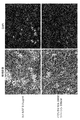
- This figure shows a representative example of the difference in the micronucleation ability of human immortalized mesenchymal stem cells due to the difference in the compound treatment conditions.
- the upper panel shows an image of cells observed when treated with 0.05 ⁇ g / mL of colcemid.
- the lower panel shows an image of cells observed when treated with 20 nM of paclitaxel and 500 nM of reversine.
- a bright field image is shown on the left panel and a DAPI image is shown on the right panel.
- This figure shows a representative example of the difference in the ability of human iPS cells to form micronuclei due to the difference in compound treatment conditions.
- the upper left panel shows an image of cells observed when treated with 0.25 ⁇ g / mL of Colcemid.
- the upper right panel shows a cell observation image when treated with 0.2 ⁇ g / mL of MAS (colchicine and vincristine mixed reagent).
- MAS colchicine and vincristine mixed reagent
- micronucleus formation images under various paclitaxel treatment concentration conditions are shown. Circles indicate micronucleated cells.
- This figure shows a representative example of the difference in the micronucleus formation ability of CHO cells due to the difference in the compound treatment conditions.
- Paclitaxel was added at a concentration of 384 nM, 24 nM, or 0 nM. A bright field image is shown on the left panel, and a red fluorescence observation image is shown on the right panel. The red fluorescent protein shows localization in the nucleus, and the morphology of the nucleus can be discriminated. This figure shows a representative example of the difference in the micronucleus formation ability of CHO cells due to the difference in the compound treatment conditions.
- Reversine was added at a concentration of 1536 nM, 24 nM, or 0 nM.
- a bright field image is shown on the left panel, and a red fluorescence observation image is shown on the right panel.
- the red fluorescent protein shows localization in the nucleus, and the morphology of the nucleus can be discriminated.
- This figure shows mouse fibroblast cell line A9 cells, mouse fetus-derived fibroblast primary culture cell line, porcine fetal-derived fibroblast primary culture cell line, chicken pre-B cell DT40 cell, rat ES cell under each compound treatment condition.
- a representative example of the difference in micronucleation ability when treated with 4 to 400 nM of paclitaxel and 0 to 1500 nM of reversine is shown.
- the upper panel shows representative examples of cell observation images by microscopic observation of mouse fibroblast cell line A9 cells, mouse embryo-derived fibroblast primary culture cell line, and pig embryo-derived fibroblast primary culture cell line.
- the lower panel shows a fluorescence microscope observation image of chicken pre B cells DT40 cells and rat ES cells by DAPI staining.
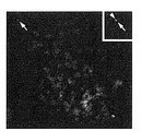
- This figure shows a FISH analysis image of cells obtained by introducing a HAC vector from human immortalized mesenchymal stem cells into human cell line HT1080 (recipient cells) by fusion with micronucleus cells.
- the HAC vector is co-stained with red and green. It is stained in red (arrow) with p11-4, a centromere probe for human chromosomes 13 and 21.
- the EGFP gene on the HAC vector is stained green (arrowhead) with the pCAG-EGFP plasmid vector.
- the small window magnified view shows the HAC vector magnified.
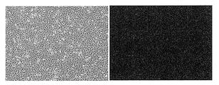
- This figure shows a fluorescence microscope image of cells in which a mouse artificial chromosome (MAC) vector was introduced from a human iPS cell (donor cell) into a human cell line HT1080 (recipient cell) by fusion with micronucleus cells.
- the left panel shows a bright field image and the right panel shows a GFP fluorescence observation image.
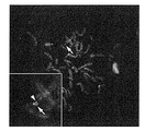
- This figure shows a FISH analysis image of cells in which a MAC vector was introduced from a human iPS cell (donor cell) into a human cell line HT1080 (recipient cell) by micronucleus cell fusion.
- the MAC vector is co-stained with red (arrow) and green (arrow head).
- the small window enlarged view shows the MAC vector in an enlarged manner.
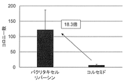
- This figure shows an average drug-resistant colony obtained when a HAC vector was introduced from a CHO cell (donor cell) into a human cell line HT1080 (recipient cell) by micronucleus cell fusion in a comparative experiment of treatment with paclitaxel and reversin and treatment with colcemid. The number of appearances (average value and error range) is shown.
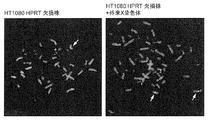
- This figure shows a FISH analysis image of cells in which the X chromosome has been introduced from human immortalized mesenchymal stem cells into human cell line HT1080 HPRT-deficient by fusion with micronucleus cells.
- the one stained with red color (arrow) is the X chromosome centromere. It is stained red with a human X chromosome-specific alpha satellite probe.
- a control experiment when an HT1080 HPRT-deficient strain in which the X chromosome was not introduced was analyzed, one X chromosome was observed because this cell line is a male-derived cell (left panel). On the other hand, two X chromosomes were observed in the X chromosome-introduced strain (right panel).
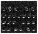
- This figure shows a multicolor FISH analysis image of cells in which the X chromosome has been introduced from a human iPS cell into a human cell line HT1080 HPRT-deficient strain by micronucleus cell fusion.
- Human chromosomes 1 to 22, X and Y chromosomes are stained and shown using each chromosome-specific dye probe. Three blue-labeled X chromosomes could be observed (left panel).
- chromosomes other than X were observed in each of four chromosomes except for chromosomes 8 and 18, and Y chromosome, it was found to be a tetraploid cell.
- Parental HT1080 cells HPRT-deficient strain is known to have two X chromosomes when it is tetraploid. From the above, it was shown that three X chromosomes were observed by introducing the foreign X chromosome derived from human iPS cells into the HT1080 HPRT-deficient strain. This figure shows the results of a HAT and 6-thioguanine sensitivity evaluation test of a cell line in which a foreign X chromosome was introduced into a HT1080 HPRT-deficient strain from human iPS cells.
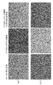
- HT selection (upper panel) and 20 ⁇ M selection of 6-thioguanine (lower panel) were performed for HT1080 wild type strain, HT1080 HPRT deficient strain (middle row panel), and HT1080 HPRT deficient strain containing foreign X chromosome (right panel).
- the microscope observation image at the time of standing is shown. Since the HT1080 wild type strain had a normal HPRT gene, it was resistant to HAT and sensitive to 6-thioguanine (FIG. 13, left panel).
- the HT1080-deficient strain was HAT-sensitive and 6-thioguanine-insensitive because the endogenous HPRT gene on the endogenous X chromosome was deleted (middle panel). Since the HT1080 HPRT-deficient strain containing the foreign X chromosome reacquired the HPRT gene, it was HAT-resistant and 6-thioguanine-sensitive as in the wild-type strain (Fig. 13, right panel).
- human cell-derived non-human animal cells (excluding cancer cells) containing a target DNA are transformed into human cell-derived micronucleus cells
- a method for producing a non-human animal cell-derived micronucleus cell wherein a human cell or a non-human animal cell containing a target DNA is a microtubule polymerization inhibitor (excluding colcemid), a microtubule depolymerization inhibitor
- human cells or non-human animal cells are not particularly limited, except for cancer cells (also referred to as tumor cells), which are cell lines derived from cancer tissues, and include any cells.
- Such human cells or non-human animal cells include, for example, organs, cells derived from tissues of organs, stem cells, etc., such as primary culture cells, established cells, immortalized cells, ATCC deposited cells, induced pluripotency Stem (iPS) cells and the like can be included.
- Examples of human cells or non-human animal cells include, but are not limited to, epithelial cells, fibroblasts, endothelial cells, hepatocytes, pancreatic cells, kidney cells, brain cells, myocytes, chondrocytes, osteoblasts, cardiomyocytes. , Hair papilla cells, nerve cells, tissue stem cells, embryonic stem cells, hematopoietic stem cells and the like.
- Preferred examples of the above human cells or non-human animal cells are, without limitation, iPS cells or immortalized mesenchymal stem cells.
- target DNA in the present specification is a DNA whose purpose of use is clear, and includes, for example, a human-derived chromosome or chromosome fragment, an artificial chromosome, etc. in consideration of the use of the MMCT method.
- artificial chromosomes include human artificial chromosomes and mammalian artificial chromosomes (eg, mouse artificial chromosome, rat artificial chromosome, etc.).
- the target DNA is preferably a normal human chromosome, or may be a human artificial chromosome or a mammalian artificial chromosome containing the target gene.
- gene of interest in the present specification is a useful gene whose purpose of use is clear, and the gene may be contained in a locus or a chromosome fragment in some cases.
- the target gene may be introduced into the artificial chromosome as a foreign gene, or may be contained in the artificial chromosome in advance.
- the gene of interest includes, but is not limited to, for example, a disease-causing gene, a therapeutic gene, a chromosomal fragment containing these genes, a gene required for cell differentiation, and the like, for example, cytokines, interferons, interleukins, chemokines ( Cell migration activity factor), granulocyte colony stimulating factor, tumor necrosis factor, growth factor (eg, platelet-derived growth factor, vascular endothelial growth factor, hepatocyte growth factor, keratinocyte growth factor, nerve growth factor, etc.), trophic factor (Eg, neurotrophic factor, brain-derived neurotrophic factor, etc.), erythropoietin, blood coagulation system protein, platelet production promoting factor, hormones, antibodies (eg, monoclonal antibody, recombinant antibody such as scFv, etc.), enzymes Can include a gene or DNA encoding a protein such as.
- cytokines interferons, interleukins,
- the target gene further includes therapeutic genes or DNAs related to diseases such as muscular dystrophy, hemophilia, neurodegenerative disease, autoimmune disease, allergic disease, inherited disease, and tumor, T cell receptor (TCR), human leukocyte. It includes immune system genes such as antigens (HLA) or DNA.
- diseases such as muscular dystrophy, hemophilia, neurodegenerative disease, autoimmune disease, allergic disease, inherited disease, and tumor, T cell receptor (TCR), human leukocyte.
- TCR T cell receptor
- human leukocyte tumor
- immune system genes such as antigens (HLA) or DNA.
- the term "artificial chromosome” refers to a centromere, long arm, and, if any, of a chromosome derived from a mammal (also referred to as “mammal”) including humans and rodents (eg, mouse, rat, etc.). It refers to an artificially produced chromosome-derived vector containing a fragment near the centromere of the short arm (genes are removed as much as possible) and a natural or artificial telomere. Therefore, the artificial chromosome of the present invention is different from conventional vector systems such as virus, YAC, BAC, PAC, cosmid, and plasmid.
- An artificial chromosome contains a site into which a desired nucleic acid such as a gene, locus, or chromosomal fragment, such as a human-derived desired nucleic acid, is inserted, and the desired nucleic acid is inserted into this site.
- a desired nucleic acid such as a gene, locus, or chromosomal fragment, such as a human-derived desired nucleic acid, is inserted, and the desired nucleic acid is inserted into this site.
- the mouse artificial chromosome is described in, for example, Japanese Patent No. 5557217 and Japanese Patent No. 4997544, and the human artificial chromosome is described in, for example, Japanese Patent No. 4895100.
- the mouse chromosome used for preparing the mouse artificial chromosome may be any of mouse chromosomes 1 to 19 and X and Y, but is preferably any of chromosomes 1 to 19.
- the long arm fragment is, for example, but not limited to, AL671968 or BX572640 (located on the centromere side of AL671968) of the long arm of the chromosome 11. , CR954170 (located on the centromere side of AL671968 and BX572640) or AL713875 (located on the centromere side of AL671968), or a long-arm fragment in which the distal region is deleted.
- the long arm fragment is composed of a long arm fragment in which a region distal to the position of AC121307, AC161799, etc. is deleted.
- the long arm fragment is composed of a long arm fragment in which a region distal to the position such as AC127687 or AC140982 is deleted.
- the human chromosome used for preparing the human artificial chromosome may be any of human chromosomes 1 to 22, X and Y, but is preferably any of chromosomes 1 to 22.
- a human iPS cell-derived chromosome can be used as the human chromosome.
- human artificial chromosomes for example, the methods described in JP 2010-004887 A, WO 2008/013067, and the methods described in the above-mentioned documents regarding the production of mouse artificial chromosomes can be referred to.
- iPS cells have a high frequency of homologous recombination, it is also possible to construct human artificial chromosomes using human iPS cell-derived chromosomes.
- LoxP (Cre recombinase recognition site), FRT (Flp recombinase recognition site), ⁇ C31attB and ⁇ C31attP ( ⁇ C31 recombinase recognition site) for inserting a desired nucleic acid (eg, gene, locus, chromosome fragment, etc.) into an artificial chromosome.
- R4attB and R4attP R4 recombinase recognition site
- TP901-1attB and TP901-1attP TP901-1 recombinase recognition site
- Bxb1attB and Bxb1attP Bxb1 recombinase recognition site).
- the artificial chromosome can include a site for inserting a desired nucleic acid sequence, by incorporating a desired nucleic acid at this site, the artificial chromosome can be introduced into any desired cell when it is introduced. It becomes possible to express the nucleic acid of.
- the desired nucleic acid includes, but is not limited to, a disease-causing gene, a therapeutic gene, a chromosomal fragment containing these genes, a reporter gene, a marker gene, and the like.
- At least one insulator sequence can be present near the insertion site of the desired nucleic acid or on both sides of the insertion site in the artificial chromosome of the present invention.
- the insulator sequence has an enhancer blocking effect (that is, adjacent genes are not affected by each other) or a chromosomal boundary effect (distinguishes a region that guarantees gene expression from a region in which gene expression is suppressed).
- Such a sequence may include, for example, human ⁇ globin HS1 to HS5, chicken ⁇ globin HS4 and the like.
- the desired DNA described above can be introduced into the donor cell on an artificial chromosome such as a human artificial chromosome or a mammalian artificial chromosome in the donor cell by using a gene introduction technique such as lipofection method. After the gene introduction, the donor cells containing the target DNA can be detected and recovered by, for example, the HAT selection method.
- an artificial chromosome such as a human artificial chromosome or a mammalian artificial chromosome in the donor cell by using a gene introduction technique such as lipofection method.
- the donor cells containing the target DNA can be detected and recovered by, for example, the HAT selection method.
- Human cells or non-human animal cells may be further provided with a drug resistance gene (for example, Blasticidin resistance gene, neomycin (G418) resistance gene, etc.) and / or a fluorescent protein for selection of the target micronucleate cell.
- a drug resistance gene for example, Blasticidin resistance gene, neomycin (G418) resistance gene, etc.
- G4108 resistance gene for example, Blasticidin resistance gene, neomycin (G418) resistance gene, etc.
- a fluorescent protein for selection of the target micronucleate cell.
- a coding gene for example, GFP, DsRed, etc.
- the method of the present invention selects human cells or non-human animal cells containing a target DNA from the group consisting of a microtubule polymerization inhibitor (however, excluding colcemid), a microtubule depolymerization inhibitor and a spindle checkpoint inhibitor. And culturing in a medium containing at least one micronucleation inducing agent.
- the microtubule polymerization inhibitor is not particularly limited, except that colcemid is removed and as long as it forms micronucleus cells, and includes, for example, a mixture of colchicine and vincristine.
- the microtubule depolymerization inhibitor is not particularly limited as long as it forms micronucleus cells, and includes, for example, paclitaxel, docetaxel, a mixture of docetaxel and paclitaxel, and the like.
- the spindle checkpoint inhibitor is not particularly limited as long as it forms micronucleated cells, and includes, for example, reversine, hesperadin, a mixture of reversin and hesperazine, and the like.
- the suitable concentration of the micronucleation inducer in the medium varies depending on the type of the micronucleation inducer, and includes, for example, reversin 0.001 nM to 1 M, paclitaxel 0.001 nM to 1 M, or colchicine and vincristine (0.01: 9.
- a mixture of 99 to 9.99: 0.01 (weight) is 0.001 nM to 1M, but is not limited to the above range as long as micronucleated cells are formed.
- a preferred micronucleation inducer is reversin or a mixture of reversin and paclitaxel.
- the medium is not limited as long as it is a medium used for culturing animal cells.
- the basal medium is, for example, Dulbecco's modified Eagle medium (DMEM), Eagle's minimum essential medium (EMEM), Ham's F-12 medium, RPMI-1640 medium, Iscove's modified Dulbecco's medium (IMDM), a mixed medium thereof, or the like, and human iPS
- DMEM Dulbecco's modified Eagle medium
- EMEM Eagle's minimum essential medium
- IMDM Iscove's modified Dulbecco's medium
- a mixed medium thereof, or the like a mixed medium thereof, or the like
- human iPS a primate ES cell medium (Reprocell, Japan) or the like can be used.
- the micronucleation induction treatment method can be performed, for example, by the following procedure.
- Human cells or non-human animal cells containing the target DNA are used as donor cells, and the donor cells are cultured in a cell culture flask (for example, a bottom area of 25 cm 2 ), and when about 70% confluent, the above culture medium is used.
- a formation-inducing drug is added, and the cells are cultured in the presence of 20% fetal calf serum (FCS) at 37 ° C. and 5% CO 2 for 12 to 96 hours or more.
- FCS fetal calf serum
- micronucleus cell population by the above culture, and further collect human cell-derived micronucleate cells or non-human animal cell-derived micronucleate cells containing the target DNA by fluorescence positive or drug selection.
- the collection of micronucleated cells can be performed, for example, as follows. After induction of micronucleation, the medium in the flask is aspirated and the flask is filled with cytochalasin B until the 9th minute. Insert the flask into a dedicated container for a large-scale high-speed centrifuge (for example, BECKMAN), add hot water (34 ° C) to an extent that the flask is not hidden, and centrifuge (Rotor ID 10.500, 8,000 rpm, 1 hour, 34 ° C). . After the completion of centrifugation, cytochalasin B is collected, and the pellet in the flask is collected in a 15 ml tube with 2 ml of serum-free medium DMEM.
- a large-scale high-speed centrifuge for example, BECKMAN
- centrifuge Rotor ID 10.500, 8,000 rpm, 1 hour, 34 ° C.
- each tube was centrifuged (1,200 rpm, 5 minutes, room temperature), and the supernatant was aspirated.
- the micronucleated cells are collected by collecting and suspending in serum-free medium DMEM and centrifuging (2000 rpm, 5 minutes).
- the above-mentioned method of the present invention is also used for producing a non-human animal cell-derived micronucleate cell from a non-human animal cell containing a target DNA (excluding cancer cells) using the above-mentioned micronucleation inducer. You can Therefore, this method is also included in the present invention.
- the cells are cultured in a medium containing at least one micronucleation inducing agent selected from the group consisting of reversin, paclitaxel, or a mixture of reversin and paclitaxel, and a mixture of colchicine and vincristine.
- a method comprising producing a nuclear cell and recovering a non-human animal cell-derived micronucleate cell containing the DNA of interest.
- Non-human animal cells refer to all animal cells including, but not limited to, rodent cells, artiodactyl cells, perissodactylous cells, primate (excluding humans) cells, canine cells, feline cells, Includes avian cells, reptile cells, amphibian cells, insect cells, etc.
- Preferred non-human animal cells are rodent cells.
- the rodent cells may include cells derived from organs of animals such as mice, rats and hamsters, tissues of organs, and the like, for example, primary culture cells thereof, established cells, ATCC deposited cells and the like.
- rodent cells include, but are not limited to, epithelial cells, fibroblasts, endothelial cells, hepatocytes, muscle cells, chondrocytes, osteoblasts, cardiomyocytes, dermal papilla cells, nerve cells, ovary cells, etc. is there.
- Preferred examples of the rodent cells are Chinese hamster ovary (CHO) cells or mouse-derived A9 cells.
- the target DNA the agent for inducing micronucleation and the like, the description of the above-mentioned human cells or non-human animal cells is also referred to here.
- the present invention is, in a third aspect, a method for preparing a human cell or non-human animal cell containing DNA of interest, which comprises the steps of 1.
- the present invention provides a method for producing a human cell or a non-human animal cell containing a target DNA, which comprises the step of recovering
- the step of cell fusion is carried out by using a modification of the MMCT (micronuclear cell fusion) method, and the above 1.
- Human cells or non-human animal cells include, for example, normal cells, immortalized cells, embryonic stem cells, somatic stem cells, (eg, human or mammalian somatic cell-derived) iPS cells, etc., but are limited as long as the target DNA can be introduced. Not be done.
- a modification of the MMCT (micronucleus cell fusion) method involves expressing the viral envelope protein on the surface of the micronucleus cell. Therefore, the above-mentioned human cell or non-human animal cell as a donor cell can be transformed in advance so that it can be expressed with the DNA encoding the viral envelope protein.
- the virus envelope protein is, for example, an envelope protein derived from a virus such as measles virus and leukemia virus, and in the case of the envelope protein derived from measles virus, for example, a hemagglutinin (H) protein and a fusion (Fusion; F) are included.
- a hemagglutinin (H) protein and a fusion (Fusion; F) are included.
- leukemia virus-derived envelope protein for example, SU protein and TM protein can be included.
- Preferred viral envelope proteins are measles virus-derived envelope proteins, such as the H and F proteins.
- the H protein can be modified by genetic engineering technology so that the H protein can bind to the surface antigen of a specific recipient cell.
- a single unit Fv which is the minimum unit of antigen recognition of an antibody, preferably an anti-CD9 antibody, an anti-CD13 antibody or an anti-CD71 antibody, or a ScFv of these antibodies is fused to an H protein to fuse in a wild-type H protein. It is possible to fuse to cell types that cannot.
- micronucleus cell fusion can be performed by transiently expressing CD46 antigen and CD120 (SLAM) antigen capable of binding to H protein to a recipient cell in order to impart a fusion ability to a chromosome recipient cell.
- SLAM transiently expressing CD46 antigen and CD120
- SLAM transiently expressing CD46 antigen and CD120
- SLAM transiently expressing CD46 antigen and CD120
- SLAM transiently expressing CD46 antigen and CD120
- SLAM transiently expressing CD46
- a plasmid containing a DNA encoding each of the above envelope proteins or a plasmid containing a DNA cassette encoding a plurality of envelope proteins is constructed by gene recombination technology, and a donor cell is transformed with these plasmids.
- micronucleus cells are prepared by deleting the CD46 antigen, the CD120 antigen of the donor cell or the surface antigen that binds to the antibody ScFv fused to the H protein so that autologous cell fusion does not occur. Fusion can be allowed.
- the human cell or the non-human animal cell as the donor cell further has a drug resistance gene (for example, Blasticidin resistance gene, neomycin (G418) resistance gene, etc.) and / or a gene encoding a fluorescent protein (for selection of a target cell).
- a drug resistance gene for example, Blasticidin resistance gene, neomycin (G418) resistance gene, etc.
- G408 resistance gene for example, Blasticidin resistance gene, neomycin (G418) resistance gene, etc.
- a gene encoding a fluorescent protein for selection of a target cell.
- GFP GFP, DsRed, etc.
- a donor cell that expresses the virus envelope protein on the cell surface and that contains the target DNA eg, artificial chromosome
- the target DNA for example, an artificial chromosome such as a human artificial chromosome or a mammalian artificial chromosome
- the target DNA can be obtained by using the feeder-free culture method and the MMCT method using a viral envelope.
- somatic cells including, for example, somatic stem cells, progenitor cells, and / or mature cells
- a specific reprogramming factor DNA or protein
- Colonies are generated in about 3 to 5 weeks by subculture.
- the reprogramming factor is, for example, a combination of Oct3 / 4, Sox2, Klf4 and c-Myc; a combination of Oct3 / 4, Sox2 and Klf4; a combination of Oct4, Sox2, Nanog and Lin28; or Oct3 / 4,
- a combination of Sox2, Klf4, c-Myc, Nanog and Lin28 is known (K. Takahashi and S.
- a mouse fetal fibroblast cell line for example, STO
- mitomycin C a fetal fibroblast cell line treated with mitomycin C
- a reprogramming factor expression vector-introduced somatic cell about 10 4
- feeder cells are not always necessary (Takahashi, K. et al., Cell; 131: 861-872 (2007)).
- the basal medium is, for example, Dulbecco's modified Eagle medium (DMEM), Eagle's minimum essential medium (EMEM), Ham's F-12 medium, RPMI-1640 medium, Iscove's modified Dulbecco's medium (IMDM), a mixed medium thereof, or the like, and human iPS
- DMEM Dulbecco's modified Eagle medium
- EMEM Eagle's minimum essential medium
- IMDM Iscove's modified Dulbecco's medium
- Reprocell a primate ES cell culture medium
- Examples of the culture medium for iPS cells in the absence of feeder cells include StemFit TM AK02N (Reprocell), TeSR TM (STEMCELL Technologies), NutriStem XF / FF Culture Medium (STEM GWNT) and the like. Further, examples of the culture substrate include Geltrrex TM LDEV-Free hESC-qualified (Thermo Fisher Scientific), iMatrix TM -511 (Nippi), hES Qualified Matrigel TM (Corning) and the like.
- a drug for example, an antibiotic
- the culture conditions for cell fusion are, for example, DMEM containing 10% fetal calf serum (FCS) in the medium, the temperature is 37 ° C., and the period is 24 hours.
- another culture condition is, for example, culture medium Stemfit, culture plate coating temperature of substrate iMatrix-511 0.5 ⁇ g / mL at 37 ° C., period of 24 hours.
- a candidate for the micronucleation inducing agent is a cell division inhibitor.
- microtubule polymerization inhibitors microtubule depolymerization inhibitors (microtubule stabilizers), and M-phase checkpoint protein inhibitors.
- micronucleus formation efficiencies were compared using colcemid, which is a microtubule polymerization inhibitor, MAS (a mixed reagent of colchicine and vincristine), and paclitaxel (Paclitaxel), which is a microtubule depolymerization inhibitor.
- colcemid which is a microtubule polymerization inhibitor
- MAS a mixed reagent of colchicine and vincristine
- paclitaxel paclitaxel
- the synergistic effect of reversin which is an inhibitor of Aurora kinases A and B, which are M-phase checkpoint proteins, was examined.
- hiMSC Human immortalized mesenchymal stem cells
- Colcemid (0.05 ⁇ g / mL, 0.1 ⁇ g / mL, 0.2 ⁇ g / mL), MAS (0.1 ⁇ g / mL, 0.2 ⁇ g / mL, 0.4 ⁇ g / mL) ), Paclitaxel (0.8 nM, 4 nM, 20 nM, 100 nM, 400 nM), while adding Reversine (no addition, 500 nM, 1000 nM, 1500 nM), a total of 26 conditions were set, and 37 ° C. It was cultured for 48 hours under the condition of 5% CO 2 .
- microtubule depolymerization inhibitor such as Paclitaxel
- a conventional microtubule polymerization inhibitor such as colcemid.
- reversin which is an M-phase checkpoint inhibitor, is useful.
- reversin (without addition) was applied while treating with 0.25 ⁇ g / mL of Colcemid, 0.2 ⁇ g / mL of MAS or Paclitaxel (20 nM, 40 nM, 60 nM, 100 nM, 400 nM). , 500 nM, 1000 nM, 1500 nM) were added for a total of 22 conditions, and the cells were cultured under the conditions of 37 ° C. and 5% CO 2 for 48 hours.
- C Verification of effect of micronucleation inducer on CHO cells
- C. 1.1 Culture of CHO cells CHO MHG # 4a expressing a red fluorescent protein showing localization in the nucleus was inoculated on a 96-well plate at 1 ⁇ 10 4 cells each, and paclitaxel (Paclitaxel) (0 nM, 96 nM, 192nM, 384nM, 768nM, 1152nM, 1536nM, 1920nM) and reversine (0nM, 192nM, 384nM, 768nM, 1536nM, 3072nM, 4608nM, 6144nM, 7680nM, 9216nM, 8816nM, 10752nM, 10752n). The cells were cultured for 72 hours under the conditions of 37 ° C. and 5% CO 2 .
- FIG. 1.2 Observation of micronucleus formation efficiency with a fluorescence microscope A representative example of the results of comparing micronucleus induction rates by observing with a fluorescence microscope after induction of micronucleus formation is shown (Figs. 4 and 5). As a result, an increase in the micronucleation rate was observed in a concentration-dependent manner with paclitaxel and reversine.
- Mouse fibroblast cell line A9 cells, mouse embryo fibroblast primary culture cell line, pig embryo fibroblast cells, chicken pre B cell DT40 cells, rat 1 ⁇ 10 5 ES cells were seeded in each well of a 24-well plate and cultured at 37 ° C. under 5% CO 2 for 24 hours.
- Paclitaxel 4nM, 25nM, 100nM, 400nM
- the cells were cultured under the conditions of 37 ° C. and 5% CO 2 , and microscopic observation was performed at 24, 48, and 72 hours.
- HAC vector labeled with drug resistance gene is introduced into human cells HT1080.
- the above-mentioned human cells were introduced into HT1080 by the micronucleus cell fusion method, using a drug treatment condition capable of efficiently inducing micronucleus formation.
- HAMS vector-containing hiMSCs that are donor (also called “donor”) cells were cultured using a cell culture flask bottom area of 25 cm 2 ⁇ 12 to reach 70% confluence. At that time, micronucleation induction treatment (Paclitaxel 1 nM to 1 ⁇ M and Reversine 1 nM to 1 ⁇ M) was performed at 37 ° C. and 5% CO 2 for 48 hours. After the induction of micronucleation, the medium in the flask was aspirated and the flask was filled with cytochalasin B until the 9th minute.
- the flask was inserted into a container dedicated to a large-scale high-speed centrifuge (BECKMAN), hot water (34 ° C.) was added to the extent that the flask was not hidden, and centrifugation (Rotor ID 10.500, 8,000 rpm, 1 h, 34 ° C.) was performed. After the centrifugation was completed, cytochalasin B was recovered, and the pellets in each flask were collected in a 15 ml tube with 2 ml of serum-free medium DMEM.
- BECKMAN large-scale high-speed centrifuge
- each tube was centrifuged (1,200 rpm, 5 minutes, room temperature (RT)), and the supernatant was aspirated, followed by pelleting of each tube.
- DMEM serum-free medium
- Recipient cells HT1080 were seeded in advance on a 6 cm diameter cell culture dish so as to be 90% confluent the day before, and this was washed with serum-free medium (DMEM).
- the purified micronuclei were resuspended in 2 ml of serum-free culture medium containing PHA-P (SIGMA) and gently seeded on HT1080 from which serum-free culture medium (DMEM) had been removed.
- the culture dish was allowed to stand for 20 minutes under the conditions of 37 ° C. and 5% CO 2 .
- the supernatant is removed, and PEG1000 (Wako, Japan) [5 g of PEG1000 is completely dissolved in serum-free DMEM medium, and 1 ml of dimethyl sulfoxide is added to sterilize by filtration. ]
- the solution was fused with 1 ml for exactly 90 seconds.
- the serum-free culture medium (DMEM) was washed 3 times with 5 ml, and incubated with 5 ml of a normal HT1080 culture medium at 37 ° C. overnight. After 24 hours, the cells were reseeded in 6 10 cm diameter culture dishes. After a further 24 hours, blasticidin S was added at 8 ⁇ g / ml to perform selective culture for 3 to 4 weeks. A total of three GFP fluorescence positive and drug resistant colonies appeared in one fusion of micronuclear cells. These were isolated and propagated, and the subsequent analysis was performed.
- DMEM serum-free culture medium
- MAC vector labeled with drug resistance gene into HT1080
- HT1080 Human iPS cells containing MAC vector labeled with drug resistance gene
- Drug resistance A gene-labeled MAC vector is introduced into HT1080 which is a human cell.
- the above-mentioned human cells are introduced into HT1080 by the micronucleus cell fusion method using a drug treatment condition that can efficiently induce micronucleus formation.
- cytochalasin B (10 ⁇ g / ml, Sigma) solution preliminarily kept warm at 37 ° C. was filled in a centrifuge flask, and centrifugation was performed at 34 ° C., 8000 rpm for 1 hour.
- Microcells were suspended in serum-free DMEM medium and purified with 8 ⁇ m, 5 ⁇ m and 3 ⁇ m filters. After purification, it was centrifuged at 2000 rpm for 10 minutes and suspended in 5 ml of serum-free DMEM medium. The microcells were suspended in 5 ml of serum-free DMEM medium and purified with 8 ⁇ m, 5 ⁇ m and 3 ⁇ m filters.
- the recipient cells HT1080 were seeded in advance on the 6 cm diameter cell culture dish so as to be 90% confluent the day before, and this was washed with serum-free medium (DMEM).
- DMEM serum-free medium
- the purified micronuclei were resuspended in 2 ml of serum-free culture medium containing PHA-P (SIGMA) and gently seeded on HT1080 from which serum-free culture medium (DMEM) had been removed.
- the culture dish was allowed to stand for 20 minutes under the conditions of 37 ° C. and 5% CO 2 .
- PEG1000 (Wako) [5 g of PEG1000 is completely dissolved in serum-free DMEM medium, and 1 mL of dimethyl sulfoxide is added to sterilize by filtration. ] The solution was fused with 1 ml for exactly 90 seconds.
- the serum-free culture medium (DMEM) was washed 3 times with 5 ml, and incubated with 5 ml of a normal HT1080 culture medium at 37 ° C. overnight. After 24 hours, the cells were reseeded in 6 10 cm diameter culture dishes. After a further 24 hours, G418 was added to 800 ⁇ g / ml to perform selective culture for 3 to 4 weeks. A total of 4 GFP fluorescence positive and drug resistant colonies (FIG. 8) appeared in one fusion of micronuclei cells. These were isolated and propagated, and the subsequent analysis was performed.
- Male-derived human iPS cells derived from donor cells HFL-I (RCB0521, RIKEN BRC) using Yamanaka factor (karyotype: 46, XY) was inoculated into a cell culture flask x 12 previously coated with 0.5 ⁇ g / cm 2 of Laminin-511, 10 ⁇ MY-27632 was added, and the cells were cultured at 37 ° C. under 5% CO 2 for 24 hours (non-vector). Introduction group).
- the CD46 of the iPS cells is deleted, and 12 ⁇ g of the anti-CD9-ScFv fusion MV-H expression plasmid vector and 12 ⁇ g of the MV-F expression plasmid are introduced using Lipofectamine 2000 according to the specifications (vector introduction group). After that, both the non-vector-introduced group and the vector-introduced group were replaced with normal culture iPS culture medium for growth, and when 90% confluent was reached, MAS 0.2 ⁇ g / mL was added, and the conditions were 37 ° C. and 5% CO 2 . The cells are cultured for 48 hours in a medium to form micronuclei.
- the culture solution is removed, and a cytochalasin B (10 ⁇ g / ml, Sigma) solution that has been preliminarily kept at 37 ° C. is filled in a centrifuge flask, and centrifugation is performed at 34 ° C., 8,000 rpm for 1 hour.
- the microcells are suspended in serum-free DMEM medium and purified with 8 ⁇ m, 5 ⁇ m and 3 ⁇ m filters. After purification, it is centrifuged at 2000 rpm for 10 minutes and suspended in 5 ml of serum-free DMEM medium.
- the microcells are suspended in 5 ml of serum-free DMEM medium and purified with 8 ⁇ m, 5 ⁇ m and 3 ⁇ m filters.
- the human iPS cells (karyotype: 46, XX) in which the HPRT gene derived from the recipient cell 201B7 (HPS0063, RIKEN BRC) was deleted in advance on the previous day or human iPS cells derived from a Lesch-Nyhan syndrome patient (JCRB0072 / KURB1995 / A human iPS cell line derived from a patient-derived fibroblast such as KURB1996, JCRB or ATCC TM CRL-1110 TM , ATCC) was seeded in a 6 cm diameter cell culture dish so as to be 90% confluent, and this was prepared. Wash with serum-free medium (DMEM).
- DMEM serum-free medium
- the purified micronuclei were resuspended in 2 ml of serum-free culture medium containing PHA-P (SIGMA) to remove human serum-free culture medium (DMEM). Gently seed on cells.
- the culture dish is allowed to stand for 20 minutes under the conditions of 37 ° C. and 5% CO 2 .
- the supernatant is removed, and PEG1000 (Wako, Japan) [5 g of PEG1000 is completely dissolved in serum-free DMEM medium, and 1 mL of dimethyl sulfoxide is added to sterilize by filtration. ] Fuse solution with 1 ml for exactly 90 seconds.
- the serum-free culture medium (DMEM) is washed 3 times with 5 ml, and incubated with 5 ml of a normal human iPS cell culture medium at 37 ° C. overnight.
- the group of micronucleated cells into which the vector has been introduced is suspended in a human iPS cell culture medium, added to the dish in which the recipient cells are being cultured, and incubated. After 24 hours, the vector-introduced group and the non-introduced group are replated on 6 10 cm diameter culture dishes. After a further 24 hours, 1 ⁇ HAT is added so that the cells are selectively cultured for 3 to 4 weeks. Drug-resistant colonies are isolated and propagated for subsequent analysis.
- HT1080 of HAC vector labeled with drug resistance gene Introduction into Drug
- the CHO MHG # 4a containing the HAC vector labeled with the drug resistance gene is introduced into the human cell HT1080 with the HAC vector labeled with the drug resistance gene.
- the above-mentioned human cells are introduced into HT1080 by the micronucleus cell fusion method using a drug treatment condition capable of efficiently inducing micronucleus formation, and the number of drug-resistant colonies that appear is compared.
- colcemid treatment F12 / 20% FBS / 0.1 ⁇ g / mL colcemid
- colcemid treatment F12 / 20% FBS / 0.1 ⁇ g / mL colcemid
- Paclitaxel and Riversin treatment were performed for 48 hours to form micronuclei.
- the medium in the flask was removed and the flask was filled with cytochalasin B until the 9th minute.
- the flask was set in a centrifuge tube, warm water (34 ° C.) was added to such an extent that the flask was not hidden, and the mixture was centrifuged at JLA-10,500 Rotor (BECKMAN) at 8,000 rpm for 1 hour at 34 ° C.
- JLA-10,500 Rotor BECKMAN
- cytochalasin B was collected and removed, and the pellet in each flask was collected in a 50 mL tube with 2 mL of serum-free DMEM medium.
- centrifugation was performed at 2,000 rpm, 10 min, and room temperature (RT). After centrifugation, the supernatant was removed, the pellet of each tube was suspended in the recipient cell culture medium, added to the dish in which the recipient cells were cultured, and incubated.
- the recipient cells used were HT1080 cells cultured at 90% confluence. After 24 hours, the cells were re-seeded on 5 10 cm diameter culture dishes. After 24 hours, selective culture was performed with 3 ⁇ g / mL blasticidin S. The cells were selectively cultured for 3 to 4 weeks.
- micronucleation induction treatment paclitaxel 1 nM to 1 ⁇ M and reversine 1 nM to 1 ⁇ M
- the micronucleation induction was completed, the medium in the flask was aspirated and the flask was filled with cytochalasin B until the 9th minute.
- the flask was inserted into a container dedicated to a large-scale high-speed centrifuge (BECKMAN), hot water (34 ° C.) was added to the extent that the flask was not hidden, and centrifugation (Rotor ID 10.500, 8,000 rpm, 1 h, 34 ° C.) was performed. After the centrifugation was completed, cytochalasin B was recovered, and the pellets in each flask were collected in a 15 ml tube with 2 ml of serum-free medium DMEM.
- BECKMAN large-scale high-speed centrifuge
- each tube was centrifuged (1,200 rpm, 5 minutes, room temperature (RT)), and the supernatant was aspirated, followed by pelleting of each tube.
- DMEM serum-free medium
- the purified micronuclei were resuspended in 2 ml of serum-free culture medium containing PHA-P (SIGMA) and gently seeded on HT1080 from which serum-free culture medium (DMEM) had been removed.
- the culture dish was allowed to stand for 20 minutes under the conditions of 37 ° C. and 5% CO 2 .
- the supernatant is removed, and PEG1000 (Wako, Japan) [5 g of PEG1000 is completely dissolved in serum-free DMEM medium, and 1 ml of dimethyl sulfoxide is added to sterilize by filtration. ]
- the solution was fused with 1 ml for exactly 90 seconds.
- the serum-free culture medium (DMEM) was washed 3 times with 5 ml, and incubated with 5 ml of a normal HT1080 culture medium at 37 ° C. overnight. After 24 hours, the cells were reseeded in 6 10 cm diameter culture dishes. After 24 hours, 50 ⁇ HAT supplement (Sigma) was added so as to have a concentration of 1 ⁇ HAT, and selective culture was performed for 3 to 4 weeks. A total of three GFP fluorescence positive and drug resistant colonies appeared in one fusion of micronuclear cells. These were isolated and propagated, and the subsequent analysis was performed.
- DMEM serum-free culture medium
- DXZ1 ChrX alpha-satellite F 5′-ATAATTTCCCATAACTAAAACACA-3 ′ (SEQ ID NO: 1)
- DXZ1 ChrX alpha-satellite R 5′-TGTGAAGAATAAAGGAAAAGGCTT-3 ′ (SEQ ID NO: 2)
- HT1080 HPRT-deficient strain in which X chromosome was not introduced and acquired drug resistant clones were performed. Since the HT1080 HPRT-deficient strain is a male-derived cell, it was shown to contain only one X chromosome (FIG. 11, left panel). On the other hand, it was shown that the acquired drug-resistant clones contained two X chromosomes (FIG. 11, right panel).
- the cells were cultured under the conditions of Paclitaxel 20 nM, Reversine 500 nM, 37 ° C., 5% CO 2 for 48 hours to form micronuclei.
- the culture solution was removed, and a cytochalasin B (10 ⁇ g / ml, Sigma) solution that had been kept warm at 37 ° C. was filled in a centrifuge flask, and centrifugation was performed at 34 ° C., 8,000 rpm for 1 hour.
- the microcells are suspended in serum-free DMEM medium and purified with 8 ⁇ m, 5 ⁇ m and 3 ⁇ m filters.
- the purified micronuclei are resuspended in 2 ml of serum-free culture medium containing PHA-P (SIGMA) and gently seeded on human iPS cells from which serum-free culture medium (DMEM) has been removed.
- the culture dish was allowed to stand for 20 minutes under the conditions of 37 ° C. and 5% CO 2 .
- the supernatant is removed, PEG1000 (Wako) [5 g of PEG1000 is completely dissolved in serum-free DMEM medium, and 1 mL of dimethyl sulfoxide is added to sterilize by filtration. ]
- the solution was fused with 1 ml for exactly 90 seconds.
- the serum-free culture medium (DMEM) was washed 3 times with 5 ml, and incubated with 5 ml of a normal culture medium at 37 ° C. overnight. After 24 hours, the cells were reseeded in 6 10 cm diameter culture dishes. After 24 hours, 1 ⁇ HAT was added to the cells, and the cells were selectively cultured for 3 to 4 weeks. Drug-resistant colonies were isolated and propagated for subsequent analysis.
- DMEM serum-free culture medium
- [B. 1.1] HAT and 6-thioguanine sensitivity evaluation test of foreign X-chromosome-introduced cell line Regarding the obtained HAT-resistant cells 1 ⁇ 10 4 cells were seeded in one well of a 6-well plate, and 6-thioguanine (6TG) was added from the next day. The cells were treated at a concentration of 20 ⁇ M for 120 hours, and the 6-thioguanine resistance performance was evaluated.
- the HT1080 wild-type strain was HAT resistant by having the normal HPRT gene on the X chromosome, metabolized 6-thioguanine, and showed cytotoxicity (FIG. 13, left panel).
- the HT1080 HPRT-deficient strain lacked the endogenous HPRT gene on the endogenous X chromosome, it did not show cytotoxic activity due to 6-thioguanine and was insensitive to 6-thioguanine. On the other hand, cell growth was stopped by 1 ⁇ HAT treatment, and HAT sensitivity was exhibited. (Panel in FIG. 13)
- the HT1080 HPRT-deficient cell line containing the foreign X chromosome which was obtained by introducing the foreign normal X chromosome into the HT1080 HPRT-deficient strain, the cytotoxic activity by 6-thioguanine was observed as in the HT1080 wild-type strain. On the other hand, it was HAT resistant (FIG. 13, right panel). From these, it was shown that the human iPS cell-derived X chromosome carrying the normal HPRT gene could be introduced from the human iPS cells into the HT1080 cells.
- the present invention relates to a technique for directly introducing a target DNA from a human cell or a non-human animal cell (for example, a rodent cell) (donor cell) containing the target DNA into a human cell or a non-human animal cell (recipient cell). Therefore, it is useful for regenerative medicine and drug development using human cells such as human iPS cells.
- a non-human animal cell for example, a rodent cell
- donor cell containing the target DNA into a human cell or a non-human animal cell (recipient cell). Therefore, it is useful for regenerative medicine and drug development using human cells such as human iPS cells.
- Sequence number 1 primer Sequence number 2: primer
Landscapes
- Health & Medical Sciences (AREA)
- Genetics & Genomics (AREA)
- Life Sciences & Earth Sciences (AREA)
- Engineering & Computer Science (AREA)
- Chemical & Material Sciences (AREA)
- Biomedical Technology (AREA)
- Biotechnology (AREA)
- Organic Chemistry (AREA)
- Bioinformatics & Cheminformatics (AREA)
- Wood Science & Technology (AREA)
- Zoology (AREA)
- Microbiology (AREA)
- Biochemistry (AREA)
- General Engineering & Computer Science (AREA)
- General Health & Medical Sciences (AREA)
- Cell Biology (AREA)
- Micro-Organisms Or Cultivation Processes Thereof (AREA)
Abstract
この出願は、目的DNAを含むヒト細胞からヒト細胞由来微小核細胞を作製するための方法であって、ヒト細胞を、微小管重合阻害剤(但し、コルセミドを除く)、微小管脱重合阻害剤及び紡錘体チェックポイント阻害剤からなる群から選択される少なくとも1つの微小核形成誘導剤を含有する培地中で培養してヒト細胞由来微小核細胞を生成する工程、及び、目的DNAを含むヒト細胞由来微小核細胞を回収する工程を含む方法、並びに、この方法を利用して目的DNAを含むヒト細胞を作製する方法を提供する。この方法はまた、非ヒト動物細胞にも使用可能である。
Description
本発明は、目的DNAを含むヒト細胞から、目的DNAを含むヒト細胞由来微小核細胞を収率よく作製するための方法に関する。
本発明はまた、目的DNAを含む非ヒト動物細胞(例えば齧歯類細胞)から、目的DNAを含む非ヒト動物細胞由来微小核細胞を収率よく作製するための方法に関する。
本発明はまた、本発明の方法によって作製された、供与細胞としての上記目的DNAを含むヒト細胞由来微小核細胞、又は上記目的DNAを含む非ヒト動物細胞(例えば齧歯類細胞)由来微小核細胞と、受容細胞としてのそれぞれヒト細胞又は非ヒト動物細胞とを細胞融合して目的DNAを含むヒト細胞又は非ヒト動物細胞を作製するための方法に関する。
微小核細胞融合法(MMCT(Microcell-Mediated Chromosome Transfer))は、供与細胞から作製した微小核細胞を受容細胞と融合させる技術である。これによって、供与細胞内の特定の外来DNA(例えば、染色体)を受容細胞に移入させることが可能である。微小核細胞は、通常、供与細胞をコルセミドで処理することによって作製することができる(非特許文献1~3)。
従来、ヒト細胞を染色体供与体とするときには、薬剤耐性遺伝子をヒト細胞に導入したのち、微小核形成が可能なマウス由来A9細胞またはハムスター由来CHO細胞と細胞融合してA9ハイブリッド細胞またはCHOハイブリッド細胞を形成し、さらにコルセミドで処理して微小核を形成し、薬剤選択により微小核細胞を精製する手法が行われている。一方、ヒト細胞を供与細胞としてヒト細胞から微小核細胞を精製し、ヒト細胞株(例えばヒトiPS細胞)と細胞融合するとき、通常のMMCT法では、目的の染色体が導入されたヒト細胞株を容易に得ることは難しい。このため、供与細胞としてA9ハイブリッド細胞またはCHOハイブリッド細胞が使用されてきた。
MMCT法では染色体を受容細胞に移入することができる。しかし、メガベース(Mb)サイズの染色体(例えばヒト染色体もしくは染色体断片)を細胞に移入するためのベクターには制限がある。例えばプラスミドでは約20kb以下のサイズのポリヌクレオチド、ウイルスベクターでは150kb以下のサイズのポリヌクレオチド、BAC/PACでは300kb以下のサイズのポリヌクレオチド、及びYACでは約1Mb未満のサイズのポリヌクレオチドであり、一方、ヒト人工染色体及びマウス人工染色体などの人工染色体では導入される染色体サイズに特に制限はない(非特許文献4)。このため、このような人工染色体には、目的の遺伝子座を含む染色体断片を搭載することができる。
上記ヒト人工染色体及びマウス人工染色体は、本発明者らによって最初に開発されたものであり、染色体の由来により呼称が異なり、例えばヒト染色体由来の場合ヒト人工染色体と称し、一方マウス由来の場合マウス人工染色体と称する(特許文献1、2及び3)。
iPS細胞は、山中伸弥博士(京都大学)によって体細胞から人工的に作製された胚性幹(ES)細胞に類似した特徴を有する幹細胞であり、無限増殖性と分化多能性を有している(非特許文献5及び6)。iPS細胞は、種々の組織の体細胞(幹細胞、前駆細胞又は成熟細胞)に分化することができるため、再生医療への応用が進められているし、また、遺伝性疾患の患者の体細胞から誘導されたiPS細胞は疾患モデル細胞を形成することから、医薬品開発のために利用されている(非特許文献7)。
もしヒト細胞(供与細胞)からヒト細胞(受容細胞、例えばヒトiPS細胞)に目的DNA(例えば染色体、人工染色体など)を容易に導入することができるならば、上記の再生医療や医薬品開発のために大きく貢献することが期待される。
M. Katoh et al., BMC Biotechnology 2010, 10:37
N. Uno et al., Cytotechnology 2013, 65:803-809
M Hiratsuka et al., BMC Biotechnology 2015, 15:58
P. Osten et al., Handb Exp Pharmacol 2007, 178:177-202
K. Takahashi and S. Yamanaka, Cell 2006, 126(4):663-676
K. Takahashi et al., Cell 2007, 131(5):861-872
V.K. Singh et al., Frontiers in Cell and Developmental Biology 2015, doi:10.3389/fcell.2015.00002
本発明の目的は、MMCT法を使用することによってヒト細胞(供与細胞)からヒト細胞(受容細胞、例えばヒトiPS細胞)、或いは非ヒト動物細胞(供与細胞)から非ヒト動物細胞(受容細胞、例えばiPS細胞)に目的DNA(例えば、染色体、人工染色体など)を容易に導入するための方法を提供することである。MMCT法に関連して、従来、ヒト細胞(供与細胞)から直接的にヒト細胞由来微小核細胞を作製する手法がなく、上記の通り、マウス由来A9ハイブリッド細胞又はCHOハイブリッド細胞を作製したのち、ヒト細胞(受容細胞)と細胞融合することが通常行われてきた。このような場合、中間宿主であるCHOハイブリッド細胞やA9ハイブリッド細胞の異種の細胞質の持ち込み及びウイルス等の感染の危険性がある。
上記方法を可能にするために、本発明はさらに、目的DNAを含むヒト細胞からヒト細胞由来微小核細胞を収率よく作製するための方法を提供することを目的とする。この方法はまた、目的DNAを含む非ヒト動物細胞から非ヒト動物細胞由来微小核細胞を効率的に作製するために使用可能である。
本発明は、以下の特徴を包含する。
(1)目的DNAを含むヒト細胞(但し、癌細胞を除く)からヒト細胞由来微小核細胞を作製するための方法であって、上記ヒト細胞を、微小管重合阻害剤(但し、コルセミドを除く)、微小管脱重合阻害剤及び紡錘体チェックポイント阻害剤からなる群から選択される少なくとも1つの微小核形成誘導剤を含有する培地中で培養してヒト細胞由来微小核細胞を生成し、並びに、前記目的DNAを含むヒト細胞由来微小核細胞を回収することを含む、上記方法。
(2)上記目的DNAが、正常ヒト染色体である、上記(1)に記載の方法。
(3)上記目的DNAが、目的遺伝子を含むヒト人工染色体又は哺乳類人工染色体である、上記(1)に記載の方法。
(4)上記微小管重合阻害剤が、コルヒチンとビンクリスチンの混合物である、上記(1)~(3)のいずれかに記載の方法。
(5)上記微小管脱重合阻害剤が、パクリタキセル、ドセタキセル、又はパクリタキセルとドセタキセルの混合物である、上記(1)~(3)のいずれかに記載の方法。
(6)上記紡錘体チェックポイント阻害剤が、リバーシン、ヘスペラジン、又はリバーシンとヘスペラジンの混合物である、上記(1)~(3)のいずれかに記載の方法。
(7)目的DNAを含む非ヒト動物細胞(但し、癌細胞を除く)から非ヒト動物細胞由来微小核細胞を作製するための方法であって、上記非ヒト動物細胞を、リバーシン、パクリタキセル、又はリバーシンとパクリタキセルの混合物、及びコルヒチンとビンクリスチンの混合物からなる群から選択される少なくとも1つの微小核形成誘導剤を含有する培地中で培養して非ヒト動物細胞由来微小核細胞を生成し、並びに、上記目的DNAを含む非ヒト動物細胞由来微小核細胞を回収することを含む、上記方法。
(8)上記非ヒト動物細胞が、齧歯類細胞である、上記(7)に記載の方法。
(9)上記齧歯類細胞が、チャイニーズハムスター卵巣(CHO)細胞又はマウスA9細胞である、上記(8)に記載の方法。
(10)上記目的DNAが、正常ヒト染色体である、上記(7)~(9)のいずれかに記載の方法。
(11)上記目的DNAが、目的遺伝子を含むヒト人工染色体又は哺乳類人工染色体である、上記(7)~(9)のいずれかに記載の方法。
(12)目的DNAを含むヒト細胞又は非ヒト動物細胞を作製するための方法であって、上記(1)~(6)のいずれかに記載の方法によって目的DNAを含むヒト細胞由来微小核細胞を作製する工程、或いは上記(7)~(11)のいずれかに記載の方法によって目的DNAを含む非ヒト動物細胞由来微小核細胞を作製する工程、供与細胞としての上記ヒト細胞由来微小核細胞又は上記非ヒト動物細胞由来微小核細胞と、受容細胞としてのそれぞれヒト細胞又は非ヒト動物細胞とを細胞融合する工程、それによって得られた上記目的DNAを含むヒト細胞又は非ヒト動物細胞を回収する工程を含む、上記方法。
(13)前記非ヒト動物細胞が、齧歯類細胞である、上記(12)に記載の方法。
(14)前記齧歯類細胞が、チャイニーズハムスター卵巣(CHO)細胞又はマウスA9細胞である、上記(13)に記載の方法。
(15)上記目的DNAが、正常ヒト染色体である、上記(12)に記載の方法。
(16)上記目的DNAが、目的遺伝子を含むヒト人工染色体又は哺乳類人工染色体である、上記(12)に記載の方法。
本明細書は本願の優先権の基礎となる日本国特許出願番号2018-191994号の開示内容を包含する。
(1)目的DNAを含むヒト細胞(但し、癌細胞を除く)からヒト細胞由来微小核細胞を作製するための方法であって、上記ヒト細胞を、微小管重合阻害剤(但し、コルセミドを除く)、微小管脱重合阻害剤及び紡錘体チェックポイント阻害剤からなる群から選択される少なくとも1つの微小核形成誘導剤を含有する培地中で培養してヒト細胞由来微小核細胞を生成し、並びに、前記目的DNAを含むヒト細胞由来微小核細胞を回収することを含む、上記方法。
(2)上記目的DNAが、正常ヒト染色体である、上記(1)に記載の方法。
(3)上記目的DNAが、目的遺伝子を含むヒト人工染色体又は哺乳類人工染色体である、上記(1)に記載の方法。
(4)上記微小管重合阻害剤が、コルヒチンとビンクリスチンの混合物である、上記(1)~(3)のいずれかに記載の方法。
(5)上記微小管脱重合阻害剤が、パクリタキセル、ドセタキセル、又はパクリタキセルとドセタキセルの混合物である、上記(1)~(3)のいずれかに記載の方法。
(6)上記紡錘体チェックポイント阻害剤が、リバーシン、ヘスペラジン、又はリバーシンとヘスペラジンの混合物である、上記(1)~(3)のいずれかに記載の方法。
(7)目的DNAを含む非ヒト動物細胞(但し、癌細胞を除く)から非ヒト動物細胞由来微小核細胞を作製するための方法であって、上記非ヒト動物細胞を、リバーシン、パクリタキセル、又はリバーシンとパクリタキセルの混合物、及びコルヒチンとビンクリスチンの混合物からなる群から選択される少なくとも1つの微小核形成誘導剤を含有する培地中で培養して非ヒト動物細胞由来微小核細胞を生成し、並びに、上記目的DNAを含む非ヒト動物細胞由来微小核細胞を回収することを含む、上記方法。
(8)上記非ヒト動物細胞が、齧歯類細胞である、上記(7)に記載の方法。
(9)上記齧歯類細胞が、チャイニーズハムスター卵巣(CHO)細胞又はマウスA9細胞である、上記(8)に記載の方法。
(10)上記目的DNAが、正常ヒト染色体である、上記(7)~(9)のいずれかに記載の方法。
(11)上記目的DNAが、目的遺伝子を含むヒト人工染色体又は哺乳類人工染色体である、上記(7)~(9)のいずれかに記載の方法。
(12)目的DNAを含むヒト細胞又は非ヒト動物細胞を作製するための方法であって、上記(1)~(6)のいずれかに記載の方法によって目的DNAを含むヒト細胞由来微小核細胞を作製する工程、或いは上記(7)~(11)のいずれかに記載の方法によって目的DNAを含む非ヒト動物細胞由来微小核細胞を作製する工程、供与細胞としての上記ヒト細胞由来微小核細胞又は上記非ヒト動物細胞由来微小核細胞と、受容細胞としてのそれぞれヒト細胞又は非ヒト動物細胞とを細胞融合する工程、それによって得られた上記目的DNAを含むヒト細胞又は非ヒト動物細胞を回収する工程を含む、上記方法。
(13)前記非ヒト動物細胞が、齧歯類細胞である、上記(12)に記載の方法。
(14)前記齧歯類細胞が、チャイニーズハムスター卵巣(CHO)細胞又はマウスA9細胞である、上記(13)に記載の方法。
(15)上記目的DNAが、正常ヒト染色体である、上記(12)に記載の方法。
(16)上記目的DNAが、目的遺伝子を含むヒト人工染色体又は哺乳類人工染色体である、上記(12)に記載の方法。
本明細書は本願の優先権の基礎となる日本国特許出願番号2018-191994号の開示内容を包含する。
本発明により、ヒト細胞又は非ヒト動物細胞(供与細胞)から直接的にヒト細胞由来微小核細胞又は非ヒト動物細胞由来微小核細胞を作製することができるようになり、MMCT法の変法によりヒト細胞又は非ヒト動物細胞(受容細胞)との細胞融合が可能となる。これによって、人工染色体が例えば疾患原因遺伝子或いは治療用遺伝子を含むときには例えばヒトiPS細胞などのヒト細胞を医薬品開発や再生医療に使用できるという利点がある。
本発明をさらに詳細に説明する。
1.ヒト細胞由来又は非ヒト動物細胞由来微小核細胞の作製
本発明は、第1の態様において、目的DNAを含むヒト細胞又は非ヒト動物細胞(但し、癌細胞を除く)からヒト細胞由来微小核細胞又は非ヒト動物細胞由来微小核細胞を作製するための方法であって目的DNAを含むヒト細胞又は非ヒト動物細胞を、微小管重合阻害剤(但し、コルセミドを除く)、微小管脱重合阻害剤及び紡錘体チェックポイント阻害剤からなる群から選択される少なくとも1つの微小核形成誘導剤を含有する培地中で培養してヒト細胞由来微小核細胞又は非ヒト動物細胞由来微小核細胞を生成する工程、並びに、目的DNAを含むヒト細胞由来微小核細胞又は非ヒト動物細胞由来微小核細胞を回収する工程を含む上記方法を提供する。
1.ヒト細胞由来又は非ヒト動物細胞由来微小核細胞の作製
本発明は、第1の態様において、目的DNAを含むヒト細胞又は非ヒト動物細胞(但し、癌細胞を除く)からヒト細胞由来微小核細胞又は非ヒト動物細胞由来微小核細胞を作製するための方法であって目的DNAを含むヒト細胞又は非ヒト動物細胞を、微小管重合阻害剤(但し、コルセミドを除く)、微小管脱重合阻害剤及び紡錘体チェックポイント阻害剤からなる群から選択される少なくとも1つの微小核形成誘導剤を含有する培地中で培養してヒト細胞由来微小核細胞又は非ヒト動物細胞由来微小核細胞を生成する工程、並びに、目的DNAを含むヒト細胞由来微小核細胞又は非ヒト動物細胞由来微小核細胞を回収する工程を含む上記方法を提供する。
以下に各工程について説明する。
はじめに、上記目的DNAを含むヒト細胞又は非ヒト動物細胞について、以下に説明する。
はじめに、上記目的DNAを含むヒト細胞又は非ヒト動物細胞について、以下に説明する。
上記ヒト細胞又は非ヒト動物細胞は、癌組織由来の細胞株である癌細胞(腫瘍細胞とも称する。)を除く以外は特に制限されないものとし、いずれの細胞も含む。そのようなヒト細胞又は非ヒト動物細胞には、例えば臓器、器官の組織に由来する細胞、幹細胞など、例えばそれらの初代培養細胞、株化細胞、不死化細胞、ATCC寄託細胞、人工多能性幹(iPS)細胞などを含むことができる。ヒト細胞又は非ヒト動物細胞の例は、非限定的に、上皮細胞、線維芽細胞、内皮細胞、肝細胞、膵細胞、腎細胞、脳細胞、筋細胞、軟骨細胞、骨芽細胞、心筋細胞、毛乳頭細胞、神経細胞、組織幹細胞、胚性幹細胞、造血幹細胞などである。
上記ヒト細胞又は非ヒト動物細胞の好ましい例は、非限定的に、iPS細胞または不死化間葉系幹細胞である。
本明細書中の「目的DNA」という用語は、使用目的が明確であるDNAであり、MMCT法の使用を考慮すると、例えばヒト由来の染色体もしくは染色体断片、人工染色体などを含む。人工染色体の例はヒト人工染色体及び哺乳類人工染色体(例えばマウス人工染色体、ラット人工染色体など)を含む。目的DNAは、好ましくは正常ヒト染色体であり、或いは目的遺伝子を含む、ヒト人工染色体又は哺乳類人工染色体であってよい。
本明細書中の「目的遺伝子」という用語は、使用目的が明確である有用な遺伝子であり、場合により当該遺伝子は遺伝子座もしくは染色体断片に含まれていてもよい。また、目的遺伝子は、外来遺伝子として上記人工染色体に導入されてもよいし、或いは、予め上記人工染色体に含まれていてもよい。
目的遺伝子は、非限定的に、例えば、疾患原因遺伝子、治療用遺伝子、それらの遺伝子を含む染色体断片、細胞分化に必要な遺伝子などを包含し、例えば、サイトカイン類、インターフェロン、インターロイキン、ケモカイン(細胞遊走活性をもつ因子)、顆粒球コロニー刺激因子、腫瘍壊死因子、成長因子(例えば血小板由来生長因子、血管内皮成長因子、肝細胞成長因子、ケラチノサイト増殖因子、神経成長因子、等)、栄養因子(例えば神経栄養因子、脳由来神経栄養因子、等)、エリスロポエチン、血液凝固系タンパク質、血小板産生促進因子、ホルモン類、抗体類(例えば、モノクローナル抗体、scFvなどの組換え抗体、等)、酵素類などのタンパク質をコードする遺伝子もしくはDNAを含むことができる。目的遺伝子はさらに、筋ジストロフィー、血友病、神経変性疾患、自己免疫疾患、アレルギー性疾患、遺伝性疾患、腫瘍などの疾患に関連する治療用遺伝子もしくはDNA、T細胞受容体(TCR)、ヒト白血球抗原(HLA)などの免疫系遺伝子又はDNAを含む。
本明細書中「人工染色体」という用語は、ヒト及び齧歯類(例えばマウス、ラットなど)を含む哺乳動物(「哺乳類」ともいう。)由来の染色体のセントロメア、長腕及び、もしあれば、短腕のセントロメア近傍の断片(可能なかぎり遺伝子類が除去されている。)、及び天然もしくは人工テロメアを含む、人工的に作製された染色体由来ベクターを指す。従って、本発明の人工染色体は、従来のベクター系であるウイルス、YAC、BAC、PAC、コスミド、プラスミドなどと異なる。
人工染色体には、例えば遺伝子、遺伝子座、染色体断片などの所望の核酸、例えばヒト由来の所望の核酸を挿入する部位が含まれており、この部位に所望の核酸が挿入される。
マウス人工染色体については、例えば日本国特許第5557217号公報、日本国特許第4997544号公報などに、またヒト人工染色体については、例えば日本国特許第4895100号公報にそれぞれ記載されている。
マウス人工染色体の作製に使用するためのマウス染色体は、マウス染色体1~19、X及びYのいずれでもよいが、好ましくは1番~19番染色体のいずれかである。例えばマウス11番染色体断片由来の人工染色体ベクターの場合には、上記長腕断片は、非限定的に例えば、該11番染色体の長腕のAL671968、或いはBX572640(AL671968よりセントロメア側に位置する。)、CR954170(AL671968及びBX572640よりセントロメア側に位置する。)又はAL713875(AL671968よりセントロメア側に位置する。)、よりも遠位の領域が削除された長腕断片からなる。或いは、マウス15番染色体断片由来のマウス人工染色体の場合、上記長腕断片は、例えば、AC121307、AC161799などの位置よりも遠位の領域が削除された長腕断片からなる。或いは、マウス16番染色体断片由来のマウス人工染色体の場合、上記長腕断片は、例えば、AC127687、AC140982などの位置よりも遠位の領域が削除された長腕断片からなる。
ヒト人工染色体の作製に使用するためのヒト染色体は、ヒト染色体1~22、X及びYのいずれでもよいが、好ましくは1番~22番染色体のいずれかである。或いは、ヒト染色体として、ヒトiPS細胞由来の染色体を使用することができる。ヒト人工染色体の作製は、例えば特開2010-004887号公報、WO2008/013067などに記載されている方法、マウス人工染色体の作製に関する上記文献に記載される手法などを参照することができる。また、iPS細胞は相同組換え頻度が高いことから、ヒトiPS細胞由来の染色体を利用して、ヒト人工染色体を構築することも可能である。
人工染色体には、所望の核酸(例えば遺伝子、遺伝子座、染色体断片など)を挿入するための、loxP(Creリコンビナーゼ認識部位)、FRT(Flpリコンビナーゼ認識部位)、φC31attB及びφC31attP(φC31リコンビナーゼ認識部位)、R4attB及びR4attP(R4リコンビナーゼ認識部位)、TP901-1attB及びTP901-1attP(TP901-1リコンビナーゼ認識部位)、或いはBxb1attB及びBxb1attP(Bxb1リコンビナーゼ認識部位)などのDNA配列挿入部位をさらに含むことができる。また、人工染色体は、所望の核酸配列を挿入するための部位を含むことができるため、この部位に、所望の核酸を組み込むことによって、該人工染色体が任意の細胞に導入されたときに該所望の核酸を発現することが可能となる。
所望の核酸は、非限定的に、例えば疾患原因遺伝子、治療用遺伝子、それらの遺伝子を含む染色体断片、レポーター遺伝子、マーカー遺伝子などを包含する。
本発明の人工染色体における所望の核酸の挿入部位の近傍又は挿入部位の両側には、少なくとも1つのインスレーター配列を存在させることができる。インスレーター配列は、エンハンサーブロッキング効果(すなわち、隣り合う遺伝子が互いに影響を受けない)又は染色体バウンダリー効果(遺伝子発現を保証する領域と遺伝子発現が抑制される領域を隔て区別する)を有する。このような配列には、例えばヒトβグロビンHS1~HS5、ニワトリβグロビンHS4などが包含されてもよい。
上記供与細胞には、上記の所望のDNAが、例えばリポフェクション法などの遺伝子導入技術を用いて供与細胞中のヒト人工染色体、哺乳類人工染色体などの人工染色体上に導入されうる。遺伝子導入後、目的DNAを含む供与細胞は、例えばHAT選択法によって検出し回収することができる。
ヒト細胞又は非ヒト動物細胞にはさらに、目的の微小核細胞の選択のために、薬剤耐性遺伝子(例えばブラストサイジン(Blasticidin)耐性遺伝子、ネオマイシン(G418)耐性遺伝子など)及び/又は蛍光タンパク質をコードする遺伝子(例えばGFP、DsRedなど)を導入することができる。
次に、微小核形成誘導剤及び微小核形成について説明する(図1)。
本発明の方法は、目的DNAを含むヒト細胞又は非ヒト動物細胞を、微小管重合阻害剤(但し、コルセミドを除く)、微小管脱重合阻害剤及び紡錘体チェックポイント阻害剤からなる群から選択される少なくとも1つの微小核形成誘導剤を含有する培地中で培養することを特徴としている。
本発明の方法は、目的DNAを含むヒト細胞又は非ヒト動物細胞を、微小管重合阻害剤(但し、コルセミドを除く)、微小管脱重合阻害剤及び紡錘体チェックポイント阻害剤からなる群から選択される少なくとも1つの微小核形成誘導剤を含有する培地中で培養することを特徴としている。
微小管重合阻害剤は、コルセミドを除く以外は、また微小核細胞を形成する限り、特に限定されないものとし、例えば、コルヒチン(Colchicine)とビンクリスチン(Vincristine)の混合物などを含む。
微小管脱重合阻害剤は、微小核細胞を形成する限り、特に限定されないものとし、例えば、パクリタキセル(Paclitaxel)、ドセタキセル(Docetaxel)、ドセタキセルとパクリタキセルの混合物などを含む。
紡錘体チェックポイント阻害剤は、微小核細胞を形成する限り、特に限定されないものとし、例えば、リバーシン(Reversine)、ヘスペラジン(Hesperadin)、リバーシンとヘスペラジンの混合物などを含む。
培地中の微小核形成誘導剤の好適濃度は、微小核形成誘導剤の種類により異なり、例えば、リバーシン0.001nM~1M、パクリタキセル0.001nM~1M、又はコルヒチンとビンクリスチン(0.01:9.99~9.99:0.01(重量))の混合物0.001nM~1Mであるが、微小核細胞が形成される限り、上記の範囲に限定されないものとする。好ましい微小核形成誘導剤は、リバーシン又は、リバーシンとパクリタキセル混合物である。
培地は、動物細胞の培養のために使用される培地であれば制限されない。基本培地は、例えばダルベッコ改変イーグル培地(DMEM)、イーグル最小必須培地(EMEM)、ハムF-12培地、RPMI-1640培地、イスコフ改変ダルベッコ培地(IMDM)、それらの混合培地などであり、ヒトiPS細胞用培地は、霊長類ES細胞用培地(リプロセル社、日本)などを使用することができる。
微小核形成誘導処理方法は、例えば以下の手順で行うことができる。
目的DNAを含むヒト細胞又は非ヒト動物細胞を供与細胞として、供与細胞を細胞培養フラスコ(例えば底面積25cm2)用いて培養し、約70%コンフルエントになった時点で、上記培養培地を用いて、形成誘導薬剤を加え、20%ウシ胎児血清(FCS)の存在下、37℃、5%CO2の条件下にて12~96時間又はそれ以上培養を行う。
目的DNAを含むヒト細胞又は非ヒト動物細胞を供与細胞として、供与細胞を細胞培養フラスコ(例えば底面積25cm2)用いて培養し、約70%コンフルエントになった時点で、上記培養培地を用いて、形成誘導薬剤を加え、20%ウシ胎児血清(FCS)の存在下、37℃、5%CO2の条件下にて12~96時間又はそれ以上培養を行う。
最後に、上記培養によって微小核細胞集団を回収し、さらに蛍光陽性や薬剤選択によって目的DNAを含むヒト細胞由来微小核細胞又は非ヒト動物細胞由来微小核細胞を回収する。
微小核細胞の回収は、例えば次のように行うことができる。
微小核形成誘導後、フラスコ内の培地をアスピレートし、そのフラスコの9分目までをサイトカラシンBで満たす。フラスコを大型高速遠心機(例えばBECKMAN)専用の容器に挿入し、温湯(34℃)をフラスコが隠れない程度に加え、遠心分離(Rortor ID10.500、8,000rpm、1時間、34℃)する。遠心分離終了後、サイトカラシンBを回収し、フラスコ内のペレットを、2mlの無血清培地DMEMにて15mlチューブに回収する。8μm→5μm→3μmフィルターの順にゆっくりとフィルトレーションした後、それぞれのチューブを遠心分離(1,200rpm、5分、室温)し、上清をアスピレートした後、各チューブのペレットをまとめて5mlの無血清培地DMEMに回収、懸濁し、遠心分離(2000rpm、5分)し、微小核細胞を回収する。
微小核形成誘導後、フラスコ内の培地をアスピレートし、そのフラスコの9分目までをサイトカラシンBで満たす。フラスコを大型高速遠心機(例えばBECKMAN)専用の容器に挿入し、温湯(34℃)をフラスコが隠れない程度に加え、遠心分離(Rortor ID10.500、8,000rpm、1時間、34℃)する。遠心分離終了後、サイトカラシンBを回収し、フラスコ内のペレットを、2mlの無血清培地DMEMにて15mlチューブに回収する。8μm→5μm→3μmフィルターの順にゆっくりとフィルトレーションした後、それぞれのチューブを遠心分離(1,200rpm、5分、室温)し、上清をアスピレートした後、各チューブのペレットをまとめて5mlの無血清培地DMEMに回収、懸濁し、遠心分離(2000rpm、5分)し、微小核細胞を回収する。
本発明の上記方法はまた、上記微小核形成誘導剤を用いて目的DNAを含む非ヒト動物細胞(但し、癌細胞を除く)から非ヒト動物細胞由来微小核細胞を作製するために使用することができる。それゆえ、この方法もまた本発明に包含されるものとする。
具体的には、第2の態様において、目的DNAを含む非ヒト動物細胞(但し、癌細胞を除く)から非ヒト動物細胞由来微小核細胞を作製するための方法であって、上記非ヒト動物細胞を、リバーシン、パクリタキセル、又はリバーシンとパクリタキセルの混合物、及びコルヒチンとビンクリスチンの混合物からなる群から選択される少なくとも1つの微小核形成誘導剤を含有する培地中で培養して非ヒト動物細胞由来微小核細胞を生成し、並びに、上記目的DNAを含む非ヒト動物細胞由来微小核細胞を回収することを含む方法が提供される。
具体的には、第2の態様において、目的DNAを含む非ヒト動物細胞(但し、癌細胞を除く)から非ヒト動物細胞由来微小核細胞を作製するための方法であって、上記非ヒト動物細胞を、リバーシン、パクリタキセル、又はリバーシンとパクリタキセルの混合物、及びコルヒチンとビンクリスチンの混合物からなる群から選択される少なくとも1つの微小核形成誘導剤を含有する培地中で培養して非ヒト動物細胞由来微小核細胞を生成し、並びに、上記目的DNAを含む非ヒト動物細胞由来微小核細胞を回収することを含む方法が提供される。
非ヒト動物細胞は、すべての動物細胞を指し、非限定的に、例えば齧歯類細胞、偶蹄類細胞、奇蹄類細胞、霊長類(ヒトを除く)細胞、イヌ科細胞、ネコ科細胞、鳥類細胞、爬虫類細胞、両生類細胞、昆虫細胞などを含む。好ましい非ヒト動物細胞は、齧歯類細胞である。
上記齧歯類細胞は、マウス、ラット、ハムスターなどの動物の臓器、器官の組織に由来する細胞など、例えばそれらの初代培養細胞、株化細胞、ATCC寄託細胞などを含むことができる。齧歯類細胞の例は、非限定的に、上皮細胞、線維芽細胞、内皮細胞、肝細胞、筋細胞、軟骨細胞、骨芽細胞、心筋細胞、毛乳頭細胞、神経細胞、卵巣細胞などである。
上記齧歯類細胞の好ましい例は、チャイニーズハムスター卵巣(CHO)細胞またはマウス由来A9細胞である。
上記目的DNA、微小核形成誘導剤などについては、上記ヒト細胞又は非ヒト動物細胞の説明をここでも引用する。
上記齧歯類細胞の好ましい例は、チャイニーズハムスター卵巣(CHO)細胞またはマウス由来A9細胞である。
上記目的DNA、微小核形成誘導剤などについては、上記ヒト細胞又は非ヒト動物細胞の説明をここでも引用する。
2.目的DNAを含むヒト細胞又は非ヒト動物細胞の作製
本発明は、第3の態様において、目的DNAを含むヒト細胞又は非ヒト動物細胞を作製するための方法であって、上記1.に記載の方法によって目的DNAを含むヒト細胞からヒト細胞由来微小核細胞、或いは目的DNAを含む非ヒト動物細胞(但し、癌細胞を除く)から非ヒト動物細胞由来微小核細胞を作製する工程、上記ヒト細胞由来微小核細胞又は非ヒト動物細胞由来微小核細胞と、それぞれヒト細胞又は非ヒト動物細胞とを細胞融合する工程、それによって得られた上記目的DNAを含むヒト細胞又は非ヒト動物細胞を回収する工程を含む、目的DNAを含むヒト細胞又は非ヒト動物細胞を作製するための方法を提供する。
本発明は、第3の態様において、目的DNAを含むヒト細胞又は非ヒト動物細胞を作製するための方法であって、上記1.に記載の方法によって目的DNAを含むヒト細胞からヒト細胞由来微小核細胞、或いは目的DNAを含む非ヒト動物細胞(但し、癌細胞を除く)から非ヒト動物細胞由来微小核細胞を作製する工程、上記ヒト細胞由来微小核細胞又は非ヒト動物細胞由来微小核細胞と、それぞれヒト細胞又は非ヒト動物細胞とを細胞融合する工程、それによって得られた上記目的DNAを含むヒト細胞又は非ヒト動物細胞を回収する工程を含む、目的DNAを含むヒト細胞又は非ヒト動物細胞を作製するための方法を提供する。
以下に、各工程について説明する。
細胞融合の工程は、MMCT(微小核細胞融合)法の変法を用いて、上記1.で作製された目的DNAを含む微小核細胞と、受容細胞としてのヒト細胞株とをフィーダー細胞非存在下で共培養して細胞融合し、目的DNAを含むヒト細胞株を作製することを含む。
細胞融合の工程は、MMCT(微小核細胞融合)法の変法を用いて、上記1.で作製された目的DNAを含む微小核細胞と、受容細胞としてのヒト細胞株とをフィーダー細胞非存在下で共培養して細胞融合し、目的DNAを含むヒト細胞株を作製することを含む。
ヒト細胞又は非ヒト動物細胞は、例えば正常細胞、不死化細胞、胚性幹細胞、体性幹細胞、(例えばヒトもしくは哺乳類体細胞由来)iPS細胞などを含むが、目的DNAの導入が可能なかぎり限定されないものとする。
MMCT(微小核細胞融合)法の変法は、微小核細胞の表面にウイスルエンベロープタンパク質を発現させることを含む。そのために、供与細胞である上記ヒト細胞又は非ヒト動物細胞は予め、ウイルスエンベロープタンパク質をコードするDNAで発現可能なように形質転換しておくことができる。
ウイスルエンベロープタンパク質は、例えば麻疹ウイルス、白血病ウイルスなどのウイルス由来のエンベロープタンパク質であり、このエンベロープタンパク質には、麻疹ウイルス由来エンベロープタンパク質の場合、例えばヘマグルチニン(Hemaglutinin;H)タンパク質及びフュージョン(Fusion;F)タンパク質、白血病ウイルス由来エンベロープタンパク質の場合、例えばSUタンパク質及びTMタンパク質を含むことができる。好ましいウイスルエンベロープタンパク質は、麻疹ウイルス由来エンベロープタンパク質、例えばHタンパク質及びFタンパク質である。
Hタンパク質を遺伝子工学技術により、Hタンパク質が特定の受容細胞の表面抗原に結合可能にするように改変を加えることができる。例えば、抗体の抗原認識の最小単位single chain Fv(ScFv)、好ましくは抗CD9抗体、抗CD13抗体もしくは抗CD71抗体、或いはこれらの抗体のScFvをHタンパク質に融合することで野生型Hタンパク質では融合できない細胞種に対しても融合可能となる。または融合能を付与するためにレシピエント細胞に染色体受容細胞に対して、Hタンパク質に結合しうるCD46抗原、CD120(SLAM)抗原を一過的に発現させることで、微小核細胞融合を可能とさせることができる。或いは、Hタンパク質にTagペプチド配列を付加することができ、受容細胞に抗His tag抗体もしくは、抗Hタンパク質抗体を発現するよう遺伝子改変を加えることで、融合を引き起こすことができる。
上記各エンベロープタンパク質をコードするDNAを含むプラスミド、或いは複数のエンベロープタンパク質をコードするDNAカセットを含むプラスミドを、遺伝子組換え技術によって構築し、これらのプラスミドで供与細胞を形質転換する。上記形質転換細胞を作成する際、自己細胞融合が起きないように、ドナー細胞のCD46抗原、CD120抗原、あるいはHタンパク質に融合された抗体ScFvと結合する表面抗原を欠損させることで、微小核細胞融合を可能とさせることができる。
供与細胞であるヒト細胞又は非ヒト動物細胞にはさらに、目的細胞の選択のために、薬剤耐性遺伝子(例えばBlasticidin耐性遺伝子、ネオマイシン(G418)耐性遺伝子など)及び/又は蛍光タンパク質をコードする遺伝子(例えばGFP、DsRedなど)を導入することができる。
上記の手法によってウイスルエンベロープタンパク質を細胞表面に発現する、かつ目的DNA(例えば人工染色体)を含む供与細胞を作製することができる。
受容細胞がヒト細胞又は非ヒト動物細胞である場合には、フィーダーフリー培養法及び、ウイルスエンベロープを用いるMMCT法を使用することによって、目的DNA(例えばヒト人工染色体、哺乳類人工染色体などの人工染色体)をヒト細胞又は非ヒト動物細胞に導入することができる。
iPS細胞は、体細胞(例えば、体性幹細胞、前駆細胞、及び/又は、成熟細胞を含む。)に、ある特定の再プログラム化因子(DNA又はタンパク質)を導入し、適当な培地にて培養、継代培養することによって約3~5週間でコロニーを生成する。再プログラム化因子は、例えばOct3/4、Sox2、Klf4及びc-Mycからなる組み合わせ;Oct3/4、Sox2及びKlf4からなる組み合わせ;Oct4、Sox2、Nanog及びLin28からなる組み合わせ;あるいは、Oct3/4、Sox2、Klf4、c-Myc、Nanog及びLin28からなる組み合わせなどが知られている(K.Takahashi and S.Yamanaka,Cell;126:663-676(2006);WO2007/069666;M.Nakagawa et al.,Nat.Biotechnol.26:101-106(2008);K.Takahashi et al.,Cell;131:861-872(2007);J.Yu et al.,Science;318:1917-1920(2007);J.Liao et al.,Cell;Res.18,600-603(2008))。培養例は、マイトマイシンC処理したマウス胎仔線維芽細胞株(例えばSTO)をフィーダー細胞とし、このフィーダー細胞層上でES細胞用培地を用いて、再プログラム化因子発現ベクター導入体細胞(約104~105細胞/cm2)を約37℃の温度で培養することを含む。このときフィーダー細胞は必ずしも必要ではない(Takahashi,K.et al.,Cell;131:861-872(2007))。基本培地は、例えばダルベッコ改変イーグル培地(DMEM)、イーグル最小必須培地(EMEM)、ハムF-12培地、RPMI-1640培地、イスコフ改変ダルベッコ培地(IMDM)、それらの混合培地などであり、ヒトiPS細胞用培地は、霊長類ES細胞用培地(リプロセル社)などを使用することができる。
フィーダー細胞の非存在下でのiPS細胞の培養培地は、例えばStemFitTM AK02N(リプロセル)、TeSRTM(STEMCELL Technologies)、NutriStem XF/FF Culture Medium(STEM GWNT)などがあげられる。また培養用基質はGeltrrexTM LDEV-Free hESC-qualified(Thermo Fisher Scientific)、iMatrixTM-511(ニッピ)、hES Qualified MatrigelTM(Corning)などがあげられる。
フィーダー細胞の非存在下、上記例示の培地にて、上記1.で作製された、目的DNAを含むヒト細胞由来微小核細胞又は目的DNAを含む非ヒト動物(例えば齧歯類)細胞由来微小核細胞と、受容細胞としてのそれぞれヒト細胞又は非ヒト動物細胞とを共培養して細胞融合を行う。その後、例えば薬剤(例えば抗生物質)を含む培地で培養することによって、薬剤耐性(及び蛍光陽性)を指標として目的DNAを含むヒト細胞又は非ヒト動物細胞を選択することができる。
細胞融合のための培養条件は、例えば、培地10%牛胎児血清(FCS)を含むDMEM、温度37℃、期間24時間である。或いは、別の培養条件は、例えば、培地Stemfit、基質iMatrix-511 0.5μg/mLでの培養皿コーティング温度37℃、期間24時間である。
以下の実施例を参照しながら、本発明をさらに具体的に説明するが、本発明の範囲は、それらの実施例によって制限されないものとする。
<実施例1>
<実施例1>
動物細胞への微小核形成誘導剤の探索
[A]ヒト不死化間葉系幹細胞への微小核形成誘導剤の探索
微小核形成誘導剤の候補として、細胞分裂阻害剤があげられる。例として、微小管重合阻害剤、微小管脱重合阻害剤(微小管安定化剤)、及びM期チェックポイントタンパク阻害剤である。具体例として、微小管重合阻害剤であるコルセミド(Colcemid)、MAS(コルヒチンおよびビンクリスチンの混合試薬)、微小管脱重合阻害剤であるパクリタキセル(Paclitaxel)を用いて微小核形成効率を比較した。それらに加えてM期チェックポイントタンパクであるオーロラキナーゼ(Aurora kinase)A及びBの阻害剤であるリバーシン(Reversine)の相乗効果を検討した。
[A]ヒト不死化間葉系幹細胞への微小核形成誘導剤の探索
微小核形成誘導剤の候補として、細胞分裂阻害剤があげられる。例として、微小管重合阻害剤、微小管脱重合阻害剤(微小管安定化剤)、及びM期チェックポイントタンパク阻害剤である。具体例として、微小管重合阻害剤であるコルセミド(Colcemid)、MAS(コルヒチンおよびビンクリスチンの混合試薬)、微小管脱重合阻害剤であるパクリタキセル(Paclitaxel)を用いて微小核形成効率を比較した。それらに加えてM期チェックポイントタンパクであるオーロラキナーゼ(Aurora kinase)A及びBの阻害剤であるリバーシン(Reversine)の相乗効果を検討した。
[A.1.1]検証用細胞の播種と各種阻害剤での処理
ヒト不死化間葉系幹細胞(hiMSC:京都大学・戸口田博士より分与)を6穴プレートの各穴に5×10⁵個ずつを播種し、37℃、5%CO2の条件下にて24時間培養した。微小核形成誘導試験として、コルセミド(Colcemid)(0.05μg/mL、0.1μg/mL、0.2μg/mL)、MAS(0.1μg/mL、0.2μg/mL、0.4μg/mL)、パクリタキセル(Paclitaxel)(0.8nM、4nM、20nM、100nM、400nM)で処理しつつ、リバーシン(Reversine)(添加なし、500nM、1000nM、1500nM)の添加、計26条件を設定し、37℃、5%CO2の条件下にて48時間培養した。
ヒト不死化間葉系幹細胞(hiMSC:京都大学・戸口田博士より分与)を6穴プレートの各穴に5×10⁵個ずつを播種し、37℃、5%CO2の条件下にて24時間培養した。微小核形成誘導試験として、コルセミド(Colcemid)(0.05μg/mL、0.1μg/mL、0.2μg/mL)、MAS(0.1μg/mL、0.2μg/mL、0.4μg/mL)、パクリタキセル(Paclitaxel)(0.8nM、4nM、20nM、100nM、400nM)で処理しつつ、リバーシン(Reversine)(添加なし、500nM、1000nM、1500nM)の添加、計26条件を設定し、37℃、5%CO2の条件下にて48時間培養した。
[A.1.2]DAPI染色法による微小核形成効率の観察
微小核形成誘導後に生細胞を観察するため、4%パラホルムアルデヒドで細胞を固定し、DAPI1μg/mLの濃度で15分間染色した。蛍光顕微鏡を用いて観察し微小核誘形成導率を比較した。各細胞に微小核が形成されているかを観察し、結果を図示した(図2)。この結果から、パクリタキセル(Paclitaxel)20nM、リバーシン(Reversine)500nMで処理した際に最も効率よく微小核が形成されることを見出した。従来のコルセミド(Colcemid)のような微小管重合阻害剤ではなく、Paclitaxelのような微小管脱重合阻害剤が有効であることを見出した。また、M期チェックポイント阻害剤であるリバーシン(Reversine)の併用が有用であると見出した。
微小核形成誘導後に生細胞を観察するため、4%パラホルムアルデヒドで細胞を固定し、DAPI1μg/mLの濃度で15分間染色した。蛍光顕微鏡を用いて観察し微小核誘形成導率を比較した。各細胞に微小核が形成されているかを観察し、結果を図示した(図2)。この結果から、パクリタキセル(Paclitaxel)20nM、リバーシン(Reversine)500nMで処理した際に最も効率よく微小核が形成されることを見出した。従来のコルセミド(Colcemid)のような微小管重合阻害剤ではなく、Paclitaxelのような微小管脱重合阻害剤が有効であることを見出した。また、M期チェックポイント阻害剤であるリバーシン(Reversine)の併用が有用であると見出した。
[B]ヒトiPS細胞への微小核形成誘導剤の探索
[B.1.1]ヒトiPS細胞の培養
ヒトiPS細胞を予め0.5μg/cm2のLamininTM-511(ニッピ)でコートした6穴プレートに対して、5×105個ずつヒトiPS細胞を播種し、10μM Y-27632を添加し、37℃、5%CO2の条件下にて24時間培養した。微小核形成誘導試験として、コルセミド(Colcemid)0.25μg/mL、MAS0.2μg/mLもしくはパクリタキセル(Paclitaxel)(20nM、40nM、60nM、100nM、400nM)で処理しつつ、リバーシン(Reversine)(添加なし、500nM、1000nM、1500nM)の添加の計22条件を設定し、37℃、5%CO2の条件下にて48時間培養した。
[B.1.1]ヒトiPS細胞の培養
ヒトiPS細胞を予め0.5μg/cm2のLamininTM-511(ニッピ)でコートした6穴プレートに対して、5×105個ずつヒトiPS細胞を播種し、10μM Y-27632を添加し、37℃、5%CO2の条件下にて24時間培養した。微小核形成誘導試験として、コルセミド(Colcemid)0.25μg/mL、MAS0.2μg/mLもしくはパクリタキセル(Paclitaxel)(20nM、40nM、60nM、100nM、400nM)で処理しつつ、リバーシン(Reversine)(添加なし、500nM、1000nM、1500nM)の添加の計22条件を設定し、37℃、5%CO2の条件下にて48時間培養した。
[B.1.2]顕微鏡による微小核形成効率の観察
微小核形成誘導後に顕微鏡を用いて観察し微小核誘形成導率を比較した。結果として、コルセミド(Colcemid)0.25μg/mLの条件に対してMAS0.2μg/mLの条件において微小核形成誘導が観察された(図3)。さらにパクリタキセル(Paclitaxel)では濃度依存的に微小核形成率の増加が観察された(図3)。また、M期チェックポイント阻害剤であるReversineの併用が有用であると見出した。
微小核形成誘導後に顕微鏡を用いて観察し微小核誘形成導率を比較した。結果として、コルセミド(Colcemid)0.25μg/mLの条件に対してMAS0.2μg/mLの条件において微小核形成誘導が観察された(図3)。さらにパクリタキセル(Paclitaxel)では濃度依存的に微小核形成率の増加が観察された(図3)。また、M期チェックポイント阻害剤であるReversineの併用が有用であると見出した。
[C]CHO細胞への微小核形成誘導剤の効果の検証
[C.1.1]CHO細胞の培養
核内局在を示す赤色蛍光タンパクを発現するCHO MHG#4aを96穴プレートに対して、1×10⁴個ずつ播種し、パクリタキセル(Paclitaxel)(0nM、96nM、192nM、384nM、768nM、1152nM、1536nM、1920nM)および、リバーシン(Reversine)(0nM、192nM、384nM、768nM、1536nM、3072nM、4608nM、6144nM、7680nM、9216nM、10752nM、12288nM)を組み合わせた96条件を振り分け、37℃、5%CO2の条件下にて72時間培養した。
[C.1.1]CHO細胞の培養
核内局在を示す赤色蛍光タンパクを発現するCHO MHG#4aを96穴プレートに対して、1×10⁴個ずつ播種し、パクリタキセル(Paclitaxel)(0nM、96nM、192nM、384nM、768nM、1152nM、1536nM、1920nM)および、リバーシン(Reversine)(0nM、192nM、384nM、768nM、1536nM、3072nM、4608nM、6144nM、7680nM、9216nM、10752nM、12288nM)を組み合わせた96条件を振り分け、37℃、5%CO2の条件下にて72時間培養した。
[C.1.2]蛍光顕微鏡による微小核形成効率の観察
微小核形成誘導後に蛍光顕微鏡を用いて観察し微小核誘形成導率を比較した結果の代表例を示す(図4、図5)。結果として、パクリタキセル(Paclitaxel)およびリバーシン(Reversine)では濃度依存的に微小核形成率の増加が観察された。
微小核形成誘導後に蛍光顕微鏡を用いて観察し微小核誘形成導率を比較した結果の代表例を示す(図4、図5)。結果として、パクリタキセル(Paclitaxel)およびリバーシン(Reversine)では濃度依存的に微小核形成率の増加が観察された。
[D]非ヒト動物細胞への微小核形成誘導剤の効果の検証
コルセミド(Colcemid)および、パクリタキセル(Paclitaxel)およびリバーシン(Reversine)の非ヒト動物由来細胞への微小核形成誘導について検証した。詳細には、マウス線維芽細胞株A9細胞、マウス胎仔由来線維芽細胞初代培養細胞株、ニワトリプレB細胞DT40細胞、ラットES細胞、ブタ胎仔由来線維芽細胞についてコルセミド(Colcemid)および、パクリタキセル(Paclitaxel)およびリバーシン(Reversine)にて処理し、顕微鏡観察にて、微小核形成の有無を評価した。
[D.1.1]検証用細胞の播種と各種阻害剤での処理
マウス線維芽細胞株A9細胞、マウス胎仔由来線維芽細胞初代培養細胞株、ブタ胎仔由来線維芽細胞、ニワトリプレB細胞DT40細胞、ラットES細胞を24穴プレートの各穴に1×10⁵個ずつを播種し、37℃、5%CO2の条件下にて24時間培養した。微小核形成誘導試験として、パクリタキセル(Paclitaxel)(4nM、25nM、100nM、400nM)で処理しつつ、リバーシン(Reversine)0nM、500nM、1000nM、1500nMの添加の有無についての二群に分け、計20条件を設定し、37℃、5%CO2の条件下にて培養しつつ、24、48、72時間時点で顕微鏡観察を行った。
コルセミド(Colcemid)および、パクリタキセル(Paclitaxel)およびリバーシン(Reversine)の非ヒト動物由来細胞への微小核形成誘導について検証した。詳細には、マウス線維芽細胞株A9細胞、マウス胎仔由来線維芽細胞初代培養細胞株、ニワトリプレB細胞DT40細胞、ラットES細胞、ブタ胎仔由来線維芽細胞についてコルセミド(Colcemid)および、パクリタキセル(Paclitaxel)およびリバーシン(Reversine)にて処理し、顕微鏡観察にて、微小核形成の有無を評価した。
[D.1.1]検証用細胞の播種と各種阻害剤での処理
マウス線維芽細胞株A9細胞、マウス胎仔由来線維芽細胞初代培養細胞株、ブタ胎仔由来線維芽細胞、ニワトリプレB細胞DT40細胞、ラットES細胞を24穴プレートの各穴に1×10⁵個ずつを播種し、37℃、5%CO2の条件下にて24時間培養した。微小核形成誘導試験として、パクリタキセル(Paclitaxel)(4nM、25nM、100nM、400nM)で処理しつつ、リバーシン(Reversine)0nM、500nM、1000nM、1500nMの添加の有無についての二群に分け、計20条件を設定し、37℃、5%CO2の条件下にて培養しつつ、24、48、72時間時点で顕微鏡観察を行った。
[D.1.2]顕微鏡観察による微小核形成効率の観察
顕微鏡を用いて観察し微小核誘形成導率を比較した。マウス線維芽細胞株A9細胞、マウス胎仔由来線維芽細胞初代培養細胞株、ブタ胎仔由来線維芽細胞、に微小核が形成されているかを観察し、明視野画像を取得した(図6上段パネル)。また、明視野では微小核形成像の観察が困難なDT40細胞、ラットES細胞については、細胞を回収し、0.075M KClにて室温、15分間懸濁し、その後、カルノア溶液を用いて固定を行い、スライドガラス上に塗布し、DAPI染色を行った。これを蛍光顕微鏡を用いて観察した(図6下段パネル)。この結果から、パクリタキセル(Paclitaxel)(4nM、25nM、100nM、400nM)で処理しつつ、リバーシン(Reversine)(0nM、500nM、1000nM、1500nM)で処理した際に、広範な非ヒト動物細胞に微小核形成誘導能を示した。
<実施例2>
顕微鏡を用いて観察し微小核誘形成導率を比較した。マウス線維芽細胞株A9細胞、マウス胎仔由来線維芽細胞初代培養細胞株、ブタ胎仔由来線維芽細胞、に微小核が形成されているかを観察し、明視野画像を取得した(図6上段パネル)。また、明視野では微小核形成像の観察が困難なDT40細胞、ラットES細胞については、細胞を回収し、0.075M KClにて室温、15分間懸濁し、その後、カルノア溶液を用いて固定を行い、スライドガラス上に塗布し、DAPI染色を行った。これを蛍光顕微鏡を用いて観察した(図6下段パネル)。この結果から、パクリタキセル(Paclitaxel)(4nM、25nM、100nM、400nM)で処理しつつ、リバーシン(Reversine)(0nM、500nM、1000nM、1500nM)で処理した際に、広範な非ヒト動物細胞に微小核形成誘導能を示した。
<実施例2>
ヒト不死化間葉系幹細胞からヒト細胞株HT1080への人工染色体ベクターの導入
[A]薬剤耐性遺伝子で標識されたHACベクターのHT1080への導入
薬剤耐性遺伝子で標識されたHACベクターを含有するhiMSCから薬剤耐性遺伝子で標識されたHACベクターをヒト細胞であるHT1080へ導入する。この際に上記のヒト細胞に微小核形成誘導が効率可能な薬剤処理条件を使用し、微小核細胞融合法によりHT1080へ導入した。
[A]薬剤耐性遺伝子で標識されたHACベクターのHT1080への導入
薬剤耐性遺伝子で標識されたHACベクターを含有するhiMSCから薬剤耐性遺伝子で標識されたHACベクターをヒト細胞であるHT1080へ導入する。この際に上記のヒト細胞に微小核形成誘導が効率可能な薬剤処理条件を使用し、微小核細胞融合法によりHT1080へ導入した。
[A.1.1]微小核細胞融合及び薬剤耐性クローンの単離
供与(「ドナー」ともいう)細胞であるHACベクター含有hiMSCを細胞培養フラスコ底面積25cm2×12本を用いて培養し70%コンフルエントになった時点で、微小核形成誘導処理(パクリタキセル(Paclitaxel)1nM~1μMおよびリバーシン(Reversine)1nM~1μM)を37℃、5%CO2の条件下にて48時間行った。微小核形成誘導が終了したら、フラスコ内の培地(medium)をアスピレートし、そのフラスコの9分目までをサイトカラシンBで満たした。フラスコを大型高速遠心機(BECKMAN)専用の容器に挿入し、温湯(34℃)をフラスコが隠れない程度に加え、遠心分離(Rortor ID10.500、8,000rpm、1h、34℃)をした。遠心分離終了後、サイトカラシンBを回収し、各フラスコ内のペレットを、それぞれ2mlの無血清培地DMEMにて15mlチューブに回収した。8μm→5μm→3μmフィルターの順にゆっくりとフィルトレーションした後、それぞれのチューブを遠心分離(1,200rpm、5分、室温(R.T))し、上清をアスピレートした後、各チューブのペレットをまとめて5mlの無血清培地DMEMに回収し、懸濁し、遠心分離(2000rpm、5分)し、微小核細胞を回収した。予め前日に受容(「レシピエント」ともいう)細胞HT1080を6cm直径細胞培養皿に90%コンフルエントになるように播種したものを用意し、これを無血清培地(DMEM)にて洗浄した。精製した微小核をPHA-P(SIGMA)を含む無血清培養液2mlに再度懸濁し、無血清培養液(DMEM)を除去したHT1080上に静かに播種した。培養皿を37℃、5%CO2の条件下にて20分間静置した。上清を除去し、PEG1000(Wako、日本)[5gのPEG1000を無血清DMEM培地に完全に溶解し、ジメチルスルホキシドを1ml添加して濾過滅菌する。]溶液を1mlで正確に90秒間融合した。無血清培養液(DMEM)を5mlで3回洗浄し、通常のHT1080の培養液5mlで37℃、一晩インキュベートした。24時間後10cm直径培養皿6枚に再播種した。更に24時間後、ブラストサイジンSを8μg/mlになるように加え、3~4週間選択培養した。1回の微小核細胞融合で合計3個のGFP蛍光陽性かつ薬剤耐性コロニーが出現した。これらを単離し増殖させ、以降の解析を行った。
供与(「ドナー」ともいう)細胞であるHACベクター含有hiMSCを細胞培養フラスコ底面積25cm2×12本を用いて培養し70%コンフルエントになった時点で、微小核形成誘導処理(パクリタキセル(Paclitaxel)1nM~1μMおよびリバーシン(Reversine)1nM~1μM)を37℃、5%CO2の条件下にて48時間行った。微小核形成誘導が終了したら、フラスコ内の培地(medium)をアスピレートし、そのフラスコの9分目までをサイトカラシンBで満たした。フラスコを大型高速遠心機(BECKMAN)専用の容器に挿入し、温湯(34℃)をフラスコが隠れない程度に加え、遠心分離(Rortor ID10.500、8,000rpm、1h、34℃)をした。遠心分離終了後、サイトカラシンBを回収し、各フラスコ内のペレットを、それぞれ2mlの無血清培地DMEMにて15mlチューブに回収した。8μm→5μm→3μmフィルターの順にゆっくりとフィルトレーションした後、それぞれのチューブを遠心分離(1,200rpm、5分、室温(R.T))し、上清をアスピレートした後、各チューブのペレットをまとめて5mlの無血清培地DMEMに回収し、懸濁し、遠心分離(2000rpm、5分)し、微小核細胞を回収した。予め前日に受容(「レシピエント」ともいう)細胞HT1080を6cm直径細胞培養皿に90%コンフルエントになるように播種したものを用意し、これを無血清培地(DMEM)にて洗浄した。精製した微小核をPHA-P(SIGMA)を含む無血清培養液2mlに再度懸濁し、無血清培養液(DMEM)を除去したHT1080上に静かに播種した。培養皿を37℃、5%CO2の条件下にて20分間静置した。上清を除去し、PEG1000(Wako、日本)[5gのPEG1000を無血清DMEM培地に完全に溶解し、ジメチルスルホキシドを1ml添加して濾過滅菌する。]溶液を1mlで正確に90秒間融合した。無血清培養液(DMEM)を5mlで3回洗浄し、通常のHT1080の培養液5mlで37℃、一晩インキュベートした。24時間後10cm直径培養皿6枚に再播種した。更に24時間後、ブラストサイジンSを8μg/mlになるように加え、3~4週間選択培養した。1回の微小核細胞融合で合計3個のGFP蛍光陽性かつ薬剤耐性コロニーが出現した。これらを単離し増殖させ、以降の解析を行った。
[A.1.2]FISH解析による薬剤耐性クローンにおけるHACベクター導入の検証 上記で得られたクローンについてShinoharaらの報告(Human Molecular Genetics,10:1163-1175,2001)に記された方法でアルフォイド配列を赤色プローブ(矢印)に、pCX-EGFPベクターを緑色プローブ(矢頭)としたFISH解析を行ったところ解析した3クローンにおいて、HACベクターの存在が示された(図7)。
<実施例3>
<実施例3>
ヒトiPS細胞からヒト細胞株HT1080への人工染色体ベクターの導入
[A]薬剤耐性遺伝子で標識されたMACベクターのHT1080への導入
薬剤耐性遺伝子で標識されたMACベクターを含有するヒトiPS細胞から薬剤耐性遺伝子で標識されたMACベクターをヒト細胞であるHT1080へ導入する。この際に上記のヒト細胞に微小核形成誘導が効率可能な薬剤処理条件を使用し、微小核細胞融合法によりHT1080へ導入する。
[A]薬剤耐性遺伝子で標識されたMACベクターのHT1080への導入
薬剤耐性遺伝子で標識されたMACベクターを含有するヒトiPS細胞から薬剤耐性遺伝子で標識されたMACベクターをヒト細胞であるHT1080へ導入する。この際に上記のヒト細胞に微小核形成誘導が効率可能な薬剤処理条件を使用し、微小核細胞融合法によりHT1080へ導入する。
[A.1.1]微小核細胞融合及び薬剤耐性クローンの単離
ドナー細胞であるヒトiPS細胞を予め0.5μg/cm2のLaminin-511でコートした細胞培養フラスコ×12に播種し、10μM Y-27632を添加し、37℃、5%CO2の条件下にて24時間培養した。その後、通常培養のiPS培養培地に交換し増殖させ、90%コンフルエントに到達した際にMAS0.2μg/mLを加え、37℃、5%CO2の条件下にて48時間培養し、微小核を形成させた。培養液を除去し、予め37℃で保温したサイトカラシンB(10μg/ml、シグマ)溶液を遠心用フラスコに満たし、34℃、8000rpm、1時間の遠心を行った。ミクロセルを無血清DMEM培地に懸濁し、8μm、5μm、3μmフィルターにて精製した。精製後、2000rpm、10分間遠心し、無血清DMEM培地5mlに懸濁した。ミクロセルを5mlの無血清DMEM培地に懸濁し、8μm、5μm、3μmフィルターにて精製した。精製後、2000rpm、10分間遠心した。予め前日にレシピエント細胞HT1080を6cm直径細胞培養皿に90%コンフルエントになるように播種したものを用意し、これを無血清培地(DMEM)にて洗浄した。精製した微小核をPHA-P(SIGMA)を含む無血清培養液2mlに再度懸濁し、無血清培養液(DMEM)を除去したHT1080上に静かに播種した。培養皿を37℃、5%CO2の条件下にて20分間静置した。上清を除去し、PEG1000(Wako)[5gのPEG1000を無血清DMEM培地に完全に溶解し、ジメチルスルホキシドを1mL添加して濾過滅菌する。]溶液を1mlで正確に90秒間融合した。無血清培養液(DMEM)を5mlで3回洗浄し、通常のHT1080の培養液5mlで37℃、一晩インキュベートした。24時間後10cm直径培養皿6枚に再播種した。更に24時間後、G418を800μg/mlになるように加え、3~4週間選択培養した。1回の微小核細胞融合で合計4個のGFP蛍光陽性かつ薬剤耐性コロニー(図8)が出現した。これらを単離し増殖させ、以降の解析を行った。
ドナー細胞であるヒトiPS細胞を予め0.5μg/cm2のLaminin-511でコートした細胞培養フラスコ×12に播種し、10μM Y-27632を添加し、37℃、5%CO2の条件下にて24時間培養した。その後、通常培養のiPS培養培地に交換し増殖させ、90%コンフルエントに到達した際にMAS0.2μg/mLを加え、37℃、5%CO2の条件下にて48時間培養し、微小核を形成させた。培養液を除去し、予め37℃で保温したサイトカラシンB(10μg/ml、シグマ)溶液を遠心用フラスコに満たし、34℃、8000rpm、1時間の遠心を行った。ミクロセルを無血清DMEM培地に懸濁し、8μm、5μm、3μmフィルターにて精製した。精製後、2000rpm、10分間遠心し、無血清DMEM培地5mlに懸濁した。ミクロセルを5mlの無血清DMEM培地に懸濁し、8μm、5μm、3μmフィルターにて精製した。精製後、2000rpm、10分間遠心した。予め前日にレシピエント細胞HT1080を6cm直径細胞培養皿に90%コンフルエントになるように播種したものを用意し、これを無血清培地(DMEM)にて洗浄した。精製した微小核をPHA-P(SIGMA)を含む無血清培養液2mlに再度懸濁し、無血清培養液(DMEM)を除去したHT1080上に静かに播種した。培養皿を37℃、5%CO2の条件下にて20分間静置した。上清を除去し、PEG1000(Wako)[5gのPEG1000を無血清DMEM培地に完全に溶解し、ジメチルスルホキシドを1mL添加して濾過滅菌する。]溶液を1mlで正確に90秒間融合した。無血清培養液(DMEM)を5mlで3回洗浄し、通常のHT1080の培養液5mlで37℃、一晩インキュベートした。24時間後10cm直径培養皿6枚に再播種した。更に24時間後、G418を800μg/mlになるように加え、3~4週間選択培養した。1回の微小核細胞融合で合計4個のGFP蛍光陽性かつ薬剤耐性コロニー(図8)が出現した。これらを単離し増殖させ、以降の解析を行った。
[A.1.2]FISH解析による薬剤耐性クローンにおけるMACベクター導入の検証 上記で得られたクローンについてShinoharaらの報告(Human Molecular Genetics,10:1163-1175,2001)に記された方法でmouse cot-1配列を赤色プローブ(矢印)に、pCX-EGFPベクターを緑色プローブ(矢頭)としたFISH解析を行ったところ、解析した4クローンにおいて、MACベクターの存在が示された(図9)。
<実施例4>
<実施例4>
ヒトiPS細胞(男性)からヒトiPS細胞(女性)へのX染色体の導入
[A]実施例3に記載の方法を応用して、ヒトiPS細胞からヒトiPS細胞に微小核細胞融合法を用いて内在のヒト染色体を導入する。
[A]実施例3に記載の方法を応用して、ヒトiPS細胞からヒトiPS細胞に微小核細胞融合法を用いて内在のヒト染色体を導入する。
[A.1.1]微小核細胞融合及び薬剤耐性クローンの単離
ドナー細胞であるHFL-I由来(RCB0521、RIKEN BRC)から山中因子を用いて誘導した男性由来ヒトiPS細胞(核型:46,XY)を予め0.5μg/cm2のLaminin-511でコートした細胞培養フラスコ×12に播種し、10μMY-27632を添加し、37℃、5%CO2の条件下にて24時間培養する(非ベクター導入群)。または、上記iPS細胞のCD46を欠損させ、抗CD9-ScFv融合MV-H発現プラスミドベクター12μg及びMV-F発現プラスミド12μgをLipofectamine2000を用いて仕様書に従い導入する(ベクター導入群)。その後、非ベクター導入群、ベクター導入群ともに、通常培養のiPS培養培地に交換し増殖させ、90%コンフルエントに到達した際にMAS0.2μg/mLを加え、37℃、5%CO2の条件下にて48時間培養し、微小核を形成させる。培養液を除去し、予め37℃で保温したサイトカラシンB(10μg/ml、シグマ)溶液を遠心用フラスコに満たし、34℃、8,000rpm、1時間の遠心を行う。ミクロセルを無血清DMEM培地に懸濁し、8μm、5μm、3μmフィルターにて精製する。精製後、2000rpm、10分間遠心し、無血清DMEM培地5mlに懸濁する。ミクロセルを5mlの無血清DMEM培地に懸濁し、8μm、5μm、3μmフィルターにて精製する。精製後、2000rpm、10分間遠心した。予め前日にレシピエント細胞である201B7(HPS0063、RIKEN BRC)由来のHPRT遺伝子を欠損させたヒトiPS細胞(核型:46,XX)またはレッシュ・ナイハン症候群患者由来のヒトiPS細胞(JCRB0072/KURB1995/KURB1996、JCRBまたはATCCTM CRL-1110TM、ATCCなどの患者由来線維芽細胞から誘導したヒトiPS細胞株)を6cm直径細胞培養皿に90%コンフルエントになるように播種したものを用意し、これを無血清培地(DMEM)にて洗浄する。ドナー細胞にベクターを導入しなかった微小核細胞の群は精製した微小核をPHA-P(SIGMA)を含む無血清培養液2mlに再度懸濁し、無血清培養液(DMEM)を除去したヒトiPS細胞上に静かに播種する。培養皿を37℃、5%CO2の条件下にて20分間静置する。上清を除去し、PEG1000(Wako、日本)[5gのPEG1000を無血清DMEM培地に完全に溶解し、ジメチルスルホキシドを1mL添加して濾過滅菌する。]溶液を1mlで正確に90秒間融合する。無血清培養液(DMEM)を5mlで3回洗浄し、通常のヒトiPS細胞の培養液5mlで37℃、一晩インキュベートする。ベクターを導入した微小核細胞の群にはヒトiPS細胞培養用培地で懸濁して、レシピエント細胞を培養しているディッシュに添加し、インキュベートする。ベクター導入群、非導入群ともに、24時間後10cm直径培養皿6枚に再播種する。更に24時間後、1xHATになるように加え、3~4週間選択培養する。薬剤耐性コロニーを単離し増殖させ、以降の解析を行う。
ドナー細胞であるHFL-I由来(RCB0521、RIKEN BRC)から山中因子を用いて誘導した男性由来ヒトiPS細胞(核型:46,XY)を予め0.5μg/cm2のLaminin-511でコートした細胞培養フラスコ×12に播種し、10μMY-27632を添加し、37℃、5%CO2の条件下にて24時間培養する(非ベクター導入群)。または、上記iPS細胞のCD46を欠損させ、抗CD9-ScFv融合MV-H発現プラスミドベクター12μg及びMV-F発現プラスミド12μgをLipofectamine2000を用いて仕様書に従い導入する(ベクター導入群)。その後、非ベクター導入群、ベクター導入群ともに、通常培養のiPS培養培地に交換し増殖させ、90%コンフルエントに到達した際にMAS0.2μg/mLを加え、37℃、5%CO2の条件下にて48時間培養し、微小核を形成させる。培養液を除去し、予め37℃で保温したサイトカラシンB(10μg/ml、シグマ)溶液を遠心用フラスコに満たし、34℃、8,000rpm、1時間の遠心を行う。ミクロセルを無血清DMEM培地に懸濁し、8μm、5μm、3μmフィルターにて精製する。精製後、2000rpm、10分間遠心し、無血清DMEM培地5mlに懸濁する。ミクロセルを5mlの無血清DMEM培地に懸濁し、8μm、5μm、3μmフィルターにて精製する。精製後、2000rpm、10分間遠心した。予め前日にレシピエント細胞である201B7(HPS0063、RIKEN BRC)由来のHPRT遺伝子を欠損させたヒトiPS細胞(核型:46,XX)またはレッシュ・ナイハン症候群患者由来のヒトiPS細胞(JCRB0072/KURB1995/KURB1996、JCRBまたはATCCTM CRL-1110TM、ATCCなどの患者由来線維芽細胞から誘導したヒトiPS細胞株)を6cm直径細胞培養皿に90%コンフルエントになるように播種したものを用意し、これを無血清培地(DMEM)にて洗浄する。ドナー細胞にベクターを導入しなかった微小核細胞の群は精製した微小核をPHA-P(SIGMA)を含む無血清培養液2mlに再度懸濁し、無血清培養液(DMEM)を除去したヒトiPS細胞上に静かに播種する。培養皿を37℃、5%CO2の条件下にて20分間静置する。上清を除去し、PEG1000(Wako、日本)[5gのPEG1000を無血清DMEM培地に完全に溶解し、ジメチルスルホキシドを1mL添加して濾過滅菌する。]溶液を1mlで正確に90秒間融合する。無血清培養液(DMEM)を5mlで3回洗浄し、通常のヒトiPS細胞の培養液5mlで37℃、一晩インキュベートする。ベクターを導入した微小核細胞の群にはヒトiPS細胞培養用培地で懸濁して、レシピエント細胞を培養しているディッシュに添加し、インキュベートする。ベクター導入群、非導入群ともに、24時間後10cm直径培養皿6枚に再播種する。更に24時間後、1xHATになるように加え、3~4週間選択培養する。薬剤耐性コロニーを単離し増殖させ、以降の解析を行う。
[A.1.2]キナクリン・ヘキスト二重染色よる核型解析
上記HAT耐性であったクローンについて、キナクリン・ヘキスト二重染色を行う。キナクリン・ヘキスト二重染色したクローンの染色体像を蛍光顕微鏡により観察することで核型:46,XXの細胞にさらにX染色体が導入されていることを確認することができる。
<実施例5>
上記HAT耐性であったクローンについて、キナクリン・ヘキスト二重染色を行う。キナクリン・ヘキスト二重染色したクローンの染色体像を蛍光顕微鏡により観察することで核型:46,XXの細胞にさらにX染色体が導入されていることを確認することができる。
<実施例5>
CHO細胞へのパクリタキセル(Paclitaxel)およびリバーシン(Reversine)処理による微小核形成誘導を介した微小核細胞融合法などによる染色体導入法の効率の改善
[A]薬剤耐性遺伝子で標識されたHACベクターのHT1080への導入
薬剤耐性遺伝子で標識されたHACベクターを含有するCHO MHG#4aから薬剤耐性遺伝子で標識されたHACベクターをヒト細胞であるHT1080へ導入する。この際に上記のヒト細胞に微小核形成誘導が効率可能な薬剤処理条件を使用し、微小核細胞融合法によりHT1080へ導入し、出現する薬剤耐性コロニー数を比較する。
[A]薬剤耐性遺伝子で標識されたHACベクターのHT1080への導入
薬剤耐性遺伝子で標識されたHACベクターを含有するCHO MHG#4aから薬剤耐性遺伝子で標識されたHACベクターをヒト細胞であるHT1080へ導入する。この際に上記のヒト細胞に微小核形成誘導が効率可能な薬剤処理条件を使用し、微小核細胞融合法によりHT1080へ導入し、出現する薬剤耐性コロニー数を比較する。
[A.1.1]微小核細胞融合及び薬剤耐性クローンの単離
ドナー細胞であるHACベクター含有CHO細胞を10cm直径培養皿1枚に播種し、90%コンフルエントにまで培養した。これにMV-H発現プラスミドベクターおよびMV-F発現プラスミド12μgをLipofectamine2000を用いて仕様書に従い導入した。24時間後に25cm2フラスコ3個にパッセージした翌日から、微小核形成誘導薬剤処理としてコルセミド処理(F12/20%FBS/0.1μg/mLコルセミド)を48時間行い、再度培地交換を行い、コルセミド処理(F12/20%FBS/0.1μg/mLコルセミド)を24時間行い、微小核を形成させた。もしくはパクリタキセル(Paclitaxel)およびリバーシン(Riversin)処理(F12/20%FBS/384nMパクリタキセル(Paclitaxel)/1536nMパクリタキセル(Paclitaxel))を48時間行い、微小核を形成させた。微小核形成誘導薬剤処理後、フラスコ内の培地を取り除き、フラスコの9分目までをサイトカラシンBで満たした。フラスコを遠沈管にセットし、温湯(34℃)をフラスコが隠れない程度に加え、JLA-10,500Rotor(BECKMAN)で8,000rpm、1時間、34℃で遠心分離した。遠心分離終了後、サイトカラシンBを回収除去し、各フラスコ内のペレットを2mL無血清DMEM培地にて50mLチューブに回収した。孔径8μm、5μm、3μmフィルターの順にフィルトレーションした後、2,000rpm、10min、室温(RT)で遠心分離した。遠心分離後、上清を除去し各チューブのペレットをレシピエント細胞培養用培地で懸濁して、レシピエント細胞を培養しているディッシュに添加し、インキュベートした。レシピエント細胞はHT1080細胞を、90%コンフルエントに培養したものを使用した。24時間後、10cm直径培養皿5枚に再播種した。更に24時間後に、3μg/mLブラストサイジンSにより選択培養を行った。3~4週間選択培養した。出現したコロニー数をカウントし、グラフとして記載した(図10)。結果として、パクリタキセル(Paclitaxel)およびリバーシン(Reversine)にて処理した群は、コルセミド(Colcemid)処理群に対して、18.3倍のHACベクター導入効率を示した(図10)
<実施例6>
ドナー細胞であるHACベクター含有CHO細胞を10cm直径培養皿1枚に播種し、90%コンフルエントにまで培養した。これにMV-H発現プラスミドベクターおよびMV-F発現プラスミド12μgをLipofectamine2000を用いて仕様書に従い導入した。24時間後に25cm2フラスコ3個にパッセージした翌日から、微小核形成誘導薬剤処理としてコルセミド処理(F12/20%FBS/0.1μg/mLコルセミド)を48時間行い、再度培地交換を行い、コルセミド処理(F12/20%FBS/0.1μg/mLコルセミド)を24時間行い、微小核を形成させた。もしくはパクリタキセル(Paclitaxel)およびリバーシン(Riversin)処理(F12/20%FBS/384nMパクリタキセル(Paclitaxel)/1536nMパクリタキセル(Paclitaxel))を48時間行い、微小核を形成させた。微小核形成誘導薬剤処理後、フラスコ内の培地を取り除き、フラスコの9分目までをサイトカラシンBで満たした。フラスコを遠沈管にセットし、温湯(34℃)をフラスコが隠れない程度に加え、JLA-10,500Rotor(BECKMAN)で8,000rpm、1時間、34℃で遠心分離した。遠心分離終了後、サイトカラシンBを回収除去し、各フラスコ内のペレットを2mL無血清DMEM培地にて50mLチューブに回収した。孔径8μm、5μm、3μmフィルターの順にフィルトレーションした後、2,000rpm、10min、室温(RT)で遠心分離した。遠心分離後、上清を除去し各チューブのペレットをレシピエント細胞培養用培地で懸濁して、レシピエント細胞を培養しているディッシュに添加し、インキュベートした。レシピエント細胞はHT1080細胞を、90%コンフルエントに培養したものを使用した。24時間後、10cm直径培養皿5枚に再播種した。更に24時間後に、3μg/mLブラストサイジンSにより選択培養を行った。3~4週間選択培養した。出現したコロニー数をカウントし、グラフとして記載した(図10)。結果として、パクリタキセル(Paclitaxel)およびリバーシン(Reversine)にて処理した群は、コルセミド(Colcemid)処理群に対して、18.3倍のHACベクター導入効率を示した(図10)
<実施例6>
ヒト不死化間葉系幹細胞からヒト細胞株HT1080へのヒトX染色体の導入
[A]実施例2に記載された方法を用いて、ヒト不死化間葉系幹細胞からヒトHT1080へ内在のX染色体を導入した。この際に上記のヒト細胞に微小核形成誘導が効率可能な薬剤処理条件を使用し、微小核細胞融合法によりHT1080へ導入した。
[A]実施例2に記載された方法を用いて、ヒト不死化間葉系幹細胞からヒトHT1080へ内在のX染色体を導入した。この際に上記のヒト細胞に微小核形成誘導が効率可能な薬剤処理条件を使用し、微小核細胞融合法によりHT1080へ導入した。
[A.1.1]微小核細胞融合及び薬剤耐性クローンの単離
供与細胞であるhiMSCを、Poly-L-Lysine(50μg/mL)にて1時間コーティングした細胞培養フラスコ底面積25cm2×12本を用いて培養し、70%コンフルエントになった時点で、微小核形成誘導処理(パクリタキセル(Paclitaxel)1nM~1μMおよびリバーシン(Reversine)1nM~1μM)を37℃、5%CO2の条件下にて48時間行った。微小核形成誘導が終了したら、フラスコ内の培地をアスピレートし、そのフラスコの9分目までをサイトカラシンBで満たした。フラスコを大型高速遠心機(BECKMAN)専用の容器に挿入し、温湯(34℃)をフラスコが隠れない程度に加え、遠心分離(Rortor ID10.500、8,000rpm、1h、34℃)をした。遠心分離終了後、サイトカラシンBを回収し、各フラスコ内のペレットを、それぞれ2mlの無血清培地DMEMにて15mlチューブに回収した。8μm→5μm→3μmフィルターの順にゆっくりとフィルトレーションした後、それぞれのチューブを遠心分離(1,200rpm、5分、室温(R.T))し、上清をアスピレートした後、各チューブのペレットをまとめて5mlの無血清培地DMEMに回収し、懸濁し、遠心分離(2000rpm、5分)し、微小核細胞を回収した。予め前日に受容細胞HT1080 HPRT欠損株を6cm直径細胞培養皿に90%コンフルエントになるように播種したものを用意し、これを無血清培地(DMEM)にて洗浄した。精製した微小核をPHA-P(SIGMA)を含む無血清培養液2mlに再度懸濁し、無血清培養液(DMEM)を除去したHT1080上に静かに播種した。培養皿を37℃、5%CO2の条件下にて20分間静置した。上清を除去し、PEG1000(Wako、日本)[5gのPEG1000を無血清DMEM培地に完全に溶解し、ジメチルスルホキシドを1ml添加して濾過滅菌する。]溶液を1mlで正確に90秒間融合した。無血清培養液(DMEM)を5mlで3回洗浄し、通常のHT1080の培養液5mlで37℃、一晩インキュベートした。24時間後10cm直径培養皿6枚に再播種した。更に24時間後、50×HATサプリメント(Sigma)を1×HAT濃度になるように加え、3~4週間選択培養した。1回の微小核細胞融合で合計3個のGFP蛍光陽性かつ薬剤耐性コロニーが出現した。これらを単離し増殖させ、以降の解析を行った。
供与細胞であるhiMSCを、Poly-L-Lysine(50μg/mL)にて1時間コーティングした細胞培養フラスコ底面積25cm2×12本を用いて培養し、70%コンフルエントになった時点で、微小核形成誘導処理(パクリタキセル(Paclitaxel)1nM~1μMおよびリバーシン(Reversine)1nM~1μM)を37℃、5%CO2の条件下にて48時間行った。微小核形成誘導が終了したら、フラスコ内の培地をアスピレートし、そのフラスコの9分目までをサイトカラシンBで満たした。フラスコを大型高速遠心機(BECKMAN)専用の容器に挿入し、温湯(34℃)をフラスコが隠れない程度に加え、遠心分離(Rortor ID10.500、8,000rpm、1h、34℃)をした。遠心分離終了後、サイトカラシンBを回収し、各フラスコ内のペレットを、それぞれ2mlの無血清培地DMEMにて15mlチューブに回収した。8μm→5μm→3μmフィルターの順にゆっくりとフィルトレーションした後、それぞれのチューブを遠心分離(1,200rpm、5分、室温(R.T))し、上清をアスピレートした後、各チューブのペレットをまとめて5mlの無血清培地DMEMに回収し、懸濁し、遠心分離(2000rpm、5分)し、微小核細胞を回収した。予め前日に受容細胞HT1080 HPRT欠損株を6cm直径細胞培養皿に90%コンフルエントになるように播種したものを用意し、これを無血清培地(DMEM)にて洗浄した。精製した微小核をPHA-P(SIGMA)を含む無血清培養液2mlに再度懸濁し、無血清培養液(DMEM)を除去したHT1080上に静かに播種した。培養皿を37℃、5%CO2の条件下にて20分間静置した。上清を除去し、PEG1000(Wako、日本)[5gのPEG1000を無血清DMEM培地に完全に溶解し、ジメチルスルホキシドを1ml添加して濾過滅菌する。]溶液を1mlで正確に90秒間融合した。無血清培養液(DMEM)を5mlで3回洗浄し、通常のHT1080の培養液5mlで37℃、一晩インキュベートした。24時間後10cm直径培養皿6枚に再播種した。更に24時間後、50×HATサプリメント(Sigma)を1×HAT濃度になるように加え、3~4週間選択培養した。1回の微小核細胞融合で合計3個のGFP蛍光陽性かつ薬剤耐性コロニーが出現した。これらを単離し増殖させ、以降の解析を行った。
[A.1.2]FISH解析による薬剤耐性クローンにおけるX染色体導入の検証
上記で得られた薬剤耐性クローンについてShinoharaらの報告(Human Molecular Genetics,10:1163-1175,2001)に記された方法で、X染色体を検出するプローブとして、下記のプライマーを用いてX染色体特異的alpha-satelliteを増幅し、赤色プローブを作製し、使用した。
DXZ1 ChrX alpha-satellite F:5'-ATAATTTCCCATAACTAAACACA-3'(配列番号1)
DXZ1 ChrX alpha-satellite R:5'-TGTGAAGATAAAGGAAAAGGCTT-3'(配列番号2)
対照実験として、X染色体を導入していない、HT1080 HPRT欠損株および、獲得した薬剤耐性クローンに行った。HT1080 HPRT欠損株は男性由来細胞であるため、X染色体が1本のみが含有されていると示された(図11、左パネル)。一方で、獲得した薬剤耐性クローンでは、2本のX染色体が含有されていること示された(図11、右パネル)。
上記で得られた薬剤耐性クローンについてShinoharaらの報告(Human Molecular Genetics,10:1163-1175,2001)に記された方法で、X染色体を検出するプローブとして、下記のプライマーを用いてX染色体特異的alpha-satelliteを増幅し、赤色プローブを作製し、使用した。
DXZ1 ChrX alpha-satellite F:5'-ATAATTTCCCATAACTAAACACA-3'(配列番号1)
DXZ1 ChrX alpha-satellite R:5'-TGTGAAGATAAAGGAAAAGGCTT-3'(配列番号2)
対照実験として、X染色体を導入していない、HT1080 HPRT欠損株および、獲得した薬剤耐性クローンに行った。HT1080 HPRT欠損株は男性由来細胞であるため、X染色体が1本のみが含有されていると示された(図11、左パネル)。一方で、獲得した薬剤耐性クローンでは、2本のX染色体が含有されていること示された(図11、右パネル)。
<実施例7>
ヒトiPS細胞(女性)からヒト細胞株HT1080HPRT欠損株へのヒトX染色体の導入
ヒトiPS細胞(女性)からヒト細胞株HT1080HPRT欠損株へのヒトX染色体の導入
[A]実施例3に記載の方法を応用して、ヒトiPS細胞からヒト細胞株HT1080 HPRT欠損株に微小核細胞融合法を用いて内在のヒト染色体を導入した。
[A.1.1]微小核細胞融合及び薬剤耐性クローンの単離
ドナー細胞である女性由来ヒトiPS細胞株201B7(RCB、HPS0063)(核型:46,XX)を予め0.5μg/cm2のLaminin-511でコートした細胞培養フラスコ×12に播種し、10μM Y-27632を添加し、37℃、5%CO2の条件下にて24時間培養したその後、通常培養のiPS培養培地に交換し増殖させ、90%コンフルエントに到達した際にPaclitaxel20nM、Reversine500nM、37℃、5%CO2の条件下にて48時間培養し、微小核を形成させた。培養液を除去し、予め37℃で保温したサイトカラシンB(10μg/ml、シグマ)溶液を遠心用フラスコに満たし、34℃、8,000rpm、1時間の遠心を行った。ミクロセルを無血清DMEM培地に懸濁し、8μm、5μm、3μmフィルターにて精製する。精製後、2000rpm、10分間遠心し、無血清DMEM培地5mlに懸濁する。ミクロセルを5mlの無血清DMEM培地に懸濁し、8μm、5μm、3μmフィルターにて精製した。精製後、2000rpm、10分間遠心した。予め前日にレシピエント細胞であるHPRT遺伝子を欠損させた6-チオグアニン耐性HT1080細胞を6cm直径細胞培養皿に90%コンフルエントになるように播種したものを用意し、これを無血清培地(DMEM)にて洗浄する。精製した微小核をPHA-P(SIGMA)を含む無血清培養液2mlに再度懸濁し、無血清培養液(DMEM)を除去したヒトiPS細胞上に静かに播種する。培養皿を37℃、5%CO2の条件下にて20分間静置した。上清を除去し、PEG1000(Wako)[5gのPEG1000を無血清DMEM培地に完全に溶解し、ジメチルスルホキシドを1mL添加して濾過滅菌する。]溶液を1mlで正確に90秒間融合した。無血清培養液(DMEM)を5mlで3回洗浄し、通常の培養液5mlで37℃、一晩インキュベートした。24時間後10cm直径培養皿6枚に再播種した。更に24時間後、1×HATになるように加え、3~4週間選択培養した。薬剤耐性コロニーを単離し増殖させ、以降の解析を行った。
ドナー細胞である女性由来ヒトiPS細胞株201B7(RCB、HPS0063)(核型:46,XX)を予め0.5μg/cm2のLaminin-511でコートした細胞培養フラスコ×12に播種し、10μM Y-27632を添加し、37℃、5%CO2の条件下にて24時間培養したその後、通常培養のiPS培養培地に交換し増殖させ、90%コンフルエントに到達した際にPaclitaxel20nM、Reversine500nM、37℃、5%CO2の条件下にて48時間培養し、微小核を形成させた。培養液を除去し、予め37℃で保温したサイトカラシンB(10μg/ml、シグマ)溶液を遠心用フラスコに満たし、34℃、8,000rpm、1時間の遠心を行った。ミクロセルを無血清DMEM培地に懸濁し、8μm、5μm、3μmフィルターにて精製する。精製後、2000rpm、10分間遠心し、無血清DMEM培地5mlに懸濁する。ミクロセルを5mlの無血清DMEM培地に懸濁し、8μm、5μm、3μmフィルターにて精製した。精製後、2000rpm、10分間遠心した。予め前日にレシピエント細胞であるHPRT遺伝子を欠損させた6-チオグアニン耐性HT1080細胞を6cm直径細胞培養皿に90%コンフルエントになるように播種したものを用意し、これを無血清培地(DMEM)にて洗浄する。精製した微小核をPHA-P(SIGMA)を含む無血清培養液2mlに再度懸濁し、無血清培養液(DMEM)を除去したヒトiPS細胞上に静かに播種する。培養皿を37℃、5%CO2の条件下にて20分間静置した。上清を除去し、PEG1000(Wako)[5gのPEG1000を無血清DMEM培地に完全に溶解し、ジメチルスルホキシドを1mL添加して濾過滅菌する。]溶液を1mlで正確に90秒間融合した。無血清培養液(DMEM)を5mlで3回洗浄し、通常の培養液5mlで37℃、一晩インキュベートした。24時間後10cm直径培養皿6枚に再播種した。更に24時間後、1×HATになるように加え、3~4週間選択培養した。薬剤耐性コロニーを単離し増殖させ、以降の解析を行った。
[A.1.2]マルチカラーFISH解析
上記HAT耐性であったクローンについて、マルチカラーFISH解析を行った。マルチカラーFISH試薬(MetaSystems)にて染色した染色体像を蛍光顕微鏡により観察した。結果として、HT1080の通常核型では、二倍体の場合、X染色体一本を含有し、四倍体の場合、X染色体二本を含有するが、得られたクローンは四倍体でありながら、X染色体三本を含有していた。このことから、外来X染色体一本が導入されていることを確認できた(図12)。
上記HAT耐性であったクローンについて、マルチカラーFISH解析を行った。マルチカラーFISH試薬(MetaSystems)にて染色した染色体像を蛍光顕微鏡により観察した。結果として、HT1080の通常核型では、二倍体の場合、X染色体一本を含有し、四倍体の場合、X染色体二本を含有するが、得られたクローンは四倍体でありながら、X染色体三本を含有していた。このことから、外来X染色体一本が導入されていることを確認できた(図12)。
[B.1.1]外来X染色体導入細胞株のHATおよび6-チオグアニン感受性評価試験
取得したHAT耐性細胞について、1×10⁴細胞を6wellプレートの1ウェルに播種し、翌日から6-チオグアニン(6TG)を20μMの濃度で、120時間処理し、6-チオグアニン耐性能を評価した。HT1080野生型株はX染色体上に正常HPRT遺伝子を持つことにより、HAT耐性であり、6-チオグアニンを代謝し、細胞毒性を示した(図13左パネル)。HT1080 HPRT欠損株については、内在X染色体上の内在HPRT遺伝子を欠損しているため、6-チオグアニンによる細胞障害活性を示さず、6-チオグアニン非感受性であった。一方で、1×HAT処理により細胞増殖が停止し、HAT感受性を示した。(図13中パネル)HT1080 HPRT欠損株に外来正常X染色体を導入し取得した、外来X染色体含有HT1080 HPRT欠損細胞株においては、HT1080 野生型株と同様に、6-チオグアニンによる細胞障害活性が認められ、一方でHAT耐性であった(図13右パネル)。これらのことからヒトiPS細胞からHT1080細胞へ正常HPRT遺伝子を保持するヒトiPS細胞由来X染色体を導入できたと示された。
取得したHAT耐性細胞について、1×10⁴細胞を6wellプレートの1ウェルに播種し、翌日から6-チオグアニン(6TG)を20μMの濃度で、120時間処理し、6-チオグアニン耐性能を評価した。HT1080野生型株はX染色体上に正常HPRT遺伝子を持つことにより、HAT耐性であり、6-チオグアニンを代謝し、細胞毒性を示した(図13左パネル)。HT1080 HPRT欠損株については、内在X染色体上の内在HPRT遺伝子を欠損しているため、6-チオグアニンによる細胞障害活性を示さず、6-チオグアニン非感受性であった。一方で、1×HAT処理により細胞増殖が停止し、HAT感受性を示した。(図13中パネル)HT1080 HPRT欠損株に外来正常X染色体を導入し取得した、外来X染色体含有HT1080 HPRT欠損細胞株においては、HT1080 野生型株と同様に、6-チオグアニンによる細胞障害活性が認められ、一方でHAT耐性であった(図13右パネル)。これらのことからヒトiPS細胞からHT1080細胞へ正常HPRT遺伝子を保持するヒトiPS細胞由来X染色体を導入できたと示された。
本発明は、目的DNAを含むヒト細胞又は非ヒト動物細胞(例えば齧歯類細胞)(供与細胞)からヒト細胞又は非ヒト動物細胞(受容細胞)に目的DNAを直接的に導入するための技術を提供するため、例えばヒトiPS細胞などのヒト細胞を利用した再生医療や医薬品開発に有用である。
配列番号1:プライマー
配列番号2:プライマー
配列番号2:プライマー
本明細書で引用した全ての刊行物、特許及び特許出願はそのまま引用により本明細書に組み入れられるものとする。
Claims (16)
- 目的DNAを含むヒト細胞(但し、癌細胞を除く)からヒト細胞由来微小核細胞を作製するための方法であって、前記ヒト細胞を、微小管重合阻害剤(但し、コルセミドを除く)、微小管脱重合阻害剤及び紡錘体チェックポイント阻害剤からなる群から選択される少なくとも1つの微小核形成誘導剤を含有する培地中で培養してヒト細胞由来微小核細胞を生成し、並びに、前記目的DNAを含むヒト細胞由来微小核細胞を回収することを含む、前記方法。
- 前記目的DNAが、正常ヒト染色体である、請求項1に記載の方法。
- 前記目的DNAが、目的遺伝子を含むヒト人工染色体又は哺乳類人工染色体である、請求項1に記載の方法。
- 前記微小管重合阻害剤が、コルヒチンとビンクリスチンの混合物である、請求項1~3のいずれか1項に記載の方法。
- 前記微小管脱重合阻害剤が、パクリタキセル、ドセタキセル、又はパクリタキセルとドセタキセルの混合物である、請求項1~3のいずれか1項に記載の方法。
- 前記紡錘体チェックポイント阻害剤が、リバーシン、ヘスペラジン、又はリバーシンとヘスペラジンの混合物である、請求項1~3のいずれか1項に記載の方法。
- 目的DNAを含む非ヒト動物細胞(但し、癌細胞を除く)から非ヒト動物細胞由来微小核細胞を作製するための方法であって、前記非ヒト動物細胞を、リバーシン、パクリタキセル、又はリバーシンとパクリタキセルの混合物、及びコルヒチンとビンクリスチンの混合物からなる群から選択される少なくとも1つの微小核形成誘導剤を含有する培地中で培養して非ヒト動物細胞由来微小核細胞を生成し、並びに、前記目的DNAを含む非ヒト動物細胞由来微小核細胞を回収することを含む、前記方法。
- 前記非ヒト動物細胞が、齧歯類細胞である、請求項7に記載の方法。
- 前記齧歯類細胞が、チャイニーズハムスター卵巣(CHO)細胞またはマウスA9細胞である、請求項8に記載の方法。
- 前記目的DNAが、正常ヒト染色体である、請求項7~9のいずれか1項に記載の方法。
- 前記目的DNAが、目的遺伝子を含むヒト人工染色体又は哺乳類人工染色体である、請求項7~9のいずれか1項に記載の方法。
- 目的DNAを含むヒト細胞又は非ヒト動物細胞を作製するための方法であって、請求項1~6のいずれか1項に記載の方法によって目的DNAを含むヒト細胞由来微小核細胞、又は請求項7~11のいずれか1項に記載の方法によって目的DNAを含む非ヒト動物細胞由来微小核細胞を作製する工程、供与細胞としての前記ヒト細胞由来微小核細胞又は前記非ヒト動物細胞由来微小核細胞と、受容細胞としてのそれぞれヒト細胞又は非ヒト動物細胞とを細胞融合する工程、それによって得られた前記目的DNAを含むヒト細胞又は非ヒト動物細胞を回収する工程を含む、前記方法。
- 前記非ヒト動物細胞が、齧歯類細胞である、請求項12に記載の方法。
- 前記齧歯類細胞が、チャイニーズハムスター卵巣(CHO)細胞又はマウスA9細胞である、請求項13に記載の方法。
- 前記目的DNAが、正常ヒト染色体である、請求項12に記載の方法。
- 前記目的DNAが、目的遺伝子を含むヒト人工染色体又は哺乳類人工染色体である、請求項12に記載の方法。
Priority Applications (3)
| Application Number | Priority Date | Filing Date | Title |
|---|---|---|---|
| JP2020551235A JP7010442B2 (ja) | 2018-10-10 | 2019-10-10 | 微小核細胞融合法による目的dnaを含む動物細胞の作製方法 |
| EP19870253.2A EP3800250A4 (en) | 2018-10-10 | 2019-10-10 | Method for producing animal cell containing dna of interest using micronucleate cell fusion method |
| US17/257,215 US20210163896A1 (en) | 2018-10-10 | 2019-10-10 | Method for producing animal cell containing dna of interest using micronucleate cell fusion method |
Applications Claiming Priority (2)
| Application Number | Priority Date | Filing Date | Title |
|---|---|---|---|
| JP2018191994 | 2018-10-10 | ||
| JP2018-191994 | 2018-10-10 |
Publications (1)
| Publication Number | Publication Date |
|---|---|
| WO2020075823A1 true WO2020075823A1 (ja) | 2020-04-16 |
Family
ID=70164632
Family Applications (1)
| Application Number | Title | Priority Date | Filing Date |
|---|---|---|---|
| PCT/JP2019/040091 Ceased WO2020075823A1 (ja) | 2018-10-10 | 2019-10-10 | 微小核細胞融合法による目的dnaを含む動物細胞の作製方法 |
Country Status (4)
| Country | Link |
|---|---|
| US (1) | US20210163896A1 (ja) |
| EP (1) | EP3800250A4 (ja) |
| JP (1) | JP7010442B2 (ja) |
| WO (1) | WO2020075823A1 (ja) |
Cited By (2)
| Publication number | Priority date | Publication date | Assignee | Title |
|---|---|---|---|---|
| WO2023085433A1 (ja) | 2021-11-15 | 2023-05-19 | 国立大学法人鳥取大学 | ヒト細胞内でヒト人工染色体ベクターを作製する方法 |
| WO2023090372A1 (ja) | 2021-11-16 | 2023-05-25 | 学校法人東京薬科大学 | プロモーター活性化配列、そのプロモーター活性化配列を含む発現ベクター、及びその発現ベクターを含む哺乳動物細胞 |
Citations (8)
| Publication number | Priority date | Publication date | Assignee | Title |
|---|---|---|---|---|
| WO2007069666A1 (ja) | 2005-12-13 | 2007-06-21 | Kyoto University | 核初期化因子 |
| WO2008013067A1 (fr) | 2006-07-07 | 2008-01-31 | Kyowa Hakko Kirin Co., Ltd. | Vecteur de chromosome artificiel humain (cah), et substance pharmaceutique à base de cellules humaines contenant un vecteur de chromosome artificiel humain (cah) |
| JP2010004887A (ja) | 2002-10-04 | 2010-01-14 | Kyowa Hakko Kirin Co Ltd | ヒト人工染色体(hac)ベクター |
| JP2011177145A (ja) * | 2010-03-03 | 2011-09-15 | Tottori Univ | 高効率のミクロセル融合法 |
| JP4895100B2 (ja) | 2006-05-01 | 2012-03-14 | 国立大学法人鳥取大学 | 内在遺伝子を含まないヒト人工染色体ベクター |
| JP4997544B2 (ja) | 2007-11-14 | 2012-08-08 | 国立大学法人鳥取大学 | ヒトチトクロームp450遺伝子(クラスター)を含む哺乳動物人工染色体ベクター及びそれを保持する非ヒト哺乳動物 |
| JP5557217B2 (ja) | 2010-01-06 | 2014-07-23 | 国立大学法人鳥取大学 | マウス人工染色体ベクター |
| JP2018191994A (ja) | 2017-05-17 | 2018-12-06 | 株式会社三洋物産 | 遊技機 |
Family Cites Families (3)
| Publication number | Priority date | Publication date | Assignee | Title |
|---|---|---|---|---|
| WO2009147695A1 (ja) * | 2008-06-02 | 2009-12-10 | 国立大学法人東京大学 | 抗がん剤 |
| CA2752603C (en) * | 2009-02-18 | 2016-04-05 | Bayer Pharma Aktiengesellschaft | Bi- and tricyclic indazole-substituted 1,4-dihydropyridine derivatives and uses thereof |
| AU2017335058B2 (en) * | 2016-09-28 | 2024-02-01 | National University Corporation Tottori University | Down syndrome model rat and method for producing same |
-
2019
- 2019-10-10 WO PCT/JP2019/040091 patent/WO2020075823A1/ja not_active Ceased
- 2019-10-10 US US17/257,215 patent/US20210163896A1/en active Pending
- 2019-10-10 EP EP19870253.2A patent/EP3800250A4/en active Pending
- 2019-10-10 JP JP2020551235A patent/JP7010442B2/ja active Active
Patent Citations (8)
| Publication number | Priority date | Publication date | Assignee | Title |
|---|---|---|---|---|
| JP2010004887A (ja) | 2002-10-04 | 2010-01-14 | Kyowa Hakko Kirin Co Ltd | ヒト人工染色体(hac)ベクター |
| WO2007069666A1 (ja) | 2005-12-13 | 2007-06-21 | Kyoto University | 核初期化因子 |
| JP4895100B2 (ja) | 2006-05-01 | 2012-03-14 | 国立大学法人鳥取大学 | 内在遺伝子を含まないヒト人工染色体ベクター |
| WO2008013067A1 (fr) | 2006-07-07 | 2008-01-31 | Kyowa Hakko Kirin Co., Ltd. | Vecteur de chromosome artificiel humain (cah), et substance pharmaceutique à base de cellules humaines contenant un vecteur de chromosome artificiel humain (cah) |
| JP4997544B2 (ja) | 2007-11-14 | 2012-08-08 | 国立大学法人鳥取大学 | ヒトチトクロームp450遺伝子(クラスター)を含む哺乳動物人工染色体ベクター及びそれを保持する非ヒト哺乳動物 |
| JP5557217B2 (ja) | 2010-01-06 | 2014-07-23 | 国立大学法人鳥取大学 | マウス人工染色体ベクター |
| JP2011177145A (ja) * | 2010-03-03 | 2011-09-15 | Tottori Univ | 高効率のミクロセル融合法 |
| JP2018191994A (ja) | 2017-05-17 | 2018-12-06 | 株式会社三洋物産 | 遊技機 |
Non-Patent Citations (17)
| Title |
|---|
| DOHERTY, AMO ET AL.: "Microcell-mediated chromosome transfer (MMCI): small cells with huge potential", MAMMALIAN GENOME, vol. 14, 2003, pages 583 - 592, XP055703931, ISSN: 1432-1777 * |
| J. LIAO ET AL., CELL; RES., vol. 18, 2008, pages 600 - 603 |
| J. YU ET AL., SCIENCE, vol. 318, 2007, pages 1917 - 1920 |
| JORDAN, MA ET AL.: "Mitotic Block Induced in HeLa Cells by Low Concentrations of Paclitaxel (Taxol) Results in Abnormal Mitotic Exit and Apoptotic Cell Death", CANCER RESEARCH, vol. 56, 1996, pages 816 - 825, XP055703937, ISSN: 0099-7013 * |
| K. TAKAHASHIS. YAMANAKA, CELL, vol. 126, no. 4, 2006, pages 663 - 676 |
| LISKOVYKH, M. ET AL.: "Moving toward a higher efficiency of microcell-mediated chromosome transfer", MOLECULAR THERAPY-METHODS & CLINICAL DEVELOPMENT, vol. 3, 2016, XP055703933, ISSN: 2329-0501 * |
| M HIRATSUKA ET AL., BMC BIOTECHNOLOGY, vol. 15, 2015, pages 58 |
| M. KATOH ET AL., BMC BIOTECHNOLOGY, vol. 10, 2010, pages 37 |
| M. NAKAGAWA ET AL., NAT. BIOTECHNOL., vol. 26, 2008, pages 101 - 106 |
| MCNEILL, CA ET AL.: "Genetic manipulation by means of microcell-mediated transfer of normal human chromosomes into recipient mouse cells", PROCEEDINGS OF THE NATIONAL ACADEMY OF SCIENCES OF THE UNITED STATES OF AMERICA, vol. 77, no. 9, 1980, pages 5394 - 5398, XP000671334, ISSN: 0027-8424 * |
| N. UNO ET AL., CYTOTECHNOLOGY, vol. 65, 2013, pages 803 - 809 |
| P. OSTEN ET AL., HANDB EXP PHARMACOL, vol. 178, 2007, pages 177 - 202 |
| SANTAGUIDA, S. ET AL.: "Dissecting the role of MPS1 in chromosome biorientation and the spindle checkpoint through the small molecule inhibitor reversine", THE JOURNAL OF CELL BIOLOGY, vol. 190, no. 1, 2010, pages 73 - 87, XP055084783, ISSN: 0021-9525 * |
| See also references of EP3800250A4 |
| SHINOHARA ET AL., HUMAN MOLECULAR GENETICS, vol. 10, 2001, pages 1163 - 1175 |
| TAKAHASHI, K. ET AL., CELL, vol. 131, no. 5, 2007, pages 861 - 872 |
| V.K. SINGH ET AL., FRONTIERS IN CELL AND DEVELOPMENTAL BIOLOGY, 2015 |
Cited By (2)
| Publication number | Priority date | Publication date | Assignee | Title |
|---|---|---|---|---|
| WO2023085433A1 (ja) | 2021-11-15 | 2023-05-19 | 国立大学法人鳥取大学 | ヒト細胞内でヒト人工染色体ベクターを作製する方法 |
| WO2023090372A1 (ja) | 2021-11-16 | 2023-05-25 | 学校法人東京薬科大学 | プロモーター活性化配列、そのプロモーター活性化配列を含む発現ベクター、及びその発現ベクターを含む哺乳動物細胞 |
Also Published As
| Publication number | Publication date |
|---|---|
| JP7010442B2 (ja) | 2022-01-26 |
| US20210163896A1 (en) | 2021-06-03 |
| JPWO2020075823A1 (ja) | 2021-06-03 |
| EP3800250A4 (en) | 2022-03-30 |
| EP3800250A1 (en) | 2021-04-07 |
Similar Documents
| Publication | Publication Date | Title |
|---|---|---|
| Filareto et al. | An ex vivo gene therapy approach to treat muscular dystrophy using inducible pluripotent stem cells | |
| CN102791857B (zh) | 小鼠人工染色体载体 | |
| JP6373459B2 (ja) | 生物学的製剤の個別生成および体細胞を再プログラム化するための方法 | |
| Qu et al. | Induced pluripotent stem cells generated from human adipose-derived stem cells using a non-viral polycistronic plasmid in feeder-free conditions | |
| JP2016165265A (ja) | 細胞不死化のための方法及びベクター | |
| US9944901B2 (en) | Fusion protein for inducing pluripotent stem cells and application method thereof | |
| JP7010442B2 (ja) | 微小核細胞融合法による目的dnaを含む動物細胞の作製方法 | |
| Lam et al. | Generation of a retina reporter hiPSC line to label progenitor, ganglion, and photoreceptor cell types | |
| Ritner et al. | An engineered cardiac reporter cell line identifies human embryonic stem cell-derived myocardial precursors | |
| JP2026012286A (ja) | 可逆的不死化細胞の製造方法 | |
| JP2021048875A (ja) | 膵内分泌細胞の製造方法、及び分化転換剤 | |
| JP7079946B2 (ja) | 外来染色体を含むヒト人工多能性幹細胞の製造方法 | |
| JP6884967B2 (ja) | ヒトミエロイド系血液細胞及び該細胞の作製方法 | |
| JP2011177145A (ja) | 高効率のミクロセル融合法 | |
| JP6232907B2 (ja) | 融合細胞およびその作製方法 | |
| JP2019103393A (ja) | 骨格筋幹細胞誘導方法 | |
| Kaneko et al. | Evaluation of cellular characteristics and genome editing responses in chicken primordial germ cell lines | |
| CN114591915B (zh) | 一种大黄鱼体外诱导多能性干细胞的方法 | |
| CN101760450B (zh) | 一种白蛋白/角蛋白双表达小鼠胚胎干细胞系及其建立方法 | |
| WO2023085433A1 (ja) | ヒト細胞内でヒト人工染色体ベクターを作製する方法 | |
| KR20150050961A (ko) | 체세포에서 신경줄기세포로 직접 리프로그래밍된 세포를 분리하는 방법 | |
| CN111718905A (zh) | 人Ⅱ型血小板减少症iPS细胞系及其制备方法与应用 | |
| Lin et al. | Blood Cell-Derived Induced Pluripotent Stem Cells Free of Reprogramming Factors Generated by Sendai Viral Vectors | |
| CN120944824A (zh) | 一种保留nfia基因缺失突变的诱导多能干细胞株及其应用 | |
| Walsh | Modeling Normal and Malignant Hematopoiesis Using Human Pluripotent Stem Cells |
Legal Events
| Date | Code | Title | Description |
|---|---|---|---|
| 121 | Ep: the epo has been informed by wipo that ep was designated in this application |
Ref document number: 19870253 Country of ref document: EP Kind code of ref document: A1 |
|
| ENP | Entry into the national phase |
Ref document number: 2020551235 Country of ref document: JP Kind code of ref document: A |
|
| ENP | Entry into the national phase |
Ref document number: 2019870253 Country of ref document: EP Effective date: 20201231 |
|
| NENP | Non-entry into the national phase |
Ref country code: DE |