WO2020171138A1 - 放射線治療による抗腫瘍免疫効果を評価する末梢血バイオマーカー - Google Patents
放射線治療による抗腫瘍免疫効果を評価する末梢血バイオマーカー Download PDFInfo
- Publication number
- WO2020171138A1 WO2020171138A1 PCT/JP2020/006616 JP2020006616W WO2020171138A1 WO 2020171138 A1 WO2020171138 A1 WO 2020171138A1 JP 2020006616 W JP2020006616 W JP 2020006616W WO 2020171138 A1 WO2020171138 A1 WO 2020171138A1
- Authority
- WO
- WIPO (PCT)
- Prior art keywords
- cell
- subject
- composition
- cells
- immune
- Prior art date
- Legal status (The legal status is an assumption and is not a legal conclusion. Google has not performed a legal analysis and makes no representation as to the accuracy of the status listed.)
- Ceased
Links
Images


Classifications
-
- G—PHYSICS
- G01—MEASURING; TESTING
- G01N—INVESTIGATING OR ANALYSING MATERIALS BY DETERMINING THEIR CHEMICAL OR PHYSICAL PROPERTIES
- G01N33/00—Investigating or analysing materials by specific methods not covered by groups G01N1/00 - G01N31/00
- G01N33/48—Biological material, e.g. blood, urine; Haemocytometers
- G01N33/50—Chemical analysis of biological material, e.g. blood, urine; Testing involving biospecific ligand binding methods; Immunological testing
- G01N33/53—Immunoassay; Biospecific binding assay; Materials therefor
- G01N33/575—Immunoassay; Biospecific binding assay; Materials therefor for cancer
- G01N33/5758—Immunoassay; Biospecific binding assay; Materials therefor for cancer involving compounds serving as markers for tumours, cancers or neoplasias, e.g. cellular determinants, receptors, heat shock/stress proteins, A-protein, oligosaccharides or metabolites
- G01N33/5759—Immunoassay; Biospecific binding assay; Materials therefor for cancer involving compounds serving as markers for tumours, cancers or neoplasias, e.g. cellular determinants, receptors, heat shock/stress proteins, A-protein, oligosaccharides or metabolites involving compounds localised on the membrane of tumour or cancer cells
-
- A—HUMAN NECESSITIES
- A61—MEDICAL OR VETERINARY SCIENCE; HYGIENE
- A61K—PREPARATIONS FOR MEDICAL, DENTAL OR TOILETRY PURPOSES
- A61K45/00—Medicinal preparations containing active ingredients not provided for in groups A61K31/00 - A61K41/00
- A61K45/06—Mixtures of active ingredients without chemical characterisation, e.g. antiphlogistics and cardiaca
-
- A—HUMAN NECESSITIES
- A61—MEDICAL OR VETERINARY SCIENCE; HYGIENE
- A61P—SPECIFIC THERAPEUTIC ACTIVITY OF CHEMICAL COMPOUNDS OR MEDICINAL PREPARATIONS
- A61P35/00—Antineoplastic agents
-
- A—HUMAN NECESSITIES
- A61—MEDICAL OR VETERINARY SCIENCE; HYGIENE
- A61P—SPECIFIC THERAPEUTIC ACTIVITY OF CHEMICAL COMPOUNDS OR MEDICINAL PREPARATIONS
- A61P43/00—Drugs for specific purposes, not provided for in groups A61P1/00-A61P41/00
-
- G—PHYSICS
- G01—MEASURING; TESTING
- G01N—INVESTIGATING OR ANALYSING MATERIALS BY DETERMINING THEIR CHEMICAL OR PHYSICAL PROPERTIES
- G01N33/00—Investigating or analysing materials by specific methods not covered by groups G01N1/00 - G01N31/00
- G01N33/48—Biological material, e.g. blood, urine; Haemocytometers
- G01N33/50—Chemical analysis of biological material, e.g. blood, urine; Testing involving biospecific ligand binding methods; Immunological testing
- G01N33/53—Immunoassay; Biospecific binding assay; Materials therefor
- G01N33/569—Immunoassay; Biospecific binding assay; Materials therefor for microorganisms, e.g. protozoa, bacteria, viruses
- G01N33/56966—Animal cells
- G01N33/56972—White blood cells
-
- A—HUMAN NECESSITIES
- A61—MEDICAL OR VETERINARY SCIENCE; HYGIENE
- A61K—PREPARATIONS FOR MEDICAL, DENTAL OR TOILETRY PURPOSES
- A61K39/00—Medicinal preparations containing antigens or antibodies
- A61K39/395—Antibodies; Immunoglobulins; Immune serum, e.g. antilymphocytic serum
- A61K39/39533—Antibodies; Immunoglobulins; Immune serum, e.g. antilymphocytic serum against materials from animals
- A61K39/39558—Antibodies; Immunoglobulins; Immune serum, e.g. antilymphocytic serum against materials from animals against tumor tissues, cells, antigens
-
- C—CHEMISTRY; METALLURGY
- C07—ORGANIC CHEMISTRY
- C07K—PEPTIDES
- C07K16/00—Immunoglobulins [IG], e.g. monoclonal or polyclonal antibodies
- C07K16/18—Immunoglobulins [IG], e.g. monoclonal or polyclonal antibodies against material from animals or humans
- C07K16/28—Immunoglobulins [IG], e.g. monoclonal or polyclonal antibodies against material from animals or humans against receptors, cell surface antigens or cell surface determinants
- C07K16/2803—Immunoglobulins [IG], e.g. monoclonal or polyclonal antibodies against material from animals or humans against receptors, cell surface antigens or cell surface determinants against the immunoglobulin superfamily
- C07K16/2827—Immunoglobulins [IG], e.g. monoclonal or polyclonal antibodies against material from animals or humans against receptors, cell surface antigens or cell surface determinants against the immunoglobulin superfamily against B7 molecules, e.g. CD80, CD86
-
- G—PHYSICS
- G01—MEASURING; TESTING
- G01N—INVESTIGATING OR ANALYSING MATERIALS BY DETERMINING THEIR CHEMICAL OR PHYSICAL PROPERTIES
- G01N2333/00—Assays involving biological materials from specific organisms or of a specific nature
- G01N2333/435—Assays involving biological materials from specific organisms or of a specific nature from animals; from humans
- G01N2333/705—Assays involving receptors, cell surface antigens or cell surface determinants
- G01N2333/70503—Immunoglobulin superfamily, e.g. VCAMs, PECAM, LFA-3
- G01N2333/70514—CD4
-
- G—PHYSICS
- G01—MEASURING; TESTING
- G01N—INVESTIGATING OR ANALYSING MATERIALS BY DETERMINING THEIR CHEMICAL OR PHYSICAL PROPERTIES
- G01N2333/00—Assays involving biological materials from specific organisms or of a specific nature
- G01N2333/435—Assays involving biological materials from specific organisms or of a specific nature from animals; from humans
- G01N2333/705—Assays involving receptors, cell surface antigens or cell surface determinants
- G01N2333/70503—Immunoglobulin superfamily, e.g. VCAMs, PECAM, LFA-3
- G01N2333/70517—CD8
-
- G—PHYSICS
- G01—MEASURING; TESTING
- G01N—INVESTIGATING OR ANALYSING MATERIALS BY DETERMINING THEIR CHEMICAL OR PHYSICAL PROPERTIES
- G01N2333/00—Assays involving biological materials from specific organisms or of a specific nature
- G01N2333/435—Assays involving biological materials from specific organisms or of a specific nature from animals; from humans
- G01N2333/705—Assays involving receptors, cell surface antigens or cell surface determinants
- G01N2333/70503—Immunoglobulin superfamily, e.g. VCAMs, PECAM, LFA-3
- G01N2333/70521—CD28, CD152
-
- G—PHYSICS
- G01—MEASURING; TESTING
- G01N—INVESTIGATING OR ANALYSING MATERIALS BY DETERMINING THEIR CHEMICAL OR PHYSICAL PROPERTIES
- G01N2333/00—Assays involving biological materials from specific organisms or of a specific nature
- G01N2333/435—Assays involving biological materials from specific organisms or of a specific nature from animals; from humans
- G01N2333/705—Assays involving receptors, cell surface antigens or cell surface determinants
- G01N2333/70546—Integrin superfamily, e.g. VLAs, leuCAM, GPIIb/GPIIIa, LPAM
- G01N2333/70553—Integrin beta2-subunit-containing molecules, e.g. CD11, CD18
-
- G—PHYSICS
- G01—MEASURING; TESTING
- G01N—INVESTIGATING OR ANALYSING MATERIALS BY DETERMINING THEIR CHEMICAL OR PHYSICAL PROPERTIES
- G01N2333/00—Assays involving biological materials from specific organisms or of a specific nature
- G01N2333/435—Assays involving biological materials from specific organisms or of a specific nature from animals; from humans
- G01N2333/705—Assays involving receptors, cell surface antigens or cell surface determinants
- G01N2333/7056—Selectin superfamily, e.g. LAM-1, GlyCAM, ELAM-1, PADGEM
- G01N2333/70564—Selectins, e.g. CD62
-
- G—PHYSICS
- G01—MEASURING; TESTING
- G01N—INVESTIGATING OR ANALYSING MATERIALS BY DETERMINING THEIR CHEMICAL OR PHYSICAL PROPERTIES
- G01N2333/00—Assays involving biological materials from specific organisms or of a specific nature
- G01N2333/435—Assays involving biological materials from specific organisms or of a specific nature from animals; from humans
- G01N2333/705—Assays involving receptors, cell surface antigens or cell surface determinants
- G01N2333/70589—CD45
-
- G—PHYSICS
- G01—MEASURING; TESTING
- G01N—INVESTIGATING OR ANALYSING MATERIALS BY DETERMINING THEIR CHEMICAL OR PHYSICAL PROPERTIES
- G01N2800/00—Detection or diagnosis of diseases
- G01N2800/52—Predicting or monitoring the response to treatment, e.g. for selection of therapy based on assay results in personalised medicine; Prognosis
Definitions
- the present invention relates to the field of cancer treatment.
- it relates to the evaluation of the immune effects of radiation therapy in cancer treatment.
- abscopal effect is thought to be a phenomenon that occurs through some kind of immune activation, but there are many unclear points about the detailed mechanism, and a biomarker for quantifying or evaluating the abscopal effect has been found. Not not.
- the present invention provides a method in which the composition of a cell subpopulation in a sample obtained from a subject is used as an index of immune activation by radiotherapy in the subject. By comparing the amount of a particular cell subpopulation described herein to a reference, one can determine the presence, absence, and/or extent of radioactivation-induced immune activation in a subject.
- Cell subpopulations that can be used as an index in the present invention include, but are not limited to, CD4 + T cell subpopulations that correlate with dendritic cell stimulation in antitumor immune responses, and dendritic cell stimulation in antitumor immune responses.
- CD4 + T cell subpopulation that correlates with dendritic cells stimulated in anti-tumor immune response for example, cell subpopulation contained CD62L low CD4 + T cell population (e.g., CD62L low CD4 + T cell subpopulations such or ICOS, + CD62L low CD4 + T cell subpopulation).
- Dendritic cell subpopulations that correlate with dendritic cell stimulation with anti-tumor immune responses are, for example, HLA-DR + CD141 + CD11c + cell subpopulations.
- a CD8 + T cell subpopulation that correlates with dendritic cell stimulation in an anti-tumor immune response is, for example, CD137 + CD62L low CD8 + T cell subpopulation.
- radiation activation immunostimulation occurring in a subject is indicated to determine whether or not to undergo cancer immunotherapy for such subject. It can be shown if it should be given. It is considered to be advantageous to administer cancer immunotherapy that uses the immune response to cancer at the time when the radiation activation causes the immune activation, but until now, radiation therapy caused the immune activation. There was no biomarker to determine if.
- the cancer immunotherapy preferably includes cancer immunotherapy including administration of an immune checkpoint inhibitor.
- Administration of the immune checkpoint inhibitor may be combined with other forms of treatment and may be combined with different agents (eg, different immune checkpoint inhibitors).
- a method of using the composition of a cell subpopulation in a sample obtained from a subject undergoing radiotherapy as an indicator of immune activation by radiotherapy in the subject comprising: Analyzing the composition of a cell subpopulation in the sample obtained from the subject, The method wherein the amount of a CD4 + T cell subpopulation that correlates with dendritic cell stimulation in the anti-tumor immune response in the sample is compared to a reference to indicate the presence or absence of immune activation in the subject.
- a method of using the composition of a cell subpopulation in a sample obtained from a subject undergoing radiotherapy as an indicator of immune activation by radiotherapy in the subject comprising: Analyzing the composition of a cell subpopulation in the sample obtained from the subject, The method wherein the amount of dendritic cell subpopulations that correlate with dendritic cell stimulation with an anti-tumor immune response in the sample is compared to a reference to indicate the presence or absence of immune activation in the subject.
- a method of using the composition of a cell subpopulation in a sample obtained from a subject undergoing radiotherapy as an indicator of immune activation by radiotherapy in the subject comprising: Analyzing the composition of a cell subpopulation in the sample obtained from the subject, The method wherein the amount of a CD8 + T cell subpopulation that correlates with dendritic cell stimulation with an anti-tumor immune response in the sample is compared to a reference to indicate the presence or absence of immune activation in the subject.
- the amount of CD4 + T cell subpopulation correlates with dendritic cell stimulation in the antitumor immune response
- the amount of dendritic cell subpopulation correlates with dendritic cell stimulation in the antitumor immune response
- the antitumor immune response Any of the preceding items, wherein the presence or absence of immune activation in the subject is indicated by comparison with a reference and at least two amounts selected from the group consisting of the amount of CD8 + T cell subpopulations that correlate with the dendritic cell stimulation of The method described in Crab.
- the CD4 + T cell subpopulations that correlate with dendritic cell stimulation in the anti-tumor immune response are ICOS + CD62L low CD4 + T cell subpopulations, LAG3 + CD62L low CD4 + T cell subpopulations, PD-1 + CD62L low.
- the method according to any of the preceding items, wherein the dendritic cell subpopulation that correlates with dendritic cell stimulation with the anti-tumor immune response is HLA-DR + CD141 + CD11c + cell subpopulation.
- a composition comprising an immune checkpoint inhibitor for treating cancer in a subject comprising: The composition is administered to a subject who has undergone radiotherapy and has been shown to have immunostimulation in the subject by the method of any of the preceding items.
- a composition comprising: (Item 20) The composition according to the preceding item, wherein the immune checkpoint inhibitor is a PD-1 inhibitor and/or a PD-L1 inhibitor.
- the additional agent comprises a second immune checkpoint inhibitor.
- (Item 23) A product comprising the composition of any of the preceding items and a package insert stating that the composition is used in combination with radiation therapy.
- (Item 24) A kit for determining whether radiotherapy causes immune activation in a subject, comprising: (A) CD4 and CD62L; (B) (i) a marker selected from ICOS, PD-1, LAG-3 and CD28, (ii) CD4, and (iii) CD62L; (C) CD11c, CD141 and HLA-DR; (D) CD11c, CD123 and HLA-DR; or (E) CD8, CD62L and CD137 A kit comprising a detection agent for.
- a composition comprising an immune checkpoint inhibitor for treating cancer, characterized in that it is administered to a subject who has undergone radiation treatment, said subject being treated prior to receiving radiation treatment.
- the immune cell composition that is not responsive to cancer immunotherapy is the amount or percentage of CD4 + CD62L low cell subpopulations below a threshold.
- the immune checkpoint inhibitor comprises an anti-PD-1 antibody and/or an anti-PD-L1 antibody.
- the present invention it is possible to quantify or evaluate the immune activation by radiotherapy. Thereby, the priming phase activation and effector T cell increase effect produced by radiotherapy can be monitored over time.
- the present invention makes it possible to determine when the immune checkpoint (eg PD-1) inhibition treatment should be performed after radiation treatment.
- the biomarker of the present invention is a biomarker for evaluating the effect of treatment for changing an immune checkpoint inhibitor ineffective type to an effective type by radiation pseudo-vaccine therapy aiming at T cell priming effect, and for such a change. Can be an index for selecting patients in need of treatment.
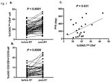
- FIG. 1 is a diagram showing changes in peripheral blood T cell composition before and after completion of chest radiotherapy.
- Left is the ratio of the CD62L low CD4 + T cell subpopulation in the CD4 + T cell population. Among shows the percentage of FOXP3 + CD25 + CD4 + T cell subpopulation in the CD4 + T cell population. The right shows the percentage of CD8 + T cell subpopulation in the T cell population. CD4 + the proportion of CD62L low CD4 + cell subpopulation in a cell population has increased significantly be understood.
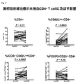
- FIG. 2 is a diagram showing changes in peripheral blood CD4 + T cell composition before and after chest radiotherapy. The upper left shows the percentage of CD4 + T cell subpopulation in the T cell population.
- the upper right shows the percentage of CD62L low CD4 + T cell subpopulations in the CD4 + T cell population.
- Lower left shows the percentage of ICOS + CD62L low CD4 + T cell subpopulations of CD62L low CD4 + T cell population.
- the lower right shows the percentage of FOXP3 + CD25 + CD4 + T cell subpopulations in the CD4 + T cell population. Proportion of CD62L low CD4 + cell subpopulations in CD4 + cell population, and the proportion of ICOS + CD62L low CD4 + cell subpopulations in CD62L low CD4 + cell subpopulations that has increased significantly be understood.
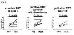
- FIG. 3 is a diagram showing changes in the ratio of the CD62L low CD4 + cell subpopulation in the CD4 + cell population for each treatment type. It is understood that radiotherapy tends to increase the ratio of the CD62L low CD4 + cell subpopulation in the CD4 + cell population in any treatment type.
- curative TRT therapeutic chest radiotherapy
- curative CRT curative chemotherapy radiotherapy
- palliative TRT palliative chest radiotherapy.
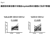
- FIG. 4 is a diagram showing changes in peripheral blood myeloid DC cell composition before and after chest radiotherapy. Left shows the proportion of HLA-DR + CD141 + CD11c + cell subpopulations of CD141 + CD11c + cell population. The right shows the percentage of CD141 + CD11c + cell subpopulations in the PBMC cell population.
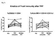
- CD141 + CD11c + percentage of HLA-DR + CD141 + CD11c + cell subpopulations in the cell population is understood to be increased significantly. 5, the percentage of HLA-DR + CD141 + CD11c + cell subpopulations in proportions and CD141 + CD11c + cell population of CD62L low CD4 + cell subpopulations in CD4 + cell population, up to the point of about 1 month after radiotherapy It is a figure including the change of. The proportions of transiently increased CD62L low CD4 + cell subpopulation and HLA-DR + CD141 + CD11c + cell subpopulation tended to return to pre-radiation treatment levels.
- FIG. 7 is a diagram showing changes in peripheral blood T cell composition before and after radiation treatment.
- Figure 7a CD4 + T cell population indicates a change in the percentage of CD62L low CD4 + T cell subpopulations (P ⁇ 0.0001, corresponding there's t test).
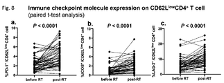
- FIG. 8 is a diagram showing changes in the expression of immune checkpoint molecules on CD62L low CD4 + T cells before and after completion of radiotherapy (paired t-test).
- FIG. 9 shows the change in T cell subpopulation based on CD4 + T cells gated with CCR7 and CD45RA before and after completion of radiotherapy. naive: CCR7 + CD45RA + , CM:CCR7 + CD45RA ⁇ , EM:CCR7 ⁇ CD45RA ⁇ (paired t-test).
- FIG. 8 is a diagram showing changes in the expression of immune checkpoint molecules on CD62L low CD4 + T cells before and after completion of radiotherapy (paired t-test).
- FIG. 9 shows the change in T cell subpopulation based on CD4 + T cells gated with CCR7 and CD
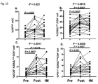
- FIG. 10 is a diagram showing the kinetics of %mDC, %CD62L low CD4 + T cells and immune checkpoint molecule expression before radiotherapy, at the time of completion of radiotherapy and after about 1 month of radiotherapy (Tukey post-hoc). One-way ANOVA with analysis).
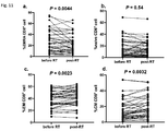
- FIG. 11 shows changes in T cell subpopulations based on CCR7 and CD45RA-gated CD8 + T cells before and at the completion of radiotherapy. naive: CCR7 + CD45RA + , CM:CCR7 + CD45RA ⁇ , EM:CCR7 ⁇ CD45RA ⁇ , EMRA:CCR7 ⁇ CD45RA + (paired t-test).
- a “biomarker” refers to a characteristic that is objectively measured and evaluated as an index of a normal biological process, a pathological process, or a pharmacological response to a therapeutic intervention.
- cancer or “cancer” are used interchangeably, have atypical heterogeneity, grow faster than normal cells, can malignantly invade surrounding tissues or cause metastasis, or a malignant tumor thereof. A state in which such a malignant tumor exists.
- cancer includes, but is not limited to, solid cancer and hematopoietic tumor.
- cancer immunotherapy refers to a method of treating cancer using a biological defense mechanism such as an immune mechanism of an organism.
- anti-tumor immune response refers to any immune response to a tumor in a living body.
- dendritic cell stimulation in an anti-tumor immune response refers to any stimulation that activates dendritic cells that occurs in the course of an immune response to a tumor in vivo. This stimulation can be one of the factors that directly or indirectly generate an antitumor immune response. Typically, but not exclusively, the dendritic cell stimulation in the anti-tumor immune response can be activation of dendritic cells by CD4 + T cells (eg effector T cells), which results in activation. The dendritic cells stimulated the CD8 + T cells, and the stimulated CD8 + T cells exert an antitumor effect.
- CD4 + T cells eg effector T cells
- correlation means that two events have a statistically significant correlation.
- the relative amount of B correlated with A means that when the event A occurs, the relative amount of B is statistically significantly affected (for example, increased or decreased).
- flow cytometry refers to a technique for measuring the number of particles of cells, solids and other biological particles suspended in a liquid, and the physical, chemical and biological properties of each individual.
- immune activation refers to an increase in the ability of the immune function to eliminate foreign substances in the body, and the amount of any factor that positively acts on the immune function (for example, immune cells or cytokines). Can be indicated by an increase in
- the “cell subpopulation” refers to a collection of arbitrary cells having some common characteristic in a cell population including cells having various characteristics. For certain names known in the art, such terms may also be used to refer to particular cell subpopulations, and may be characterized by describing any property (eg, expression of a cell surface marker). One can also refer to a cell subpopulation of
- the “amount” of a certain cell subpopulation includes the absolute number of certain cells and the relative amount of proportion in the cell population.
- the “amount of CD62L LOW CD4 + T cell subpopulation” may be a relative amount with respect to the amount of CD4 + cells.
- the “cell ratio” means the amount of the cell subpopulation
- the “CD62L LOW CD4 + T cell ratio” means the amount of the CD62L LOW CD4 + T cell subpopulation.
- relative amount with respect to cells is used herein interchangeably with “ratio.”
- ratio the terms “relative amount” and “percentage” refer to the desired cell subpopulation (eg, CD62L low CD4 + ) relative to the number of cells forming a particular cell population (eg, CD4 + T cell population). T cell subpopulation).
- the “reference” refers to an amount to be compared for determining an increase or decrease in the amount of the marker described in the present specification.
- the “reference” includes, for example, the amount before the treatment and a value generally used in the art.
- the term “radiation” refers to the propagation of energy in a space or substance by the shape of waves or particles.
- radiation treatment refers to any treatment method using irradiation.
- the composition of a cell subpopulation in a subject who has undergone radiation treatment is used as an indicator of immune activation by radiation treatment in the subject.
- the method can include analyzing the composition of the cell subpopulation in the sample. Analysis of the composition of cell subpopulations can be done by any method described herein or known to those of skill in the art. The method may be in vitro or in silico.
- the amount of cell subpopulation is compared to a suitable criterion to indicate the presence or absence of immune activation in the subject.
- the cell subpopulation can be a cell subpopulation that correlates with dendritic cell stimulation in the anti-tumor immune response.
- the indicative cell subpopulation is a CD4 + T cell subpopulation that correlates with dendritic cell stimulation in the anti-tumor immune response.
- CD4 + T cell subpopulations that correlate with dendritic cell stimulation in the anti-tumor immune response are also believed to be available as indicators of immune activation by radiotherapy.
- Examples of the CD4 + T cell subpopulation that correlates with dendritic cell stimulation in the antitumor immune response include, for example, CD4 + T cell subpopulation in which expression of homing molecules to secondary lymphoid organs is reduced, and effector-type T cells are primed. and CD4 + T cell subpopulations, CD4 + T cell subpopulations that have undergone priming with antigen recognition, and include but are regulatory T cell subpopulations not limited thereto.
- CD4 + T cell subpopulations that correlate with dendritic cell stimulation include, for example, CD62L low CD4 + T cell subpopulations, CCR7 ⁇ CD4 + T cell subpopulations, LAG-3 + CD62L low CD4 + T cell subpopulations, ICOS + CD62L low CD4 + T cell subpopulation, CCR4 + CD25 + CD4 + T cell subpopulation, CD45RA ⁇ CD4 + T cell subpopulation, CD45RO + CD4 + T cell subpopulation, CD28 + CD62L low CD4 + T cell subpopulation , CD62L high CD25 + CD4 + T cell subpopulations, CD127 + CD25 + CD4 + T cell subpopulations, CD45RA ⁇ Foxp3 + CD4 + T cell subpopulations, and Foxp3 + CD25 + CD4 + T cell subpopulations. , But is not limited to these.
- the CD4 + T cell subpopulation that correlates with dendritic cell stimulation in the anti-tumor immune response may be, for example, a cell subpopulation contained in the CD62L low CD4 + T cell population.
- the CD4 + T cell subpopulations that correlate with dendritic cell stimulation in the anti-tumor immune response include CD62L low CD4 + T cell subpopulations (ie, CD62L low CD4 + T cell populations themselves), ICOS + CD4 + T cell subpopulations.
- CD62L low CD4 + T cell subpopulation PD-1 + CD4 + T cell subpopulation
- PD-1 + CD62L low CD4 + T cell subpopulation LAG-3 + CD4 + T cell subpopulation
- LAG- 3 + CD62L low CD4 + T cell subpopulations CD28 + CD4 + T cell subpopulations, and CD28 + CD62L low CD4 + T cell subpopulations, but are not limited thereto.
- the CD4 + T cell subpopulation that correlates with dendritic cell stimulation in the anti-tumor immune response is progression free survival (Progression Free). Survival; PFS).
- the expression level of an appropriate surface marker molecule in an appropriate cell may be used as an index.
- the expression levels of ICOS, PD-1, LAG-3, CD28 and the like expressed in CD4 + T cells may be used as an index.
- the expression levels of ICOS, PD-1, LAG-3, CD28 and the like expressed in CD62L low CD4 + T cells may be used as an index.
- the indicative cell subpopulation is a dendritic cell subpopulation that correlates with dendritic cell stimulation with an anti-tumor immune response.
- a dendritic cell subpopulation that correlates with dendritic cell stimulation with an anti-tumor immune response is a dendritic cell subpopulation that correlates with dendritic cell stimulation with an anti-tumor immune response.
- HLA-DR mediates activation of dendritic cells by CD4 + T cells.
- Dendritic cell subpopulations that correlate with dendritic cell stimulation with an anti-tumor immune response are also likely to be useful as indicators of immune activation by radiotherapy.
- CD11c + CD141 + CD123 ⁇ cell population is generally considered to be functionally highly Th1-inducing myeloid dendritic cells (mDCs, DCs important for antitumor immunity), but CD141 and CD123 are generally exclusive. It should be noted that the expression herein identifies the same cell population as CD141 + CD11c + as well as CD123 ⁇ CD11c + .
- Examples of the dendritic cell subpopulation that correlates with dendritic cell stimulation in the antitumor immune response include, for example, dendritic cell subpopulations that increase due to an increase in cell subpopulations with reduced expression of homing molecules in the CD4 + T cell population. population, dendritic cell subpopulation increased due to the increase of primed CD4 + T cell subpopulations to effector T cells in CD4 + T cell population, and, receiving the priming by antigen recognition of CD4 + T cell population Dendritic cell subpopulations that increase due to increased CD4 + T cell subpopulations.
- examples of the dendritic cell subpopulation include HLA-DR + dendritic cell subpopulation, CD80 + dendritic cell subpopulation, CD86 + dendritic cell subpopulation, and PD-L1 + dendritic cell subpopulation.
- examples of dendritic cells include, but are not limited to, bone marrow dendritic cells (mDC, CD141 + CD11c + dendritic cells) and plasmacytoid dendritic cells (pDC, CD123 + CD11c + dendritic cells).
- Dendritic cell subpopulations that correlate with dendritic cell stimulation with anti-tumor immune responses include HLA-DR + CD141 + CD11c + cell subpopulations.
- the expression level of an appropriate surface marker molecule in an appropriate cell may be used as an index.
- the expression level of HLA-DR and the like expressed in CD141 + CD11c + cells may be used as an index.
- the indicative cell subpopulation is a CD8 + T cell subpopulation that correlates with dendritic cell stimulation with an anti-tumor immune response.
- CD137 expressed on CD62L low CD8 + T cells there is a significant difference before and after radiotherapy (Table 4).
- Dendritic cells activated by CD4 + T cells stimulate CD8 + T cells, and the stimulated CD8 + T cells finally exert antitumor activity.
- CD8 + T CD137 on the cells which mediate the stimulation of CD8 + T cells by dendritic cells.
- CD8 + T cell subpopulations that correlate with dendritic cell stimulation in the anti-tumor immune response are also likely to serve as indicators of immune activation by radiotherapy.
- CD8 + T cell subpopulations which correlates with dendritic cells stimulated in anti-tumor immune response for example, expression of homing molecules in CD4 + T cell population is increased due to an increase in reduced cell subpopulation CD8 + T cell subpopulations
- CD4 + T cells increase due to an increase in CD4 + T cell subpopulation primed effector T cells in a population CD8 + T cell subpopulation
- the priming by antigen recognition of CD4 + T cell population CD8 + T cell subpopulations which increased due to the increase of the received CD4 + T cell subpopulation increases due to the increase in HLA-DR + dendritic cell subpopulation in dendritic cell population CD8 + T cell variant Population, increased due to an increase in CD80 + dendritic cell subpopulation in the dendritic cell population CD8 + T cell subpopulation, increased due to an increase in PD-L1 + dendritic cell subpopulation in the dendritic cell population CD8 + T cell subpopulations, including but
- CD8 + T cell subpopulations that correlate with dendritic cell stimulation with anti-tumor immune responses include the CD137 + CD62L low CD8 + T cell subpopulations.
- the expression level of an appropriate surface marker molecule in an appropriate cell may be used as an index.
- the expression level of CD137, PD-1, or CD28 expressed in CD62L low CD8 + T cells may be used as an index.
- bone marrow-derived immunosuppressive cells (MDSC, CD33 + CD14 + HLA-DR ⁇ cell population)
- significant changes were observed before and after radiotherapy, and the ratio of such cell population can also be used as an index.
- the amount of cell subpopulations described herein can be used in combination with multiple amounts as an indicator. Combining the indicators can make the prediction of responsiveness more accurate.
- the amount of CD4 + T cell subpopulations that correlates with dendritic cell stimulation in the anti-tumor immune response the amount of dendritic cell subpopulations that correlates with dendritic cell stimulation in the anti-tumor immune response in the sample, and Comparison of at least two amounts selected from the group consisting of the amount of CD8 + T cell subpopulations that correlate with dendritic cell stimulation with an anti-tumor immune response with a reference can indicate the presence or absence of immune activation in a subject ..
- the percentage of CD62L low cells in CD4 + T cells (X) and the percentage of CD25 + FoxP3 + in CD4 + T cells (Y) in a sample is combined with X 2 /Y.
- the index of can be used. It will be appreciated by those skilled in the art that, in addition to or in addition to these indicators, further indicators that may indicate immune activation by radiation therapy may be used. In the present invention, instead of the amount of the desired cell subpopulation, or in addition to the amount of the desired cell subpopulation, the amount of cell subpopulation contained in the desired cell subpopulation is used. You may.
- the following factors can be used as markers.
- % ⁇ / ⁇ when representing the percentage of cell subpopulations in the form of, for example, CD62L low / CD4 + T cells, the CD62L low CD4 + T cells to CD4 + T cells
- the cells listed in the numerator have all the characteristics of the cells listed in the denominator. If the denominator for a cell subpopulation is not specified, a ratio to any cell population containing the cell subpopulation, such as a CD4 + T cell population or a CD8 + T cell population, may be included.
- factors that increase immediately after the termination of RT include: (Percentage of CD62L low CD4 + T cells to CD4 + T cells) ⁇ % CD62L low / CD4 + T cells; % LAG3 + /CD62L low CD4 + T cells (may be LAG3 + /effector memory (CCR7 ⁇ CD45RA ⁇ )CD4 + T cells); % ICOS + /CD62L low CD4 + T cells (% ICOS + /effector memory (CCR7 ⁇ CD45RA ⁇ )CD4 + T cells may be used); % CD28 + /CD62L low CD4 + T cells (% CD28 + /effector memory (CCR7 ⁇ CD45RA ⁇ ) CD4 + T cells may be used); and %HLA-DR + /CD11c + CD141 + cells (HLA-DR high).
- CD11c + CD123 ⁇ mDC, %mDC/CD3 ⁇ CD14 ⁇ CD19 ⁇ cells may be used) -% PD-1 + /CD62L low CD4 + T cells-% CD62L low CCR7 + CD45RA ⁇ /CD4 + T cells (CCR7 + CD45RA ⁇ CD4 + cells may be used) -% CCR7 - CD45RA - CD4 + T cells-% CCR7 - CD45RA - CD8 + T cells (EM CD8 + T cells) -% CCR7 + CD45RA - CD8 + T cells (CM CD8 + T cells) Is mentioned.
- Factors that decrease immediately after RT are: -% CCR7 + CD45RA + CD4 + cells (naive CD4 + T cells) -% CCR7 - CD45RA + CD8 + T cells (EMRA CD8 + T cells) Is mentioned.
- factors that increase by 1 month after the end of RT include: %PD-1 + /CD62L low CD4 + T cells (% PD-1 + /effector memory (CCR7 ⁇ CD45RA ⁇ )CD4 + T cells may be used); % CD137 + /CD62L low CD4 + T cells (% CD137 + /effector memory (CCR7 ⁇ CD45RA ⁇ ) CD4 + T cells may be used); %PD-1 + /CD62L low CD8 + T cells (%PD-1 + /effector (CCR7 + CD45RA ⁇ )CD8 + T cells, %PD-1 + /effector memory (CCR7 ⁇ CD45RA ⁇ )CD8 + T cells , And %CD137 + /CD62L low CD8 + T cells (%CD137 + /effector (CCR7 + CD45RA ⁇ )CD8 + T cells,% CD137 + /effector memory (CCR7 ⁇ CD45RA ⁇ )CD8 + T
- % CD25 + FoxP3 + /CD4 + T cells regulatory T cells
- % CD33 + CD14 + HLA-DR low cells Myeloid-delivered suppressor (MDSC)
- the amount of cell subpopulation can be compared with an appropriate standard, and the presence or absence of immune activation in a subject can be determined by the comparison.
- An increase in the amount of cell subpopulations in the sample above the baseline may indicate that the subject has undergone radiotherapy-induced immune activation.
- the fact that the amount of cell subpopulations in the sample is not increased above the reference may indicate that the subject has not undergone radiotherapy-induced immune activation.
- Criteria include, but are not limited to, for example, the amount of corresponding cell subpopulations in a sample of a subject prior to radiation treatment.
- a value statistically calculated by regression analysis or the like based on data obtained from samples of a plurality of subjects may be used. From the data obtained from the subject, the reference may be calculated by machine learning, artificial intelligence, or the like.
- the increase compared to the standard is that the amount of cell subpopulation after radiation treatment exceeds the standard or is 1, 2, 3, 4, 5, 10, 15, 20, 30% of the standard. It may be indicated by an increase of more than, or an increase of more than 1.5 times, 2 times, 3 times, 5 times of the reference. Typically, if the value exceeds the standard value, it is considered to increase compared to the standard value. If the reference is calculated empirically, it can be said to be increased compared to the reference if an increase is observed that is more than an appropriate error from the reference value. Suitable errors include, for example, 1 standard deviation, 2 standard deviations, 3 standard deviations, or more.
- a sample for fractionation/separation of T cells can be appropriately collected from a subject by a conventional method. For example, it can be performed from peripheral blood, bone marrow, tumor tissue, hematopoietic tissue, spleen, normal tissue, lymph fluid, etc. of the subject. Sampling from peripheral blood can be advantageous because it is non-invasive and convenient.
- composition of T cells in a sample of a subject can be measured by those skilled in the art by a conventional method. Usually, it is possible to measure the number of cells positive for a marker (for example, CD4) defining a target cell subpopulation in a sample by using flow cytometry or the like.
- Flow cytometry is generally used to measure the composition of the cell population, but in addition, immunostaining for a sample containing cells, a method using an antibody array, analysis of protein expression in a sample containing cells ( For example, Western blot, mass spectrometry, HPLC, etc.), mRNA expression analysis in a sample containing cells (eg, microarray, next-generation sequencing, etc.) and the like may be used.
- cells other than the subpopulation of each cell may be experimentally removed from the whole cells.
- human Moitenyi Biotech
- the total number of living cells may be counted and recorded, and the number of cells obtained by using this kit may be counted and recorded.
- Antibodies are capable of specifically recognizing and binding to molecules expressed on individual cells, and develop color when the antibody is bound to molecules expressed on the cell surface or intracellularly. The number of cells that develop color is measured by detecting as possible.
- the molecule expressed on the cell surface or in the cell is a protein
- the mRNA encoding the protein is also formed in the cell. That is, the mRNA in each cell may be examined for the presence or absence of mRNA encoding the protein molecule of interest. This is made possible by single cell gene expression analysis, that is, single cell level mRNA analysis.
- Gene candidates to be examined for expression include ⁇ TCR, CD3, CD4, CD25, CTLA4, GITR, FoxP3, STAT5, FoxO1, FoxO3, IL-10, TGFbeta, IL-35, SMAD2, SMAD3, SMAD4, CD62Llow, CD44, IL. -7R (CD127), IL-15R, CCR7low, BLIMP1, etc.
- CD62L high CD4 + T cells as genes whose expression is higher than that of CD62L low CD4 + T cells, BACH2, CCL28, CCR7, CD27, CD28, CD62L, CSNK1D, FOXP1, FOXP3, IGF1R, IL16, IL27RA, IL6R, LEF1, MAL, and TCF7 are mentioned (WO2018/147291).
- BACH2 CCL28, CCR7, CD27, CD28, CD62L, CSNK1D, FOXP1, FOXP3, IGF1R, IL16, IL27RA, IL6R, LEF1, MAL, and TCF7 are mentioned (WO2018/147291).
- the measurement of the proportion of the cell subpopulation or the comparison with the threshold value may be performed using a standard sample having a defined signal. Compare the signal between a standard (eg, particles with fluorescent dye attached) prepared to produce a fluorescent signal corresponding to a given cell subpopulation and a sample containing the cell population, and compare with the standard Can measure the amount or proportion of cell subpopulations in a sample.
- a standard for example, a particle to which a fluorescent dye is attached
- the signal between a standard for example, a particle to which a fluorescent dye is attached
- a standard for example, a particle to which a fluorescent dye is attached
- an indicator of immune activation by radiotherapy is provided.
- radiation therapy by irradiating radiation, cancer cells are reduced by destroying DNA or RNA of cancer cells to prevent cell division and/or induce apoptosis (cell death).
- Electromagnetic waves include X-rays and ⁇ -rays.
- a particle beam is a substance particle that flows with high kinetic energy, and examples thereof include ⁇ rays, ⁇ rays, neutron rays, proton rays, heavy ion rays, and meson rays.
- Irradiation methods in radiation therapy can be divided into “external irradiation”, which irradiates radiation from outside the body, and “internal irradiation,” which irradiates radiation from inside the body to the cancer and its surroundings. It is also possible to perform a combination of external irradiation and internal irradiation.
- External irradiation is the irradiation of radiation from outside the body through the skin.
- the most common method is to irradiate high-energy X-rays.
- External irradiation includes various modes, for example, X-ray irradiation by a linac (linear accelerator), three-dimensional conformal irradiation (3D-CRT), intensity-modulated radiation therapy (IMRT), stereotactic radiation therapy (SRT), particles Radiation therapy (proton radiation therapy/heavy particle radiation therapy), image-guided radiation therapy (IGRT), etc. are mentioned, but not limited thereto.
- linac linear accelerator
- 3D-CRT three-dimensional conformal irradiation
- IMRT intensity-modulated radiation therapy
- SRT stereotactic radiation therapy
- IGRT image-guided radiation therapy
- Examples of the internal irradiation method include, but are not limited to, brachytherapy (internal tissue irradiation, intracavitary irradiation) and treatment with non-sealed radioisotope (internal therapy). ..
- the radiation treatment which can be a target in the present invention is not limited in its aspect as long as it is irradiation in a manner capable of causing immune activation.
- the irradiation field in radiotherapy may be an irradiation range including tumor tissue.
- tumor cells undergoing radiotherapy undergo immunogenic cell death, which is important for anti-tumor effector T cell expansion.
- radiation therapy includes chest radiation, bone metastases, lymph node metastases, adrenal metastases, liver metastases, brain metastases, etc. ..
- the biomarker of the present invention can be used for studying a schedule of radiation treatment intended to cause immune activation.
- the absence of radiotherapy-induced immune activation in a subject may indicate that the subject should be reradiated.
- the immune activation resulting from radiation treatment in the subject may indicate that radiation treatment should be terminated.
- Radiation treatment can be performed by irradiating a dose of about 1 to 3 Gy per dose about 1 to 2 times per day for 3 to 8 weeks.
- immune cells eg, T cells
- Radiation for 1 to 2 weeks may be preferred.
- Cancer immunotherapy is a method of treating cancer using the biological defense mechanism of an organism. Cancer immunotherapy is broadly divided into cancer immunotherapy by strengthening the immune function against cancer and cancer immunotherapy by inhibiting the immune evasion function of cancer. Further, cancer immunotherapy includes active immunotherapy that activates the immune function in the body, and passive immunotherapy that returns immune cells that activate or proliferate the immune function outside the body to the body. is there. Since the biomarker of the present invention shows immune activation by radiation therapy, it is possible to know the suitable timing for carrying out cancer immunotherapy utilizing the immune function.
- cancer immunotherapy examples include non-specific immunostimulants, cytokine therapy, cancer vaccine therapy, dendritic cell therapy, adoptive immunotherapy, non-specific lymphocyte therapy, cancer antigen-specific T cell therapy, antibody Therapy, immune checkpoint inhibition therapy, etc.
- PD-1 inhibitors include the anti-PD-1 antibodies nivolumab (sold as Nivolumab; Obdivo TM ), pembrolizumab, spartalizumab, and semiprimab (Cemiplimab). Not limited. In one preferred embodiment, nivolumab may be selected as a subject.
- PD-L1 inhibitors can be used as well as PD-1 inhibitors.
- Anti-PD-1 antibody is considered to exert an anti-cancer effect by releasing the suppression of T cell activation by PD-1 signal.
- Anti-PD-L1 antibody is also considered to exert an anti-cancer effect by releasing the suppression of T cell activation by PD-1 signal.
- T cell activation is suppressed by recruiting SHP-1, 2 which is one of tyrosine dephosphorylating enzymes and inactivating ZAP70 which is a T cell receptor signaling protein.
- SHP-1, 2 which is one of tyrosine dephosphorylating enzymes and inactivating ZAP70 which is a T cell receptor signaling protein.
- PD-1 is highly expressed in killer T cells and natural killer cells infiltrating cancer tissues. Further, it is considered that PD-L1 on the tumor attenuates the PD-1 signal-mediated immune response by PD-1. PD-L1 attenuates this PD-1 signaling-mediated immune response, whereas inhibition of PD-1 and PD-L1 interaction and/or signal transduction resulting from the interaction by anti-PD-1 antibodies leads to The effect of enhancing the tumor immune response is obtained.
- immune checkpoint inhibitors include PD-L1 inhibitors (eg, anti-PD-L1 antibody avelumab, durvalumab or atezolizumab).
- PD-L1 inhibitors eg, anti-PD-L1 antibody avelumab, durvalumab or atezolizumab.
- the PD-L1 inhibitor binds and inhibits the above PD-1 pathway to the side of PD-L1, inhibits the interaction between PD-1 and PD-L1 and/or the signal transduction caused by the interaction, Raises an anti-tumor immune response.
- immune checkpoint inhibitors include CTLA-4 inhibitors (eg, anti-CTLA-4 antibodies ipilimumab or tremerilumab).
- CTLA-4 inhibitors activate T cells and generate an anti-tumor immune response. T cells are activated by the interaction of surface CD28 with CD80 or CD86. However, even once activated T cells, CTLA-4 (cytotoxic T-lymphocytote-associated antigen 4) expressed on the surface preferentially interacts with CD80 or CD86 with higher affinity than CD20. However, it is believed that activation is suppressed thereby.
- CTLA-4 inhibitors generate an anti-tumor immune response by inhibiting the inhibition of the interaction of CD20 with CD80 or CD86 by inhibiting CTLA-4.
- the immune checkpoint inhibitor is TIM-3 (T-cell immunoglobulin and mucin maintaining protein-3), LAG-3 (lymphocyte activation gene-3), B7-H3, B7-H4, B7-H5. (VISTA), or immune checkpoint proteins such as TIGIT (T cell immunoreceptor with Ig and ITIM domain) may be targeted.
- TIM-3 T-cell immunoglobulin and mucin maintaining protein-3
- LAG-3 lymphocyte activation gene-3
- B7-H3, B7-H4, B7-H5. VISTA
- immune checkpoint proteins such as TIGIT (T cell immunoreceptor with Ig and ITIM domain) may be targeted.
- T cell immune checkpoints suppress the immune response to self-tissues, but even when antigens such as viruses are present in the body for a long period of time, T cell immune checkpoints increase. Since tumor tissue is an antigen that exists in the body for a long time, it is considered that these immune checkpoints evade the antitumor immune response. Such an avoidance function is invalidated and an antitumor effect is exerted.
- the immune checkpoint inhibitor may be appropriately used in combination with other cancer treatments.
- Other cancer treatments include, but are not limited to, radiation therapy, as well as other cancer immunotherapy (eg, adoptive cell transfer), chemotherapy, hyperthermia, surgical procedures, and the like. Absent.
- the immune checkpoint inhibitor may be administered in combination with one or more additional agents.
- the one or more additional agents may be any chemotherapeutic agent or may include a second immune checkpoint inhibitor.
- a composition comprising an immune checkpoint inhibitor is provided.
- the composition containing the immune checkpoint inhibitor of the present invention is usually administered systemically or locally in oral or parenteral form.
- a composition comprising an immune checkpoint inhibitor of the present invention is administered by a method described herein to a subject who has been shown to undergo immune activation by radiotherapy to achieve a significant treatment. It is thought that the effect can be achieved.
- the dose varies depending on age, body weight, symptoms, therapeutic effect, administration method, treatment time, etc., but usually, for example, per adult, in the range of 0.1 mg to 100 mg, once to several times per day. Orally administered orally, or per adult, in the range of 0.01 mg to 30 mg, once to several times per day, parenterally (preferably intravenously), or once a day. It is continuously administered intravenously in the range of 1 to 24 hours.
- a dose smaller than the above dose may be sufficient in some cases, or a dose exceeding the range may be necessary in some cases.
- the composition containing the immune checkpoint inhibitor can be in the form of a solid preparation for oral administration, a liquid preparation for oral administration, and an injection, an external preparation, a suppository for parenteral administration upon administration. ..
- the solid preparations for oral administration include tablets, pills, capsules, powders and granules. Capsules include hard capsules and soft capsules.
- compositions of the present invention may optionally comprise one or more active ingredients as such (eg, antibodies against immune checkpoint proteins) or excipients (lactose, mannitol, glucose, microcrystalline cellulose, starch, etc.). ), binders (hydroxypropyl cellulose, polyvinylpyrrolidone, magnesium aluminometasilicate, etc.), disintegrants (calcium fibrin glycolate, etc.), lubricants (magnesium stearate, etc.), stabilizers, solubilizers (glutamic acid, Aspartic acid, etc.) and the like, and are used by formulating them according to a conventional method.
- active ingredients eg, antibodies against immune checkpoint proteins
- excipients lactose, mannitol, glucose, microcrystalline cellulose, starch, etc.
- binders hydroxypropyl cellulose, polyvinylpyrrolidone, magnesium aluminometasilicate, etc.
- disintegrants calcium
- a coating agent sucrose, gelatin, hydroxypropylcellulose, hydroxypropylmethylcellulose phthalate, etc.
- capsules of absorbable material such as gelatin.
- composition of the present invention when formulated as a liquid solution for oral administration for oral administration, it contains a pharmaceutically acceptable aqueous solution, suspension, emulsion, syrup, elixir and the like.
- a liquid agent one or more active substances are dissolved, suspended or emulsified in a commonly used diluent (purified water, ethanol or a mixed solution thereof).
- this liquid agent may contain a wetting agent, a suspending agent, an emulsifying agent, a sweetening agent, a flavoring agent, an aromatic agent, a preservative, a buffering agent and the like.
- the injections for parenteral administration include solutions, suspensions, emulsions and solid injections that are dissolved or suspended in a solvent at the time of use.
- the injection is used by dissolving, suspending or emulsifying one or more active substances in a solvent.
- the solvent for example, distilled water for injection, physiological saline, vegetable oil, propylene glycol, polyethylene glycol, alcohols such as ethanol, and the like, and combinations thereof are used.
- this injection may contain a stabilizer, a solubilizing agent (glutamic acid, aspartic acid, polysorbate 80 (registered trademark), etc.), a suspending agent, an emulsifying agent, a soothing agent, a buffering agent, a preservative and the like.
- composition of the present invention may be put into a box or the like to be commercialized together with a package insert describing that it is used in combination with radiation therapy.
- the package insert may state that it is desirable that the composition of the invention be administered within a predetermined period of time after radiation treatment.
- the package insert may be explicitly instructed to be used in combination with radiation therapy, or may only mention possible combinations.
- melanoma malignant melanoma
- non-small cell lung cancer renal cell carcinoma
- malignant lymphoma Hodgkin lymphoma or non-Hodgkin lymphoma
- head and neck cancer urological cancer (bladder cancer, Urothelial cancer, prostate cancer, small cell lung cancer, thymic cancer, gastric cancer, esophageal cancer, gastroesophageal junction cancer, liver cancer (hepatocellular carcinoma, intrahepatic cholangiocellular carcinoma), primary brain tumor (glioblastoma, central nervous system) Primary lymphoma), malignant pleural mesothelioma, gynecological cancer (ovarian cancer, cervical cancer, endometrial cancer), soft tissue sarcoma, biliary tract cancer, multiple myeloma, breast cancer, colon cancer, etc. It is not limited to these.
- the subject may be subjected to radiation treatment and a sample taken from the subject who has undergone radiation treatment.
- a sample may be taken from the subject prior to receiving radiation therapy.
- the composition of cell subpopulations in the sample can be used as a basis for comparison.
- the time point for collecting the sample is not particularly limited. Whether or not the immune activation caused by radiotherapy occurs and how long it lasts depends on the subject, and a sample can be taken at any time point and the immune activation at that time point can be investigated. Changes in cell subpopulations, eg, changes in HLA-DR positivity of dendritic cells (CD11c + CD141 + cells) and ICOS positivity of CD62L low CD4 + T cells, may persist for 2-3 months. .. Some subjects maintain a high CD62L low CD4 + T cell ratio for one year or longer.
- the applicable period of Durvalumab (anti-PD-L1 antibody) after chemoradiotherapy, which is covered by insurance, is set to be from the first day of chemoradiotherapy until the disease is not aggravated. If so, it is considered possible to detect a change in immune activation state after radiotherapy and determine whether or not to perform cancer immunotherapy.
- the samples in dendritic cells or T cell subpopulations reached a peak immediately after the end of radiotherapy, and thereafter, the proportion of cell subpopulations such as the CD62L low CD4 + T cell proportion tended to return to the original level. Therefore, although not essential, obtaining a sample between the time of radiotherapy and after a certain period of time may increase the possibility of detection of immune activation for performing cancer immunotherapy.
- the sample may be from the time of radiation treatment up to about 1 year, up to about 6 months, up to about 3 months, up to about 2 months, up to about 4 weeks. Alternatively, it can be obtained up to about 14 days later, or at a time such as immediately after radiation therapy, but is not limited thereto.
- samples can be obtained from the subject at multiple time points and the immune activation due to the radiation treatment in the subject can be monitored. For example, a subject may be given radiation therapy again if the immune activation has not occurred. When the immune activation due to the radiation treatment occurs, the radiation treatment can be terminated and the cancer immunotherapy can be given.
- the plurality of time points may be after each irradiation of radiation, or after irradiation once every several times (for example, twice, three times, four times or five times or more) of irradiation. Alternatively, it may be once every several days (eg, 2 days, 3 days, 4 days, 5 days, 1 week, or 2 weeks or more) independent of irradiation.
- the subject may be subjected to cancer immunotherapy, but the timing of the cancer immunotherapy is such that the immune activated state is maintained. There is no particular limitation as long as it exists. Although it is not essential because the immune activation state may be restored by the passage of time from the time of radiation treatment, it is not necessary, but the time point of radiation treatment is indicated when it is shown that immune activation is occurring. For a period of time, for example, up to about 4 weeks after the time of radiation treatment, or up to about 14 days after the time of radiation treatment, the subject may undergo cancer immunotherapy. As a result, it is possible to increase the possibility of receiving the benefits of the Abscopal effect. In one embodiment, radiotherapy-induced immune activation has occurred in the subject, at which point the subject may be given cancer immunotherapy, including an immune checkpoint inhibitor.
- a factor that acts on immunosuppression may be further used to determine when to administer cancer immunotherapy. For example, about 1 month after radiation treatment, regulatory T cells and MDSCs may increase, and before the increase of such cell subpopulation (that is, when the ratio of such cell subpopulation is below a certain level). ) The subject may be given cancer immunotherapy including an immune checkpoint inhibitor.
- specific cell subpopulations eg, CD4 + T cell subpopulations that correlate with dendritic cell stimulation in antitumor immune responses, trees in antitumor immune responses that are included in immune cells in a subject by radiation therapy. It has been found that the amount or proportion of CD8 + T cell subpopulations that correlate with dendritic cell stimulation or dendritic cell subpopulations that correlate with dendritic cell stimulation with an anti-tumor immune response) varies. This provides, in one aspect of the invention, for modulating the proportion of a cell subpopulation in a subject by subjecting the subject to radiation therapy.
- CD4 + CD62L low cell subpopulation contained in the cell population can increase the amount or percentage.
- Specific cell subpopulations in a subject eg, CD4 + CD62L low cell subpopulations
- Radiation therapy can alter the amount or proportion of subpopulations, which can cause a subject to become responsive to cancer immunotherapy (eg, immune checkpoint inhibitors). Conceivable.
- a method of treating cancer in a subject having an immune cell composition that is not responsive to cancer immunotherapy comprises administering radiation therapy to a subject.
- the method optionally includes the step of subjecting the subject to cancer immunotherapy.
- the method may also include the step of measuring immune cell composition in the subject. This makes it possible to confirm that the subject has changed to responsiveness to the cancer immunotherapy by the radiation treatment and to administer the cancer immunotherapy.
- a composition comprising an immune checkpoint inhibitor for treating cancer, wherein the immune cell composition is not responsive to cancer immunotherapy (eg, low CD4 + CD62L low cell subpopulation amount or percentage).
- a composition that has been administered to a subject who had had radiation therapy can be the immune cell composition in peripheral blood.
- the immune cell composition that is not responsive to (or is responsive to) cancer immunotherapy can be determined by those skilled in the art with reference to WO2018/147291 and the like.
- the immune cell composition is considered not to be responsive to cancer immunotherapy if the proportion of CD62L low T cells in CD4 + T cells is lower than a threshold value (ineffective group threshold value).
- a threshold value ineffective group threshold value
- the immune cell composition may be determined using the relative value between the CD62L low CD4 + ratio and the regulatory T cells (eg, CD25 + Foxp3 + CD4 + cell ratio).
- X/Y or X 2 /Y may be used, where CD62L low CD4 + ratio is X and CD25 + Foxp3 + CD4 + cell ratio is Y.
- the threshold can be 7.35 for the ratio (X/Y) and 192 can be the threshold for X 2 /Y.
- kits for determining whether a subject has undergone radiotherapy-induced immune activation.
- the kit may include one or more detection agents for the appropriate molecule to detect the cell subpopulations described herein. Combinations of such detection agents can be used to determine the T cell composition of a subject.
- Such kits can be used to measure the proportion of a particular cell subpopulation in a subject as the novel biomarkers described herein.
- the kit comprises (A) CD4 and CD62L; (B) (i) a marker selected from ICOS, PD-1, LAG-3 and CD28, (ii) CD4, and (iii) CD62L; (C) CD11c, CD141 and HLA-DR; (D) CD11c, CD123 and HLA-DR; or (E) CD8, CD62L and CD137
- a detection agent for In one embodiment, the detection agent is an antibody.
- the antibody is appropriately labeled to facilitate detection of the marker.
- Example 1 Index of immune activation by radiotherapy
- Example 2 Index of immune activation by radiotherapy
- peripheral blood was collected from the following 20 subjects (before and after the radiotherapy (and later, if applicable)), and the composition of the cell population in the peripheral blood was examined.
- PBMC analysis Sampling and PBMC analysis at each time point were performed as follows. A BD Vacutina CPTTM blood collection tube for mononuclear cell separation was used for PBMC analysis. FCM analysis was performed on PBMC.
- the figure shows the changes before and after radiotherapy for some cell subpopulations.
- CD4 + percentage of CD62L low CD4 + cell subpopulations in the cell population, and CD62L low CD4 + percentage of ICOS + CD62L low CD4 + cell subpopulations in cell subpopulations was significantly increased ( Figure 1 and (Fig. 2).
- An increase in the proportion of CD62L low CD4 + cell subpopulations in the CD4 + cell population was observed for each type of radiation therapy (curative TRT: therapeutic chest radiotherapy, curative CRT: therapeutic chemotherapy combined radiation therapy, palliative TRT: palliative TRT). Chest radiotherapy) was also observed (Fig. 3).
- the proportion of HLA-DR + CD141 + CD11c + cell subpopulations in CD141 + CD11c + cell population was significantly increased (Fig. 4).
- the figure shows the changes before and after radiotherapy for some cell subpopulations. Approximately 30 days after radiation treatment, the proportions of transiently increased CD62L low CD4 + cell subpopulation and HLA-DR + CD141 + CD11c + cell subpopulation tended to return to pre-radiation treatment levels. (Fig. 5).
- the T cell subpopulations in which the CD62L low CD4 + cell subpopulation has a strong positive correlation are type 1 helper CD4 + T cells (Th1), effector memory CD4 + T cells, CD8 + T cells, and effector CD8 + T cells. .. These are cell subpopulations important for cell killing function in cell-mediated immunity.
- type 2 helper CD4 + T cells (Th2) and regulatory T cells have a negative correlation. These are known as cell subpopulations that suppress cell-mediated immunity.
- an increase in CD62L low CD4 + cell subpopulation is considered to indicate activation of antitumor cell-mediated immunity.
- the HLA-DR + CD141 + CD11c + dendritic cell subpopulation and the CD62L low CD4 + cell subpopulation have a positive correlation. It is considered that activated dendritic cells express the MHC class II-restricted antigen, resulting in an increase in the CD62L low CD4 + cell subpopulation that recognizes the MHC class II-restricted antigen.
- the low CD4 + cell subpopulation is considered to be a CD4 + T cell subpopulation that correlates with dendritic cell stimulation in the tumor immune response.
- the HLA-DR + CD141 + CD11c + dendritic cell subpopulation is considered to be a dendritic cell subpopulation that correlates with dendritic cell stimulation in the tumor immune response.
- the CD62L low CD4 + T cell subpopulation, PD-1 + CD62L low CD8 + T cell subpopulation, CD137 + CD62L low CD8 + T cell subpopulation are also T cell subpopulations that correlate with dendritic cell stimulation in tumor immune responses. It is considered that there is (WO2018/147291).
- the CD62L low CD4 + T cell subpopulation that received the cancer antigen stimulation from the dendritic cells during the radiotherapy still expresses CD137 and ICOS and remains activated.
- the CD25 + Foxp3 + CD4 + regulatory T cell subpopulation and the CD33 + CD14 + HLA-DR - MDSC cell subpopulation is observed. It is a well-known event that regulatory T cells and MDSCs are cells that suppress T cell immunity, and it can be understood that this is negative feedback for controlling activated T cell subpopulations. it can.
- the CD62L low CD4 + T cell subpopulation is used as a molecule and the regulatory T cell subpopulation is used as a denominator as an index of the T cell immune state showing the effect of the PD-1 inhibitor.
- An immune checkpoint inhibitor is administered to a subject having a certain proportion of CD4 + CD62L low T cell subpopulations in peripheral blood.
- the CD4 + CD62L low T cell subpopulation percentage may decrease after administration of the immune checkpoint inhibitor.
- the responsiveness of the subject to immune checkpoint inhibitors is monitored by monitoring the CD4 + CD62L low T cell subpopulation percentage in the subject.
- a subject is given radiation therapy if the proportion of the CD4 + CD62L low T cell subpopulation is reduced. Radiation therapy increases the proportion of CD4 + CD62L low T cell subpopulations and restores the subject's responsiveness to immune checkpoint inhibitors.
- Example 3 Correlation between CD62L low CD4 + T cells and effector memory cells
- Example 3 Correlation between CD62L low CD4 + T cells and effector memory cells
- 84 specimens were Fortessa analysis, and percentage of CD62L low CD4 + T cells CD4 + T cells, CD4 + T effector memory cells in the cell (CCR7 - CD45RA -) were analyzed the relationship between the ratio of CD4 + T cells.
- Example 4 Examination of indicators of immune activation by radiotherapy in different patient populations
- biomarker a parameter that was significantly changing in subjects before and after radiation treatment.
- FIGS. 7-11 The results are shown in Figures 7-11.
- “Before RT” in FIGS. 7 to 11 corresponds to “pre-TRT” in FIGS. 1 to 6 and indicates immediately before the radiotherapy (RT).
- “Post-RT” in FIGS. 7 to 11, “post-TRT”, “at TRT completion”, and “TRT complete” in FIGS. 1 to 6 are all immediately after completion of radiation therapy (RT).
- HLA-DR high CD11c + CD123 ⁇ mDC is a cell population corresponding to Tables 3 and 4 in Example 1 and HLA-DR high CD11c + CD141 + mDC in FIG.
- FIG. 8 is a diagram showing changes in the expression of immune checkpoint molecules on CD62L low CD4 + T cells before and after completion of radiotherapy (paired t-test). Consistent with the results of Example 1 (Table 3), expression of LAG-3 and ICOS on CD62L low CD4 + T cells was significantly increased (FIGS. 8b and c). In addition, in this example, a significant difference was also observed in the increase in PD-1 expression on CD62L low CD4 + T cells (Fig. 8a). This is considered to be due to the increase in the number of samples.
- FIG. 9 shows the change in T cell subpopulation based on CD4 + T cells gated with CCR7 and CD45RA before and after completion of radiotherapy.
- CCR7 and CD45RA are generally used to fractionate naive T cells (CCR7 + CD45RA + ), central memory (CM) T cells (CCR7 + CD45RA ⁇ ), effector memory T cells (CCR7 ⁇ CD45RA ⁇ ). Is a cell surface marker that can be.
- FIG. 10 is a diagram showing the kinetics of %mDC, %CD62L low CD4 + T cells and immune checkpoint molecule expression before radiotherapy, at the time of completion of radiotherapy and after about 1 month of radiotherapy (Tukey post-hoc).
- One-way ANOVA with analysis The P value at the top of each figure is the result of the ANOVA analysis.
- the post-hoc analysis shows the p-value between the two points where a significant difference was found together with the bar.
- CD62L low CD4 + T cells started to decline by 4 weeks after radiotherapy in half of the patients. Although there was no statistically significant difference between the time of completion of the radiotherapy and about 1 month after the radiotherapy, it decreased with time in about half of the patients, similar to the results in Table 4 in Example 1.
- the increased ICOS and PD-1 expression of mDC, CD62L low CD4 + T cells, and CD62L low CD4 + T cells after radiation treatment tended to decrease during the following month. This indicates that the immune function against the tumor once activated is then attenuated, and it is considered that it is better to start the administration of the immune checkpoint inhibitor or the like before these cells are reduced.
- FIG. 11 shows changes in T cell subpopulations based on CCR7 and CD45RA-gated CD8 + T cells before and at the completion of radiotherapy.
- FIG. 1 shows that there is no significant change in the amount of the CD8 + cell population before and after the radiotherapy, and it is considered that the cells actually having the ability to kill cancer cells are the CD8 + cells. Therefore, the presence of alterations in specific subpopulations in the CD8 + T cell population upon radiotherapy was analyzed by gating with CCR7 and CD45RA.
- CCR7 and CD45RA generally fractionate naive T cells (CCR7 + CD45RA + ), central memory (CM) T cells (CCR7 + CD45RA ⁇ ), effector memory T cells (CCR7 ⁇ CD45RA ⁇ ).
- CCR7 ⁇ CD45RA + which is a cell surface marker that can be used for the treatment, corresponds to EM terminally differentiated cells (EMRA).
- EM Fig. 11c
- CM Fig. 11d
- EMRA Fig. 11a
- the immune system is activated by radiation therapy, and the immune checkpoint inhibition becomes effective, and it returns to its original state with the passage of time.
- the number of cells that differentiate into a specific cell population such as CD62L low CD4 +
- the number of undifferentiated (naive) cells decreases, and stimulated and activated dendritic cells (HLA).
- HLA dendritic cells
- young EM in the CD8 positive cell population is increased. Changes in any of these cell populations can be assessed to determine if immune checkpoint inhibition is amenable to post-radiation therapy.
- this effective state naturally returns to the original state with the passage of time, it is possible to judge whether or not it is necessary to re-perform radiation therapy to make the immune checkpoint inhibition effective again. ..
- T cells undergo cytotoxicity by chemotherapeutic agents and die (EM fraction reduction effect), and the EMRA population that has already lost the proliferative ability does not enter the cell cycle and undergoes cytotoxicity even after receiving antigen presentation, Received an effect of prolonging survival time by antigen stimulation (effect of temporarily increasing EMRA fraction) Such a mechanism is assumed.
- the present invention can be used in cancer treatment.
- INDUSTRIAL APPLICABILITY According to the present invention, it is possible to evaluate immune activation by radiotherapy and it can be used for cancer immunotherapy (for example, immune checkpoint inhibitor) used in combination with radiotherapy.
Landscapes
- Health & Medical Sciences (AREA)
- Life Sciences & Earth Sciences (AREA)
- Engineering & Computer Science (AREA)
- Immunology (AREA)
- Chemical & Material Sciences (AREA)
- Hematology (AREA)
- Cell Biology (AREA)
- Biomedical Technology (AREA)
- Molecular Biology (AREA)
- Urology & Nephrology (AREA)
- Medicinal Chemistry (AREA)
- General Health & Medical Sciences (AREA)
- General Physics & Mathematics (AREA)
- Food Science & Technology (AREA)
- Physics & Mathematics (AREA)
- Analytical Chemistry (AREA)
- Biochemistry (AREA)
- Microbiology (AREA)
- Biotechnology (AREA)
- Pathology (AREA)
- Animal Behavior & Ethology (AREA)
- Veterinary Medicine (AREA)
- Pharmacology & Pharmacy (AREA)
- Public Health (AREA)
- Virology (AREA)
- Tropical Medicine & Parasitology (AREA)
- Zoology (AREA)
- Chemical Kinetics & Catalysis (AREA)
- General Chemical & Material Sciences (AREA)
- Nuclear Medicine, Radiotherapy & Molecular Imaging (AREA)
- Organic Chemistry (AREA)
- Bioinformatics & Cheminformatics (AREA)
- Epidemiology (AREA)
- Measuring Or Testing Involving Enzymes Or Micro-Organisms (AREA)
- Investigating Or Analysing Biological Materials (AREA)
- Oncology (AREA)
- Peptides Or Proteins (AREA)
- Micro-Organisms Or Cultivation Processes Thereof (AREA)
Abstract
Description
(項目1)
放射線治療を受けた被験体から得られたサンプルにおける細胞亜集団の組成を、該被験体における放射線治療による免疫活性化の指標とする方法であって、
該被験体から得られた該サンプルにおける細胞亜集団の組成を分析する工程を含み、
該サンプルにおける抗腫瘍免疫応答における樹状細胞刺激と相関するCD4+T細胞亜集団の量と基準との比較により、該被験体における免疫活性化の有無が示される、方法。
(項目2)
放射線治療を受けた被験体から得られたサンプルにおける細胞亜集団の組成を、該被験体における放射線治療による免疫活性化の指標とする方法であって、
該被験体から得られた該サンプルにおける細胞亜集団の組成を分析する工程を含み、
該サンプルにおける抗腫瘍免疫応答での樹状細胞刺激と相関する樹状細胞亜集団の量と基準との比較により、該被験体における免疫活性化の有無が示される、方法。
(項目3)
放射線治療を受けた被験体から得られたサンプルにおける細胞亜集団の組成を、該被験体における放射線治療による免疫活性化の指標とする方法であって、
該被験体から得られた該サンプルにおける細胞亜集団の組成を分析する工程を含み、
該サンプルにおける抗腫瘍免疫応答での樹状細胞刺激と相関するCD8+T細胞亜集団の量と基準との比較により、該被験体における免疫活性化の有無が示される、方法。
(項目4)
前記サンプルにおける抗腫瘍免疫応答における樹状細胞刺激と相関するCD4+T細胞亜集団の量、抗腫瘍免疫応答での樹状細胞刺激と相関する樹状細胞亜集団の量および抗腫瘍免疫応答での樹状細胞刺激と相関するCD8+T細胞亜集団の量からなる群から選択される少なくとも2つの量と基準との比較により該被験体における免疫活性化の有無が示される、前記項目のいずれかに記載の方法。
(項目5)
前記抗腫瘍免疫応答での樹状細胞刺激と相関するCD4+T細胞亜集団が、CD62LlowCD4+T細胞集団に含まれる細胞亜集団である、前記項目のいずれかに記載の方法。
(項目6)
前記抗腫瘍免疫応答での樹状細胞刺激と相関するCD4+T細胞亜集団が、CD62LlowCD4+T細胞亜集団またはCCR7+CD45RA-CD62LlowCD4+T細胞亜集団である、前記項目のいずれかに記載の方法。
(項目7)
前記抗腫瘍免疫応答での樹状細胞刺激と相関するCD4+T細胞亜集団が、ICOS+CD62LlowCD4+T細胞亜集団、LAG3+CD62LlowCD4+T細胞亜集団、PD-1+CD62LlowCD4+T細胞亜集団またはCD28+CD62LlowCD4+T細胞亜集団である、前記項目のいずれかに記載の方法。
(項目8)
前記抗腫瘍免疫応答での樹状細胞刺激と相関する樹状細胞亜集団が、HLA-DR+CD141+CD11c+細胞亜集団である、前記項目のいずれかに記載の方法。
(項目9)
前記抗腫瘍免疫応答での樹状細胞刺激と相関するCD8+T細胞亜集団が、CD62LlowCD8+T細胞集団に含まれる細胞亜集団である、前記項目のいずれかに記載の方法。
(項目10)
前記抗腫瘍免疫応答での樹状細胞刺激と相関するCD8+T細胞亜集団が、CD137+CD62LlowCD8+T細胞亜集団である、前記項目のいずれかに記載の方法。
(項目11)
前記サンプルが、末梢血サンプルである、前記項目のいずれかに記載の方法。
(項目12)
前記基準が、前記放射線治療前の前記被験体のサンプルにおける前記細胞亜集団の量である、前記項目のいずれかに記載の方法。
(項目13)
前記サンプルにおける前記細胞亜集団の量が、前記基準より増加していることは、前記被験体において放射線治療による免疫活性化が生じていることを示す、前記項目のいずれかに記載の方法。
(項目14)
前記サンプルにおける前記細胞亜集団の量が、前記基準より増加していないことは、前記被験体において放射線治療による免疫活性化が生じていないことを示す、前記項目のいずれかに記載の方法。
(項目15)
前記放射線治療が、腫瘍を含む照射範囲になされているものである、前記項目のいずれかに記載の方法。
(項目16)
前記被験体において放射線治療による免疫活性化が生じていることにより、その時点で該被験体に免疫チェックポイント阻害剤を含むがん免疫療法を施すべきことがさらに示される、前記項目のいずれかに記載の方法。
(項目17)
前記被験体において放射線治療による免疫活性化が生じていないことにより、該被験体に再度放射線治療を施すべきことがさらに示される、前記項目のいずれかに記載の方法。
(項目18)
被験体から複数の時点で得られたサンプルにおける細胞亜集団の組成を、該被験体における放射線治療による免疫活性化をモニタリングするための指標とする方法としてさらに規定される、前記項目のいずれかに記載の方法であって、該被験体から複数の時点で得られたサンプルにおける細胞亜集団の組成を分析する工程を含む、方法。
(項目19)
被験体におけるがんを処置するための免疫チェックポイント阻害剤を含む組成物であって、
該組成物は、放射線治療を受けた被験体であって、前記項目のいずれかに記載の方法によって該被験体において免疫活性化が生じていることが示されている被験体に投与されることを特徴とする、組成物。
(項目20)
前記免疫チェックポイント阻害剤は、PD-1阻害剤および/またはPD-L1阻害剤である、前記項目に記載の組成物。
(項目21)
1または複数のさらなる薬剤と組み合わせて投与されることをさらに特徴とする、前記項目のいずれかに記載の組成物。
(項目22)
前記さらなる薬剤が、第2の免疫チェックポイント阻害剤を含む、前記項目のいずれかに記載の組成物。
(項目23)
前記項目のいずれかに記載の組成物と、前記組成物が放射線治療と併用されることが記載された添付文書と、を含む製品。
(項目24)
被験体において放射線治療による免疫活性化が生じているかを判定するためのキットであって、
(A)CD4およびCD62L;
(B)(i)ICOS、PD-1、LAG-3およびCD28から選択されるマーカー、(ii)CD4、ならびに(iii)CD62L;
(C)CD11c、CD141およびHLA-DR;
(D)CD11c、CD123およびHLA-DR;あるいは
(E)CD8、CD62LおよびCD137
に対する検出剤を含む、キット。
(項目25)
がんを処置するための免疫チェックポイント阻害剤を含む組成物であって、放射線治療を受けた被験体に対して投与されることを特徴とし、該被験体は、放射線治療を受ける前に、がん免疫療法に対して応答性ではない免疫細胞組成を有していた、組成物。
(項目26)
前記がん免疫療法に対して応答性ではない免疫細胞組成は、閾値よりも低いCD4+CD62Llow細胞亜集団の量または割合である、前記項目に記載の組成物。
(項目27)
前記免疫チェックポイント阻害剤が、抗PD-1抗体および/または抗PD-L1抗体を含む、前記項目のいずれかに記載の組成物。
本明細書において、「バイオマーカー」とは、通常の生物学的過程、病理学的過程、もしくは治療的介入に対する薬理学的応答の指標として、客観的に測定され評価される特性をいう。
本発明の実施形態において、放射線治療を受けた被験体における細胞亜集団の組成を、被験体における放射線治療による免疫活性化の指標とする方法が提供される。方法は、サンプルにおける細胞亜集団の組成を分析する工程を含み得る。細胞亜集団の組成の分析は、本明細書に記載されるか、または当業者にとって公知である任意の方法によって行うことが可能である。方法は、インビトロまたはインシリコのものであってもよい。本発明の1つの実施形態において、細胞亜集団の量と適切な基準との比較により、被験体における免疫活性化の有無が示される。特に、細胞亜集団は、抗腫瘍免疫応答における樹状細胞刺激と相関する細胞亜集団を用いることができる。
・%CD62Llow/CD4+T細胞(CD4+T細胞に対するCD62LlowCD4+T細胞の割合);
・%LAG3+/CD62LlowCD4+T細胞(LAG3+/エフェクターメモリー(CCR7-CD45RA-)CD4+T細胞でもよい);
・%ICOS+/CD62LlowCD4+T細胞(%ICOS+/エフェクターメモリー(CCR7-CD45RA-)CD4+T細胞でもよい);
・%CD28+/CD62LlowCD4+T細胞(%CD28+/エフェクターメモリー(CCR7-CD45RA-)CD4+T細胞でもよい);および
・%HLA-DR+/CD11c+CD141+細胞(HLA-DRhighCD11c+CD123-mDC、%mDC/CD3-CD14-CD19-細胞でもよい)
・%PD-1+/CD62LlowCD4+T細胞
・%CD62LlowCCR7+CD45RA-/CD4+T細胞(CCR7+CD45RA-CD4+細胞でもよい)
・%CCR7-CD45RA-CD4+T細胞
・%CCR7-CD45RA-CD8+T細胞(EM CD8+T細胞)
・%CCR7+CD45RA-CD8+T細胞(CM CD8+T細胞)
が挙げられる。RT終了直後に減少する因子としては、
・%CCR7+CD45RA+CD4+細胞(ナイーブCD4+T細胞)
・%CCR7-CD45RA+CD8+T細胞(EMRA CD8+T細胞)
が挙げられる。
・%PD-1+/CD62LlowCD4+T細胞(%PD-1+/エフェクターメモリー(CCR7-CD45RA-)CD4+T細胞でもよい);
・%CD137+/CD62LlowCD4+T細胞(%CD137+/エフェクターメモリー(CCR7-CD45RA-)CD4+T細胞でもよい);
・%PD-1+/CD62LlowCD8+T細胞(%PD-1+/エフェクター(CCR7+CD45RA-)CD8+T細胞、%PD-1+/エフェクターメモリー(CCR7-CD45RA-)CD8+T細胞でもよい);および
・%CD137+/CD62LlowCD8+T細胞(%CD137+/エフェクター(CCR7+CD45RA-)CD8+T細胞、%CD137+/エフェクターメモリー(CCR7-CD45RA-)CD8+T細胞でもよい)
が挙げられる。
%CD25+FoxP3+/CD4+T細胞(制御性T細胞);および
%CD33+CD14+HLA-DRlow細胞(Myeloid-derived suppressor(MDSC))
が挙げられる。理論に拘束されることを望むものではないが、これらは、活性化した細胞性免疫を抑制するネガティブフィードバックと考えられる。放射線治療後の免疫状態を評価する因子として有用であると考えられる。
T細胞の分画・分離のためのサンプルは、常法によって、被験体から適切に採取することができる。例えば、被験体の末梢血、骨髄、腫瘍組織、造血組織、脾臓、正常組織、リンパ液等から行うことができる。末梢血からのサンプル採取は、非侵襲的で簡便であるため、有利であり得る。
本発明の実施形態においては、放射線治療による免疫活性化の指標が提供される。放射線治療においては、放射線を照射することにより、がん細胞のDNAやRNAを破壊して細胞分裂を抑止し、および/またはアポトーシス(細胞死)を誘導することにより、がん細胞を減少させることができる。一般的には、正常細胞の許容線量の限界(約50~60Gy)までの線量を分割(1日約2Gy)して組織に照射する。正常細胞は遺伝子の破壊を修復して生き残るが、自己修復作用が正常細胞より遅いがん細胞は破壊された遺伝子を修復する以前に再度照射を受けて遺伝子を修復できないために細胞死が誘導される。これにより、照射野において腫瘍退縮を実現させる。
がん免疫療法とは、生物の有する生体防御機構を用いてがんを治療する方法である。がん免疫療法には、大きく分けて、がんに対する免疫機能を強化することによるがん免疫療法と、がんの免疫回避機能を阻害することによるがん免疫療法が存在する。さらに、がん免疫療法には、体内での免疫機能を賦活化する能動免疫療法と、体外で免疫機能を賦活化させた、または増殖させた免疫細胞を体内に戻すことによる受動免疫療法とがある。本発明のバイオマーカーによって、放射線治療による免疫活性化が示されることにより、免疫機能を利用するがん免疫療法を行う好適なタイミングを知ることが可能である。
本発明において対象とされるがんとしては、メラノーマ(悪性黒色腫)、非小細胞肺癌、腎細胞癌、悪性リンパ腫(ホジキンリンパ腫または非ホジキンリンパ腫)、頭頸部癌、泌尿器科癌(膀胱癌、尿路上皮癌、前立腺癌)、小細胞肺癌、胸腺癌、胃癌、食道癌、胃食道接合部癌、肝癌(肝細胞癌、肝内胆管細胞癌)、原発性脳腫瘍(膠芽腫、中枢神経系原発リンパ腫)、悪性胸膜中皮腫、婦人科癌(卵巣癌、子宮頸癌、子宮体癌)、軟部肉腫、胆道癌、多発性骨髄腫、乳癌、大腸癌などを挙げることができるが、これらに限定されるものではない。
被験体に対して放射線治療を行い、放射線治療を受けた被験体からサンプルを採取し得る。当該サンプル中の細胞亜集団の組成を分析することにより、本明細書に記載されるように、被験体における放射線治療による免疫活性化の有無を決定し得る。必要に応じて、放射線治療を受ける前の被験体からサンプルを採取しておいてもよい。当該サンプル中の細胞亜集団の組成は、比較のための基準として使用し得る。
本発明の1つの実施形態において、被験体において放射線治療による免疫活性化が生じているかを判定するためのキットが提供される。キットは、本明細書に記載される細胞亜集団を検出するために適切な分子に対する1または複数の検出剤を含み得る。このような検出剤の組み合わせを、被験体のT細胞組成の決定に用いることができる。このようなキットは、被験体における、本明細書に記載される新規なバイオマーカーとしての特定の細胞亜集団の割合の測定に用いることができる。
(A)CD4およびCD62L;
(B)(i)ICOS、PD-1、LAG-3およびCD28から選択されるマーカー、(ii)CD4、ならびに(iii)CD62L;
(C)CD11c、CD141およびHLA-DR;
(D)CD11c、CD123およびHLA-DR;あるいは
(E)CD8、CD62LおよびCD137
に対する検出剤を含み得る。1つの実施形態では、検出剤は抗体である。好ましくは、抗体は適切に標識されマーカーの検出を容易にする。
(概要)
本実施例において、被験体における特定の細胞亜集団の組成が、放射線治療前後で被験体において有意に変化しているパラメータ(すなわち、バイオマーカー)であることを実証する。
[被験体]
本実施例において、以下の被験体(20名)について、放射線治療の前後(該当する場合は後日も)に末梢血を回収し、末梢血内の細胞集団の組成を調べた。
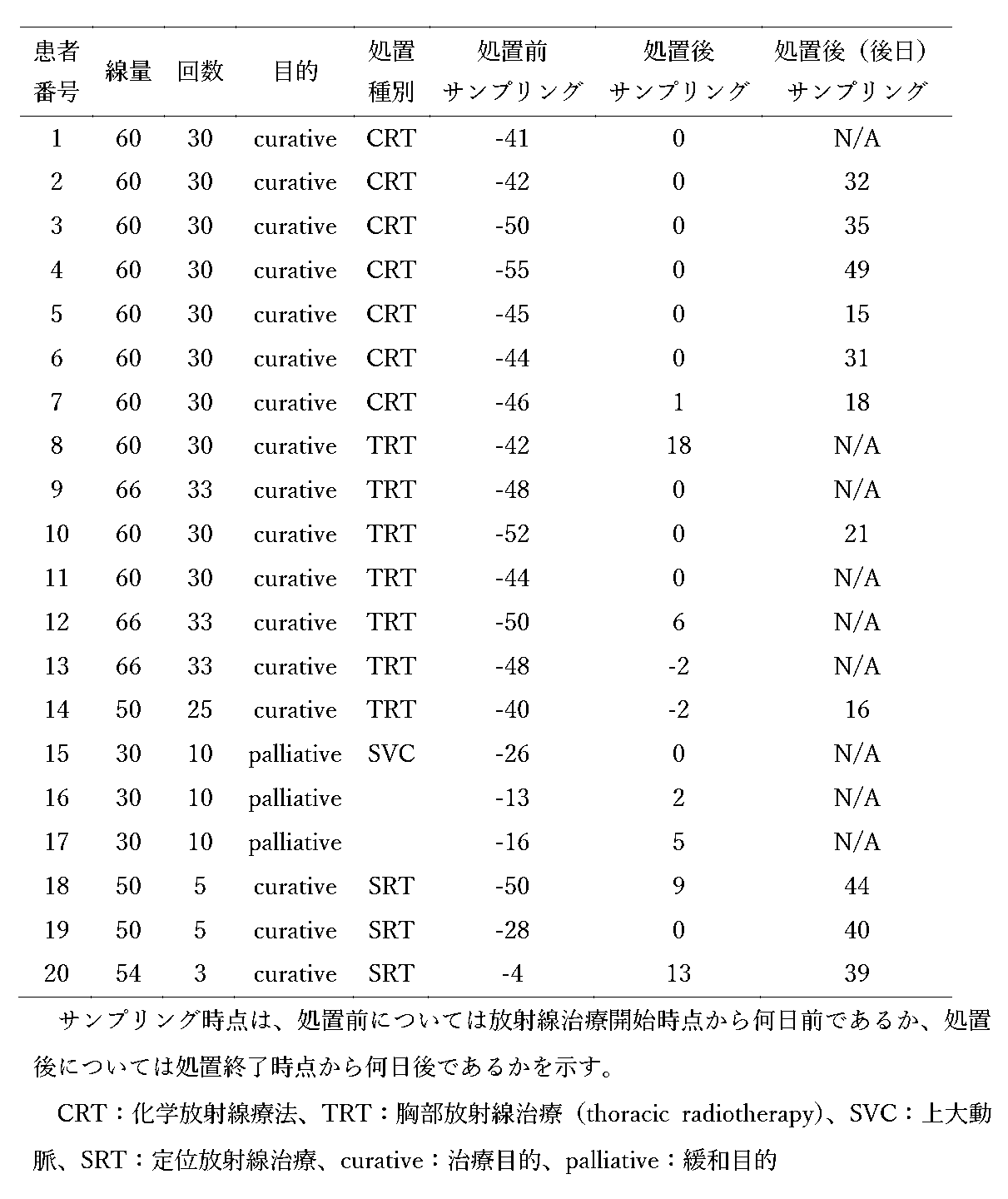
各時点でのサンプリングおよびPBMC解析は以下のとおり行った。PBMC解析には単核球分離用のBDバキュティナCPTTM採血管を用いた。PBMCについてFCM解析を行った。
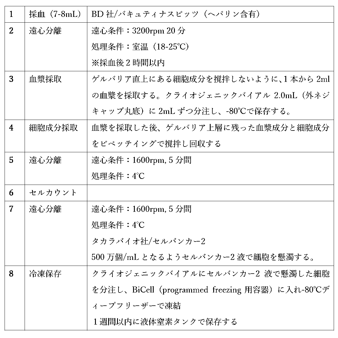
1.Myeloid系細胞解析
解凍後、CD11c、CD141、HLA-DR、CD33、CD14等の抗体染色を行いフローサイトメトリー解析を行った。
2.T細胞解析
1)RPMI1640+10%FCSに懸濁し5%CO2インキュベーター内37℃で36時間程度静置(32-48時間)。
2)T細胞表面マーカー用抗体染色を行いフローサイトメトリー解析を行った。
20名の被験体における放射線治療の前後の各細胞集団の割合について、対応ありのスチューデント両側t検定によるt統計量を算出した。結果は以下の表3に記載する。t統計量0.05以下のものを、太字・下線で示した。
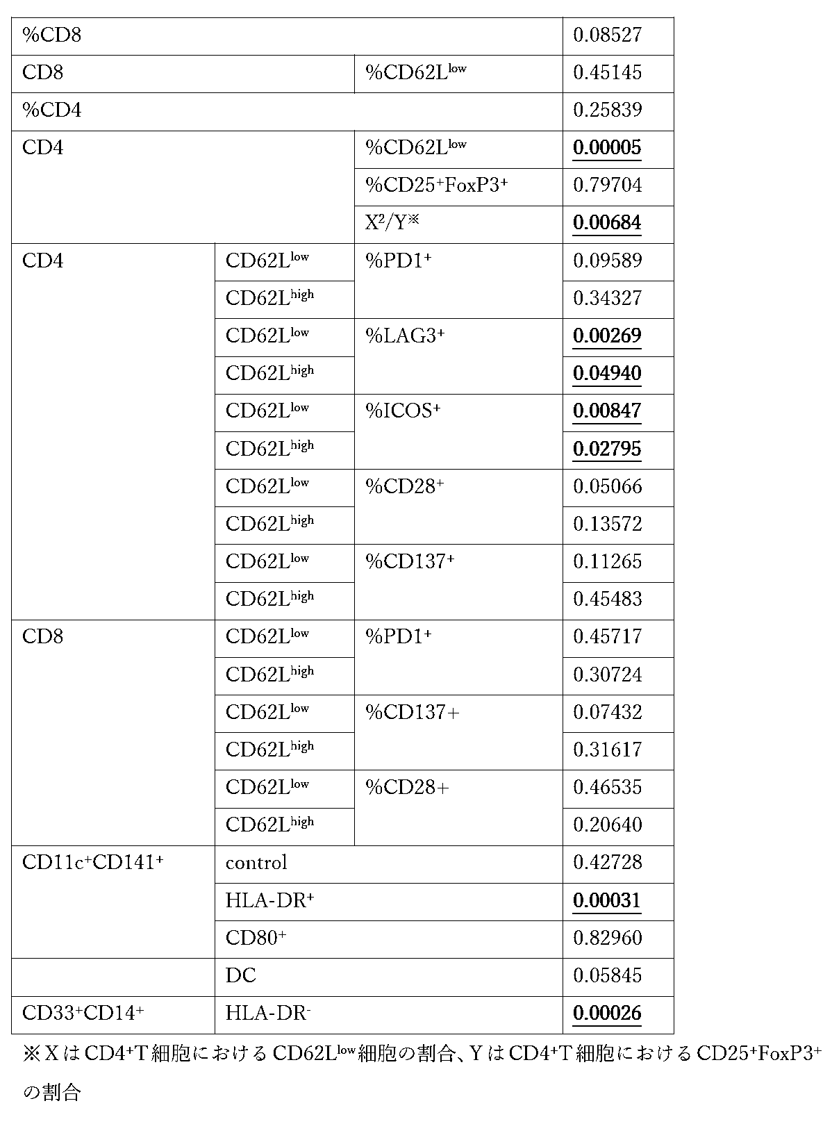

CD62LlowCD4+細胞亜集団が強く正の相関を有するT細胞亜集団は、タイプ1ヘルパーCD4+T細胞(Th1)、エフェクターメモリーCD4+T細胞、CD8+T細胞、エフェクターCD8+T細胞である。これらは、細胞性免疫において殺細胞機能に重要な細胞亜集団である。一方、負の相関を有するのは、タイプ2ヘルパーCD4+T細胞(Th2)、制御性T細胞である。これらは、細胞性免疫を抑制する細胞亜集団として知られている。よって、CD62LlowCD4+細胞亜集団の増加は、抗腫瘍細胞性免疫の活性化を示すと考えられる。また、HLA-DR+CD141+CD11c+樹状細胞亜集団とCD62LlowCD4+細胞亜集団は正の相関関係を有している。これは、活性化された樹状細胞がMHC classII拘束性抗原を表出することで、MHC classII拘束性抗原を認識するCD62LlowCD4+細胞亜集団の増加を来していると考えられ、CD62LlowCD4+細胞亜集団は、腫瘍免疫応答における樹状細胞刺激と相関するCD4+T細胞亜集団であると考えられる。HLA-DR+CD141+CD11c+樹状細胞亜集団は、腫瘍免疫応答における樹状細胞刺激と相関する樹状細胞亜集団であると考えられる。また、LAG3+CD62LlowCD4+T細胞亜集団、ICOS+CD62LlowCD4+T細胞亜集団、CD28+CD62LlowCD4+T細胞亜集団、PD-1+CD62LlowCD4+T細胞亜集団、CD137+CD62LlowCD4+T細胞亜集団、PD-1+CD62LlowCD8+T細胞亜集団、CD137+CD62LlowCD8+T細胞亜集団も、腫瘍免疫応答における樹状細胞刺激と相関するT細胞亜集団であると考えられる(WO2018/147291)。
末梢血中のCD4+CD62LlowT細胞亜集団割合が一定以上の被験体に対し、免疫チェックポイント阻害剤を投与する。奏効集団において、免疫チェックポイント阻害剤の投与後にCD4+CD62LlowT細胞亜集団割合が低下することがある。被験体におけるCD4+CD62LlowT細胞亜集団割合をモニターすることで、被験体の免疫チェックポイント阻害剤に対する応答性をモニターする。CD4+CD62LlowT細胞亜集団の割合が低下した場合に、被験体に放射線治療を施す。放射線治療によって、CD4+CD62LlowT細胞亜集団の割合が増加し、被験体の免疫チェックポイント阻害剤に対する応答性が回復する。
stageI~IVの肺癌患者42名から得た、84検体(20名の手術前後、22名のEGFR-TKI前後)をFortessa解析し、CD4+T細胞中のCD62LlowCD4+T細胞の割合と、CD4+T細胞中のエフェクターメモリー細胞(CCR7-CD45RA-)CD4+T細胞の割合との関係を解析した。
(概要)
実施例1における検討に加えて、異なる患者集団における特定の細胞亜集団の組成が、放射線治療前後で被験体において有意に変化しているパラメータ(すなわち、バイオマーカー)であるかどうかを検討した。
[被験体]
本実施例は、埼玉医科大学国際医療センターで、治療目的化学放射線療法(CRT)または胸部放射線療法を受けた56人の連続局所進行NSCLC患者を対象として行われた。22人の患者は、CRT後にデュルバルマブの併用療法を受けた。末梢血サンプルは、放射線治療の前後と、場合により放射線治療の約1か月後に採取した。PBMCをLSR FortessaTMで分析した。各時点でのサンプリングは実施例1と同様の手順に従った。
対象とした患者および施された処置の内容については以下のとおりである。

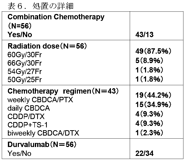
結果は図7~11に示される。図7~11における「before RT」は、図1~6における「pre-TRT」と対応し、放射線治療(RT)の直前を示す。図7~11における「post-RT」、図1~6の「post-TRT」、「at TRT completion」、「TRT complete」は、いずれも放射線治療(RT)終了直後である。
放射線治療の際には、放射線治療で免疫が活性化されて免疫チェックポイント阻害が効きやすい状態になり、時間の経過とともに、元の状態に戻る。免疫が活性化された状態においては、特定の細胞集団(CD62LlowCD4+など)に分化する細胞が増え、未分化(ナイーブ)の細胞は減少し、刺激、活性化された樹状細胞(HLA-DRhighCD11c+CD123-mDCなど)が増え、CD8陽性の細胞集団における若いEMが増加する。これらのいずれかの細胞集団の変化を見ると、放射線治療後に免疫チェックポイント阻害が効きやすい状態かどうかを判定することができる。加えて、この効きやすい状態は時間の経過とともに自然と元に戻ることから、再度放射線治療を行って、再度免疫チェックポイント阻害が効きやすい状態にする必要があるか否かも、判定することができる。
放射線治療では、放射線による局所の免疫原性癌細胞死が生じ、樹状細胞が活性化され、エフェクターT細胞が増殖する
化学療法剤を用いる抗癌剤治療では、化学療法剤による免疫原性癌細胞死が生じ、樹状細胞が活性化するが、同時に化学療法剤及び制吐剤としてのステロイドによる樹状細胞減少効果が生じ、樹状細胞活性化によるエフェクターT細胞増殖が生じても、細胞周期に入ったT細胞は化学療法剤により細胞傷害を受けて死滅(EM分画減少効果)し、増殖能を既に失っているEMRA集団は抗原提示を受けても細胞周期に入らず細胞傷害を受けず、抗原刺激による生存時間延長効果を受ける(EMRA分画の一時的増加効果)
といった機序が想定される。
以上のように、本発明の好ましい実施形態を用いて本発明を例示してきたが、本発明は、この実施形態に限定して解釈されるべきものではない。本発明は、特許請求の範囲によってのみその範囲が解釈されるべきであることが理解される。当業者は、本発明の具体的な好ましい実施形態の記載から、本発明の記載および技術常識に基づいて等価な範囲を実施することができることが理解される。本明細書において引用した特許、特許出願および文献は、その内容自体が具体的に本明細書に記載されているのと同様にその内容が本明細書に対する参考として援用されるべきであることが理解される。本願は、日本国特許出願第2019-028502号(2019年2月20日出願)および同第2019-175852号(2019年9月26日出願)に対して優先権を主張するものであり、その内容の全体は、本願において参考として援用される。
Claims (27)
- 放射線治療を受けた被験体から得られたサンプルにおける細胞亜集団の組成を、該被験体における放射線治療による免疫活性化の指標とする方法であって、
該被験体から得られた該サンプルにおける細胞亜集団の組成を分析する工程を含み、
該サンプルにおける抗腫瘍免疫応答における樹状細胞刺激と相関するCD4+T細胞亜集団の量と基準との比較により、該被験体における免疫活性化の有無が示される、方法。 - 放射線治療を受けた被験体から得られたサンプルにおける細胞亜集団の組成を、該被験体における放射線治療による免疫活性化の指標とする方法であって、
該被験体から得られた該サンプルにおける細胞亜集団の組成を分析する工程を含み、
該サンプルにおける抗腫瘍免疫応答での樹状細胞刺激と相関する樹状細胞亜集団の量と基準との比較により、該被験体における免疫活性化の有無が示される、方法。 - 放射線治療を受けた被験体から得られたサンプルにおける細胞亜集団の組成を、該被験体における放射線治療による免疫活性化の指標とする方法であって、
該被験体から得られた該サンプルにおける細胞亜集団の組成を分析する工程を含み、
該サンプルにおける抗腫瘍免疫応答での樹状細胞刺激と相関するCD8+T細胞亜集団の量と基準との比較により、該被験体における免疫活性化の有無が示される、方法。 - 前記サンプルにおける抗腫瘍免疫応答における樹状細胞刺激と相関するCD4+T細胞亜集団の量、抗腫瘍免疫応答での樹状細胞刺激と相関する樹状細胞亜集団の量および抗腫瘍免疫応答での樹状細胞刺激と相関するCD8+T細胞亜集団の量からなる群から選択される少なくとも2つの量と基準との比較により該被験体における免疫活性化の有無が示される、請求項1~3のいずれか1項に記載の方法。
- 前記抗腫瘍免疫応答での樹状細胞刺激と相関するCD4+T細胞亜集団が、CD62LlowCD4+T細胞集団に含まれる細胞亜集団である、請求項1または4に記載の方法。
- 前記抗腫瘍免疫応答での樹状細胞刺激と相関するCD4+T細胞亜集団が、CD62LlowCD4+T細胞亜集団またはCCR7+CD45RA-CD62LlowCD4+T細胞亜集団である、請求項5に記載の方法。
- 前記抗腫瘍免疫応答での樹状細胞刺激と相関するCD4+T細胞亜集団が、ICOS+CD62LlowCD4+T細胞亜集団、LAG3+CD62LlowCD4+T細胞亜集団、PD-1+CD62LlowCD4+T細胞亜集団またはCD28+CD62LlowCD4+T細胞亜集団である、請求項5に記載の方法。
- 前記抗腫瘍免疫応答での樹状細胞刺激と相関する樹状細胞亜集団が、HLA-DR+CD141+CD11c+細胞亜集団である、請求項2または4に記載の方法。
- 前記抗腫瘍免疫応答での樹状細胞刺激と相関するCD8+T細胞亜集団が、CD62LlowCD8+T細胞集団に含まれる細胞亜集団である、請求項3または4に記載の方法。
- 前記抗腫瘍免疫応答での樹状細胞刺激と相関するCD8+T細胞亜集団が、CD137+CD62LlowCD8+T細胞亜集団である、請求項9に記載の方法。
- 前記サンプルが、末梢血サンプルである、請求項1~10のいずれか1項に記載の方法。
- 前記基準が、前記放射線治療前の前記被験体のサンプルにおける前記細胞亜集団の量である、請求項1~11のいずれか1項に記載の方法。
- 前記サンプルにおける前記細胞亜集団の量が、前記基準より増加していることは、前記被験体において放射線治療による免疫活性化が生じていることを示す、請求項1~12のいずれか1項に記載の方法。
- 前記サンプルにおける前記細胞亜集団の量が、前記基準より増加していないことは、前記被験体において放射線治療による免疫活性化が生じていないことを示す、請求項1~13のいずれか1項に記載の方法。
- 前記放射線治療が、腫瘍を含む照射範囲になされているものである、請求項1~14のいずれか1項に記載の方法。
- 前記被験体において放射線治療による免疫活性化が生じていることにより、その時点で該被験体に免疫チェックポイント阻害剤を含むがん免疫療法を施すべきことがさらに示される、請求項1~15のいずれか1項に記載の方法。
- 前記被験体において放射線治療による免疫活性化が生じていないことにより、該被験体に再度放射線治療を施すべきことがさらに示される、請求項1~15のいずれか1項に記載の方法。
- 被験体から複数の時点で得られたサンプルにおける細胞亜集団の組成を、該被験体における放射線治療による免疫活性化をモニタリングするための指標とする方法としてさらに規定される、請求項1~17のいずれか1項に記載の方法であって、該被験体から複数の時点で得られたサンプルにおける細胞亜集団の組成を分析する工程を含む、方法。
- 被験体におけるがんを処置するための免疫チェックポイント阻害剤を含む組成物であって、
該組成物は、放射線治療を受けた被験体であって、請求項1~18のいずれか1項に記載の方法によって該被験体において免疫活性化が生じていることが示されている被験体に投与されることを特徴とする、組成物。 - 前記免疫チェックポイント阻害剤は、PD-1阻害剤および/またはPD-L1阻害剤である、請求項19に記載の組成物。
- 1または複数のさらなる薬剤と組み合わせて投与されることをさらに特徴とする、請求項19または20に記載の組成物。
- 前記さらなる薬剤が、第2の免疫チェックポイント阻害剤を含む、請求項21に記載の組成物。
- 請求項19~22のいずれか1項に記載の組成物と、前記組成物が放射線治療と併用されることが記載された添付文書と、を含む製品。
- 被験体において放射線治療による免疫活性化が生じているかを判定するためのキットであって、
(A)CD4およびCD62L;
(B)(i)ICOS、PD-1、LAG-3およびCD28から選択されるマーカー、(ii)CD4、ならびに(iii)CD62L;
(C)CD11c、CD141およびHLA-DR;
(D)CD11c、CD123およびHLA-DR;あるいは
(E)CD8、CD62LおよびCD137
に対する検出剤を含む、キット。 - がんを処置するための免疫チェックポイント阻害剤を含む組成物であって、放射線治療を受けた被験体に対して投与されることを特徴とし、該被験体は、放射線治療を受ける前に、がん免疫療法に対して応答性ではない免疫細胞組成を有していた、組成物。
- 前記がん免疫療法に対して応答性ではない免疫細胞組成は、閾値よりも低いCD4+CD62Llow細胞亜集団の量または割合である、請求項25に記載の組成物。
- 前記免疫チェックポイント阻害剤が、抗PD-1抗体および/または抗PD-L1抗体を含む、請求項26に記載の組成物。
Priority Applications (4)
| Application Number | Priority Date | Filing Date | Title |
|---|---|---|---|
| US17/432,744 US12442820B2 (en) | 2019-02-20 | 2020-02-19 | Peripheral blood biomarker for evaluating anti-tumor immune effect of radiation therapy |
| JP2021502106A JP7606225B2 (ja) | 2019-02-20 | 2020-02-19 | 放射線治療による抗腫瘍免疫効果を評価する末梢血バイオマーカー |
| EP20759107.4A EP3928792A4 (en) | 2019-02-20 | 2020-02-19 | BIOMARKERS IN PERIPHERAL BLOOD FOR ASSESSING THE ANTI-TUMORS IMMUNE EFFECT OF RADIATION THERAPY |
| CN202080029760.9A CN113747919A (zh) | 2019-02-20 | 2020-02-19 | 评估由放射治疗获得的抗肿瘤免疫效果的外周血生物标志物 |
Applications Claiming Priority (4)
| Application Number | Priority Date | Filing Date | Title |
|---|---|---|---|
| JP2019-028502 | 2019-02-20 | ||
| JP2019028502 | 2019-02-20 | ||
| JP2019175852 | 2019-09-26 | ||
| JP2019-175852 | 2019-09-26 |
Publications (1)
| Publication Number | Publication Date |
|---|---|
| WO2020171138A1 true WO2020171138A1 (ja) | 2020-08-27 |
Family
ID=72143423
Family Applications (1)
| Application Number | Title | Priority Date | Filing Date |
|---|---|---|---|
| PCT/JP2020/006616 Ceased WO2020171138A1 (ja) | 2019-02-20 | 2020-02-19 | 放射線治療による抗腫瘍免疫効果を評価する末梢血バイオマーカー |
Country Status (6)
| Country | Link |
|---|---|
| US (1) | US12442820B2 (ja) |
| EP (1) | EP3928792A4 (ja) |
| JP (1) | JP7606225B2 (ja) |
| CN (1) | CN113747919A (ja) |
| TW (1) | TW202035702A (ja) |
| WO (1) | WO2020171138A1 (ja) |
Cited By (2)
| Publication number | Priority date | Publication date | Assignee | Title |
|---|---|---|---|---|
| WO2021132636A1 (ja) * | 2019-12-27 | 2021-07-01 | 特定非営利活動法人North East Japan Study Group | がん治療方法及び医薬 |
| WO2022054796A1 (ja) * | 2020-09-08 | 2022-03-17 | 学校法人 埼玉医科大学 | 癌治療に対する応答を予測するためのバイオマーカー |
Citations (4)
| Publication number | Priority date | Publication date | Assignee | Title |
|---|---|---|---|---|
| WO2018147291A1 (en) | 2017-02-07 | 2018-08-16 | Saitama Medical University | Immunological biomarker for predicting clinical effect of cancer immunotherapy |
| JP2018532414A (ja) * | 2015-10-28 | 2018-11-08 | ライフ テクノロジーズ エーエス | 細胞表面シグナル及びシグナル比を変えることによる、異なるt細胞亜集団の選択的増殖の方法 |
| JP2019028502A (ja) | 2017-07-25 | 2019-02-21 | ファナック株式会社 | 交換回転位置決定装置および交換回転位置決定方法 |
| JP2019175852A (ja) | 2017-01-06 | 2019-10-10 | 日立化成株式会社 | リチウムイオン二次電池用負極材、リチウムイオン二次電池用負極及びリチウムイオン二次電池 |
Family Cites Families (23)
| Publication number | Priority date | Publication date | Assignee | Title |
|---|---|---|---|---|
| US5827642A (en) | 1994-08-31 | 1998-10-27 | Fred Hutchinson Cancer Research Center | Rapid expansion method ("REM") for in vitro propagation of T lymphocytes |
| JP2006280307A (ja) | 2005-04-01 | 2006-10-19 | Kyoto Univ | 制御性t細胞の製造方法 |
| BRPI0620187A2 (pt) | 2005-12-21 | 2011-11-01 | Sentoclone Ab | método para a expansão de linfócitos t auxiliares cd4+ e/ou linfócitos t cd8+ reativos a tumor, linfócito-t reativo a tumor, composição farmacêutica, métodos para tratar um indivìduo sofrendo de uma doença de origem neoplástica e para realizar a regressão de tumor em um indivìduo, uso de linfócitos-t reativos a tumor, e, kit |
| CN101801462A (zh) | 2007-07-13 | 2010-08-11 | 健泰科生物技术公司 | 用于癌症、炎性病症和自身免疫性病症的治疗和诊断 |
| EP3375868A1 (en) * | 2007-11-08 | 2018-09-19 | Dana Farber Cancer Institute, Inc. | Stimulation of anti-tumor immunity using dendritic cell/tumor cell fusions and anti-cd3/cd28 |
| EP2073009A1 (en) | 2007-12-19 | 2009-06-24 | Cell Med Research GMBH | Method for T, NK and NKT cells |
| US9176122B2 (en) | 2008-03-24 | 2015-11-03 | University Of South Florida | Biomarkers for predicting response to immunosuppressive therapy |
| US20120088678A1 (en) | 2010-09-08 | 2012-04-12 | Sanford-Burnham Medical Research Institute | Method for prediction of response to rheumatoid arthritis therapeutics |
| US9910039B2 (en) * | 2011-07-01 | 2018-03-06 | Beckman Coulter, Inc. | Regulatory T cells and methods of identifying, obtaining and using to treat immuno-based disorders |
| JP2015529825A (ja) | 2012-09-04 | 2015-10-08 | 国立大学法人 新潟大学 | 癌幹細胞に発現する分子をターゲットとした癌を診断、治療する方法 |
| JP6368308B2 (ja) | 2012-09-07 | 2018-08-01 | トラスティーズ・オブ・ダートマス・カレッジ | 癌の診断および治療のためのvista調節剤 |
| US20140288149A1 (en) | 2013-03-15 | 2014-09-25 | Graham Lord | Mir-142 and antagonists thereof for treating disease |
| KR20160093012A (ko) | 2013-11-05 | 2016-08-05 | 코그네이트 바이오서비시즈, 인코포레이티드 | 암 치료를 위한 체크포인트 억제제 및 치료제의 배합물 |
| CA2961950A1 (en) | 2014-09-28 | 2016-03-31 | The Regents Of The University Of California | Modulation of stimulatory and non-stimulatory myeloid cells |
| AU2015327868A1 (en) | 2014-10-03 | 2017-04-20 | Novartis Ag | Combination therapies |
| CA2966132A1 (en) | 2014-10-23 | 2016-04-28 | Institut Gustave Roussy | Methods and products for modulating microbiota composition for improving the efficacy of a cancer treatment with an immune checkpoint blocker |
| US10702526B2 (en) | 2015-04-20 | 2020-07-07 | Effector Therapeutics Inc. | Inhibitors of immune checkpoint modulators and related methods |
| JP2018519327A (ja) | 2015-06-29 | 2018-07-19 | ベラステム・インコーポレーテッドVerastem,Inc. | 治療用組成物、組合せ物および使用の方法 |
| WO2017032867A1 (en) | 2015-08-27 | 2017-03-02 | INSERM (Institut National de la Santé et de la Recherche Médicale) | Methods for predicting the survival time of patients suffering from a lung cancer |
| EP3417293A1 (en) | 2016-02-18 | 2018-12-26 | Institut Gustave Roussy | Methods and kits for predicting the sensitivity of a subject to immunotherapy |
| WO2018140644A1 (en) * | 2017-01-25 | 2018-08-02 | Musc Foundation For Research Development | Modified t cells and uses thereof |
| JP7162600B2 (ja) | 2017-02-06 | 2022-10-28 | ノヴァルティス アーゲー | 免疫療法に対する応答を予測する方法 |
| WO2018212237A1 (ja) | 2017-05-16 | 2018-11-22 | 学校法人 久留米大学 | テーラーメイド型ペプチドワクチン剤に対する脳腫瘍患者の適格性を判定する方法 |
-
2020
- 2020-02-19 EP EP20759107.4A patent/EP3928792A4/en active Pending
- 2020-02-19 US US17/432,744 patent/US12442820B2/en active Active
- 2020-02-19 JP JP2021502106A patent/JP7606225B2/ja active Active
- 2020-02-19 CN CN202080029760.9A patent/CN113747919A/zh active Pending
- 2020-02-19 TW TW109105342A patent/TW202035702A/zh unknown
- 2020-02-19 WO PCT/JP2020/006616 patent/WO2020171138A1/ja not_active Ceased
Patent Citations (4)
| Publication number | Priority date | Publication date | Assignee | Title |
|---|---|---|---|---|
| JP2018532414A (ja) * | 2015-10-28 | 2018-11-08 | ライフ テクノロジーズ エーエス | 細胞表面シグナル及びシグナル比を変えることによる、異なるt細胞亜集団の選択的増殖の方法 |
| JP2019175852A (ja) | 2017-01-06 | 2019-10-10 | 日立化成株式会社 | リチウムイオン二次電池用負極材、リチウムイオン二次電池用負極及びリチウムイオン二次電池 |
| WO2018147291A1 (en) | 2017-02-07 | 2018-08-16 | Saitama Medical University | Immunological biomarker for predicting clinical effect of cancer immunotherapy |
| JP2019028502A (ja) | 2017-07-25 | 2019-02-21 | ファナック株式会社 | 交換回転位置決定装置および交換回転位置決定方法 |
Non-Patent Citations (7)
| Title |
|---|
| JM, QIAN ET AL.: "Timing and type of immune checkpoint therapy affect the early radiographic response of melanoma brain metastases to stereotactic radiosurgery", CANCER, vol. 122, no. 19, 10 June 2016 (2016-06-10), pages 3051 - 3058, XP002962301, Retrieved from the Internet <URL:https://doi.org/10.1002/cncr.30138> * |
| KENJI MURATA, TOMOHIDE TSUKAHARA, TOSHIHIKO TORIGOE: "Cancer immunotherapy and immunological memory", JAPANESE JOURNAL OF CLINICAL IMMUNOLOGY, vol. 39, no. 1, 2016, pages 18 - 22, XP009523399, DOI: 10.2177/jsci.39.18 * |
| MARCISCANO, AE ET AL.: "Elective Nodal Irradiation Attenuates the Combinatorial Efficacy of Stereotactic Radiation Therapy and Immunotherapy", CLIN CANCER RES., vol. 24, no. 20, 15 October 2018 (2018-10-15), pages 5058 - 5071, XP055735527 * |
| OKAZAKI, T.CHIKUMA, S.IWAI, Y. ET AL.: "A rheostat for immune responses: the unique properties of PD-1 and their advantages for clinical application", NAT. IMMUNOL., vol. 14, 2013, pages 1212 - 1218 |
| See also references of EP3928792A4 |
| SHETAL, AP ET AL.: "Combination Cancer Therapy With Immune Checkpoint Blockade: Mechanisms and Strategies", IMMUNITY, vol. 48, no. 3, 20 March 2018 (2018-03-20), pages 417 - 433, XP055735529 * |
| YOSHIMOTO, YUYA ET AL.: "Special issue, New development of immunotherapy, Combination therapy including immunotherapy, Combination of immunotherapy and radiation therapy", JOURNAL OF MOLECULAR TARGETED THERAPY FOR CANCER, vol. 15, no. 2, 24 July 2017 (2017-07-24), pages 164 - 168, XP009523477, ISSN: 1347-6955 * |
Cited By (4)
| Publication number | Priority date | Publication date | Assignee | Title |
|---|---|---|---|---|
| WO2021132636A1 (ja) * | 2019-12-27 | 2021-07-01 | 特定非営利活動法人North East Japan Study Group | がん治療方法及び医薬 |
| WO2022054796A1 (ja) * | 2020-09-08 | 2022-03-17 | 学校法人 埼玉医科大学 | 癌治療に対する応答を予測するためのバイオマーカー |
| JPWO2022054796A1 (ja) * | 2020-09-08 | 2022-03-17 | ||
| JP7175068B2 (ja) | 2020-09-08 | 2022-11-18 | 学校法人 埼玉医科大学 | 癌治療に対する応答を予測するためのバイオマーカー |
Also Published As
| Publication number | Publication date |
|---|---|
| JP7606225B2 (ja) | 2024-12-25 |
| US20220107324A1 (en) | 2022-04-07 |
| EP3928792A4 (en) | 2022-12-14 |
| US12442820B2 (en) | 2025-10-14 |
| TW202035702A (zh) | 2020-10-01 |
| CN113747919A (zh) | 2021-12-03 |
| EP3928792A1 (en) | 2021-12-29 |
| JPWO2020171138A1 (ja) | 2021-12-23 |
Similar Documents
| Publication | Publication Date | Title |
|---|---|---|
| EP4039260B1 (en) | Cd62llow cd4+ t-cells for treating or preventing cancer | |
| JP7564122B2 (ja) | 光免疫療法のための方法および関連バイオマーカー | |
| US20220273722A1 (en) | Anti-egfr/high affinity nk-cells compositions and methods for chordoma treatment | |
| EP3928793A1 (en) | Method and composition for predicting long-term survival in cancer immunotherapy | |
| CN112512538A (zh) | 改进的靶向t细胞疗法 | |
| AU2017290803A1 (en) | Nant cancer vaccine | |
| CN114173794A (zh) | Pdl1阳性nk细胞癌症治疗 | |
| JP7606225B2 (ja) | 放射線治療による抗腫瘍免疫効果を評価する末梢血バイオマーカー | |
| Masucci et al. | Case report: severe cutaneous adverse event associated with checkpoint inhibition in the setting of CAR T-cell therapy: beyond CRS | |
| Giordano et al. | Potential predictive biomarker for response to radiotherapy and CXCL12-inhibition in glioblastoma in the phase I/II GLORIA trial. | |
| Kobayashi et al. | The effect of radiotherapy on NKT cells in patients with advanced head and neck cancer | |
| WO2023235511A1 (en) | Targeted elimination of senescent cells by gamma-delta t cells | |
| CN116113435A (zh) | 用于预测对癌症治疗的响应的生物标志物 | |
| Vynnyk et al. | Hodgkin's lymphoma: current views on the problem | |
| Rückert | Immunological basis of abscopal antitumor responses induced by combination of distinct radiotherapy fractionation schedules with autologous tumor vaccines and checkpoint inhibition | |
| Grottker | Effect of Radiotherapy and Chemotherapy on the Immune Phenotype of Head and Neck Cancer Cells as Well as the Consecutive Activation on Dendritic Cells | |
| Zirnbauer et al. | Autologous tumor-immune effusion co-cultures enable ex vivo functional profiling of radiotherapy-immunotherapy combinations | |
| Keenan | Identifying the therapeutic potential of novel compounds in the treatment of Glioblastoma Multiforme. | |
| Giamberini | TARGETING DELLE CELLULE DI MIELOMA MULTIPLO CON ACIDO ZOLEDRONICO CONIUGATO AD ANTICORPI ANTI ANTIGENE DI MATURAZIONE DELLE CELLULE B | |
| TW202536418A (zh) | 用於將免疫療法最佳化之因素 | |
| WO2025072692A1 (en) | Compositions for treating hematological conditions and methods of making and using the same | |
| Ramsey | Combining Immunogenic Cell Death, Major Histocompatibility Complex Class I and Copper: Is This The Way Forward? | |
| Rosenbrock | The Effect of Prescription Dose and Dose Fractionation on the Abscopal Effect in a Mouse Model of Non-small Cell Lung Cancer | |
| Phillips | An Investigation Into the Role of T Follicular Helper Cells in Follicular Lymphoma | |
| Pillai | Immune Monitoring by Flow Cytometry in Renal Cell Carcinoma |
Legal Events
| Date | Code | Title | Description |
|---|---|---|---|
| 121 | Ep: the epo has been informed by wipo that ep was designated in this application |
Ref document number: 20759107 Country of ref document: EP Kind code of ref document: A1 |
|
| ENP | Entry into the national phase |
Ref document number: 2021502106 Country of ref document: JP Kind code of ref document: A |
|
| NENP | Non-entry into the national phase |
Ref country code: DE |
|
| ENP | Entry into the national phase |
Ref document number: 2020759107 Country of ref document: EP Effective date: 20210920 |
|
| WWG | Wipo information: grant in national office |
Ref document number: 17432744 Country of ref document: US |