WO2020189794A1 - 創薬研究用培養器具 - Google Patents
創薬研究用培養器具 Download PDFInfo
- Publication number
- WO2020189794A1 WO2020189794A1 PCT/JP2020/012570 JP2020012570W WO2020189794A1 WO 2020189794 A1 WO2020189794 A1 WO 2020189794A1 JP 2020012570 W JP2020012570 W JP 2020012570W WO 2020189794 A1 WO2020189794 A1 WO 2020189794A1
- Authority
- WO
- WIPO (PCT)
- Prior art keywords
- cell
- needle
- storage container
- cells
- cell culture
- Prior art date
- Legal status (The legal status is an assumption and is not a legal conclusion. Google has not performed a legal analysis and makes no representation as to the accuracy of the status listed.)
- Ceased
Links
Images
Classifications
-
- C—CHEMISTRY; METALLURGY
- C12—BIOCHEMISTRY; BEER; SPIRITS; WINE; VINEGAR; MICROBIOLOGY; ENZYMOLOGY; MUTATION OR GENETIC ENGINEERING
- C12M—APPARATUS FOR ENZYMOLOGY OR MICROBIOLOGY; APPARATUS FOR CULTURING MICROORGANISMS FOR PRODUCING BIOMASS, FOR GROWING CELLS OR FOR OBTAINING FERMENTATION OR METABOLIC PRODUCTS, i.e. BIOREACTORS OR FERMENTERS
- C12M25/00—Means for supporting, enclosing or fixing the microorganisms, e.g. immunocoatings
- C12M25/14—Scaffolds; Matrices
-
- C—CHEMISTRY; METALLURGY
- C12—BIOCHEMISTRY; BEER; SPIRITS; WINE; VINEGAR; MICROBIOLOGY; ENZYMOLOGY; MUTATION OR GENETIC ENGINEERING
- C12M—APPARATUS FOR ENZYMOLOGY OR MICROBIOLOGY; APPARATUS FOR CULTURING MICROORGANISMS FOR PRODUCING BIOMASS, FOR GROWING CELLS OR FOR OBTAINING FERMENTATION OR METABOLIC PRODUCTS, i.e. BIOREACTORS OR FERMENTERS
- C12M25/00—Means for supporting, enclosing or fixing the microorganisms, e.g. immunocoatings
-
- B—PERFORMING OPERATIONS; TRANSPORTING
- B01—PHYSICAL OR CHEMICAL PROCESSES OR APPARATUS IN GENERAL
- B01L—CHEMICAL OR PHYSICAL LABORATORY APPARATUS FOR GENERAL USE
- B01L3/00—Containers or dishes for laboratory use, e.g. laboratory glassware; Droppers
- B01L3/50—Containers for the purpose of retaining a material to be analysed, e.g. test tubes
- B01L3/508—Rigid containers without fluid transport within
- B01L3/5082—Test tubes per se
-
- B—PERFORMING OPERATIONS; TRANSPORTING
- B33—ADDITIVE MANUFACTURING TECHNOLOGY
- B33Y—ADDITIVE MANUFACTURING, i.e. MANUFACTURING OF THREE-DIMENSIONAL [3D] OBJECTS BY ADDITIVE DEPOSITION, ADDITIVE AGGLOMERATION OR ADDITIVE LAYERING, e.g. BY 3D PRINTING, STEREOLITHOGRAPHY OR SELECTIVE LASER SINTERING
- B33Y80/00—Products made by additive manufacturing
-
- C—CHEMISTRY; METALLURGY
- C12—BIOCHEMISTRY; BEER; SPIRITS; WINE; VINEGAR; MICROBIOLOGY; ENZYMOLOGY; MUTATION OR GENETIC ENGINEERING
- C12M—APPARATUS FOR ENZYMOLOGY OR MICROBIOLOGY; APPARATUS FOR CULTURING MICROORGANISMS FOR PRODUCING BIOMASS, FOR GROWING CELLS OR FOR OBTAINING FERMENTATION OR METABOLIC PRODUCTS, i.e. BIOREACTORS OR FERMENTERS
- C12M23/00—Constructional details, e.g. recesses, hinges
-
- C—CHEMISTRY; METALLURGY
- C12—BIOCHEMISTRY; BEER; SPIRITS; WINE; VINEGAR; MICROBIOLOGY; ENZYMOLOGY; MUTATION OR GENETIC ENGINEERING
- C12N—MICROORGANISMS OR ENZYMES; COMPOSITIONS THEREOF; PROPAGATING, PRESERVING, OR MAINTAINING MICROORGANISMS; MUTATION OR GENETIC ENGINEERING; CULTURE MEDIA
- C12N5/00—Undifferentiated human, animal or plant cells, e.g. cell lines; Tissues; Cultivation or maintenance thereof; Culture media therefor
- C12N5/06—Animal cells or tissues; Human cells or tissues
- C12N5/0602—Vertebrate cells
- C12N5/067—Hepatocytes
- C12N5/0671—Three-dimensional culture, tissue culture or organ culture; Encapsulated cells
-
- G—PHYSICS
- G01—MEASURING; TESTING
- G01N—INVESTIGATING OR ANALYSING MATERIALS BY DETERMINING THEIR CHEMICAL OR PHYSICAL PROPERTIES
- G01N33/00—Investigating or analysing materials by specific methods not covered by groups G01N1/00 - G01N31/00
- G01N33/48—Biological material, e.g. blood, urine; Haemocytometers
- G01N33/50—Chemical analysis of biological material, e.g. blood, urine; Testing involving biospecific ligand binding methods; Immunological testing
- G01N33/5005—Chemical analysis of biological material, e.g. blood, urine; Testing involving biospecific ligand binding methods; Immunological testing involving human or animal cells
- G01N33/5008—Chemical analysis of biological material, e.g. blood, urine; Testing involving biospecific ligand binding methods; Immunological testing involving human or animal cells for testing or evaluating the effect of chemical or biological compounds, e.g. drugs, cosmetics
- G01N33/5014—Chemical analysis of biological material, e.g. blood, urine; Testing involving biospecific ligand binding methods; Immunological testing involving human or animal cells for testing or evaluating the effect of chemical or biological compounds, e.g. drugs, cosmetics for testing toxicity
-
- G—PHYSICS
- G01—MEASURING; TESTING
- G01N—INVESTIGATING OR ANALYSING MATERIALS BY DETERMINING THEIR CHEMICAL OR PHYSICAL PROPERTIES
- G01N33/00—Investigating or analysing materials by specific methods not covered by groups G01N1/00 - G01N31/00
- G01N33/48—Biological material, e.g. blood, urine; Haemocytometers
- G01N33/50—Chemical analysis of biological material, e.g. blood, urine; Testing involving biospecific ligand binding methods; Immunological testing
- G01N33/5005—Chemical analysis of biological material, e.g. blood, urine; Testing involving biospecific ligand binding methods; Immunological testing involving human or animal cells
- G01N33/5008—Chemical analysis of biological material, e.g. blood, urine; Testing involving biospecific ligand binding methods; Immunological testing involving human or animal cells for testing or evaluating the effect of chemical or biological compounds, e.g. drugs, cosmetics
- G01N33/502—Chemical analysis of biological material, e.g. blood, urine; Testing involving biospecific ligand binding methods; Immunological testing involving human or animal cells for testing or evaluating the effect of chemical or biological compounds, e.g. drugs, cosmetics for testing non-proliferative effects
- G01N33/5038—Chemical analysis of biological material, e.g. blood, urine; Testing involving biospecific ligand binding methods; Immunological testing involving human or animal cells for testing or evaluating the effect of chemical or biological compounds, e.g. drugs, cosmetics for testing non-proliferative effects involving detection of metabolites per se
-
- B—PERFORMING OPERATIONS; TRANSPORTING
- B01—PHYSICAL OR CHEMICAL PROCESSES OR APPARATUS IN GENERAL
- B01L—CHEMICAL OR PHYSICAL LABORATORY APPARATUS FOR GENERAL USE
- B01L2200/00—Solutions for specific problems relating to chemical or physical laboratory apparatus
- B01L2200/06—Fluid handling related problems
- B01L2200/0647—Handling flowable solids, e.g. microscopic beads, cells, particles
- B01L2200/0668—Trapping microscopic beads
-
- B—PERFORMING OPERATIONS; TRANSPORTING
- B01—PHYSICAL OR CHEMICAL PROCESSES OR APPARATUS IN GENERAL
- B01L—CHEMICAL OR PHYSICAL LABORATORY APPARATUS FOR GENERAL USE
- B01L2300/00—Additional constructional details
- B01L2300/08—Geometry, shape and general structure
- B01L2300/0848—Specific forms of parts of containers
- B01L2300/0851—Bottom walls
-
- B—PERFORMING OPERATIONS; TRANSPORTING
- B01—PHYSICAL OR CHEMICAL PROCESSES OR APPARATUS IN GENERAL
- B01L—CHEMICAL OR PHYSICAL LABORATORY APPARATUS FOR GENERAL USE
- B01L2400/00—Moving or stopping fluids
- B01L2400/08—Regulating or influencing the flow resistance
- B01L2400/084—Passive control of flow resistance
- B01L2400/086—Passive control of flow resistance using baffles or other fixed flow obstructions
-
- B—PERFORMING OPERATIONS; TRANSPORTING
- B01—PHYSICAL OR CHEMICAL PROCESSES OR APPARATUS IN GENERAL
- B01L—CHEMICAL OR PHYSICAL LABORATORY APPARATUS FOR GENERAL USE
- B01L3/00—Containers or dishes for laboratory use, e.g. laboratory glassware; Droppers
- B01L3/50—Containers for the purpose of retaining a material to be analysed, e.g. test tubes
- B01L3/508—Rigid containers without fluid transport within
- B01L3/5085—Rigid containers without fluid transport within for multiple samples, e.g. microtitration plates
-
- C—CHEMISTRY; METALLURGY
- C12—BIOCHEMISTRY; BEER; SPIRITS; WINE; VINEGAR; MICROBIOLOGY; ENZYMOLOGY; MUTATION OR GENETIC ENGINEERING
- C12N—MICROORGANISMS OR ENZYMES; COMPOSITIONS THEREOF; PROPAGATING, PRESERVING, OR MAINTAINING MICROORGANISMS; MUTATION OR GENETIC ENGINEERING; CULTURE MEDIA
- C12N2513/00—3D culture
Definitions
- the present invention relates to a cell culture device specialized for drug discovery research.
- Patent Document 1 a technique for producing a three-dimensional structure of cells by temporarily fixing cell aggregates (spheroids) to needles arranged in a sword-shaped shape.
- the feature of this technology is that it can be three-dimensionalized only by cells. It has long been known that spheroid culture has higher cell activity than normal planar culture, but the cell structure three-dimensionalized by the above technique has metabolic activity because the cells are three-dimensional. The efficiency of nutrient gas exchange is high because the cells are in direct contact with the culture medium.
- Non-Patent Document 1 There is also a possibility that a three-dimensional structure constructed by cardiomyocytes can be used as a pulsation analysis tool.
- bio 3D printer a dedicated device (bio 3D printer) is required to pierce Kenzan with spheroids, and the device is expensive and difficult to obtain.
- bio 3D printer In drug discovery research, it is not necessary to use a bio 3D printer that handles a large number of spheroids (the specifications are large) because the pharmacological activity can be sufficiently analyzed if the number of spheroids is nine.
- a screening system capable of large-scale analysis with a small amount of cells is required.
- a screening system that can analyze a small amount of cells on a large scale is required.
- a simple and inexpensive method for producing a cell structure is required for drug discovery tests.
- the present inventor has made the central part of the bottom surface convex, and by using a cell containing container having a concave portion formed between the central part and the side wall, the above-mentioned problem. Succeeded in solving the above, and completed the present invention.
- a cell culture device having a cell storage container and a sword-shaped member in which a needle-shaped body is arranged on a substrate.
- the central portion of the bottom surface of the cell storage container forms a convex portion, a concave portion is formed between the central portion and the side wall, and a through hole through which the needle-shaped body penetrates is provided on the bottom surface of the concave portion.
- the needle-shaped bodies are arranged so as to correspond to the positions of the through holes.
- a cell culture device in which the sword-shaped member is arranged from the bottom surface side or the top surface side of the cell storage container so that a part of the tip end side of the needle-shaped body penetrates the through hole.
- a cell culture device having a cell storage container and a sword-shaped member in which a needle-shaped body is arranged on a substrate.
- the central portion of the bottom surface of the cell storage container forms a convex portion, and a concave portion is formed between the central portion and the side wall.
- the needle-shaped bodies are arranged so as to correspond to the position of the bottom surface of the recess.
- a cell culture device in which the sword-shaped member is arranged from the upper surface side of the cell storage container with the tip of the needle-shaped body facing the bottom surface of the recess.
- a simple and inexpensive cell structure can be produced, which makes it possible to carry out drug discovery tests such as cytotoxicity.
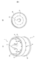
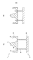
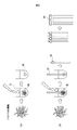
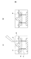
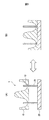
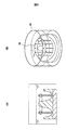
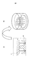
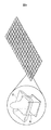
- FIG. 1 It is a figure which shows one aspect of the cell culture apparatus of this invention. It is a central sectional view of the cell culture apparatus of this invention. It is a figure which shows the mode in which a cell forms a spheroid while entraining a spicule. It is a figure which shows the aspect of producing the cell structure using the cell culture apparatus of this invention. It is a figure which shows the mode that the cell containing container was slid in the longitudinal direction of a needle-like body. It is a figure which shows the mode in which the cell structure was held in a ring shape at the tip of a needle-like body.
- the present invention is a cell culture device having a cell storage container and a sword-shaped member in which a needle-shaped body is arranged on a substrate.
- the central portion of the bottom surface of the cell storage container forms a convex portion, a concave portion is formed between the central portion and the side wall, and a through hole through which the needle-shaped body penetrates is provided on the bottom surface of the concave portion.
- the needle-shaped bodies are arranged so as to correspond to the positions of the through holes.
- the sword-shaped member is arranged from the bottom surface side or the top surface side of the cell storage container so that a part of the tip end side of the needle-shaped body penetrates the through hole.
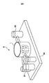
- FIG. 1A is a perspective view of the cell culture device of the present invention.
- the cell culture device 1 includes a cell storage container 10 and a sword-shaped member 2 in which a needle-shaped body 21 is arranged on a substrate 20.
- the center of the bottom surface of the cell storage container 10 forms a convex portion 11, and a concave portion 13 is formed between the cell storage container 10 and the side wall 12.
- FIG. 1B is a plan view of the cell culture device 10 as viewed from above.
- 10 needle-shaped bodies 21 are arranged on the substrate 20 and needle-shaped in the through hole 14 (described later) of the cell storage container 10. It penetrates the body 21 (the arrangement of the needle-shaped body 21 corresponds to the position of the through hole 14).
- FIG. 2 is a central cross-sectional view of the cell culture device 1.
- the bottom surface 13 of the cell storage container 10 is provided with a through hole 14 through which the needle-shaped body 21 of the sword-shaped member 2 penetrates. Therefore, the needle-shaped body 21 is arranged on the substrate 20 in the normal direction of the substrate 20, for example, so as to correspond to the position of the through hole 14.
- FIG. 2B shows a mode in which the sword-shaped member 2 penetrates a part of the needle-shaped body 21 through the through hole 14 from the bottom surface of the cell storage container 10.
- FIG. 3 is a schematic view of a mode in which cells form a spheroid while involving a needle-like body.
- FIG. 3A is a diagram showing an aspect of spheroid formation in the absence of needle-shaped body 21, and the disjointed cells 32 in the cell suspension 31 naturally aggregate to form spheroid 33.
- the cells 32 form the spheroid 33 while involving the needle-shaped body 21. Since a plurality of needle-shaped bodies 21 are arranged at equal intervals, for example, the formed spheroids 33 are fused with each other to produce a cell structure 34.
- the type of cell used in the present invention is not particularly limited, and any cell that forms a spheroid can be used.
- cells forming spheroids include undifferentiated cells such as stem cells (ES cells, cord blood-derived cells, undifferentiated mesenchymal stem cells, adult mesenchymal stem cells, etc.) or differentiated cells thereof.
- Tissues from which these cells are derived include, for example, articular cartilage, bone, adipose tissue, ligaments, tendons, teeth, pinna, nose, liver, pancreas, blood vessels, nerves, heart, etc., among which liver cells and myocardial cells Etc. are preferable.
- spheroids do not necessarily have to be formed as aggregates of a single type of cell, and as long as spheroids are formed, they are derived from multiple types of cells (for example, a mixture of liver cells and vascular endothelial cells). It may be formed.
- the culture period until the formation of spheroids and cell structures varies depending on the size of the cell culture device 1, but is approximately 2 to 4 days under general culture conditions (for example, 37 ° C., 5% CO 2 atmosphere). Is.
- the cell storage container 10 is preferably a cell non-adhesive coating (for example, made of polydimethylsiloxane) with fluorine or the like.
- a cell non-adhesive coating for example, made of polydimethylsiloxane
- fluorine for example, made of polydimethylsiloxane
- those processed with polyhydroxyethyl methacrylate polymer can also be used, and further, acrylic resin, ABS resin, polyester resin, polycarbonate resin, polypropylene, polyethylene, polyacetal, polyether ether.
- acrylic resin, ABS resin, polyester resin, polycarbonate resin, polypropylene, polyethylene, polyacetal, polyether ether Those made of ketone, nylon, etc. can also be used.
- FIG. 4 shows an embodiment in which a cell structure 34 is produced using the cell culture device 1 of the present invention based on the formation mechanism of the cell structure 34 described in FIG. 3B.
- the culture solution is immersed in a container 40 larger than the cell culture device 1 of the present invention, and the cell culture device 1 is placed therein.
- the culture instrument 1 has a mode in which a part of the tip side of the needle-shaped body 21 of the sword-shaped member 2 penetrates the through hole 14 of the cell storage container 10.
- the cell suspension 31 is put into the recess 13 of the cell storage container 10
- the cells form a spheroid while involving the needle-shaped body 21, and the spheroids fuse with each other to form a cell structure 34.
- the shape of the outer circumference of the cell storage container 10 is circular, and the concave portion 13 surrounds the convex portion 11 at the center of the bottom surface in a circular shape. Therefore, the shape of the produced cell structure is ring-shaped.
- the shape of the outer circumference of the cell storage container 10 is not limited to a circle, but may be a polygon such as a quadrangle, a pentagon, a hexagon, or an octagon.
- the number of needle-shaped bodies 21 is not limited, and may be, for example, 2 to 20.
- the diameter thereof and the diameter of the recess 13 are not particularly limited, and may be, for example, 0.6 mm to 30.0 mm.
- FIG. 5 shows a mode in which the cell storage container 10 is slid in the longitudinal direction of the needle-shaped body 21 in the cell culture device 1 of the present invention.
- the cell storage container 10 When culturing cells, as shown in FIG. 5A, the cell storage container 10 is arranged so that a part of the tip side of the needle-shaped body 21 slightly protrudes from the through hole 14.
- the cell storage container 10 is slid downward (on the substrate 20 side) (FIG. 5B). As a result, the cell structure 34 is held in a ring shape at the tip of the needle-shaped body 21 (FIG. 6).
- the installation area of the cell structure 34 in the cell storage container 10 is reduced, and the cell structure 34 can be analyzed immediately.
- the activity and movement of the cell structure 34, or toxicity to the cell structure 34, etc. Can be tested.
- FIGS. 1 and 2 show a mode in which the needle-shaped body 21 of the sword-shaped member 2 penetrates the recess 13 from the bottom surface side of the cell storage container 10.
- FIG. 7 shows the needle-shaped body 21 of the sword-shaped member 2. This is an embodiment in which the tip of the cell is arranged from the upper surface of the cell storage container 10 toward the recess 13. In this case, it is not necessary to provide the through hole 14, but it is optional whether or not the through hole 14 is provided. However, in order to fix the needle-shaped body 21, a hole (not shown) that does not penetrate the recess 13 of the cell storage container 10 may be provided, and the needle-shaped body 21 may be inserted into the hole. As shown in FIG.
- the sword-shaped member 2 is arranged in the direction opposite to that of FIG. 1, and the cell suspension is charged to form the spheroid and the cell structure 34, and then the sword-shaped member 2 is formed.
- the cell structure 34 can be held at the tip of the needle-shaped body 21. In this case, the substrate 20 can be removed.
- FIG. 8 is a diagram showing a mode in which a plurality of cell culture instruments 1 of the present invention are arranged in an array.
- the cell storage container 10 has a rectangular shape, and the needle-shaped body 21 penetrates into the recesses at four places (four corners). In this way, by arranging the plurality of cell culture instruments 1 of the present invention in an array, a large number of cell structures 34 can be produced, and various tests can be performed at once.
- the array-like arrangement can be integrally molded.
- FIG. 9 is a diagram showing a cell test device 9 including the cell culture device 1 of the present invention.
- the cell culture device 1 is placed on the substrate 90, and the cell culture solution supply unit 91 and the subject are covered.
- the inspection material supply unit 92 is installed.
- An electrode (not shown) can also be connected to the needle-shaped body 21 in the cell test apparatus 9. This makes it possible to test the metabolic activity of cell structures and the pulsation of cardiomyocytes in three dimensions. Therefore, the present invention provides a cell test method, which comprises contacting a cell structure with a test substance to test its toxicity to the cell or the metabolic activity of the cell.
- test substances include various naturally or artificially synthesized peptides, proteins (including enzymes and antibodies), nucleic acids (polynucleotides (DNA, RNA), oligonucleotides (siRNA, etc.), peptide nucleic acids (PNA), etc.), Examples thereof include low molecular weight compounds and high molecular weight organic compounds.
- "contact” means to bring the test substance into contact with the cell structure of the present invention, to put the test substance into a culture vessel containing the cell structure, and to put the test substance in a medium containing the test substance. Examples include culturing the structure.
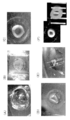
- Method Four needles (0.17 mm) were set up in a hat-shaped container (diameter 4 mm) made of polydimethylsiloxane (PDMS) resin (Fig. 10A), and a cell suspension (number of cells: 5x10, 6 ) was placed (Fig.). 10B). As cells, human fibroblasts were used.
- PDMS polydimethylsiloxane
- FIG. 10C After 3 days, it was shown that the cells aggregated and involved the needle (Fig. 10C). By sliding the hat-shaped container down, the ring-shaped cell structure was held at the tip of the needle (FIGS. 10D, E). In the present invention, it can be used for a pharmacological test in this state, but as shown in FIG. 10F, a cell structure can be pulled out from a needle to perform a pharmacological test, and a pathological tissue can also be prepared. Is.
Landscapes
- Health & Medical Sciences (AREA)
- Engineering & Computer Science (AREA)
- Life Sciences & Earth Sciences (AREA)
- Chemical & Material Sciences (AREA)
- Biomedical Technology (AREA)
- Bioinformatics & Cheminformatics (AREA)
- Organic Chemistry (AREA)
- Zoology (AREA)
- Wood Science & Technology (AREA)
- Immunology (AREA)
- Biotechnology (AREA)
- General Health & Medical Sciences (AREA)
- Biochemistry (AREA)
- Microbiology (AREA)
- Genetics & Genomics (AREA)
- Hematology (AREA)
- General Engineering & Computer Science (AREA)
- Urology & Nephrology (AREA)
- Molecular Biology (AREA)
- Sustainable Development (AREA)
- Toxicology (AREA)
- Cell Biology (AREA)
- Analytical Chemistry (AREA)
- Food Science & Technology (AREA)
- Pathology (AREA)
- General Physics & Mathematics (AREA)
- Tropical Medicine & Parasitology (AREA)
- Physics & Mathematics (AREA)
- Medicinal Chemistry (AREA)
- Clinical Laboratory Science (AREA)
- Manufacturing & Machinery (AREA)
- Gastroenterology & Hepatology (AREA)
- Materials Engineering (AREA)
- Chemical Kinetics & Catalysis (AREA)
- Apparatus Associated With Microorganisms And Enzymes (AREA)
- Measuring Or Testing Involving Enzymes Or Micro-Organisms (AREA)
- Micro-Organisms Or Cultivation Processes Thereof (AREA)
Abstract
Description
以前から、通常の平面培養よりもスフェロイド培養の方が細胞活性が高いことは知られているが、上記技術で立体化された細胞構造体は、細胞が三次元化しているため、代謝活性が高く、また培養液に細胞が直接接しているため栄養ガス交換効率が高い。
創薬研究では、スフェロイドの個数として9個あれば十分に薬理活性を解析することができるため、多数のスフェロイドを扱うバイオ3Dプリンターを使用するまでもない(スペックが大きい)。
他方、創薬を行うには、大量の検体を一気に処理することが必要である。このため、少量の細胞で大規模に解析できるスクリーニング系が求められる。
(1)細胞収容容器と、基板に針状体が配置された剣山状部材とを有する細胞培養器具であって、
前記細胞収容容器の底面中央部は凸部を形成して当該中央部と側壁との間は凹部を形成するとともに、当該凹部の底面に、前記針状体が貫通する貫通孔が設けられ、
前記針状体は、前記貫通孔の位置に対応づけて配列されており、
前記細胞収容容器の底面側又は上面側から、前記針状体の先端側の一部が貫通孔を貫通するように前記剣山状部材を配置させてなる、細胞培養器具。
(2)細胞収容容器と、基板に針状体が配置された剣山状部材とを有する細胞培養器具であって、
前記細胞収容容器の底面中央部は凸部を形成して当該中央部と側壁との間は凹部を形成し、
前記針状体は、前記凹部の底面の位置に対応づけて配列しており、
前記細胞収容容器の上面側から、前記針状体の先端を前記凹部の底面に向けて前記剣山状部材を配置させてなる、細胞培養器具。
(3)前記細胞収容容器及び剣山状部材がアレイ状に複数配列された、(1)又は(2)に記載の細胞培養器具。
(4)細胞収容容器が、細胞非接着コーティング処理されたものである、(1)~(3)のいずれか1項に記載の細胞培養器具。
(5) (1)~(4)のいずれか1項に記載の細胞培養器具の細胞収容容器に細胞懸濁液を投入した後、細胞が針状体を巻き込むように細胞を凝集させることを特徴とする、細胞構造体の製造方法。
(6)細胞が肝細胞又は心筋細胞である、(5)に記載の方法。
(7) (5)又は(6)に記載の方法により製造された細胞構造体に被検物質を接触させ、当該細胞に対する毒性又は当該細胞の代謝活性を試験することを特徴とする、細胞試験方法。
(8)細胞が肝細胞又は心筋細胞である(7)に記載の方法。
(9)(1)~(4)のいずれか1項に記載の細胞培養器具を含む、細胞試験装置。
(10)細胞が肝細胞又は心筋細胞である(9)に記載の装置。
前記細胞収容容器の底面中央部は凸部を形成して当該中央部と側壁との間は凹部を形成するとともに、当該凹部の底面に、前記針状体が貫通する貫通孔が設けられ、
前記針状体は、前記貫通孔の位置に対応づけて配列されており、
前記細胞収容容器の底面側又は上面側から、前記針状体の先端側の一部が貫通孔を貫通するように前記剣山状部材を配置させてなる、細胞培養器具を提供する。
図1Aは、本発明の細胞培養器具の斜視図であり、細胞培養器具1は、細胞収容容器10と、基板20に針状体21が配置された剣山状部材2とを備える。
細胞収容容器10の底面の中央は凸部11を形成し、側壁12との間で凹部13を形成する。底面中央部が凸部となり、側壁との間に凹部を形成する形状はいわゆるメキシカンハットに似ていることから、本明細書では、細胞収容容器10を「メキシカンハット型培養容器」又は単に「ハット型培養容器」ともいう。
図1Bは、細胞培養器具10を上から見た平面図であり、この態様では、基板20に針状体21を10本配置して、細胞収容容器10の貫通孔14(後述)に針状体21を貫通させている(針状体21の配置は、貫通孔14の位置に対応している)。
図3は、細胞が針状体を巻き込みながらスフェロイドを形成する態様の模式図である。
また、スフェロイド及び細胞構造体を形成するまでの培養期間は、細胞培養器具1の大きさにより異なるが、一般的培養条件(例えば37℃、5%CO2雰囲気下)で概ね2日~4日である。
また、細胞収容容器10は、フッ素等により細胞非接着コーティングされたもの(例えばポリジメチルシロキサン製)であることが好ましい。但し、フッ素加工されたもののほか、ポリヒドロキシエチルメタクリレートポリマー加工されたものを使用することもでき、さらには、アクリル樹脂、ABS樹脂、ポリエステル系樹脂、ポリカーボネート樹脂、ポリプロピレン、ポリエチレン、ポリアセタール、ポリエーテルエーテルケトン、ナイロン製のもの等を用いることもできる。
まず、本発明の細胞培養器具1よりも大きな容器40に培養液を浸し、この中に細胞培養器具1を入れる。培養器具1は、剣山状部材2の針状体21の先端側の一部が細胞収容容器10の貫通孔14を貫通している態様となっている。細胞収容容器10の凹部13に細胞懸濁液31を投入すると、細胞が針状体21を巻き込みながらスフェロイドを形成するとともに、スフェロイド同士が融合して細胞構造体34を形成する。図1や図4では、細胞収容容器10の外周の形状は円形であり、凹部13は、底面中央部の凸部11の周囲を円形状に取り囲んでいる。このため、製造される細胞構造体の形状はリング状となる。
細胞を培養するときは、図5Aに示すように、針状体21の先端側の一部が貫通孔14をわずかに突出するように細胞収容容器10を配置しておく。細胞構造体34が構築された後は、細胞収容容器10を下方(基板20側)にスライドする(図5B)。これにより、細胞構造体34は、針状体21の先端部にリング状になって保持される(図6)。このように、細胞収容容器10を下方にスライドすることで、細胞構造体34の細胞収容容器10への設置面積が少なくなり、細胞構造体34に対して解析を即時に行うことができる。例えば図6に示す細胞構造体34の状態で、細胞収容容器10及び/又は培養容器40内に被検物質を投入した後、細胞構造体34の活性や動き、あるいは細胞構造体34に対する毒性などを試験することができる。
図8では、細胞収容容器10の形状は方形としてあり、4か所(四隅)の凹部に針状体21が貫通している。このように、本発明の複数の細胞培養器具1をアレイ状の配置とすることにより、細胞構造体34を多数製造することができ、各種試験を一度に行うことができる。なお、アレイ状の配置は、一体成型することが可能である。
これにより、細胞構造体の代謝活性や、心筋細胞の拍動などを三次元で試験することができる。
従って、本発明は、細胞構造体に被検物質を接触させ、当該細胞に対する毒性又は当該細胞の代謝活性を試験することを特徴とする、細胞試験方法を提供する。
また「接触」とは、本発明の細胞構造体に被検物質を接触させることを意味し、細胞構造体を含む培養容器中に被検物質を投入すること、被検物質を含む培地で細胞構造体を培養することなどが挙げられる。
以下、実施例により本発明をさらに具体的に説明する。但し、本発明の範囲はこれらの実施例により限定されるものではない。
[実施例1]
ポリジメチルシロキサン(PDMS)樹脂で作製したハット型容器(直径4mm)に針(0.17mm)を4本立て(図10A)、細胞懸濁液(細胞数:5x10⁶個)を投入した(図10B)。細胞は、ヒト線維芽細胞を用いた。
ハット型容器を下にスライドすることにより、リング状の細胞構造体が針の先端部に保持された(図10D、E)。本発明においては、この状態で薬理試験に使用することもできるが、図10Fに示すように、細胞構造体を針から抜いて薬理試験を行うこともでき、また病理組織を作製することも可能である。
[符号の説明]
1:本発明の細胞培養器具、2:剣山状部材、9:細胞試験装置
10:細胞収容容器、11:凸部、12:側壁、13:凹部、14:貫通孔
20:基板、21針状体、
31:細胞懸濁液、32:細胞、33:スフェロイド、34:細胞構造体
40:培養容器
90:基板、91:細胞培養液供給部、92:被検物質供給部
Claims (10)
- 細胞収容容器と、基板に針状体が配置された剣山状部材とを有する細胞培養器具であって、
前記細胞収容容器の底面中央部は凸部を形成して当該中央部と側壁との間は凹部を形成するとともに、当該凹部の底面に、前記針状体が貫通する貫通孔が設けられ、
前記針状体は、前記貫通孔の位置に対応づけて配列されており、
前記細胞収容容器の底面側又は上面側から、前記針状体の先端側の一部が貫通孔を貫通するように前記剣山状部材を配置させてなる、細胞培養器具。 - 細胞収容容器と、基板に針状体が配置された剣山状部材とを有する細胞培養器具であって、
前記細胞収容容器の底面中央部は凸部を形成して当該中央部と側壁との間は凹部を形成し、
前記針状体は、前記凹部の底面の位置に対応づけて配列しており、
前記細胞収容容器の上面側から、前記針状体の先端を前記凹部の底面に向けて前記剣山状部材を配置させてなる、細胞培養器具。 - 前記細胞収容容器及び剣山状部材がアレイ状に複数配列された、請求項1又は2に記載の細胞培養器具。
- 細胞収容容器が、細胞非接着コーティング処理されたものである、請求項1~3のいずれか1項に記載の細胞培養器具。
- 請求項1~4のいずれか1項に記載の細胞培養器具の細胞収容容器に細胞懸濁液を投入した後、細胞が針状体を巻き込むように細胞を凝集させることを特徴とする、細胞構造体の製造方法。
- 細胞が肝細胞又は心筋細胞である、請求項5に記載の方法。
- 請求項5又は6に記載の方法により製造された細胞構造体に被検物質を接触させ、当該細胞に対する毒性又は当該細胞の代謝活性を試験することを特徴とする、細胞試験方法。
- 細胞が肝細胞又は心筋細胞である請求項7に記載の方法。
- 請求項1~4のいずれか1項に記載の細胞培養器具を含む、細胞試験装置。
- 細胞が肝細胞又は心筋細胞である請求項9に記載の装置。
Priority Applications (4)
| Application Number | Priority Date | Filing Date | Title |
|---|---|---|---|
| EP20773449.2A EP3940054A4 (en) | 2019-03-15 | 2020-03-13 | CULTURE DEVICE FOR USE IN DRUG DEVELOPMENT STUDIES |
| CN202080020829.1A CN113574163B (zh) | 2019-03-15 | 2020-03-13 | 用于药物开发研究的培养器具 |
| JP2021507437A JP7227653B2 (ja) | 2019-03-15 | 2020-03-13 | 創薬研究用培養器具 |
| US17/438,614 US12378512B2 (en) | 2019-03-15 | 2020-03-13 | Culture apparatus for drug discovery research |
Applications Claiming Priority (2)
| Application Number | Priority Date | Filing Date | Title |
|---|---|---|---|
| JP2019048228 | 2019-03-15 | ||
| JP2019-048228 | 2019-03-15 |
Publications (1)
| Publication Number | Publication Date |
|---|---|
| WO2020189794A1 true WO2020189794A1 (ja) | 2020-09-24 |
Family
ID=72520943
Family Applications (1)
| Application Number | Title | Priority Date | Filing Date |
|---|---|---|---|
| PCT/JP2020/012570 Ceased WO2020189794A1 (ja) | 2019-03-15 | 2020-03-13 | 創薬研究用培養器具 |
Country Status (5)
| Country | Link |
|---|---|
| US (1) | US12378512B2 (ja) |
| EP (1) | EP3940054A4 (ja) |
| JP (1) | JP7227653B2 (ja) |
| CN (1) | CN113574163B (ja) |
| WO (1) | WO2020189794A1 (ja) |
Cited By (1)
| Publication number | Priority date | Publication date | Assignee | Title |
|---|---|---|---|---|
| WO2023003046A1 (ja) * | 2021-07-20 | 2023-01-26 | 国立大学法人佐賀大学 | 培養システム |
Citations (4)
| Publication number | Priority date | Publication date | Assignee | Title |
|---|---|---|---|---|
| WO2008123614A1 (ja) * | 2007-03-30 | 2008-10-16 | Kyushu University, National University Corporation | 細胞の立体構造体の製造方法 |
| JP2015213452A (ja) * | 2014-05-08 | 2015-12-03 | 大日本印刷株式会社 | くし形治具付針状治具 |
| JP2017012109A (ja) * | 2015-07-02 | 2017-01-19 | 富士フイルム株式会社 | 細胞培養容器、細胞培養装置および細胞培養方法 |
| JP2017114069A (ja) * | 2015-12-25 | 2017-06-29 | 株式会社リコー | 中空構造体及びその製造方法 |
Family Cites Families (8)
| Publication number | Priority date | Publication date | Assignee | Title |
|---|---|---|---|---|
| KR20130030309A (ko) | 2008-02-06 | 2013-03-26 | 가부시키가이샤 구라레 | 세포 배양 방법 및 스크리닝 방법 |
| JP5896104B2 (ja) | 2011-06-24 | 2016-03-30 | 国立大学法人佐賀大学 | 細胞の立体構造体製造装置 |
| WO2016046938A1 (ja) * | 2014-09-25 | 2016-03-31 | 株式会社サイフューズ | 細胞トレイ、並びに細胞構造体製造装置、方法、及びシステム |
| CN108026498A (zh) * | 2015-09-17 | 2018-05-11 | Agc科技玻璃株式会社 | 细胞培养容器 |
| JP6882658B2 (ja) * | 2015-10-29 | 2021-06-02 | 澁谷工業株式会社 | 細胞の集合構造体の作製方法および作製装置 |
| JP7068285B2 (ja) | 2017-05-12 | 2022-05-16 | 株式会社フコク | 細胞培養容器 |
| JP6334837B1 (ja) * | 2017-10-30 | 2018-05-30 | 株式会社サイフューズ | 細胞構造体製造装置 |
| US20230212490A1 (en) * | 2018-03-19 | 2023-07-06 | Tissuebynet Corporation | Production Device, Production System and Production Method for Cell Structure |
-
2020
- 2020-03-13 WO PCT/JP2020/012570 patent/WO2020189794A1/ja not_active Ceased
- 2020-03-13 JP JP2021507437A patent/JP7227653B2/ja active Active
- 2020-03-13 US US17/438,614 patent/US12378512B2/en active Active
- 2020-03-13 EP EP20773449.2A patent/EP3940054A4/en active Pending
- 2020-03-13 CN CN202080020829.1A patent/CN113574163B/zh active Active
Patent Citations (5)
| Publication number | Priority date | Publication date | Assignee | Title |
|---|---|---|---|---|
| WO2008123614A1 (ja) * | 2007-03-30 | 2008-10-16 | Kyushu University, National University Corporation | 細胞の立体構造体の製造方法 |
| JP4517125B2 (ja) | 2007-03-30 | 2010-08-04 | 国立大学法人九州大学 | 細胞の立体構造体の製造方法 |
| JP2015213452A (ja) * | 2014-05-08 | 2015-12-03 | 大日本印刷株式会社 | くし形治具付針状治具 |
| JP2017012109A (ja) * | 2015-07-02 | 2017-01-19 | 富士フイルム株式会社 | 細胞培養容器、細胞培養装置および細胞培養方法 |
| JP2017114069A (ja) * | 2015-12-25 | 2017-06-29 | 株式会社リコー | 中空構造体及びその製造方法 |
Non-Patent Citations (1)
| Title |
|---|
| See also references of EP3940054A4 |
Cited By (3)
| Publication number | Priority date | Publication date | Assignee | Title |
|---|---|---|---|---|
| WO2023003046A1 (ja) * | 2021-07-20 | 2023-01-26 | 国立大学法人佐賀大学 | 培養システム |
| JPWO2023003046A1 (ja) * | 2021-07-20 | 2023-01-26 | ||
| JP7697712B2 (ja) | 2021-07-20 | 2025-06-24 | 国立大学法人佐賀大学 | 培養システム |
Also Published As
| Publication number | Publication date |
|---|---|
| US12378512B2 (en) | 2025-08-05 |
| CN113574163A (zh) | 2021-10-29 |
| JP7227653B2 (ja) | 2023-02-22 |
| CN113574163B (zh) | 2024-06-18 |
| EP3940054A4 (en) | 2022-12-14 |
| JPWO2020189794A1 (ja) | 2020-09-24 |
| US20220213424A1 (en) | 2022-07-07 |
| EP3940054A1 (en) | 2022-01-19 |
Similar Documents
| Publication | Publication Date | Title |
|---|---|---|
| Andrysiak et al. | Human-induced pluripotent stem cell-derived cardiomyocytes, 3D cardiac structures, and heart-on-a-chip as tools for drug research | |
| US10829727B2 (en) | Engineered platform to generate 3D cardiac tissues | |
| Sacchetto et al. | Modeling cardiovascular diseases with hiPSC-derived cardiomyocytes in 2D and 3D cultures | |
| Adams et al. | In vitro electrochemistry of biological systems | |
| Wen et al. | Intracellular delivery and sensing system based on electroplated conductive nanostraw arrays | |
| US11833346B2 (en) | Integrated circuits for neurotechnology and other applications | |
| US11807844B2 (en) | Microfluid device and three-dimensional microculture method for cell | |
| Han et al. | High-efficiency DNA injection into a single human mesenchymal stem cell using a nanoneedle and atomic force microscopy | |
| Davis et al. | Pluripotent stem cell models of cardiac disease and their implication for drug discovery and development | |
| Drewitz et al. | Towards automated production and drug sensitivity testing using scaffold‐free spherical tumor microtissues | |
| Bielawski et al. | Real-time force and frequency analysis of engineered human heart tissue derived from induced pluripotent stem cells using magnetic sensing | |
| Tamargo et al. | milliPillar: a platform for the generation and real-time assessment of human engineered cardiac tissues | |
| Unal et al. | Micro and nano-scale technologies for cell mechanics | |
| Koudan et al. | Multiparametric analysis of tissue spheroids fabricated from different types of cells | |
| US20060105357A1 (en) | Tissue sensor and uses thereof | |
| Zhao et al. | Systematic analysis of different cell spheroids with a microfluidic device using scanning electrochemical microscopy and gene expression profiling | |
| Cagnin et al. | Overview of micro-and nano-technology tools for stem cell applications: micropatterned and microelectronic devices | |
| Mukherjee et al. | Single cell transcriptomics reveals reduced stress response in stem cells manipulated using localized electric fields | |
| Yang et al. | Exploring Diamond Nanoneedle arrays: Fabrication and emerging applications in biomedical engineering | |
| JP7227653B2 (ja) | 創薬研究用培養器具 | |
| Alamdari et al. | Micropatterning of ECM proteins on glass substrates to regulate cell attachment and proliferation | |
| Shiku | Electrochemical biosensing system for single cells, cellular aggregates and microenvironments | |
| CN211471437U (zh) | 一种培养组件 | |
| CN115896229A (zh) | 一种化妆品原料美白功效的测定方法与应用 | |
| Aitova et al. | Biomimetic cardiac tissue models for in vitro arrhythmia studies |
Legal Events
| Date | Code | Title | Description |
|---|---|---|---|
| 121 | Ep: the epo has been informed by wipo that ep was designated in this application |
Ref document number: 20773449 Country of ref document: EP Kind code of ref document: A1 |
|
| ENP | Entry into the national phase |
Ref document number: 2021507437 Country of ref document: JP Kind code of ref document: A |
|
| NENP | Non-entry into the national phase |
Ref country code: DE |
|
| WWE | Wipo information: entry into national phase |
Ref document number: 2020773449 Country of ref document: EP |
|
| WWG | Wipo information: grant in national office |
Ref document number: 17438614 Country of ref document: US |