WO2020232433A1 - Mesothelin cars and uses thereof - Google Patents
Mesothelin cars and uses thereof Download PDFInfo
- Publication number
- WO2020232433A1 WO2020232433A1 PCT/US2020/033382 US2020033382W WO2020232433A1 WO 2020232433 A1 WO2020232433 A1 WO 2020232433A1 US 2020033382 W US2020033382 W US 2020033382W WO 2020232433 A1 WO2020232433 A1 WO 2020232433A1
- Authority
- WO
- WIPO (PCT)
- Prior art keywords
- polypeptide
- seq
- cell
- cells
- amino acid
- Prior art date
- Legal status (The legal status is an assumption and is not a legal conclusion. Google has not performed a legal analysis and makes no representation as to the accuracy of the status listed.)
- Ceased
Links
Classifications
-
- C—CHEMISTRY; METALLURGY
- C07—ORGANIC CHEMISTRY
- C07K—PEPTIDES
- C07K14/00—Peptides having more than 20 amino acids; Gastrins; Somatostatins; Melanotropins; Derivatives thereof
- C07K14/435—Peptides having more than 20 amino acids; Gastrins; Somatostatins; Melanotropins; Derivatives thereof from animals; from humans
- C07K14/705—Receptors; Cell surface antigens; Cell surface determinants
- C07K14/70503—Immunoglobulin superfamily
- C07K14/7051—T-cell receptor (TcR)-CD3 complex
-
- A—HUMAN NECESSITIES
- A61—MEDICAL OR VETERINARY SCIENCE; HYGIENE
- A61K—PREPARATIONS FOR MEDICAL, DENTAL OR TOILETRY PURPOSES
- A61K40/00—Cellular immunotherapy
- A61K40/10—Cellular immunotherapy characterised by the cell type used
- A61K40/11—T-cells, e.g. tumour infiltrating lymphocytes [TIL] or regulatory T [Treg] cells; Lymphokine-activated killer [LAK] cells
-
- A—HUMAN NECESSITIES
- A61—MEDICAL OR VETERINARY SCIENCE; HYGIENE
- A61K—PREPARATIONS FOR MEDICAL, DENTAL OR TOILETRY PURPOSES
- A61K40/00—Cellular immunotherapy
- A61K40/30—Cellular immunotherapy characterised by the recombinant expression of specific molecules in the cells of the immune system
- A61K40/31—Chimeric antigen receptors [CAR]
-
- A—HUMAN NECESSITIES
- A61—MEDICAL OR VETERINARY SCIENCE; HYGIENE
- A61K—PREPARATIONS FOR MEDICAL, DENTAL OR TOILETRY PURPOSES
- A61K40/00—Cellular immunotherapy
- A61K40/40—Cellular immunotherapy characterised by antigens that are targeted or presented by cells of the immune system
- A61K40/41—Vertebrate antigens
- A61K40/42—Cancer antigens
- A61K40/4254—Adhesion molecules, e.g. NRCAM, EpCAM or cadherins
- A61K40/4255—Mesothelin [MSLN]
-
- A—HUMAN NECESSITIES
- A61—MEDICAL OR VETERINARY SCIENCE; HYGIENE
- A61P—SPECIFIC THERAPEUTIC ACTIVITY OF CHEMICAL COMPOUNDS OR MEDICINAL PREPARATIONS
- A61P35/00—Antineoplastic agents
-
- C—CHEMISTRY; METALLURGY
- C07—ORGANIC CHEMISTRY
- C07K—PEPTIDES
- C07K14/00—Peptides having more than 20 amino acids; Gastrins; Somatostatins; Melanotropins; Derivatives thereof
- C07K14/435—Peptides having more than 20 amino acids; Gastrins; Somatostatins; Melanotropins; Derivatives thereof from animals; from humans
- C07K14/46—Peptides having more than 20 amino acids; Gastrins; Somatostatins; Melanotropins; Derivatives thereof from animals; from humans from vertebrates
- C07K14/47—Peptides having more than 20 amino acids; Gastrins; Somatostatins; Melanotropins; Derivatives thereof from animals; from humans from vertebrates from mammals
- C07K14/4701—Peptides having more than 20 amino acids; Gastrins; Somatostatins; Melanotropins; Derivatives thereof from animals; from humans from vertebrates from mammals not used
- C07K14/4747—Apoptosis related proteins
-
- C—CHEMISTRY; METALLURGY
- C07—ORGANIC CHEMISTRY
- C07K—PEPTIDES
- C07K14/00—Peptides having more than 20 amino acids; Gastrins; Somatostatins; Melanotropins; Derivatives thereof
- C07K14/435—Peptides having more than 20 amino acids; Gastrins; Somatostatins; Melanotropins; Derivatives thereof from animals; from humans
- C07K14/705—Receptors; Cell surface antigens; Cell surface determinants
- C07K14/70503—Immunoglobulin superfamily
- C07K14/70517—CD8
-
- C—CHEMISTRY; METALLURGY
- C07—ORGANIC CHEMISTRY
- C07K—PEPTIDES
- C07K14/00—Peptides having more than 20 amino acids; Gastrins; Somatostatins; Melanotropins; Derivatives thereof
- C07K14/435—Peptides having more than 20 amino acids; Gastrins; Somatostatins; Melanotropins; Derivatives thereof from animals; from humans
- C07K14/705—Receptors; Cell surface antigens; Cell surface determinants
- C07K14/70503—Immunoglobulin superfamily
- C07K14/70521—CD28, CD152
-
- C—CHEMISTRY; METALLURGY
- C07—ORGANIC CHEMISTRY
- C07K—PEPTIDES
- C07K14/00—Peptides having more than 20 amino acids; Gastrins; Somatostatins; Melanotropins; Derivatives thereof
- C07K14/435—Peptides having more than 20 amino acids; Gastrins; Somatostatins; Melanotropins; Derivatives thereof from animals; from humans
- C07K14/705—Receptors; Cell surface antigens; Cell surface determinants
- C07K14/70596—Molecules with a "CD"-designation not provided for elsewhere
-
- C—CHEMISTRY; METALLURGY
- C07—ORGANIC CHEMISTRY
- C07K—PEPTIDES
- C07K16/00—Immunoglobulins [IG], e.g. monoclonal or polyclonal antibodies
- C07K16/18—Immunoglobulins [IG], e.g. monoclonal or polyclonal antibodies against material from animals or humans
- C07K16/28—Immunoglobulins [IG], e.g. monoclonal or polyclonal antibodies against material from animals or humans against receptors, cell surface antigens or cell surface determinants
- C07K16/30—Immunoglobulins [IG], e.g. monoclonal or polyclonal antibodies against material from animals or humans against receptors, cell surface antigens or cell surface determinants from tumour cells
-
- C—CHEMISTRY; METALLURGY
- C12—BIOCHEMISTRY; BEER; SPIRITS; WINE; VINEGAR; MICROBIOLOGY; ENZYMOLOGY; MUTATION OR GENETIC ENGINEERING
- C12N—MICROORGANISMS OR ENZYMES; COMPOSITIONS THEREOF; PROPAGATING, PRESERVING, OR MAINTAINING MICROORGANISMS; MUTATION OR GENETIC ENGINEERING; CULTURE MEDIA
- C12N5/00—Undifferentiated human, animal or plant cells, e.g. cell lines; Tissues; Cultivation or maintenance thereof; Culture media therefor
- C12N5/06—Animal cells or tissues; Human cells or tissues
- C12N5/0602—Vertebrate cells
- C12N5/0634—Cells from the blood or the immune system
- C12N5/0636—T lymphocytes
-
- A—HUMAN NECESSITIES
- A61—MEDICAL OR VETERINARY SCIENCE; HYGIENE
- A61K—PREPARATIONS FOR MEDICAL, DENTAL OR TOILETRY PURPOSES
- A61K39/00—Medicinal preparations containing antigens or antibodies
- A61K2039/505—Medicinal preparations containing antigens or antibodies comprising antibodies
-
- A—HUMAN NECESSITIES
- A61—MEDICAL OR VETERINARY SCIENCE; HYGIENE
- A61K—PREPARATIONS FOR MEDICAL, DENTAL OR TOILETRY PURPOSES
- A61K2121/00—Preparations for use in therapy
-
- A—HUMAN NECESSITIES
- A61—MEDICAL OR VETERINARY SCIENCE; HYGIENE
- A61K—PREPARATIONS FOR MEDICAL, DENTAL OR TOILETRY PURPOSES
- A61K2239/00—Indexing codes associated with cellular immunotherapy of group A61K40/00
- A61K2239/31—Indexing codes associated with cellular immunotherapy of group A61K40/00 characterized by the route of administration
-
- A—HUMAN NECESSITIES
- A61—MEDICAL OR VETERINARY SCIENCE; HYGIENE
- A61K—PREPARATIONS FOR MEDICAL, DENTAL OR TOILETRY PURPOSES
- A61K2239/00—Indexing codes associated with cellular immunotherapy of group A61K40/00
- A61K2239/38—Indexing codes associated with cellular immunotherapy of group A61K40/00 characterised by the dose, timing or administration schedule
-
- A—HUMAN NECESSITIES
- A61—MEDICAL OR VETERINARY SCIENCE; HYGIENE
- A61K—PREPARATIONS FOR MEDICAL, DENTAL OR TOILETRY PURPOSES
- A61K2300/00—Mixtures or combinations of active ingredients, wherein at least one active ingredient is fully defined in groups A61K31/00 - A61K41/00
-
- C—CHEMISTRY; METALLURGY
- C07—ORGANIC CHEMISTRY
- C07K—PEPTIDES
- C07K2317/00—Immunoglobulins specific features
- C07K2317/60—Immunoglobulins specific features characterized by non-natural combinations of immunoglobulin fragments
- C07K2317/62—Immunoglobulins specific features characterized by non-natural combinations of immunoglobulin fragments comprising only variable region components
- C07K2317/622—Single chain antibody (scFv)
-
- C—CHEMISTRY; METALLURGY
- C07—ORGANIC CHEMISTRY
- C07K—PEPTIDES
- C07K2317/00—Immunoglobulins specific features
- C07K2317/70—Immunoglobulins specific features characterized by effect upon binding to a cell or to an antigen
- C07K2317/73—Inducing cell death, e.g. apoptosis, necrosis or inhibition of cell proliferation
-
- C—CHEMISTRY; METALLURGY
- C07—ORGANIC CHEMISTRY
- C07K—PEPTIDES
- C07K2319/00—Fusion polypeptide
-
- C—CHEMISTRY; METALLURGY
- C07—ORGANIC CHEMISTRY
- C07K—PEPTIDES
- C07K2319/00—Fusion polypeptide
- C07K2319/01—Fusion polypeptide containing a localisation/targetting motif
- C07K2319/02—Fusion polypeptide containing a localisation/targetting motif containing a signal sequence
-
- C—CHEMISTRY; METALLURGY
- C07—ORGANIC CHEMISTRY
- C07K—PEPTIDES
- C07K2319/00—Fusion polypeptide
- C07K2319/01—Fusion polypeptide containing a localisation/targetting motif
- C07K2319/03—Fusion polypeptide containing a localisation/targetting motif containing a transmembrane segment
-
- C—CHEMISTRY; METALLURGY
- C07—ORGANIC CHEMISTRY
- C07K—PEPTIDES
- C07K2319/00—Fusion polypeptide
- C07K2319/33—Fusion polypeptide fusions for targeting to specific cell types, e.g. tissue specific targeting, targeting of a bacterial subspecies
-
- C—CHEMISTRY; METALLURGY
- C07—ORGANIC CHEMISTRY
- C07K—PEPTIDES
- C07K2319/00—Fusion polypeptide
- C07K2319/40—Fusion polypeptide containing a tag for immunodetection, or an epitope for immunisation
- C07K2319/41—Fusion polypeptide containing a tag for immunodetection, or an epitope for immunisation containing a Myc-tag
-
- C—CHEMISTRY; METALLURGY
- C07—ORGANIC CHEMISTRY
- C07K—PEPTIDES
- C07K2319/00—Fusion polypeptide
- C07K2319/50—Fusion polypeptide containing protease site
-
- C—CHEMISTRY; METALLURGY
- C12—BIOCHEMISTRY; BEER; SPIRITS; WINE; VINEGAR; MICROBIOLOGY; ENZYMOLOGY; MUTATION OR GENETIC ENGINEERING
- C12N—MICROORGANISMS OR ENZYMES; COMPOSITIONS THEREOF; PROPAGATING, PRESERVING, OR MAINTAINING MICROORGANISMS; MUTATION OR GENETIC ENGINEERING; CULTURE MEDIA
- C12N2510/00—Genetically modified cells
Definitions
- the first transmembrane domain of the PD-1 DN comprises a CD8 polypeptide, a CD28 polypeptide, a CD3z polypeptide, a CD4 polypeptide, a 4-1BB polypeptide, an OX40 polypeptide, a CD166 polypeptide, a CD166 polypeptide, a CD8a polypeptide, a CD8b polypeptide, an ICOS polypeptide, an ICAM-1 polypeptide, a CTLA-4 polypeptide, a CD27 polypeptide, a CD40/My88 peptide, a NKGD2 peptide, or a combination thereof.
- the first transmembrane domain of the PD-1 DN comprises a CD8 polypeptide, a CD28 polypeptide, a CD3z polypeptide, a CD4 polypeptide, a 4-1BB polypeptide, an OX40 polypeptide, a CD166 polypeptide, a CD166 polypeptide, a CD8a polypeptide,
- the extracellular antigen-binding domain of the CAR specifically binds to human mesothelin with an EC50 value of from about 1 nM to about 25 nM. In certain embodiments, the extracellular antigen-binding domain of the CAR specifically binds to human mesothelin with an EC50 value of about 20 nM.
- the extracellular antigen-binding domain of the CAR comprises a single-chain variable fragment (scFv), a Fab that is optionally crosslinked, or a F(ab)2.
- the extracellular antigen-binding domain of the CAR comprises a human scFv.
- the extracellular antigen-binding domain of the CAR recognizes human mesothelin with a mesothelin expression level of about 1,000 or more mesothelin binding sites/cell.
- the heavy chain variable region comprises an amino acid sequence that is at least about 80%, at least about 81%, at least about 82%, at least about 83%, at least about 84%, at least about 85%, at least about 86%, at least about 87%, at least about 88%, at least about 89%, at least about 90%, at least about 91%, at least about 92%, at least about 93%, at least about 94%, at least about 95%, at least about 96%, at least about 97%, at least about 98%, at least about 99%, or at least about 100%
- the heavy chain variable region comprises the amino acid sequence set forth in SEQ ID NO:82.
- the light chain variable region comprises an amino acid sequence that is at least about 80%, at least about 81%, at least about 82%, at least about 83%, at least about 84%, at least about 85%, at least about 86%, at least about 87%, at least about 88%, at least about 89%, at least about 90%, at least about 91%, at least about 92%, at least about 93%, at least about 94%, at least about 95%, at least about 96%, at least about 97%, at least about 98%, at least about 99%, or at least about 100%
- the light chain variable region comprises the amino acid sequence set forth in SEQ ID NO: 83.
- the heavy chain variable region comprises an amino acid sequence that is at least about 80%, at least about 81%, at least about 82%, at least about 83%, at least about 84%, at least about 85%, at least about 86%, at least about 87%, at least about 88%, at least about 89%, at least about 90%, at least about 91%, at least about 92%, at least about 93%, at least about 94%, at least about 95%, at least about 96%, at least about 97%, at least about 98%, at least about 99%, or at least about 100%
- the extracellular antigen-binding domain of the CAR comprises a linker between the heavy chain variable region and the light chain variable region.
- the ITAM2 variant comprises or consists of the amino acid sequence set forth in SEQ ID NO: 29.
- the ITAM3 variant comprises or consists of the amino acid sequence set forth in SEQ ID NO: 33.
- the modified CD3z polypeptide comprises a native ITAM1.
- the native ITAM1 comprises or consists of the amino acid sequence set forth in SEQ ID NO: 23.
- the modified CD3z polypeptide comprises or consists of the amino acid sequence set forth in SEQ ID NO: 35.
- compositions comprising an immunoresponsive cell disclosed herein.
- the composition is a pharmaceutical composition that further comprises a pharmaceutically acceptable excipient.
- the pharmaceutical composition comprises between about 10 4 and 10 6 of the immunoresponsive cells.
- the composition comprises between about 10 4 and 10 6 of the immunoresponsive cells.
- the pharmaceutical composition comprises at least about 10 5 of the immunoresponsive cells. In certain embodiments, the pharmaceutical composition comprises about 10 5 of the immunoresponsive cells. In certain embodiments, the pharmaceutical composition is for preventing and/or treating a neoplasm in a subject, treating a subject having a relapse of a neoplasm, reducing tumor burden in a subject, increasing or lengthening survival of a subject having a neoplasm, preventing and/or treating an inflammatory disease in a subject, and/or preventing graft rejection in a subject who is a recipient of an organ transplant.
- the presently disclosed subject matter provides nucleic acid compositions comprising a polynucleotide encoding a polypeptide composition disclosed herein.
- the polynucleotide comprises the nucleotide sequence set forth in SEQ ID NO: 123.
- the polynucleotide comprises the nucleotide sequence set forth in SEQ ID NO: 124.
- the presently disclosed subject matter further provides vectors comprising the presently disclosed nucleic acid compositions.
- the vector is a retroviral vector.
- the retroviral vector is a g-retroviral vector or a lentiviral vector.
- the presently disclosed subject matter provides methods for producing an immunoresponsive cell disclosed herein.
- the method comprises introducing into an immunoresponsive cell a presently disclosed polypeptide
- composition a presently disclosed nucleic acid composition, or a presently disclosed vector.
- the presently disclosed subject matter provides various methods of using the above-described immunoresponsive cell.
- the presently disclosed subject matter provides methods of reducing tumor burden in a subject, wherein the method comprises administering to the subject an effective amount of the
- immunoresponsive cell or a presently disclosed pharmaceutical composition.
- the tumor or neoplasm is a solid tumor.
- the solid tumor is selected from the group consisting of mesothelioma, lung cancer, pancreatic cancer, ovarian cancer, breast cancer, colon cancer, pleural tumor, glioblastoma, esophageal cancer, gastric cancer, synovial sarcoma, thymic carcinoma, endometrial carcinoma, stomach cancer, cholangiocarcinoma, cervical cancer, salivary gland cancer, and a combination thereof.
- the presently disclosed subject matter provides methods of treating a subject having a relapse of a neoplasm, the method comprising administering to the subject an effective amount of the immunoresponsive cells or the pharmaceutical composition disclosed herein.
- the subject received an immunotherapy prior to said administration of the immunoresponsive cells or the composition.
- the above-described various methods can comprise administering at least one immunomodulatory agent.
- the at least one immunomodulatory agent is selected from the group consisting of immunostimulatory agents, checkpoint immune blockade agents, radiation therapy agents, chemotherapy agents, and combinations thereof.
- the immunostimulatory agents are selected from the group consisting of IL-12, an agonist costimulatory monoclonal antibody, and combinations thereof.
- the immunostimulatory agent is IL-12.
- the agonist costimulatory monoclonal antibody is selected from the group consisting of an anti-4-1BB antibody, an anti-OX40 antibody, an anti-ICOS antibody, and combinations thereof.
- the immunoresponsive cell is pleurally or intrapleurally administered to the subject.
- the presently disclosed subject matter further provides a method of preventing and/or treating an inflammatory disease in a subject.
- the method comprises administering the presently disclosed immunoresponsive cell or the
- the composition to the subject.
- the pharmaceutical composition to the subject.
- the presently disclosed subject matter further provides a method of preventing graft rejection in a subject who is a recipient of an organ transplant.
- the method comprises administering the presently disclosed
- the polypeptide composition comprises a CAR comprising an anti-mesothelin (MSLN) scFv, a CD28 transmembrane domain, a CD28 cytoplasmic signaling domain, a CD3zeta signaling domain (e.g., comprising an ITAM2 variant and an ITAM3 variant).
- MSLN anti-mesothelin
- the CAR is fused to the PD1DNR (and PD1 signaling domain) via a cleavable P2A peptide.
- SP signaling peptide
- scFv single-chain variable fragment
- TM transmembrane domain
- cyt cytosolic domain
- DNR dominant negative receptor
- LTR long terminal repeat.
- Figure 2 depicts various constructs disclosed in Example 2.
- Figures 3A-3D depict virus production in producer cell line RD114.
- RD114 cells were transduced with different dilutions of H29 viral supernatant (undiluted, 1:2, and 1:4) and stained for CAR expression by flow cytometry using an anti-Fab antibody.
- RD114 empty served as a negative control.
- Figure 3A shows RD114 empty (as a negative control).
- Figure 3B shows undiluted;
- Figure 3C shows sup 1:2 diluted; and
- Figure 3D shows sup 1:4 diluted.
- FIGs 4A-4E depict transduction of human T cells with M28z1XX-P2A- PD1DNR– donor H116-2.
- PHA-activated T cells were transduced with different concentrations of RD114 viral supernatant (Figure 4A shows 1:2, Figure 4B shows 1:5, Figure 4C shows 1:7, Figure 4D shows 1:15, and Figure 4E shows un-transduced
- UT (“UT”)
- UT stained for CAR expression by anti-Fab staining and PD1DNR by anti-PD1 staining using flow cytometry.
- Figure 5A-5E depict transduction of human T cells with M28z1XX-P2A- PD1DNR– donor H18.
- PHA-activated T cells were transduced with different concentrations of RD114 viral supernatant (Figure 5A shows 1:2, Figure 5B shows 1:5, Figure 5C shows 1:10, Figure 5D shows 1:15, and Figure 5E shows un-transduced (“UT”)) and stained for CAR expression by anti-Fab staining and PD1DNR by anti-PD1 staining using flow cytometry.
- Figures 6A-6F depict transduction of human T cells with M28z1XX-P2A- PD1DNR– donor H19.
- PHA-activated T cells were transduced with different concentrations of RD114 viral supernatant (Figure 6A shows 1:2, Figure 6B shows 1:5, Figure 6C shows 1:7; Figure 6D shows 1:10, Figure 6E shows 1:15, and Figure 6F shows un-transduced (“UT”)) and stained for CAR expression by anti-Fab staining and
- Figure 8 depicts that cytotoxicity for transduced T cells from 3 different donors.
- MSLN high target cells MGM
- M28z1xx-PD1DNR CAR T cells from different donors at different E:T ratios using an impedance-based assay.
- M28z1xx-PD1DNR CAR T cells killed high MSLN target cells.
- Figure 9 depicts an example of impedance-based cytotoxicity measurement (eCTL).
- Figure 10 depicts parameters of comparative analysis of various constructs using eCTL.
- MGM Mesothelioma
- MGM-PDL1 shown in Figure 11B
- MSTOG shown in Figure 11C
- lung cancer A549GM (shown in Figure 11D) and A549G (shown in Figure 11e)
- MGM, MGM-PDL1 and A549GM overexpressed MSLN
- MGM- PDL1 cells additionally overexpressed PD-L1.
- FIGS 12A-12E depict CAR and PD1 expression of transduced T cells.
- Human T cells transduced with M28z (as shown in Figure 12A), M28z1xx (as shown in Figure 12B), M28z-PD1DNR (as shown in Figure 12C) and M28z1xx-PD1DNR (as shown in Figure 12D) were analyzed for CAR expression by anti-myc staining and PD1/PD1DNR expression by anti-PD1 staining using flow cytometry.
- Figure 12E shows the un- transduced (“UT”) T cells.
- FIGs 13A-13C depict comparative analysis of anti-tumor efficacy of CAR T cells bearing the 1XX domain and PD1DNR for MSLN high tumor cells (MGM).
- MSLN high target cells MGM
- MGM MSLN high target cells
- Figure 13A shows the E:T ratio of about 3:1
- Figure 13B shows the E:T ratio of about 1:1
- Figure 13C shows the E:T ratio of about 0.33:1.
- Figures 15A-15C depict comparative analysis of anti-tumor efficacy of CAR T cells bearing the 1xx domain and PD1DNR for MSLN negative tumor cells (MSTOG).
- MSLN negative target cells MSTOG
- FIG 15A shows the E:T ratio of about 3:1.
- Figure 15B shows the E:T ratio of about 1:1.
- Figure 15C shows the E:T ratio of about 0.33:1.
- Figures 17A-17C depict comparative analysis of anti-tumor efficacy of CAR T cells bearing the 1XX domain and PD1DNR for MSLN high tumor cells overexpressing PDL1.
- MSLN high target cells overexpressing PDL1 (MGM-PDL1) were co-cultured with either M28z, M28z1XX, M28z-PD1DNR, M28z1XX-PD1DNR or untransduced T cells at the indicated E:T ratios.
- Anti-tumor efficacy was assessed using an impedance- based assay.
- Figure 17A shows the E:T ratio of about 3:1.
- Figure 17B shows the E:T ratio of about 1:1.
- Figure 17C shows the E:T ratio of about 0.33:1.
- Figures 19A-19C depict comparative analysis of anti-tumor efficacy of CAR T cells bearing the 1xx domain and PD1DNR: MSLN low tumor cells (A549G).
- MSLN low target cells (A549G) were co-cultured with either M28z, M28z1xx, M28z-PD1DNR, M28z1xx-PD1DNR or untransduced T cells at the indicated E:T ratios.
- Anti-tumor efficacy was assessed using an impedance-based assay.
- Figure 19A shows the E:T ratio of about 10:1.
- Figure 19B shows the E:T ratio of about 5:1.
- Figure 19C shows the E:T ratio of about 2:1.
- Figure 21 depicts structure and components of M28z1XXPD1DNR CAR.
- M28z1XXPD1DNR CAR T cells bear a mutated CD3z signaling domain with a single functional ITAM and co-express a PD1DNR that consists of CD8 transmembrane and hinge domains and lacks the intracellular PD1 signaling domain present in endogenous PD1.
- Figures 24A-24D depict orthotopic MPM mouse model.
- Figure 24A shows the gross appearance of human MPM (left upper panel) is reproduced in the mouse model of MPM (right upper panel), with tumor encasing heart, lungs, and mediastinal structures and the tumor invading the chest wall (bottom panel).
- Figure 24B shows extensive vascularity of the tumor is demonstrated by the CD34 immunofluorescences.
- Figure 24C shows tumor burden progression monitored by BLI correlates with tumor volume measurements by MRI at respective time points.
- Figure 24D shows tumor burden progression monitored by serial BLI and MRI.
- Figure 27 depicts CAR expression measured by MFI correlates with VCN.
- Human T cells derived from 3 different donors were transduced with different dilutions of retroviral supernatant encoding for either M28z1XXPD1DNR or
- the MFI of CAR-positive T cells was plotted against the VCN (as determined by qPCR).
- the R2 value was derived from a linear regression analysis (black line).
- FIG. 28A shows percent CAR surface expression of mycM28z and mycM28z1XXPD1DNR CAR T cells.
- Figure 28B shows percent CD3-positive cells positive for PD1 surface expression.
- Figure 28C shows MFI of PD1 surface expression of CD3-positive cells.
- Figure 28D shows relative mRNA expression of PD1 extracellular and PD1 intracellular domain shown as fold change, compared with that of untransduced T cells.
- Figure 29 depicts M28z1XXPD1DNR-expressing T cells with or without myc-tag exhibit identical antitumor efficacy in vitro.
- Human T cells derived from 3 different donors were transduced with either M28z1XXPD1DNR (red) or mycM28z1XXPD1DNR (green) (transduction range, 37%-63%) and cocultured with MGM cells (green; arrow indicates time of T-cell addition).
- the antitumor efficacy of both constructs was compared at the indicted E:T ratios using an impedance-based cytotoxicity assay.
- FIG. 30 shows that mycM28z1XXPD1DNR CAR T cells mediate antigen-specific, HLA-independent tumor lysis.
- mycM28z1XXPD1DNR blue or mycM28z (red) were co-cultured with either MGM, MGM-PDL1, or MSTOG tumor cells at the indicated E:T ratios.
- the cytotoxicity of CAR T cells was assessed by 51Cr-release assay after 18 h of coculture. Untransduced T cells (orange) served as control.
- FIG 32 shows that mycM28z1XXPD1DNR CAR T cells exhibited similar cytotoxicity to mycM28z CAR T cells upon initial antigen stimulation.
- Human T cells transduced with mycM28z1XXPD1DNR (blue) or mycM28z (red) were cocultured with 51Cr-labeled MGM or MGM-PDL1 target cells at the indicated E:T ratios. Cytotoxicity was assessed after 18 h using a 51Cr-release assay. Untransduced T cells (orange) served as control.
- FIG. 33 shows that mycM28z1XXPD1DNR CAR T cells retained antitumor efficacy upon repeated antigen stimulation. Human T cells transduced with
- mycM28z1XXPD1DNR blue or mycM28z (red) were repeatedly exposed to MGM (left panels) or MGM-PDL1 (right panels) target cells for 48 h at an E:T ratio of 3:1 for 4 stimulations followed by an E:T ratio of 1:1 for 2 additional stimulations.
- cytotoxicity of CAR T cells was assessed by 51Cr-release assay upon the fourth and seventh antigen stimulations at the indicated E:T ratios after 18 h of coculture.
- Figure 34 shows that mycM28z1XXPD1DNR CAR T cells secreted effector cytokines upon antigen stimulation.
- mycM28z1XXPD1DNR blue or mycM28z (red) were repeatedly exposed to MGM (top row) or MGM-PDL1 (bottom row) target cells for 48 h at an E:T ratio of 1:1.
- Cell-free supernatant was collected 24 h after the first, third, and sixth antigen exposures, and effector cytokine secretion was assessed by Luminex assay.
- Figure 35 depicts intrapleural administration of a single low dose of 3 ⁇ 10 4 to mycM28z1XXPD1DNR CAR T cells demonstrates antitumor efficacy in vivo.
- Figures 36A-36D depict that intrapleurally administered mycM28z1XXPD1DNR CAR T cells exhibited antitumor efficacy in vivo and increase survival.
- Figure 36B shows corresponding serial tumor BLI (average of dorsal and ventral) indicating tumor burden of each treated mouse.
- Figure 36C shows corresponding mice weights following treatment.
- Figure 36D shows Kaplan-Meier survival analysis comparing the in vivo efficacy of mycM28z and mycM28z1XXPD1DNR CAR T cells. The survival curve was analyzed using the log-rank test. *p ⁇ 0.05, **p ⁇ 0.01.
- FIG. 37 depicts detection of mycM28z1XXPD1DNR CAR T cells in the primary tumor of intrapleurally treated mice.
- Pleural MGM tumor from mice treated with 5 ⁇ 10 5 untransduced T cells left
- mycM28z CAR T cells right
- mycM28z1XXPD1DNR CAR T cells (right). Tumor tissue was collected 3 days after intrapleural T-cell injection, fixed, and stained ex vivo for tumor mesothelin (green), human CD45-positive cells (red), and DAPI (cell nucleus, blue) by immunofluorescence.
- Figure 38B shows serial BLI indicating tumor burden following a single intrapleural dose of mycM28z (2 mice, red lines) or mycM28z1XXPD1DNR (3 mice, black lines) CAR T cells and tumor rechallenge starting at treatment day 68.
- Black arrows indicate time points of intraperitoneal tumor rechallenge with escalating doses.
- Figures 39A-39C depict M28z1XXPD1DNR CAR T cells manufactured using the vector stocks for the clinical trial possess antitumor efficacy in vivo and prolong survival.
- Figure 39B shows corresponding mice weights following treatment.
- Figure 39C shows Kaplan-Meier survival analysis.
- Figure 40 depicts average body weights at the interim sacrifice for male mice. Groups 1 (nontumor control), 3 (control vehicle), and 5 (test article) are shown.
- Figure 42 depicts average body weights at the final sacrifice for male mice.
- Groups 7 (nontumor control), 9 (control vehicle), and 11 (test article) are shown.
- Figure 43 depicts average body weights at the final sacrifice for female mice.
- Groups 8 (nontumor control), 10 (control vehicle), and 12 (test article) are shown.
- Figure 45 depicts identification of human T cells in the spleens of CAR T cell– treated and vehicle-treated mice.
- Spleen tissue cells derived from CAR T cell–treated and vehicle-treated mice were stained with DAPI, anti-human CD45 APC/CY7, and anti- human CD3 PE/CY7 antibodies to detect viable human T cells by flow cytometry.
- Shown are density plots of human CD3 expression (X-axis) and human CD45 expression (Y- axis) of DAPI-negative (alive) single cells.
- the gate indicates cells stained positive for human CD45 and human CD3, representing human T cells.
- Figure 47 depicts BLI of female mice.
- the presently disclosed subject matter provides polypeptide compositions comprising a chimeric antigen receptor (CAR) targeting mesothelin and a dominant negative form of programmed death 1 (PD-1 DN), and immunoresponsive cells (e.g., T cells or NK cells) comprising the polypeptide composition.
- CAR chimeric antigen receptor
- PD-1 DN dominant negative form of programmed death 1
- immunoresponsive cells e.g., T cells or NK cells
- the presently disclosed subject matter also provides methods of using such polypeptide composition for inducing and/or enhancing an immune response of an immunoresponsive cell to a target antigen, and/or treating and/or preventing neoplasms or other diseases/disorders where an decrease in immune cell exhaustion is desired.
- T cells Persistent antigen exposure of T cells, such as in cancer, leads to an altered T-cell differentiation state, termed exhaustion, that renders CAR T cells dysfunctional
- Another hurdle CAR T cells encounter in the solid tumor microenvironment is inhibition of their cytolytic activity mediated through PD1, an inhibitory receptor that is expressed upon antigen-mediated T-cell activation.
- tumor cells augment the expression of coinhibitory ligands such as PD-L1 following exposure to T-cell-secreted proapoptotic cytokines (McGray et al., Mol Ther.2014;22(1):206-218; Spranger et al., Sci Transl Med.2013;5(200):200ra116; Moon et al., Clin Cancer Res.2014;20(16):4262- 4273).
- the inventors incorporated the 1XX and PD1DNR components into the second-generation CAR vector design, which allows these cells to perform efficiently in the highly immunosuppressive
- CDRs are defined as the complementarity determining region amino acid sequences of an antibody which are the hypervariable regions of
- immunoglobulin heavy and light chains See, e.g., Kabat et al., Sequences of Proteins of Immunological Interest, 4th U. S. Department of Health and Human Services, National Institutes of Health (1987).
- antibodies comprise three heavy chain and three light chain CDRs or CDR regions in the variable region.
- CDRs provide the majority of contact residues for the binding of the antibody to the antigen or epitope.
- the CDRs regions are delineated using the Kabat system (Kabat, E. A., et al. (1991) Sequences of Proteins of Immunological Interest, Fifth Edition, U.S.
- the CDRs are identified according to the Kabat system.
- single-chain variable fragment or“scFv” is a fusion protein of the variable regions of the heavy (VH) and light chains (VL) of an
- VH and VL heterodimer are either joined directly or joined by a peptide-encoding linker (e.g., 10, 15, 20, 25 amino acids), which connects the N-terminus of the VH with the C-terminus of the VL, or the C- terminus of the VH with the N-terminus of the VL.
- the linker is usually rich in glycine for flexibility, as well as serine or threonine for solubility.
- “Linker”, as used herein, shall mean a functional group (e.g., chemical or polypeptide) that covalently attaches two or more polypeptides or nucleic acids so that they are connected to one another.
- a“peptide linker” refers to one or more amino acids used to couple two proteins together (e.g., to couple V H and V L domains).
- the linker comprises or consists of the amino acid sequence set forth in SEQ ID NO: 66, which is provided below:
- SEQ ID NO: 50 An exemplary nucleotide sequence encoding the amino acid sequence of SEQ ID NO: 66 is set forth in SEQ ID NO: 50, which is provided below:
- SEQ ID NO: 51 An exemplary nucleotide sequence encoding the amino acid sequence f SEQ ID NO:66 is set forth in SEQ ID NO: 51, which is provided below.
- polypeptide antibodies can be expressed from a nucleic acid including VH - and VL -encoding sequences as described by Huston, et al. (Proc. Nat. Acad. Sci. USA, 85:5879- 5883, 1988). See, also, U.S. Patent Nos.5,091,513, 5,132,405 and 4,956,778; and U.S. Patent Publication Nos.20050196754 and 20050196754.
- Antagonistic scFvs having inhibitory activity have been described (see, e.g., Zhao et al., Hyrbidoma (Larchmt) 2008 27(6):455-51; Peter et al., J Cachexia Sarcopenia Muscle 2012 August 12; Shieh et al., J Imunol2009183(4):2277-85; Giomarelli et al., Thromb Haemost 200797(6):955-63; Fife eta., J Clin Invst 2006116(8):2252-61; Brocks et al., Immunotechnology 19973(3):173- 84; Moosmayer et al., Ther Immunol 19952(10:31-40).
- Agonistic scFvs having stimulatory activity have been described (see, e.g., Peter et al., J Bioi Chern 2003
- F(ab) refers to a fragment of an antibody structure that binds to an antigen but is monovalent and does not have a Fc portion, for example, an antibody digested by the enzyme papain yields two F(ab) fragments and an Fc fragment (e.g., a heavy (H) chain constant region; Fc region that does not bind to an antigen).
- an antibody digested by the enzyme papain yields two F(ab) fragments and an Fc fragment (e.g., a heavy (H) chain constant region; Fc region that does not bind to an antigen).
- F(ab')2 refers to an antibody fragment generated by pepsin digestion of whole IgG antibodies, wherein this fragment has two antigen binding (ab') (bivalent) regions, wherein each (ab') region comprises two separate amino acid chains, a part of a H chain and a light (L) chain linked by an S-S bond for binding an antigen and where the remaining H chain portions are linked together.
- a "F(ab')2" fragment can be split into two individual Fab' fragments.
- the term“vector” refers to any genetic element, such as a plasmid, phage, transposon, cosmid, chromosome, virus, virion, etc., which is capable of replication when associated with the proper control elements and which can transfer gene sequences into cells.
- the term includes cloning and expression vehicles, as well as viral vectors and plasmid vectors.
- affinity is meant a measure of binding strength
- Affinity can depend on the closeness of stereochemical fit between antibody combining sites and antigen determinants, on the size of the area of contact between them, and/or on the distribution of charged and hydrophobic groups.
- Methods for calculating the affinity of an antibody for an antigen are known in the art, including, but not limited to, various antigen-binding experiments, e.g., functional assays (e.g., flow cytometry assay).
- chimeric antigen receptor or“CAR” as used herein refers to a molecule comprising an extracellular antigen-binding domain that is fused to an intracellular signaling domain that is capable of activating or stimulating an
- the CAR is selected to have high binding affinity for the antigen.
- the percent homology between two amino acid sequences is equivalent to the percent identity between the two sequences.
- the comparison of sequences and determination of percent identity between two sequences can be accomplished using a mathematical algorithm.
- the percent homology between two amino acid sequences can be determined using the algorithm of E. Meyers and W. Miller (Comput. Appl. Biosci., 4:11-17 (1988)) which has been incorporated into the ALIGN program (version 2.0), using a PAM120 weight residue table, a gap length penalty of 12 and a gap penalty of 4.
- the percent homology between two amino acid sequences can be determined using the Needleman and Wunsch (J. Mol.
- Biol.48:444-453 (1970)) algorithm which has been incorporated into the GAP program in the GCG software package (available at www.gcg.com), using either a Blossum 62 matrix or a PAM250 matrix, and a gap weight of 16, 14, 12, 10, 8, 6, or 4 and a length weight of 1, 2, 3, 4, 5, or 6.
- Gapped BLAST can be utilized as described in Altschul et al., (1997) Nucleic Acids Res.25(17):3389-3402.
- the default parameters of the respective programs e.g., XBLAST and NBLAST
- the term“constitutive expression” or“constitutively expressed” as used herein refers to expression or expressed under all physiological conditions.
- disease is meant any condition, disease or disorder that damages or interferes with the normal function of a cell, tissue, or organ, e.g., neoplasm, and pathogen infection of cell.
- modulate is meant positively or negatively alter.
- exemplary modulations include a about 1%, about 2%, about 5%, about 10%, about 25%, about 50%, about 75%, or about 100% change.
- By“increase” is meant to alter positively by at least about 5%.
- An alteration may be by about 5%, about 10%, about 25%, about 30%, about 50%, about 75%, about 100% or more.
- By“reduce” is meant to alter negatively by at least about 5%.
- An alteration may be by about 5%, about 10%, about 25%, about 30%, about 50%, about 75%, or even by about 100%.
- isolated cell is meant a cell that is separated from the molecular and/or cellular components that naturally accompany the cell.
- nucleic acid or peptide is purified if it is substantially free of cellular material, viral material, or culture medium when produced by recombinant DNA techniques, or chemical precursors or other chemicals when chemically synthesized. Purity and homogeneity are typically determined using analytical chemistry techniques, for example, polyacrylamide gel electrophoresis or high performance liquid chromatography.
- purified can denote that a nucleic acid or protein gives rise to essentially one band in an electrophoretic gel. For a protein that can be subjected to modifications, for example, phosphorylation or glycosylation, different modifications may give rise to different isolated proteins, which can be separately purified.
- Neoplasm is meant a disease characterized by the pathological proliferation of a cell or tissue and its subsequent migration to or invasion of other tissues or organs. Neoplastic growth is typically uncontrolled and progressive, and occurs under conditions that would not elicit, or would cause cessation of, multiplication of normal cells.
- Neoplasm can affect a variety of cell types, tissues, or organs, including but not limited to an organ selected from the group consisting of bladder, bone, brain, breast, cartilage, glia, esophagus, fallopian tube, gallbladder, heart, intestines, kidney, liver, lung, lymph node, nervous tissue, ovaries, pancreas, prostate, skeletal muscle, skin, spinal cord, spleen, stomach, testes, thymus, thyroid, trachea, urogenital tract, ureter, urethra, uterus, and vagina, or a tissue or cell type thereof.
- Neoplasm include cancers, such as sarcomas, carcinomas, or plasmacytomas (malignant tumor of the plasma cells).
- the neoplasm is a solid tumor.
- the neoplasm can a primary tumor or primary cancer.
- the neoplasm can be in metastatic status.
- a conservative sequence modification refers to an amino acid modification that does not significantly affect or alter the binding
- Modifications can be introduced into the extracellular antigen-binding domain of the presently disclosed CAR by standard techniques known in the art, such as site-directed mutagenesis and PCR-mediated mutagenesis.
- Amino acids can be classified into groups according to their physicochemical properties such as charge and polarity. Conservative amino acid substitutions are ones in which the amino acid residue is replaced with an amino acid within the same group.
- amino acids can be classified by charge: positively-charged amino acids include lysine, arginine, histidine, negatively-charged amino acids include aspartic acid, glutamic acid, neutral charge amino acids include alanine, asparagine, cysteine, glutamine, glycine, isoleucine, leucine, methionine, phenylalanine, proline, serine, threonine, tryptophan, tyrosine, and valine.
- positively-charged amino acids include lysine, arginine, histidine
- negatively-charged amino acids include aspartic acid
- glutamic acid neutral charge amino acids include alanine, asparagine, cysteine, glutamine, glycine, isoleucine, leucine, methionine, phenylalanine, proline, serine, threonine, tryptophan, tyrosine, and valine.
- amino acids can be classified by polarity: polar amino acids include arginine (basic polar), asparagine, aspartic acid (acidic polar), glutamic acid (acidic polar), glutamine, histidine (basic polar), lysine (basic polar), serine, threonine, and tyrosine; non-polar amino acids include alanine, cysteine, glycine, isoleucine, leucine, methionine, phenylalanine, proline, tryptophan, and valine.
- one or more amino acid residues within a CDR region can be replaced with other amino acid residues from the same group and the altered antibody can be tested for retained function (i.e., the functions set forth in (c) through (l) above) using the functional assays described herein.
- the altered antibody can be tested for retained function (i.e., the functions set forth in (c) through (l) above) using the functional assays described herein.
- leader sequence is meant a peptide sequence (e.g., 5, 10, 15, 20, 25 or 30 amino acids) present at the N-terminus of newly synthesized proteins that directs their entry to the secretory pathway.
- exemplary leader sequences include, but is not limited to, a human IL-2 signal sequence (e.g. MYRMQLLSCIALSLALVTNS [SEQ ID NO: 67]), a mouse IL-2 signal sequence (e.g., MYSMQLASCVTLTLVLLVNS [SEQ ID NO: 68]); a human kappa leader sequence (e.g.,
- a mouse kappa leader sequence e.g., METDTLLLWVLLLWVPGSTG [SEQ ID NO: 70]
- a human CD8 leader sequence e.g.
- the CAR comprises a CD8 signal peptide at the N- terminus, e.g., the signal peptide is connected to the extracellular antigen-binding domain of the CAR.
- the CD8 signal peptide comprises or consists of the amino acid sequence set forth in SEQ ID NO: 71.
- treatment refers to clinical intervention in an attempt to alter the disease course of the individual or cell being treated, and can be performed either for prophylaxis or during the course of clinical pathology.
- Therapeutic effects of treatment include, without limitation, preventing occurrence or recurrence of disease, alleviation of symptoms, diminishment of any direct or indirect pathological consequences of the disease, preventing metastases, decreasing the rate of disease progression, amelioration or palliation of the disease state, and remission or improved prognosis.
- a treatment can prevent deterioration due to a disorder in an affected or diagnosed subject or a subject suspected of having the disorder, but also a treatment may prevent the onset of the disorder or a symptom of the disorder in a subject at risk for the disorder or suspected of having the disorder.
- An“individual” or“subject” herein is a vertebrate, such as a human or non-human animal, for example, a mammal. Mammals include, but are not limited to, humans, primates, farm animals, sport animals, rodents and pets. Non-limiting examples of non- human animal subjects include rodents such as mice, rats, hamsters, and guinea pigs; rabbits; dogs; cats; sheep; pigs; goats; cattle; horses; and non-human primates such as apes and monkeys.
- the term“immunocompromised” as used herein refers to a subject who has an immunodeficiency. The subject is very vulnerable to opportunistic infections, infections caused by organisms that usually do not cause disease in a person with a healthy immune system, but can affect people with a poorly functioning or suppressed immune system.
- an immunoresponsive cell such as a T cell, or a precursor cell thereof, is engineered to express a dominant negative form (DN form) of PD-1.
- DN form a dominant negative form
- Malignant cells adapt to generate an immunosuppressive microenvironment that protects the cells from immune recognition and elimination (Sharpe et al., Dis. Model Mech.2015;8:337-350).
- the immunosuppressive microenvironment puts limitations on immunotherapy methods.
- the presently disclosed subject matter addresses this limitation by expressing in an immunoresponsive cell, or precursor cell thereof, a DN form of an inhibitor of a cell-mediated immune response. Details of DN forms of inhibitors of a cell- mediated immune response are disclosed in WO2017/040945 and WO2017/100428, the contents of each of which are incorporated herein in their entireties.
- the PD-1 DN comprises the extracellular ligand binding domain of PD-1. In certain embodiments, the PD-1 DN comprises the extracellular ligand binding domain of a PD-1 polypeptide, and the transmembrane domain of a PD-1 polypeptide. In certain embodiments, the PD-1 DN comprises or consists of the amino acid sequence set forth in SEQ ID NO: 58 (or amino acids 21 to 165 of SEQ ID NO: 48). SEQ ID NO: 58 is provided below.
- SEQ ID NO: 58 An exemplary nucleotide sequence encoding SEQ ID NO: 58 (or amino acids 21 to 165 of SEQ ID NO: 48) is set forth in SEQ ID NO: 59, which is provided below.
- the PD-1 DN comprises or consists of amino acids 21 to 151 of SEQ ID NO:48. In certain embodiments, a PD-1 DN comprises or consists of amino acids 1 to 151 of SEQ ID NO:48. In certain embodiments, a PD-1 DN comprises or consists of amino acids 21 to 151 of SEQ ID NO:48. In certain embodiments, thePD-1 DN comprises or consists of an amino acid sequence starting at amino acid 21 of SEQ ID NO:48 through an amino acid between amino acids 151 to 165 of SEQ ID NO:48.
- the PD-1 DN further comprises a CD8 polypeptide.
- the PD-1 DN comprises the extracellular domain of PD-1 or a portion thereof (e.g., the extracellular ligand binding domain) fused to the transmembrane domain and/or the hinge domain of CD8.
- the PD-1 DN comprises the transmembrane domain of CD8 (e.g., amino acids 183 to 203 of SEQ ID NO:86).
- Such embodiments are representative of a chimeric DN form comprising a transmembrane domain from a different (heterologous) polypeptide.
- the PD-1 DN comprises an additional sequence from the heterologous polypeptide C-terminal of the transmembrane domain of CD8.
- the additional C-terminal sequence is amino acids 204 to 209 of SEQ ID NO:86.
- SEQ ID NO: 64 An exemplary nucleotide sequence encoding the amino acid sequence set forth in SEQ ID NO: 49 is set forth in SEQ ID NO: 64, which is provided below:
- the PD-1 DN comprises the amino acid sequence set forth in SEQ ID NO:118, which is provided below.
- CARs are engineered receptors, which graft or confer a specificity of interest onto an immune effector cell.
- CARs can be used to graft the specificity of a monoclonal antibody onto a T cell; with transfer of their coding sequence facilitated by retroviral vectors.
- CARs are typically composed of an extracellular antigen-binding domain (e.g., a scFv), which is fused to a transmembrane domain, which is fused to cytoplasmic/intracellular signaling domain.
- a scFv extracellular antigen-binding domain
- the extracellular antigen-binding domain of the CAR specifically binds to mesothelin, e.g., human mesothelin.
- the extracellular antigen- binding domain is an scFv.
- the scFv is a human scFv.
- the scFv is a humanized scFv.
- the scFv is a murine scFv.
- the extracellular antigen-binding domain of the CAR is a Fab, which is optionally crosslinked.
- the extracellular antigen-binding domain of the CAR is a F(ab) 2.
- any of the foregoing molecules may be comprised in a fusion protein with a heterologous sequence to form the extracellular antigen-binding domain.
- the scFv is identified by screening scFv phage library with an antigen-Fc fusion protein.
- the scFv can be derived from a mouse bearing human V L and/or V H genes.
- the scFv can also be substituted with a camelid Heavy chain (e.g., VHH, from camel, lama, etc.) or a partial natural ligand for a cell surface receptor.
- Mesothelin is an immunogenic cell surface antigen that is highly expressed in solid cancers. Mesothelin is involved in cell proliferation, adhesion, invasion, cell
- mesothelin is expressed only in the pleura, pericardium, and
- the anti-mesothelin recombinant immunotoxin SS1P has shown in vivo specificity and significant antitumor activity in patients.
- patients with survival advantage had consistent CD8 + T cell
- the CAR binds to human mesothelin.
- the human mesothelin comprises or consists of the amino acid sequence with a NCBI Reference No: AAV87530.1 (SEQ ID NO: 75) or a fragment thereof.
- SEQ ID NO:75 is provided below:
- Binding of the extracellular antigen-binding domain (embodiment, for example, in a scFv or an analog thereof) of the CAR can be confirmed by, for example, enzyme- linked immunosorbent assay (ELISA), radioimmunoassay (RIA), FACS analysis,
- the radioactive isotope can be detected by such means as the use of a g counter or a scintillation counter or by autoradiography.
- the mesothelin-targeted human scFv is labeled with GFP.
- the extracellular antigen-binding domain of the CAR comprises a heavy chain variable region (V H ) comprising a CDR1 comprising or consisting of the amino acid sequence set forth in SEQ ID NO: 76, or a conservative modification thereof, a CDR2 comprising or consisting of the amino acid sequence set forth in SEQ ID NO: 77 or a conservative modification thereof, and a CDR3 comprising or consisting of the amino acid sequence set forth in SEQ ID NO: 78, a conservative modification thereof.
- V H heavy chain variable region
- the extracellular antigen-binding domain of the CAR comprises a light chain variable region (V L ) comprising a CDR1 comprising or consisting of the amino acid sequence set forth in SEQ ID NO: 79 or a conservative modification thereof, a CDR2 comprising or consisting of the amino acid sequence set forth in SEQ ID NO: 80 or a conservative modification thereof, and a CDR3 comprising or consisting of the amino acid sequence set forth in SEQ ID NO: 81 or a conservative modification thereof.
- V L light chain variable region
- the VH comprises a CDR1 comprising or consisting of the amino acid sequence set forth in SEQ ID NO: 76 or a conservative modification thereof, a CDR2 comprising or consisting of the amino acid sequence set forth in SEQ ID NO: 77 or a conservative modification thereof, and a CDR3 comprising or consisting of the amino acid sequence set forth in SEQ ID NO: 78, a conservative modification thereof; and the V L comprises a CDR1 comprising or consisting of the amino acid sequence set forth in SEQ ID NO: 79 or a conservative modification thereof, a CDR2 comprising or consisting of the amino acid sequence set forth in SEQ ID NO: 80 or a conservative modification thereof, and a CDR3 comprising or consisting of the amino acid sequence set forth in SEQ ID NO: 81 or a conservative modification thereof.
- a CDR1 comprising or consisting of the amino acid sequence set forth in SEQ ID NO: 76 or a conservative modification thereof
- a CDR2 comprising or consisting of the amino acid sequence set forth
- An exemplary nucleic acid sequence encoding the amino acid sequence of SEQ ID NO:83 is set forth in SEQ ID NO:53.
- the extracellular antigen-binding domain of the CAR comprises or consists of the amino acid sequence set forth in SEQ ID NO: 84.
- the extracellular antigen-binding domain of the CAR e.g., a scFv
- specifically binds to a human mesothelin polypeptide e.g., a human mesothelin polypeptide comprising the amino acid sequence set forth in SEQ ID NO: 75).
- SEQ ID NO:39 An exemplary nucleic acid sequence encoding the amino acid sequence of SEQ ID NO:38 is set forth in SEQ ID NO:39, which is provided below.
- the transmembrane domain of the CAR comprises a native or modified transmembrane domain of a CD84 polypeptide or a portion thereof.
- the CD84 polypeptide can have an amino acid sequence that is at least about 80%, at least about 85%, at least about 90%, at least about 95%, at least about 96%, at least about 97%, at least about 98%, at least about 99% or at least about 100% homologous or identical to the sequence with a NCBI Reference No: NP_001171808.1 (SEQ ID No: 1) or a fragment thereof, and/or may optionally comprise up to one or up to two or up to three conservative amino acid substitutions.
- the CD166 polypeptide comprises or consists of an amino acid sequence that is a consecutive portion of SEQ ID NO: 3 which is at least about 20, or at least about 30, or at least about 40, or at least about 50, at least about 100, and up to about 583 amino acids in length.
- the CD166 polypeptide comprises or consists of an amino acid sequence of amino acids 1 to 583, 1 to 50, 50 to 100, 100 to 150, 150 to 200, 200 to 300, 300 to 400, 400 to 500, 528 to 549, or 500 to 583 of SEQ ID NO: 3.
- the CD166 polypeptide comprised in the transmembrane domain of a presently disclosed CAR comprises or consists of an amino acid sequence of amino acids 528 to 553 of SEQ ID NO: 3.
- transmembrane domain of the CAR comprises or consists of an amino acid sequence of amino acids 528 to 549 of SEQ ID NO: 3.
- SEQ ID NO: 3 is provided below:
- ID NO: 5 is provided below:
- SEQ ID NO: 6 An exemplary nucleotide sequence encoding amino acids 183 to 207 of SEQ ID NO: 5 is set forth in SEQ ID NO: 6, which is provided below.
- the transmembrane domain of the CAR comprises a native or modified transmembrane domain of a CTLA-4 polypeptide or a portion thereof.
- the CTLA-4 polypeptide can have an amino acid sequence that is at least about 80%, at least about 85%, at least about 90%, at least about 95%, at least about 96%, at least about 97%, at least about 98%, at least about 99% or at least about 100% homologous or identical to the sequence with a NCBI Reference No: NP_005205.2 (SEQ ID No: 11) or a fragment thereof, and/or may optionally comprise up to one or up to two or up to three conservative amino acid substitutions.
- the CTLA-4 polypeptide comprises or consists of an amino acid sequence that is a consecutive portion of SEQ ID NO: 11 which is at least about 20, or at least about 30, or at least about 40, or at least about 50, and up to about 223 amino acids in length.
- the CTLA- 4 polypeptide comprises or consists of an amino acid sequence of amino acids 1 to 223, 1 to 50, 50 to 100, 100 to 150, 162 to 186, 150 to 200, or 200 to 223 of SEQ ID NO: 11.
- the transmembrane domain of the CAR comprises a CTLA-4 polypeptide comprising or consisting of amino acids 162 to 186 of SEQ ID NO: 11.
- SEQ ID NO: 11 is provided below:
- the transmembrane domain of the CAR comprises a native or modified transmembrane domain of an ICAM-1 polypeptide or a portion thereof.
- the ICAM-1 polypeptide can have an amino acid sequence that is at least about 80%, at least about 85%, at least about 90%, at least about 95%, at least about 96%, at least about 97%, at least about 98%, at least about 99% or at least about 100%
- the ICAM- 1 polypeptide comprises or consists of an amino acid sequence that is a consecutive portion of SEQ ID NO: 13 which is at least about 20, or at least about 30, or at least about 40, or at least about 50, and up to about 220 amino acids in length.
- the ICAM-1 polypeptide comprises or consists of an amino acid sequence of amino acids 1 to 532, 1 to 50, 50 to 100, 100 to 150, 150 to 200, 200 to 300, 300 to 400, 481 to 507, 400 to 500, or 500 to 532 of SEQ ID NO: 13.
- the transmembrane domain of the CAR comprises a ICAM-1 polypeptide comprising or consisting of amino acids 481 to 507 of SEQ ID NO: 13. SEQ ID NO: 13 is provided below:
- the hinge/spacer region can be the hinge region from IgG1, or the CH2CH3 region of immunoglobulin and portions of CD3, a portion of a CD28
- the hinge/spacer region of the CAR comprises a native or modified hinge region of a CD28 polypeptide or a portion thereof, as described herein.
- the hinge/spacer region of the CAR comprises a CD28 polypeptide comprising or consisting of the amino acid sequence set forth in SEQ ID NO: 15 (or amino acids 114 to 152 of SEQ ID NO: 90). SEQ ID NO: 15 is provided below.
- the hinge/spacer region of the CAR comprises a native or modified hinge region of a CD166 polypeptide or a portion thereof, as described herein.
- the hinge/spacer region of the CAR comprises a CD166 polypeptide comprising or consisting of amino acids 489 to 527 of SEQ ID NO:3.
- An exemplary nucleotide sequence encoding amino acids 489 to 527 of SEQ ID NO: 3 is set forth in SEQ ID NO: 17, which is provided below.
- the hinge/spacer region of the CAR comprises a CD166
- the hinge/spacer region of the CAR comprises a CD166
- polypeptide comprising or consisting of the amino acid sequence set forth in SEQ ID NO: 109 or SEQ ID NO: 110.
- SEQ ID Nos: 109 and 110 are provided below.
- the CD166 polypeptide comprised in the hinge/spacer region and the transmembrane domain of the CAR comprises or consists of the amino acid sequence set forth in SEQ ID NO: 111, SEQ ID NO: 112, SEQ ID NO: 113, SEQ ID NO: 114, SEQ ID NO: 115, SEQ ID NO: 116, or SEQ ID NO: 117.
- SEQ ID Nos: 111- 117 are provided below.
- the hinge/spacer region of the CAR comprises a native or modified hinge region of a CD8b polypeptide as described herein.
- the CD8b polypeptide comprised in the hinge/spacer region of the CAR comprises or consists of amino acids 132 to 170 of SEQ ID NO: 7.
- An exemplary nucleotide sequence encoding amino acids 132 to 170 of SEQ ID NO: 7 is set forth in SEQ ID NO: 19, which is provided below.
- the hinge/spacer region of the CAR comprises a native or modified hinge region of an ICOS polypeptide or portion thereof, as described herein.
- the hinge/spacer region of the CAR comprises an ICOS polypeptide comprising or consisting of amino acids 102 to 140 of SEQ ID NO: 9.
- An exemplary nucleotide sequence encoding amino acids 102 to 140 of SEQ ID NO: 9 is set forth in SEQ ID NO: 20, which is provided below.
- the hinge/spacer region of the CAR comprises a native or modified hinge region of a CTLA-4 polypeptide or a portion thereof, as described herein.
- the hinge/spacer region of the CAR comprises a CTLA-4 polypeptide comprising or consisting of amino acids 123 to 161 of SEQ ID NO: 11.
- An exemplary nucleotide sequence encoding amino acids 123 to 161 of SEQ ID NO: 11 is set forth in SEQ ID NO: 21, which is provided below.
- the hinge/spacer region of the CAR comprises a native or modified hinge region of a ICAM-1 polypeptide or a portion thereof, as described herein.
- the hinge/spacer region of the CAR comprises an ICAM-1 polypeptide comprising or consisting of amino acids 442 to 480 of SEQ ID NO: 13.
- An exemplary nucleotide sequence encoding amino acids 442 to 480 of SEQ ID NO: 13 is set forth in SEQ ID NO: 22, which is provided below.
- the mesothelin-targeted CAR comprises a hinge/spacer region.
- the hinge/spacer region is positioned between the extracellular antigen-binding domain and the transmembrane domain.
- the hinge/spacer region comprises a CD8 polypeptide, a CD28
- polypeptide a CD3z polypeptide, a CD4 polypeptide, a 4-1BB polypeptide, an OX40 polypeptide, a CD166 polypeptide, a CD8a polypeptide, a CD8b polypeptide, an ICOS polypeptide, an ICAM-1 polypeptide, a CTLA-4 polypeptide, a CD27 polypeptide, a CD40/My88 peptide, a NKGD2 peptide, a synthetic polypeptide (not based on a protein associated with the immune response), or a combination thereof.
- the transmembrane domain comprises a CD8 polypeptide, a CD28 polypeptide, a CD3z polypeptide, a CD4 polypeptide, a 4-1BB polypeptide, an OX40 polypeptide, a CD166 polypeptide, a CD8a polypeptide, a CD8b polypeptide, an ICOS polypeptide, an ICAM-1 polypeptide, a CTLA-4 polypeptide, a CD27 polypeptide, a CD40/My88 peptide, a NKGD2 peptide, a synthetic polypeptide (not based on a protein associated with the immune response), or a combination thereof.
- the transmembrane domain and the hinge/spacer region are derived from the same molecule. In certain embodiments, the transmembrane domain and the hinge/spacer region are derived from different molecules. In certain
- the hinge/spacer region of the CAR comprises a CD28 polypeptide and the transmembrane domain of the CAR comprises a CD28 polypeptide. In certain embodiments, the hinge/spacer region of the CAR comprises a CD28 polypeptide and the transmembrane domain of the CAR comprises a CD28 polypeptide. In certain embodiments, the hinge/spacer region of the CAR comprises a CD84 polypeptide and the transmembrane domain of the CAR comprises a CD84 polypeptide. In certain embodiments, the hinge/spacer region of the CAR comprises a CD166 polypeptide and the transmembrane domain of the CAR comprises a CD166 polypeptide.
- the hinge/spacer region of the CAR comprises a CD8a polypeptide and the transmembrane domain of the CAR comprises a CD8a polypeptide. In certain embodiments, the hinge/spacer region of the CAR comprises a CD8b polypeptide and the transmembrane domain of the CAR comprises a CD8b polypeptide. In certain embodiments, the hinge/spacer region of the CAR comprises a CD28 polypeptide and the transmembrane domain of the CAR comprises an ICOS polypeptide.
- the CAR comprises an intracellular signaling domain.
- the intracellular signaling domain of the CAR comprises a CD3z polypeptide, which can activate or stimulate a cell (e.g., a cell of the lymphoid lineage, e.g., a T cell).
- Wild type (“native”) CD3z comprises three immunoreceptor tyrosine- based activation motifs (“ITAMs”) (e.g., ITAM1, ITAM2 and ITAM3), three basic-rich stretch (BRS) regions (BRS1, BRS2 and BRS3), and transmits an activation signal to the cell (e.g., a cell of the lymphoid lineage, e.g., a T cell) after antigen is bound.
- the intracellular signaling domain of the native CD3z-chain is the primary transmitter of signals from endogenous TCRs.
- the intracellular signaling domain of the CAR comprises a native CD3z polypeptide.
- the native CD3z polypeptide comprises or consists of an amino acid sequence that is at least about 80%, at least about 85%, at least about 90%, at least about 95%, at least about 96%, at least about 97%, at least about 98%, or at least about 99%, at least about 100% homologous or identical to the sequence with a NCBI Reference No: NP_932170 (SEQ ID No: 94) or a fragment thereof.
- the native CD3z polypeptide comprises or consists of an amino acid sequence that is a consecutive portion of SEQ ID NO: 94, which is at least about 20, or at least about 30, or at least about 40, or at least about 50, or at least about 100, or at least about 110, and up to about 164 amino acids in length.
- a native CD3z polypeptide comprises or consists of an amino acid sequence of amino acids 1 to 50, 50 to 100, 100 to 150, 50 to 164, 55 to 164, or 150 to 164 of SEQ ID NO: 94.
- a native CD3z polypeptide comprises or consists of an amino acid sequence of amino acids 52 to 164 of SEQ ID NO: 94.
- SEQ ID NO: 94 is provided below:
- a CD3z polypeptide comprises or consists of an amino acid sequence that is at least about 80%, at least about 85%, at least about 90%, at least about 95%, at least about 96%, at least about 97%, at least about 98%, or at least about 99%, at least about 100% homologous or identical to the amino acid sequence set forth in SEQ ID NO: 95 or a fragment thereof, and/or may optionally comprise up to one or up to two or up to three conservative amino acid substitutions.
- SEQ ID NO: 95 is provided below:
- the intracellular signaling domain of the CAR comprises a modified CD3z polypeptide.
- the intracellular signaling domain of the CAR comprises a modified human CD3z polypeptide.
- the modified CD3z polypeptide comprises or consists of an amino acid sequence that is at least about 80%, at least about 85%, at least about 90%, at least about 95%, at least about 96%, at least about 97%, at least about 98%, or at least about 99%, at least about 100% homologous or identical to the amino acid sequence set forth in SEQ ID NO: 35 or a fragment thereof, and/or may optionally comprise up to one or up to two or up to three conservative amino acid substitutions.
- SEQ ID NO: 35 is provided below:
- SEQ ID NO: 55 An exemplary nucleotide sequence encoding the amino acid sequence of SEQ ID NO: 35 is set forth in SEQ ID NO: 55, which is provided below.
- the modified CD3z polypeptide comprises one, two or three ITAM variants. In certain embodiments, the modified CD3z polypeptide comprises a native ITAM1. In certain embodiments, the native ITAM1 comprises or consist of the amino acid sequence set forth in SEQ ID NO: 23.
- SEQ ID NO: 24 An exemplary nucleic acid sequence encoding the amino acid sequence of SEQ ID NO: 23 is set forth in SEQ ID NO: 24, which is provided below.
- the modified CD3z polypeptide comprises an ITAM1 variant comprising one or more loss-of-function mutations.
- the ITAM1 variant comprises or consists of two loss-of-function mutations.
- each of the one or more (e.g., two) loss of function mutations comprises or consists of a mutation of a tyrosine residue in ITAM1.
- the ITAM1 variant (e.g., the variant consisting of two loss-of-function mutations) comprises or consists of the amino acid sequence set forth in SEQ ID NO: 25, which is provided below.
- SEQ ID NO: 26 An exemplary nucleic acid sequence encoding the amino acid sequence of SEQ ID NO: 25 is set forth in SEQ ID NO: 26, which is provided below.
- the modified CD3z polypeptide comprises a native ITAM2.
- the native ITAM2 comprises or consists of the amino acid sequence set forth in SEQ ID NO: 27, which is provided below.
- An exemplary nucleic acid sequence encoding the amino acid sequence of SEQ ID NO: 27 is set forth in SEQ ID NO: 28, which is provided below.
- the modified CD3z polypeptide comprises an ITAM2 variant comprising one or more loss-of-function mutations.
- the ITAM2 variant comprises or consists of two loss-of-function mutations.
- each of the one or more (e.g., two) the loss of function mutations comprises or consists of a mutation of a tyrosine residue in ITAM2.
- the ITAM2 variant (e.g., a variant consisting of two loss-of-function mutations) comprises or consists of the amino acid sequence set forth in SEQ ID NO: 29, which is provided below.
- SEQ ID NO: 30 An exemplary nucleic acid sequence encoding the amino acid sequence of SEQ ID NO: 29 is set forth in SEQ ID NO: 30, which is provided below.
- the modified CD3z polypeptide comprises a native ITAM3.
- the native ITAM3 comprises or consists of the amino acid sequence set forth in SEQ ID NO: 31, which is provided below.
- SEQ ID NO: 32 An exemplary nucleic acid sequence encoding the amino acid sequence of SEQ ID NO: 31 is set forth in SEQ ID NO: 32, which is provided below.
- the modified CD3z polypeptide comprises an ITAM3 variant comprising one or more loss-of-function mutations.
- the ITAM3 variant comprises or consists of two loss-of-function mutations.
- each of the one or more (e.g., two) the loss of function mutations comprises or consists of a mutation of a tyrosine residue in ITAM3.
- the ITAM3 variant (e.g., a variant consisting of two loss-of-function mutations) comprises or consists of the amino acid sequence set forth in SEQ ID NO: 33, which is provided below.
- SEQ ID NO: 34 An exemplary nucleic acid sequence encoding the amino acid sequence of SEQ ID NO: 33 is set forth in SEQ ID NO: 34, which is provided below.
- the intracellular signaling domain of the CAR comprises a modified CD3z polypeptide comprising an ITAM1 variant comprising or consisting of one or more loss-of-function mutations, an ITAM2 variant comprising or consisting of one or more loss-of-function mutations, and/or an ITAM3 variant comprising or consisting of one or more loss-of-function mutations, or a combination thereof.
- the intracellular signaling domain of the CAR comprises a modified CD3z polypeptide comprising an ITAM2 variant comprising or consisting of one or more (e.g., two) loss-of-function mutations and an ITAM3 variant comprising or consisting of one or more (e.g., two) loss-of-function mutations.
- the intracellular signaling domain of the CAR comprises a modified CD3z polypeptide comprising a native ITAM1, an ITAM2 variant comprising or consisting of two loss-of- function mutations and an ITAM3 variant comprising or consisting of two loss-of- function mutations.
- the intracellular signaling domain of the CAR comprises a modified CD3z polypeptide comprising a native ITAM1 consisting of the amino acid sequence set forth in SEQ ID NO: 23, an ITAM2 variant consisting of the amino acid sequence set forth in SEQ ID NO: 29, and an ITAM3 variant consisting of the amino acid sequence set forth in SEQ ID NO: 33 (e.g., a construct designated as“1XX”).
- the modified CD3z polypeptide comprises or consists of the amino acid sequence set forth in SEQ ID NO: 35.
- the intracellular signaling domain of the CAR comprises a modified CD3z polypeptide comprising an ITAM1 variant comprising or consisting of one or more (e.g., two) loss-of-function mutations and an ITAM3 variant comprising or consisting of one or more (e.g., two) loss-of-function mutations.
- the intracellular signaling domain of the CAR comprises a modified CD3z polypeptide comprising an ITAM1 variant comprising or consisting of two loss-of-function mutations, a native ITAM2, and an ITAM3 variant comprising or consisting of two loss-of-function mutations.
- the intracellular signaling domain of the CAR comprises a modified CD3z polypeptide comprising an ITAM1 variant consisting of the amino acid sequence set forth in SEQ ID NO: 25, a native ITAM2 consisting of the amino acid sequence set forth in SEQ ID NO: 27, and an ITAM3 variant consisting of the amino acid sequence set forth in SEQ ID NO: 33 (e.g., a construct designated as“X2X”).
- the intracellular signaling domain of the CAR comprises a modified CD3z polypeptide comprising an ITAM1 variant comprising or consisting of one or more (e.g., two) loss-of-function mutations and an ITAM2 variant comprising or consisting of one or more (e.g., two) loss-of-function mutations.
- the intracellular signaling domain of the CAR comprises a modified CD3z polypeptide comprising or consisting of an ITAM1 variant comprising two loss-of-function mutations, an ITAM2 variant comprising or consisting of two loss-of-function mutations, and a native ITAM3.
- the intracellular signaling domain of the CAR comprises a modified CD3z polypeptide comprising an ITAM1 variant consisting of the amino acid sequence set forth in SEQ ID NO: 25, an ITAM2 variant consisting of the amino acid sequence set forth in SEQ ID NO: 29, and a native ITAM3 consisting of the amino acid sequence set forth in SEQ ID NO: 31 (e.g., a construct designated as“XX3”).
- the intracellular signaling domain of the CAR comprises a modified CD3z polypeptide comprising an ITAM1 variant comprising one or more (e.g., two) loss-of-function mutations. In certain embodiments, the intracellular signaling domain of the CAR comprises a modified CD3z polypeptide comprising an ITAM1 variant comprising or consisting of two loss-of-function mutations, a native ITAM2, and a native ITAM3.
- the intracellular signaling domain of the CAR comprises a modified CD3z polypeptide comprising an ITAM1 variant consisting of the amino acid sequence set forth in SEQ ID NO: 25, a native ITAM2 consisting of the amino acid sequence set forth in SEQ ID NO: 27 and a native ITAM3 consisting of the amino acid sequence set forth in SEQ ID NO: 31 (e.g., a construct designated as“X23”).
- the intracellular signaling domain of the CAR comprises a modified CD3z polypeptide comprising a native ITAM1, a native ITAM2, and an ITAM3 variant comprising one or more (e.g., two) loss-of-function mutations.
- the intracellular signaling domain of the CAR comprises a modified CD3z polypeptide comprising a native ITAM1, a native ITAM2, and an ITAM1 variant comprising or consisting of two loss-of-function mutations.
- the intracellular signaling domain of the CAR comprises a modified CD3z polypeptide comprising a native ITAM1 consisting of the amino acid sequence set forth in SEQ ID NO: 23, a native ITAM2 consisting of the amino acid sequence set forth in SEQ ID NO: 27 and an ITAM3 variant consisting of the amino acid sequence set forth in SEQ ID NO: 33 (e.g., a construct designated as“12X”).
- the intracellular signaling domain of the CAR comprises a modified CD3z polypeptide comprising a native ITAM1, an ITAM2 variant comprising one or more (e.g., two) loss-of-function mutations, and a native ITAM3.
- the intracellular signaling domain of the CAR comprises a modified CD3z polypeptide comprising a native ITAM1, an ITAM2 variant comprising or consisting of two loss-of-function mutations, and a native ITAM3.
- the intracellular signaling domain of the CAR comprises a modified CD3z polypeptide comprising a native ITAM1 consisting of the amino acid sequence set forth in SEQ ID NO: 23, an ITAM2 variant consisting of the amino acid sequence set forth in SEQ ID NO: 29 and a native ITAM3 variant consisting of the amino acid sequence set forth in SEQ ID NO: 31 (e.g., a construct designated as“1X3”).
- the intracellular signaling domain of the CAR comprises a modified CD3z polypeptide comprising a deletion of one or two ITAMs.
- the modified CD3z polypeptide comprises or consists of a deletion of ITAM1 and ITAM2, e.g., the modified CD3z polypeptide comprises a native ITAM3 or an ITAM3 variant, and does not comprise an ITAM1 or an ITAM2.
- the modified CD3z polypeptide comprises a native ITAM3 consisting of the amino acid sequence set forth in SEQ ID NO: 31, and does not comprise an ITAM1 (native or modified), or an ITAM2 (native or modified) (e.g., a construct designated as “D12”).
- the modified CD3z polypeptide comprises or consists of a deletion of ITAM2 and ITAM3, e.g., the modified CD3z polypeptide comprises a native ITAM1 or an ITAM1 variant, and does not comprise an ITAM2 or an ITAM3.
- the modified CD3z polypeptide comprises a native ITAM1 consisting of the amino acid sequence set forth in SEQ ID NO: 23, and does not comprise an ITAM2 (native or modified), or an ITAM3 (native or modified) (e.g., a construct designated as “D23”).
- the modified CD3z polypeptide comprises or consists of a deletion of ITAM1 and ITAM3, e.g., the modified CD3z polypeptide comprises a native ITAM2 or an ITAM2 variant, and does not comprise an ITAM1 or an ITAM3.
- the modified CD3z polypeptide comprises a native ITAM2 consisting of the amino acid sequence set forth in SEQ ID NO: 27, and does not comprise an ITAM1 (native or modified), or an ITAM3 (native or modified) (e.g., a construct designated as “D13”).
- the modified CD3z polypeptide comprises or consists of a deletion of ITAM1, e.g., the modified CD3z polypeptide comprises a native ITAM2 or an ITAM2 variant, and a native ITAM3 or an ITAM3 variant, and does not comprise an ITAM1 (native or modified).
- the modified CD3z polypeptide comprises or consists of a deletion of ITAM2, e.g., the modified CD3z polypeptide comprises a native ITAM1 or an ITAM1 variant, and a native ITAM3 or an ITAM3 variant, and does not comprise an ITAM2 (native or modified).
- the modified CD3z polypeptide comprises or consists of a deletion of ITAM3, e.g., the modified CD3z polypeptide comprises a native ITAM1 or an ITAM1 variant, and a native ITAM2 or an ITAM2 variant, and does not comprise an ITAM3 (native or modified).
- the intracellular signaling domain of the CAR further comprises at least a co-stimulatory signaling region.
- the co- stimulatory signaling region comprises at least a portion of a co-stimulatory molecule, which can provide optimal lymphocyte activation.
- co-stimulatory molecules refer to cell surface molecules other than antigen receptors or their ligands that are required for an efficient response of lymphocytes to antigen.
- Non-limiting examples of co-stimulatory molecules include CD28, 4-1BB, OX40, ICOS, DAP-10, CD27, CD40, and NKGD2.
- the co-stimulatory molecule can bind to a co-stimulatory ligand, which is a protein expressed on cell surface that upon binding to its receptor produces a co-stimulatory response, i.e., an intracellular response that effects the stimulation provided when an antigen binds to its CAR molecule.
- Co-stimulatory ligands include, but are not limited to CD80, CD86, CD70, OX40L, and 4-1BBL.
- a 4-1BB ligand i.e., 4-1BBL
- 4-1BB also known as“CD137”
- the co- stimulatory signaling region comprises an intracellular domain of human CD28 or a portion thereof.
- the co-stimulatory signaling region comprises a CD28 polypeptide comprising or consisting of amino acids 180 to 220 of SEQ ID NO: 90.
- the co-stimulatory signaling region comprises a CD28 polypeptide comprising or consisting of the amino acid sequence set forth in SEQ ID NO: 101 (or amino acids 180 to 220 of SEQ ID NO: 90).
- SEQ ID NO: 101 is provided below.
- the co-stimulatory signaling region comprises a CD28 polypeptide comprising or consisting of the amino acid sequence set forth in SEQ ID NO: 108 (or amino acids 180 to 219 of SEQ ID NO: 90).
- SEQ ID NO: 108 is provided below.
- the co-stimulatory signaling region comprises an intracellular domain of mouse CD28 or a portion thereof. In certain embodiments, the co-stimulatory signaling region comprises or consists of amino acids 178 to 218 of SEQ ID NO: 97.
- SEQ ID NO: 98 An exemplary nucleotide sequence encoding amino acids 178 to 218 of SEQ ID NO: 97 is set forth in SEQ ID NO: 98, which is provided below.
- the co-stimulatory signaling region comprises or consists of a CD28 polypeptide comprising or consisting of the amino acid sequence set forth in SEQ ID NO: 99.
- SEQ ID NO: 99 is provided below:
- SEQ ID NO: 99 An exemplary nucleic acid sequence encoding the amino acid sequence of SEQ ID NO: 99 is set forth in SEQ ID NO: 100, which is provided below.
- the co-stimulatory signaling region comprises a portion of a first co-stimulatory molecule and a portion of a second co-stimulatory molecule, e.g., an intracellular domain of CD28 and an intracellular domain of 4-1BB or an intracellular domain of CD28 and an intracellular domain of OX40.
- the co-stimulatory signaling region comprises a 4-1BB polypeptide (e.g., an intracellular domain of 4-1BB or a portion thereof). In certain embodiments, the co-stimulatory signaling region comprises an intracellular domain of human 4-1BB or a portion thereof. 4-1BB can act as a tumor necrosis factor (TNF) ligand and have stimulatory activity.
- TNF tumor necrosis factor
- the 4-1BB polypeptide comprises or consists of an amino acid sequence that is at least about 80%, at least about 85%, at least about 90%, at least about 95%, at least about 96%, at least about 97%, at least about 98%, or at least about 99%, at least about 100% homologous or identical to the sequence with a NCBI Reference No: NP_001552.2 (SEQ ID NO: 103) or a fragment thereof, and/or may optionally comprise up to one or up to two or up to three conservative amino acid substitutions.
- the 4-1BB polypeptide comprises or consists of an amino acid sequence that is a consecutive portion of SEQ ID NO: 103 which is at least about 20, at least about 25, or at least about 30, or at least about 40, or at least about 50, and up to about 255 amino acids in length.
- the 4- 1BB polypeptide comprises or consists of an amino acid sequence of amino acids 1 to 255, 1 to 50, 50 to 100, 100 to 150, 150 to 200, 214 to 255, or 200 to 255 of SEQ ID NO: 103.
- the co-stimulatory signaling region comprises a 4-1BB polypeptide comprising or consisting of SEQ ID NO: 104 (or amino acids 214 to 255 of SEQ ID NO: 103).
- SEQ ID NOs: 103 and 104 are provided below:
- SEQ ID NO: 105 An exemplary nucleotide sequence encoding the amino acid sequence of SEQ ID NO: 104 (or amino acids 214 to 255 of SEQ ID NO: 103) is set forth in SEQ ID NO: 105, which is provided below.
- the co-stimulatory signaling region comprises an OX40 polypeptide (e.g., an intracellular domain of OX40 or a portion thereof). In certain embodiments, the co-stimulatory signaling region comprises an intracellular domain of human OX40 or a portion thereof.
- the OX40 polypeptide comprises or consists of an amino acid sequence that is at least about 80%, at least about 85%, at least about 90%, at least about 95%, at least about 96%, at least about 97%, at least about 98%, or at least about 99%, at least about 100% homologous or identical to the sequence with a NCBI Reference No: NP_003318.1 (SEQ ID NO: 106) or a fragment thereof, and/or may optionally comprise up to one or up to two or up to three conservative amino acid substitutions.
- the OX40 polypeptide comprises or consists of an amino acid sequence that is a consecutive portion of SEQ ID NO: 106 which is at least about 20, at least about 25, or at least about 30, or at least about 40, or at least about 50, and up to about 277 amino acids in length.
- the OX40 polypeptide comprises or consists of an amino acid sequence of amino acids 1 to 277, 1 to 50, 50 to 100, 100 to 150, 150 to 200, or 200 to 277 of SEQ ID NO: 106.
- SEQ ID NO: 106 is provided below.
- the co-stimulatory signaling region comprises an ICOS polypeptide (e.g., an intracellular domain of ICOS or a portion thereof). In certain embodiments, the co-stimulatory signaling region comprises an intracellular domain of human ICOS or a portion thereof.
- the ICOS polypeptide comprises or consists of an amino acid sequence that is at least about 80%, at least about 85%, at least about 90%, at least about 95%, at least about 96%, at least about 97%, at least about 98%, or at least about 99%, at least about 100% homologous or identical homologous to the sequence with a NCBI Reference No: NP_036224 (SEQ ID NO: 65) or a fragment thereof, and/or may optionally comprise up to one or up to two or up to three conservative amino acid substitutions.
- the ICOS polypeptide comprises or consists of an amino acid sequence that is a consecutive portion of SEQ ID NO: 65 which is at least about 20, at least about 25, or at least about 30, or at least about 40, or at least about 50, and up to about 199 amino acids in length. In certain embodiments, the ICOS polypeptide comprises or consists of an amino acid sequence of amino acids 1 to 199, 1 to 50, 50 to 100, 100 to 150, or 150 to 199 of SEQ ID NO: 65. SEQ ID NO: 65 is provided below.
- a presently disclosed mesothelin-targeted CAR further comprises an inducible promoter, for expressing nucleic acid sequences in human cells.
- Promoters for use in expressing CAR genes can be a constitutive promoter, such as ubiquitin C (UbiC) promoter.
- the mesothelin-targeted CAR comprises:
- an extracellular antigen-binding domain comprising a VH comprising a CDR1 consisting of the amino acid sequence set forth in SEQ ID NO: 76, a CDR2 consisting of the amino acid sequence set forth in SEQ ID NO: 77, and a CDR3 consisting of the amino acid sequence set forth in SEQ ID NO: 78; and a V L comprising a CDR1 consisting of the amino acid sequence set forth in SEQ ID NO: 79, a CDR2 consisting of the amino acid sequence set forth in SEQ ID NO: 80, and a CDR3 consisting of the amino acid sequence set forth in SEQ ID NO: 81;
- transmembrane domain comprising a CD28 polypeptide (e.g., a
- transmembrane domain of human CD28 or a portion thereof
- a CD28 hinge/spacer region e.g., a hinge/spacer region of human CD28 or a portion thereof
- an intracellular signaling domain comprising (i) a modified CD3z polypeptide (e.g., a modified human CD3z polypeptide) comprising a native ITAM1, an ITAM2 variant consisting of two loss-of-function mutations, and an ITAM3 variant consisting of two loss-of-function mutations, and (ii) a co-stimulatory signaling region comprising a CD28 polypeptide (e.g., a human CD28 polypeptide, e.g., an intracellular domain of a human CD28 or a portion thereof).
- a modified CD3z polypeptide e.g., a modified human CD3z polypeptide
- a co-stimulatory signaling region comprising a CD28 polypeptide (e.g., a human CD28 polypeptide, e.g., an intracellular domain of a human CD28 or a portion thereof).
- the transmembrane domain comprises a CD28 polypeptide consisting of the amino acid sequence set forth in SEQ ID NO: 92 (or amino acids 153 to 179 of SEQ ID NO: 90).
- the CD28 hinge/spacer region consists of the amino acid sequence set forth in SEQ ID NO: 15 (or amino acids 114 to 152 of SEQ ID NO: 90).
- the modified CD3z polypeptide consists of the amino acid sequence set forth in SEQ ID NO: 35.
- the co-stimulatory signaling region comprises a CD28 polypeptide consisting of the amino acid sequence set forth in SEQ ID NO: 101 (or amino acids 180 to 220 of SEQ ID NO: 90).
- the CAR comprises an amino acid sequence that is at least about 80%, at least about 85%, at least about 90%, at least about 95%, at least about 96%, at least about 97%, at least about 98%, or at least about 99%, at least about 100% homologous or identical to the amino acid sequence set forth in SEQ ID NO: 56.
- the CAR comprises an amino acid sequence set forth in SEQ ID NO: 56. SEQ ID NO: 56 is provided below.
- SEQ ID NO: 56 An exemplary nucleotide sequence encoding the amino acid sequence of SEQ ID NO: 56 is set forth in SEQ ID NO: 57, which is provided below.
- the CAR further comprises a CD8 leader.
- the CD8 leader comprises or consists of the amino acid sequence set forth in SEQ ID NO: 71.
- SEQ ID NO: 120 An exemplary nucleotide sequence encoding the amino acid sequence of SEQ ID NO: 71 is set forth in SEQ ID NO: 120, which is provided below.
- the CAR comprises an amino acid sequence that is at least about 80%, at least about 85%, at least about 90%, at least about 95%, at least about 96%, at least about 97%, at least about 98%, or at least about 99%, at least about 100% homologous or identical to the amino acid sequence set forth in SEQ ID NO: 43, which is provided below.
- the CAR comprises or consists of the amino acid sequence set forth in SEQ ID NO: 43.
- SEQ ID NO: 43 includes a CD8 leader consists of the amino acid sequence set forth in SEQ ID NO: 71.
- SEQ ID NO: 43 is provided below:
- the presently disclosed subject matter provides immunoresponsive cells comprising a polypeptide composition disclosed herein.
- the CAR is capable of activating the immunoresponsive cell.
- the polypeptide composition is capable of promoting an anti-tumor effect of the
- Natural killer (NK) cells can be lymphocytes that are part of cell-mediated immunity and act during the innate immune response. NK cells do not require prior activation in order to perform their cytotoxic effect on target cells.
- TILs tumor infiltrating lymphocytes
- the T cells used for generating the presently disclosed CAR-T cells are CD8 + T cells. In certain such embodiments, the T cells are less than about 50% CD8 + T cells. In certain embodiments, the T cells are predominantly CD8 + T cells. In certain embodiments, the T cells (e.g., the sensitized T cells and/or CAR-T cells described herein) are stored in a cell library or bank before they are administered to the subject.
- a presently disclosed immunoresponsive cell can further comprise at least one exogenous co-stimulatory ligand, such that the immunoresponsive cell co-expresses or is induced to co-express the mesothelin-specific CAR and the at least one exogenous co- stimulatory ligand.
- the interaction between the mesothelin-specific CAR and at least one co-stimulatory ligand provides a non-antigen-specific signal important for full activation of an immunoresponsive cell (e.g., T cell).
- Co-stimulatory ligands include, without limitation, members of the tumor necrosis factor (TNF) superfamily, and immunoglobulin (Ig) superfamily ligands.
- Assays may be used to compare the influence of co-stimulatory signaling on enhancing mesothelin-targeted CAR-transduced T cell proliferation, effector function, and accumulation upon repeated (weekly) antigen stimulation.
- Peripheral blood lymphocytes PBL
- Gene transfer efficiency may be monitored by FACS analysis to quantify the fraction of GFP + (transduced) T cells and/or by quantitative PCR.
- cocultivation system Gade, T.P., et al. Cancer Res.65, 9080-9088 (2005); Gong, M.C., et al.
- Neoplasia 1, 123-127 (1999); Latouche, J.B. & Sadelain, M. Nat.Biotechnol.18, 405-409 (2000)), it may be determined whether fibroblast AAPCs expressing mesothelin (vs mesothelin-controls) direct cytokine release from transduced T cells (cell supernatant LUMINEX assay for IL-2, IL-4, IL-10, IFN-g, TNF-a, and GM- CSF), T cell proliferation (by CFSE labeling), and T cell survival (by Annexin V staining).
- T cells may be exposed to repeated stimulation by
- the cytotoxicity and cytokine production of the immunoresponsive cell are proportional to the expression level of the mesothelin-specific CAR in the cell. For example, the higher the CAR expression level in an immunoresponsive cell, the greater cytotoxicity and cytokine production the immunoresponsive cell exhibits.
- the cytotoxicity and cytokine production of a presently disclosed immunoresponsive cell are proportional to the expression level of human mesothelin in a target tissue or a target cell. For example, the higher the expression level of human mesothelin in the target, the greater cytotoxicity and cytokine production the immunoresponsive cell exhibits.
- the target cells are heterogeneous MSLN-expressing cells, which are a population of cells comprising low MSLN-expressing cells and high MSLN-expressing cells.
- the presently disclosed immunoresponsive cell can exhibit increased cytotoxicity and antitumor activity to low MSLN-expressing cells (e.g., about 2,000 or less, about 1,000 or less, about 900 or less, about 800 or less, about 700 or less, about 600 or less, about 500 or less, about 400 or less, about 300 or less, about 200 or less, or about 100 or less MSLN binding sites/cell) in the presence of high MSLN- expressing cells.
- a large proportion of terminally differentiated cells can be initially removed by a relatively crude separation.
- magnetic bead separations can be used initially to remove large numbers of irrelevant cells.
- at least about 80%, usually at least 70% of the total hematopoietic cells will be removed prior to cell isolation.
- Techniques for separation and analysis include, but are not limited to, flow cytometry, which can have varying degrees of sophistication, e.g., a plurality of color channels, low angle and obtuse light scattering detecting channels, impedance channels.
- the cells can be selected against dead cells, by employing dyes associated with dead cells such as propidium iodide (PI).
- the cells are collected in a medium comprising 2% fetal calf serum (FCS) or 0.2% bovine serum albumin (BSA) or any other suitable, e.g., sterile, isotonic medium.
- FCS fetal calf serum
- BSA bovine serum albumin
- the exogenous promoter is selected from an elongation factor (EF)-1 promoter, a CMV promoter, a SV40 promoter, a PGK promoter, and a metallothionein promoter.
- EF elongation factor-1 promoter
- CMV CMV
- SV40 SV40
- PGK PGK promoter
- metallothionein promoter a promoter that promotes metallothionein promoter.
- nucleic acid compositions can be administered to subjects or and/delivered into cells by art-known methods or as described herein.
- Combinations of retroviral vector and an appropriate packaging line are also suitable, where the capsid proteins will be functional for infecting human cells.
- Various amphotropic virus-producing cell lines are known, including, but not limited to, PA12 (Miller, et al. (1985) Mol. Cell. Biol.5:431-437); PA317 (Miller, et al. (1986) Mol. Cell. Biol.6:2895-2902); and CRIP (Danos, et al. (1988) Proc. Natl. Acad. Sci. USA 85:6460- 6464).
- Non-amphotropic particles are suitable too, e.g., particles pseudotyped with VSVG, RD114 or GALV envelope and any other known in the art.
- Possible methods of transduction also include direct co-culture of the cells with producer cells, e.g., by the method of Bregni, et al. (1992) Blood 80:1418-1422, or culturing with viral supernatant alone or concentrated vector stocks with or without appropriate growth factors and polycations, e.g., by the method of Xu, et al. (1994) Exp. Hemat.22:223-230; and Hughes, et al. (1992) J. Clin. Invest.89:1817.
- transducing viral vectors can be used to modify an immunoresponsive cell.
- the chosen vector exhibits high efficiency of infection and stable integration and expression (see, e.g., Cayouette et al., Human Gene Therapy 8:423-430, 1997; Kido et al., Current Eye Research 15:833-844, 1996; Bloomer et al., Journal of Virology 71:6641-6649, 1997; Naldini et al., Science 272:263-267, 1996; and Miyoshi et al., Proc. Natl. Acad. Sci. U.S.A.94:10319, 1997).
- viral vectors that can be used include, for example, adenoviral, lentiviral, and adena-associated viral vectors, vaccinia virus, a bovine papilloma virus, or a herpes virus, such as Epstein-Barr Virus (also see, for example, the vectors of Miller, Human Gene Therapy 15-14, 1990; Friedman, Science 244:1275-1281, 1989; Eglitis et al., BioTechniques 6:608-614, 1988; Tolstoshev et al., Current Opinion in Biotechnology 1:55-61, 1990; Sharp, The Lancet 337:1277-1278, 1991; Cornetta et al., Nucleic Acid Research and Molecular Biology 36:311-322, 1987; Anderson, Science 226:401-409, 1984; Moen, Blood Cells 17:407-416, 1991; Miller et al., Biotechnology 7:980-990, 1989; LeGal La Salle et al., Science 259:988
- the vector comprises or consists of the nucleotide sequence set forth in SEQ ID NO: 124, which is provided below:
- Non-viral approaches can also be employed for genetic modification of an immunoresponsive cell.
- a nucleic acid molecule can be introduced into an immunoresponsive cell by administering the nucleic acid in the presence of lipofection (Feigner et al., Proc. Natl. Acad. Sci. U.S.A.84:7413, 1987; Ono et al., Neuroscience Letters 17:259, 1990; Brigham et al., Am. J. Med.
- Recombinant receptors can also be derived or obtained using transposases or targeted nucleases (e.g. Zinc finger nucleases, meganucleases, or TALE nucleases, CRISPR). Transient expression may be obtained by RNA electroporation.
- transposases or targeted nucleases e.g. Zinc finger nucleases, meganucleases, or TALE nucleases, CRISPR.
- Transient expression may be obtained by RNA electroporation.
- any targeted genome editing methods can be used to express the polypeptide composition.
- a CRISPR system is used to express the polypeptide composition disclosed herein.
- zinc-finger nucleases are used to express the polypeptide composition disclosed herein.
- TALEN system is used to express the polypeptide composition disclosed herein.
- a zinc-finger nuclease is an artificial restriction enzyme, which is generated by combining a zinc finger DNA-binding domain with a DNA-cleavage domain.
- a zinc finger domain can be engineered to target specific DNA sequences which allows a zinc-finger nuclease to target desired sequences within genomes.
- the DNA- binding domains of individual ZFNs typically contain a plurality of individual zinc finger repeats and can each recognize a plurality of basepairs.
- the most common method to generate new zinc-finger domain is to combine smaller zinc-finger "modules" of known specificity.
- the most common cleavage domain in ZFNs is the non-specific cleavage domain from the type IIs restriction endonuclease FokI.
- ZFNs can be used to insert the CAR expression cassette into genome.
- the HR machinery searches for homology between the damaged chromosome and the homologous DNA template, and then copies the sequence of the template between the two broken ends of the
- chromosome whereby the homologous DNA template is integrated into the genome.
- Analogs can differ from a polypeptide disclosed herein by amino acid sequence differences, by post-translational modifications, or by both. Analogs can exhibit at least about 85%, about 90%, about 91%, about 92%, about 93%, about 94%, about 95%, about 96%, about 97%, about 98%, about 99% or more homologous to all or part of an amino, acid sequence of the presently disclosed subject matter.
- the length of sequence comparison is at least 5, 10, 15 or 20 amino acid residues, e.g., at least 25, 50, or 75 amino acid residues, or more than 100 amino acid residues.
- a BLAST program may be used, with a probability score between e -3 and e -100 indicating a closely related sequence.
- Modifications include in vivo and in vitro chemical derivatization of polypeptides, e.g., acetylation, carboxylation, phosphorylation, or glycosylation; such modifications may occur during polypeptide synthesis or processing or following treatment with isolated modifying enzymes. Analogs can also differ from the
- polypeptides by alterations in primary sequence. These include genetic variants, both natural and induced (for example, resulting from random mutagenesis by irradiation or exposure to ethanemethylsulfate or by site-specific mutagenesis as described in
- cyclized peptides, molecules, and analogs which contain residues other than L-amino acids, e.g., D-amino acids or non- naturally occurring or synthetic amino acids, e.g., b or g amino acids.
- a fragment means at least 5, 10, 13, or 15 amino acids.
- a fragment comprises at least 20 contiguous amino acids, at least 30 contiguous amino acids, or at least 50 contiguous amino acids.
- a fragment comprises at least 60 to 80, 100, 200, 300 or more contiguous amino acids.
- Fragments can be generated by methods known to those skilled in the art or may result from normal protein processing (e.g., removal of amino acids from the nascent polypeptide that are not required for biological activity or removal of amino acids by alternative mRNA splicing or alternative protein processing events).
- Non-protein analogs have a chemical structure designed to mimic the functional activity of a protein disclosed herein. Such analogs may exceed the physiological activity of the original polypeptide.
- Methods of analog design are well known in the art, and synthesis of analogs can be carried out according to such methods by modifying the chemical structures such that the resultant analogs increase the anti-neoplastic activity of the original polypeptide when expressed in an immunoresponsive cell. These chemical modifications include, but are not limited to, substituting alternative R groups and varying the degree of saturation at specific carbon atoms of a reference polypeptide.
- the protein analogs are relatively resistant to in vivo degradation, resulting in a more prolonged therapeutic effect upon administration.
- Assays for measuring functional activity include, but are not limited to, those described in the Examples below.
- the polynucleotides encoding an extracellular antigen-binding domain that specifically binds to human mesothelin can be modified by codon optimization. Codon optimization can alter both naturally occurring and recombinant gene sequences to achieve the highest possible levels of productivity in any given expression system. Factors that are involved in different stages of protein expression include codon adaptability, mRNA structure, and various cis- elements in transcription and translation. Any suitable codon optimization methods or technologies that are known to ones skilled in the art can be used to modify the polynucleotides of the presently disclosed subject matter, including, but not limited to, OptimumGeneTM, Encor optimization, and Blue Heron.
- Codon optimization can be performed based on four different algorithms (e.g., Blue Heron and Encore algorithms). The codon optimization sequences obtained from all four algorithms are blended, and all CPGs and BAM-H1 are removed for optimal cloning. In certain embodiments, the codon optimized nucleotide sequence is about 70% homologous to the original sequence prior to codon optimization.
- the codon optimized nucleotide sequence is ligated to a CD8 leader, e.g., a polynucleotide encoding SEQ ID NO:71.
- the CD8 leader provides optimal signal cleavage preceding scFv heavy chain (QVQL). Codon optimization optimize mesothelin CAR expression in an immunoresponsive cell, e.g., multiple human donor primary T cells, with good
- the codon optimized mesothelin-targeted CAR with a vector copy number of 1-4 provides highly efficient cytotoxicity against high mesothelin expressing targets, yet minimal reactivity against low mesothelin expressing targets, i.e. normal tissue.
- the above-described genetic engineering in generating a specific mesothelin CAR that is reactive against cancer cells expressing high mesothelin while sparing normal tissue expressing low mesothelin is optimal for use as clinical vector for cancer therapy while assuring safety.
- the presently disclosed subject matte provides compositions comprising the presently disclosed cells (e.g., as disclosed in Section 5.3).
- the amount of cells comprised in the compositions can vary depending on the purpose of the uses for the composition, and/or the size, age, sex, weight, and condition of the subject who receives the compositions.
- the composition comprises between about 10 4 and about 10 10 , between about 10 4 and about 10 6 , between about 10 5 and about 10 6 , between about 10 5 and about 10 7 , between about 10 5 and about 10 9 , or between about 10 6 and about 10 8 of the presently disclosed immunoresponsive cells.
- the composition comprises between about 10 4 and about 10 10 , between about 10 4 and about 10 6 , between about 10 5 and about 10 6 , between about 10 5 and about 10 7 , between about 10 5 and about 10 9 , or between about 10 6 and about 10 8 of the presently disclosed immunoresponsive cells.
- the composition comprises at least about 1 ⁇ 10 5 , at least about 5 ⁇ 10 5 , at least about 1 ⁇ 10 6 , at least about 1 ⁇ 10 7 , at least about 1 ⁇ 10 8 of the presently disclosed immunoresponsive cells. In certain embodiments, the composition comprises about 1 ⁇ 10 5 of the presently disclosed cells.
- compositions comprising the presently disclosed immunoresponsive cells can be provided systemically or directly to a subject for inducing and/or enhancing an immune response to an antigen and/or treating and/or preventing a neoplasm, pathogen infection, or infectious disease, inflammatory disease, or graft rejection.
- the presently disclosed immunoresponsive cells or compositions comprising thereof are directly injected into an organ of interest (e.g., an organ affected by a neoplasm).
- the presently disclosed immunoresponsive cells or compositions comprising thereof are provided indirectly to the organ of interest, for example, by administration into the circulatory system (e.g., the tumor vasculature).
- Expansion and differentiation agents can be provided prior to, during or after administration of the cells or compositions to increase production of T cells, NK cells, or CTL cells in vitro or in vivo.
- the presently disclosed immunoresponsive cells can be administered in any physiologically acceptable vehicle, normally intravascularly, although they may also be introduced into bone or other convenient site where the cells may find an appropriate site for regeneration and differentiation (e.g., thymus). Usually, at least about l ⁇ l0 5 cells will be administered, eventually reaching about l ⁇ l0 10 or more.
- the presently disclosed immunoresponsive cells can comprise a purified population of cells. Those skilled in the art can readily determine the percentage of the presently disclosed immunoresponsive cells in a population using various well-known methods, such as fluorescence activated cell sorting (FACS). Suitable ranges of purity in populations comprising the presently disclosed immunoresponsive cells are about 50% to about 55%, about 5% to about 60%, and about 65% to about 70%.
- the purity is about 70% to about 75%, about 75% to about 80%, or about 80% to about 85%. In certain embodiments, the purity is about 85% to about 90%, about 90% to about 95%, and about 95% to about 100%. Dosages can be readily adjusted by those skilled in the art (e.g., a decrease in purity may require an increase in dosage).
- the cells can be introduced by injection, catheter, or the like.
- compositions can be pharmaceutical compositions comprising the presently disclosed immunoresponsive cells or their progenitors and a pharmaceutically acceptable carrier.
- Administration can be autologous or heterologous.
- immunoresponsive cells, or progenitors can be obtained from one subject, and administered to the same subject or a different, compatible subject.
- Peripheral blood derived immunoresponsive cells or their progeny e.g., in vivo, ex vivo or in vitro derived
- a therapeutic composition of the presently disclosed subject matter e.g., a pharmaceutical composition comprising a presently disclosed immunoresponsive cell
- it can be formulated in a unit dosage injectable form (solution, suspension, emulsion).
- compositions comprising the presently disclosed immunoresponsive cells can be conveniently provided as sterile liquid preparations, e.g., isotonic aqueous solutions, suspensions, emulsions, dispersions, or viscous compositions, which may be buffered to a selected pH.
- sterile liquid preparations e.g., isotonic aqueous solutions, suspensions, emulsions, dispersions, or viscous compositions, which may be buffered to a selected pH.
- Liquid preparations are normally easier to prepare than gels, other viscous compositions, and solid compositions. Additionally, liquid compositions are somewhat more convenient to administer, especially by injection. Viscous compositions, on the other hand, can be formulated within the appropriate viscosity range to provide longer contact periods with specific tissues.
- Liquid or viscous compositions can comprise carriers, which can be a solvent or dispersing medium containing, for example, water, saline, phosphate buffered saline, polyol (for example, glycerol, propylene glycol, liquid polyethylene glycol, and the like) and suitable mixtures thereof.
- Sterile injectable solutions can be prepared by incorporating the genetically modified immunoresponsive cells in the required amount of the appropriate solvent with various amounts of the other ingredients, as desired.
- Such compositions may be in admixture with a suitable carrier, diluent, or excipient such as sterile water, physiological saline, glucose, dextrose, or the like.
- the compositions can also be lyophilized.
- compositions can contain auxiliary substances such as wetting, dispersing, or emulsifying agents (e.g., methylcellulose), pH buffering agents, gelling or viscosity enhancing additives, preservatives, flavoring agents, colors, and the like, depending upon the route of administration and the preparation desired.
- auxiliary substances such as wetting, dispersing, or emulsifying agents (e.g., methylcellulose), pH buffering agents, gelling or viscosity enhancing additives, preservatives, flavoring agents, colors, and the like, depending upon the route of administration and the preparation desired.
- Standard texts such as“REMINGTON’S PHARMACEUTICAL SCIENCE”, 17th edition, 1985, incorporated herein by reference, may be consulted to prepare suitable preparations, without undue experimentation.
- compositions including antimicrobial preservatives, antioxidants, chelating agents, and buffers, can be added.
- antimicrobial preservatives for example, parabens, chlorobutanol, phenol, sorbic acid, and the like.
- Prolonged absorption of the injectable pharmaceutical form can be brought about by the use of agents delaying absorption, for example, aluminum monostearate and gelatin. According to the presently disclosed subject matter, however, any vehicle, diluent, or additive used would have to be compatible with the genetically modified
- compositions can be isotonic, i.e., they can have the same osmotic pressure as blood and lacrimal fluid.
- the desired isotonicity of the compositions may be
- sodium chloride or other pharmaceutically acceptable agents such as dextrose, boric acid, sodium tartrate, propylene glycol or other inorganic or organic solutes.
- Sodium chloride can be particularly for buffers containing sodium ions.
- Viscosity of the compositions can be maintained at the selected level using a pharmaceutically acceptable thickening agent.
- a pharmaceutically acceptable thickening agent for example, methylcellulose is readily and economically available and is easy to work with.
- suitable thickening agents include, for example, xanthan gum, carboxymethyl cellulose, hydroxypropyl cellulose, carbomer, and the like.
- concentration of the thickener can depend upon the agent selected. The important point is to use an amount that will achieve the selected viscosity.
- liquid dosage form e.g., whether the composition is to be formulated into a solution, a suspension, gel or another liquid form, such as a time release form or liquid-filled form.
- the quantity of cells to be administered will vary for the subject being treated. In certain embodiments, between about 10 4 and about 10 10 , between about 10 5 and about 10 9 , between about 10 4 and about 10 6 , between about 10 5 and about 10 6 , between about 10 5 and about 10 7 , or between about 10 6 and about 10 8 of the presently disclosed immunoresponsive cells are administered to a human subject. More effective cells may be administered in even smaller numbers. In certain embodiments, at least about 1 ⁇ 10 5 , at least about 1 ⁇ 10 6 , at least about 1 ⁇ 10 7 , 1 ⁇ 10 8 , at least about 2 ⁇ 10 8 , at least about 3 ⁇ 10 8 , at least about 4 ⁇ 10 8 , or at least about 5 ⁇ 10 8 of the presently disclosed
- immunoresponsive cells are administered to a human subject.
- the precise determination of what would be considered an effective dose may be based on factors individual to each subject, including their size, age, sex, weight, and condition of the particular subject. Dosages can be readily ascertained by those skilled in the art from this disclosure and the knowledge in the art. In certain embodiments, about 1 ⁇ 10 5 of the presently disclosed cells are administered to a subject.
- any additives in addition to the active cell(s) and/or agent(s) are present in an amount of 0.001 to 50% (weight) solution in phosphate buffered saline, and the active ingredient is present in the order of micrograms to milligrams, such as about 0.0001 to about 5 wt %, about 0.0001 to about 1 wt %, about 0.0001 to about 0.05 wt% or about 0.001 to about 20 wt %, about 0.01 to about 10 wt %, or about 0.05 to about 5 wt %.
- any composition to be administered to an animal or human the followings can be determined: toxicity such as by determining the lethal dose (LD) and LD50 in a suitable animal model e.g., rodent such as mouse; the dosage of the composition(s), concentration of components therein and timing of administering the composition(s), which elicit a suitable response.
- toxicity such as by determining the lethal dose (LD) and LD50 in a suitable animal model e.g., rodent such as mouse
- the dosage of the composition(s), concentration of components therein and timing of administering the composition(s), which elicit a suitable response Such determinations do not require undue experimentation from the knowledge of the skilled artisan, this disclosure and the documents cited herein. And, the time for sequential administrations can be ascertained without undue experimentation. 5.8. Methods of Treatment
- the immunoresponsive cells and compositions comprising thereof of the presently disclosed subject matter can be used for the treatment and/or prevention of a neoplasm, pathogen infection, infectious disease, inflammatory disease, or graft rejection.
- Such immunoresponsive cells can be administered to a subject (e.g., a human subject) in need thereof for the treatment or prevention of a solid tumor (e.g. mesothelioma, lung cancer, pancreatic cancer, ovarian cancer, breast cancer, colon cancer, pleural tumor,
- a solid tumor e.g. mesothelioma, lung cancer, pancreatic cancer, ovarian cancer, breast cancer, colon cancer, pleural tumor,
- the immunoresponsive cell is a T cell.
- the T cell can be a CD4 + T cell or a CD8 + T cell.
- the T cell is a CD4 + T cell.
- the presently disclosed subject matter provides methods for inducing and/or increasing an immune response in a subject in need thereof.
- the presently disclosed immunoresponsive cells and compositions comprising thereof can be used for treating and/or preventing a neoplasm in a subject.
- the presently disclosed immunoresponsive cells and compositions comprising thereof can be used for prolonging the survival of a subject suffering from a neoplasm.
- the presently disclosed immunoresponsive cells and compositions comprising thereof can also be used for treating and/or preventing a pathogen infection or other infectious disease in a subject, such as an
- Such methods comprise administering the presently disclosed immunoresponsive cells in an amount effective or a composition (e.g., pharmaceutical composition) comprising thereof to achieve the desired effect, be it palliation of an existing condition or prevention of recurrence.
- the amount administered is an amount effective in producing the desired effect.
- An effective amount can be provided in one or a series of administrations.
- An effective amount can be provided in a bolus or by continuous perfusion.
- an“effective amount” is an amount sufficient to effect a beneficial or desired clinical result upon treatment.
- An effective amount can be administered to a subject in one or more doses.
- an effective amount is an amount that is sufficient to palliate, ameliorate, stabilize, reverse or slow the progression of the disease, or otherwise reduce the pathological consequences of the disease.
- the effective amount is generally determined by the physician on a case-by- case basis and is within the skill of one in the art. Several factors are typically taken into account when determining an appropriate dosage to achieve an effective amount. These factors include age, sex and weight of the subject, the condition being treated, the severity of the condition and the form and effective concentration of the immunoresponsive cells administered.
- cell doses in the range of about 10 6 to about 10 10 are typically infused.
- a lesser amount of the presently disclosed cells is required to achieve the desired effects.
- about 1 ⁇ 10 5 of the presently disclosed cells are sufficient to achieve the desired effects.
- the immunoresponsive cells Upon administration of the immunoresponsive cells into the subject and subsequent differentiation, the immunoresponsive cells are induced that are specifically directed against one specific antigen (e.g., human mesothelin).“Induction” of T cells can include inactivation of antigen-specific T cells such as by deletion or anergy. Inactivation is particularly useful to establish or reestablish tolerance such as in autoimmune disorders.
- the immunoresponsive cells of the presently disclosed subject matter can be administered by any methods known in the art, including, but not limited to, pleural administration, intravenous administration, subcutaneous administration, intranodal administration, intratumoral administration, intrathecal administration, intrapleural administration, intraperitoneal administration, and direct administration to the thymus.
- the immunoresponsive cells and/or the compositions comprising thereof are pleurally administered to the subject in need.
- the immunoresponsive cells and/or the compositions comprising thereof are intrapleurally administered to the subject in need.
- the presently disclosed subject matter provides various methods of using the immunoresponsive cells (e.g., T cells).
- the presently disclosed subject matter provides methods of reducing tumor burden in a subject.
- the method of reducing tumor burden comprises administering an effective amount of the presently disclosed immunoresponsive cells or a composition comprising thereof to the subject.
- the presently disclosed immunoresponsive cell can reduce the number of tumor cells, reduce tumor size, and/or eradicate the tumor in the subject.
- the tumor can be a solid tumor.
- solid tumor include mesothelioma, lung cancer, pancreatic cancer, ovarian cancer, breast cancer, colon cancer, pleural tumor,
- glioblastoma esophageal cancer
- gastric cancer synovial sarcoma
- thymic carcinoma thymic carcinoma
- endometrial carcinoma stomach cancer
- cholangiocarcinoma cholangiocarcinoma
- the presently disclosed subject matter also provides methods of increasing or lengthening survival of a subject having a neoplasm.
- the method of increasing or lengthening survival of a subject having neoplasia neoplasm comprises administering an effective amount of the presently disclosed immunoresponsive cells or a composition comprising thereof to the subject.
- the method can reduce or eradicate tumor burden in the subject.
- the presently disclosed subject matter provides methods for increasing an immune response in a subject, comprising administering the presently disclosed immunoresponsive cell or a composition comprising thereof to the subject.
- the presently disclosed subject matter further provides methods for treating and/or preventing a neoplasm in a subject, comprising administering the presently disclosed immunoresponsive cell or a composition comprising thereof to the subject.
- the neoplasm is a solid tumor.
- the neoplasm can a primary tumor or primary cancer.
- the neoplasm can be in metastatic status.
- Cancers whose growth may be inhibited using the immunoresponsive cells of the presently disclosed subject matter comprise cancers typically responsive to
- Non-limiting examples of cancers for treatment include mesothelioma, lung cancer (e.g. non-small cell lung cancer), pancreatic cancer, ovarian cancer, breast cancer (e.g., metastatic breast cancer, metastatic triple-negative breast cancer), colon cancer, pleural tumor, glioblastoma, esophageal cancer, gastric cancer, synovial sarcoma, thymic carcinoma, endometrial carcinoma, stomach cancer, cholangiocarcinoma, cervical cancer, and salivary gland cancer.
- the presently disclosed subject matter comprises refractory or recurrent malignancies whose growth may be inhibited using the immunoresponsive cells of the presently disclosed subject matter.
- neoplasms or cancers examples include bone cancer, intestinal cancer, liver cancer, skin cancer, cancer of the head or neck, melanoma (cutaneous or intraocular malignant melanoma), renal cancer (e.g. clear cell carcinoma), throat cancer, prostate cancer (e.g. hormone refractory prostate adenocarcinoma), blood cancers (e.g.
- leukemias e.g., acute leukemia, acute lymphocytic leukemia, acute myelocytic leukemia, acute myeloblastic leukemia, acute promyelocytic leukemia, acute myelomonocytic leukemia, acute monocytic leukemia, acute erythroleukemia, chronic leukemia, chronic myelocytic leukemia, chronic lymphocytic leukemia), polycythemia vera, lymphoma (Hodgkin’s disease, non-Hodgkin’s disease), cancer of the small intestine, cancer of the endocrine system, cancer of the thyroid gland, cancer of the parathyroid
- endotheliosarcoma lymphangiosarcoma, lymphangioendotheliosarcoma, synovioma, mesothelioma, Ewing’s tumor, leiomyosarcoma, rhabdomyosarcoma, squamous cell carcinoma, basal cell carcinoma, adenocarcinoma, sweat gland carcinoma, sebaceous gland carcinoma, papillary carcinoma, papillary adenocarcinomas, cystadenocarcinoma, medullary carcinoma, bronchogenic carcinoma, hepatoma, nile duct carcinoma, choriocarcinoma, seminoma, embryonal carcinoma, Wilm’s tumor, cervical cancer, salivary gland cancer, uterine cancer, testicular cancer, bladder carcinoma, epithelial carcinoma, glioma, astrocytoma, medulloblastoma, craniopharyngioma, ependymoma, pinealoma, heman
- the presently disclosed subject matter provides methods of increasing immune-activating cytokine production in response to a cancer cell or a pathogen in a subject.
- the method comprises administering the presently disclosed immunoresponsive cell or composition comprising thereof to the subject.
- the immune-activating cytokine can be granulocyte macrophage colony stimulating factor (GM-CSF), IFN- a, IFN-b, IFN-g, TNF-a, IL-2, IL-3, IL-6, IL-11, IL- 7, IL-12, IL-15, IL-21, interferon regulatory factor 7 (IRF7), and combinations thereof.
- the immunoresponsive cells increase the production of GM-CSF, IFN-g, and/or TNF-a.
- Solid tumors e.g., mesothelioma, lung cancer, pancreatic cancer, ovarian cancer, breast cancer, colon cancer, pleural tumor, glioblastoma, esophageal cancer, gastric cancer, synovial sarcoma, thymic carcinoma, endometrial carcinoma, stomach cancer, and cholangiocarcinoma.
- Solid tumors can be primary tumors or tumors in metastatic state.
- Certain solid tumors are heterogeneous MSLN expressing tumors, e.g., breast cancer (e.g., TNBC), lung cancer, ovarian cancer, pancreatic cancer, esophagus cancer, colon cancer, gastric cancer, and malignant pleural mesothelioma (MPM).
- Heterogeneous MSLN expressing cells e.g., tumor cells
- the presently disclosed immunoresponsive cell can exhibit increased cytotoxicity and antitumor activity to low MSLN-expressing cells (e.g., about 2,000 or less, about 1,000 or less, about 900 or less, about 800 or less, about 700 or less, about 600 or less, about 500 or less, about 400 or less, about 300 or less, about 200 or less, or about 100 or less MSLN binding sites/cell), in the presence of high MSLN-expressing cells.
- low MSLN-expressing cells e.g., about 2,000 or less, about 1,000 or less, about 900 or less, about 800 or less, about 700 or less, about 600 or less, about 500 or less, about 400 or less, about 300 or less, about 200 or less, or about 100 or less MSLN binding sites/cell
- immunoresponsive cell even in the presence of high MSLN-expressing cells, immunoresponsive cell does not exhibit increased cytotoxicity or nonspecific kill to MSLN-negative cells.
- the immunoresponsive cell can exhibit increased cytotoxicity and antitumor activity to low MSLN-expressing cells in the presence of high MSLN-expressing cells while retain safety to MSLN-negative cells.
- the presently disclosed subject matter provides methods for treating subjects with a pathogen infection (e.g., viral infection, bacterial infection, fungal infection, parasite infection, or protozoal infection).
- a pathogen infection e.g., viral infection, bacterial infection, fungal infection, parasite infection, or protozoal infection.
- the presently disclosed subject matter is particularly useful for enhancing an immune response in an
- exemplary viral infections susceptible to treatment using a method of the invention include, but are not limited to, Cytomegalovirus (CMV), Epstein Barr Virus (EBV), Human Immunodeficiency Virus (HIV), and influenza virus infections.
- CMV Cytomegalovirus
- EBV Epstein Barr Virus
- HAV Human Immunodeficiency Virus
- influenza virus infections exemplary viral infections susceptible to treatment using a method of the invention.
- the presently disclosed subject matter provides a method of treating or preventing a pathogen infection in a subject, the method comprising administering an effective amount of the presently disclosed immunoresponsive cells or composition comprising thereof.
- the presently disclosed immunoresponsive cells or composition comprising thereof not only can exhibit tumor-targeted adoptive T-cell therapy but can enhance T cell function through the design of improved antigen receptors and through intervention in the host microenvironment by immunomodulation using IL- 12.
- IL-12 a multifunctional cytokine
- BC Boggio, K., et al., Cancer Res 60, 359-364 (2000); Czerniecki, B.J., et al., Cancer Res 67, 1842-1852 (2007); Nanni, P., et al., J Exp Med 194, 1195-1205 (2001)).
- IL-12 is considered a master regulator of adaptive type 1 cell-mediated immunity, the critical pathway involved in antitumor responses (Del Vecchio, M., et al., Clin Cancer Res 13, 4677-4685 (2007)).
- IL-12 modulates antitumor responses at various levels, including polarization of CD4 T cells toward a Th1 phenotype (Wesa, et al., J Immunother 30, 75-82 (2007)), boosting of T cell and NK effector functions (Curtsinger et al., J Exp Med 197, 1141-1151 (2003).), remodeling the innate immune response (Chmielewski et al., Cancer Res 71, 5697-5706 (2011)), and regulating tumor angiogenesis (Voest et al., J Natl Cancer Inst 87, 581-586 (1995)).
- IL-12 immunomodulating and antiangiogenic functions of IL-12 have provided the rationale for using this cytokine in combination with the immunoresponsive cell of the presently disclosed subject matter for treating cancers, e.g., BC (e.g., TNBC) .
- BC e.g., TNBC
- IL-12 administered to patients with cancer (36 of which were reported recently), successful phase II studies with intraperitoneal (Lenzi et al. Clin.Cancer Res.8, 3686-3695 (2002)) or subcutaneous (Mahvi et al. Cancer Gene Ther. 14, 717-723 (2007); Kang et al. Hum.Gene Ther.12, 671-684 (2001)).
- IL-12 have shown that paracrine secretion of IL-12, generated by gene transfer, can induce immunity against the tumor locally and at a distant site.
- BC breast cancer
- studies have documented the anticancer effectiveness of IL-12 in preclinical models of breast cancer (BC) (Boggio et al. Cancer Res 60, 359-364 (2000); Nanni et al. J Exp Med 194, 1195-1205 (2001))
- the significant toxicity resulting from administration of recombinant human IL-12 observed in several clinical trials in advanced cancers precludes its clinical use.
- IL-12 can have considerable promise as an anticancer agent, and its use as a co-stimulant in an adoptive T cell therapy approach is well-justified.
- the immunomodulatory agent is a checkpoint immune blockade agent.
- checkpoint immune blockade agents include anti-PD-L1 antibodies, anti-CTLA-4 antibodies, anti-PD-1 antibodies, anti-LAG3 antibodies, anti-B7-H3 antibodies, and anti-TIM3 antibodies.
- the checkpoint immune blockade agent is an anti-PD-L1 antibody.
- the immunoresponsive cell of the presently disclosed subject matter or composition comprising thereof in combination with anti-PD-L1 antibody can be used to treat breast cancer (BC), e.g., TNBC.
- BC breast cancer
- TNBC breast cancer
- Programmed cell death ligand 1 is an inhibitory signal typically expressed in actively inflamed tissues, serving as a negative feedback loop to limit T cell activation.
- PD-L1 expression is typically absent from uninflamed normal tissues (including breast (Dong et al. Nature medicine 8, 793-800 (2002))) and is instead most prevalent in cancer tissues, particularly in those with an inflammatory infiltrate (Spranger et al. Science translational medicine 5, 200ra116 (2013)). This association with inflammation is likely due to PD-L1 upregulation upon tumor cell exposure to T cell–secreted cytokines generated upon T cell activation.
- BCs This pattern of expression is exhibited by BCs, with 50%-75% of BC specimens staining positive for PD-L1 and with expression strongly associated with severe lymphocytic infiltrate (Brown et al. Journal of immunology 170, 1257-1266 (2003); Ghebeh et al. Neoplasia 8, 190-198 (2006); Ghebeh et al. BMC cancer 8, 57 (2008)).
- BC-infiltrating T cells also expressed PD-L1 in 54% of patients (Ghebeh et al. BMC cancer 8, 57 (2008)).
- BCs may also innately express PD- L1 secondary to oncogenic signaling.
- PI(3)K pathway results in PD-L1 protein upregulation in BC cells, and PI(3)K activation in patient tumors significantly correlates with PD-L1 expression (Crane et al. Oncogene 28, 306-312 (2009)).
- the expression of PD-1 by activated T cells spatially and temporally links ligand with receptor expression within the immunosuppressive TME.
- Expression of PD-L1 in BC tissues suggests it as an immunotherapeutic target for these patients.
- Efficacy of PD- L1/PD-1 blockade in multiple preclinical cancer models including breast (Ge et al.
- the immunomodulatory agent is a radiation therapy agent.
- the localized, radiation-induced immunological milieu not only can provide the preconditions to enhance the engraftment of targeted T cells in the tumor (thereby eliminating the need for systemic lymphodepleting regimens), but that the immunological responses resulting from a combination of radiation therapy and adoptive T cell therapy also enhance abscopal antitumor efficacy.
- 4-1BB co- stimulatory signaling in CAR T cells can overcome immunoinhibition.
- the immunomodulatory agent is a chemotherapy agents, including, but not limited to, cisplatin. Cisplatin-induced secretion of chemokines and cytokines can promote MSLN-targeted and endogenous T-cell responses.
- LAC lung adenocarcinoma
- MPM malignant pleural mesothelioma
- cTILs cytotoxic tumor infiltrating lymphocytes
- Regs regulatory T cells
- An adoptive T-cell therapy using a MSLN-targeted CAR can be used to promote cTILs in LAC and MPM.
- Servais (2012) and Kachala (2013) report that MSLN is over-expressed and promotes aggressiveness in LAC and MPM—justifying the choice of MSLN as a target for CAR T-cell therapy.
- the higher proportion of TILs following cisplatin and radiation therapy are associated with improved outcomes both in mouse models and in patients. Tumor radiation– and cisplatin therapy–induced tumoral and abscopal
- immunomodulation can provide the preconditioning required for better engraftment of adoptively transferred T cells; T-cell co-stimulatory strategies to exploit the tumor and stromal immunomodulation can potentiate the antitumor efficacy of both endogenous and adoptively transferred T cells.
- immunoresponsive cells e.g., T cells
- a mesothelin-specific CAR e.g., for treating cancer in a subject, or for reducing tumor burden in a subject
- Immunoresponsive cells e.g., T cells
- a mesothelin-specific CAR can target and kill the MSLN expressed on the membrane (referred to as“cell membrane MSLN”) of a tumor or cancerous cell but not cytoplasmic MSLN.
- Certain tumors or cancers e.g., lung cancer, and mesothelioma
- Cancer cell antigen modulation can increase the expression of cell membrane MSLN in a tumor or cancerous cell, which can make the tumor or cancerous cell more likely be targeted by the CAR-expressing immunoresponsive cell, and thus, more susceptible to the killing by the
- the cancer cell antigen modulation is radiation.
- T cells e.g., T cells
- T cells graft versus-host disease
- a potential solution to this problem is engineering a suicide gene into the CAR-expressing T cells.
- Suitable suicide genes include, but are not limited to, Herpes simplex virus thymidine kinase (hsv-tk), inducible Caspase 9 Suicide gene (iCasp- 9), and a truncated human epidermal growth factor receptor (EGFRt) polypeptide.
- hsv-tk Herpes simplex virus thymidine kinase
- iCasp- 9 inducible Caspase 9 Suicide gene
- EGFRt truncated human epidermal growth factor receptor
- the suicide gene is an EGFRt polypeptide.
- the EGFRt polypeptide can enable T cell elimination by administering anti-EGFR monoclonal antibody (e.g., cetuximab).
- EGFRt can be covalently joined to the 3’ terminus of the intracellular signaling domain of the mesothelin-targeted CAR.
- the suicide gene can be included within the vector comprising nucleic acids encoding the presently disclosed mesothelin- specific CARs.
- a prodrug designed to activate the suicide gene e.g., a prodrug (e.g., AP1903 that can activate iCasp-9) during malignant T-cell transformation (e.g., GVHD) triggers apoptosis in the suicide gene-activated CAR- expressing T cells.
- a prodrug designed to activate the suicide gene e.g., a prodrug (e.g., AP1903 that can activate iCasp-9) during malignant T-cell transformation (e.g., GVHD) triggers apoptosis in the suicide gene-activated CAR- expressing T cells.
- the presently disclosed subject matter provides a method of preventing and/or treating an inflammatory disease in a subject.
- the method comprises administering the presently disclosed immunoresponsive cell or composition comprising thereof to the subject.
- the immunoresponsive cell is an immunoinhibitory cell.
- the immunoinhibitory cell is a regulatory T cell.
- the inflammatory disease is pancreatitis.
- the subject is a human.
- the subject is a recipient of an organ transplant, e.g., a recipient of a pancreas transplant.
- the presently disclosed subject matter provides a method of preventing graft rejection in a subject who is a recipient of an organ transplant.
- the method comprises administering the presently disclosed
- the immunoresponsive cell is an immunoinhibitory cell.
- the immunoinhibitory cell is a regulatory T cell.
- the subject is a human.
- the subject is a recipient of a pancreas transplant.
- a presently disclosed mesothelin-targeted CAR can be transduced into an immunoinhibitory cell, e.g., a regulatory T cell.
- the transduced immunoinhibitory cell can be administered to a subject (e.g., a human) having inflammatory conditions or an inflammatory disease.
- a subject e.g., a human
- the inflamed site or the site of the inflammatory disease has a high expression level of mesothelin, which is recognized by the presently disclosed MSLN-CAR.
- the inflammatory condition can be extreme, e.g., severe pancreatitis.
- the transduced immunoinhibitory cell can be
- a presently disclosed mesothelin-targeted CAR as well as a second CAR targeting an MHC antigen can be co-transduced into an immunoinhibitory cell (e.g., regulatory T cell) so that the immunoinhibitory cell can specifically collect at the site of the transplanted pancreas.
- an immunoinhibitory cell e.g., regulatory T cell
- a MHC class I subject receives a pancreas transplant from a MHC class II donor; the regulatory T cells of the recipient are transduced with the presently disclosed MSLN-specific CAR and a second CAR targeting a MHC class II antigen, and thus, the transduced regulatory T cells of the recipient collect/pool at the site of the transplanted pancreas and avoid graft or organ rejection.
- Suitable human subjects for therapy typically comprise two treatment groups that can be distinguished by clinical criteria.
- Subjects with“advanced disease” or“high tumor burden” are those who bear a clinically measurable tumor.
- a clinically measurable tumor is one that can be detected on the basis of tumor mass (e.g., by palpation, CAT scan, sonogram, mammogram or X-ray; positive biochemical or histopathologic markers on their own are insufficient to identify this population).
- a pharmaceutical composition is administered to these subjects to elicit an anti-tumor response, with the objective of palliating their condition.
- reduction in tumor mass occurs as a result, but any clinical improvement constitutes a benefit.
- Clinical improvement includes decreased risk or rate of progression or reduction in pathological consequences of the tumor.
- a second group of suitable subjects is known in the art as the“adjuvant group.” These are individuals who have had a history of neoplasm, but have been responsive to another mode of therapy.
- the prior therapy can have included, but is not restricted to, surgical resection, radiotherapy, and traditional chemotherapy.
- these individuals have no clinically measurable tumor.
- they are suspected of being at risk for progression of the disease, either near the original tumor site, or by metastases.
- This group can be further subdivided into high-risk and low-risk individuals. The subdivision is made on the basis of features observed before or after the initial treatment. These features are known in the clinical arts, and are suitably defined for each different neoplasm.
- Features typical of high-risk subgroups are those in which the tumor has invaded neighboring tissues, or who show involvement of lymph nodes.
- Another group have a genetic predisposition to neoplasm but have not yet evidenced clinical signs of neoplasm. For instance, women testing positive for a genetic mutation associated with breast cancer, but still of childbearing age, can wish to receive one or more of the immunoresponsive cells described herein in treatment prophylactically to prevent the occurrence of neoplasm until it is suitable to perform preventive surgery.
- adoptively transferred T or NK cells are endowed with augmented and selective cytolytic activity at the tumor site.
- the T cells turn the tumor or viral infection site into a highly conductive environment for a wide range of immune cells involved in the physiological anti-tumor or antiviral response (tumor infiltrating lymphocytes, NK-, NKT-cells, dendritic cells, and macrophages).
- a pathogen infection e.g., viral infection, bacterial infection, fungal infection, parasite infection, or protozoal infection
- the method can comprise administering an effective amount of the presently disclosed immunoresponsive cells or a composition comprising thereof to a subject having a pathogen infection.
- viral infections susceptible to treatment include, but are not limited to, Cytomegalovirus (CMV), Epstein Barr Virus (EBV), Human Immunodeficiency Virus (HIV), and influenza virus infections.
- immunoresponsive cells e.g., T cells
- T cells to avert or minimize the risks of immunological complications
- immunological complications e.g., graft versus-host disease (GvHD)
- GvHD graft versus-host disease
- a potential solution to this problem is engineering a suicide gene into the presently disclosed immunoresponsive cells.
- Suitable suicide genes include, but are not limited to, Herpes simplex virus thymidine kinase (hsv- tk), inducible Caspase 9 Suicide gene (iCasp-9), and a truncated human epidermal growth factor receptor (EGFRt) polypeptide.
- the suicide gene is an EGFRt polypeptide.
- the EGFRt polypeptide can enable T cell elimination by administering anti-EGFR monoclonal antibody (e.g., cetuximab).
- EGFRt can be covalently joined to the upstream of the antigen-recognizing receptor of a presently disclosed CAR.
- the suicide gene can be included within the vector comprising nucleic acids encoding a presently disclosed CAR.
- a prodrug designed to activate the suicide gene e.g., a prodrug (e.g., AP1903 that can activate iCasp-9) during malignant T-cell transformation (e.g., GVHD) triggers apoptosis in the suicide gene-activated CAR-expressing T cells.
- a prodrug e.g., AP1903 that can activate iCasp-9
- GVHD malignant T-cell transformation
- kits for inducing and/or enhancing an immune response and/or treating and/or preventing a neoplasm or a pathogen infection in a subject.
- the kit comprises an effective amount of presently disclosed immunoresponsive cells or a pharmaceutical composition comprising thereof.
- the kit comprises a sterile container; such containers can be boxes, ampules, bottles, vials, tubes, bags, pouches, blister-packs, or other suitable container forms known in the art.
- Such containers can be made of plastic, glass, laminated paper, metal foil, or other materials suitable for holding medicaments.
- the kit includes an isolated nucleic acid molecule encoding an anti- mesothelin CAR and an isolated nucleic acid molecule encoding a PD-1 DN in expressible form, which may optionally be comprised in the same or different vectors.
- the immunoresponsive cells and/or nucleic acid molecules are provided together with instructions for administering the cells or nucleic acid molecules to a subject having or at risk of developing a neoplasm or pathogen or immune disorder.
- the instructions generally include information about the use of the composition for the treatment and/or prevention of a neoplasm or a pathogen infection.
- the instructions include at least one of the following: description of the therapeutic agent; dosage schedule and administration for treatment or prevention of a neoplasm, pathogen infection, or immune disorder or symptoms thereof; precautions; warnings; indications; counter-indications; over-dosage information; adverse reactions; animal pharmacology; clinical studies; and/or references.
- the instructions may be printed directly on the container (when present), or as a label applied to the container, or as a separate sheet, pamphlet, card, or folder supplied in or with the container.
- the polypeptide composition comprises: (i) a CAR that binds to human mesothelin and (ii) a dominant negative form of programmed death 1 (PD-1 DN), as shown in Figure 1.
- the mesothelin- targeted CAR comprises (a) a CD8 signal peptide (e.g., a CD8 signal peptide consisting of the amino acid sequence set forth in SEQ ID NO: 71), (b) an extracellular antigen- binding domain that is a scFv comprising a VH comprising a CDR1 consisting of the amino acid sequence set forth in SEQ ID NO: 76, a CDR2 consisting of the amino acid sequence set forth in SEQ ID NO: 77, and a CDR3 having the amino acid sequence set forth in SEQ ID NO: 78; and a VL comprising a CDR1 consisting of the amino acid sequence set forth in SEQ ID NO: 79, a CDR2 consisting of the amino acid
- the PD-1 DN comprises a PD-1 signal peptide consisting of amino acids 1 to 20 of SEQ ID NO: 48 , a PD-1 extracellular domain consisting of amino acids 21 to 165 of SEQ ID NO: 48, and a CD8 polypeptide consisting of amino acids 137 to 207 of SEQ ID NO: 86.
- the polypeptide composition also comprises a P2A peptide having the amino acid sequence set forth in SEQ ID NO: 121, and is positioned between the CAR and the PD-1 DN, as shown in Figure 1.
- the polypeptide composition is designed as“M28z1XXPD1DNR”.
- the CAR comprised in the polypeptide construct has the amino acid sequence set forth in SEQ ID NO: 56.
- An exemplary nucleotide sequence encoding the polypeptide construct is set forth in SEQ ID NO: 123.
- Another exemplary nucleotide sequence encoding the polypeptide composition is set forth in SEQ ID NO: 124.
- M28z1XX-P2A-PD1DNR having the structure of the polypeptide composition as described in Example 1 was studied.
- the structures of alternative and control constructs were compared to M28z1XX-P2A-PD1DNR as shown in Figure 2.
- Viral vectors comprising the CAR constructs were generated in producer cell line RD114 as shown in Figures 3A-3D.
- RD114 cells were transduced with different dilutions of H29 viral supernatant (undiluted, 1:2, and 1:4) and stained for CAR expression by flow cytometry using an anti-Fab antibody.
- RD114 empty served as a negative control.
- Human T cells were successful transduced with M28z1XX-P2A-PD1DNR as shown in Figures 4A-4E, 5A-5E, and 6A-6F.
- PHA-activated T cells were transduced with different concentrations of RD114 viral supernatant and stained for CAR expression by anti-Fab staining and PD1DNR by anti-PD1 staining using flow cytometry. Whether the vector copy number (VCN) was correlated with median fluorescence intensity (MFI) was studied.
- PHA-activated T cells were transduced with different concentrations of RD114 viral supernatant and stained for CAR expression by anti-Fab staining and flow cytometry analysis.
- Genomic DNA of transduced T cells was isolated and vector copy number was determined as VCN/ ⁇ g DNA using qPCR. As shown in Figures 7A-7C, the MFI of CAR- positive cells was correlated with the VCN/ ⁇ g DNA for all three tested donors.
- MSLN high target cells were co-cultured with M28z1XX-PD1DNR CAR T cells from different donors at different E:T ratios using an impedance-based assay. The results are shown in Figure 8. As shown in Figure 8, M28z1XX-PD1DNR CAR transduced T cells demonstrated effective cytotoxicity for all three tested different donors. Effector cytotoxicity was across multiple E:T ratios (data not shown).
- M28z1xx-PD1DNR vectors were successfully produced in RD114 cells. Stable producer cell lines were successfully established for all constructs. Viral vectors were titrated to yield transduction of ⁇ 40-60% in multiple donor T cells. CD4 and CD8 T cells were successfully transduced to express CAR and PD1DNR. A correlation was observed between vector copy number and transduction.
- This example describes the comparative analysis of various constructs including M28z1XX-PD1DNR.
- the cytotoxicity was measured by using impedance assay.
- the principle of impedance-based cytotoxicity measurement (eCTL) is shown in Figure 9.
- the parameters of the comparative analysis are shown in Figure 10, including the CAR constructs, donors, CAR targets and E:T ratios.
- MSLN and PD-L1 expressions in target cell lines were measured.
- Mesothelioma (MGM, MGM-PDL1 and MSTOG) and lung cancer (A549GM and A549G) cell lines were assessed for MSLN and PD-L1 expressions by flow cytometry.
- the results are shown in Figures 11A-11E.
- MGM, MGM-PDL1 and A549GM overexpressed MSLN.
- MGM-PDL1 cells additionally overexpressed PD-L1.
- MSLN high target cells (MGM) labeled with chromium-51 were co-cultured with either M28z, M28z1xx, M28z- PD1DNR, M28z1xx-PD1DNR or untransduced T cells at various E:T ratio for 18 hours. Cytotoxicity was determined by chromium-51 CTL. The results are shown in Figure 14.
- MSLN negative target cells labeled with chromium-51 were co-cultured with either M28z, M28z1XX, M28z-PD1DNR, M28z1XX-PD1DNR or untransduced T cells at various E:T ratio for 18 hours. Cytotoxicity was determined by chromium-51 CTL. The results are shown in Figure 16.
- M28z1xx-PD1DNR constructs killed MSLN + target cells in an E:T ratio- dependent manner, where the results were reproduced with different T cells donors in different cancers (lung cancer and mesothelioma cell line).
- the targeted killing was correlated with the levels of MSLN expression and was efficient against MSLN + target cells with high expression of PD-L1.
- Example 4 Regional delivery of clinical-grade mesothelin-targeted CAR T cells with cell-intrinsic PD-1 checkpoint blockade: Translation to a phase I trial
- Example 5 A next-gen CAR T-cell with cell-intrinsic PD-1 blockade: Clinical rationale, preclinical and clinical trial protocol development
- Malignant pleural mesothelioma is a low mutational burden and low- PDL1 expressing cancer with discouraging responses to anti-PD1 antibodies.
- M28z CAR mesothelin-targeted chimeric antigen receptor
- Clinical-grade M28z and M28z1XXPD1DNR modified CD3z domain with PD-1 dominant negative receptor
- CAR Clinical-grade M28z and M28z1XXPD1DNR (modified CD3z domain with PD-1 dominant negative receptor) CAR were transduced in multiple donor T cells as effectors, and MPM cells with low- and high-PDL1 were used as targets.
- E:T ratios comparative in vitro, and in vivo anti-tumor efficacy was assessed in mice with orthotopic MPM.
- Systemic anti-tumor immunity was tested by repeated tumor challenges at a distant site.
- M28z1XXPD1DNR CARs antigen-specific cytotoxicity, accumulation, and effector cytokine secretion.
- a single dose (1 x 10 5 CAR T cells) of intrapleurally administered M28z CAR T cells either with repeated administration of anti-PD1 antibody or with cell-intrinsic PD1DNR led to comparable tumor eradication, enhanced survival with weight gain. See Figure 20A and Table 3.
- M28z1XXPD1DNR CAR T cells demonstrate feasibility, safety, tumor eradication, functional persistence and systemic an-ti-tumor immunity.
- MPM Malignant pleural mesothelioma
- MPM localized nature, potential accessibility, and relative lack of metastases at presentation make it a suitable candidate for regional targeted therapies (Nelson et al., J Clin Oncol.2017;35(29):3354-3362).
- This Example describes the nonclinical studies conducted to support the clinical use of an intrapleural dose of M28z1XXPD1DNR chimeric antigen receptor (CAR) T cells, an investigational new drug for the treatment of patients with a diagnosis (histologically or cytologically documented) of MPM who have received at least one chemotherapeutic regimen and are documented to have tumor.
- CAR chimeric antigen receptor
- M28z1XXPD1DNR CAR T cells/kg There are 5 planned dose levels in this study: 1 ⁇ 10 6 , 3 ⁇ 10 6 , 6 ⁇ 10 6 , 1 ⁇ 10 7 , and 3 ⁇ 10 7 M28z1XXPD1DNR CAR T cells/kg, provided there are no dose- limiting toxicities. M28z1XXPD1DNR CAR T cells are infused through an indwelling pleural catheter. Patients are screened for the expression of mesothelin by
- M28z1XXPD1DNR CAR T cells are autologous T cells transduced ex vivo with a gamma retroviral vector stock supernatant generated from a vector-producing master cell bank, 293VEC-GALV-SFG-M28z1XXPD1DNR.
- the main components of the CAR encoded in the vector are:
- PD1DNR Programmed cell death protein 1
- PD1DNR Programmed cell death protein 1 dominant negative receptor
- Mesothelin is a cancer cell-surface antigen that is overexpressed in majority of MPM, lung cancers, triple-negative breast cancers, pancreatic cancers, and ovarian cancers and in some esophageal cancers (Pastan et al., Cancer Res.2014;74(11):2907- 2912; Kachala et al., Clin Cancer Res.2014;20(4):1020-1028; Tang et al., Anticancer Agents Med Chem.2013;13(2):276-280; Servais et al., Clin Cancer Res.2012; Kelly et al., Mol Cancer Ther.2012;11(3):517-525; Tchou et al., Breast Cancer Res Treat.
- mesothelin expression is relatively high in tumors, compared with normal tissues, it is also expressed at very low levels on normal peritoneal, pleural, and pericardial mesothelial surfaces (Villena-Vargas et al., Ann Cardiothorac Surg.
- mesothelin-targeted CAR T cells were given intravenously to humans (3 ⁇ 10 8 cells/m 2 or 4.8 ⁇ 10 7 cells/dose) in a clinical study conducted at the University of Pennsylvania (NCT01355965), where the CAR comprises a murine scFv.
- CAR T cells were detected in the peripheral blood >100 days after intrapleural administration, indicating persistence of these cells in the patient’s body.
- M28z1XXPD1DNR CAR T cells consists of a series of orthogonal in vitro specificity, cytotoxicity, accumulation, and cytokine secretion studies and in vivo tumor efficacy and survival studies in mice, the results of which suggest an effective dose for translation to clinical use.
- Table 4 provides an integrated summary of the nonclinical pharmacology and toxicology assays performed along with their key findings.
- CAR T cells comprising a myc-tag at the N-terminus of the mesothelin-specific scFv
- mycM28z1XXPD1DNR and M28z1XXPD1DNR CAR T cells the transduction efficiencies of the viral supernatants were compared and a consistent, concentration- dependent expression of vector components between the two constructs was observed.
- both CAR and PD1DNR were expressed proportionally within each transduced cell due to the presence of P2A self-cleaving peptide, which efficiently mediates bicistronic transgene expression.
- a positive linear association between the dilution of viral supernatant used for transduction and the resulting vector copy number (VCN) was observed.
- mycM28z1XXPD1DNR CAR T cells exhibited a 157-fold increase in PD1 extracellular domain and only a 2-fold increase in PD1 intracellular domain. That expression of PD1 extracellular domain higher by orders of magnitude indicates high expression of PD1DNR, which serves to combat checkpoint inhibition.
- mycM28z1XXPD1DNR CAR T cells were analyzed.
- mycM28z1XXPD1DNR CAR T cells exhibited antigen-specific and human leukocyte antigen (HLA)–independent cytotoxicity against mesothelin-positive tumor cells with both constitutive expression and overexpression of PD-L1.
- HLA human leukocyte antigen
- Nonspecific cytotoxicity against mesothelin-negative tumor cells was not observed.
- mycM28z1XXPD1DNR CAR T cells did not express any cytotoxicity against PD-L1-overexpressing targets in the absence of mesothelin antigen expression.
- MPM tumor cells co-transduced with firefly luciferase (ffLuc) were administered intrapleurally to establish tumors representing an orthotopic cancer model of MPM.
- ffLuc firefly luciferase
- mice bearing orthotopic tumors were treated with a single intrapleural dose of 3 ⁇ 10 4 mycM28z1XXPD1DNR CAR T cells and compared with mice treated with a single intrapleural dose of control CAR T cells specific for prostate-specific membrane antigen (P28z).
- P28z prostate-specific membrane antigen
- mice with orthotopic tumors were distributed into 3 groups, each receiving a single intrapleural dose of 1 ⁇ 10 5 or 5 ⁇ 10 4
- Serial tumor imaging showed a decrease in tumor burden as early as 5 days after CAR T-cell administration, with complete tumor eradication, determined by a decrease in the bioluminescence imaging (BLI) signal to baseline level, at approximately day 19 for mice treated with 1 ⁇ 10 5 mycM28z1XXPD1DNR CAR T cells and approximately day 26 for mice treated with 1 ⁇ 10 5 mycM28z CAR T cells.
- Mice treated with either dose of mycM28z1XXPD1DNR CAR T cells remained tumor-free until termination of the study (68 days).
- mice with orthotopic MPM that received a single intrapleural dose of either 1 ⁇ 10 5 mycM28z1XXPD1DNR or mycM28z CAR T cells were rechallenged with escalating doses (2 ⁇ 10 6 to 11 ⁇ 10 6 cells/dose) of mesothelin-positive tumor cells administered intraperitoneally (repeated administration of cells is more feasible in the peritoneal cavity than in the pleural cavity) every 4-8 days up to 10 times.
- the BLI signal for mice treated with mycM28z1XXPD1DNR CAR T cells peaked shortly after each tumor rechallenge and returned to baseline level at all of the rechallenge time points.
- mice treated with mycM28z CAR T cells showed the same trend for up to 5 tumor
- M28z1XXPD1DNR CAR T cells generated by the MSK Cell Therapy and Cell Engineering Facility were thawed (viability: 88% after thawing) and injected intrapleurally into mice with orthotopic MPM at a dose of 6 ⁇ 10 4 and 2 ⁇ 10 5 CAR T cells/mouse. Tumor regression and eradication was observed for both doses with 100% of the mice surviving until the end of the observation period (day 70), whereas tumor progressed in untreated mice, causing death by day 19.
- Cryopreserved CAR T cells demonstrated high viability after thawing and were efficacious without any signs of toxicity.
- Section 3 of this Example describes a study conducted in mice to specifically evaluate the potential toxicity of
- mycM28z1XXPD1DNR CAR T cells in an orthotopic mouse model of MPM Mortality, morbidity, weights, clinical signs, hematology and clinical chemistry, gross necropsy, and histopathologic evaluations were assessed in 96 (48 male and 48 female) NSG mice, bearing 8-days-old orthotopic mesothelioma that were randomly assigned to control and treatment groups.
- a dose of 1 ⁇ 10 5 CAR T cells/mouse or control vehicle (5 ⁇ 10 6 CAR T cells/kg) were administered once via orthotopic injection.
- mice On day 2 and day 14 after CAR T-cell or vehicle administration (interim and final sacrifice, respectively), mice were sedated for necropsy and assessment of hematology and clinical chemistry parameters.
- Day 14 was chosen as the time point for final sacrifice as the tumor had either regressed significantly or been eradicated at this time point (as evidenced by BLI or necropsy from prior experiments). Performing sacrifice and necropsy at this time point allows examination of any on-target, off-tumor effects (the scFv used in our CAR reacts to mouse mesothelin) (Feng et al., Mol Cancer Ther.2009) on normal tissue—specifically pleura, peritoneum, and pericardium—with low levels of expression of mesothelin, following peak CAR T-cell expansion in the absence of tumor burden with high antigen expression.
- the scFv used in our CAR reacts to mouse mesothelin
- mice that received control vehicle showed a progressive decrease in body weight during the study period and a significant difference in weight, compared with nontumor controls and mice treated with mycM28z1XXPD1DNR CAR T cells. This was attributed to the increasing tumor burden of the control vehicle-treated animals. No significant clinical signs were observed for mice treated with mycM28z1XXPD1DNR CAR T cells.
- One test article-treated mouse was observed to have slight scabbing, which was attributed to irritation caused by the surgical clips, as no other animals were affected and animal activity was normal. Mice appeared normal throughout the monitoring period.
- the reference range established for percent monocytes is 0.9%-18%. However, this did not correlate with any microscopic findings. No other significant or abnormal results were observed for the hematology parameters assessed. Any differences between test article–treated groups and the corresponding vehicle-treated groups were within normal reference ranges or were not biologically relevant or statistically significant.
- test article–treated groups were within normal reference ranges or were not biologically relevant or statistically significant.
- mice mycM28z1XXPD1DNR CAR T-cell administration.
- Microscopic findings for animals in the CAR T-cell interim sacrifice groups included the presence of mixed cellular infiltration within the xenograft tumors. This was considered to be related to test article administration but not to any test article toxicity. Any other observed findings were determined to occur sporadically, at a similar incidence as in controls, or were common in the species/strain utilized.
- mycM28z1XXPD1DNR CAR T cells were found in the tumor and spleen 8 days after intrapleural administration, and BLI revealed that, at the 2-week time point, the tumor burden was significantly decreased in CAR T cell-treated mice, confirming both successful administration and the pharmacologic activity of the test article.
- Mouse plasma cytokine levels obtained at the same time point showed slightly higher levels of IL-4 in mice treated with CAR T cells than in mice treated with vehicle control. Levels of IL-10, IL-6, KC/GRO, and TNF-a were generally low and were not significantly different between mice receiving CAR T cells and mice receiving vehicle control. IFN-g, IL- 12p70, IL-1b, IL-2, and IL-5 were not detectable (below the limit of quantitation).
- M28z1XXPD1DNR CAR T cells are well- tolerated.
- the dose of 1 ⁇ 10 5 cells/mouse is 5-fold higher than the starting dose for patients (1 ⁇ 10 6 cells/kg on a body weight basis), which corresponds to 5 ⁇ 10 6 cells/kg.
- Mesothelin-targeted CAR constructs contain the mesothelin-specific scFv (clone m912) (Feng et al., Mol Cancer Ther.2009) fused to a CD28 costimulatory domain and a CD3z signaling domain (M28z).
- the CD3z chain was mutated in two of its three ITAMs, resulting in a single functional ITAM (termed 1XX) (Feucht et al., Nat Med.
- the CAR is fused to PD1DNR through a P2A site derived from porcine teschovirus-1.
- PD1DNR is composed of the PD1 signaling peptide and PD1 extracellular domain fused to the CD8 transmembrane and hinge domains (Cherkassky et al., J Clin Invest.2016;126(8):3130-3144). This decoy receptor is depleted of the PD1 signaling domain, thereby providing T-cell-intrinsic checkpoint blockade.
- a myc-tag (amino acid sequence EQKLISEEDL x2) was fused to the N-terminus of the scFv in the constructs mycM28z and mycM28z1XXPD1DNR.
- the clinical-grade construct M28z1XXPD1DNR does not contain a myc-tag.
- the protein expression is codon-optimized to avoid any immunogenicity in the construction of the CAR and PD1DNR.
- the detailed structure of the constructs used in nonclinical studies is depicted in Figure 22.
- the expression of the CAR constructs is under the control of the Moloney murine leukemia virus long terminal repeat (LTR) of the retroviral SFG vector (Riviere et al., Proc Natl Acad Sci U S A.1995;92(15):6733-6737). Expression of both CAR and PD1DNR is driven by the retroviral LTR.
- LTR Moloney murine leukemia virus long terminal repeat
- All CAR vectors were transfected into 293T H29 packaging cell lines, and the viral supernatant produced by these cells was used to transduce and generate stable 293T RD114 cell lines.
- CAR T cells Human primary T lymphocytes were isolated from the blood of healthy volunteer donors under an institutional review board–approved protocol.
- PBMCs peripheral blood mononuclear cells
- transduced PBMCs were maintained in RPMI-1640 supplemented with 10% fetal bovine serum (FBS), 2 mM L- glutamine, 100 units/mL penicillin, 100 ⁇ g/mL streptomycin, and 20 units/mL IL-2.
- FBS fetal bovine serum
- 2 mM L- glutamine 100 units/mL penicillin, 100 ⁇ g/mL streptomycin, and 20 units/mL IL-2.
- Transduction efficiencies were determined by flow cytometry analysis of myc-tag expression on the scFv of the tagged CARs or by staining with a F(ab') 2 fragment– specific anti-human IgG antibody to detect the expression of the scFv of the untagged CAR of M28z1XXPD1DNR.
- CAR T cells were tested for viability >70%, T-cell purity >95% by anti-human CD3 staining, transduction efficiency of 35%-70% by flow cytometry, and CD4/CD8 expression.
- the characteristics of T cells used in nonclinical studies are summarized in Table 6.
- Tumor cells Cells from the MSTO-211H human pleural mesothelioma cell line (ATCC CRL-2081) were genetically modified and used for in vitro and in vivo studies (Table 7).
- MSTO-211H is a biphasic MPM cancer cell line that lacks expression of endogenous CD80/86 costimulatory ligands.
- MSTO-211H cells were retrovirally transduced to express GFP and the ffLuc protein, termed MSTOG, allowing noninvasive in vivo BLI using SFG retroviral vectors constructed at MSK.
- Media containing filtered virus was added to cells permeabilized using 8 ug/mL Polybrene (Sigma-Aldrich, St. Louis, MO). Cells were re-infected with freshly collected virus 24 h later.
- MGM mesothelin+ MSTO- 211H cells
- MGM cells were transduced with PD-L1 (OriGene cDNA subcloned into SFG vector), resulting in MGM-PDL1.
- Tumor cells were maintained in RPMI-1640 media with 10% FBS, 2mM L-glutamine, 100 units/mL penicillin, and 100 ug/mL streptomycin in a 5% CO 2 humidified incubator at 37°C.
- Flow cytometry was performed using the Attune NxT Flow Cytometer (ThermoScientific, Waltham, MA) or BD LSRFortessa (BD Biosciences, San Jose, CA).
- Human mesothelin cell-surface expression on tumor cells was detected using a phycoerythrin-conjugated anti-human mesothelin rat IgG2a (R&D Systems, Minneapolis, MN).
- Human PD-L1 cell-surface expression on tumor cells was detected using a phycoerythrin-cyanine 7–conjugated anti-human PD-L1 mouse IgG1 (BD Biosciences).
- Human T cells were analyzed for their cell-surface expression of human CD3 using an allophycocyanin-cyanine 7–conjugated anti-human CD3 mouse IgG2a or phycoerythrin- cyanine 7–conjugated anti-human CD3 mouse IgG1 antibody (BioLegend, San Diego, CA) and either human CD4 or human CD8 using a fluorescein isothiocyanate–conjugated anti-human CD4 mouse IgG1 (BioLegend) or an Alexa Fluor 488–conjugated anti-human CD8 mouse IgG1 (BioLegend), respectively.
- an allophycocyanin-cyanine 7–conjugated anti-human CD3 mouse IgG2a or phycoerythrin- cyanine 7–conjugated anti-human CD3 mouse IgG1 antibody BioLegend, San Diego, CA
- human CD4 or human CD8 using a fluorescein isothiocyanate–conjugated anti-
- Cell-surface expression of CAR was quantified using a phycoerythrin-conjugated anti–myc-tag antibody (Cell Signaling Technology, Danvers, MA) or an Alexa Fluor 647–conjugated F(ab')2 fragment–specific goat anti-human F(ab')2 fragment (Jackson ImmunoResearch, West Grove, PA).
- Cell- surface expression of PD1 on CAR T cells was analyzed with a Brilliant Violet 711– conjugated anti-human PD1 mouse IgG1 (BioLegend).
- processed mouse tissue was stained with a phycoerythrin-cyanine 7–conjugated anti-human CD3 mouse IgG1 antibody and with an allophycocyanin-cyanine 7– conjugated anti-human CD45 mouse IgG1 antibody (BioLegend). Discrimination of live cells from dead cells was performed by staining cells with either 4 ⁇ ,6-diamidino-2- phenylindole (DAPI, ThermoFisher Scientific, Waltham, MA) or eFluor 506
- VCN Total genomic DNA from CAR T cells was isolated using the Miniprep Kit (Qiagen, Hilden, Germany). TaqMan PCR primers and probes were used to detect SFG and the housekeeping gene albumin (ALB). Human SFG probe and primer sequences:
- Reverse primer sequence 5’-CTCTCCTTCTCAGAAAGTGTGCATAT-3’ [SEQ ID NO: 131]
- the amplification reaction (25 mL) contained 5 mL (150 ⁇ g) of genomic DNA and 12.5 mL of TaqMan Fast Advanced Master Mix (ThermoFisher Scientific), 0.8 mL of primers (forward and reverse), 0.2 mL of TaqMan probe, and 5.7 uL of distilled water.
- qPCR conditions were as follows: 50°C (2 min), 95°C (20 min), followed by 42 cycles of 95°C (15 sec) and 60°C (1 min) using a QuantStudio 7-Flex Real-Time PCR System (ThermoFisher Scientific). All PCR measures were performed in triplicate. VCN per cell was calculated as the ratio of (mean quantity of SFG/mean quantity of ALB)*2. Mean quantities were extrapolated from SFG and ALB standard curves.
- RNA from CAR T cells was isolated using the Miniprep Kit (Qiagen) and subjected to reverse-transcriptional reaction using the High-Capacity cDNA Reverse Transcription Kit (ThermoFisher Scientific).
- the SYBR Green assay was used to detect extracellular and intracellular domains of human PDCD1.
- Human glyceraldehyde 3-phosphate dehydrogenase (GAPDH) was used for normalization. The following primers were used.
- Reverse primer sequence 5’-CATGGGTGGAATCATATTGGAA-3’ [SEQ ID NO: 133]
- cDNA was diluted 5 times for subsequent qPCR assay.
- the amplification reaction (20 mL) contained cDNA from 200 ng of total RNA and 10 mL of QuantiTect SYBR Green PCR Mix (Qiagen), 4 mL of primers (forward and reverse, each 200 nM), and distilled water.
- qPCR conditions were as follows: 95°C (15 min), 95°C (20 min), followed by 45 cycles of 94°C (15 sec), 60°C (30 sec), 72°C (30 sec), and 50°C (20 sec for data collection) using a QuantStudio 7 Flex Real-Time PCR system (ThermoFisher Scientific). All PCR measures were performed in triplicate. All primers were synthesized by Integrated DNA Technologies (Coralville, IA), and amplification efficacy (E) values were calculated. Relative expression of target genes was normalized to the reference group as a ratio according to the Pfaffl formula (Pfaff, Nucleic Acids Res.
- Target PD1 extracellular/intracellular domain
- Ref GAPDH
- control un-transduced T cells.
- cytotoxicity assay 5 1 Cr cytotoxicity assay.
- 5 ⁇ 10 5 to 1 ⁇ 10 6 total T cells in 200 ⁇ L of RPMI with 10% FBS, 2 mM L-glutamine, 100 units/mL penicillin, and 100 ug/mL streptomycin were serially diluted 1:2 in 100 mL of media.
- Target cells were incubated with 75 mCi of 51 Cr per 1 ⁇ 10 6 cells for 2 h and were resuspended at a final concentration of 5 ⁇ 10 3 cells/100 mL.
- T cells transduced with mycM28z1XXPD1DNR or mycM28z as control were cocultured with 3.3 ⁇ 10 5 irradiated target cells (E:T ratio, 1:1 to 3:1) in 1 mL of RPMI with 10% FBS, 2 mM L-glutamine, 100 units/mL penicillin, and 100 ug/mL streptomycin in 24-well cell-culture plates.
- T cells were pooled, counted, analyzed for their expression of CAR by flow cytometry, and replated at the same E:T ratio with irradiated target cells for up to 6 rounds of repeated antigen exposure. After 1, 3, and 6 rounds of antigen exposure, the cytotoxicity of CAR T cells was assessed using 51 Cr-release and impedance-based assays.
- Accumulation was assessed by coculturing 3.3 ⁇ 10 5 T cells transduced with mycM28z1XXPD1DNR or mycM28z as control with 3.3 ⁇ 10 5 irradiated target cells (E:T ratio, 1:1) in 1 mL of RPMI with 10% FBS, 2 mM L-glutamine, 100 units/mL penicillin, and 100 ug/mL streptomycin in 24-well cell-culture plates. After 48 h of coculture, T cells were pooled, counted, analyzed for their expression of CAR by flow cytometry, and replated at the same E:T ratio with irradiated target cells for up to 6 rounds of repeated antigen exposure. The number of CAR T cells after each antigen stimulation cycle was used to determine the accumulation of CAR T cells over time by absolute T-cell count.
- Cytokine quantification Cytokine-release assays were performed by coculturing 3.3 ⁇ 10 5 T cells transduced with mycM28z1XXPD1DNR or mycM28z as control with 3.3 ⁇ 10 3 target cells (E:T ratio, 1:1) in RPMI with 10% FBS, 2 mM L-glutamine, 100 units/mL penicillin, and 100 ug/mL streptomycin in 24-well cell-culture plates. After 48 h of coculture, T cells were pooled, counted, analyzed for their expression of CAR by flow cytometry, and replated at the same E:T ratio with irradiated target cells for up to 6 rounds of repeated antigen exposure.
- cytokine quantification For cytokine quantification, supernatants were collected 24 h after coculture for repeated antigen stimulations 1, 3, and 6 and were centrifuged at 800 g for 10 min at room temperature to remove cells and debris. Cytokine levels were determined in duplicate using the Human Cytokine Magnetic 30-plex Panel (Invitrogen, Carlsbad, CA) and the MAGPIX system (Luminex, Austin, TX), in accordance with the manufacturers’ instructions.
- Orthotopic mouse model Orthotopic tumor models are considered more clinically relevant and better at predicting drug efficacy than standard subcutaneous models. Due to the fact that tumor cells are implanted directly into the organ of origin, these tumors reflect the original situation (e.g., microenvironment) much better than conventional subcutaneous xenograft tumor models. The combination of luciferase gene-transfected tumor cells together with orthotopic implantation of these cells allows noninvasive visualization of tumor growth, tumor distribution, and growth of metastases.
- mice Female and male NOD/SCID gamma mice at 6-10 weeks of age (The Jackson Laboratory, Bar Harbor, ME) were used to generate the orthotopic model. All procedures were performed under approved Institutional Animal Care and Use Committee (IACUC) protocols. Mice were anesthetized using inhaled isoflurane and oxygen. To establish orthotopic MPM tumors, direct intrapleural injection of mesothelin-expressing cells (8 ⁇ 10 5 tumor cells) in 200 ⁇ L of serum-free media was performed via a right thoracic incision. Tumor was established in >95% of mice following inoculation at 8-12 days post-injection. Mice were sacrificed when moribund, in accordance with IACUC guidelines.
- IACUC Institutional Animal Care and Use Committee
- BLI is a sensitive modality in vivo that is capable of detecting as few as 1 ⁇ 10 3 tumor cells in the pleural space. Standardization and sensitivity are based on our own experiments (Kachala et al., Clin Cancer Res.2014;20(4):1020-1028; Servais et al., Clin Cancer Res.2012; Servais et al., Curr Protoc Pharmacol.2011;Chapter 14:Unit1421; Servais et al., PLoS One.2011;6(10):e26722; Servais et al., J Mol Med (Berl).
- mice were euthanized with CO2, and pleural tumor and spleen were collected in a 50 mL conical tube with ice-cold RPMI-1640.
- the tissue was ground through a 40 ⁇ m cell strainer and centrifuged at 450 g for 5 min at 4°C. If the cell pellet appeared bloody, it was resuspended in 2 mL of ACK lysis buffer (Lonza, Basel, Switzerland) and incubated for 5 min at room temperature. After an additional centrifugation step at 450 g for 5 min at 4°C, the cell pellet was resuspended in PBS with 5% bovine serum albumin for washing and antibody staining for immediate use in flow cytometry.
- mice with established MGM pleural tumor were injected with 5 ⁇ 10 5 mycM28z1XXPD1DNR CAR T cells, mycM28z CAR T cells, or untransduced T cells.
- mice were sacrificed, and pleural tumors were isolated, fixed in 4% paraformaldehyde overnight at room temperature, and processed for paraffin embedding using a Leica ASP6025 tissue processor (Leica Biosystems, Wetzlar, Germany).
- the sections were pretreated with Leica Bond ER2 Buffer (Leica Biosystems) for 20 min at 100°C before each staining. After staining, the sections were mounted with Mowiol for digital scanning with a Vectra 3.0 multispectral microscope (Perkin Elmer) using a 20X objective.
- MRI Magnetic resonance Imaging
- TR ition time
- TE echo time
- 12 averages a RARE fast spin-echo sequence
- the slice thickness was 0.7 mm
- the in-plane image resolution was 117 x 156 mm.
- Tumor volumes (mm 3 ) were measured by tracing tumor boundaries in each slice using Bruker ParaVision Xtip software (Bruker Biospin) and then calculated from the areas of tumor regions in each slice.
- BLI was used to noninvasively image tumor burden. Mice were injected intraperitoneally with D-Luciferin at a dose of 150 mg/kg. Tumor bioluminescence was measured after 15 min, with mice in the dorsal and ventral position, using an IVIS Spectrum in vivo imaging system (PerkinElmer). The average total flux of dorsal and ventral was reported as the BLI signal in photons per second.
- cytokine levels in plasma in a subset of mice from the toxicology study were quantified using the 10-plex V-Plex Mouse Proinflammatory Panel 1 assay (Meso Scale Diagnostics, Rockville, MD).
- the cytokine quantification was performed by the Immune Monitoring Facility at MSK.
- Viral supernatant encoding M28z1XXPD1DNR or mycM28z1XXPD1DNR obtained from stable 293T RD114 cell lines was titrated to assess the transduction efficacy of human T cells by flow cytometry.
- T cells were successfully transduced with CAR and PD1DNR, with donor-dependent surface expression levels ranging from 32% to 73% CAR and 22% to 64% PD1 (including PD1DNR and endogenous PD1 expression) for the tested dilutions of viral supernatant (see Figure 26).
- CAR and PD1DNR were expressed proportionally due to the bicistronic transgene expression mediated by the P2A self-cleaving peptide.
- VCN Vector Copy Number
- M28z1XXPD1DNR CAR T cells overexpress the PD1 extracellular domain
- Human T cells transduced with M28z1XXPD1DNR express PD1DNR, a decoy receptor depleted of the intracellular PD1 signaling domain.
- the relative PD1 protein surface and mRNA expression in mycM28z1XXPD1DNR CAR T cells and mycM28z CAR T cells were investigated by flow cytometry and qPCR, respectively.
- cell-surface PD1 staining led to detection of higher PD1 levels, both in percent positive cells (2-fold) and intensity (3.4- fold), on mycM28z1XXPD1DNR CAR T cells, compared with mycM28z CAR T cells (see Figures 28B and 28C). Detection of PD1 cell-surface expression was limited to the extracellular domain, which was present in PD1DNR as well as endogenous PD1.
- mRNA expression by qPCR was investigated.
- primers specific for the PD1 extracellular and intracellular domains were designed and mRNA expression of both the PD1 extracellular and intracellular domains of mycM28z1XXPD1DNR and mycM28z CAR T cells relative to un-transduced T cells was measured.
- mycM28z CAR T cells did not express PD1DNR and hence express only endogenous PD1 at an equimolar ratio of extracellular to intracellular domain.
- M28z1XXPD1DNR CAR T cells were generated with a myc-tag fused to the N-terminus of the anti-mesothelin scFv, resulting in mycM28z1XXPD1DNR.
- mycM28z1XXPD1DNR a head-to-head comparison of antitumor efficacy between M28z1XXPD1DNR (without tag) and mycM28z1XXPD1DNR CAR T cells was performed.
- mycM28z1XXPD1DNR versus mycM28z CAR T cells was determined against a panel of tumor cell lines, including human mesothelioma cells (MSTO-211H) with (GM) and without (G) mesothelin expression as well as with constitutive PD-L1 expression, by 51 Cr-release assay.
- mycM28z1XXPD1DNR CAR T cells efficiently killed MGM and MGM-PDL1 targets, similarly to mycM28z CAR T cells, upon 18 h coculture with mesothelin-expressing tumor cells at multiple E:T ratios, as shown in the cytotoxicity assay results below (see Figure 30).
- mycM28z1XXPD1DNR CAR T cells sustain T-cell accumulation on repeated antigen stimulation by mesothelin-expressing tumor cells with inducible PD-L1 expression (MGM) or constitutive PD-L1 expression (MGM-PDL1)
- MGM mesothelin-expressing tumor cells with inducible PD-L1 expression
- MGM-PDL1 expression MGM-PDL1 expression
- mycM28z1XXPD1DNR CAR T cells expanded up to 622-fold, similarly to mycM28z CAR T cells (see Figure 31).
- mycM28z1XXPD1DNR and mycM28z CAR T cells demonstrated comparable cytotoxicity during the fourth antigen stimulation (see Figure 33).
- CAR T cells were then co-cultured with target cells at an E:T ratio of 1:1 in the fifth and sixth cycles of antigen stimulation.
- E:T ratio 1:1 in the fifth and sixth cycles of antigen stimulation.
- another sample of CAR T cells was collected and subjected to the seventh cycle of antigen stimulation in a 51 Cr cytotoxicity assay. Cytotoxicity upon the seventh antigen stimulation was substantially reduced for mycM28z CAR T cells against both target cell lines.
- mycM28z1XXPD1DNR CAR T cells retained cytotoxicity on target cells.
- mycM28z1XXPD1DNR CAR T cells compared with mycM28z CAR T cells, exhibited superior tumor cell kill on target cells, with high constitutive PD-L1 expression ( see Figure 33), although cytotoxicity was reduced compared with the initial and fourth antigen stimulations.
- mycM28z CAR T cells reach an exhaustive state upon chronic antigen exposure, whereas mycM28z1XXPD1DNR CAR T cells are capable of maintaining antitumor activity even in environments of high antigen stress. The observed effects were dependent on biologic parameters such as donor, E:T ratio, and coculture time between tumor and CAR T cells.
- mycM28z1XXPD1DNR and mycM28z CAR T cells was assessed by Luminex assay. Both mycM28z1XXPD1DNR and mycM28z CAR T cells secreted high levels of IL-2, IFN-g, and TNF-a after the first antigen stimulation. However, effector cytokine secretion decreased with both CAR T cells upon repeated antigen stimulation (see Figure 34).
- CAR T cells To investigate the antitumor efficacy of mycM28z1XXPD1DNR CAR T cells, a single low dose of 3 ⁇ 10 4 CAR T cells was administered intrapleurally into female NSG mice 13 days after inoculation with orthotopic MGM tumor. The low dose was purposefully chosen to mimic the high tumor antigen burden faced by CAR T cells.
- mice administered P28z CAR T cells started to become moribund, as expected, from high tumor burden, whereas no signs of toxicity were observed in mice that received mycM28z1XXPD1DNR CAR T cells.
- E:T ratio 1:1000 to 1:2000; 2.5 ⁇ 10 6 to 5 ⁇ 10 6 cells/kg; average mouse weight, 20g
- E:T ratios were estimated from tumor burden quantification, as described by us previously (Servais et al., Curr Protoc Pharmacol. 2011;Chapter 14:Unit1421; Servais et al., PLoS One.2011;6(10):e26722).
- Mice treated with either dose of mycM28z1XXPD1DNR CAR T cells gained weight in a linear fashion throughout the time of the study, whereas mice treated with mycM28z CAR T cells lost weight toward the end of the study (see Figure 36C).
- Median survival was 12 days for untreated mice and 50 days for mice treated with 1 ⁇ 10 5 mycM28z CAR T cells; median survival was not reached for mice treated with either dose of mycM28z1XXPD1DNR CAR T cells (see Figure 36D).
- mice In a subgroup of mice, after interpleural injection of 5 ⁇ 10 5
- mycM28z1XXPD1DNR CAR T cells mycM28z CAR T cells, or un-transduced T cells into MGM pleural tumor–bearing NSG mice
- pleural tumor was isolated for 3 days post- injection and analyzed for human T-cell infiltration by human CD45 and human tumor mesothelin staining using immunofluorescence.
- mice treated with un-transduced T cells only a few un-transduced T cells were found in the tumor periphery and parenchyma, whereas mycM28z1XXPD1DNR CAR T cells and mycM28z CAR T cells were found in a higher density in the peritumoral area and at the interface between the tumor and the peritumoral area (see Figure 37).
- BLI peak signals increased following subsequent escalating doses of tumor cells, and mice treated with mycM28z CAR T cells showed substantially higher increases in BLI signal, compared with mice treated with the same dose of mycM28z1XXPD1DNR CAR T cells.
- the BLI signal for mice treated with mycM28z1XXPD1DNR CAR T cells peaked shortly after each tumor rechallenge but returned to baseline BLI signal even at the highest rechallenge dose (see Figure 38B).
- mice treated with mycM28z CAR T cells showed a decrease in BLI signal following the initial increase in BLI for up to 5 tumor rechallenges, but they eventually lost their ability to control tumor reestablishment following higher tumor doses, leading to a continuous increase in BLI signal and tumor burden and a moribund state.
- mycM28z1XXPD1DNR CAR T cells are capable of controlling tumor not only locally in the pleural space but also at distant locations within the body. In vivo,
- mycM28z1XXPD1DNR CAR T cells remained functional upon chronic antigen exposure via repeated antigen challenge and persisted long-term, whereas mycM28z CAR T cells became dysfunctional upon repeated antigen rechallenge, indicating superior functional persistence and enhanced long-term antitumor activity of mycM28z1XXPD1DNR CAR T cells.
- This enhanced efficacy is not due to graft-versus-host disease.
- mice demonstrated high viability after thawing, exhibited antitumor efficacy in vivo, and were well tolerated in mice.
- M28z1XXPD1DNR CAR T cell binding and activity are specific to human mesothelin, and thus there is no ideal pharmacologically relevant species in which to conduct nonclinical safety studies. Additionally, variability in the expression pattern of target antigens and differences in the clearing mechanisms and immunogenicity of human polypeptides, such as the CAR in immunocompetent mice, hinder the usefulness of animals to predict the toxicity of CAR T cells in humans. Because M28z1XXPD1DNR CAR T cells have a relevant pharmacodynamic effect (cytokines, accumulation, tumor regression) in an orthotopic, immune-deficient mouse model expressing human mesothelin, we conducted a safety study in this xenogeneic model. This was a Good Laboratory Practice (GLP) study conducted at MSK by the Antitumor Assessment Core Facility. The design, methods, and results of this study are discussed in this section.
- GLP Good Laboratory Practice
- the dose chosen of the CAR T cells for this study was 1 ⁇ 10 5
- mycM28z1XXPD1DNR CAR T cells This dose was chosen because it is 3-4x higher than the minimum effective dose tested (3 ⁇ 10 4 mycM28z1XXPD1DNR CAR T cells) for treatment of orthotopic mesothelioma tumors in our preclinical mouse model.
- the selected dose of the CAR T cells (1 ⁇ 10 5 cells/mouse or 5 ⁇ 10 6 cells/kg) is 5x higher than the starting dose proposed in the current study (1 ⁇ 10 6 cells/kg).
- mesothelin-targeted CAR with PD1DNR (Cherkassky et al., J Clin Invest.2016;126(8):3130-3144. at a dose of 40,000 to 50,000 CAR T cells, was shown to eradicate high pleural tumor burden in mice with orthotopic pleural
- the CAR T cells were delivered once via intrapleural injection into NSG mice harboring mesothelioma xenografts in the pleural cavity. Unlike other agents, such as antibodies, for which intrapleural administration is initially limited to the pleural cavity, it has been shown that, in both mice and humans, intrapleurally administered CAR T cells circulate systemically within a day or two and are not limited to the pleural cavity (Adusumilli et al., Sci Transl Med.2014;6(261):261ra151; Cherkassky et al., J Clin Invest.2016;126(8):3130-3144; Adusumilli et al., Cancer Res.2019;79(13)).
- CAR T cells intrapleurally administered CAR T cells are antigen-activated and proliferate 5-10-fold higher than the initially administered dose within a short period (Adusumilli et al., Sci Transl Med.
- mycM28z1XXPD1DNR CAR T cells these mice would have died within 20-22 days of tumor implant. Within 7 days, tumor burden was substantially reduced, and animals remained cured beyond 68 days; in contrast, untreated controls had a median survival of 12 days, and mycM28z-treated controls had a median survival of 50 days. Mortality and morbidity, weight, clinical signs, hematology and clinical chemistry, gross necropsy, and histopathologic data were assessed in 96 (48 male and 48 female) NSG mice (The Jackson Laboratory) with 8-day-old orthotopic MGM tumors randomly assigned to control and treatments groups.
- a dose of 1 ⁇ 10 5 cells/mouse (approximately 5 ⁇ 10 6 cells/kg) in RPMI-1640 was selected, which corresponds to at least 3-4x the approximate minimum effective dose determined in in vitro and in vivo proof-of- principle experiments in this model (3 ⁇ 10 4 cells/mouse).
- mycM28z1XXPD1DNR CAR T cells or control vehicle were administered once via orthotopic injection on study day 1 (male mice) or study day 2 (female mice). All animals were observed for mortality and morbidity twice per week before study day -8, followed by daily monitoring (weekdays) until study day 1. After dosing, animals were monitored daily until the end of the study (study day 15). Body weights were recorded twice per week before study day -8, followed by daily monitoring (weekdays) until study day 1. After dosing, body weights were recorded daily until the end of the study (study day 15). Clinical signs were recorded twice per week before study day 1, followed by daily monitoring from study day 1 to 15.
- mice were sedated with isoflurane, and blood was collected for hematology and clinical chemistry.
- tissue were collected and fixed in formalin. No tissues were discarded during necropsy.
- gross examinations of each animal were performed by members of the Antitumor Assessment Core Facility. Any macroscopic lesions or other abnormal findings were recorded using standard terminology and provided to the pathologist for correlation with microscopic findings. For histopathologic analysis, all tissues of necropsied animals were preserved in formalin.
- Animals that received control vehicle showed a progressive decrease in body weight during the study and a significant difference in weight compared with nontumor controls and mice treated with mycM28z1XXPD1DNR CAR T cells. This was attributed to the increasing tumor burden of the control animals.
- mycM28z1XXPD1DNR CAR T cells One test article–treated mouse was observed to have slight scabbing, which was attributed to irritation caused by the surgical clips, as no other animals were affected and animal activity was normal. Mice appeared normal throughout the monitoring period.
- the reference range established for percent monocytes is 0.9%–18%. However, this did not correlate with any microscopic findings. No other significant or abnormal results were observed for the hematology parameters assessed. Any differences between test article– treated groups and the corresponding vehicle-treated groups were within normal reference ranges or were not biologically relevant or statistically significant.
- the reference range established for total protein is 4.1–6.4 g/dL. However, this did not correlate with any microscopic findings. No other adverse effects on clinical chemistry parameters were observed with test article administration. Any differences between test article–treated groups and the corresponding vehicle-treated groups were within normal reference ranges or were not biologically relevant or statistically significant.
- IL-10, IL-6, KC/GRO, and TNF-a were generally low and were not significantly different between mice receiving CAR T cells and those receiving vehicle control.
- IFN-g, IL-12p70, IL-1b, IL-2, and IL-5 were not detectable (below the limit of quantitation).
- the purpose of this study was to assess the acute and delayed toxicity of mycM28z1XXPD1DNR CAR T cells in NSG mice following a single orthotopic injection. Throughout the study, body weight, clinical signs, hematology, clinical chemistry, and histopathologic data were collected and analyzed. The results of this study indicate that single orthotopic administration of 1 ⁇ 10 5 mycM28z1XXPD1DNR CAR T cells in a mesothelioma xenograft model is well-tolerated.
- mice in the tumor groups received 800,000 MGM cells/mouse via intrapleural injection on study day 1 (male mice) and study day 2 (female mice). These mice are expected to develop symptoms of pleural disease approximately 5-20 days after injection.
- test article The test article (mycM28z1XXPD1DNR CAR T cells) was prepared by the inventors at MSK. PBMCs from a healthy donor were thawed on 11/01/2019 and transduced with retroviral particles encoding for mycM28z1XXPD1DNR on 11/07/2019. Prior to injection, transduced PBMCs were maintained in RPMI-1640 media with 10% FBS, 100 units/mL penicillin, 100 ⁇ g/mL streptomycin, and 20 units/mL IL-2.
- transduced cells On injection days 11/13/2019 (male mice) and 11/14/2019 (female mice), transduced cells were pooled, analyzed by flow cytometry for CD3 and CAR expression, washed, and resuspended in vehicle (RPMI-1640 without FBS and without phenol red) and stored on ice until injection.
- vehicle RPMI-1640 without FBS and without phenol red
- mycM28z1XXPD1DNR CAR T cells were resuspended in vehicle at a final concentration of 5 ⁇ 10 5 viable CAR T cells/mL. Cell viability was measured. The prepared solution was then transferred to the Animal Facility on ice for immediate use. The test article, ready for use and stored on ice, was considered stable under these conditions throughout the study period. Formulation of the CAR T cells was performed in the inventors’ Laboratory within a laminar flow hood at room temperature under aseptic conditions. The viability and CAR expression was determined pre and post-dose.
- the vehicle used in preparation of the CAR T cells formulations and for administration to the control group was sterile RPMI-1640 media without FBS and without phenol red (GIBCO, cat# 32404, lot number 2099376). Vehicle was transferred to the Animal Facility on ice for immediate use. Vehicle, ready for use and stored on ice, was considered stable under these conditions throughout the study period, as supported by the certificate of analysis by the manufacturer. Preparation of the vehicle was performed in the inventors’ Laboratory within a laminar flow hood at room temperature under aseptic conditions.
- mice in the tumor groups received 8 ⁇ 10 5 MGM cells via orthotopic injection in the pleura 8 days before test article administration.
- mycM28z1XXPD1DNR CAR T cells, or a control vehicle were administered similarly via orthotopic injection once on study day 1 (male mice) or study day 2 (female mice).
- Tumor cells, vehicle, and test article were administered at an injection volume of 200 ⁇ L/mouse.
- Tumor– Control Vehicle Mice with MGM tumor + RPMI1640
- Tumor mice with MGM tumor + 1 ⁇ 10 5 mycM28z1XXPD1DNR CAR T cells
- Clinical signs and body weights were collected throughout the study to assess morbidity, and acute and delayed toxicity were assessed 1 and 14 days after dosing, respectively. Hematology, clinical chemistry, and histopathologic data were collected and assessed at the acute and delayed time points to determine test article tolerability.
- Body weight Body weight. Body weights were recorded twice per week before study day -8, followed by daily monitoring (weekdays) until study day 1. After dosing, body weights were recorded daily until the end of the study (study day 15).
- Clinical signs were recorded twice per week before study day 1, followed by daily monitoring from study day 1 to 15.
- mice were sedated with isoflurane, and whole blood was collected in an EDTA tube for the following measurements as shown in Table 10: Table 10. Hematology parameters.
- mice were sedated with isoflurane. Whole blood was collected in a serum separator tube. Serum was separated and analyzed for the following measurements as shown in Table 11:
- mice were euthanized via CO 2 inhalation. Gross necropsies were performed on animals in groups 1, 2, 7, and 8, and complete necropsies were performed on animals in groups 3-6 and 9-12. Tissues were collected and fixed in formalin. No tissues were discarded during necropsy.
- Cytokine analysis (non-GLP). In study days 9 (male mice) and 10 (female mice), blood was collected from mice in groups 13, 15 (males), 14, and 16 (females) in EDTA tubes. Plasma was separated and frozen. Cytokine assessment was performed by the Immune Monitoring Core Facility at MSK (non-GLP).
- Toxicokinetics Identification of CAR T cells via flow cytometry (non-GLP). On study days 9 (male mice) and 10 (female mice), blood was collected from mice in groups 13, 15 (males), 14, and 16 (females). Gross necropsy was performed on all animals, at which time tumor tissue and spleens were placed in RPMI media. Samples were immediately provided to the inventors for identification of mycM28z1XXPD1DNR CAR T cells via flow cytometry (non-GLP).
- mice treated with mycM28z1XXPD1DNR CAR T cells regained their initial weight before surgery within 2-3 days after surgery and progressively gained weight during the remainder of the study whereas mice treated with vehicle showed little to no recovery in body weight after surgery and progressively lost weight with the concurrent increase in tumor burden (see Figure 42 and 43).
- Animals in group 17 demonstrated signs of hair loss (85, 87) and scabbing (87), which were attributed to an aggressor animal inside the cage. Once the animals were separated, signs lessened, indicating that observations were due to animals fighting.
- Animal 86 was observed to have labored breathing and reduced activity on study day 12, which was attributed to tumor burden and resulted in an elected sacrifice.
- Animal 93 (group 19, test article) was observed to have slight scabbing; however, this was attributed to irritation caused by the surgical clips, as no other animal was affected in the cage and animal activity was normal.
- Animal 88 (group 18, control vehicle) was observed to have reddened eyes 2 days before elected sacrifice. This clinical sign may have been an early indication of animal discomfort before the elected sacrifice due to tumor burden.
- Group 12 female mice, test article
- group 10 tumor control vehicle
- percent monocytes 0.9%– 18%. However, this did not correlate with any microscopic findings.
- test article–treated groups were within normal reference ranges or were not biologically relevant or statistically significant.
- Group 11 male mice, test article
- group 9 tumor control vehicle
- the reference range established for total protein is 4.1–6.4 g/dL.
- test article administration No other adverse effects on clinical chemistry parameters were observed with test article administration. Any differences between test article–treated groups and the corresponding vehicle-treated groups were within normal reference ranges or were not biologically relevant or statistically significant.
- mice Histopathologic review determined that there were no microscopic findings at the interim (day 2) and final (day 14) sacrifice days related to acute or delayed toxicity from test article administration.
- the following cytokines were analyzed: IFN-g, IL-1b, IL-2, IL-4, IL-5, IL-6, IL-10, IL-12p70, KC/GRO, and TNF-a.
- results from this analysis demonstrated undetectable levels of IFN-g, IL-12p70, IL-1b, IL-2, and IL-5 (below the limit of quantitation) for all animals whether they received CAR T cells or vehicle control.
- IL-4 was detectable at low levels and was the only cytokine that showed slightly higher levels in mice receiving CAR T cells than in those receiving vehicle control.
- Levels of all other cytokines that were detected above the limit of quantitation, such as IL-10, IL-6, KC/GRO, and TNF-a were generally low and were not significantly different between mice receiving CAR T cells and those receiving vehicle control.
Landscapes
- Health & Medical Sciences (AREA)
- Life Sciences & Earth Sciences (AREA)
- Chemical & Material Sciences (AREA)
- Organic Chemistry (AREA)
- Immunology (AREA)
- General Health & Medical Sciences (AREA)
- Genetics & Genomics (AREA)
- Medicinal Chemistry (AREA)
- Biochemistry (AREA)
- Zoology (AREA)
- Proteomics, Peptides & Aminoacids (AREA)
- Molecular Biology (AREA)
- Biophysics (AREA)
- Cell Biology (AREA)
- Gastroenterology & Hepatology (AREA)
- Toxicology (AREA)
- Animal Behavior & Ethology (AREA)
- Public Health (AREA)
- Veterinary Medicine (AREA)
- Engineering & Computer Science (AREA)
- Epidemiology (AREA)
- Biomedical Technology (AREA)
- Biotechnology (AREA)
- Wood Science & Technology (AREA)
- Bioinformatics & Cheminformatics (AREA)
- Pharmacology & Pharmacy (AREA)
- Nuclear Medicine, Radiotherapy & Molecular Imaging (AREA)
- Chemical Kinetics & Catalysis (AREA)
- General Chemical & Material Sciences (AREA)
- Microbiology (AREA)
- Hematology (AREA)
- General Engineering & Computer Science (AREA)
- Medicines Containing Material From Animals Or Micro-Organisms (AREA)
- Medicines That Contain Protein Lipid Enzymes And Other Medicines (AREA)
- Micro-Organisms Or Cultivation Processes Thereof (AREA)
- Peptides Or Proteins (AREA)
- Preparation Of Compounds By Using Micro-Organisms (AREA)
- Medicines Containing Antibodies Or Antigens For Use As Internal Diagnostic Agents (AREA)
- Oncology (AREA)
- Mycology (AREA)
Abstract
Description


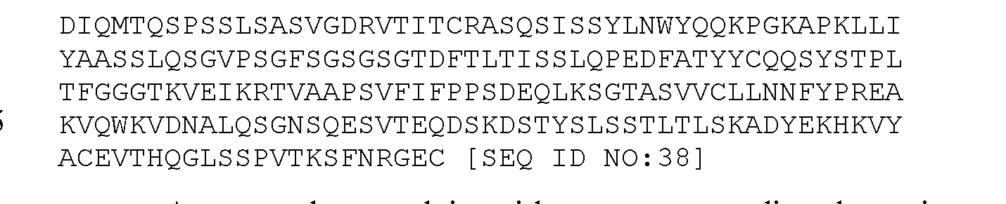


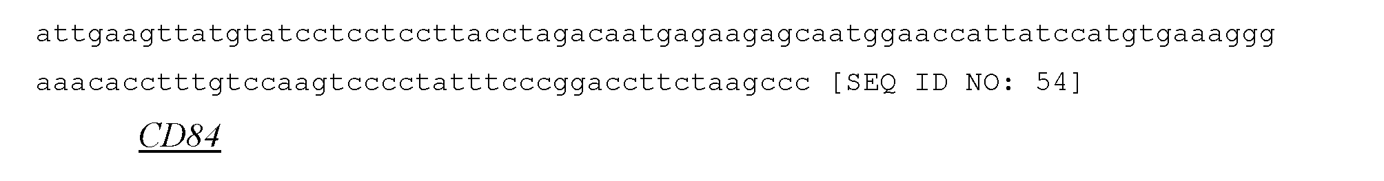


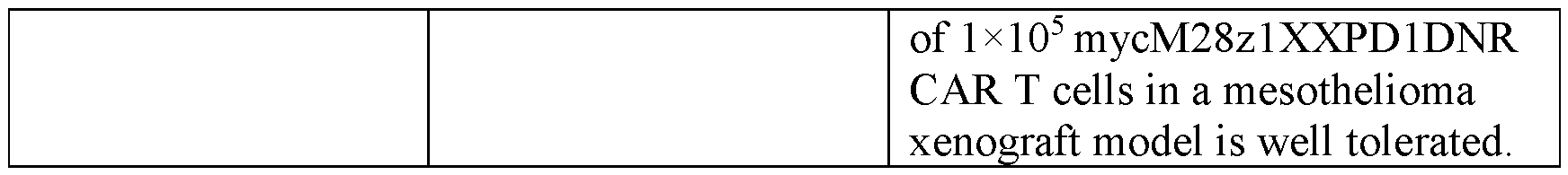


Claims
Priority Applications (12)
| Application Number | Priority Date | Filing Date | Title |
|---|---|---|---|
| CN202080050015.2A CN114585641B (en) | 2019-05-16 | 2020-05-18 | Mesothelin CAR and uses thereof |
| KR1020217041005A KR20220009996A (en) | 2019-05-16 | 2020-05-18 | Mesothelin CAR and uses thereof |
| BR112021022795A BR112021022795A2 (en) | 2019-05-16 | 2020-05-18 | Polypeptide composition comprising mesothelin cars, immunoresponsive cells and their method of production, pharmaceutical composition, nucleic acid composition, vector and kit comprising polypeptide composition |
| EP20806380.0A EP3969470A4 (en) | 2019-05-16 | 2020-05-18 | Mesothelin cars and uses thereof |
| MX2021013960A MX2021013960A (en) | 2019-05-16 | 2020-05-18 | MESOTHELIN CHIMERIC ANTIGEN RECEPTORS (CAR) AND THEIR USES. |
| SG11202112676VA SG11202112676VA (en) | 2019-05-16 | 2020-05-18 | Mesothelin cars and uses thereof |
| CA3139989A CA3139989A1 (en) | 2019-05-16 | 2020-05-18 | Mesothelin cars and uses thereof |
| AU2020276117A AU2020276117A1 (en) | 2019-05-16 | 2020-05-18 | Mesothelin cars and uses thereof |
| JP2021568282A JP7645197B2 (en) | 2019-05-16 | 2020-05-18 | Mesothelin CAR and uses thereof |
| IL287997A IL287997A (en) | 2019-05-16 | 2021-11-10 | Mesothelin cars and uses thereof |
| US17/526,812 US20220125905A1 (en) | 2019-05-16 | 2021-11-15 | Mesothelin cars and uses thereof |
| ZA2021/09069A ZA202109069B (en) | 2019-05-16 | 2021-11-15 | Mesothelin cars and uses thereof |
Applications Claiming Priority (4)
| Application Number | Priority Date | Filing Date | Title |
|---|---|---|---|
| US201962848983P | 2019-05-16 | 2019-05-16 | |
| US62/848,983 | 2019-05-16 | ||
| US202062975966P | 2020-02-13 | 2020-02-13 | |
| US62/975,966 | 2020-02-13 |
Related Child Applications (1)
| Application Number | Title | Priority Date | Filing Date |
|---|---|---|---|
| US17/526,812 Continuation US20220125905A1 (en) | 2019-05-16 | 2021-11-15 | Mesothelin cars and uses thereof |
Publications (1)
| Publication Number | Publication Date |
|---|---|
| WO2020232433A1 true WO2020232433A1 (en) | 2020-11-19 |
Family
ID=73289727
Family Applications (1)
| Application Number | Title | Priority Date | Filing Date |
|---|---|---|---|
| PCT/US2020/033382 Ceased WO2020232433A1 (en) | 2019-05-16 | 2020-05-18 | Mesothelin cars and uses thereof |
Country Status (13)
| Country | Link |
|---|---|
| US (1) | US20220125905A1 (en) |
| EP (1) | EP3969470A4 (en) |
| JP (1) | JP7645197B2 (en) |
| KR (1) | KR20220009996A (en) |
| CN (1) | CN114585641B (en) |
| AU (1) | AU2020276117A1 (en) |
| BR (1) | BR112021022795A2 (en) |
| CA (1) | CA3139989A1 (en) |
| IL (1) | IL287997A (en) |
| MX (1) | MX2021013960A (en) |
| SG (1) | SG11202112676VA (en) |
| WO (1) | WO2020232433A1 (en) |
| ZA (1) | ZA202109069B (en) |
Cited By (4)
| Publication number | Priority date | Publication date | Assignee | Title |
|---|---|---|---|---|
| WO2022123316A1 (en) * | 2020-12-09 | 2022-06-16 | Takeda Pharmaceutical Company Limited | Compositions of guanylyl cyclase c (gcc) antigen binding agents and methods of use thereof |
| WO2022157500A1 (en) * | 2021-01-20 | 2022-07-28 | Coding Bio Limited | Methods for high throughput screening of chimeric antigen receptors |
| WO2023131063A1 (en) * | 2022-01-10 | 2023-07-13 | 成都科伦精准生物科技有限公司 | Chimeric antigen receptors specifically binding to msln and use thereof |
| EP4469559A4 (en) * | 2022-01-28 | 2025-12-03 | A2 Biotherapeutics Inc | Compositions and methods for the treatment of mesothelin-positive cancers |
Families Citing this family (6)
| Publication number | Priority date | Publication date | Assignee | Title |
|---|---|---|---|---|
| WO2017040945A1 (en) * | 2015-09-04 | 2017-03-09 | Memorial Sloan Kettering Cancer Center | Immune cell compositions and methods of use |
| US10574678B2 (en) * | 2016-12-13 | 2020-02-25 | Forescout Technologies, Inc. | Name translation monitoring |
| CA3262929A1 (en) * | 2022-07-25 | 2024-02-01 | Sloan-Kettering Institute For Cancer Research | Manufacturing processes for adoptive cell therapies |
| CN120826416A (en) * | 2022-12-02 | 2025-10-21 | 杰科生物医药公司 | Anti-mesothelin bispecific antibodies and methods of use thereof |
| WO2025195483A1 (en) * | 2024-03-21 | 2025-09-25 | Utc Therapeutics (Shanghai) Co., Ltd. | Mesothelin targetting antibodies, chimeric antigen receptors, and uses thereof |
| WO2025199322A1 (en) * | 2024-03-21 | 2025-09-25 | The General Hospital Corporation | Mesothelin and muc16 bispecific chimeric antigen receptor (car) t cells |
Citations (6)
| Publication number | Priority date | Publication date | Assignee | Title |
|---|---|---|---|---|
| US20170096638A1 (en) * | 2015-03-02 | 2017-04-06 | Innovative Cellular Therapeutics CO., LTD. | Reducing Immune Tolerance Induced by PD-L1 |
| WO2018044866A1 (en) * | 2016-08-30 | 2018-03-08 | Memorial Sloan Kettering Cancer Center | Immune cell compositions and methods of use for treating viral and other infections |
| WO2018067993A1 (en) * | 2016-10-07 | 2018-04-12 | TCR2 Therapeutics Inc. | Compositions and methods for t-cell receptors reprogramming using fusion proteins |
| WO2018132506A1 (en) * | 2017-01-10 | 2018-07-19 | The General Hospital Corporation | Chimeric antigen receptors based on alternative signal 1 domains |
| WO2018144535A1 (en) * | 2017-01-31 | 2018-08-09 | Novartis Ag | Treatment of cancer using chimeric t cell receptor proteins having multiple specificities |
| US20180251546A1 (en) * | 2014-06-06 | 2018-09-06 | Memorial Sloan-Kettering Cancer Center | Mesothelin-targeted chimeric antigen receptors and uses thereof |
Family Cites Families (7)
| Publication number | Priority date | Publication date | Assignee | Title |
|---|---|---|---|---|
| CA2718321A1 (en) * | 2008-03-27 | 2009-10-01 | Dimiter S. Dimitrov | Human anti-mesothelin monoclonal antibodies |
| WO2017040945A1 (en) | 2015-09-04 | 2017-03-09 | Memorial Sloan Kettering Cancer Center | Immune cell compositions and methods of use |
| WO2017100428A1 (en) * | 2015-12-09 | 2017-06-15 | Memorial Sloan Kettering Cancer Center | Immune cell compositions and methods of using same |
| AU2018231190B2 (en) | 2017-03-08 | 2023-05-25 | Memorial Sloan Kettering Cancer Center | Immune cell compositions and methods of use |
| CA3287539A1 (en) | 2017-06-21 | 2026-03-02 | Icell Gene Therapeutics Llc | CHIMERIC ANTIGEN RECEPTORS (CARs), COMPOSITIONS AND METHODS THEREOF |
| SG11202006050XA (en) * | 2017-12-29 | 2020-07-29 | Memorial Sloan Kettering Cancer Center | Enhanced chimeric antigen receptors and uses thereof |
| CN108715616A (en) * | 2018-04-27 | 2018-10-30 | 上海恒润达生生物科技有限公司 | The Chimeric antigen receptor method and purposes of targeting humanized mesothelin |
-
2020
- 2020-05-18 KR KR1020217041005A patent/KR20220009996A/en not_active Withdrawn
- 2020-05-18 CA CA3139989A patent/CA3139989A1/en active Pending
- 2020-05-18 CN CN202080050015.2A patent/CN114585641B/en active Active
- 2020-05-18 MX MX2021013960A patent/MX2021013960A/en unknown
- 2020-05-18 AU AU2020276117A patent/AU2020276117A1/en active Pending
- 2020-05-18 WO PCT/US2020/033382 patent/WO2020232433A1/en not_active Ceased
- 2020-05-18 EP EP20806380.0A patent/EP3969470A4/en active Pending
- 2020-05-18 SG SG11202112676VA patent/SG11202112676VA/en unknown
- 2020-05-18 BR BR112021022795A patent/BR112021022795A2/en unknown
- 2020-05-18 JP JP2021568282A patent/JP7645197B2/en active Active
-
2021
- 2021-11-10 IL IL287997A patent/IL287997A/en unknown
- 2021-11-15 US US17/526,812 patent/US20220125905A1/en active Pending
- 2021-11-15 ZA ZA2021/09069A patent/ZA202109069B/en unknown
Patent Citations (6)
| Publication number | Priority date | Publication date | Assignee | Title |
|---|---|---|---|---|
| US20180251546A1 (en) * | 2014-06-06 | 2018-09-06 | Memorial Sloan-Kettering Cancer Center | Mesothelin-targeted chimeric antigen receptors and uses thereof |
| US20170096638A1 (en) * | 2015-03-02 | 2017-04-06 | Innovative Cellular Therapeutics CO., LTD. | Reducing Immune Tolerance Induced by PD-L1 |
| WO2018044866A1 (en) * | 2016-08-30 | 2018-03-08 | Memorial Sloan Kettering Cancer Center | Immune cell compositions and methods of use for treating viral and other infections |
| WO2018067993A1 (en) * | 2016-10-07 | 2018-04-12 | TCR2 Therapeutics Inc. | Compositions and methods for t-cell receptors reprogramming using fusion proteins |
| WO2018132506A1 (en) * | 2017-01-10 | 2018-07-19 | The General Hospital Corporation | Chimeric antigen receptors based on alternative signal 1 domains |
| WO2018144535A1 (en) * | 2017-01-31 | 2018-08-09 | Novartis Ag | Treatment of cancer using chimeric t cell receptor proteins having multiple specificities |
Cited By (5)
| Publication number | Priority date | Publication date | Assignee | Title |
|---|---|---|---|---|
| WO2022123316A1 (en) * | 2020-12-09 | 2022-06-16 | Takeda Pharmaceutical Company Limited | Compositions of guanylyl cyclase c (gcc) antigen binding agents and methods of use thereof |
| JP2023553159A (en) * | 2020-12-09 | 2023-12-20 | 武田薬品工業株式会社 | Compositions of guanylyl cyclase C (GCC) antigen binding agents and methods of using the same |
| WO2022157500A1 (en) * | 2021-01-20 | 2022-07-28 | Coding Bio Limited | Methods for high throughput screening of chimeric antigen receptors |
| WO2023131063A1 (en) * | 2022-01-10 | 2023-07-13 | 成都科伦精准生物科技有限公司 | Chimeric antigen receptors specifically binding to msln and use thereof |
| EP4469559A4 (en) * | 2022-01-28 | 2025-12-03 | A2 Biotherapeutics Inc | Compositions and methods for the treatment of mesothelin-positive cancers |
Also Published As
| Publication number | Publication date |
|---|---|
| CA3139989A1 (en) | 2020-11-19 |
| EP3969470A1 (en) | 2022-03-23 |
| SG11202112676VA (en) | 2021-12-30 |
| JP7645197B2 (en) | 2025-03-14 |
| BR112021022795A2 (en) | 2022-01-18 |
| CN114585641B (en) | 2025-05-23 |
| AU2020276117A1 (en) | 2021-12-09 |
| JP2022532747A (en) | 2022-07-19 |
| ZA202109069B (en) | 2022-08-31 |
| EP3969470A4 (en) | 2023-06-28 |
| MX2021013960A (en) | 2022-04-27 |
| IL287997A (en) | 2022-01-01 |
| US20220125905A1 (en) | 2022-04-28 |
| KR20220009996A (en) | 2022-01-25 |
| CN114585641A (en) | 2022-06-03 |
Similar Documents
| Publication | Publication Date | Title |
|---|---|---|
| US20220125905A1 (en) | Mesothelin cars and uses thereof | |
| US20230340113A1 (en) | CHIMERIC ANTIGEN RECEPTORS (CARs), COMPOSITIONS AND METHODS THEREOF | |
| JP7228900B2 (en) | Engineered natural killer cells and uses thereof | |
| AU2014225788B2 (en) | Engager cells for immunotherapy | |
| US10124028B2 (en) | Oncolytic adenovirus encoding a B7 protein or active fragment | |
| US20230071098A1 (en) | Anti-gpc3 single-chain antibody-containing car | |
| AU2019347873A1 (en) | Immunoresponsive cells expressing dominant negative Fas and uses thereof | |
| US20230087125A1 (en) | Chimeric antigen receptors targeting cd127 and use thereof | |
| WO2017112877A1 (en) | Chimeric antigen receptors and enhancement of anti-tumor activity | |
| JP2026065070A (en) | Dendritic cell-activated chimeric antigen receptor and its use | |
| CA2950763A1 (en) | Mesothelin-targeted chimeric antigen receptors and uses thereof | |
| CN107523549A (en) | A kind of CAR-T cell that efficiently and stably expresses activating antibody and its use | |
| IL293598A (en) | Cells expressing c-kit mutations and uses thereof | |
| US20240254183A1 (en) | Membrane-bound il-12 for cellular immunotherapy | |
| US20230058774A1 (en) | Novel dominant negative fas polypeptides, cells comprising thereof and uses thereof | |
| EA051091B1 (en) | Chimeric antigen receptors targeting CD19 and their applications |
Legal Events
| Date | Code | Title | Description |
|---|---|---|---|
| 121 | Ep: the epo has been informed by wipo that ep was designated in this application |
Ref document number: 20806380 Country of ref document: EP Kind code of ref document: A1 |
|
| ENP | Entry into the national phase |
Ref document number: 3139989 Country of ref document: CA |
|
| ENP | Entry into the national phase |
Ref document number: 2021568282 Country of ref document: JP Kind code of ref document: A |
|
| NENP | Non-entry into the national phase |
Ref country code: DE |
|
| REG | Reference to national code |
Ref country code: BR Ref legal event code: B01A Ref document number: 112021022795 Country of ref document: BR |
|
| ENP | Entry into the national phase |
Ref document number: 2020276117 Country of ref document: AU Date of ref document: 20200518 Kind code of ref document: A |
|
| ENP | Entry into the national phase |
Ref document number: 20217041005 Country of ref document: KR Kind code of ref document: A |
|
| WWE | Wipo information: entry into national phase |
Ref document number: 2020806380 Country of ref document: EP |
|
| ENP | Entry into the national phase |
Ref document number: 2020806380 Country of ref document: EP Effective date: 20211216 |
|
| ENP | Entry into the national phase |
Ref document number: 112021022795 Country of ref document: BR Kind code of ref document: A2 Effective date: 20211112 |
|
| WWW | Wipo information: withdrawn in national office |
Ref document number: 1020217041005 Country of ref document: KR |
|
| WWG | Wipo information: grant in national office |
Ref document number: 202080050015.2 Country of ref document: CN |