WO2021038571A1 - A single-domain antibody for targeting prostate specific membrane antigen (psma) - Google Patents
A single-domain antibody for targeting prostate specific membrane antigen (psma) Download PDFInfo
- Publication number
- WO2021038571A1 WO2021038571A1 PCT/IL2020/050940 IL2020050940W WO2021038571A1 WO 2021038571 A1 WO2021038571 A1 WO 2021038571A1 IL 2020050940 W IL2020050940 W IL 2020050940W WO 2021038571 A1 WO2021038571 A1 WO 2021038571A1
- Authority
- WO
- WIPO (PCT)
- Prior art keywords
- psma
- antigen
- binding polypeptide
- binding
- seq
- Prior art date
- Legal status (The legal status is an assumption and is not a legal conclusion. Google has not performed a legal analysis and makes no representation as to the accuracy of the status listed.)
- Ceased
Links
Classifications
-
- A—HUMAN NECESSITIES
- A61—MEDICAL OR VETERINARY SCIENCE; HYGIENE
- A61K—PREPARATIONS FOR MEDICAL, DENTAL OR TOILETRY PURPOSES
- A61K47/00—Medicinal preparations characterised by the non-active ingredients used, e.g. carriers or inert additives; Targeting or modifying agents chemically bound to the active ingredient
- A61K47/50—Medicinal preparations characterised by the non-active ingredients used, e.g. carriers or inert additives; Targeting or modifying agents chemically bound to the active ingredient the non-active ingredient being chemically bound to the active ingredient, e.g. polymer-drug conjugates
- A61K47/51—Medicinal preparations characterised by the non-active ingredients used, e.g. carriers or inert additives; Targeting or modifying agents chemically bound to the active ingredient the non-active ingredient being chemically bound to the active ingredient, e.g. polymer-drug conjugates the non-active ingredient being a modifying agent
- A61K47/62—Medicinal preparations characterised by the non-active ingredients used, e.g. carriers or inert additives; Targeting or modifying agents chemically bound to the active ingredient the non-active ingredient being chemically bound to the active ingredient, e.g. polymer-drug conjugates the non-active ingredient being a modifying agent the modifying agent being a protein, peptide or polyamino acid
- A61K47/64—Drug-peptide, drug-protein or drug-polyamino acid conjugates, i.e. the modifying agent being a peptide, protein or polyamino acid which is covalently bonded or complexed to a therapeutically active agent
-
- C—CHEMISTRY; METALLURGY
- C07—ORGANIC CHEMISTRY
- C07K—PEPTIDES
- C07K16/00—Immunoglobulins [IG], e.g. monoclonal or polyclonal antibodies
- C07K16/18—Immunoglobulins [IG], e.g. monoclonal or polyclonal antibodies against material from animals or humans
- C07K16/28—Immunoglobulins [IG], e.g. monoclonal or polyclonal antibodies against material from animals or humans against receptors, cell surface antigens or cell surface determinants
- C07K16/30—Immunoglobulins [IG], e.g. monoclonal or polyclonal antibodies against material from animals or humans against receptors, cell surface antigens or cell surface determinants from tumour cells
- C07K16/3069—Reproductive system, e.g. ovaria, uterus, testes, prostate
-
- A—HUMAN NECESSITIES
- A61—MEDICAL OR VETERINARY SCIENCE; HYGIENE
- A61P—SPECIFIC THERAPEUTIC ACTIVITY OF CHEMICAL COMPOUNDS OR MEDICINAL PREPARATIONS
- A61P25/00—Drugs for disorders of the nervous system
-
- A—HUMAN NECESSITIES
- A61—MEDICAL OR VETERINARY SCIENCE; HYGIENE
- A61P—SPECIFIC THERAPEUTIC ACTIVITY OF CHEMICAL COMPOUNDS OR MEDICINAL PREPARATIONS
- A61P35/00—Antineoplastic agents
-
- A—HUMAN NECESSITIES
- A61—MEDICAL OR VETERINARY SCIENCE; HYGIENE
- A61K—PREPARATIONS FOR MEDICAL, DENTAL OR TOILETRY PURPOSES
- A61K39/00—Medicinal preparations containing antigens or antibodies
- A61K2039/505—Medicinal preparations containing antigens or antibodies comprising antibodies
-
- C—CHEMISTRY; METALLURGY
- C07—ORGANIC CHEMISTRY
- C07K—PEPTIDES
- C07K2317/00—Immunoglobulins specific features
- C07K2317/20—Immunoglobulins specific features characterized by taxonomic origin
- C07K2317/22—Immunoglobulins specific features characterized by taxonomic origin from camelids, e.g. camel, llama or dromedary
-
- C—CHEMISTRY; METALLURGY
- C07—ORGANIC CHEMISTRY
- C07K—PEPTIDES
- C07K2317/00—Immunoglobulins specific features
- C07K2317/30—Immunoglobulins specific features characterized by aspects of specificity or valency
- C07K2317/34—Identification of a linear epitope shorter than 20 amino acid residues or of a conformational epitope defined by amino acid residues
-
- C—CHEMISTRY; METALLURGY
- C07—ORGANIC CHEMISTRY
- C07K—PEPTIDES
- C07K2317/00—Immunoglobulins specific features
- C07K2317/50—Immunoglobulins specific features characterized by immunoglobulin fragments
- C07K2317/56—Immunoglobulins specific features characterized by immunoglobulin fragments variable (Fv) region, i.e. VH and/or VL
- C07K2317/567—Framework region [FR]
-
- C—CHEMISTRY; METALLURGY
- C07—ORGANIC CHEMISTRY
- C07K—PEPTIDES
- C07K2317/00—Immunoglobulins specific features
- C07K2317/50—Immunoglobulins specific features characterized by immunoglobulin fragments
- C07K2317/56—Immunoglobulins specific features characterized by immunoglobulin fragments variable (Fv) region, i.e. VH and/or VL
- C07K2317/569—Single domain, e.g. dAb, sdAb, VHH, VNAR or nanobody®
-
- C—CHEMISTRY; METALLURGY
- C07—ORGANIC CHEMISTRY
- C07K—PEPTIDES
- C07K2317/00—Immunoglobulins specific features
- C07K2317/70—Immunoglobulins specific features characterized by effect upon binding to a cell or to an antigen
- C07K2317/76—Antagonist effect on antigen, e.g. neutralization or inhibition of binding
-
- C—CHEMISTRY; METALLURGY
- C07—ORGANIC CHEMISTRY
- C07K—PEPTIDES
- C07K2317/00—Immunoglobulins specific features
- C07K2317/70—Immunoglobulins specific features characterized by effect upon binding to a cell or to an antigen
- C07K2317/77—Internalization into the cell
-
- C—CHEMISTRY; METALLURGY
- C07—ORGANIC CHEMISTRY
- C07K—PEPTIDES
- C07K2317/00—Immunoglobulins specific features
- C07K2317/90—Immunoglobulins specific features characterized by (pharmaco)kinetic aspects or by stability of the immunoglobulin
- C07K2317/92—Affinity (KD), association rate (Ka), dissociation rate (Kd) or EC50 value
Definitions
- the present invention is in the field of single-domain antibodies.
- PCa Prostate cancer
- PSA pro state -specific antigen
- chemotherapies are often used to treat castration-resistant PCa, some potentially effective chemotherapies against PCa, such as doxorubicin (DOX), do not sufficiently accumulate within tumors and have a large distribution volume, resulting in low treatment efficacy and high non-specific toxicity. Novel means for both the detection of PCa and the targeted delivery of cytotoxic agents are, therefore, urgently required.
- DOX doxorubicin
- PSMA prostate-specific membrane antigen
- a transmembrane protein that is overexpressed in PCa, possibly due to its folate hydrolase activity, which induces cell proliferation.
- PSMA is mostly expressed on the membranes of PCa cells, although it is also expressed on the neovasculature of many carcinomas, including PCa.
- the overexpression of PSMA is associated with malignant, castration-resistant PCa, reduced androgen- receptor expression, and poor PCa prognosis; therefore, it can be used to detect PCa, identify the stage of the disease, and promote personalized, tumor- specific medicine.
- targeting PSMA can be especially important in the treatment of aggressive, androgen-independent PCa tumors, where its expression increases while that of PSA decreases, and where first-line treatments often fail making chemotherapeutic drugs a necessity.
- PSMA has been extensively exploited as a target by multiple research groups, which presented promising compounds for PSMA-targeted diagnostics and inhibition, mostly in the field of nuclear medicine. Yet, to date, most proteins that were found to bind the extracellular region of PSMA with a sufficiently high affinity (nanomolar range) are monoclonal antibodies or antibody fragments, which have several caveats for both molecular imaging and cancer treatment purposes. For instance, the long serum half-life and broad biodistribution of antibodies often reduce the signal-to-noise ratio and maintain them in the circulation for long periods of time.
- NBs also known as VHHs, are the single-chain variable domains of heavy- chain antibodies (HCAb).
- HCAb heavy- chain antibodies
- the NB is the only fragment of the HCAb that mediates antigen binding, it can be expressed separately from the rest of the HCAb without reducing affinity, resulting in a minute ( ⁇ 15 kDa), non-immunogenic, highly target- specific protein, which is an excellent candidate for use as scaffold for in vivo imaging and targeted therapy applications.
- the present invention in some embodiments thereof, is directed to an antigen binding polypeptide having increased binding affinity to prostate specific membrane antigen (PSMA).
- PSMA prostate specific membrane antigen
- the antigen-binding polypeptide is a single domain antibody.
- the single-domain antibody of the present invention comprises three complementary-determining regions (CDRs).
- an antigen-binding polypeptide comprising three complementary-determining region (CDRs) selected from the group consisting of: (i) GYTDSNYYMS (CDR-H1; SEQ ID NO: 1), GVNTGRGSTSYADSVKG (CDR-H2; SEQ ID NO: 2), and
- AACHFCDSLPKTQDEYIL CDR-H3; SEQ ID NO: 3
- GWPYSTYSMN CDR- Hl; SEQ ID NO: 4
- GISSTMSGIIFAES CDR-H2; SEQ ID NO: 5
- RRDYSLSSSSDDFDY CDR-H3; SEQ ID NO: 6
- GYTASFS CDR-H1; SEQ ID NO: 7
- GVAVINVGVGSTYYADSV CDR-H2; SEQ ID NO: 8
- S LRW S RPPNPIS ED A YN Y CDR-H3; SEQ ID NO: 9
- composition comprising a therapeutic or diagnostic effective amount of the antigen-binding polypeptide of the invention, and a pharmaceutically acceptable carrier.
- a method of targeting PSMA comprising contacting a sample comprising the PSMA with the antigen binding polypeptide of the invention, thereby targeting PSMA.
- the CDR-H2 comprises the amino acid sequence as set forth in SEQ ID NO: 10 (GIS STMS GIIF AES KAGQFTIS QDN A) .
- the antigen-binding polypeptide is a single-domain antibody.
- the antigen-binding polypeptide comprises the amino acid sequence:
- the antigen-binding polypeptide comprises the amino acid sequence:
- the antigen-binding polypeptide comprises the amino acid sequence:
- the antigen-binding polypeptide comprises the amino acid sequence:
- the antigen-binding polypeptide has a specific binding affinity to a prostate specific membrane antigen (PSMA).
- PSMA prostate specific membrane antigen
- the antigen-binding polypeptide is characterized by binding constant (K a ) of at least 10 4 Molar 1 sec 1 to said PSMA.
- the antigen-binding polypeptide is characterized by binding constant (K a ) of 7.1 x 10 5 Molar 1 sec 1 to the PSMA.
- the antigen-binding polypeptide is characterized by binding constant (K a ) of 2 x 10 4 Molar 1 sec 1 to the PSMA.
- the antigen-binding polypeptide is characterized by binding constant (K a ) of 3.6 x 10 4 Molar 1 sec 1 to the PSMA.
- the antigen-binding polypeptide is characterized by binding constant (K a ) of 2.2 x 10 4 Molar 1 sec 1 to the PSMA.
- the antigen-binding polypeptide is characterized by dissociation constant (K D ) of less than 15 nM to the PSMA.
- the antigen-binding polypeptide is characterized by dissociation constant (K D ) of 55 pM to the PSMA.
- the antigen-binding polypeptide is characterized by dissociation constant (K D ) of 6 nM to the PSMA.
- the antigen-binding polypeptide is characterized by dissociation constant (K D ) of 0.6 nM to the PSMA. [027] In some embodiments, the antigen-binding polypeptide is characterized by dissociation constant (K D ) of 3.4 nM to the PSMA.
- the antigen-binding polypeptide is characterized by molecular weight of less than 25 kDa.
- the antigen-binding polypeptide has a specific binding affinity to a non-catalytic site of said PSMA enzyme.
- the antigen-binding polypeptide further comprises at least one non-naturally occurring amino acid.
- the pharmaceutical composition further comprises a therapeutic or diagnostic effective amount of at least one agent selected from a therapeutic agent, a diagnostic agent, and a theranostic agent.
- the method is used for imaging the PSMA in a subject afflicted by or suspected of being afflicted by a PSMA-associated disorder, the method comprising: (a) administering to the subject an effective amount of antigen-binding polypeptide of the invention, and an imaging agent; and (b) detecting the PSMA in the subject, thereby imaging cells comprising PSMA.
- the method is used for treating a PSMA-associated disorder in a subject in need thereof, the method comprising: administering to the subject a pharmaceutical composition comprising an effective amount of the antigen binding polypeptide of the invention, a cytotoxic agent or a theranostic agent, and an acceptable carrier, thereby treating the PSMA-associated disorder in a subject in need thereof.
- the PSMA-associated disorder is prostate cancer.
- the PSMA-associated disorder is a neurological disorder selected from the group consisting of: Parkinson disease, Alzheimer disease, Huntington disease, amyotrophic lateral sclerosis (ALS), schizophrenia, and any combination thereof.
- Figs. 1A-1F include graphs showing that nanobodies (NBs) bind to prostate-specific membrane antigen (PSMA) in vitro and to PSMA-expressing prostate cancer cells.
- the response units (RU) measured using surface plasmon resonance (SPR) and a 1:1 Langmuir kinetic model, were used to calculate the affinity (KD) of immobilized NB7 (1A), NB8 (IB), NB13 (1C), and NB37 (ID) to PSMA.
- the PSMA concentrations were 25, 50, 100, 1,600, or 3,200 pM for the NB7 sensograms, and 2.94, 5.88, 11.75, 23.50, or 47.00 nM for the NB8, NB13, and NB37 sensograms.
- Figs. 2A-2H include illustration showing structural analysis of NBs and their PSMA-binding epitopes.
- the PSMA monomers are labeled individually by Roman numerals; biological dimers are formed by I+II and III+IV, while non-biological dimers are formed by I+III and II+IV.
- (2G) NB7 is encircled in full black line. Key interactions (according to Fig. 20) are shown as black dashed lines.
- (2H) NB37 is encircled in full black line. Key interactions (Fig. 21) are shown as black dashed lines.
- Figs. 3A-3G include micrographs and graphs showing in vivo whole-body NIR imaging of labeled NBs.
- PC3-flu (PSMA-) and PC3-PIP (PSMA + ) PCa cells were co injected as xenografts into the left and right upper flanks, respectively, of athymic nude mice.
- mice were intravenously injected with fluorescently labeled NBs (from left to right: NB7, NB13, NB8, and NB37; the KD of each NB is shown in parentheses for convenience) and whole-body images were captured 3 h (3A) and 6 h (3B) post-injection, and again when the signal was no longer detectable (3C); 32 h for NB8 and NB13, and 24 h for NB37; mice injected with NB7 still showed a fluorescent signal 56 h post-injection, at which point they were imaged and then euthanized).
- fluorescently labeled NBs from left to right: NB7, NB13, NB8, and NB37; the KD of each NB is shown in parentheses for convenience
- whole-body images were captured 3 h (3A) and 6 h (3B) post-injection, and again when the signal was no longer detectable (3C); 32 h for NB8 and NB13, and 24 h for NB37
- the left mouse was injected with tumor cells but not with NBs
- the middle mouse was injected with NBs but not with tumor cells
- the right mouse was injected with both tumor cells and NBs.
- 3D-3G Quantification of the AFexo fluorescent signals from the dissected organs at each time point for NB7 (3D), NB8 (3E), NB13 (3F), and NB37 (3G).
- PC3-PIP tumors express PSMA, whereas PC3-flu tumors do not.
- Figs. 4A-4I include fluorescent micrographs showing confocal imaging of the internalization of NBs into prostate cancer cells.
- PC3-PIP (PSMA + ) cells (4A-4D) and PC3-flu (PSMA-) cells (4E-4H) were incubated for 10 min with a Hoechst reagent (nuclei staining), a PE-anti-PSMA antibody, and 100 nM of either NB7 (4A, 4E), NB8 (4B, 4F), NB13 (4C, 4G), or NB37 (4D, 4H), each labeled with Dylight 488.
- PC3-PIP cells were incubated for 10 min with the PE-anti-PSMA antibody without any NB (41).
- Scale bar 10 mhi.
- Figs. 5A-5H include fluorescent micrographs showing confocal imaging of the internalization of NB7cys, DOX, and the NB7cysDOX conjugate into PCa cells.
- PC3- PIP (PSMA + ; 5A-5D) and PC3-flu (PSMA-; 5E-5H) cells were incubated with either DOX (auto-fluorescence; 5B, 5F), NB7cys labeled with Dylight 650 (5C, 5G), or NB7cysDOX labeled with Dylight 650 (5D, 5H).
- Un-treated control cells (5A, 5E). Images were taken after 15 min of incubation.
- Figs. 6A-6D include graphs and micrographs showing in vivo and in-situ effects of NB7cysDOX on PC3-PIP (PSMA + ) tumors.
- PC3-PIP xenografts in athymic nude mice were treated with either saline (control), 2 mg/kg commercial DOX, or 1.4 mg/kg NB7cysDOX.
- H&E Hematoxylin and Eosin staining
- TUNEL terminal deoxynucleotidyl transferase dUTP nick end labeling
- PI propidium iodide staining
- Figs. 7A-7C include graphs and a micrograph showing NB selection and purification.
- 7A Enzyme-linked immunosorbent assay (ELISA) results demonstrating the binding of PSMA by individual bacterial colonies expressing different NB sequences. Sequences that were chosen for purification: NB7 (clones 7, 9, 22, 28, 31- 33, and 46), NB8 (clones 8 and 21), NB13 (clones 13 and 18), and NB37 (clone 37).
- 7B A representative size-exclusion chromatography for NB7 (the chromatograms for NB8, NB 13, and NB37 were similar to the one shown here).
- 7C SDS-PAGE gel results showing the four purified NBs. All proteins were in the expected size of ⁇ 16 kDa.
- Fig. 8 includes a vertical bar graph showing PSMA activity assay.
- PSMA was incubated with a substrate in the presence or absence of NBs to measure glutamate carboxypeptidase activity.
- Figs. 9A-9B include small angle X-ray scattering (SAXS) analysis and the R g of the monomeric PSMA.
- SAXS small angle X-ray scattering
- R g The radius of gyration (R g ) values for free PSMA increases slightly with higher concentrations due to the interaction between species in the solution.
- Figs. 10A-10D include graphs showing SAXS curves and the corresponding Guinier plots of PSMA with increasing concentrations of NBs.
- PSMA 0.5 mg/ml
- NBs 10A: NB7, 10B: NB8, IOC: NB13, andlOD: NB37
- Fig. 11 includes a graph showing the effect of NBs on the R g of PSMA.
- SAXS results showing the R g values of PSMA (0.5 mg/ml) at increasing concentrations of NBs (0.05-0.58 mg/ml).
- the dashed line indicates the R g of PSMA without NBs.
- Figs. 12A-12E include graphs showing the results of the custom-made script analysis of the SAXS data. The analysis was done using an automated procedure based on a script and the computer program GNOM. The “Total estimate” score was used to choose the best result. PSMA (12A), PSMA+NB7 (12B), PSMA+NB8 (12C), PSMA+NB13 (12D), and PSMA+NB37 (12E).
- Fig. 13 includes a graph showing the distance distribution function, P(r), of PSMA and NBs at a molar ratio of 1 :2. P( r ) was determined using the program GNOM.
- Figs. 14A-14D include micrographs showing ex vivo optical imaging of prostate cancer xenografts. The ex-vivo signal is shown at three time points in various dissected organs of mice that were injected with either NB7 (14A), NB13 (14B), NB8 (14C), or NB37 (14D) (the data are shown in Fig. 3). Intensity bars are shown below the images of NB 13 and apply to all images.
- Fig. 15 includes a graph showing the internalization of the NBs into PC3-PIP cells.
- PC3-PIP cells were incubated in 96-wells plates for 1 h with either NB7, NB8, NB13, or NB37. Then, the wells were imaged using Operetta and the number of cells with NBs on their membranes or inside their cytoplasm was counted every 40 min, so that the first imaging round was completed 1 h and 40 min after incubation.
- the internalization process is reflected in the number of cells with NBs in their cytoplasm relative to the number of cells with NBs in their membrane ("cytoplasm/membrane ratio"), such that a higher ratio indicated more NBs that were internalized into the cytoplasm.
- FIGs. 16A-16D include an illustration of a process and graphs showing the conjugation of NB7cys to DOX.
- MALDI-TOF matrix-assisted laser desorption/ionization time-of-flight
- Fig. 17 includes a graph showing ⁇ -nuclear magnetic resonance (NMR) spectrum of DOX-BMPH linker.
- Figs. 18A-18C include graphs showing the effect of NB7cysDOX on cell viability.
- (18A) The number of PC3-PIP cells was counted after a 24 h treatment with either NB7cys, DOX, or NB7cysDOX. The experiment was performed in triplicate and the results are presented as means ⁇ SEM. *p ⁇ 0.05, **p ⁇ 0.01 (Student’s t-test, as compared with untreated cells).
- (18B FACS analysis of PC3-PIP cells treated with either NB7 (1), DOX (2), or NB7cysDOX (3), or left untreated (4, partially masked by the “1” histogram), and then incubated with PI.
- PC3-PIP cells were treated with NB7, DOX, NB7cysDOX, or FCCP, or were left untreated, and then incubated with TMRE (“no TMRE”) was used as a negative control, and the fluorescent signal of TMRE was measured by using a plate reader. The fluorescence of the untreated sample was set as 1, and all other samples were normalized and compared to it. The experiment was performed in triplicate and results are presented as means ⁇ SEM. *p ⁇ 0.05, **p ⁇ 0.01, ***p ⁇ 0.005 (Student’s t-test).
- Figs. 19A-19B include graphs showing in vivo tumor growth inhibition by NB7cysDOX.
- PC3-PIP xenografts in athymic nude mice were treated with either saline (control), DOX (2 mg/kg), or NB7cysDOX (1.4 mg/kg).
- Fig. 20 includes a table showing predicted interactions between NB7 and PSMA (A: monomer A of PSMA; B: monomer B of PSMA).
- CDR1 shows an electrostatic interaction with monomer B, between aspartic acid 29 of the NB and lysine 223 of PSMA.
- CDR2 shows an electrostatic interaction with monomer A, between aspartic acid 62 of the NB and lysine 718 of PSMA.
- CDR3 shows two electrostatic interactions to monomer B: one between glutamic acid 113 of the NB and arginine 281 of PSMA, and the other between lysine 109 of the NB and glutamic acid 285 of PSMA.
- Fig. 21 includes a table showing predicted interactions between NB37 and PSMA.
- CDRs display electrostatic interactions between glutamic acid 62 with two arginine residues of PSMA (arginine 363 and arginine 411). Another electrostatic interaction is between arginine 19 (a non-CDR residue) and aspartic acid 654 of PMSA.
- Fig. 22A-22F include micrographs, illustration, and graphs, showing the incorporation of non-natural amino acids into NB7.
- 22A Western blot analysis showing the incorporation of the unnatural amino acid BOC Lysine in different positions on NB7 (A14, A40, G42, K43, and A75).
- 22B Western blot analysis showing incorporation of the unnatural amino acid BOC Lysine in different positions of NB7- Cys (K43, and A75).
- 22C an illustration of a non-limiting scheme showing binding interaction between PSMA and NB7 cys (+doxorubicin), NB7 K43prop (+Cy5.5), or NB7 cys K43prop (+doxorubicin and Cy5.5).
- (22D) is a graph showing size-exclusion chromatography. The absorbance of 280 nm for NB7 (1), NB7cys (2), and NB7 K43prop (3) is presented.
- the present invention is directed to an antigen-binding polypeptide having increased binding affinity to prostate specific membrane antigen (PSMA).
- PSMA prostate specific membrane antigen
- the antigen-binding polypeptide is a single-domain antibody.
- PSMA state specific membrane antigen
- glutamate carboxypeptidase II also known as N-acetyl-L-aspartyl-L- glutamate peptidase I (NAALADase I or NAAG peptidase).
- human PSMA has the UniProt accession no. Q04609.
- antibody and "antigen-binding polypeptide” (also referred to as an “immunoglobulin” or “Ig”) refer to a polypeptide or group of polypeptides that include at least one binding domain that is specific for one antigen.
- immunoglobulin also referred to as an “immunoglobulin” or “Ig” refer to a polypeptide or group of polypeptides that include at least one binding domain that is specific for one antigen.
- the use of a chimeric antibody or a humanized antibody is also encompassed by the invention.
- the term “antibody fragments” refers to a portion of an intact antibody, preferably comprising the antigen binding region thereof.
- the terms “single-domain antibody” refers to an antibody fragment consisting of a single variable domain (V H H). Single-domain antibody is a smaller functional fragment of the antibody that also can bind a specific antigen. In some embodiments, the single-domain antibody has better tissue penetration than conventional antibodies and therefore they are beneficial for clinical/diagnostic use.
- the single-domain antibody of the present invention comprises three complementary-determining regions (CDRs).
- variable heavy chain refers to variable heavy chain.
- variable heavy chain comprises an amino acid sequence capable of binding a specific PSMA.
- Kabat et al. defined a numbering system for variable domain sequences that is applicable to any antibody.
- One of ordinary skill in the art can unambiguously assign this system of "Kabat numbering" to any variable domain sequence, without reliance on any experimental data beyond the sequence itself.
- Kabat numbering refers to the numbering system set forth by Kabat et al, U.S. Dept of Health and Human Services, "Sequence of Proteins of Immunological Interest" (1983).
- the antigen-binding polypeptide comprises three CDRs comprising: GYTDSNYYMS (CDR-H1; SEQ ID NO: 1),
- AACHFCDSLPKTQDEYIL CDR-H3; SEQ ID NO: 3).
- the antigen-binding polypeptide comprises three CDRs comprising: GWPYSTYSMN (CDR-H1; SEQ ID NO: 4), GISSTMSGIIFAES (CDR- H2; SEQ ID NO: 5), and RRDYSLSSSSDDFDY (CDR-H3; SEQ ID NO: 6).
- the antigen-binding polypeptide comprises three CDRs comprising: GYTASFS (CDR-H1; SEQ ID NO: 7), G V A VIN V G V GS T Y Y ADS V (CDR-H2; SEQ ID NO: 8) and S LRW S RPPNPIS ED A YN Y (CDR-H3; SEQ ID NO: 9).
- the antigen-binding polypeptide comprises three CDRs comprising: GWPYSTYSMN (CDR-H1; SEQ ID NO: 4),
- the antigen-binding polypeptide comprises the amino acid sequence:
- Xi is selected from Alanine or an artificial or non-naturally occurring amino acid
- X2 is selected from Alanine or an artificial or non-naturally occurring amino acid
- X3 is selected from Glycine or an artificial or non-naturally occurring amino acid
- X4 is selected from Lysine or an artificial or non-naturally occurring amino acid
- X5 is selected from Alanine or an artificial or non-naturally occurring amino acid.
- the artificial or non-naturally occurring amino acid comprises or consists of the amino acid BOC
- the antigen-binding polypeptide comprises the amino acid sequence:
- the antigen -binding polypeptide comprises the amino acid sequence:
- Xi is selected from Alanine or an artificial or non-naturally occurring amino acid
- X2 is selected from Alanine or an artificial or non-naturally occurring amino acid
- X3 is selected from Glycine or an artificial or non-naturally occurring amino acid
- X4 is selected from Lysine or an artificial or non-naturally occurring amino acid
- X5 is selected from Alanine or an artificial or non-naturally occurring amino acid.
- the artificial or non-naturally occurring amino acid comprises or consists of the amino acid BOC-Lysine.
- the antigen-binding polypeptide comprises the amino acid sequence:
- the antigen -binding polypeptide comprises the amino acid sequence:
- Xi is selected from Alanine or an artificial or non-naturally occurring amino acid
- X2 is selected from Glycine or an artificial or non-naturally occurring amino acid
- X3 is selected from Lysine or an artificial or non-naturally occurring amino acid.
- the artificial or non-naturally occurring amino acid comprises or consists of the amino acid BOC-Lysine.
- the antigen-binding polypeptide comprises the amino acid sequence:
- the antigen-binding polypeptide comprises the amino acid sequence:
- Xi is selected from Alanine or an artificial or non-naturally occurring amino acid
- X2 is selected from Alanine or an artificial or non-naturally occurring amino acid
- X3 is selected from Glycine or an artificial or non-naturally occurring amino acid
- X4 is selected from Lysine or an artificial or non-naturally occurring amino acid
- X5 is selected from Alanine or an artificial or non-naturally occurring amino acid.
- the artificial or non-naturally occurring amino acid comprises or consists of the amino acid BOC-
- the antigen-binding polypeptide comprises the amino acid sequence:
- the antigen-binding polypeptide has a specific binding affinity to PSMA.
- the term “specific binding” refers to a non-covalent physical association of a first and a second moiety of two entities.
- the association between the first and second moieties is at least 10 times as strong, at least 50 times as strong, or at least 100 times as strong as the association of other moieties present in the environment in which binding occurs.
- the binding of two or more entities may be considered specific if the equilibrium "dissociation constant", KD, is less than 10 -3 M, less than 10 -4 M, less than 10 -5 M, less than 10 -6 M, less than 10 _7 M, less than 10 -8 M, less than 10 -9 M, less than 10 -10 M, less than 10 _u M, or less than 10 -12 M, or any value and range therebetween.
- the binding of two or more entities may be considered specific if the equilibrium "dissociation constant", KD, is KG 10 M - KG 3 M, KG 12 M - KG 4 M.
- specific binding can be accomplished by a plurality of weaker interactions. Calculation of a peptide's dissociation constant (KD) is known to a skilled artisan and is also show in the Examples section herein below.
- binding constant refers to a special case of the equilibrium constant K a , which is the inverse of the dissociation constant.
- the antigen-binding polypeptide is characterized by binding constant (K a ) of at least 10 3 , at least lOxlO 3 , at least 10 4 , at least 2xl0 4 , at least 10 5 , or at least 5x10 s Molar 1 sec 1 (M -1 s 1 ) to PSMA, or any value and range therebetween.
- K a binding constant of at least 10 3 , at least lOxlO 3 , at least 10 4 , at least 2xl0 4 , at least 10 5 , or at least 5x10 s Molar 1 sec 1 (M -1 s 1 ) to PSMA, or any value and range therebetween.
- the antigen-binding polypeptide comprising the amino acid sequence as set forth in SEQ ID NO: 11 is characterized by binding constant (K a ) of about 7.1xl0 5 M 1 s 1 to PSMA.
- the antigen-binding polypeptide comprising the amino acid sequence as set forth in SEQ ID NO: 12 is characterized by binding constant (K a ) of about 2xl0 4 M 1 s 1 to PSMA.
- the antigen-binding polypeptide comprising the amino acid sequence as set forth in SEQ ID NO: 13 is characterized by binding constant (K a ) of about 3.6xl0 4 M 1 s 1 to PSMA.
- the antigen-binding polypeptide comprising the amino acid sequence as set forth in SEQ ID NO: 14 is characterized by binding constant (K a ) of about 2.2xl0 4 M 1 s 1 to PSMA.
- the antigen-binding polypeptide is characterized by dissociation constant (K D ) of less than 10 pM, less than 50 pM, less than 500 pM, less than 15 nM, less than 50 nM, or less than 500 nM to PSMA, or any value and range therebetween.
- K D dissociation constant
- the antigen-binding polypeptide comprising the amino acid sequence as set forth in SEQ ID NO: 11 is characterized by dissociation constant (KD) of about 55 pM to PSMA.
- the antigen-binding polypeptide comprising the amino acid sequence as set forth in SEQ ID NO: 12 is characterized by dissociation constant (KD) of about 6 nM to PSMA (as calculated herein below in the Examples section).
- the antigen-binding polypeptide comprising the amino acid sequence as set forth in SEQ ID NO: 13 is characterized by dissociation constant (K D ) of about 0.6 nM to PSMA.
- the antigen-binding polypeptide comprising the amino acid sequence as set forth in SEQ ID NO: 14 is characterized by dissociation constant (KD) of about 3.4 nM to PSMA.
- the polypeptide binds to a non-catalytic site of PSMA. In one embodiment, the polypeptide binds to an extracellular domain of PSMA.
- peptide As used herein, the terms “peptide”, “polypeptide” and “protein” are used interchangeably to refer to a polymer of amino acid residues. In another embodiment, the terms “peptide”, “polypeptide” and “protein” as used herein encompass native peptides, peptidomimetics (typically including non-peptide bonds or other synthetic modifications) and the peptide analogues peptoids and semipeptoids or any combination thereof.
- the polypeptide binding to PSMA is characterized by allowing further interaction to PSMA. In some embodiments, the polypeptide binding to PSMA is characterized by retaining PSMA enzyme activity. Methods of determining PSMA activity are known in the art and are also exemplified herein below, as a non-limiting example.
- the antigen-binding polypeptide is characterized by molecular weight of less than 15 kDa, less than 20 kDa, less than 25 kDa, less than 35 kDa, or less than 50 kDa, or any value and range therebetween.
- molecular weight of less than 15 kDa, less than 20 kDa, less than 25 kDa, less than 35 kDa, or less than 50 kDa, or any value and range therebetween.
- the antigen-binding polypeptide is characterized by thermal stability (T m ) of at least 60 °C, at least 70 °C, at least 90 °C, or at least 95 °C, or any value and range therebetween.
- T m thermal stability
- thermal stability refers to a substance resistance to irreversible change in its chemical or physical structure at an elevated temperature.
- T m indicates the thermal energy that caused the denaturation/unfolding of a protein or a peptide.
- the N- or C-terminus of the antigen-binding polypeptide comprises a tag motif.
- the tag motif comprises at least six amino acids.
- the antigen-binding polypeptide comprises histidine (His)-tag.
- the antigen-binding polypeptide comprises human influenza hemagglutinin (HA)-tag.
- polypeptides of the invention encompass truncated forms and/or fragments of any one of SEQ ID NOs: 1-14 as long as they are capable of binding PSMA.
- Conservative substitution of amino acids as known to those skilled in the art are within the scope of the present invention.
- Conservative amino acid substitutions include replacement of one amino acid with another having the same type of functional group or side chain e.g. aliphatic, aromatic, positively charged, negatively charged.
- One of skill will recognize that individual substitutions, deletions or additions to peptide, polypeptide, or protein sequence which alters, adds or deletes a single amino acid or a small percentage of amino acids in the encoded sequence is a "conservatively modified variant" where the alteration results in the substitution of an amino acid with a chemically similar amino acid.
- Conservative substitution tables providing functionally similar amino acids are well known in the art.
- the following six groups each contain amino acids that are conservative substitutions for one another: 1) Alanine (A), Serine (S), Threonine (T); 2) Aspartic acid (D), Glutamic acid (E); 3) Asparagine (N), Glutamine (Q); 4) Arginine (R), Lysine (K); 5) Isoleucine (I), Leucine (L), Methionine (M), Valine (V); and 6) Phenylalanine (L), Tyrosine (Y), Tryptophan (W) (see, e.g., Creighton, Proteins, 1984).
- substitution also includes the use of a chemically derivatized residue in place of a non-derivatized residue provided that such peptide displays the requisite function of modulating the immune system's innate response as specified herein.
- the polypeptide of the invention comprises a non- naturally occurring amino acid.
- any non-naturally occurring amino acid is envisioned by the current invention as long as the resulting polypeptide comprising the non- naturally occurring amino acid maintains its activity, e.g., high affinity binding to PSMA, or any other activity such as disclosed herein.
- a non-naturally occurring amino acid is selected from: 3-Iodo-L-tyrosine, N e -Benzyloxycarbonyllysine (ZLys), N e -Acetyllysine (AcLys), N e - Cyclopentyloxycarbon yl-L- lysine (Cyc), N e -(((lR,2R)-2- azidocyclopentyloxy)c arbonyl)-L-lysine (ACPK), o-Nitrobenzyl-Otyrosine, o- N i t ro hen zy 1 o x y carbo n y 1 - N e -Llysine, N e -[(l-(6-Nitrobenzo [d][l,3]dioxol-5yl) ethoxy )carbonyl]- L-lysine, N e -[(l-(6-
- a non-naturally occurring amino acid comprises or consists of BOC-Lysine.
- the present invention provides a method for targeting PSMA by contacting a sample comprising PSMA with an antigen-binding polypeptide of the invention, thereby targeting PSMA.
- the present invention provides a method for treating, diagnosing, prognosticating or determining the suitability for treatment of a subject suffering from a PSMA- associated disorder, the method comprising administering to the subject a pharmaceutical composition comprising an effective amount of the antigen-binding polypeptide of the invention, a cytotoxic agent or a theranostic agent, and a pharmaceutical acceptable carrier, thereby treating diagnosing, prognosticating or determining the suitability for treatment of a subject suffering from a PSMA- associated disorder in said subject.
- a method for imaging PSMA in a subject comprising administering to the subject a composition comprising an effective amount of the antigen -binding polypeptide of the invention, and an imaging agent; and detecting the PSMA in the subject, thereby imaging PSMA in a subject.
- the imaging agent is selected from, without being limited thereto, a fluorescent label (e.g., fluorescein isothiocyanate), a chromophore, a radioactive label, a paramagnetic ion (e.g., Gd +3 ), and any combination thereof.
- a fluorescent label e.g., fluorescein isothiocyanate
- a chromophore e.g., chromophore
- radioactive label e.g., Gd +3
- paramagnetic ion e.g., Gd +3
- the term "chromophore” refers to a material that absorbs certain wavelength of light from UV to near infrared region and may be or may not be emissive.
- the imaging agent is a radioactive label (e.g., isotope).
- the therapeutic agent is a radioactive label (e.g., isotope).
- the isotope is selected from, but not limited to:
- the imaging techniques are selected from, without being limited thereto, computed X-ray tomography (CT), ultrasound (US), and magnetic resonance imaging (MRI), positron emission tomography (PET), single photon emission computed tomography (SPECT), fluorescence and radio assays, cytofluorimetry, and fluorescence activated cell sorting.
- CT computed X-ray tomography
- US ultrasound
- MRI magnetic resonance imaging
- PET positron emission tomography
- SPECT single photon emission computed tomography
- fluorescence and radio assays fluorescence and radio assays
- cytofluorimetry fluorescence activated cell sorting.
- Non-limiting exemplary embodiments demonstrate the diagnosis of prostate tumors in vivo by near infra-red (NIR) imaging after 24 hours from the administration of the antigen-binding polypeptide conjugated to a fluorescent label.
- NIR near infra-red
- the method further comprises determining the relative percentage of the PSMA subpopulations by the administration of antigen-binding polypeptide.
- the antigen -binding polypeptide of the present invention can be used in conjunction with other therapeutic treatment modalities, including surgery, cryosurgery, radiation, thermo therapy, hormone treatment, chemotherapy, immunotherapy, vaccines, and any combination thereof.
- the therapeutic agent can include any agent (e.g., molecule, drug, pharmaceutical composition, etc.) capable of preventing, inhibiting, or arresting the symptoms and/or progression of a disease.
- agent e.g., molecule, drug, pharmaceutical composition, etc.
- the therapeutic agent is selected from, but not limited to: a chemotherapeutic agent (e.g., methotrexate, cisplatin and paclitaxel), an anti- oncogenic agent, an anti-angiogenic agent, a tumor suppressor agent, an anti microbial agent, or an expression construct comprising a nucleic acid encoding a therapeutic protein.
- a chemotherapeutic agent e.g., methotrexate, cisplatin and paclitaxel
- an anti- oncogenic agent e.g., methotrexate, cisplatin and paclitaxel
- an anti- oncogenic agent e.g., an anti-angiogenic agent
- a tumor suppressor agent e.g., an anti-angiogenic agent
- an anti microbial agent e.g., an anti-angiogenic agent
- an anti-angiogenic agent e.g., a tumor suppressor agent
- an anti microbial agent e
- the PSMA-associated disorder is a neurological disorder.
- the neurological disorder is selected from, but not limited to: Parkinson disease, Alzheimer disease, Huntington disease, amyotrophic lateral sclerosis (ALS), and schizophrenia.
- compositions for human medical use comprising at least one antigen-binding polypeptide as described herein.
- the present invention also contemplates the use of an antigen-binding polypeptide as described herein, for the manufacture of a pharmaceutical composition for the treatment, diagnosis, theranostic or prophylaxis of cancer or neurological disorder.
- the pharmaceutical composition comprises a therapeutic or diagnostic effective amount of the antigen-binding polypeptide described herein, with optionally any one of additional therapeutic ingredient(s), imaging agent(s), and combination thereof, and one or more pharmaceutically acceptable carriers.
- compositions of the invention can be formulated in the form of a pharmaceutically acceptable salt of the polypeptide of the invention or their analogs thereof.
- Pharmaceutically acceptable salts include those salts formed with free amino groups such as salts derived from non-toxic inorganic or organic acids such as hydrochloric, phosphoric, acetic, oxalic, tartaric acids, and the like, and those salts formed with free carboxyl groups such as salts derived from non-toxic inorganic or organic bases such as sodium, potassium, ammonium, calcium, ferric hydroxides, isopropylamine, triethylamine, 2-ethylamino ethanol, histidine, procaine, and the like.
- compositions of the present invention are manufactured by processes well known in the art, e.g., by means of conventional mixing, dissolving, granulating, dragee-making, levigating, emulsifying, encapsulating, entrapping or lyophilizing processes.
- analog includes any peptide having an amino acid sequence substantially identical to one of the sequences specifically shown herein in which one or more residues have been conservatively substituted with a functionally similar residue and which displays the abilities as described herein.
- conservative substitutions include the substitution of one non-polar (hydrophobic) residue such as isoleucine, valine, leucine or methionine for another, the substitution of one polar (hydrophilic) residue for another such as between arginine and lysine, between glutamine and asparagine, between glycine and serine, the substitution of one basic residue such as lysine, arginine or histidine for another, or the substitution of one acidic residue, such as aspartic acid or glutamic acid for another.
- a non-polar (hydrophobic) residue such as isoleucine, valine, leucine or methionine for another
- substitution of one polar (hydrophilic) residue for another such as between arginine and lysine, between glutamine and
- pharmaceutically acceptable means suitable for administration to a subject, e.g., a human.
- pharmaceutically acceptable can mean approved by a regulatory agency of the Federal or a state government or listed in the U. S. Pharmacopeia or other generally recognized pharmacopeia for use in animals, and more particularly in humans.
- carrier refers to a diluent, adjuvant, excipient, or vehicle with which the therapeutic compound is administered.
- Such pharmaceutical carriers can be sterile liquids, such as water and oils, including those of petroleum, animal, vegetable or synthetic origin, such as peanut oil, soybean oil, mineral oil, sesame oil and the like, polyethylene glycols, glycerin, propylene glycol or other synthetic solvents. Water is a preferred carrier when the pharmaceutical composition is administered intravenously. Saline solutions and aqueous dextrose and glycerol solutions can also be employed as liquid carriers, particularly for injectable solutions.
- Suitable pharmaceutical excipients include starch, glucose, lactose, sucrose, gelatin, malt, rice, flour, chalk, silica gel, sodium stearate, glycerol monostearate, talc, sodium chloride, dried skim milk, glycerol, propylene glycol, water, ethanol and the like.
- the composition can also contain minor amounts of wetting or emulsifying agents, or pH buffering agents such as acetates, citrates or phosphates.
- Antibacterial agents such as benzyl alcohol or methyl parabens; antioxidants such as ascorbic acid or sodium bisulfite; and agents for the adjustment of tonicity such as sodium chloride or dextrose are also envisioned.
- the carrier may constitute, in total, from about 0.1% to about 99.99999% by weight of the pharmaceutical compositions presented herein.
- compositions can take the form of solutions, suspensions, emulsions, tablets, pills, capsules, powders, gels, creams, ointments, foams, pastes, sustained- release formulations and the like.
- the compositions can be formulated as a suppository, with traditional binders and carriers such as triglycerides, microcrystalline cellulose, gum tragacanth or gelatin.
- Oral formulation can include standard carriers such as pharmaceutical grades of mannitol, lactose, starch, magnesium stearate, sodium saccharine, cellulose, magnesium carbonate, etc. Examples of suitable pharmaceutical carriers are described in: Remington's Pharmaceutical Sciences" by E.W. Martin, the contents of which are hereby incorporated by reference herein.
- Such compositions will contain a therapeutically effective amount of the active agent and the antigen-binding polypeptide of the invention, preferably in a substantially purified form, together with a suitable amount of carrier so as to provide the form for proper administration to the subject.
- An embodiment of the invention relates to an antigen-binding polypeptide presented in unit dosage form and is prepared by any of the methods well known in the art of pharmacy.
- the unit dosage form is in the form of a tablet, capsule, lozenge, wafer, patch, ampoule, vial or pre-filled syringe.
- in vitro assays may optionally be employed to help identify optimal dosage ranges.
- the precise dose to be employed in the formulation will also depend on the route of administration, and the nature of the disease or disorder, and should be decided according to the judgment of the practitioner and each patient's circumstances. Effective doses can be extrapolated from dose-response curves derived from in-vitro or in-vivo animal model test bioassays or systems.
- the antigen-binding polypeptide of the present invention can be supplied in any manner suitable for the provision of the antigen-binding polypeptide to cells within the tissue of interest.
- a composition comprising the antigen-binding polypeptide can be introduced, for example, into the systemic circulation, which will distribute the antigen-binding polypeptide to the tissue of interest.
- a composition can be applied topically to the tissue of interest (e.g., injected, or pumped as a continuous infusion, or as a bolus within a tissue, applied to all or a portion of the surface of the skin, etc.).
- the antigen-binding polypeptide is administered via oral, rectal, vaginal, topical, nasal, ophthalmic, transdermal, subcutaneous, intramuscular, intraperitoneal or intravenous routes of administration.
- the route of administration of the pharmaceutical composition will depend on the disease or condition to be treated. Suitable routes of administration include, but are not limited to, parenteral injections, e.g., intradermal, intravenous, intramuscular, intralesional, subcutaneous, intrathecal, and any other mode of injection as known in the art.
- compositions of the invention can be administered by other routes, by using appropriate formulations it is envisaged that it will be possible to administer the compositions of the invention via transdermal, oral, rectal, vaginal, topical, nasal, inhalation and ocular modes of treatment.
- the antigen-binding polypeptide of the present invention can be combined with a pharmaceutically acceptable carrier, an imaging agent, and one or more therapeutic agents, so that an effective dosage is delivered, based on the desired activity.
- the carrier can be in the form of, for example, and not by way of limitation, an ointment, cream, gel, paste, foam, aerosol, suppository, pad or gelled stick.
- the pharmaceutical composition may be in the form of tablets or capsules, which can contain any of the following ingredients, or compounds of a similar nature: a binder such as microcrystalline cellulose, gum tragacanth or gelatin; an excipient such as starch or lactose; a disintegrating agent such as alginic acid, Primogel, or corn starch; a lubricant such as magnesium stearate; or a glidant such as colloidal silicon dioxide.
- a binder such as microcrystalline cellulose, gum tragacanth or gelatin
- an excipient such as starch or lactose
- a disintegrating agent such as alginic acid, Primogel, or corn starch
- a lubricant such as magnesium stearate
- a glidant such as colloidal silicon dioxide.
- dosage unit form is a capsule, it can contain, in addition to materials of the above type, a liquid carrier such as fatty oil.
- dosage unit forms can contain various other materials which modify the physical form
- solutions in sesame or peanut oil or in aqueous propylene glycol can be employed, as well as sterile aqueous solutions of the corresponding water-soluble salts.
- aqueous solutions may be suitably buffered, if necessary, and the liquid diluent first rendered isotonic with sufficient saline or glucose.
- aqueous solutions are especially suitable for intravenous, intramuscular, subcutaneous and intraperitoneal injection purposes.
- compositions of the present invention are generally administered in the form of a pharmaceutical composition comprising the antigen-binding polypeptide of this invention together with a pharmaceutically acceptable carrier or diluent.
- a pharmaceutically acceptable carrier or diluent e.g., a pharmaceutically acceptable styrene, aminoethylcholine, aminoethylcholine, aminoethylcholine, glyctyl, a pharmaceutically acceptable carrier or diluent.
- the compositions of this invention can be administered either individually or together in any conventional oral, parenteral or transdermal dosage form.
- compositions according to embodiments of the invention may contain 0.1%-95% of the antigen-binding polypeptide(s) of this invention and active/imaging agent(s), preferably l%-70%.
- the composition or formulation to be administered may contain a quantity of antigen-binding polypeptide and active and/or imaging agents according to embodiments of the invention in an amount effective to treat or diagnose the condition or disease of the subject being administered.
- compositions also comprise preservatives, such as benzalkonium chloride and thimerosal and the like; chelating agents, such as EDTA sodium and others; buffers such as phosphate, citrate and acetate; tonicity agents such as sodium chloride, potassium chloride, glycerin, mannitol and others; antioxidants such as ascorbic acid, acetylcystine, sodium metabisulfote and others; aromatic agents; viscosity adjustors, such as polymers, including cellulose and derivatives thereof; and polyvinyl alcohol and acid and bases to adjust the pH of these aqueous compositions as needed.
- the compositions may also comprise local anesthetics or other actives.
- compositions may further comprise binders (e.g. acacia, cornstarch, gelatin, carbomer, ethyl cellulose, guar gum, hydroxypropyl cellulose, hydroxypropyl methyl cellulose, povidone), disintegrating agents (e.g.
- binders e.g. acacia, cornstarch, gelatin, carbomer, ethyl cellulose, guar gum, hydroxypropyl cellulose, hydroxypropyl methyl cellulose, povidone
- disintegrating agents e.g.
- cornstarch potato starch, alginic acid, silicon dioxide, croscarmellose sodium, crospovidone, guar gum, sodium starch glycolate), buffers (e.g., Tris-HCL, acetate, phosphate) of various pH and ionic strength, additives such as albumin or gelatin to prevent absorption to surfaces, detergents (e.g., Tween 20, Tween 80, Pluronic F68, bile acid salts), protease inhibitors, surfactants (e.g.
- sodium lauryl sulfate permeation enhancers
- solubilizing agents e.g., glycerol, polyethylene glycerol
- anti-oxidants e.g., ascorbic acid, sodium metabisulfite, butylated hydroxyanisole
- stabilizers e.g. hydroxypropyl cellulose, hydroxypropylmethyl cellulose
- viscosity increasing agents e.g. carbomer, colloidal silicon dioxide, ethyl cellulose, guar gum
- sweeteners e.g. aspartame, citric acid
- preservatives e.g., Thimerosal, benzyl alcohol, parabens
- lubricants e.g.
- stearic acid magnesium stearate, polyethylene glycol, sodium lauryl sulfate), flow- aids (e.g. colloidal silicon dioxide), plasticizers (e.g. diethyl phthalate, triethyl citrate), emulsifiers (e.g. carbomer, hydroxypropyl cellulose, sodium lauryl sulfate), polymer coatings (e.g., poloxamers or poloxamines), coating and film forming agents (e.g. ethyl cellulose, acrylates, polymethacrylates) and/or adjuvants.
- plasticizers e.g. diethyl phthalate, triethyl citrate
- emulsifiers e.g. carbomer, hydroxypropyl cellulose, sodium lauryl sulfate
- polymer coatings e.g., poloxamers or poloxamines
- coating and film forming agents e.g. ethyl cellulose
- the antigen-binding polypeptide of the present invention or analog thereof can be delivered in a controlled release system.
- an infusion pump can be used to administer the antigen-binding polypeptide such as the one that is used, for example, for delivering insulin or chemotherapy to specific organs or tumors.
- the antigen-binding polypeptide of the invention is administered in combination with a biodegradable, biocompatible polymeric implant, which releases the antigen-binding polypeptide over a controlled period of time at a selected site.
- polymeric materials include, but are not limited to, polyanhydrides, polyorthoesters, polyglycolic acid, polylactic acid, polyethylene vinyl acetate, copolymers and blends thereof (See, Medical applications of controlled release, Langer and Wise (eds.), 1974, CRC Pres., Boca Raton, Fla., the contents of which are hereby incorporated by reference in their entirety).
- a controlled release system can be placed in proximity to a therapeutic target, thus requiring only a fraction of the systemic dose.
- compositions of the present invention are presented in a pack or dispenser device, such as an FDA approved kit, which contain one or more unit dosage forms containing the active ingredient.
- the pack or dispenser device is accompanied by instructions for administration.
- the antigen-binding polypeptide of the present invention can be provided to the individual with active agents to achieve an improved therapeutic effect as compared to treatment without a targeting agent.
- measures e.g., dosing and selection of the complementary agent
- a "therapeutically effective amount" of the active agent and the antigen binding polypeptide is the amount sufficient to provide a beneficial effect to the subject to which the composition is administered. More specifically, a therapeutically effective amount means an amount of the active agent and the antigen-binding polypeptide effective to prevent, alleviate or ameliorate tissue damage or symptoms of a disease of the subject being treated.
- preparation of effective amount or dose can be estimated initially from in vitro assays.
- a dose can be formulated in animal models and such information can be used to more accurately determine useful doses in humans.
- toxicity and therapeutic efficacy of the active/targeting agents described herein can be determined by standard pharmaceutical procedures in vitro , in cell cultures or experimental animals.
- the data obtained from these in vitro and cell culture assays and animal studies can be used in formulating a range of dosage for use in human.
- the dosages vary depending upon the dosage form employed and the route of administration utilized.
- the exact formulation, route of administration and dosage can be chosen by the individual physician in view of the patient's condition. [See e.g., Fingl, et al., (1975) "The Pharmacological Basis of Therapeutics", Ch. 1 p.l]
- dosing can be of a single or a plurality of administrations, with course of treatment lasting from several days to several weeks or until cure is effected or diminution of the disease state is achieved.
- the amount of a composition to be administered will, of course, be dependent on the subject being treated, the severity of the affliction, the manner of administration, the judgment of the prescribing physician, etc.
- compositions including the preparation of the present invention formulated in a compatible pharmaceutical carrier are also prepared, placed in an appropriate container, and labeled for treatment of an indicated condition.
- the term “about” refers to ⁇ 10 %.
- the terms “comprises”, “comprising”, “includes”, “including”, “having” and their conjugates mean “including but not limited to”.
- the term “consisting of’ means “including and limited to”.
- the term “consisting essentially of” means that the composition, method or structure may include additional ingredients, steps and/or parts, but only if the additional ingredients, steps and/or parts do not materially alter the basic and novel characteristics of the claimed composition, method or structure.
- method refers to manners, means, techniques and procedures for accomplishing a given task including, but not limited to, those manners, means, techniques and procedures either known to, or readily developed from known manners, means, techniques and procedures by practitioners of the chemical, pharmacological, biological, biochemical and medical arts.
- treating includes abrogating, substantially inhibiting, slowing or reversing the progression of a condition, substantially ameliorating clinical or aesthetical symptoms of a condition or substantially preventing the appearance of clinical or aesthetical symptoms of a condition.
- the protocol for NB generation was adapted from Pardon et al. and Vincke el ah. Briefly, a camel ( Camelus dromedarius ) was immunized seven times, with two weeks between successive injections, with 1 mg of the purified extracellular domain of PSMA [residues 44-750; purchased from Caltech Protein Expression Center, CA]. The RNA from camel lymphocytes was then isolated and converted to DNA, and the DNA encoding for variable heavy homodimer (VHH) was amplified and ligated to a pMECS vector.
- VHH variable heavy homodimer
- This DNA library was transformed to TGI Escherichia coli competent cells and the resulting library (10 7 clones) was subjected to selection using phage display through infection with an M13 helper phage. After two rounds of panning against PSMA, 47 bacterial colonies were individually evaluated for PSMA binding using ELISA, and then sequenced (NIBN sequencing laboratory, Ben-Gurion University of the Negev, Israel). The DNA encoding for the four selected NBs (NB7, NB8, NB13, and NB37), and for NB7 with an added cysteine in the C-terminus (NB7cys), was transformed to WK6 E. coli.
- TES buffer 500 mM sucrose, 200 mM Tris- HC1, 0.5 mM EDTA, pH 8
- the NBs were further purified using affinity chromatography on Ni-NTA gravitational beads (Invitrogen, CA).
- the eluted fraction was subjected to FPLC purification using a Superdex 75 16/600 column (GE Healthcare, MA).
- the size and purity of the proteins was evaluated by using SDS-PAGE gel electrophoresis and mass spectrometry, confirming the expected size of ⁇ 16 kDa and >95% purity.
- each NB to PSMA was determined by using surface plasmon resonance (SPR) spectroscopy on a ProteOn XPR36 chip (Bio-Rad, CA). The chip was activated by using sulfo-NHS (0.1 M N-hydroxysuccinimide) and EDC [0.4 M l-ethyl-3-(3-dimethylaminopropyl)-carbodiimide].
- SPR surface plasmon resonance
- EDC 0.4 M l-ethyl-3-(3-dimethylaminopropyl)-carbodiimide.
- Each NB (0.2 pg) was immobilized in a 10 mM sodium acetate buffer, pH 5.0, at a flow rate of 30 pl/min.
- Bovine serum albumin (BSA) (3 pg) was immobilized on the chip as a negative control.
- Unbound esters were deactivated with 1 M ethanolamine HC1 at pH 8.5.
- the soluble PSMA was then applied over the chip at concentrations of 2.94, 5.88, 11.75, 23.50, or 47.00 nM (for NBs 8, 13, and 37) or of 25, 50, 100, 1,600, or 3,200 pM (for NB7), at a flow rate of 25 pl/min.
- the association between the NBs and PSMA was measured.
- the dissociation was measured while flowing 50 pl/min PBST (namely, a phosphate-buffered saline with 0.005% Tween).
- a binding sensogram was generated by subtracting the values of the PSMA response to BSA from those of the PSMA response to the NBs.
- the dissociation constant (KD) was determined from the Langmuir 1:1 kinetic model. The temperature throughout the binding measurements was set at 25 °C.
- PSMA The enzymatic N-acetylated-alpha-linked-acidic dipeptidase (NAALADase) activity of PSMA was determined by using the assay protocol suggested by R&D systems for recombinant PSMA. Briefly, PSMA was diluted to 0.4 pg/ml and an Ac- Asp-Glu substrate (Sigma Aldrich) was diluted to 40 pM in 50 mM HEPES, 0.1 M NaCl, pH 7.5. A working solution was generated by combining 125 pi of the PSMA and substrate solutions. For a negative control, the PSMA was deactivated by thermal denaturation.
- NAALADase N-acetylated-alpha-linked-acidic dipeptidase
- a commercial PSMA inhibitor (PMPA, Tocris, Israel) was added to the solution containing the PSMA and the substrate.
- NB7, NB8, NB13, and NB37 (100 nM each) were added to this solution and incubated for 1 h at 37 °C and then for 5 min at 95 °C.
- 250 pi of 15 mM phthaldialdehyde (Sigma Aldrich) in 0.2 M NaOH and 0.1% beta-mercaptoethanol were added to each sample. The samples were incubated at room temperature for 10 min and their fluorescence was measured (excitation: 330 nm, emission: 450 nm). The fluorescence value of the untreated PSMA sample was set as 1, and all other samples were normalized accordingly.
- PC 3 -PIP (PSMA-positive, PSMA + ) cells and PC3-flu (PSMA-negative, PSMA ) cells were grown in RPMI 1640 medium supplemented with 10% fetal bovine serum (FBS), L-glutamine, penicillin, and streptomycin (Biological Industries, Israel). Once the cells reached 70% confluence, 10 5 cells were added to each well of 96- well U-shaped bottom plates (Greiner Bio-One, Austria), centrifuged at 150 g for 5 min, and washed with PBSA (namely, PBS + 1 g/1 BSA). NBs were added to the cells in concentrations of 0.1, 0.5, 2, 5, 10, 20, 50, 100, 500, or 1000 nM.
- the cells were incubated with the NBs for 2 h, followed by three PBSA washing steps.
- An anti-His antibody conjugated to fluorescein isothiocyanate (FITC) (Invitrogen) was then added at a dilution of 1:100, incubated with the cells for 1 h, and washed three times with PBSA.
- the cells were kept on ice throughout the experiment.
- the fluorescence of each sample was measured using an Accuri C6 flow cytometry analyzer (BD Biosciences, CA). Each experimental condition was repeated three times.
- NB7, NB8, and NB37 (5 mg/ml) were mixed at a 1:1 (v/v) ratio with a reservoir solution and crystallized, at room temperature, by the sitting-drop vapor diffusion method over a reservoir containing either 1.7 M ammonium sulfate and 6.57% 2-propanol (for NB7); 0.1 M trisodium citrate, pH 3.5, and 3 M NaCl (for NB8); or 0.1 M trisodium citrate, pH 3.5, and 25% polyethylene glycol 3350 (for NB37). The crystals were then harvested, cryo-protected, and flash-cooled in liquid nitrogen.
- X-ray diffraction (XRD) data were collected at beamline ID30B of the European Synchrotron Radiation Facility (ESRF, Grenoble, France). Data were collected at 100 K from one crystal of each NB that diffracted to a maximum resolution of 1.5 A for NB8 and NB37 and of 2.65 A for NB7.
- the NB7 crystal belongs to the space group P21, with unit cell dimensions of a 53.563, b 171.716, and c 83.479, and it contains eight copies of the protein in the asymmetric unit.
- the NB8 crystal belongs to the space group 1222, with unit cell dimensions of a 55.945, b 68.857, and c 75.647, and it contains one copy of the protein in the asymmetric unit.
- the NB37 crystal belongs to the space group 1222, with unit cell dimensions of a 55.949, b 69.087, and c 75.869, and it contains one copy of the protein in the asymmetric unit.
- X-ray data were merged and scaled using XDS and solved by molecular replacement using Phaser in CCP4.
- Protein Data Bank (PDB) ID: 5M7Q was used as a search model. Refinement included alternating cycles of manual rebuilding in COOT and automated refinement using Phenix.
- the coordinates and structure factors were submitted to the PDB under the accession codes 6XXN (NB7), 6XXO (NB8), and 6XXP (NB37).
- SAXS small-angle X-ray scattering
- concentration of PSMA was 0.5 mg/ml and the concentrations of the NBs were 0.1- 0.5 mg/ml. Measurements were performed in beamline BM29 at the ESRF.
- the X- ray wavelength was 1.5 A and the temperature was 4 °C.
- the detector was Pilatus 1 M and the sample-to-detector distance was set at 2.86 m, with a scattering vector (q) range of 0.0025-0.5 A 1 .
- the magnitude of the scattering vector (q) is defined as:
- the inventors analyzed the small-angle region (0.012 ⁇ q ⁇ 0.08 A -1 ) of the scattering profiles using the Guinier approximation embedded in the GNOM method.
- the scattering curve reflects structural characteristics in reciprocal space. Scattering profiles were translated into real space by Fourier transformation, resulting in the pairwise-distance distribution function P(r). This function reflects the distances between pairs of scattering points within the macromolecule, allowing the determination of the maximum dimension of the particle (D m ax). To obtain a reliable quantification of D m ax, the inventors incorporated GNOM with in-house scripts. The R g of monomeric PSMA extracted from SAXS data was compared to the calculated R g from the crystal structure of monomeric PSMA (PDB 3D7D) using CRYSOL.
- the protein crystal structure of PSMA was selected for the docking procedure (PDB 1Z8L).
- NB37 (PDB 6XXP) and NB7 (PDB 6XXN) were docked to a monomer form and to a homodimer form of the PSMA crystal structure by using Discovery Studio 4.5 (Biovia, Dassault Systems, San Diego, CA) with ZDOCK.
- the ZRANK method was then used to quickly and accurately re-rank the docked protein complexes predicted by ZDOCK. For each docking simulation, the final top 2000 complexes of docking solution orientations were clustered into groups.
- Classification was based on the spatial proximities of the solution, using a maximal ligand interface RMSD cutoff of 6 A from the cluster center and an interface cutoff of 9 A, which defines the interface region between PSMA and the NB, to obtain better defined clusters. This process allowed us to select the most promising docking solutions for further analysis.
- the geometry of the selected docking solution was optimized by using an energy minimization protocol and the Biovia Smart Minimizer algorithm. For the selected minimized solution, the binding interface between two protein domains was identified and the interactions between the domains were calculated.
- the interface residues namely, residues whose solvent-accessible surface area is different when the proteins are in a complex versus isolated — were identified and the types of interaction (hydrogen bonds, electrostatic and hydrophobic interactions, etc.) were determined.
- PSMA and the NBs Prior to docking all proteins, PSMA and the NBs were subjected to the Prepare Protein protocol, which corrects the enumeration of hydrogens by using either standard or predicted pKa values for Asp, Glu, Arg, Lys, His, Tyr, Cys, and the N-termini and C- termini of each chain, which are titratable.
- the outcomes of using this protocol are the preferred hydrogen representations and protonation states of chain termini and side- chains.
- Tumor xenografts were generated in 6-week old male athymic nude mice by using PC3-PIP and PC3-flu cells. Each mouse was simultaneously injected subcutaneously with 2xl0 6 cells of each line, diluted 1:1 with Matrigel (Corning, USA); PC3-PIP cells were injected above the right upper flank, while PC3-flu cells were injected above the left one.
- mice Nine days after the inoculation, as the tumors reached a size of -200 mm 3 , these mice were injected intravenously with 1.5 nmole of either NB7, NB8, NB13, or NB37 (four mice per group) labeled with NHS-ester AlexaFluor680 (Invitrogen). In addition to these 16 mice, four tumor-bearing mice were not injected with any NB, while four other mice were injected with the labeled NBs (a different NB per mouse) but were not implanted with a xenograft.
- NHS-ester AlexaFluor680 Invitrogen
- mice were anesthetized with isoflurane at different time points (see below) and the distribution of the fluorescently labeled protein was measured in near infra-red (NIR) optical imaging using the IVIS Lumina system (PerkinElmer, USA). Exposure time was set at 1 s. The fluorescence signal was measured at the time of injection, and 0.5, 1, 2, 3, 6, 10, 18, 24, 28, 32, 36, 48 and 56 h after injection. Images of the mice were acquired 3 and 6 h after injection, and again when a signal was no longer detected (24-56 h after injection). At each time point, one mouse from each group of tumor bearing mice that had been injected with a NB was euthanized for an ex vivo quantification of the fluorescent signal in its organs, using the Living Image software.
- NIR near infra-red
- doxorubicin conjugate represented in Fig. 16 (1) was synthesized according to standard procedures (Fig. 16A). N- ⁇ [> - m a 1 c i m i do p o p i o n i c acid) hydrazide trifluoroacetic acid salt (2, 39 mg, 0.13 mmol) was added to a solution of doxorubicin hydrochloride (DOX, 3, 29 mg, 0.05 mmol) in 10 ml of anhydrous methanol. Trifluoroacetic acid (3 pi) was added to the reaction mixture, which was then stirred at room temperature for 18 h in the dark.
- DOX doxorubicin hydrochloride
- reaction mixture was concentrated to a volume of 1 ml and added dropwise to acetonitrile (20 ml) while stirring.
- the resulting solution was allowed to stand at 4 °C for at least 24 h.
- the final product (1) was isolated by centrifugation, washed with fresh 1:10 methanol/acetonitrile solution, and dried under vacuum to yield 1, 25 mg, 71% yield.
- the NBs were labeled at a 1:3 molar ratio with Dylight 488 NHS-ester (Thermo Scientific, IL).
- Phycoerythrin (PE)-anti PSMA antibody BioLegend, CA
- Hoechst 33342 Invitrogen
- NB7cys and NB7cysDOX were labeled at a 1:3 molar ratio with Dylight 650 NHS-ester.
- Hoechst 33342 and 1.5 pg/ml DOX (Teva, Israel) or an equivalent molar amount of labeled NB7cys or labeled NB7cysDOX were incubated with PC3-PIP and PC3-flu cells, grown as described above.
- the cells were imaged with an Olympus FV1000 confocal microscope (Olympus, Japan), with a long-working distance x60/1.35 numerical aperture, oil-immersion objective.
- NB7, NB8, NB 13, and NB37 were individually incubated for 1 h (at 100 nM) in a 96-well plate.
- 1.5 x 10 4 PC3-PIP cells were seeded and grown overnight, and then the wells were imaged every 40 min for a total of 16 h, using the Operetta CLS high-content analysis system (Perkin Elmer).
- Each well was imaged as 24 fields, which were later combined to create an image of the entire well.
- the cells were qualitatively classified into two groups according to the distribution of NBs: (i) mostly on the cell membrane, and (ii) mostly inside the cytoplasm. The number of cells in each group was quantified at each time point and the ratio between the numbers of cells in each group was calculated.
- PC3-PIP cells (5 x 10 4 ) were seeded in 24-well plates. After the cells were attached to the plate, they were either left untreated or were treated with DOX (1.5 pg/ml) or an equivalent molar amount of NB7cys or NB7cysDOX. After 24 h of treatment, the number of cells in each well was counted using the Countess II automated cell counter (Invitrogen).
- PC3-PIP cells were grown and treated as described in the cell quantification assay section, above. The cells were harvested, incubated with 0.5 pg propidium iodide (PI; Biolegend), and their fluorescence intensity was measured in a BD C6 flow cytometer.
- PI propidium iodide
- PC3-PIP cells (2 x 10 4 ) were seeded on 96-well plates. After the cells adhered to the plate, they were treated with either 1.5 pg/ml DOX or an equivalent molar amount of NB7cys or NB7cysDOX, or they were left untreated as a control. After 24 h, tetramethylrhodamine ethyl ester (TMRE; Abeam, UK) was added according to the protocol provided by the manufacturer. Fluorescence intensity was measured at an excitation wavelength of 549 nm and an emission wavelength of 575 nm. Carbonyl cyanide 4-(trifluoromethoxy) phenylhydrazone (FCCP) served as a negative control, used according to the manufacturer's protocol.
- TMRE tetramethylrhodamine ethyl ester
- a mouse was euthanized when tumor volume reached 1,500 mm 3 or when its physical condition deteriorated, according to the guidelines of the Committee for the Ethical Care and Use of Animals in Research at BGU.
- the estimated tumor volume prior to euthanasia and the rate-based T/C were determined as described previously (Aston et ah, 2017).
- mice Four days following the final dose of each treatment in the in vivo tumor growth inhibition assay, the mice were euthanized, and their xenografts were fixated in 4% formaldehyde and embedded in paraffin. Tumor sections (5 pm thickness) were subjected to hematoxylin and eosin (H&E) staining, TUNEL assay, and immunofluorescence (IF), as previously described (Pittala et al., 2018, Fischer et al., 2008). For IF, anti-PSMA conjugated to PE and anti-HIS conjugated to FITC were used to detect PSMA and NB7cysDOX, respectively.
- DAPI 4',6-diamidino-2-phenylindole
- the inventors changed the TAG stop codon in the 3' of NB7 gene on pMECS to TAA. Then, the inventors mutated 5 different AA positions into TAG: A14, A40, G42, K43 and A75, so that each NB7 gene contains one of these stop codon options.
- the inventors co-transformed pMECS, containing the mutated NB7 genes, and pEVOL into WK6 bacteria. All five mutants were grown for small- scale purification at 37 °C until they reached OD0.5, induced with IPTG and the temperature was set at 28 °C for O.N. The bacteria were provided BOC-lysine in their media.
- RNA extracted from the lymphocytes of a PSMA-injected camel served as the basis for a NB phage-display library in the size of 107 variants.
- the phage-display panning process against PSMA yielded 47 bacterial colonies that express NB variants, wherein 32 unique NB sequences were identified.
- four NBs whose sequences repeated several times, and which showed the strongest binding to PSMA in ELISA, were chosen for purification (Figs. 7A-7B).
- the purified NBs termed NB7, NB8, NB13, and NB37 — were of the expected size of ⁇ 16 kDa (Fig. 7C), and the yield was 4-18 mg/1 culture.
- NBs bind to PSMA with a pico- to nano-molar affinity
- Values represent means ⁇ SD.
- FACS-based titration curves showed that all four NBs bind to PC3-PIP (PSMA+) prostate cancer cells in a dose-dependent manner (Fig. IE), but they do not bind to PC3-flu (PSMA-) cells (Fig. IF). Notably, the FACS binding curves did not reach a plateau, presumably because the NBs were internalized into the cells (see below); therefore, this dataset was not used to calculate the KD values.
- An enzymatic activity assay revealed that the NBs do not compromise the enzymatic NAALADase activity of PSMA (Fig. 8), suggesting that they bind to non-functional epitopes of the protein.
- the R g of the PSMA-NB complexes shifted from the original R g of PSMA (43 A, Fig. 11) to a higher R g for NB7 and to a lower R g for NB8, NB13, and NB37.
- the R g of PSMA alone was comparable to the calculated R g value based on the crystal structure of the PSMA monomer.
- the distribution of pairwise distances within the particle (Fig. 12) is represented by P( r) (see Methods).
- the D max of the PSMA P( r) was 115A (Fig. 13), and the shape of the P( r ) distribution indicates an elongated structure.
- the binding of PSMA to NB7 and to NB13 increased D max , while its binding to NB8 and to NB37 decreased it (Table 5).
- the inventors calculated 20 reconstituted ab-initio models from the data that were averaged using DAMMIN and DAMAVER.
- the inventors used the crystal structures of PSMA, NB7, and NB37 and fit them to the reconstituted structures of a sample containing either PSMA alone (0.5 mg/ml) or PSMA (0.5 mg/ml) with NB7 or NB37 (0.2 mg/ml in each case) (Figs. 2D-2F).
- the inventors assumed that the binding mechanism of NB8 is similar to that of NB37 due to their high sequence and structure homologies, and we did not generate a model of NB13 because we did not have its crystal structure.
- NB7 binds each monomer in the biological dimer with a different complementarity-determining region (CDR) (Fig. 2E), leading to an increase in the complex size and in R g .
- CDR complementarity-determining region
- NB37 appears to bind to PSMA at the N-terminus (Fig. 2F), thus disrupting the non-biological dimer, leading to a decrease in R g .
- NB7 (PDB 6XXN) with PSMA (PDB 1Z8L) revealed that NB7 binds to PSMA close to the dimerization interface and simultaneously interacts with both monomers (Fig. 2G, and Fig. 20).
- NB7 interacts with one PSMA monomer mainly via CDR3 and CDR1, while CDR2 and several non-CDR residues interact with the second monomer in the homodimer (the main contributing interactions are presented in Fig. 2G and are further detailed in Fig. 20).
- NB37 (PDB 6XXP) binds to an epitope close to the N-terminus of PSMA (Fig. 2H, Fig. 21).
- NB7 has more interactions than NB37, as the ligand contact surface area of the former is 969.34 A 2 , as compared with 443.72 A 2 of the latter (Table 8).
- NBs accumulate in PSMA-expressing tumors in vivo
- the inventors aimed to determine whether the NBs bind specifically to PSMA-expressing PCa tumors in vivo , and whether differences between their affinities correlate with their in vivo accumulation in tumors.
- the inventors acquired whole-body near infra-red (NIR) optical images of nude mice inoculated with PC3-PIP and PC3-flu xenografts.
- the inventors captured the images 3 h and 6 h after injecting the labeled NB (early and middle time points, respectively), and again when the fluorescent signal could no longer be detected in vivo (late time point).
- the signal was still detectable 56 h after the injection; we euthanized these mice due to ethical considerations and we denote the late time point in these cases as >56 h.
- the NBs were detected both in the kidneys and in the PC3-PIP tumors, but not in the PC3-flu tumors. At the middle imaging time point, however, they were completely cleared from the kidneys and remained only in the PC3-PIP tumors (Fig. 3).
- the duration until the fluorescent signal was no longer detected (late time point) depended on the affinity of the NB to PSMA, such that NBs with higher affinities (lower KD) required longer durations for signal clearance (24 h for NB37, 32 h for NB8 and NB13, and >56 h for NB7).
- NBs are internalized into PSMA-expressing cells
- the NBs To test the internalization capability of the four NBs, the inventors labeled them fluorescently and incubated them with either live PC3-PIP (PSMA + ) or live PC3-flu (PSMA-) cells, together with a PE-anti-PSMA antibody and a Hoechst nuclear staining solution. Confocal imaging of the PC3-PIP cells revealed that the NBs colocalize with PSMA and appear both in the cell membranes and in clusters inside the cells (Figs. 4A-4D).
- the anti- PSMA antibody was not found inside the cells in the absence of a NB (Fig. 41), suggesting that the NB may prompt the internalization of PSMA while it is still bound to the anti-PSMA antibody.
- Imaging of the PC3-flu cells showed that neither the NBs nor the anti-PSMA antibodies bind to or internalize into the cells (Figs. 4E-4H).
- a long-term internalization assay revealed that NBs with higher affinities to PSMA (namely, NB7 and NB13) were internalized into the PSMA-expressing cells much faster than those with lower affinities (Fig. 15). Based on the in vitro and in vivo affinities to PSMA and on the purification yields of each NB, the inventors chose to generate a NB-drug conjugate using NB7.
- the conjugated protein termed NB7cysDOX
- NB7cysDOX was purified using size- exclusion chromatography (Fig. 16B).
- the fluorescence of DOX led to the absorbance of only the conjugated protein at 488 nm, which is sufficiently close to 495, and further distinguished between the conjugated and non-conjugated protein fractions.
- This analysis revealed that the mass of the conjugated protein is higher by 185 Da than that of NB7cys alone (namely, 16,141 Da, as compared with 16,326 Da, respectively; Fig. 16C); this difference reflects the combined size of NB7cys and the BMPH linker, indicating that all NB molecules are conjugated to DOX and that DOX is released under acidic conditions.
- a FACS analysis of the binding of NB7cys and NB7cysDOX to the PSMA-expressing PC3-PIP cells revealed that the conjugation of DOX does not compromise the binding of NB7cys to these cells.
- NB7cysDOX is cytotoxic to PSMA-expressing cells
- NB7cys accumulated only in PC3-PIP cells, where it was found mostly in the cell membrane and had begun internalizing into the cytosol.
- the distribution of NB7cysDOX was very similar to that of NB7cys — namely, in defined regions on the membranes and cytosols of PC3-PIPcells, but not of PC3-flu cells — but DOX was scattered in multiple regions within the cells, mostly separate from NB7cys (although small amounts of NB7cys were found within the DOX clusters).
- the inventors incubated PC3-PIP cells for 24 h with 1.5 pg/ml DOX or with an equivalent molar amount of NB7cysDOX or NB7cys, counted the number of cells in each well, and compared it to that of untreated cells (Fig. 18A).
- This assay revealed that the incubation with DOX or, to a greater extent, with NB7cysDOX significantly reduced the number of cells in the well.
- NB7cysDOX inhibits tumor growth in mice
- NB7cysDOX To test the activity of NB7cysDOX in vivo, the inventors created PC3-PIP tumor xenografts in athymic nude mice and, once the tumors reached -200 mm 3 , the inventors intravenously treated them — twice a week for three weeks — with either saline (control); 2 mg/kg (2.86 pmol/kg) commercial DOX, which was previously shown to be effective in mice and is similar to that used in humans; or 1.4 mg/kg (0.087 pmol/kg) NB7cysDOX, which represents a molar dose of DOX that is 42-fold lower than that used for DOX alone.
- mice measured the size of the tumor before each injection, but some mice had to be euthanized due to ethical considerations (namely, large tumor burden or physical deterioration) by 8 d following treatment initiation; in these mice, we estimated the tumor size in successive time points by extrapolation.
- the inventors extracted tumors from the treated mice 4 d after the final dose of NB7cysDOX and labeled them with PE-anti-PSMA and FITC-anti-His.
- a histological analysis revealed that, while PSMA was localized mostly to the membranes of the tumor cells, NB7cysDOX appeared either colocalized with PSMA or in the cytoplasm (Fig. 6C), indicating that NB7cysDOX indeed reaches and remains within tumors for at least four days.
- Table 3 Crystallographic statistics for NB8 1 Numbers in parentheses indicate statistics for the highest resolution shell
- Table 4 Crystallographic statistics table for NB37
- Table 5 Parameters for the SAXS analysis of PSMA with and without NBs.
- the inventors showed that incorporation of a non-naturally occurring amino acid to the polypeptide of the invention is feasible (Fig. 22). Specifically, the inventors showed that substitution of K43 by K43PrK mutation did not significantly affect NB7 binding of PSMA (Fig. 22E).
Landscapes
- Health & Medical Sciences (AREA)
- Chemical & Material Sciences (AREA)
- Life Sciences & Earth Sciences (AREA)
- Organic Chemistry (AREA)
- Medicinal Chemistry (AREA)
- General Health & Medical Sciences (AREA)
- Immunology (AREA)
- Proteomics, Peptides & Aminoacids (AREA)
- Pharmacology & Pharmacy (AREA)
- Animal Behavior & Ethology (AREA)
- Public Health (AREA)
- Veterinary Medicine (AREA)
- Molecular Biology (AREA)
- General Chemical & Material Sciences (AREA)
- Nuclear Medicine, Radiotherapy & Molecular Imaging (AREA)
- Chemical Kinetics & Catalysis (AREA)
- Bioinformatics & Cheminformatics (AREA)
- Engineering & Computer Science (AREA)
- Biophysics (AREA)
- Genetics & Genomics (AREA)
- Reproductive Health (AREA)
- Cell Biology (AREA)
- Gynecology & Obstetrics (AREA)
- Biochemistry (AREA)
- Pregnancy & Childbirth (AREA)
- Epidemiology (AREA)
- Neurosurgery (AREA)
- Neurology (AREA)
- Biomedical Technology (AREA)
- Peptides Or Proteins (AREA)
- Medicines That Contain Protein Lipid Enzymes And Other Medicines (AREA)
- Investigating Or Analysing Biological Materials (AREA)
- Medicines Containing Antibodies Or Antigens For Use As Internal Diagnostic Agents (AREA)
Abstract
Description


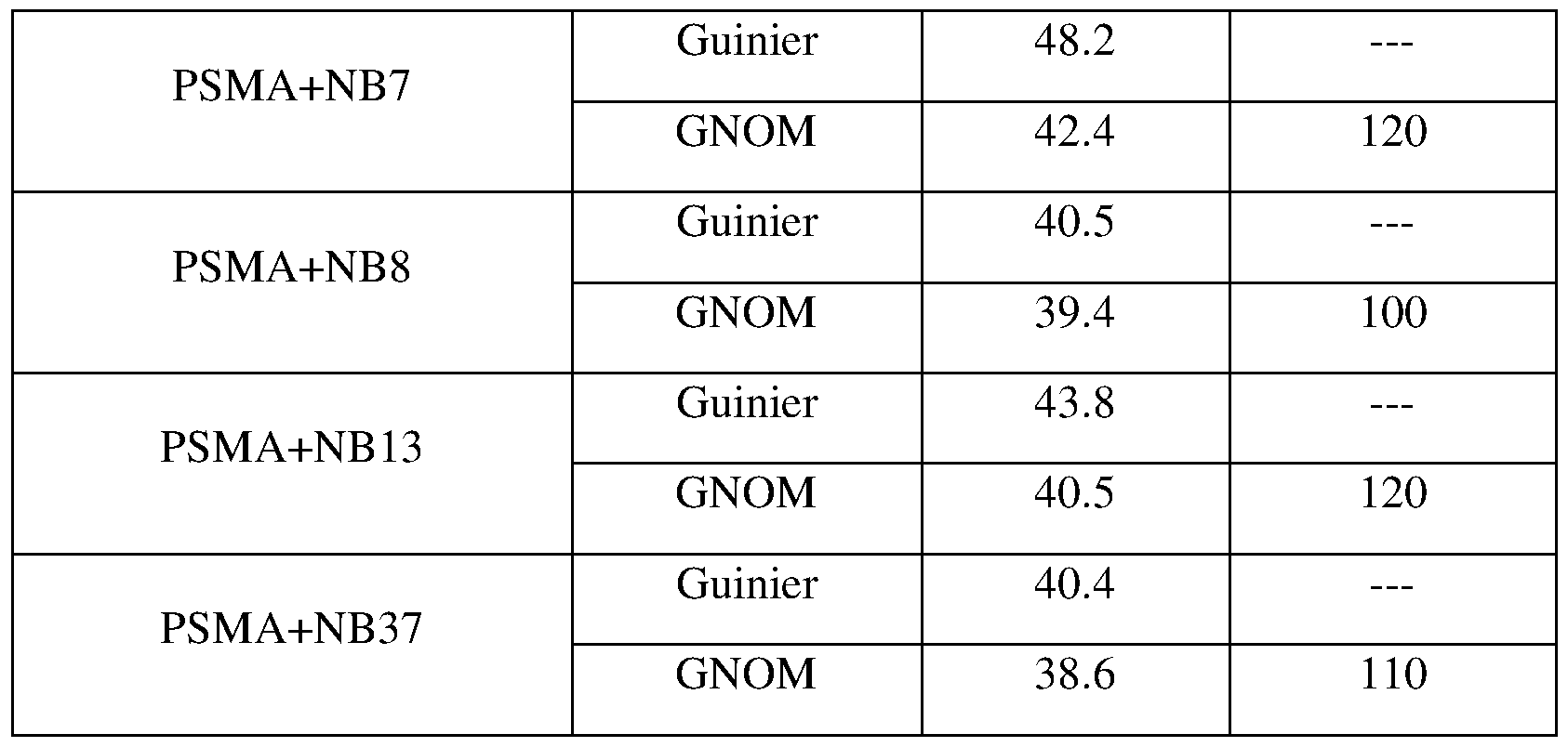
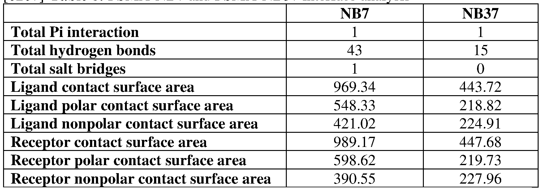
Claims
Priority Applications (10)
| Application Number | Priority Date | Filing Date | Title |
|---|---|---|---|
| CA3149754A CA3149754A1 (en) | 2019-08-29 | 2020-08-30 | A single-domain antibody for targeting prostate specific membrane antigen (psma) |
| JP2022513205A JP2022546422A (en) | 2019-08-29 | 2020-08-30 | Single domain antibodies for targeting prostate specific membrane antigen (PSMA) |
| CN202080075834.2A CN114787192B (en) | 2019-08-29 | 2020-08-30 | Single domain antibodies targeting Prostate Specific Membrane Antigen (PSMA) |
| AU2020335394A AU2020335394A1 (en) | 2019-08-29 | 2020-08-30 | A single-domain antibody for targeting prostate specific membrane antigen (PSMA) |
| US17/639,103 US12516130B2 (en) | 2019-08-29 | 2020-08-30 | Single-domain antibody for targeting prostate specific membrane antigen (PSMA) |
| MX2022002371A MX2022002371A (en) | 2019-08-29 | 2020-08-30 | A SINGLE DOMAIN ANTIBODY TO TARGET PROSTATE SPECIFIC MEMBRANE ANTIGEN (PSMA). |
| EP20857277.6A EP4021941A4 (en) | 2019-08-29 | 2020-08-30 | SINGLE-DOMAIN ANTIBODIES TO PROSTATE-SPECIFIC MEMBRANE ANTIGEN (PSMA) |
| BR112022003735A BR112022003735A2 (en) | 2019-08-29 | 2020-08-30 | Single domain antibody to target prostate-specific membrane antigen (psma) |
| KR1020227009507A KR20220106956A (en) | 2019-08-29 | 2020-08-30 | Single-Domain Antibodies to Target Prostate Specific Membrane Antigen (PSMA) |
| IL290963A IL290963A (en) | 2019-08-29 | 2022-02-27 | A single-domain antibody that binds a prostate-specific membrane antigen |
Applications Claiming Priority (2)
| Application Number | Priority Date | Filing Date | Title |
|---|---|---|---|
| US201962893264P | 2019-08-29 | 2019-08-29 | |
| US62/893,264 | 2019-08-29 |
Publications (2)
| Publication Number | Publication Date |
|---|---|
| WO2021038571A1 true WO2021038571A1 (en) | 2021-03-04 |
| WO2021038571A8 WO2021038571A8 (en) | 2022-11-17 |
Family
ID=74684983
Family Applications (1)
| Application Number | Title | Priority Date | Filing Date |
|---|---|---|---|
| PCT/IL2020/050940 Ceased WO2021038571A1 (en) | 2019-08-29 | 2020-08-30 | A single-domain antibody for targeting prostate specific membrane antigen (psma) |
Country Status (11)
| Country | Link |
|---|---|
| US (1) | US12516130B2 (en) |
| EP (1) | EP4021941A4 (en) |
| JP (1) | JP2022546422A (en) |
| KR (1) | KR20220106956A (en) |
| CN (1) | CN114787192B (en) |
| AU (1) | AU2020335394A1 (en) |
| BR (1) | BR112022003735A2 (en) |
| CA (1) | CA3149754A1 (en) |
| IL (1) | IL290963A (en) |
| MX (1) | MX2022002371A (en) |
| WO (1) | WO2021038571A1 (en) |
Cited By (1)
| Publication number | Priority date | Publication date | Assignee | Title |
|---|---|---|---|---|
| WO2025021953A1 (en) | 2023-07-25 | 2025-01-30 | Cymab Aps | Antigen-binding molecules capable of binding interferon gamma |
Families Citing this family (1)
| Publication number | Priority date | Publication date | Assignee | Title |
|---|---|---|---|---|
| WO2025195495A1 (en) * | 2024-03-21 | 2025-09-25 | 启德医药科技(苏州)有限公司 | Radionuclide drug conjugate, and preparation method therefor and use thereof |
Citations (1)
| Publication number | Priority date | Publication date | Assignee | Title |
|---|---|---|---|---|
| WO2018039180A1 (en) * | 2016-08-24 | 2018-03-01 | Teneobio, Inc. | Transgenic non-human animals producing modified heavy chain-only antibodies |
Family Cites Families (3)
| Publication number | Priority date | Publication date | Assignee | Title |
|---|---|---|---|---|
| WO2009080764A2 (en) * | 2007-12-20 | 2009-07-02 | Abylnx N.V. | Oral or nasal administration of compounds comprising amino acid sequences |
| WO2017075537A1 (en) * | 2015-10-30 | 2017-05-04 | Aleta Biotherapeutics Inc. | Compositions and methods for treatment of cancer |
| CN105524170A (en) * | 2016-02-03 | 2016-04-27 | 南昌大学 | Nanobodies capable of specifically binding to prostate-specific membrane antigen |
-
2020
- 2020-08-30 CN CN202080075834.2A patent/CN114787192B/en active Active
- 2020-08-30 WO PCT/IL2020/050940 patent/WO2021038571A1/en not_active Ceased
- 2020-08-30 CA CA3149754A patent/CA3149754A1/en active Pending
- 2020-08-30 US US17/639,103 patent/US12516130B2/en active Active
- 2020-08-30 BR BR112022003735A patent/BR112022003735A2/en not_active Application Discontinuation
- 2020-08-30 EP EP20857277.6A patent/EP4021941A4/en active Pending
- 2020-08-30 JP JP2022513205A patent/JP2022546422A/en active Pending
- 2020-08-30 AU AU2020335394A patent/AU2020335394A1/en not_active Abandoned
- 2020-08-30 KR KR1020227009507A patent/KR20220106956A/en not_active Ceased
- 2020-08-30 MX MX2022002371A patent/MX2022002371A/en unknown
-
2022
- 2022-02-27 IL IL290963A patent/IL290963A/en unknown
Patent Citations (1)
| Publication number | Priority date | Publication date | Assignee | Title |
|---|---|---|---|---|
| WO2018039180A1 (en) * | 2016-08-24 | 2018-03-01 | Teneobio, Inc. | Transgenic non-human animals producing modified heavy chain-only antibodies |
Non-Patent Citations (6)
| Title |
|---|
| CHATALIC, KRISTELL LS ET AL.: "A novel 11lln-labeled anti-prostate-specific membrane antigen nanobody for targeted SPECT/CT imaging of prostate cancer", JOURNAL OF NUCLEAR MEDICINE, vol. 56, no. 7, 14 May 2015 (2015-05-14), pages 1094 - 1099, XP055354159, Retrieved from the Internet <URL:http://jnm.snmjournals.org/content/56/7/1094.short> [retrieved on 20201202], DOI: 10.2967/ jnumed.115.156729 * |
| EVANS, JAMES C. ET AL.: "The therapeutic and diagnostic potential of the prostate specific membrane antigen/glutamate carboxypeptidase II (PSMA/GCPII) in cancer and neurological disease", BRITISH JOURNAL OF PHARMACOLOGY, vol. 173, no. 21, 15 August 2016 (2016-08-15), pages 3041 - 3079, XP055796107, Retrieved from the Internet <URL:https://bpspubs.onlinelibrary.wiley.com/doi/full/10.1111/bph.13576> [retrieved on 20201203], DOI: 10.1111/bph. 13576 * |
| EVAZALIPOUR, MEHDI ET AL.: "Generation and characterization of nanobodies targeting PSMA for molecular imaging of prostate cancer", CONTRAST MEDIA & MOLECULAR IMAGING, vol. 9, no. 3, 1 May 2014 (2014-05-01), pages 211 - 220, XP055353576, [retrieved on 20201203], DOI: https:// pubmed.ncbi.nlm.nih.gov/24700748 * |
| ROSENFELD, LIOR ET AL.: "Nanobodies Targeting Prostate-specific Membrane Antigen for the Imaging and Therapy of Prostate Cancer", JOURNAL OF MEDICINAL CHEMISTRY, 22 May 2020 (2020-05-22), XP055796102, Retrieved from the Internet <URL:https://pubs.acs.org/doi/abs/10.1021/acs.jmedchem.0c00418> [retrieved on 20201201], DOI: 10.1021/acs.jmedchem.0c00418 * |
| See also references of EP4021941A4 * |
| ZARE, HAMED ET AL.: "Production of nanobodies against prostate-specific membrane antigen (PSMA) recognizing LnCaP cells.", THE INTERNATIONAL JOURNAL OF BIOLOGICAL MARKERS, vol. 29, no. 2, 25 June 2014 (2014-06-25), pages 169 - 179, XP055353599, Retrieved from the Internet <URL:https://journals.sagepub.com/doi/full/10.5301/jbm.5000063> [retrieved on 20201201], DOI: 10.5301/jbm.5000063 * |
Cited By (1)
| Publication number | Priority date | Publication date | Assignee | Title |
|---|---|---|---|---|
| WO2025021953A1 (en) | 2023-07-25 | 2025-01-30 | Cymab Aps | Antigen-binding molecules capable of binding interferon gamma |
Also Published As
| Publication number | Publication date |
|---|---|
| WO2021038571A8 (en) | 2022-11-17 |
| AU2020335394A1 (en) | 2022-03-24 |
| CN114787192B (en) | 2024-08-09 |
| CN114787192A (en) | 2022-07-22 |
| IL290963A (en) | 2022-04-01 |
| JP2022546422A (en) | 2022-11-04 |
| EP4021941A1 (en) | 2022-07-06 |
| KR20220106956A (en) | 2022-08-01 |
| CA3149754A1 (en) | 2021-03-04 |
| US20220306764A1 (en) | 2022-09-29 |
| US12516130B2 (en) | 2026-01-06 |
| BR112022003735A2 (en) | 2022-06-21 |
| EP4021941A4 (en) | 2023-08-30 |
| MX2022002371A (en) | 2022-06-08 |
Similar Documents
| Publication | Publication Date | Title |
|---|---|---|
| US12492238B2 (en) | Glypican-3-binding fibronectin based scaffold molecules | |
| US10406225B2 (en) | Anti-EphA2 antibodies and methods of use thereof | |
| US9879093B2 (en) | Single domain antibodies as inhibitors of PCSK9 | |
| KR20200016899A (en) | Activatable anti-PDL1 antibody, and methods of using the same | |
| JP7346378B2 (en) | Therapeutic PSMA binding molecules | |
| EA037590B1 (en) | PD-L1-BINDING POLYPEPTIDES BASED ON 10FN3 DOMAINS FOR VISUALIZATION | |
| JP2019515645A (en) | Protein A binding polypeptides, anti-EphA2 antibodies, and methods of using them | |
| WO2013142965A1 (en) | Sh2 domain variants | |
| US12516130B2 (en) | Single-domain antibody for targeting prostate specific membrane antigen (PSMA) | |
| US12258424B2 (en) | Polypeptides and compositions comprising the same | |
| KR20220143701A (en) | Molecules targeting mutant RAS proteins | |
| US20210061861A1 (en) | D-peptidic compounds for vegf | |
| SG11202111656WA (en) | Anti-tie2 antibody and use thereof | |
| CN112770786B (en) | Cancer treatment using antibodies | |
| US20140296479A1 (en) | D-aptide and retro-inverso aptide with maintained target affinity and improved stability | |
| WO2023231877A1 (en) | Anti-cd228 antibody and drug conjugate thereof | |
| CA2977888C (en) | Antibody which binds lrg1 and methods of use | |
| WO2020158959A1 (en) | Anti-mc16 antibody |
Legal Events
| Date | Code | Title | Description |
|---|---|---|---|
| 121 | Ep: the epo has been informed by wipo that ep was designated in this application |
Ref document number: 20857277 Country of ref document: EP Kind code of ref document: A1 |
|
| ENP | Entry into the national phase |
Ref document number: 2022513205 Country of ref document: JP Kind code of ref document: A |
|
| WWE | Wipo information: entry into national phase |
Ref document number: 290963 Country of ref document: IL |
|
| ENP | Entry into the national phase |
Ref document number: 3149754 Country of ref document: CA |
|
| NENP | Non-entry into the national phase |
Ref country code: DE |
|
| REG | Reference to national code |
Ref country code: BR Ref legal event code: B01A Ref document number: 112022003735 Country of ref document: BR |
|
| ENP | Entry into the national phase |
Ref document number: 2020335394 Country of ref document: AU Date of ref document: 20200830 Kind code of ref document: A |
|
| ENP | Entry into the national phase |
Ref document number: 2020857277 Country of ref document: EP Effective date: 20220329 |
|
| REG | Reference to national code |
Ref country code: BR Ref legal event code: B01E Ref document number: 112022003735 Country of ref document: BR Free format text: 1) APRESENTAR, EM ATE 60 (SESSENTA) DIAS, TRADUCAO DO RELATORIO DESCRITIVO CONFORME DEPOSITADO ORIGINALMENTE, POIS A MESMA NAO FOI APRESENTADA ATE O MOMENTO TENDO SIDO APRESENTADA SOMENTE A MODIFICACAO DO PEDIDO CONFORME DESCRITO NA PETICAO NO 870220028568 DE 14/03/2022.2) APRESENTAR, EM ATE 60 (SESSENTA) DIAS, NOVO RESUMO COM O TITULO EQUIVALENTE AO APRESENTADO NO RELATORIO DESCRITIVO DA PETICAO NO 870220028568 DE 14/03/2022. |
|
| ENP | Entry into the national phase |
Ref document number: 112022003735 Country of ref document: BR Kind code of ref document: A2 Effective date: 20220225 |
|
| WWG | Wipo information: grant in national office |
Ref document number: 17639103 Country of ref document: US |
|
| WWR | Wipo information: refused in national office |
Ref document number: 1020227009507 Country of ref document: KR |