WO2021065894A1 - 運動機能の状態を評価するための方法、プログラム及び装置 - Google Patents
運動機能の状態を評価するための方法、プログラム及び装置 Download PDFInfo
- Publication number
- WO2021065894A1 WO2021065894A1 PCT/JP2020/036854 JP2020036854W WO2021065894A1 WO 2021065894 A1 WO2021065894 A1 WO 2021065894A1 JP 2020036854 W JP2020036854 W JP 2020036854W WO 2021065894 A1 WO2021065894 A1 WO 2021065894A1
- Authority
- WO
- WIPO (PCT)
- Prior art keywords
- patient
- index value
- motor function
- physiological index
- brain
- Prior art date
- Legal status (The legal status is an assumption and is not a legal conclusion. Google has not performed a legal analysis and makes no representation as to the accuracy of the status listed.)
- Ceased
Links
Images







Classifications
-
- G—PHYSICS
- G16—INFORMATION AND COMMUNICATION TECHNOLOGY [ICT] SPECIALLY ADAPTED FOR SPECIFIC APPLICATION FIELDS
- G16H—HEALTHCARE INFORMATICS, i.e. INFORMATION AND COMMUNICATION TECHNOLOGY [ICT] SPECIALLY ADAPTED FOR THE HANDLING OR PROCESSING OF MEDICAL OR HEALTHCARE DATA
- G16H30/00—ICT specially adapted for the handling or processing of medical images
- G16H30/40—ICT specially adapted for the handling or processing of medical images for processing medical images, e.g. editing
-
- A—HUMAN NECESSITIES
- A61—MEDICAL OR VETERINARY SCIENCE; HYGIENE
- A61B—DIAGNOSIS; SURGERY; IDENTIFICATION
- A61B5/00—Measuring for diagnostic purposes; Identification of persons
- A61B5/0033—Features or image-related aspects of imaging apparatus, e.g. for MRI, optical tomography or impedance tomography apparatus; Arrangements of imaging apparatus in a room
- A61B5/004—Features or image-related aspects of imaging apparatus, e.g. for MRI, optical tomography or impedance tomography apparatus; Arrangements of imaging apparatus in a room adapted for image acquisition of a particular organ or body part
- A61B5/0042—Features or image-related aspects of imaging apparatus, e.g. for MRI, optical tomography or impedance tomography apparatus; Arrangements of imaging apparatus in a room adapted for image acquisition of a particular organ or body part for the brain
-
- A—HUMAN NECESSITIES
- A61—MEDICAL OR VETERINARY SCIENCE; HYGIENE
- A61B—DIAGNOSIS; SURGERY; IDENTIFICATION
- A61B5/00—Measuring for diagnostic purposes; Identification of persons
- A61B5/05—Detecting, measuring or recording for diagnosis by means of electric currents or magnetic fields; Measuring using microwaves or radio waves
- A61B5/055—Detecting, measuring or recording for diagnosis by means of electric currents or magnetic fields; Measuring using microwaves or radio waves involving electronic [EMR] or nuclear [NMR] magnetic resonance, e.g. magnetic resonance imaging
-
- A—HUMAN NECESSITIES
- A61—MEDICAL OR VETERINARY SCIENCE; HYGIENE
- A61B—DIAGNOSIS; SURGERY; IDENTIFICATION
- A61B5/00—Measuring for diagnostic purposes; Identification of persons
- A61B5/40—Detecting, measuring or recording for evaluating the nervous system
- A61B5/4058—Detecting, measuring or recording for evaluating the nervous system for evaluating the central nervous system
- A61B5/4064—Evaluating the brain
-
- A—HUMAN NECESSITIES
- A61—MEDICAL OR VETERINARY SCIENCE; HYGIENE
- A61B—DIAGNOSIS; SURGERY; IDENTIFICATION
- A61B5/00—Measuring for diagnostic purposes; Identification of persons
- A61B5/40—Detecting, measuring or recording for evaluating the nervous system
- A61B5/4076—Diagnosing or monitoring particular conditions of the nervous system
- A61B5/4082—Diagnosing or monitoring movement diseases, e.g. Parkinson, Huntington or Tourette
-
- A—HUMAN NECESSITIES
- A61—MEDICAL OR VETERINARY SCIENCE; HYGIENE
- A61B—DIAGNOSIS; SURGERY; IDENTIFICATION
- A61B5/00—Measuring for diagnostic purposes; Identification of persons
- A61B5/48—Other medical applications
- A61B5/4848—Monitoring or testing the effects of treatment, e.g. of medication
-
- G—PHYSICS
- G01—MEASURING; TESTING
- G01R—MEASURING ELECTRIC VARIABLES; MEASURING MAGNETIC VARIABLES
- G01R33/00—Arrangements or instruments for measuring magnetic variables
- G01R33/20—Arrangements or instruments for measuring magnetic variables involving magnetic resonance
- G01R33/44—Arrangements or instruments for measuring magnetic variables involving magnetic resonance using nuclear magnetic resonance [NMR]
- G01R33/48—NMR imaging systems
- G01R33/54—Signal processing systems, e.g. using pulse sequences ; Generation or control of pulse sequences; Operator console
- G01R33/56—Image enhancement or correction, e.g. subtraction or averaging techniques, e.g. improvement of signal-to-noise ratio and resolution
- G01R33/563—Image enhancement or correction, e.g. subtraction or averaging techniques, e.g. improvement of signal-to-noise ratio and resolution of moving material, e.g. flow contrast angiography
- G01R33/56341—Diffusion imaging
-
- G—PHYSICS
- G16—INFORMATION AND COMMUNICATION TECHNOLOGY [ICT] SPECIALLY ADAPTED FOR SPECIFIC APPLICATION FIELDS
- G16H—HEALTHCARE INFORMATICS, i.e. INFORMATION AND COMMUNICATION TECHNOLOGY [ICT] SPECIALLY ADAPTED FOR THE HANDLING OR PROCESSING OF MEDICAL OR HEALTHCARE DATA
- G16H50/00—ICT specially adapted for medical diagnosis, medical simulation or medical data mining; ICT specially adapted for detecting, monitoring or modelling epidemics or pandemics
- G16H50/30—ICT specially adapted for medical diagnosis, medical simulation or medical data mining; ICT specially adapted for detecting, monitoring or modelling epidemics or pandemics for calculating health indices; for individual health risk assessment
Definitions
- the present disclosure relates to a method of assessing the state of motor function of a patient with or suspected of having a brain injury, as well as programs, image analyzers and MRI devices that can be used in the practice of such methods.
- the present disclosure relates to a method for predicting motor function recovery after regenerative treatment in a patient with local brain injury.
- Non-Patent Document 1 In cell therapy, the recovery condition of patients who actually administered cells varies, so it is important to determine which patient is suitable for cell therapy before starting treatment (Non-Patent Document 1).
- the present disclosure provides a means for assessing the state of motor function of a patient who has or is suspected of having a brain injury such as a local brain injury.
- the disclosure specifically provides a means for determining whether such a patient is suitable for regenerative therapy, i.e., whether the patient is expected to respond to regenerative therapy prior to initiation of treatment.
- the present inventors have found that in a patient with local brain injury, the state of motor function correlates with the parameters obtained from the diffusion-weighted image of the patient's brain.
- the present disclosure provides: (Item 1) A step of obtaining a physiological index value with the outer surface region on the traumatic hemispherical side of the brain of a patient having or suspected of having a brain injury as a region of interest.
- a method for evaluating the state of motor function of a patient which comprises a step of comparing the physiological index value with a control physiological index value and performing an operation for calculating a value indicating the state of motor function of the patient.
- DTI diffusion tensor imaging
- DKI diffusion kurtosis imaging
- the physiological index value includes an FA (fractional anisotropy) value.
- the region of interest is set in the precentral gyrus of the brain.
- the state of motor function is the state of motor function after regenerative treatment.
- the control physiological index value is obtained by using the outer surface region on the non-injured hemisphere side of the patient's brain as a region of interest.
- the step of performing the calculation compares the calculated value indicating the state of the motor function of the patient with a reference value prepared in advance, whereby the desired degree of recovery of the motor function of the patient after the regenerative treatment.
- the method according to any one of the above items, which calculates the height of the possibility of reaching.
- (Item 10) A computer program that causes a computer to perform a process of a method of evaluating the state of motor function of a patient having or suspected of having a brain injury, wherein the method is as follows: A step of causing the computer to obtain a physiological index value with the outer surface region on the traumatic hemisphere side of the patient's brain as the region of interest.
- a program comprising the step of causing the computer to perform an operation for comparing the physiological index value with a control physiological index value and calculating a value indicating a state of motor function of the patient.
- a recording medium comprising a step of causing the computer to perform an operation for comparing the physiological index value with a control physiological index value and calculating a value indicating a state of motor function of the patient.
- a system for evaluating the state of motor function of the brain of a patient who has or is suspected of having a brain injury A means for obtaining a physiological index value with the outer surface region on the traumatic hemisphere side of the patient's brain as the region of interest.
- a system comprising a means of comparing the physiological index value with a control physiological index value and performing an operation for calculating a value indicating a state of motor function of the patient.
- the outer surface region on the injured hemisphere side is the first region of interest
- the outer surface region on the non-injured hemisphere side is the second interest region.
- the area of interest setting section to be set as A physiological index value calculation unit that calculates a damaged hemispherical side physiological index value and an uninjured hemispherical side physiological index value in each of the first region of interest and the second region of interest,
- An image characterized by including an arithmetic unit that performs an operation for comparing a physiological index value on the injured hemisphere side and a physiological index value on the non-injured hemisphere side and calculating a value indicating a state of motor function of the patient.
- Analytical device. (Item 14) A nuclear magnetic resonance imaging unit that images the brain of a patient who has or is suspected of having a brain injury.
- An image generator that generates a diffusion-weighted image from the echo data acquired by the nuclear magnetic resonance imaging unit, In the diffusion-weighted image, a region of interest setting unit that sets the outer surface region on the damaged hemisphere side as the first region of interest and the outer surface region on the non-damaged hemisphere side as the second region of interest.
- a physiological index value calculation unit that calculates a damaged hemispherical side physiological index value and an uninjured hemispherical side physiological index value in each of the first region of interest and the second region of interest, and a physiological index value calculation unit.
- An MRI comprising a calculation unit that compares an injured hemisphere side physiological index value with an uninjured hemisphere side physiological index value and performs an calculation for calculating a value indicating a state of motor function of the patient. apparatus.
- the disclosure also provides: (1) In the diffusion tensor image of the brain of a patient with local brain injury, the step of setting the precentral gyrus on the injured hemisphere side as the first region of interest and the precentral gyrus on the non-injured hemisphere side as the second region of interest.
- a method of predicting motor function recovery of a patient after regenerative treatment including a step of performing an operation for predicting motor function recovery of the patient after regenerative treatment based on the calculated rFA.
- the calculated rFA is substituted into a regression line prepared in advance with rFA and the degree of recovery of motor function as variables, so that the degree of recovery of motor function after regenerative treatment of the patient is performed.
- the method according to (1) which is a step of calculating the predicted value of.
- the step of performing the above calculation is a step of calculating the degree of probability that the patient will reach the desired degree of recovery of motor function after regenerative treatment by comparing the calculated rFA with the standard rFA prepared in advance. Yes, the method according to (1).
- a program for predicting the recovery of motor function of the patient after regenerative treatment to execute a process of performing an operation for predicting the recovery of motor function of the patient after regenerative treatment based on the calculated rFA. .. (5)
- the process of performing the above calculation substitutes the calculated rFA into a regression line having a prepared rFA and the degree of recovery of motor function as variables, so that the degree of recovery of motor function after regenerative treatment of the patient is performed.
- the program according to (4) which is a process of calculating the predicted value of. (6)
- the calculated rFA is compared with the standard rFA prepared in advance to calculate the high or low probability that the patient will reach the desired degree of recovery of motor function after regenerative treatment. Yes, the program described in (4).
- a computer-readable storage medium that stores the program according to any one of (4) to (6).
- the precentral gyrus on the injured hemisphere side is set as the first precentral gyrus and the precentral gyrus on the non-injured hemisphere side is set as the second interest region.
- the FA calculation unit that calculates the damaged hemisphere side FA and the non-damaged hemisphere side FA, which are the anisotropic ratios in the first region of interest and the second region of interest, respectively.
- the rFA calculation unit that calculates the rFA represented by the damaged hemisphere side FA / non-damaged hemisphere side FA,
- An image analysis apparatus including a recovery prediction calculation unit that performs a calculation for predicting the recovery of motor function of the patient after regenerative treatment based on the calculated rFA.
- a nuclear magnetic resonance imaging unit that images the brain of a patient with local brain damage
- An image generator that generates a diffusion tensor image from the echo data acquired by the nuclear magnetic resonance imaging unit, In the diffusion tensor image, the precentral gyrus on the damaged hemisphere side is set as the first region of interest, and the precentral gyrus on the non-damaged hemisphere side is set as the second region of interest.
- the FA calculation unit that calculates the damaged hemisphere side FA and the non-damaged hemisphere side FA, which are the anisotropic ratios in the first region of interest and the second region of interest, respectively.
- the rFA calculation unit that calculates the rFA represented by the damaged hemisphere side FA / non-damaged hemisphere side FA,
- An MRI apparatus comprising: a recovery prediction calculation unit that performs a calculation for predicting the recovery of motor function of the patient after regenerative treatment based on the calculated rFA.
- the present disclosure it is possible to evaluate the state of motor function of a patient who has or is suspected of having brain damage such as local brain damage, and thereby predicts the recovery of motor function of the patient after regenerative treatment. Also, it can be determined whether the patient is suitable for regenerative treatment before the start of treatment.
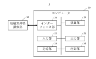
- FIG. 1 is a block diagram showing an overall configuration of an MRI apparatus according to one embodiment of the present disclosure.
- FIG. 2 is a functional block diagram showing the configurations of a storage unit, a calculation unit, an input unit, and an output unit in the MRI apparatus according to one embodiment of the present disclosure.
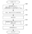
- FIG. 3 is a flowchart showing each step in the prediction method according to one embodiment of the present disclosure.
- FIG. 4 is a flowchart showing the details of the calculation steps in the prediction method according to one embodiment of the present disclosure.
- FIG. 5 is a flowchart showing the details of the calculation steps in the prediction method according to one embodiment of the present disclosure.
- FIG. 1 is a block diagram showing an overall configuration of an MRI apparatus according to one embodiment of the present disclosure.
- FIG. 2 is a functional block diagram showing the configurations of a storage unit, a calculation unit, an input unit, and an output unit in the MRI apparatus according to one embodiment of the present disclosure.
- FIG. 3 is a flowchart showing each step in
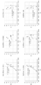
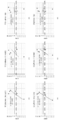
- FIG. 6 shows the rFA calculated by setting the ROI at one of the internal capsule hind legs and the pons in the precentral gyrus and the motor function recovery degree ⁇ BI of the patient in the brain DTI image of the cerebral infarction patient. It is a plotted scatter diagram.
- FIG. 7 shows the rFA calculated by setting the ROI at any two of the internal capsule hind legs and the pons in the precentral gyrus and the motor function recovery degree ⁇ BI of the patient in the brain DTI image of the cerebral infarction patient. It is a plotted scatter diagram.
- FIG. 7 shows the rFA calculated by setting the ROI at any two of the internal capsule hind legs and the pons in the precentral gyrus and the motor function recovery degree ⁇ BI of the patient in the brain DTI image of the cerebral infarction patient. It is a plotted scatter diagram.
- FIG. 7 shows the rFA calculated by setting the ROI at any two of the internal capsule hind legs and the pons in the precentral gyrus
- FIG. 8 is a scatter plot of the rFA calculated by setting the ROI at three locations of the internal capsule hind leg and the pons in the brain DTI image of the cerebral infarction patient, and the motor function recovery degree ⁇ BI of the patient.
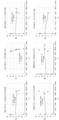
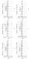
- FIG. 9 shows the rFA calculated by setting the ROI at one of the internal capsule hind legs and the pons in the precentral gyrus and the motor function recovery degree ⁇ FMA of the patient in the brain DTI image of the cerebral infarction patient. It is a plotted scatter diagram. In FIG.
- FIG. 10 in the brain DTI image of a cerebral infarction patient, the rFA calculated by setting the ROI at any two of the precentral gyrus, the internal capsule hind leg and the pons, and the motor function recovery degree ⁇ FMA of the patient are shown. It is a plotted scatter diagram.
- FIG. 11 is a scatter plot of the rFA calculated by setting the ROI at three locations of the internal capsule hind leg and the pons in the brain DTI image of the cerebral infarction patient, and the motor function recovery degree ⁇ FMA of the patient.
- FIG. 12 is a schematic diagram showing a procedure of image processing by DKI in one embodiment of the present disclosure.
- FIG. 12 is a schematic diagram showing a procedure of image processing by DKI in one embodiment of the present disclosure.
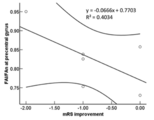
- FIG. 13 is a graph showing the correlation between the precentral gyrus FA ratio (lesion / healthy) and the change in mRS by DKI according to the embodiment of the present disclosure.
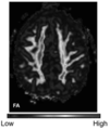
- FIG. 14A is a photograph showing a case of cerebral infarction in the left cerebral hemisphere with a poor degree of functional recovery. It can be seen that the previous FA in the left center is lower than that in the right side.
- FIG. 14B is a photograph showing a case of cerebral infarction in the left cerebral hemisphere with a large degree of functional recovery. It can be seen that the difference between the left and right sides of the precentral gyrus FA is small.
- the "outer surface region” refers to the region of the outer surface of the cerebrum, and particularly refers to the gyrus.
- the superior temporal gyrus, superior temporal gyrus, medial frontal gyrus, inferior temporal gyrus, superior temporal gyrus, medial temporal gyrus, inferior temporal gyrus, fusiform gyrus, paracapital gyrus, lateral temporal gyrus Includes inferior temporal gyrus, superior temporal gyrus, superior temporal gyrus, superior temporal gyrus, zonal gyrus, dentate gyrus, paracenter leaflet, wedge, anterior wedge, tongue gyrus, etc.
- the "physiological index value” refers to an index value obtained by diffusion-weighted imaging of the brain such as diffusion tensor image (DTI) and diffusion kurtosis image (DKI), and MK (meankurtosis) and AK (axial). Includes kurtosis), RK (radial kurtosis), FA (fractional anisotropy), KFA (kurtosisfractional anisotropy), MD (mean diffusion), AD (axial diffusion), and RD (radialdiffusivity).
- DTI diffusion tensor image
- DKI diffusion kurtosis image
- MK meankurtosis
- AK axial.
- kurtosis kurtosis
- RK radial kurtosis
- FA fractional anisotropy
- KFA kurtosisfractional anisotropy
- MD mean diffusion
- AD axial diffusion
- RD radialdiffusivity
- the "state of motor function” refers to the state of motor function that is comprehensively controlled by the function of the brain
- the “evaluation of the state of motor function” refers to the current state of the brain of the subject. Or, after treatment, it includes evaluating or predicting whether motor function is normal or partially or completely paralyzed. For example, “evaluation of the state of motor function” includes prediction of recovery of brain function that controls movement by regenerative therapy or the like.
- a method of assessing the state of motor function of the patient including a step of making a comparison and performing an operation to calculate a value indicating the state of motor function of the patient.
- the physiological index value can be obtained from an image of the patient's brain obtained by the diffusion tensor imaging (DTI) method or the diffusion kurtosis imaging (DKI) method.
- DTI diffusion tensor imaging
- DKI diffusion kurtosis imaging
- a region of interest (ROI) created by tracing an anatomical structure is set, and physiological index values such as FA value and MD value in the region of interest are measured to measure the corticospinal cord. You can track the road.
- the DKI method can be used as a quantitative image index that does not depend on the measurer.
- the DTI method assumes that the diffusion of water molecules is a normal distribution, it is known that the diffusion of water molecules in a living body does not show a normal distribution. Therefore, a diffusion imaging method that does not assume a normal distribution, such as the DKI method, can more accurately reflect the diffusion of water molecules in the living body. Therefore, in one embodiment of the present disclosure, according to the DKI method, it is possible to obtain a diffusion image index that can predict the cell administration therapeutic effect in a cerebral infarction patient with high accuracy and without depending on the experience of the measurer or the like. ..
- the physiologic index value obtained from the diffusion weighted image of the patient's brain obtained by the diffusion tensor imaging (DTI) method or the diffusion kurtosis imaging (DKI) method as described above is the diffusion of water molecules in the tissue.
- the index is not particularly limited as long as it is an index obtained by a diffusion-weighted image that images the direction and speed of RK (mean kurtosis), AK (axial kurtosis), RK (radial kurtosis), FA ( Diffusion indices such as fractionalanisotropy), KFA (kurtosis fractional anisotropy), MD (mean diffusivity), AD (axialdiffusivity), and RD (radial diffusivity) can be used.
- these diffusion indices can be used alone or in combination of two or more.
- the ROI in DTI or DKI may be set anywhere as long as it is on the outer surface region of the brain, and is not particularly limited. In one embodiment, the ROI is preferably set at the precentral gyrus.
- the state of motor function of the subject is based on the physiological index value obtained from the image of the patient's brain obtained by the diffusion tensor imaging (DTI) method or the diffusion kurtosis imaging (DKI) method as described above.
- This motor function may be current or post-treatment, and the recovery state of motor function after regenerative treatment can also be predicted.
- a physiological index value obtained when the ROI is placed on the injured hemisphere side of the brain and an ROI obtained when the ROI is placed on the uninjured hemisphere side of the brain can be performed by comparing with a physiological index value (referred to as a control physiological index value), and preferably by calculating a value represented by a physiological index value / a control physiological index value.
- the evaluation of the state of motor function is a regression in which the value represented by the physiological index value / control physiological index value is a value indicating the state of motor function of the patient and the degree of recovery of motor function as variables. It can also be done by substituting into a straight line. In another embodiment, the possibility is shown by comparing the value represented by the physiological index value / control physiological index value with a reference value indicating that the patient is likely to reach the desired degree of recovery of motor function after regenerative treatment. You can also.
- the method of the present disclosure it is possible to evaluate the state of motor function of a subject, and it is possible to evaluate the state of motor function after regenerative treatment, that is, whether or not motor function is restored by regenerative treatment. Since it can be predicted, it becomes possible to determine which patient is suitable for cell therapy before the start of treatment, and it is also possible to select a patient having a high therapeutic effect.
- the present disclosure also provides a computer program for causing a computer to execute the above method, a recording medium for storing the program, and a system for executing the above method.
- a computer program that causes a computer to perform a process of a method of evaluating the state of motor function of a patient having or suspected of having a brain injury, wherein the method is the following step: A step of causing the computer to obtain a physiological index value with the outer surface region on the traumatic hemisphere side of the patient's brain as the region of interest.
- a program is provided that includes a step of causing the computer to perform an operation for comparing the physiological index value with a control physiological index value and calculating a value indicating the state of motor function of the patient.
- a recording medium containing a computer program that causes a computer to perform a process of assessing the state of motor function of a patient with or suspected of having brain damage.
- a recording medium is provided including a step of causing the computer to perform an operation for comparing the physiological index value with a control physiological index value and calculating a value indicating a state of motor function of the patient.
- a system for assessing the state of motor function of a patient with or suspected of having a brain injury in another aspect of the present disclosure A means for obtaining a physiological index value with the outer surface region on the traumatic hemisphere side of the patient's brain as the region of interest.
- a system is provided that includes means for comparing the physiological index value with a control physiological index value and performing an operation for calculating a value indicating the state of motor function of the patient.
- an image analysis device and an MRI device that can be used in carrying out a method for evaluating the state of motor function of a patient having or suspected of having a brain injury as described above. ..
- the outer surface region on the injured hemisphere side is the first region of interest and the outer surface region on the non-injured hemisphere side.
- the area of interest setting unit that sets A physiological index value calculation unit that calculates a damaged hemispherical side physiological index value and an uninjured hemispherical side physiological index value in each of the first region of interest and the second region of interest, and a physiological index value calculation unit.
- An image characterized by including an arithmetic unit that performs an operation for comparing a physiological index value on the injured hemisphere side and a physiological index value on the non-injured hemisphere side and calculating a value indicating a state of motor function of the patient.
- An analyzer is provided.
- a nuclear magnetic resonance imaging unit that images the brain of a patient with or suspected of having a brain injury.
- An image generator that generates a diffusion-weighted image from the echo data acquired by the nuclear magnetic resonance imaging unit, In the diffusion-weighted image, a region of interest setting unit that sets the outer surface region on the damaged hemisphere side as the first region of interest and the outer surface region on the non-damaged hemisphere side as the second region of interest.
- a physiological index value calculation unit that calculates a damaged hemispherical side physiological index value and an uninjured hemispherical side physiological index value in each of the first region of interest and the second region of interest,
- An MRI comprising a calculation unit that compares an injured hemisphere side physiological index value with an uninjured hemisphere side physiological index value and performs an calculation for calculating a value indicating a state of motor function of the patient.
- Equipment is provided.
- the same configuration as the image analysis device and the MRI device described in detail elsewhere in this specification can be provided.
- aspects of the disclosure relate to methods of predicting motor function recovery after regenerative treatment in patients with local brain injury, as well as programs, image analyzers and MRI devices that can be used in the practice of such methods.
- FIGS. 1 to 5 An exemplary embodiment of a method for predicting motor function recovery after regenerative treatment in a patient with local brain injury and an MRI apparatus that can be used in carrying out the method will be described with reference to FIGS. 1 to 5.
- FIG. 1 shows a block diagram showing the overall configuration of the MRI apparatus according to the embodiment of the present disclosure.
- the MRI apparatus 1 has a nuclear magnetic resonance imaging unit 10 and a computer 20.
- the nuclear magnetic resonance imaging unit 10 is an imaging unit used in any known MRI apparatus capable of acquiring echo data required for a DTI image.
- the nuclear magnetic resonance imaging unit 10 has a sleeper unit; a magnet mount unit including a static magnetic field magnet, a gradient magnetic field coil, and an RF coil; and a sequence control unit.
- the bed portion on which the patient is placed is inserted into the opening portion of the magnet mount portion.
- the static magnetic field magnet of the magnet mount generates a static magnetic field, and the gradient magnetic field coil also applies a gradient magnetic field.
- the transmitting RF coil then generates a high frequency magnetic field, which causes the receiving RF coil to receive the echo signal emitted by the patient.
- the received echo signal is digitized and transmitted as echo data to the sequence control unit.
- the sequence control unit controls imaging based on the sequence information transmitted from the computer 20, and also transfers the received echo data to the computer 20.
- the computer 20 is a device that controls the nuclear magnetic resonance imaging unit 10, collects data, reconstructs an image, and analyzes an image.
- the computer 20 is an interface unit 21, an input unit 22, a storage unit 23, a calculation unit 24, and an output unit. It has 25 and a control unit 26.
- the interface unit 21 controls the input / output of various data such as echo data exchanged with the sequence control unit of the nuclear magnetic resonance imaging unit 10.
- the interface unit 21 transmits sequence information for controlling the nuclear magnetic resonance imaging unit 10 to the sequence control unit.
- the interface unit 21 also receives echo data from the sequence control unit and stores it in the storage unit 23.
- the input unit 22 is a device such as a keyboard, mouse, button, switch, etc., and inputs a signal corresponding to the operation of the operator for these devices.
- the storage unit 23 includes a storage medium such as a hard disk, a flash memory, RAM, and a ROM, and a reading device for reading the information stored in the storage medium.
- the storage unit 23 includes echo data transmitted from the sequence control unit, various MRI image data generated from the echo data, reference information for predicting the recovery of motor function of the patient, a program for image generation and analysis, and a control unit. Stores programs for controlling each functional unit of the MRI apparatus 1 executed by 26, as well as various setting information.
- the calculation unit 24 is composed of hardware such as a CPU.
- the calculation unit 24 reads the program stored in the storage unit 23, reconstructs an image from the echo data stored in the storage unit 23, and generates a DTI image from the reconstructed image.
- the calculation unit 24 also analyzes the DTI image of the patient's brain stored in the storage unit 23, and performs calculations for predicting the recovery of motor function of the patient.
- the output unit 25 is a display connected to the outside of the computer 20, typically the output unit 25, with various information such as the MRI image generated by the calculation unit 24 and the predicted value of the motor function recovery degree obtained by performing the calculation. Output to.
- the control unit 26 is connected to each functional unit constituting the MRI apparatus 1, reads a program stored in the storage unit 23, and controls these operations. For example, the control unit 26 generates sequence information from the imaging conditions set by the operator and transmits the sequence information to the sequence control unit to control the imaging of the nuclear magnetic resonance imaging unit 10.
- the imaging conditions are set values of various DTI imaging parameters such as b value, TR, TE, NEX, voxel size, number of slices, diffusion gradient direction, etc. The value can be set as appropriate.
- FIG. 2 is a functional block diagram showing the configurations of a storage unit, a calculation unit, an input unit, and an output unit according to the embodiment of the present disclosure.
- the storage unit 23 includes an echo data storage unit 231, a reconstructed image storage unit 232, a DTI image storage unit 233, and a reference information storage unit 234. Further, the calculation unit 24 has an image generation unit 241 and an image analysis unit 242.
- the image generation unit 241 has an image reconstruction unit 241a and a DTI image generation unit 241b
- the image analysis unit 242 has an interest region setting unit 242a, an FA calculation unit 242b, an rFA calculation unit 242c, and a recovery prediction calculation unit 242d.
- the function of the image generation unit 241 that generates a DTI image from echo data will be explained.
- the echo data storage unit 231 stores the echo data transmitted from the sequence control unit for each patient.
- the image reconstruction unit 241a generates a reconstruction image such as a diffusion-weighted image (DWI image) by performing a reconstruction process such as a Fourier transform on the echo data stored by the echo data storage unit 231.
- the reconstructed image storage unit 232 stores the generated reconstructed image.
- the DTI image generation unit 241b performs DTI analysis on the generated reconstructed image and generates a DTI image.
- the diffusion coefficients D xx , D xy , D xz , D yy , D yz , and D zz which are the components of the diffusion tensor D represented by the 3 ⁇ 3 symmetric matrix of Equation 1, are calculated for each voxel.
- the eigenvalues ( ⁇ 1 , ⁇ 2 , ⁇ 3 ) shown in Equation 2 are calculated.
- D xx , D yy and D zz are diffusion coefficients when a gradient magnetic field is applied in the x-axis direction, the y-axis direction and the z-axis direction of the MRI apparatus coordinate system, respectively.
- the DTI image generation unit 241b generates a DTI image such as a ⁇ 1 map, a ⁇ 2 map, a ⁇ 3 map, an ADC map, or an FA map by mapping these parameters.
- the DTI image storage unit 233 stores the generated DTI image.
- FIG. 3 is a flowchart showing each step in the prediction method according to the embodiment of the present disclosure implemented by using each of the functional units
- FIGS. 4 and 5 are flowcharts showing details of the calculation steps in the prediction method. Is.
- the region of interest setting unit 242a sets the first region of interest (ROI-1) at a position corresponding to the precentral gyrus on the damaged hemisphere side on the DTI image stored in the DTI image storage unit 233, typically on the FA map.
- the second region of interest (ROI-2) is set at the position corresponding to the precentral gyrus on the undamaged hemisphere side (step S10).
- the ROI may be set manually based on the anatomical position, superimposed on other medical images, or automatically based on the functionality of known DTI image analysis software. May be good. When setting the ROI manually, the operation of setting the ROI by the operator is accepted via the input unit 22.
- the FA calculation unit 242b calculates the damaged hemisphere side FA and the non-damaged hemisphere side FA, which are the FAs of ROI-1 and ROI-2, respectively (step S11).
- the damaged hemispherical FA and the non-damaged hemispherical FA are the average values of FA of all voxels contained in ROI-1 and ROI-2, respectively.
- the rFA calculation unit 242c calculates the rFA represented by the damaged hemisphere side FA / non-damaged hemisphere side FA (step S12).
- the recovery prediction calculation unit 242d performs a calculation for predicting the recovery of motor function of the patient after regenerative treatment based on the rFA calculated by the rFA calculation unit 242c (step S13), and outputs the calculation result to the output unit 25. Output (step S14).
- the calculation for predicting the recovery of motor function is performed based on the reference information stored in the reference information storage unit 234 in addition to the calculated rFA.
- the reference information is a prepared regression line with variables of rFA of a patient with local brain injury before regenerative treatment and the degree of motor function recovery of the patient after treatment. is there.
- the degree of motor function recovery is the recovery of motor function by treatment, which is evaluated using a known evaluation method capable of evaluating motor function, such as BathelIndex (BI) or Fugl-Meyer Assessment (FMA). It is a degree and can be expressed as the difference between the pre-treatment score and the post-treatment score in the same patient.
- the reference information is a pre-prepared reference rFA.
- Criteria rFA distinguishes between patients with local brain damage who are expected to achieve the desired degree of motor function recovery after regenerative treatment and those who have local brain damage who are not expected to achieve the desired degree of motor function recovery. Is the cutoff value for.
- the present inventors have found that the degree of recovery of motor function after regenerative treatment correlates with rFA before treatment in patients with local brain injury. Therefore, by referring to the rFA calculated from the pretreatment brain DTI image of the patient who has already received the regenerative treatment and the degree of recovery of motor function after the treatment, the post-treatment exercise of the patient who is going to receive the regenerative treatment from now on. Functional recovery can be predicted and whether the patient is expected to be adaptable to regenerative therapy, i.e., can be expected to respond to regenerative therapy.
- a regression line in which rFA calculated from the pretreatment brain DTI image of a patient who has already undergone regenerative treatment and the degree of recovery of motor function after treatment are used as variables is prepared in advance, and regenerative treatment is to be performed from now on.
- the predicted value of the degree of motor function recovery after regenerative treatment of the test patient can be calculated (step S130a, step S130a, S130b).
- the desired exercise is based on a scatter diagram or regression line with rFA calculated from the pretreatment brain DTI image of a patient who has already undergone regenerative treatment and the degree of recovery of motor function after treatment as variables.
- the pretreatment rFA corresponding to the degree of functional recovery is determined in advance as the reference rFA, and whether or not the rFA calculated from the brain DTI image of the test patient who is about to receive regenerative treatment exceeds the reference rFA is determined. It is possible to calculate the likelihood that the test patient will reach the desired degree of recovery of motor function after regenerative treatment (steps S130b to S133b).
- the reference rFA can be appropriately set according to the target degree of recovery of motor function.
- the reference information such as the regression line and the reference rFA is obtained from a patient having the same kind of disease as the patient whose motor function recovery is predicted after the regenerative treatment.
- the reference information uses rFA calculated from the pretreatment brain DTI image of the cerebral infarction patient who received regenerative treatment and the degree of motor function recovery after treatment. It is preferable that it is prepared.
- the reference information is also preferably obtained from a patient who has received the same type of treatment as the treatment to be indicated.
- the treatment is administration of bone marrow mesenchymal stem cells (BMSC)
- the reference information is rFA calculated from pretreatment brain DTI images of patients receiving BMSC and the degree of motor function recovery after treatment. It is preferable that it is prepared by using.
- An exemplary embodiment of the image analysis apparatus according to the present disclosure that can be used in the implementation of the method is an apparatus having an image analysis unit 242 of the MRI apparatus 1, the details of which are as described in the above description. ..
- the program according to the present disclosure that can be used in the implementation of the method is a program for causing a computer to execute each step of the method, and the details thereof are as described in the above description.
- the computer-readable storage medium that stores the program can be any storage medium such as a hard disk, flash memory, CD, or DVD.
- the methods and programs according to the present disclosure can also be expressed as methods and programs for assessing a patient's adaptability to regenerative therapy, and methods and programs that assist in assessing a patient's adaptability to regenerative therapy. The same applies to other aspects of the present disclosure.
- brain injury means that some kind of damage occurs in the brain, and includes damage to blood vessels in the brain.
- the cause of the injury is not particularly limited, and for example, "brain injury” includes traumatic brain injury, stroke, cerebral infarction, oxygen-deficient brain injury, brain tumor encephalitis, and local brain injury.
- local brain damage refers to a state in which local damage is caused in the brain
- patient with local brain damage refers to a patient in which local damage is caused in the brain.
- local brain injury include cerebral infarction, head trauma and cerebral hemorrhage.
- the patient having a local brain injury is not limited as long as the patient has a local injury in the brain and has impaired motor function, and is in the acute phase, subacute phase, and chronic phase. It may be a patient at any time.
- regenerative treatment for a patient with local brain injury refers to the injured nervous system cells of the patient using the cells of the patient (autologous) or others (allogeneic allogeneic) or their secretions. It means treatment to regenerate and recover various dysfunctions associated with injury.
- the cells that can be used for regenerative treatment of patients with local brain damage may be cells capable of differentiating into neural cells, and examples thereof include pluripotent cells such as mesenchymal stem cells and neural stem cells.
- Pluripotent stem cells such as pluripotent stem cells, induced pluripotent stem cells (iPS cells), embryonic stem cells (ES cells), embryonic tumor cells (EC cells) and embryonic germ stem cells (EG cells) can be mentioned. it can.
- cells preferably used for treatment are mesenchymal stem cells, particularly bone marrow mesenchymal stem cells (BMSC, also referred to as bone marrow stem cells).
- BMSC bone marrow mesenchymal stem cells
- the cells exemplified above are the patient's own nerves due to transdifferentiation of the transplanted cells themselves into neural cells and due to cytokines, nutritional factors, exosomes, etc. contained in the secretions from the transplanted cells. It is thought to bring about a therapeutic effect by activating stem cells and promoting neural repair (nursing effect).
- cell secretions exemplified above such as cell-derived exosomes, cell culture supernatants, etc., can also be used for the treatment in the present disclosure.
- the above cells and their secretions can be prepared by a known method using a biological sample isolated from the patient himself or others.
- the pluripotent stem cell may be obtained by inducing differentiation of the pluripotent stem cell.
- the doses and routes of administration of the cells and their secretions are appropriately set by those skilled in the art with reference to known dosing regimens relating thereto.
- 10 4 to 10 9 cells preferably 10 5 to 10 8 cells per 1 kg of patient body weight are locally administered by direct administration in the brain or intrathecal administration by systemic administration such as intravenous administration or intraarterial administration.
- 10 2 to 10 9 cells preferably 10 4 to 10 6 cells per kg of patient body weight, may be administered to the patient in one or multiple doses.
- Example 1 Evaluation of the state of motor function by DTI
- Target patients Patients in the acute stage of cerebral infarction (early onset) who are 20 years old or older and under 80 years old at the time of obtaining consent; within 14 days after the onset of cerebral infarction at the time of obtaining consent; Cerebral infarction occurring in the perfused area of the carotid artery; mRS before the onset of cerebral infarction is 0 or 1.
- NIHSS's "5. Upper limb exercise” and "6. Lower limb exercise” items must have a total of 6 points or more), do not violate the prescribed exclusion criteria, and consent to this study.
- the subjects were 6 patients who had a stroke. Five of the six patients were patients with hemorrhagic infarction, that is, cerebral infarction with cerebral hemorrhage.
- the degree of recovery of motor function was defined as ⁇ BI and ⁇ FMA, which were subtracted from the BI and FMA scores of the patients (female E and female F) for 6 months, hereinafter in the chronic phase).
- Image analysis Default except that the minimal FA is changed from 0.15 to 0.18 and the maximum angle change is changed to 25 ° using the image analysis software (MR Fiber Trak, Extended MR Work Space version 2.6.3.4) attached to the MRI device. DTI images were analyzed using the settings.
- the ROI for the precentral gyrus was set based on the anatomical position.
- the ROI setting for each of the hind legs and pons of the internal capsule is the site corresponding to the corticospinal tract displayed on the color map by diffusion tensor tractography (DTT) analysis (Z-axis (head and tail)).
- DTT diffusion tensor tractography
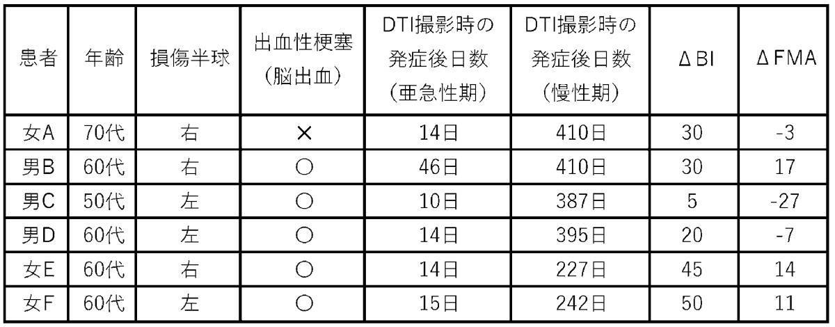
- Table 1 shows the results of motor function evaluation including patient background and DTI imaging time.
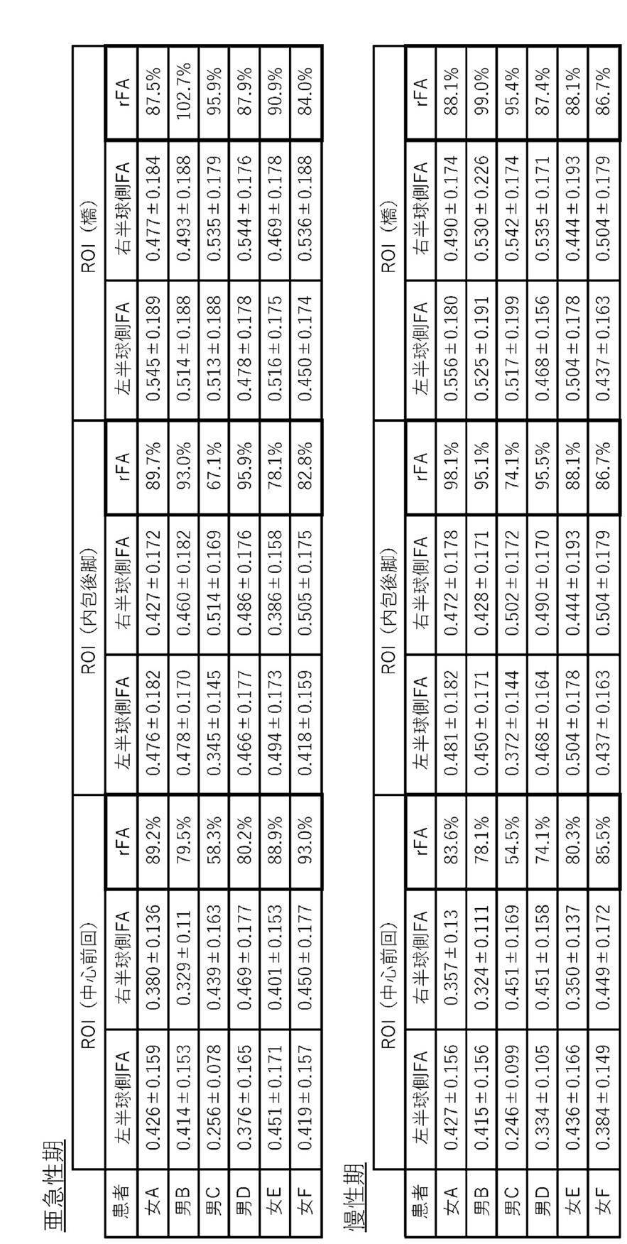
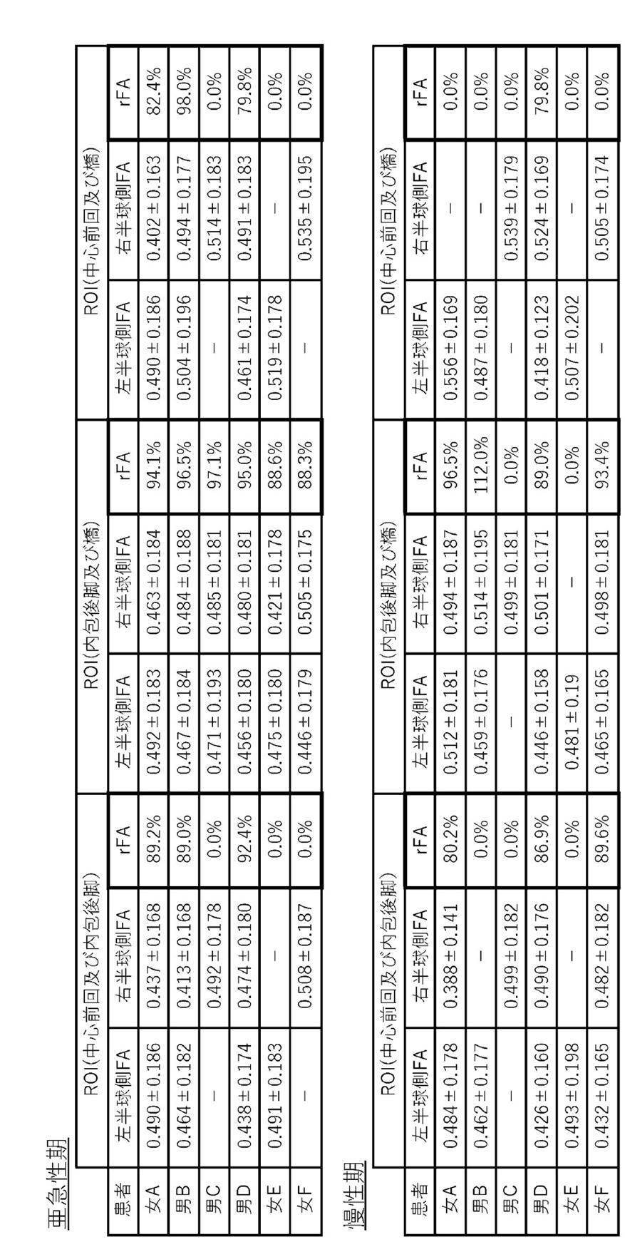
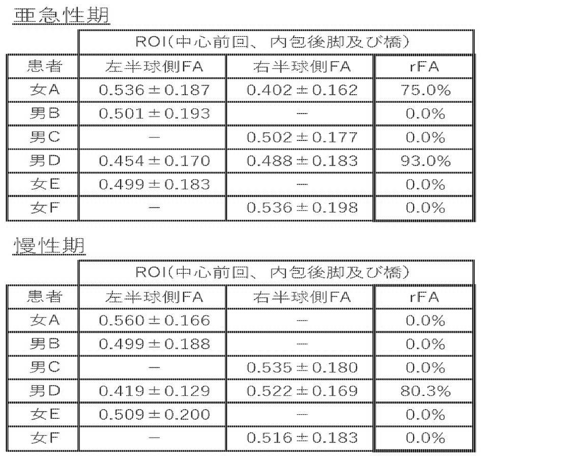
- the calculated FA and rFA are shown in Tables 2 to 4
- a scatter plot in which rFA and motor function recovery degree ⁇ BI are plotted is shown in FIGS. 6 to 8
- a scatter diagram in which rFA and motor function recovery degree ⁇ FMA are plotted are shown in FIGS. It is shown in 9-11. “-” In the table indicates that FA exceeding the threshold value was not calculated.
- DKI Diffusion-weighted imaging
- MK mean kurtosis
- AK axial kurtosis
- RK radial kurtosis
- FA fractional anisotropy
- KFA
- Table 5 shows the correlation between the diffusion image data before cell administration and the degree of functional recovery.
- the degree of decrease in FA associated with cerebral infarction seems to reflect functional recovery. From FIG. 13, it was found that the precentral gyrus FA on the lesion side and the healthy side must be 0.84 or more in order to obtain a change in mRS of 0.1 or more.
- the left-right difference of FA in cases with different degrees of functional recovery is shown in FIGS. 14A and 14B.
- Example 3 Evaluation of the state of motor function by an index other than BI and FMA by DTI
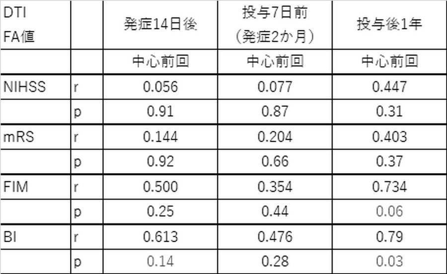
- correlation with motor function was evaluated using indicators other than BI and FMA (modified Rankin Scale (mRS), National Institute of Health Stroke Scale (NIHSS), functional independence measure (FIM)).
- mRS modified Rankin Scale
- NIHSS National Institute of Health Stroke Scale
- FAM functional independence measure
- This disclosure is useful in industries such as treatment and diagnosis of central nervous system diseases.
- the present disclosure can be used in fields such as regenerative medicine and development of cell medicine.
Landscapes
- Health & Medical Sciences (AREA)
- Life Sciences & Earth Sciences (AREA)
- Physics & Mathematics (AREA)
- Nuclear Medicine, Radiotherapy & Molecular Imaging (AREA)
- Engineering & Computer Science (AREA)
- General Health & Medical Sciences (AREA)
- Public Health (AREA)
- Medical Informatics (AREA)
- Radiology & Medical Imaging (AREA)
- Biomedical Technology (AREA)
- Pathology (AREA)
- Neurology (AREA)
- Surgery (AREA)
- Animal Behavior & Ethology (AREA)
- Molecular Biology (AREA)
- Veterinary Medicine (AREA)
- Heart & Thoracic Surgery (AREA)
- Biophysics (AREA)
- High Energy & Nuclear Physics (AREA)
- Neurosurgery (AREA)
- Physiology (AREA)
- Epidemiology (AREA)
- Primary Health Care (AREA)
- Signal Processing (AREA)
- Condensed Matter Physics & Semiconductors (AREA)
- General Physics & Mathematics (AREA)
- Vascular Medicine (AREA)
- Developmental Disabilities (AREA)
- Data Mining & Analysis (AREA)
- Databases & Information Systems (AREA)
- Psychology (AREA)
- Magnetic Resonance Imaging Apparatus (AREA)
- Materials For Medical Uses (AREA)
- Medicines Containing Material From Animals Or Micro-Organisms (AREA)
- Medical Treatment And Welfare Office Work (AREA)
Abstract
Description
(項目1) 脳損傷を有するまたは有すると疑われる患者の脳の損傷半球側の外側表面領域を関心領域とした生理指標値を得るステップと、
前記生理指標値を対照生理指標値と比較し、前記患者の運動機能の状態を示す値を算出するための演算を行うステップと
を含む、前記患者の運動機能の状態を評価する方法。
(項目2) 前記生理指標値が、拡散テンソル画像(DTI)法または拡散尖度画像(DKI)法によって得られる前記患者の脳の拡散強調画像から得られる、項目1に記載の方法。
(項目3) 前記生理指標値が、FA(fractional anisotropy)値を含む、上記項目のいずれか一項に記載の方法。
(項目4) 前記関心領域が脳の中心前回に設定される、上記項目のいずれか一項に記載の方法。
(項目5) 前記運動機能の状態が、再生治療後の運動機能の状態である、上記項目のいずれか一項に記載の方法。
(項目6) 前記対照生理指標値が、前記患者の脳の非損傷半球側の外側表面領域を関心領域として得られる、上記項目のいずれか一項に記載の方法。
(項目7) 前記演算を行うステップが、前記生理指標値/前記対照生理指標値で表される値を算出する、上記項目のいずれか一項に記載の方法。
(項目8) 前記演算を行うステップが、算出された前記患者の運動機能の状態を示す値を、予め用意された、患者の運動機能の状態を示す値と運動機能回復度とを変数とする回帰直線に代入することで、前記患者の運動機能の状態を評価する、上記項目のいずれか一項に記載の方法。
(項目9) 前記演算を行うステップが、算出された前記患者の運動機能の状態を示す値を、予め用意された基準値と比較することで、前記患者が再生治療後に所望の運動機能回復度に達する見込みの高低を算出する、上記項目のいずれか一項に記載の方法。
(項目10) 脳損傷を有するまたは有すると疑われる患者の脳の運動機能の状態を評価する方法の処理をコンピュータに実行させるコンピュータプログラムであって、前記方法は以下の工程:
前記コンピュータに、前記患者の脳の損傷半球側の外側表面領域を関心領域とした生理指標値を得させるステップと、
前記コンピュータに、前記生理指標値を対照生理指標値と比較し、前記患者の運動機能の状態を示す値を算出するための演算を行わせるステップと
を含む、プログラム。
(項目11) 脳損傷を有するまたは有すると疑われる患者の脳の運動機能の状態を評価する方法の処理をコンピュータに実行させるコンピュータプログラムを格納する記録媒体であって、前記方法は以下の工程:
前記コンピュータに、前記患者の脳の損傷半球側の外側表面領域を関心領域とした生理指標値を得させるステップと、
前記コンピュータに、前記生理指標値を対照生理指標値と比較し、前記患者の運動機能の状態を示す値を算出するための演算を行わせるステップと
を含む、記録媒体。
(項目12) 脳損傷を有するまたは有すると疑われる患者の脳の運動機能の状態を評価するシステムであって、
前記患者の脳の損傷半球側の外側表面領域を関心領域とした生理指標値を得る手段と、
前記生理指標値を対照生理指標値と比較し、前記患者の運動機能の状態を示す値を算出するための演算を行う手段と
を含む、システム。
(項目13) 脳損傷を有するまたは有すると疑われる患者の脳の拡散強調画像において、損傷半球側の外側表面領域を第1関心領域として、及び非損傷半球側の外側表面領域を第2関心領域として設定する関心領域設定部と、
第1関心領域及び第2関心領域のそれぞれにおける損傷半球側生理指標値及び非損傷半球側生理指標値を算出する生理指標値算出部と、
損傷半球側生理指標値と非損傷半球側生理指標値とを比較して、当該患者の運動機能の状態を示す値を算出するための演算を行う演算部と
を備えることを特徴とする、画像解析装置。
(項目14) 脳損傷を有するまたは有すると疑われる患者の脳を撮像する核磁気共鳴撮像部と、
核磁気共鳴撮像部が取得したエコーデータから拡散強調画像を生成する画像生成部と、
拡散強調画像において、損傷半球側の外側表面領域を第1関心領域として、及び非損傷半球側の外側表面領域を第2関心領域として設定する関心領域設定部と、
第1関心領域及び第2関心領域のそれぞれにおける損傷半球側生理指標値及び非損傷半球側生理指標値を算出する生理指標値算出部と、
損傷半球側生理指標値と非損傷半球側生理指標値とを比較して、当該患者の運動機能の状態を示す値を算出するための演算を行う演算部と
を備えることを特徴とする、MRI装置。
(1) 脳局所損傷を有する患者の脳の拡散テンソル画像において、損傷半球側の中心前回を第1関心領域として、及び非損傷半球側の中心前回を第2関心領域として設定するステップと、
第1関心領域及び第2関心領域のそれぞれにおける異方性比率である損傷半球側FA及び非損傷半球側FAを算出するステップと、
損傷半球側FA/非損傷半球側FAで表されるrFAを算出するステップと、
算出されたrFAに基づいて、再生治療後の当該患者の運動機能回復を予測するための演算を行うステップと
を含む、当該患者の再生治療後の運動機能回復を予測する方法。
(2) 前記演算を行うステップが、算出されたrFAを、予め用意されたrFAと運動機能回復度とを変数とする回帰直線に代入することで、当該患者の再生治療後の運動機能回復度の予測値を算出するステップである、(1)に記載の方法。
(3) 前記演算を行うステップが、算出されたrFAを、予め用意された基準rFAと比較することで、当該患者が再生治療後に所望の運動機能回復度に達する見込みの高低を算出するステップである、(1)に記載の方法。
(4) コンピュータに、
脳局所損傷を有する患者の脳の拡散テンソル画像において、損傷半球側の中心前回を第1関心領域として、及び非損傷半球側の中心前回を第2関心領域として設定する処理と、
第1関心領域及び第2関心領域のそれぞれにおける異方性比率である損傷半球側FA及び非損傷半球側FAを算出する処理と、
損傷半球側FA/非損傷半球側FAで表されるrFAを算出する処理と、
算出されたrFAに基づいて、再生治療後の当該患者の運動機能回復を予測するための演算を行う処理と
を実行させるための、当該患者の再生治療後の運動機能回復を予測するためのプログラム。
(5) 前記演算を行う処理が、算出されたrFAを、予め用意されたrFAと運動機能回復度とを変数とする回帰直線に代入することで、当該患者の再生治療後の運動機能回復度の予測値を算出する処理である、(4)に記載のプログラム。
(6) 前記演算を行う処理が、算出されたrFAを、予め用意された基準rFAと比較することで、当該患者が再生治療後に所望の運動機能回復度に達する見込みの高低を算出する処理である、(4)に記載のプログラム。
(7) (4)~(6)のいずれか一項に記載のプログラムを記憶した、コンピュータ読み取り可能な記憶媒体。
(8) 脳局所損傷を有する患者の脳の拡散テンソル画像において、損傷半球側の中心前回を第1関心領域として、及び非損傷半球側の中心前回を第2関心領域として設定する関心領域設定部と、
第1関心領域及び第2関心領域のそれぞれにおける異方性比率である損傷半球側FA及び非損傷半球側FAを算出するFA算出部と、
損傷半球側FA/非損傷半球側FAで表されるrFAを算出するrFA算出部と、
算出されたrFAに基づいて、再生治療後の当該患者の運動機能回復を予測するための演算を行う回復予測演算部と
を備えることを特徴とする、画像解析装置。
(9) 脳局所損傷を有する患者の脳を撮像する核磁気共鳴撮像部と、
核磁気共鳴撮像部が取得したエコーデータから拡散テンソル画像を生成する画像生成部と、
拡散テンソル画像において、損傷半球側の中心前回を第1関心領域として、及び非損傷半球側の中心前回を第2関心領域として設定する関心領域設定部と、
第1関心領域及び第2関心領域のそれぞれにおける異方性比率である損傷半球側FA及び非損傷半球側FAを算出するFA算出部と、
損傷半球側FA/非損傷半球側FAで表されるrFAを算出するrFA算出部と、
算出されたrFAに基づいて、再生治療後の当該患者の運動機能回復を予測するための演算を行う回復予測演算部と
を備えることを特徴とする、MRI装置。
以下に本開示の好ましい実施形態を説明する。以下に提供される実施形態は、本開示のよりよい理解のために提供されるものであり、本開示の範囲は以下の記載に限定されるべきでない。したがって、当業者は、本明細書中の記載を参酌して、本開示の範囲内で適宜改変を行うことができることは明らかである。また、本開示の以下の実施形態は単独でも使用されあるいはそれらを組み合わせて使用することができる。
前記コンピュータに、前記患者の脳の損傷半球側の外側表面領域を関心領域とした生理指標値を得させるステップと、
前記コンピュータに、前記生理指標値を対照生理指標値と比較し、前記患者の運動機能の状態を示す値を算出するための演算を行わせるステップと
を含む、プログラムが提供される。
本開示の他の局面において、脳損傷を有するまたは有すると疑われる患者の脳の運動機能の状態を評価する方法の処理をコンピュータに実行させるコンピュータプログラムを格納する記録媒体であって、前記方法は以下の工程:
前記コンピュータに、前記患者の脳の損傷半球側の外側表面領域を関心領域とした生理指標値を得させるステップと、
前記コンピュータに、前記生理指標値を対照生理指標値と比較し、前記患者の運動機能の状態を示す値を算出するための演算を行わせるステップと
を含む、記録媒体が提供される。
前記患者の脳の損傷半球側の外側表面領域を関心領域とした生理指標値を得る手段と、
前記生理指標値を対照生理指標値と比較し、前記患者の運動機能の状態を示す値を算出するための演算を行う手段と
を含む、システムが提供される。
第1関心領域及び第2関心領域のそれぞれにおける損傷半球側生理指標値及び非損傷半球側生理指標値を算出する生理指標値算出部と、
損傷半球側生理指標値と非損傷半球側生理指標値とを比較して、当該患者の運動機能の状態を示す値を算出するための演算を行う演算部と
を備えることを特徴とする、画像解析装置が提供される。
核磁気共鳴撮像部が取得したエコーデータから拡散強調画像を生成する画像生成部と、
拡散強調画像において、損傷半球側の外側表面領域を第1関心領域として、及び非損傷半球側の外側表面領域を第2関心領域として設定する関心領域設定部と、
第1関心領域及び第2関心領域のそれぞれにおける損傷半球側生理指標値及び非損傷半球側生理指標値を算出する生理指標値算出部と、
損傷半球側生理指標値と非損傷半球側生理指標値とを比較して、当該患者の運動機能の状態を示す値を算出するための演算を行う演算部と
を備えることを特徴とする、MRI装置が提供される。




北海道大学病院治験審査委員会の承認を受け、以下の研究を実施した。
(1)対象患者
脳梗塞急性期(発症早期)の患者であって、同意取得時における年齢が20歳以上、80歳未満である;同意取得時において脳梗塞発症後14日以内である;内頚動脈灌流域に生じた脳梗塞である;脳梗塞発症前のmRSが0又は1である;脳梗塞による中等~重度の神経症状(NIHSS(NationalInstitutes of Health Stroke Scale):≧6)を有する(ただしNIHSSの「5.上肢の運動」と「6.下肢の運動」項目において総計6点以上であること)という5つの基準を満たし、所定の除外基準に抵触せず、かつ本研究に同意が得られた患者6名を対象とした。患者6名中5名は、出血性梗塞、すなわち脳出血を伴った脳梗塞の患者であった。
症例登録後、速やかに患者の骨髄を採取し、北海道大学病院臨床研究開発センター細胞プロセッシング室において細胞培養および骨髄幹細胞の調製を施行した。調製した骨髄幹細胞(2000万個又は5000万個/患者)を、骨髄採取の3~5週後に患者の脳内に直接投与した。
患者の運動機能は、Bathel Index(BI)、Fugl-Meyer Assessment (FMA)を用いて評価した。これらの指標は日本脳卒中学会の出している脳卒中治療ガイドライン2015の脳卒中リハビリテーション評価の項で用いる様に勧められているものである。運動機能評価は、症例登録時(脳梗塞後14日目)、細胞投与7日前(脳梗塞後平均50日)、細胞投与後1ヶ月、細胞投与後3ヶ月、細胞投与後6ヶ月、細胞投与後12ヶ月の時点で行った。急性期治療がほぼ終了した亜急性期(脳梗塞発症から10日~50日、以下亜急性期)のBI及びFMAの評点を、細胞投与後12ヶ月(本申請の時点で12ヶ月に至っていない患者(女E及び女F)は6ヶ月、以下慢性期)のBI及びFMAの評点から減算したΔBI及びΔFMAを運動機能回復度とした。
それぞれの患者について、亜急性期及び慢性期に相当する時期に、脳のDTI画像を撮影した。撮影はMRI装置(3T Achieva TX(Philips Medical Systems))をデフォルトの設定(b=0, 1000 s mm-2, TR/TE = 5032/ 85 msec, NEX = 1, voxel size =3 x 3 x3 mm3, no. of slices = 43, 32 diffusion gradient directions)で用いて行った。
MRI装置に付属の画像解析ソフトウェア(MRFiber Trak, Extended MR Work Space version 2.6.3.4)を用いて、minimal FAを0.15から0.18に、maximumangle changeを25°に変更した以外はデフォルトの設定を用いてDTI画像を解析した。
患者背景及びDTI撮影時期を含めた運動機能評価結果を表1に示す。また、算出されたFA及びrFAを表2~4に、rFAと運動機能回復度ΔBIとをプロットした散布図を図6~8に、rFAと運動機能回復度ΔFMAとをプロットした散布図を図9~11に示す。表中の「-」は、閾値を超えるFAが算出されなかったことを示す。
脳梗塞患者における細胞投与治療効果を、高精度かつ測定者の経験等に依存せずに予測可能な拡散画像の指数を得るために拡散尖度画像(DKI)法を用いた。本実施例では、拡散尖度画像(DKI)法による主な指数のマップを作成し、標準脳に変換した後、両側中心前回と内包後脚の各々の値を測定した。
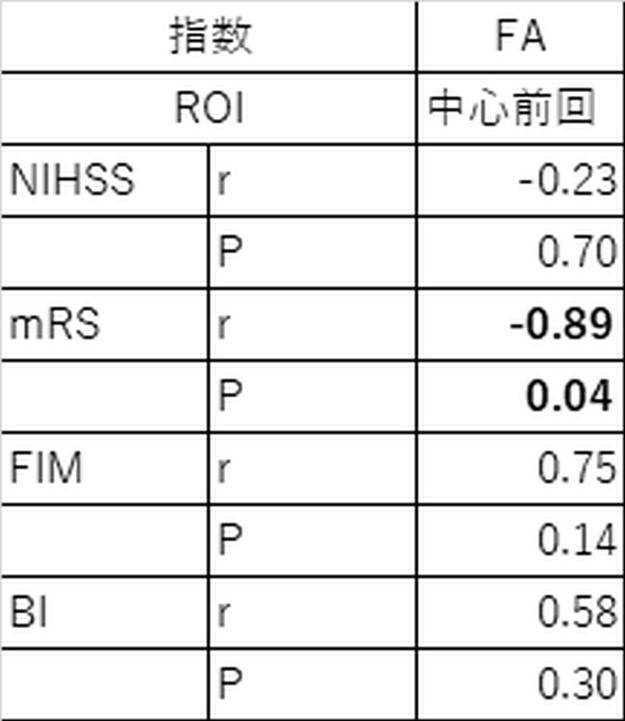
本実施例においては、BIおよびFMA以外の指標(modified Rankin Scale (mRS)、National Institute of Health StrokeScale (NIHSS), functional independence measure (FIM))を用いて運動機能との相関を評価した。運動機能評価は、症例登録時(脳梗塞後14日目)、細胞投与7日前(脳梗塞後平均50日)、細胞投与後1年の時点で行った。結果を表6に示す。
以上のように、本開示の好ましい実施形態を用いて本開示を例示してきたが、本開示は、特許請求の範囲によってのみその範囲が解釈されるべきであることが理解される。本明細書において引用した特許、特許出願及び他の文献は、その内容自体が具体的に本明細書に記載されているのと同様にその内容が本明細書に対する参考として援用されるべきであることが理解される。本願は、日本国特許庁に2019年9月30日に出願された特願2019-181051に対して優先権主張をするものであり、その内容はその全体があたかも本願の内容を構成するのと同様に参考として援用される。
10 核磁気共鳴撮像部
20 コンピュータ
21 インターフェース部
22 入力部
23 記憶部
231 エコーデータ記憶部
232 再構成画像記憶部
233 DTI画像記憶部
234 参照情報記憶部
24 演算部
241 画像生成部
241a 画像再構成部
241b DTI画像生成部
242 画像解析部
242a 関心領域設定部
242b FA算出部
242c rFA算出部
242d 回復予測演算部
25 出力部
26 制御部
Claims (14)
- 脳損傷を有するまたは有すると疑われる患者の脳の損傷半球側の外側表面領域を関心領域とした生理指標値を得るステップと、
前記生理指標値を対照生理指標値と比較し、前記患者の運動機能の状態を示す値を算出するための演算を行うステップと
を含む、前記患者の運動機能の状態を評価する方法。 - 前記生理指標値が、拡散テンソル画像(DTI)法または拡散尖度画像(DKI)法によって得られる前記患者の脳の拡散強調画像から得られる、請求項1に記載の方法。
- 前記生理指標値が、FA(fractional anisotropy)値を含む、請求項1または2に記載の方法。
- 前記関心領域が脳の中心前回に設定される、請求項1~3のいずれか一項に記載の方法。
- 前記運動機能の状態が、再生治療後の運動機能の状態である、請求項1~4のいずれか一項に記載の方法。
- 前記対照生理指標値が、前記患者の脳の非損傷半球側の外側表面領域を関心領域として得られる、請求項1~5のいずれか一項に記載の方法。
- 前記演算を行うステップが、前記生理指標値/前記対照生理指標値で表される値を算出する、請求項1~6のいずれか一項に記載の方法。
- 前記演算を行うステップが、算出された前記患者の運動機能の状態を示す値を、予め用意された、患者の運動機能の状態を示す値と運動機能回復度とを変数とする回帰直線に代入することで、前記患者の運動機能の状態を評価する、請求項1~7のいずれか一項に記載の方法。
- 前記演算を行うステップが、算出された前記患者の運動機能の状態を示す値を、予め用意された基準値と比較することで、前記患者が再生治療後に所望の運動機能回復度に達する見込みの高低を算出する、請求項1~8のいずれか一項に記載の方法。
- 脳損傷を有するまたは有すると疑われる患者の脳の運動機能の状態を評価する方法の処理をコンピュータに実行させるコンピュータプログラムであって、前記方法は以下の工程:
前記コンピュータに、前記患者の脳の損傷半球側の外側表面領域を関心領域とした生理指標値を得させるステップと、
前記コンピュータに、前記生理指標値を対照生理指標値と比較し、前記患者の運動機能の状態を示す値を算出するための演算を行わせるステップと
を含む、プログラム。 - 脳損傷を有するまたは有すると疑われる患者の脳の運動機能の状態を評価する方法の処理をコンピュータに実行させるコンピュータプログラムを格納する記録媒体であって、前記方法は以下の工程:
前記コンピュータに、前記患者の脳の損傷半球側の外側表面領域を関心領域とした生理指標値を得させるステップと、
前記コンピュータに、前記生理指標値を対照生理指標値と比較し、前記患者の運動機能の状態を示す値を算出するための演算を行わせるステップと
を含む、記録媒体。 - 脳損傷を有するまたは有すると疑われる患者の脳の運動機能の状態を評価するシステムであって、
前記患者の脳の損傷半球側の外側表面領域を関心領域とした生理指標値を得る手段と、
前記生理指標値を対照生理指標値と比較し、前記患者の運動機能の状態を示す値を算出するための演算を行う手段と
を含む、システム。 - 脳損傷を有するまたは有すると疑われる患者の脳の拡散強調画像において、損傷半球側の外側表面領域を第1関心領域として、及び非損傷半球側の外側表面領域を第2関心領域として設定する関心領域設定部と、
第1関心領域及び第2関心領域のそれぞれにおける損傷半球側生理指標値及び非損傷半球側生理指標値を算出する生理指標値算出部と、
損傷半球側生理指標値と非損傷半球側生理指標値とを比較して、当該患者の運動機能の状態を示す値を算出するための演算を行う演算部と
を備えることを特徴とする、画像解析装置。 - 脳損傷を有するまたは有すると疑われる患者の脳を撮像する核磁気共鳴撮像部と、
核磁気共鳴撮像部が取得したエコーデータから拡散強調画像を生成する画像生成部と、
拡散強調画像において、損傷半球側の外側表面領域を第1関心領域として、及び非損傷半球側の外側表面領域を第2関心領域として設定する関心領域設定部と、
第1関心領域及び第2関心領域のそれぞれにおける損傷半球側生理指標値及び非損傷半球側生理指標値を算出する生理指標値算出部と、
損傷半球側生理指標値と非損傷半球側生理指標値とを比較して、当該患者の運動機能の状態を示す値を算出するための演算を行う演算部と
を備えることを特徴とする、MRI装置。
Priority Applications (3)
| Application Number | Priority Date | Filing Date | Title |
|---|---|---|---|
| EP20871340.4A EP4040445A4 (en) | 2019-09-30 | 2020-09-29 | METHOD, PROGRAM AND DEVICE FOR ASSESSING THE CONDITION OF AN ENGINE FUNCTION |
| US17/763,280 US12290335B2 (en) | 2019-09-30 | 2020-09-29 | Method, program, and device for evaluating state of motor function |
| JP2021506591A JP6966828B2 (ja) | 2019-09-30 | 2020-09-29 | 運動機能の状態を評価するための方法、プログラム及び装置 |
Applications Claiming Priority (2)
| Application Number | Priority Date | Filing Date | Title |
|---|---|---|---|
| JP2019181051 | 2019-09-30 | ||
| JP2019-181051 | 2019-09-30 |
Publications (1)
| Publication Number | Publication Date |
|---|---|
| WO2021065894A1 true WO2021065894A1 (ja) | 2021-04-08 |
Family
ID=75338345
Family Applications (1)
| Application Number | Title | Priority Date | Filing Date |
|---|---|---|---|
| PCT/JP2020/036854 Ceased WO2021065894A1 (ja) | 2019-09-30 | 2020-09-29 | 運動機能の状態を評価するための方法、プログラム及び装置 |
Country Status (4)
| Country | Link |
|---|---|
| US (1) | US12290335B2 (ja) |
| EP (1) | EP4040445A4 (ja) |
| JP (3) | JP6966828B2 (ja) |
| WO (1) | WO2021065894A1 (ja) |
Families Citing this family (1)
| Publication number | Priority date | Publication date | Assignee | Title |
|---|---|---|---|---|
| US12511741B2 (en) * | 2023-02-10 | 2025-12-30 | Uniwersytet W Bialymstoku | Method of diagnosing degree of neuropathy of a nerve by diffusion tensor mapping |
Citations (7)
| Publication number | Priority date | Publication date | Assignee | Title |
|---|---|---|---|---|
| JP2008510560A (ja) * | 2004-08-25 | 2008-04-10 | モトリカ リミテッド | 脳の可塑性による運動訓練 |
| JP2008132032A (ja) * | 2006-11-27 | 2008-06-12 | Hitachi Ltd | 神経線維束計測システム及び画像処理システム |
| JP2011139799A (ja) * | 2010-01-07 | 2011-07-21 | Toshiba Corp | 画像処理装置及び磁気共鳴イメージング装置 |
| JP2011143105A (ja) * | 2010-01-15 | 2011-07-28 | Toshiba Corp | 医用画像処理装置 |
| JP2015534844A (ja) * | 2012-10-17 | 2015-12-07 | アシスタンス ピュブリク−オピトー ドゥ パリAssistance Publique − Hopitaux De Paris | 脳損傷を定量するための方法 |
| US20180168499A1 (en) * | 2015-06-04 | 2018-06-21 | Peter John BERGOLD | Diagnosis of mild traumatic brain injury |
| JP2019181051A (ja) | 2018-04-17 | 2019-10-24 | 酵素玄米Labo株式会社 | 炊飯器 |
Family Cites Families (6)
| Publication number | Priority date | Publication date | Assignee | Title |
|---|---|---|---|---|
| JP2006508707A (ja) | 2002-09-04 | 2006-03-16 | ワシントン・ユニバーシティ | 中枢神経系損傷の治療法 |
| US8606360B2 (en) * | 2006-03-09 | 2013-12-10 | The Cleveland Clinic Foundation | Systems and methods for determining volume of activation for spinal cord and peripheral nerve stimulation |
| US8170305B2 (en) * | 2006-10-19 | 2012-05-01 | Brown University | Quantitative tract-of-interest metrics for white matter integrity based on diffusion tensor MRI data |
| US10481233B2 (en) * | 2014-09-09 | 2019-11-19 | The Trustees Of The University Of Pennsylvania | Edema invariant tractography |
| WO2017010864A1 (en) * | 2015-07-10 | 2017-01-19 | Erasmus University Medical Center Rotterdam | Apparatus, system and method for assisting in providing a diagnosis of a medical condition of a mammal brain as well a computer readable medium comprising a program for carrying out the method |
| TWI651688B (zh) | 2017-03-17 | 2019-02-21 | 長庚大學 | 利用磁振造影影像預測神經疾病的臨床嚴重度的方法 |
-
2020
- 2020-09-29 EP EP20871340.4A patent/EP4040445A4/en active Pending
- 2020-09-29 WO PCT/JP2020/036854 patent/WO2021065894A1/ja not_active Ceased
- 2020-09-29 US US17/763,280 patent/US12290335B2/en active Active
- 2020-09-29 JP JP2021506591A patent/JP6966828B2/ja active Active
-
2021
- 2021-10-15 JP JP2021169636A patent/JP2022002767A/ja active Pending
-
2025
- 2025-09-02 JP JP2025145016A patent/JP2025172133A/ja active Pending
Patent Citations (7)
| Publication number | Priority date | Publication date | Assignee | Title |
|---|---|---|---|---|
| JP2008510560A (ja) * | 2004-08-25 | 2008-04-10 | モトリカ リミテッド | 脳の可塑性による運動訓練 |
| JP2008132032A (ja) * | 2006-11-27 | 2008-06-12 | Hitachi Ltd | 神経線維束計測システム及び画像処理システム |
| JP2011139799A (ja) * | 2010-01-07 | 2011-07-21 | Toshiba Corp | 画像処理装置及び磁気共鳴イメージング装置 |
| JP2011143105A (ja) * | 2010-01-15 | 2011-07-28 | Toshiba Corp | 医用画像処理装置 |
| JP2015534844A (ja) * | 2012-10-17 | 2015-12-07 | アシスタンス ピュブリク−オピトー ドゥ パリAssistance Publique − Hopitaux De Paris | 脳損傷を定量するための方法 |
| US20180168499A1 (en) * | 2015-06-04 | 2018-06-21 | Peter John BERGOLD | Diagnosis of mild traumatic brain injury |
| JP2019181051A (ja) | 2018-04-17 | 2019-10-24 | 酵素玄米Labo株式会社 | 炊飯器 |
Non-Patent Citations (2)
| Title |
|---|
| "Stem Cell Therapies as an Emerging Paradigm in Stroke Participants", STROKE, vol. 40, no. 2, 2009, pages 510 - 5 |
| See also references of EP4040445A4 |
Also Published As
| Publication number | Publication date |
|---|---|
| JP2025172133A (ja) | 2025-11-20 |
| US12290335B2 (en) | 2025-05-06 |
| JPWO2021065894A1 (ja) | 2021-10-21 |
| EP4040445A1 (en) | 2022-08-10 |
| EP4040445A4 (en) | 2023-10-25 |
| JP6966828B2 (ja) | 2021-11-17 |
| US20220346649A1 (en) | 2022-11-03 |
| JP2022002767A (ja) | 2022-01-11 |
Similar Documents
| Publication | Publication Date | Title |
|---|---|---|
| Budde et al. | Axonal injury detected by in vivo diffusion tensor imaging correlates with neurological disability in a mouse model of multiple sclerosis | |
| Wozniak et al. | What does diffusion tensor imaging reveal about the brain and cognition in fetal alcohol spectrum disorders? | |
| CN111867464B (zh) | 脑活动训练装置、脑活动训练方法以及脑活动训练程序 | |
| Wen et al. | Diffusion tensor imaging of somatosensory tract in cervical spondylotic myelopathy and its link with electrophysiological evaluation | |
| Elvsåshagen et al. | Evidence for cortical structural plasticity in humans after a day of waking and sleep deprivation | |
| Massaro et al. | Neonatal neurobehavior after therapeutic hypothermia for hypoxic ischemic encephalopathy | |
| Foland-Ross et al. | Mood-state effects on amygdala volume in bipolar disorder | |
| Rosso et al. | Anatomical and functional correlates of cortical motor threshold of the dominant hand | |
| Lin et al. | Diffusion fMRI detects white-matter dysfunction in mice with acute optic neuritis | |
| US20250204783A1 (en) | System and method for assessment of neuro-inflammation using magnetic resonance imaging (mri) | |
| JP2025172133A (ja) | 運動機能の状態を評価するための方法、プログラム及び装置 | |
| Wang et al. | White matter abnormalities in medication-naïve adult patients with major depressive disorder: tract-based spatial statistical analysis | |
| Jeromel et al. | Quantification of synovitis in the cranio-cervical region: dynamic contrast enhanced and diffusion weighted magnetic resonance imaging in early rheumatoid arthritis—a feasibility follow up study | |
| WO2022145433A1 (ja) | 運動機能の状態を評価するための方法、プログラム及び装置 | |
| Hatem et al. | Assessment of spinal somatosensory systems with diffusion tensor imaging in syringomyelia | |
| Singhi et al. | Diffusion tensor imaging of the maturing paediatric cervical spinal cord: from the neonate to the young adult | |
| Ando et al. | Microstructural white matter changes following gait training with Hybrid Assistive Limb initiated within 1 week of stroke onset | |
| Malamateniou et al. | The effect of preterm birth on neonatal cerebral vasculature studied with magnetic resonance angiography at 3 Tesla | |
| Sun et al. | Evaluation of hyperbaric oxygen treatment in acute traumatic spinal cord injury in rats using diffusion tensor imaging | |
| Ohn et al. | Analysis of high-voltage electrical spinal cord injury using diffusion tensor imaging | |
| Sawada et al. | Ontogenetic pattern of gyrification in fetuses of cynomolgus monkeys | |
| Özmen et al. | Quantitative evaluation of the cerebellum in patients with depression and healthy adults by VolBrain method | |
| Choi et al. | Effects of gender and age on anterior commissure volume | |
| Jack Jr | Medial temporal lobe volumetrics in traumatic brain injury | |
| Joni et al. | Investigating the role of susceptibility weighted imaging for assessment of ischemic penumbra with respect to Venus blood flow in ischemic stroke patients |
Legal Events
| Date | Code | Title | Description |
|---|---|---|---|
| ENP | Entry into the national phase |
Ref document number: 2021506591 Country of ref document: JP Kind code of ref document: A |
|
| 121 | Ep: the epo has been informed by wipo that ep was designated in this application |
Ref document number: 20871340 Country of ref document: EP Kind code of ref document: A1 |
|
| NENP | Non-entry into the national phase |
Ref country code: DE |
|
| ENP | Entry into the national phase |
Ref document number: 2020871340 Country of ref document: EP Effective date: 20220502 |
|
| WWG | Wipo information: grant in national office |
Ref document number: 17763280 Country of ref document: US |