WO2021068949A1 - 抗人Trop-2抗体及其应用 - Google Patents
抗人Trop-2抗体及其应用 Download PDFInfo
- Publication number
- WO2021068949A1 WO2021068949A1 PCT/CN2020/120277 CN2020120277W WO2021068949A1 WO 2021068949 A1 WO2021068949 A1 WO 2021068949A1 CN 2020120277 W CN2020120277 W CN 2020120277W WO 2021068949 A1 WO2021068949 A1 WO 2021068949A1
- Authority
- WO
- WIPO (PCT)
- Prior art keywords
- amino acid
- acid sequence
- seq
- sequence shown
- antibody
- Prior art date
- Legal status (The legal status is an assumption and is not a legal conclusion. Google has not performed a legal analysis and makes no representation as to the accuracy of the status listed.)
- Ceased
Links
Images


Classifications
-
- A—HUMAN NECESSITIES
- A61—MEDICAL OR VETERINARY SCIENCE; HYGIENE
- A61P—SPECIFIC THERAPEUTIC ACTIVITY OF CHEMICAL COMPOUNDS OR MEDICINAL PREPARATIONS
- A61P35/00—Antineoplastic agents
-
- A—HUMAN NECESSITIES
- A61—MEDICAL OR VETERINARY SCIENCE; HYGIENE
- A61K—PREPARATIONS FOR MEDICAL, DENTAL OR TOILETRY PURPOSES
- A61K47/00—Medicinal preparations characterised by the non-active ingredients used, e.g. carriers or inert additives; Targeting or modifying agents chemically bound to the active ingredient
- A61K47/50—Medicinal preparations characterised by the non-active ingredients used, e.g. carriers or inert additives; Targeting or modifying agents chemically bound to the active ingredient the non-active ingredient being chemically bound to the active ingredient, e.g. polymer-drug conjugates
- A61K47/51—Medicinal preparations characterised by the non-active ingredients used, e.g. carriers or inert additives; Targeting or modifying agents chemically bound to the active ingredient the non-active ingredient being chemically bound to the active ingredient, e.g. polymer-drug conjugates the non-active ingredient being a modifying agent
- A61K47/68—Medicinal preparations characterised by the non-active ingredients used, e.g. carriers or inert additives; Targeting or modifying agents chemically bound to the active ingredient the non-active ingredient being chemically bound to the active ingredient, e.g. polymer-drug conjugates the non-active ingredient being a modifying agent the modifying agent being an antibody, an immunoglobulin or a fragment thereof, e.g. an Fc-fragment
- A61K47/6801—Drug-antibody or immunoglobulin conjugates defined by the pharmacologically or therapeutically active agent
- A61K47/6803—Drugs conjugated to an antibody or immunoglobulin, e.g. cisplatin-antibody conjugates
- A61K47/68037—Drugs conjugated to an antibody or immunoglobulin, e.g. cisplatin-antibody conjugates the drug being a camptothecin [CPT] or derivatives
-
- A—HUMAN NECESSITIES
- A61—MEDICAL OR VETERINARY SCIENCE; HYGIENE
- A61K—PREPARATIONS FOR MEDICAL, DENTAL OR TOILETRY PURPOSES
- A61K47/00—Medicinal preparations characterised by the non-active ingredients used, e.g. carriers or inert additives; Targeting or modifying agents chemically bound to the active ingredient
- A61K47/50—Medicinal preparations characterised by the non-active ingredients used, e.g. carriers or inert additives; Targeting or modifying agents chemically bound to the active ingredient the non-active ingredient being chemically bound to the active ingredient, e.g. polymer-drug conjugates
- A61K47/51—Medicinal preparations characterised by the non-active ingredients used, e.g. carriers or inert additives; Targeting or modifying agents chemically bound to the active ingredient the non-active ingredient being chemically bound to the active ingredient, e.g. polymer-drug conjugates the non-active ingredient being a modifying agent
- A61K47/68—Medicinal preparations characterised by the non-active ingredients used, e.g. carriers or inert additives; Targeting or modifying agents chemically bound to the active ingredient the non-active ingredient being chemically bound to the active ingredient, e.g. polymer-drug conjugates the non-active ingredient being a modifying agent the modifying agent being an antibody, an immunoglobulin or a fragment thereof, e.g. an Fc-fragment
- A61K47/6835—Medicinal preparations characterised by the non-active ingredients used, e.g. carriers or inert additives; Targeting or modifying agents chemically bound to the active ingredient the non-active ingredient being chemically bound to the active ingredient, e.g. polymer-drug conjugates the non-active ingredient being a modifying agent the modifying agent being an antibody, an immunoglobulin or a fragment thereof, e.g. an Fc-fragment the modifying agent being an antibody or an immunoglobulin bearing at least one antigen-binding site
- A61K47/6851—Medicinal preparations characterised by the non-active ingredients used, e.g. carriers or inert additives; Targeting or modifying agents chemically bound to the active ingredient the non-active ingredient being chemically bound to the active ingredient, e.g. polymer-drug conjugates the non-active ingredient being a modifying agent the modifying agent being an antibody, an immunoglobulin or a fragment thereof, e.g. an Fc-fragment the modifying agent being an antibody or an immunoglobulin bearing at least one antigen-binding site the antibody targeting a determinant of a tumour cell
-
- A—HUMAN NECESSITIES
- A61—MEDICAL OR VETERINARY SCIENCE; HYGIENE
- A61K—PREPARATIONS FOR MEDICAL, DENTAL OR TOILETRY PURPOSES
- A61K47/00—Medicinal preparations characterised by the non-active ingredients used, e.g. carriers or inert additives; Targeting or modifying agents chemically bound to the active ingredient
- A61K47/50—Medicinal preparations characterised by the non-active ingredients used, e.g. carriers or inert additives; Targeting or modifying agents chemically bound to the active ingredient the non-active ingredient being chemically bound to the active ingredient, e.g. polymer-drug conjugates
- A61K47/51—Medicinal preparations characterised by the non-active ingredients used, e.g. carriers or inert additives; Targeting or modifying agents chemically bound to the active ingredient the non-active ingredient being chemically bound to the active ingredient, e.g. polymer-drug conjugates the non-active ingredient being a modifying agent
- A61K47/68—Medicinal preparations characterised by the non-active ingredients used, e.g. carriers or inert additives; Targeting or modifying agents chemically bound to the active ingredient the non-active ingredient being chemically bound to the active ingredient, e.g. polymer-drug conjugates the non-active ingredient being a modifying agent the modifying agent being an antibody, an immunoglobulin or a fragment thereof, e.g. an Fc-fragment
- A61K47/6835—Medicinal preparations characterised by the non-active ingredients used, e.g. carriers or inert additives; Targeting or modifying agents chemically bound to the active ingredient the non-active ingredient being chemically bound to the active ingredient, e.g. polymer-drug conjugates the non-active ingredient being a modifying agent the modifying agent being an antibody, an immunoglobulin or a fragment thereof, e.g. an Fc-fragment the modifying agent being an antibody or an immunoglobulin bearing at least one antigen-binding site
- A61K47/6875—Medicinal preparations characterised by the non-active ingredients used, e.g. carriers or inert additives; Targeting or modifying agents chemically bound to the active ingredient the non-active ingredient being chemically bound to the active ingredient, e.g. polymer-drug conjugates the non-active ingredient being a modifying agent the modifying agent being an antibody, an immunoglobulin or a fragment thereof, e.g. an Fc-fragment the modifying agent being an antibody or an immunoglobulin bearing at least one antigen-binding site the antibody being a hybrid immunoglobulin
- A61K47/6877—Medicinal preparations characterised by the non-active ingredients used, e.g. carriers or inert additives; Targeting or modifying agents chemically bound to the active ingredient the non-active ingredient being chemically bound to the active ingredient, e.g. polymer-drug conjugates the non-active ingredient being a modifying agent the modifying agent being an antibody, an immunoglobulin or a fragment thereof, e.g. an Fc-fragment the modifying agent being an antibody or an immunoglobulin bearing at least one antigen-binding site the antibody being a hybrid immunoglobulin the antibody being an immunoglobulin containing regions, domains or residues from different species
-
- C—CHEMISTRY; METALLURGY
- C07—ORGANIC CHEMISTRY
- C07K—PEPTIDES
- C07K16/00—Immunoglobulins [IG], e.g. monoclonal or polyclonal antibodies
- C07K16/18—Immunoglobulins [IG], e.g. monoclonal or polyclonal antibodies against material from animals or humans
- C07K16/28—Immunoglobulins [IG], e.g. monoclonal or polyclonal antibodies against material from animals or humans against receptors, cell surface antigens or cell surface determinants
- C07K16/2803—Immunoglobulins [IG], e.g. monoclonal or polyclonal antibodies against material from animals or humans against receptors, cell surface antigens or cell surface determinants against the immunoglobulin superfamily
-
- C—CHEMISTRY; METALLURGY
- C07—ORGANIC CHEMISTRY
- C07K—PEPTIDES
- C07K16/00—Immunoglobulins [IG], e.g. monoclonal or polyclonal antibodies
- C07K16/18—Immunoglobulins [IG], e.g. monoclonal or polyclonal antibodies against material from animals or humans
- C07K16/28—Immunoglobulins [IG], e.g. monoclonal or polyclonal antibodies against material from animals or humans against receptors, cell surface antigens or cell surface determinants
- C07K16/30—Immunoglobulins [IG], e.g. monoclonal or polyclonal antibodies against material from animals or humans against receptors, cell surface antigens or cell surface determinants from tumour cells
-
- C—CHEMISTRY; METALLURGY
- C12—BIOCHEMISTRY; BEER; SPIRITS; WINE; VINEGAR; MICROBIOLOGY; ENZYMOLOGY; MUTATION OR GENETIC ENGINEERING
- C12N—MICROORGANISMS OR ENZYMES; COMPOSITIONS THEREOF; PROPAGATING, PRESERVING, OR MAINTAINING MICROORGANISMS; MUTATION OR GENETIC ENGINEERING; CULTURE MEDIA
- C12N15/00—Mutation or genetic engineering; DNA or RNA concerning genetic engineering, vectors, e.g. plasmids, or their isolation, preparation or purification; Use of hosts therefor
- C12N15/09—Recombinant DNA-technology
- C12N15/63—Introduction of foreign genetic material using vectors; Vectors; Use of hosts therefor; Regulation of expression
- C12N15/70—Vectors or expression systems specially adapted for E. coli
-
- A—HUMAN NECESSITIES
- A61—MEDICAL OR VETERINARY SCIENCE; HYGIENE
- A61K—PREPARATIONS FOR MEDICAL, DENTAL OR TOILETRY PURPOSES
- A61K39/00—Medicinal preparations containing antigens or antibodies
- A61K2039/505—Medicinal preparations containing antigens or antibodies comprising antibodies
-
- A—HUMAN NECESSITIES
- A61—MEDICAL OR VETERINARY SCIENCE; HYGIENE
- A61K—PREPARATIONS FOR MEDICAL, DENTAL OR TOILETRY PURPOSES
- A61K39/00—Medicinal preparations containing antigens or antibodies
- A61K2039/505—Medicinal preparations containing antigens or antibodies comprising antibodies
- A61K2039/507—Comprising a combination of two or more separate antibodies
-
- A—HUMAN NECESSITIES
- A61—MEDICAL OR VETERINARY SCIENCE; HYGIENE
- A61K—PREPARATIONS FOR MEDICAL, DENTAL OR TOILETRY PURPOSES
- A61K39/00—Medicinal preparations containing antigens or antibodies
- A61K2039/545—Medicinal preparations containing antigens or antibodies characterised by the dose, timing or administration schedule
-
- C—CHEMISTRY; METALLURGY
- C07—ORGANIC CHEMISTRY
- C07K—PEPTIDES
- C07K2317/00—Immunoglobulins specific features
- C07K2317/20—Immunoglobulins specific features characterized by taxonomic origin
- C07K2317/24—Immunoglobulins specific features characterized by taxonomic origin containing regions, domains or residues from different species, e.g. chimeric, humanized or veneered
-
- C—CHEMISTRY; METALLURGY
- C07—ORGANIC CHEMISTRY
- C07K—PEPTIDES
- C07K2317/00—Immunoglobulins specific features
- C07K2317/50—Immunoglobulins specific features characterized by immunoglobulin fragments
- C07K2317/54—F(ab')2
-
- C—CHEMISTRY; METALLURGY
- C07—ORGANIC CHEMISTRY
- C07K—PEPTIDES
- C07K2317/00—Immunoglobulins specific features
- C07K2317/50—Immunoglobulins specific features characterized by immunoglobulin fragments
- C07K2317/55—Fab or Fab'
-
- C—CHEMISTRY; METALLURGY
- C07—ORGANIC CHEMISTRY
- C07K—PEPTIDES
- C07K2317/00—Immunoglobulins specific features
- C07K2317/50—Immunoglobulins specific features characterized by immunoglobulin fragments
- C07K2317/56—Immunoglobulins specific features characterized by immunoglobulin fragments variable (Fv) region, i.e. VH and/or VL
-
- C—CHEMISTRY; METALLURGY
- C07—ORGANIC CHEMISTRY
- C07K—PEPTIDES
- C07K2317/00—Immunoglobulins specific features
- C07K2317/70—Immunoglobulins specific features characterized by effect upon binding to a cell or to an antigen
- C07K2317/77—Internalization into the cell
-
- C—CHEMISTRY; METALLURGY
- C07—ORGANIC CHEMISTRY
- C07K—PEPTIDES
- C07K2317/00—Immunoglobulins specific features
- C07K2317/90—Immunoglobulins specific features characterized by (pharmaco)kinetic aspects or by stability of the immunoglobulin
-
- C—CHEMISTRY; METALLURGY
- C07—ORGANIC CHEMISTRY
- C07K—PEPTIDES
- C07K2317/00—Immunoglobulins specific features
- C07K2317/90—Immunoglobulins specific features characterized by (pharmaco)kinetic aspects or by stability of the immunoglobulin
- C07K2317/92—Affinity (KD), association rate (Ka), dissociation rate (Kd) or EC50 value
-
- C—CHEMISTRY; METALLURGY
- C07—ORGANIC CHEMISTRY
- C07K—PEPTIDES
- C07K2319/00—Fusion polypeptide
Definitions
- the present invention belongs to the field of biomedicine, and relates to a new anti-human Trop-2 antibody or functional fragment thereof.
- the present invention also relates to the application of the antibody or its functional fragment.
- Trop-2 Tumor-associated calcium signal transducer 2
- TACSTD2 tumor-associated calcium signal transducer 2
- epithelial glycoprotein-1 antigen epithelial glycoprotein-1, EGP-1
- gastrointestinal Tumor-associated antigen 1 gastrointestinal tumor-associated antigen, GA733-1
- TACSTD2 tumor-associated calcium signal transducer 2
- epithelial glycoprotein-1 antigen epithelial glycoprotein-1, EGP-1
- gastrointestinal Tumor-associated antigen 1 gastrointestinal tumor-associated antigen, GA733-1
- Trop-2 is a transmembrane glycoprotein. Unlike other proto-oncogenes, Trop2 has no mutations, which means that it does not cause changes in the genetic makeup of overexpression. Trop-2 stimulates cell growth through the ERK/MAPK and cyclin D1 pathways, thereby promoting tumor invasion, angiogenesis, tumor progression, and drug resistance. It has been found that Trop-2 is highly expressed in a variety of tumors, especially triple-negative breast cancer, non-small cell lung cancer, etc., and is related to prognosis. In contrast, Trop-2 is extremely low in normal tissues, making it an ideal target for ADC drugs.
- Antibody drugs targeting trop-2 are currently mainly developed as ADC drugs. Incomplete statistics. There are more than 3 varieties under clinical research.
- the small molecule conjugates mainly include irinotecan derivatives and tubulin inhibitors. . At present, it is believed that it is ideal to use the novel low-toxic topoisomerase inhibitor metabolite SN-38.
- SN-38 is different from existing microtubule inhibitors and DNA alkylating agents in tumor suppression, and is especially suitable for tumors with high heterogeneity and multi-drug resistance, such as triple-negative breast cancer, pancreatic cancer, gastric cancer and other tumors .
- the project with the fastest clinical progress is the IMMU-132 project of Immunomedics.
- Phase III clinical trials for relapsed and metastatic triple-negative breast cancer have been carried out, single-use or combined with carboplatin to treat triple-negative breast cancer (NCT02161679)
- Other antibody-conjugated drug projects with similar technologies include Daiichi Sankyo, Pfizer, and major ADC-based drug companies.
- the advantage is that the immunogen is easy to purify, the foreign antigen has a single structure, and it is easy to screen and obtain monoclonal antibodies against the recombinant protein.
- the modification such as glycosylation
- folding mode of the recombinantly expressed protein are often affected by the recombinant expression system.
- the purified free recombinant Trop-2 or its extracellular domain is different from the natural conformation of the Trop-2 extracellular domain bound to the cell membrane surface. This difference in protein conformation or spatial structure results in the specificity of the antibody produced by its immunity and the effective affinity for the natural conformation of the extracellular region of Trop-2 in vivo.
- Trop-2 positive cells on the cell membrane surface are used as immunogens and screening antigens, the advantage is that the Trop-2 extracellular region has a natural conformation, and it has a high affinity for the Trop-2 extracellular region in vitro.
- the monoclonal antibody is expected to have high affinity for the natural conformation of the extracellular region of Trop-2 in vivo.
- Trop-2 positive cells on the cell membrane surface are complex as immunogenic antigens, which can easily shield the immunogenicity of the extracellular region of Trop-2; in addition, the cost of flow cytometric screening using Trop-2 positive cells on the cell membrane surface as the screening antigen High and low screening efficiency.
- the present invention is based on the advantages and disadvantages of the Trop-2 recombinant protein and Trop-2 positive cells in the preparation of monoclonal antibodies.
- Using recombinant Trop-2 protein as immunogen to immunize animals to prepare hybridoma cells avoids the shielding effect of Trop-2 when using complex antigens as immunogens; using recombinant Trop-2 protein as coating antigens, preliminary screening is carried out by ELISA screening strategy In order to make the screening high-throughput; the Trop-2 positive cells on the membrane surface are used to re-screen the hybridomas that are initially screened to ensure that the obtained positive antibodies can bind to the natural conformation of the extracellular region of Trop-2.
- the further technical problem to be solved by the present invention is to make the anti-Trop-2 monoclonal antibody in the pre-clinical animal test stage closer to real human in vivo test results. Therefore, based on the common structure of human Trop-2 extracellular domain and monkey Trop-2 extracellular domain, the common structure that can specifically bind to human Trop-2 extracellular domain and monkey Trop-2 extracellular domain was obtained through species cross-reaction screening. Of monoclonal antibodies. In order to ensure the high specificity of anti-Trop-2 monoclonal antibodies, antibodies with specific binding ability to the extracellular region of mouse Trop-2 are excluded.
- the present invention provides an anti-Trop-2 antibody through hybridoma screening and humanization technology.
- the antibody has high affinity to human Trop-2 and has a specific killing effect on cancer cells; at the same time, the Antibodies have high internalization ability and are particularly suitable for the development of ADC drugs.
- the present invention provides the following technical solutions.
- the present invention provides a method for preparing an anti-Trop-2 monoclonal antibody, which includes the following steps:
- step (3) Re-screen the positive hybridomas in step (2) with Trop-2 positive cells on the membrane surface;
- the anti-Trop-2 monoclonal antibody specifically recognizes and binds to the natural epitope of the extracellular region of Trop-2.
- the method for preparing the anti-Trop-2 monoclonal antibody of the present invention wherein the Trop-2 positive cells on the membrane surface in the step (3) are recombinant animal cells, and the animal cells are derived from the step (1) The same species as the animal immunized when preparing the hybridoma cells.
- the method for preparing the anti-Trop-2 monoclonal antibody of the present invention wherein, in the step (1), the mouse is immunized to prepare hybridoma cells, and in the step (3), the Trop-2 positive cells on the membrane surface are expressed Recombinant mouse cells of exogenous Trop-2 protein.
- the method for preparing the anti-Trop-2 monoclonal antibody of the present invention wherein the step (2) adopts enzyme-linked immunosorbent assay (ELISA) to screen positive hybridoma cells that secrete the anti-Trop-2 monoclonal antibody; Step (3) Re-screening using flow cytometry (FACS) to obtain hybridomas that secrete antibodies that specifically recognize and bind to the natural epitope of the extracellular region of Trop-2.
- ELISA enzyme-linked immunosorbent assay
- FACS flow cytometry
- the preparation method of the anti-Trop-2 monoclonal antibody of the present invention further comprises (4) the step of identifying an antibody that specifically recognizes and binds to the natural epitope of the extracellular region of Trop-2. Including affinity identification and specificity identification.
- the method for preparing the anti-Trop-2 monoclonal antibody of the present invention wherein the step (4) is selected to have specific binding ability to human Trop-2 and monkey Trop-2, but not to mouse Of monoclonal antibodies.
- the present invention provides an antibody or fragment thereof, the antibody or fragment thereof comprising a heavy chain variable region (VH) and a light chain variable region (VL), wherein the heavy chain variable region (VH) And the light chain variable region (VL) comprises a combination of CDRs selected from the following (HCDR1, HCDR2, HCDR3; LCDR1, LCDR2, LCDR3):
- the antibodies or fragments thereof bind to human Trop-2.
- the heavy chain variable region comprises a sequence selected from:
- variable region of the light chain comprises a sequence selected from:
- the heavy chain variable region and light chain variable region contained in the antibody or fragment thereof of the present invention can be selected from the following combinations:
- amino acid sequence shown in SEQ ID NO:1 or an amino acid sequence having at least 75% identity with the amino acid sequence shown in SEQ ID NO:1; and, the amino acid sequence shown in SEQ ID NO:18 Or an amino acid sequence having at least 75% identity with the amino acid sequence shown in SEQ ID NO: 18;
- amino acid sequence shown in SEQ ID NO: 3 or the amino acid sequence having at least 75% identity with the amino acid sequence shown in SEQ ID NO: 3; and, the amino acid sequence shown in SEQ ID NO: 20 Or an amino acid sequence having at least 75% identity with the amino acid sequence shown in SEQ ID NO: 30;
- amino acid sequence shown in SEQ ID NO: 10 or the amino acid sequence having at least 75% identity with the amino acid sequence shown in SEQ ID NO: 10; and, the amino acid sequence shown in SEQ ID NO: 27 Or an amino acid sequence having at least 75% identity with the amino acid sequence shown in SEQ ID NO: 27;
- amino acid sequence shown in SEQ ID NO: 10 or the amino acid sequence having at least 75% identity with the amino acid sequence shown in SEQ ID NO: 10; and, the amino acid sequence shown in SEQ ID NO: 28 Or an amino acid sequence having at least 75% identity with the amino acid sequence shown in SEQ ID NO: 28;
- amino acid sequence shown in SEQ ID NO: 14 or the amino acid sequence having at least 75% identity with the amino acid sequence shown in SEQ ID NO: 14; and, the amino acid sequence shown in SEQ ID NO: 34 Or an amino acid sequence having at least 75% identity with the amino acid sequence shown in SEQ ID NO: 34;
- amino acid sequence shown in SEQ ID NO: 14 or the amino acid sequence having at least 75% identity with the amino acid sequence shown in SEQ ID NO: 14; and, the amino acid sequence shown in SEQ ID NO: 35 Or an amino acid sequence having at least 75% identity with the amino acid sequence shown in SEQ ID NO: 35;
- amino acid sequence shown in SEQ ID NO: 14 or the amino acid sequence having at least 75% identity with the amino acid sequence shown in SEQ ID NO: 14; and, the amino acid sequence shown in SEQ ID NO: 36 Or an amino acid sequence having at least 75% identity with the amino acid sequence shown in SEQ ID NO: 36;
- the antibodies or fragments thereof are in any form such as monoclonal antibodies, single-chain antibodies, bifunctional antibodies, single-domain antibodies, nanobodies, fully or partially humanized antibodies, or chimeric antibodies, or the antibody Or a fragment thereof is a half antibody or an antigen-binding fragment of a half antibody, such as scFv, BsFv, dsFv, (dsFv) 2 , Fab, Fab', F(ab') 2 or Fv; the antibody or fragment thereof can be a mouse , Rats, humans or any other source;
- the antibody or fragment thereof further comprises a human or murine constant region, preferably a human or murine light chain constant region (CL) and/or a heavy chain constant region (CH);
- a human or murine constant region preferably a human or murine light chain constant region (CL) and/or a heavy chain constant region (CH);
- the antibody or fragment thereof comprises a heavy chain constant region selected from IgG, IgA, IgM, IgD or IgE and/or a kappa or lambda light chain constant region.
- the antibody is a monoclonal antibody, preferably a murine, chimeric or humanized monoclonal antibody; preferably, the heavy chain constant region of the monoclonal antibody is IgG1 or IgG4. Type, the light chain constant region is ⁇ type;
- the heavy chain constant region of the monoclonal antibody comprises an amino acid sequence as shown in SEQ ID NO: 37 or an amino acid sequence having at least 75% identity with the amino acid sequence;
- the light chain constant region of the monoclonal antibody comprises an amino acid sequence as shown in SEQ ID NO: 38 or an amino acid sequence having at least 75% identity with the amino acid sequence.
- the above-mentioned at least 75% identity of the present invention is at least 80%, preferably at least 85%, more preferably at least 90%, further preferably at least 91%, 92%, 93%, 94%, 95%, 96%, 97 %, 98%, or even 99% identity, etc. Any percentage of identity ⁇ 75%.
- the present invention also provides a nucleic acid molecule that encodes the heavy chain CDR, light chain CDR, heavy chain variable region, and light chain CDR of any antibody or fragment thereof Variable region, heavy chain or light chain.
- the present invention provides a vector comprising the nucleic acid molecule of the present invention.
- the vector can be a eukaryotic expression vector, a prokaryotic expression vector, an artificial chromosome, a phage vector, and the like.
- the vector or nucleic acid molecule of the present invention can be used to transform or transfect a host cell or enter the host cell in any manner for the purpose of preservation or expression of antibodies.
- the present invention provides a host cell comprising the nucleic acid molecule and/or vector of the present invention, or the host cell is transformed or transfected by the nucleic acid molecule and/or vector of the present invention.
- the host cell can be any prokaryotic or eukaryotic cell, such as a bacterial or insect, fungal, plant or animal cell.
- the antibodies or fragments thereof, nucleic acid molecules, vectors and/or host cells provided by the present invention can be obtained by using any conventional technical methods known in the art.
- the antibodies or fragments thereof, nucleic acid molecules, vectors and/or host cells may be included in pharmaceutical compositions, more particularly in pharmaceutical preparations, so as to be used for various purposes according to actual needs.
- the present invention also provides a pharmaceutical composition
- a pharmaceutical composition comprising the antibody or fragment thereof, nucleic acid molecule, vector and/or host cell of the present invention, and optionally pharmaceutically acceptable Accepted excipients.
- the antibody or fragments thereof of the present invention can be used in combination with other antibody drugs with macrophage phagocytosis. Therefore, preferably, the antibody drug promotes the phagocytosis of the cell by macrophages by binding to the protein expressed on the cell surface. Therefore, the pharmaceutical composition provided by the present invention may also contain the other antibody drugs, preferably macrophage immune checkpoint antibodies; according to a specific embodiment of the present invention, the antibody is an anti-CD47 antibody.
- the present invention also provides related applications of the above-mentioned subject matter.
- the present invention provides the use of the antibody or fragments thereof, nucleic acid molecules, vectors, host cells, and/or pharmaceutical compositions in the preparation of medicines, which are preferably used for the treatment of Trop-2 High expression cancer; preferably, the Trop-2 high expression cancer is gastric cancer, pancreatic cancer, bowel cancer, ovarian cancer, squamous lung cancer, non-small cell lung cancer, small cell lung cancer, urothelial cancer, triple negative breast cancer or Cervical cancer.
- the Trop-2 high expression cancer is gastric cancer, pancreatic cancer, bowel cancer, ovarian cancer, squamous lung cancer, non-small cell lung cancer, small cell lung cancer, urothelial cancer, triple negative breast cancer or Cervical cancer.
- the use covers the use of the antibody or fragment thereof of the present invention in combination with other antibody drugs described above to prepare the drug.
- the antibodies or fragments thereof provided by the present invention can also be fused or conjugated with other parts.
- the present invention provides a fusion protein or conjugate comprising the antibody or fragment thereof of the present invention.
- the fusion protein may include any other part that modifies the antibody or fragment thereof of the present invention, such as amino acids, polypeptides, or proteins.
- the conjugate may include the antibody of the present invention or a fragment thereof and a drug conjugated therewith, wherein the drug is, for example, a cytotoxic agent.
- the conjugate is an antibody drug conjugate (ADC) represented by the following formula: (antibody or fragment thereof of the present invention)-(linker)-(cytotoxic agent);
- ADC antibody drug conjugate
- the cytotoxic agent is a tubulin inhibitor (such as paclitaxel, docetaxel, etc.) or a DNA replication inhibitor (such as irinotecan or its metabolic active substance SN-38, etc.).
- a tubulin inhibitor such as paclitaxel, docetaxel, etc.
- a DNA replication inhibitor such as irinotecan or its metabolic active substance SN-38, etc.
- the conjugate is an "anti-TROP-2 antibody-linker-SN-38 antibody drug conjugate".
- the present invention also provides the use of the antibody or fragments thereof, nucleic acid molecules, vectors, host cells and/or pharmaceutical compositions in the preparation of antibody drug conjugates (ADC), and the antibody drug conjugates are preferably used in therapy Trop-2 high expression cancer; preferably, the Trop-2 high expression cancer is gastric cancer, pancreatic cancer, bowel cancer, ovarian cancer, squamous lung cancer, non-small cell lung cancer, small cell lung cancer, urothelial cancer, triple negative Breast cancer or cervical cancer.
- ADC antibody drug conjugates
- the present invention provides a method for preventing and/or treating diseases, the method comprising administering the antibody or fragment thereof, nucleic acid molecule, vector, host cell, pharmaceutical composition, fusion of the present invention to a subject in need thereof Protein or conjugate, and optionally other drugs or means.
- the optional other drugs or means refer to other drugs or means that can be administered in combination with the antibody or fragments, nucleic acid molecules, vectors, host cells, pharmaceutical compositions, fusion proteins or conjugates of the present invention, such as small molecule Drugs, targeted drugs, antibodies and other recombinant protein drugs, vaccines, ADCs, oncolytic viruses, gene and nucleic acid therapy drugs, and radiotherapy.
- the combined administration of the two can be carried out in any form, for example, simultaneously, continuously or at a certain time interval.
- the disease is a cancer with high Trop-2 expression; further preferably, the cancer with high Trop-2 expression is gastric cancer, pancreatic cancer, bowel cancer, ovarian cancer, squamous lung cancer, non-small cell lung cancer, and small cell lung cancer , Urothelial cancer, triple-negative breast cancer or cervical cancer.
- the subject is a mammal, preferably, the subject is a human.
- immunoglobulin sequence is used as a general term, including full-size antibodies, their individual chains, and all of their parts, domains or fragments (including but not limited to antigen-binding domains or fragments such as VHH, respectively). Domain or VH/VL domain).
- antibody should be understood to encompass antibody molecules comprising two immunoglobulin heavy chains and two immunoglobulin light chains (ie, "complete antibody molecules") and antigen-binding fragments thereof.
- antigen-binding portion of an antibody, “antigen-binding fragment” of an antibody, and similar terms include any naturally occurring, enzymatically obtainable, synthetic or genetically engineered polypeptide that specifically binds to an antigen to form a complex Or glycoprotein.
- the term “antigen-binding fragment” or “antibody fragment” of an antibody refers to one or more fragments of the antibody that retain the ability to specifically bind to Trop-2.
- Antibody fragments may include Fab fragments, F(ab')2 fragments, Fv fragments, dAb fragments, CDR-containing fragments, or isolated CDRs.
- Antigen-binding fragments of antibodies can be derived from, for example, complete antibody molecules using any suitable standard techniques, such as proteolytic digestion or recombination involving manipulation and expression of the variable and (optionally) constant domains of the DNA-encoding antibody Genetic engineering technology. Such DNA is known and/or is readily available from, for example, commercial sources, DNA libraries (including, for example, phage-antibody libraries), or may be synthetic.
- DNA can be sequenced and manipulated chemically or by using molecular biology techniques, such as arranging one or more variable and/or constant domains into a suitable configuration, or introducing codons; forming cysteine residues; Modification, addition or removal of amino acids, etc.
- Non-limiting examples of antigen-binding fragments include: (i) Fab fragments; (ii) F(ab')2 fragments; (iii) Fd fragments; (iv) Fv fragments; (v) single-chain Fv (scFv) molecules; (vi) dAb fragment; and (vii) the smallest recognition unit (for example, isolated complementarity determining region (CDR), such as CDR3 peptide) composed of amino acid residues in the hypervariable region of the mimic antibody, or restricted FR3-CDR3- FR4 peptide.
- CDR complementarity determining region
- engineered molecules such as the following are also encompassed in the expression "antigen-binding fragment” as used herein: domain-specific antibodies, single-domain antibodies, domain-deletion antibodies, chimeric antibodies, CDR-grafted antibodies , Bifunctional antibodies, trifunctional antibodies, tetrafunctional antibodies, mini-antibodies, nanobodies (e.g. monovalent nanobodies, bivalent nanobodies, etc.), small modular immunopharmaceuticals (SMIP) and shark variable IgNAR domains.
- SMIP small modular immunopharmaceuticals
- the antigen-binding fragment of an antibody will typically contain at least one variable domain.
- the variable domain can be of any size or amino acid composition, and will generally comprise at least one CDR, which is adjacent to or in frame with one or more framework sequences.
- the VH and VL domains can be positioned relative to each other in any suitable arrangement.
- the variable region can be dimeric and contain VH-VH, VH-VL, or VL-VL dimers.
- the antigen-binding fragment of the antibody may contain a monomeric VH or VL domain.
- the antigen-binding fragment of an antibody may contain at least one variable domain covalently linked to at least one constant domain.
- variable and constant domains that can be found in the antigen-binding fragments of the antibodies of the present invention include: (i) VH-CH1; (ii) VH-CH2; (iii) VH-CH3; ( iv) VH-CH1-CH2; (v) VH-CH1-CH2-CH3; (vi) VH-CH2-CH3; (vii) VH-CL; (viii) VL-CH1; (ix) VL-CH2; ( x) VL-CH3; (xi) VL-CH1-CH2; (xii) VL-CH1-CH2-CH3; (xiii) VL-CH2-CH3; and (xiv) VL-CL.
- variable and constant domains may be directly linked to each other or may be linked through a full or partial hinge or connector region .
- the hinge region can be composed of at least 2 (for example, 5, 10, 15, 20, 40, 60 or more) amino acids, which produce flexible or semi-flexible bonds between adjacent variable and/or constant domains in a single polypeptide molecule United.
- the antigen-binding fragment of the antibody of the present invention may comprise any of the variable and constant domain configurations listed above non-covalently associated with each other and/or with one or more monomeric VH or VL domains (e.g. Through disulfide bonds) homodimers or heterodimers (or other multimers).
- the antigen-binding fragments can be monospecific or multispecific (e.g., bispecific).
- Multispecific antigen-binding fragments of antibodies will typically contain at least two different variable domains, where each variable domain is capable of specifically binding to a separate antigen or different epitopes on the same antigen.
- Any multispecific antibody format (including the exemplary bispecific antibody format disclosed herein) may be suitable for use in the context of the antigen-binding fragments of the antibodies of the present invention using conventional techniques available in the art.
- chimeric antibody refers to an antibody in which (a) the constant region or part thereof is changed, substituted or exchanged so that the antigen binding site (variable region, CDR or part thereof) is different or changed from Types, effector functions and/or types of constant regions are connected; or (b) the variable region or part of it has a different or altered antigen-specific variable region (for example, CDRs from different species and Framework area) change, replace or exchange.
- the chimeric antibody may comprise variable region fragments, for example, a recombinant antibody comprising two Fab or Fv regions or scFv. As indicated above, the chimera may also include an Fc region from a different source than the joined Fv region. In some cases, chimeric antibodies comprise chimeras located in the Fv region.
- An example of such a chimeric antibody is a humanized antibody in which the Fvs and CDRs are from different sources.
- humanized antibody is an antibody in which the antigen-binding loops obtained from the VH and VL regions of a non-human antibody, that is, CDRs, are grafted to human framework sequences. Humanization can be carried out according to the method described in the following documents, that is, the non-human CDR sequence is replaced with the corresponding sequence of the human antibody, for example, US Patent No. 5,545,806; No. 5,569,825; No. 5,633,425; No.
- Transgenic mice or other organisms such as other mammals can also be used to express humanized or human antibodies, as disclosed in US Patent No. 6,673,986.
- percent (%) identity means that after aligning sequences and introducing blanks, when necessary, to achieve the maximum percent identity (that is, the candidate and reference sequences can be aligned for optimal alignment). One or both of them introduce blanks, and can be used for comparison purposes regardless of non-homologous sequences), candidate sequences, such as the isolated anti-IL1-RAP antibody of the present invention, have the same amino acid (or nucleic acid) residues as the reference sequence The percentage of amino acid (or nucleic acid) residues.
- the BLAST2.0 software can use standard settings to realize the comparison for the purpose of determining percent identity. Alignment can be performed to achieve maximum alignment over the entire length of the compared sequence.
- the given candidate sequence and, and or for a given reference sequence (which can optionally be expressed as a given candidate sequence, which has or contains and, and or for a given reference sequence) is calculated as follows: A certain percentage of amino acid (or nucleic acid) sequence identity) The percentage of amino acid (or nucleic acid) sequence identity:
- A is the number of amino acid (or nucleic acid) residues scored the same in the alignment of the candidate sequence and the reference sequence
- B is the total number of amino acid (or nucleic acid) residues in the reference sequence.
- the percent amino acid (or nucleic acid) sequence identity of the candidate sequence to the reference sequence will not be equal to the percent amino acid (or nucleic acid) sequence identity of the reference sequence to the candidate sequence Sex.
- a reference sequence aligned for comparison with a candidate sequence can be shown to exhibit 50% over the full length of the candidate sequence or a selected portion of consecutive amino acid (or nucleic acid) residues of the candidate sequence.
- Candidate sequence with 100% identity The length of the candidate sequence aligned for comparison purposes is at least 30%, for example, at least 40%, for example, at least 50%, 60%, 70%, 80%, 90%, or 100% of the length of the reference sequence.
- antibody target refers to molecules, compounds, or complexes that can be recognized by antibodies, that is, can be specifically bound by antibodies.
- the term can refer to any molecule that can be specifically recognized by antibodies, for example, polypeptides, polynucleotides, carbohydrates, lipids, chemical moieties, or combinations of the above (e.g., phosphorylated or glycosylated polypeptides, etc.).
- polypeptides polynucleotides
- carbohydrates e.g., phosphorylated or glycosylated polypeptides, etc.
- chemical moieties e.g., phosphorylated or glycosylated polypeptides, etc.
- isolated refers to those obtained from the natural state by artificial means. If a certain "isolated” substance or component appears in nature, it may be that the natural environment in which it is located has changed, or the substance has been isolated from the natural environment, or both. For example, a certain unisolated polynucleotide or polypeptide naturally exists in a living animal, and the same high-purity polynucleotide or polypeptide isolated from this natural state is called isolation. of.
- isolated or “isolated” does not exclude the mixing of artificial or synthetic materials, nor does it exclude the presence of other impure materials that do not affect the activity of the material.
- host cell refers to a cell that can be used to introduce a vector, which includes, but is not limited to, prokaryotic cells such as Escherichia coli, fungal cells such as yeast cells, insect cells such as S2 fruit fly cells or Sf9, or Fibroblasts, CHO cells, COS cells, NSO cells, HeLa cells, BHK cells, HEK 293 cells or human cells and other animal cells.
- prokaryotic cells such as Escherichia coli
- fungal cells such as yeast cells
- insect cells such as S2 fruit fly cells or Sf9
- Fibroblasts CHO cells, COS cells, NSO cells, HeLa cells, BHK cells, HEK 293 cells or human cells and other animal cells.
- KD refers to the dissociation equilibrium constant (KD) of a specific antibody-antigen interaction, which is used to describe the binding affinity between an antibody and an antigen.
- KD dissociation equilibrium constant
- the antibody binds to the antigen with a dissociation equilibrium constant of less than about 10 -5 M, for example, less than about 10 -6 M, 10 -7 M, 10 -8 M, 10 -9 M, or 10 -10 M or less, for example , As measured in a BIACORE instrument using surface plasmon resonance (SPR).
- SPR surface plasmon resonance
- Antibodies include but are not limited to polyclonal, monoclonal, chimeric, dAb (domain antibody), single chain, Fab, Fab' and F(ab') 2 fragments, Fv, scFv and Fab expression libraries.
- Monoclonal antibody (mAb) is an antibody obtained from a single cloned cell line, and the cell line is not limited to eukaryotic, prokaryotic or phage cloned cell lines.
- Monoclonal antibodies or antigen-binding fragments can be obtained by recombination using, for example, hybridoma technology, recombination technology, phage display technology, and synthesis technology such as CDR grafting or other existing technologies.
- TROP2 belongs to the TACSTD family, is a cell surface glycoprotein encoded and expressed by the TACSTD2 gene, also known as tumor-associated calcium signal transducer 2 (TACSTD2), epidermal glycoprotein 1 (EGP-1), stomach Intestinal tumor-associated antigen (GA733-1), surface marker 1 (M1S1). Trop-2 is overexpressed in a variety of malignant tumors and is an oncogene related to the occurrence, invasion and metastasis of malignant tumors.
- TACSTD2 tumor-associated calcium signal transducer 2
- EGP-1 epidermal glycoprotein 1
- G733-1 stomach Intestinal tumor-associated antigen
- M1S1 surface marker 1
- the Trop-2 gene is located on the short arm of chromosome 1, and is specifically located as 1p32.1 [3]. The full length of the gene is 9072bp, with no introns and only one exon. The sequence similarity between mouse Trop-2 and human homologous gene is 87.4%.
- the primary structure of Trop-2 protein is a 36kD polypeptide composed of 323 amino acids, which is a single-pass surface glycoprotein.
- Trop-2 consists of a hydrophobic leader peptide (AA1-26), an extracellular domain (AA27-274), a transmembrane domain (AA275-297) and a cytoplasmic tail (AA298-323).
- TROP2 protein The N-terminus of TROP2 protein is the extracellular domain (ECD), which is connected to the intracellular short tail (IC) through a unidirectional transmembrane helix (TM), thereby being fixed to the cell membrane.
- ECD extracellular domain
- IC intracellular short tail
- TM transmembrane helix
- PIP2 phosphatidylinositol 4,5-bisphosphate
- the mutation of Serine residue 303 eliminated the ability of Trop-2 to stimulate tumor growth. The phosphorylation of this residue is responsible for protein kinase C (PKC).
- PPC protein kinase C
- ADC antibody-drug conjugate
- the antibody molecules in ADC drugs often use humanized monoclonal antibodies to modify the crystallizable fragment (Fc) segment to reduce antibody-dependent cell-mediated cytotoxicity (ADCC) and complement dependence Cytotoxicity (complement dependent cytotoxicity, CDC), etc.
- ADCC antibody-dependent cell-mediated cytotoxicity
- CDC complement dependence Cytotoxicity
- the most important role of antibody molecules is targeting, that is, the targeted delivery of small molecule compounds to the antigen-antibody binding site.
- the antibody selectivity is poor or the antigen is present in normal tissues, it will cause the delivery of cytotoxic drugs to normal cells, resulting in targeted toxicity.
- the shedding of small molecules in the circulation process can lead to a certain degree of off-target toxicity.
- the Fc of an antibody molecule has the activity of binding to Fc receptors of immune cells such as Fc ⁇ Rs/FcRN, it is easy to bind to immune cells and cause the killing of immune cells.
- ADC drugs as exogenous biological macromolecules, may also phagocytose cells in the circulation and enter the cell through pinocytosis to cause cell death.
- linkers for ADC drugs mainly include hydrazone bonds, disulfide bonds and peptide bonds.
- the hydrazone bond can be hydrolyzed under acidic conditions and is a relatively unstable linker.
- Mylotarg uses the hydrazone key as a linker.
- Disulfide bonds can be hydrolyzed in the high concentration of glutathione in cells, so they are not easy to fall off outside the cell.
- the peptide bond is the most tightly bound, and it is only broken by the action of lysosomal proteolytic enzymes.
- the stability of the linker directly affects the unanticipated dissociation of cytotoxic drugs, and this fragmentation causes exposure of small molecule cytotoxic drugs in the body, that is, off-target toxicity.
- cytotoxic drugs for ADC drugs are chemotherapeutics routinely used in clinical practice, which determine the main toxic effect spectrum of ADC drugs. Because it has been widely used clinically, its toxicity characteristics are generally clear. According to the type of drug, such as tubulin polymerization inhibitor or DNA damaging agent/DNA replication inhibitor, the toxicity risk can be better grasped.
- tubulin inhibitors tubulin inhibitors include dorestin and its auristatin derivatives auristatins (MMAE, MMAF, MMAD), maytansinoids and maytansinoids (DM1, DM2 , DM3, DM4), paclitaxel and taxol derivatives (docetaxel), docetaxel, vincristine, etc., the DNA damaging agent/DNA replication inhibitor such as irinotecan or its metabolic active substance SN-38 Wait.
- MMAE auristatin derivatives auristatins
- DM1, DM2 , DM3, DM4 maytansinoids and maytansinoids
- paclitaxel and taxol derivatives docetaxel
- docetaxel vincristine, etc.
- the DNA damaging agent/DNA replication inhibitor such as irinotecan or its metabolic active substance SN-38 Wait.
- composition means a mixture containing one or more of the compounds of the present invention or their physiologically/pharmaceutically acceptable salts or prodrugs and other chemical components, as well as other components such as physiologically/pharmaceutically acceptable Carriers and excipients.
- the purpose of the pharmaceutical composition is to promote the administration to the organism, which is beneficial to the absorption of the active ingredient and thus the biological activity.
- the therapeutic composition should generally be sterile and stable under the conditions of manufacture and storage.
- the composition can be formulated as a solution, microemulsion, dispersion, liposome, or other ordered structure suitable for high antibody concentration.
- Sterile injectable solutions can be prepared by incorporating the active compound (ie antibody or antibody portion) in the required amount together with one of the ingredients or combinations of ingredients listed above in a suitable solvent, as required, followed by filtration and sterilization. .
- the method, composition, and combination therapy described in the present invention can be combined with other active agents or treatment methods.
- the method includes administering the anti-Trop inhibitor described in the present invention to a subject in an amount effective to treat or prevent a disease (e.g., cancer).
- a disease e.g., cancer
- -2 antibody molecule optionally, with PD-1, PD-L1, PD-L2, LAG-3, CTLA-4, Tim-3 antibody (immunotherapy) or other tumor treatment antibodies, Her-2, EGFR, Combinations of one or more inhibitors of VEGF, VEGFR antibodies, etc., as well as ADC (antibody drug conjugates, such as T-DM1), bispecific antibodies, chemotherapeutic drugs, etc., including the administration of additional active agents or all of them can be Is administered in an amount or dose that is higher, lower or equal to the amount or dose of each active agent used alone (e.g., as a monotherapy).
- the additional active agent or the total administered amount or dose is lower than
- the present invention provides a new type of anti-human Trop-2 antibody, which has good biological activity: whether it is a Trop-2 recombinant protein or a Trop-2 antigen expressed on the cell surface, the antibody provided by the present invention (including the chimeric antibody) Both synthetic antibody and humanized antibody) can effectively bind, which is similar to the control antibody Sacituzumab.
- the antibody provided by the present invention has a high affinity for human Trop-2: Compared with the control antibody Sacituzumab, the humanized antibody of the present invention has even higher specific binding ability to human Trop-2 protein, and the affinity is higher than that of Sacituzumab. . Therefore, the antibody of the present invention has a good pharmacological effect.
- the antibody of the present invention also has good internalization ability: the internalization rate of the humanized antibody is similar to that of the control antibody Sacituzumab; the internalization ability is significantly enhanced after being labeled as ADC. Therefore, the antibody of the present invention has the potential to be used in the development of ADC drugs.
- the Trop-2 antibody of the present invention can also have a synergistic effect with other antibodies.
- the antibody of the present invention can be used in combination with CD47 to further promote the phagocytosis of tumor cells by macrophages.
- the antibodies of the present invention also proved to have good in vivo efficacy.
- the antibody of the present invention to prepare ADC, it is found that the anti-Trop2-ADC antibody has a dose-dependent inhibitory effect on tumor growth.
- each ADC antibody has the same efficacy as the control antibody Sacituzumab, and it is not observed
- the significant toxic effect of ADC small molecule SN38, the weight of each experimental group of animals increased steadily, and there was no significant difference from the control.
- Figure 1 shows the results of the screening of positive hybridoma supernatant and Trop-2 binding on the surface of CHO cells.
- Figure 2 shows the results of screening of positive hybridoma supernatant and Trop-2 binding on the surface of CHO cells.
- Figure 3 shows the cross-reaction results of ELISA detection of positive hybridoma clone supernatant and different species of recombinant Trop-2.
- Figure 4 shows the results of ELISA detecting the binding activity of anti-human Trop-2 chimeric antibody to Trop-2 recombinant protein, where Figure 4A: ch3-11; Figure 4B: ch4-3; Figure 4C: ch23-12; Figure 4D: ch11-4; Figure 4E: ch17-1.
- Figure 5 shows the results of FACS detection of the binding activity of the anti-human Trop-2 chimeric antibody to the Trop-2 recombinant protein on the cell surface, where Figure 5A: ch3-11; Figure 5B: ch23-12; Figure 5C: ch11-4; Figure 5A: ch3-11; Figure 5B: ch23-12; Figure 5C: ch11-4; 5D: ch4-3; Figure 5E: ch17-1.
- Figure 6 shows the species-specific results of the binding of anti-human Trop-2 antibodies to Trop-2 by ELISA, where Figure 6A: h23-12; Figure 6B: h4-3; Figure 6C: Sacituzumab.
- Figure 7 shows the results of the affinity analysis of the anti-human Trop-2 antibody to the recombinant protein of the extracellular domain of human Trop-2, where Figure 7A: Sacituzumab; Figure 7B: h23-12; Figure 7C: h4-3.
- Figure 8 shows the internalization observation results of anti-Trop-2 humanized antibody binding to Trop-2 on the surface of N87 cells.
- Figure 9 shows the drug-time curve of a single administration of anti-Trop-2 humanized antibody in nude mice (detected by Trop-2), where Figure 9A: h23-12; Figure 9B: h4-3.
- Figure 10 shows the inhibition rate of anti-Trop-2-ADC antibody on cell growth.
- Figure 11 shows the change curve of body weight of Balb/C nu rumen cancer-bearing N87 mouse model mice, where Figure 11A: after ch4-3-SN38 administration; Figure 11B: after h23-12-SN38 administration; Figure 11C: After administration of isotype control antibody; Figure 11D: After high dose administration of ch4-3-SN38, h23-12-SN38.
- Figure 12 shows the tumor volume change curve of the Balb/C nu rumen cancer N87 mouse model, where Figure 12A: after ch4-3-SN38 administration; Figure 12B: after h23-12-SN38 administration; Figure 12C: Isotype After administration of control antibody; Figure 12D: After high-dose administration of ch4-3-SN38 and h23-12-SN38.
- Figure 13 shows the body weight change curve of SKOV3 subcutaneously transplanted tumor mouse model mice using anti-Trop2 antibody and anti-CD47 antibody in combination.
- Figure 14 shows the tumor volume change curve of the SKOV3 subcutaneously transplanted tumor mouse model in which anti-Trop2 antibody and anti-CD47 antibody are used in combination.
- Figure 15 shows the tumor volume change curve of the Balb/C nu rumen-bearing NCI-N87 subcutaneously transplanted tumor mouse model.
- Figure 16 shows the weight change curve of the Balb/C nu rumen cancer NCI-N87 subcutaneously transplanted tumor mouse model mouse.
- mice were immunized with human Trop-2 recombinant protein (serial number: NP_002344.2, 1aa-274aa), and human Trop-2-his recombinant protein (serial number: NP_002344.2, 1aa-274aa) was coated )
- 96-well ELISA plate was used to detect serum titer by ELISA; mice whose serum titer reached the fusion requirement were used for the next step of cell fusion.
- Cell fusion and hybridoma preparation select the mice with the required titer for shock immunization, take the mouse spleen aseptically after 3 days, prepare a B lymphocyte suspension, and mix it with SP2/0 myeloma cells at a ratio of 4:1 , The two kinds of cells fuse under the action of PEG After the fused cells were resuspended in HAT medium, they were divided into 96-well cell culture plates. Place 37°C, 5% CO 2 incubator for culture.
- the top 21 clones with high reading values were selected according to the principle of high to low readings: m1-1, m3-11, m4-3, m5-5, m6-6, m7-13, m11 -4, m12-2, m12-4, m13-2, m14-2, m15-3, m16-7, m17-1, m18-4, m19-5, m20-4, m21-1, m22-1 , M23-12, m24-3 for the next step of FACS binding screening.
- the reading frame of the Trop-2 gene was cloned from the vector containing Trop-2 cDNA (Cat.:HG10428-M, Beijing Yiqiao Shenzhou) by PCR, and cloned into the glutamylamine synthetase (GS )
- electrotransfect Nucleofector IIb, Lonza
- CD CHO AGTTM containing 50 ⁇ M MSX (Cat.:M5379, Sigma).
- the hybridoma supernatants of the 21 clones were selected after 100-fold dilution and incubated with the constructed CHO cell (CHO/Trop-2 cell) suspension at 37°C for 30 min.
- the following controls were set: (1) Positive control ( PC): mouse IgG constant region form of Sacituzumab, 1ug/ml; (2) Negative control (NC): irrelevant mouse antibody, 1ug/ml.
- PC Positive control
- NC negative control
- F9006 goat anti-mouse IgG-FITC
- the average fluorescence intensity (MFI) of the cells was measured by a flow cytometer (model B49007AD, SNAW31211, BECKMAN COULTER) to verify whether the antibody secreted by the hybridoma can bind to Trop-2 on the surface of the CHO cell. The results are shown in the figure. 1.
- the antibodies were diluted to 13nM and 0.66nM, and then incubated with a suspension of recombinant human Trop-2 expressing CHO cells (CHO/Trop-2 cells) at 37°C for 30 minutes, and the following controls were set: (1) Positive control (PC): The mouse IgG constant region form of Sacituzumab, 1ug/ml; (2) Negative control (NC): irrelevant mouse antibody, 1ug/ml. After washing the cells 3 times with PBS, goat anti-mouse IgG-FITC (Cat.: F9006, Sigma) diluted 1:200 was added and incubated for 30 min.
- PC Positive control
- NC Negative control
- the average fluorescence intensity (MFI) of the cells was measured by a flow cytometer (model B49007AD, SNAW31211, BECKMAN COULTER) to verify whether the antibody secreted by the hybridoma can bind to Trop-2 on the surface of CHO cells, as shown in Figure 2. As shown, the antibodies from the supernatants of 15 clones bound well to Trop-2 on the surface of CHO cells.
- the human Trop-2-His recombinant protein (sequence number: NP_002344.2, 1aa-274aa), the cynomolgus Trop-2-His recombinant protein (sequence number: UniProtKB-A0A2K5UE71, 1aa-272aa), mouse Trop-2 -His recombinant protein (Cat.:50922-M08H, Beijing Yiqiao Shenzhou) was coated overnight at 4°C, and the coating concentration was 0.2, 1, and 1 ⁇ g/mL respectively; after washing the plate 3 times with PBS, add 5% BSA PBS, 37 Block at °C for 60 minutes, wash the plate with PBST 3 times; add PBS to dilute the 15 purified mouse antibodies to 1 ⁇ g/mL, set the following controls: (1) Positive control (PC): Sacituzumab (WHO Drug Information (Vol.
- Blank control PBS.
- the hybridoma cells m3-11, m4-3, m11-4, m17-1, and m23-12 secreting anti-human Trop-2 antibodies were expanded and cultured, and then used Mouse Monoclonal Antibody IgG Subclass Test Card (Cat.: A12403, VicNovo ) And Mouse Monoclonal Antibody Light/Heavy Chain Test Card (Cat.:A12401, VicNovo) according to the reagent operating procedures for subtype detection, the subtype identification is: heavy chain is IgG1, light chain is Kappa chain, m3-11, m4 -3.
- the cloning of antibody genes of m11-4, m17-1, and m23-12 provides a basis.
- the m3-11, m4-3, m11-4, m17-1, m23-12 hybridoma cells were extracted according to the instructions of the TRIzol kit (Cat.: 15596026, Invitrogen) to extract total cellular RNA; using M-MuLV reverse transcriptase (Cat.:M0253S, NEB) Reverse transcription of total RNA from hybridoma cells into cDNA; use degenerate primers (refer to the book [Dong Zhiwei, Wang Yan. Antibody Engineering (Second Edition).
- Phusion kit Cat.: E0553L, NEB
- Phusion kit Cat.: E0553L, NEB
- gel recovery kit Cat.: AP- GX-250, Axygen
- purify the PCR amplification product according to the instructions of the T vector cloning kit (Cat.: ZC205, ZC205)
- the amplified PCR product is connected to the T vector and transformed into E. coli competent cells, the strain is amplified, After extracting the plasmid, DNA sequencing was performed to obtain the monoclonal antibody variable region sequence.
- the nucleotide sequence of the heavy chain variable region DNA of the mouse antibody of clone m3-11 is shown in SEQ ID NO: 41, and the amino acid sequence of the heavy chain variable region of the mouse antibody of clone m3-11 is inferred from the DNA sequence, and the amino acid sequence of the heavy chain variable region of the mouse antibody of clone m3-11 is shown in SEQ ID NO: 1;
- the nucleotide sequence of the light chain variable region DNA of the mouse antibody of clone m3-11 is shown in SEQ ID NO: 42, and the amino acid sequence of the light chain variable region of the clone m3-11 mouse antibody is inferred from the DNA sequence. See SEQ ID NO :18.
- the nucleotide sequence of the heavy chain variable region DNA of the mouse antibody of clone m4-3 is shown in SEQ ID NO: 43, and the amino acid sequence of the heavy chain variable region of the mouse antibody of clone m4-3 is inferred from the DNA sequence, and the amino acid sequence of the heavy chain variable region of the mouse antibody of clone m4-3 is shown in SEQ ID NO: 2;
- the nucleotide sequence of the light chain variable region DNA of the mouse antibody of clone m4-3 is shown in SEQ ID NO: 44, and the amino acid sequence of the light chain variable region of the mouse antibody of clone m4-3 is inferred from the DNA sequence. See SEQ ID NO:19.
- the nucleotide sequence of the heavy chain variable region DNA of the mouse antibody of clone m11-4 is shown in SEQ ID NO: 47, and the amino acid sequence of the heavy chain variable region of the mouse antibody of clone m11-4 is inferred from the DNA sequence, and the amino acid sequence of the heavy chain variable region of the mouse antibody of clone m11-4 is shown in SEQ ID NO: 5;
- the nucleotide sequence of the light chain variable region DNA of the mouse antibody of clone m11-4 is shown in SEQ ID NO: 48, and the amino acid sequence of the light chain variable region of the mouse antibody of clone m11-4 is inferred from the DNA sequence. See SEQ ID NO :twenty four.
- the nucleotide sequence of the variable region DNA of the murine antibody heavy chain of clone m17-1 is shown in SEQ ID NO: 51, and the amino acid sequence of the variable region of the murine antibody heavy chain of clone m17-1 is inferred from the DNA sequence, and the amino acid sequence of the variable region of the murine antibody heavy chain of clone m17-1 is shown in SEQ ID NO: 11;
- the nucleotide sequence of the light chain variable region DNA of the cloned m17-1 mouse antibody is shown in SEQ ID NO: 52, and the amino acid sequence of the light chain variable region of the cloned m17-1 mouse antibody is inferred from the DNA sequence. See SEQ ID NO :30.
- the nucleotide sequence of the variable region DNA of the murine antibody heavy chain of clone m23-12 is shown in SEQ ID NO: 53, and the amino acid sequence of the variable region of the murine antibody heavy chain of clone m23-12 is inferred from the DNA sequence, and the amino acid sequence of the variable region of the murine antibody heavy chain of clone m23-12 is shown in SEQ ID NO: 12;
- the nucleotide sequence of the light chain variable region DNA of the murine antibody of clone m23-12 is shown in SEQ ID NO:54, and the amino acid sequence of the light chain variable region of the murine antibody of clone m23-12 is inferred from the DNA sequence. See SEQ ID NO: 31.
- the light and heavy chain sequences of the control antibody (Sacituzumab) were fully synthesized, and the light and heavy chain sequences were cloned into the eukaryotic transient expression vector to obtain the control antibody light chain and heavy chain expression plasmids, which were transferred to E. coli for amplification, and a large number of controls were isolated and obtained
- the antibody light chain and heavy chain plasmids using these plasmids, and according to the operating instructions of the transfection reagent 293fectin (Cat.:12347019, Gibco), respectively transfer the light and heavy chain plasmids of the control antibody into HEK293 cells for recombinant expression.
- control antibody Sacituzumab amino acid sequence is derived from WHO Drug Information (Vol. 31, No. 1, 2017), the heavy chain amino acid sequence is shown in SEQ ID NO: 39, and the light chain amino acid sequence is shown in SEQ ID NO: 40.
- the light chain variable region and heavy chain variable region genes of the corresponding murine antibody 3-11, 4-3, 11-4, 17-1, 23-12 obtained from each clone were introduced into the restriction site by PCR , Cloned into the eukaryotic transient expression vector upstream of the human-kappa light chain constant region and human IgG1 heavy chain constant region coding genes, respectively, to obtain the human-mouse chimeric light chain (pKN019-ch3-11L, pKN019-ch4-3L , PKN019-ch11-4L, pKN019-ch17-1L, pKN019-ch23-12L) and human-mouse chimeric heavy chain (pKN041-ch3-11H, pKN019-ch4-3H, pKN019-ch11-4H, pKN019-ch17- 1H, pKN019-ch23-12H) expression plasmid, transformed into E.
- the transfection reagent 293fectin Cat.: 12347019, Gibco
- Example 5 ELISA to detect the binding activity of anti-human Trop-2 chimeric antibody and Trop-2 recombinant protein
- Human Trop-2-his recombinant protein (sequence number: NP_002344.2, 1aa-274aa), concentration 0.2 ⁇ g/mL, coated overnight at 4°C, sealed with 5% BSA in a constant temperature incubator at 37°C for 60 minutes.
- Example 6 FACS detection of the binding activity of anti-human Trop-2 chimeric antibody to human Trop-2 recombinant protein on the surface of CHO cells
- the binding ability of ch3-11, ch4-3, ch17-1, ch11-4, ch23-12 and the control antibody Sacituzumab to human Trop-2 recombinant protein on the surface of CHO cells was determined by FACS, and its half effective binding concentration (EC50) The values were 0.993nM, 3.326nM, 2.918nM, 1.154nM, 2.748nM and 2.316nM, respectively ( Figure 5). Compared with the control antibody Sacituzumab, the binding activity of ch3-11, ch11-4 is better, and the binding activity of ch4-3, ch17-1, and ch23-12 are similar. The results show that the anti-human Trop-2 chimeric antibodies ch3-11, ch4-3, ch17-1, ch11-4, and ch23-12 can effectively bind to human Trop-2 recombinant protein on the surface of CHO cells.
- Example 7 Anti-human Trop-2 chimeric antibody binds to the internalization activity of cell surface Trop-2
- Each antibody is divided into four groups (1h, 3h, 5h experimental group and control group), each with 2 tubes.
- the experimental group was placed in a 37°C electric constant temperature incubator, incubated for 1h, 3h, and 5h, and then placed on ice.
- the control group was kept on ice as a negative control; after all samples were incubated, centrifuged at 1,500rpm, 4°C for 3min, and discarded the supernatant.
- the chimeric antibodies ch3-11, ch4-3, ch11-4, and ch23-12 were placed in PBS containing 10% N,N-dimethylacetamide (DMA) (Cat.:ARK2190) at a concentration of 5 mg/mL. , Shanghai Feibo Chemical) in PBS and 20% DMA-containing PBS, placed at 37°C for 2 hours, use an ultrafiltration centrifuge tube to remove DMA from the sample, replace the buffer with PBS, and use G3000Wxl liquid chromatography analysis column (Cat. : SEC-0046, Tosoh), the purity of the sample was analyzed by high performance molecular exclusion chromatography (SEC-HPLC), and the results of the purity analysis are shown in Table 2.
- DMA N,N-dimethylacetamide
- SEC-HPLC high performance molecular exclusion chromatography
- a comprehensive analysis of the murine antibody heavy chain sequence is performed to determine the antigen complementarity determinant (CDR) region where the antibody binds to the antigen and the framework that supports the conservative three-dimensional conformation of the antibody. Then, according to the result of homology comparison, search for the most similar human antibody template in the human antibody germline library (http://www2.mrc-lmb.cam.ac.uk/vbase/alignments2.php#VHEX), and combine the full sequence
- humanized heavy chain variable region h23-12_VH1 nucleotide sequence of 23-12 antibody CDR grafting is shown in SEQ ID NO: 55, and the amino acid sequence is shown in SEQ ID NO: 13; humanized light chain variable region h23-12_VL1 nucleus See SEQ ID NO:56 for the nucleotide sequence and SEQ ID NO:32 for the amino acid sequence.
- the sequence of the CDR region of the humanized light and heavy chain variable region of the CDR transplantation was designed for mutation, and the mutation sites are shown in Table 3 below.
- h23-12_VL1, h23-12_VH1 The humanized design of h23-12 antibody light and heavy chain variable region (h23-12_VL1, h23-12_VH1) sequences were fully synthesized, and the humanized h23-12_VH1 was cloned into the eukaryotic transient expression vector pKN041 by restriction enzyme digestion.
- the upstream of the human light chain C ⁇ coding gene of the transient expression vector pKN019, the light chain constant region nucleotide sequence is shown in SEQ ID NO: 60, and the amino acid sequence is shown in SEQ ID NO: 38, to construct humanized 23-12 light and heavy chains Expression vector, obtain light chain (pKN019-h23-12L1) and heavy chain (pKN041-h23-12H1) expression plasmids, transfer them into E. coli for amplification, and isolate and obtain h23-12 antibody light chain and heavy chain plasmids h23-12L1 h23-12H1.
- the StarMut gene site-directed mutagenesis kit (GenStar, Cat.: T111-01) was used to express plasmids in the light chain (pKN019-h23-12L1) and heavy chain (pKN041-h23-12H1). Site-directed mutagenesis was carried out on the above and transformed into Escherichia coli for amplification to obtain expression plasmids with mutations in the CDR regions of the light and heavy chains of the h23-12 antibody (h23-12H2 ⁇ h23-12H7, h23-12L2 ⁇ h23-12L7), respectively corresponding to those in Table 3.
- h23-12-1 means that the antibody is composed of a 23-12 humanized antibody light chain h23-12L1 and a humanized heavy chain h23-12H1, others analogy.
- Human Trop-2-His recombinant protein (sequence number: NP_002344.2, 1aa-274aa) was used as the mobile phase, and the concentration of Trop-2-His recombinant protein was 60 nM. The binding time is 100s, and the dissociation time is 300s. After the experiment was completed, the blank control response value was subtracted, and the software was used to perform 1:1 Langmuir binding mode fitting to calculate the kinetic constant of antigen-antibody binding.
- the combined antibody affinity is 5.02E-10M, named h23-12, for further functional verification, the nucleotide sequence of the heavy chain variable region of the antibody is shown in SEQ ID NO: 57, amino acid For the sequence, see SEQ ID NO: 14; for the nucleotide sequence of the light chain variable region, see SEQ ID NO: 58, and for the amino acid sequence, see SEQ ID NO: 33.
- a comprehensive analysis of the murine antibody heavy chain sequence is performed to determine the antigen complementarity determinant (CDR) region where the antibody binds to the antigen and the framework that supports the conservative three-dimensional conformation of the antibody. Then, according to the result of homology comparison, search for the most similar human antibody template in the human antibody germline library (http://www2.mrc-lmb.cam.ac.uk/vbase/alignments2.php#VHEX), and combine the full sequence
- SEQ ID NO: 45 for the nucleotide sequence of the humanized heavy chain variable region h4-3_VH1 of the 4-3 antibody CDR grafting, and SEQ ID NO: 3 for the amino acid sequence; see the humanized light chain variable region h4-3_VL1 nucleus See SEQ ID NO: 46 for the nucleotide sequence and SEQ ID NO: 20 for the amino acid sequence.
- the CDR region sequence of the humanized light and heavy chain variable region of the CDR grafted was designed for mutation, and the mutation sites are shown in Table 6 below.
- h4-3 antibody light and heavy chain variable region (h4-3_VL1, h4-3_VH1) sequence was fully synthesized, and the humanized h4-3_VH1 was cloned into the eukaryotic transient expression vector pKN041 by restriction enzyme digestion.
- the upstream of the human light chain C ⁇ coding gene of the transient expression vector pKN019, the light chain constant region nucleotide sequence is shown in SEQ ID NO: 60, and the amino acid sequence is shown in SEQ ID NO: 38, to construct humanized 4-3 light and heavy chains Expression vector to obtain the light chain (pKN019-h4-3L1) and heavy chain (pKN041-h4-3H1) expression plasmids, transfer them into E. coli for amplification, and isolate the h4-3 antibody light chain and heavy chain plasmid h4-3L1 h4-3H1.
- the StarMut gene site-directed mutagenesis kit (Cat.: T111-01, GenStar) was used to express plasmids in the light chain (pKN019-h4-3L1) and heavy chain (pKN041-h4-3H1) respectively.
- Site-directed mutagenesis was carried out on the above and transformed into Escherichia coli for amplification, and the expression plasmids (h4-3H2 ⁇ h4-3H4, h4-3L2 ⁇ h4-3L5) of the h4-3 antibody light and heavy chain CDR region mutations were obtained, respectively corresponding to the ones in Table 6.
- h4-3-1 means that the antibody is composed of 4-3 humanized antibody light chain h4-3L1 and humanized heavy chain h4-3H1, others analogy.
- AHC Anti-human antibody Fc segment capture antibody
- Human Trop-2-His recombinant protein (sequence number: NP_002344.2, 1aa-274aa) was used as the mobile phase, and the concentration of Trop-2-His recombinant protein was 60 nM.
- the binding time is 100s, and the dissociation time is 300s.
- the blank control response value is subtracted, and the software is used to perform 1:1 Langmuir binding mode fitting to calculate the kinetic constant of antigen-antibody binding.
- Antibody combination KD value (M) Antibody combination KD value (M) Sacituzumab 7.23E-10 h4-3-10 7.63E-10 ch4-3 3.00E-10 h4-3-11 8.02E-10 h4-3-1 3.04E-10 h4-3-12 1.03E-09 h4-3-2 4.11E-10 h4-3-13 2.81E-10 h4-3-3 5.01E-10 h4-3-14 5.71E-10 h4-3-4 6.36E-10 h4-3-15 6.87E-10 h4-3-5 2.73E-10 h4-3-16 1.53E-09 h4-3-6 3.97E-10 h4-3-17 5.42E-10 h4-3-7 4.66E-10 h4-3-18 6.71E-10 h4-3-8 8.62E-10 h4-3-19 5.99E-10 h4-3-9 3.17E-10 h4-3-20 8.92E-10
- h4-3-1 the combined antibody affinity (KD) is 3.04E-10M, named h4-3, for further functional verification, the antibody heavy chain variable region nucleotide sequence is shown in SEQ ID NO: 45, amino acid For the sequence, see SEQ ID NO: 3; for the nucleotide sequence of the light chain variable region, see SEQ ID NO: 46, and for the amino acid sequence, see SEQ ID NO: 20.
- a comprehensive analysis of the murine antibody heavy chain sequence is performed to determine the antigen complementarity determinant (CDR) region where the antibody binds to the antigen and the framework that supports the conservative three-dimensional conformation of the antibody. Then, according to the result of homology comparison, search for the most similar human antibody template in the human antibody germline library (http://www2.mrc-lmb.cam.ac.uk/vbase/alignments2.php#VHEX), and combine the full sequence
- the humanized heavy chain variable region h11-4_VH1 nucleotide sequence of 11-4 antibody CDR grafting is shown in SEQ ID NO: 49, and the amino acid sequence is shown in SEQ ID NO: 6; the light chain variable region h11-4_VL1 nucleotide sequence See SEQ ID NO: 50, and the amino acid sequence see SEQ ID NO: 25.
- the CDR grafted humanized light and heavy chain variable region sequence was subjected to mutation design, and the mutation sites are shown in Table 9 below.
- h11-4_VL1, h11-4_VH1 The humanized design of the h11-4 antibody light and heavy chain variable region (h11-4_VL1, h11-4_VH1) sequence was fully synthesized, and the humanized h11-4_VH1 was cloned into the eukaryotic transient expression vector pKN041 by restriction enzyme digestion.
- the upstream of the human light chain C ⁇ coding gene of the transient expression vector pKN019, the light chain constant region nucleotide sequence is shown in SEQ ID NO: 60, and the amino acid sequence is shown in SEQ ID NO: 38, to construct the humanized 11-4 light and heavy chains Expression vector, obtain light chain (pKN019-h11-4L1) and heavy chain (pKN041-h11-4H1) expression plasmids, transfer them into E. coli for amplification, and isolate and obtain h11-4 antibody light chain and heavy chain plasmids h11-4L1 h11-4H1.
- the StarMut gene site-directed mutagenesis kit (Cat.: T111-01, GenStar) was used to express plasmids in the light chain (pKN019-h11-4L1) and heavy chain (pKN041-h11-4H1).
- h11-4 antibody light and heavy chain mutant expression plasmids (h11-4H2 ⁇ h11-4H7, h11-4L2 ⁇ h11-4L5), respectively corresponding to 11- in Table 9 4 Humanized sequence; according to the operating instructions of the transfection reagent 293fectin (Cat.:12347019, Gibco), combine the light and heavy chain plasmids of the 11-4 humanized antibody, see Table 10 for the combination, and transfer it into HEK293 cells Recombinant expression.
- h11-4-1 means that the antibody is composed of 11-4 humanized antibody light chain h11-4L1 and humanized heavy chain h11-4H1, others analogy.
- the 11-4 antibody and the control antibody Sacituzumab were diluted to 4 ⁇ g/mL with PBS buffer, and flowed over the surface of the AHC probe (Cat.:18-0015, PALL) for 120s.
- Human Trop-2-His recombinant protein (sequence number: NP_002344.2, 1aa-274aa) was used as the mobile phase, and the concentration of Trop-2-His recombinant protein was 60 nM. The binding time is 100s, and the dissociation time is 300s. After the experiment was completed, the blank control response value was subtracted, and the software was used to perform 1:1 Langmuir binding mode fitting to calculate the kinetic constant of antigen-antibody binding.
- Antibody combination KD value (M) Antibody combination KD value (M) Sacituzumab 7.84E-10 h11-4-10 3.51E-10 ch11-4 2.64E-10 h11-4-11 3.75E-10 h11-4-1 2.16E-09 h11-4-12 2.82E-10 h11-4-2 9.91E-10 h11-4-13 2.11E-10 h11-4-3 8.68E-10 h11-4-14 3.52E-10 h11-4-4 8.11E-10 h11-4-15 3.89E-10 h11-4-5 1.08E-09 h11-4-16 2.31E-10 h11-4-6 9.52E-10 h11-4-17 2.38E-10 h11-4-7 8.03E-10 h11-4-18 3.94E-10 h11-4-8 2.22E-10 h11-4-19 2.54E-10 h11-4-9 3.01E-10 h11-4-20 2.10E-10
- Example 10 ELISA to detect the species specificity of anti-Trop-2 humanized antibody binding to Trop-2
- the human Trop-2-his recombinant protein (sequence number: NP_002344.2, 1aa-274aa), the cynomolgus Trop-2-His recombinant protein (sequence number: UniProtKB-A0A2K5UE71, 1aa-272aa), mouse Trop-2 -His recombinant protein (Cat.:50922-M08H, Beijing Yiqiao Shenzhou) coated overnight at 4°C with a coating concentration of 1 ⁇ g/mL; after washing the plate 3 times with PBS, add 5% BSA PBS, block at 37°C for 60 min, wash with PBST Plate 3 times; add h23-12 of different dilution multiples (initial concentration is 10 ⁇ g/mL, 3 times gradient is diluted 14 concentrations), h4-3 (initial concentration is 3 ⁇ g/mL, 3 times gradient is sequentially diluted 12 Concentration), Sacituzumab (initial concentration 3 ⁇ g/m
- the anti-human antibody Fc segment capture antibody (AHC) biological probe is used to capture the Fc segment of the antibody to determine the affinity of the antibody.
- the antibodies (h23-12, h4-3 and control antibody Sacituzumab) were diluted to 4ug/mL with PBS buffer, and flowed over the surface of the AHC probe (Cat.:18-0015, PALL) for 120s.
- Human Trop-2-his recombinant protein (serial number: NP_002344.2, 1aa-274aa) is used as the mobile phase, and the corresponding Trop-2-his concentration of each antibody is h23-12: 23, 30, 45, 75 nM; h4 -3: 23, 30, 45, 60 nM; Sacituzumab: 23, 30, 45, 75 nM.
- the binding time is 100s, and the dissociation time is 300s.
- the blank control response value was subtracted, and the software was used to perform 1:1 Langmuir binding mode fitting to calculate the kinetic constant of antigen-antibody binding.
- the reaction curves of h23-12, h4-3 and control antibody Sacituzumab with human Trop-2 recombinant protein are shown in Figure 7.
- the curve is fitted and the affinity is calculated.
- the affinity (KD) for h23-12 is 6.40E-10M, h4-3
- the affinity (KD) is 5.45E-10M
- the affinity (KD) of sacituzumab is 9.41E-10M.
- the detailed kinetic parameters are shown in Table 13. The results show that h23-12 and h4-3 have high affinity with human Trop-2, which is comparable to the control antibody Sacituzumab, and the dissociation value of h23-12 is better than that of Sacituzumab.
- Example 12 Anti-human Trop-2 humanized antibody binds to the internalization activity of cell surface Trop-2
- the h23-12 labeled with Mix-n-Stain TM CF TM 488A (Cat.: MX488AS100, Sigma) and the control antibody Sacituzumab were diluted with RPMI 1640 (containing 10% FBS) to 15 ⁇ g/mL, and added to NCI-N87 cells
- RPMI 1640 containing 10% FBS
- One group was placed in a 37°C electric thermostat incubator, and the other group was placed in a 4°C refrigerator as a negative control; the negative control was incubated for 30 minutes and washed with PBS three times, observed and photographed with a fluorescence microscope, and the experimental group was incubated at 37°C for 5 hours and then observed with a fluorescence microscope And take pictures.
- Example 7 The method shown in Example 7 was used to detect the internalization rate on BxPC cells at 3 h by FACS. The results are shown in Table 14, suggesting that the internalization rate after humanization is equivalent to Sac.
- mice Four healthy female 5-week-old Balb/C nude mice were divided into a group, and a single single dose (20mg/kg) was injected intraperitoneally with h4-3 at 4h, 8h, 24h, 48h, 96h, and 144h. Serum was collected at 192h, 240h and stored at -20°C to observe its pharmacokinetic properties.
- the anti-human antibody Fc segment capture antibody (AHC) biological probe is used to capture the Fc segment of the antibody to determine the affinity of the antibody.
- ADC drug SN38-labeled antibody First, prepare ADC drug SN38-labeled antibody. Use 20 equivalents of dithiothreitol (DTT) to reduce the antibody in a sodium phosphate buffer within the pH range of 7.0 ⁇ 0.5 for 2 hours, and then use an ultrafiltration centrifuge tube to purify the reduced antibody to remove excess DTT. The reduced antibody is replaced with a sodium phosphate solution with a pH of 7.0 ⁇ 0.5. Using 7-15% v/v DMSO as a co-solvent, the reduced antibody was incubated with CL2A-SN-38 at ambient temperature for 30 minutes. Finally, the excess small molecules are removed by ultrafiltration centrifuge tubes. Use mass spectrometry to analyze the molecular weight of the antibody-conjugated drug, and calculate the antibody-conjugated ratio (DAR) value of the antibody. In the end, each antibody carries an average of 7.5 SN38 molecules.
- DTT dithiothreitol
- the SN38-labeled antibodies prepared as described above: h23-12-SN38, ch4-3-SN38, ch11-4-SN38, positive control antibody Sacituzumab-SN38, and naked antibodies h23-12, ch4-3, ch11 -4.
- the positive control antibody Sacituzumab is diluted to 4 ⁇ g/mL with PBS buffer, and flows over the surface of the AHC probe (Cat.:18-0015, PALL) for 120s.
- Human Trop-2-his recombinant protein serial number: NP_002344.2, 1aa-274aa
- the binding time is 300s, and the dissociation time is 300s.
- the blank control response value was subtracted, and the software was used to perform 1:1 Langmuir binding mode fitting to calculate the kinetic constant of antigen-antibody binding.
- the antibodies to be tested include the ADC drug SN38-labeled antibodies prepared as described above: h23-12-SN38, ch4-3-SN38, ch11-4-SN38, positive control antibody Sacituzumab-SN38, and naked anti-h232-12, ch4- 3. ch11-4, positive control antibody Sacituzumab and negative isotype control antibody NC-IgG1, 10ug/ml.
- the internalization ratio of h23-12 naked antibody and SN38-labeled ADC antibody is similar, and the degree of internalization is similar to that of the control antibody; while ch4-3 and ch11-4-labeled SN38 ADC antibody are more The internalization ratio of naked antibodies is high.
- mice Take five-week-old, female BALB/c nude mice, subcutaneously inoculate 3 ⁇ 10 6 human gastric cancer cells (NCI-N87), and randomly group them when the tumor grows to about 150 mm 3 , 6 mice per group, grouping and dosage, The frequency is shown in Table 21.
- Each group is administered intravenously twice a week, and the tumor volume and mouse body weight are measured at the same time. When the weight of the mouse drops by more than 15%, or the tumor volume of a single animal exceeds 3000mm 3 or a group of animals The experiment was stopped when the average tumor volume exceeded 2000 mm 3 and the mice were euthanized.
- the anti-Trop2-ADC antibody has a dose-dependent inhibitory effect on tumor growth.
- high doses (10mg/kg) no difference in efficacy between the ADC antibodies is observed, and No obvious toxic effect of ADC small molecule SN38 was observed, and the weight of animals in each experimental group increased steadily, which was not significantly different from the control.
- Example 18 Pharmacodynamic evaluation of anti-Trop2 antibody and anti-CD47 antibody in combination with SKOV3 subcutaneous xenograft tumor model
- mice Five-week-old, female BALB/c nude mice were inoculated subcutaneously in the right flank of each mouse with 3 ⁇ 10 6 human ovarian cancer cells (SKOV3). When the tumor grew to about 150 mm 3 , they were randomly divided into groups, 6 mice/ The group, grouping, dosage and frequency of administration are shown in Table 22. Each group was administered by intraperitoneal injection twice a week for a total of 5 times. The tumor volume and the weight of the mice were measured at the same time, and the state of the mice was observed. After the last dose, the mice were euthanized.
- the anti-CD47 antibody refer to the patent application publication US20150183874A1, which is humanized 5F9version 2.
- the h23-12+Anti-CD47 combination group showed certain anti-tumor activity, while the Anti-CD47 and h23-12 single-drug groups had no obvious anti-tumor effect.
- the Trop2 antibody and the anti-CD47 antibody of the present invention can synergistically promote the phagocytosis of tumor cells by macrophages, and therefore have a synergistic anti-tumor effect.
- mice Take five-week-old, female BALB/c nude mice, subcutaneously inoculate 3 ⁇ 10 6 human gastric cancer cells (NCI-N87), and randomly group them when the tumor grows to about 100 mm 3 , 6 mice/group, grouping and dosage, The frequency is shown in Table 23.
- Each group was administered intravenously twice a week for a total of 6 weeks.
- the tumor volume and the weight of the mouse were measured at the same time. When the weight of the mouse fell by more than 15%, or the tumor volume of a single animal exceeded The experiment was stopped when 3000 mm 3 or the average tumor volume of a group of animals exceeded 2000 mm 3 and the mice were euthanized.
- anti-Trop2-ADC antibody has a dose-dependent inhibitory effect on tumor growth.
- ADC antibody ch3-11-SN38 and ch11-4-SN38 are slightly more effective than Sacituzumab -SN38, and no obvious toxic effect of ADC small molecule SN38 has been observed.
Landscapes
- Health & Medical Sciences (AREA)
- Chemical & Material Sciences (AREA)
- Life Sciences & Earth Sciences (AREA)
- Immunology (AREA)
- Engineering & Computer Science (AREA)
- Organic Chemistry (AREA)
- General Health & Medical Sciences (AREA)
- Bioinformatics & Cheminformatics (AREA)
- Medicinal Chemistry (AREA)
- Public Health (AREA)
- Pharmacology & Pharmacy (AREA)
- Veterinary Medicine (AREA)
- Animal Behavior & Ethology (AREA)
- Genetics & Genomics (AREA)
- Biophysics (AREA)
- Biochemistry (AREA)
- Molecular Biology (AREA)
- Epidemiology (AREA)
- Proteomics, Peptides & Aminoacids (AREA)
- Cell Biology (AREA)
- Chemical Kinetics & Catalysis (AREA)
- General Chemical & Material Sciences (AREA)
- Nuclear Medicine, Radiotherapy & Molecular Imaging (AREA)
- Biomedical Technology (AREA)
- Wood Science & Technology (AREA)
- Biotechnology (AREA)
- General Engineering & Computer Science (AREA)
- Zoology (AREA)
- Plant Pathology (AREA)
- Microbiology (AREA)
- Physics & Mathematics (AREA)
- Peptides Or Proteins (AREA)
- Preparation Of Compounds By Using Micro-Organisms (AREA)
- Pharmaceuticals Containing Other Organic And Inorganic Compounds (AREA)
- Medicines Containing Material From Animals Or Micro-Organisms (AREA)
- Micro-Organisms Or Cultivation Processes Thereof (AREA)
- Medicinal Preparation (AREA)
- Medicines That Contain Protein Lipid Enzymes And Other Medicines (AREA)
- Medicines Containing Antibodies Or Antigens For Use As Internal Diagnostic Agents (AREA)
Abstract
Description
| HCDR1 | HCDR2 | HCDR3 | HCDR1 | HCDR2 | HCDR3 | |
| 1 | SYWMY | EINPSNGRTNYNEKFKS | EGHNYDGSLGAMDH | RSSQSLTNSYGNTFLS | GISNRFS | FQSTHQPYT |
| 2 | DYVIG | EIYLGSGTIYYTEKFKG | GSIFPFDY | SASSSVSYMY | DTSTLAS | QQWSSYPYT |
| 3 | DYVIG | EIYLGSGTIYYAEKFKG | GSIFPFDY | SASSSVSYMY | DTSTLAS | QQWSSYPYT |
| 4 | DYVIG | EIYLGSGTIYYTEKFKG | GSIFPFDY | RASSSVSYMY | DTSTLAS | QQWSSYPYT |
| 5 | DYVIG | EIYLGSGTIYYAEKFKG | GSIFPFDY | RASSSVSYMY | DTSTLAS | QQWSSYPYT |
| 6 | DYVIG | EIYLGSGTIYYTEKFKG | GSIFPFDY | SASSSVSYMY | DASTLAS | QQWSSYPYT |
| 7 | DYVIG | EIYLGSGTIYYTEKFKG | GSIFPFDY | SASSSVSYMY | DTSTLQS | QQWSSYPYT |
| 8 | SYWIN | NIYPSNSYTNYNQKFKD | YRSDGFAY | RASQNIGTSIH | FASESIS | QQSNSWPFT |
| 9 | SYWIN | NIYPSNSYTNYNQKFKD | YRSDGFAY | RASQNIGTSIE | FASESIS | QQSNSWPFT |
| 10 | SYWIN | NIYPSNSYTNYNQKFKD | YRSEGFAY | RASQNIGTSIE | FASESIS | QQSNSWPFT |
| 11 | SYWIN | NIYPSNSYTNYNQKFKD | YRSGGFAY | RASQNIGTSIE | FASESIS | QQSNSWPFT |
| 12 | SYWIN | NIYPSNSYTNYNQKFKD | YRSDAFAY | RASQNIGTSIE | FASESIS | QQSNSWPFT |
| 13 | SYWIN | NIYPSNSYTNYNQKFKD | YRSDGFAY | RASQNIGTSIS | FASESIS | QQSNSWPFT |
| 14 | SYWIN | NIYPSNSYTNYNQKFKD | YRSEGFAY | RASQNIGTSIS | FASESIS | QQSNSWPFT |
| 15 | SYWIN | NIYPSNSYTNYNQKFKD | YRSGGFAY | RASQNIGTSIS | FASESIS | QQSNSWPFT |
| 16 | SYWIN | NIYPSNSYTNYNQKFKD | YRSDAFAY | RASQNIGTSIS | FASESIS | QQSNSWPFT |
| 17 | SYWIN | NIYPSNSYTNYNQKFKD | YRSDGFAY | RASQNIGTSIA | FASESIS | QQSNSWPFT |
| 18 | SYWIN | NIYPSNSYTNYNQKFKD | YRSEGFAY | RASQNIGTSIA | FASESIS | QQSNSWPFT |
| 19 | SYWIN | NIYPSNSYTNYNQKFKD | YRSGGFAY | RASQNIGTSIA | FASESIS | QQSNSWPFT |
| 20 | SYWIN | NIYPSNSYTNYNQKFKD | YRSDAFAY | RASQNIGTSIA | FASESIS | QQSNSWPFT |
| 21 | DSAMS | SISRGDDTYYPDSVKG | DRFGFAY | KSGQSLLDSDGKTYFN | LVSMLDS | WQGTHFPFT |
| 22 | SYWMH | EITPSDNYTSYNQKFKG | GHGNYVSFDY | RASQDISNYLN | YTSRLHS | QQGYTLPPYT |
| 23 | SYWMH | EITPSDNYTSYNQKFKG | GEGNYVSFDY | RASQDISNYLN | YTSRLHS | QQGYTLPPYT |
| 24 | SYWMH | EITPSDNYGSYNQKFKG | GHGNYVSFDY | RASQDISNYLN | YTSRLES | QQGYTLPPYT |
| 25 | SYWMH | EITPSDNYTSYNQKFKG | GEGNYVSFDY | RASQDISNYLN | YTSRLES | QQGYTLPPYT |
| 26 | SYWMH | EITPSDNYGSYNQKFKG | GHGNYVSFDY | RASQDISNYLN | YTSRLQS | QQGYTLPPYT |
| 27 | SYWMH | EITPSDNYGSYNQKFKG | GHGNYVSFDY | RASQDISNYLN | YTSRLHS | QQYYTLPPYT |
| 28 | SYWMH | EITPGDNYTSYNQKFKG | GHGNYVSFDY | RASQDISNYLN | YTSRLHS | QQGYSLPPYT |
| 29 | SYWMH | EITPSDNYGSYNQKFKG | GHGNYVSFDY | RASQDISNYLN | YTSRLHS | QQGYSLPPYT |
| 30 | SYWMH | EITPSDNYTSYNQKFKG | GEGNYVSFDY | RASQDISNYLN | YTSRLHS | QQGYSLPPYT |


| ch23-12 | ch3-11 | ch4-3 | ch11-4 | |
| PBS | 97.52% | 97.57% | 99.47% | 99.25% |
| 10%DMA | 97.18% | 96.29% | 98.44% | 99.18% |
| 20%DMA | 93% | 96.27% | 94% | 98.96% |


| h4-3H1 | h4-3H2 | h4-3H3 | h4-3H4 | |
| h4-3L1 | h4-3-1 | h4-3-2 | h4-3-3 | h4-3-4 |
| h4-3L2 | h4-3-5 | h4-3-6 | h4-3-7 | h4-3-8 |
| h4-3L3 | h4-3-9 | h4-3-10 | h4-3-11 | h4-3-12 |
| h4-3L4 | h4-3-13 | h4-3-14 | h4-3-15 | h4-3-16 |
| h4-3L5 | h4-3-17 | h4-3-18 | h4-3-19 | h4-3-20 |
| 抗体组合 | KD值(M) | 抗体组合 | KD值(M) |
| Sacituzumab | 7.23E-10 | h4-3-10 | 7.63E-10 |
| ch4-3 | 3.00E-10 | h4-3-11 | 8.02E-10 |
| h4-3-1 | 3.04E-10 | h4-3-12 | 1.03E-09 |
| h4-3-2 | 4.11E-10 | h4-3-13 | 2.81E-10 |
| h4-3-3 | 5.01E-10 | h4-3-14 | 5.71E-10 |
| h4-3-4 | 6.36E-10 | h4-3-15 | 6.87E-10 |
| h4-3-5 | 2.73E-10 | h4-3-16 | 1.53E-09 |
| h4-3-6 | 3.97E-10 | h4-3-17 | 5.42E-10 |
| h4-3-7 | 4.66E-10 | h4-3-18 | 6.71E-10 |
| h4-3-8 | 8.62E-10 | h4-3-19 | 5.99E-10 |
| h4-3-9 | 3.17E-10 | h4-3-20 | 8.92E-10 |


| 抗体组合 | KD值(M) | 抗体组合 | KD值(M) |
| Sacituzumab | 7.84E-10 | h11-4-10 | 3.51E-10 |
| ch11-4 | 2.64E-10 | h11-4-11 | 3.75E-10 |
| h11-4-1 | 2.16E-09 | h11-4-12 | 2.82E-10 |
| h11-4-2 | 9.91E-10 | h11-4-13 | 2.11E-10 |
| h11-4-3 | 8.68E-10 | h11-4-14 | 3.52E-10 |
| h11-4-4 | 8.11E-10 | h11-4-15 | 3.89E-10 |
| h11-4-5 | 1.08E-09 | h11-4-16 | 2.31E-10 |
| h11-4-6 | 9.52E-10 | h11-4-17 | 2.38E-10 |
| h11-4-7 | 8.03E-10 | h11-4-18 | 3.94E-10 |
| h11-4-8 | 2.22E-10 | h11-4-19 | 2.54E-10 |
| h11-4-9 | 3.01E-10 | h11-4-20 | 2.10E-10 |

| KD值(M) | kon(1/Ms) | kdis(1/s) | |
| h23-12 | 6.40E-10 | 1.78E+05 | 1.14E-04 |
| h4-3 | 5.45E-10 | 2.67E+05 | 1.46E-04 |
| Sacituzumab | 9.41E-10 | 2.08E+05 | 1.94E-04 |
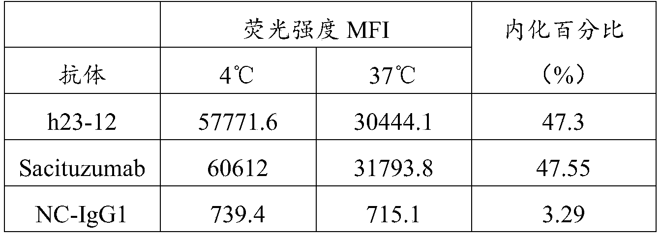
| T 1/2(Trop-2检测) | |
| Sacituzumab(n=2) | 168±14 |
| h23-12(n=2) | 174±12 |
| h4-3(n=4) | 180±57 |


| 名称 | IC 50(ug/ml) |
| 空白细胞 | / |
| h23-12-SN38 | 1.409 |
| Sacituzumab-SN38 | 4.311 |
| ch4-3-SN38 | 3.299 |
| ch11-4-SN38 | 2.879 |

| 组别 | 药物 | 给药剂量 | 给药频率 |
| 1 | Anti-CD47 | 10mg/kg | Biw |
| 2 | h23-12 | 2mg/kg | Biw |
| 3 | h23-12+Anti-CD47 | 2mg/kg+10mg/kg | Biw |
| 4 | 阴性对照hIgG4 | 2mg/kg | Biw |
| 组别 | 药物 | 给药剂量 | 给药频率 |
| 1 | ch3-11-SN38 | 5mg/kg | Biw×6 |
| 2 | ch11-4-SN38 | 5mg/kg | Biw×6 |
| 3 | Sacituzumab-SN38 | 5mg/kg | Biw×6 |
| 4 | 阴性对照hIgG1 | 5mg/kg | Biw×6 |
| 5 | ADC对照hIgG1-SN38 | 5mg/kg | Biw×6 |
Claims (25)
- 一种抗Trop-2单克隆抗体的制备方法,其包括以下步骤:(1)以重组Trop-2蛋白为免疫原免疫动物制备杂交瘤细胞;(2)以重组Trop-2蛋白为包被抗原筛选分泌抗Trop-2单克隆抗体的阳性杂交瘤细胞;(3)以膜表面Trop-2阳性细胞对步骤(2)中的阳性杂交瘤进行复筛;所述抗Trop-2单克隆抗体特异性识别并结合Trop-2胞外区天然表位。
- 根据权利要求1所述抗Trop-2单克隆抗体的制备方法,其特征在于所述步骤(3)中膜表面Trop-2阳性细胞为重组动物细胞,且所述动物细胞来自与步骤(1)制备杂交瘤细胞时免疫的动物相同的物种。
- 根据权利要求2所述抗Trop-2单克隆抗体的制备方法,其特征在于步骤(1)中免疫小鼠制备杂交瘤细胞,步骤(3)中膜表面Trop-2阳性细胞为表达外源Trop-2蛋白的重组小鼠细胞。
- 根据权利要求1所述抗Trop-2单克隆抗体的制备方法,其特征在于所述步骤(2)采用酶联免疫吸附测定(ELISA)筛选分泌抗Trop-2单克隆抗体的阳性杂交瘤细胞;步骤(3)采用流式细胞分析(FACS)复筛获得分泌特异性识别并结合Trop-2胞外区天然表位的抗体的杂交瘤。
- 根据权利要求1所述抗Trop-2单克隆抗体的制备方法,其特征在于还包括(4)对特异性识别并结合Trop-2胞外区天然表位的抗体进行鉴定的步骤,所述鉴定包括亲和力鉴定和特异性鉴定。
- 根据权利要求5所述抗Trop-2单克隆抗体的制备方法,其特征在于选择对人Trop-2、猴Trop-2具有特异性结合能力,而对鼠没有特异性结合能力的单克隆抗体。
- 根据权利要求1至6中任一项所述抗Trop-2单克隆抗体的制备方法获得的抗Trop-2单克隆抗体或其片段。
- 根据权利要求7所述抗Trop-2单克隆抗体或其片段,其特征在于所述单克隆抗体与人Trop-2胞外区重组蛋白的亲和力常数KD值小于1×10 -8、5×10 -9、1×10 -9、或5×10 -10。
- 一种抗体或其片段,所述抗体或其片段包含重链可变区(VH)和轻链可变区(VL),其中所述重链可变区(VH)和轻链可变区(VL)包含选 自以下的CDR组合(HCDR1、HCDR2、HCDR3;LCDR1、LCDR2、LCDR3):
HCDR1 HCDR2 HCDR3 HCDR1 HCDR2 HCDR3 1 SYWMY EINPSNGRTNYNEKFKS EGHNYDGSLGAMDH RSSQSLTNSYGNTFLS GISNRFS FQSTHQPYT 2 DYVIG EIYLGSGTIYYTEKFKG GSIFPFDY SASSSVSYMY DTSTLAS QQWSSYPYT 3 DYVIG EIYLGSGTIYYAEKFKG GSIFPFDY SASSSVSYMY DTSTLAS QQWSSYPYT 4 DYVIG EIYLGSGTIYYTEKFKG GSIFPFDY RASSSVSYMY DTSTLAS QQWSSYPYT 5 DYVIG EIYLGSGTIYYAEKFKG GSIFPFDY RASSSVSYMY DTSTLAS QQWSSYPYT 6 DYVIG EIYLGSGTIYYTEKFKG GSIFPFDY SASSSVSYMY DASTLAS QQWSSYPYT 7 DYVIG EIYLGSGTIYYTEKFKG GSIFPFDY SASSSVSYMY DTSTLQS QQWSSYPYT 8 SYWIN NIYPSNSYTNYNQKFKD YRSDGFAY RASQNIGTSIH FASESIS QQSNSWPFT 9 SYWIN NIYPSNSYTNYNQKFKD YRSDGFAY RASQNIGTSIE FASESIS QQSNSWPFT 10 SYWIN NIYPSNSYTNYNQKFKD YRSEGFAY RASQNIGTSIE FASESIS QQSNSWPFT 11 SYWIN NIYPSNSYTNYNQKFKD YRSGGFAY RASQNIGTSIE FASESIS QQSNSWPFT 12 SYWIN NIYPSNSYTNYNQKFKD YRSDAFAY RASQNIGTSIE FASESIS QQSNSWPFT 13 SYWIN NIYPSNSYTNYNQKFKD YRSDGFAY RASQNIGTSIS FASESIS QQSNSWPFT 14 SYWIN NIYPSNSYTNYNQKFKD YRSEGFAY RASQNIGTSIS FASESIS QQSNSWPFT 15 SYWIN NIYPSNSYTNYNQKFKD YRSGGFAY RASQNIGTSIS FASESIS QQSNSWPFT 16 SYWIN NIYPSNSYTNYNQKFKD YRSDAFAY RASQNIGTSIS FASESIS QQSNSWPFT 17 SYWIN NIYPSNSYTNYNQKFKD YRSDGFAY RASQNIGTSIA FASESIS QQSNSWPFT 18 SYWIN NIYPSNSYTNYNQKFKD YRSEGFAY RASQNIGTSIA FASESIS QQSNSWPFT 19 SYWIN NIYPSNSYTNYNQKFKD YRSGGFAY RASQNIGTSIA FASESIS QQSNSWPFT 20 SYWIN NIYPSNSYTNYNQKFKD YRSDAFAY RASQNIGTSIA FASESIS QQSNSWPFT 21 DSAMS SISRGDDTYYPDSVKG DRFGFAY KSGQSLLDSDGKTYFN LVSMLDS WQGTHFPFT 22 SYWMH EITPSDNYTSYNQKFKG GHGNYVSFDY RASQDISNYLN YTSRLHS QQGYTLPPYT 23 SYWMH EITPSDNYTSYNQKFKG GEGNYVSFDY RASQDISNYLN YTSRLHS QQGYTLPPYT 24 SYWMH EITPSDNYGSYNQKFKG GHGNYVSFDY RASQDISNYLN YTSRLES QQGYTLPPYT 25 SYWMH EITPSDNYTSYNQKFKG GEGNYVSFDY RASQDISNYLN YTSRLES QQGYTLPPYT 26 SYWMH EITPSDNYGSYNQKFKG GHGNYVSFDY RASQDISNYLN YTSRLQS QQGYTLPPYT 27 SYWMH EITPSDNYGSYNQKFKG GHGNYVSFDY RASQDISNYLN YTSRLHS QQYYTLPPYT 28 SYWMH EITPGDNYTSYNQKFKG GHGNYVSFDY RASQDISNYLN YTSRLHS QQGYSLPPYT 29 SYWMH EITPSDNYGSYNQKFKG GHGNYVSFDY RASQDISNYLN YTSRLHS QQGYSLPPYT 30 SYWMH EITPSDNYTSYNQKFKG GEGNYVSFDY RASQDISNYLN YTSRLHS QQGYSLPPYT - 根据权利要求9所述的抗体或其片段,其特征在于,所述重链可变区包含选自以下的序列:SEQ ID NO:1至SEQ ID NO:17中任一个所示的氨基酸序列或与所示的氨基酸序列具有至少75%同一性的氨基酸序列;和/或所述轻链可变区包含选自以下的序列:SEQ ID NO:18至SEQ ID NO:36中任一个所示的氨基酸序列或与所示的氨基酸序列具有至少75%同一性的氨基酸序列。
- 根据权利要求9或10所述的抗体或其片段,其特征在于,所述抗体或其片段包含的重链可变区和轻链可变区选自以下组合:(1)如SEQ ID NO:1所示的氨基酸序列或与如SEQ ID NO:1所示的氨基酸序列具有至少75%同一性的氨基酸序列;和,如SEQ ID NO:18所示的氨基酸序列或与如SEQ ID NO:18所示的氨基酸序列具有至少75%同一性的氨基酸序列;(2)如SEQ ID NO:2所示的氨基酸序列或与如SEQ ID NO:2所示的氨基酸序列具有至少75%同一性的氨基酸序列;和,如SEQ ID NO:19所示的氨基酸序列或与如SEQ ID NO:19所示的氨基酸序列具有至少75%同一性的氨基酸序列;(3)如SEQ ID NO:3所示的氨基酸序列或与如SEQ ID NO:3所示的氨基酸序列具有至少75%同一性的氨基酸序列;和,如SEQ ID NO:20所示的氨基酸序列或与如SEQ ID NO:30所示的氨基酸序列具有至少75%同一性的氨基酸序列;(4)如SEQ ID NO:4所示的氨基酸序列或与如SEQ ID NO:4所示的氨基酸序列具有至少75%同一性的氨基酸序列;和,如SEQ ID NO:20所示的氨基酸序列或与如SEQ ID NO:20所示的氨基酸序列具有至少75%同一性的氨基酸序列;(5)如SEQ ID NO:3所示的氨基酸序列或与如SEQ ID NO:3所示的氨基酸序列具有至少75%同一性的氨基酸序列;和,如SEQ ID NO:21所示的氨基酸序列或与如SEQ ID NO:21所示的氨基酸序列具有至少75%同一性的氨基酸序列;(6)如SEQ ID NO:4所示的氨基酸序列或与如SEQ ID NO:4所示的氨基酸序列具有至少75%同一性的氨基酸序列;和,如SEQ ID NO:21所示的氨基酸序列或与如SEQ ID NO:21所示的氨基酸序列具有至少75%同一性的氨基酸序列;(7)如SEQ ID NO:3所示的氨基酸序列或与如SEQ ID NO:3所示的氨基酸序列具有至少75%同一性的氨基酸序列;和,如SEQ ID NO:22所示的氨基酸序列或与如SEQ ID NO:22所示的氨基酸序列具有至少75%同一性的氨基酸序列;(8)如SEQ ID NO:3所示的氨基酸序列或与如SEQ ID NO:3所示的氨基酸序列具有至少75%同一性的氨基酸序列;和,如SEQ ID NO:23所示的氨基酸序列或与如SEQ ID NO:23所示的氨基酸序列具有至少75%同一性的氨基酸序列;(9)如SEQ ID NO:5所示的氨基酸序列或与如SEQ ID NO:5所示的氨基酸序列具有至少75%同一性的氨基酸序列;和,如SEQ ID NO:24所示的氨基酸序列或与如SEQ ID NO:24所示的氨基酸序列具有至少75%同一性的氨基酸序列;(10)如SEQ ID NO:6所示的氨基酸序列或与如SEQ ID NO:6所示的氨基酸序列具有至少75%同一性的氨基酸序列;和,如SEQ ID NO:25所示的氨基酸序列或与如SEQ ID NO:25所示的氨基酸序列具有至少75%同一性的氨基酸序列;(11)如SEQ ID NO:7所示的氨基酸序列或与如SEQ ID NO:7所示的氨基酸序列具有至少75%同一性的氨基酸序列;和,如SEQ ID NO:26所示的氨基酸序列或与如SEQ ID NO:26所示的氨基酸序列具有至少75%同一性的氨基酸序列;(12)如SEQ ID NO:7所示的氨基酸序列或与如SEQ ID NO:7所示的氨基酸序列具有至少75%同一性的氨基酸序列;和,如SEQ ID NO:27所示的氨基酸序列或与如SEQ ID NO:27所示的氨基酸序列具有至少75%同一性的氨基酸序列;(13)如SEQ ID NO:8所示的氨基酸序列或与如SEQ ID NO:8所示的氨基酸序列具有至少75%同一性的氨基酸序列;和,如SEQ ID NO:27所示的氨基酸序列或与如SEQ ID NO:27所示的氨基酸序列具有至少75%同一性的 氨基酸序列;(14)如SEQ ID NO:9所示的氨基酸序列或与如SEQ ID NO:9所示的氨基酸序列具有至少75%同一性的氨基酸序列;和,如SEQ ID NO:27所示的氨基酸序列或与如SEQ ID NO:27所示的氨基酸序列具有至少75%同一性的氨基酸序列;(15)如SEQ ID NO:10所示的氨基酸序列或与如SEQ ID NO:10所示的氨基酸序列具有至少75%同一性的氨基酸序列;和,如SEQ ID NO:27所示的氨基酸序列或与如SEQ ID NO:27所示的氨基酸序列具有至少75%同一性的氨基酸序列;(16)如SEQ ID NO:7所示的氨基酸序列或与如SEQ ID NO:7所示的氨基酸序列具有至少75%同一性的氨基酸序列;和,如SEQ ID NO:28所示的氨基酸序列或与如SEQ ID NO:28所示的氨基酸序列具有至少75%同一性的氨基酸序列;(17)如SEQ ID NO:8所示的氨基酸序列或与如SEQ ID NO:8所示的氨基酸序列具有至少75%同一性的氨基酸序列;和,如SEQ ID NO:28所示的氨基酸序列或与如SEQ ID NO:28所示的氨基酸序列具有至少75%同一性的氨基酸序列;(18)如SEQ ID NO:9所示的氨基酸序列或与如SEQ ID NO:9所示的氨基酸序列具有至少75%同一性的氨基酸序列;和,如SEQ ID NO:28所示的氨基酸序列或与如SEQ ID NO:28所示的氨基酸序列具有至少75%同一性的氨基酸序列;(19)如SEQ ID NO:10所示的氨基酸序列或与如SEQ ID NO:10所示的氨基酸序列具有至少75%同一性的氨基酸序列;和,如SEQ ID NO:28所示的氨基酸序列或与如SEQ ID NO:28所示的氨基酸序列具有至少75%同一性的氨基酸序列;(20)如SEQ ID NO:7所示的氨基酸序列或与如SEQ ID NO:7所示的氨基酸序列具有至少75%同一性的氨基酸序列;和,如SEQ ID NO:29所示的氨基酸序列或与如SEQ ID NO:29所示的氨基酸序列具有至少75%同一性的氨基酸序列;(21)如SEQ ID NO:8所示的氨基酸序列或与如SEQ ID NO:8所示的氨基酸序列具有至少75%同一性的氨基酸序列;和,如SEQ ID NO:29所示的 氨基酸序列或与如SEQ ID NO:29所示的氨基酸序列具有至少75%同一性的氨基酸序列;(22)如SEQ ID NO:9所示的氨基酸序列或与如SEQ ID NO:9所示的氨基酸序列具有至少75%同一性的氨基酸序列;和,如SEQ ID NO:29所示的氨基酸序列或与如SEQ ID NO:29所示的氨基酸序列具有至少75%同一性的氨基酸序列;(23)如SEQ ID NO:10所示的氨基酸序列或与如SEQ ID NO:10所示的氨基酸序列具有至少75%同一性的氨基酸序列;和,如SEQ ID NO:29所示的氨基酸序列或与如SEQ ID NO:29所示的氨基酸序列具有至少75%同一性的氨基酸序列;(24)如SEQ ID NO:11所示的氨基酸序列或与如SEQ ID NO:11所示的氨基酸序列具有至少75%同一性的氨基酸序列;和,如SEQ ID NO:30所示的氨基酸序列或与如SEQ ID NO:30所示的氨基酸序列具有至少75%同一性的氨基酸序列;(25)如SEQ ID NO:12所示的氨基酸序列或与如SEQ ID NO:12所示的氨基酸序列具有至少75%同一性的氨基酸序列;和,如SEQ ID NO:31所示的氨基酸序列或与如SEQ ID NO:31所示的氨基酸序列具有至少75%同一性的氨基酸序列;(26)如SEQ ID NO:13所示的氨基酸序列或与如SEQ ID NO:13所示的氨基酸序列具有至少75%同一性的氨基酸序列;和,如SEQ ID NO:32所示的氨基酸序列或与如SEQ ID NO:32所示的氨基酸序列具有至少75%同一性的氨基酸序列;(27)如SEQ ID NO:16所示的氨基酸序列或与如SEQ ID NO:16所示的氨基酸序列具有至少75%同一性的氨基酸序列;和,如SEQ ID NO:32所示的氨基酸序列或与如SEQ ID NO:32所示的氨基酸序列具有至少75%同一性的氨基酸序列;(28)如SEQ ID NO:14所示的氨基酸序列或与如SEQ ID NO:14所示的氨基酸序列具有至少75%同一性的氨基酸序列;和,如SEQ ID NO:33所示的氨基酸序列或与如SEQ ID NO:33所示的氨基酸序列具有至少75%同一性的氨基酸序列;(29)如SEQ ID NO:16所示的氨基酸序列或与如SEQ ID NO:16所示的 氨基酸序列具有至少75%同一性的氨基酸序列;和,如SEQ ID NO:33所示的氨基酸序列或与如SEQ ID NO:33所示的氨基酸序列具有至少75%同一性的氨基酸序列;(30)如SEQ ID NO:14所示的氨基酸序列或与如SEQ ID NO:14所示的氨基酸序列具有至少75%同一性的氨基酸序列;和,如SEQ ID NO:34所示的氨基酸序列或与如SEQ ID NO:34所示的氨基酸序列具有至少75%同一性的氨基酸序列;(31)如SEQ ID NO:14所示的氨基酸序列或与如SEQ ID NO:14所示的氨基酸序列具有至少75%同一性的氨基酸序列;和,如SEQ ID NO:35所示的氨基酸序列或与如SEQ ID NO:35所示的氨基酸序列具有至少75%同一性的氨基酸序列;(32)如SEQ ID NO:15所示的氨基酸序列或与如SEQ ID NO:15所示的氨基酸序列具有至少75%同一性的氨基酸序列;和,如SEQ ID NO:36所示的氨基酸序列或与如SEQ ID NO:36所示的氨基酸序列具有至少75%同一性的氨基酸序列;(33)如SEQ ID NO:14所示的氨基酸序列或与如SEQ ID NO:14所示的氨基酸序列具有至少75%同一性的氨基酸序列;和,如SEQ ID NO:36所示的氨基酸序列或与如SEQ ID NO:36所示的氨基酸序列具有至少75%同一性的氨基酸序列;(34)如SEQ ID NO:16所示的氨基酸序列或与如SEQ ID NO:16所示的氨基酸序列具有至少75%同一性的氨基酸序列;和,如SEQ ID NO:36所示的氨基酸序列或与如SEQ ID NO:36所示的氨基酸序列具有至少75%同一性的氨基酸序列。
- 根据权利要求1至11中任一项所述的抗体或其片段,其特征在于,所述抗体或其片段为单克隆抗体、单链抗体、双功能抗体、单域抗体、纳米抗体、完全或部分人源化的抗体或者嵌合抗体等任意形式,或者,所述抗体或其片段为半抗体或半抗体的抗原结合片段,例如scFv、BsFv、dsFv、(dsFv) 2、Fab、Fab'、F(ab') 2或Fv;优选地,所述抗体或其片段还包含人或鼠的恒定区,优选包含人或鼠的轻链恒定区(CL)和/或重链恒定区(CH);更优选地,所述抗体或其片段包含选自IgG、IgA、IgM、IgD或IgE的 重链恒定区和/或κ或λ型轻链恒定区。
- 根据权利要求1至12中任一项所述的抗体或其片段,其特征在于,所述抗体为单克隆抗体,优选为鼠源、嵌合或人源化的单克隆抗体;优选地,所述单克隆抗体的重链恒定区为IgG1或IgG4亚型,轻链恒定区为κ型;优选地,所述单克隆抗体的重链恒定区包含如SEQ ID NO:37所示的氨基酸序列或者与所述氨基酸序列具有至少75%同一性的氨基酸序列;优选地,所述单克隆抗体的轻链恒定区包含如SEQ ID NO:38所示氨基酸序列或者与所述氨基酸序列具有至少75%同一性的氨基酸序列。
- 一种核酸分子,其编码权利要求1至13中任一项所述的抗体或其片段或者编码所述抗体或其片段中包含的重链CDR、轻链CDR、轻链可变区、重链可变区、重链或轻链。
- 一种载体,其包含权利要求14所述的核酸分子。
- 一种宿主细胞,所述宿主细胞包含权利要求14所述的核酸分子和/或权利要求15所述的载体,或者所述宿主细胞被权利要求14所述的核酸分子和/或权利要求15所述的载体转化或转染。
- 一种药物组合物,其包含权利要求1至13中任一项所述的抗体或其片段、权利要求14所述的核酸分子、权利要求15所述的载体或权利要求16所述的宿主细胞,以及任选地药学上可接受的辅料。
- 根据权利要求17所述的药物组合物,其特征在于,所述药物组合物还包含其他抗体类药物;优选地,所述抗体类药物为巨噬细胞类免疫检查点抗体,更优选为抗CD47抗体。
- 权利要求1至13中任一项所述的抗体或其片段、权利要求14所述的核酸分子、权利要求15所述的载体、权利要求16所述的宿主细胞或权利要求17或18所述的药物组合物在制备药物中的用途;优选地,所述药物用于治疗Trop-2高表达癌症;优选地,所述Trop-2高表达癌症为胃癌、胰腺癌、肠癌、卵巢癌、鳞状肺癌、非小细胞肺癌、小细胞肺癌、尿路上皮癌、三阴性乳腺癌或宫颈癌。
- 一种试剂盒,所述试剂盒包括权利要求1至13中任一项所述的抗体分子或其片段、权利要求14所述的核酸分子、权利要求15所述的载体、权利要求16所述的宿主细胞或权利要求17或18所述的药物组合物。
- 一种融合蛋白,所述融合蛋白包含权利要求1至13中任一项所述的抗体或其片段。
- 一种缀合物,所述缀合物包含权利要求1至13中任一项所述的抗体或其片段以及与其缀合的药物,其中所述药物为细胞毒剂。
- 根据权利要求22所述的缀合物,所述缀合物为由下式所示的抗体药物缀合物(ADC):(权利要求1至13中任一项所述的抗体或其片段)-(接头)-(细胞毒剂);优选地,所述细胞毒剂为微管蛋白抑制剂(如紫杉醇、多西他赛等)或DNA复制抑制剂(如伊立替康或其代谢活性物SN-38等)。
- 权利要求1至13中任一项所述的抗体或其片段、权利要求14所述的核酸分子、权利要求15所述的载体或权利要求16所述的宿主细胞在制备抗体药物缀合物(ADC)中的用途;优选地,所述抗体药物缀合物用于治疗Trop-2高表达癌症;优选地,所述Trop-2高表达癌症为胃癌、胰腺癌、肠癌、卵巢癌、鳞状肺癌、非小细胞肺癌、小细胞肺癌、尿路上皮癌、三阴性乳腺癌或宫颈癌。
- 一种预防和/或治疗疾病的方法,所述方法包括给有此需要的受试者施用权利要求1至13中任一项所述的抗体或其片段、权利要求14所述的核酸分子、权利要求15所述的载体、权利要求16所述的宿主细胞、权利要求17或18所述的药物组合物、权利要求21所述的融合蛋白、权利要求22或23所述的缀合物,以及任选的其他药物或手段;优选地,所述疾病为Trop-2高表达癌症;优选地,所述Trop-2高表达癌症为胃癌、胰腺癌、肠癌、卵巢癌、鳞状肺癌、非小细胞肺癌、小细胞肺癌、尿路上皮癌、三阴性乳腺癌或宫颈癌。
Priority Applications (7)
| Application Number | Priority Date | Filing Date | Title |
|---|---|---|---|
| EP20874923.4A EP4043495A4 (en) | 2019-10-11 | 2020-10-12 | ANTI-HUMAN TROP-2 ANTIBODIES AND USE THEREOF |
| US17/768,189 US12558577B2 (en) | 2019-10-11 | 2020-10-12 | Anti-human Trop-2 antibody and application thereof |
| AU2020362827A AU2020362827A1 (en) | 2019-10-11 | 2020-10-12 | Anti-human Trop-2 antibody and application thereof |
| CN202080071403.9A CN114585649B (zh) | 2019-10-11 | 2020-10-12 | 抗人Trop-2抗体及其应用 |
| JP2022521211A JP2022553645A (ja) | 2019-10-11 | 2020-10-12 | 抗ヒトTrop-2抗体およびその用途 |
| CA3154454A CA3154454A1 (en) | 2019-10-11 | 2020-10-12 | Anti-human trop-2 antibody and application thereof |
| JP2025128509A JP2025163135A (ja) | 2019-10-11 | 2025-07-31 | 抗ヒトTrop-2抗体およびその用途 |
Applications Claiming Priority (2)
| Application Number | Priority Date | Filing Date | Title |
|---|---|---|---|
| CN201910962965.1A CN112646038B (zh) | 2019-10-11 | 2019-10-11 | 抗人Trop-2抗体及其应用 |
| CN201910962965.1 | 2019-10-11 |
Publications (1)
| Publication Number | Publication Date |
|---|---|
| WO2021068949A1 true WO2021068949A1 (zh) | 2021-04-15 |
Family
ID=75342761
Family Applications (1)
| Application Number | Title | Priority Date | Filing Date |
|---|---|---|---|
| PCT/CN2020/120277 Ceased WO2021068949A1 (zh) | 2019-10-11 | 2020-10-12 | 抗人Trop-2抗体及其应用 |
Country Status (8)
| Country | Link |
|---|---|
| US (1) | US12558577B2 (zh) |
| EP (1) | EP4043495A4 (zh) |
| JP (2) | JP2022553645A (zh) |
| CN (2) | CN112646038B (zh) |
| AU (1) | AU2020362827A1 (zh) |
| CA (1) | CA3154454A1 (zh) |
| TW (1) | TWI854047B (zh) |
| WO (1) | WO2021068949A1 (zh) |
Cited By (3)
| Publication number | Priority date | Publication date | Assignee | Title |
|---|---|---|---|---|
| JP2024518245A (ja) * | 2021-04-23 | 2024-05-01 | ビオシオン インコーポレイテッド | Trop2結合抗体及びその用途 |
| EP4438627A4 (en) * | 2021-12-07 | 2026-02-11 | Chia Tai Tianqing Pharmaceutical Group Co Ltd | ANTI-TROP-2 ANTIBODY OR ANTIGEN-BINDING FRAGMENT OF IT |
| EP4408893A4 (en) * | 2021-09-29 | 2026-02-18 | Kisoji Biotechnology Inc | Binding agents targeting tumor cells expressing too much 2 |
Families Citing this family (13)
| Publication number | Priority date | Publication date | Assignee | Title |
|---|---|---|---|---|
| CN116589587A (zh) * | 2020-06-22 | 2023-08-15 | 四川百利药业有限责任公司 | 一种抗trop2抗体 |
| CN115536747B (zh) * | 2021-06-30 | 2024-08-27 | 益科思特(北京)医药科技发展有限公司 | 一种结合trop2的抗体及靶向trop2和cd3的双特异性抗体及其制备方法与应用 |
| CN116478279A (zh) * | 2021-12-31 | 2023-07-25 | 武汉瑞法医疗器械有限公司 | 乙型肝炎病毒表面抗原纳米抗体吸附剂及制备方法和应用 |
| CN115368465B (zh) * | 2022-07-19 | 2026-02-24 | 合肥天港免疫药物有限公司 | 双特异性抗体及其应用 |
| CN117430710B (zh) * | 2022-07-21 | 2025-07-04 | 中国医学科学院医药生物技术研究所 | 一种针对Trop2的单克隆抗体及其应用 |
| CN120500502A (zh) * | 2023-03-14 | 2025-08-15 | 浙江道尔生物科技有限公司 | Trop2单域抗体、融合蛋白及其应用 |
| KR20260012304A (ko) * | 2023-04-17 | 2026-01-26 | 피크 바이오, 인크. | 항체 및 항체-약물 접합체 및 사용 방법 및 합성 공정 및 중간체 |
| EP4701667A1 (en) * | 2023-04-24 | 2026-03-04 | Merck Sharp & Dohme LLC | Trop2 binders and conjugates thereof |
| WO2025091232A1 (zh) * | 2023-10-31 | 2025-05-08 | 上海优卡迪生物医药科技有限公司 | 结合Trop2的抗体或Trop2抗原结合片段以及抗Trop2 CAR和应用 |
| CN119978125A (zh) * | 2023-11-10 | 2025-05-13 | 成都社泰医疗科技有限公司 | 核素标记的抗Trop2单域抗体 |
| CN117264072B (zh) * | 2023-11-22 | 2024-03-22 | 江苏迈威康新药研发有限公司 | 一种抗sn38单抗及其应用 |
| TW202602943A (zh) * | 2024-03-22 | 2026-01-16 | 日商衛材R&D企管股份有限公司 | 抗trop2抗體藥物偶聯物及使用方法 |
| WO2025250825A1 (en) * | 2024-05-30 | 2025-12-04 | Sutro Biopharma, Inc. | Anti-trop2 antibodies, compositions comprising anti-trop2 antibodies and methods of making and using anti-trop2 antibodies |
Citations (10)
| Publication number | Priority date | Publication date | Assignee | Title |
|---|---|---|---|---|
| US5545806A (en) | 1990-08-29 | 1996-08-13 | Genpharm International, Inc. | Ransgenic non-human animals for producing heterologous antibodies |
| US5569825A (en) | 1990-08-29 | 1996-10-29 | Genpharm International | Transgenic non-human animals capable of producing heterologous antibodies of various isotypes |
| US5633425A (en) | 1990-08-29 | 1997-05-27 | Genpharm International, Inc. | Transgenic non-human animals capable of producing heterologous antibodies |
| US5661016A (en) | 1990-08-29 | 1997-08-26 | Genpharm International Inc. | Transgenic non-human animals capable of producing heterologous antibodies of various isotypes |
| US6673986B1 (en) | 1990-01-12 | 2004-01-06 | Abgenix, Inc. | Generation of xenogeneic antibodies |
| CN104114580A (zh) * | 2011-11-22 | 2014-10-22 | 株式会社立富泰克 | 在体内具有抗肿瘤活性的抗人trop-2抗体 |
| US20150183874A1 (en) | 2010-05-14 | 2015-07-02 | The Board Of Trustees Of The Leland Stanford Junior University | Humanized and chimeric monoclonal antibodies to cd47 |
| CN105849126A (zh) * | 2013-12-25 | 2016-08-10 | 第三共株式会社 | 抗trop2抗体-药物偶联物 |
| WO2017084763A1 (en) * | 2015-11-19 | 2017-05-26 | Abruzzo Theranostic Srl | Use of circulating serum trop-2 as new tumor biomarker |
| CN107446050A (zh) * | 2017-08-11 | 2017-12-08 | 百奥泰生物科技(广州)有限公司 | Trop2阳性疾病治疗的化合物及方法 |
Family Cites Families (10)
| Publication number | Priority date | Publication date | Assignee | Title |
|---|---|---|---|---|
| CA2750038C (en) | 2009-02-05 | 2016-10-04 | Oncoxx S.R.L. | Anti-trop-2 monoclonal antibodies and uses thereof in the treatment and diagnosis of tumors |
| EA201500220A1 (ru) * | 2010-05-17 | 2015-10-30 | Ливтех, Инк. | Антитело против trop-2 человека, обладающее противоопухолевой активностью in vivo |
| WO2011155579A1 (ja) * | 2010-06-10 | 2011-12-15 | 北海道公立大学法人札幌医科大学 | 抗Trop-2抗体 |
| TWI495644B (zh) * | 2011-11-11 | 2015-08-11 | Rinat Neuroscience Corp | 營養層細胞表面抗原(Trop-2)之專一性抗體類及彼等之用途 |
| WO2015047510A1 (en) * | 2013-09-27 | 2015-04-02 | Immunomedics, Inc. | Anti-trop-2 antibody-drug conjugates and uses thereof |
| EP3227340B1 (en) * | 2014-12-04 | 2020-06-17 | Mediterranea Theranostic S.R.L. | Humanized anti-trop-2 monoclonal antibodies and uses thereof |
| EP3303392B1 (en) * | 2015-06-01 | 2020-08-05 | Medigene Immunotherapies GmbH | Method for generating antibodies against t cell receptor |
| CN108601841A (zh) | 2016-02-10 | 2018-09-28 | 免疫医疗公司 | Abcg2抑制剂与sacituzumab govitecan(immu-132)的组合克服表达trop-2的癌中对sn-38的抗性 |
| WO2018102212A1 (en) * | 2016-12-01 | 2018-06-07 | Immunomedics, Inc. | Therapy for metastatic urothelial cancer with the antibody-drug conjugate, sacituzumab govitecan (immu-132) |
| EP3600283A4 (en) * | 2017-03-27 | 2020-12-16 | Immunomedics, Inc. | TREATMENT OF TROP-2 EXPRESSIVE TRIPLE NEGATIVE BREAST CANCER WITH SACITUZUMAB GOVITECAN AND A RAD51 INHIBITOR |
-
2019
- 2019-10-11 CN CN201910962965.1A patent/CN112646038B/zh active Active
-
2020
- 2020-10-12 WO PCT/CN2020/120277 patent/WO2021068949A1/zh not_active Ceased
- 2020-10-12 TW TW109135130A patent/TWI854047B/zh active
- 2020-10-12 JP JP2022521211A patent/JP2022553645A/ja active Pending
- 2020-10-12 AU AU2020362827A patent/AU2020362827A1/en active Pending
- 2020-10-12 EP EP20874923.4A patent/EP4043495A4/en active Pending
- 2020-10-12 US US17/768,189 patent/US12558577B2/en active Active
- 2020-10-12 CN CN202080071403.9A patent/CN114585649B/zh active Active
- 2020-10-12 CA CA3154454A patent/CA3154454A1/en active Pending
-
2025
- 2025-07-31 JP JP2025128509A patent/JP2025163135A/ja active Pending
Patent Citations (10)
| Publication number | Priority date | Publication date | Assignee | Title |
|---|---|---|---|---|
| US6673986B1 (en) | 1990-01-12 | 2004-01-06 | Abgenix, Inc. | Generation of xenogeneic antibodies |
| US5545806A (en) | 1990-08-29 | 1996-08-13 | Genpharm International, Inc. | Ransgenic non-human animals for producing heterologous antibodies |
| US5569825A (en) | 1990-08-29 | 1996-10-29 | Genpharm International | Transgenic non-human animals capable of producing heterologous antibodies of various isotypes |
| US5633425A (en) | 1990-08-29 | 1997-05-27 | Genpharm International, Inc. | Transgenic non-human animals capable of producing heterologous antibodies |
| US5661016A (en) | 1990-08-29 | 1997-08-26 | Genpharm International Inc. | Transgenic non-human animals capable of producing heterologous antibodies of various isotypes |
| US20150183874A1 (en) | 2010-05-14 | 2015-07-02 | The Board Of Trustees Of The Leland Stanford Junior University | Humanized and chimeric monoclonal antibodies to cd47 |
| CN104114580A (zh) * | 2011-11-22 | 2014-10-22 | 株式会社立富泰克 | 在体内具有抗肿瘤活性的抗人trop-2抗体 |
| CN105849126A (zh) * | 2013-12-25 | 2016-08-10 | 第三共株式会社 | 抗trop2抗体-药物偶联物 |
| WO2017084763A1 (en) * | 2015-11-19 | 2017-05-26 | Abruzzo Theranostic Srl | Use of circulating serum trop-2 as new tumor biomarker |
| CN107446050A (zh) * | 2017-08-11 | 2017-12-08 | 百奥泰生物科技(广州)有限公司 | Trop2阳性疾病治疗的化合物及方法 |
Non-Patent Citations (7)
| Title |
|---|
| FISHWILD ET AL., NATURE BIOTECHNOLOGY, vol. 14, 1996, pages 845 - 51 |
| MARKS ET AL., BIO/TECHNOLOGY, vol. 10, 1992, pages 779 - 783 |
| MORRISON, NATURE, vol. 368, 1994, pages 812 - 13 |
| RIECHMANN ET AL., NATURE, vol. 332, 1988, pages 323 - 327 |
| See also references of EP4043495A4 |
| WHO DRUG INFORMATION, vol. 31, no. 1, 2017 |
| ZHIWEI DONGYAN WANG: "Antibody Engineering", 2001, PEKING UNIVERSITY MEDICAL PRESS, pages: 313 - 314 |
Cited By (6)
| Publication number | Priority date | Publication date | Assignee | Title |
|---|---|---|---|---|
| JP2024518245A (ja) * | 2021-04-23 | 2024-05-01 | ビオシオン インコーポレイテッド | Trop2結合抗体及びその用途 |
| EP4326399A4 (en) * | 2021-04-23 | 2024-10-09 | Biosion, Inc. | TROP2-BINDING ANTIBODIES AND USES THEREOF |
| JP7678250B2 (ja) | 2021-04-23 | 2025-05-16 | ビオシオン インコーポレイテッド | Trop2結合抗体及びその用途 |
| AU2022262101B2 (en) * | 2021-04-23 | 2026-04-02 | Biosion Inc. | Antibodies binding trop2 and uses thereof |
| EP4408893A4 (en) * | 2021-09-29 | 2026-02-18 | Kisoji Biotechnology Inc | Binding agents targeting tumor cells expressing too much 2 |
| EP4438627A4 (en) * | 2021-12-07 | 2026-02-11 | Chia Tai Tianqing Pharmaceutical Group Co Ltd | ANTI-TROP-2 ANTIBODY OR ANTIGEN-BINDING FRAGMENT OF IT |
Also Published As
| Publication number | Publication date |
|---|---|
| CA3154454A1 (en) | 2021-04-15 |
| TW202128773A (zh) | 2021-08-01 |
| EP4043495A1 (en) | 2022-08-17 |
| TWI854047B (zh) | 2024-09-01 |
| US12558577B2 (en) | 2026-02-24 |
| JP2022553645A (ja) | 2022-12-26 |
| CN112646038B (zh) | 2025-06-03 |
| JP2025163135A (ja) | 2025-10-28 |
| US20240092930A1 (en) | 2024-03-21 |
| AU2020362827A1 (en) | 2022-05-26 |
| EP4043495A4 (en) | 2024-06-19 |
| CN114585649B (zh) | 2025-10-28 |
| CN112646038A (zh) | 2021-04-13 |
| CN114585649A (zh) | 2022-06-03 |
Similar Documents
| Publication | Publication Date | Title |
|---|---|---|
| TWI854047B (zh) | 抗人Trop-2抗體及其應用 | |
| US20210230272A1 (en) | Antibody targeting cldn18.2, bispecific antibody, adc, and car, and applications thereof | |
| CN116059395A (zh) | 用于选择性制造抗体-药物缀合物的方法 | |
| CN111110862A (zh) | 抗cldn18.2抗体的药物偶联体及其制备方法和用途 | |
| CN116669730A (zh) | 抗cldn18.2抗体、药物偶联物及其制备方法和用途 | |
| US20230374132A1 (en) | Anti-cd3 antibody and uses thereof | |
| WO2022171134A1 (zh) | 包含抗cldn18.2的抗体或其抗原结合片段的抗体药物偶联物及其用途 | |
| US12428479B2 (en) | CLDN18.2-targeting antibody, bispecific antibody and use thereof | |
| TW202233692A (zh) | 結合trop2的抗體及其用途 | |
| US20250277054A1 (en) | Psma antibodies and uses thereof | |
| IL323554A (en) | Cytotoxic ligand-drug conjugates and their pharmaceutical uses | |
| WO2023241629A1 (zh) | 抗cldn18.2抗体、其药物组合物及用途 | |
| WO2025157214A1 (zh) | 抗FGFR2b抗体及抗体药物偶联物 | |
| JP2026512687A (ja) | ヒト化l1cam抗体-薬物複合体 | |
| HK40089485B (zh) | 抗cldn-18.2抗体药物偶联物及其用途 | |
| HK40089485A (zh) | 抗cldn-18.2抗体药物偶联物及其用途 | |
| WO2023025303A1 (zh) | 抗cldn-18.2抗体药物偶联物及其用途 | |
| WO2026007196A1 (zh) | 特异性结合FGFR2b的抗体及其与药物的缀合物 | |
| WO2024240064A1 (zh) | 靶向抗原表位多肽的抗体药物偶联物及其应用 | |
| CN121311508A (zh) | 抗her2互补性双特异抗体-药物偶联物及其制备方法和用途 | |
| WO2025045242A1 (zh) | 一种靶向gpc3的双特异性抗体或抗原结合片段及其用途 | |
| WO2026082207A1 (zh) | 抗EGFR和cMET双特异性抗体的抗体药物偶联物 | |
| CN118725129A (zh) | 抗4-1bb×cldn18.2双特异性抗体及其用途 | |
| HK40127117A (zh) | 配体-细胞毒性药物偶联物及其药物用途 | |
| HK40129185A (zh) | Psma抗体及其用途 |
Legal Events
| Date | Code | Title | Description |
|---|---|---|---|
| 121 | Ep: the epo has been informed by wipo that ep was designated in this application |
Ref document number: 20874923 Country of ref document: EP Kind code of ref document: A1 |
|
| ENP | Entry into the national phase |
Ref document number: 2022521211 Country of ref document: JP Kind code of ref document: A |
|
| ENP | Entry into the national phase |
Ref document number: 3154454 Country of ref document: CA |
|
| WWE | Wipo information: entry into national phase |
Ref document number: 17768189 Country of ref document: US |
|
| NENP | Non-entry into the national phase |
Ref country code: DE |
|
| ENP | Entry into the national phase |
Ref document number: 2020874923 Country of ref document: EP Effective date: 20220511 |
|
| ENP | Entry into the national phase |
Ref document number: 2020362827 Country of ref document: AU Date of ref document: 20201012 Kind code of ref document: A |
|
| WWG | Wipo information: grant in national office |
Ref document number: 202080071403.9 Country of ref document: CN |
|
| WWG | Wipo information: grant in national office |
Ref document number: 17768189 Country of ref document: US |