WO2021075306A1 - 手術用情報処理装置、手術用情報処理方法及び手術用情報処理プログラム - Google Patents
手術用情報処理装置、手術用情報処理方法及び手術用情報処理プログラム Download PDFInfo
- Publication number
- WO2021075306A1 WO2021075306A1 PCT/JP2020/037804 JP2020037804W WO2021075306A1 WO 2021075306 A1 WO2021075306 A1 WO 2021075306A1 JP 2020037804 W JP2020037804 W JP 2020037804W WO 2021075306 A1 WO2021075306 A1 WO 2021075306A1
- Authority
- WO
- WIPO (PCT)
- Prior art keywords
- surgical
- image
- information
- processing
- information processing
- Prior art date
- Legal status (The legal status is an assumption and is not a legal conclusion. Google has not performed a legal analysis and makes no representation as to the accuracy of the status listed.)
- Ceased
Links
Images






Classifications
-
- G—PHYSICS
- G02—OPTICS
- G02B—OPTICAL ELEMENTS, SYSTEMS OR APPARATUS
- G02B27/00—Optical systems or apparatus not provided for by any of the groups G02B1/00 - G02B26/00, G02B30/00
- G02B27/64—Imaging systems using optical elements for stabilisation of the lateral and angular position of the image
- G02B27/646—Imaging systems using optical elements for stabilisation of the lateral and angular position of the image compensating for small deviations, e.g. due to vibration or shake
-
- A—HUMAN NECESSITIES
- A61—MEDICAL OR VETERINARY SCIENCE; HYGIENE
- A61B—DIAGNOSIS; SURGERY; IDENTIFICATION
- A61B1/00—Instruments for performing medical examinations of the interior of cavities or tubes of the body by visual or photographical inspection, e.g. endoscopes; Illuminating arrangements therefor
- A61B1/00002—Operational features of endoscopes
- A61B1/00004—Operational features of endoscopes characterised by electronic signal processing
- A61B1/00006—Operational features of endoscopes characterised by electronic signal processing of control signals
-
- A—HUMAN NECESSITIES
- A61—MEDICAL OR VETERINARY SCIENCE; HYGIENE
- A61B—DIAGNOSIS; SURGERY; IDENTIFICATION
- A61B1/00—Instruments for performing medical examinations of the interior of cavities or tubes of the body by visual or photographical inspection, e.g. endoscopes; Illuminating arrangements therefor
- A61B1/00002—Operational features of endoscopes
- A61B1/00004—Operational features of endoscopes characterised by electronic signal processing
- A61B1/00009—Operational features of endoscopes characterised by electronic signal processing of image signals during a use of endoscope
- A61B1/000095—Operational features of endoscopes characterised by electronic signal processing of image signals during a use of endoscope for image enhancement
-
- A—HUMAN NECESSITIES
- A61—MEDICAL OR VETERINARY SCIENCE; HYGIENE
- A61B—DIAGNOSIS; SURGERY; IDENTIFICATION
- A61B90/00—Instruments, implements or accessories specially adapted for surgery or diagnosis and not covered by any of the groups A61B1/00 - A61B50/00, e.g. for luxation treatment or for protecting wound edges
- A61B90/36—Image-producing devices or illumination devices not otherwise provided for
- A61B90/361—Image-producing devices, e.g. surgical cameras
-
- G—PHYSICS
- G06—COMPUTING OR CALCULATING; COUNTING
- G06T—IMAGE DATA PROCESSING OR GENERATION, IN GENERAL
- G06T5/00—Image enhancement or restoration
-
- H—ELECTRICITY
- H04—ELECTRIC COMMUNICATION TECHNIQUE
- H04N—PICTORIAL COMMUNICATION, e.g. TELEVISION
- H04N7/00—Television systems
- H04N7/18—Closed-circuit television [CCTV] systems, i.e. systems in which the video signal is not broadcast
- H04N7/183—Closed-circuit television [CCTV] systems, i.e. systems in which the video signal is not broadcast for receiving images from a single remote source
- H04N7/185—Closed-circuit television [CCTV] systems, i.e. systems in which the video signal is not broadcast for receiving images from a single remote source from a mobile camera, e.g. for remote control
-
- A—HUMAN NECESSITIES
- A61—MEDICAL OR VETERINARY SCIENCE; HYGIENE
- A61B—DIAGNOSIS; SURGERY; IDENTIFICATION
- A61B1/00—Instruments for performing medical examinations of the interior of cavities or tubes of the body by visual or photographical inspection, e.g. endoscopes; Illuminating arrangements therefor
- A61B1/00002—Operational features of endoscopes
- A61B1/00043—Operational features of endoscopes provided with output arrangements
- A61B1/00045—Display arrangement
-
- A—HUMAN NECESSITIES
- A61—MEDICAL OR VETERINARY SCIENCE; HYGIENE
- A61B—DIAGNOSIS; SURGERY; IDENTIFICATION
- A61B1/00—Instruments for performing medical examinations of the interior of cavities or tubes of the body by visual or photographical inspection, e.g. endoscopes; Illuminating arrangements therefor
- A61B1/00163—Optical arrangements
-
- A—HUMAN NECESSITIES
- A61—MEDICAL OR VETERINARY SCIENCE; HYGIENE
- A61B—DIAGNOSIS; SURGERY; IDENTIFICATION
- A61B90/00—Instruments, implements or accessories specially adapted for surgery or diagnosis and not covered by any of the groups A61B1/00 - A61B50/00, e.g. for luxation treatment or for protecting wound edges
- A61B90/20—Surgical microscopes characterised by non-optical aspects
-
- G—PHYSICS
- G02—OPTICS
- G02B—OPTICAL ELEMENTS, SYSTEMS OR APPARATUS
- G02B21/00—Microscopes
- G02B21/36—Microscopes arranged for photographic purposes or projection purposes or digital imaging or video purposes including associated control and data processing arrangements
- G02B21/365—Control or image processing arrangements for digital or video microscopes
-
- G—PHYSICS
- G02—OPTICS
- G02B—OPTICAL ELEMENTS, SYSTEMS OR APPARATUS
- G02B23/00—Telescopes, e.g. binoculars; Periscopes; Instruments for viewing the inside of hollow bodies; Viewfinders; Optical aiming or sighting devices
- G02B23/24—Instruments or systems for viewing the inside of hollow bodies, e.g. fibrescopes
-
- G—PHYSICS
- G06—COMPUTING OR CALCULATING; COUNTING
- G06T—IMAGE DATA PROCESSING OR GENERATION, IN GENERAL
- G06T2207/00—Indexing scheme for image analysis or image enhancement
- G06T2207/10—Image acquisition modality
- G06T2207/10016—Video; Image sequence
-
- G—PHYSICS
- G06—COMPUTING OR CALCULATING; COUNTING
- G06T—IMAGE DATA PROCESSING OR GENERATION, IN GENERAL
- G06T2207/00—Indexing scheme for image analysis or image enhancement
- G06T2207/10—Image acquisition modality
- G06T2207/10056—Microscopic image
-
- G—PHYSICS
- G06—COMPUTING OR CALCULATING; COUNTING
- G06T—IMAGE DATA PROCESSING OR GENERATION, IN GENERAL
- G06T2207/00—Indexing scheme for image analysis or image enhancement
- G06T2207/10—Image acquisition modality
- G06T2207/10068—Endoscopic image
-
- G—PHYSICS
- G06—COMPUTING OR CALCULATING; COUNTING
- G06T—IMAGE DATA PROCESSING OR GENERATION, IN GENERAL
- G06T2207/00—Indexing scheme for image analysis or image enhancement
- G06T2207/20—Special algorithmic details
- G06T2207/20172—Image enhancement details
- G06T2207/20201—Motion blur correction
Definitions
- This disclosure relates to a surgical information processing device, a surgical information processing method, and a surgical information processing program.
- the conventional technique cannot always perform image processing appropriately according to the surgical image.
- camera shake is performed on a surgical image in which camera shake is detected. Therefore, for example, when stationary information such as character information is added to a surgical image, there is a problem that the stationary information is blurred due to camera shake correction. There is.
- image processing such as image stabilization on the surgical image may not result in appropriate image processing.
- this disclosure proposes a surgical information processing device, a surgical information processing method, and a surgical information processing program that can appropriately perform image processing according to a surgical image.
- an acquisition unit that acquires a surgical image
- a determination unit that determines whether additional information is added to the surgical image acquired by the acquisition unit, and the additional information by the determination unit. If it is determined that the additional information is not given, the first image processing is performed, and if it is determined that the additional information is given, the processing unit that does not perform the first image processing is provided.
- First Embodiment 1-1 Outline of information processing according to the first embodiment of the present disclosure 1-1-1. Targets and effects of information processing 1-1-2. Other judgment examples 1-2. Configuration of surgical information processing system according to the first embodiment 1-3. Configuration of Surgical Information Processing Device According to First Embodiment 1-4. Information processing procedure according to the first embodiment 1-5. An example of a specific configuration of a surgical information processing system 1-6. Conceptual diagram of functional configuration example of surgical information processing system 1-6-1. Places where surgical information processing systems are expected to be implemented 1-6-2. Information processing device for surgery 1-6-3. Surgical image judgment 1-6-4. Overlapping area detection 1-6-5. User notification section, user IF 1-6-6. Processing judgment unit 1-6-7.
- Image processing execution unit 1-7 Example of processing mode of image processing 1-7-1. First processing mode 1-7-2. Second processing mode 1-7-3. Third processing mode 1-7-4. Fourth processing mode 2. Second Embodiment 2-1. Configuration of the surgical information processing apparatus according to the second embodiment of the present disclosure 2-2. Conceptual diagram of functional configuration example of surgical information processing system 3. Other Embodiments 3-1. Judgment example based on brightness 3-1-1. Conceptual diagram of functional configuration example of surgical information processing system 3-1-2. Example of judgment based on brightness 3-2. Other configuration examples 3-3. Others 4. Effect of this disclosure 5. Hardware configuration
- FIG. 1 is a diagram showing an example of information processing according to the first embodiment of the present disclosure.
- the information processing according to the first embodiment of the present disclosure is realized by the surgical information processing apparatus 100 shown in FIG.
- the surgical information processing device 100 is a surgical information processing device (also simply referred to as "information processing device") that executes information processing according to the first embodiment.
- information processing device also simply referred to as "information processing device”
- image processing is performed when there is no additional information (hereinafter, also referred to as “first image processing”). To say).
- first image processing the surgical information processing apparatus 100 does not perform the first image processing when it is determined that the additional information is added, and the image processing when the additional information is present (hereinafter, also referred to as “second image processing”). To say).
- the surgical information processing apparatus 100 targets a surgical image such as a moving image in which a site to be surgically operated is captured, and is additional information added to the surgical image? The case of judging whether or not is shown.
- a surgical image such as a moving image in which a site to be surgically operated is captured
- additional information is added (added) to an image captured by a medical observation device 10 (see FIG. 2) or the like.
- Any information can be used as long as it is the information to be provided.
- the surgical information processing apparatus 100 may determine whether or not the acquired image is a surgical image before determining whether or not additional information is added, and details on this point will be described later.
- the surgical information processing device 100 acquires the surgical image IM1 (step S11).
- the surgical information processing device 100 acquires the surgical image IM1 from the medical observation device 10.
- the surgical information processing apparatus 100 acquires a surgical image IM1 in which a site to be surgically operated is imaged by an endoscope.
- the surgical information processing device 100 determines whether or not additional information is added to the surgical image IM1 (step S12).
- the surgical information processing apparatus 100 determines whether or not additional information is added to the surgical image IM1 based on the surgical image IM1. For example, the surgical information processing apparatus 100 determines whether additional information is added to the surgical image IM1 based on the comparison of a plurality of images including the surgical image IM1.
- the surgical information processing apparatus 100 determines whether additional information is added to the surgical image IM1 based on the difference from the image captured before the surgical image IM1. For example, the surgical information processing apparatus 100 calculates the difference of each pixel between the images of each frame, and determines whether or not additional information is added based on the integrated value of the difference of each pixel. For example, the surgical information processing apparatus 100 calculates the absolute difference value of each pixel between the image of each frame and the image one frame before each frame. Then, the surgical information processing device 100 integrates the difference absolute value for each pixel.
- the number of images (frames) to be integrated may be appropriately set depending on the number of images, the number of seconds, and the like.
- the number of images (number of frames) to be integrated may be appropriately set such as 3 seconds or 180 frames (sheets).
- the surgical information processing apparatus 100 determines whether or not additional information is added by comparing the difference absolute value and the threshold value for each of the integrated pixels.
- the surgical information processing apparatus 100 determines that additional information is added when there is a region (corresponding region) in which the absolute difference value is less than the threshold value.
- the surgical information processing apparatus 100 may determine that additional information is added. Further, the surgical information processing apparatus 100 may determine that additional information is not given when the corresponding area is smaller than a predetermined size.
- the surgical information processing apparatus 100 acquires a plurality of surgical images including the surgical image IM1 and compares the plurality of surgical images to determine whether or not additional information is added to the surgical image IM1.
- the above is an example, and the surgical information processing apparatus 100 may determine that additional information is added by appropriately using various information.
- the surgical information processing device 100 determines that additional information is not added to the surgical image IM1. Therefore, the surgical information processing apparatus 100 performs the first image processing on the surgical image IM1 (step S13). For example, the surgical information processing apparatus 100 performs the first image processing on the entire surgical image IM1. The surgical information processing apparatus 100 executes camera shake correction as the first image processing for the entire surgical image IM1. As a result, the surgical information processing apparatus 100 generates the surgical image IM10 on which the first image processing is executed.
- the processing content of the first image processing is not limited to camera shake correction, and may be various processing.
- the processing content of the first image processing may be zooming, rotation correction, processing of adding PinP (Picture in Picture), or the like.
- the processing content of the first image processing is not limited to camera shake correction, and various processing such as processing for enlarging an image with digital zoom, processing for performing rotation correction on an image, and processing for adding another image to an image. It may be the content.
- the surgical information processing device 100 transmits the surgical image IM10 on which the first image processing has been executed to the image receiving device 50 (see FIG. 2).
- the surgical information processing device 100 transmits the surgical image IM10 on which the first image processing has been performed to the image receiving device 50, which is a display device.
- the image receiving device 50 that has received the surgical image IM10 from the surgical information processing device 100 displays the surgical image IM10.
- the surgical information processing device 100 acquires the surgical image IM2 (step S21). For example, the surgical information processing device 100 acquires the surgical image IM2 from the medical observation device 10.
- the surgical information processing apparatus 100 obtains the surgical image IM2 to which the image captured by the medical observation apparatus 10 is covered with the mask MS1 which is an electronic mask (hereinafter, also simply referred to as “mask”) and the character information INF1 is added.
- the mask MS1 is an electronic mask (mask image) attached to an image obtained by mask processing, and is information (image information) for masking a region other than a predetermined area in the image with a specific color (black in FIG. 1). is there.
- the surgical information processing device 100 acquires the surgical image IM2 to which the image captured by the medical observation device 10 is given the character information INF1 by a device having an image processing function such as a camera control unit 200 (see FIG. 8).
- the character information INF1 is OSD (On-Screen Display) information (hereinafter, also simply referred to as "OSD").
- the OSD information is not limited to character information, but may be an icon or the like.
- the OSD information may be various kinds of information such as information indicating a predetermined figure or a predetermined color.
- the surgical information processing apparatus 100 acquires the surgical image IM2 to which the area other than the region AR1 is masked by the mask MS1 and the character information INF1 is added.
- the surgical information processing device 100 determines whether or not additional information is added to the surgical image IM2 (step S22).
- the surgical information processing apparatus 100 determines whether or not additional information is added to the surgical image IM2 based on the surgical image IM2.
- the surgical information processing apparatus 100 integrates the absolute difference value for each pixel.
- the surgical information processing apparatus 100 determines whether or not additional information is added by comparing the difference absolute value and the threshold value for each of the integrated pixels. Further, the surgical information processing apparatus 100 may determine a region having an absolute difference value equal to or greater than a threshold value as a surgical region. In the example of FIG. 1, the surgical information processing apparatus 100 may determine the region AR1 as the surgical region.
- the surgical information processing apparatus 100 determines that additional information is added to the surgical image IM2. Therefore, the surgical information processing apparatus 100 performs a second image processing on the surgical image IM2 (step S23). For example, the surgical information processing apparatus 100 executes a second image processing that performs the same kind of image processing as the first image processing on a part of the image. For example, the surgical information processing apparatus 100 executes a second image processing of the surgical image IM2 that performs the same kind of image processing as the first image processing on the region AR1. For example, the surgical information processing apparatus 100 executes camera shake correction only in the area AR1 of the surgical image IM2. Further, the surgical information processing apparatus 100 does not perform image stabilization for the character information INF1.
- the surgical information processing apparatus 100 does not perform image stabilization for areas other than the area AR1 of the surgical image IM2. As a result, the surgical information processing apparatus 100 generates the surgical image IM20 on which the second image processing is executed.
- the surgical information processing apparatus 100 may or may not perform image processing on a portion other than the region where the character information INF1 of the mask MS1 is arranged.
- the surgical information processing device 100 transmits the surgical image IM20 on which the second image processing has been executed to the image receiving device 50 (see FIG. 2).
- the surgical information processing device 100 transmits the surgical image IM 20 on which the second image processing has been executed to the image receiving device 50 which is a display device.
- the image receiving device 50 that has received the surgical image IM 20 from the surgical information processing device 100 displays the surgical image IM 20.
- the surgical information processing apparatus 100 performs the first image processing when it is determined that the surgical image is not given additional information, and when it is determined that the additional information is given, the first image processing is performed. Perform the image processing of 2. As a result, the surgical information processing apparatus 100 can appropriately perform image processing according to the surgical image.
- the medical observation device is equipped with an image processing function such as a camera shake correction function, but if the medical observation device is not equipped with an image processing function such as a camera shake correction function, the medical observation device may be used for camera shake correction or the like. Image processing cannot be performed. In this case, it is necessary to perform image processing such as camera shake correction and rotation correction on the image output from the medical observation device later.
- additional information OSD information
- OSD information setting of the image processing device and character information indicating an alert is often superimposed on the image output from the image processing device such as the surgical information processing device.
- the image stabilizer when the image stabilizer is applied in the endoscopic device, the image stabilizer is applied before the additional information such as the OSD information is superimposed.
- the image stabilization does not affect the additional information such as.
- the camera shake correction is applied after the output image from the endoscope device, that is, the additional information such as the OSD information is superimposed, the surgical image has the additional information such as the OSD information shaken instead of reducing the blur. As a result, the operator's eyes may go there, and the visibility of additional information such as OSD information may deteriorate.
- the surgical information processing apparatus 100 does not perform the first image processing when it is determined that the additional information is added.
- the surgical information processing apparatus 100 executes a second image processing that corrects camera shake only in the area AR1 of the surgical image IM2.
- additional information such as OSD information is subject to image stabilization, and the additional information fluctuates, so that the operator's attention is taken to the additional information and the OSD information and the like are added. It is possible to prevent the visibility of information from being deteriorated. Therefore, the surgical information processing apparatus 100 can perform image processing on the surgical image without lowering the visibility of information such as OSD information or reducing the operator's attention to the surgical image. it can. Therefore, the surgical information processing apparatus 100 can realize the efficiency of the surgery.
- the surgical information processing system 1 is OSD when performing image processing not on the medical observation device 10 side but in a configuration (surgical information processing device 100) at a stage after the medical observation device 10. Image processing is performed in consideration of additional information such as information. Therefore, the surgical information processing system 1 can perform image processing without providing a new device in the medical observation device 10. Therefore, the surgical information processing system 1 can realize the efficiency of the surgery without changing the existing medical observation device 10.
- the above-mentioned surgical information processing system 1 targets surgical moving images such as surgical endoscopic surgery, robotic surgery, and videomicroscopic surgery.
- the surgical information processing system 1 determines whether or not it is a surgical operation moving image (surgical image) such as a surgical endoscopic operation, a robot operation, or a videomicroscopic operation.
- the surgical information processing system 1 detects a region superimposed on an image with respect to a graphic such as an OSD or GUI or an image on which a CT image or the like is superimposed.
- the surgical information processing system 1 notifies the user of the presence or absence of a superposed region.
- the information processing system 1 for surgery determines whether or not to execute image processing or determines whether or not to partially execute image processing according to the information on whether or not the image is a surgical image and the presence or absence of a superposed area, and instructs it. Generates an image processing control signal.
- the surgical information processing system 1 executes / stops image processing or partially executes image processing based on the image processing control signal.
- the surgical information processing system 1 executes image processing such as blur correction, electronic zoom, image rotation processing, PinP processing, and image enhancement.
- the user can instruct the execution or stop of image processing via the user IF (Interface).
- the surgical information processing system 1 manually refers to the history of the superimposed area of the superimposed image that the user manually stopped in the past with respect to the image on which the graphics such as OSD and GUI and the CT image are superimposed, and manually in the past. Image processing is stopped only in the superposed area of the stopped superposed image. The details of these points will be described later.
- the surgical information processing system 1 detects the presence or absence of an area in an image on which graphics such as OSD and GUI and a CT image are superimposed, and automatically stops or partially stops image processing. This solves the problem of moving the superposed area and disappearing the superposed area due to image processing, and reduces user discomfort.
- the surgical information processing system 1 detects the presence or absence of an area in an image on which graphics such as OSD and GUI and a CT image are superimposed, and notifies the user to prompt the user to stop image processing. .. Further, the surgical information processing system 1 reduces the discomfort of the user by receiving the notification and causing the user to manually stop the image processing.
- the surgical information processing apparatus 100 may determine whether additional information is added to the surgical image based on the metadata of the surgical image.
- the surgical information processing device 100 may determine whether additional information is added to the surgical image based on the connected device information indicating the device connected to the surgical information processing device 100. Does the surgical information processing device 100 add additional information to the surgical image based on the connected device information indicating the connected medical observation device 10 that is the transmission source for transmitting the image to the surgical information processing device 100? May be determined.
- the surgical information processing apparatus 100 may determine whether additional information is added to the surgical image based on the information regarding DICOM (Digital Imaging and COmmunications in Medicine) of the surgical image.
- the surgical information processing device 100 may determine whether additional information is added to the surgical image based on the information indicating the medical observation device 10 that has captured the image included in the information regarding DICOM.
- the surgical information processing device 100 may determine that the surgical image is given the additional information when the medical observation device 10 is a device to which the additional information is given.
- the surgical information processing device 100 may determine whether additional information is added to the surgical image based on the result of image analysis on the surgical image.
- the surgical information processing apparatus 100 may determine whether additional information is added to the surgical image by analyzing the image by a technique such as image analysis.
- the surgical information processing apparatus 100 may determine that additional information is added to the surgical image when the result is obtained by image analysis of the surgical image that the surgical image contains character information or the like.
- the surgical information processing device 100 may determine whether additional information is added to the surgical image based on the user setting information indicating the setting by the user.
- the surgical information processing apparatus 100 may determine whether additional information is added to the surgical image based on the setting of the surgeon performing the surgery.
- the surgical information processing apparatus 100 may determine that the surgical image is not given the additional information when the surgeon performing the surgery is set not to give the additional information.
- the surgical information processing apparatus 100 may determine that the surgical image is given the additional information when the surgeon performing the surgery is set to give the additional information.
- the surgical information processing apparatus 100 may determine whether or not to perform the first image processing according to the user's selection. For example, the surgical information processing apparatus 100 determines that the first image processing is performed when the user selects to perform the first image processing even when additional information is added to the surgical image. You may. The surgical information processing apparatus 100 determines that the first image processing is not performed when the user selects not to perform the first image processing even when additional information is not added to the surgical image. You may. For example, the surgical information processing apparatus 100 may determine whether or not to erase the OSD information according to the type of OSD information. For example, the surgical information processing apparatus 100 may perform a process of erasing the OSD information of the type "icon" when the user selects to hide the OSD information of the type "icon".
- the surgical information processing device 100 may make a determination by changing the threshold value according to the device that has captured the surgical image. For example, the surgical information processing device 100 may make a determination by changing the threshold value according to the type of the medical observation device 10. For example, the surgical information processing device 100 may determine whether or not additional information is given by using the threshold values corresponding to each type of the medical observation device 10. For example, in the surgical information processing device 100, when the type of the medical observation device 10 that has captured the surgical image is "endoscope", additional information is given by using the threshold value corresponding to the type "endoscope". It may be determined whether or not it is done. For example, in the surgical information processing device 100, when the type of the medical observation device 10 that has captured the surgical image is "microscope", whether or not additional information is given by using the threshold value corresponding to the type "microscope". You may judge.
- the surgical information processing apparatus 100 may perform processing using a plurality of levels of threshold values. For example, the surgical information processing apparatus 100 may determine that image processing is not performed on pixels whose integrated value of difference is smaller than the first level threshold value, which is the lowest threshold value. The surgical information processing apparatus 100 fills pixels having a predetermined color or brightness value (also referred to as “luminance”) in pixels having a difference integrated value larger than the first level threshold value and smaller than the second level threshold value. It may be determined that the process (pixel filling) is performed. The surgical information processing apparatus 100 may determine that pixels in which the integrated value of the difference is smaller than the second level threshold value are pixel-filled to fill the pixels of the color and brightness of the original pixels.
- a predetermined color or brightness value also referred to as “luminance”
- the surgical information processing apparatus 100 may determine that image processing is performed on pixels whose integrated difference value is equal to or higher than the second level threshold value. Further, the surgical information processing apparatus 100 may determine the transparency of the additional information given to the surgical image. The surgical information processing apparatus 100 may use a plurality of levels of threshold values to determine whether the additional information given to the surgical image is transparent. In this case, the surgical information processing apparatus 100 may determine that the pixel whose integrated difference value is smaller than the first level threshold value is additional information having no transparency. The surgical information processing apparatus 100 may determine that pixels in which the integrated value of the difference is equal to or greater than the first level threshold value and smaller than the second level threshold value are additional information having transparency.
- the surgical information processing apparatus 100 may determine that the pixels whose integrated difference value is equal to or higher than the second level threshold value are not additional information. For example, the surgical information processing apparatus 100 may determine that the pixels whose integrated difference value is equal to or higher than the second level threshold value are the pixels corresponding to the surgical image. The above is an example, and the surgical information processing apparatus 100 may appropriately use various information to determine whether or not additional information is added to the surgical image.
- the surgical information processing system 1 shown in FIG. 2 will be described.
- the surgical information processing system 1 includes a medical observation device 10, an image receiving device 50, and a surgical information processing device 100.
- the medical observation device 10, the image receiving device 50, and the surgical information processing device 100 are connected to each other via a predetermined communication network (network N or the like in FIG. 3) so as to be communicable by wire or wirelessly.
- FIG. 2 is a diagram showing a configuration example of the surgical information processing system according to the first embodiment.
- the surgical information processing system 1 shown in FIG. 2 may include a plurality of medical observation devices 10, a plurality of image receiving devices 50, and a plurality of surgical information processing devices 100. ..
- the surgical information processing system 1 also functions as a transmission system that realizes the transmission of information between each device.
- the medical observation device 10 is also called a modality and is a device used for medical purposes.
- the medical observation device 10 includes an imaging unit such as an image sensor and has an imaging function.
- the medical observation device 10 images an imaging target such as a living tissue and generates a medical image.
- the medical observation device 10 images not only living tissues but also various objects.
- the medical observation device 10 images a site to be operated on and generates a surgical image.
- the medical observation device 10 is a medical device such as a surgical field camera, an endoscope, or a microscope.
- the medical observation device 10 is not limited to a surgical field camera, an endoscope, a microscope, or the like, and may be any device having a function of capturing an image, and its configuration is not particularly limited.
- the image receiving device 50 is a device that receives images from various external devices.
- the image receiving device 50 is a device that displays and records images received from various external devices.
- the image receiving device 50 is a device (image management server) such as a display device for displaying an image and a storage device for recording an image.
- the image receiving device 50 is a monitor (display) or a projector which is a display device, a recorder which is a storage device, or the like.
- the image receiving device 50 receives the image output from the surgical information processing device 100.
- the image receiving device 50 receives the image transmitted from the surgical information processing device 100.
- the surgical information processing device 100 is an information processing device (computer) that performs image processing on an image.
- the surgical information processing device 100 executes image processing on the image acquired from the external device, and transmits the image processed image to another external device.
- the surgical information processing device 100 executes image processing described later on a medical image such as a surgical image acquired from the medical observation device 10.
- the surgical information processing device 100 outputs a medical image that has undergone image processing to the image receiving device 50.
- the surgical information processing device 100 may be any device as long as it can perform image processing such as camera shake correction and rotation correction on an image such as a surgical image.
- the surgical information processing apparatus 100 may be an IP (Interlace / Progressive) converter that converts an electrical signal of an image into an optical signal.
- IP Interlace / Progressive
- the surgical information processing apparatus 100 appropriately uses various techniques to perform image processing such as camera shake correction and rotation correction.
- the surgical information processing device 100 corrects camera shake by executing a motion vector extraction process on the surgical image and moving the surgical image in the opposite phase to the extracted motion vector. You may.
- the surgical information processing apparatus 100 executes a motion vector extraction process for the surgical region in the surgical image, and displays the surgical region in the opposite phase to the extracted motion vector.
- the camera shake may be corrected by moving it.
- the surgical information processing apparatus 100 determines whether or not additional information is added to the surgical image, and if it is determined that the additional information is not added, the first image processing is performed and the additional information is added. If it is determined, the first image processing is not performed.
- the surgical information processing apparatus 100 determines whether or not the image is a surgical image.
- the surgical information processing device 100 determines whether or not the image acquired from the medical observation device 10 is a surgical image. When the image is a surgical image, the surgical information processing apparatus 100 determines whether or not additional information is added to the image, and when it is determined that the additional information is not added, the surgical information processing device 100 performs the first image processing. If it is determined that the additional information is added, the first image processing is not performed.
- FIG. 3 is a diagram showing a configuration example of the surgical information processing apparatus 100 according to the first embodiment.
- the surgical information processing device 100 includes a communication unit 110, a storage unit 120, and a control unit 130.
- the surgical information processing device 100 includes an input unit (for example, a keyboard, a mouse, etc.) that receives various operations from the administrator of the surgical information processing device 100, and a display unit (for example, a liquid crystal display) for displaying various information. It may have a display etc.).
- the communication unit 110 is realized by, for example, a NIC (Network Interface Card) or the like. Then, the communication unit 110 is connected to the network N by wire or wirelessly, and transmits / receives information to / from other information processing devices (computers) such as the medical observation device 10 and the image receiving device 50. In addition, the communication unit 110 may send and receive information to and from a user terminal (not shown) used by the user.
- a NIC Network Interface Card
- the storage unit 120 is realized by, for example, a semiconductor memory element such as a RAM (Random Access Memory) or a flash memory (Flash Memory), or a storage device such as a hard disk or an optical disk.
- the storage unit 120 includes a threshold information storage unit 121 and a processing condition information storage unit 122.
- the storage unit 120 stores various types of information in addition to the information shown in the threshold value information storage unit 121 and the processing condition information storage unit 122.
- the storage unit 120 stores various information for performing image processing.
- the storage unit 120 stores information indicating the specific processing content of the first image processing and the specific processing content of the second image processing.
- the storage unit 120 stores a program for executing the first image processing (first image processing program) and a program for executing the second image processing (second image processing program). ..
- the threshold information storage unit 121 stores various information related to the threshold value.
- the threshold information storage unit 121 stores various information related to the threshold used for various determinations.
- FIG. 4 is a diagram showing an example of the threshold information storage unit according to the first embodiment.
- the threshold information storage unit 121 shown in FIG. 4 includes items such as “threshold ID”, “target”, and “threshold”.
- Theshold ID indicates identification information for identifying the threshold value.
- “Target” refers to a target for which a threshold is used.
- the “threshold value” indicates a specific value of the threshold value identified by the corresponding threshold ID.
- the threshold value (threshold value TH1) identified by the threshold value ID “TH1” indicates that the target is the “absolute difference value”. That is, it is shown that the threshold value TH1 is used for comparison with the absolute difference value. In this case, the threshold value TH1 is used for comparison with the absolute difference value, and indicates that it is used for determining the presence or absence of additional information. Further, the value of the threshold value TH1 indicates that it is "VL1". In the example of FIG. 4, although it is indicated by an abstract code such as "VL1", the value of the threshold value TH1 is a specific numerical value.
- the threshold information storage unit 121 is not limited to the above, and may store various information depending on the purpose.
- the processing condition information storage unit 122 stores various information related to the processing conditions.
- the processing condition information storage unit 122 stores various information regarding a combination of various conditions and image processing performed when the conditions are satisfied.
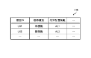
- FIG. 5 is a diagram showing an example of a processing condition information storage unit according to the first embodiment.
- the processing condition information storage unit 122 shown in FIG. 5 includes items such as "processing condition ID", "condition”, and "processing content”.
- Processing condition ID indicates identification information for identifying the processing condition.
- Condition indicates a condition identified by the processing condition ID.
- the “condition” includes items such as “image type” and “additional information”. The “condition” is not limited to the “image type” and the “additional information", and any item may be included as long as it is a condition.
- “Processing content” indicates the content of processing performed when the condition identified by the processing condition ID is satisfied.
- the processing condition (processing condition PS1) identified by the processing condition ID "PS1" indicates that the image type is "surgical image” and the additional information is "none". Further, it is shown that the processing executed when the processing condition PS1 is satisfied is the first image processing. That is, it indicates that the first image processing is executed for the image whose image type is "surgical image” and whose additional information is "none".
- processing condition PS2 identified by the processing condition ID "PS2" indicates that the image type is "surgical image” and the additional information is "presence”. Further, it is shown that the processing executed when the processing condition PS2 is satisfied is the second image processing. That is, it indicates that the second image processing is executed for the image whose image type is "surgical image” and whose additional information is "presence”.
- processing condition PS3 identified by the processing condition ID "PS3" indicates that the image type is "other than the surgical image” and the additional information is "-(no condition)". Further, it is shown that the processing executed when the processing condition PS3 is satisfied is the first image processing. That is, when the image type is "other than the surgical image”, it indicates that the first image processing is executed regardless of the presence or absence of additional information. When the image type is "other than the surgical image", the surgical information processing device 100 performs the first image processing and the second image processing according to the presence or absence of additional information, as in the case of the surgical image. You may switch.
- the processing condition information storage unit 122 is not limited to the above, and various information may be stored depending on the purpose.
- the processing condition information storage unit 122 indicates which of the first processing modes to the fourth processing modes shown in FIGS. 15 to 18 is to perform the image processing. Information may be stored.
- the processing condition information storage unit 122 determines which of the first processing modes to the fourth processing modes shown in FIGS. 15 to 18 is to perform image processing. The indicated information may be stored. Further, the processing condition information storage unit 122 may store various processing conditions other than the processing conditions PS1 to PS3.
- the control unit 130 uses, for example, a program stored inside the surgical information processing apparatus 100 by a CPU (Central Processing Unit), an MPU (Micro Processing Unit), or the like (for example, information such as a surgical information processing program according to the present disclosure).
- the processing program is realized by executing the RAM (Random Access Memory) or the like as a work area.
- the control unit 130 is realized by, for example, an integrated circuit such as an ASIC (Application Specific Integrated Circuit) or an FPGA (Field Programmable Gate Array).
- the control unit 130 includes an acquisition unit 131, a first determination unit 132, a second determination unit 133, a processing unit 134, a notification unit 135, and a transmission unit 136. Realize or execute the information processing functions and actions described in.
- the internal configuration of the control unit 130 is not limited to the configuration shown in FIG. 3, and may be another configuration as long as it is a configuration for performing information processing described later.
- the connection relationship of each processing unit included in the control unit 130 is not limited to the connection relationship shown in FIG. 3, and may be another connection relationship.
- the acquisition unit 131 acquires various information.
- the acquisition unit 131 acquires various information from an external information processing device.
- the acquisition unit 131 acquires various information from the medical observation device 10 and the image receiving device 50.
- the acquisition unit 131 acquires various information from the storage unit 120.
- the acquisition unit 131 acquires various types of information from the threshold information storage unit 121 and the processing condition information storage unit 122.
- the acquisition unit 131 acquires the surgical image.
- the acquisition unit 131 acquires a surgical image from a medical observation device.
- the acquisition unit 131 acquires the metadata of the surgical image.
- the acquisition unit 131 acquires metadata including information about DICOM of the surgical image.
- the acquisition unit 131 acquires user setting information indicating the setting by the user.
- the acquisition unit 131 acquires connected device information indicating a device connected to the surgical information processing device.
- Acquisition unit 131 acquires a plurality of surgical images including surgical images.
- the acquisition unit 131 acquires information indicating a target area in which additional information corresponding to the device that captured the surgical image is displayed.
- the acquisition unit 131 acquires a first threshold value, which is a threshold value for difference, and a second threshold value, which is a threshold value for brightness.
- the acquisition unit 131 acquires various information analyzed by the first determination unit 132.
- the acquisition unit 131 acquires various information generated by the second determination unit 133.
- the acquisition unit 131 acquires various information generated by the second determination unit 133.
- the acquisition unit 131 acquires various information processed by the processing unit 134.
- the acquisition unit 131 acquires the first information that triggers the dialogue, the second information that indicates the response to the first information, and the third information that indicates the reaction to the second information.
- the acquisition unit 131 acquires the first information which is a question, the second information which is the answer to the first information, and the third information which is the answer to the second information.
- the acquisition unit 131 acquires the first information corresponding to the utterance of the first subject, the second information corresponding to the utterance of the second subject, and the third information corresponding to the utterance of the third subject.
- the acquisition unit 131 acquires the first information, the second information corresponding to the utterance of the second subject different from the first subject, and the third information corresponding to the utterance of the third subject which is the first subject. ..
- the acquisition unit 131 includes the first information corresponding to the utterance of the first subject who is the agent of the dialogue system, the second information corresponding to the utterance of the second subject who is the user, and the third subject who is the agent of the dialogue system. Acquire the third information corresponding to the utterance.
- the acquisition unit 131 acquires the first information, the second information, and the third information in which at least one of the first information, the second information, and the third information is input by the user.
- the acquisition unit 131 acquires the first information presented to the input user, the second information input by the input user, and the third information input by the input user.
- the acquisition unit 131 acquires the meta information of the input user.
- the acquisition unit 131 is the information of the constituent unit of the dialogue corresponding to the combination of the first information that triggers the dialogue, the second information indicating the response to the first information, and the third information indicating the reaction to the second information. Acquires multiple unit information that is.
- the acquisition unit 131 acquires a plurality of unit information of the constituent unit which is a combination of the first information, the second information, and the third information.
- the acquisition unit 131 acquires the designation information of the connection method between the combinations by the user who presented the plurality of unit information.
- the acquisition unit 131 acquires connection information, which is information that connects the combination of the first information, the second information, and the third information.
- the acquisition unit 131 acquires the connection information specified by the user.
- the acquisition unit 131 acquires a plurality of unit information of each of the first information, the second information, and the third information.
- the acquisition unit 131 acquires the surgical image IM1.
- the acquisition unit 131 acquires the surgical image IM2.
- the first determination unit 132 determines various information.
- the first determination unit 132 determines various information based on the information from the external information processing device.
- the first determination unit 132 determines various types of information based on information from other information processing devices such as the medical observation device 10 and the image receiving device 50.
- the first determination unit 132 determines various types of information based on the information stored in the threshold information storage unit 121 and the processing condition information storage unit 122.
- the first determination unit 132 determines various information based on the information acquired by the acquisition unit 131.
- the first determination unit 132 determines various information based on various information acquired by the acquisition unit 131.
- the first determination unit 132 determines whether the image is a surgical image.
- the first determination unit 132 determines whether the image is a surgical image by analyzing the image.
- the second determination unit 133 determines various information.
- the second determination unit 133 determines various types of information based on the information from the external information processing device and the information stored in the storage unit 120.
- the second determination unit 133 determines various types of information based on information from other information processing devices such as the medical observation device 10 and the image receiving device 50.
- the second determination unit 133 determines various types of information based on the information stored in the threshold information storage unit 121 and the processing condition information storage unit 122.
- the second determination unit 133 determines various information based on the information acquired by the acquisition unit 131.
- the second determination unit 133 determines various information based on various information acquired by the acquisition unit 131.
- the second determination unit 133 determines various information based on the various information determined by the first determination unit 132.
- the second determination unit 133 determines various information based on the determination result by the first determination unit 132.
- the first determination unit 132 and the second determination unit 133 may be an integrated determination unit.
- the second determination unit 133 determines whether or not additional information is added to the surgical image acquired by the acquisition unit 131.
- the second determination unit 133 determines whether or not additional information is added to the surgical image based on the metadata acquired by the acquisition unit 131.
- the second determination unit 133 determines whether or not additional information is added to the surgical image based on the result of image analysis on the surgical image.
- the second determination unit 133 determines whether or not additional information is added to the surgical image based on the result of image analysis regarding the color of the surgical image.
- the second determination unit 133 determines whether or not additional information is added to the surgical image based on the result of image analysis of the surgical image using machine learning.
- the second determination unit 133 determines whether or not additional information is added to the surgical image based on the user setting information acquired by the acquisition unit 131.
- the second determination unit 133 determines whether or not additional information is added to the surgical image based on the connected device information acquired by the acquisition unit 131.
- the second determination unit 133 determines the area of additional information given to the surgical image.
- the second determination unit 133 determines the area of additional information given to the surgical image in response to the zoom process.
- the second determination unit 133 determines an area of additional information which is a freeze image given to the surgical image.
- the second determination unit 133 determines an area of additional information which is character information given to the surgical image.
- the second determination unit 133 determines an area in which the character information given to the surgical image is arranged.
- the second determination unit 133 determines the area of additional information that is a frame given to the surgical image.
- the second determination unit 133 determines the area of the frame given to the surgical image.
- the second determination unit 133 determines the area of the frame arranged at the end of the surgical image.
- the second determination unit 133 determines the region of the frame surrounding the peripheral end of the surgical image.
- the second determination unit 133 determines the mask to be applied to the surgical image.
- the second determination unit 133 determines the mask to be placed at the peripheral end of the surgical image.
- the second determination unit 133 determines a region of additional information having transparency given to the surgical image.
- the second determination unit 133 determines the transparency of the additional information given to the surgical image.
- the second determination unit 133 determines the transparency of the additional information given to the surgical image.
- the second determination unit 133 determines whether additional information is added to the surgical image by comparing a plurality of surgical images. The second determination unit 133 determines whether or not additional information is added to the surgical image based on the color comparison of the plurality of surgical images. The second determination unit 133 determines the portion where the difference between the pixel values of the plurality of surgical images does not exceed the threshold value as additional information. The second determination unit 133 makes a determination by changing the threshold value according to the device that captured the surgical image.
- the second determination unit 133 determines whether or not additional information is added by comparing the observation light images.
- the second determination unit 133 compares the observation light images. Determine if additional information is added.
- the second determination unit 133 adds additional information based on the comparison between the added value of the difference absolute values of the plurality of surgical images and the first threshold value, and the comparison between the added value of the brightness values of the plurality of surgical images and the second threshold value. To judge. The second determination unit 133 determines the mask image based on the comparison between the added value of the difference absolute values of the plurality of surgical images and the first threshold value, and the comparison between the added value of the brightness values of the plurality of surgical images and the second threshold value. And the OSD information are discriminated. The second determination unit 133 determines the additional information with the addition value of the difference absolute values of the plurality of surgical images as the first axis and the addition value of the brightness values of the plurality of surgical images as the second axis.
- the second determination unit 133 determines that the surgical image IM1 is not given additional information.
- the second determination unit 133 determines that the surgical image IM2 is provided with additional information.
- the processing unit 134 executes various information processing.
- the processing unit 134 performs various image processing.
- the processing unit 134 executes various information processing based on the information from the external information processing device and the information stored in the storage unit 120.
- the processing unit 134 executes various information processing based on information from other information processing devices such as the medical observation device 10 and the image receiving device 50.
- the processing unit 134 executes various information processing based on the information stored in the threshold information storage unit 121 and the processing condition information storage unit 122.
- the processing unit 134 executes various information processing based on various information acquired by the acquisition unit 131.
- the processing unit 134 executes various information processing based on various information determined by the first determination unit 132.
- the processing unit 134 executes various information processing based on the determination result by the first determination unit 132.
- the processing unit 134 executes various information processing based on various information determined by the second determination unit 133.
- the processing unit 134 executes various information processing based on the determination result by the second determination unit 133.
- the processing unit 134 performs analysis processing related to the image.
- the processing unit 134 performs various processes related to image processing.
- the processing unit 134 processes the image information (image) acquired by the acquisition unit 131.
- the processing unit 134 performs processing on the image by appropriately using a technique related to image processing.
- the processing unit 134 performs the first image processing when it is determined by the second determination unit 133 that the additional information is not given, and when it is determined that the additional information is given, the first image processing Do not do. When it is determined that the additional information is given, the second image processing different from the first image processing is performed.
- the processing unit 134 performs the first image processing, which is an image processing for the entire surgical image.
- the processing unit 134 performs a second image processing including the same kind of image processing as the first image processing for the area other than the additional information area corresponding to the additional information in the surgical image.
- the processing unit 134 performs a second image processing including an image processing different from the first image processing targeting the additional information area.
- the processing unit 134 performs a second image processing including a pixel filling process for the additional information area.
- the processing unit 134 performs a first image processing including at least one of zoom, image stabilization, rotation correction, and PinP (Picture in Picture).
- the processing unit 134 may execute the image processing by using any technique as long as the desired image processing can be executed, and executes the image processing by appropriately using various image processing techniques. ..
- the processing unit 134 may execute image processing by appropriately using various information.
- the processing unit 134 performs various image processing on an image signal such as RAW data acquired from, for example, the medical observation device 10.
- the image processing includes, for example, development processing, high image quality processing (band enhancement processing, super-resolution processing, NR (Noise reduction) processing and / or camera shake correction processing, etc.), and / or enlargement processing (electronic zoom processing), etc. , Various known signal processings are included.
- the processing unit 134 performs detection processing on the image signal for performing AE, AF, and AWB.
- the processing unit 134 executes the first image processing on the image IM1.
- the processing unit 134 executes the first image processing on the image IM1 to generate the image IM10.
- the processing unit 134 executes a second image processing on the image IM2.
- the processing unit 134 executes a second image processing on the image IM2 to generate the image IM20.
- the notification unit 135 notifies the information.
- the notification unit 135 notifies the user of information.
- the notification unit 135 notifies (transmits) information to a terminal device or the like used by the user.
- the surgical information processing device 100 has an output unit such as a display (display screen) or a speaker, the notification unit 135 may notify the user of information by the output unit.
- the notification unit 135 notifies various information based on various information acquired by the acquisition unit 131.
- the notification unit 135 notifies various information based on various information collected by the first determination unit 132.
- the notification unit 135 notifies various information based on the determination result by the first determination unit 132.
- the notification unit 135 notifies various information based on various information generated by the second determination unit 133.
- the notification unit 135 notifies various information based on the determination result by the second determination unit 133.
- the notification unit 135 notifies various information based on various information processed by the processing unit 134.
- the transmission unit 136 provides various information to an external information processing device.
- the transmission unit 136 transmits various information to an external information processing device.
- the transmission unit 136 transmits various information to other information processing devices such as the medical observation device 10 and the image receiving device 50.
- the transmission unit 136 provides the information stored in the storage unit 120.
- the transmission unit 136 transmits the information stored in the storage unit 120.
- the transmission unit 136 provides various information based on information from other information processing devices such as the medical observation device 10 and the image receiving device 50.
- the transmission unit 136 provides various information based on the information stored in the storage unit 120.
- the transmission unit 136 provides various types of information based on the information stored in the threshold information storage unit 121 and the processing condition information storage unit 122.
- the transmission unit 136 transmits the images IM10 and IM20 image-processed by the processing unit 134 to the image receiving device 50.
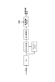
- FIG. 6 is a flowchart showing an information processing procedure according to the first embodiment. Specifically, FIG. 6 is a flowchart showing a procedure of collection processing by the surgical information processing apparatus 100.
- the surgical information processing apparatus 100 acquires a surgical image (step S101).
- the surgical information processing apparatus 100 determines whether or not additional information is added to the surgical image (step S102).
- the surgical information processing apparatus 100 executes the first image processing (step S103) when there is no additional information in the surgical image (step S103: No).
- the surgical information processing apparatus 100 executes the second image processing (step S104) when the surgical image has additional information (step S103: Yes).
- the surgical information processing apparatus 100 executes the second image processing without executing the first image processing.
- FIG. 7 is a schematic diagram showing a specific example of the surgical information processing system.
- the surgical information processing system 150 is composed of a surgical field camera 151, an endoscope 152, a microscope 153, an IP converter 154, an optical switcher 155, a server 156, an IP converter 157, a monitor 158, a recorder 159, and a projector 160.
- the electrical signals of the medical images captured by the surgical field camera 151, the endoscope 152, and the microscope 153 are converted into optical signals by the IP converter 154 and output to an arbitrary IP converter 157 by the optical switcher 155.
- the IP converter 157 converts the optical signal supplied from the optical switcher into an electric signal and supplies it to the monitor 158, the recorder 159, and the projector 160, respectively.
- the server 156 instructs the optical switcher 155 which IP converter 154 and which IP converter 157 are to be connected.
- the IP converter 154 or the IP converter 157 corresponds to the surgical information processing apparatus 100 shown in FIG.
- the surgical field camera 151, the endoscope 152, and the microscope 153 correspond to the medical observation device 10 shown in FIG.
- the monitor 158, the recorder 159, and the projector 160 correspond to the image receiving device 50 shown in FIG.
- the image receiving device 50 may be a server 156.
- the surgical information processing device 100 corresponds to at least one of the IP converter 154, the IP converter 157, and the server 156.
- the server 156 may not be provided in FIG. 7, and in that case, the surgical information processing apparatus 100 corresponds to at least one of the IP converter 154 and the IP converter 157.
- the IP converter 154 or the IP converter 157 which is the surgical information processing device 100, acquires a surgical image from the surgical field camera 151, the endoscope 152, or the microscope 153, which is the medical observation device 10, and determines the presence or absence of additional information.
- the surgical image that has undergone image processing accordingly is transmitted to the image receiving device 50.
- the specific configuration of the surgical information processing system 150 shown in FIG. 7 is an example of the surgical information processing system 1, and the surgical information processing system 1 is as long as the configuration shown in FIG. 2 is realized. Good.
- FIGS. 8 to 13 Each functional configuration shown in FIGS. 8 to 13 is realized by the configuration of the surgical information processing device 100, the medical observation device 10, and the like in the surgical information processing system 1.
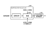
- FIG. 8 is a diagram showing an example of a conceptual diagram of the functional configuration of the surgical information processing system. Specifically, FIG. 8 is a diagram conceptually showing the flow of information in a surgical information processing system.
- the surgical information processing apparatus 100 acquires an image in which additional information such as OSD information is added to the captured image by the camera control unit 200.
- the camera control unit 200 is provided before the surgical information processing device 100, and adds additional information such as OSD information to the captured image before the surgical information processing device 100 performs processing.
- the camera control unit 200 may be integrated with the medical observation device 10 or may be separate from the medical observation device 10.
- the surgical information processing system 1 mainly targets surgical videos (surgical images) such as surgical endoscopic surgery, robotic surgery, and video microscope surgery. Further, the surgical information processing system 1 uses an image other than the surgical image as an input image, but in that case, the image processing is applied to the entire screen without determining the superimposed region described later.
- the surgical information processing apparatus 100 inputs an image processed by the camera control unit 200 to the surgical image taken by the camera.
- image processing such as digital development and high image quality
- an image in which graphics such as OSD and GUI are superimposed and external input information such as a CT image taken in advance is superimposed is generated and output. Since the image of the camera control unit 200 is input and the image processing is performed by the surgical information processing device 100 in the subsequent stage, it is important to determine the presence or absence of the additional information as described above and switch the image processing according to the determination result. It becomes.
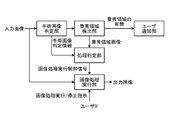
- FIG. 9 is a diagram showing an example of a conceptual diagram of the functional configuration of the surgical information processing system. Specifically, FIG. 9 is a diagram showing an example of a functional block diagram of the surgical information processing apparatus.
- the surgical image determination unit in FIG. 9 corresponds to the first determination unit 132.
- the superimposed region determination unit and the processing determination unit in FIG. 9 correspond to the second determination unit 133.
- the image processing execution unit in FIG. 9 corresponds to the processing unit 134.
- the user notification unit in FIG. 9 corresponds to the notification unit 135.
- the image processing unit inputs, for example, the output image from the camera control unit 200 shown in FIG.
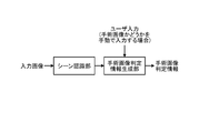
- the surgical image determination unit determines whether or not the video is a surgical operation such as a surgical endoscopic operation, a robot operation, or a videomicroscopic operation.
- the surgical image determination unit generates information indicating the determination result of whether or not the surgical image is a surgical image, and transmits the information to the processing determination unit.
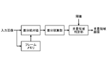
- the superimposed area detection unit detects areas such as graphic information and CT images superimposed by the camera control unit 200.
- the superimposition area detection unit generates information indicating the superimposition area image. For example, the superimposition area detection unit generates information indicating the position and range of the superimposition area image.
- the user notification unit notifies the user of the presence or absence of the superimposed area.
- the processing determination unit determines what kind of processing is to be performed according to the information on whether or not the surgical operation moving image is determined by the surgical image determination unit and the presence or absence of the superimposed region detected by the superimposed region detection unit.
- the processing determination unit determines whether to execute the image processing in the image processing execution unit, does not execute the image processing, or partially executes the image processing, and generates an image processing control signal instructing the execution.
- the image processing execution unit executes image processing such as blur correction, electronic zoom, image rotation processing, PinP processing, and image enhancement.
- the user can instruct the surgical information processing system to execute or stop the image processing via the user IF.
- FIG. 10 is a diagram showing an example of a conceptual diagram of a functional configuration of a surgical information processing system. Specifically, FIG. 10 is a diagram showing an example of a functional block diagram of the surgical image determination unit corresponding to the first determination unit 132.
- the surgical information processing device 100 can input images other than surgical moving images (surgical images) such as surgical endoscopic surgery, robot surgery, and video microscope surgery.
- the surgical image determination unit (first determination unit 132) has a scene recognition unit and a surgical image determination information generation unit. For example, the surgical image determination unit recognizes a scene of a surgical image by a scene recognition unit, and based on the recognition result, a surgical image determination information generation unit generates determination information indicating whether or not the surgical image is a surgical image.
- the surgical image determination unit determines whether the input image is a surgical moving image.
- the surgical image judgment unit uses scene recognition technology such as endoscopic mask recognition, surgical tool recognition such as forceps, and blood detection to determine whether or not it is a surgical video (surgical image).
- the surgical information processing apparatus 100 may acquire the user's designation as to whether or not it is a surgical moving image. In this case, the user may manually specify whether or not it is a surgical video.
- FIGS. 11 to 13 are diagrams showing an example of a conceptual diagram of the functional configuration of the surgical information processing system. Specifically, FIGS. 11 to 13 are diagrams showing an example of a functional block diagram of the superimposed region detection unit corresponding to the second determination unit 133.
- the superimposition area detection unit (second determination unit 133) has a difference integration unit and a superimposition area determination unit.
- the superimposition area detection unit makes a determination by the superimposition area determination unit based on the integration result by the difference integration unit.
- the superimposition region detection unit holds the image one frame before the input surgical moving image, and calculates the absolute pixel difference value between the current frame and the image one frame before.
- the superimposition area detection unit integrates the difference absolute values in the time direction.
- the superimposition area detection unit performs threshold processing on the integrated image, and detects a region lower than the threshold value as a superimposition region.
- the superimposed region detection unit (second determination unit 133) of FIG. 12 differs from the configuration of FIG. 11 in that has a function of NR (noise reduction).
- NR noise reduction
- the superimposition region detection unit of FIG. 12 executes NR as a preprocessing before the superimposition region determination, and performs the superimposition region determination with the noise removed. To do.
- the superimposed region detection unit (second determination unit 133) of FIG. 13 differs from the configuration of FIG. 11 in that it has a special optical image determination unit, a selection unit, and a superimposed region holding unit.
- the superimposition area detection unit (second determination unit 133) of FIG. 13 performs the superimposition area detection for the normal light observation scene, and holds the area information in the superimposition area holding unit.
- the superimposition region detection unit determines whether the input image is an image during normal observation or an image during special light observation from the distribution of the image histogram or the like by the special light image determination unit.
- the superimposed region detection unit selects the superimposed region image held in the superimposed region holding unit as an output.
- the superimposition area detection unit directly outputs the superimposition area image of the superimposition area determination unit.
- the user notification unit notifies the user of the presence or absence of the superimposition area detected by the superimposition area detection unit.
- the user can stop the execution of the image processing of the image processing execution unit via the user IF (for example, the input unit or the like).
- the user can resume the execution of the image processing execution unit that has been temporarily stopped via the user IF.
- FIG. 14 is a flowchart showing an example of a processing procedure by the surgical information processing system. Specifically, FIG. 14 is a flowchart showing an example of a processing procedure by the processing determination unit.
- the surgical information processing apparatus 100 determines whether or not the input image is a surgical image (step S201). For example, the processing determination unit of the surgical information processing apparatus 100 determines whether or not the input image is a surgical image based on the information from the surgical image determination unit.
- the surgical information processing apparatus 100 determines whether or not a superposed region exists (step S202). For example, when the input image is a surgical image, the processing determination unit of the surgical information processing apparatus 100 determines whether or not a superposed region exists in the input image.
- step S202 when the superposed region exists (step S202: Yes), the surgical information processing apparatus 100 does not execute the image processing or executes the partial image processing (step S203). For example, the processing determination unit of the surgical information processing apparatus 100 determines that when the input image has a superposed region, the first image processing is not executed and the second image processing is executed.
- the surgical information processing apparatus 100 executes image processing when the input image is not a surgical image (step S201: No) or when the superimposed region does not exist (step S202: No) (step S204).
- the processing determination unit of the surgical information processing apparatus 100 determines that the first image processing is executed when the input image is not a surgical image or the input image does not have a superposed region.
- the processing determination unit determines that the image processing execution unit executes image processing when the operation image determination unit determines that the input image is an image other than the surgical operation moving image. Give down.
- the processing determination unit executes image processing in the image processing execution unit depending on the superimposition region detected by the superposition region detection unit, with or without execution of image processing, or partial image processing execution. Determine if you want to.
- Image processing execution unit When the processing determination unit determines that there is a superposed area, the image processing execution unit executes various image processing controls as follows. The control example shown below is an example, and the image processing execution unit executes control related to various image processing. For example, information including the presence / absence of image processing execution and the area to be partially executed is transmitted to the image processing execution unit as an image processing execution control signal. The image processing execution unit receives an image processing execution control signal from the processing determination unit for information including the presence / absence of image processing execution and a partial execution area.
- the image processing execution unit receives an image processing execution control signal from the processing determination unit, and executes, stops, or partially executes the image processing. Alternatively, the image processing execution unit executes or stops the image processing according to the instruction from the user to execute / stop the image processing via the user IF.
- the image processing performed by the image processing execution unit includes blur correction, electronic zoom, image rotation processing, PinP processing, and image enhancement.
- the image processing execution unit automatically stops image processing when there is a superposed area.
- the image processing execution unit sets the conditions for the detected superimposed region, and stops the image processing when the conditions are satisfied.
- the image processing execution unit may be conditional on the area of the superposed region being equal to or more than a certain area and the position of the center of gravity of the superposed region being equal to or less than the center of the image (stop condition).
- the image processing execution unit may stop the image processing based on the above-mentioned stop condition.
- FIGS. 15 to 18 are diagrams showing an example of a processing mode of image processing.
- 15 to 18 are views for explaining variations of the region in which image processing is stopped.
- FIG. 15 is a diagram showing an example of a first processing mode of image processing.
- FIG. 16 is a diagram showing an example of a second processing mode of image processing.
- FIG. 17 is a diagram showing an example of a third processing mode of image processing.
- FIG. 18 is a diagram showing an example of a fourth processing mode of image processing.
- the first processing mode will be described with reference to FIG.
- the surgical image IM11 in FIG. 15 is masked with the mask MS11 and is given the character information INF11. Further, in the example of FIG. 15, the character information INF11 overlaps with the region AR11 which is the endoscopic surgery image region. Therefore, the surgical information processing apparatus 100 stops image processing only in the detected superposed region. For example, the surgical information processing apparatus 100 stops image processing only in the area AR11 in which the character information INF11 "ABCDE" overlaps. The surgical information processing apparatus 100 stops image processing only in the area AR11 in which a part of the character information INF11 "CDE" overlaps.
- the second processing mode will be described with reference to FIG.
- the surgical image IM12 in FIG. 16 is masked with the mask MS12 and is given the character information INF12. Further, in the example of FIG. 16, the character information INF12 overlaps with the region AR12 which is the endoscopic surgery image region. Therefore, the surgical information processing apparatus 100 stops image processing only in the area AA12, which is a rectangular area surrounding the detected superimposed area. For example, the surgical information processing apparatus 100 stops image processing only in the region AR12 in which the region AR12 overlaps.
- the third processing mode in FIG. 17 differs from the second processing mode in that the rectangular region is wider than that of the second processing mode in FIG.
- the surgical image IM13 in FIG. 17 is masked with the mask MS13 and is given the character information INF13. Further, in the example of FIG. 17, the character information INF13 overlaps with the region AR13 which is the endoscopic surgery image region. Therefore, the surgical information processing apparatus 100 stops the image processing only in the area AA13 which is a rectangular area surrounding the detected superposed area. For example, the surgical information processing apparatus 100 stops image processing only in the region AR13 in which the region AR13 overlaps.
- the fourth processing mode in FIG. 18 is different from the first processing mode in FIG. 15 in that the processing of the portion other than the character information in the area AA14 is different from the first processing mode.
- the surgical image IM14 in FIG. 18 is masked with the mask MS13 and is given the character information INF14. Therefore, the surgical information processing apparatus 100 stops image processing only in the detected superposed region. For example, the surgical information processing apparatus 100 stops image processing only in the area AR14 in which the character information INF11 “ABCDE” overlaps. The surgical information processing apparatus 100 stops image processing only in the area AR11 in which a part of the character information INF14 "CDE" overlaps. Further, the surgical information processing apparatus 100 performs a process of filling the region other than the superposed region with a specific fixed color or the average value or the mode of the pixel values of the region within the region AA14.
- FIG. 19 is a diagram showing a configuration example of the surgical information processing apparatus according to the second embodiment of the present disclosure.
- FIG. 20 is a diagram showing an example of an additional history information storage unit according to the second embodiment.
- the surgical information processing system 1 according to the second embodiment includes a surgical information processing device 100A instead of the surgical information processing device 100. That is, the surgical information processing system 1 according to the second embodiment includes a medical observation device 10, an image receiving device 50, and a surgical information processing device 100A.
- the surgical information processing device 100A includes a communication unit 110, a storage unit 120A, and a control unit 130A.
- the storage unit 120A is realized by, for example, a semiconductor memory element such as a RAM or a flash memory, or a storage device such as a hard disk or an optical disk. As shown in FIG. 19, the storage unit 120A according to the second embodiment includes a threshold information storage unit 121, a processing condition information storage unit 122, and an additional history information storage unit 123.
- the additional history information storage unit 123 stores various information related to the history.
- the additional history information storage unit 123 stores history information regarding the arrangement of past additional information.
- FIG. 20 is a diagram showing an example of an additional history information storage unit according to the second embodiment.
- the additional history information storage unit 123 shown in FIG. 20 includes items such as "history ID”, "device type”, and "additional arrangement information".
- History ID indicates identification information for identifying the history.
- the “device type” indicates the device type corresponding to the history identified by the history ID.
- Additional arrangement information indicates information regarding the arrangement of additional information identified by the history ID.
- the history (history LG1) identified by the history ID "LG1" indicates that the device type is "endoscope". That is, the history LG1 indicates that the arrangement of the past additional information regarding the endoscope was "AL1".
- the additional arrangement information is indicated by an abstract reference numeral such as “VL1”, but the additional arrangement information includes information indicating the arrangement position of the additional information, information indicating the superimposed area, and the like.
- the additional history information storage unit 123 is not limited to the above, and may store various information depending on the purpose.
- the additional history information storage unit 123 stores the past history regarding the superposed area.
- control unit 130A for example, a program stored inside the surgical information processing apparatus 100A by a CPU, MPU, or the like (for example, an information processing program such as the surgical information processing program according to the present disclosure) uses a RAM or the like as a work area. It is realized by being executed. Further, the control unit 130A is realized by, for example, an integrated circuit such as an ASIC or FPGA.
- the control unit 130A includes an acquisition unit 131, a first determination unit 132, a second determination unit 133A, and a transmission unit 136, and has functions and operations of information processing described below. Realize or execute.
- the internal configuration of the control unit 130A is not limited to the configuration shown in FIG. 19, and may be another configuration as long as it is a configuration for performing information processing described later.
- the connection relationship of each processing unit included in the control unit 130A is not limited to the connection relationship shown in FIG. 19, and may be another connection relationship.
- the second determination unit 133A determines whether or not the additional information is added to the surgical image for the target area where the additional information corresponding to the device that captured the surgical image is displayed. For example, the second determination unit 133A determines whether or not the additional information is added to the surgical image based on the information indicating the past superimposed region stored in the additional history information storage unit 123.
- FIG. 21 is a diagram showing an example of a conceptual diagram of a functional configuration of a surgical information processing system. Specifically, FIG. 21 is a diagram showing an example of a functional block diagram of the surgical information processing apparatus. The functional block diagram of the surgical information processing apparatus shown in FIG. 21 is different from the functional block diagram of the surgical information processing apparatus shown in FIG. 9 in that it has a superposed region accumulating portion. In the following, the same points as in FIG. 9 will be omitted as appropriate.
- the surgical image determination unit in FIG. 21 corresponds to the first determination unit 132.
- the superimposed region determination unit and the processing determination unit in FIG. 21 correspond to the second determination unit 133A.
- the image processing execution unit in FIG. 21 corresponds to the processing unit 134.
- the user notification unit in FIG. 21 corresponds to the notification unit 135.
- the image processing execution unit in FIG. 21 corresponds to the processing unit 134.
- the superimposed area storage unit in FIG. 21 corresponds to the additional history information storage unit 123.
- the superimposition area detection unit detects that there is a superimposition region, and the superimposition region when the user stops image processing is stored in the superimposition region storage unit as a history.
- the processing determination unit collates the current superimposed area image detected by the superimposed area detection unit with the accumulated superimposed image of the superimposed area storage unit, and collates the current superimposed area image with the accumulated image.
- the processing determination unit determines the execution of image processing when the superimposed region image and the accumulated superimposed region image are different regions.
- the surgical information processing apparatus 100A refers to the history of the superimposed area manually stopped by the user in the past with respect to the image on which the graphics such as OSD and GUI and the CT image are superimposed, and in the past. By stopping the image processing only in the superimposed area that was manually stopped, the image processing can be stopped only in the superimposed area that the user felt uncomfortable in the past, and the discomfort of the user is reduced.
- the process is not limited to the process according to the first embodiment and the second embodiment, and various processes may be executed. This point will be described below. In the following, the case where the surgical information processing device 100 performs the processing will be described as an example, but the surgical information processing device 100A may perform the processing.
- the surgical information processing apparatus 100 may make a determination using the luminance information.
- the surgical information processing apparatus 100 may make a determination using two threshold values.
- the surgical information processing apparatus 100 may make a determination using a threshold value for difference (also referred to as “first threshold value”) and a threshold value for brightness (also referred to as “second threshold value”).
- the surgical information processing apparatus 100 is added based on the comparison between the added value of the difference absolute values of the plurality of surgical images and the first threshold value, and the comparison between the added value of the brightness values of the plurality of surgical images and the second threshold value. Information may be determined.
- the surgical information processing apparatus 100 uses the threshold information as shown in the threshold information storage unit 121A in FIG. 22.
- the surgical information processing apparatus 100 according to the modified example has the threshold information storage unit 121A in FIG. 22 in the storage unit 120 instead of the threshold information storage unit 121 in FIG.
- the threshold information storage unit 121A stores various information related to the threshold value.
- the threshold information storage unit 121A stores various information related to the threshold used for various determinations.
- FIG. 22 is a diagram showing an example of a threshold information storage unit according to a modified example of the present disclosure.
- the threshold information storage unit 121A shown in FIG. 22 includes items such as “threshold ID”, “target”, and “threshold”.
- Theshold ID indicates identification information for identifying the threshold value.
- “Target” refers to a target for which a threshold is used.
- the “threshold value” indicates a specific value of the threshold value identified by the corresponding threshold ID.
- the threshold value (threshold value TH1) identified by the threshold value ID “TH1” indicates the first threshold value whose target is the “absolute difference value”. That is, it is shown that the threshold value TH1 is used for comparison with the absolute difference value. In this case, the threshold value TH1 is used for comparison with the absolute difference value, and indicates that it is used for determining the presence or absence of additional information. Further, the value of the threshold value TH1 indicates that it is "VL1". In the example of FIG. 22, although it is indicated by an abstract code such as "VL1", the value of the threshold value TH1 is a specific numerical value.
- the threshold value (threshold value TH2) identified by the threshold value ID "TH2" indicates a second threshold value in which the target is "luminance". That is, it is shown that the threshold value TH2 is used for comparison with the absolute difference value. In this case, the threshold value TH2 is used for comparison with the absolute difference value, and indicates that it is used for determining the presence or absence of additional information. Further, the value of the threshold value TH2 indicates that it is "VL2". In the example of FIG. 22, although it is indicated by an abstract code such as "VL2", the value of the threshold value TH2 is a specific numerical value.
- the threshold information storage unit 121A is not limited to the above, and may store various information depending on the purpose.
- the surgical information processing device 100 uses the information of the threshold information storage unit 121A to discriminate between the mask image and the OSD (On-Screen Display) information.
- the surgical information processing apparatus 100 masks the sum of the absolute difference values of the plurality of surgical images and the first threshold value, and the sum of the brightness values of the plurality of surgical images and the second threshold value.
- the image and OSD (On-Screen Display) information are discriminated.
- FIG. 23 is a diagram showing an example of a conceptual diagram of the functional configuration of the surgical information processing system. Specifically, FIG. 23 is a diagram showing an example of a functional block diagram of the superimposed region detection unit corresponding to the second determination unit 133.
- the functional block diagram of the surgical information processing apparatus shown in FIG. 23 is different from the functional block diagram of the surgical information processing apparatus shown in FIG. 11 in that it uses a luminance integrated value and two threshold values and has a discriminating unit. In the following, the same points as in FIG. 11 will be omitted as appropriate.
- the superimposition area detection unit (second determination unit 133) has a brightness integration value, a difference integration unit, and a discrimination unit.
- the superimposition area detection unit makes a determination by the discrimination unit based on the integration result by the difference integration unit and the integration result by the brightness integration value.
- the determination unit generates information indicating the determination result.
- the superimposition area detection unit makes a determination by the discrimination unit based on the integration result by the difference integration unit.
- the superimposition region detection unit holds the image one frame before the input surgical moving image, and calculates the absolute pixel difference value between the current frame and the image one frame before.
- the superimposition area detection unit integrates the absolute difference values in the time direction and calculates the integrated difference value.
- the superimposition area detection unit integrates the brightness values of each image in the time direction with respect to the input surgical moving image, and calculates the brightness integration value.
- the superimposition area detection unit performs threshold processing on the integrated image.
- the superimposed region detection unit determines that the region where the difference integrated value is lower than the first threshold value and the luminance integrated value is lower than the second threshold value is a mask. For example, the superimposed region detection unit determines the region where the difference integrated value is lower than the first threshold value and the luminance integrated value is equal to or higher than the second threshold value as OSD information. The determination unit generates information indicating a determination result of mask or OSD information.
- FIG. 24 is a diagram showing an example of discrimination between the mask and the OSD.
- the graph GR31 in FIG. 24 shows a graph in the case where the vertical axis is the added value of the luminance (integrated luminance value) and the horizontal axis is the added value of the absolute difference value (integrated difference value).
- the surgical information processing apparatus 100 determines the additional information with the added value of the difference absolute values of the plurality of surgical images as the first axis and the added value of the brightness values of the plurality of surgical images as the second axis. ..
- the surgical information processing apparatus 100 discriminates between the mask and the OSD information by using the difference integrated value integrated in the time direction for each pixel and the luminance integrated value integrated in the time direction for each pixel.
- the surgical information processing apparatus 100 masks the pixel PT1 plotted in an area where the horizontal axis direction is smaller than the first threshold value “VL1” and the vertical axis direction is smaller than the second threshold value “VL2”. Is determined. Further, the surgical information processing apparatus 100 determines that the pixel PT2 plotted in the region where the horizontal axis direction is smaller than the first threshold value “VL1” and the vertical axis direction is the second threshold value “VL2” or more is OSD information. The surgical information processing apparatus 100 discriminates between the mask and the OSD information by using the information of the difference absolute value in the time direction and the information of the luminance value (luminance) for each pixel.
- the surgical information processing apparatus 100 discriminates between the mask and the OSD information by using the first threshold value for the absolute difference value and the second threshold value for the brightness. As described above, the surgical information processing apparatus 100 can appropriately determine the additional information by discriminating between the mask and the OSD information using the two threshold values.
- the surgical information processing apparatus 100 can appropriately discriminate additional information such as OSD information. Further, the surgical information processing apparatus 100 may determine that the pixels whose difference integrated value is equal to or higher than the first threshold value “VL1” are the pixels corresponding to the surgical image. For example, in the example of FIG. 24, the surgical information processing apparatus 100 may determine that the pixels plotted in the region where the horizontal axis direction is the first threshold value “VL1” or more are the pixels corresponding to the surgical image. .. If the surgical information processing apparatus 100 does not discriminate between the mask and additional information such as OSD information as described above, the mask may also be determined as additional information. For example, the surgical information processing apparatus 100 may determine whether or not additional information including a mask is given.
- processing related to each of the above-described embodiments and modifications may be performed in various different forms (modifications) other than the above-described embodiments and modifications.
- the device that performs image processing surgical information processing device 100 or surgical information processing device 100A
- the device that displays an image may be integrated.
- the above is an example, and the surgical information processing system 1 may be realized by various configurations.
- each component of each device shown in the figure is a functional concept, and does not necessarily have to be physically configured as shown in the figure. That is, the specific form of distribution / integration of each device is not limited to the one shown in the figure, and all or part of the device is functionally or physically distributed / physically in arbitrary units according to various loads and usage conditions. Can be integrated and configured.
- the surgical information processing apparatus (surgical information processing apparatus 100, 100A in the embodiment) according to the present disclosure includes an acquisition unit (acquisition unit 131 in the embodiment) and a determination unit (second determination in the embodiment).
- a unit 133, 133A) and a processing unit (processing unit 134 in the embodiment) are provided.
- the acquisition unit acquires a surgical image.
- the determination unit determines whether additional information is added to the surgical image acquired by the acquisition unit.
- the processing unit performs the first image processing when it is determined by the determination unit that the additional information is not given, and does not perform the first image processing when it is determined that the additional information is given.
- the surgical information processing apparatus determines whether or not additional information is added to the surgical image, and if it is determined that the additional information is not added, the first image processing is performed and the additional information is added. When it is determined that the information is given, the image processing can be appropriately performed according to the surgical image by performing the first image processing.
- the acquisition department acquires surgical images from a medical observation device.
- the surgical information processing device can appropriately perform image processing on the surgical image acquired from the medical observation device.
- the processing unit when it is determined that the additional information is added, the processing unit performs a second image processing different from the first image processing.
- the surgical information processing device performs the first image processing on the image to which the additional information is not added, and performs the second image processing on the image to which the additional information is added, thereby performing the surgical image.
- Image processing can be appropriately performed according to the above.
- the first image processing is an image processing that targets the entire surgical image.
- the second image processing includes the same type of image processing as the first image processing for the area other than the additional information area corresponding to the additional information in the surgical image.
- the surgical information processing device performs the same type of image processing as the first image processing for the image to which the additional information is added, which targets the area other than the additional information area corresponding to the additional information.
- Image processing can be performed appropriately according to the surgical image.
- the second image processing includes image processing different from that of the first image processing targeting the additional information area.
- the surgical information processing device is appropriate according to the surgical image by performing image processing different from the first image processing for the additional information area on the image to which the additional information is added. Image processing can be performed.
- the acquisition department acquires the metadata of the surgical image.
- the determination unit determines whether additional information is added to the surgical image based on the metadata acquired by the acquisition unit.
- the surgical information processing device can appropriately determine whether or not additional information is added to the surgical image based on the metadata of the surgical image, and appropriately performs image processing according to the surgical image. Can be done.
- the determination unit determines whether additional information is added to the surgical image based on the result of image analysis on the surgical image.
- the surgical information processing device can appropriately determine whether or not additional information is added to the surgical image based on the image analysis of the surgical image, and appropriately performs image processing according to the surgical image. Can be done.
- the acquisition unit acquires user setting information indicating the setting by the user.
- the determination unit determines whether additional information is added to the surgical image based on the user setting information acquired by the acquisition unit.
- the surgical information processing apparatus can appropriately determine whether or not additional information is added to the surgical image based on the user setting information indicating the setting by the user, and appropriately performs image processing according to the surgical image. It can be performed.
- the acquisition unit acquires the connected device information indicating the device connected to the surgical information processing device.
- the determination unit determines whether additional information is added to the surgical image based on the connected device information acquired by the acquisition unit.
- the surgical information processing device can appropriately determine whether or not additional information is added to the surgical image based on the connected device information indicating the device connected to the surgical information processing device.
- Image processing can be appropriately performed according to the above.
- the determination unit determines the area of additional information given to the surgical image.
- the surgical information processing apparatus can determine the area of additional information given to the surgical image, and can appropriately perform image processing according to the determined area.
- the determination unit determines the area of additional information that is the character information given to the surgical image.
- the surgical information processing apparatus can determine the area of additional information which is the character information given to the surgical image, and can appropriately perform image processing according to the determined area.
- the first image processing includes at least one of zoom, image stabilization, rotation correction, and PinP (Picture in Picture).
- the surgical information processing device responds to the surgical image by performing image processing including at least one of zoom, camera shake correction, rotation correction, or PinP on the image to which additional information is not added. Image processing can be performed appropriately.
- the acquisition department acquires a plurality of surgical images including surgical images.
- the determination unit determines whether additional information is added to the surgical image by comparing a plurality of surgical images.
- the surgical information processing apparatus can appropriately determine whether or not additional information is added to the surgical images by comparing a plurality of surgical images. Therefore, the surgical information processing apparatus can appropriately perform image processing according to the surgical image based on the determination result.
- the determination unit determines the portion where the difference between the pixel values of the plurality of surgical images does not exceed the threshold value as additional information.
- the surgical information processing apparatus can determine the portion where the difference between the pixel values of the plurality of surgical images does not exceed the threshold value as additional information. Therefore, the surgical information processing apparatus can appropriately perform image processing according to the surgical image based on the determination result.
- the judgment unit makes a judgment by changing the threshold value according to the device that captured the surgical image.
- the surgical information processing apparatus can appropriately determine whether or not additional information is given by changing the threshold value according to the device that captured the surgical image and performing the determination. Therefore, the surgical information processing apparatus can appropriately perform image processing according to the surgical image based on the determination result.
- the acquisition unit acquires information indicating a target area in which additional information corresponding to the device that captured the surgical image is displayed.
- the determination unit determines whether or not additional information is added to the surgical image for the target area.
- the surgical information processing device adds the surgical image to the surgical image by determining whether or not the additional information is added to the surgical image in the target area where the additional information corresponding to the device that captured the surgical image is displayed. It can be appropriately determined whether or not the information is given. Therefore, the surgical information processing apparatus can appropriately perform image processing according to the surgical image based on the determination result.
- the determination unit determines whether additional information is added by comparing the observation light images.
- the surgical information processing apparatus uses the observation light image of the special light image and the observation light image to determine whether or not the additional information is added, so that the additional information is added to the surgical image. Can be appropriately determined. Therefore, the surgical information processing apparatus can appropriately perform image processing according to the surgical image based on the determination result.
- the acquisition unit acquires a first threshold value, which is a threshold value for difference, and a second threshold value, which is a threshold value for brightness.
- the determination unit determines the additional information based on the comparison between the added value of the difference absolute values of the plurality of surgical images and the first threshold value, and the comparison between the added value of the brightness values of the plurality of surgical images and the second threshold value. ..
- the surgical information processing apparatus is assigned to the surgical image by determining additional information using the first threshold value, which is the threshold value for difference, and the second threshold value, which is the threshold value for brightness.
- the additional information can be appropriately determined. Therefore, the surgical information processing apparatus can appropriately perform image processing according to the surgical image based on the determination result.
- FIG. 25 is a hardware configuration diagram showing an example of a computer 1000 that realizes the functions of information processing devices (computers) such as surgical information processing devices 100 and 100A.
- the computer 1000 includes a CPU 1100, a RAM 1200, a ROM (Read Only Memory) 1300, an HDD (Hard Disk Drive) 1400, a communication interface 1500, and an input / output interface 1600.
- Each part of the computer 1000 is connected by a bus 1050.
- the CPU 1100 operates based on the program stored in the ROM 1300 or the HDD 1400, and controls each part. For example, the CPU 1100 expands the program stored in the ROM 1300 or the HDD 1400 into the RAM 1200 and executes processing corresponding to various programs.
- the ROM 1300 stores a boot program such as a BIOS (Basic Input Output System) executed by the CPU 1100 when the computer 1000 is started, a program that depends on the hardware of the computer 1000, and the like.
- BIOS Basic Input Output System
- the HDD 1400 is a computer-readable recording medium that non-temporarily records a program executed by the CPU 1100 and data used by the program.
- the HDD 1400 is a recording medium for recording an information processing program (surgical information processing program, etc.) according to the present disclosure, which is an example of program data 1450.
- the communication interface 1500 is an interface for the computer 1000 to connect to an external network 1550 (for example, the Internet).
- the CPU 1100 receives data from another device or transmits data generated by the CPU 1100 to another device via the communication interface 1500.
- the input / output interface 1600 is an interface for connecting the input / output device 1650 and the computer 1000.
- the CPU 1100 receives data from an input device such as a keyboard or mouse via the input / output interface 1600. Further, the CPU 1100 transmits data to an output device such as a display, a speaker, or a printer via the input / output interface 1600. Further, the input / output interface 1600 may function as a media interface for reading a program or the like recorded on a predetermined recording medium (media).
- the media is, for example, an optical recording medium such as a DVD (Digital Versatile Disc) or PD (Phase change rewritable Disk), a magneto-optical recording medium such as an MO (Magneto-Optical disk), a tape medium, a magnetic recording medium, or a semiconductor memory.
- an optical recording medium such as a DVD (Digital Versatile Disc) or PD (Phase change rewritable Disk)
- a magneto-optical recording medium such as an MO (Magneto-Optical disk)
- a tape medium such as a magnetic tape
- magnetic recording medium such as a magnetic tape
- semiconductor memory for example, an optical recording medium such as a DVD (Digital Versatile Disc) or PD (Phase change rewritable Disk), a magneto-optical recording medium such as an MO (Magneto-Optical disk), a tape medium, a magnetic recording medium, or a semiconductor memory.
- the CPU 1100 of the computer 1000 realizes the functions of the control unit 130 and the like by executing the information processing program loaded on the RAM 1200. .. Further, the information processing program according to the present disclosure and the data in the storage unit 120 are stored in the HDD 1400.
- the CPU 1100 reads the program data 1450 from the HDD 1400 and executes the program, but as another example, these programs may be acquired from another device via the external network 1550.
- the present technology can also have the following configurations.
- Department and Surgical information processing device equipped with (2) The acquisition unit The surgical information processing device according to (1) above, which acquires the surgical image from a medical observation device. (3) The processing unit The surgical information processing apparatus according to (1) or (2), wherein when it is determined that the additional information is added, a second image processing different from the first image processing is performed.
- the first image processing is Image processing for the entire surgical image.
- the second image processing is The surgical information processing apparatus according to (3) above, which includes image processing of the same type as the first image processing for a region other than the additional information region corresponding to the additional information among the surgical images.
- the second image processing is The surgical information processing apparatus according to (4) above, which includes image processing different from that of the first image processing for the additional information region.
- the second image processing is The surgical information processing apparatus according to (4) or (5) above, which includes a pixel filling process for the additional information area.
- the acquisition unit Obtain the metadata of the surgical image and The determination unit The surgical information processing apparatus according to any one of (1) to (6) above, which determines whether or not the additional information is added to the surgical image based on the metadata acquired by the acquisition unit. ..
- the acquisition unit The surgical information processing apparatus according to (7) above, which acquires the metadata including information on DICOM (Digital Imaging and COmmunications in Medicine) of the surgical image.
- the determination unit The surgical information processing apparatus according to any one of (1) to (8) above, which determines whether or not the additional information is added to the surgical image based on the result of image analysis on the surgical image.
- the determination unit The surgical information processing apparatus according to (9), wherein it is determined whether or not the additional information is added to the surgical image based on the result of the image analysis regarding the color of the surgical image.
- the determination unit The surgical information processing apparatus according to (9) or (10), wherein it is determined whether or not the additional information is added to the surgical image based on the result of the image analysis of the surgical image using machine learning.
- the acquisition unit Acquire user setting information indicating the setting by the user, The determination unit The surgical information processing according to any one of (1) to (11), which determines whether or not the additional information is added to the surgical image based on the user setting information acquired by the acquisition unit. apparatus.
- the acquisition unit Acquire the connected device information indicating the device connected to the surgical information processing device, and obtain The determination unit The surgical information processing according to any one of (1) to (12) above, which determines whether or not the additional information is added to the surgical image based on the connected device information acquired by the acquisition unit. apparatus.
- the determination unit The surgical information processing apparatus according to any one of (1) to (13) above, which determines an area of the additional information given to the surgical image.
- the determination unit The surgical information processing apparatus according to (14), wherein the area of the additional information given to the surgical image is determined in response to the zoom process.
- the determination unit The surgical information processing apparatus according to (14) or (15), which determines a region of the additional information which is a freeze image given to the surgical image.
- the determination unit The surgical information processing apparatus according to any one of (14) to (16), which determines an area of the additional information which is character information given to the surgical image.
- the determination unit The surgical information processing apparatus according to any one of (14) to (17), which determines an area of the additional information that is a frame given to the surgical image.
- the determination unit The surgical information processing apparatus according to any one of (14) to (18) above, which determines a region of the additional information having transparency given to the surgical image.
- the first image processing is The surgical information processing apparatus according to any one of (1) to (19) above, which comprises at least one of zoom, image stabilization, rotation correction, and PinP (Picture in Picture).
- the acquisition unit A plurality of surgical images including the surgical image are acquired, and
- the determination unit The surgical information processing apparatus according to any one of (1) to (20) above, which determines whether additional information is added to the surgical images by comparing the plurality of surgical images.
- the determination unit The surgical information processing apparatus according to (21), wherein it is determined whether or not additional information is added to the surgical images based on the color comparison of the plurality of surgical images.
- the determination unit The surgical information processing apparatus according to (21) or (22) above, wherein a portion where the difference between the pixel values of the plurality of surgical images does not exceed the threshold value is determined as additional information.
- the determination unit The surgical information processing apparatus according to (23), wherein the threshold value is changed according to the device that captured the surgical image to make a determination.
- the acquisition unit The information indicating the target area in which the additional information corresponding to the device that captured the surgical image is displayed is acquired, and the information is obtained.
- the determination unit The surgical information processing apparatus according to any one of (21) to (24) above, which determines whether additional information is added to the surgical image for the target area.
- the determination unit When the plurality of surgical images including the special light image and the observation light image are acquired, it is determined whether or not the additional information is added by comparing the observation light images (21) to (25).
- the surgical information processing apparatus according to any one of ().
- the determination unit When the plurality of surgical images including the special light image which is an IR (InfraRed) light image and the observation light image which is a visible light image are acquired, the additional information is obtained by comparing the observation light images.
- the surgical information processing apparatus according to (26) above.
- the acquisition unit The first threshold value, which is the threshold value for the difference, and the second threshold value, which is the threshold value for the brightness, are acquired.
- the determination unit The additional information is determined based on the comparison between the added value of the difference absolute values of the plurality of surgical images and the first threshold value, and the comparison between the added value of the brightness values of the plurality of surgical images and the second threshold value.
- the surgical information processing apparatus according to any one of (21) to (27) above.
- the determination unit Mask image and OSD information based on the comparison between the added value of the difference absolute values of the plurality of surgical images and the first threshold value, and the comparison between the added value of the brightness values of the plurality of surgical images and the second threshold value.
- the additional information is determined by using the added value of the absolute difference values of the plurality of surgical images as the first axis and the added value of the brightness values of the plurality of surgical images as the second axis.
- the surgical information processing device described. (31) Get surgical images, It is determined whether additional information is added to the acquired surgical image, and it is determined. A surgical information processing method in which the first image processing is performed when it is determined that the additional information is not given, and the first image processing is not performed when it is determined that the additional information is given. (32) Get surgical images, It is determined whether additional information is added to the acquired surgical image, and it is determined. A surgical information processing program that performs the first image processing when it is determined that the additional information is not given, and does not perform the first image processing when it is determined that the additional information is given.
- Surgical information processing system 100, 100A Surgical information processing device 110 Communication unit 120, 120A Storage unit 121 Threshold information storage unit 122 Processing condition information storage unit 123 Additional history information storage unit 130, 130A Control unit 131 Acquisition unit 132 First Judgment unit 133, 133A Second judgment unit 134 Processing unit 135 Notification unit 136 Transmission unit
Landscapes
- Health & Medical Sciences (AREA)
- Life Sciences & Earth Sciences (AREA)
- Engineering & Computer Science (AREA)
- Physics & Mathematics (AREA)
- Surgery (AREA)
- Optics & Photonics (AREA)
- General Physics & Mathematics (AREA)
- Medical Informatics (AREA)
- Veterinary Medicine (AREA)
- Nuclear Medicine, Radiotherapy & Molecular Imaging (AREA)
- Pathology (AREA)
- Public Health (AREA)
- General Health & Medical Sciences (AREA)
- Biomedical Technology (AREA)
- Heart & Thoracic Surgery (AREA)
- Signal Processing (AREA)
- Molecular Biology (AREA)
- Animal Behavior & Ethology (AREA)
- Radiology & Medical Imaging (AREA)
- Biophysics (AREA)
- Theoretical Computer Science (AREA)
- Oral & Maxillofacial Surgery (AREA)
- Multimedia (AREA)
- Endoscopes (AREA)
- Image Processing (AREA)
Abstract
本開示に係る手術用情報処理装置は、手術画像を取得する取得部と、前記取得部により取得された前記手術画像に付加情報が付与されているかを判定する判定部と、前記判定部により前記付加情報が付与されていないと判定された場合、第1の画像処理を行い、前記付加情報が付与されていると判定された場合、前記第1の画像処理を行わない処理部と、を備える。
Description
本開示は、手術用情報処理装置、手術用情報処理方法及び手術用情報処理プログラムに関する。
内視鏡や顕微鏡等の医療用観察装置において、手術の効率化のために様々な画像処理が提案されている。例えば、手振れや床振動等の揺れにより、撮像される画像に乱れ(画像ブレ)が発生する。特に近年、医療用観察装置の画質が高精細化するに伴い、画像ブレによる画像の視認性への影響が大きくなっている。そこで、画像ブレの補正処理が提案されている。また、術式によっては医療用観察装置の表示画像を回転させることが求められることもある。そこで、画像の回転の補正処理が提案されている。このように、手術の効率化のために補正処理等の画像処理を行う種々の技術が提案されている。
従来技術によれば、医療用観察装置等のカメラヘッドにジャイロを設け、ジャイロにより手振れを検知して出力画像に手振れ補正を行う内視鏡が提案されている(例えば、特許文献1)。
しかしながら、従来技術は、手術画像に応じて適切に画像処理を行うことができるとは限らない。従来技術では、手振れを検知した手術画像に対して手振れを行うため、例えば、手術画像に文字情報などの静止した情報が付加されている場合、手振れ補正により静止している情報がぶれる等の問題がある。このように、単に手振れ補正等の画像処理を手術画像に対して行うだけでは、適切な画像処理とならない場合がある。
そこで、本開示では、手術画像に応じて適切に画像処理を行うことができる手術用情報処理装置、手術用情報処理方法及び手術用情報処理プログラムを提案する。
上記の課題を解決するために、手術画像を取得する取得部と、前記取得部により取得された前記手術画像に付加情報が付与されているかを判定する判定部と、前記判定部により前記付加情報が付与されていないと判定された場合、第1の画像処理を行い、前記付加情報が付与されていると判定された場合、前記第1の画像処理を行わない処理部と、を備える。
以下に、本開示の実施形態について図面に基づいて詳細に説明する。なお、この実施形態により本願にかかる手術用情報処理装置、手術用情報処理方法及び手術用情報処理プログラムが限定されるものではない。また、以下の各実施形態において、同一の部位には同一の符号を付することにより重複する説明を省略する。
以下に示す項目順序に従って本開示を説明する。
1.第1の実施形態
1-1.本開示の第1の実施形態に係る情報処理の概要
1-1-1.情報処理の対象及び効果等
1-1-2.他の判定例
1-2.第1の実施形態に係る手術用情報処理システムの構成
1-3.第1の実施形態に係る手術用情報処理装置の構成
1-4.第1の実施形態に係る情報処理の手順
1-5.手術用情報処理システムの具体的構成の一例
1-6.手術用情報処理システムの機能構成例の概念図
1-6-1.手術用情報処理システムの実施が想定される場
1-6-2.手術用情報処理装置
1-6-3.手術画像判定
1-6-4.重畳領域検出
1-6-5.ユーザ通知部、ユーザIF
1-6-6.処理判定部
1-6-7.画像処理実行部
1-7.画像処理の処理態様例
1-7-1.第1の処理態様
1-7-2.第2の処理態様
1-7-3.第3の処理態様
1-7-4.第4の処理態様
2.第2の実施形態
2-1.本開示の第2の実施形態に係る手術用情報処理装置の構成
2-2.手術用情報処理システムの機能構成例の概念図
3.その他の実施形態
3-1.輝度に基づく判定例
3-1-1.手術用情報処理システムの機能構成例の概念図
3-1-2.輝度に基づく判定の例
3-2.その他の構成例
3-3.その他
4.本開示に係る効果
5.ハードウェア構成
1.第1の実施形態
1-1.本開示の第1の実施形態に係る情報処理の概要
1-1-1.情報処理の対象及び効果等
1-1-2.他の判定例
1-2.第1の実施形態に係る手術用情報処理システムの構成
1-3.第1の実施形態に係る手術用情報処理装置の構成
1-4.第1の実施形態に係る情報処理の手順
1-5.手術用情報処理システムの具体的構成の一例
1-6.手術用情報処理システムの機能構成例の概念図
1-6-1.手術用情報処理システムの実施が想定される場
1-6-2.手術用情報処理装置
1-6-3.手術画像判定
1-6-4.重畳領域検出
1-6-5.ユーザ通知部、ユーザIF
1-6-6.処理判定部
1-6-7.画像処理実行部
1-7.画像処理の処理態様例
1-7-1.第1の処理態様
1-7-2.第2の処理態様
1-7-3.第3の処理態様
1-7-4.第4の処理態様
2.第2の実施形態
2-1.本開示の第2の実施形態に係る手術用情報処理装置の構成
2-2.手術用情報処理システムの機能構成例の概念図
3.その他の実施形態
3-1.輝度に基づく判定例
3-1-1.手術用情報処理システムの機能構成例の概念図
3-1-2.輝度に基づく判定の例
3-2.その他の構成例
3-3.その他
4.本開示に係る効果
5.ハードウェア構成
[1.第1の実施形態]
[1-1.本開示の第1の実施形態に係る情報処理の概要]
図1は、本開示の第1の実施形態に係る情報処理の一例を示す図である。本開示の第1の実施形態に係る情報処理は、図1に示す手術用情報処理装置100によって実現される。
[1-1.本開示の第1の実施形態に係る情報処理の概要]
図1は、本開示の第1の実施形態に係る情報処理の一例を示す図である。本開示の第1の実施形態に係る情報処理は、図1に示す手術用情報処理装置100によって実現される。
手術用情報処理装置100は、第1の実施形態に係る情報処理を実行する手術用情報処理装置(単に「情報処理装置」ともいう)である。図1の例では、手術用情報処理装置100は、手術画像に付加情報が付与(付加)されていないと判定した場合、付加情報が無い場合の画像処理(以下「第1の画像処理」ともいう)を行う。また、手術用情報処理装置100は、付加情報が付与されていると判定された場合、第1の画像処理を行わず、付加情報が有る場合の画像処理(以下「第2の画像処理」ともいう)を行う。
また、図1の例では、手術用情報処理装置100は、外科手術の対象となっている部位が撮像された動画像等の手術画像を対象として、その手術画像に付加情報が付与されているかどうかを判定する場合を示す。なお、図1の例では、付加情報の一例として、文字情報が付与されている場合を示すが、付加情報は、医療用観察装置10(図2参照)等により撮像された画像に付与(付加)される情報であればどのような情報であってもよい。
ここから、図1に示す処理について具体的に説明する。図1の例では、取得する画像が手術画像であると判定済みの場合を一例として説明する。なお、手術用情報処理装置100は、付加情報の付与有無の判定の前に、取得した画像が手術画像かどうかを判定してもよいが、この点についての詳細は後述する。まず、手術用情報処理装置100は、手術画像IM1を取得する(ステップS11)。例えば、手術用情報処理装置100は、医療用観察装置10から手術画像IM1を取得する。手術用情報処理装置100は、内視鏡により外科手術の対象となっている部位が撮像された手術画像IM1を取得する。
そして、手術用情報処理装置100は、手術画像IM1に付加情報が付与されているかを判定する(ステップS12)。手術用情報処理装置100は、手術画像IM1に基づいて、手術画像IM1に付加情報が付与されているかを判定する。例えば、手術用情報処理装置100は、手術画像IM1を含む複数の画像の比較に基づいて、手術画像IM1に付加情報が付与されているかを判定する。
例えば、手術用情報処理装置100は、手術画像IM1よりも前に撮像された画像との差分に基づいて、手術画像IM1に付加情報が付与されているかを判定する。例えば、手術用情報処理装置100は、各フレームの画像間の各画素の差分を算出し、各画素の差分の積算値を基に付加情報が付与されているかを判定する。例えば、手術用情報処理装置100は、各フレームの画像と、各フレームの1フレーム前の画像との各画素の差分絶対値を算出する。そして、手術用情報処理装置100は、各画素について差分絶対値を積算する。なお、積算の対象とする画像数(フレーム数)は、枚数や秒数などにより適宜設定されてもよい。例えば、積算の対象とする画像数(フレーム数)は、3秒や180フレーム(枚)など適宜設定されてもよい。手術用情報処理装置100は、積算された各画素について差分絶対値と閾値とを比較することにより、付加情報が付与されているかを判定する。手術用情報処理装置100は、差分絶対値が閾値未満の領域(該当領域)がある場合、付加情報が付与されていると判定する。手術用情報処理装置100は、該当領域が所定の大きさ以上である場合、付加情報が付与されていると判定してもよい。また、手術用情報処理装置100は、該当領域が所定の大きさ未満である場合、付加情報が付与されていないと判定してもよい。このように、手術用情報処理装置100は、手術画像IM1を含む複数の手術画像を取得し、複数の手術画像を比較することにより、手術画像IM1に付加情報が付与されているかを判定する。なお、上記は一例であり、手術用情報処理装置100は、種々の情報を適宜用いて、付加情報が付与されていると判定してもよい。
図1の例では、手術用情報処理装置100は、手術画像IM1に付加情報が付与されていないと判定する。そのため、手術用情報処理装置100は、手術画像IM1に第1の画像処理を行う(ステップS13)。例えば、手術用情報処理装置100は、手術画像IM1全体を対象として、第1の画像処理を行う。手術用情報処理装置100は、手術画像IM1全体を対象として、手振れ補正を第1の画像処理として実行する。これにより、手術用情報処理装置100は、第1の画像処理が実行された手術画像IM10を生成する。なお、第1の画像処理の処理内容は、手振れ補正に限らず、種々の処理であってもよい。例えば、第1の画像処理の処理内容は、ズームや回転補正やPinP(Picture in Picture)を追加する処理などであってもよい。このように、第1の画像処理の処理内容は、手振れ補正に限られず、デジタルズームで画像を拡大する処理や画像に回転補正を施す処理や画像に別の画像を追加する処理等の種々の処理内容であってもよい。
そして、手術用情報処理装置100は、第1の画像処理が実行された手術画像IM10を画像受信装置50(図2参照)に送信する。例えば、手術用情報処理装置100は、第1の画像処理が実行された手術画像IM10を表示装置である画像受信装置50に送信する。手術用情報処理装置100から手術画像IM10を受信した画像受信装置50は、手術画像IM10を表示する。
手術用情報処理装置100は、手術画像IM2を取得する(ステップS21)。例えば、手術用情報処理装置100は、医療用観察装置10から手術画像IM2を取得する。手術用情報処理装置100は、医療用観察装置10により撮像された画像に電子マスク(以下単に「マスク」ともいう)であるマスクMS1がかけられ、文字情報INF1が付与された手術画像IM2を取得する。例えば、マスクMS1は、マスク処理による画像に付される電子マスク(マスク画像)であり、画像中の所定の領域以外を特定の色(図1では黒)でマスクするため情報(画像情報)である。手術用情報処理装置100は、医療用観察装置10により撮像された画像がカメラコントロールユニット200(図8参照)等の画像処理機能を有する装置により、文字情報INF1が付与された手術画像IM2を取得する。例えば、文字情報INF1は、OSD(On-Screen Display)情報(以下単に「OSD」ともいう)である。なお、OSD情報は、文字情報に限らず、アイコン等であってもよい。OSD情報は、所定の図形や所定の色を示す情報等、種々の情報であってもよい。図1の例では、手術用情報処理装置100は、領域AR1以外がマスクMS1によりマスクされ、文字情報INF1が付与された手術画像IM2を取得する。
そして、手術用情報処理装置100は、手術画像IM2に付加情報が付与されているかを判定する(ステップS22)。手術用情報処理装置100は、手術画像IM2に基づいて、手術画像IM2に付加情報が付与されているかを判定する。上述のように、手術用情報処理装置100は、各画素について差分絶対値を積算する。手術用情報処理装置100は、積算された各画素について差分絶対値と閾値とを比較することにより、付加情報が付与されているかを判定する。また、手術用情報処理装置100は、差分絶対値が閾値以上の領域を手術領域と判定してもよい。図1の例では、手術用情報処理装置100は、領域AR1を手術領域と判定してもよい。
図1の例では、手術用情報処理装置100は、手術画像IM2に付加情報が付与されていると判定する。そのため、手術用情報処理装置100は、手術画像IM2に第2の画像処理を行う(ステップS23)。例えば、手術用情報処理装置100は、第1の画像処理と同種の画像処理を、画像の一部に行う第2の画像処理を実行する。例えば、手術用情報処理装置100は、手術画像IM2のうち、領域AR1に対して第1の画像処理と同種の画像処理を行う第2の画像処理を実行する。例えば、手術用情報処理装置100は、手術画像IM2の領域AR1のみを対象として、手振れ補正を実行する。また、手術用情報処理装置100は、文字情報INF1に対しては、手振れ補正を実行しない。例えば、手術用情報処理装置100は、手術画像IM2の領域AR1以外に対しては、手振れ補正を実行しない。これにより、手術用情報処理装置100は、第2の画像処理が実行された手術画像IM20を生成する。なお、手術用情報処理装置100は、マスクMS1の文字情報INF1が配置された領域以外の部分には画像処理を実行してもよいし、実行しなくてもよい。
そして、手術用情報処理装置100は、第2の画像処理が実行された手術画像IM20を画像受信装置50(図2参照)に送信する。例えば、手術用情報処理装置100は、第2の画像処理が実行された手術画像IM20を表示装置である画像受信装置50に送信する。手術用情報処理装置100から手術画像IM20を受信した画像受信装置50は、手術画像IM20を表示する。
[1-1-1.情報処理の対象及び効果等]
上述したように、手術用情報処理装置100は、手術画像に付加情報が付与されていないと判定した場合、第1の画像処理を行い、付加情報が付与されていると判定された場合、第2の画像処理を行う。これにより、手術用情報処理装置100は、手術画像に応じて適切に画像処理を行うことができる。
上述したように、手術用情報処理装置100は、手術画像に付加情報が付与されていないと判定した場合、第1の画像処理を行い、付加情報が付与されていると判定された場合、第2の画像処理を行う。これにより、手術用情報処理装置100は、手術画像に応じて適切に画像処理を行うことができる。
例えば、医療用観察装置に手振れ補正機能等の画像処理の機能が搭載されていればよいが、手振れ補正機能等の画像処理の機能が搭載されていない場合、医療用観察装置で手振れ補正等の画像処理を行うことができない。この場合、医療用観察装置から出力された画像に対して後から手振れ補正や回転補正等の画像処理を行う必要がある。ここで、手術用情報処理装置等の画像処理装置から出力される画像には、画像処理装置の設定やアラートを示す文字情報といった付加情報(OSD情報)が重畳されることが多い。このため、このような画像に手振れ補正や回転補正等の画像処理を施すと、付加情報も画像処理が施されてしまい、付加情報の視認性が低下したり、付加情報によって術者の注意力がそがれてしまうという問題があった。
例えば、図1に示す手術画像IM2のような内視鏡画像の場合、内視鏡装置内で手振れ補正をかける場合はOSD情報等の付加情報を重畳する前に手振れ補正をかけるので、OSD情報等の付加情報に手振れ補正の影響は生じない。しかし、内視鏡装置からの出力映像、すなわちOSD情報等の付加情報が重畳された後に手振れ補正をかけると、手術映像はブレが低減するかわりにOSD情報等の付加情報が揺れてしまう。これにより、術者の目がそちらにいってしまったり、OSD情報等の付加情報の視認性が悪くなったりする。
例えば、外科内視鏡向け手術画像のぶれを補正し安定化する技術に関しては、下記のような文献に開示がされている。
・国際公開2017/169139号公報
・国際公開2017/169139号公報
OSDやGUI(Graphical User Interface)といったグラフィックスや、CT(Computed Tomography)画像などが重畳された画像に対し、上記の技術を適用すると、手術画像の領域は安定化されるがグラフィックス等の重畳された領域が、揺れてしまいユーザにとって不快となる。また、ぶれ補正技術においては、補正後の画像に置いて画像の端や内視鏡マスクの端が揺れてしまいユーザにとって不快であるため、電子マスクを用いて、画像の端や内視鏡マスクの端の領域を消すことが一般的である。その場合、画像の端や内視鏡マスクの端にグラフィックス情報が重畳されていた場合に、電子マスクによって、重畳された領域が消えたり、途切れてしまったりすることが生じる。
一方で、手術用情報処理装置100は、付加情報が付与されていると判定された場合、第1の画像処理を行なわない。図1の例では、手術用情報処理装置100は、手術画像IM2のうち、領域AR1のみに対して手振れ補正を行う第2の画像処理を実行する。これにより、手術用情報処理装置100は、OSD情報等の付加情報が手振れ補正の対象となり、付加情報が揺れたりすることにより、術者の注意が付加情報に取られたり、OSD情報等の付加情報の視認性が悪くなったりすることを抑制することができる。したがって、手術用情報処理装置100は、OSD情報等の情報の視認性を下げることなく、または術者の手術映像への注意力を低減させることなく、手術映像に対して画像処理を行うことができる。そのため、手術用情報処理装置100は、手術の効率化を実現することができる。
また、上述のように、手術用情報処理システム1は、医療用観察装置10側ではなく、医療用観察装置10よりも後段の構成(手術用情報処理装置100)で画像処理をする際にOSD情報等の付加情報を考慮して画像処理を行う。したがって、手術用情報処理システム1は、医療用観察装置10に新たな装置を設けることなく、画像処理を行うことができる。そのため、手術用情報処理システム1は、既存の医療用観察装置10を変更することなく、手術の効率化を実現することができる。
上述した手術用情報処理システム1は、例えば、外科内視鏡手術、ロボット手術、ビデオ顕微鏡手術などの外科手術動画を対象とする。手術用情報処理システム1は、外科内視鏡手術、ロボット手術、ビデオ顕微鏡手術などの外科手術動画(手術画像)かどうか判定する。手術用情報処理システム1は、OSDやGUIといったグラフィックスや、CT画像などが重畳された画像に対し、画像に重畳された領域を検出する。手術用情報処理システム1は、重畳領域の有無をユーザに通知する。手術用情報処理システム1は、手術画像かどうかの情報と、重畳領域の有無に応じて、画像処理の実行有無を判定したり、部分的に実行するかを判定したりし、それを指示する画像処理制御信号を生成する。手術用情報処理システム1は、画像処理制御信号に基づき、画像処理を実行/停止、あるいは部分的実行を行う。手術用情報処理システム1は、ぶれ補正、電子ズーム、画像の回転処理、PinP処理、画像強調等の画像処理を実行する。手術用情報処理システム1においては、ユーザIF(Interface)を介して、画像処理の実行や停止をユーザが指示できる。手術用情報処理システム1は、OSDやGUIといったグラフィックスや、CT画像などが重畳された画像に対し、過去にユーザが手動で停止した重畳画像の重畳領域の履歴を参照し、過去に手動で停止した重畳画像の重畳領域のみ画像処理を停止する。なお、これらの点についての詳細は後述する。
上述したように、手術用情報処理システム1は、OSDやGUIといったグラフィックスや、CT画像などが重畳された画像に対し、その領域の有無を検出し、自動で画像処理を停止あるいは部分停止することで、画像処理による重畳領域の移動や重畳領域が消えてしまう課題を解消し、ユーザの不快感を低減する。また、手術用情報処理システム1は、OSDやGUIといったグラフィックスや、CT画像などが重畳された画像に対し、その領域の有無を検出し、ユーザに通知することで、画像処理の停止を促す。また、手術用情報処理システム1は、通知を受けて、ユーザに画像処理の手動での停止を実行させることで、ユーザの不快感を低減する。
[1-1-2.他の判定例]
なお、付加情報の付与有無の判定処理については、上記に限らず種々の情報を適宜用いて行われてもよい。この点について以下詳述する。例えば、手術用情報処理装置100は、手術画像のメタデータに基づいて、手術画像に付加情報が付与されているかを判定してもよい。手術用情報処理装置100は、手術用情報処理装置100に接続される機器を示す接続機器情報に基づいて、手術画像に付加情報が付与されているかを判定してもよい。手術用情報処理装置100は、手術用情報処理装置100に画像を送信する送信元となる接続される医療用観察装置10を示す接続機器情報に基づいて、手術画像に付加情報が付与されているかを判定してもよい。例えば、手術用情報処理装置100は、手術画像のDICOM(Digital Imaging and COmmunications in Medicine)に関する情報に基づいて、手術画像に付加情報が付与されているかを判定してもよい。手術用情報処理装置100は、DICOMに関する情報に含まれる画像を撮像した医療用観察装置10を示す情報に基づいて、手術画像に付加情報が付与されているかを判定してもよい。例えば、手術用情報処理装置100は、医療用観察装置10が付加情報が付与される装置である場合、手術画像に付加情報が付与されていると判定してもよい。
なお、付加情報の付与有無の判定処理については、上記に限らず種々の情報を適宜用いて行われてもよい。この点について以下詳述する。例えば、手術用情報処理装置100は、手術画像のメタデータに基づいて、手術画像に付加情報が付与されているかを判定してもよい。手術用情報処理装置100は、手術用情報処理装置100に接続される機器を示す接続機器情報に基づいて、手術画像に付加情報が付与されているかを判定してもよい。手術用情報処理装置100は、手術用情報処理装置100に画像を送信する送信元となる接続される医療用観察装置10を示す接続機器情報に基づいて、手術画像に付加情報が付与されているかを判定してもよい。例えば、手術用情報処理装置100は、手術画像のDICOM(Digital Imaging and COmmunications in Medicine)に関する情報に基づいて、手術画像に付加情報が付与されているかを判定してもよい。手術用情報処理装置100は、DICOMに関する情報に含まれる画像を撮像した医療用観察装置10を示す情報に基づいて、手術画像に付加情報が付与されているかを判定してもよい。例えば、手術用情報処理装置100は、医療用観察装置10が付加情報が付与される装置である場合、手術画像に付加情報が付与されていると判定してもよい。
手術用情報処理装置100は、手術画像に対する画像解析の結果に基づいて、手術画像に付加情報が付与されているかを判定してもよい。手術用情報処理装置100は、画像解析等の技術により、画像を解析することにより、手術画像に付加情報が付与されているかを判定してもよい。手術用情報処理装置100は、手術画像に対する画像解析により、手術画像に文字情報等が含まれると結果が得られた場合、手術画像に付加情報が付与されていると判定してもよい。
手術用情報処理装置100は、ユーザによる設定を示すユーザ設定情報に基づいて、手術画像に付加情報が付与されているかを判定してもよい。手術用情報処理装置100は、手術を行う術者の設定に基づいて、手術画像に付加情報が付与されているかを判定してもよい。手術用情報処理装置100は、手術を行う術者が付加情報を付与しない設定を行っている場合、手術画像に付加情報が付与されていないと判定してもよい。手術用情報処理装置100は、手術を行う術者が付加情報を付与する設定を行っている場合、手術画像に付加情報が付与されていると判定してもよい。
また、手術用情報処理装置100は、ユーザの選択に応じて、第1の画像処理を行うかどうかを判定してもよい。例えば、手術用情報処理装置100は、手術画像に付加情報が付与されている場合であっても、ユーザが第1の画像処理を行うことを選択した場合、第1の画像処理を行うと判定してもよい。手術用情報処理装置100は、手術画像に付加情報が付与されていない場合であっても、ユーザが第1の画像処理を行わないことを選択した場合、第1の画像処理を行わないと判定してもよい。例えば、手術用情報処理装置100は、OSD情報の種類に応じて、そのOSD情報を消すか消さないかを判定してもよい。例えば、手術用情報処理装置100は、ユーザが種類「アイコン」のOSD情報を非表示にすると選択した場合、種類「アイコン」のOSD情報を消す処理を行ってもよい。
また、手術用情報処理装置100は、手術画像を撮像した機器に応じて閾値を変更して判定を行ってもよい。例えば、手術用情報処理装置100は、医療用観察装置10の種別に応じて、閾値を変更して判定を行ってもよい。例えば、手術用情報処理装置100は、医療用観察装置10の各種別に対応する閾値を用いて、付加情報が付与されているかを判定してもよい。例えば、手術用情報処理装置100は、手術画像を撮像した医療用観察装置10の種別が「内視鏡」である場合、種別「内視鏡」に対応する閾値を用いて、付加情報が付与されているかを判定してもよい。例えば、手術用情報処理装置100は、手術画像を撮像した医療用観察装置10の種別が「顕微鏡」である場合、種別「顕微鏡」に対応する閾値を用いて、付加情報が付与されているかを判定してもよい。
また、手術用情報処理装置100は、複数のレベルの閾値を用いて処理を行ってもよい。例えば、手術用情報処理装置100は、差分の積算値が最も低い閾値である第1レベル閾値よりも小さい画素には、画像処理を行わないと判定してもよい。手術用情報処理装置100は、差分の積算値が第1レベル閾値よりも値が大きい第2レベル閾値よりも小さい画素には、所定の色や輝度値(「輝度」ともいう)の画素を埋める処理(画素埋め)を行うと判定してもよい。手術用情報処理装置100は、差分の積算値が第2レベル閾値よりも小さい画素には、元の画素の色や輝度の画素を埋める画素埋めを行うと判定してもよい。手術用情報処理装置100は、差分の積算値が第2レベル閾値以上の画素には、画像処理を行うと判定してもよい。また、手術用情報処理装置100は、手術画像に付与される付加情報の透過を判定してもよい。手術用情報処理装置100は、複数のレベルの閾値を用いて、手術画像に付与される付加情報が透過性を有するかを判定してもよい。この場合、手術用情報処理装置100は、差分の積算値が第1レベル閾値よりも小さい画素を、透過性を有しない付加情報であると判定してもよい。手術用情報処理装置100は、差分の積算値が第1レベル閾値以上かつ第2レベル閾値よりも小さい画素を、透過性を有する付加情報であると判定してもよい。手術用情報処理装置100は、差分の積算値が第2レベル閾値以上の画素を、付加情報ではないと判定してもよい。例えば、手術用情報処理装置100は、差分の積算値が第2レベル閾値以上の画素を、手術画像に対応する画素であると判定してもよい。なお、上記は一例であり、手術用情報処理装置100は、種々の情報を適宜用いて、手術画像に付加情報が付与されているかを判定してもよい。
[1-2.第1の実施形態に係る手術用情報処理システムの構成]
図2に示す手術用情報処理システム1について説明する。図2に示すように、手術用情報処理システム1は、医療用観察装置10と、画像受信装置50と、手術用情報処理装置100とが含まれる。医療用観察装置10と、画像受信装置50と、手術用情報処理装置100とは所定の通信網(図3中のネットワークN等)を介して、有線または無線により通信可能に接続される。図2は、第1の実施形態に係る手術用情報処理システムの構成例を示す図である。なお、図2に示した手術用情報処理システム1には、複数台の医療用観察装置10や、複数台の画像受信装置50や、複数台の手術用情報処理装置100が含まれてもよい。例えば、手術用情報処理システム1は、各装置間での情報の伝送を実現する伝送システムをしても機能する。
図2に示す手術用情報処理システム1について説明する。図2に示すように、手術用情報処理システム1は、医療用観察装置10と、画像受信装置50と、手術用情報処理装置100とが含まれる。医療用観察装置10と、画像受信装置50と、手術用情報処理装置100とは所定の通信網(図3中のネットワークN等)を介して、有線または無線により通信可能に接続される。図2は、第1の実施形態に係る手術用情報処理システムの構成例を示す図である。なお、図2に示した手術用情報処理システム1には、複数台の医療用観察装置10や、複数台の画像受信装置50や、複数台の手術用情報処理装置100が含まれてもよい。例えば、手術用情報処理システム1は、各装置間での情報の伝送を実現する伝送システムをしても機能する。
医療用観察装置10は、モダリティとも称され、医療用に用いられる装置である。医療用観察装置10は、画像センサ等の撮像部を備え、撮像機能を有する。医療用観察装置10は、例えば生体組織等の撮像対象を撮像し、医療画像を生成する。医療用観察装置10は、生体組織に限らず、種々の対象を撮像する。例えば、医療用観察装置10は、手術対象となる部位を撮像し、手術画像を生成する。例えば、医療用観察装置10は、術野カメラや内視鏡や顕微鏡等の医療機器である。医療用観察装置10は、術野カメラや内視鏡や顕微鏡等に限らず、画像を撮像する機能を有する装置であればよく、その構成は特に限定されない。
画像受信装置50は、各種の外部装置から画像を受信する装置である。画像受信装置50は、各種の外部装置から受信した画像を表示したり、記録したりする装置である。画像受信装置50は、画像を表示する表示装置や画像を記録する記憶装置等の装置(画像管理サーバ)である。例えば、画像受信装置50は、表示装置であるモニタ(ディスプレイ)やプロジェクタ、または記憶装置であるレコーダ等である。画像受信装置50は、手術用情報処理装置100から出力された画像を受信する。画像受信装置50は、手術用情報処理装置100から送信された画像を受信する。
手術用情報処理装置100は、画像に対して画像処理を行う情報処理装置(コンピュータ)である。手術用情報処理装置100は、外部装置から取得した画像に対して、画像処理を実行し、画像処理を実行した画像を他の外部装置へ送信する。
手術用情報処理装置100は、医療用観察装置10から取得した手術画像などの医療画像に対して後述する画像処理を実行する。手術用情報処理装置100は、画像処理を実行した医療画像を画像受信装置50に出力する。
手術用情報処理装置100は、手術画像等の画像に対して手振れ補正や回転補正等の画像処理を施すことが可能な装置であれば、どのような装置であってもよい。例えば、手術用情報処理装置100は、画像の電気信号を光信号に変換するIP(Interlace/Progressive)コンバータであってもよい。手術用情報処理装置100は、種々の技術を適宜用いて、手振れ補正や回転補正等の画像処理を行う。例えば、手術用情報処理装置100は、手振れ補正を行う場合、手術画像に対して動きベクトルの抽出処理を実行し、抽出した動きベクトルに対して逆位相に手術画像を動かすことにより、手振れを補正してもよい。例えば、手術用情報処理装置100は、付加情報が付与されている場合、手術画像のうち手術領域に対して動きベクトルの抽出処理を実行し、抽出した動きベクトルに対して逆位相に手術領域を動かすことにより、手振れを補正してもよい。
手術用情報処理装置100は、手術画像に付加情報が付与されているかを判定し、付加情報が付与されていないと判定された場合、第1の画像処理を行い、付加情報が付与されていると判定された場合、第1の画像処理を行わない。手術用情報処理装置100は、画像が手術画像であるかどうかを判定する。手術用情報処理装置100は、医療用観察装置10から取得した画像が手術画像であるかどうかを判定する。手術用情報処理装置100は、画像が手術画像である場合、その画像に付加情報が付与されているかを判定し、付加情報が付与されていないと判定された場合、第1の画像処理を行い、付加情報が付与されていると判定された場合、第1の画像処理を行わない。
[1-3.第1の実施形態に係る手術用情報処理装置の構成]
次に、第1の実施形態に係る情報処理を実行する手術用情報処理装置の一例である手術用情報処理装置100の構成について説明する。図3は、第1の実施形態に係る手術用情報処理装置100の構成例を示す図である。
次に、第1の実施形態に係る情報処理を実行する手術用情報処理装置の一例である手術用情報処理装置100の構成について説明する。図3は、第1の実施形態に係る手術用情報処理装置100の構成例を示す図である。
図3に示すように、手術用情報処理装置100は、通信部110と、記憶部120と、制御部130とを有する。なお、手術用情報処理装置100は、手術用情報処理装置100の管理者等から各種操作を受け付ける入力部(例えば、キーボードやマウス等)や、各種情報を表示するための表示部(例えば、液晶ディスプレイ等)を有してもよい。
通信部110は、例えば、NIC(Network Interface Card)等によって実現される。そして、通信部110は、ネットワークNと有線または無線で接続され、医療用観察装置10や画像受信装置50等の他の情報処理装置(コンピュータ)との間で情報の送受信を行う。また、通信部110は、ユーザが利用するユーザ端末(図示省略)との間で情報の送受信を行ってもよい。
記憶部120は、例えば、RAM(Random Access Memory)、フラッシュメモリ(Flash Memory)等の半導体メモリ素子、または、ハードディスク、光ディスク等の記憶装置によって実現される。第1の実施形態に係る記憶部120は、図3に示すように、閾値情報記憶部121と、処理条件情報記憶部122とを有する。なお、図示は省略するが、記憶部120は、閾値情報記憶部121や処理条件情報記憶部122に示す情報以外にも各種の情報を記憶する。記憶部120は、画像処理を行うための各種情報を記憶する。例えば、記憶部120は、第1の画像処理の具体的な処理内容や、第2の画像処理の具体的な処理内容を示す情報を記憶する。例えば、記憶部120は、第1の画像処理を実行するためのプログラム(第1の画像処理プログラム)や、第2の画像処理を実行するためのプログラム(第2の画像処理プログラム)を記憶する。
第1の実施形態に係る閾値情報記憶部121は、閾値に関する各種情報を記憶する。閾値情報記憶部121は、各種の判定に用いる閾値に関する各種情報を記憶する。図4は、第1の実施形態に係る閾値情報記憶部の一例を示す図である。図4に示す閾値情報記憶部121には、「閾値ID」、「対象」、「閾値」といった項目が含まれる。
「閾値ID」は、閾値を識別するための識別情報を示す。「対象」は、閾値が用いられる対象を示す。「閾値」は、対応する閾値IDにより識別される閾値の具体的な値を示す。
図4の例では、閾値ID「TH1」により識別される閾値(閾値TH1)は、対象が「差分絶対値」であることを示す。すなわち、閾値TH1は、差分絶対値との比較に用いられることを示す。この場合、閾値TH1は、差分絶対値との比較に用いられ、付加情報の有無の判定に用いられることを示す。また、閾値TH1の値は、「VL1」であることを示す。なお、図4の例では、「VL1」といった抽象的な符号で示すが、閾値TH1の値は具体的な数値である。
なお、閾値情報記憶部121は、上記に限らず、目的に応じて種々の情報を記憶してもよい。
第1の実施形態に係る処理条件情報記憶部122は、処理条件に関する各種情報を記憶する。処理条件情報記憶部122は、各種の条件と、その条件を満たした場合に行われる画像処理との組合せに関する各種情報を記憶する。図5は、第1の実施形態に係る処理条件情報記憶部の一例を示す図である。図5に示す処理条件情報記憶部122には、「処理条件ID」、「条件」、「処理内容」といった項目が含まれる。
「処理条件ID」は、処理条件を識別するための識別情報を示す。「条件」は、処理条件IDにより識別される条件を示す。「条件」には、「画像種別」や「付加情報」といった項目が含まれる。なお、「条件」には、「画像種別」や「付加情報」のみに限らず、条件になるものであれば、どのような項目が含まれてもよい。「処理内容」は、処理条件IDにより識別される条件を満たした場合に行われる処理の内容を示す。
図5の例では、処理条件ID「PS1」により識別される処理条件(処理条件PS1)は、画像種別が「手術画像」であり、付加情報が「無し」であることを示す。また、処理条件PS1を満たした場合に実行される処理は、第1の画像処理であることを示す。すなわち、画像種別が「手術画像」であり、付加情報が「無し」である画像に対しては、第1の画像処理が実行されることを示す。
また、処理条件ID「PS2」により識別される処理条件(処理条件PS2)は、画像種別が「手術画像」であり、付加情報が「有り」であることを示す。また、処理条件PS2を満たした場合に実行される処理は、第2の画像処理であることを示す。すなわち、画像種別が「手術画像」であり、付加情報が「有り」である画像に対しては、第2の画像処理が実行されることを示す。
また、処理条件ID「PS3」により識別される処理条件(処理条件PS3)は、画像種別が「手術画像以外」であり、付加情報が「-(条件なし)」であることを示す。また、処理条件PS3を満たした場合に実行される処理は、第1の画像処理であることを示す。すなわち、画像種別が「手術画像以外」である場合、付加情報の有無に関わらず、第1の画像処理が実行されることを示す。なお、手術用情報処理装置100は、画像種別が「手術画像以外」の場合も手術画像の場合と同様に、付加情報の有無に応じて、第1の画像処理と第2の画像処理とを切り替えてもよい。
なお、上記は一例であり、処理条件情報記憶部122は、上記に限らず、目的に応じて種々の情報を記憶してもよい。処理条件情報記憶部122は、第2の画像処理を行う場合、図15~図18に示す第1の処理態様~第4の処理態様のうち、いずれの処理態様の画像処理を行うかを示す情報を記憶してもよい。処理条件情報記憶部122は、付加情報が手術画像に重畳する場合、図15~図18に示す第1の処理態様~第4の処理態様のうち、いずれの処理態様の画像処理を行うかを示す情報を記憶してもよい。また、処理条件情報記憶部122は、処理条件PS1~PS3以外にも各種の処理条件を記憶してもよい。
図3に戻り、説明を続ける。制御部130は、例えば、CPU(Central Processing Unit)やMPU(Micro Processing Unit)等によって、手術用情報処理装置100内部に記憶されたプログラム(例えば、本開示に係る手術用情報処理プログラム等の情報処理プログラム)がRAM(Random Access Memory)等を作業領域として実行されることにより実現される。また、制御部130は、例えば、ASIC(Application Specific Integrated Circuit)やFPGA(Field Programmable Gate Array)等の集積回路により実現される。
図3に示すように、制御部130は、取得部131と、第1判定部132と、第2判定部133と、処理部134と、通知部135と、送信部136とを有し、以下に説明する情報処理の機能や作用を実現または実行する。なお、制御部130の内部構成は、図3に示した構成に限られず、後述する情報処理を行う構成であれば他の構成であってもよい。また、制御部130が有する各処理部の接続関係は、図3に示した接続関係に限られず、他の接続関係であってもよい。
取得部131は、各種情報を取得する。取得部131は、外部の情報処理装置から各種情報を取得する。取得部131は、医療用観察装置10や画像受信装置50から各種情報を取得する。
取得部131は、記憶部120から各種情報を取得する。取得部131は、閾値情報記憶部121や処理条件情報記憶部122から各種情報を取得する。
取得部131は、手術画像を取得する。取得部131は、医療用観察装置から手術画像を取得する。取得部131は、手術画像のメタデータを取得する。取得部131は、手術画像のDICOMに関する情報を含むメタデータを取得する。取得部131は、ユーザによる設定を示すユーザ設定情報を取得する。取得部131は、手術用情報処理装置に接続される機器を示す接続機器情報を取得する。
取得部131は、手術画像を含む複数の手術画像を取得する。取得部131は、手術画像を撮像した機器に対応する付加情報が表示される対象領域を示す情報を取得する。取得部131は、差分用の閾値である第1閾値と、輝度用の閾値である第2閾値とを取得する。
取得部131は、第1判定部132が解析した各種情報を取得する。取得部131は、第2判定部133が生成した各種情報を取得する。取得部131は、第2判定部133が生成した各種情報を取得する。取得部131は、処理部134が処理した各種情報を取得する。
取得部131は、対話の契機となる第1情報と、第1情報に対する応答を示す第2情報と、第2情報に対する反応を示す第3情報と、を取得する。取得部131は、質問である第1情報と、第1情報に対する回答である第2情報と、第2情報に対する回答である第3情報と、を取得する。取得部131は、第1主体の発話に対応する第1情報と、第2主体の発話に対応する第2情報と、第3主体の発話に対応する第3情報と、を取得する。取得部131は、第1情報と、第1主体とは異なる第2主体の発話に対応する第2情報と、第1主体である第3主体の発話に対応する第3情報と、を取得する。
取得部131は、対話システムのエージェントである第1主体の発話に対応する第1情報と、ユーザである第2主体の発話に対応する第2情報と、対話システムのエージェントである第3主体の発話に対応する第3情報と、を取得する。取得部131は、第1情報、第2情報、及び第3情報のうち少なくとも1つがユーザにより入力された第1情報と、第2情報と、第3情報とを取得する。取得部131は、入力ユーザに提示された第1情報と、入力ユーザにより入力された第2情報と、入力ユーザにより入力された第3情報と、を取得する。取得部131は、入力ユーザのメタ情報を取得する。
取得部131は、対話の契機となる第1情報と、第1情報に対する応答を示す第2情報と、第2情報に対する反応を示す第3情報と、の組合せに対応する対話の構成単位の情報である複数の単位情報を取得する。取得部131は、第1情報と第2情報と第3情報との組合せである構成単位の複数の単位情報を取得する。取得部131は、複数の単位情報が提示されたユーザによる組合せ間のつなぎ方の指定情報を取得する。取得部131は、第1情報と第2情報と第3情報との組合せ間をつなぐ情報であるつなぎ情報を取得する。取得部131は、ユーザにより指定されたつなぎ情報を取得する。取得部131は、第1情報、第2情報、及び第3情報の各々である構成単位の複数の単位情報を取得する。
図1の例では、取得部131は、手術画像IM1を取得する。取得部131は、手術画像IM2を取得する。
第1判定部132は、各種情報を判定する。第1判定部132は、外部の情報処理装置からの情報に基づいて、各種情報を判定する。第1判定部132は、医療用観察装置10や画像受信装置50等の他の情報処理装置からの情報に基づいて、各種情報を判定する。第1判定部132は、閾値情報記憶部121や処理条件情報記憶部122に記憶された情報に基づいて、各種情報を判定する。第1判定部132は、取得部131により取得された情報に基づいて、各種情報を判定する。
第1判定部132は、取得部131により取得された各種情報に基づいて、各種情報を判定する。第1判定部132は、画像が手術画像であるかを判定する。第1判定部132は、画像を解析することにより、画像が手術画像であるかを判定する。
第2判定部133は、各種情報を判定する。第2判定部133は、外部の情報処理装置からの情報や記憶部120に記憶された情報に基づいて、各種情報を判定する。第2判定部133は、医療用観察装置10や画像受信装置50等の他の情報処理装置からの情報に基づいて、各種情報を判定する。第2判定部133は、閾値情報記憶部121や処理条件情報記憶部122に記憶された情報に基づいて、各種情報を判定する。第2判定部133は、取得部131により取得された情報に基づいて、各種情報を判定する。
第2判定部133は、取得部131により取得された各種情報に基づいて、各種情報を判定する。第2判定部133は、第1判定部132により判定された各種情報に基づいて、各種情報を判定する。第2判定部133は、第1判定部132による判定結果に基づいて、各種情報を判定する。なお、第1判定部132と第2判定部133とは、一体となった判定部であってもよい。
第2判定部133は、取得部131により取得された手術画像に付加情報が付与されているかを判定する。第2判定部133は、取得部131により取得されたメタデータに基づいて、手術画像に付加情報が付与されているかを判定する。第2判定部133は、手術画像に対する画像解析の結果に基づいて、手術画像に付加情報が付与されているかを判定する。第2判定部133は、手術画像の色に関する画像解析の結果に基づいて、手術画像に付加情報が付与されているかを判定する。第2判定部133は、機械学習を用いた手術画像の画像解析の結果に基づいて、手術画像に付加情報が付与されているかを判定する。
第2判定部133は、取得部131により取得されたユーザ設定情報に基づいて、手術画像に付加情報が付与されているかを判定する。第2判定部133は、取得部131により取得された接続機器情報に基づいて、手術画像に付加情報が付与されているかを判定する。
第2判定部133は、手術画像に付与されている付加情報の領域を判定する。第2判定部133は、ズーム処理に対応して手術画像に付与される付加情報の領域を判定する。第2判定部133は、手術画像に付与されるフリーズ画像である付加情報の領域を判定する。第2判定部133は、手術画像に付与される文字情報である付加情報の領域を判定する。第2判定部133は、手術画像に付与される文字情報が配置された領域を判定する。第2判定部133は、手術画像に付与される枠である付加情報の領域を判定する。第2判定部133は、手術画像に付与される枠の領域を判定する。第2判定部133は、手術画像の端部に配置される枠の領域を判定する。第2判定部133は、手術画像の周端部を囲む枠の領域を判定する。第2判定部133は、手術画像にかけられるマスクを判定する。第2判定部133は、手術画像の周端部に配置されるマスクを判定する。第2判定部133は、手術画像に付与される透過性を有する付加情報の領域を判定する。第2判定部133は、手術画像に付与される付加情報の透過を判定する。第2判定部133は、手術画像に付与される付加情報の透過を判定する。
第2判定部133は、複数の手術画像を比較することにより、手術画像に付加情報が付与されているかを判定する。第2判定部133は、複数の手術画像の色の比較に基づいて、手術画像に付加情報が付与されているかを判定する。第2判定部133は、複数の手術画像の画素値の差分が閾値を超えない部分を付加情報と判定する。第2判定部133は、手術画像を撮像した機器に応じて閾値を変更して判定を行う。
第2判定部133は、特殊光画像と、観察光画像とを含む複数の手術画像が取得された場合、観察光画像を比較することにより、付加情報が付与されているかを判定する。第2判定部133は、IR(InfraRed)光画像である特殊光画像と、可視光画像である観察光画像とを含む複数の手術画像が取得された場合、観察光画像を比較することにより、付加情報が付与されているかを判定する。
第2判定部133は、複数の手術画像の差分絶対値の加算値と第1閾値との比較、及び複数の手術画像の輝度値の加算値と第2閾値との比較に基づいて、付加情報を判定する。第2判定部133は、複数の手術画像の差分絶対値の加算値と第1閾値との比較、及び複数の手術画像の輝度値の加算値と第2閾値との比較に基づいて、マスク画像とOSD情報とを判別する。第2判定部133は、複数の手術画像の差分絶対値の加算値を第1軸とし、複数の手術画像の輝度値の加算値を第2軸として、付加情報を判定する。
図1の例では、第2判定部133は、手術画像IM1に付加情報が付与されていないと判定する。第2判定部133は、手術画像IM2に付加情報が付与されていると判定する。
処理部134は、各種情報処理を実行する。処理部134は、各種の画像処理を行う。例えば、処理部134は、外部の情報処理装置からの情報や記憶部120に記憶された情報に基づいて、各種情報処理を実行する。処理部134は、医療用観察装置10や画像受信装置50等の他の情報処理装置からの情報に基づいて、各種情報処理を実行する。処理部134は、閾値情報記憶部121や処理条件情報記憶部122に記憶された情報に基づいて、各種情報処理を実行する。
処理部134は、取得部131により取得された各種情報に基づいて、各種情報処理を実行する。処理部134は、第1判定部132により判定された各種情報に基づいて、各種情報処理を実行する。処理部134は、第1判定部132による判定結果に基づいて、各種情報処理を実行する。処理部134は、第2判定部133により判定された各種情報に基づいて、各種情報処理を実行する。処理部134は、第2判定部133による判定結果に基づいて、各種情報処理を実行する。
処理部134は、画像に関する解析処理を行う。処理部134は、画像処理に関する各種処理を行う。処理部134は、取得部131により取得された画像情報(画像)に対して処理を行う。処理部134は、画像処理に関する技術を適宜用いて、画像に対する処理を行う。
処理部134は、第2判定部133により付加情報が付与されていないと判定された場合、第1の画像処理を行い、付加情報が付与されていると判定された場合、第1の画像処理を行わない。付加情報が付与されていると判定された場合、第1の画像処理とは異なる第2の画像処理を行う。処理部134は、手術画像全体を対象とする画像処理である第1の画像処理を行う。処理部134は、手術画像のうち、付加情報に対応する付加情報領域以外の領域を対象とする第1の画像処理と同種の画像処理を含む第2の画像処理を行う。処理部134は、付加情報領域を対象とする第1の画像処理とは異種の画像処理を含む第2の画像処理を行う。
処理部134は、付加情報領域を対象とする画素埋め処理を含む第2の画像処理を行う。処理部134は、ズーム、手振れ補正、回転補正、またはPinP(Picture in Picture)の少なくともいずれか1つを含む第1の画像処理を行う。
なお、処理部134は、所望の画像処理を実行可能であれば、どのような技術を用いて画像処理を実行してもよく、種々の画像処理の技術を適宜用いて、画像処理を実行する。処理部134は、種々の情報を適宜用いて画像処理を実行してもよい。処理部134は、例えば医療用観察装置10から取得されたRAWデータ等の画像信号に対して各種の画像処理を施す。当該画像処理としては、例えば現像処理、高画質化処理(帯域強調処理、超解像処理、NR(Noise reduction)処理及び/又は手振れ補正処理等)、並びに/又は拡大処理(電子ズーム処理)等、各種の公知の信号処理が含まれる。また、処理部134は、AE、AF及びAWBを行うための、画像信号に対する検波処理を行う。
図1の例では、処理部134は、画像IM1に対して第1の画像処理を実行する。処理部134は、画像IM1に対して第1の画像処理を実行し、画像IM10を生成する。また、処理部134は、画像IM2に対して第2の画像処理を実行する。処理部134は、画像IM2に対して第2の画像処理を実行し、画像IM20を生成する。
通知部135は、情報の通知を行う。通知部135は、ユーザへの情報通知を行う。通知部135は、ユーザが利用する端末装置等へ情報を通知(送信)する。通知部135は、手術用情報処理装置100がディスプレイ(表示画面)やスピーカー等の出力部を有する場合、出力部により、ユーザへの情報通知を行ってもよい。
通知部135は、取得部131により取得された各種情報に基づいて、各種情報を通知する。通知部135は、第1判定部132により収集された各種情報に基づいて、各種情報を通知する。通知部135は、第1判定部132による判定結果に基づいて、各種情報を通知する。通知部135は、第2判定部133により生成された各種情報に基づいて、各種情報を通知する。通知部135は、第2判定部133による判定結果に基づいて、各種情報を通知する。通知部135は、処理部134により処理された各種情報に基づいて、各種情報を通知する。
送信部136は、外部の情報処理装置へ各種情報を提供する。送信部136は、外部の情報処理装置へ各種情報を送信する。例えば、送信部136は、医療用観察装置10や画像受信装置50等の他の情報処理装置へ各種情報を送信する。送信部136は、記憶部120に記憶された情報を提供する。送信部136は、記憶部120に記憶された情報を送信する。
送信部136は医療用観察装置10や画像受信装置50等の他の情報処理装置からの情報に基づいて、各種情報を提供する。送信部136は、記憶部120に記憶された情報に基づいて、各種情報を提供する。送信部136は、閾値情報記憶部121や処理条件情報記憶部122に記憶された情報に基づいて、各種情報を提供する。
図1の例では、送信部136は、処理部134により画像処理が行われた画像IM10やIM20を画像受信装置50へ送信する。
[1-4.第1の実施形態に係る情報処理の手順]
次に、図6を用いて、第1の実施形態に係る各種情報処理の手順について説明する。
次に、図6を用いて、第1の実施形態に係る各種情報処理の手順について説明する。
図6は、第1の実施形態に係る情報処理の手順を示すフローチャートである。具体的には、図6は、手術用情報処理装置100による収集処理の手順を示すフローチャートである。
図6に示すように、手術用情報処理装置100は、手術画像を取得する(ステップS101)。手術用情報処理装置100は、手術画像に付加情報が付与されているかを判定する(ステップS102)。
手術用情報処理装置100は、手術画像に付加情報が無い場合(ステップS103:No)、第1の画像処理を実行する(ステップS103)。
一方、手術用情報処理装置100は、手術画像に付加情報が有る場合(ステップS103:Yes)、第2の画像処理を実行する(ステップS104)。手術用情報処理装置100は、手術画像に付加情報が有る場合、第1の画像処理を実行せずに、第2の画像処理を実行する。
[1-5.手術用情報処理システムの具体的構成の一例]
次に、図7を用いて手術用情報処理システム1の具体例である手術用情報処理システム150を説明する。図7は、手術用情報処理システムの具体例を示す模式図である。
次に、図7を用いて手術用情報処理システム1の具体例である手術用情報処理システム150を説明する。図7は、手術用情報処理システムの具体例を示す模式図である。
手術用情報処理システム150は、術野カメラ151、内視鏡152、顕微鏡153、IPコンバータ154、光スイッチャ155、サーバ156、IPコンバータ157、モニタ158、レコーダ159及びプロジェクタ160から構成されている。
術野カメラ151、内視鏡152及び顕微鏡153でそれぞれ撮像された医療画像の電気信号はIPコンバータ154によって光信号に変換され、光スイッチャ155によって任意のIPコンバータ157に出力される。
IPコンバータ157は光スイッチャから供給された光信号を電気信号に変換し、モニタ158、レコーダ159及びプロジェクタ160にそれぞれ供給する。サーバ156は光スイッチャ155にいずれのIPコンバータ154といずれのIPコンバータ157を接続するかを指示する。例えば、IPコンバータ154またはIPコンバータ157は、図2に示す手術用情報処理装置100に対応する。
例えば、術野カメラ151や内視鏡152や顕微鏡153は、図2に示す医療用観察装置10に対応する。また、モニタ158やレコーダ159やプロジェクタ160は、図2に示す画像受信装置50に対応する。また、画像受信装置50は、サーバ156であってもよい。
手術用情報処理装置100は、IPコンバータ154、IPコンバータ157及びサーバ156の少なくともいずれか一つに相当する。なお、図7においてサーバ156は設けられなくてもよく、その場合、手術用情報処理装置100はIPコンバータ154及びIPコンバータ157の少なくともいずれか一つに相当する。例えば、手術用情報処理装置100であるIPコンバータ154またはIPコンバータ157は、医療用観察装置10である術野カメラ151や内視鏡152や顕微鏡153から手術画像を取得し、付加情報の有無に応じて画像処理を実行した手術画像を画像受信装置50に送信する。なお、図7に示す手術用情報処理システム150の具体的構成は手術用情報処理システム1の一例であって、手術用情報処理システム1は図2に示す構成が実現されているものであればよい。
[1-6.手術用情報処理システムの機能構成例の概念図]
次に、手術用情報処理システムの機能構成例を図8~図13を用いて説明する。なお、図8~図13に示す各機能構成は、手術用情報処理システム1中の手術用情報処理装置100や医療用観察装置10等の構成により実現される。
次に、手術用情報処理システムの機能構成例を図8~図13を用いて説明する。なお、図8~図13に示す各機能構成は、手術用情報処理システム1中の手術用情報処理装置100や医療用観察装置10等の構成により実現される。
[1-6-1.手術用情報処理システムの実施が想定される場]
まず、図8を用いて、手術用情報処理システムの実施が想定される場について説明する。図8は、手術用情報処理システムの機能構成の概念図の一例を示す図である。具体的には、図8は、手術用情報処理システムにおける情報の流れを概念的に示す図である。
まず、図8を用いて、手術用情報処理システムの実施が想定される場について説明する。図8は、手術用情報処理システムの機能構成の概念図の一例を示す図である。具体的には、図8は、手術用情報処理システムにおける情報の流れを概念的に示す図である。
図8に示すように、手術用情報処理装置100は、カメラコントロールユニット200により、撮像画像にOSD情報などの付加情報が付与された画像を取得する。カメラコントロールユニット200は、手術用情報処理装置100よりも前段に設けられ、手術用情報処理装置100が処理を行う前に、撮像画像にOSD情報などの付加情報を付与する。カメラコントロールユニット200は、医療用観察装置10と一体であってもよいし、医療用観察装置10と別体であってもよい。
上述したように、手術用情報処理システム1は、主に外科内視鏡手術、ロボット手術、ビデオ顕微鏡手術などの外科手術動画(手術画像)を対象とする。また、手術用情報処理システム1は、手術画像以外の画像も入力画像とするが、その場合は後述する重畳領域の判定は行わず全画面に画像処理が適用される。図8に示すように、手術用情報処理装置100は、カメラで撮影された手術画像に対し、カメラコントロールユニット200で画像処理を実施した画像を入力とする。カメラコントロールユニット200では、デジタル現像や高画質化といった画像処理の他に、OSDやGUIといったグラフィックスの重畳や、予め撮影したCT画像などの外部入力情報を重畳した画像を生成し、出力する。カメラコントロールユニット200の画像を入力として、後段の手術用情報処理装置100で画像処理を実施するため、上述のような付加情報の有無を判定し、判定結果に応じて画像処理を切り替えることが重要となる。
[1-6-2.手術用情報処理装置]
次に、図9を用いて、手術用情報処理装置の機能構成について説明する。図9は、手術用情報処理システムの機能構成の概念図の一例を示す図である。具体的には、図9は、手術用情報処理装置の機能ブロック図の一例を示す図である。
次に、図9を用いて、手術用情報処理装置の機能構成について説明する。図9は、手術用情報処理システムの機能構成の概念図の一例を示す図である。具体的には、図9は、手術用情報処理装置の機能ブロック図の一例を示す図である。
例えば、図9中の手術画像判定部は、第1判定部132に対応する。また、図9中の重畳領域判定部や処理判定部は、第2判定部133に対応する。また、図9中の画像処理実行部は、処理部134に対応する。また、図9中のユーザ通知部は、通知部135に対応する。
画像処理部は、例えば図8に示すカメラコントロールユニット200からの出力画像を入力とする。手術画像判定部は、外科内視鏡手術、ロボット手術、ビデオ顕微鏡手術などの外科手術動画かどうか判定する。手術画像判定部は、手術画像かどうかの判定結果を示す情報を生成し、処理判定部へ送信する。重畳領域検出部は、カメラコントロールユニット200で重畳されたグラフィック情報やCT画像等の領域を検出する。重畳領域検出部は、重畳領域画像を示す情報を生成する。例えば、重畳領域検出部は、重畳領域画像の位置や範囲を示す情報を生成する。ユーザ通知部は、重畳領域の有無をユーザに通知する。処理判定部は、手術画像判定部で判定された外科手術動画かどうかの情報と、重畳領域検出部で検出された重畳領域の有無に応じて、どのような処理を行うかを判定する。処理判定部は、画像処理実行部における画像処理を実行することや、実行しないことや、部分的に実行することなどを判定し、それを指示する画像処理制御信号を生成する。画像処理実行部は、ぶれ補正、電子ズーム、画像の回転処理、PinP処理、画像強調などの画像処理を実行する。また、ユーザはユーザIFを介して、画像処理の実行や停止を手術用情報処理システムに対して指示できる。
[1-6-3.手術画像判定]
次に、図10を用いて、手術画像判定の機能構成について説明する。図10は、手術用情報処理システムの機能構成の概念図の一例を示す図である。具体的には、図10は、第1判定部132に対応する手術画像判定部の機能ブロック図の一例を示す図である。
次に、図10を用いて、手術画像判定の機能構成について説明する。図10は、手術用情報処理システムの機能構成の概念図の一例を示す図である。具体的には、図10は、第1判定部132に対応する手術画像判定部の機能ブロック図の一例を示す図である。
手術用情報処理装置100は、外科内視鏡手術、ロボット手術、ビデオ顕微鏡手術などの外科手術動画(手術画像)以外の画像も入力可能である。手術画像判定部(第1判定部132)は、シーン認識部や手術画像判定情報生成部を有する。例えば、手術画像判定部は、シーン認識部により手術画像のシーンを認識し、その認識結果を基に手術画像判定情報生成部により手術画像かどうかを示す判定情報を生成する。手術画像判定部は、入力画像が外科手術動画かどうかを判定する。手術画像判定部は、外科手術動画(手術画像)かどうかの判定には、内視鏡のマスク認識、鉗子などの術具認識、血液検出などのシーン認識技術を用いて認識した結果、マスク、術具、血液等があると判定された場合には、手術画像と判定する。なお、マスク認識等については公知の技術であるため説明を省略する。また、手術用情報処理装置100は、ユーザによる手術動画かどうかの指定を取得してもよい。この場合、ユーザが手術動画かどうかを手動で指定してもよい。
[1-6-4.重畳領域検出]
次に、図11~図13を用いて、重畳領域検出の機能構成について説明する。図11~図13は、手術用情報処理システムの機能構成の概念図の一例を示す図である。具体的には、図11~図13は、第2判定部133に対応する重畳領域検出部の機能ブロック図の一例を示す図である。
次に、図11~図13を用いて、重畳領域検出の機能構成について説明する。図11~図13は、手術用情報処理システムの機能構成の概念図の一例を示す図である。具体的には、図11~図13は、第2判定部133に対応する重畳領域検出部の機能ブロック図の一例を示す図である。
まず、図11の構成例について説明する。重畳領域検出部(第2判定部133)は、差分積算部や重畳領域判定部を有する。重畳領域検出部は、差分積算部による積算結果を基に、重畳領域判定部による判定を行う。重畳領域検出部は、入力手術動画像に対して、1フレーム前の画像を保持して、現フレームと1フレーム前の画像の画素差分絶対値を算出する。重畳領域検出部は、その差分絶対値を時間方向で積算する。重畳領域検出部は、積算された画像に対し、閾値処理を行い、閾値よりも低い領域を重畳領域として検出する。
次に、図12の構成例について説明する。図12の重畳領域検出部(第2判定部133)は、は、NR(ノイズリダクション)の機能を有する点で、図11の構成と相違する。手術シーンにおいては、白色光源下で観察する通常光観察シーンと、蛍光観察などの特殊光観察シーンがある。そのため、特殊光観察の画像はノイズが多い傾向があるので、図12の重畳領域検出部は、重畳領域判定の前にNRを前処理として実行し、ノイズを除去した状態で重畳領域判定を実施する。
次に、図13の構成例について説明する。図13の重畳領域検出部(第2判定部133)は、特殊光画像判定部と選択部と重畳領域保持部とを有する点で、図11の構成と相違する。
上述のように、手術シーンにおいては、白色光源下で観察する通常光観察シーンと、蛍光観察などの特殊光観察シーンがある。また、特殊光観察においては、通常光観察シーンと比較してレベルが低い領域が多く、その領域の差分絶対値は小さくなるため、重畳領域と誤検出する場合がある。そのため、図13の重畳領域検出部(第2判定部133)は、重畳領域検出は通常光観察シーンに対して行い、その領域情報を重畳領域保持部で保持しておく。重畳領域検出部は、入力画像が通常観察時の画像か特殊光観察時の画像かを画像ヒストグラムの分布などから、特殊光画像判定部で判定を行う。重畳領域検出部は、特殊光観察シーンである場合は、重畳領域保持部に保持された重畳領域画像を出力として選択する。重畳領域検出部は、通常観察の場合は重畳領域判定部の重畳領域画像を直接出力する。
[1-6-5.ユーザ通知部、ユーザIF]
次に、ユーザ通知部及びユーザIFについて説明する。ユーザ通知部(通知部135)は、重畳領域検出部で検出された重畳領域の有無を、ユーザに通知する。ユーザは通知を受け取り、重畳領域がある場合には、ユーザIF(例えば入力部等)を介して画像処理実行部の画像処理の実行を停止できる。また、ユーザは、一旦停止した画像処理実行部の実行を、ユーザIFを介して再開できる。
次に、ユーザ通知部及びユーザIFについて説明する。ユーザ通知部(通知部135)は、重畳領域検出部で検出された重畳領域の有無を、ユーザに通知する。ユーザは通知を受け取り、重畳領域がある場合には、ユーザIF(例えば入力部等)を介して画像処理実行部の画像処理の実行を停止できる。また、ユーザは、一旦停止した画像処理実行部の実行を、ユーザIFを介して再開できる。
[1-6-6.処理判定部]
次に、図14を用いて、処理判定部の処理について説明する。図14は、手術用情報処理システムによる処理手順の一例を示すフローチャートである。具体的には、図14は、処理判定部による処理手順の一例を示すフローチャートである。
次に、図14を用いて、処理判定部の処理について説明する。図14は、手術用情報処理システムによる処理手順の一例を示すフローチャートである。具体的には、図14は、処理判定部による処理手順の一例を示すフローチャートである。
図14に示すように、手術用情報処理装置100は、入力画像が手術画像かどうかを判定する(ステップS201)。例えば、手術用情報処理装置100の処理判定部は、手術画像判定部からの情報を基に入力画像が手術画像かどうかを判定する。
手術用情報処理装置100は、入力画像が手術画像である場合(ステップS201:Yes)、重畳領域が存在するかを判定する(ステップS202)。例えば、手術用情報処理装置100の処理判定部は、入力画像が手術画像である場合、入力画像に重畳領域が存在するかを判定する。
そして、手術用情報処理装置100は、重畳領域が存在する場合(ステップS202:Yes)、画像処理を実行しないか、または部分的な画像処理を実行する(ステップS203)。例えば、手術用情報処理装置100の処理判定部は、入力画像に重畳領域が存在する場合、第1の画像処理を実行せず、第2の画像処理を実行すると判定する。
一方、手術用情報処理装置100は、入力画像が手術画像でない場合(ステップS201:No)、または重畳領域が存在しない場合(ステップS202:No)、画像処理を実行する(ステップS204)。例えば、手術用情報処理装置100の処理判定部は、入力画像が手術画像でない場合または入力画像に重畳領域が存在しない場合、第1の画像処理を実行すると判定する。
上述のように、処理判定部(第2判定部133)は、手術画像判定部で入力画像が外科手術動画以外の画像と判定された場合は、画像処理実行部における画像処理を実行する判定を下す。一方、外科手術動画の場合は、処理判定部は、重畳領域検出部で検出された重畳領域に応じて、画像処理実行部における画像処理を実行の有/無、あるいは部分的な画像処理実行をするかを判定する。
[1-6-7.画像処理実行部]
また、処理判定部により重畳領域があると判定された場合、画像処理実行部は、以下のような各種の画像処理の制御を実行する。なお、以下に示す制御例は一例であり、画像処理実行部は、種々の画像処理に関する制御を実行する。例えば、画像処理実行の有無や、部分的な実行する領域を含む情報については、画像処理実行制御信号として、画像処理実行部に送信される。画像処理実行部は、画像処理実行の有無や、部分的な実行する領域を含む情報については、画像処理実行制御信号を処理判定部から受信する。
また、処理判定部により重畳領域があると判定された場合、画像処理実行部は、以下のような各種の画像処理の制御を実行する。なお、以下に示す制御例は一例であり、画像処理実行部は、種々の画像処理に関する制御を実行する。例えば、画像処理実行の有無や、部分的な実行する領域を含む情報については、画像処理実行制御信号として、画像処理実行部に送信される。画像処理実行部は、画像処理実行の有無や、部分的な実行する領域を含む情報については、画像処理実行制御信号を処理判定部から受信する。
画像処理実行部は、処理判定部から画像処理実行制御信号を受け取り、画像処理の実行あるいは停止または部分実行を行う。または、画像処理実行部は、ユーザIFを介したユーザからの画像処理の実行/停止の指示に従い、画像処理の実行あるいは停止を行う。画像処理実行部が行う画像処理は、ぶれ補正、電子ズーム、画像の回転処理、PinP処理、画像強調が含まれる。
例えば、画像処理実行部は、重畳領域がある場合は画像処理を自動停止する。画像処理実行部は、検出された重畳領域の条件を設定し、条件を満たす場合に画像処理を停止する。この場合、画像処理実行部は、重畳領域の面積が一定の面積以上であることや、重畳領域の重心の位置が画像中心から一定以下であることを条件(停止条件)としてもよい。画像処理実行部は、上記のような停止条件を基に、画像処理を停止してもよい。
[1-7.画像処理の処理態様例]
例えば、手術用情報処理装置100は、重畳領域または、その周辺のみ画像処理を停止し、それ以外の領域は画像処理を実行する。この点について、画像処理の処理態様例を図15~図18を用いて説明する。図15~図18は、画像処理の処理態様の一例を示す図である。図15~図18は、画像処理を停止する領域のバリエーションについて説明した図である。具体的には、図15は、画像処理の第1の処理態様の一例を示す図である。また、図16は、画像処理の第2の処理態様の一例を示す図である。また、図17は、画像処理の第3の処理態様の一例を示す図である。また、図18は、画像処理の第4の処理態様の一例を示す図である。
例えば、手術用情報処理装置100は、重畳領域または、その周辺のみ画像処理を停止し、それ以外の領域は画像処理を実行する。この点について、画像処理の処理態様例を図15~図18を用いて説明する。図15~図18は、画像処理の処理態様の一例を示す図である。図15~図18は、画像処理を停止する領域のバリエーションについて説明した図である。具体的には、図15は、画像処理の第1の処理態様の一例を示す図である。また、図16は、画像処理の第2の処理態様の一例を示す図である。また、図17は、画像処理の第3の処理態様の一例を示す図である。また、図18は、画像処理の第4の処理態様の一例を示す図である。
[1-7-1.第1の処理態様]
まず、図15を用いて第1の処理態様について説明する。図15中の手術画像IM11は、マスクMS11がかけられ、文字情報INF11が付与されている。また、図15の例では、文字情報INF11は、内視鏡手術映像領域である領域AR11に重複している。そのため、手術用情報処理装置100は、検出された重畳領域のみ画像処理を停止する。例えば、手術用情報処理装置100は、領域AR11のうち、文字情報INF11である「ABCDE」が重複している領域のみ画像処理を停止する。手術用情報処理装置100は、領域AR11のうち、文字情報INF11の一部「CDE」が重複している領域のみ画像処理を停止する。
まず、図15を用いて第1の処理態様について説明する。図15中の手術画像IM11は、マスクMS11がかけられ、文字情報INF11が付与されている。また、図15の例では、文字情報INF11は、内視鏡手術映像領域である領域AR11に重複している。そのため、手術用情報処理装置100は、検出された重畳領域のみ画像処理を停止する。例えば、手術用情報処理装置100は、領域AR11のうち、文字情報INF11である「ABCDE」が重複している領域のみ画像処理を停止する。手術用情報処理装置100は、領域AR11のうち、文字情報INF11の一部「CDE」が重複している領域のみ画像処理を停止する。
[1-7-2.第2の処理態様]
まず、図16を用いて第2の処理態様について説明する。図16中の手術画像IM12は、マスクMS12がかけられ、文字情報INF12が付与されている。また、図16の例では、文字情報INF12は、内視鏡手術映像領域である領域AR12に重複している。そのため、手術用情報処理装置100は、検出された重畳領域を囲む矩形領域である領域AA12のみ画像処理を停止する。例えば、手術用情報処理装置100は、領域AR12のうち、領域AR12が重複している領域のみ画像処理を停止する。
まず、図16を用いて第2の処理態様について説明する。図16中の手術画像IM12は、マスクMS12がかけられ、文字情報INF12が付与されている。また、図16の例では、文字情報INF12は、内視鏡手術映像領域である領域AR12に重複している。そのため、手術用情報処理装置100は、検出された重畳領域を囲む矩形領域である領域AA12のみ画像処理を停止する。例えば、手術用情報処理装置100は、領域AR12のうち、領域AR12が重複している領域のみ画像処理を停止する。
[1-7-3.第3の処理態様]
まず、図17を用いて第3の処理態様について説明する。図17中の第3の処理態様は、図16中の第2の処理態様よりも矩形領域が広い点で、第2の処理態様と相違する。図17中の手術画像IM13は、マスクMS13がかけられ、文字情報INF13が付与されている。また、図17の例では、文字情報INF13は、内視鏡手術映像領域である領域AR13に重複している。そのため、手術用情報処理装置100は、検出された重畳領域を囲む矩形領域である領域AA13のみ画像処理を停止する。例えば、手術用情報処理装置100は、領域AR13のうち、領域AR13が重複している領域のみ画像処理を停止する。
まず、図17を用いて第3の処理態様について説明する。図17中の第3の処理態様は、図16中の第2の処理態様よりも矩形領域が広い点で、第2の処理態様と相違する。図17中の手術画像IM13は、マスクMS13がかけられ、文字情報INF13が付与されている。また、図17の例では、文字情報INF13は、内視鏡手術映像領域である領域AR13に重複している。そのため、手術用情報処理装置100は、検出された重畳領域を囲む矩形領域である領域AA13のみ画像処理を停止する。例えば、手術用情報処理装置100は、領域AR13のうち、領域AR13が重複している領域のみ画像処理を停止する。
[1-7-4.第4の処理態様]
まず、図18を用いて第4の処理態様について説明する。図18中の第4の処理態様は、図15中の第1の処理態様とは領域AA14内の文字情報以外の箇所の処理が異なる点で、第2の処理態様と相違する。図18中の手術画像IM14は、マスクMS13がかけられ、文字情報INF14が付与されている。そのため、手術用情報処理装置100は、検出された重畳領域のみ画像処理を停止する。例えば、手術用情報処理装置100は、領域AR14のうち、文字情報INF11である「ABCDE」が重複している領域のみ画像処理を停止する。手術用情報処理装置100は、領域AR11のうち、文字情報INF14の一部「CDE」が重複している領域のみ画像処理を停止する。また、手術用情報処理装置100は、領域AA14内で、重畳領域以外の領域は、特定の固定色あるいはその領域の画素値の平均値や最頻値で埋める処理を行う。
まず、図18を用いて第4の処理態様について説明する。図18中の第4の処理態様は、図15中の第1の処理態様とは領域AA14内の文字情報以外の箇所の処理が異なる点で、第2の処理態様と相違する。図18中の手術画像IM14は、マスクMS13がかけられ、文字情報INF14が付与されている。そのため、手術用情報処理装置100は、検出された重畳領域のみ画像処理を停止する。例えば、手術用情報処理装置100は、領域AR14のうち、文字情報INF11である「ABCDE」が重複している領域のみ画像処理を停止する。手術用情報処理装置100は、領域AR11のうち、文字情報INF14の一部「CDE」が重複している領域のみ画像処理を停止する。また、手術用情報処理装置100は、領域AA14内で、重畳領域以外の領域は、特定の固定色あるいはその領域の画素値の平均値や最頻値で埋める処理を行う。
[2.第2の実施形態]
[2-1.本開示の第2の実施形態に係る手術用情報処理装置の構成]
手術用情報処理システム1は、上記第1の実施形態に係る処理に限らず、種々情報を用いた各種の処理を実行してもよい。例えば、手術用情報処理システム1は、履歴情報を用いて判定を行ってもよい。この点について、図19及び図20を用いて説明する。図19は、本開示の第2の実施形態に係る手術用情報処理装置の構成例を示す図である。図20は、第2の実施形態に係る付加履歴情報記憶部の一例を示す図である。
[2-1.本開示の第2の実施形態に係る手術用情報処理装置の構成]
手術用情報処理システム1は、上記第1の実施形態に係る処理に限らず、種々情報を用いた各種の処理を実行してもよい。例えば、手術用情報処理システム1は、履歴情報を用いて判定を行ってもよい。この点について、図19及び図20を用いて説明する。図19は、本開示の第2の実施形態に係る手術用情報処理装置の構成例を示す図である。図20は、第2の実施形態に係る付加履歴情報記憶部の一例を示す図である。
なお、第1の実施形態と同様の点については、適宜説明を省略する。また、例えば、第2の実施形態に係る手術用情報処理システム1は、手術用情報処理装置100に代えて手術用情報処理装置100Aを含む。すなわち、第2の実施形態に係る手術用情報処理システム1は、医療用観察装置10と、画像受信装置50と、手術用情報処理装置100Aとが含まれる。
まず、第2の実施形態に係る手術用情報処理装置の構成について説明する。図19に示すように、手術用情報処理装置100Aは、通信部110と、記憶部120Aと、制御部130Aとを有する。
記憶部120Aは、例えば、RAM、フラッシュメモリ等の半導体メモリ素子、または、ハードディスク、光ディスク等の記憶装置によって実現される。第2の実施形態に係る記憶部120Aは、図19に示すように、閾値情報記憶部121と、処理条件情報記憶部122と、付加履歴情報記憶部123とを有する。
第2の実施形態に係る付加履歴情報記憶部123は、履歴に関する各種情報を記憶する。付加履歴情報記憶部123は、過去の付加情報の配置に関する履歴情報を記憶する。図20は、第2の実施形態に係る付加履歴情報記憶部の一例を示す図である。図20に示す付加履歴情報記憶部123には、「履歴ID」、「機器種別」、「付加配置情報」といった項目が含まれる。
「履歴ID」は、履歴を識別するための識別情報を示す。「機器種別」は、履歴IDにより識別される履歴に対応する機器種別を示す。「付加配置情報」は、履歴IDにより識別される付加情報の配置に関する情報を示す。
図20の例では、履歴ID「LG1」により識別される履歴(履歴LG1)は、機器種別が「内視鏡」であることを示す。すなわち、履歴LG1は、内視鏡に関する過去の付加情報の配置が「AL1」であったことを示す。なお、図20の例では、付加配置情報を「VL1」といった抽象的な符号で示すが、付加配置情報は、付加情報の配置位置を示す情報や重畳領域を示す情報等が含まれる。
なお、付加履歴情報記憶部123は、上記に限らず、目的に応じて種々の情報を記憶してもよい。付加履歴情報記憶部123は、重畳領域に関する過去の履歴を記憶する。
図19に戻り、説明を続ける。制御部130Aは、例えば、CPUやMPU等によって、手術用情報処理装置100A内部に記憶されたプログラム(例えば、本開示に係る手術用情報処理プログラム等の情報処理プログラム)がRAM等を作業領域として実行されることにより実現される。また、制御部130Aは、例えば、ASICやFPGA等の集積回路により実現される。
図19に示すように、制御部130Aは、取得部131と、第1判定部132と、第2判定部133Aと、送信部136とを有し、以下に説明する情報処理の機能や作用を実現または実行する。なお、制御部130Aの内部構成は、図19に示した構成に限られず、後述する情報処理を行う構成であれば他の構成であってもよい。また、制御部130Aが有する各処理部の接続関係は、図19に示した接続関係に限られず、他の接続関係であってもよい。
第2判定部133Aは、手術画像を撮像した機器に対応する付加情報が表示される対象領域を対象として手術画像に付加情報が付与されているかを判定する。例えば、第2判定部133Aは、付加履歴情報記憶部123に記憶された過去の重畳領域を示す情報に基づいて、手術画像に付加情報が付与されているかを判定する。
[2-2.手術用情報処理システムの機能構成例の概念図]
次に、図21を用いて、手術用情報処理装置の機能構成について説明する。図21は、手術用情報処理システムの機能構成の概念図の一例を示す図である。具体的には、図21は、手術用情報処理装置の機能ブロック図の一例を示す図である。図21に示す手術用情報処理装置の機能ブロック図は、重畳領域蓄積部を有する点で、図9に示す手術用情報処理装置の機能ブロック図と相違する。なお、以下では、図9と同様の点については適宜説明を省略する。
次に、図21を用いて、手術用情報処理装置の機能構成について説明する。図21は、手術用情報処理システムの機能構成の概念図の一例を示す図である。具体的には、図21は、手術用情報処理装置の機能ブロック図の一例を示す図である。図21に示す手術用情報処理装置の機能ブロック図は、重畳領域蓄積部を有する点で、図9に示す手術用情報処理装置の機能ブロック図と相違する。なお、以下では、図9と同様の点については適宜説明を省略する。
例えば、図21中の手術画像判定部は、第1判定部132に対応する。また、図21中の重畳領域判定部や処理判定部は、第2判定部133Aに対応する。また、図21中の画像処理実行部は、処理部134に対応する。また、図21中のユーザ通知部は、通知部135に対応する。また、図21中の画像処理実行部は、処理部134に対応する。また、図21中の重畳領域蓄積部は、付加履歴情報記憶部123に対応する。
手術用情報処理装置100Aは、重畳領域検出部(第2判定部133A)が重畳領域の有無を、ユーザ通知部によってユーザに通知した後、ユーザはユーザIFを介して、画像処理の実行/停止を指示できる。手術用情報処理装置100Aは、重畳領域検出部で重畳領域が有りと検出され、ユーザが画像処理を停止した場合の重畳領域を履歴として、重畳領域蓄積部に保存しておく。その後、処理判定部(第2判定部133A)は、重畳領域検出部で検出された現在の重畳領域画像と、重畳領域蓄積部の蓄積された重畳画像を照合し、現在の重畳領域画像と蓄積重畳領域画像が同じ領域である場合に、画像処理を停止、あるいは部分的な停止を判定する。一方で、処理判定部は、重畳領域画像と蓄積重畳領域画像が異なる領域の場合、画像処理の実行を判定する。
上記のように、手術用情報処理装置100Aは、OSDやGUIといったグラフィックスや、CT画像などが重畳された画像に対し、過去にユーザが手動で停止した重畳領域の履歴を参照し、過去に手動で停止した重畳領域のみ画像処理を停止することで、過去にユーザが不快に感じた重畳領域のみ画像処理の停止させることができ、ユーザの不快感を低減する。
[3.その他の実施形態]
また、第1の実施形態や第2の実施形態に係る処理に限らず、種々の処理が実行されてもよい。この点について、以下説明する。なお、以下では、手術用情報処理装置100が処理を行う場合を一例として説明するが、手術用情報処理装置100Aが行ってもよい。
また、第1の実施形態や第2の実施形態に係る処理に限らず、種々の処理が実行されてもよい。この点について、以下説明する。なお、以下では、手術用情報処理装置100が処理を行う場合を一例として説明するが、手術用情報処理装置100Aが行ってもよい。
[3-1.輝度に基づく判定例]
例えば、手術用情報処理装置100は、輝度の情報を用いて判定を行ってもよい。この場合、手術用情報処理装置100は、2つの閾値を用いて判定を行ってもよい。例えば、手術用情報処理装置100は、差分用の閾値(「第1閾値」ともいう)と、輝度用の閾値(「第2閾値」ともいう)とを用いて判定を行ってもよい。手術用情報処理装置100は、複数の手術画像の差分絶対値の加算値と第1閾値との比較、及び複数の手術画像の輝度値の加算値と第2閾値との比較に基づいて、付加情報を判定してもよい。
例えば、手術用情報処理装置100は、輝度の情報を用いて判定を行ってもよい。この場合、手術用情報処理装置100は、2つの閾値を用いて判定を行ってもよい。例えば、手術用情報処理装置100は、差分用の閾値(「第1閾値」ともいう)と、輝度用の閾値(「第2閾値」ともいう)とを用いて判定を行ってもよい。手術用情報処理装置100は、複数の手術画像の差分絶対値の加算値と第1閾値との比較、及び複数の手術画像の輝度値の加算値と第2閾値との比較に基づいて、付加情報を判定してもよい。
この場合、手術用情報処理装置100は、図22中の閾値情報記憶部121Aに示すような閾値情報を用いる。変形例に係る手術用情報処理装置100は、図4中の閾値情報記憶部121に代えて、図22中の閾値情報記憶部121Aを記憶部120に有する。
変形例に係る閾値情報記憶部121Aは、閾値に関する各種情報を記憶する。閾値情報記憶部121Aは、各種の判定に用いる閾値に関する各種情報を記憶する。図22は、本開示の変形例に係る閾値情報記憶部の一例を示す図である。図22に示す閾値情報記憶部121Aには、「閾値ID」、「対象」、「閾値」といった項目が含まれる。
「閾値ID」は、閾値を識別するための識別情報を示す。「対象」は、閾値が用いられる対象を示す。「閾値」は、対応する閾値IDにより識別される閾値の具体的な値を示す。
図22の例では、閾値ID「TH1」により識別される閾値(閾値TH1)は、対象が「差分絶対値」である第1閾値を示す。すなわち、閾値TH1は、差分絶対値との比較に用いられることを示す。この場合、閾値TH1は、差分絶対値との比較に用いられ、付加情報の有無の判定に用いられることを示す。また、閾値TH1の値は、「VL1」であることを示す。なお、図22の例では、「VL1」といった抽象的な符号で示すが、閾値TH1の値は具体的な数値である。
また、閾値ID「TH2」により識別される閾値(閾値TH2)は、対象が「輝度」である第2閾値を示す。すなわち、閾値TH2は、差分絶対値との比較に用いられることを示す。この場合、閾値TH2は、差分絶対値との比較に用いられ、付加情報の有無の判定に用いられることを示す。また、閾値TH2の値は、「VL2」であることを示す。なお、図22の例では、「VL2」といった抽象的な符号で示すが、閾値TH2の値は具体的な数値である。
なお、閾値情報記憶部121Aは、上記に限らず、目的に応じて種々の情報を記憶してもよい。
手術用情報処理装置100は、閾値情報記憶部121Aの情報を用いて、マスク画像とOSD(On-Screen Display)情報とを判別する。手術用情報処理装置100は、複数の手術画像の差分絶対値の加算値と第1閾値との比較、及び複数の手術画像の輝度値の加算値と第2閾値との比較に基づいて、マスク画像とOSD(On-Screen Display)情報とを判別する。
[3-1-1.手術用情報処理システムの機能構成例の概念図]
次に、図23を用いて、手術用情報処理装置の機能構成について説明する。図23は、手術用情報処理システムの機能構成の概念図の一例を示す図である。具体的には、図23は、第2判定部133に対応する重畳領域検出部の機能ブロック図の一例を示す図である。図23に示す手術用情報処理装置の機能ブロック図は、輝度積算値や2つの閾値を用い、判別部を有する点で、図11に示す手術用情報処理装置の機能ブロック図と相違する。なお、以下では、図11と同様の点については適宜説明を省略する。
次に、図23を用いて、手術用情報処理装置の機能構成について説明する。図23は、手術用情報処理システムの機能構成の概念図の一例を示す図である。具体的には、図23は、第2判定部133に対応する重畳領域検出部の機能ブロック図の一例を示す図である。図23に示す手術用情報処理装置の機能ブロック図は、輝度積算値や2つの閾値を用い、判別部を有する点で、図11に示す手術用情報処理装置の機能ブロック図と相違する。なお、以下では、図11と同様の点については適宜説明を省略する。
重畳領域検出部(第2判定部133)は、輝度積算値や差分積算部や判別部を有する。重畳領域検出部は、差分積算部による積算結果や輝度積算値による積算結果を基に、判別部による判別を行う。判別部は、判定結果を示す情報を生成する。重畳領域検出部は、差分積算部による積算結果を基に、判別部による判定を行う。重畳領域検出部は、入力手術動画像に対して、1フレーム前の画像を保持して、現フレームと1フレーム前の画像の画素差分絶対値を算出する。重畳領域検出部は、その差分絶対値を時間方向で積算し、差分積算値を算出する。重畳領域検出部は、入力手術動画像に対して、各画像の輝度値を時間方向で積算し、輝度積算値を算出する。重畳領域検出部は、積算された画像に対し、閾値処理を行う。
例えば、重畳領域検出部は、差分積算値が第1閾値よりも低く、輝度積算値が第2閾値よりも低い領域をマスクと判別する。例えば、重畳領域検出部は、差分積算値が第1閾値よりも低く、輝度積算値が第2閾値以上である領域をOSD情報と判別する。判別部は、マスクかOSD情報かの判定結果を示す情報を生成する。
[3-1-2.輝度に基づく判定の例]
図24を用いて、上述した判別例を説明する。図24は、マスクとOSDの判別の一例を示す図である。図24中のグラフGR31は、縦軸に輝度の加算値(輝度積算値)を取り、横軸に差分絶対値の加算値(差分積算値)を取った場合のグラフを示す。このように、手術用情報処理装置100は、複数の手術画像の差分絶対値の加算値を第1軸とし、複数の手術画像の輝度値の加算値を第2軸として、付加情報を判定する。手術用情報処理装置100は、各画素について時間方向に積算した差分積算値と、各画素について時間方向に積算した輝度積算値とを用いることにより、マスクとOSD情報とを判別する。
図24を用いて、上述した判別例を説明する。図24は、マスクとOSDの判別の一例を示す図である。図24中のグラフGR31は、縦軸に輝度の加算値(輝度積算値)を取り、横軸に差分絶対値の加算値(差分積算値)を取った場合のグラフを示す。このように、手術用情報処理装置100は、複数の手術画像の差分絶対値の加算値を第1軸とし、複数の手術画像の輝度値の加算値を第2軸として、付加情報を判定する。手術用情報処理装置100は、各画素について時間方向に積算した差分積算値と、各画素について時間方向に積算した輝度積算値とを用いることにより、マスクとOSD情報とを判別する。
図24の例では、手術用情報処理装置100は、横軸方向が第1閾値「VL1」よりも小さく、縦軸方向が第2閾値「VL2」よりも小さい領域にプロットされる画素PT1をマスクと判定する。また、手術用情報処理装置100は、横軸方向が第1閾値「VL1」よりも小さく、縦軸方向が第2閾値「VL2」以上の領域にプロットされる画素PT2をOSD情報と判定する。手術用情報処理装置100は、各画素について時間方向の差分絶対値の情報と、輝度値(輝度)の情報とを用いることにより、マスクとOSD情報とを判別する。この場合、手術用情報処理装置100は、差分絶対値用の第1閾値と、輝度用の第2閾値とを用いることにより、マスクとOSD情報とを判別する。このように、手術用情報処理装置100は、2つの閾値を用いて、マスクとOSD情報とを判別することにより、適切に付加情報を判定することができる。
上述した処理により、手術用情報処理装置100は、OSD情報等の付加情報を適切に判別することができる。また、手術用情報処理装置100は、差分積算値が第1閾値「VL1」以上である画素を、手術画像に対応する画素であると判定してもよい。例えば、図24の例では、手術用情報処理装置100は、横軸方向が第1閾値「VL1」以上である領域にプロットされる画素を手術画像に対応する画素であると判定してもよい。なお、手術用情報処理装置100は、上記のように、マスクとOSD情報等の付加情報とを判別しない場合、マスクも付加情報として判定してもよい。例えば、手術用情報処理装置100は、マスクを含む付加情報が付与されているかどうかを判定してもよい。
[3-2.その他の構成例]
また、上述した各実施形態や変形例に係る処理は、上記実施形態や変形例以外にも種々の異なる形態(変形例)にて実施されてよい。例えば、画像処理を行う装置(手術用情報処理装置100や手術用情報処理装置100A)と、画像を表示する装置は一体であってもよい。なお、上記は一例であり、手術用情報処理システム1は種々の構成により実現されてもよい。
また、上述した各実施形態や変形例に係る処理は、上記実施形態や変形例以外にも種々の異なる形態(変形例)にて実施されてよい。例えば、画像処理を行う装置(手術用情報処理装置100や手術用情報処理装置100A)と、画像を表示する装置は一体であってもよい。なお、上記は一例であり、手術用情報処理システム1は種々の構成により実現されてもよい。
[3-3.その他]
また、上記各実施形態において説明した各処理のうち、自動的に行われるものとして説明した処理の全部または一部を手動的に行うこともでき、あるいは、手動的に行われるものとして説明した処理の全部または一部を公知の方法で自動的に行うこともできる。この他、上記文書中や図面中で示した処理手順、具体的名称、各種のデータやパラメータを含む情報については、特記する場合を除いて任意に変更することができる。例えば、各図に示した各種情報は、図示した情報に限られない。
また、上記各実施形態において説明した各処理のうち、自動的に行われるものとして説明した処理の全部または一部を手動的に行うこともでき、あるいは、手動的に行われるものとして説明した処理の全部または一部を公知の方法で自動的に行うこともできる。この他、上記文書中や図面中で示した処理手順、具体的名称、各種のデータやパラメータを含む情報については、特記する場合を除いて任意に変更することができる。例えば、各図に示した各種情報は、図示した情報に限られない。
また、図示した各装置の各構成要素は機能概念的なものであり、必ずしも物理的に図示の如く構成されていることを要しない。すなわち、各装置の分散・統合の具体的形態は図示のものに限られず、その全部または一部を、各種の負荷や使用状況などに応じて、任意の単位で機能的または物理的に分散・統合して構成することができる。
また、上述してきた各実施形態及び変形例は、処理内容を矛盾させない範囲で適宜組み合わせることが可能である。
また、本明細書に記載された効果はあくまで例示であって限定されるものでは無く、他の効果があってもよい。
[4.本開示に係る効果]
上述のように、本開示に係る手術用情報処理装置(実施形態では手術用情報処理装置100、100A)は、取得部(実施形態では取得部131)と、判定部(実施形態では第2判定部133、133A)と、処理部(実施形態では処理部134)とを備える。取得部は、手術画像を取得する。判定部は、取得部により取得された手術画像に付加情報が付与されているかを判定する。処理部は、判定部により付加情報が付与されていないと判定された場合、第1の画像処理を行い、付加情報が付与されていると判定された場合、第1の画像処理を行わない。
上述のように、本開示に係る手術用情報処理装置(実施形態では手術用情報処理装置100、100A)は、取得部(実施形態では取得部131)と、判定部(実施形態では第2判定部133、133A)と、処理部(実施形態では処理部134)とを備える。取得部は、手術画像を取得する。判定部は、取得部により取得された手術画像に付加情報が付与されているかを判定する。処理部は、判定部により付加情報が付与されていないと判定された場合、第1の画像処理を行い、付加情報が付与されていると判定された場合、第1の画像処理を行わない。
これにより、本開示に係る手術用情報処理装置は、手術画像に付加情報が付与されているかを判定し、付加情報が付与されていないと判定された場合、第1の画像処理を行い、付加情報が付与されていると判定された場合、第1の画像処理を行うことにより、手術画像に応じて適切に画像処理を行うことができる。
また、取得部は、医療用観察装置から手術画像を取得する。これにより、手術用情報処理装置は、医療用観察装置から取得した手術画像に対して適切に画像処理を行うことができる。
また、処理部は、付加情報が付与されていると判定された場合、第1の画像処理とは異なる第2の画像処理を行う。これにより、手術用情報処理装置は、付加情報が付与されていない画像には第1の画像処理を行い、付加情報が付与されている画像には第2の画像処理を行うことで、手術画像に応じて適切に画像処理を行うことができる。
また、第1の画像処理は、手術画像全体を対象とする画像処理である。第2の画像処理は、手術画像のうち、付加情報に対応する付加情報領域以外の領域を対象とする第1の画像処理と同種の画像処理を含む。これにより、手術用情報処理装置は、付加情報が付与されている画像には、付加情報に対応する付加情報領域以外の領域を対象とする第1の画像処理と同種の画像処理を行うことで、手術画像に応じて適切に画像処理を行うことができる。
また、第2の画像処理は、付加情報領域を対象とする第1の画像処理とは異種の画像処理を含む。これにより、手術用情報処理装置は、付加情報が付与されている画像には、付加情報領域を対象とする第1の画像処理とは異種の画像処理を行うことで、手術画像に応じて適切に画像処理を行うことができる。
また、取得部は、手術画像のメタデータを取得する。判定部は、取得部により取得されたメタデータに基づいて、手術画像に付加情報が付与されているかを判定する。これにより、手術用情報処理装置は、手術画像のメタデータに基づいて、手術画像に付加情報が付与されているかを適切に判定することができ、手術画像に応じて適切に画像処理を行うことができる。
また、判定部は、手術画像に対する画像解析の結果に基づいて、手術画像に付加情報が付与されているかを判定する。これにより、手術用情報処理装置は、手術画像に対する画像解析に基づいて、手術画像に付加情報が付与されているかを適切に判定することができ、手術画像に応じて適切に画像処理を行うことができる。
また、取得部は、ユーザによる設定を示すユーザ設定情報を取得する。判定部は、取得部により取得されたユーザ設定情報に基づいて、手術画像に付加情報が付与されているかを判定する。これにより、手術用情報処理装置は、ユーザによる設定を示すユーザ設定情報に基づいて、手術画像に付加情報が付与されているかを適切に判定することができ、手術画像に応じて適切に画像処理を行うことができる。
また、取得部は、手術用情報処理装置に接続される機器を示す接続機器情報を取得する。判定部は、取得部により取得された接続機器情報に基づいて、手術画像に付加情報が付与されているかを判定する。これにより、手術用情報処理装置は、手術用情報処理装置に接続される機器を示す接続機器情報に基づいて、手術画像に付加情報が付与されているかを適切に判定することができ、手術画像に応じて適切に画像処理を行うことができる。
また、判定部は、手術画像に付与されている付加情報の領域を判定する。これにより、手術用情報処理装置は、手術画像に付与されている付加情報の領域を判定することができ、判定した領域に応じて適切に画像処理を行うことができる。
また、判定部は、手術画像に付与される文字情報である付加情報の領域を判定する。これにより、手術用情報処理装置は、手術画像に付与される文字情報である付加情報の領域を判定することができ、判定した領域に応じて適切に画像処理を行うことができる。
また、第1の画像処理は、ズーム、手振れ補正、回転補正、またはPinP(Picture in Picture)の少なくともいずれか1つを含む。これにより、手術用情報処理装置は、付加情報が付与されていない画像には、ズーム、手振れ補正、回転補正、またはPinPの少なくともいずれか1つを含む画像処理を行うことで、手術画像に応じて適切に画像処理を行うことができる。
また、取得部は、手術画像を含む複数の手術画像を取得する。判定部は、複数の手術画像を比較することにより、手術画像に付加情報が付与されているかを判定する。これにより、手術用情報処理装置は、複数の手術画像を比較することにより、手術画像に付加情報が付与されているかを適切に判定することができる。したがって、手術用情報処理装置は、判定結果を基に手術画像に応じて適切に画像処理を行うことができる。
また、判定部は、複数の手術画像の画素値の差分が閾値を超えない部分を付加情報と判定する。これにより、手術用情報処理装置は、複数の手術画像の画素値の差分が閾値を超えない部分を付加情報と判定することができる。したがって、手術用情報処理装置は、判定結果を基に手術画像に応じて適切に画像処理を行うことができる。
また、判定部は、手術画像を撮像した機器に応じて閾値を変更して判定を行う。これにより、手術用情報処理装置は、手術画像を撮像した機器に応じて閾値を変更して判定を行うことで、適切に付加情報の付与有無を判定することができる。したがって、手術用情報処理装置は、判定結果を基に手術画像に応じて適切に画像処理を行うことができる。
また、取得部は、手術画像を撮像した機器に対応する付加情報が表示される対象領域を示す情報を取得する。判定部は、対象領域を対象として手術画像に付加情報が付与されているかを判定する。これにより、手術用情報処理装置は、手術画像を撮像した機器に対応する付加情報が表示される対象領域を対象として手術画像に付加情報が付与されているかを判定することにより、手術画像に付加情報が付与されているかを適切に判定することができる。したがって、手術用情報処理装置は、判定結果を基に手術画像に応じて適切に画像処理を行うことができる。
また、判定部は、特殊光画像と、観察光画像とを含む複数の手術画像が取得された場合、観察光画像を比較することにより、付加情報が付与されているかを判定する。これにより、手術用情報処理装置は、特殊光画像と観察光画像とのうち、観察光画像を用いて、付加情報が付与されているかを判定することにより、手術画像に付加情報が付与されているかを適切に判定することができる。したがって、手術用情報処理装置は、判定結果を基に手術画像に応じて適切に画像処理を行うことができる。
また、取得部は、差分用の閾値である第1閾値と、輝度用の閾値である第2閾値とを取得する。判定部は、複数の手術画像の差分絶対値の加算値と第1閾値との比較、及び複数の手術画像の輝度値の加算値と第2閾値との比較に基づいて、付加情報を判定する。これにより、手術用情報処理装置は、差分用の閾値である第1閾値と、輝度用の閾値である第2閾値とを用いて、付加情報を判定することにより、手術画像に付与されている付加情報を適切に判定することができる。したがって、手術用情報処理装置は、判定結果を基に手術画像に応じて適切に画像処理を行うことができる。
[5.ハードウェア構成]
上述してきた各実施形態や変形例に係る手術用情報処理装置100、100A等の情報機器は、例えば図25に示すような構成のコンピュータ1000によって実現される。図25は、手術用情報処理装置100、100A等の情報処理装置(コンピュータ)の機能を実現するコンピュータ1000の一例を示すハードウェア構成図である。以下、実施形態に係る手術用情報処理装置100を例に挙げて説明する。コンピュータ1000は、CPU1100、RAM1200、ROM(Read Only Memory)1300、HDD(Hard Disk Drive)1400、通信インターフェイス1500、及び入出力インターフェイス1600を有する。コンピュータ1000の各部は、バス1050によって接続される。
上述してきた各実施形態や変形例に係る手術用情報処理装置100、100A等の情報機器は、例えば図25に示すような構成のコンピュータ1000によって実現される。図25は、手術用情報処理装置100、100A等の情報処理装置(コンピュータ)の機能を実現するコンピュータ1000の一例を示すハードウェア構成図である。以下、実施形態に係る手術用情報処理装置100を例に挙げて説明する。コンピュータ1000は、CPU1100、RAM1200、ROM(Read Only Memory)1300、HDD(Hard Disk Drive)1400、通信インターフェイス1500、及び入出力インターフェイス1600を有する。コンピュータ1000の各部は、バス1050によって接続される。
CPU1100は、ROM1300又はHDD1400に格納されたプログラムに基づいて動作し、各部の制御を行う。例えば、CPU1100は、ROM1300又はHDD1400に格納されたプログラムをRAM1200に展開し、各種プログラムに対応した処理を実行する。
ROM1300は、コンピュータ1000の起動時にCPU1100によって実行されるBIOS(Basic Input Output System)等のブートプログラムや、コンピュータ1000のハードウェアに依存するプログラム等を格納する。
HDD1400は、CPU1100によって実行されるプログラム、及び、かかるプログラムによって使用されるデータ等を非一時的に記録する、コンピュータが読み取り可能な記録媒体である。具体的には、HDD1400は、プログラムデータ1450の一例である本開示に係る情報処理プログラム(手術用情報処理プログラム等)を記録する記録媒体である。
通信インターフェイス1500は、コンピュータ1000が外部ネットワーク1550(例えばインターネット)と接続するためのインターフェイスである。例えば、CPU1100は、通信インターフェイス1500を介して、他の機器からデータを受信したり、CPU1100が生成したデータを他の機器へ送信したりする。
入出力インターフェイス1600は、入出力デバイス1650とコンピュータ1000とを接続するためのインターフェイスである。例えば、CPU1100は、入出力インターフェイス1600を介して、キーボードやマウス等の入力デバイスからデータを受信する。また、CPU1100は、入出力インターフェイス1600を介して、ディスプレイやスピーカーやプリンタ等の出力デバイスにデータを送信する。また、入出力インターフェイス1600は、所定の記録媒体(メディア)に記録されたプログラム等を読み取るメディアインターフェイスとして機能してもよい。メディアとは、例えばDVD(Digital Versatile Disc)、PD(Phase change rewritable Disk)等の光学記録媒体、MO(Magneto-Optical disk)等の光磁気記録媒体、テープ媒体、磁気記録媒体、または半導体メモリ等である。
例えば、コンピュータ1000が実施形態に係る手術用情報処理装置100として機能する場合、コンピュータ1000のCPU1100は、RAM1200上にロードされた情報処理プログラムを実行することにより、制御部130等の機能を実現する。また、HDD1400には、本開示に係る情報処理プログラムや、記憶部120内のデータが格納される。なお、CPU1100は、プログラムデータ1450をHDD1400から読み取って実行するが、他の例として、外部ネットワーク1550を介して、他の装置からこれらのプログラムを取得してもよい。
なお、本技術は以下のような構成も取ることができる。
(1)
手術画像を取得する取得部と、
前記取得部により取得された前記手術画像に付加情報が付与されているかを判定する判定部と、
前記判定部により前記付加情報が付与されていないと判定された場合、第1の画像処理を行い、前記付加情報が付与されていると判定された場合、前記第1の画像処理を行わない処理部と、
を備える手術用情報処理装置。
(2)
前記取得部は、
医療用観察装置から前記手術画像を取得する
前記(1)に記載の手術用情報処理装置。
(3)
前記処理部は、
前記付加情報が付与されていると判定された場合、前記第1の画像処理とは異なる第2の画像処理を行う
前記(1)または(2)に記載の手術用情報処理装置。
(4)
前記第1の画像処理は、
前記手術画像全体を対象とする画像処理であり、
前記第2の画像処理は、
前記手術画像のうち、前記付加情報に対応する付加情報領域以外の領域を対象とする前記第1の画像処理と同種の画像処理を含む
前記(3)に記載の手術用情報処理装置。
(5)
前記第2の画像処理は、
前記付加情報領域を対象とする前記第1の画像処理とは異種の画像処理を含む
前記(4)に記載の手術用情報処理装置。
(6)
前記第2の画像処理は、
前記付加情報領域を対象とする画素埋め処理を含む
前記(4)または(5)に記載の手術用情報処理装置。
(7)
前記取得部は、
前記手術画像のメタデータを取得し、
前記判定部は、
前記取得部により取得された前記メタデータに基づいて、前記手術画像に前記付加情報が付与されているかを判定する
前記(1)~(6)のいずれか1項に記載の手術用情報処理装置。
(8)
前記取得部は、
前記手術画像のDICOM(Digital Imaging and COmmunications in Medicine)に関する情報を含む前記メタデータを取得する
前記(7)に記載の手術用情報処理装置。
(9)
前記判定部は、
前記手術画像に対する画像解析の結果に基づいて、前記手術画像に前記付加情報が付与されているかを判定する
前記(1)~(8)のいずれか1項に記載の手術用情報処理装置。
(10)
前記判定部は、
前記手術画像の色に関する前記画像解析の結果に基づいて、前記手術画像に前記付加情報が付与されているかを判定する
前記(9)に記載の手術用情報処理装置。
(11)
前記判定部は、
機械学習を用いた前記手術画像の前記画像解析の結果に基づいて、前記手術画像に前記付加情報が付与されているかを判定する
前記(9)または(10)に記載の手術用情報処理装置。
(12)
前記取得部は、
ユーザによる設定を示すユーザ設定情報を取得し、
前記判定部は、
前記取得部により取得された前記ユーザ設定情報に基づいて、前記手術画像に前記付加情報が付与されているかを判定する
前記(1)~(11)のいずれか1項に記載の手術用情報処理装置。
(13)
前記取得部は、
前記手術用情報処理装置に接続される機器を示す接続機器情報を取得し、
前記判定部は、
前記取得部により取得された前記接続機器情報に基づいて、前記手術画像に前記付加情報が付与されているかを判定する
前記(1)~(12)のいずれか1項に記載の手術用情報処理装置。
(14)
前記判定部は、
前記手術画像に付与されている前記付加情報の領域を判定する
前記(1)~(13)のいずれか1項に記載の手術用情報処理装置。
(15)
前記判定部は、
ズーム処理に対応して前記手術画像に付与される前記付加情報の領域を判定する
前記(14)に記載の手術用情報処理装置。
(16)
前記判定部は、
前記手術画像に付与されるフリーズ画像である前記付加情報の領域を判定する
前記(14)または(15)に記載の手術用情報処理装置。
(17)
前記判定部は、
前記手術画像に付与される文字情報である前記付加情報の領域を判定する
前記(14)~(16)のいずれか1項に記載の手術用情報処理装置。
(18)
前記判定部は、
前記手術画像に付与される枠である前記付加情報の領域を判定する
前記(14)~(17)のいずれか1項に記載の手術用情報処理装置。
(19)
前記判定部は、
前記手術画像に付与される透過性を有する前記付加情報の領域を判定する
前記(14)~(18)のいずれか1項に記載の手術用情報処理装置。
(20)
前記第1の画像処理は、
ズーム、手振れ補正、回転補正、またはPinP(Picture in Picture)の少なくともいずれか1つを含む
前記(1)~(19)のいずれか1項に記載の手術用情報処理装置。
(21)
前記取得部は、
前記手術画像を含む複数の手術画像を取得し、
前記判定部は、
前記複数の手術画像を比較することにより、前記手術画像に付加情報が付与されているかを判定する
前記(1)~(20)のいずれか1項に記載の手術用情報処理装置。
(22)
前記判定部は、
前記複数の手術画像の色の比較に基づいて、前記手術画像に付加情報が付与されているかを判定する
前記(21)に記載の手術用情報処理装置。
(23)
前記判定部は、
前記複数の手術画像の画素値の差分が閾値を超えない部分を付加情報と判定する
前記(21)または(22)に記載の手術用情報処理装置。
(24)
前記判定部は、
前記手術画像を撮像した機器に応じて前記閾値を変更して判定を行う
前記(23)に記載の手術用情報処理装置。
(25)
前記取得部は、
前記手術画像を撮像した機器に対応する前記付加情報が表示される対象領域を示す情報を取得し、
前記判定部は、
前記対象領域を対象として前記手術画像に付加情報が付与されているかを判定する
前記(21)~(24)のいずれか1項に記載の手術用情報処理装置。
(26)
前記判定部は、
特殊光画像と、観察光画像とを含む前記複数の手術画像が取得された場合、前記観察光画像を比較することにより、前記付加情報が付与されているかを判定する
前記(21)~(25)のいずれか1項に記載の手術用情報処理装置。
(27)
前記判定部は、
IR(InfraRed)光画像である前記特殊光画像と、可視光画像である前記観察光画像とを含む前記複数の手術画像が取得された場合、前記観察光画像を比較することにより、前記付加情報が付与されているかを判定する
前記(26)に記載の手術用情報処理装置。
(28)
前記取得部は、
差分用の閾値である第1閾値と、輝度用の閾値である第2閾値とを取得し、
前記判定部は、
前記複数の手術画像の差分絶対値の加算値と前記第1閾値との比較、及び前記複数の手術画像の輝度値の加算値と前記第2閾値との比較に基づいて、前記付加情報を判定する
前記(21)~(27)のいずれか1項に記載の手術用情報処理装置。
(29)
前記判定部は、
前記複数の手術画像の差分絶対値の加算値と前記第1閾値との比較、及び前記複数の手術画像の輝度値の加算値と前記第2閾値との比較に基づいて、マスク画像とOSD情報とを判別する
前記(28)に記載の手術用情報処理装置。
(30)
前記判定部は、
前記複数の手術画像の差分絶対値の加算値を第1軸とし、前記複数の手術画像の輝度値の加算値を第2軸として、前記付加情報を判定する
前記(28)または(29)に記載の手術用情報処理装置。
(31)
手術画像を取得し、
取得した前記手術画像に付加情報が付与されているかを判定し、
前記付加情報が付与されていないと判定した場合、第1の画像処理を行い、前記付加情報が付与されていると判定した場合、前記第1の画像処理を行わない
手術用情報処理方法。
(32)
手術画像を取得し、
取得した前記手術画像に付加情報が付与されているかを判定し、
前記付加情報が付与されていないと判定した場合、第1の画像処理を行い、前記付加情報が付与されていると判定した場合、前記第1の画像処理を行わない
手術用情報処理プログラム。
(1)
手術画像を取得する取得部と、
前記取得部により取得された前記手術画像に付加情報が付与されているかを判定する判定部と、
前記判定部により前記付加情報が付与されていないと判定された場合、第1の画像処理を行い、前記付加情報が付与されていると判定された場合、前記第1の画像処理を行わない処理部と、
を備える手術用情報処理装置。
(2)
前記取得部は、
医療用観察装置から前記手術画像を取得する
前記(1)に記載の手術用情報処理装置。
(3)
前記処理部は、
前記付加情報が付与されていると判定された場合、前記第1の画像処理とは異なる第2の画像処理を行う
前記(1)または(2)に記載の手術用情報処理装置。
(4)
前記第1の画像処理は、
前記手術画像全体を対象とする画像処理であり、
前記第2の画像処理は、
前記手術画像のうち、前記付加情報に対応する付加情報領域以外の領域を対象とする前記第1の画像処理と同種の画像処理を含む
前記(3)に記載の手術用情報処理装置。
(5)
前記第2の画像処理は、
前記付加情報領域を対象とする前記第1の画像処理とは異種の画像処理を含む
前記(4)に記載の手術用情報処理装置。
(6)
前記第2の画像処理は、
前記付加情報領域を対象とする画素埋め処理を含む
前記(4)または(5)に記載の手術用情報処理装置。
(7)
前記取得部は、
前記手術画像のメタデータを取得し、
前記判定部は、
前記取得部により取得された前記メタデータに基づいて、前記手術画像に前記付加情報が付与されているかを判定する
前記(1)~(6)のいずれか1項に記載の手術用情報処理装置。
(8)
前記取得部は、
前記手術画像のDICOM(Digital Imaging and COmmunications in Medicine)に関する情報を含む前記メタデータを取得する
前記(7)に記載の手術用情報処理装置。
(9)
前記判定部は、
前記手術画像に対する画像解析の結果に基づいて、前記手術画像に前記付加情報が付与されているかを判定する
前記(1)~(8)のいずれか1項に記載の手術用情報処理装置。
(10)
前記判定部は、
前記手術画像の色に関する前記画像解析の結果に基づいて、前記手術画像に前記付加情報が付与されているかを判定する
前記(9)に記載の手術用情報処理装置。
(11)
前記判定部は、
機械学習を用いた前記手術画像の前記画像解析の結果に基づいて、前記手術画像に前記付加情報が付与されているかを判定する
前記(9)または(10)に記載の手術用情報処理装置。
(12)
前記取得部は、
ユーザによる設定を示すユーザ設定情報を取得し、
前記判定部は、
前記取得部により取得された前記ユーザ設定情報に基づいて、前記手術画像に前記付加情報が付与されているかを判定する
前記(1)~(11)のいずれか1項に記載の手術用情報処理装置。
(13)
前記取得部は、
前記手術用情報処理装置に接続される機器を示す接続機器情報を取得し、
前記判定部は、
前記取得部により取得された前記接続機器情報に基づいて、前記手術画像に前記付加情報が付与されているかを判定する
前記(1)~(12)のいずれか1項に記載の手術用情報処理装置。
(14)
前記判定部は、
前記手術画像に付与されている前記付加情報の領域を判定する
前記(1)~(13)のいずれか1項に記載の手術用情報処理装置。
(15)
前記判定部は、
ズーム処理に対応して前記手術画像に付与される前記付加情報の領域を判定する
前記(14)に記載の手術用情報処理装置。
(16)
前記判定部は、
前記手術画像に付与されるフリーズ画像である前記付加情報の領域を判定する
前記(14)または(15)に記載の手術用情報処理装置。
(17)
前記判定部は、
前記手術画像に付与される文字情報である前記付加情報の領域を判定する
前記(14)~(16)のいずれか1項に記載の手術用情報処理装置。
(18)
前記判定部は、
前記手術画像に付与される枠である前記付加情報の領域を判定する
前記(14)~(17)のいずれか1項に記載の手術用情報処理装置。
(19)
前記判定部は、
前記手術画像に付与される透過性を有する前記付加情報の領域を判定する
前記(14)~(18)のいずれか1項に記載の手術用情報処理装置。
(20)
前記第1の画像処理は、
ズーム、手振れ補正、回転補正、またはPinP(Picture in Picture)の少なくともいずれか1つを含む
前記(1)~(19)のいずれか1項に記載の手術用情報処理装置。
(21)
前記取得部は、
前記手術画像を含む複数の手術画像を取得し、
前記判定部は、
前記複数の手術画像を比較することにより、前記手術画像に付加情報が付与されているかを判定する
前記(1)~(20)のいずれか1項に記載の手術用情報処理装置。
(22)
前記判定部は、
前記複数の手術画像の色の比較に基づいて、前記手術画像に付加情報が付与されているかを判定する
前記(21)に記載の手術用情報処理装置。
(23)
前記判定部は、
前記複数の手術画像の画素値の差分が閾値を超えない部分を付加情報と判定する
前記(21)または(22)に記載の手術用情報処理装置。
(24)
前記判定部は、
前記手術画像を撮像した機器に応じて前記閾値を変更して判定を行う
前記(23)に記載の手術用情報処理装置。
(25)
前記取得部は、
前記手術画像を撮像した機器に対応する前記付加情報が表示される対象領域を示す情報を取得し、
前記判定部は、
前記対象領域を対象として前記手術画像に付加情報が付与されているかを判定する
前記(21)~(24)のいずれか1項に記載の手術用情報処理装置。
(26)
前記判定部は、
特殊光画像と、観察光画像とを含む前記複数の手術画像が取得された場合、前記観察光画像を比較することにより、前記付加情報が付与されているかを判定する
前記(21)~(25)のいずれか1項に記載の手術用情報処理装置。
(27)
前記判定部は、
IR(InfraRed)光画像である前記特殊光画像と、可視光画像である前記観察光画像とを含む前記複数の手術画像が取得された場合、前記観察光画像を比較することにより、前記付加情報が付与されているかを判定する
前記(26)に記載の手術用情報処理装置。
(28)
前記取得部は、
差分用の閾値である第1閾値と、輝度用の閾値である第2閾値とを取得し、
前記判定部は、
前記複数の手術画像の差分絶対値の加算値と前記第1閾値との比較、及び前記複数の手術画像の輝度値の加算値と前記第2閾値との比較に基づいて、前記付加情報を判定する
前記(21)~(27)のいずれか1項に記載の手術用情報処理装置。
(29)
前記判定部は、
前記複数の手術画像の差分絶対値の加算値と前記第1閾値との比較、及び前記複数の手術画像の輝度値の加算値と前記第2閾値との比較に基づいて、マスク画像とOSD情報とを判別する
前記(28)に記載の手術用情報処理装置。
(30)
前記判定部は、
前記複数の手術画像の差分絶対値の加算値を第1軸とし、前記複数の手術画像の輝度値の加算値を第2軸として、前記付加情報を判定する
前記(28)または(29)に記載の手術用情報処理装置。
(31)
手術画像を取得し、
取得した前記手術画像に付加情報が付与されているかを判定し、
前記付加情報が付与されていないと判定した場合、第1の画像処理を行い、前記付加情報が付与されていると判定した場合、前記第1の画像処理を行わない
手術用情報処理方法。
(32)
手術画像を取得し、
取得した前記手術画像に付加情報が付与されているかを判定し、
前記付加情報が付与されていないと判定した場合、第1の画像処理を行い、前記付加情報が付与されていると判定した場合、前記第1の画像処理を行わない
手術用情報処理プログラム。
1 手術用情報処理システム
100、100A 手術用情報処理装置
110 通信部
120、120A 記憶部
121 閾値情報記憶部
122 処理条件情報記憶部
123 付加履歴情報記憶部
130、130A 制御部
131 取得部
132 第1判定部
133、133A 第2判定部
134 処理部
135 通知部
136 送信部
100、100A 手術用情報処理装置
110 通信部
120、120A 記憶部
121 閾値情報記憶部
122 処理条件情報記憶部
123 付加履歴情報記憶部
130、130A 制御部
131 取得部
132 第1判定部
133、133A 第2判定部
134 処理部
135 通知部
136 送信部
Claims (20)
- 手術画像を取得する取得部と、
前記取得部により取得された前記手術画像に付加情報が付与されているかを判定する判定部と、
前記判定部により前記付加情報が付与されていないと判定された場合、第1の画像処理を行い、前記付加情報が付与されていると判定された場合、前記第1の画像処理を行わない処理部と、
を備える手術用情報処理装置。 - 前記取得部は、
医療用観察装置から前記手術画像を取得する
請求項1に記載の手術用情報処理装置。 - 前記処理部は、
前記付加情報が付与されていると判定された場合、前記第1の画像処理とは異なる第2の画像処理を行う
請求項1に記載の手術用情報処理装置。 - 前記第1の画像処理は、
前記手術画像全体を対象とする画像処理であり、
前記第2の画像処理は、
前記手術画像のうち、前記付加情報に対応する付加情報領域以外の領域を対象とする前記第1の画像処理と同種の画像処理を含む
請求項3に記載の手術用情報処理装置。 - 前記第2の画像処理は、
前記付加情報領域を対象とする前記第1の画像処理とは異種の画像処理を含む
請求項4に記載の手術用情報処理装置。 - 前記取得部は、
前記手術画像のメタデータを取得し、
前記判定部は、
前記取得部により取得された前記メタデータに基づいて、前記手術画像に前記付加情報が付与されているかを判定する
請求項1に記載の手術用情報処理装置。 - 前記判定部は、
前記手術画像に対する画像解析の結果に基づいて、前記手術画像に前記付加情報が付与されているかを判定する
請求項1に記載の手術用情報処理装置。 - 前記取得部は、
ユーザによる設定を示すユーザ設定情報を取得し、
前記判定部は、
前記取得部により取得された前記ユーザ設定情報に基づいて、前記手術画像に前記付加情報が付与されているかを判定する
請求項1に記載の手術用情報処理装置。 - 前記取得部は、
前記手術用情報処理装置に接続される機器を示す接続機器情報を取得し、
前記判定部は、
前記取得部により取得された前記接続機器情報に基づいて、前記手術画像に前記付加情報が付与されているかを判定する
請求項1に記載の手術用情報処理装置。 - 前記判定部は、
前記手術画像に付与されている前記付加情報の領域を判定する
請求項1に記載の手術用情報処理装置。 - 前記判定部は、
前記手術画像に付与される文字情報である前記付加情報の領域を判定する
請求項10に記載の手術用情報処理装置。 - 前記第1の画像処理は、
ズーム、手振れ補正、回転補正、またはPinP(Picture in Picture)の少なくともいずれか1つを含む
請求項1に記載の手術用情報処理装置。 - 前記取得部は、
前記手術画像を含む複数の手術画像を取得し、
前記判定部は、
前記複数の手術画像を比較することにより、前記手術画像に付加情報が付与されているかを判定する
請求項1に記載の手術用情報処理装置。 - 前記判定部は、
前記複数の手術画像の画素値の差分が閾値を超えない部分を付加情報と判定する
請求項13に記載の手術用情報処理装置。 - 前記判定部は、
前記手術画像を撮像した機器に応じて前記閾値を変更して判定を行う
請求項14に記載の手術用情報処理装置。 - 前記取得部は、
前記手術画像を撮像した機器に対応する前記付加情報が表示される対象領域を示す情報を取得し、
前記判定部は、
前記対象領域を対象として前記手術画像に付加情報が付与されているかを判定する
請求項13に記載の手術用情報処理装置。 - 前記判定部は、
特殊光画像と、観察光画像とを含む前記複数の手術画像が取得された場合、前記観察光画像を比較することにより、前記付加情報が付与されているかを判定する
請求項13に記載の手術用情報処理装置。 - 前記取得部は、
差分用の閾値である第1閾値と、輝度用の閾値である第2閾値とを取得し、
前記判定部は、
前記複数の手術画像の差分絶対値の加算値と前記第1閾値との比較、及び前記複数の手術画像の輝度値の加算値と前記第2閾値との比較に基づいて、前記付加情報を判定する
請求項13に記載の手術用情報処理装置。 - 手術画像を取得し、
取得した前記手術画像に付加情報が付与されているかを判定し、
前記付加情報が付与されていないと判定した場合、第1の画像処理を行い、前記付加情報が付与されていると判定した場合、前記第1の画像処理を行わない
手術用情報処理方法。 - 手術画像を取得し、
取得した前記手術画像に付加情報が付与されているかを判定し、
前記付加情報が付与されていないと判定した場合、第1の画像処理を行い、前記付加情報が付与されていると判定した場合、前記第1の画像処理を行わない
手術用情報処理プログラム。
Priority Applications (3)
| Application Number | Priority Date | Filing Date | Title |
|---|---|---|---|
| EP20876893.7A EP4024115A4 (en) | 2019-10-17 | 2020-10-06 | Surgical information processing device, surgical information processing method, and surgical information processing program |
| JP2021552332A JP7567804B2 (ja) | 2019-10-17 | 2020-10-06 | 手術用情報処理装置、手術用情報処理方法及び手術用情報処理プログラム |
| US17/639,585 US12150617B2 (en) | 2019-10-17 | 2020-10-06 | Medical information processing apparatus, medical information processing method, and medical information processing program |
Applications Claiming Priority (2)
| Application Number | Priority Date | Filing Date | Title |
|---|---|---|---|
| JP2019189952 | 2019-10-17 | ||
| JP2019-189952 | 2019-10-17 |
Publications (1)
| Publication Number | Publication Date |
|---|---|
| WO2021075306A1 true WO2021075306A1 (ja) | 2021-04-22 |
Family
ID=75537484
Family Applications (1)
| Application Number | Title | Priority Date | Filing Date |
|---|---|---|---|
| PCT/JP2020/037804 Ceased WO2021075306A1 (ja) | 2019-10-17 | 2020-10-06 | 手術用情報処理装置、手術用情報処理方法及び手術用情報処理プログラム |
Country Status (4)
| Country | Link |
|---|---|
| US (1) | US12150617B2 (ja) |
| EP (1) | EP4024115A4 (ja) |
| JP (1) | JP7567804B2 (ja) |
| WO (1) | WO2021075306A1 (ja) |
Cited By (1)
| Publication number | Priority date | Publication date | Assignee | Title |
|---|---|---|---|---|
| US20250097559A1 (en) * | 2022-01-27 | 2025-03-20 | Sony Group Corporation | Endoscope system, control method, and program |
Families Citing this family (4)
| Publication number | Priority date | Publication date | Assignee | Title |
|---|---|---|---|---|
| JP7546270B2 (ja) * | 2020-09-23 | 2024-09-06 | 株式会社Aiメディカルサービス | 検査支援装置、検査支援方法および検査支援プログラム |
| US12408815B2 (en) * | 2021-03-31 | 2025-09-09 | Riken | Tomographic image processing device, tomographic image processing method, program, information recording medium, and puncture member |
| US12238265B2 (en) | 2022-12-12 | 2025-02-25 | Cilag Gmbh International | Optical filter for improved multispectral imaging performance in stereo camera |
| US12316965B2 (en) | 2023-02-27 | 2025-05-27 | Cilag Gmbh International | Adaptive overlay stabilization of false color overlay heatmaps |
Citations (15)
| Publication number | Priority date | Publication date | Assignee | Title |
|---|---|---|---|---|
| JP2000287202A (ja) * | 1999-03-30 | 2000-10-13 | Fuji Photo Optical Co Ltd | 電子内視鏡装置 |
| JP2003058866A (ja) * | 2001-08-09 | 2003-02-28 | Konica Corp | 文字情報照合装置、画像配信装置、画像配信方法、医用画像配信システム、医用画像保管装置、医用画像表示端末、文字情報格納装置、及びプログラムを記録した情報記録媒体 |
| JP2005011309A (ja) * | 2003-05-28 | 2005-01-13 | Olympus Corp | 医療用画像記録システム |
| JP2007185215A (ja) * | 2006-01-11 | 2007-07-26 | Hitachi Medical Corp | 医用画像表示装置 |
| JP2007296079A (ja) * | 2006-04-28 | 2007-11-15 | Konica Minolta Medical & Graphic Inc | 医用画像処理装置及びプログラム |
| JP2007313133A (ja) * | 2006-05-26 | 2007-12-06 | Pentax Corp | 電子内視鏡用プロセッサ |
| JP2008188329A (ja) * | 2007-02-07 | 2008-08-21 | Konica Minolta Medical & Graphic Inc | 診断支援システム |
| JP2011156262A (ja) * | 2010-02-03 | 2011-08-18 | Hoya Corp | 電子スコープ |
| JP2013090035A (ja) * | 2011-10-14 | 2013-05-13 | Olympus Corp | 三次元内視鏡装置 |
| JP2013165755A (ja) * | 2012-02-14 | 2013-08-29 | Toshiba Corp | 医用画像診断装置 |
| JP2016218702A (ja) * | 2015-05-20 | 2016-12-22 | 株式会社トプコン | 眼科検査支援システム、眼科検査支援サーバ及び眼科検査支援装置 |
| JP2017108792A (ja) * | 2015-12-14 | 2017-06-22 | オリンパス株式会社 | 内視鏡業務支援システム |
| JP2017185254A (ja) | 2017-05-23 | 2017-10-12 | ソニー株式会社 | 内視鏡システム、内視鏡画像処理装置、および画像処理方法 |
| WO2018043205A1 (ja) * | 2016-09-05 | 2018-03-08 | ソニー株式会社 | 医療用画像処理装置、医療用画像処理方法、プログラム |
| WO2018211885A1 (ja) * | 2017-05-17 | 2018-11-22 | ソニー株式会社 | 画像取得システム、制御装置及び画像取得方法 |
Family Cites Families (25)
| Publication number | Priority date | Publication date | Assignee | Title |
|---|---|---|---|---|
| JP2955873B2 (ja) | 1989-08-10 | 1999-10-04 | 富士写真フイルム株式会社 | 画像処理装置 |
| US20040125877A1 (en) * | 2000-07-17 | 2004-07-01 | Shin-Fu Chang | Method and system for indexing and content-based adaptive streaming of digital video content |
| EP1605402A2 (en) * | 2004-06-10 | 2005-12-14 | Sony Corporation | Image processing device and method, recording medium, and program for blur correction |
| JP5689662B2 (ja) * | 2009-12-09 | 2015-03-25 | 株式会社東芝 | 超音波診断装置、超音波画像処理装置、超音波画像処理プログラム、医用画像診断装置、医用画像処理装置及び医用画像処理プログラム |
| JP2015171450A (ja) * | 2014-03-12 | 2015-10-01 | ソニー株式会社 | 画像処理装置、画像処理方法、プログラム、および内視鏡装置 |
| US10750930B2 (en) * | 2014-11-06 | 2020-08-25 | Sony Corporation | Endoscope apparatus and method for operating endoscope apparatus |
| CN107529976B (zh) * | 2015-09-18 | 2019-10-29 | 奥林巴斯株式会社 | 第一信号处理装置和内窥镜系统 |
| CN108135458B (zh) * | 2015-10-19 | 2020-03-06 | 奥林巴斯株式会社 | 医疗信息记录装置 |
| EP3437546B1 (en) * | 2016-03-29 | 2024-04-24 | Sony Group Corporation | Image processing device, image processing method, and medical system |
| JPWO2017212653A1 (ja) * | 2016-06-10 | 2019-04-04 | オリンパス株式会社 | 画像処理装置、画像処理方法および画像処理プログラム |
| JP7095679B2 (ja) * | 2017-03-07 | 2022-07-05 | ソニーグループ株式会社 | 情報処理装置、支援システム及び情報処理方法 |
| WO2018168261A1 (ja) * | 2017-03-16 | 2018-09-20 | ソニー株式会社 | 制御装置、制御方法、及びプログラム |
| WO2018211709A1 (ja) * | 2017-05-19 | 2018-11-22 | オリンパス株式会社 | ブレ補正装置、内視鏡装置及びブレ補正方法 |
| JP6840846B2 (ja) * | 2017-06-02 | 2021-03-10 | 富士フイルム株式会社 | 医療画像処理装置、内視鏡システム、診断支援装置、並びに医療業務支援装置 |
| KR102478396B1 (ko) * | 2017-11-29 | 2022-12-19 | 삼성전자주식회사 | 이미지에서 텍스트를 인식할 수 있는 전자 장치 |
| JP7045453B2 (ja) * | 2018-06-19 | 2022-03-31 | オリンパス株式会社 | 内視鏡画像処理装置、内視鏡画像処理装置の作動方法およびプログラム |
| WO2020012564A1 (ja) * | 2018-07-10 | 2020-01-16 | オリンパス株式会社 | 内視鏡装置及び内視鏡装置の作動方法 |
| JP7337073B2 (ja) * | 2018-08-17 | 2023-09-01 | 富士フイルム株式会社 | 医用画像処理装置及び内視鏡システム並びに医用画像処理装置の作動方法 |
| CN112584741B (zh) * | 2018-08-20 | 2024-10-29 | 富士胶片株式会社 | 医疗图像处理系统 |
| JP7252970B2 (ja) * | 2018-10-12 | 2023-04-05 | 富士フイルム株式会社 | 医用画像処理装置、内視鏡システム、及び医用画像処理装置の作動方法 |
| WO2021070188A1 (en) * | 2019-10-11 | 2021-04-15 | Beyeonics Surgical Ltd. | System and method for improved electronic assisted medical procedures |
| WO2021095659A1 (ja) * | 2019-11-15 | 2021-05-20 | ソニーグループ株式会社 | 医療イメージングシステム、医療イメージング方法及び画像処理装置 |
| US12183448B2 (en) * | 2020-03-18 | 2024-12-31 | Sony Olympus Medical Solutions Inc. | Medical image processing apparatus, trocar, medical observation system, image processing method, and computer readable recording medium |
| JP2021145859A (ja) * | 2020-03-18 | 2021-09-27 | ソニー・オリンパスメディカルソリューションズ株式会社 | 医療用画像処理装置及び医療用観察システム |
| CN118695823A (zh) * | 2021-10-15 | 2024-09-24 | 威博外科公司 | 用于术中可视化的装置、系统和方法 |
-
2020
- 2020-10-06 US US17/639,585 patent/US12150617B2/en active Active
- 2020-10-06 WO PCT/JP2020/037804 patent/WO2021075306A1/ja not_active Ceased
- 2020-10-06 JP JP2021552332A patent/JP7567804B2/ja active Active
- 2020-10-06 EP EP20876893.7A patent/EP4024115A4/en active Pending
Patent Citations (15)
| Publication number | Priority date | Publication date | Assignee | Title |
|---|---|---|---|---|
| JP2000287202A (ja) * | 1999-03-30 | 2000-10-13 | Fuji Photo Optical Co Ltd | 電子内視鏡装置 |
| JP2003058866A (ja) * | 2001-08-09 | 2003-02-28 | Konica Corp | 文字情報照合装置、画像配信装置、画像配信方法、医用画像配信システム、医用画像保管装置、医用画像表示端末、文字情報格納装置、及びプログラムを記録した情報記録媒体 |
| JP2005011309A (ja) * | 2003-05-28 | 2005-01-13 | Olympus Corp | 医療用画像記録システム |
| JP2007185215A (ja) * | 2006-01-11 | 2007-07-26 | Hitachi Medical Corp | 医用画像表示装置 |
| JP2007296079A (ja) * | 2006-04-28 | 2007-11-15 | Konica Minolta Medical & Graphic Inc | 医用画像処理装置及びプログラム |
| JP2007313133A (ja) * | 2006-05-26 | 2007-12-06 | Pentax Corp | 電子内視鏡用プロセッサ |
| JP2008188329A (ja) * | 2007-02-07 | 2008-08-21 | Konica Minolta Medical & Graphic Inc | 診断支援システム |
| JP2011156262A (ja) * | 2010-02-03 | 2011-08-18 | Hoya Corp | 電子スコープ |
| JP2013090035A (ja) * | 2011-10-14 | 2013-05-13 | Olympus Corp | 三次元内視鏡装置 |
| JP2013165755A (ja) * | 2012-02-14 | 2013-08-29 | Toshiba Corp | 医用画像診断装置 |
| JP2016218702A (ja) * | 2015-05-20 | 2016-12-22 | 株式会社トプコン | 眼科検査支援システム、眼科検査支援サーバ及び眼科検査支援装置 |
| JP2017108792A (ja) * | 2015-12-14 | 2017-06-22 | オリンパス株式会社 | 内視鏡業務支援システム |
| WO2018043205A1 (ja) * | 2016-09-05 | 2018-03-08 | ソニー株式会社 | 医療用画像処理装置、医療用画像処理方法、プログラム |
| WO2018211885A1 (ja) * | 2017-05-17 | 2018-11-22 | ソニー株式会社 | 画像取得システム、制御装置及び画像取得方法 |
| JP2017185254A (ja) | 2017-05-23 | 2017-10-12 | ソニー株式会社 | 内視鏡システム、内視鏡画像処理装置、および画像処理方法 |
Non-Patent Citations (1)
| Title |
|---|
| See also references of EP4024115A4 |
Cited By (2)
| Publication number | Priority date | Publication date | Assignee | Title |
|---|---|---|---|---|
| US20250097559A1 (en) * | 2022-01-27 | 2025-03-20 | Sony Group Corporation | Endoscope system, control method, and program |
| US12604076B2 (en) * | 2022-01-27 | 2026-04-14 | Sony Group Corporation | Endoscope system, control method, and program |
Also Published As
| Publication number | Publication date |
|---|---|
| EP4024115A1 (en) | 2022-07-06 |
| US20220296082A1 (en) | 2022-09-22 |
| EP4024115A4 (en) | 2022-11-02 |
| JP7567804B2 (ja) | 2024-10-16 |
| US12150617B2 (en) | 2024-11-26 |
| JPWO2021075306A1 (ja) | 2021-04-22 |
Similar Documents
| Publication | Publication Date | Title |
|---|---|---|
| JP7567804B2 (ja) | 手術用情報処理装置、手術用情報処理方法及び手術用情報処理プログラム | |
| US8907989B2 (en) | Display control apparatus and display control method | |
| US9569688B2 (en) | Apparatus and method of detecting motion mask | |
| EP2901915A1 (en) | Image processing device, program, and image processing method | |
| KR102943206B1 (ko) | 장치, 이것의 제어방법, 및 기억매체 | |
| CN104412304B (zh) | 信息处理装置、信息处理方法和信息处理程序 | |
| US10210610B2 (en) | Image processing apparatus for generating combined image signal of region-of-interest image signal and second image signal, the region-of-interest image signal being generated based on blank portion and initial region-of-interest of first image signal | |
| US9965688B2 (en) | Display apparatus, display method, and storage medium | |
| US11263759B2 (en) | Image processing apparatus, image processing method, and storage medium | |
| US20200045242A1 (en) | Display control device, display control method, and program | |
| US20160372157A1 (en) | Display control apparatus, display control method, and storage medium | |
| JP7521538B2 (ja) | 医療イメージングシステム、医療イメージング方法及び画像処理装置 | |
| JP5974205B1 (ja) | 画像処理装置、画像処理装置の作動方法 | |
| JP4279181B2 (ja) | 監視システム | |
| WO2018158817A1 (ja) | 画像診断装置、画像診断方法、及びプログラム | |
| US11818464B2 (en) | Medical image transmission system, medical image processing apparatus, and medical image transmission method | |
| WO2024200705A2 (en) | Systems and methods for improving a quality of a video stream from an imaging bay and facilitate a remote expert user to visually guide a local operator in image acquisition workflow | |
| JP5911352B2 (ja) | 画像処理装置及びその制御方法 | |
| JP3711119B2 (ja) | 撮像装置、撮像システム及び撮像方法 | |
| US12063432B2 (en) | Information processing apparatus, information processing method, and storage medium | |
| US20230122606A1 (en) | Image capturing system, control method, and storage medium | |
| JP2026027693A (ja) | 画像処理装置、画像処理装置の制御方法、及びプログラム | |
| KR101093896B1 (ko) | 의료용 모니터의 이미지 처리 방법 및 장치 | |
| CN112017760A (zh) | 循血绿荧光影像分析方法及装置 | |
| JP2009136479A (ja) | 医用画像表示装置 |
Legal Events
| Date | Code | Title | Description |
|---|---|---|---|
| 121 | Ep: the epo has been informed by wipo that ep was designated in this application |
Ref document number: 20876893 Country of ref document: EP Kind code of ref document: A1 |
|
| ENP | Entry into the national phase |
Ref document number: 2021552332 Country of ref document: JP Kind code of ref document: A |
|
| ENP | Entry into the national phase |
Ref document number: 2020876893 Country of ref document: EP Effective date: 20220331 |
|
| NENP | Non-entry into the national phase |
Ref country code: DE |