WO2021101244A1 - 항-cd300c 단클론 항체를 포함하는 암 예방 또는 치료용 조성물 - Google Patents
항-cd300c 단클론 항체를 포함하는 암 예방 또는 치료용 조성물 Download PDFInfo
- Publication number
- WO2021101244A1 WO2021101244A1 PCT/KR2020/016264 KR2020016264W WO2021101244A1 WO 2021101244 A1 WO2021101244 A1 WO 2021101244A1 KR 2020016264 W KR2020016264 W KR 2020016264W WO 2021101244 A1 WO2021101244 A1 WO 2021101244A1
- Authority
- WO
- WIPO (PCT)
- Prior art keywords
- cancer
- cd300c
- monoclonal antibody
- anticancer
- anticancer agent
- Prior art date
- Legal status (The legal status is an assumption and is not a legal conclusion. Google has not performed a legal analysis and makes no representation as to the accuracy of the status listed.)
- Ceased
Links
Images


Classifications
-
- A—HUMAN NECESSITIES
- A61—MEDICAL OR VETERINARY SCIENCE; HYGIENE
- A61P—SPECIFIC THERAPEUTIC ACTIVITY OF CHEMICAL COMPOUNDS OR MEDICINAL PREPARATIONS
- A61P35/00—Antineoplastic agents
-
- A—HUMAN NECESSITIES
- A61—MEDICAL OR VETERINARY SCIENCE; HYGIENE
- A61K—PREPARATIONS FOR MEDICAL, DENTAL OR TOILETRY PURPOSES
- A61K45/00—Medicinal preparations containing active ingredients not provided for in groups A61K31/00 - A61K41/00
- A61K45/06—Mixtures of active ingredients without chemical characterisation, e.g. antiphlogistics and cardiaca
-
- C—CHEMISTRY; METALLURGY
- C07—ORGANIC CHEMISTRY
- C07K—PEPTIDES
- C07K16/00—Immunoglobulins [IG], e.g. monoclonal or polyclonal antibodies
- C07K16/18—Immunoglobulins [IG], e.g. monoclonal or polyclonal antibodies against material from animals or humans
- C07K16/28—Immunoglobulins [IG], e.g. monoclonal or polyclonal antibodies against material from animals or humans against receptors, cell surface antigens or cell surface determinants
- C07K16/2803—Immunoglobulins [IG], e.g. monoclonal or polyclonal antibodies against material from animals or humans against receptors, cell surface antigens or cell surface determinants against the immunoglobulin superfamily
-
- C—CHEMISTRY; METALLURGY
- C07—ORGANIC CHEMISTRY
- C07K—PEPTIDES
- C07K16/00—Immunoglobulins [IG], e.g. monoclonal or polyclonal antibodies
- C07K16/18—Immunoglobulins [IG], e.g. monoclonal or polyclonal antibodies against material from animals or humans
- C07K16/28—Immunoglobulins [IG], e.g. monoclonal or polyclonal antibodies against material from animals or humans against receptors, cell surface antigens or cell surface determinants
- C07K16/2803—Immunoglobulins [IG], e.g. monoclonal or polyclonal antibodies against material from animals or humans against receptors, cell surface antigens or cell surface determinants against the immunoglobulin superfamily
- C07K16/2818—Immunoglobulins [IG], e.g. monoclonal or polyclonal antibodies against material from animals or humans against receptors, cell surface antigens or cell surface determinants against the immunoglobulin superfamily against CD28 or CD152
-
- A—HUMAN NECESSITIES
- A61—MEDICAL OR VETERINARY SCIENCE; HYGIENE
- A61K—PREPARATIONS FOR MEDICAL, DENTAL OR TOILETRY PURPOSES
- A61K39/00—Medicinal preparations containing antigens or antibodies
- A61K2039/505—Medicinal preparations containing antigens or antibodies comprising antibodies
-
- A—HUMAN NECESSITIES
- A61—MEDICAL OR VETERINARY SCIENCE; HYGIENE
- A61K—PREPARATIONS FOR MEDICAL, DENTAL OR TOILETRY PURPOSES
- A61K39/00—Medicinal preparations containing antigens or antibodies
- A61K2039/505—Medicinal preparations containing antigens or antibodies comprising antibodies
- A61K2039/507—Comprising a combination of two or more separate antibodies
-
- C—CHEMISTRY; METALLURGY
- C07—ORGANIC CHEMISTRY
- C07K—PEPTIDES
- C07K2317/00—Immunoglobulins specific features
- C07K2317/30—Immunoglobulins specific features characterized by aspects of specificity or valency
- C07K2317/33—Crossreactivity, e.g. for species or epitope, or lack of said crossreactivity
-
- C—CHEMISTRY; METALLURGY
- C07—ORGANIC CHEMISTRY
- C07K—PEPTIDES
- C07K2317/00—Immunoglobulins specific features
- C07K2317/70—Immunoglobulins specific features characterized by effect upon binding to a cell or to an antigen
- C07K2317/73—Inducing cell death, e.g. apoptosis, necrosis or inhibition of cell proliferation
-
- C—CHEMISTRY; METALLURGY
- C07—ORGANIC CHEMISTRY
- C07K—PEPTIDES
- C07K2317/00—Immunoglobulins specific features
- C07K2317/70—Immunoglobulins specific features characterized by effect upon binding to a cell or to an antigen
- C07K2317/76—Antagonist effect on antigen, e.g. neutralization or inhibition of binding
-
- C—CHEMISTRY; METALLURGY
- C07—ORGANIC CHEMISTRY
- C07K—PEPTIDES
- C07K2317/00—Immunoglobulins specific features
- C07K2317/90—Immunoglobulins specific features characterized by (pharmaco)kinetic aspects or by stability of the immunoglobulin
- C07K2317/92—Affinity (KD), association rate (Ka), dissociation rate (Kd) or EC50 value
Definitions
- the present invention relates to an anti-CD300c monoclonal antibody, a composition for preventing or treating cancer comprising the same, and a composition for anticancer immunotherapy comprising the same.
- Cancer is one of the most important causes of death in modern people. It is a disease caused by changes in normal cells due to mutations in genes caused by various causes, and does not follow normal cell differentiation, proliferation, and growth patterns. It refers to malignant tumors that do not. Cancer is characterized by “uncontrolled cell growth,” and this abnormal cell growth forms a mass of cells called a tumor, which infiltrates the surrounding tissues, and in severe cases, may metastasize to other organs in the body. . Cancer is an intractable chronic disease that, even if treated with surgery, radiation, and drug therapy, cannot be fundamentally cured in many cases, causes pain to the patient, and ultimately leads to death. In particular, in recent years, the global cancer incidence rate is increasing by more than 5% every year due to an increase in the elderly population and environmental deterioration. It is estimated that 10,000 people will die of cancer.
- Drug treatment for cancer that is, anticancer drugs
- anticancer drugs are compounds that generally have cytotoxicity and treat cancer by attacking and killing cancer cells. Because they damage not only cancer cells but also normal cells, they exhibit high side effects. Therefore, target anticancer drugs have been developed to reduce side effects. However, in the case of these target anticancer drugs, side effects could be lowered, but resistance was shown with a high probability. Therefore, in recent years, interest in immune anticancer drugs that reduce problems due to toxicity and resistance using the body's immune system is increasing rapidly.
- an immune checkpoint inhibitor has been developed that specifically binds to PD-L1 on the surface of cancer cells and inhibits the binding of T cells to PD-1 to activate T cells and attack cancer cells ( Republic of Korea Patent Publication 10-2018-0099557).
- these immune checkpoint inhibitors even in the case of these immune checkpoint inhibitors, the types of cancers exhibiting effects are not diverse, and therefore, the development of new anti-inflammatory immune therapeutics that exhibit the same therapeutic effect in various cancers is urgently needed.
- the present invention was conceived to solve the problems of the prior art as described above, and an anti-CD300c monoclonal antibody, a composition for preventing or treating cancer comprising the same, and a composition for anticancer immunotherapy comprising the same, and the like. It is done.
- the present invention is SEQ ID NO: 2, 4, 6, 8, 10, 12, 14, 16, 18, 20, 22, 24, 26, 28, 30, 32, 34, 36, 38, 40, 42, 44, 46 , It provides an anti-CD300c monoclonal antibody comprising any one or more CDR (complementarity-determining regions) sequences selected from the group consisting of 48 and 50.
- the CDR sequences are 2, 4, 6, 8, 10, 12, 14, 16, 18, 20, 22, 24, 26, 28, 30, 32, 34, 36, 38, 40, 42, 44, 46, It may include an amino acid sequence having sequence homology of 90% or more, more preferably 95% or more, and most preferably 98% or more with any one or more CDR sequences selected from the group consisting of 48 and 50.
- the “% of sequence homology” for an amino acid sequence is identified by comparing the optimally aligned sequence with the comparison region, and a portion of the amino acid sequence in the comparison region further contains a reference sequence for the optimal alignment of the sequence (not including additions or deletions). ) Can include additions or deletions (i.e., gaps).
- the anti-CD300c monoclonal antibody may have cross-species cross-reactivity, and the cross-species cross-reaction may preferably mean cross-reactivity to a human-derived CD300c antigen and a mammal-derived CD300c antigen. , More preferably, it may be cross-reactive to human antigens and mouse antigens.
- the present invention provides a pharmaceutical composition for preventing or treating cancer comprising the anti-CD300c monoclonal antibody as an active ingredient.
- the cancer is preferably colon cancer, rectal cancer, colon cancer, thyroid cancer, oral cancer, pharyngeal cancer, laryngeal cancer, cervical cancer, brain cancer, lung cancer, ovarian cancer, bladder cancer, kidney cancer, liver cancer, pancreatic cancer, Prostate cancer, skin cancer, tongue cancer, breast cancer, uterine cancer, gastric cancer, bone cancer, blood cancer, etc., or any type of cancer that expresses the CD300c protein on the surface of cancer cells is not limited thereto.
- the pharmaceutical composition may additionally contain other existing anticancer agents or anticancer agents, and the anticancer agents are preferably anti-PD-1, anti-PD-L1, anti- CTLA-4, anti-KIR, anti-LAG3, anti-CD137, anti-OX40, anti-CD276, anti-CD27, anti-GITR, anti-TIM3, anti-41BB, anti-CD226, anti-CD40, anti- CD70, anti-ICOS, anti-CD40L, anti-BTLA, anti-TCR, anti-TIGIT, etc., but are not limited to those substances that are currently used as immune anticancer agents.
- the anticancer agents are preferably anti-PD-1, anti-PD-L1, anti- CTLA-4, anti-KIR, anti-LAG3, anti-CD137, anti-OX40, anti-CD276, anti-CD27, anti-GITR, anti-TIM3, anti-41BB, anti-CD226, anti-CD40, anti- CD70, anti-ICOS, anti-CD40L, anti-BTLA
- the anticancer agent is preferably doxorubicin, cisplatin, gemcitabine, oxaliplatin, 5-FU, cetuximab, panitumumab, nimotuzumab, nesitumumab, cancer antigen, anticancer virus, etc. If so, it is not limited thereto.
- the cancer antigen is a cancer vaccine specific for carcinoma, preferably NY-ESO-1 as a bladder cancer specific cancer antigen, HER2 as a breast cancer specific cancer antigen, CEA as a colon cancer specific cancer antigen, and lung cancer specific cancer antigen. Examples of typical cancer antigens may be VEGFR1, VEGFR2, and the like, but the type of cancer antigen known as a cancer vaccine is not limited thereto.
- anticancer viruses examples include Imrijik and Pexabek, but are not limited to known anticancer viruses.
- the addition of the anticancer agent is preferably administered in combination, may be a form bound to the monoclonal antibody of the present invention, or may be included in the delivery system of the anticancer agent.
- the pharmaceutical composition is characterized in that it inhibits the proliferation, survival, metastasis, recurrence, resistance of anticancer drugs, etc. of cancer or cancer stem cells, generated by the pharmaceutical composition of the present invention. If it is an effect, it is not limited to this.
- the present invention provides an anti-cancer agent comprising the anti-CD300c monoclonal antibody as an active ingredient.
- the present invention provides an adjuvant for anticancer therapy comprising the anti-CD300c monoclonal antibody as an active ingredient.
- the adjuvant may improve the anticancer therapeutic effect by activating the immune function of immune cells.
- the anti-cancer treatment regimen may be radiation therapy, anti-cancer treatment, immuno-anti-cancer treatment, and the like.
- the present invention provides a method for treating cancer comprising administering to an individual a composition containing the anti-CD300c monoclonal antibody as an active ingredient.
- the present invention provides a use of a composition comprising the anti-CD300c monoclonal antibody as an active ingredient for preventing or treating cancer.
- the present invention provides the use of the anti-CD300c monoclonal antibody for producing a medicament used for cancer treatment.
- the anti-CD300c monoclonal antibody according to the present invention specifically binds to CD300c expressed on the surface of various cancers with high avidity, thereby activating T cells and promoting differentiation into M1 macrophages, thereby inhibiting the proliferation of cancer cells. By effectively suppressing it, it can be effectively used as an immunotherapy for various cancers.
- the anti-CD300c monoclonal antibody according to the present invention can further increase its therapeutic effect through co-administration with an existing anticancer agent, and has cross-species cross-reactivity, so it can be widely applied to various mammals.
- the anti-CD300c monoclonal antibody of the present invention when the anti-CD300c monoclonal antibody of the present invention is treated with resistant cancer cells showing the ability to resist apoptosis, it is expected to show excellent efficacy in preventing recurrence of cancer by remarkably weakening the resistance of cancer cells.
- cancer cells avoid the immune system by inhibiting the production of IL-2, a proinflammatory cytokine.
- Anti-CD300c monoclonal antibody restores the production of IL-2 blocked by these cancer cells, thereby preventing cancer cell death through the activated immune system. As it has been confirmed to induce, it is believed that it can be used as a more fundamental immune anticancer agent.
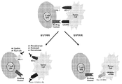
- FIG. 1 is a schematic diagram briefly showing the mechanism of showing the anti-cancer effect of the anti-CD300c monoclonal antibody and/or CD300c siRNA of the present invention.
- FIG. 2 is a schematic diagram briefly showing the mechanism by which the anti-CD300c monoclonal antibody of the present invention acts on each Monocyte, T cell, and cancer cell.
- FIG. 3 is a diagram showing the results of SDS-PAGE under non-reducing conditions of an anti-CD300c monoclonal antibody according to an embodiment of the present invention.
- FIG. 4 is a diagram showing SDS-PAGE results under reducing conditions of an anti-CD300c monoclonal antibody according to an embodiment of the present invention.
- FIG. 5 is a view showing the result of confirming the binding ability of the anti-CD300c monoclonal antibody to the CD300c antigen according to an embodiment of the present invention.
- FIG. 6 is a view showing the result of confirming the T cell activation ability of the anti-CD300c monoclonal antibody according to an embodiment of the present invention using ELISA.
- FIG. 7 is a view showing the result of confirming the differentiation ability of an anti-CD300c monoclonal antibody to M1 macrophages according to an embodiment of the present invention using ELISA.
- FIG. 8 is a view showing the results of confirming the differentiation ability of an anti-CD300c monoclonal antibody to M1 macrophages according to an embodiment of the present invention using ELISA.
- FIG. 9 is a view showing the results of confirming the differentiation ability of an anti-CD300c monoclonal antibody to M1 macrophages according to concentrations according to an embodiment of the present invention using ELISA.
- FIG. 10 is a view showing the result of confirming the differentiation ability of the anti-CD300c monoclonal antibody to M1 macrophages according to the concentration according to an embodiment of the present invention using ELISA.
- FIG. 11 is a view showing the results of confirming the differentiation ability of an anti-CD300c monoclonal antibody to M1 macrophages according to an embodiment of the present invention in the form of a cell.
- FIG. 12 is a view showing the results of confirming the differentiation ability of the CL7 anti-CD300c monoclonal antibody to M1 macrophages according to an embodiment of the present invention using ELISA.
- FIG. 13 is a view showing the results of comparing the differentiation ability of the M1 macrophage of the anti-CD300c monoclonal antibody according to an embodiment of the present invention and an immunological anticancer agent using ELISA.
- FIG. 14 is a view showing a result of comparing the differentiation ability of a CL7 anti-CD300c monoclonal antibody and an immunological anticancer agent into M1 macrophages according to an embodiment of the present invention using ELISA.
- 15 is a view showing a result of comparing the differentiation ability of a CL7 anti-CD300c monoclonal antibody and an immunological anticancer agent into M1 macrophages according to an embodiment of the present invention using ELISA.
- 16 is a view showing the results of comparing the differentiation ability of a CL7 anti-CD300c monoclonal antibody and an immunological anticancer agent into M1 macrophages according to an embodiment of the present invention using ELISA.
- 17 is a view showing the results of comparing the differentiation ability of an anti-CD300c monoclonal antibody and an immunological anticancer agent from M0 macrophage to M1 macrophage according to an embodiment of the present invention using ELISA.
- 18 is a diagram showing a result of comparing the differentiation ability of an anti-CD300c monoclonal antibody and an immunological anticancer agent into M1 macrophages according to an embodiment of the present invention using ELISA.
- FIG. 19 is a diagram showing the results of confirming the re-differentiation ability of an anti-CD300c monoclonal antibody from M2 macrophage to M1 macrophage according to an embodiment of the present invention using ELISA.
- FIG. 20 is a view showing the results of confirming the re-differentiation ability of an anti-CD300c monoclonal antibody from M2 macrophage to M1 macrophage according to an embodiment of the present invention using ELISA.
- FIG. 21 is a diagram showing the results of confirming the re-differentiation ability of an anti-CD300c monoclonal antibody from M2 macrophage to M1 macrophage according to an embodiment of the present invention using ELISA.
- FIG. 22 is a view showing the results of confirming the re-differentiation ability of an anti-CD300c monoclonal antibody from M0, M1, and M2 macrophages to M1 macrophages according to an embodiment of the present invention using ELISA.
- 23 is a view showing the result of confirming the effect of co-administration of an anti-CD300c monoclonal antibody and an anti-PD-L1 immuno-anticancer agent according to an embodiment of the present invention by M1 macrophage differentiation ability.
- 24 is a view showing the result of confirming the effect of co-administration of an anti-CD300c monoclonal antibody and an immuno-anticancer agent according to an embodiment of the present invention with M1 macrophage differentiation ability.
- 25 is a diagram showing the results of confirming the effect of inhibiting cancer cell growth under the condition of 0% FBS of the anti-CD300c monoclonal antibody according to an embodiment of the present invention.
- 26 is a view showing the results of confirming the cancer cell growth inhibitory effect in the condition of 0.1% FBS of the anti-CD300c monoclonal antibody according to an embodiment of the present invention.
- FIG. 27 is a view showing a result of comparing the anti-CD300c monoclonal antibody according to an embodiment of the present invention and the anti-cancer agent on the growth of cancer cells (lung cancer).
- FIG. 28 is a view showing the results of comparing the anti-CD300c monoclonal antibody according to an embodiment of the present invention and the anti-cancer agent for inhibiting the growth of cancer cells (breast cancer).
- 29 is a view showing the results of confirming the cancer cell growth inhibitory effect by concentration of the anti-CD300c monoclonal antibody according to an embodiment of the present invention.
- FIG. 30 is a diagram showing the results of confirming the effect of inhibiting cancer cell growth according to co-administration of an anti-CD300c monoclonal antibody according to an embodiment of the present invention and an immune anticancer agent.
- 31 is a view showing the results of confirming the cancer cell growth inhibitory effect according to the combination administration of an anti-CD300c monoclonal antibody and an anticancer agent according to an embodiment of the present invention.
- FIG. 32 is a diagram showing the results of confirming the mechanism of action according to co-administration of an anti-CD300c monoclonal antibody and an immune anticancer agent according to an embodiment of the present invention.
- 33 is a view showing the result of confirming the binding specificity of the anti-CD300c monoclonal antibody according to an embodiment of the present invention.
- 34 is a view showing the results of confirming the cross-reactivity in mice of the anti-CD300c monoclonal antibody according to an embodiment of the present invention.
- 35 is a diagram showing the results of confirming the anti-cancer effect (colorectal cancer) in mice of the anti-CD300c monoclonal antibody according to an embodiment of the present invention.
- 36 is a diagram schematically showing an experimental method confirming the effect of an anti-CD300c monoclonal antibody on cancer growth in vivo according to an embodiment of the present invention.
- FIG. 37 is a view showing the results of confirming the in vivo cancer growth inhibitory effect of the anti-CD300c monoclonal antibody according to an embodiment of the present invention.
- FIG. 38 is a view showing the results of confirming the effect of the anti-CD300c monoclonal antibody according to an embodiment of the present invention on the increase of tumor-infiltrating lymphocytes in the tumor microenvironment in vivo.
- Scale bar represents 50 um.
- 39 is a view showing the results confirming the effect of the anti-CD300c monoclonal antibody according to an embodiment of the present invention on the increase in M1 macrophage in vivo.
- the anti-CD300c monoclonal antibody of the present invention specifically binds to the CD300c protein with high avidity, effectively inhibits the mechanism of CD300c, activates T cells, and promotes differentiation into M1 macrophages, thereby promoting the growth of cancer cells and cancer. Since it can effectively inhibit development and metastasis of cancer, it can be effectively used in the treatment of various cancers expressing the CD300c antigen on the surface.
- antibody refers to an immunoglobulin molecule immunologically reactive with a specific antigen
- polyclonal antibodies, monoclonal antibodies, and functional fragments thereof Includes all.
- the term may include forms produced by genetic engineering such as chimeric antibodies (eg, humanized murine antibodies) and heterologous antibodies (eg, bispecific antibodies).
- Double monoclonal antibodies are antibodies that exhibit a single binding specificity and affinity for a single antigenic site (epitope).
- monoclonal antibodies are antigens. Because it shows binding specificity and affinity only for a single epitope on the phase, quality control as a therapeutic is easy.
- the anti-CD300c monoclonal antibody of the present invention specifically binds to cancer cells expressing CD300c and exhibits anti-cancer activity by itself, and can maximize cancer cell-dependent anti-cancer activity by stimulating immune cells.
- the antibody comprises a variable region of a heavy chain and/or a light chain of the composition, the variable region as a primary structure and comprises a portion that forms an antigen-binding site of the antibody molecule, ,
- the antibody of the present invention may be composed of some fragments containing the variable region, preferably the variable region may be replaced with a soluble receptor for CD300c, but has the same effect as the anti-CD300c monoclonal antibody of the present invention. If indicated, it is not limited thereto.
- immunoglobulin is a concept including both antibody-like molecules and antibodies that have the same structural characteristics as an antibody and do not have antigen specificity.
- single-chain variable fragment refers to a protein in which the variable regions of the light and heavy chains of an antibody are linked by a linker consisting of a peptide sequence to which about 15 amino acids are linked,
- the sequence of the light chain variable region-linker-heavy chain variable region, or the heavy chain variable region-linker-light chain variable region is possible, and has the same or similar antigen specificity as the original antibody.
- the linking site is a hydrophilic flexible peptide chain mainly composed of glycine and serine, and the 15 amino acid sequence of "(Gly-Gly-Gly-Gly-Ser) 3 " or a sequence similar thereto is mainly used.
- the term "immune anticancer agent” refers to a cancer treatment method that activates the immune function of the body's immune cells to fight cancer cells, and examples thereof include anti-PD-1, anti-PD-L1, anti -CTLA-4, anti-KIR, anti-LAG3, anti-CD137, anti-OX40, anti-CD276, anti-CD27, anti-GITR, anti-TIM3, anti-41BB, anti-CD226, anti-CD40, anti -CD70, anti-ICOS, anti-CD40L, anti-BTLA, anti-TCR, anti-TIGIT, etc., but are not limited thereto.
- the term "adjuvant” improves and/or enhances the therapeutic effect by assisting the drug effect of the main drug, that is, an anticancer drug, or suppressing the resistance to the main drug to improve and/or enhance the therapeutic effect. It refers to an adjuvant drug used for the purpose of reducing or preventing or alleviating the harmful action of the main drug, and the adjuvant of the present invention is not limited as long as it contains an anti-CD300c monoclonal antibody as an active ingredient.
- prevention refers to any action that suppresses or delays the onset of diseases such as cancer by administration of the pharmaceutical composition according to the present invention.
- treatment refers to any action in which symptoms such as cancer are improved or beneficially changed by administration of the pharmaceutical composition according to the present invention.
- “individual” refers to a subject to which the pharmaceutical composition of the present invention can be administered, and the subject is not limited.
- the term "pharmaceutical composition” may be characterized in that it is in the form of capsules, tablets, granules, injections, ointments, powders, or beverages, and the pharmaceutical composition is intended for humans.
- the pharmaceutical compositions are not limited thereto, but may be formulated and used in the form of oral dosage forms such as powders, granules, capsules, tablets, aqueous suspensions, etc., external preparations, suppositories, and sterile injectable solutions according to a conventional method.
- the pharmaceutical composition of the present invention may contain a pharmaceutically acceptable carrier.
- Pharmaceutically acceptable carriers can be used as binders, lubricants, disintegrants, excipients, solubilizers, dispersants, stabilizers, suspending agents, colors, flavors, etc. for oral administration, and buffers, preservatives, and painlessness in the case of injections.
- Agents, solubilizers, isotonic agents, stabilizers, etc. can be mixed and used, and in the case of topical administration, base agents, excipients, lubricants, preservatives, etc. can be used.
- the formulation of the pharmaceutical composition of the present invention can be variously prepared by mixing with a pharmaceutically acceptable carrier as described above.
- the oral administration it can be prepared in the form of tablets, troches, capsules, elixir, suspension, syrup, wafers, etc., and in the case of injections, it can be prepared in the form of unit dose ampoules or multiple doses. have.
- it can be formulated as a solution, suspension, tablet, capsule, sustained-release preparation, and the like.
- suitable carriers, excipients and diluents for formulation include lactose, dextrose, sucrose, sorbitol, mannitol, xylitol, erythritol, malditol, starch, gum acacia, alginate, gelatin, calcium phosphate, calcium silicate, Cellulose, methyl cellulose, microcrystalline cellulose, polyvinylpyrrolidone, water, methylhydroxybenzoate, propylhydroxybenzoate, talc, magnesium stearate or mineral oil may be used.
- fillers, anti-aggregating agents, lubricants, wetting agents, flavoring agents, emulsifying agents, preservatives, and the like may additionally be included.
- the route of administration of the pharmaceutical composition according to the present invention is not limited thereto, but oral, intravenous, intramuscular, intraarterial, intramedullary, intrathecal, intracardiac, transdermal, subcutaneous, intraperitoneal, intranasal, intestinal, local , Sublingual or rectal. Oral or parenteral administration is preferred.
- parenteral as used herein includes subcutaneous, intradermal, intravenous, intramuscular, intraarticular, intrasynovial, intrasternal, intrathecal, intralesional and intracranial injection or infusion techniques.
- the pharmaceutical composition of the present invention may also be administered in the form of suppositories for rectal administration.
- the pharmaceutical composition of the present invention depends on a number of factors, including the activity of the specific compound used, age, weight, general health, sex, formulation, time of administration, route of administration, excretion rate, drug formulation, and the severity of the specific disease to be prevented or treated. It may vary in various ways, and the dosage of the pharmaceutical composition varies depending on the patient's condition, weight, degree of disease, drug form, route of administration and duration, but may be appropriately selected by those skilled in the art, and 0.0001 to 500 mg per day /kg or 0.001 to 500 mg/kg. Administration may be administered once a day, or may be divided several times. The above dosage does not limit the scope of the present invention in any way.
- the pharmaceutical composition according to the present invention may be formulated as a pill, dragee, capsule, liquid, gel, syrup, slurry, or suspension.
- biopanning was performed using a lambda phage library, a kappa phage library, a VH3VL1 phage library, and an OPALTL phage library.
- CD300c antigen at a concentration of 5 ⁇ g/mL was added to an immunotube and reacted for 1 hour to adsorb the antigen on the surface of the test tube.
- 3% of skim milk was added to suppress non-specific reactions, and then 10 12 PFU of antibody phage library dispersed in 3% of skim milk was added to each immunity test tube to bind to the antigen.
- scFv single-chain variable fragment
- the formed Escherichia coli colonies were suspended using 3 mL of SB (super broth)-carbenicilli culture medium, and some were stored at -80°C until use by adding 15% glycerol, and the rest was SB-carbenicillin-2.
- % Glucose solution was re-inoculated and incubated at 37°C. Then, the obtained culture was centrifuged, and biopanning was repeated three times again using the supernatant containing phage particles to obtain and concentrate antigen-specific antibodies.
- E. coli containing the antibody gene was applied to LB agar medium containing carbenicillin and incubated at 37°C for 16 hours, and the formed E. coli colonies were again SB-carbenicillin-2%. After re-inoculating the glucose solution and incubating at 37°C until the absorbance (OD 600nm ) became 0.5, IPTG was added and further incubated at 30°C for 16 hours. Thereafter, periplasmic extraction was performed, and a library pool of antibodies specifically binding to the CD300c antigen was obtained through the above results.
- ELISA was performed using a library pool obtained in the same manner as in Example 1.1.
- CD300c antigen and CD300a antigen were dispensed into an ELISA plate at a concentration of 5 ⁇ g/mL per well in a coating buffer (0.1 M sodium carbonate, pH 9.0), and then reacted at room temperature for 3 hours. To bind the antigen to the plate.
- PBST phosphate buffered saline-Tween20
- BSA bovine serum albumin
- the nucleotide sequence of the selected anti-CD300c monoclonal antibody was confirmed using the same method as in Example 1.2.
- DNA sequencing was performed to analyze complementarity-determining regions (CDRs) sequences.
- CDRs complementarity-determining regions
- anti-CD300c monoclonal antibodies selected using the lambda phage library, SL18 comprising the CDR sequence of SEQ ID NO: 36 (DNA sequence is SEQ ID NO: 35), and CL4 (DNA sequence) comprising the CDR sequence of SEQ ID NO: 8 Was SEQ ID NO: 7), and CL5 (DNA sequence is SEQ ID NO: 9) comprising the CDR sequences of SEQ ID NO: 10.
- anti-CD300c monoclonal antibodies selected using the kappa phage library, CK1 containing the CDR sequence of SEQ ID NO: 2 (DNA sequence is SEQ ID NO: 1), CK2 containing the CDR sequence of SEQ ID NO: 4 (DNA sequence Is SEQ ID NO: 3), CK3 comprising the CDR sequence of SEQ ID NO: 6 (DNA sequence is SEQ ID NO: 5), SK11 comprising the CDR sequence of SEQ ID NO: 22 (DNA sequence is SEQ ID NO: 21), CDR sequence of SEQ ID NO: 24 SK12 comprising a (DNA sequence is SEQ ID NO: 23), SK13 comprising the CDR sequence of SEQ ID NO: 26 (DNA sequence is SEQ ID NO: 25), SK14 comprising the CDR sequence of SEQ ID NO: 28 (DNA sequence is SEQ ID NO: 27) , SK15 comprising the CDR sequence of SEQ ID NO: 30 (DNA sequence is SEQ ID NO: 29), SK16 comprising the CDR sequence of SEQ
- CB301_OPALTL_B5 comprising the CDR sequence of SEQ ID NO: 48 (DNA sequence is SEQ ID NO: 47)
- CB301_OPALTL_E6 DNA sequence comprising the CDR sequence of SEQ ID NO: 50 was SEQ ID NO: 49.
- an expression vector was prepared in which the heavy and light chains capable of expressing the antibody were separated.
- a gene was inserted into the pCIW3.3 vector to express the heavy and light chains, respectively, using the analyzed CDR sequences.
- the prepared heavy and light chain expression vectors were mixed with PEI (polyethylenimine) in a mass ratio of 1:1 and transfected into 293T cells to induce antibody expression, and then the culture medium was centrifuged on day 8 to remove the cells, and the culture medium was removed. Obtained.
- PEI polyethylenimine
- the obtained culture was filtered and then resuspended using a solution in which 0.1 M NaH 2 PO 4 and 0.1 M Na 2 HPO 4 (pH 7.0) were mixed. And the resuspended solution was purified through affinity chromatography using protein A bead (GE healthcare), and finally elution buffer (Thermofisher) was used to elution.
- Binding ELISA was performed in order to select a monoclonal antibody that specifically binds to the CD300c antigen with better binding power among the anti-CD300c monoclonal antibodies prepared in the same manner as in Example 1.4.
- a coating buffer 0.1 M sodium carbonate, pH 9.0
- PBST phosphate buffered saline-Tween20
- BSA bovine serum albumin
- the anti-CD300c monoclonal antibody of the present invention binds to the CD300c antigen with strong binding force in the sigmoid curve according to the result of Binding ELISA.
- IL-2 Interleukin-2
- IL-2 is an immune factor that helps T cell growth, proliferation, and differentiation. Increasing IL-2 production activates T cells by increasing stimulation that induces T cell differentiation, proliferation, and growth. It means to let you do.
- anti-CD3 monoclonal antibody and anti-CD28 monoclonal antibody were added to a 96-well plate to a concentration of 2 ⁇ g/well and fixed for 24 hours, followed by 1 ⁇ 10 5 cells/well of Jurkat T cells (human T lymphocyte cell line) and 10 ⁇ g/well of anti-CD300c monoclonal antibody were treated together.
- the amount of IL-2 produced was measured using an ELISA kit (IL-2 Quantikine kit, R&D systems), and then compared with a control group not treated with an anti-CD300c monoclonal antibody. The results are shown in FIG. 6.
- THP-1 human monocyte cell line
- ELISA kit Human TNF- ⁇ Quantikine kit
- the CL7 anti-CD300c monoclonal antibody was treated at concentrations of 10, 5, 2.5, 1.25, 0.625, 0.313, 0.157, and 0.079 ⁇ g/mL, and the amount of TNF- ⁇ produced was confirmed. The results are shown in FIG. 10.
- THP-1 was treated with 10 ⁇ g/ml of anti-CD300c monoclonal antibody, and cultured for 48 hours. The morphology of the cells was observed under a microscope. The results are shown in FIG. 11.
- the amount of secretion of TNF- ⁇ , IL-1 ⁇ (Interleukin-1 ⁇ ), and IL-8 (Interleukin-8) was measured by ELISA. It was measured using kit (R&D systems). In more detail, 1.5 ⁇ 10 4 cells/well of THP-1 was dispensed in a 96-well plate, and 10 ⁇ g/mL of anti-CD300c monoclonal antibody was treated.
- TNF- ⁇ , IL-1 ⁇ , and IL-8 which are differentiation markers of M1 macrophages.
- ELISA kit Human TNF- ⁇ Quantikine kit, R&D systems
- the anti-CD300c monoclonal antibody significantly increased the amount of TNF- ⁇ production compared to the comparison group treated with Imfinji (Imf), which is known as an immunological anticancer agent, alone.
- Imf Imfinji
- the anti-CD300c monoclonal antibody significantly increased the differentiation ability to M1 macrophages than the previously known immunological anticancer agents.
- the anti-PD-L1 immuno-anti-cancer drug Impingi the anti-PD-1 immuno-anti-cancer drug, Keytruda, and the Isotype control (Immunoglobulin G) antibody were each at a concentration of 10 ⁇ g/mL.
- the production amount of TNF- ⁇ , IL-1 ⁇ , and IL-8 was confirmed with an ELISA kit. The results are shown in Figs. 14 to 16.
- THP-1 1.5x10 4 cells/well of THP-1 was dispensed into a 96-well plate, treated with 320 nM of PMA, and pretreated for 6 hours. After that, 20 ng/mL of IL-4 (Interleukin-4) and IL-13 (Interleukin-13), and 10 ⁇ g/mL of anti-CD300c monoclonal antibody were treated and reacted for 18 hours. And the production amount of TNF- ⁇ , IL-1 ⁇ and IL-8 was confirmed with an ELISA kit. The results are shown in Figs. 19 to 21.
- anti-CD300c monoclonal As shown in Figure 22, compared to the M0 macrophage control group treated with PMA alone, the M1 macrophage control group treated with LPS alone, and the M2 macrophage control group treated with IL-4 and IL-13 alone, anti-CD300c monoclonal It was confirmed that the production amount of TNF- ⁇ was significantly increased in all the experimental groups pretreated with the antibody. Through the above results, it was confirmed that the anti-CD300c monoclonal antibody has excellent ability to differentiate M0, M1 and M2 macrophages into M1 macrophages.
- the anti-CD300c monoclonal antibody promotes the differentiation into M1 macrophages than the existing anticancer drugs, induces an anticancer immune action, and suppresses the growth of cancer tissues.
- the anticancer effect can be exhibited by re-differentiating M2 macrophages, which are known to be involved in promoting the proliferation and metastasis of cancer cells, into M1 macrophages.
- NF- ⁇ B nuclear factor kappa-light-chain-enhancer of activated B cells
- the amount of p-NF- ⁇ B was increased in the experimental group treated with the anti-CD300c monoclonal antibody, and Impingi and the anti-CD300c monoclonal antibody It was confirmed that the amount of p-NF- ⁇ B was further increased in the experimental group administered concurrently. Through the above results, it was confirmed that the differentiation into M1 macrophages was promoted when the anti-CD300c monoclonal antibody and impingi were administered in combination.
- p38 MAPK p38 mitogen-activated protein kinasese
- ERK extracellular signal-regulated kinase
- the anti-CD300c monoclonal antibody promotes the differentiation of M1 macrophages through MAPK signal transduction, and its effect is further increased when co-administered with an immune anticancer agent.
- anti-CD300c monoclonal antibody The antibody can be used alone as an immuno-anticancer agent, but it was confirmed that the anti-cancer treatment effect can be further increased through co-administration with an existing immuno-cancer agent.
- cell proliferation analysis was performed using A549 (human lung cancer cell line).
- A549 human lung cancer cell line
- 2x10 4 cells were dispensed in a 96-well plate under the condition of 0% fetal bovine serum (FBS), and 6x10 3 cells were dispensed under the condition of 0.1% fetal bovine serum.
- FBS fetal bovine serum
- 6x10 3 cells were dispensed under the condition of 0.1% fetal bovine serum.
- 10 ⁇ g/mL of anti-CD300c monoclonal antibody was treated and incubated for 5 days.
- CCK-8 DOJINDO
- absorbance was measured at OD 450nm to confirm the cancer cell growth inhibitory effect of the anti-CD300c monoclonal antibody. The results are shown in Figs. 25 and 26.
- A549 human lung cancer cell line
- MDA-MB-231 human breast cancer cell line
- 2x10 4 cells were dispensed in a 96-well plate under the condition of 0% fetal bovine serum (FBS), and 6x10 3 cells were dispensed under the condition of 0.1% fetal bovine serum.
- 10 ⁇ g/mL of anti-CD300c monoclonal antibody was treated and incubated for 5 days, and then observed with an optical microscope. The results are shown in Figs. 27 and 28.
- anti-CD300c monoclonal antibody In order to confirm the mechanism of action by co-administration of an anti-CD300c monoclonal antibody and an immune anticancer agent in the mechanism of apoptosis signaling of cancer cells, anti-CD300c monoclonal antibody, impingi, and/or kitruda were added to A549 cells. After treatment at a concentration of ⁇ g/mL, the amount of Caspase-9 cleaved form, a cell death marker, was confirmed by Western blotting (Cell signaling). The results are shown in FIG. 32.
- Example 7 Anti-cancer effect of anti-CD300c monoclonal antibody in vivo
- mice with a tumor size of 50 to 100 mm 3 were treated with anti-CD300c monoclonal antibody CL7, anti-PD-1 antibody, and monoclonal antibody CL7 and anti-PD-1 antibody (Combo).
- PBS phosphate buffered saline
- mice on day 25 tested in the same manner as in Example 7.1 were euthanized.
- 1% PFA para-formaldehyde
- cancer tissue was obtained.
- the obtained cancer tissue was fixed using 1% PFA, and dehydrated using 10%, 20%, and 30% sucrose solutions sequentially.
- Dehydrated cancer tissues are frozen in an OCT compound (Optimal cutting temperature compound), and then the cancer tissues are sectioned to a thickness of 50 ⁇ m using a cryotome, and tumor-infiltrating lymphocyte markers, CD8 + T cells and CD31 + cancer. Blood vessel cells were stained. The results are shown in FIG. 38.
- CD8 + T cells were increased in the experimental group administered with the anti-CD300c monoclonal antibody compared to the experimental group administered with the anti-PD-1 antibody alone, through which CD300c monoclonal antibody It was confirmed that the tumor-infiltrating lymphocytes increased in the tumor microenvironment to show anticancer effect.
- cancer tissue sections prepared in the same manner as in Example 7.2 were used to stain the M1 macrophage marker iNOS and the M2 macrophage marker CD206. . The results are shown in FIG. 39.
- the M1 macrophage was partially increased compared to the control, but in the experimental group treated with the anti-CD300c monoclonal antibody, the M1 macrophage was significantly increased, and the M2 macrophage was almost It was confirmed that it was not observed. In addition, it was confirmed that the M1 macrophage was increased in the experimental group co-administered with the anti-CD300c monoclonal antibody and the anti-PD-1 antibody. Through the above results, it was confirmed that the anti-CD300c monoclonal antibody can effectively promote the differentiation of M1 macrophages compared with the existing immuno-cancer drugs.
- the anti-CD300c monoclonal antibody of the present invention can bind to the CD300c antigen with high specificity, and exhibit cross-reactivity between species such as mice, and thus can be used in a variety of individuals.
- the anti-CD300c monoclonal antibody can effectively inhibit the proliferation and metastasis of cancer cells by acting as an immune anticancer agent by activating T cells and promoting differentiation into M1 macrophages.
- the therapeutic effect could be further increased through co-administration with an existing anticancer drug, so it was confirmed that it can be effectively used for anticancer immunotherapy of various cancers expressing the CD300c antigen.
- the anti-CD300c monoclonal antibody of the present invention specifically binds to the CD300c antigen, it can be used not only for the treatment of all cancers secreting the CD300c antigen, but also for the anticancer treatment of various individuals by having cross-reactivity between species .
- it since it exhibits an anticancer effect through its action as an immune anticancer agent, it can effectively suppress the proliferation, development, and metastasis of cancer, so that it can be effectively used for immunotherapy of various cancers.
Landscapes
- Health & Medical Sciences (AREA)
- Chemical & Material Sciences (AREA)
- Immunology (AREA)
- Organic Chemistry (AREA)
- Life Sciences & Earth Sciences (AREA)
- Medicinal Chemistry (AREA)
- General Health & Medical Sciences (AREA)
- Biochemistry (AREA)
- Genetics & Genomics (AREA)
- Animal Behavior & Ethology (AREA)
- Proteomics, Peptides & Aminoacids (AREA)
- Public Health (AREA)
- Veterinary Medicine (AREA)
- Molecular Biology (AREA)
- Pharmacology & Pharmacy (AREA)
- Biophysics (AREA)
- Chemical Kinetics & Catalysis (AREA)
- General Chemical & Material Sciences (AREA)
- Epidemiology (AREA)
- Nuclear Medicine, Radiotherapy & Molecular Imaging (AREA)
- Peptides Or Proteins (AREA)
- Medicines Containing Antibodies Or Antigens For Use As Internal Diagnostic Agents (AREA)
- Medicines That Contain Protein Lipid Enzymes And Other Medicines (AREA)
Abstract
Description
| CB301 항체 | EC50 (μg/mL) |
| CK1 | 0.056 |
| CK2 | 0.033 |
| CK3 | 0.793 |
| CL4 | 0.031 |
| CL5 | 0.032 |
| CL6 | 0.148 |
| CL7 | 0.047 |
| CL8 | 49.7 |
| CL9 | 0.094 |
| CL10 | 0.039 |
| SK11 | 0.052 |
| SK12 | 0.067 |
| SK13 | 0.044 |
| SK14 | 0.065 |
| SK15 | 14.74 |
| SK16 | 2.42 |
| SK17 | 0.054 |
| SL18 | 0.17 |
Claims (18)
- 서열번호 2, 4, 6, 8, 10, 12, 14, 16, 18, 20, 22, 24, 26, 28, 30, 32, 34, 36, 38, 40, 42, 44, 46, 48 및 50으로 이루어진 군으로 선택된 어느 하나 이상의 CDR(complementarity-determining regions) 서열을 포함하는, 항-CD300c 단클론 항체.
- 제 1 항에 있어서,상기 항-CD300c 단클론 항체는 종간 교차 반응성을 가지는 것을 특징으로 하는, 항-CD300c 단클론 항체.
- 제 2 항에 있어서,상기 교차 반응성은 인간 항원과 마우스 항원에 대한 교차 반응성인 것을 특징으로 하는, 항-CD300c 단클론 항체.
- 항-CD300c 단클론 항체를 유효성분으로 포함하는, 암 예방 또는 치료용 약학적 조성물.
- 제 4 항에 있어서,상기 항체는 서열번호 2, 4, 6, 8, 10, 12, 14, 16, 18, 20, 22, 24, 26, 28, 30, 32, 34, 36, 38, 40, 42, 44, 46, 48 및 50으로 이루어진 군으로 선택된 어느 하나 이상의 CDR(complementarity-determining regions) 서열을 포함하는 것을 특징으로 하는, 약학적 조성물.
- 제 4 항에 있어서,상기 암은 대장암, 직장암, 결장암, 갑상선암, 구강암, 인두암, 후두암, 자궁경부암, 뇌암, 폐암, 난소암, 방광암, 신장암, 간암, 췌장암, 전립선암, 피부암, 혀암, 유방암, 자궁암, 위암, 골암 및 혈액암으로 이루어진 군으로부터 선택된 어느 하나 이상인 것을 특징으로 하는, 약학적 조성물.
- 제 4 항에 있어서,상기 약학적 조성물은 면역항암제를 추가로 포함하는 것을 특징으로 하는, 약학적 조성물.
- 제 7 항에 있어서,상기 면역항암제는 anti-PD-1, anti-PD-L1, anti-CTLA-4, anti-KIR, anti-LAG3, anti-CD137, anti-OX40, anti-CD276, anti-CD27, anti-GITR, anti-TIM3, anti-41BB, anti-CD226, anti-CD40, anti-CD70, anti-ICOS, anti-CD40L, anti-BTLA, anti-TCR, 및 anti-TIGIT로 이루어진 군으로부터 선택된 어느 하나 이상인 것을 특징으로 하는, 약학적 조성물.
- 제 4 항에 있어서,상기 약학적 조성물은 암의 증식, 생존, 전이, 재발 또는 항암제 내성을 억제하는 것을 특징으로 하는, 약학적 조성물.
- 항-CD300c 단클론 항체를 유효성분으로 포함하는, 면역항암제.
- 제 10 항에 있어서,상기 항체는 서열번호 2, 4, 6, 8, 10, 12, 14, 16, 18, 20, 22, 24, 26, 28, 30, 32, 34, 36, 38, 40, 42, 44, 46, 48 및 50으로 이루어진 군으로 선택된 어느 하나 이상의 CDR(complementarity-determining regions) 서열을 포함하는 것을 특징으로 하는, 면역항암제.
- 제 10 항에 있어서,상기 면역항암제는 암의 증식, 생존, 전이, 재발 또는 항암제 내성을 억제하는 것을 특징으로 하는, 면역항암제.
- 제 10 항에 있어서,상기 암은 대장암, 직장암, 결장암, 갑상선암, 구강암, 인두암, 후두암, 자궁경부암, 뇌암, 폐암, 난소암, 방광암, 신장암, 간암, 췌장암, 전립선암, 피부암, 혀암, 유방암, 자궁암, 위암, 골암 및 혈액암으로 이루어진 군으로부터 선택된 어느 하나 이상인 것을 특징으로 하는, 면역항암제.
- 항-CD300c 단클론 항체를 유효성분으로 포함하는, 항암 치료 요법의 보조제.
- 제 14 항에 있어서,상기 보조제는 면역 세포의 면역 기능을 활성화 시켜 항암 치료 효과를 향상시키는 것을 특징으로 하는, 보조제.
- 제 14 항에 있어서,상기 항암 치료 요법은 방사선 치료 요법, 항암제 치료 요법 또는 면역항암제 치료 요법인, 보조제.
- 항-CD300c 단클론 항체를 유효성분으로 포함하는 조성물을 개체에 투여하는 단계를 포함하는, 암의 예방 또는 치료 방법.
- 항-CD300c 단클론 항체를 유효성분으로 포함하는 조성물의 암 예방 또는 치료 용도.
Priority Applications (6)
| Application Number | Priority Date | Filing Date | Title |
|---|---|---|---|
| AU2020387199A AU2020387199B2 (en) | 2019-11-18 | 2020-11-18 | Composition for preventing or treating cancer, comprising anti-CD300c monoclonal antibodies |
| JP2022528613A JP7539609B2 (ja) | 2019-11-18 | 2020-11-18 | 抗-cd300c単クローン抗体を含むがん予防または治療用組成物 |
| CN202080093900.9A CN114980925A (zh) | 2019-11-18 | 2020-11-18 | 包含抗CD300c单克隆抗体的用于预防或治疗癌症的组合物 |
| EP20891244.4A EP4062934A4 (en) | 2019-11-18 | 2020-11-18 | COMPOSITION FOR PREVENTING OR TREATING CANCER WITH ANTI-CD300C MONOCLONAL ANTIBODIES |
| CA3158715A CA3158715A1 (en) | 2019-11-18 | 2020-11-18 | Composition for preventing or treating cancer, comprising anti-cd300c monoclonal antibodies |
| US17/746,569 US20220275103A1 (en) | 2019-11-18 | 2022-05-17 | Anti-cd300c monoclonal antibody and biomarker thereof for preventing or treating cancer |
Applications Claiming Priority (4)
| Application Number | Priority Date | Filing Date | Title |
|---|---|---|---|
| KR20190147496 | 2019-11-18 | ||
| KR10-2019-0147496 | 2019-11-18 | ||
| KR10-2020-0154076 | 2020-11-17 | ||
| KR1020200154076A KR102464507B1 (ko) | 2019-11-18 | 2020-11-17 | 항-CD300c 단클론 항체를 포함하는 암 예방 또는 치료용 조성물 |
Related Parent Applications (1)
| Application Number | Title | Priority Date | Filing Date |
|---|---|---|---|
| PCT/KR2022/006939 Continuation WO2022240261A1 (ko) | 2019-11-18 | 2022-05-13 | 항-cd300c 단클론 항체 및 이의 암 예방 또는 치료용 바이오마커 |
Related Child Applications (1)
| Application Number | Title | Priority Date | Filing Date |
|---|---|---|---|
| US17/746,569 Continuation-In-Part US20220275103A1 (en) | 2019-11-18 | 2022-05-17 | Anti-cd300c monoclonal antibody and biomarker thereof for preventing or treating cancer |
Publications (1)
| Publication Number | Publication Date |
|---|---|
| WO2021101244A1 true WO2021101244A1 (ko) | 2021-05-27 |
Family
ID=75981399
Family Applications (1)
| Application Number | Title | Priority Date | Filing Date |
|---|---|---|---|
| PCT/KR2020/016264 Ceased WO2021101244A1 (ko) | 2019-11-18 | 2020-11-18 | 항-cd300c 단클론 항체를 포함하는 암 예방 또는 치료용 조성물 |
Country Status (6)
| Country | Link |
|---|---|
| EP (1) | EP4062934A4 (ko) |
| JP (1) | JP7539609B2 (ko) |
| CN (1) | CN114980925A (ko) |
| AU (1) | AU2020387199B2 (ko) |
| CA (1) | CA3158715A1 (ko) |
| WO (1) | WO2021101244A1 (ko) |
Cited By (3)
| Publication number | Priority date | Publication date | Assignee | Title |
|---|---|---|---|---|
| EP4339210A4 (en) * | 2021-05-13 | 2025-09-24 | Centricsbio Inc | COMBINATION THERAPY USING AN ANTI-CD300C ANTIBODY |
| EP4339211A4 (en) * | 2021-05-13 | 2025-10-01 | Centricsbio Inc | ANTI-CD300C MONOCLONAL ANTIBODY AND ITS BIOMARKER FOR THE PREVENTION OR TREATMENT OF CANCER |
| EP4520770A4 (en) * | 2022-05-02 | 2026-04-22 | Centricsbio Inc | Anti-CD300C antibody or antigen-binding fragment thereof, and its uses in preventing or treating neurodegenerative brain disease |
Citations (4)
| Publication number | Priority date | Publication date | Assignee | Title |
|---|---|---|---|---|
| US20160194631A1 (en) * | 2009-09-30 | 2016-07-07 | Harvard Medical School | Methods for Modulation of Autophagy Through the Modulation of Autophagy-Inhibiting Gene Products |
| WO2018094460A1 (en) * | 2016-11-22 | 2018-05-31 | Dendrocyte Biotech Pty Ltd | Anti-cd300f antibody and uses thereof |
| KR20180099557A (ko) | 2017-02-28 | 2018-09-05 | 한양대학교 산학협력단 | 종양 특이적 살상 아데노바이러스 및 면역 관문 억제제를 포함하는 항암용 조성물 |
| KR20190136949A (ko) * | 2018-05-31 | 2019-12-10 | 주식회사 센트릭스바이오 | CD300c의 발현 억제제 또는 활성 억제제를 포함하는 암 예방 또는 치료용 약학적 조성물 |
Family Cites Families (2)
| Publication number | Priority date | Publication date | Assignee | Title |
|---|---|---|---|---|
| US9856308B2 (en) * | 2012-05-02 | 2018-01-02 | Spiber Technologies Ab | Spider silk fusion protein structures incorporating immunoglobulin fragments as affinity ligands |
| WO2019231188A1 (ko) | 2018-05-31 | 2019-12-05 | 주식회사 센트릭스바이오 | Cd300c의 발현 억제제 또는 활성 억제제를 포함하는 암 예방 또는 치료용 약학적 조성물 |
-
2020
- 2020-11-18 AU AU2020387199A patent/AU2020387199B2/en active Active
- 2020-11-18 EP EP20891244.4A patent/EP4062934A4/en active Pending
- 2020-11-18 JP JP2022528613A patent/JP7539609B2/ja active Active
- 2020-11-18 WO PCT/KR2020/016264 patent/WO2021101244A1/ko not_active Ceased
- 2020-11-18 CA CA3158715A patent/CA3158715A1/en active Pending
- 2020-11-18 CN CN202080093900.9A patent/CN114980925A/zh active Pending
Patent Citations (4)
| Publication number | Priority date | Publication date | Assignee | Title |
|---|---|---|---|---|
| US20160194631A1 (en) * | 2009-09-30 | 2016-07-07 | Harvard Medical School | Methods for Modulation of Autophagy Through the Modulation of Autophagy-Inhibiting Gene Products |
| WO2018094460A1 (en) * | 2016-11-22 | 2018-05-31 | Dendrocyte Biotech Pty Ltd | Anti-cd300f antibody and uses thereof |
| KR20180099557A (ko) | 2017-02-28 | 2018-09-05 | 한양대학교 산학협력단 | 종양 특이적 살상 아데노바이러스 및 면역 관문 억제제를 포함하는 항암용 조성물 |
| KR20190136949A (ko) * | 2018-05-31 | 2019-12-10 | 주식회사 센트릭스바이오 | CD300c의 발현 억제제 또는 활성 억제제를 포함하는 암 예방 또는 치료용 약학적 조성물 |
Non-Patent Citations (4)
| Title |
|---|
| CHENG CUI, MIN SU, YUJUN LIN, LAIJUN LAI: "A CD300c-Fc fusion protein inhibits T cell immunity", FRONTIERS IN IMMUNOLOGY, vol. 9, 15 November 2018 (2018-11-15), pages 1 - 14, XP055660033, DOI: 10.3389/fimmu.2018.02657 * |
| MILENA DIMITROVA, ZENARRUZABEITIA OLATZ, BORREGO FRANCISCO, SIMHADRI VENKATESWARA R.: "CD300c is uniquely expressed on CD56bright Natural Killer Cells and differs from CD300a upon ligand recognition", SCIENTIFIC REPORTS, vol. 6, no. 1, 4 April 2016 (2016-04-04), pages 1 - 12, XP055660025, DOI: 10.1038/srep23942 * |
| See also references of EP4062934A4 |
| VENKATESWARA R. SIMHADRI, MARIANO JOHN L., GIL-KRZEWSKA ALEKSANDRA, ZHOU QING, BORREGO FRANCISCO: "CD300c is an Activating Receptor Expressed on Human Monocytes", JOURNAL OF INNATE IMMUNITY, S. KARGER AG, CH, vol. 5, no. 4, 6 April 2013 (2013-04-06), CH, pages 389 - 400, XP055660026, ISSN: 1662-811X, DOI: 10.1159/000350523 * |
Cited By (3)
| Publication number | Priority date | Publication date | Assignee | Title |
|---|---|---|---|---|
| EP4339210A4 (en) * | 2021-05-13 | 2025-09-24 | Centricsbio Inc | COMBINATION THERAPY USING AN ANTI-CD300C ANTIBODY |
| EP4339211A4 (en) * | 2021-05-13 | 2025-10-01 | Centricsbio Inc | ANTI-CD300C MONOCLONAL ANTIBODY AND ITS BIOMARKER FOR THE PREVENTION OR TREATMENT OF CANCER |
| EP4520770A4 (en) * | 2022-05-02 | 2026-04-22 | Centricsbio Inc | Anti-CD300C antibody or antigen-binding fragment thereof, and its uses in preventing or treating neurodegenerative brain disease |
Also Published As
| Publication number | Publication date |
|---|---|
| EP4062934A4 (en) | 2024-03-20 |
| JP2023503567A (ja) | 2023-01-31 |
| AU2020387199B2 (en) | 2025-05-22 |
| CN114980925A (zh) | 2022-08-30 |
| JP7539609B2 (ja) | 2024-08-26 |
| AU2020387199A1 (en) | 2022-06-02 |
| EP4062934A1 (en) | 2022-09-28 |
| CA3158715A1 (en) | 2021-05-27 |
Similar Documents
| Publication | Publication Date | Title |
|---|---|---|
| WO2020076105A1 (ko) | 신규 항-c-kit 항체 | |
| WO2015005632A1 (ko) | Dll4와 vegf에 특이적으로 결합하는 신규 이중표적 단백질 및 이의 용도 | |
| WO2017023138A1 (ko) | 키메라 항원 수용체 및 키메라 항원 수용체가 발현된 t 세포 | |
| WO2017030370A1 (ko) | 항-코티닌 항체가 연결된 키메라 항체 수용체 및 이의 용도 | |
| WO2021107566A1 (ko) | C-kit에 대한 항체 및 이의 용도 | |
| WO2022231032A1 (ko) | 항-cntn4 특이적 항체 및 그의 용도 | |
| WO2022169269A1 (ko) | 항 ctla-4 항체 및 이의 용도 | |
| WO2019132533A1 (ko) | 항-pd-l1 항체 및 이의 용도 | |
| WO2023214778A1 (ko) | 항-cd300c 항체 또는 이의 항원 결합 단편 및 그의 퇴행성 뇌 질환 예방 또는 치료용 용도 | |
| WO2018186706A1 (ko) | Nk 세포 활성화 융합 단백질, nk 세포 및 이들을 포함하는 약제학적 조성물 | |
| WO2020004934A1 (ko) | 항-bcma 항체 및 그 용도 | |
| KR102464507B1 (ko) | 항-CD300c 단클론 항체를 포함하는 암 예방 또는 치료용 조성물 | |
| JP7539609B2 (ja) | 抗-cd300c単クローン抗体を含むがん予防または治療用組成物 | |
| WO2021118246A1 (en) | Anti-bcma/anti-4-1bb bispecific antibodies and uses thereof | |
| WO2021210939A1 (ko) | 항-her2 어피바디 및 이를 스위치 분자로 이용하는 스위처블 키메라 항원 수용체 | |
| WO2021132746A1 (en) | Anti-bcma/anti-4-1bb bispecific antibodies and uses thereof | |
| WO2024172363A1 (ko) | 항-cntn4 항체 및 그의 용도 | |
| WO2024096522A1 (ko) | Cd22에 결합하는 항체, 이를 포함하는 이중특이적 항체, 키메라 항원 수용체, 항체 약물 복합체 및 이의 용도 | |
| WO2023249463A1 (ko) | 면역관문 제어 인자를 발현하는 면역세포 및 이의 용도 | |
| WO2024049161A1 (ko) | 신규한 항-pd-l1 키메릭 항원 수용체 및 이를 발현하는 면역세포 | |
| WO2022250433A1 (ko) | Cd300c 항원 또는 그의 수용체에 특이적으로 결합하는 키메라 항원 수용체 | |
| WO2023014128A1 (ko) | Api5에 특이적으로 결합하는 항체 및 이의 용도 | |
| WO2021172690A1 (ko) | 메소텔린 특이적인 키메라 항원 수용체 및 이의 용도 | |
| WO2024191168A1 (ko) | 신규한 cd3 결합항체 및 이를 포함한 다중특이 항체 | |
| WO2021125787A1 (ko) | Dcr1에 특이적인 단일클론 항체 |
Legal Events
| Date | Code | Title | Description |
|---|---|---|---|
| 121 | Ep: the epo has been informed by wipo that ep was designated in this application |
Ref document number: 20891244 Country of ref document: EP Kind code of ref document: A1 |
|
| ENP | Entry into the national phase |
Ref document number: 2022528613 Country of ref document: JP Kind code of ref document: A |
|
| ENP | Entry into the national phase |
Ref document number: 3158715 Country of ref document: CA |
|
| ENP | Entry into the national phase |
Ref document number: 2020387199 Country of ref document: AU Date of ref document: 20201118 Kind code of ref document: A |
|
| NENP | Non-entry into the national phase |
Ref country code: DE |
|
| ENP | Entry into the national phase |
Ref document number: 2020891244 Country of ref document: EP Effective date: 20220620 |