WO2021241729A1 - 抗cd73抗体およびその用途 - Google Patents
抗cd73抗体およびその用途 Download PDFInfo
- Publication number
- WO2021241729A1 WO2021241729A1 PCT/JP2021/020384 JP2021020384W WO2021241729A1 WO 2021241729 A1 WO2021241729 A1 WO 2021241729A1 JP 2021020384 W JP2021020384 W JP 2021020384W WO 2021241729 A1 WO2021241729 A1 WO 2021241729A1
- Authority
- WO
- WIPO (PCT)
- Prior art keywords
- antibody
- seq
- cells
- human
- cancer
- Prior art date
- Legal status (The legal status is an assumption and is not a legal conclusion. Google has not performed a legal analysis and makes no representation as to the accuracy of the status listed.)
- Ceased
Links
Images


Classifications
-
- A—HUMAN NECESSITIES
- A61—MEDICAL OR VETERINARY SCIENCE; HYGIENE
- A61P—SPECIFIC THERAPEUTIC ACTIVITY OF CHEMICAL COMPOUNDS OR MEDICINAL PREPARATIONS
- A61P43/00—Drugs for specific purposes, not provided for in groups A61P1/00-A61P41/00
-
- A—HUMAN NECESSITIES
- A61—MEDICAL OR VETERINARY SCIENCE; HYGIENE
- A61P—SPECIFIC THERAPEUTIC ACTIVITY OF CHEMICAL COMPOUNDS OR MEDICINAL PREPARATIONS
- A61P35/00—Antineoplastic agents
-
- C—CHEMISTRY; METALLURGY
- C07—ORGANIC CHEMISTRY
- C07K—PEPTIDES
- C07K16/00—Immunoglobulins [IG], e.g. monoclonal or polyclonal antibodies
- C07K16/18—Immunoglobulins [IG], e.g. monoclonal or polyclonal antibodies against material from animals or humans
-
- C—CHEMISTRY; METALLURGY
- C07—ORGANIC CHEMISTRY
- C07K—PEPTIDES
- C07K16/00—Immunoglobulins [IG], e.g. monoclonal or polyclonal antibodies
- C07K16/18—Immunoglobulins [IG], e.g. monoclonal or polyclonal antibodies against material from animals or humans
- C07K16/28—Immunoglobulins [IG], e.g. monoclonal or polyclonal antibodies against material from animals or humans against receptors, cell surface antigens or cell surface determinants
- C07K16/2896—Immunoglobulins [IG], e.g. monoclonal or polyclonal antibodies against material from animals or humans against receptors, cell surface antigens or cell surface determinants against molecules with a "CD"-designation, not provided for elsewhere
-
- C—CHEMISTRY; METALLURGY
- C07—ORGANIC CHEMISTRY
- C07K—PEPTIDES
- C07K16/00—Immunoglobulins [IG], e.g. monoclonal or polyclonal antibodies
- C07K16/40—Immunoglobulins [IG], e.g. monoclonal or polyclonal antibodies against enzymes
-
- G—PHYSICS
- G01—MEASURING; TESTING
- G01N—INVESTIGATING OR ANALYSING MATERIALS BY DETERMINING THEIR CHEMICAL OR PHYSICAL PROPERTIES
- G01N33/00—Investigating or analysing materials by specific methods not covered by groups G01N1/00 - G01N31/00
- G01N33/48—Biological material, e.g. blood, urine; Haemocytometers
- G01N33/50—Chemical analysis of biological material, e.g. blood, urine; Testing involving biospecific ligand binding methods; Immunological testing
- G01N33/5005—Chemical analysis of biological material, e.g. blood, urine; Testing involving biospecific ligand binding methods; Immunological testing involving human or animal cells
- G01N33/5008—Chemical analysis of biological material, e.g. blood, urine; Testing involving biospecific ligand binding methods; Immunological testing involving human or animal cells for testing or evaluating the effect of chemical or biological compounds, e.g. drugs, cosmetics
- G01N33/5011—Chemical analysis of biological material, e.g. blood, urine; Testing involving biospecific ligand binding methods; Immunological testing involving human or animal cells for testing or evaluating the effect of chemical or biological compounds, e.g. drugs, cosmetics for testing antineoplastic activity
-
- A—HUMAN NECESSITIES
- A61—MEDICAL OR VETERINARY SCIENCE; HYGIENE
- A61K—PREPARATIONS FOR MEDICAL, DENTAL OR TOILETRY PURPOSES
- A61K39/00—Medicinal preparations containing antigens or antibodies
- A61K2039/505—Medicinal preparations containing antigens or antibodies comprising antibodies
-
- C—CHEMISTRY; METALLURGY
- C07—ORGANIC CHEMISTRY
- C07K—PEPTIDES
- C07K2317/00—Immunoglobulins specific features
- C07K2317/20—Immunoglobulins specific features characterized by taxonomic origin
- C07K2317/24—Immunoglobulins specific features characterized by taxonomic origin containing regions, domains or residues from different species, e.g. chimeric, humanized or veneered
-
- C—CHEMISTRY; METALLURGY
- C07—ORGANIC CHEMISTRY
- C07K—PEPTIDES
- C07K2317/00—Immunoglobulins specific features
- C07K2317/50—Immunoglobulins specific features characterized by immunoglobulin fragments
- C07K2317/54—F(ab')2
-
- C—CHEMISTRY; METALLURGY
- C07—ORGANIC CHEMISTRY
- C07K—PEPTIDES
- C07K2317/00—Immunoglobulins specific features
- C07K2317/50—Immunoglobulins specific features characterized by immunoglobulin fragments
- C07K2317/56—Immunoglobulins specific features characterized by immunoglobulin fragments variable (Fv) region, i.e. VH and/or VL
- C07K2317/565—Complementarity determining region [CDR]
-
- C—CHEMISTRY; METALLURGY
- C07—ORGANIC CHEMISTRY
- C07K—PEPTIDES
- C07K2317/00—Immunoglobulins specific features
- C07K2317/70—Immunoglobulins specific features characterized by effect upon binding to a cell or to an antigen
-
- C—CHEMISTRY; METALLURGY
- C07—ORGANIC CHEMISTRY
- C07K—PEPTIDES
- C07K2317/00—Immunoglobulins specific features
- C07K2317/70—Immunoglobulins specific features characterized by effect upon binding to a cell or to an antigen
- C07K2317/73—Inducing cell death, e.g. apoptosis, necrosis or inhibition of cell proliferation
-
- C—CHEMISTRY; METALLURGY
- C07—ORGANIC CHEMISTRY
- C07K—PEPTIDES
- C07K2317/00—Immunoglobulins specific features
- C07K2317/70—Immunoglobulins specific features characterized by effect upon binding to a cell or to an antigen
- C07K2317/74—Inducing cell proliferation
-
- C—CHEMISTRY; METALLURGY
- C07—ORGANIC CHEMISTRY
- C07K—PEPTIDES
- C07K2317/00—Immunoglobulins specific features
- C07K2317/70—Immunoglobulins specific features characterized by effect upon binding to a cell or to an antigen
- C07K2317/76—Antagonist effect on antigen, e.g. neutralization or inhibition of binding
-
- C—CHEMISTRY; METALLURGY
- C07—ORGANIC CHEMISTRY
- C07K—PEPTIDES
- C07K2317/00—Immunoglobulins specific features
- C07K2317/70—Immunoglobulins specific features characterized by effect upon binding to a cell or to an antigen
- C07K2317/77—Internalization into the cell
-
- C—CHEMISTRY; METALLURGY
- C07—ORGANIC CHEMISTRY
- C07K—PEPTIDES
- C07K2317/00—Immunoglobulins specific features
- C07K2317/90—Immunoglobulins specific features characterized by (pharmaco)kinetic aspects or by stability of the immunoglobulin
- C07K2317/92—Affinity (KD), association rate (Ka), dissociation rate (Kd) or EC50 value
-
- G—PHYSICS
- G01—MEASURING; TESTING
- G01N—INVESTIGATING OR ANALYSING MATERIALS BY DETERMINING THEIR CHEMICAL OR PHYSICAL PROPERTIES
- G01N33/00—Investigating or analysing materials by specific methods not covered by groups G01N1/00 - G01N31/00
- G01N33/48—Biological material, e.g. blood, urine; Haemocytometers
- G01N33/50—Chemical analysis of biological material, e.g. blood, urine; Testing involving biospecific ligand binding methods; Immunological testing
- G01N33/5005—Chemical analysis of biological material, e.g. blood, urine; Testing involving biospecific ligand binding methods; Immunological testing involving human or animal cells
- G01N33/5008—Chemical analysis of biological material, e.g. blood, urine; Testing involving biospecific ligand binding methods; Immunological testing involving human or animal cells for testing or evaluating the effect of chemical or biological compounds, e.g. drugs, cosmetics
- G01N33/5044—Chemical analysis of biological material, e.g. blood, urine; Testing involving biospecific ligand binding methods; Immunological testing involving human or animal cells for testing or evaluating the effect of chemical or biological compounds, e.g. drugs, cosmetics involving specific cell types
- G01N33/5047—Cells of the immune system
- G01N33/505—Cells of the immune system involving T-cells
Definitions
- the present invention relates to an antibody having cytotoxic activity specifically for a cancer expressing the CD73 antigen, and a composition and method for cancer treatment and cancer testing containing the antibody.
- CD73 is an enzyme that catalyzes the reaction that decomposes AMP into adenosine in vivo, and is known to be expressed on the cell surface of cancer cells, inhibitory T cells (Treg cells), and vascular endothelial cells. It is known that the adenosine produced by CD73 on these cells is released into the microenvironment, binds to the adenosine receptor on immune cells, and induces exhaustion of immune cells.
- CD73 antigen is highly expressed in multiple carcinomas, and that cases with high expression of the CD73 antigen have a poor prognosis. It is thought that this is because the increase in adenosine in the microenvironment around the cancer cells suppresses antitumor immunity, and the released adenosine promotes the suppression of immune activity by Treg cells. Has been done.
- An object of the present invention is to develop an antibody having higher function targeting the CD73 antigen, more specifically, an antibody for cancer treatment having higher activity.
- the present invention is an antibody that activates T cells that are binding to the CD73 antigen and are damaging to cancer cells, including heavy and light chain complementarity determining regions having a specific amino acid sequence.
- a human antibody derivative e.g., he found a human antibody derivative and showed that the above problems could be solved.
- it is a monoclonal antibody that specifically binds to the CD73 antigen and has the following functions.
- the reaction of CD73 to degrade AMP to adenosine is performed with the same sequence as the published variable region of MEDI9447. It has a stronger inhibitory effect than the antibody it has; -Releases adenosine-induced T cell exhaustion and activates T cells.
- [1] (1) Heavy chain complementarity determining regions, CDR1 (DX 1 NMD (X 1 is C or S), SEQ ID No .: 1), CDR2 (DINPNNGGTIYNQKFKG, SEQ ID No .: 2), and CDR3 (TNWDYAMDY, SEQ ID No .: 3) and light chain complementarity determining regions, CDR1 (KASQDINSX 2 LS (X 2 is N, D or Q), SEQ ID No .: 4), CDR2 (RANRLID, SEQ) ID No .: 5), and CDR3 (X 3 QYDVFPRT (X 3 is L or Q), SEQ ID No .: 6); (2) Heavy chain complementarity determining regions, CDR1 (SFGMH, SEQ ID No .: 9), CDR2 (YISSGSRTIYYADTVRG, SEQ ID No .: 10), and CDR3 (DF
- CDR1 (RASESVDNYGISFMN, SEQ ID No .: 12), CDR2 (AASNQGS, SEQ ID No .: 13), and CDR3 (QQSKEVPWT, SEQ ID No .: 14); (3) Heavy chain complementarity determining regions, CDR1 (GYWMN, SEQ ID No .: 17), CDR2 (RIDPYDSETHYSQKFKD, SEQ ID No .: 18), and CDR3 (SSPITTAPFDY, SEQ ID No .: 19), and light Chain complementarity determining regions, CDR1 (RASESVDYYGFSFMN, SEQ ID No .: 20), CDR2 (AASTQGS, SEQ ID No .: 21), and CDR3 (QQSKEVPYT, SEQ ID No .: 22); (4) Heavy chain complementarity determining regions, CDR1 (SYGVS, SEQ ID No .: 25), CDR2 (VIWGDG
- Antibodies or human antibody derivatives [3]: The human antibody derivative is selected from a human antibody variant selected from humanized antibodies, chimeric antibodies, polyvalent antibodies, and multispecific antibodies or functional fragments thereof, [1] or [ 2] The antibody or human antibody derivative described in [2]; [4]: The antibody or human antibody derivative according to any one of [1] to [3], wherein the functional fragment is F (ab') 2.
- the amino acid sequence of the heavy chain variable region VH domain of the antibody or human antibody derivative is (1) SEQ ID No .: 7, (2) SEQ ID No .: 15, (3) SEQ ID No .: 23, (4) SEQ ID No .: 31, (5) SEQ ID No .: 39, (6) SEQ ID No .: 47, (7) SEQ ID No .: 55, and (8) SEQ ID No.
- the antibody or human antibody derivative according to any one of [1] to [4] selected from 63; [6]: The amino acid sequence of the light chain variable region VL domain of the antibody or human antibody derivative is (1) SEQ ID No .: 8, (2) SEQ ID No .: 16, (3) SEQ ID No .: 24, (4) SEQ ID No .: 32, (5) SEQ ID No .: 40, (6) SEQ ID No .: 48, (7) SEQ ID No .: 56, and (8) SEQ ID No.
- Process Methods for measuring cytotoxicity against cancer cells, including [13]: To measure whether the cell viability of cancer cells is reduced or whether immune cells derived from peripheral blood lymphocytes are activated in the presence of peripheral blood lymphocytes of the same subject, [13]. 12] The method for measuring cytotoxicity against cancer cells; [14]: The antibody or human antibody derivative according to any one of [1] to [9] is used from the viewpoint of in vitro AMP metabolism suppression or cytotoxicity of cancer cells collected from a subject.
- the antibody or human antibody derivative obtained by the present invention activates immune cells by suppressing the function of CD73 expressed in cancer cells or immune cells and releasing the exhaustion of immune cells (particularly T cells). , Tumor growth inhibitory effect and cancer treatment effect.
- the present invention can be widely used as a therapeutic / diagnostic agent for cancer. Since the antibody according to the present invention has an effect of relieving the exhaustion of immune cells, it is expected to be particularly effective in combination therapy with other immunotherapies.
- FIG. 1 is a diagram showing the AMP degradation inhibitory function (H322 cells) of the 002_m antibody prepared in Example 1.
- FIG. 2 is a diagram showing amino acid sequences of the heavy chain variable region and the light chain variable region of the 002_m antibody.
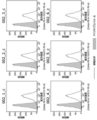
- FIG. 3 is a diagram showing the binding properties of the obtained chimeric antibodies 002_1_c to 002_6_c to human lung cancer cell line H322 cells expressing CD73.
- FIG. 4 is a diagram showing the AMP degradation inhibitory function of human lung cancer H322 cells for the 002_1_c antibody to 002_6_c antibody.
- FIG. 1 is a diagram showing the AMP degradation inhibitory function (H322 cells) of the 002_m antibody prepared in Example 1.
- FIG. 2 is a diagram showing amino acid sequences of the heavy chain variable region and the light chain variable region of the 002_m antibody.
- FIG. 3 is a diagram showing the binding properties of the obtained chimeric antibodies
- FIG. 5-1 is a diagram showing the amino acid sequence of the heavy chain variable region of the obtained humanized antibody (002_6_h1 to 002_6_h8) of 002_6_c.
- FIG. 5-2 is a diagram showing the amino acid sequence of the light chain variable region of the obtained humanized antibody (002_6_h1 to 002_6_h8) of 002_6_c.
- FIG. 6-1 is a diagram showing the binding property of humanized antibodies (002_6_h1 to 002_6_h8) to human lung cancer cell line H322 cells expressing CD73.
- FIG. 6-2 is a diagram showing the binding property of humanized antibodies (002_6_h1 to 002_6_h8) to human lung cancer cell line H322 cells expressing CD73.
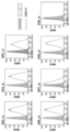
- FIG. 7 is a diagram showing the AMP degradation inhibitory function of three types of humanized antibodies (002_6_h2, 002_6_h4, 002_6_h8) using human lung cancer H322 cells.
- FIG. 8-1 is a diagram showing the binding property of the humanized antibody 002_6_h4 to various cells in comparison with the existing antibody and the base chimeric antibody 002_6_c.
- FIG. 8-2 is a diagram showing the binding property of the humanized antibody 002_6_h4 to various cells in comparison with the existing antibody and the base chimeric antibody 002_6_c.
- FIG. 8-1 is a diagram showing the binding property of the humanized antibody 002_6_h4 to various cells in comparison with the existing antibody and the base chimeric antibody 002_6_c.
- FIG. 9 is a diagram showing the AMP degradation inhibitory function of humanized antibody 002_6_h4 using human breast cancer MDA-MB-231 cells as compared with the existing antibody and the base chimeric antibody 002_6_c.
- FIG. 10 is a diagram showing the AMP degradation inhibitory function of 002_6_h4 on the recombinant CD73 antigen.
- FIG. 11 is a diagram showing the effect of 002_6_h4 on the division (proliferation) of human peripheral blood T cells.
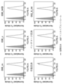
- FIG. 12 is a diagram showing the AMP degradation inhibitory function (H322 cells) of the 003_m antibody to 009_m antibody prepared in Example 1.
- FIG. 13 is a diagram showing the AMP degradation inhibitory function (MDA-MB-231 cells) of the four antibodies (004_m, 006_m, 007_m, 008_m) judged to have high functionality in FIG.
- FIG. 14-1 is a diagram showing the amino acid sequence of the heavy chain variable region of the obtained antibody (003_m antibody to 009_m antibody).
- FIG. 14-2 is a diagram showing the amino acid sequence of the light chain variable region of the obtained antibody (003_m antibody to 009_m antibody).
- FIG. 15 is a diagram showing the binding property of the 003_m antibody to 009_m antibody to the target expressing cells (H322 cells).
- FIG. 16 is a diagram showing the binding property of the 003_m antibody to 009_m antibody to the target expressing cells (MDA-MB-231 cells).
- FIG. 17 is a diagram showing the binding property of the 003_m antibody to 009_m antibody to the target expressing cells (CD73 forced expression HEK-293T cells).
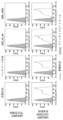
- FIG. 18 is a diagram showing the results of comparing the proportions of dividing cells when the antibody concentration was 1 nM.
- FIG. 19 is a diagram showing the binding activity of an antibody to cells expressing the human CD73 gene.
- FIG. 20-1 is a diagram showing the antibody binding activity to cells expressing the human CD73 gene.
- FIG. 20-2 is a diagram showing the antibody binding activity to cells expressing the human CD73 gene.
- FIG. 21 is a diagram showing the antibody binding activity to cells expressing cynomolgus monkey CD73.
- FIG. 22 is a diagram showing that the antibody of the present invention has an inhibitory activity against the AMP degradation action of CD73 on the cell surface.
- FIG. 23 is a diagram showing that the antibody of the present invention has a division-promoting effect on CD4T cells and CD8T cells, and has an effect of enhancing the production ability of IFN ⁇ , TNF, and IL-2 from these cells. be.
- FIG. 24 is a diagram showing a fluorescence microscope image showing the intracellular uptake of the 002_6_h4 antibody and the 006_hKB antibody.
- FIG. 25 is a diagram showing that the antibody of the present invention bound to CD73 on the surface of T cells decreases with time (internalized in T cells) when these cells are cultured.
- FIG. 26 is a diagram showing that the antibody of the present invention bound to CD73 on the surface of cancer cells decreases with time (internalized in cancer cells) when these cells are cultured.
- FIG. 27 is a diagram showing the results of measuring the number of tumor-infiltrating T cells in a mouse on day 35 after administration of 002_6_h4 antibody or isotype control antibody.
- FIG. 28-1 is a diagram showing whether the developed antibody of the present invention enhances the cancer cell injury activity by T cells.
- FIG. 28-1 is a diagram showing whether the developed antibody of the present invention enhances the cancer cell injury activity by T cells.
- FIG. 28-2 is a diagram showing whether the developed antibody of the present invention enhances the cancer cell injury activity by T cells.
- FIG. 29 is a diagram showing the results of animal experiments on whether 002_6_h4 can enhance the antitumor effect of the human immune system simulated in mice.
- FIG. 30 is a diagram showing the result of volume change of a tumor mass in an immunodeficient mouse body.
- FIG. 31 is a diagram confirming the antitumor effect of the anti-CD73 antibody of the present invention in a knock-in mouse expressing human CD73.
- the present invention in one embodiment, is a binding agent to a CD73 antigen, particularly an antibody, that activates T cells that are binding to the CD73 antigen on cancer cells or immune cells and are damaging to the cancer cells.
- a human antibody derivative can be provided.
- the binding agent of the present invention particularly an antibody or a human antibody derivative, can be used to shrink or eliminate a tumor because it inhibits the growth of cancer cells for the treatment of cancer cells expressing the CD73 antigen. It can also be used to activate T cells that are injurious to cancer cells when they are exhausted.
- the present invention provides an antibody or human antibody derivative that has binding to the CD73 antigen.
- This antibody or human antibody derivative is characterized by activating T cells that are damaging to cancer cells.
- the antibody or human antibody derivative of the present invention suppresses the function of CD73 expressed in cancer cells and causes exhaustion of immune cells (particularly T cells) caused by accumulation of AMP metabolite (adenosine) around the cancer cells. By releasing it, it activates immune cells and exerts tumor growth inhibitory effect and cancer therapeutic effect, suppresses the function of CD73 expressed in immune cells (particularly T cells), activates immune cells, and activates immune cells. It may be a tumor growth inhibitory effect and a cancer therapeutic effect, a cancer therapeutic effect by controlling the environment around the tumor other than immune cells, or a cancer therapeutic effect by direct control on the tumor.
- the antibody may be derived from any animal species of mammals, and the species of origin of the antibody is not limited to humans, and mice, rats, guinea pigs, hamsters, etc. It may be a rabbit or the like.
- the present invention may also be a human antibody derivative of the above-mentioned antibody as long as it has a binding property to the CD73 antigen and has a functional feature of suppressing cancer.
- a human antibody derivative in the present invention as one embodiment, the amino acid sequences of the CDRs at six positions of the above antibody (heavy chain complementarity determining regions (CDRs) 1 to CDR3 and light chain CDR1 to CDR3), and a human antibody. It is possible to provide a derivative of an antibody characterized by having the amino acid sequences of the constant regions of origin and the other amino acid sequences being a combination of the original antibody-derived amino acid sequence and the human antibody-derived amino acid sequence. can.
- a humanized antibody in which a region other than the complementarity determination region (CDR) of the above-mentioned "antibody” is replaced with an amino acid sequence derived from a human antibody, or a variable region of the above antibody is a constant of a human antibody.
- CDR complementarity determination region
- a variable region of the above antibody is a constant of a human antibody.
- chimeric antibodies such as those linked to a region, multivalent antibodies in which one type of antibody has multiple antigen binding sites, and multispecific antibodies (bispecific antibodies) in which one type of antibody has multiple specificities.
- bispecific antibodies multispecific antibodies
- the antibody or human antibody derivative of the present invention is characterized by having a total of 6 amino acid sequences of CDR1 to 3 of the same heavy chain and CDR1 to 3 of the light chain, which are the same as the antibody having binding property to the CD73 antigen. It is something that can be done.
- amino acid sequences of 6 CDRs of an antibody having binding to such a CD73 antigen include the following (1) to (8) :.
- CDR1 Heavy chain complementarity determining regions, CDR1 (DX 1 NMD (X 1 is C or S), SEQ ID No .: 1), CDR2 (DINPNNGGTIYNQKFKG, SEQ ID No .: 2), and CDR3 (TNWDYAMDY, SEQ ID No .: 3), and light chain complementarity determining regions, CDR1 (KASQDINSX 2 LS (X 2 is N, D or Q), SEQ ID No .: 4), CDR2 (RANRLID, SEQ ID No .:) 5), and CDR3 (X 3 QYDVFPRT (X 3 is L or Q), SEQ ID No .: 6); (2) Heavy chain complementarity determining regions, CDR1 (SFGMH, SEQ ID No .: 9), CDR2 (YISSGSRTIYYADTVRG, SEQ ID No .: 10), and CDR3 (DFGSSSPNYFDY, SEQ ID No .: 11), and light.
- CDR1 (RASESVDNYGISFMN, SEQ ID No .: 12), CDR2 (AASNQGS, SEQ ID No .: 13), and CDR3 (QQSKEVPWT, SEQ ID No .: 14); (3) Heavy chain complementarity determining regions, CDR1 (GYWMN, SEQ ID No .: 17), CDR2 (RIDPYDSETHYSQKFKD, SEQ ID No .: 18), and CDR3 (SSPITTAPFDY, SEQ ID No .: 19), and light Chain complementarity determining regions, CDR1 (RASESVDYYGFSFMN, SEQ ID No .: 20), CDR2 (AASTQGS, SEQ ID No .: 21), and CDR3 (QQSKEVPYT, SEQ ID No .: 22); (4) Heavy chain complementarity determining regions, CDR1 (SYGVS, SEQ ID No .: 25), CDR2 (VIWGDG
- the antibody or human antibody derivative of the present invention is also characterized by having the ability to activate T cells.
- T cells by suppressing the function of CD73 expressed in cancer cells or immune cells and releasing the exhaustion of immune cells (particularly T cells), immune cells are activated, and cancer growth inhibitory effect and cancer therapeutic effect are achieved. It is preferable to exert it.
- Such activation of T cells may occur in vitro or in vivo. Whether or not it has the ability to activate T cells that are damaging to such cancer cells is screened for whether or not they actually activate T cells from among the antibodies that have binding to the CD73 antigen. Can be obtained by doing.
- the exhaustion of immune cells refers to a state in which immune cells have become dysfunctional. Specifically, it causes a decrease in cytokine secretion ability and cytotoxicity in T cells, a decrease in cytokine secretion ability and cytotoxicity in NK cells, a decrease in antigen presentation ability of dendritic cells, activation of regulatory T cells, etc. be.
- an individual's cancer immunity to cancer cells does not function or functions poorly, and the activity to eliminate cancer cells decreases.
- Such exhaustion of immune cells is caused by the increase of checkpoint proteins such as PD-1 and CTLA-4 on the surface of T cells, and the cancer cells that continue to proliferate force the immune system to activate for a long period of time. It is known that it occurs in effector T cells, but it is a reversible state, and it is thought that it is possible to eliminate the exhausted state by activating the same cells.
- the activation of T cells can be grasped by using the proliferation of T cells, the increase in cytotoxicity of T cells against cancer cells, the secretion of cytokines from T cells, and the like as indicators. If these T cells are activated, it indicates that the antibody of the present invention or the human antibody derivative inhibits the activity of CD73 in T cells and suppresses the reaction of degrading AMP to adenosine. ..
- In vivo screening involves transplanting target cancer cells into an animal such as a wild-type mouse or a mouse in which immune cells are transplanted into an immunodeficient mouse such as a nude mouse or a SCID mouse, and then using the antibody of the present invention or a human.
- Infiltration of T cells around the tumor mass in the body by measuring the change in the size of the tumor mass in the body when the type antibody derivative is administered, or when the antibody of the present invention or the human type antibody derivative is administered. Can be done by anatomically examining.
- In vitro screening involves contacting peripheral blood T cells with the antibody or humanoid antibody derivative of the invention under culture conditions to measure T cell proliferation, T cell cytokine secretion, or This can be done by investigating whether T cells cause cell death against cancer cells.
- the antibody or human antibody derivative of the present invention can be specified as having the amino acid sequence of the following heavy chain variable region VH domain: (1) 002_m heavy chain variable region (SEQ ID No .: 7), (2) 003_m heavy chain variable region (SEQ ID No .: 15), (3) 004_m heavy chain variable region (SEQ ID No .: 23), (4) 005_m heavy chain variable region (SEQ ID No.
- the numbers (1) to (8) correspond to the numbers of the combination of the sequences of the heavy chain CDR1 to CDR3 described above.
- the amino acid sequence of SEQ ID NO: 7 is a sequence including the amino acid sequences of the heavy chains CDR1 to CDR3 specified by SEQ ID NO: 1 to SEQ ID NO: 3.
- the antibody or human antibody derivative of the present invention can be identified as having the amino acid sequence of the following light chain variable region VL domain: (1) 002_m light chain variable region (SEQ ID No .: 8), (2) 003_m light chain variable region (SEQ ID No .: 16), (3) 004_m light chain variable region (SEQ ID No .: 24), (4) 005_m light chain variable region (SEQ ID No. : 32), (5) 006_m light chain variable region (SEQ ID No .: 40), (6) 007_m light chain variable region (SEQ ID No .: 48), (7) 008_m light chain variable region (SEQ ID No.
- the numbers (1) to (8) correspond to the numbers of the combination of the sequences of the light chain CDR1 to CDR3 described above.
- the amino acid sequence of SEQ ID NO: 8 is a sequence including the amino acid sequences of light chain CDR1 to CDR3 specified by SEQ ID NO: 4 to SEQ ID NO: 6.
- the human antibody derivative of the present invention also includes the above-mentioned antibody or a functional fragment of the human antibody derivative.
- Functional fragments of the antibody or human antibody derivative in the present invention include F (ab') 2, Fab', Fab, single chain Fv (scFv) and the like.
- the functional fragment of the present invention may be any fragment thereof as long as it can induce injury to cancer cells, for example, F (ab') 2 fragment and the like. , Can be used as such a functional fragment.
- the antibody or human antibody derivative of the present invention is obtained by administering the CD73 antigen as an immunogen to an animal of the above-mentioned derived species and culturing antibody-producing cells collected from the animal body.
- a vector for protein expression containing a DNA sequence capable of defining the amino acid sequence of an antibody or human antibody derivative is designed, and the vector is introduced into cells for protein production and obtained recombinantly. You may.
- the DNA sequence capable of defining the amino acid sequence of the antibody or human antibody derivative of the present invention can be obtained from a cell producing the target antibody or human antibody derivative, depending on the animal species used in the expression system based on the amino acid sequence. It can be made by a method of designing based on optimized codons or a method of using a combination of these methods.
- the prepared DNA sequence is obtained by incorporating it into an expression vector suitable for a protein expression cell type (for example, CHO cell) in which the antibody or human antibody derivative is to be expressed and introducing it into the protein expression cell type. It can be obtained by using a method well known to those skilled in the art.
- a protein expression cell type for example, CHO cell
- introducing it into the protein expression cell type it can be obtained by using a method well known to those skilled in the art.
- a vector containing a DNA sequence defining a heavy chain amino acid sequence and a DNA defining a light chain amino acid sequence are used for a protein expression cell type.
- a method of introducing a vector containing a sequence and expressing both proteins in the cell to produce an antibody in the cell, or a DNA sequence defining a heavy chain amino acid sequence and a DNA sequence defining a light chain amino acid sequence It can be prepared by a method of introducing a vector containing both of them and expressing both proteins in the cells to produce an antibody in the cells.
- human antibody derivative obtained as one embodiment in the present invention
- a human antibody derivative is prepared from the above-mentioned antibody (1).
- Antibodies containing the 002_6_h2 heavy chain variable region (SEQ ID NO: 67) and the 002_6_h2 light chain variable region SEQ ID NO: 68).
- the antibody or human-type antibody derivative of the present invention is based on the above-mentioned characteristic of having the ability to activate immune cells (particularly T cells) having cytotoxicity against cancer cells.
- a pharmaceutical composition comprising an antibody of the invention or a human antibody derivative that activates immune cells (particularly T cells) that are damaging to cancer cells in a subject in need of treatment or prevention of cancer. Goods, can be provided.
- Cancer cells that can be targeted in the present invention include, for example, leukemia (including chronic lymphocytic leukemia and acute lymphocytic leukemia), lymphoma (non-hodgkin lymphoma, hodgkin lymphoma, T-cell line lymphoma, B-cell line lymphoma, etc.).
- leukemia including chronic lymphocytic leukemia and acute lymphocytic leukemia
- lymphoma non-hodgkin lymphoma, hodgkin lymphoma, T-cell line lymphoma, B-cell line lymphoma, etc.
- Berkit lymphoma malignant lymphoma, diffuse lymphoma, follicular lymphoma), myeloma (including multiple myeloma), melanoma, lung cancer, breast cancer, colon cancer, kidney cancer, stomach cancer, ovarian cancer, pancreas Cancer, cervical cancer, uterine body cancer, endometrial cancer, esophageal cancer, liver cancer, head and neck cancer, head and neck squamous epithelial cancer, skin cancer, urinary tract cancer, prostate Lymphoma, pharyngeal cancer, laryngeal cancer, lymphoma, male embryoma, endometrial hyperplasia, endometriosis, germoma, fibrosarcoma, caposic sarcoma, hemangiomas, spongy hemangiomas, Hemangioblastoma, retinal blastoma, stellate cell tumor, neurofibromas, rare projectile cystoma, medullary blastom
- the antibody or human antibody derivative of the present invention is characterized by activating immune cells (particularly T cells) having cytotoxicity against target cancer cells as described above. That is, when administered to a living body, activation of immune cells (particularly T cells) occurs only in immune cells having cytotoxicity against cancer cells expressing the target CD73 antigen, and expresses the CD73 antigen. However, in the case of normal cells, it is required not to induce cytotoxicity that may cause clinical problems.
- the antibody or human antibody derivative of the present invention can also be provided as a composition in combination with other antibodies or other agents such as anticancer agents.
- the antibody or human antibody derivative of the present invention can also be bound to a drug to form an antibody drug conjugate (ADC).
- ADC antibody drug conjugate
- a preparation containing the antibody or human type antibody derivative of the present invention together with a physiologically acceptable diluent or carrier.
- suitable carriers include buffers (phosphate buffer, citric acid buffer, acetate buffer, etc.), salts (sodium chloride, etc.), sugars (glucose, trehalose, mannitol, sorbitol, etc.), additives (arginine, etc.). Amino acids, surfactants such as polysorbates, etc.), but are not limited to these.
- the antibody or human antibody derivative of the present invention can be freeze-dried and reconstituted and used by adding a buffered aqueous solution as described above when necessary.
- the preparation containing the antibody of the present invention or the human antibody derivative can be administered in various dosage forms, and can be, for example, a parenteral administration agent such as an injection or an infusion.
- the dose of the antibody or humanoid antibody derivative of the present invention varies depending on symptoms, age, body weight, etc., but usually, in parenteral administration, 0.01 mg to 1000 mg per kg body weight, preferably 0.05 per kg body weight per day.
- Intraperitoneal injection subcutaneous injection in the range of mg to 500 mg, 0.05 mg to 100 mg, 0.05 mg to 50 mg, 0.05 mg to 20 mg, more preferably 0.1 mg to 10 mg per kg of body weight per day.
- Intramuscular injection, intratumor injection, or intravenous injection which can be administered by the appropriate route of administration depending on the type of cancer.
- the present invention requires treatment or prevention of cancer in another embodiment.
- a method of treating or preventing cancer in a subject comprising administering to the subject an effective amount of an antibody or humanoid antibody derivative of the invention.
- Treatment or prevention of cancer with the antibody or human antibody derivative of the present invention is that the antibody or human antibody derivative activates immune cells (particularly T cells) having cytotoxicity against cancer cells in the body. Caused by.
- an adjuvant for example, Clin. Microbiol
- Particles that can be administered with (such as those described in Rev., 7: 277-289, 1994), liposome formulations, particulate formulations bound to beads with a diameter of several ⁇ m, lipid-bound formulations, etc. It can also be administered in the form of a formulation.
- the antibodies or human-type antibody derivatives of the present invention are used to detect the CD73 antigen in a sample due to the characteristic of having binding property to the CD73 antigen.
- the antibody or human antibody derivative of the present invention can be used for a sample containing cancer cells or immune cells (for example, T cells) collected from a subject, such as an immunoprecipitation method, an aggregation reaction, and a magnetic bead method.
- a sample containing cancer cells or immune cells (for example, T cells) collected from a subject such as an immunoprecipitation method, an aggregation reaction, and a magnetic bead method.
- the antibody or human antibody derivative of the present invention is also based on the feature that it has a binding property to the CD73 antigen and activates immune cells (particularly T cells) having damage to cancer cells.
- a method for measuring the cytotoxicity to cancer cells in such a subject the following steps: A step of contacting a cancer cell collected from a subject with an antibody of the present invention or a human antibody derivative under culture conditions (that is, in vitro).
- the step of measuring whether the AMP metabolism of cancer cells (the reaction that decomposes AMP into adenosine) is reduced, the step of measuring whether the cell viability is reduced, or the secretion of immunostimulatory substances. Steps to measure whether or not
- a method for measuring cytotoxicity against cancer cells using the antibody of the present invention or a human antibody derivative including the above can be mentioned.
- AMP metabolism reaction that decomposes AMP to adenosine
- T cells are activated and tumors shrink, and cancer against cancer cells is known. It is believed that immunity is activated.
- cancer cells collected from a subject are brought into contact with the antibody of the present invention or a human antibody derivative in vitro, and the cancer cells are subjected to culture conditions. By measuring whether or not AMP metabolism is reduced, cytotoxicity to cancer cells can be measured.
- cancer cells collected from a subject are brought into contact with the antibody of the present invention or a human antibody derivative, and the degradation reaction from AMP to adenosine is measured under culture conditions (that is, in vitro). Therefore, when the antibody of the present invention or the human antibody derivative is administered to the subject, the cytotoxicity to the cancer cells in the subject (in vivo) can be measured.
- the decomposition reaction from AMP to adenosine can be measured by HPLC method, LC-MS method, ELISA method, or AMP-dependent chemiluminescence method (Cell titer glo, etc.).
- peripheral blood lymphocytes cause a decrease in the cell survival rate of cancer cells.
- cancer cells collected from a subject are brought into contact with the antibody of the present invention or a human antibody derivative in vitro, and the cell viability is increased under culture conditions.
- the cytotoxicity to cancer cells can be measured. More specifically, cancer cells collected from a subject are brought into contact with the antibody or human antibody derivative of the present invention in the presence of peripheral blood lymphocytes of the same subject under culture conditions (ie, in. Cancer in vivo when the antibody or human antibody derivative of the present invention is administered to the subject by measuring whether the cell viability of the cancer cells is reduced in vivo). The cytotoxicity to cells can be measured.
- cancer cells are activated and tumors shrink when the secretion of immunostimulatory substances from peripheral blood lymphocytes is enhanced in the living body, and cancer immunity against cancer cells is known. Is believed to be activated.
- cancer cells collected from a subject are brought into contact with the antibody of the present invention or a human antibody derivative in vitro, and under culture conditions, an immunostimulatory substance. Cytotoxicity to cancer cells can be measured by measuring whether or not the secretion of the antibody is enhanced. More specifically, cancer cells collected from a subject are brought into contact with the antibody of the present invention or a human antibody derivative in the presence of peripheral blood lymphocytes of the same subject under culture conditions (that is, in vitro).
- IFN- ⁇ , TNF ⁇ and the like can be mentioned as examples as the immunostimulatory substance to be measured for determining the cytotoxicity to cancer cells.
- IFN ⁇ is known to activate immune cells such as NK cells and exert an antitumor effect.
- the invention also comprises AMP metabolism, cytotoxicity, secretion of immunostimulatory substances, or activation of immune cells to cancer cells harvested from a subject, including the antibodies of the invention or humanoid antibody derivatives. Measurement kits for measurement in vitro can also be provided.
- the measurement kit for measuring AMP metabolism includes a reagent for measuring AMP as a substrate and adenosine, which is a metabolite thereof (AMP-dependent chemiluminescent reagent). , ELISA reagents) and equipment (HPLC system, LC-MS system).
- the measurement kit for measuring cytotoxicity includes known means for measuring cell proliferation (for example, uptake of thymidin, uptake of BrdU, free release). Measurement of lactate dehydrogenase (LDH) activity, measurement of living cell-derived substances (reducing enzyme activity, esterase activity, ATP, etc.) can be included.
- LDH lactate dehydrogenase
- the measurement kit for measuring the secretion of the immunostimulatory substance includes means for detecting the immunostimulatory substance to be measured (for example, primary for the immunostimulatory substance).
- Antibodies and secondary antibodies for the detection of the primary antibody, etc. can be included.
- the measurement kit for measuring the activation of immune cells can include, in addition to the antibody or human antibody derivative of the present invention, a labeling reagent for measuring the division of immune cells with a flow cytometer.
- Example 1 Preparation of antibody The purpose of this example was to obtain a monoclonal antibody against the CD73 antigen using mice.
- a single cell suspension of spleen cells was prepared from the spleen according to a known method, and B cells were purified using a MACS-pan B cell isolation kit (Miltenny 130-104-443). Purified B cells were stained with ZOMBIE-APC-Cy7 (Biolegend), Alexa488anti-IgM (Biolegend), VB421anti-IgG (Biolegend), PE-CD73 (self-prepared product) and tested on FACSAria (BD). , IgG-positive and CD73 antigen-binding cell populations were separated into 96-well plates one by one.
- the genes corresponding to the heavy chain variable region and the light chain variable region of the antibody were amplified for each well by the RT-PCR method.
- As the primer used for amplification based on the information in the publicly known literature, a sequence containing the 5'end and 3'end regions of the heavy chain variable region and the light chain variable region, respectively, was adopted, and the diversity of the antibody gene sequence was taken into consideration. (Heavy chain: H1 mix and H2 mix, light chain: k mix) (Lotta von Boehmer et al., Nature Protocols, vol 11, p.1908-1923 (2016)) ..
- a linker sequence for insertion into the antibody expression vector is added to the end of each gene by a PCR reaction, and a vector for heavy chain expression or a vector for light chain expression (vector for heavy chain expression or light chain expression).
- a CMV promoter, a secretory signal, a gene for a heavy chain variable region or a light chain variable region, and a gene for a heavy chain constant region or a light chain constant region were tandemly linked to each other) by a ligation reaction.
- the heavy chain gene expression plasmid (derived from H1 or H2 mixed amplification product) and the light chain gene expression plasmid (derived from k mix amplification product) are transferred to CHO cells. I did an expression. Antibodies produced in the culture supernatant were collected from CHO cells, and the antigen specificity of each antibody was confirmed by antigen-fixed ELISA.
- ATP Sigma, A2383, final concentration 100 ⁇ M
- CellTiterGlo Promega, G9243
- chemiluminescence was measured using a microplate reader. That is, using the principle that AMP inhibits chemiluminescence due to the reaction between ATP and CellTiterGlo, the enzyme activity inhibitory effect of the antibody was evaluated from the amount of residual AMP.
- an antibody having the same sequence as the published variable region of MEDI9447 was produced using the gene recombination technology based on the sequence published in the advanced development product MEDI9447 (Special Table 2018-501197). "Benchmark antibody”) was adopted.
- mouse antibodies As a result of antibody function screening, eight types of mouse antibodies named 002_m, 003_m, 004_m, 005_m, 006_m, 007_m, 008_m, 009_m could be selected as CD73-binding activity-positive antibodies.
- Example 2 Antibody function evaluation (concentration-dependent evaluation) The purpose of this example was to evaluate the function of the 002_m antibody obtained in Example 1.
- the antibody function evaluation was carried out by the same method as the antibody function screening described in Example 1 (1-3). That is, human lung cancer cell line H322 cells or human breast cancer cell line MDA-MB-231 cells (ATCC® HTB-26 TM ) were used as cells.
- the antibody concentration dependence of the AMP decomposition inhibitory function was analyzed by measuring the AMP decomposition reaction in the same manner as in Example 1 (1-3), and the IC50 value (50% inhibition concentration: CD73 in this case). The concentration of the antibody that can inhibit 50% of the enzyme function of the antibody) was calculated, and the function of the antibody was quantitatively evaluated.
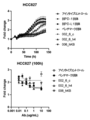
- Figure 1 shows the AMP degradation inhibitory function (H322 cells) of the 002_m antibody prepared in Example 1. Although this antibody is a mouse antibody, it was revealed that it shows an IC50 value close to that of the benchmark antibody (see Table 1).
- Example 3 Antibody gene sequence analysis This example was performed for the purpose of determining the amino acid sequence of the 002_m antibody obtained in Example 1.
- the obtained plasmid DNA was analyzed for the gene sequences defining the heavy chain variable region and the light chain variable region by the Sanger sequence, and the amino acid sequence was determined based on the same sequence.
- the amino acid sequences of the heavy chain variable region and the light chain variable region of the 002_m antibody are shown in FIG. 2 (SEQ ID NO: 7 and SEQ ID NO: 8 respectively). Further, in this figure, the heavy chains CDR1, CDR2, CDR3 (SEQ ID NO: 1, SEQ ID NO: 2, SEQ ID NO: 3, respectively) and the light chains CDR1, CDR2, CDR3 (SEQ ID NO, respectively). : 4, SEQ ID NO: 5, SEQ ID NO: 6) are underlined.
- Example 4 Preparation of human-mouse human chimeric antibody The purpose of this example is to prepare a chimeric antibody having a variable region of a mouse antibody and a constant region of a human antibody for the 002_m antibody sequenced in Example 3. rice field.
- the gene sequence of the variable region of the mouse antibody was artificially synthesized and incorporated into a vector for expressing a human constant region (Fc) chimeric recombinant antibody.
- a light chain expression element EF-1 ⁇ promoter, a secretory signal, a DNA sequence defining a light chain variable region, and a DNA sequence defining a light chain constant region are linked in tandem
- a heavy chain expression element EF
- a plasmid having a -1 ⁇ promoter, a secretory signal, a DNA sequence defining a heavy chain variable region, and a DNA sequence defining a heavy chain constant region was tandemly linked) was constructed.
- the plasmid was transfected into ExpiCHO cells (Thermo) to secrete the antibody into the medium, and then the antibody was purified using a Protein A column (GL Science).
- the chimeric antibody thus prepared was defined as 002_1_c antibody.
- the amino acid sequence of the heavy chain variable region of this antibody is composed of the amino acid sequence of SEQ ID NO: 7
- the amino acid sequence of the light chain variable region is composed of the amino acid sequence of SEQ ID NO: 8.
- Example 5 Preparation of CDR-modified antibody
- a modified antibody is prepared by substituting a residue that may undergo post-translational modification in CDR with an unmodified residue for the 002_m antibody. I went there for the purpose.
- the 002_m antibody heavy chain variable region (SEQ ID NO: 7) 32nd amino acid cysteine (C) is used as serine (S), and the 002_m antibody light chain variable region (SEQ ID NO: 8) 32nd amino acid asparagine (SEQ ID NO: 8) N) was replaced with aspartic acid (D) or glutamine (Q), respectively (see Table 2 below).
- a gene containing the relevant region was artificially synthesized and inserted into a human Fc chimeric recombinant antibody expression vector by ligation. Antibody expression and purification were carried out in the same manner as for human chimeric antibodies.
- Binding analysis of each of the obtained chimeric antibodies 002_1_c to 002_6_c to target-expressing cells was performed by FACS analysis using CD73 native-expressing cells (human lung cancer cell line H322). After reacting the test antibody or the isotype control antibody with the cells, the antibody was stained with a fluorescently labeled secondary antibody and subjected to flow cytometric analysis, and the antibody binding property was evaluated based on the obtained fluorescence intensity.
- Example 6 Preparation of humanized antibody
- a humanized antibody was prepared based on the 002_6_c antibody (SEQ ID NO: 7 (C32S) / SEQ ID NO: 8 (N32Q)) selected in Example 5. I went for the purpose of doing.
- the humanized antibody was prepared using the public software Tabhu (http://circe.med.uniroma1.it/tabhu/). Using the same software, we extracted from a human antibody database that has sequences that are similar in sequence and conformation to 002_6_c, picked up 4 types of sequences (FJ039788, AF146404, 3SQO_H, AY686911) as candidate sequences, and selected these. The sequences other than the CDR regions of the antibody and the sequences of the CDR regions of 002_6_c were graphed.
- amino acid residues to be backmutated were selected up to 10 amino acid residues in each sequence based on the large structural difference between the humanized antibody and the mouse antibody (higher TubHu score).
- the method described in the non-patent document Bioinformatics. 2015 Feb 1; 31 (3): 434-5 was followed.
- Fig. 5 The amino acid sequences of the variable regions of the obtained 002_6_c humanized antibodies (002_6_h1 to 002_6_h8) are shown in Fig. 5 (heavy chain variable region is shown in Fig. 5-1 and light chain variable region is shown in Fig. 5-2). Each amino acid sequence was assigned SEQ ID NO: as shown in Table 4 below.
- Binding analysis of each of the obtained humanized antibodies (002_6_h1 to 002_6_h8) to target-expressing cells was performed by FACS analysis using CD73 native-expressing cells (human lung cancer cell line H322). The results are shown in FIG. It was confirmed that all humanized antibodies showed the same binding properties as the base chimeric antibody 002_6_c antibody.
- the humanized antibody 002_6_h4 selected as the lead antibody above was compared with existing antibodies (commercially available anti-CD73 antibody (biolegend), benchmark antibody) and the base chimeric antibody 002_6_c, and the binding property to various cells was measured by flow cytometry. confirmed.
- CD73 native-expressing cells human breast cancer cell line MDA-MB-231
- human breast cancer cell line MDA-MB-231 cells expressing CD73 antigen
- HEK293T wild type not expressing CD73 antigen FACS analysis was performed using cells (derived from human fetal kidney cells) and cells in which CD73 antigen was forcibly expressed in HEK293T cells.
- CD73 forced expression cells were generated using the lentiviral vector system (Origene).
- Lentivirus particles carrying the human CD73 gene (NM_002526) and puromycin resistance gene are prepared, infected with HEK293T cells, and then the cells are cultured in the presence of puromycin, and the target gene is integrated into the genome to be CD73 positive.
- HEK293T cells were collected and used for binding analysis. After reacting the test antibody or the isotype control antibody with the cells, the antibody was stained with a fluorescently labeled secondary antibody and subjected to flow cytometric analysis, and the antibody binding property was evaluated based on the obtained fluorescence intensity.
- the humanized antibody 002_6_h4 was compared with the existing antibody (commercially available anti-CD73 antibody (biolegend), benchmark antibody) and the base chimeric antibody 002_6_c, and the same method as in (1-3) was used for human breast cancer MDA-MB-.
- An antibody function screening was performed to measure the AMP degradation inhibitory function using 231 cells. The results are shown in FIG. From this result, the IC50 value of the humanized antibody against the AMP degradation inhibitory function using human breast cancer MDA-MB-231 cells was calculated as shown in Table 6 below. Together with the results, it was shown that the humanized antibody 002_6_h4 has an AMP degradation inhibitory function in multiple cancer types, similar to the chimeric antibody 002_6_c.
- Example 7 Suppressive function against recombinant antigen The purpose of this example was to confirm the enzymatic reaction inhibitory function of the anti-CD73 antibody obtained in Example 6 against the recombinant CD73 antigen.
- the CD73 antigen (terminal His-Tag, self-prepared product) was immobilized on a 96-well plate for ELISA via an anti-His-Tag antibody, and the humanized anti-CD73 antibody 002_6_h4 and the substrate AMP (final concentration 100 ⁇ M) were used.
- the humanized anti-CD73 antibody 002_6_h4 and the substrate AMP (final concentration 100 ⁇ M) were used.
- ATP final concentration 100 ⁇ M
- CellTiterGlo Promega, G9243
- chemiluminescence was measured using a microplate reader.
- AMP enzyme activity inhibitory effect of the antibody was evaluated from the amount of residual AMP.
- the same experiment was performed using a benchmark antibody as a positive control.
- Figure 10 shows the AMP degradation inhibitory function of 002_6_h4 for the recombinant CD73 antigen. As a result, it was suggested that 002_6_h4 showed an AMP degradation inhibitory function against the recombinant CD73 antigen and directly inhibited the enzyme.
- Example 8 Evaluation of effect on T cell division This example was performed for the purpose of confirming the proliferation promoting activity of the anti-CD73 antibody obtained in Example 6 on human peripheral blood T cells.
- PBMC peripheral blood mononuclear cells
- CD4 + T Cell Isolation Kit human (Miltenyi Biotech, 130-096-533) was used to purify CD4 positive T cells, and CellTrace CFSE cell proliferation kit (ThermoFisher Scientific, #) Cells were fluorescently labeled with C34554).
- Figure 11 shows the effect of 002_6_h4 on the division (proliferation) of human peripheral blood T cells.
- the proportion of divided T cells increased in an antibody concentration-dependent manner, indicating that the anti-CD73 antibody had the effect of promoting T cell division.
- Example 9 Evaluation of antibody function (concentration-dependent evaluation) The purpose of this example is to evaluate the functions of antibodies other than the 002_m antibody obtained in Example 1 (that is, 003_m antibody, 004_m antibody, 005_m antibody, 006_m antibody, 007_m antibody, 008_m antibody, 009_m antibody). went.
- the antibody function evaluation was carried out by the same method as the antibody function screening described in Example 1 (1-3). That is, human lung cancer cell line H322 cells or human breast cancer cell line MDA-MB-231 cells (ATCC® HTB-26 TM ) were used as cells.
- the antibody concentration dependence of the AMP degradation inhibitory function was analyzed by measuring the AMP degradation reaction in the same manner as in Example 1, and the IC50 value (50% inhibitory concentration: 50 of the enzyme function of CD73 here). %) was calculated and the function of the antibody was quantitatively evaluated.
- the AMP degradation inhibitory function of the 003_m antibody, 004_m antibody, 005_m antibody, 006_m antibody, 007_m antibody, 008_m antibody, and 009_m antibody prepared in Example 1 against H322 cells is shown in FIG. 12, and the AMP degradation inhibitory function against MDA-MB-231 cells. Is shown in FIG. 13, respectively. Some of these antibodies, which are mouse antibodies, were found to show IC50 values that were lower or closer to the benchmark antibodies (see Table 7).
- clones judged to have high functionality were selected from the tests shown in FIGS. 12 and 13 above (004_m, 006_m, 007_m, 008_m).
- the AMP degradation inhibitory function (MDA-MB-231 cells) of these four antibodies is shown.
- the IC50 values of these clones were good compared to the benchmark antibody (under clinical development) and were about the same as 002_6_c (see Table 8).
- Example 10 Antibody gene sequence analysis The purpose of this example was to determine the amino acid sequences of the 003_m antibody to 009_m antibody obtained in Example 1.
- the obtained plasmid DNA was analyzed for the gene sequences defining the heavy chain variable region and the light chain variable region by the Sanger sequence, and the amino acid sequence was determined based on the same sequence.
- the heavy chain variable region amino acid sequence (Fig. 14-1) and light chain variable region amino acid sequence (Fig. 14-2) of 003_m antibody, 004_m antibody, 005_m antibody, 006_m antibody, 007_m antibody, 008_m antibody, 009_m antibody are shown.
- SEQ ID NOs were assigned to each amino acid sequence as shown in Table 9 below.
- Example 11 Analysis of binding to target-expressing cells The purpose of this example was to evaluate the binding of each antibody of 003_m to 009_m obtained in Example 1.
- Binding analysis to target-expressing cells was performed by FACS analysis using CD73 native-expressing cells (human lung cancer cell line H322 or human breast cancer cell line MDA-MB-231) or CD73 forced-expressing cells.
- CD73 forced expression cells were generated using the lentiviral vector system (Origene). After preparing lentivirus particles carrying the human CD73 gene (NM_002526) and puromycin resistance gene and infecting HEK293T cells, the cells were cultured in the presence of puromycin, and the target gene was integrated into the genome to be CD73 positive. The resulting HEK293T cells were collected and used for binding analysis. As a negative control, HEK293T cells expressing CD73 were used.
- the antibody After reacting the test antibody or isotype control antibody with the cells, the antibody was stained with a fluorescently labeled secondary antibody and subjected to flow cytometric analysis, and the antibody binding property was evaluated based on the obtained fluorescence intensity.
- FIG. 15 The binding of 003_m antibody to 009_m antibody to target-expressing cells (H322 cells) is shown in Fig. 15, and the binding of 003_m antibody to 009_m antibody to target-expressing cells (MDA-MB-231 cells) is shown in Fig. 16 and 003_m.
- the binding properties of the antibody to 009_m antibody to the target-expressing cells are shown in FIG. 17, respectively.
- solid lines and gray fills show fluorescence intensity histograms of the test antibody and the isotype control antibody, respectively.
- Example 12 Evaluation of effect on T cell division The purpose of this example was to confirm the proliferation-promoting activity of the anti-CD73 antibody obtained in Example 9 on human peripheral blood T cells.
- Example 8 The same method as in Example 8 was adopted, and the ratio of dividing cells was compared when the antibody concentration was 1 nM. The results are shown in FIG. The dotted line shows the proportion of dividing cells in the isotype control antibody.
- CD4 + T Cell Isolation Kit human (Miltenyi Biotech, 130-096-533) is used to purify CD4-positive T cells from peripheral blood mononuclear cells (PBMC) derived from healthy volunteers, and CellTrace CFSE cell proliferation kit. Cells were fluorescently labeled with (ThermoFisher Scientific, # C34554).
- Figure 18 shows the effect of 1 nM anti-CD73 antibody (006_m antibody, 007_m antibody, or 008_m antibody) on the division (proliferation) of human peripheral blood T cells.
- “w / o AMP” and “with AMP” (100 ⁇ M AMP) were incorporated to see the effect of the presence or absence of AMP without antibody because AMP has a T cell division inhibitory effect. ..
- the proportion of divided T cells increased in an antibody concentration-dependent manner (data shows). (Not shown), the effect of anti-CD73 antibody on promoting T cell division was shown.
- Example 13 Preparation of humanized antibody (2)
- a humanized antibody is prepared based on the 004_m antibody, 006_m antibody, 007_m antibody, and 008_m antibody selected in Example 9 among the respective antibodies of 003_m to 009_m obtained in Example 1. I went for the purpose.
- the humanized antibody was prepared using the public software Tabhu (http://circe.med.uniroma1.it/tabhu/) as in Example 6. Using this software, 004_m antibody, 007_m antibody, 008_m antibody and 004_m antibody are extracted from the human antibody database having sequences having characteristics similar in sequence and three-dimensional structure, 004_m antibody is 5 types, 007_m antibody is 4 types, and 008_m antibody is. Each of the four sequences was picked up as a candidate sequence, and the sequences other than the CDR region of these antibodies were graphed with the sequences of the CDR regions of each of the 004_m antibody, 007_m antibody, and 008_m antibody.
- Table 10 below shows the amino acid sequences of the variable regions of the humanized antibodies (004_h3 antibody, 006_hKB antibody, 007_h1 antibody, 008_h4 antibody) obtained based on the 004_m antibody, 006_m antibody, 007_m antibody, and 008_m antibody, respectively. As shown in Table 10 below, each amino acid sequence was assigned SEQ ID NO: 81 to SEQ ID NO: 88.
- Binding analysis to human target-expressing cells was performed by FACS analysis using CD73 native-expressing cells (human lung cancer cell line H322). Binding analysis to target-expressing cells was performed on CD73 native-expressing cells (human breast cancer cell line MDA-MB-231), human breast cancer cell line MDA-MB-231 cells expressing CD73 antigen, and HEK293T wild type not expressing CD73 antigen. FACS analysis was performed using cells (derived from human fetal kidney cells) and cells in which CD73 antigen was forcibly expressed in HEK293T cells. CD73 forced expression cells were generated using the lentiviral vector system (Origene).
- Lentivirus particles carrying the human CD73 gene (NM_002526), the red-tailed monkey CD73 gene (XM_001086989.2) (the amino acid sequence is the same as that of the crab monkey CD73) and the puromycin resistance gene were prepared, infected with HEK293T cells, and then puromycin.
- the cells were cultured in the presence of HEK293T cells in which the target gene was integrated into the genome and became CD73 positive, and used for binding analysis. After reacting the test antibody or the isotype control antibody with the cells, the antibody was stained with a fluorescently labeled secondary antibody and subjected to flow cytometric analysis, and the antibody binding property was evaluated based on the obtained fluorescence intensity.
- FIGS. 19 to 21 The results are shown in FIGS. 19 to 21.
- FIGS. 19 and 20 are diagrams confirming the binding of the antibody to cells expressing the human CD73 gene
- FIG. 21 confirms the binding of the antibody to cells expressing the rhesus monkey CD73 gene. It is a figure.
- the 004_h3 antibody, 006_hKB antibody, and 008_h4 antibody were confirmed to show the same binding property to the human CD73 antigen as the 002_6_h4 antibody when compared, and the binding property to the CD73 antigen could be confirmed.
- rice field the humanized antibodies examined.
- the 006_hKB antibody among them showed the same binding property to the rhesus monkey CD73 antigen (cynomolgus monkey CD73 antigen) as the 002_6_h4 antibody, and the effect of the antibody of the present invention was confirmed by animal experiments in the future. It was shown that rhesus monkeys or cynomolgus monkeys can be used for examination.
- Example 14 Suppressive function against recombinant antigen The purpose of this example was to confirm the enzymatic reaction inhibitory function of the humanized anti-CD73 antibody obtained in Example 13 against the recombinant CD73 antigen.
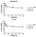
- Example 1 For antibody function evaluation, 004_h3 antibody, 006_hKB antibody, 007_h1 antibody, 008_h4 antibody were used as test antibodies, and 002_6_c antibody, 002_6_h4 antibody, isotype control antibody and benchmark antibody (MEDI9447 antibody) were used as controls, and Example 1 (1-). It was carried out by the same method as the antibody function screening described in 3). That is, human breast cancer cell line MDA-MB-231 cells (ATCC® HTB-26 TM ) were seeded on 96-well plates and incubated overnight at 37 ° C.
- human breast cancer cell line MDA-MB-231 cells ATCC® HTB-26 TM
- Example 1 As in Example 1 (1-3), AMP (Sigma, A1752), which is a substrate of CD73, was added to a final concentration of 200 ⁇ M, and the mixture was incubated at 37 ° C. for 4.5 hours to AMP with CD73.
- AMP Sigma, A1752
- the antibody concentration dependence of the AMP degradation inhibitory function was analyzed. A decomposition reaction was carried out.
- ATP Sigma, A2383, final concentration 10 ⁇ M
- Cell TiterGlo Promega, G9243
- Figure 22 shows the AMP degradation inhibitory function of the 004_h3 antibody, 006_hKB antibody, 007_h1 antibody, and 008_h4 antibody. All of these antibodies have been shown to have the function of suppressing AMP degradation by CD73 present on the cell surface (Figs. 22 and 23), and among these antibodies, the 006_hKB antibody is similar to 002_6_h4. , It was shown to have an inhibitory activity equal to or higher than that of the benchmark antibody.
- Example 15 Evaluation of effects on T cell division This example confirms the growth-promoting activity of the anti-CD73 antibody obtained in Example 13 and showing a particularly high effect in Example 14 on human peripheral blood T cells. I went for that purpose.
- PBMC peripheral blood mononuclear cells
- T cell marker staining antibody BioLegend, anti-CD3: UCHT1, anti-CD4: RPA-T4, anti-CD8a: HIT8a. Beads for cell count were added to the stained cells and analyzed with a flow cytometer to calculate the number of CD4 and CD8 positive cells per well.
- IL-2 BAF202 (R & D Systems)
- TNF ⁇ BAF210 (R & D Systems)
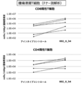
- Figure 23 shows the results of the evaluation of the effect of the antibody on T cell division.
- the 006_hKB antibody is as high as the 002_6_h4 antibody for both CD4-positive and CD8-positive T cells. It was shown to have a T cell division promoting effect (upper part of FIG. 23). It was also confirmed that high concentrations of IFN ⁇ , TNF ⁇ , and IL-2 were released into the culture supernatant from the cells to which the 006_hKB antibody was administered (Fig. 23, lower part).
- Example 16 Evaluation of intracellular uptake of antibody
- the anti-CD73 antibody obtained in Example 6 and the anti-CD73 antibody obtained in Example 13 and showing a particularly high effect in Example 14 were used. The purpose was to evaluate the uptake into cells.
- Human breast cancer cells MDA-MB-231 (1 ⁇ 10 ⁇ 5 cells) are seeded on a chamber slide (Thermo Fisher Scientific, 154534PK), and 10 ⁇ g / ml of various antibody solutions (002_6_h4, 006_hKB, MEDI9447) are added to 37 ° C. Incubated for 15 minutes or 19 hours. Samples before and after incubation were fixed and permeabilized (Thermo Fisher Scientific, FIX & PERM Fixation and Permeabilization Kit, GAS003) and stained with Anti-human IgG Alexa Fluor 594 (Thermo Fisher Scientific, A-11014). The distribution of the antibody was observed with a fluorescence microscope (Thermo Fisher Scientific, EVOS FLoid Imaging System, 4471136) to determine the presence or absence of the antibody uptake into the cells.
- a fluorescence microscope Thermo Fisher Scientific, EVOS FLoid Imaging System, 4471136
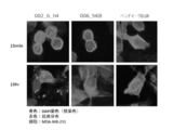
- Figure 24 shows the intracellular uptake of 002_6_h4 and 006_hKB.
- the antibody was distributed on the cell surface at the time of 15 minutes of incubation (upper part of Fig. 24), whereas the antibody was accumulated in the cell 19 hours after incubation (lower part of Fig. 24). Was shown to have taken up into the cells. This result suggests that the uptake of anti-CD73 antibody may induce the disappearance of the antigen from the cell surface.
- PBMC Peripheral blood mononuclear cells
- % Control (anti-CD73 antibody (X minutes) MFI-isotype control (X minutes) MFI) / (anti-CD73 antibody (0 minutes) MFI-isotype control (0 minutes) MFI) x 100
- % CD73 Ag (anti-CD73 antibody (X minutes) MFI-isotype control (X minutes) MFI) / (anti-CD73 antibody (0 minutes) MFI-isotype control (0 minutes) MFI) x 100
- Example 17 Measurement of KD value
- the anti-CD73 antibody obtained in Example 6 and the anti-CD73 antibody obtained in Example 13 and showing a particularly high effect in Example 14 bind to the CD73 antigen.
- the purpose was to evaluate the dissociation constant (KD value), which is an index of affinity.
- the KD value of the antibody bound to the target antigen was measured with Biacore 8K (GE Healthcare). Each antibody of 002_6_h4 antibody and 006_hKB antibody was immobilized on the sensor chip (CM5) using Human Antibody Capture Kit Type2 (Cytiva, 26-2346-00). A solution of human CD73 antigen diluted serially from 5 ⁇ 10 -9 M in 4 steps was prepared and used for multicycle kinetic analysis. In the 1: 1 coupling model, the KD values are as shown in Table 11 below.
- Example 18 Human Immune Mouse Test This example is a human CD34-positive cell of the anti-CD73 antibody obtained in Example 6 and the anti-CD73 antibody obtained in Example 13 and showing a particularly high effect in Example 14. The purpose was to confirm the antitumor effect in the transplanted mice.
- Human breast cancer cell line MDA-MB-231 was transplanted into human breast cancer cell line MDA-MB-231 into the mammary fat pad of human CD34-positive cell transplant NSG mice (hu-NSG, JACKSON laboratories) prepared from multiple healthy donors.
- a breast cancer cell-carrying mouse model was created.
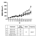
- 002_6_h4 antibody or isotype control antibody is administered at a dose of 20 mg / kg once every 4 days for a total of 7 times (indicated as "Q4Dx7").
- Administered intraperitoneally The administration start date was set to day 0, and observation was performed until day 34.
- Figure 27 shows the results of measuring the number of tumor-infiltrating T cells in mice on day 34 after administration of 002_6_h4 antibody or isotype control antibody.
- the administration of 002_6_h4 antibody to tumor-infiltrating T cells showed an increasing tendency as compared with the case of administration of isotype control antibody, suggesting that the anti-CD73 antibody has a tumor immunological effect.
- the developed antibody enhances the cancer cell injury activity by T cells, and if so, the degree thereof in comparison with the benchmark antibody.
- Human lung cancer cells NCI-H292 or HCC827 introduced with RFP protein were seeded on 96-well plates and cultured overnight. After removing the medium the next day, IL-2 (final concentration 30 IU) suspended in X-VIVO-15 medium (Lonza, 04-418Q) and various antibodies were added, and the mixture was incubated at 37 ° C. for 30 minutes. After that, healthy volunteer PBMCs stimulated with Dynabeads TM Human T-Activator CD3 / CD28 for T Cell Expansion and Activation (Veritas, DB11131) for 10 days to proliferate T cells were added at an E / T ratio of 2 to 37 ° C.
- NCI-H292 was added to a final concentration of 300 [mu] M ATP in a final concentration of 500 [mu] M ATP, HCC827 to, 37 °C, 5% CO over time the growth of cancer cells by IncuCyte ZOOM System (Sartorius) with 2 conditions The change was observed.
- the results are shown in Fig. 28.
- the developed antibody (002_6_h4 and 006_hKB were used as examples in this example, but other developed antibodies of the present invention are also used) suppresses T cells even when anti-PD-1 antibody and anti-PD-1 antibody are allowed to act.
- the effect of suppressing the growth of cancer cells of T cells was enhanced under the condition that the effect of releasing the antibody was hardly observed and the growth of cancer cells could not be stopped.
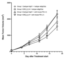
- mice For the mammary fat pad of NSG mice (hu-NSG, JACKSON laboratories) (hereinafter referred to as "immunized humanized mice") transplanted with human CD34-positive cells prepared from multiple healthy donors.
- the human breast cancer cell line MDA-MB-231 was transplanted to create a human breast cancer-carrying mouse model.
- 002_6_h4 antibody or isotype control antibody is administered at a dose of 20 mg / kg once every 4 days for a total of 7 times (“Q4Dx7").
- a checkpoint inhibitor anti-PD-1 antibody pembrolizumab, keytruda, MSD
- isotype control antibody at a dose of 10 mg / kg for the first time only and 5 mg / kg for the second and subsequent times.
- Q5Dx6 A total of 6 times (indicated as "Q5Dx6") were administered intraperitoneally once every 5 days.
- the administration start date was set to day 0, and the tumor size was measured twice a week until day 34.
- Figure 29 shows the average value and standard error of each administration group of the increase rate of tumor volume from Day 0 to day 34 in a typical donor.
- the average tumor volume increase rate in the combination administration group (Group 4) was lower than that in the control group (Group 1) and the checkpoint inhibitor administration group (Group 3). rice field.
- the average tumor volume increase rate was lower in the 002_6_h4 antibody-administered group (Group 2) than in the control group (Group 1), and the anti-CD73 antibody was used alone or in immune humanized mice. , It was suggested that it has an antitumor effect when used in combination with a checkpoint inhibitor (pembrolizumab).
- Example 19 Immunodeficient mouse test This example was conducted for the purpose of confirming the antitumor effect of the anti-CD73 antibody obtained in Example 6 in an immunodeficient mouse body.
- the human breast cancer cell line MDA-MB-231 was transplanted into the mammary fat pad of NSG mice (JACKSON laboratories).
- 002_6_h4 antibody or isotype control antibody was administered at each dose once every 4 days for a total of 7 times (Q4Dx7) in the group composition shown in Table 12 below.
- Administered intraperitoneally The tumor size was measured twice a week with the administration start date as day 0 and the observation period up to day 35.
- Figure 30 shows the results of volume changes in the tumor mass in immunodeficient mice.
- the 002_6_h4 antibody was shown to be more effective in suppressing tumor growth than the isotype control.
- Example 20 Antitumor effect (knock-in model) The purpose of this example was to confirm the antitumor effect of the anti-CD73 antibody obtained in Example 6 in a knock-in mouse expressing human CD73.
- a colon cancer cell line (Biocytogen, MC38-hCD73) that lacks mouse CD73 and expresses human CD73 is 5 ⁇ 10 ⁇ 5 were transplanted.
- 002_6_h4 antibody or isotype control antibody was administered at a dose of 10 mg / kg twice a week for a total of 7 times (denoted as "BIWx7").
- Figure 31 shows the transition of the average tumor volume from Day 0 to day 18 as the average value and standard error of each administration group.
- the mean tumor volume of the combination administration group (Group 4) was lower than that of the control group (Group 1) and the checkpoint inhibitor administration group (Group 3), and the anti-CD73 antibody was a mouse expressing human CD73. It was suggested that it has an antitumor effect when used in combination with a checkpoint inhibitor.
- the antibody or human antibody derivative obtained by the present invention activates immune cells by suppressing the function of CD73 expressed in cancer cells or immune cells and releasing the exhaustion of immune cells (particularly T cells). , Tumor growth inhibitory effect and cancer treatment effect.
- the present invention can be widely used as a therapeutic / diagnostic agent for cancer. Since the antibody according to the present invention has an effect of relieving the exhaustion of immune cells, it is expected to be particularly effective in combination therapy with other immunotherapies.
Landscapes
- Health & Medical Sciences (AREA)
- Chemical & Material Sciences (AREA)
- Life Sciences & Earth Sciences (AREA)
- Immunology (AREA)
- Organic Chemistry (AREA)
- Medicinal Chemistry (AREA)
- General Health & Medical Sciences (AREA)
- Molecular Biology (AREA)
- Engineering & Computer Science (AREA)
- Biochemistry (AREA)
- Biomedical Technology (AREA)
- Genetics & Genomics (AREA)
- Proteomics, Peptides & Aminoacids (AREA)
- Biophysics (AREA)
- Hematology (AREA)
- Urology & Nephrology (AREA)
- Public Health (AREA)
- General Chemical & Material Sciences (AREA)
- Chemical Kinetics & Catalysis (AREA)
- Nuclear Medicine, Radiotherapy & Molecular Imaging (AREA)
- Pharmacology & Pharmacy (AREA)
- Animal Behavior & Ethology (AREA)
- Veterinary Medicine (AREA)
- Bioinformatics & Cheminformatics (AREA)
- Cell Biology (AREA)
- Pathology (AREA)
- General Physics & Mathematics (AREA)
- Analytical Chemistry (AREA)
- Physics & Mathematics (AREA)
- Food Science & Technology (AREA)
- Microbiology (AREA)
- Biotechnology (AREA)
- Tropical Medicine & Parasitology (AREA)
- Toxicology (AREA)
- Peptides Or Proteins (AREA)
- Medicines Containing Antibodies Or Antigens For Use As Internal Diagnostic Agents (AREA)
Abstract
Description
・CD73による、AMPをアデノシンに分解する反応を、ベンチマーク抗体(公表されているMEDI9447の可変領域と同じ配列を有する抗体)よりも強く抑制する効果を示し;
・アデノシンによって誘導されるT細胞の疲弊を解除し、T細胞を活性化する。
[1]:(1) 重鎖の相補性決定領域、CDR1(DX1NMD(X1はCまたはS)、SEQ ID No.: 1)、CDR2(DINPNNGGTIYNQKFKG、SEQ ID No.: 2)、およびCDR3(TNWDYAMDY、SEQ ID No.: 3)、および
軽鎖の相補性決定領域、CDR1(KASQDINSX2LS(X2はN、DまたはQ)、SEQ ID No.: 4)、CDR2(RANRLID、SEQ ID No.: 5)、およびCDR3(X3QYDVFPRT(X3はLまたはQ)、SEQ ID No.: 6);
(2) 重鎖の相補性決定領域、CDR1(SFGMH、SEQ ID No.: 9)、CDR2(YISSGSRTIYYADTVRG、SEQ ID No.: 10)、およびCDR3(DFGSSSPNYFDY、SEQ ID No.: 11)、および
軽鎖の相補性決定領域、CDR1(RASESVDNYGISFMN、SEQ ID No.: 12)、CDR2(AASNQGS、SEQ ID No.: 13)、およびCDR3(QQSKEVPWT、SEQ ID No.: 14);
(3) 重鎖の相補性決定領域、CDR1(GYWMN、SEQ ID No.: 17)、CDR2(RIDPYDSETHYSQKFKD、SEQ ID No.: 18)、およびCDR3(SSPITTAPFDY、SEQ ID No.: 19)、および
軽鎖の相補性決定領域、CDR1(RASESVDYYGFSFMN、SEQ ID No.: 20)、CDR2(AASTQGS、SEQ ID No.: 21)、およびCDR3(QQSKEVPYT、SEQ ID No.: 22);
(4) 重鎖の相補性決定領域、CDR1(SYGVS、SEQ ID No.: 25)、CDR2(VIWGDGSTNYHSALIS、SEQ ID No.: 26)、およびCDR3(TNIFYDYDWYLDV、SEQ ID No.: 27)、および
軽鎖の相補性決定領域、CDR1(RSSQSLVHSNGNTYLH、SEQ ID No.: 28)、CDR2(KVSNRFS、SEQ ID No.: 29)、およびCDR3(SHSTHVPWT、SEQ ID No.: 30);
(5) 重鎖の相補性決定領域、CDR1(SYWMH、SEQ ID No.: 33)、CDR2(EINPSNARTNYNENFKS、SEQ ID No.: 34)、およびCDR3(RGTSGNYFDF、SEQ ID No.: 35)、および
軽鎖の相補性決定領域、CDR1(KASQDINTYLS、SEQ ID No.: 36)、CDR2(RANRLVD、SEQ ID No.: 37)、およびCDR3(LQYDEFPYT、SEQ ID No.: 38);
(6) 重鎖の相補性決定領域、CDR1(SYWMN、SEQ ID No.: 41)、CDR2(KIDPYDSETHYNQKFKD、SEQ ID No.: 42)、およびCDR3(IRYGTFDY、SEQ ID No.: 43)、および
軽鎖の相補性決定領域、CDR1(KASQDINNYLS、SEQ ID No.: 44)、CDR2(RANILVD、SEQ ID No.: 45)、およびCDR3(LQYDEFPYT、SEQ ID No.: 46);
(7) 重鎖の相補性決定領域、CDR1(SYWMH、SEQ ID No.: 49)、CDR2(EINPSNGRTNYNEKFKN、SEQ ID No.: 50)、およびCDR3(RGTSGNYFDY、SEQ ID No.: 51)、および
軽鎖の相補性決定領域、CDR1(KASQDINTYLS、SEQ ID No.: 52)、CDR2(RANRLVD、SEQ ID No.: 53)、およびCDR3(LQYDEFPYT、SEQ ID No.: 54);
(8) 重鎖の相補性決定領域、CDR1(SYWMN、SEQ ID No.: 57)、CDR2(RIDPYDSEAHYNQKFKD、SEQ ID No.: 58)、およびCDR3(IRYGTFDY、SEQ ID No.: 59)、および
軽鎖の相補性決定領域、CDR1(KASQDINSYLS、SEQ ID No.: 60)、CDR2(RSNSLVD、SEQ ID No.: 61)、およびCDR3(LQYDEFPYT、SEQ ID No.: 62);
からなる群から選択されるいずれかの重鎖・軽鎖の相補性決定領域を含む、CD73抗原に対して結合性を有し、がん細胞に対する傷害性を有するT細胞を活性化する、抗体またはヒト型抗体誘導体;
[2]: T細胞の活性化が、T細胞の増殖、T細胞のがん細胞に対する細胞傷害性の上昇、およびT細胞のサイトカイン分泌促進からなる群から選択される、[1]に記載の抗体またはヒト型抗体誘導体;
[3]: ヒト型抗体誘導体が、ヒト化抗体、キメラ抗体、多価抗体、および多重特異性抗体から選択されるヒト型抗体改変体またはその機能的断片から選択される、[1]または[2]に記載の抗体またはヒト型抗体誘導体;
[4]: 機能的断片が、F(ab')2である、[1]~[3]のいずれか1項に記載の抗体またはヒト型抗体誘導体;
[5]: 抗体またはヒト型抗体誘導体の重鎖可変領域VHドメインのアミノ酸配列が、(1)SEQ ID No.: 7、(2)SEQ ID No.: 15、(3)SEQ ID No.: 23、(4)SEQ ID No.: 31、(5)SEQ ID No.: 39、(6)SEQ ID No.: 47、(7)SEQ ID No.: 55、および(8)SEQ ID No.: 63から選択される、[1]~[4]のいずれか1項に記載の抗体またはヒト型抗体誘導体;
[6]: 抗体またはヒト型抗体誘導体の軽鎖可変領域VLドメインのアミノ酸配列が、(1)SEQ ID No.: 8、(2)SEQ ID No.: 16、(3)SEQ ID No.: 24、(4)SEQ ID No.: 32、(5)SEQ ID No.: 40、(6)SEQ ID No.: 48、(7)SEQ ID No.: 56、および(8)SEQ ID No.: 64から選択される、[1]~[5]のいずれか1項に記載の抗体またはヒト型抗体誘導体;
[7]: がん細胞に対する細胞傷害性を誘導するが、正常細胞に対する細胞傷害性を誘導しない、[1]~[6]のいずれか1項に記載の抗体またはヒト型抗体誘導体;
[8]: がん細胞が、メラノーマ、乳がん、肺がん、大腸がんからなる群から選択される、[1]~[7]のいずれか1項に記載の抗体またはヒト型抗体誘導体;
[9]: 薬物と結合され、抗体薬物複合体(ADC)を形成する、[1]~[8]のいずれか1項に記載の抗体またはヒト型抗体誘導体;
[10]: [1]~[9]のいずれか1項に記載の抗体またはヒト型抗体誘導体を含む、がん治療のための医薬組成物;
[11]: がんが、メラノーマ、乳がん、肺がん、大腸がんからなる群から選択される、[10]に記載の医薬組成物;
[12]: 被験体から採取されたがん細胞を、in vitroにおいて[1]~[9]のいずれか1項に記載の抗体またはヒト型抗体誘導体と接触させる工程、
培養条件下で、がん細胞のAMP代謝が低下するかどうかを測定する工程 、若しくは細胞生存率が低下するかどうかを測定する工程、若しくは免疫活性化物質の分泌が亢進するかどうかを測定する工程、
を含む、がん細胞に対する細胞傷害性測定方法;
[13]: 同一被験体の末梢血リンパ球の存在下で、がん細胞の細胞生存率が低下するかどうか、若しくは末梢血リンパ球由来の免疫細胞が活性化するかどうかを測定する、[12]に記載のがん細胞に対する細胞傷害性測定方法;
[14]: 被験体から採取されたがん細胞のin vitroでのAMP代謝抑制若しくは細胞傷害性から、[1]~[9]のいずれか1項に記載の抗体またはヒト型抗体誘導体をその被験体に投与した場合のがん細胞に対する細胞傷害性の亢進を測定する、[12]または[13]に記載のがん細胞に対する細胞傷害性測定方法;
[15]: in vitroでの免疫活性化物質の分泌の亢進から、[1]~[9]のいずれか1項に記載の抗体またはヒト型抗体誘導体をその被験体に投与した場合のがん細胞に対する細胞傷害性の亢進を測定する、[12]または[13]に記載のがん細胞に対する細胞傷害性測定方法;
[16]: [1]~[9]のいずれか1項に記載の抗体またはヒト型抗体誘導体の、被験体から採取されたがん細胞に対するAMP代謝、細胞傷害性、免疫活性化物質の分泌、若しくは免疫細胞の活性化を、in vitroにおいて測定するための、前記抗体を含む測定キット。
本発明は、一態様において、CD73抗原に対して結合性を有する、抗体またはヒト型抗体誘導体を提供する。この抗体またはヒト型抗体誘導体は、がん細胞に対する傷害性を有するT細胞を活性化することを特徴としている。
(1) 重鎖の相補性決定領域、CDR1(DX1NMD(X1はCまたはS)、SEQ ID No.: 1)、CDR2(DINPNNGGTIYNQKFKG、SEQ ID No.: 2)、およびCDR3(TNWDYAMDY、SEQ ID No.: 3)、および
軽鎖の相補性決定領域、CDR1(KASQDINSX2LS(X2はN、DまたはQ)、SEQ ID No.: 4)、CDR2(RANRLID、SEQ ID No.: 5)、およびCDR3(X3QYDVFPRT(X3はLまたはQ)、SEQ ID No.: 6);
(2) 重鎖の相補性決定領域、CDR1(SFGMH、SEQ ID No.: 9)、CDR2(YISSGSRTIYYADTVRG、SEQ ID No.: 10)、およびCDR3(DFGSSSPNYFDY、SEQ ID No.: 11)、および
軽鎖の相補性決定領域、CDR1(RASESVDNYGISFMN、SEQ ID No.: 12)、CDR2(AASNQGS、SEQ ID No.: 13)、およびCDR3(QQSKEVPWT、SEQ ID No.: 14);
(3) 重鎖の相補性決定領域、CDR1(GYWMN、SEQ ID No.: 17)、CDR2(RIDPYDSETHYSQKFKD、SEQ ID No.: 18)、およびCDR3(SSPITTAPFDY、SEQ ID No.: 19)、および
軽鎖の相補性決定領域、CDR1(RASESVDYYGFSFMN、SEQ ID No.: 20)、CDR2(AASTQGS、SEQ ID No.: 21)、およびCDR3(QQSKEVPYT、SEQ ID No.: 22);
(4) 重鎖の相補性決定領域、CDR1(SYGVS、SEQ ID No.: 25)、CDR2(VIWGDGSTNYHSALIS、SEQ ID No.: 26)、およびCDR3(TNIFYDYDWYLDV、SEQ ID No.: 27)、および
軽鎖の相補性決定領域、CDR1(RSSQSLVHSNGNTYLH、SEQ ID No.: 28)、CDR2(KVSNRFS、SEQ ID No.: 29)、およびCDR3(SHSTHVPWT、SEQ ID No.: 30);
(5) 重鎖の相補性決定領域、CDR1(SYWMH、SEQ ID No.: 33)、CDR2(EINPSNARTNYNENFKS、SEQ ID No.: 34)、およびCDR3(RGTSGNYFDF、SEQ ID No.: 35)、および
軽鎖の相補性決定領域、CDR1(KASQDINTYLS、SEQ ID No.: 36)、CDR2(RANRLVD、SEQ ID No.: 37)、およびCDR3(LQYDEFPYT、SEQ ID No.: 38);
(6) 重鎖の相補性決定領域、CDR1(SYWMN、SEQ ID No.: 41)、CDR2(KIDPYDSETHYNQKFKD、SEQ ID No.: 42)、およびCDR3(IRYGTFDY、SEQ ID No.: 43)、および
軽鎖の相補性決定領域、CDR1(KASQDINNYLS、SEQ ID No.: 44)、CDR2(RANILVD、SEQ ID No.: 45)、およびCDR3(LQYDEFPYT、SEQ ID No.: 46);
(7) 重鎖の相補性決定領域、CDR1(SYWMH、SEQ ID No.: 49)、CDR2(EINPSNGRTNYNEKFKN、SEQ ID No.: 50)、およびCDR3(RGTSGNYFDY、SEQ ID No.: 51)、および
軽鎖の相補性決定領域、CDR1(KASQDINTYLS、SEQ ID No.: 52)、CDR2(RANRLVD、SEQ ID No.: 53)、およびCDR3(LQYDEFPYT、SEQ ID No.: 54);
(8) 重鎖の相補性決定領域、CDR1(SYWMN、SEQ ID No.: 57)、CDR2(RIDPYDSEAHYNQKFKD、SEQ ID No.: 58)、およびCDR3(IRYGTFDY、SEQ ID No.: 59)、および
軽鎖の相補性決定領域、CDR1(KASQDINSYLS、SEQ ID No.: 60)、CDR2(RSNSLVD、SEQ ID No.: 61)、およびCDR3(LQYDEFPYT、SEQ ID No.: 62);
からなる群から選択されるいずれかの重鎖・軽鎖の相補性決定領域を含むものが考えられるが、CD73抗原に対して結合性を有するという特徴を有する限りにおいて、上記のCDRの組み合わせ以外のCDRの組み合わせで特定される抗体またはヒト型抗体誘導体もまた含まれる。
本発明の抗体またはヒト型抗体誘導体は、前述した由来種の動物に対してCD73抗原を免疫原として投与し、その動物体内から採取した抗体産生細胞を培養することにより取得することもできるが、抗体またはヒト型抗体誘導体のアミノ酸配列を規定できるDNA配列を含むタンパク質発現用のベクターを設計し、そのベクターをタンパク質産生用の細胞に導入し、組換え的に取得してもよい。
002_6_h1重鎖可変領域(SEQ ID NO: 65)および002_6_h1軽鎖可変領域(SEQ ID NO: 66)を含む抗体、
002_6_h2重鎖可変領域(SEQ ID NO: 67)および002_6_h2軽鎖可変領域(SEQ ID NO: 68)を含む抗体、
002_6_h3重鎖可変領域(SEQ ID NO: 69)および002_6_h3軽鎖可変領域(SEQ ID NO: 70)を含む抗体、
002_6_h4重鎖可変領域(SEQ ID NO: 71)および002_6_h4軽鎖可変領域(SEQ ID NO: 72)を含む抗体、
002_6_h5重鎖可変領域(SEQ ID NO: 73)および002_6_h5軽鎖可変領域(SEQ ID NO: 74)を含む抗体、
002_6_h6重鎖可変領域(SEQ ID NO: 75)および002_6_h6軽鎖可変領域(SEQ ID NO: 76)を含む抗体、
002_6_h7重鎖可変領域(SEQ ID NO: 77)および002_6_h7軽鎖可変領域(SEQ ID NO: 78)を含む抗体、
002_6_h8重鎖可変領域(SEQ ID NO: 79)および002_6_h8軽鎖可変領域(SEQ ID NO: 80)を含む抗体、
004_h3_重鎖可変領域(SEQ ID NO: 81)および004_h3_軽鎖可変領域(SEQ ID NO: 82)を含む抗体、
006_hKB_重鎖可変領域(SEQ ID NO: 83)および006_hKB_軽鎖可変領域(SEQ ID NO: 84)を含む抗体、
007_h1_重鎖可変領域(SEQ ID NO: 85)および007_h1_軽鎖可変領域(SEQ ID NO: 86)を含む抗体、
008_h4_重鎖可変領域(SEQ ID NO: 87)および008_h4_軽鎖可変領域(SEQ ID NO: 88)を含む抗体、
などが含まれるが、これらには限定されず、その他の抗体から作成されるヒト型抗体誘導体も本発明の態様として含まれる。
本発明の抗体またはヒト型抗体誘導体は、上述したがん細胞に対する細胞傷害性を有する免疫細胞(特にT細胞)を活性化する能力を有するという特徴に基づき、一態様において、がんの治療または予防を必要としている被験体において、がん細胞に対する傷害性を有する免疫細胞(特にT細胞)を活性化する、本発明の抗体またはヒト型抗体誘導体を含む医薬組成物、を提供することができる。
本発明の抗体またはヒト型抗体誘導体は、CD73抗原に対して結合性を有するという特徴に基づき、試料中のCD73抗原を検出するために使用することができる。具体的には、本発明の抗体またはヒト型抗体誘導体は、被験体から採取したがん細胞あるいは免疫細胞(例えばT細胞)を含む試料に対して、免疫沈降法、凝集反応、磁気ビーズ法などの抗体を用いた精製法、ELISA法、ウェスタンブロット、免疫組織化学などのイムノアッセイ、フローサイトメトリーなどの免疫細胞化学等、抗体を用いて行うことができるCD73抗原の様々な検出方法を実施する際に使用することができる。それぞれの場合において、本発明の抗体またはヒト型抗体誘導体は、当業者に一般的に知られている検出用標識(例えば、蛍光、酵素、など)を用いて検出することができる。
被験体から採取されたがん細胞を、培養条件下(すなわち、in vitro)において本発明の抗体またはヒト型抗体誘導体と接触させる工程、
培養条件下で、がん細胞のAMP代謝(AMPをアデノシンに分解する反応)が低下するかどうかを測定する工程、細胞生存率が低下するかどうかを測定する工程、若しくは免疫活性化物質の分泌が亢進するかどうかを測定する工程、
を含む、本発明の抗体またはヒト型抗体誘導体を用いてがん細胞に対する細胞傷害性を測定する方法を例として挙げることができる。
本実施例は、マウスを使用してCD73抗原に対するモノクローナル抗体を取得することを目的として行った。
マウス(BALB/cAJcl メス7週齢、日本クレア)に対して、CD73組換え抗原(自家調製品)10μgを、二週間おきに4~8回、腹腔内投与して免疫化した。最終投与から一週間後に尾静脈より採血し、血中のCD73抗原に特異的な抗体価の上昇を、従来法に基づいて抗原固定ELISAによって確認した後、CD73抗原に特異的な抗体化が有意に上昇した抗体を有するマウスの脾臓を回収した。
抗原固定ELISAで、抗原(CD73)結合性が認められた抗体について、CHO細胞の培養上清を回収し、培養上清中の抗体をMonospinProGカラム(GLサイエンス)によって精製した。
ヒト肺がん細胞株H322(ATCC(登録商標)CRL-5806TM)を96 wellプレートに播種し、精製した抗体を添加して37℃で一時間インキュベートした。CD73の基質であるAMP(Sigma, A1752)を終濃度200μMとなるように添加し、37℃で一晩インキュベートして、CD73によるAMPのアデノシンへの分解反応を行った。
本実施例は、実施例1で取得された002_m抗体の機能評価を行うことを目的として行った。
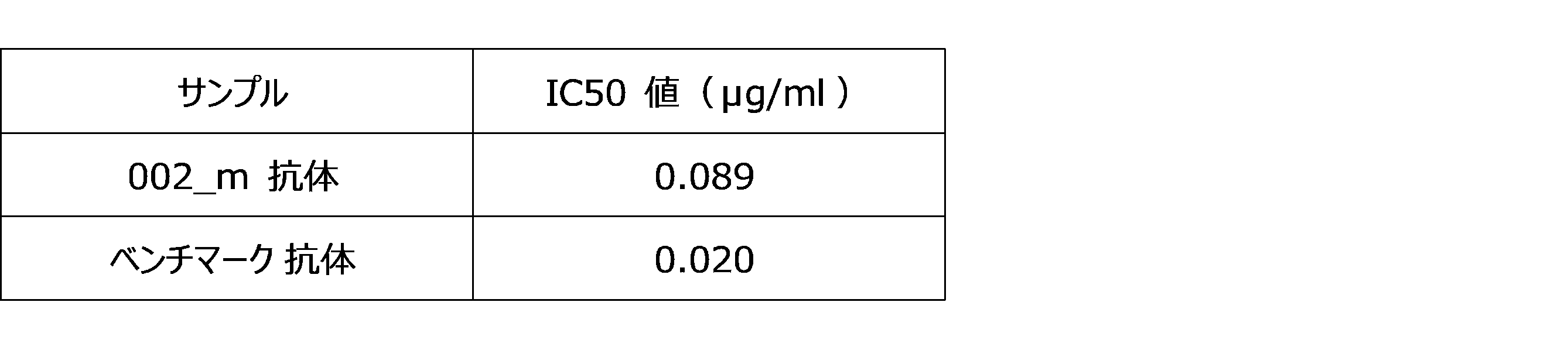
本実施例は、実施例1で取得された002_m抗体のアミノ酸配列を決定することを目的として行った。
本実施例は、実施例3で配列決定した002_m抗体について、マウス抗体の可変領域およびヒト抗体の定常領域を有するキメラ抗体を作製することを目的として行った。
本実施例は、002_m抗体について、CDR中の翻訳後修飾を受ける可能性がある残基を、修飾を受けない残基に置換した改変抗体を作製することを目的として行った。


本実施例は、実施例5で選択した002_6_c抗体(SEQ ID NO: 7(C32S)/SEQ ID NO: 8(N32Q))に基づいて、ヒト化抗体を作製することを目的として行った。


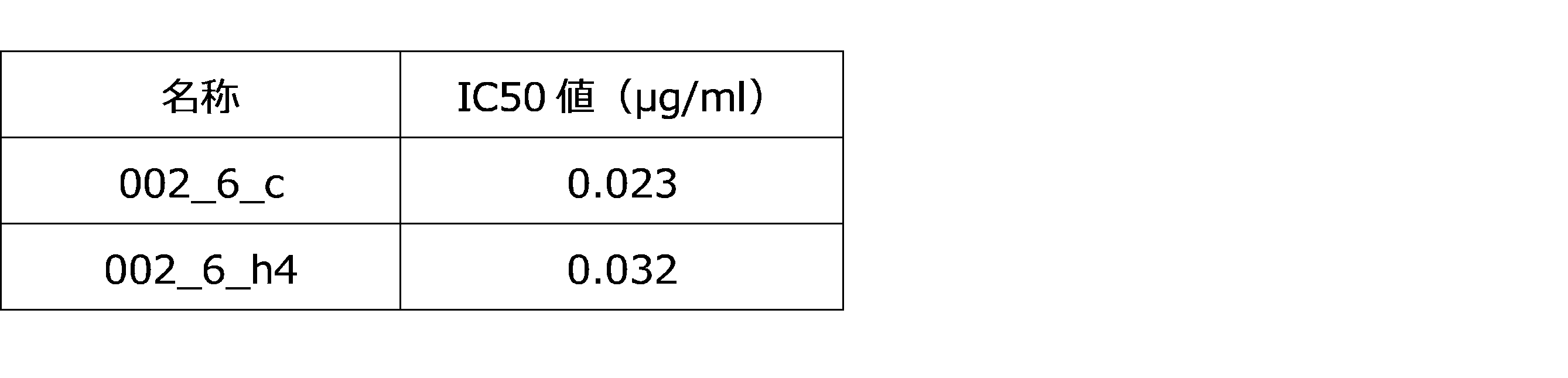
本実施例は、実施例6で得られた抗CD73抗体の、組換えCD73抗原に対する酵素反応抑制機能を確認することを目的として行った。
本実施例は、実施例6で得られた抗CD73抗体の、ヒト末梢血T細胞に対する増殖促進活性を確認することを目的として行った。
本実施例は、実施例1で取得された002_m抗体以外の抗体(すなわち、003_m抗体、004_m抗体、005_m抗体、006_m抗体、007_m抗体、008_m抗体、009_m抗体)の機能評価を行うことを目的として行った。

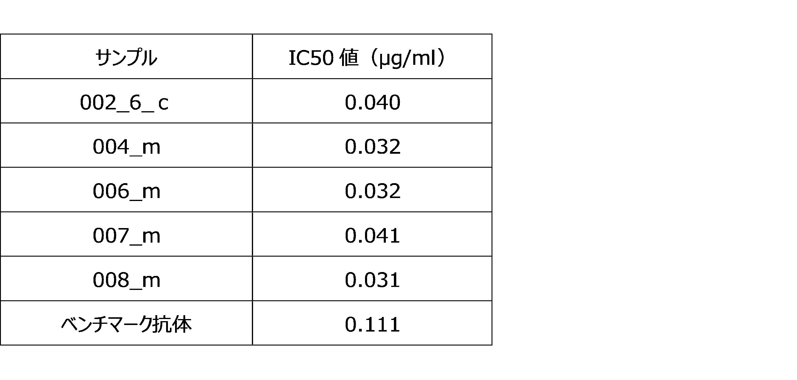
本実施例は、実施例1で取得された003_m抗体~009_m抗体のアミノ酸配列を決定することを目的として行った。

本実施例は、実施例1において取得された003_m~009_mのそれぞれの抗体の結合性評価を行うことを目的として行った。
本実施例は、実施例9で得られた抗CD73抗体の、ヒト末梢血T細胞に対する増殖促進活性を確認することを目的として行った。
本実施例は、実施例1において取得された003_m~009_mのそれぞれの抗体のうち、実施例9で選択した004_m抗体、006_m抗体、007_m抗体、008_m抗体に基づいて、ヒト化抗体を作製することを目的として行った。

本実施例は、実施例13で得られたヒト化抗CD73抗体の、組換えCD73抗原に対する酵素反応抑制機能を確認することを目的として行った。
本実施例は、実施例13で得られ、実施例14で特に高い効果を示した抗CD73抗体の、ヒト末梢血T細胞に対する増殖促進活性を確認することを目的として行った。
本実施例は、実施例6で得られた抗CD73抗体および実施例13で得られ、実施例14で特に高い効果を示した抗CD73抗体の、細胞内への取り込みを評価することを目的として行った。
%対照=(抗CD73抗体(X分)MFI-アイソタイプコントロール(X分)MFI)/(抗CD73抗体(0分)MFI-アイソタイプコントロール(0分)MFI)×100
%CD73 Ag=(抗CD73抗体(X分)MFI-アイソタイプコントロール(X分)MFI)/(抗CD73抗体(0分)MFI-アイソタイプコントロール(0分)MFI)×100
本実施例は、実施例6で得られた抗CD73抗体および実施例13で得られ、実施例14で特に高い効果を示した抗CD73抗体の、CD73抗原に対する結合親和性の指標である解離定数(KD値)を評価することを目的として行った。

本実施例は、実施例6で得られた抗CD73抗体および実施例13で得られ、実施例14で特に高い効果を示した抗CD73抗体の、ヒトCD34陽性細胞移植マウス体内における抗腫瘍効果を確認することを目的として行った。
複数の健常人ドナーから作製したヒトCD34陽性細胞を移植したNSGマウス(hu-NSG, JACKSON laboratories)(以下、「免疫ヒト化マウス」と呼ぶ)の乳腺脂肪体(Mammary fat pad)に対して、ヒト乳癌細胞株MDA-MB-231を移植し、ヒト乳癌担持マウスモデルを作出した。このマウスモデルにおいて、移植したヒト乳癌細胞が増殖して100 mm3になった段階で、002_6_h4抗体もしくはアイソタイプコントロール抗体を、20 mg/kgの用量にて4日に一度、合計7回(「Q4Dx7」と示す)と、チェックポイント阻害剤(抗PD-1抗体であるペンブロリズマブ、キートルーダ、MSD)もしくはアイソタイプコントロール抗体を、初回のみ10 mg/kg、2回目以降は5 mg/kgの用量にて、5日に一度、合計6回(「Q5Dx6」と示す)を組み合わせて腹腔内投与した。投与開始日をday0とし、day34まで週2回腫瘍サイズの計測を行った。抗体の投与は以下の通りである:
Group 1=アイソタイプコントロール抗体のみ投与群
Group 2=002_6_h4抗体およびアイソタイプコントロール抗体投与群
Group 3=チェックポイント阻害剤(ペンブロリズマブ)およびアイソタイプコントロール抗体投与群
Group 4=002_6_h4抗体およびチェックポイント阻害剤(ペンブロリズマブ)の併用投与群。
本実施例は、実施例6で得られた抗CD73抗体の、免疫不全マウス体内における抗腫瘍効果を確認することを目的として行った。

本実施例は、実施例6で得られた抗CD73抗体の、ヒトCD73を発現するノックインマウス体内における抗腫瘍効果を確認することを目的として行った。
Group 1=アイソタイプコントロール抗体のみ投与群
Group 2=002_6_h4抗体およびアイソタイプコントロール抗体投与群
Group 3=チェックポイント阻害剤(抗マウスPD-1抗体)およびアイソタイプコントロール抗体投与群
Group 4=002_6_h4抗体とチェックポイント阻害剤(抗マウスPD-1抗体)の併用投与を行った群。
Claims (16)
- (1) 重鎖の相補性決定領域、CDR1(DX1NMD(X1はCまたはS)、SEQ ID No.: 1)、CDR2(DINPNNGGTIYNQKFKG、SEQ ID No.: 2)、およびCDR3(TNWDYAMDY、SEQ ID No.: 3)、および
軽鎖の相補性決定領域、CDR1(KASQDINSX2LS(X2はNまたはDまたはQ)、SEQ ID No.: 4)、CDR2(RANRLID、SEQ ID No.: 5)、およびCDR3(X3QYDVFPRT(X3はLまたはQ)、SEQ ID No.: 6);
(2) 重鎖の相補性決定領域、CDR1(SFGMH、SEQ ID No.: 9)、CDR2(YISSGSRTIYYADTVRG、SEQ ID No.: 10)、およびCDR3(DFGSSSPNYFDY、SEQ ID No.: 11)、および
軽鎖の相補性決定領域、CDR1(RASESVDNYGISFMN、SEQ ID No.: 12)、CDR2(AASNQGS、SEQ ID No.: 13)、およびCDR3(QQSKEVPWT、SEQ ID No.: 14);
(3) 重鎖の相補性決定領域、CDR1(GYWMN、SEQ ID No.: 17)、CDR2(RIDPYDSETHYSQKFKD、SEQ ID No.: 18)、およびCDR3(SSPITTAPFDY、SEQ ID No.: 19)、および
軽鎖の相補性決定領域、CDR1(RASESVDYYGFSFMN、SEQ ID No.: 20)、CDR2(AASTQGS、SEQ ID No.: 21)、およびCDR3(QQSKEVPYT、SEQ ID No.: 22);
(4) 重鎖の相補性決定領域、CDR1(SYGVS、SEQ ID No.: 25)、CDR2(VIWGDGSTNYHSALIS、SEQ ID No.: 26)、およびCDR3(TNIFYDYDWYLDV、SEQ ID No.: 27)、および
軽鎖の相補性決定領域、CDR1(RSSQSLVHSNGNTYLH、SEQ ID No.: 28)、CDR2(KVSNRFS、SEQ ID No.: 29)、およびCDR3(SHSTHVPWT、SEQ ID No.: 30);
(5) 重鎖の相補性決定領域、CDR1(SYWMH、SEQ ID No.: 33)、CDR2(EINPSNARTNYNENFKS、SEQ ID No.: 34)、およびCDR3(RGTSGNYFDF、SEQ ID No.: 35)、および
軽鎖の相補性決定領域、CDR1(KASQDINTYLS、SEQ ID No.: 36)、CDR2(RANRLVD、SEQ ID No.: 37)、およびCDR3(LQYDEFPYT、SEQ ID No.: 38);
(6) 重鎖の相補性決定領域、CDR1(SYWMN、SEQ ID No.: 41)、CDR2(KIDPYDSETHYNQKFKD、SEQ ID No.: 42)、およびCDR3(IRYGTFDY、SEQ ID No.: 43)、および
軽鎖の相補性決定領域、CDR1(KASQDINNYLS、SEQ ID No.: 44)、CDR2(RANILVD、SEQ ID No.: 45)、およびCDR3(LQYDEFPYT、SEQ ID No.: 46);
(7) 重鎖の相補性決定領域、CDR1(SYWMH、SEQ ID No.: 49)、CDR2(EINPSNGRTNYNEKFKN、SEQ ID No.: 50)、およびCDR3(RGTSGNYFDY、SEQ ID No.: 51)、および
軽鎖の相補性決定領域、CDR1(KASQDINTYLS、SEQ ID No.: 52)、CDR2(RANRLVD、SEQ ID No.: 53)、およびCDR3(LQYDEFPYT、SEQ ID No.: 54);
(8) 重鎖の相補性決定領域、CDR1(SYWMN、SEQ ID No.: 57)、CDR2(RIDPYDSEAHYNQKFKD、SEQ ID No.: 58)、およびCDR3(IRYGTFDY、SEQ ID No.: 59)、および
軽鎖の相補性決定領域、CDR1(KASQDINSYLS、SEQ ID No.: 60)、CDR2(RSNSLVD、SEQ ID No.: 61)、およびCDR3(LQYDEFPYT、SEQ ID No.: 62);
からなる群から選択されるいずれかの重鎖・軽鎖の相補性決定領域を含む、CD73抗原に対して結合性を有し、がん細胞に対する傷害性を有するT細胞を活性化する、抗体またはヒト型抗体誘導体。 - T細胞の活性化が、T細胞の増殖、T細胞のがん細胞に対する細胞傷害性の上昇、およびT細胞のサイトカイン分泌促進からなる群から選択される、請求項1に記載の抗体またはヒト型抗体誘導体。
- ヒト型抗体誘導体が、ヒト化抗体、キメラ抗体、多価抗体、および多重特異性抗体から選択されるヒト型抗体改変体またはその機能的断片から選択される、請求項1または2に記載の抗体またはヒト型抗体誘導体。
- 機能的断片が、F(ab')2である、請求項1~3のいずれか1項に記載の抗体またはヒト型抗体誘導体。
- 抗体またはヒト型抗体誘導体の重鎖可変領域VHドメインのアミノ酸配列が、(1)SEQ ID No.: 7、(2)SEQ ID No.: 15、(3)SEQ ID No.: 23、(4)SEQ ID No.: 31、(5)SEQ ID No.: 39、(6)SEQ ID No.: 47、(7)SEQ ID No.: 55、および(8)SEQ ID No.: 63から選択される、請求項1~4のいずれか1項に記載の抗体またはヒト型抗体誘導体。
- 抗体またはヒト型抗体誘導体の軽鎖可変領域VLドメインのアミノ酸配列が、(1)SEQ ID No.: 8、(2)SEQ ID No.: 16、(3)SEQ ID No.: 24、(4)SEQ ID No.: 32、(5)SEQ ID No.: 40、(6)SEQ ID No.: 48、(7)SEQ ID No.: 56、および(8)SEQ ID No.: 64から選択される、請求項1~5のいずれか1項に記載の抗体またはヒト型抗体誘導体。
- がん細胞に対する細胞傷害性を誘導するが、正常細胞に対する細胞傷害性を誘導しない、請求項1~6のいずれか1項に記載の抗体またはヒト型抗体誘導体。
- がん細胞が、メラノーマ、乳がん、肺がん、大腸がんからなる群から選択される、請求項1~7のいずれか1項に記載の抗体またはヒト型抗体誘導体。
- 薬物と結合され、抗体薬物複合体(ADC)を形成する、請求項1~8のいずれか1項に記載の抗体またはヒト型抗体誘導体。
- 請求項1~9のいずれか1項に記載の抗体またはヒト型抗体誘導体を含む、がん治療のための医薬組成物。
- がんが、メラノーマ、乳がん、肺がん、大腸がんからなる群から選択される、請求項10に記載の医薬組成物。
- 被験体から採取されたがん細胞を、in vitroにおいて請求項1~9のいずれか1項に記載の抗体またはヒト型抗体誘導体と接触させる工程、
培養条件下で、がん細胞のAMP代謝が低下するかどうかを測定する工程、若しくは細胞生存率が低下するかどうかを測定する工程、若しくは免疫活性化物質の分泌が亢進するかどうかを測定する工程、
を含む、がん細胞に対する細胞傷害性測定方法。 - 同一被験体の末梢血リンパ球の存在下で、がん細胞の細胞生存率が低下するかどうか、若しくは末梢血リンパ球由来の免疫細胞が活性化するかどうかを測定する、請求項12に記載のがん細胞に対する細胞傷害性測定方法。
- 被験体から採取されたがん細胞のin vitroでのAMP代謝抑制若しくは細胞傷害性から、請求項1~9のいずれか1項に記載の抗体またはヒト型抗体誘導体をその被験体に投与した場合のがん細胞に対する細胞傷害性の亢進を測定する、請求項12または13に記載のがん細胞に対する細胞傷害性測定方法。
- in vitroでの免疫活性化物質の分泌の亢進から、請求項1~9のいずれか1項に記載の抗体またはヒト型抗体誘導体をその被験体に投与した場合のがん細胞に対する細胞傷害性の亢進を測定する、請求項12または13に記載のがん細胞に対する細胞傷害性測定方法。
- 請求項1~9のいずれか1項に記載の抗体またはヒト型抗体誘導体の、被験体から採取されたがん細胞に対するAMP代謝、細胞傷害性、免疫活性化物質の分泌、若しくは免疫細胞の活性化を、in vitroにおいて測定するための、前記抗体を含む測定キット。
Priority Applications (4)
| Application Number | Priority Date | Filing Date | Title |
|---|---|---|---|
| CN202180039122.XA CN115867582A (zh) | 2020-05-29 | 2021-05-28 | 抗cd73抗体及其用途 |
| EP21812743.9A EP4159763A4 (en) | 2020-05-29 | 2021-05-28 | ANTI-CD73 ANTIBODIES AND USE THEREOF |
| JP2022526665A JP7760166B2 (ja) | 2020-05-29 | 2021-05-28 | 抗cd73抗体およびその用途 |
| US18/000,064 US20230220104A1 (en) | 2020-05-29 | 2021-05-28 | Anti-cd73 antibody and use thereof |
Applications Claiming Priority (2)
| Application Number | Priority Date | Filing Date | Title |
|---|---|---|---|
| JP2020094545 | 2020-05-29 | ||
| JP2020-094545 | 2020-05-29 |
Publications (1)
| Publication Number | Publication Date |
|---|---|
| WO2021241729A1 true WO2021241729A1 (ja) | 2021-12-02 |
Family
ID=78744840
Family Applications (1)
| Application Number | Title | Priority Date | Filing Date |
|---|---|---|---|
| PCT/JP2021/020384 Ceased WO2021241729A1 (ja) | 2020-05-29 | 2021-05-28 | 抗cd73抗体およびその用途 |
Country Status (5)
| Country | Link |
|---|---|
| US (1) | US20230220104A1 (ja) |
| EP (1) | EP4159763A4 (ja) |
| JP (1) | JP7760166B2 (ja) |
| CN (1) | CN115867582A (ja) |
| WO (1) | WO2021241729A1 (ja) |
Cited By (12)
| Publication number | Priority date | Publication date | Assignee | Title |
|---|---|---|---|---|
| WO2023201267A1 (en) | 2022-04-13 | 2023-10-19 | Gilead Sciences, Inc. | Combination therapy for treating trop-2 expressing cancers |
| WO2024032777A1 (zh) * | 2022-08-12 | 2024-02-15 | 南京蓬勃生物科技有限公司 | 抗cd73抗体或其抗原片段及其应用 |
| WO2024040195A1 (en) | 2022-08-17 | 2024-02-22 | Capstan Therapeutics, Inc. | Conditioning for in vivo immune cell engineering |
| US12018089B2 (en) | 2020-01-03 | 2024-06-25 | Incyte Corporation | Anti-CD73 antibodies and uses thereof |
| US12060433B2 (en) | 2020-01-03 | 2024-08-13 | Incyte Corporation | CD73 inhibitor and A2A/A2B adenosine receptor inhibitor combination therapy |
| WO2024249954A1 (en) | 2023-05-31 | 2024-12-05 | Capstan Therapeutics, Inc. | Lipid nanoparticle formulations and compositions |
| WO2025076127A1 (en) | 2023-10-05 | 2025-04-10 | Capstan Therapeutics, Inc. | Constrained ionizable cationic lipids and lipid nanoparticles |
| WO2025076113A1 (en) | 2023-10-05 | 2025-04-10 | Capstan Therapeutics, Inc. | Ionizable cationic lipids with conserved spacing and lipid nanoparticles |
| WO2025137640A1 (en) | 2023-12-22 | 2025-06-26 | Gilead Sciences, Inc. | Azaspiro wrn inhibitors |
| WO2025179294A2 (en) | 2024-02-22 | 2025-08-28 | Capstan Therapeutics, Inc. | Immune engineering amplification |
| WO2025217454A2 (en) | 2024-04-11 | 2025-10-16 | Capstan Therapeutics, Inc. | Ionizable cationic lipids and lipid nanoparticles |
| WO2025217452A1 (en) | 2024-04-11 | 2025-10-16 | Capstan Therapeutics, Inc. | Constrained ionizable cationic lipids and lipid nanoparticles |
Citations (3)
| Publication number | Priority date | Publication date | Assignee | Title |
|---|---|---|---|---|
| JP2015143226A (ja) * | 2006-07-10 | 2015-08-06 | 学校法人藤田学園 | 抗体及びその用途 |
| JP2018501197A (ja) | 2014-11-10 | 2018-01-18 | メディミューン リミテッド | Cd73特異的結合分子及びその使用 |
| JP2019503709A (ja) * | 2015-12-09 | 2019-02-14 | コーバス・ファーマシューティカルズ・インコーポレイテッド | ヒト化抗cd73抗体 |
Family Cites Families (5)
| Publication number | Priority date | Publication date | Assignee | Title |
|---|---|---|---|---|
| ES2869972T3 (es) * | 2015-01-26 | 2021-10-26 | Cellectis | Sistemas de receptores de antígenos quiméricos dirigidos por mAb para clasificar/agotar células inmunitarias genomanipuladas |
| US11130817B2 (en) * | 2015-10-12 | 2021-09-28 | Innate Pharma | CD73 blocking agents |
| CN110267982B (zh) * | 2016-10-19 | 2024-02-23 | 斯克利普斯研究所 | 具有人源化靶向部分和/或经过优化的嵌合抗原受体相互作用结构域的嵌合抗原受体效应细胞开关以及其用途 |
| EP3694552A1 (en) * | 2017-10-10 | 2020-08-19 | Tilos Therapeutics, Inc. | Anti-lap antibodies and uses thereof |
| EP3569618A1 (en) * | 2018-05-19 | 2019-11-20 | Boehringer Ingelheim International GmbH | Antagonizing cd73 antibody |
-
2021
- 2021-05-28 EP EP21812743.9A patent/EP4159763A4/en not_active Withdrawn
- 2021-05-28 US US18/000,064 patent/US20230220104A1/en active Pending
- 2021-05-28 CN CN202180039122.XA patent/CN115867582A/zh active Pending
- 2021-05-28 WO PCT/JP2021/020384 patent/WO2021241729A1/ja not_active Ceased
- 2021-05-28 JP JP2022526665A patent/JP7760166B2/ja active Active
Patent Citations (3)
| Publication number | Priority date | Publication date | Assignee | Title |
|---|---|---|---|---|
| JP2015143226A (ja) * | 2006-07-10 | 2015-08-06 | 学校法人藤田学園 | 抗体及びその用途 |
| JP2018501197A (ja) | 2014-11-10 | 2018-01-18 | メディミューン リミテッド | Cd73特異的結合分子及びその使用 |
| JP2019503709A (ja) * | 2015-12-09 | 2019-02-14 | コーバス・ファーマシューティカルズ・インコーポレイテッド | ヒト化抗cd73抗体 |
Non-Patent Citations (6)
| Title |
|---|
| BIOINFORMATICS, vol. 31, no. 3, 1 February 2015 (2015-02-01), pages 434 - 5 |
| CLIN. MICROBIOL. REV, vol. 7, 1994, pages 277 - 289 |
| HAMMAMI, A. ET AL.: "Targeting the adenosine pathway for cancer immunotherapy", SEMINARS IN IMMUNOLOGY, vol. 42, no. 101304, 2019, pages 1 - 7, XP085854510, DOI: 10.1016/j.smim.2019.101304 * |
| LOTTA VON BOEHMER ET AL., NATURE PROTOCOLS, vol. 11, pages 1908 - 1923 |
| ONCOIMMUNOLOGY, vol. 5, no. 8, August 2016 (2016-08-01), pages e1208875 |
| See also references of EP4159763A4 |
Cited By (13)
| Publication number | Priority date | Publication date | Assignee | Title |
|---|---|---|---|---|
| US12018089B2 (en) | 2020-01-03 | 2024-06-25 | Incyte Corporation | Anti-CD73 antibodies and uses thereof |
| US12060433B2 (en) | 2020-01-03 | 2024-08-13 | Incyte Corporation | CD73 inhibitor and A2A/A2B adenosine receptor inhibitor combination therapy |
| WO2023201267A1 (en) | 2022-04-13 | 2023-10-19 | Gilead Sciences, Inc. | Combination therapy for treating trop-2 expressing cancers |
| WO2024032777A1 (zh) * | 2022-08-12 | 2024-02-15 | 南京蓬勃生物科技有限公司 | 抗cd73抗体或其抗原片段及其应用 |
| WO2024040195A1 (en) | 2022-08-17 | 2024-02-22 | Capstan Therapeutics, Inc. | Conditioning for in vivo immune cell engineering |
| WO2024040194A1 (en) | 2022-08-17 | 2024-02-22 | Capstan Therapeutics, Inc. | Conditioning for in vivo immune cell engineering |
| WO2024249954A1 (en) | 2023-05-31 | 2024-12-05 | Capstan Therapeutics, Inc. | Lipid nanoparticle formulations and compositions |
| WO2025076127A1 (en) | 2023-10-05 | 2025-04-10 | Capstan Therapeutics, Inc. | Constrained ionizable cationic lipids and lipid nanoparticles |
| WO2025076113A1 (en) | 2023-10-05 | 2025-04-10 | Capstan Therapeutics, Inc. | Ionizable cationic lipids with conserved spacing and lipid nanoparticles |
| WO2025137640A1 (en) | 2023-12-22 | 2025-06-26 | Gilead Sciences, Inc. | Azaspiro wrn inhibitors |
| WO2025179294A2 (en) | 2024-02-22 | 2025-08-28 | Capstan Therapeutics, Inc. | Immune engineering amplification |
| WO2025217454A2 (en) | 2024-04-11 | 2025-10-16 | Capstan Therapeutics, Inc. | Ionizable cationic lipids and lipid nanoparticles |
| WO2025217452A1 (en) | 2024-04-11 | 2025-10-16 | Capstan Therapeutics, Inc. | Constrained ionizable cationic lipids and lipid nanoparticles |
Also Published As
| Publication number | Publication date |
|---|---|
| CN115867582A (zh) | 2023-03-28 |
| JPWO2021241729A1 (ja) | 2021-12-02 |
| US20230220104A1 (en) | 2023-07-13 |
| EP4159763A4 (en) | 2024-06-12 |
| JP7760166B2 (ja) | 2025-10-27 |
| EP4159763A1 (en) | 2023-04-05 |
Similar Documents
| Publication | Publication Date | Title |
|---|---|---|
| WO2021241729A1 (ja) | 抗cd73抗体およびその用途 | |
| CN111542539B (zh) | 通过cd39/cd73轴恢复t细胞活性 | |
| JP6901400B2 (ja) | TGF−β及びPD−1の阻害物質を使用する癌の治療法 | |
| CN111372606B (zh) | 增强atp释放作用 | |
| JP6279903B2 (ja) | 新規モジュレーターと使用の方法 | |
| CN107207593A (zh) | 抗pd‑1抗体及其使用方法 | |
| AU2012369202A1 (en) | Cancer treatment and monitoring methods using OX40 agonists | |
| JP2021514649A (ja) | ヒトpd−l1結合免疫グロブリン | |
| JP2018524300A (ja) | 免疫応答を調節するための方法および抗体 | |
| CA2990520C (en) | Methods for detecting tissue infiltrating nk cells | |
| JP2020522281A (ja) | Cd38調節抗体 | |
| JP6908710B2 (ja) | ケモカイン受容体ccr4を標的にするキメラ抗原受容体(car)およびその使用 | |
| CN113840840A (zh) | Tigit抗体及其用途 | |
| TW202402805A (zh) | 抗cd147抗體、醫藥組成物、該等之用途及抗cd147抗體之製造方法 | |
| JP2024530166A (ja) | Cd3標的化抗体及びそれらの使用 | |
| CN119013050A (zh) | 用于治疗子宫内膜异位症和其它良性妇科肿瘤的药剂 | |
| KR102716165B1 (ko) | 항-b7s1 폴리펩티드 및 이의 용도 | |
| CN114651011B (zh) | 靶向cd43的独特癌相关表位的单克隆抗体 | |
| CN118974100A (zh) | 针对人tim-3和人cd39的双特异性抗体和其用途 | |
| CN119053340A (zh) | 针对pd-l1和cd137的多特异性结合剂与抗pd-1抗体组合用于治疗癌症 | |
| CN115611984A (zh) | NKp46抗体及其制备方法和应用 | |
| JP7852904B2 (ja) | 免疫調節抗体およびその使用方法 | |
| EP4103611B1 (en) | Bispecific antibodies binding hvem and cd9 | |
| JP7134417B2 (ja) | Pd-1/pd-l1を標的とする阻害薬とcox-2阻害薬との併用 | |
| JP2026506893A (ja) | Btn3a活性化抗体と免役チェックポイント阻害剤の併用 |
Legal Events
| Date | Code | Title | Description |
|---|---|---|---|
| 121 | Ep: the epo has been informed by wipo that ep was designated in this application |
Ref document number: 21812743 Country of ref document: EP Kind code of ref document: A1 |
|
| ENP | Entry into the national phase |
Ref document number: 2022526665 Country of ref document: JP Kind code of ref document: A |
|
| NENP | Non-entry into the national phase |
Ref country code: DE |
|
| ENP | Entry into the national phase |
Ref document number: 2021812743 Country of ref document: EP Effective date: 20230102 |
|
| WWW | Wipo information: withdrawn in national office |
Ref document number: 2021812743 Country of ref document: EP |