WO2022045093A1 - 新規化合物、αシヌクレイン凝集体結合剤及びその利用 - Google Patents
新規化合物、αシヌクレイン凝集体結合剤及びその利用 Download PDFInfo
- Publication number
- WO2022045093A1 WO2022045093A1 PCT/JP2021/030899 JP2021030899W WO2022045093A1 WO 2022045093 A1 WO2022045093 A1 WO 2022045093A1 JP 2021030899 W JP2021030899 W JP 2021030899W WO 2022045093 A1 WO2022045093 A1 WO 2022045093A1
- Authority
- WO
- WIPO (PCT)
- Prior art keywords
- compound
- brain
- synuclein
- aggregates
- reference example
- Prior art date
- Legal status (The legal status is an assumption and is not a legal conclusion. Google has not performed a legal analysis and makes no representation as to the accuracy of the status listed.)
- Ceased
Links
- 0 *CC(CON)OC#C Chemical compound *CC(CON)OC#C 0.000 description 11
- PQXAPVOKLYINEI-UHFFFAOYSA-N CCC1CC(C)CC1 Chemical compound CCC1CC(C)CC1 PQXAPVOKLYINEI-UHFFFAOYSA-N 0.000 description 2
- IDYCNDPJQBXEAX-UHFFFAOYSA-N CC(C)(C)OC(N(C(OC(C)(C)C)=O)c(nc1)cnc1I)=O Chemical compound CC(C)(C)OC(N(C(OC(C)(C)C)=O)c(nc1)cnc1I)=O IDYCNDPJQBXEAX-UHFFFAOYSA-N 0.000 description 1
- GWHMVMFUIQKTRH-UHFFFAOYSA-N CC(C)(C)OC(N(C)c(nc1)cnc1C#CC=O)=O Chemical compound CC(C)(C)OC(N(C)c(nc1)cnc1C#CC=O)=O GWHMVMFUIQKTRH-UHFFFAOYSA-N 0.000 description 1
- BSJDXXKVMFCIJL-UHFFFAOYSA-N CC(C)(C)OC(N(C)c(nc1)cnc1C#CCO)=O Chemical compound CC(C)(C)OC(N(C)c(nc1)cnc1C#CCO)=O BSJDXXKVMFCIJL-UHFFFAOYSA-N 0.000 description 1
- LYWSSOGETSOJAS-UHFFFAOYSA-N CC(C)(C)OC(N(C)c(nc1)ncc1C#CC=O)=O Chemical compound CC(C)(C)OC(N(C)c(nc1)ncc1C#CC=O)=O LYWSSOGETSOJAS-UHFFFAOYSA-N 0.000 description 1
- AAEQCQWGOACICD-UHFFFAOYSA-N CC(C)(C)OC(N(C)c(nc1)ncc1C#CCO)=O Chemical compound CC(C)(C)OC(N(C)c(nc1)ncc1C#CCO)=O AAEQCQWGOACICD-UHFFFAOYSA-N 0.000 description 1
- GDOPTJXRTPNYNR-UHFFFAOYSA-N CC1CCCC1 Chemical compound CC1CCCC1 GDOPTJXRTPNYNR-UHFFFAOYSA-N 0.000 description 1
- IXGNNDFHXUUVAK-CSKARUKUSA-N CCC(COc1ccc2nc(/C=C/C#Cc(nc3)cnc3N(C)C(OC(C)(C)C)=O)[s]c2c1)CF Chemical compound CCC(COc1ccc2nc(/C=C/C#Cc(nc3)cnc3N(C)C(OC(C)(C)C)=O)[s]c2c1)CF IXGNNDFHXUUVAK-CSKARUKUSA-N 0.000 description 1
- HLDULBXZHSNYEW-HWKANZROSA-N CNc(nc1)ncc1C#C/C=C/c([s]c1c2)nc1ccc2OCC(CF)O Chemical compound CNc(nc1)ncc1C#C/C=C/c([s]c1c2)nc1ccc2OCC(CF)O HLDULBXZHSNYEW-HWKANZROSA-N 0.000 description 1
- QELWHPVICJQCGY-HWKANZROSA-N CNc1ncc(C#C/C=C/c([s]c2c3)nc2ccc3OCC(CF)O)nc1 Chemical compound CNc1ncc(C#C/C=C/c([s]c2c3)nc2ccc3OCC(CF)O)nc1 QELWHPVICJQCGY-HWKANZROSA-N 0.000 description 1
- QELWHPVICJQCGY-KQIUPUNMSA-N CNc1ncc(C#C/C=C/c2nc(ccc(OC[C@H](CF)O)c3)c3[s]2)nc1 Chemical compound CNc1ncc(C#C/C=C/c2nc(ccc(OC[C@H](CF)O)c3)c3[s]2)nc1 QELWHPVICJQCGY-KQIUPUNMSA-N 0.000 description 1
- VVTVALQVXWRFAM-UHFFFAOYSA-N Nc(nc1)cnc1I Chemical compound Nc(nc1)cnc1I VVTVALQVXWRFAM-UHFFFAOYSA-N 0.000 description 1
Images




Classifications
-
- A—HUMAN NECESSITIES
- A61—MEDICAL OR VETERINARY SCIENCE; HYGIENE
- A61P—SPECIFIC THERAPEUTIC ACTIVITY OF CHEMICAL COMPOUNDS OR MEDICINAL PREPARATIONS
- A61P43/00—Drugs for specific purposes, not provided for in groups A61P1/00-A61P41/00
-
- C—CHEMISTRY; METALLURGY
- C07—ORGANIC CHEMISTRY
- C07D—HETEROCYCLIC COMPOUNDS
- C07D417/00—Heterocyclic compounds containing two or more hetero rings, at least one ring having nitrogen and sulfur atoms as the only ring hetero atoms, not provided for by group C07D415/00
- C07D417/02—Heterocyclic compounds containing two or more hetero rings, at least one ring having nitrogen and sulfur atoms as the only ring hetero atoms, not provided for by group C07D415/00 containing two hetero rings
- C07D417/06—Heterocyclic compounds containing two or more hetero rings, at least one ring having nitrogen and sulfur atoms as the only ring hetero atoms, not provided for by group C07D415/00 containing two hetero rings linked by a carbon chain containing only aliphatic carbon atoms
-
- A—HUMAN NECESSITIES
- A61—MEDICAL OR VETERINARY SCIENCE; HYGIENE
- A61K—PREPARATIONS FOR MEDICAL, DENTAL OR TOILETRY PURPOSES
- A61K31/00—Medicinal preparations containing organic active ingredients
- A61K31/33—Heterocyclic compounds
- A61K31/395—Heterocyclic compounds having nitrogen as a ring hetero atom, e.g. guanethidine or rifamycins
- A61K31/495—Heterocyclic compounds having nitrogen as a ring hetero atom, e.g. guanethidine or rifamycins having six-membered rings with two or more nitrogen atoms as the only ring heteroatoms, e.g. piperazine or tetrazines
- A61K31/4965—Non-condensed pyrazines
- A61K31/497—Non-condensed pyrazines containing further heterocyclic rings
-
- A—HUMAN NECESSITIES
- A61—MEDICAL OR VETERINARY SCIENCE; HYGIENE
- A61K—PREPARATIONS FOR MEDICAL, DENTAL OR TOILETRY PURPOSES
- A61K31/00—Medicinal preparations containing organic active ingredients
- A61K31/33—Heterocyclic compounds
- A61K31/395—Heterocyclic compounds having nitrogen as a ring hetero atom, e.g. guanethidine or rifamycins
- A61K31/495—Heterocyclic compounds having nitrogen as a ring hetero atom, e.g. guanethidine or rifamycins having six-membered rings with two or more nitrogen atoms as the only ring heteroatoms, e.g. piperazine or tetrazines
- A61K31/505—Pyrimidines; Hydrogenated pyrimidines, e.g. trimethoprim
- A61K31/506—Pyrimidines; Hydrogenated pyrimidines, e.g. trimethoprim not condensed and containing further heterocyclic rings
-
- A—HUMAN NECESSITIES
- A61—MEDICAL OR VETERINARY SCIENCE; HYGIENE
- A61K—PREPARATIONS FOR MEDICAL, DENTAL OR TOILETRY PURPOSES
- A61K49/00—Preparations for testing in vivo
- A61K49/001—Preparation for luminescence or biological staining
- A61K49/0013—Luminescence
- A61K49/0017—Fluorescence in vivo
- A61K49/0019—Fluorescence in vivo characterised by the fluorescent group, e.g. oligomeric, polymeric or dendritic molecules
- A61K49/0021—Fluorescence in vivo characterised by the fluorescent group, e.g. oligomeric, polymeric or dendritic molecules the fluorescent group being a small organic molecule
-
- A—HUMAN NECESSITIES
- A61—MEDICAL OR VETERINARY SCIENCE; HYGIENE
- A61K—PREPARATIONS FOR MEDICAL, DENTAL OR TOILETRY PURPOSES
- A61K51/00—Preparations containing radioactive substances for use in therapy or testing in vivo
- A61K51/02—Preparations containing radioactive substances for use in therapy or testing in vivo characterised by the carrier, i.e. characterised by the agent or material covalently linked or complexing the radioactive nucleus
- A61K51/04—Organic compounds
- A61K51/041—Heterocyclic compounds
- A61K51/044—Heterocyclic compounds having nitrogen as a ring hetero atom, e.g. guanethidine, rifamycins
- A61K51/0459—Heterocyclic compounds having nitrogen as a ring hetero atom, e.g. guanethidine, rifamycins having six-membered rings with two nitrogen atoms as the only ring hetero atoms, e.g. piperazine
-
- C—CHEMISTRY; METALLURGY
- C07—ORGANIC CHEMISTRY
- C07D—HETEROCYCLIC COMPOUNDS
- C07D417/00—Heterocyclic compounds containing two or more hetero rings, at least one ring having nitrogen and sulfur atoms as the only ring hetero atoms, not provided for by group C07D415/00
- C07D417/14—Heterocyclic compounds containing two or more hetero rings, at least one ring having nitrogen and sulfur atoms as the only ring hetero atoms, not provided for by group C07D415/00 containing three or more hetero rings
Definitions
- the present invention relates to a novel compound, an ⁇ -synuclein aggregate binder and its use, and more specifically, a novel compound, an ⁇ -synuclein aggregate binder containing the novel compound, a composition for optical imaging of an ⁇ -synuclein aggregate, ⁇ .
- the present invention relates to a composition for radiative imaging of synuclein aggregates, an optical imaging method of ⁇ -synuclein aggregates in the brain, a radiative imaging method of ⁇ -synuclein aggregates in the brain, and an intermediate for synthesizing a novel compound.
- Alpha-synuclein aggregates form the core pathology of Parkinson's disease, Lewy body dementia (DLB), and multiple system atrophy (MSA), and are thought to have a close causal relationship with neurodegenerative disease. Since the definitive diagnosis of such a disease is made by using the presence of ⁇ -synuclein aggregate (also referred to as “ ⁇ -synuclein lesion” in the present specification) as an index in the pathological analysis of the autopsy brain, the diagnosis cannot be confirmed before life. However, if ⁇ -synuclein aggregates can be visualized in the living brain, it will be possible to obtain information close to definitive information (confirmed diagnosis) regarding the diagnosis of these diseases from an early stage.
- ⁇ -synuclein aggregates can be visualized in the living brain, it will be possible to obtain information close to definitive information (confirmed diagnosis) regarding the diagnosis of these diseases from an early stage.
- ⁇ -synuclein aggregates can be visualized in the living brain of a disease model animal, it can be useful for evaluating the efficacy of candidate substances for therapeutic or prophylactic agents targeting ⁇ -synuclein aggregates by imaging over time.
- [ 11C ] BF-227 is a PET (positron emission tomography) probe that has been conventionally shown to bind to ⁇ -synuclein in the living brain (Non-Patent Documents 1 and 2).
- [ 11C ] BF-227 does not have sufficient binding affinity for ⁇ -synuclein aggregates, and among the above-mentioned diseases, ⁇ -synuclein lesions can be detected only in some MSA patients.
- [11C] BF-227 has a problem of non-specific accumulation in the brain and a problem of low binding selectivity to ⁇ -synuclein aggregate because it binds to amyloid ⁇ aggregate.
- Patent Document 1 a compound for imaging (imaging) tau protein accumulated in the brain (see Patent Document 1). Since the compound described in Patent Document 1 can image tau protein accumulated in the brain, the technique of Patent Document 1 is useful for the treatment and prevention of diseases caused by the accumulation of tau protein, for example, Alzheimer's disease (AD). However, Patent Document 1 does not describe binding to ⁇ -synuclein aggregates.
- the present invention has been made in view of the above circumstances, and is an ⁇ -synuclein aggregate binder having high binding selectivity to ⁇ -synuclein aggregates, an imaging method using this ⁇ -synuclein aggregate conjugate, and ⁇ -synuclein. It is an object of the present invention to provide a novel compound that can be used as an aggregate binder.
- the present inventors have found that a compound having a specific structure has high binding selectivity to ⁇ -synuclein aggregates, and have further studied to complete the present invention. More specifically, the present invention provides the following.
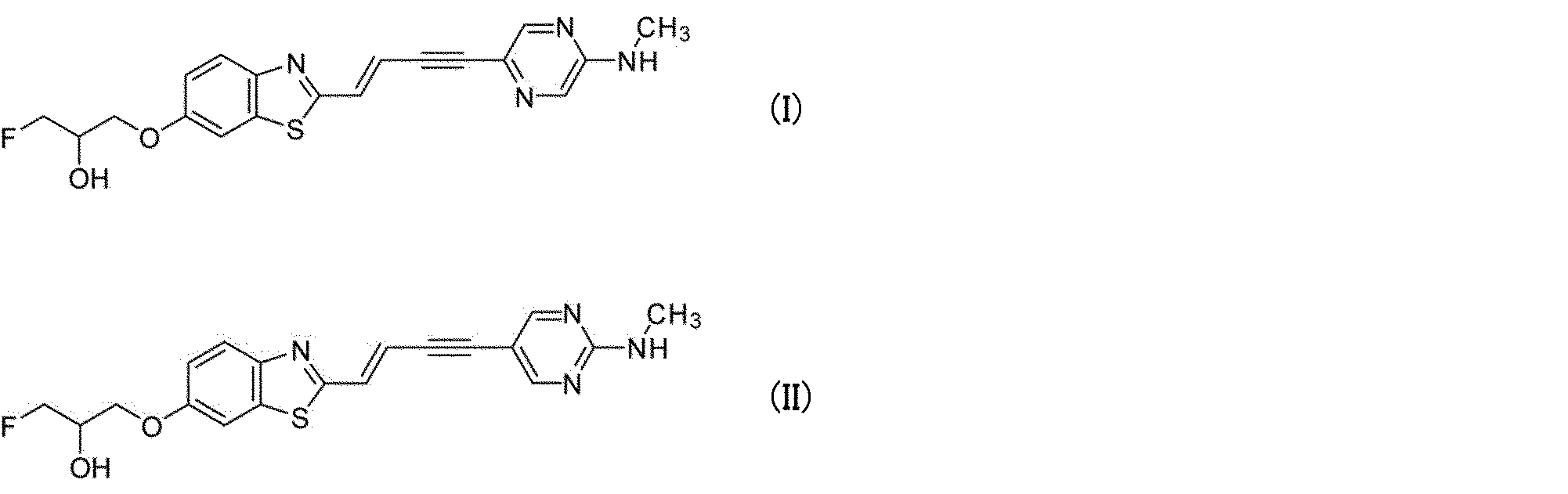
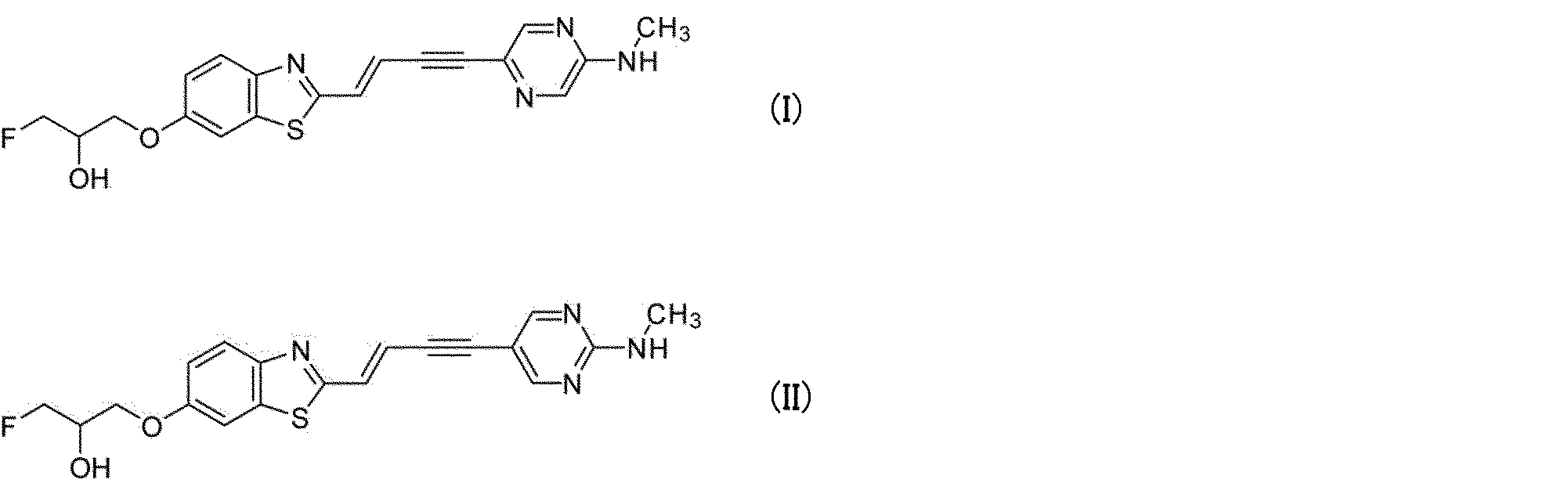
- One aspect of the present invention is a compound represented by the following formula (I) or (II), a pharmaceutically acceptable salt thereof, or a solvate thereof.
- ⁇ -synuclein aggregate binder having high binding selectivity to ⁇ -synuclein aggregates.
- a and / or B is intended to be at least one of A and B.
- the term "medically acceptable salt” refers to a salt that is not harmful to mammals, especially humans.
- Pharmaceutically acceptable salts can be formed using non-toxic acids or bases, including inorganic acids or bases, or organic acids or bases.
- examples of pharmaceutically acceptable salts include metal salts formed from aluminum, calcium, lithium, magnesium, potassium, sodium and zinc, or lysine, N, N'-dibenzylethylenediamine, chloroprocine, choline, There are organic salts formed from diethanolamine, ethylenediamine, meglumine (N-methylglucamine), prokine and the like.
- pharmaceutically acceptable salts include acid-added salts and base-added salts.
- the term "medically acceptable carrier” means a pharmaceutically acceptable material, composition, or vehicle, such as an aqueous solution of physiological saline, a liquid or solid filler, a diluent, a solvent, or an encapsulating material.
- pharmaceutically acceptable carriers include water, saline, saline or phosphate buffered saline (PBS), sodium chloride injection, Ringer injection, isotonic dextrose injection, sterile water injection. , Dextrose, and lactic acid Ringer injection.
- the term "effective amount” refers to the amount of a compound or composition capable of obtaining the desired effect.
- the effective amount refers to the amount of compound or composition capable of optical or radioimaging of substances accumulating in the brain, such as ⁇ -synuclein aggregates.
- solvate means a solvent-containing compound formed by the association of one or more solvent molecules with respect to the compound.
- Solvates include, for example, monosolvates, disolvates, trisolvates, and tetrasolvates.
- the solvate also contains a hydrate.
- hydrate means a compound or a salt thereof further containing a stoichiometric or non-stoichiometric amount of water constrained by a non-covalent intermolecular force. Hydrate includes, for example, monohydrate, dihydrate, trihydrate, tetrahydrate and the like.
- treatment means reducing or ameliorating the progression, severity and / or duration of a disease or condition.
- prevention means reducing the risk of acquiring or developing a given disease or condition, or reducing or suppressing the recurrence, initiation, or progression of one or more symptoms of a given disease or condition. ..
- binding property means the binding strength of a compound to a specific protein aggregate.
- binding selectivity means that when a compound's binding to a particular protein aggregate is compared to its binding to other protein aggregates, there is a difference in their binding (a particular protein agglomeration). (Higher or lower binding to aggregates than to other protein aggregates). “High binding selectivity” means that the above difference in binding properties is large. For example, when a compound has "high binding selectivity to ⁇ -synuclein aggregates", there is a large difference in the binding property of the compound to ⁇ -synuclein aggregates and other protein aggregates, and there is a large difference in the binding properties of ⁇ -synuclein aggregates. It shows that the aggregate has higher binding properties.
- the present invention is a (E) -1-fluoro-3-((2- (4- (5- (methylamino) pyrazine-2-yl) porcine) porcine represented by the following structural formula (I). -1-en-3-in-1-yl) benzo [d] thiazole-6-yl) oxy) propane-2-ol, or (E) -1-fluoro-represented by the following structural formula (II) 3-((2- (4- (2- (Methylamino) pyrimidine-5-yl) buta-1-en-3-in-1-yl) benzo [d] thiazole 6-yl) oxy) propane-2 -Providing an oar, a pharmaceutically acceptable salt thereof, or a solvate thereof.
- compound (I) and compound (II) are a compound in which one or more atoms are radioisotopes of the atom, a salt thereof, or a solvate thereof.
- the radioisotope is selected from the group consisting of 15 O, 13 N, 11 C, 18 F and the like, but is not particularly limited.
- the radioisotope is 11 C or 18 F.
- the half-life of 11C is about 20 minutes and the half-life of 18F is about 110 minutes
- the compound labeled with 18F has higher commercial utility value. Conceivable. Therefore, most preferably, the radioisotope is 18F .
- a methylamino group attached to a pyrimidine ring or a pyrazine ring, and a 3-fluoro-2-hydroxypropoxy group attached to a benzothiazole ring (-O-CH 2 -CH (OH) -CH 2 F).
- a benzothiazole ring -O-CH 2 -CH (OH) -CH 2 F).
- the 3-fluoro-2-hydroxypropoxy group attached to the benzothiazole ring is a group containing a radioisotope.
- the fluorine atom in the 3-fluoro-2-hydroxypropoxy group is the radioisotope.
- the radioisotope-containing compound (I) is preferably [ 18 F]-(E) -1-fluoro-3-((2- (4- (5- (methylamino) pyrazine-)-. 2-Il) Buta-1-en-3-in-1-yl) Benzo [d] Thiazole-6-yl) Oxy) Propane-2-ol.
- the radioisotope-containing compound (II) is preferably [ 18 F]-(E) -1-fluoro-3-((2- (4- (2- (methylamino) pyrimidin-). 5-Il) Buta-1-en-3-in-1-yl) Benzo [d] Thiazole 6-yl) Oxy) Propane-2-ol.
- X and Y are nitrogen atoms on one side and unsubstituted carbon atoms on the other side.
- "unsubstituted carbon atom” indicates CH. That is, in the formula (III), when X is a nitrogen atom (N), Y is an unsubstituted carbon atom (CH), and when X is an unsubstituted carbon atom (CH), Y is a nitrogen atom (N). ).
- R 1 is a hydroxy group or a group represented by the following formula (i).
- Ts represents a p-toluenesulfonyl group
- THP represents a tetrahydro-2H-pyran-2-yl group
- * represents a binding position with a benzothiazole ring.
- R2 is a hydrogen atom or a tert-butoxycarbonyl (Boc) group.
- intermediate compounds are suitably used for the synthesis of compound (I) and compound (II), and further for the synthesis of radioisotope-labeled compound (I) and compound (II). Moreover, these intermediate compounds may be salts.
- compound (I) or compound (II) contains various isomers such as stereoisomers (including optical isomers and rotational isomers), tautomers, or polar isomers, these isomers are contained.
- the body is also included in compound (I) or compound (II).
- Each of these isomers can be obtained as a single product by a known synthesis method and separation method.
- Compound (III) can also include various isomers.
- Compound (I), compound (II) or compound (III) may also be a crystal produced by a known crystallization method.
- the compound (I) and the compound (II) and the intermediate compound (III) can be produced according to the method according to the production method shown below. If desired, deprotection reaction, amidation reaction, ureaization reaction, alkylation reaction, Mitsunobu reaction, oxidation reaction, reduction reaction, halogenation reaction, coupling reaction, nucleophilic addition reaction with carboanion, Grignard reaction, dehydration.
- the compound (I), the compound (II) and the compound (III) can be produced by carrying out the reaction or the like individually or in combination of two or more thereof.
- reaction conditions such as the solvent, the reagent and the temperature in each reaction may be appropriately set based on the common general knowledge of those skilled in the art.
- the protecting and deprotecting reaction of the functional group is carried out according to a known reaction method, a reference example or a method described in Examples, and a conventional protecting group is used.
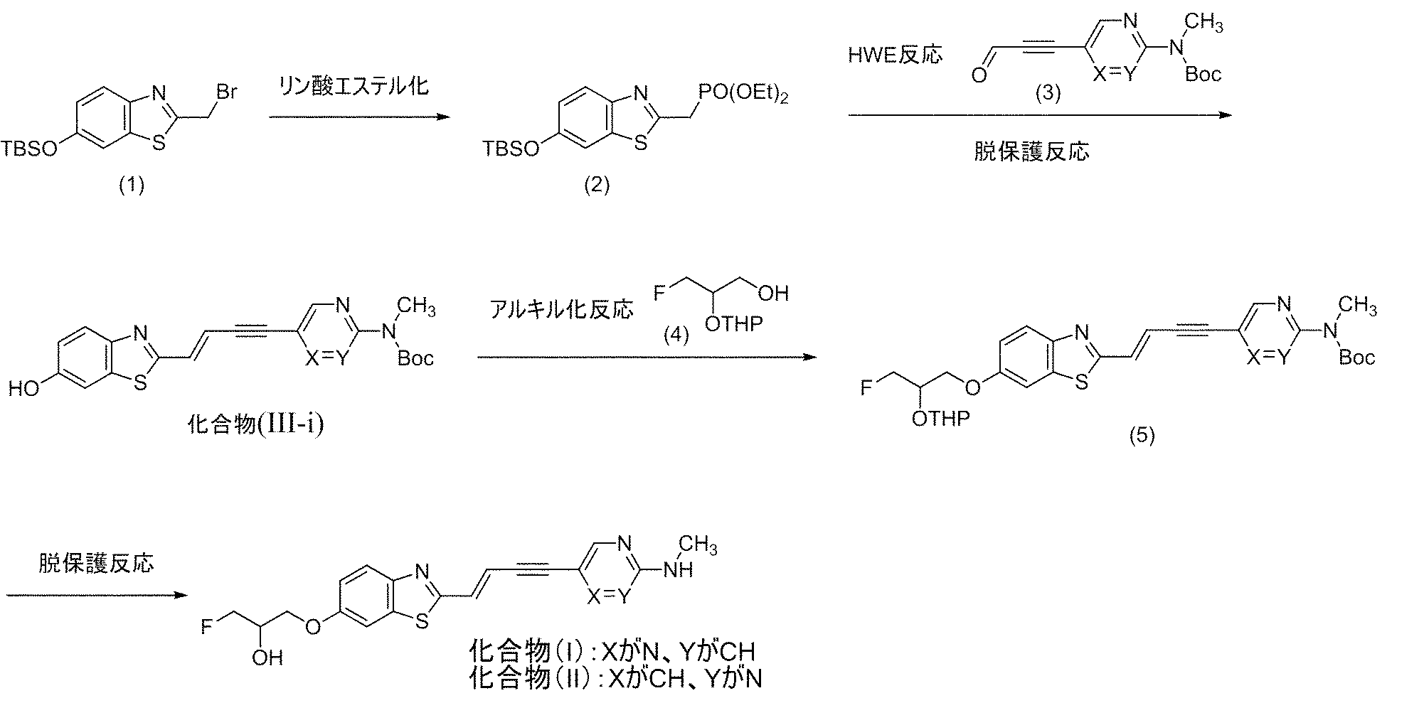
- Compound (2) can be produced by reacting compound (1) with a phosphorous acid triester.
- TBS is a tert-butyldimethylsilyl group.
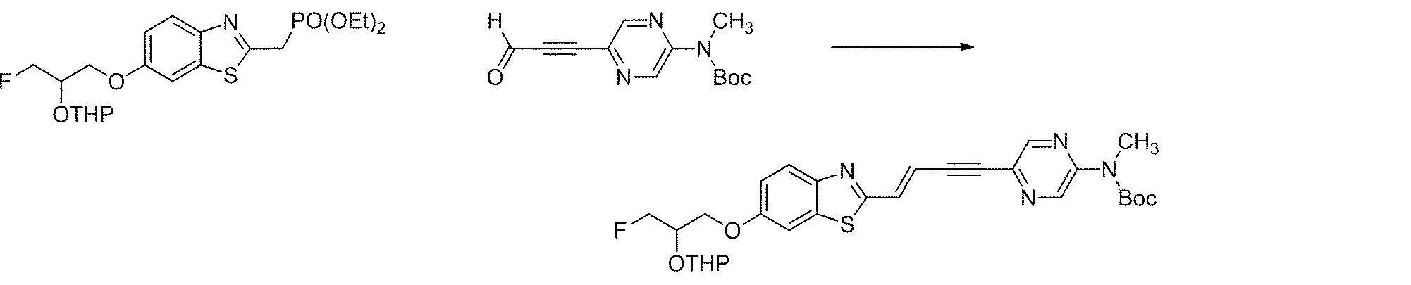
- Compound (III-i) can be produced by the Horner-Wadsworth-Emmons reaction (HWE reaction) between compound (2) and compound (3) described later, and the deprotection reaction that proceeds during the reaction.
- HWE reaction Horner-Wadsworth-Emmons reaction
- X and Y are nitrogen atoms on one side and unsubstituted carbon atoms (CH) on the other side.
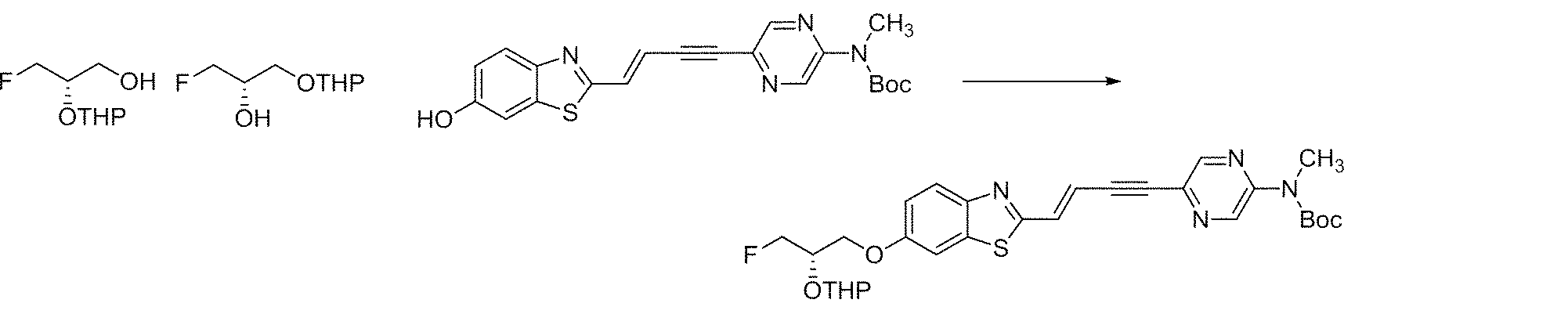
- Compound (5) can be produced by an alkylation reaction such as a Mitsunobu reaction between compound (III-i) and compound (4) described later.
- Compound (I) and compound (II) can be produced by the deprotection reaction of compound (5).
- the compound (3) used in the production method A can be produced from the compound (6) by the following method shown in the production scheme 2.
- H represents a halogen atom (eg, chlorine atom, bromine atom, iodine atom).
- the compound (7) can be produced by a coupling reaction between the compound (6) and 2-propyne-1-ol or the like.
- Compound (3) can be produced by an oxidation reaction of compound (7).
- the compound (4) used in the production method A can be produced from the compound (8) by the following method shown in the production scheme 3.
- Compound (9) can be produced by the protective reaction of compound (8).
- Compound (4) can be produced by the debenzyl reaction of compound (9).
- the compound (5) used in the production method A can also be produced from the compound (2) by the following method shown in the production scheme 4.
- Compound (10) can be produced by the deprotection reaction of compound (2).
- Compound (11) can be produced by an alkylation reaction such as a Mitsunobu reaction between compound (10) and compound (4).
- Compound (5) can also be produced by HWE reaction between compound (11) and compound (3).
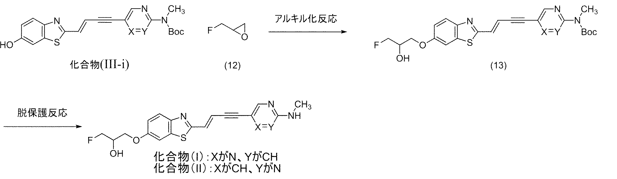
- Compound (13) can be produced by an alkylation reaction to compound (III-i) by an oxylane ring-opening reaction of compound (12).
- Compound (I) and compound (II) can be produced by the deprotection reaction of compound (13).
- Compound (15) can be produced by an alkylation reaction such as a Mitsunobu reaction between compound (III-i) and compound (14) described later.
- Compound (16) can be produced by the deprotection reaction of compound (15).
- Compound (III-iii) can be produced by the protective reaction of compound (16).
- the compound (14) used in the production method D can be produced by the following method shown in the production scheme 8.
- Compound (18) can be produced by the protective reaction of compound (17).
- Compound (14) can be produced by the debenzyl reaction of compound (18).
- the invention provides an alpha-synuclein aggregate binder.
- the ⁇ -synuclein aggregate binder of the present embodiment (hereinafter, also referred to as a binder or an ⁇ -synuclein aggregate binder) is compound (I) or compound (II), a pharmaceutically acceptable salt thereof, or a solvate thereof. Contains substances.
- Compound (I) and compound (II) have higher binding selectivity to ⁇ -synuclein aggregates than aggregates of tau protein and amyloid ⁇ .
- pharmaceutically acceptable salts of compound (I) and compound (II), and solvates of compound (I) and compound (II) are also ⁇ -synucleins rather than aggregates of tau protein and amyloid ⁇ .
- compound (I) and compound (II) fluoresce.
- one or more atoms can be used as a radioisotope of the atom. Therefore, the binder of the present embodiment can be used as a molecular probe for optical imaging or radiographic imaging of ⁇ -synuclein aggregates accumulated in the brain.
- ⁇ -synuclein is a protein localized in neuronal synapses even in a normal brain, and ⁇ -synuclein aggregates are ⁇ -synuclein aggregates.
- the ⁇ -synuclein aggregate binder can contain a pharmaceutically acceptable carrier.
- pharmaceutically acceptable carriers include water, saline, saline or phosphate buffered saline (PBS), sodium chloride injection, Ringer injection, isotonic dextrose injection, sterile water injection. There are solutions, dextrose, and lactic acid Ringer injection.
- the content of compound (I) or compound (II) contained in the ⁇ -synuclein aggregate binder, a pharmaceutically acceptable salt thereof, or a solvate thereof, and a pharmaceutically acceptable carrier is not particularly limited. These contents are the type of compound used; the age, weight, health status, sex and dietary content of the mammal to be administered; the number of administrations and the route of administration; the treatment period; other drugs used at the same time, etc. , Determined by various factors.
- the pharmaceutically acceptable carrier content can be 1 to 99% by weight of the ⁇ -synuclein aggregate binder.
- the ⁇ -synuclein aggregate binder is prepared so that compound (I) or compound (II) can be administered, for example, in an amount of 5 ng / kg to 5 mg / kg per body weight (kg) of the subject. ..
- the lower limit of the amount of the compound is 5 ng / kg or more, 0.01 mg / kg or more, 0.05 mg / kg or more, or 0.1 mg / kg or more.
- the upper limit of the amount of the compound is 5 mg / kg or less, 3 mg / kg or less, 1 mg / kg or less, or 20 ⁇ g / kg or less.
- the present invention provides a composition for optical imaging of ⁇ -synuclein aggregates.
- the composition for optical imaging of the ⁇ -synuclein aggregate of the present embodiment contains the above-mentioned binder according to the present embodiment.
- the optical imaging includes in vitro, ex vivo, and in vivo imaging.
- optical imaging examples include a fluorescence microscope measurement method, a multiphoton imaging method, a two-photon imaging method, and a near-infrared fluorescence imaging method.
- composition for optical imaging can contain a pharmaceutically acceptable carrier.
- pharmaceutically acceptable carriers include water, saline, saline or phosphate buffered saline (PBS), sodium chloride injection, Ringer injection, isotonic dextrose injection, sterile water injection. There are solutions, dextrose, and lactic acid Ringer injection.
- the content of compound (I) or compound (II) contained in the composition for optical imaging, a pharmaceutically acceptable salt thereof, or a solvate thereof, and a pharmaceutically acceptable carrier is not particularly limited. These contents are the type of compound used; the age, weight, health status, sex and dietary content of the mammal to be administered; the number of administrations and the route of administration; the treatment period; other drugs used at the same time, etc. , Determined by various factors.
- the pharmaceutically acceptable carrier content can be from 1 to 99% by weight of the optical imaging composition.
- the composition for optical imaging contains compound (I) or compound (II), for example, the amount of compound (mg) per subject body weight (kg) is 0.01 mg / kg to 5 mg / kg, preferably 0.05 mg. It is prepared so that it can be administered in an amount of / kg to 3 mg / kg, more preferably 0.1 mg / kg to 1 mg / kg.
- composition for radiation imaging of ⁇ -synuclein aggregates contains the above-mentioned binder according to the present embodiment.
- the radiation imaging includes in vitro, ex vivo, and in vivo imaging.
- Examples of radiation imaging include positron emission tomography (PET), single photon emission tomography (SPECT), and autoradiography.
- PET positron emission tomography
- SPECT single photon emission tomography
- autoradiography autoradiography
- composition for radiation imaging can contain a pharmaceutically acceptable carrier.
- pharmaceutically acceptable carriers include water, saline, saline or phosphate buffered saline (PBS), sodium chloride injection, Ringer injection, isotonic dextrose injection, sterile water injection. There are solutions, dextrose, and lactic acid Ringer injection.
- the content of compound (I) or compound (II) contained in the composition for radiation imaging, a pharmaceutically acceptable salt thereof, or a solvate thereof, and a pharmaceutically acceptable carrier is not particularly limited. These contents are the type of compound used; the age, weight, health status, sex and dietary content of the mammal to be administered; the number of administrations and the route of administration; the treatment period; other drugs used at the same time, etc. , Determined by various factors.
- the pharmaceutically acceptable carrier content can be from 1 to 99% by weight of the radioimaging composition.
- composition for radiation imaging contains compound (I) or compound (II), for example, the amount of the compound per body weight (kg) of the subject is 5 ng / kg to 5 mg / kg, preferably 5 ng / kg to 20 ⁇ g / kg. Prepared for administration in volume.
- the present invention provides a diagnostic agent for a disease associated with ⁇ -synuclein aggregates, or a companion diagnostic agent for the treatment or prevention of the disease.
- the diagnostic agent for a disease related to ⁇ -synuclein aggregates of the present embodiment, or the companion diagnostic agent for the treatment or prevention of the disease (hereinafter, also referred to as a companion diagnostic agent) is the above-mentioned binding agent according to the present embodiment.
- a companion diagnostic drug for treatment is a diagnostic drug for determining whether or not treatment can be expected when a disease is found.
- a companion diagnostic drug for prevention is a diagnostic drug for predicting future onset or determining whether prevention to suppress the onset is expected when it is found to be a precursor state of the disease. be.
- the diagnostic agent Using the diagnostic agent according to the present embodiment, data on the amount and / or distribution amount of ⁇ -synuclein aggregates in the brain obtained from the subject is obtained with the disease and the amount and / or distribution amount of ⁇ -synuclein aggregates obtained in advance. By collating with the correlation, it is possible to make a diagnosis regarding the target disease (specifically, the presence or absence of the disease, the severity, the possibility of seizure, etc.).
- a companion diagnostic agent data on the amount and / or distribution of ⁇ -synuclein aggregates in the brain obtained from the subject can be obtained with the disease and the amount and / or distribution of ⁇ -synuclein aggregates in the brain obtained in advance.
- the disease state of the target can be grasped. Therefore, based on this, a disease prevention / treatment plan (type, combination, dose, use, etc. of the prophylactic or therapeutic agent to be administered) can be formulated.
- One embodiment of the present invention is a pharmaceutical for treating or preventing a disease associated with ⁇ -synuclein aggregates, which is administered based on data on the amount and / or distribution of ⁇ -synuclein aggregates in the brain obtained by companion diagnostics. It also relates to the medicines given in the plan.
- the diagnostic kit for diseases related to substances accumulated in the brain of the present invention includes the above-mentioned binder according to the present embodiment.
- Substances that accumulate in the brain include at least ⁇ -synuclein aggregates, and other examples include tau protein or amyloid ⁇ aggregates.
- Diseases associated with substances that accumulate in the brain include at least ⁇ -synuclein aggregate-related diseases, such as Parkinson's disease, Lewy body dementias (DLB), and multiple system atrophy (MSA). Can be mentioned.
- Other diseases associated with substances that accumulate in the brain include Alzheimer's disease (AD) and frontotemporal lobar degeneration, which are diseases associated with aggregates of tau protein or amyloid ⁇ .
- AD Alzheimer's disease
- frontotemporal lobar degeneration which are diseases associated with aggregates of tau protein or amyloid ⁇ .
- the binder of this embodiment has higher binding selectivity to ⁇ -synuclein aggregates than aggregates of tau protein and amyloid ⁇ .
- the compound described in Patent Document 1 has higher binding property to the tau protein aggregate than the ⁇ -synuclein aggregate.
- the diagnostic kit comprises a compound (I) or compound (II) having high binding selectivity to ⁇ -synuclein aggregates, a pharmaceutically acceptable salt thereof or a solvate thereof, and a tau protein. It can contain both other compounds having high binding selectivity to aggregates (eg, compounds described in Patent Document 1).
- a diagnostic kit by comparing the former detection result (amount and / or distribution of detected light or radiation) with the latter detection result, which substance is present in each imaged region. I can identify it. Specifically, ⁇ -synuclein aggregates and tau protein aggregates can be classified, and the abundance of each can be quantified.
- the ratio of the binding property to the ⁇ -synuclein aggregate to the binding property to the tau protein aggregate is determined by the binder of the present embodiment and a substance having high binding selectivity to the tau protein aggregate (for example, Patent Document).
- a substance having high binding selectivity to the tau protein aggregate for example, Patent Document.
- Each of the compounds described in 1) is specified in advance. Then, if the amount ratio of light or radiation in each region of the brain after administration of the former and the latter in the subject is measured, ⁇ -synuclein aggregates can be found in the region due to the relationship between the ratio specified in advance and the measured dose ratio. It is possible to classify which of the tau protein aggregates is present and quantify each amount.
- the above-mentioned diagnostic kit of the present embodiment can be further used as a diagnostic kit in combination with an amyloid ⁇ imaging agent.
- the diagnostic kit in which the diagnostic kit of the present embodiment and the imaging agent of amyloid ⁇ are combined can identify which substance is present in each imaged region. Specifically, ⁇ -synuclein aggregates, tau protein aggregates, and amyloid ⁇ aggregates can be classified, and the abundance of each can be quantified. Therefore, it is possible to diagnose a disease associated with ⁇ -synuclein aggregate, a disease related to tau protein, and / or a disease associated with amyloid ⁇ with high accuracy.
- One embodiment of the present invention is a pharmaceutical for treating or preventing a disease related to ⁇ -synuclein aggregates, and the amount of a substance accumulated in the brain containing ⁇ -synuclein aggregates obtained by the diagnostic kit according to the present embodiment. And / or related to drugs administered in a dosing regimen based on data on distribution.
- the invention provides an optical imaging method.
- the optical imaging method of the present embodiment is a first method, which is emitted from the living body brain of a subject to which the binder according to the present embodiment is administered, after irradiating the living body brain with light of the first wavelength from outside the brain. Includes a step of detecting light having a second wavelength different from that of.
- the binder transferred to the living brain binds to the ⁇ -synuclein aggregate present in the living brain.
- Light of the first wavelength that excites the binder is irradiated from outside the brain to the living body brain of the subject to which the binder is administered, and light of the second wavelength emitted from the binder in the brain (for example, fluorescence).
- optical imaging (imaging) of ⁇ -sinucrane aggregates can be performed.
- Mammals include, for example, humans, rats, mice, rabbits, guinea pigs, hamsters, monkeys, dogs, ferrets, or mini pigs.
- the administration method is not particularly limited, and examples thereof include oral administration and parenteral administration such as intravenous administration or intraperitoneal administration. Intravenous administration or intraperitoneal administration is preferable. Most preferably, it is administered intravenously.
- the dose is preferably 0.01 mg / kg to 5 mg / kg, 0.05 mg / kg to 3 mg / kg, or 0.1 mg / kg to 1 mg / kg, most preferably 0.1 mg / kg to. It is 1 mg / kg.
- the invention provides a radiation imaging method.
- the radioimaging method of the present embodiment comprises compound (I) or compound (II) in which one or more atoms are radioisotopes of the atom, a pharmaceutically acceptable salt thereof, or a solvate thereof.
- the step includes a step of detecting the radiation emitted from the living brain of the subject to which the binder according to the present embodiment is administered.
- the binder transferred to the living brain binds to the ⁇ -synuclein aggregate present in the living brain.
- the radiation imaging imaging
- mammals examples include, for example, humans, rats, mice, rabbits, guinea pigs, hamsters, monkeys, dogs, ferrets, or mini pigs.
- the mammal is a human.
- the administration method is not particularly limited, and examples thereof include oral administration and parenteral administration such as intravenous administration or intraperitoneal administration. Intravenous administration or intraperitoneal administration is preferable. Most preferably, it is administered intravenously.
- the dose is preferably 5 ng / kg to 5 mg / kg, more preferably 5 ng to 20 ⁇ g / kg.
- the dose of radioactivity is preferably 37 MBq to 7.4 GBq per individual, and more preferably 370 MBq to 3,700 MBq.
- the present invention provides a method for screening a therapeutic or prophylactic agent for a disease associated with ⁇ -synuclein aggregates in the brain.
- the method for screening a disease related to ⁇ -synuclein aggregates in the brain of the present embodiment or a preventive agent is an optical imaging according to the present embodiment before and after administration of a candidate substance to a subject. It comprises the step of selecting a candidate substance based on the difference in the amount and / or distribution of light or radiation detected by the method or radiation imaging method.
- the subject and administration method are the same as those described in the above-mentioned [optical imaging method] and [radiation imaging method].
- the candidate substance is the disease or symptom.
- the candidate compound may be useful as a therapeutic compound for the disease or condition.
- data on the amount (intensity) and / or distribution of light or radiation such as fluorescence of the binder before and after administration of the candidate substance obtained from the subject to which the candidate substance was administered was obtained in advance for feeding the candidate substance without administration.
- changes in the amount and / or distribution of light or radiation, such as the fluorescence of the binder before and after the onset of the disease associated with ⁇ -synuclein aggregates in the brain in animals. Then, the increase in the light or radiation dose such as fluorescence observed after the onset of the disease is suppressed by the administration of the candidate substance, and / or the light or radiation dose such as the fluorescence is suppressed after the administration of the candidate substance.
- Candidate substances may be useful as prophylactic compounds for the disease or condition if they show values close to those of normal mammals as before administration. Similarly, the change in the distribution of light or radiation such as fluorescence observed after the onset of the disease is suppressed by the administration of the candidate substance, and / or the change in the distribution of light or radiation such as fluorescence is the candidate substance.
- the candidate substance may be useful as a preventive compound for the disease or symptom after administration of the above, even if the distribution is close to that in a normal mammal as before administration.
- the present invention provides a method for quantifying or determining the accumulation of ⁇ -synuclein aggregates in the brain.
- the above-mentioned binder containing compound (I) or compound (II), a pharmaceutically acceptable salt thereof, or a solvate thereof is administered.
- the detection includes a step of irradiating the living body brain of the subject with light of a first wavelength from outside the brain, and then detecting light of a second wavelength different from the first wavelength emitted from the brain.
- the accumulation of ⁇ -synuclein aggregates in the brain is quantified or determined based on the amount and / or distribution of the emitted light.
- This method is a method of quantifying or determining the accumulation of ⁇ -synuclein aggregates in the brain by optical imaging.
- the subject and administration method are the same as those described in the above [optical imaging method].
- compound (I) or compound (II), wherein one or more atoms are radioactive isotopes of the atom is administered, and the amount of the detected radiation and / Or, based on the distribution, the accumulation of ⁇ -synuclein aggregates in the brain is quantified or determined.
- This method is a method for quantifying or determining the accumulation of ⁇ -synuclein aggregates in the brain by radioimaging.
- Quantifying the accumulation of ⁇ -synuclein aggregates in the brain and in the brain by determining the difference between the amount and / or distribution of detected light or radiation of the subject and that of other normal mammals. It is possible to determine the presence or absence of accumulation of ⁇ -synuclein aggregates.
- the present invention provides a method for determining the classification and accumulation of substances that accumulate in the brain.
- the method for determining the classification and accumulation of substances accumulating in the brain of the present invention comprises a binding according to the present embodiment comprising compound (I) or compound (II), a pharmaceutically acceptable salt thereof, or a solvate thereof.
- the amount of light detected in the first step including the second step of detecting the light of the fourth wavelength different from the third wavelength emitted from the brain after irradiating the light of the wavelength of Based on the and / or distribution data and the amount and / or distribution data of the light detected in the second step, the classification and accumulation of the substances accumulated in the brain are determined.
- This method is a method of determining the classification and accumulation of substances accumulated in the brain by optical imaging.
- the subject and administration method are the same as those described in the above [optical imaging method].
- the first step of detecting the light emitted from the brain caused by the administration of a binder having high binding selectivity to ⁇ -synuclein aggregates, and the substance having high binding selectivity to tau protein aggregates in the same subject Since it includes both a second step of detecting the light emitted from the brain due to administration, the detection result of the first step (amount and / or distribution of the detected light) and the detection result of the second step. By comparing with, it is possible to determine the classification and accumulation of whether or not the substance accumulated in the brain is ⁇ -synuclein aggregate and / or tau protein aggregate.
- compound (I) or compound (II), wherein one or more atoms are radioactive isotopes of the atom In another aspect of the method of determining the classification and accumulation of substances accumulating in the brain of this embodiment, compound (I) or compound (II), wherein one or more atoms are radioactive isotopes of the atom.
- a second step of detecting radiation emitted from the brain of the subject to which a substance having high binding selectivity (for example, a compound described in Patent Document 1) is administered at a time different from that of the first step is included.
- This method is a method of determining the classification and accumulation of substances accumulated in the brain by radiation imaging.
- One aspect of the present invention is a compound represented by the following formula (I) or (II), a pharmaceutically acceptable salt thereof, or a solvate thereof.
- One aspect of the present invention is an ⁇ -synuclein aggregate binder containing the compound, a pharmaceutically acceptable salt thereof, or a solvate thereof.
- One aspect of the present invention is a composition for optical imaging of ⁇ -synuclein aggregates containing the binder.
- One aspect of the present invention is a composition for radiation imaging of ⁇ -synuclein aggregates containing the binder.
- One aspect of the present invention is a second aspect different from the first wavelength emitted from the brain after irradiating the living body brain of the subject to which the binding agent is administered with light having a first wavelength from outside the brain. It is an optical imaging method of ⁇ -synuclein aggregates in the brain, which comprises a step of detecting light of a wavelength.
- One aspect of the present invention is a radiation imaging method for ⁇ -synuclein aggregates in the brain, which comprises a step of detecting radiation emitted from the living brain of a subject to which the binder is administered.
- One aspect of the present invention is an intermediate for synthesizing the compound represented by the following formula (III).
- One of X and Y is a nitrogen atom (N) and the other is an unsubstituted carbon atom (CH) (that is, if X is a nitrogen atom, Y is an unsubstituted carbon atom (CH). , If X is an unsubstituted carbon atom (CH), then Y is a nitrogen atom); R 1 is a hydroxy group or a group represented by the following formula (i);
- Ts represents a p-toluenesulfonyl group and represents.
- THP represents a tetrahydro-2H-pyran-2-yl group and represents * Represents the binding position with the benzothiazole ring.
- R2 is a hydrogen atom or a tert-butoxycarbonyl (Boc) group).
- One aspect of the present invention is a diagnostic kit containing the compound (I) or compound (II) having high binding selectivity to ⁇ -synuclein aggregates.
- the "room temperature" in the following production examples usually indicates about 10 ° C to about 35 ° C. % Indicates weight% unless otherwise specified. Further, with respect to the description of the compound used in each production example, "manufactured in Reference Example (Example) X" is intended to include the case of “manufactured in the same manner as in Reference Example (Example) X”.
- elution in column chromatography of the production example was performed under observation by TLC (Thin Layer Chromatography, thin layer chromatography).
- TLC Thin Layer Chromatography, thin layer chromatography
- 60 F254 manufactured by Merck was used as the TLC plate
- the solvent used as the elution solvent in the column chromatography was used as the developing solvent.
- a UV detector was used for detection.
- M Molar concentration DMSO-d 6 : Deuterated dimethyl sulfoxide 1 H NMR: Proton nuclear magnetic resonance DIEA: N-ethyl-N-isopropylpropane-2-amine DMF: N, N-dimethylformamide (also referred to as "dimethylformamide" in the present specification).
- THF Tetrahydrofuran MeOH: Methanol EtOH: Ethanol DMSO: Dimethyl sulfoxide
- TEA Triethylamine
- TFA Trifluoroacetic acid.
- Methyl iodide (3.45 ml) was added to a solution of tert-butyl (5-iodopyrazine-2-yl) carbamate (13.7 g) and cesium carbonate (20.9 g) in DMF (85 ml) prepared in Reference Example 2. Added at ° C. The mixture was stirred at room temperature from 0 ° C. overnight, cooled to 0 ° C. and diluted with water. The precipitated solid was collected by filtration, washed with water and dried to give the title compound (13.0 g) as a beige solid.
- the filtrate was concentrated under reduced pressure, the residue was diluted with ethyl acetate, acidified with a 5% citric acid aqueous solution, filtered through cerite, and the filtrate was extracted with ethyl acetate.
- the extract was washed with water and brine, dried over anhydrous sodium sulfate, and concentrated under reduced pressure through a silica gel pad to give the title compound (10.2 g) as a brown solid.
- the precipitated solid was collected by filtration, washed with water and ethyl acetate, and dried.
- the filtrate and washing solution were combined and extracted with ethyl acetate.
- the extract was washed with water and brine, dried over anhydrous sodium sulfate, and concentrated under reduced pressure.
- the solid residue was suspended in ethyl acetate, and the solid obtained by filtration was washed with ethyl acetate and dried.
- This solid and the solid collected by filtration before extraction were suspended in ethyl acetate, and the solid obtained by filtration was washed with ethyl acetate and dried to give the title compound (1.07 g) as a yellow solid.
- Example 8 (E) -3-((2- (4- (5-((tert-butoxycarbonyl) (methyl) amino) pyrazine-2-yl) pig-1-en-3-in-1-) Il) Benzo [d] Thiazole-6-yl) Oxy) -2-((Tetrahydro-2H-pyran-2-yl) Oxy) Propyl 4-methylbenzenesulfonate
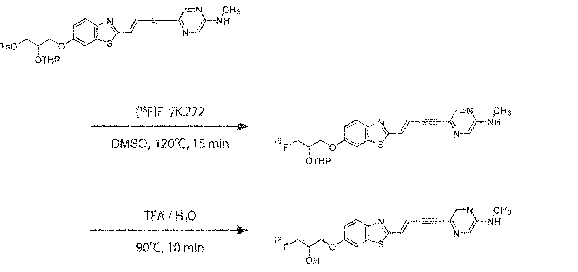
- Example 10 Production Example 1 of [ 18 F] SCAL-T-06
- SPAR-T-06 (hereinafter, [ 18 F] SPAR-T-06) having a fluorine atom of 18 F was produced.
- Example 11 Production Example 2 of [ 18 F] SCAL-T-06 [ 18F ] SCAL-T-06 was produced by the following scheme.
- Optical imaging of human brain Postmortem human brains were obtained from autopsy performed on patients with Lewy body dementia (DLB) and patients with Alzheimer's disease (AD). Frozen DLB tissue was sliced 20 ⁇ m thick in a cryostat (HM560, Carl Zeiss). In addition, AD brain tissue was fixed in 10% neutral buffered formalin, embedded in a paraffin block, and sliced to a thickness of 6 ⁇ m.
- the arrowhead indicates the fluorescence of the compound bound to the ⁇ -synuclein aggregate in the brain of the DLB patient
- the arrow indicates the fluorescence of the compound bound to the amyloid ⁇ aggregate in the brain of the AD patient
- the asterisk indicates the tau aggregate in the brain of the AD patient. Shows the fluorescence of the bound compound.
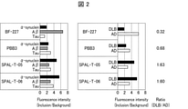
- the fluorescence brightness of the lesion region and the lesion non-formation region (background) was quantified using analysis software (Image J). The results are shown in FIG. As the test compounds, SCAL-T-05 and SCAL-T-06 were used.
- BF-227 (2- (2- [2-dimethylaminothiazole-5-yl] ethenyl) -6- (2- [fluoro] ethoxy) benzoxazole obtained from Nard Institute (catalog). Numbers NP039-0)) and PBB3 (2-((1E, 3E) -4- (6- (methylamino) pyridin-3-yl) pig-1,3-dienyl) benzo [d] thiazole-6-ol ) (Catalog number NP039-0) was used.
- SCAL-T-06 and SCAL-T-05 bind to ⁇ -synuclein aggregates formed in the patient's brain of DLB with a stronger intensity than PBB3. .. It was also confirmed that the binding of SCAL-T-06 and SCAL-T-05 to ⁇ -synuclein aggregates is stronger than the binding of tau or amyloid ⁇ formed in the patient brain of AD to the aggregates. .. That is, it was shown that the compound of the present invention has high binding selectivity to ⁇ -synuclein aggregates.
- the brains of patients with multiple system atrophy (MSA) were also measured with an in vitro fluorescence microscope in the same manner as the brains of DLB patients, and similar results were obtained.
- BF-227 which has been reported as a PET probe for ⁇ -synuclein lesions, mainly binds to amyloid ⁇ aggregates of AD, and the binding to ⁇ -synuclein aggregates is to SPAL-T-06 and SPAL-T-05. It was confirmed that it was weak in comparison. That is, it was shown that BF-227 has low binding selectivity to ⁇ -synuclein aggregates.
- Mouse ⁇ -synuclein is expressed and extracted in E. coli as a recombinant protein and incubated in vitro to form insoluble aggregates.
- this ⁇ -synuclein aggregate is inoculated into the striatum of a mouse, the ⁇ -synuclein aggregate propagates to the surrounding area via a neural circuit, and after several months, an ⁇ -synuclein lesion is observed in the neocortex ( Masuda-Suzukake et al. Acta Neuropathol Commun 2, 88, 2014; Shimozawa et al.
- mouse ⁇ -synuclein was expressed and extracted in Escherichia coli as a recombinant protein and incubated in vitro to form insoluble ⁇ -synuclein aggregates. Then, when this ⁇ -synuclein aggregate was inoculated into the striatum of a mouse, the ⁇ -synuclein aggregate propagated to the surrounding region via a neural circuit, and a few months later, an ⁇ -synuclein lesion was observed in the neocortex. Was done.
- mice The striatum of mice was inoculated with this ⁇ -synuclein aggregate by the following method. First, remove the hair on the head of a 9-week-old C57 / BL / 6 male mouse anesthetized with 1.5% (v / v) isoflurane, disinfect the scalp with isodine, apply xylocaine, and make a notch in the scalp. The scalp was exposed.
- ⁇ -synuclein fiber solution (mouse ⁇ -synuclein fiber 4 mg / ml in saline) was injected at a depth of 2 ⁇ m using a glass pipette. After that, the scalp was returned and sutured.
- the right side is anti-phosphorylated ⁇ -synuclein antibody staining.
- On the left side are SPAL-T-05 and SPAL-T-06 fluorescent stains.
- arrowheads show lesions consisting of phosphorylated ⁇ -synuclein in ⁇ -synuclein fiber-inoculated mouse brain and fluorescence of compounds bound to them.
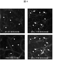
- mice were fixed under a two-photon laser fluorescence microscope, 100 ⁇ l of physiological saline containing 5 mM of sulforhodamine 101 was intraperitoneally administered, and then biological two-photon fluorescence imaging was performed at an excitation wavelength of 900 nm.
- the detection wavelength for BF-227, PBB3, SCAL-T-05 and SPAR-T-06 was 500 to 550 nm, and the detection wavelength for sulforhodamine 101 was 573 to 648 nm.
- the results are shown in FIG. In FIG. 4, the arrowheads indicate blood vessels, and the arrows indicate fluorescence of the compound bound to the ⁇ -synuclein lesion.
- PET scans are 0.851 mm thick (intercentric) 95 slices, 19.0 cm axial field of view (FOV), and 7.6 cm intra-section FOV, provided by micro PET Focus 220 Animal Scanner (Siemens Medical Solutions).
- FOV axial field of view
- micro PET Focus 220 Animal Scanner Animal Scanner (Siemens Medical Solutions).
- Prior to scanning ⁇ -synuclein fiber-inoculated mice and saline-inoculated mice (controls) were anesthetized with 1.5% (v / v) isoflurane.
- Emission scans were performed in 3D list mode for 90 minutes after intravenous injection of [ 18F ] SPAR-T-06 (a compound labeled SVAL-T-06 with positron-releasing nuclides) (30.1 ⁇ 0.13MBq).
- the energy window was 350-750 keV.
- Injections and scans of the radioactive compound were performed in dim light to avoid photoisomerization of the compound.
- All list mode data was sorted into 3D synograms and then converted into 2D synograms by Fourier-rebining (frames: 10x1, 6x5, 5x10 minutes). Addition images at 0-30 minutes, 30-60 minutes, and 60-90 minutes after injection of the radioactive compound were obtained by reconstruction by maximum a posteriori estimation.
- the dynamic image was reconstructed by the filter correction back projection method using a 0.5 mm Hanning filter.
- the volume of interest (VOI) was set in the striatum, cerebral cortex and cerebellum using PMOD image analysis software (PMOD Technologies) with reference to the MRI template. The results are shown in FIGS. 5 to 7.
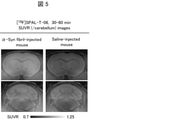
- FIG. 5 shows the results in the case of ⁇ -synuclein fiber-inoculated mice 30 to 60 minutes after intravenous injection of [ 18F ] SPAR-T-06
- the right side of FIG. 5 shows the results in the case of saline-inoculated mice.
- the upper part is an image of the coronal section of the brain including the striatum
- the lower part is the image of the coronal section of the brain including the cerebellum, which are displayed superimposed on the standard brain MRI image.
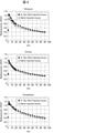
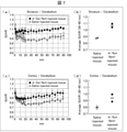
- FIG. 6 is a time-radioactivity curve after intravenous injection of [ 18 F] SPAR-T-06 in the striatum, cerebral cortex, and cerebellum
- FIG. 7 (a) to (d) are [ 18 F].
- Comparison with water-injected mice (n 2, mean ⁇ SEMs).
- FIG. 7 (a) shows the temporal transition of the striatum sUVR with the cerebellum as the control region after [ 18 F] SPAR-T-06 intravenous injection
- FIG. 7 (b) shows the striatum sUVR.
- the average of ( control area: cerebellum) at 30 to 60 minutes is shown in FIG. 7 (c).
- (d) of FIG. 7 shows the average of SUVR (control area: cerebellum) of the cerebral cortex at 30 to 60 minutes, respectively.
- SPAN-T-06 was labeled with positron-releasing nuclei, and PET scans were performed by intravenous injection into ⁇ -synuclein fiber-inoculated model mice and saline-injected mice (controls). As shown in FIGS. 5 to 7, appropriate. It has been shown that it has a good brain transferability or clearance rate from the brain, and exhibits good properties as a probe for imaging ⁇ -synuclein aggregates with PET.
- an intraorgan tube was intubated into a marmoset immobilized with ketamine (5-10 mg / kg) and xylazine (0.2-0.5 mg / kg), and then anesthetized with 1-3% (v / v) isoflurane. ..
- the hair on the marmoset's head was removed, the scalp was disinfected with isodine, then xylocaine was applied, and a notch was made in the scalp to expose the skull.
- PET scans are 0.851 mm thick (intercentric) 95 slices, 19.0 cm axial field of view (FOV), and 7.6 cm intra-section FOV, provided by micro PET Focus 220 Animal Scanner (Siemens Medical Solutions).
- FOV axial field of view
- micro PET Focus 220 Animal Scanner Animal Scanner (Siemens Medical Solutions).
- Prior to scanning alpha-synuclein fiber-inoculated marmosets were anesthetized with 1-3% (v / v) isoflurane.
- the transmission scan was performed for about 20 minutes using a PET calibration source Ge-68.
- Emission scans were performed 90 minutes after intravenous injection of [ 18F ] SPAR-T-06 (a compound labeled SVAL-T-06 with positron-emitting nuclides) (73.0MBq) in 3D wrist mode, energy window 350-750keV. I went there. Injections and scans of the radioactive compound were performed in dim light to avoid photoisomerization of the compound. All list mode data was sorted into 3D synograms and then converted into 2D synograms by Fourier-rebining (frames: 10x1, 6x5, 5x10 minutes). Addition images at 0-30 minutes, 30-60 minutes, and 60-90 minutes after injection of the radioactive compound were obtained by reconstruction by maximum a posteriori estimation.
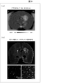
- FIG. 9A is a coronal cross-sectional image of the brain containing the caudate nucleus of the ⁇ -synuclein fiber-inoculated marmoset 20 to 60 minutes after the intravenous injection of [ 18F ] SPAR-T-06, superimposed on the standard brain MRI image. Is displayed.
- 9B is an anti-phosphorylated ⁇ -synuclein antibody-stained image of a coronal section of the brain containing the caudate nucleus of an ⁇ -synuclein fiber-inoculated marmoset.
- 2 is an enlarged image of antiphospholipid ⁇ -synuclein antibody staining of the caudate nucleus of the right brain infused with ⁇ -synuclein fiber
- 1 is an enlarged image of antiphospholipid ⁇ -synuclein antibody staining of the caudate nucleus of the left brain infused with physiological saline.
- B / F separation (B: binding ligand, F: free ligand) is performed by suction filtration and washing, and the radioactivity captured on the glass filter is subjected to a gamma counter (2480WIZARD2, ParkinElmer). It was measured. For each concentration of unlabeled SCAL-T-06, the test was performed in triplets. The results were analyzed using Prism 6J (GraphPad), and the substitution curve of the test substance, IC 50 , was calculated. The results are shown in FIG.
- the sections were then washed twice with 50 mM Tris-HCl (containing 20% EtOH) at 4 ° C. for 2 minutes and rinsed with MilliQ. After air-drying the sections, the sections and the imaging plate were contacted in a cassette for 5 minutes, and then an autoradiograph was obtained using BAS-5000 (Fuji Film). The results are shown in FIG.
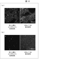
- the arrowhead in FIG. 11 shows a region containing a lesion consisting of phosphorylated ⁇ -synuclein in the brain of a DLB patient and the brain of an MSA patient.
- FIG. 12 (a) (indicated by “1” in the figure) is anti-phosphorylated ⁇ -synuclein antibody staining and SPAL-T-06 fluorescent staining of DLB patient brain
- FIG. 12 (b) (in the figure, “1”). 2 ”) are anti-phosphorylated ⁇ -synuclein antibody staining and SPAL-T-06 fluorescent staining of MSA patient brain.
- SPAL-T-06 was labeled with positron-releasing nuclei and autoradiography was performed using DLB patient brain ectopic section, MSA patient brain cerebellar section, and healthy subject brain frontal cortex section. Binding of [ 18F ] SPAL-T-06 to a lesion-rich region of phosphorylated ⁇ -synuclein in the brain of MSA patients was confirmed. Fluorescence staining and immunohistochemical staining were performed on the autoradiographed sections, and it was confirmed that SPAR-T-06 was bound to the ⁇ -synuclein lesions contained in these brain sections. In addition, in the brain frontal cortex sections of healthy subjects, almost no non-specific binding of [ 18F ] SPAL-T-06 was observed in both gray matter and white matter.
- an ⁇ -synuclein aggregate binder having high binding selectivity to ⁇ -synuclein aggregates. Then, it is possible to provide an imaging method using this ⁇ -synuclein aggregate binder. In addition, it is possible to provide an ⁇ -synuclein aggregate binder or a novel compound that can be used for other purposes.
Landscapes
- Health & Medical Sciences (AREA)
- Chemical & Material Sciences (AREA)
- Organic Chemistry (AREA)
- Life Sciences & Earth Sciences (AREA)
- Animal Behavior & Ethology (AREA)
- General Health & Medical Sciences (AREA)
- Public Health (AREA)
- Veterinary Medicine (AREA)
- Epidemiology (AREA)
- Medicinal Chemistry (AREA)
- Pharmacology & Pharmacy (AREA)
- Engineering & Computer Science (AREA)
- Optics & Photonics (AREA)
- Physics & Mathematics (AREA)
- Proteomics, Peptides & Aminoacids (AREA)
- Biomedical Technology (AREA)
- Bioinformatics & Cheminformatics (AREA)
- Chemical Kinetics & Catalysis (AREA)
- General Chemical & Material Sciences (AREA)
- Nuclear Medicine, Radiotherapy & Molecular Imaging (AREA)
- Medicines Containing Antibodies Or Antigens For Use As Internal Diagnostic Agents (AREA)
- Epoxy Resins (AREA)
- Organic Low-Molecular-Weight Compounds And Preparation Thereof (AREA)
- Acyclic And Carbocyclic Compounds In Medicinal Compositions (AREA)
Abstract
Description
用語「医薬として許容し得る塩」とは、哺乳動物、特にヒトに対して有害でない塩を指す。医薬として許容し得る塩は、無機酸若しくは無機塩基、又は有機酸若しくは有機塩基を含む、無毒性の酸又は塩基を用いて形成することができる。医薬として許容し得る塩の例を挙げると、アルミニウム、カルシウム、リチウム、マグネシウム、カリウム、ナトリウム及び亜鉛などから形成される金属塩、又はリジン、N,N’-ジベンジルエチレンジアミン、クロロプロカイン、コリン、ジエタノールアミン、エチレンジアミン、メグルミン(N-メチルグルカミン)及びプロカインなどから形成される有機塩などがある。また、医薬として許容し得る塩は、酸付加塩及び塩基付加塩を包含する。
一実施形態において、本発明は、下記構造式(I)で表される(E)-1-フルオロ-3-((2-(4-(5-(メチルアミノ)ピラジン-2-イル)ブタ-1-エン-3-イン-1-イル)ベンゾ[d]チアゾール-6-イル)オキシ)プロパン-2-オール、又は下記構造式(II)で表される(E)-1-フルオロ-3-((2-(4-(2-(メチルアミノ)ピリミジン-5-イル)ブタ-1-エン-3-イン-1-イル)ベンゾ[d]チアゾール6-イル)オキシ)プロパン-2-オール、その医薬として許容し得る塩、又はその溶媒和物を提供する。

下記の化合物の製造方法に示されている通り、下記式(III)で表される化合物は、化合物(I)及び化合物(II)の製造中間体化合物(本明細書において、単に“中間体”ともいう。)である。


化合物(I)及び化合物(II)並びに中間体である化合物(III)は、以下に示す製造法に準ずる方法に従い製造することができる。また、所望により、脱保護反応、アミド化反応、ウレア化反応、アルキル化反応、光延反応、酸化反応、還元反応、ハロゲン化反応、カップリング反応、カルボアニオンによる求核付加反応、Grignard反応、脱水反応などを各々、単独又はその二つ以上を組み合わせて行うことにより化合物(I)、化合物(II)及び化合物(III)を製造することができる。また、各反応における溶媒、試薬及び温度等の反応条件は、当業者の技術常識に基づき適宜設定すればよい。官能基の保護及び脱保護反応は、公知の反応方法、参考例又は実施例に記載された方法に準じて行われ、保護基としては慣用のものが用いられる。
化合物(I)、化合物(II)及びR1がヒドロキシ基でありR2がBoc基である化合物(III)(化合物(III-i))は、製造スキーム1で示す以下の方法で製造することができる。以下では、各スキーム中、(1)、(2)等で示す化合物を、化合物(1)、化合物(2)等と称する。


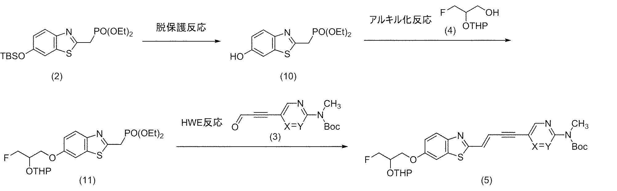
化合物(I)及び化合物(II)は、製造スキーム5で示す以下の方法で製造することもできる。
R1がヒドロキシ基でありR2が水素原子である化合物(III)(化合物(III-ii))は、下記製造スキーム6で示すように、化合物(III-i)の脱保護反応により製造することができる。

R1が式(i)である化合物(III)(化合物(III-iii))は、製造スキーム7で示す以下の方法で製造することができる。


一実施形態において、本発明は、αシヌクレイン凝集体結合剤を提供する。本実施形態のαシヌクレイン凝集体結合剤(以下、結合剤又はαシヌクレイン凝集体結合剤とも記載する)は、化合物(I)若しくは化合物(II)、その医薬として許容し得る塩、又はその溶媒和物を含有する。
一実施形態において、本発明は、αシヌクレイン凝集体の光学イメージング用組成物を提供する。本実施形態のαシヌクレイン凝集体の光学イメージング用組成物(以下、光学イメージング用組成物とも記載する)は、上述の本実施形態に係る結合剤を含む。当該光学イメージングは、インビトロ、エキソビボ、及びインビボイメージングを含む。
一実施形態において、本発明は、αシヌクレイン凝集体の放射イメージング用組成物を提供する。本実施形態のαシヌクレイン凝集体の照射イメージング用組成物(以下、放射イメージング用組成物とも記載する)は、上述の本実施形態に係る結合剤を含む。当該放射イメージングは、インビトロ、エキソビボ、及びインビボイメージングを含む。
一実施形態において、本発明は、αシヌクレイン凝集体が関連する疾患の診断薬、又は当該疾患の治療又は予防のためのコンパニオン診断薬を提供する。本実施形態のαシヌクレイン凝集体が関連する疾患の診断薬、又は当該疾患の治療又は予防のためのコンパニオン診断薬(以下、コンパニオン診断薬ともいう)は、上述の本実施形態に係る結合剤を含む。治療のためのコンパニオン診断薬とは、疾患であることが判明した場合に、治療が見込めるかどうかを判断するための診断薬である。また、予防のためのコンパニオン診断薬とは、疾患の前駆状態であることが判明した場合に、今後の発症を予測する、若しくは発症を抑制する予防が見込めるかどうかを判断するための診断薬である。
本発明の脳内に蓄積する物質が関連する疾患の診断キット(以下、診断キットともいう)は、上述の本実施形態に係る結合剤を含む。
一実施形態において、本発明は、光学イメージング方法を提供する。本実施形態の光学イメージング方法は、上述の本実施形態に係る結合剤を投与された被検体の生体脳に脳外から第1の波長の光を照射した後に、該脳から発せられる、第1の波長とは異なる第2の波長の光を検出する工程を含む。
一実施形態において、本発明は、放射イメージング方法を提供する。本実施形態の放射イメージング方法は、1個又はそれ以上の原子が該原子の放射性同位体である化合物(I)又は化合物(II)、その医薬として許容し得る塩、又はその溶媒和物を含む本実施形態に係る結合剤を投与された被検体の生体脳から発せられる放射線を検出する工程を含む。
一実施形態において、本発明は、脳内αシヌクレイン凝集体が関連する疾患の治療又は予防薬のスクリーニング方法を提供する。本実施形態の脳内αシヌクレイン凝集体が関連する疾患の治療又は予防薬のスクリーニング方法(以下、スクリーニング方法ともいう)は、被検体への候補物質の投与前後における、本実施形態に係る光学イメージング方法又は放射イメージング方法によって検出される光又は放射線の量及び/又は分布の差異に基づいて、候補物質を選抜する工程を有する。
一実施形態において、本発明は、脳内αシヌクレイン凝集体の蓄積を定量又は判定する方法を提供する。本実施形態の脳内αシヌクレイン凝集体の蓄積を定量又は判定する方法は、化合物(I)又は化合物(II)、その医薬として許容し得る塩、又はその溶媒和物を含む上記結合剤を投与された被検体の生体脳に脳外から第1の波長の光を照射した後に、該脳から発せられる、第1の波長とは異なる第2の波長の光を検出する工程を含み、該検出した光の量及び/又は分布に基づいて、脳内αシヌクレイン凝集体の蓄積を定量又は判定する。この方法は、光学イメージングにより、脳内αシヌクレイン凝集体の蓄積を定量又は判定する方法である。
一実施形態において、本発明は、脳内に蓄積する物質の分類及び蓄積を判定する方法を提供する。本発明の脳内に蓄積する物質の分類及び蓄積を判定する方法は、化合物(I)又は化合物(II)、その医薬として許容し得る塩、又はその溶媒和物を含む本実施形態に係る結合剤を投与された被検体の生体脳に脳外から第1の波長の光を照射した後に、該脳から発せられる、第1の波長とは異なる第2の波長の光を検出する第1の工程と、タウタンパク質凝集体への結合選択性が高い物質(例えば、特許文献1記載の化合物)を、第1の工程と異なる時期に投与された該被検体の生体脳に脳外から第3の波長の光を照射した後に、該脳から発せられる、第3の波長とは異なる第4の波長の光を検出する第2の工程と、を含み、第1の工程において検出した光の量及び/又は分布データと、第2の工程において検出した光の量及び/又は分布データと、に基づいて、脳内に蓄積する物質の分類及び蓄積を判定する。この方法は、光学イメージングにより、脳内に蓄積する物質の分類及び蓄積を判定する方法である。
以下に本発明の実施形態をまとめる。

X及びYの一方は窒素原子(N)であり、他方は置換されていない炭素原子(CH)であり(すなわち、Xが窒素原子の場合、Yは置換されていない炭素原子(CH)であり、Xが置換されていない炭素原子(CH)の場合、Yは窒素原子であり);
R1は、ヒドロキシ基又は下記式(i)で表される基であり;

THPはテトラヒドロ-2H-ピラン-2-イル基を表し、
*はベンゾチアゾール環との結合位置を表し、
R2は、水素原子又はtert-ブトキシカルボニル(Boc)基である)。
以下の製造例(参考例及び実施例)中の「室温」は通常約10℃~約35℃を示す。%は特に断らない限り重量%を示す。また各製造例の使用化合物の説明に関し、「参考例(実施例)Xで製造した」は、「参考例(実施例)Xと同様にして製造した」場合も含むことを意図している。
M:モル濃度
DMSO-d6:重ジメチルスルホキシド
1H NMR:プロトン核磁気共鳴
DIEA:N-エチル-N-イソプロピルプロパン-2-アミン
DMF:N,N-ジメチルホルムアミド(本明細書中「dimethylformamide」とも記載する)
THF:テトラヒドロフラン
MeOH:メタノール
EtOH:エタノール
DMSO:ジメチルスルホキシド
TEA:トリエチルアミン
TFA:トリフルオロ酢酸。

1H NMR(300MHz,DMSO-d6)δ1.37-1.41(18H,m),8.65(1H,d,J=1.3Hz),8.90(1H,d,J=1.5Hz)。

1H NMR(300MHz,DMSO-d6)δ1.47(9H,s),8.55(1H,d,J=1.3Hz),8.87(1H,d,J=1.5Hz),10.29(1H,brs)。

1H NMR(300MHz,DMSO-d6)δ1.49(9H,s),3.27(3H,s),8.72(1H,d,J=1.5Hz),8.83(1H,d,J=1.5Hz)。

1H NMR(300MHz,DMSO-d6)δ1.50(9H,s),3.31(3H,s),4.35(2H,d,J=6.0Hz),5.48(1H,t,J=6.0Hz),8.52(1H,d,J=1.5Hz),8.98(1H,d,J=1.5Hz)。

1H NMR(300MHz,DMSO-d6)δ1.44(9H,s),3.31(3H,s),4.34(2H,d,J=5.7Hz),5.44(1H,t,J=5.8Hz),8.77(2H,s)。

1H NMR(300MHz,DMSO-d6)δ1.52(9H,s),3.35(3H,s),8.82(1H,d,J=1.3Hz),9.17(1H,d,J=1.5Hz),9.48(1H,s)。

1H NMR(300MHz,DMSO-d6)δ1.47(9H,s),3.36(3H,s),9.00(2H,s),9.46(1H,s)。

1H NMR(300MHz,DMSO-d6)δ1.35-1.54(4H,m),1.56-1.80(2H,m),3.36-3.47(1H,m),3.48-3.63(2H,m),3.80(1H,ddd,J=11.2,7.9,3.4Hz),3.90-4.06(1H,m),4.35-4.70(2H,m),4.51(2H,d,J=2.1Hz),4.74-4.81(1H,m),7.24-7.41(5H,m)。

1H NMR(300MHz,DMSO-d6)δ1.47(4H,br dd,J=7.4,3.9Hz),1.56-1.80(2H,m),3.35-3.47(1H,m),3.47-3.63(2H,m),3.80(1H,ddd,J=11.3,8.0,3.4Hz),3.89-4.11(1H,m),4.34-4.69(4H,m),4.73-4.83(1H,m),7.19-7.49(5H,m)。

1H NMR(300MHz,DMSO-d6)δ1.47(4H,br dd,J=7.4,3.9Hz),1.56-1.79(2H,m),3.35-3.47(1H,m),3.48-3.62(2H,m),3.80(1H,ddd,J=11.2,7.9,3.3Hz),3.90-4.08(1H,m),4.35-4.70(4H,m),4.73-4.81(1H,m),7.24-7.42(5H,m)。

1H NMR(300MHz,DMSO-d6)δ-0.02(6H,d,J=1.1Hz),0.79(9H,s),2.41(3H,s),3.36(2H,d,J=5.3Hz),3.86-4.05(3H,m),4.43(2H,s),7.20-7.39(5H,m),7.47(2H,d,J=8.1Hz),7.77(2H,d,J=8.3Hz)。

1H NMR(300MHz,DMSO-d6)δ1.33-1.55(4H,m),1.56-1.86(2H,m),3.36-3.58(3H,m),3.68-3.88(2H,m),4.31-4.70(2H,m),4.71-4.86(2H,m)。

1H NMR(300MHz,DMSO-d6)δ1.35-1.53(4H,m),1.55-1.81(2H,m),3.37-3.62(3H,m),3.67-3.91(2H,m),4.29-4.69(2H,m),4.71-4.87(2H,m)。

1H NMR(300MHz,DMSO-d6)δ1.33-1.56(4H,m),1.57-1.81(2H,m),3.35-3.59(3H,m),3.69-3.90(2H,m),4.31-4.68(2H,m),4.71-4.87(2H,m)。

1H NMR(300MHz,DMSO-d6)δ-0.01(3H,s),0.01(3H,s),0.80(9H,s),2.42(3H,s),3.20-3.38(2H,m),3.75-3.91(2H,m),4.02-4.10(1H,m),4.81(1H,t,J=5.6Hz),7.49(2H,dd,J=8.6,0.7Hz),7.77(2H,d,J=8.3Hz)。

1H NMR(300MHz,DMSO-d6)δ1.33-1.54(4H,m),1.56-1.85(2H,m),3.28-3.96(5H,m),4.18-4.92(3H,m),4.92-5.20(1H,m)。

1H NMR(300MHz,DMSO-d6)δ0.22(6H,s),0.97(9H,s),1.23(6H,t,J=7.0Hz),3.80-3.92(2H,m),4.01-4.13(4H,m),7.00(1H,dd,J=8.7,2.5Hz),7.56(1H,d,J=2.4Hz),7.81(1H,d,J=8.8Hz)。


1H NMR(300MHz,DMSO-d6)δ1.22(6H,t,J=7.0Hz),1.37-1.56(4H,m),1.59-1.80(2H,m),3.41-3.53(1H,m),3.78-3.94(3H,m),4.01-4.11(4H,m),4.12-4.29(3H,m),4.48-4.65(1H,m),4.65-4.81(1H,m),4.83-4.94(1H,m),7.12(1H,dt,J=9.0,2.1Hz),7.70(1H,d,J=2.4Hz),7.84(1H,d,J=8.9Hz)。
1H NMR(300MHz,DMSO-d6)δ1.39-1.58(13H,m),1.59-1.81(2H,m),3.33(3H,s),3.41-3.52(1H,m),3.78-3.95(1H,m),4.13-4.31(3H,m),4.48-4.66(1H,m),4.66-4.83(1H,m),4.84-4.93(1H,m),7.01(1H,d,J=16.2Hz),7.17(1H,dt,J=9.0,2.2Hz),7.48(1H,d,J=16.0Hz),7.77(1H,d,J=2.4Hz),7.92(1H,d,J=8.9Hz),8.65(1H,d,J=1.5Hz),9.06(1H,d,J=1.5Hz)。

1H NMR(300MHz,DMSO-d6)δ1.51(9H,s),3.33(3H,s),6.94(1H,d,J=16.0Hz),7.00(1H,dd,J=8.9,2.4Hz),7.39(1H,d,J=2.3Hz),7.44(1H,d,J=16.0Hz),7.82(1H,d,J=8.9Hz),8.64(1H,d,J=1.5Hz),9.05(1H,d,J=1.5Hz),10.14(1H,brs)。

1H NMR(300MHz,DMSO-d6)δ1.46(9H,s),3.34(3H,s),6.92(1H,d,J=16.0Hz),7.00(1H,dd,J=8.9,2.4Hz),7.38(1H,d,J=16.0Hz),7.39(1H,d,J=2.4Hz),7.82(1H,d,J=8.9Hz),8.87(2H,s),10.01(1H,s)。

1H NMR(300MHz,DMSO-d6)δ1.51(13H,s),1.60-1.79(2H,m),3.34(3H,s),3.42-3.54(1H,m),3.79-3.96(1H,m),4.14-4.31(3H,m),4.49-4.82(2H,m),4.84-4.96(1H,m),7.02(1H,d,J=16.1Hz),7.17(1H,dt,J=9.0,2.1Hz),7.48(1H,d,J=16.1Hz),7.77(1H,d,J=2.5Hz),7.92(1H,d,J=9.0Hz),8.65(1H,d,J=1.4Hz),9.06(1H,d,J=1.5Hz)。
1H NMR(300MHz,DMSO-d6)δ1.51(13H,s),1.60-1.79(2H,m),3.34(3H,s),3.42-3.54(1H,m),3.79-3.95(1H,m),4.13-4.30(3H,m),4.49-4.82(2H,m),4.84-4.96(1H,m),7.02(1H,d,J=16.2Hz),7.17(1H,dd,J=8.8,2.4Hz),7.48(1H,d,J=16.0Hz),7.77(1H,d,J=2.3Hz),7.92(1H,d,J=9.0Hz),8.65(1H,d,J=1.3Hz),9.06(1H,d,J=1.3Hz)。

1H NMR(300MHz,DMSO-d6)δ0.04(3H,s),0.04(3H,s),0.81(9H,s),1.51(9H,s),2.38(3H,s),3.34(3H,s),3.90-4.19(4H,m),4.20-4.32(1H,m),7.02(1H,d,J=16.0Hz),7.02-7.08(1H,m),7.45(2H,brd,J=8.1Hz),7.48(1H,brd,J=16.0Hz),7.66(1H,d,J=2.3Hz),7.79(2H,d,J=8.3Hz),7.91(1H,d,J=9.0Hz),8.65(1H,d,J=1.5Hz),9.06(1H,d,J=1.5Hz)。

1H NMR(300MHz,DMSO-d6)δ1.46(9H,s),3.34(3H,s),4.02-4.16(3H,m),4.39-4.46(1H,m),4.56-4.63(1H,m),5.49(1H,d,J=5.3Hz),6.99(1H,d,J=16.2Hz),7.16(1H,dd,J=9.0,2.6Hz),7.41(1H,d,J=16.2Hz),7.73(1H,d,J=2.3Hz),7.91(1H,d,J=9.0Hz),8.87(2H,s)。

1H NMR(300MHz,DMSO-d6)δ1.51(9H,s),2.35(3H,s),3.34(3H,s),3.91-4.19(5H,m),5.59(1H,d,J=4.9Hz),7.02(1H,d,J=16.1Hz),7.00-7.07(1H,m),7.35-7.43(2H,m),7.48(1H,d,J=16.1Hz),7.62(1H,d,J=2.5Hz),7.77(2H,d,J=8.3Hz),7.89(1H,d,J=9.0Hz),8.65(1H,d,J=1.5Hz),9.06(1H,d,J=1.5Hz)。

1H NMR(400MHz,DMSO-d6)δ2.34(3H,s),2.84(3H,d,J=4.8Hz),3.90-3.98(2H,m),4.02-4.17(3H,m),5.60(1H,d,J=4.8Hz),6.95(1H,d,J=16.0Hz),7.00(1H,dd,J=8.8,2.4Hz),7.29(1H,d,J=16.0Hz),7.40(2H,d,J=8.0Hz),7.59(1H,d,J=2.4Hz),7.68(1H,brs),7.77(2H,d,J=8.4Hz),7.86(1H,d,J=8.8Hz),7.94(1H,d,J=1.6Hz),8.21(1H,d,J=1.2Hz)。

1H NMR(300MHz,DMSO-d6)δ2.84(3H,d,J=4.9Hz),6.81-6.92(1H,m),6.98(1H,dd,J=8.8,2.4Hz),7.26(1H,d,J=15.8Hz),7.37(1H,d,J=2.3Hz),7.63(1H,q,J=5.0Hz),7.79(1H,d,J=8.7Hz),7.94(1H,d,J=1.5Hz),8.20(1H,d,J=1.1Hz),9.97(1H,s)。

1H NMR(300MHz,DMSO-d6)δ2.84(3H,d,J=4.9Hz),4.00-4.16(3H,m),4.37-4.49(1H,m),4.53-4.66(1H,m),5.51(1H,d,J=5.1Hz),6.95(1H,d,J=16.0Hz),7.15(1H,dd,J=9.0,2.6Hz),7.29(1H,d,J=16.0Hz),7.67(1H,q,J=4.6Hz),7.71(1H,d,J=2.6Hz),7.89(1H,d,J=9.0Hz),7.94(1H,d,J=1.5Hz),8.21(1H,d,J=1.3Hz)。

1H NMR(300MHz,DMSO-d6)δ2.84(3H,d,J=4.8Hz),4.01-4.17(3H,m),4.37-4.49(1H,m),4.53-4.65(1H,m),5.50(1H,d,J=5.1Hz),6.95(1H,d,J=16.1Hz),7.15(1H,dd,J=8.9,2.6Hz),7.29(1H,d,J=16.1Hz),7.66(1H,q,J=4.9Hz),7.71(1H,d,J=2.5Hz),7.89(1H,d,J=9.0Hz),7.94(1H,d,J=1.4Hz),8.21(1H,d,J=1.3Hz)。

1H NMR(300MHz,DMSO-d6)δ2.84(3H,d,J=4.7Hz),4.00-4.17(3H,m),4.37-4.49(1H,m),4.53-4.65(1H,m),5.51(1H,d,J=4.7Hz),6.95(1H,d,J=16.0Hz),7.15(1H,dd,J=8.9,2.5Hz),7.29(1H,d,J=16.2Hz),7.62-7.70(1H,m),7.71(1H,d,J=2.4Hz),7.89(1H,d,J=8.9Hz),7.94(1H,d,J=1.3Hz),8.21(1H,d,J=1.1Hz)。

1H NMR(300MHz,DMSO-d6)δ2.84(3H,d,J=4.5Hz),4.00-4.16(3H,m),4.36-4.50(1H,m),4.52-4.66(1H,m),5.49(1H,d,J=5.3Hz),6.86-6.99(1H,m),7.10-7.18(1H,m),7.25(1H,d,J=15.8Hz),7.67-7.77(2H,m),7.88(1H,d,J=9.0Hz),8.38-8.58(2H,m)。

1H NMR(300MHz,DMSO-d6)δ1.32-1.72(15H,m),2.35(3H,s),3.34-3.46(4H,m),3.61-3.93(1H,m),4.01-4.41(5H,m),4.66-4.90(1H,m),6.99-7.05(1H,m),7.02-7.09(1H,m),7.41(2H,brd,J=8.1Hz),7.48(1H,d,J=16.2Hz),7.65(1H,d,J=2.1Hz),7.79(2H,brd,J=7.3Hz),7.90(1H,d,J=8.9Hz),8.65(1H,d,J=1.1Hz),9.06(1H,d,J=0.9Hz)。

1H NMR(300MHz,DMSO-d6)δ1.32-1.71(6H,m),2.35(3H,s),2.84(3H,d,J=4.9Hz),3.35-3.46(1H,m),3.62-3.87(1H,m),4.05-4.19(3H,m),4.20-4.36(2H,m),4.65-4.87(1H,m),6.96(1H,d,J=16.2Hz),7.02(1H,dd,J=8.9,1.6Hz),7.30(1H,d,J=16.0Hz),7.41(2H,d,J=7.9Hz),7.62(1H,d,J=2.6Hz),7.67(1H,q,J=4.4Hz),7.79(2H,dd,J=8.4,1.6Hz),7.86(1H,d,J=9.0Hz),7.94(1H,d,J=1.5Hz),8.21(1H,d,J=1.3Hz)。
以下のスキームにより、フッ素原子が18FであるSPAL-T-06(以下、[18F]SPAL-T-06)を製造した。

以下のスキームにより、[18F]SPAL-T-06を製造した。
(解剖脳組織)
死後ヒト脳を、レビー小体型認知症(DLB)患者、及び、アルツハイマー病(AD)患者に対して行われた剖検から得た。凍結DLB組織をクリオスタット(HM560、Carl Zeiss)内で20μm厚にスライスした。また、AD脳組織を10%中性緩衝ホルマリンに固定し、パラフィンブロックに埋め込み、6μm厚にスライスした。
DLB患者脳偏桃体組織の後固定新鮮凍結切片、及び、脱パラフィンしたAD患者脳中前頭回組織のホルマリン固定パラフィン包埋切片を使用した。30μMの試験化合物と脳切片を50%エタノール溶液中にて30分間室温でインキュベートした。その後切片を50%エタノール溶液で5分間、超純水で3分間、2回洗浄した。封入剤(VECTASHIELD H-1000、Vector Laboratories)を用いて切片を封入後、蛍光顕微鏡(DM4000、Leica、励起波長391-437nm)を用いて切片上の病変蓄積領域の画像を取得した、蛍光画像を図1に示す。図1において、矢頭はDLB患者脳のαシヌクレイン凝集体に結合した化合物の蛍光を、矢印はAD患者脳のアミロイドβ凝集体に結合した化合物の蛍光を、アスタリスクはAD患者脳のタウ凝集体に結合した化合物の蛍光を示す。解析ソフトウェア(Image J)を用いて病変領域及び病変非形成領域(バックグラウンド)の蛍光輝度を定量した。結果を図2に示す。なお、試験化合物としては、SPAL-T-05及びSPAL-T-06を用いた。また対照の化合物としては、ナード研究所から入手したBF-227(2-(2-[2-ジメチルアミノチアゾール-5-イル]エテニル)-6-(2-[フルオロ]エトキシ)ベンゾオキサゾール(カタログ番号NP039-0))及びPBB3(2-((1E,3E)-4-(6-(メチルアミノ)ピリジン-3-イル)ブタ-1,3-ジエニル)ベンゾ[d]チアゾール-6-オール)(カタログ番号NP039-0)を使用した。
(αシヌクレイン線維接種マウスモデルの作製)
マウスαシヌクレインをリコンビナントタンパク質として大腸菌で発現させて抽出し、試験管内でインキュベートすると、不溶性の凝集体を形成する。このαシヌクレイン凝集体をマウスの線条体に接種すると、神経回路を経路としてαシヌクレインの凝集体が周囲の領域に伝播し、数か月後には大脳新皮質でαシヌクレイン病変が観察される(Masuda-Suzukake et al. Acta Neuropathol Commun 2, 88, 2014; Shimozawa et al. Acta Neuropathol Commun 5, 12, 2017)。このマウスの脳を摘出し、切片を作製して蛍光染色で解析すると、リン酸化αシヌクレインからなる病変に、本発明の化合物が結合するかどうかを確認することができる。
まず、マウスαシヌクレインをリコンビナントタンパク質として大腸菌で発現させて抽出し、試験管内でインキュベートして、不溶性のαシヌクレイン凝集体を形成した。そして、このαシヌクレイン凝集体をマウスの線条体に接種したところ、神経回路を経路としてαシヌクレインの凝集体が周囲の領域に伝播し、数か月後には大脳新皮質でαシヌクレイン病変が観察された。なお、このαシヌクレイン凝集体のマウスの線条体への接種は、以下の方法で行った。まず、1.5%(v/v)イソフルランで麻酔した9週齢のC57/BL/6オスマウスの頭部の毛を取り除き、頭皮をイソジンで消毒後、キシロカインを塗り、頭皮に切れ込みを入れて頭蓋を露出させた。そして、Bregma 0.05mm、Lateral 2mmの位置の頭蓋にドリルで穴を開け、ガラスピペットを用いて2μmの深さ位置にαシヌクレイン線維溶液(マウスαシヌクレイン線維 4mg/ml in saline)3μlを注入した後、頭皮を戻して縫合した。
このαシヌクレイン線維接種マウスの脳を摘出し、切片を作製して蛍光染色で解析した。具体的には、αシヌクレイン線維接種マウス脳切片と30μMの化合物を20%エタノール溶液中にて30分間室温でインキュベートした。その後切片を20%エタノール溶液で5分間、超純水で3分間、2回洗浄した。封入剤(VECTASHIELD H-1000)を用いて切片を封入後、蛍光顕微鏡(DM4000(励起波長391-437nm))を用いて切片上のαシヌクレイン凝集体蓄積領域の画像を取得した。リン酸緩衝液を用いて同一切片を洗浄後、抗原性賦活化のためにオートクレーブで処理した。抗リン酸化αシヌクレインモノクローナル抗体(pS129、abcam、ab59264)(1:1000)による免疫組織化学染色を行い、封入剤(VECTASHIELD H-1000)を用いて切片を封入後、蛍光顕微鏡(DM4000(励起波長460-500nm))を用いて上記と同一領域の画像を取得した。結果を図3に示す。図3の(a)及び(b)はそれぞれSPAL-T-05及びSPAL-T-06についての結果を示し、図3の(a)及び(b)において、右側が抗リン酸化αシヌクレイン抗体染色、左側がSPAL-T-05及びSPAL-T-06蛍光染色である。図3において、矢頭はαシヌクレイン線維接種マウス脳のリン酸化αシヌクレインからなる病変、及びそれらに結合した化合物の蛍光を示している。
αシヌクレイン線維溶液注入6週間以降のモデルマウスを1.5%(v/v)イソフルランで麻酔し、Seylaz-Tomita法(Tomita et al. J Cereb Blood Flow Metab 25, 858-67, 2005)に従い頭蓋窓を設置した。頭蓋窓設置から2週間以降のマウスを1.5%(v/v)イソフルランで麻酔し、BF-227、PBB3、SPAL-T-05又はSPAL-T-06を0.1%含むDMSO溶液50μlを腹腔内投与した。投与30分後にマウスを二光子レーザー蛍光顕微鏡下に固定し、スルホローダミン101を5mM含む生理食塩水100μlを腹腔内投与した後、励起波長900nmにて生体二光子蛍光イメージングを行った。BF-227、PBB3、SPAL-T-05及びSPAL-T-06に対する検出波長を500~550nm、スルホローダミン101に対する検出波長を573~648nmとした。結果を図4に示す。図4において、矢頭は血管、矢印はαシヌクレイン病変に結合した化合物の蛍光を示している。
PETスキャンは0.851mm厚で(中心間)95枚のスライス、19.0cm体軸方向視野(FOV)、及び7.6cm断面内FOVを提供する、micro PET Focus 220動物スキャナー(Siemens Medical Solutions)を用いて行った。スキャン前に、αシヌクレイン線維接種マウス及び生理食塩水注入マウス(対照)を、1.5%(v/v)イソフルランで麻酔した。エミッションスキャンは、[18F]SPAL-T-06(SPAL-T-06をポジトロン放出核種で標識した化合物)(30.1±0.13MBq)の静脈内注射後に、90分間、3Dリストモード、エネルギーウィンドウ350-750keVで行った。放射性化合物の注射及びスキャンは、該化合物の光異性化を避けるように薄暗下で行った。全てのリストモードデータを、3Dサイノグラムにソートし、その後、Fourier-rebiningにより2Dサイノグラムに変換した(フレーム:10×1、6×5、5×10分)。放射性化合物の注射後、0~30分、30~60分、及び60~90分間での加算イメージを、最大事後確率推定法による再構成(maximum a posteriori reconstruction)により得た。また、ダイナミックイメージを、0.5mmハニングフィルターを用いて、フィルター補正逆投影法により再構成した。関心体積(Volume of interest(VOI))を、MRIテンプレートを参照して、線条体、大脳皮質及び小脳に、PMODイメージ分析ソフトウェア(PMOD Technologies)を用いて設定した。結果を図5~7に示す。
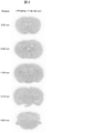
生理食塩水注入マウスを1.5%(v/v)イソフルランで麻酔し、[18F]SPAL-T-06(28.9MBq)を静脈内注射した。[18F]SPAL-T-06投与40分後に脳を採取して凍結し、クリオスタット(HM560)内で20μm厚のBregma 0.50、-0.46、-1.94、-3.16、-6.64mmを含む冠状断凍結切片を作製した。切片を風乾後、カセット内にて切片とイメージングプレートを15分間コンタクトし、その後BAS-5000(Fuji Film)を用いてオートラジオグラフを取得した。結果を図8に示す。
(αシヌクレイン線維接種マーモセットモデル作製)
まず、マーモセットαシヌクレインをリコンビナントタンパク質として大腸菌で発現させて抽出し、試験管内でインキュベートして、不溶性のαシヌクレイン凝集体を形成した。そして、このαシヌクレイン凝集体をマーモセットの尾状核及び被殻に接種したところ、神経回路を経路としてαシヌクレインの凝集体が周囲の領域に伝播し、数か月後には黒質でαシヌクレイン病変が観察された。このαシヌクレイン凝集体のマーモセットの尾状核及び被殻への接種は、以下の方法で行った。まず、ケタミン(5~10mg/kg)及びキシラジン(0.2~0.5mg/kg)により不動化したマーモセットに器官内チューブを挿管した後、1~3%(v/v)イソフルランで麻酔した。マーモセットの頭部の毛を取り除き、頭皮をイソジンで消毒後、キシロカインを塗り、頭皮に切れ込みを入れて頭蓋を露出させた。Interaural 9.75mmの位置の頭蓋にドリルで穴(直径~3mm)を開け、ハミルトンシリンジを用いて、右脳の尾状核及び被殻にαシヌクレイン線維溶液(マーモセットαシヌクレイン線維4mg/ml in saline)50μlをそれぞれ注入した。さらに、左脳の尾状核及び被殻に同様の方法にて生理食塩水50μlをそれぞれ注入した後、頭皮を戻して縫合した。
PETスキャンは0.851mm厚で(中心間)95枚のスライス、19.0cm体軸方向視野(FOV)、及び7.6cm断面内FOVを提供する、micro PET Focus 220動物スキャナー(Siemens Medical Solutions)を用いて行った。スキャン前に、αシヌクレイン線維接種マーモセットを1~3%(v/v)イソフルランで麻酔した。トランスミッションスキャンは、PET校正用線源Ge-68を用いて約20分間実施した。エミッションスキャンは、[18F]SPAL-T-06(SPAL-T-06をポジトロン放出核種で標識した化合物)(73.0MBq)の静脈内注射後に90分間、3Dリストモード、エネルギーウィンドウ350-750keVで行った。放射性化合物の注射及びスキャンは、該化合物の光異性化を避けるように薄暗下で行った。全てのリストモードデータを、3Dサイノグラムにソートし、その後、Fourier-rebiningにより2Dサイノグラムに変換した(フレーム:10×1、6×5、5×10分)。放射性化合物の注射後、0~30分、30~60分、及び60~90分間での加算イメージを、最大事後確率推定法による再構成(maximum a posteriori reconstruction)により得た。また、ダイナミックイメージを、0.5mmハニングフィルターを用いて、フィルター補正逆投影法により再構成した。結果を図9に示す。図9の(a)は[18F]SPAL-T-06静注後20~60分における、αシヌクレイン線維接種マーモセットの尾状核を含む脳の冠状断面画像で、標準脳MRI画像に重ね合わせて表示している。
このαシヌクレイン線維接種マーモセットの脳を摘出し、切片を作製して免疫組織化学染色で解析した。具体的には、リン酸緩衝液を用いてαシヌクレイン線維接種マーモセット脳切片を洗浄後、抗原性賦活化のためにオートクレーブで処理した。抗リン酸化αシヌクレインモノクローナル抗体(pS129、abcam、ab59264)(1:1000)による免疫組織化学染色を行い、封入剤(VECTASHIELD H-1000)を用いて切片を封入後、蛍光顕微鏡(BZ-X710,KEYENCE(励起波長450-490nm)、及びDM4000(励起波長460-500nm))を用いて画像を取得した。結果を図9に示す。図9の(b)はαシヌクレイン線維接種マーモセットの尾状核を含む脳の冠状切片の抗リン酸化αシヌクレイン抗体染色像である。2はαシヌクレイン線維を接種した右脳の尾状核の抗リン酸化αシヌクレイン抗体染色の拡大画像、1は生理食塩水を注入した左脳の尾状核の抗リン酸化αシヌクレイン抗体染色の拡大画像である。
DLB患者脳偏桃体の新鮮凍結組織、及び、AD患者脳前頭皮質の新鮮凍結組織を使用した。新鮮凍結脳組織に、組織の湿重量の10倍量の緩衝液とジルコニアビーズ(ZB-20,TOMY)を加え、Micro Smash(MS-100R,TOMY)で粉砕した後、-80℃で保存した。Tris-HCl緩衝液(20%エタノールを含む)中にDLB患者脳ホモジェネート又はAD患者脳ホモジェネート、[18F]SPAL-T-06(最終濃度5nM)、非標識SPAL-T-06(最終濃度0.01nM~1μM)を混合した。室温で30分インキュベートした後、吸引ろ過及び洗浄によるB/F分離(B:結合リガンド、F:遊離リガンド)を行い、グラスフィルター上に捕捉された放射能をガンマカウンター(2480WIZARD2、ParkinElmer)にて測定した。非標識SPAL-T-06の各濃度について、試験は三連にて実施した。結果をPrism 6J(GraphPad)を用いて解析し、被験物質の置換曲線、IC50を算出した。結果を図10に示す。
(解剖脳組織)
死後ヒト脳を、DLB患者、多系統萎縮症(MSA)患者、及び、健常対象者に対して行われた剖検から得た。凍結DLB組織、及び、凍結健常対象者組織をクリオスタット(HM560、Carl Zeiss)内で20μm厚にスライスした。また、MSA脳組織を10%中性緩衝ホルマリンに固定し、パラフィンブロックに埋め込み、6μm厚にスライスした。
DLB患者脳偏桃体組織、及び、健常対象者脳前頭皮質組織の後固定新鮮凍結切片、及び、脱パラフィンしたMSA患者脳小脳組織のホルマリン固定パラフィン包埋切片を使用した。[18F]SPAL-T-06(最終濃度10nM)と脳組織切片を、20%エタノールを含む50mM Tris-HCl中にて室温で1時間インキュベートした。特異結合を検出するため、[18F]SPAL-T-06(最終濃度10nM)と非標識SPAL-T-06(最終濃度10μM)を含む50mM Tris-HCl(20%エタノールを含む)中においても脳組織切片をインキュベートした。その後4℃の50mM Tris-HCl(20%EtOHを含む)にて切片を2分間、2回洗浄し、MilliQでリンスした。切片を風乾後、カセット内にて切片とイメージングプレートを5分間コンタクトし、その後BAS-5000(Fuji Film)を用いてオートラジオグラフを取得した。結果を図11に示す。図11の矢頭は、DLB患者脳、及び、MSA患者脳のリン酸化αシヌクレインからなる病変を含む領域を示している。
オートラジオグラフィを実施した切片について、蛍光染色で解析した。具体的には、切片と30μMのSPAL-T-06を50%エタノール溶液中にて30分間室温でインキュベートした。その後切片を50%エタノール溶液で5分間、超純水で3分間、2回洗浄した。封入剤(VECTASHIELD H-1000)を用いて切片を封入後、蛍光顕微鏡(DM4000(励起波長391-437nm))を用いて切片上のαシヌクレイン凝集体蓄積領域の画像を取得した。また、隣接する脳切片を、リン酸緩衝液を用いて洗浄後、抗原性賦活化のためにオートクレーブで処理した。抗リン酸化αシヌクレインモノクローナル抗体(pS129、abcam、ab59264)(1:1000)による免疫組織化学染色を行い、封入剤(VECTASHIELD H-1000)を用いて切片を封入後、蛍光顕微鏡(BZ-X710(励起波長450-490nm)及びDM4000(励起波長460-500nm))を用いて画像を取得した。結果を図12に示す。図12の(a)(図中「1」で示す)はDLB患者脳の抗リン酸化αシヌクレイン抗体染色、及び、SPAL-T-06蛍光染色であり、図12の(b)(図中「2」で示す)はMSA患者脳の抗リン酸化αシヌクレイン抗体染色、及び、SPAL-T-06蛍光染色である。
Claims (9)
- 下記式(I)又は(II)で表される化合物、その医薬として許容し得る塩、又はその溶媒和物。
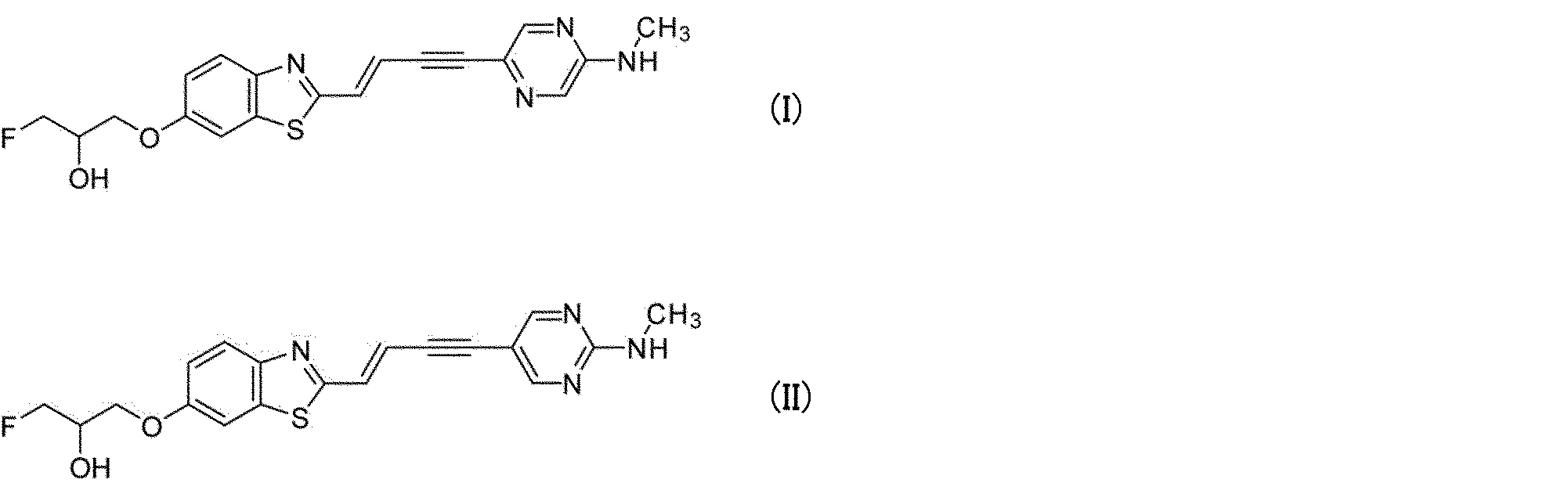
- 前記式(I)又は(II)で表される化合物において1個又はそれ以上の原子が該原子の放射性同位体である、請求項1に記載の化合物、その医薬として許容し得る塩、又はその溶媒和物。
- 請求項1に記載の化合物、その医薬として許容し得る塩、又はその溶媒和物を含有する、αシヌクレイン凝集体結合剤。
- 請求項2に記載の化合物、その医薬として許容し得る塩、又はその溶媒和物を含有する、αシヌクレイン凝集体結合剤。
- 請求項3に記載のαシヌクレイン凝集体結合剤を含む、αシヌクレイン凝集体の光学イメージング用組成物。
- 請求項4に記載のαシヌクレイン凝集体結合剤を含む、αシヌクレイン凝集体の放射イメージング用組成物。
- 請求項3に記載のαシヌクレイン凝集体結合剤を投与された被検体の生体脳に脳外から第1の波長の光を照射した後に、前記脳から発せられる、第1の波長とは異なる第2の波長の光を検出する工程を含む、脳内αシヌクレイン凝集体の光学イメージング方法。
- 請求項4に記載のαシヌクレイン凝集体結合剤を投与された被検体の生体脳から発せられる放射線を検出する工程を含む、脳内αシヌクレイン凝集体の放射イメージング方法。
- 下記式(III)で表される、請求項1又は2に記載の化合物を合成するための中間体。

X及びYの一方は窒素原子(N)であり、他方は置換されていない炭素原子(CH)であり、
R1は、ヒドロキシ基又は下記式(i)で表される基であり、

THPはテトラヒドロ-2H-ピラン-2-イル基を表し、
*はベンゾチアゾール環との結合位置を表し、
R2は、水素原子又はtert-ブトキシカルボニル(Boc)基である)
Priority Applications (7)
| Application Number | Priority Date | Filing Date | Title |
|---|---|---|---|
| EP21861518.5A EP4206200A4 (en) | 2020-08-25 | 2021-08-24 | NOVEL COMPOUND, BINDER FOR ALPHA-SYNUCLEIN AGGREGATE AND USE THEREOF |
| US18/022,800 US12630544B2 (en) | 2020-08-25 | 2021-08-24 | Compound, α-synuclein aggregate binder, and use thereof |
| CN202180052639.2A CN115989029B (zh) | 2020-08-25 | 2021-08-24 | 化合物、α突触核蛋白凝集体结合剂及其利用 |
| AU2021331023A AU2021331023A1 (en) | 2020-08-25 | 2021-08-24 | Novel compound, alpha-synuclein aggregate binder, and use thereof |
| CA3190550A CA3190550A1 (en) | 2020-08-25 | 2021-08-24 | Novel compound, .alpha.-synuclein aggregate binder, and use thereof |
| KR1020237007767A KR20230057381A (ko) | 2020-08-25 | 2021-08-24 | 신규 화합물, α-시누클레인 응집체 결합제 및 그 이용 |
| JP2022544605A JP7655510B2 (ja) | 2020-08-25 | 2021-08-24 | 新規化合物、αシヌクレイン凝集体結合剤及びその利用 |
Applications Claiming Priority (2)
| Application Number | Priority Date | Filing Date | Title |
|---|---|---|---|
| JP2020142080 | 2020-08-25 | ||
| JP2020-142080 | 2020-08-25 |
Publications (1)
| Publication Number | Publication Date |
|---|---|
| WO2022045093A1 true WO2022045093A1 (ja) | 2022-03-03 |
Family
ID=80353305
Family Applications (1)
| Application Number | Title | Priority Date | Filing Date |
|---|---|---|---|
| PCT/JP2021/030899 Ceased WO2022045093A1 (ja) | 2020-08-25 | 2021-08-24 | 新規化合物、αシヌクレイン凝集体結合剤及びその利用 |
Country Status (7)
| Country | Link |
|---|---|
| EP (1) | EP4206200A4 (ja) |
| JP (1) | JP7655510B2 (ja) |
| KR (1) | KR20230057381A (ja) |
| CN (1) | CN115989029B (ja) |
| AU (1) | AU2021331023A1 (ja) |
| CA (1) | CA3190550A1 (ja) |
| WO (1) | WO2022045093A1 (ja) |
Cited By (2)
| Publication number | Priority date | Publication date | Assignee | Title |
|---|---|---|---|---|
| WO2023163033A1 (ja) | 2022-02-24 | 2023-08-31 | 国立研究開発法人量子科学技術研究開発機構 | 新規化合物、αシヌクレイン凝集体結合剤及びその利用 |
| US12630544B2 (en) | 2020-08-25 | 2026-05-19 | National Institutes for Quantum Science and Technology | Compound, α-synuclein aggregate binder, and use thereof |
Families Citing this family (1)
| Publication number | Priority date | Publication date | Assignee | Title |
|---|---|---|---|---|
| TWI908094B (zh) * | 2024-05-24 | 2025-12-11 | 國家原子能科技研究院 | 一種腦神經退化造影劑及其製造方法 |
Citations (5)
| Publication number | Priority date | Publication date | Assignee | Title |
|---|---|---|---|---|
| WO2014097474A1 (ja) | 2012-12-21 | 2014-06-26 | 独立行政法人放射線医学総合研究所 | 脳内に蓄積したタウタンパク質をイメージングするための新規化合物 |
| WO2018138088A1 (en) * | 2017-01-26 | 2018-08-02 | Ucb Biopharma Sprl | Bicyclic bis-heteroaryl derivatives as modulators of protein aggregation |
| WO2018234864A2 (en) * | 2017-06-19 | 2018-12-27 | Kainos Medicine Inc. | ALPHA-SYNUCLEINE MODULATORS |
| WO2019121661A1 (en) * | 2017-12-21 | 2019-06-27 | F. Hoffmann-La Roche Ag | Radiolabeled compounds |
| WO2020174963A1 (ja) * | 2019-02-27 | 2020-09-03 | 国立研究開発法人量子科学技術研究開発機構 | αシヌクレイン凝集体結合剤及びイメージング方法 |
Family Cites Families (2)
| Publication number | Priority date | Publication date | Assignee | Title |
|---|---|---|---|---|
| JP6831802B2 (ja) * | 2018-01-12 | 2021-02-17 | 上海富吉医療器械有限公司Shanghai Chartwell Medical Instrument Co., Ltd. | 放射性核種標識化合物及びこれを含有するイメージング剤 |
| CN112105393A (zh) * | 2018-05-16 | 2020-12-18 | 爱默蕾大学 | 苯乙烯基苯并噻唑衍生物及在成像中的用途 |
-
2021
- 2021-08-24 JP JP2022544605A patent/JP7655510B2/ja active Active
- 2021-08-24 CN CN202180052639.2A patent/CN115989029B/zh active Active
- 2021-08-24 AU AU2021331023A patent/AU2021331023A1/en active Pending
- 2021-08-24 CA CA3190550A patent/CA3190550A1/en active Pending
- 2021-08-24 KR KR1020237007767A patent/KR20230057381A/ko active Pending
- 2021-08-24 EP EP21861518.5A patent/EP4206200A4/en active Pending
- 2021-08-24 WO PCT/JP2021/030899 patent/WO2022045093A1/ja not_active Ceased
Patent Citations (5)
| Publication number | Priority date | Publication date | Assignee | Title |
|---|---|---|---|---|
| WO2014097474A1 (ja) | 2012-12-21 | 2014-06-26 | 独立行政法人放射線医学総合研究所 | 脳内に蓄積したタウタンパク質をイメージングするための新規化合物 |
| WO2018138088A1 (en) * | 2017-01-26 | 2018-08-02 | Ucb Biopharma Sprl | Bicyclic bis-heteroaryl derivatives as modulators of protein aggregation |
| WO2018234864A2 (en) * | 2017-06-19 | 2018-12-27 | Kainos Medicine Inc. | ALPHA-SYNUCLEINE MODULATORS |
| WO2019121661A1 (en) * | 2017-12-21 | 2019-06-27 | F. Hoffmann-La Roche Ag | Radiolabeled compounds |
| WO2020174963A1 (ja) * | 2019-02-27 | 2020-09-03 | 国立研究開発法人量子科学技術研究開発機構 | αシヌクレイン凝集体結合剤及びイメージング方法 |
Non-Patent Citations (6)
| Title |
|---|
| KIKUCHI, A ET AL., BRAIN, vol. 133, 2010, pages 1772 - 1778 |
| MASUDA-SUZUKAKE ET AL., ACTA NEUROPATHOL COMMUN, vol. 2, 2014, pages 88 |
| See also references of EP4206200A4 |
| SHIMOZAWA ET AL., ACTA NEUROPATHOL COMMUN, vol. 5, 2017, pages 12 |
| TOMITA ET AL., J CEREB BLOOD FLOW METAB, vol. 25, 2005, pages 858 - 67 |
| VERDURAND, M. ET AL., CONTRAST MEDIA MOL. IMAGING, vol. 2018, 2018, pages 9165458 |
Cited By (2)
| Publication number | Priority date | Publication date | Assignee | Title |
|---|---|---|---|---|
| US12630544B2 (en) | 2020-08-25 | 2026-05-19 | National Institutes for Quantum Science and Technology | Compound, α-synuclein aggregate binder, and use thereof |
| WO2023163033A1 (ja) | 2022-02-24 | 2023-08-31 | 国立研究開発法人量子科学技術研究開発機構 | 新規化合物、αシヌクレイン凝集体結合剤及びその利用 |
Also Published As
| Publication number | Publication date |
|---|---|
| EP4206200A4 (en) | 2024-08-28 |
| JP7655510B2 (ja) | 2025-04-02 |
| CA3190550A1 (en) | 2022-03-03 |
| US20230399323A1 (en) | 2023-12-14 |
| CN115989029A (zh) | 2023-04-18 |
| KR20230057381A (ko) | 2023-04-28 |
| CN115989029B (zh) | 2025-07-22 |
| AU2021331023A1 (en) | 2023-04-06 |
| EP4206200A1 (en) | 2023-07-05 |
| JPWO2022045093A1 (ja) | 2022-03-03 |
Similar Documents
| Publication | Publication Date | Title |
|---|---|---|
| EP2767532B1 (en) | Novel compound for imaging tau protein accumulated in the brain | |
| US20250339405A1 (en) | a-SYNUCLEIN AGGREGATE BINDING AGENT AND IMAGING METHOD | |
| JP5048655B2 (ja) | 新規アミロイド親和性化合物 | |
| JPWO2009057578A1 (ja) | 新規アミロイド親和性化合物の使用及び製造方法 | |
| EP2437792B1 (en) | Imaging of myelin basic protein | |
| JP7655510B2 (ja) | 新規化合物、αシヌクレイン凝集体結合剤及びその利用 | |
| JPWO2008065785A1 (ja) | 新規アミロイド親和性化合物 | |
| CN116262744A (zh) | 用于α-突触核蛋白聚集体成像的小分子探针 | |
| CN116251199A (zh) | 结合α-突触核蛋白聚集体的小分子探针及其用途 | |
| US11518754B2 (en) | Radiolabeled pharmaceuticals and methods of making and using same | |
| KR101854228B1 (ko) | 신규 아밀로이드 친화성 화합물 | |
| JP5954737B2 (ja) | 放射性フッ素標識キノキサリン化合物 | |
| JP2007223952A (ja) | アミロイドβ蛋白が蓄積する疾患の画像診断プローブ | |
| JPWO2009057576A1 (ja) | 新規アミロイド親和性化合物の使用及び製造方法 | |
| US12630544B2 (en) | Compound, α-synuclein aggregate binder, and use thereof | |
| WO2005042461A1 (ja) | アミロイド親和性化合物 | |
| CN101918039B (zh) | 对淀粉样蛋白具有亲和性的新化合物的应用及制备方法 | |
| EP4484425A1 (en) | Novel compound, alpha-synuclein aggregate binder, and use thereof | |
| CN111556868B (zh) | 放射性核素标记化合物和含有该化合物的显影剂 | |
| CN111356698B (zh) | 用于低氧成像、映射和治疗的标记物、缀合物、组合物和方法 | |
| JP5190893B2 (ja) | ベンゾキサゾール誘導体 | |
| Imai et al. | Development of a Novel Methylated Pet Tracer Designed to Enhance the Photostability of Pm-Pbb3 | |
| HK1148951A (en) | Use of novel compound having affinity for amyloid, and process for production of the same | |
| HK1193605A (en) | Novel compound with amyloid affinity | |
| HK1148950A (en) | Use of novel compound having affinity for amyloid, and process for production of the same |
Legal Events
| Date | Code | Title | Description |
|---|---|---|---|
| 121 | Ep: the epo has been informed by wipo that ep was designated in this application |
Ref document number: 21861518 Country of ref document: EP Kind code of ref document: A1 |
|
| ENP | Entry into the national phase |
Ref document number: 2022544605 Country of ref document: JP Kind code of ref document: A |
|
| ENP | Entry into the national phase |
Ref document number: 3190550 Country of ref document: CA |
|
| ENP | Entry into the national phase |
Ref document number: 20237007767 Country of ref document: KR Kind code of ref document: A |
|
| NENP | Non-entry into the national phase |
Ref country code: DE |
|
| ENP | Entry into the national phase |
Ref document number: 2021861518 Country of ref document: EP Effective date: 20230327 |
|
| ENP | Entry into the national phase |
Ref document number: 2021331023 Country of ref document: AU Date of ref document: 20210824 Kind code of ref document: A |
|
| WWG | Wipo information: grant in national office |
Ref document number: 202180052639.2 Country of ref document: CN |