WO2022065264A1 - 医療システム及び医療情報処理装置 - Google Patents
医療システム及び医療情報処理装置 Download PDFInfo
- Publication number
- WO2022065264A1 WO2022065264A1 PCT/JP2021/034457 JP2021034457W WO2022065264A1 WO 2022065264 A1 WO2022065264 A1 WO 2022065264A1 JP 2021034457 W JP2021034457 W JP 2021034457W WO 2022065264 A1 WO2022065264 A1 WO 2022065264A1
- Authority
- WO
- WIPO (PCT)
- Prior art keywords
- data
- information
- unit
- blood
- blood flow
- Prior art date
- Legal status (The legal status is an assumption and is not a legal conclusion. Google has not performed a legal analysis and makes no representation as to the accuracy of the status listed.)
- Ceased
Links
Images






Classifications
-
- A—HUMAN NECESSITIES
- A61—MEDICAL OR VETERINARY SCIENCE; HYGIENE
- A61B—DIAGNOSIS; SURGERY; IDENTIFICATION
- A61B3/00—Apparatus for testing the eyes; Instruments for examining the eyes
- A61B3/10—Objective types, i.e. instruments for examining the eyes independent of the patients' perceptions or reactions
- A61B3/12—Objective types, i.e. instruments for examining the eyes independent of the patients' perceptions or reactions for looking at the eye fundus, e.g. ophthalmoscopes
-
- A—HUMAN NECESSITIES
- A61—MEDICAL OR VETERINARY SCIENCE; HYGIENE
- A61B—DIAGNOSIS; SURGERY; IDENTIFICATION
- A61B3/00—Apparatus for testing the eyes; Instruments for examining the eyes
- A61B3/10—Objective types, i.e. instruments for examining the eyes independent of the patients' perceptions or reactions
- A61B3/102—Objective types, i.e. instruments for examining the eyes independent of the patients' perceptions or reactions for optical coherence tomography [OCT]
-
- A—HUMAN NECESSITIES
- A61—MEDICAL OR VETERINARY SCIENCE; HYGIENE
- A61B—DIAGNOSIS; SURGERY; IDENTIFICATION
- A61B3/00—Apparatus for testing the eyes; Instruments for examining the eyes
- A61B3/10—Objective types, i.e. instruments for examining the eyes independent of the patients' perceptions or reactions
- A61B3/12—Objective types, i.e. instruments for examining the eyes independent of the patients' perceptions or reactions for looking at the eye fundus, e.g. ophthalmoscopes
- A61B3/1225—Objective types, i.e. instruments for examining the eyes independent of the patients' perceptions or reactions for looking at the eye fundus, e.g. ophthalmoscopes using coherent radiation
- A61B3/1233—Objective types, i.e. instruments for examining the eyes independent of the patients' perceptions or reactions for looking at the eye fundus, e.g. ophthalmoscopes using coherent radiation for measuring blood flow, e.g. at the retina
-
- A—HUMAN NECESSITIES
- A61—MEDICAL OR VETERINARY SCIENCE; HYGIENE
- A61B—DIAGNOSIS; SURGERY; IDENTIFICATION
- A61B5/00—Measuring for diagnostic purposes; Identification of persons
- A61B5/0059—Measuring for diagnostic purposes; Identification of persons using light, e.g. diagnosis by transillumination, diascopy, fluorescence
- A61B5/0062—Arrangements for scanning
- A61B5/0066—Optical coherence imaging
-
- A—HUMAN NECESSITIES
- A61—MEDICAL OR VETERINARY SCIENCE; HYGIENE
- A61B—DIAGNOSIS; SURGERY; IDENTIFICATION
- A61B5/00—Measuring for diagnostic purposes; Identification of persons
- A61B5/02—Detecting, measuring or recording for evaluating the cardiovascular system, e.g. pulse, heart rate, blood pressure or blood flow
- A61B5/02007—Evaluating blood vessel condition, e.g. elasticity, compliance
-
- A—HUMAN NECESSITIES
- A61—MEDICAL OR VETERINARY SCIENCE; HYGIENE
- A61B—DIAGNOSIS; SURGERY; IDENTIFICATION
- A61B5/00—Measuring for diagnostic purposes; Identification of persons
- A61B5/02—Detecting, measuring or recording for evaluating the cardiovascular system, e.g. pulse, heart rate, blood pressure or blood flow
- A61B5/026—Measuring blood flow
-
- A—HUMAN NECESSITIES
- A61—MEDICAL OR VETERINARY SCIENCE; HYGIENE
- A61B—DIAGNOSIS; SURGERY; IDENTIFICATION
- A61B5/00—Measuring for diagnostic purposes; Identification of persons
- A61B5/02—Detecting, measuring or recording for evaluating the cardiovascular system, e.g. pulse, heart rate, blood pressure or blood flow
- A61B5/026—Measuring blood flow
- A61B5/0261—Measuring blood flow using optical means, e.g. infrared light
-
- A—HUMAN NECESSITIES
- A61—MEDICAL OR VETERINARY SCIENCE; HYGIENE
- A61B—DIAGNOSIS; SURGERY; IDENTIFICATION
- A61B5/00—Measuring for diagnostic purposes; Identification of persons
- A61B5/72—Signal processing specially adapted for physiological signals or for diagnostic purposes
- A61B5/7235—Details of waveform analysis
- A61B5/7264—Classification of physiological signals or data, e.g. using neural networks, statistical classifiers, expert systems or fuzzy systems
- A61B5/7267—Classification of physiological signals or data, e.g. using neural networks, statistical classifiers, expert systems or fuzzy systems involving training the classification device
-
- A—HUMAN NECESSITIES
- A61—MEDICAL OR VETERINARY SCIENCE; HYGIENE
- A61B—DIAGNOSIS; SURGERY; IDENTIFICATION
- A61B5/00—Measuring for diagnostic purposes; Identification of persons
- A61B5/72—Signal processing specially adapted for physiological signals or for diagnostic purposes
- A61B5/7271—Specific aspects of physiological measurement analysis
- A61B5/7275—Determining trends in physiological measurement data; Predicting development of a medical condition based on physiological measurements, e.g. determining a risk factor
-
- G—PHYSICS
- G16—INFORMATION AND COMMUNICATION TECHNOLOGY [ICT] SPECIALLY ADAPTED FOR SPECIFIC APPLICATION FIELDS
- G16H—HEALTHCARE INFORMATICS, i.e. INFORMATION AND COMMUNICATION TECHNOLOGY [ICT] SPECIALLY ADAPTED FOR THE HANDLING OR PROCESSING OF MEDICAL OR HEALTHCARE DATA
- G16H20/00—ICT specially adapted for therapies or health-improving plans, e.g. for handling prescriptions, for steering therapy or for monitoring patient compliance
-
- G—PHYSICS
- G16—INFORMATION AND COMMUNICATION TECHNOLOGY [ICT] SPECIALLY ADAPTED FOR SPECIFIC APPLICATION FIELDS
- G16H—HEALTHCARE INFORMATICS, i.e. INFORMATION AND COMMUNICATION TECHNOLOGY [ICT] SPECIALLY ADAPTED FOR THE HANDLING OR PROCESSING OF MEDICAL OR HEALTHCARE DATA
- G16H40/00—ICT specially adapted for the management or administration of healthcare resources or facilities; ICT specially adapted for the management or operation of medical equipment or devices
- G16H40/60—ICT specially adapted for the management or administration of healthcare resources or facilities; ICT specially adapted for the management or operation of medical equipment or devices for the operation of medical equipment or devices
- G16H40/67—ICT specially adapted for the management or administration of healthcare resources or facilities; ICT specially adapted for the management or operation of medical equipment or devices for the operation of medical equipment or devices for remote operation
-
- G—PHYSICS
- G16—INFORMATION AND COMMUNICATION TECHNOLOGY [ICT] SPECIALLY ADAPTED FOR SPECIFIC APPLICATION FIELDS
- G16H—HEALTHCARE INFORMATICS, i.e. INFORMATION AND COMMUNICATION TECHNOLOGY [ICT] SPECIALLY ADAPTED FOR THE HANDLING OR PROCESSING OF MEDICAL OR HEALTHCARE DATA
- G16H50/00—ICT specially adapted for medical diagnosis, medical simulation or medical data mining; ICT specially adapted for detecting, monitoring or modelling epidemics or pandemics
- G16H50/20—ICT specially adapted for medical diagnosis, medical simulation or medical data mining; ICT specially adapted for detecting, monitoring or modelling epidemics or pandemics for computer-aided diagnosis, e.g. based on medical expert systems
-
- G—PHYSICS
- G16—INFORMATION AND COMMUNICATION TECHNOLOGY [ICT] SPECIALLY ADAPTED FOR SPECIFIC APPLICATION FIELDS
- G16H—HEALTHCARE INFORMATICS, i.e. INFORMATION AND COMMUNICATION TECHNOLOGY [ICT] SPECIALLY ADAPTED FOR THE HANDLING OR PROCESSING OF MEDICAL OR HEALTHCARE DATA
- G16H50/00—ICT specially adapted for medical diagnosis, medical simulation or medical data mining; ICT specially adapted for detecting, monitoring or modelling epidemics or pandemics
- G16H50/30—ICT specially adapted for medical diagnosis, medical simulation or medical data mining; ICT specially adapted for detecting, monitoring or modelling epidemics or pandemics for calculating health indices; for individual health risk assessment
-
- G—PHYSICS
- G16—INFORMATION AND COMMUNICATION TECHNOLOGY [ICT] SPECIALLY ADAPTED FOR SPECIFIC APPLICATION FIELDS
- G16H—HEALTHCARE INFORMATICS, i.e. INFORMATION AND COMMUNICATION TECHNOLOGY [ICT] SPECIALLY ADAPTED FOR THE HANDLING OR PROCESSING OF MEDICAL OR HEALTHCARE DATA
- G16H50/00—ICT specially adapted for medical diagnosis, medical simulation or medical data mining; ICT specially adapted for detecting, monitoring or modelling epidemics or pandemics
- G16H50/50—ICT specially adapted for medical diagnosis, medical simulation or medical data mining; ICT specially adapted for detecting, monitoring or modelling epidemics or pandemics for simulation or modelling of medical disorders
-
- A—HUMAN NECESSITIES
- A61—MEDICAL OR VETERINARY SCIENCE; HYGIENE
- A61B—DIAGNOSIS; SURGERY; IDENTIFICATION
- A61B5/00—Measuring for diagnostic purposes; Identification of persons
- A61B5/0002—Remote monitoring of patients using telemetry, e.g. transmission of vital signals via a communication network
- A61B5/0015—Remote monitoring of patients using telemetry, e.g. transmission of vital signals via a communication network characterised by features of the telemetry system
- A61B5/0022—Monitoring a patient using a global network, e.g. telephone networks, internet
-
- A—HUMAN NECESSITIES
- A61—MEDICAL OR VETERINARY SCIENCE; HYGIENE
- A61B—DIAGNOSIS; SURGERY; IDENTIFICATION
- A61B5/00—Measuring for diagnostic purposes; Identification of persons
- A61B5/02—Detecting, measuring or recording for evaluating the cardiovascular system, e.g. pulse, heart rate, blood pressure or blood flow
- A61B5/02028—Determining haemodynamic parameters not otherwise provided for, e.g. cardiac contractility or left ventricular ejection fraction
- A61B5/02035—Determining blood viscosity
-
- A—HUMAN NECESSITIES
- A61—MEDICAL OR VETERINARY SCIENCE; HYGIENE
- A61B—DIAGNOSIS; SURGERY; IDENTIFICATION
- A61B5/00—Measuring for diagnostic purposes; Identification of persons
- A61B5/02—Detecting, measuring or recording for evaluating the cardiovascular system, e.g. pulse, heart rate, blood pressure or blood flow
- A61B5/02042—Determining blood loss or bleeding, e.g. during a surgical procedure
Definitions
- the present invention relates to a medical system and a medical information processing apparatus.
- Patent Document 1 describes a technique for determining the risk of an infectious disease without using advanced medical knowledge, based on the presence or absence of abnormalities in arterial oxygen saturation, body temperature, and heart rate. Is disclosed.
- One object of the present invention is to provide a new technique for non-invasively detecting the condition of the patient's circulatory system.
- the medical system includes a data acquisition unit and a data processing unit.
- the data acquisition unit acquires data from the fundus of the patient using at least one optical method.
- the data processing unit processes the data acquired by the data acquisition unit in order to generate information about the patient's circulatory system.
- information about the circulatory system includes information about thrombophilia tendencies.
- information about thrombophilia tendencies includes information about blood properties.
- the information regarding blood properties includes information indicating changes in blood properties due to an increase in the blood coagulation / fibrinolysis system.
- information about the circulatory system includes information about thrombotic symptoms.
- information about thrombotic symptoms includes information indicating the distribution of blood flow velocities within blood vessels.
- information about thrombotic symptoms includes information about structures formed within blood vessels.
- the information about the circulatory system includes information about the condition of the circulatory system associated with the infection.
- the information about the circulatory system is information about a condition about sepsis, information about a condition about disseminated intravascular coagulation (DIC), information about a condition about thrombosis, and a condition about vascular occlusion. Includes at least one of the information indicating.
- At least one optical technique is of optical coherence tomography blood flow measurement (OCT blood flow measurement), optical coherence tomography angiography (OCT-A), and color fundus photography. Includes at least one.
- the at least one optical technique comprises an OCT blood flow measurement and the data processing unit is based on at least the blood flow data obtained by the OCT blood flow measurement. Generate information about the fused system.
- the data acquisition unit includes an OCT device and a calculation unit.
- the OCT device applies an optical coherence tomography (OCT) scan to the patient's fundus to collect data.
- OCT optical coherence tomography
- the calculation unit calculates the blood flow velocity and the blood vessel diameter based on at least the data collected by the OCT device.
- the data processing unit generates information about the blood coagulation / fibrinolysis system based on at least the blood flow velocity and blood vessel diameter calculated by the calculation unit.
- the data processing unit includes a WSR calculation unit.
- the WSR calculation unit calculates the wall shear rate (WSR) based on at least the blood flow velocity and the blood vessel diameter.
- the data processing unit includes a storage unit and a WSS calculation unit.
- the storage unit stores blood viscosity information acquired in advance.
- the WSS calculation unit calculates the wall shear stress (WSS) based on at least the wall shear rate and blood viscosity information.
- the data acquisition unit includes an OCT device and a blood flow information generation unit.
- the OCT device repeatedly applies optical coherence tomography (OCT) scans to a predetermined area of the patient's fundus to collect time series data.
- OCT optical coherence tomography
- the blood flow information generation unit generates blood flow information representing the spatial distribution and temporal change of the blood flow velocity based on at least the time series data collected by the OCT device.
- the data processing unit generates information about the blood coagulation / fibrinolysis system based on at least the blood flow information generated by the blood flow information generation unit.
- the data processor will generate information about the structures formed within the blood vessel, at least based on blood flow information.
- the data processing unit includes a WSR information generation unit.
- the WSR information generation unit generates WSR information representing the spatial distribution and time variation of the wall shear rate (WSR) based on at least the blood flow information.
- the data processor will generate information about the structures formed within the blood vessel, at least based on blood flow information and WSR information.
- the data processing unit includes a storage unit and a WSS information generation unit.
- the storage unit stores the blood viscosity distribution information acquired in advance.
- the WSS information generation unit generates WSS information representing the spatial distribution and time variation of the wall shear stress (WSS) based on at least the WSR information and the blood viscosity distribution information.
- WSS wall shear stress
- the data processor will generate information about the structures formed within the blood vessel, at least based on blood flow information and WSS information.
- the data processing unit includes a first inference processing unit.
- the first inference processing unit uses a first trained model constructed by machine learning using the first training data including the first data acquired from the fundus of the eye using at least one optical method and the diagnosis result data. It is used to perform inference processing that inputs data acquired from the patient's fundus by the data acquisition unit and outputs information about the patient's circulatory system.
- the data processing unit includes a second inference processing unit.
- the second inference processing unit is machine learning using the second training data including the second data and the diagnosis result data generated by processing the first data acquired from the fundus using at least one optical method.
- inference processing is performed by inputting the data generated by processing the data acquired from the patient's fundus by the data acquisition unit and outputting the information about the patient's circulatory system. Execute.
- the medical system further includes a transmitter.
- the transmitting unit transmits information about the circulatory system generated by the data processing unit to the doctor terminal located at a remote position from the data acquisition unit.
- the medical system further includes a physician terminal.
- the medical system further includes an operation unit for remotely controlling the data acquisition unit.
- the medical information processing apparatus includes a data receiving unit and a data processing unit.
- the data receiving unit receives data acquired from the fundus of the patient using at least one optical method.
- the data processing unit processes the data received by the data receiving unit in order to generate information about the patient's circulatory system.
- the medical information processing apparatus further includes a first transmitter.
- the first transmission unit transmits information about the circulatory system generated by the data processing unit to the doctor terminal located at a remote position with respect to the place where the data was acquired.
- the medical system includes a medical information processing apparatus according to an exemplary embodiment and a doctor terminal.
- the medical system further includes a data acquisition device and a second transmitter.
- the data acquisition device acquires data from the patient's fundus using at least one optical method.
- the second transmission unit transmits the data acquired by the data acquisition device to the medical information processing device.
- the data receiving unit receives the data transmitted by the second transmitting unit.
- the data processing unit processes the data transmitted by the second transmitting unit and received by the data receiving unit in order to generate information regarding the patient's circulatory system.
- Some exemplary embodiments are to generate information about a patient's circulatory system by computerizing data obtained from the patient's fundus by at least one optical method (optical modality). ..
- This computer processing may include inference. This inference may be performed, for example, by an algorithm using a trained model (inference model) constructed by machine learning, an algorithm using no trained model, or a combination thereof.
- the data to be subjected to computer processing in some exemplary embodiments may be data acquired by any ophthalmic examination, for example data acquired by any ophthalmologic modality device.
- the ophthalmologic modality device may be, for example, an optical coherence tomography (OCT) device, a fundus camera, a scanning laser ophthalmoscope, a slit lamp microscope, a surgical microscope, or the like.
- OCT optical coherence tomography
- the optical coherence tomography apparatus is used, for example, for optical coherence tomography blood flow measurement, optical coherence tomography angiography (OCT-A), and the like.
- fundus imaging devices such as fundus cameras, scanning laser ophthalmoscopes, slit lamp microscopes, and surgical microscopes are used, for example, for color fundus photography.
- the data provided for computer processing is not limited to these, for example, other types of test data, electronic medical record data, medical examination data, patient background information (age, treatment history, medical history, medication history, surgery history, etc.). Etc. may be further included.
- An exemplary embodiment is configured to generate predetermined information about the patient's circulatory system from such data.
- the information generated by some exemplary embodiments may include at least one of quantitative and qualitative information, eg, any of the information shown below: with respect to thrombus formation tendencies.
- the information on the thrombus formation tendency is information showing the tendency of the thrombus to be formed in the circulatory system (intravascular, intracardiac) of the patient, and includes, for example, information on the risk of thrombus formation.
- the information regarding the thrombus formation tendency may include either information regarding blood properties or information indicating changes in blood properties due to an increase in the blood coagulation / fibrinolysis system.
- Information on blood properties includes information on the properties of blood and / or information on the condition of blood.
- Information indicating changes in blood properties due to an increase in the blood coagulation / fibrinolysis system includes information indicating changes in blood properties due to activation of the action system (coagulation system, blood coagulation factor) that coagulates blood, and / or blood clots and It contains information indicating changes in blood properties due to activation of the action system (fiber element dissolving system, fibrinolytic system) that dissolves blood clots.
- Information on thrombophilia tendencies includes, for example, viscosity, wall shear stress, wall shear rate, amount or proportion of specific components, ratio between specific components, information indicating changes in any of these, distribution of any of these. It may contain information and the like.
- the information on the thrombotic symptom is information on the symptom caused by the thrombus, and may include, for example, information indicating the distribution of blood flow velocity in the blood vessel and information on the structure formed in the blood vessel. ..
- the distribution of blood flow velocity in a blood vessel is, for example, one-dimensional distribution, two-dimensional distribution, three-dimensional distribution, or a combination of any one or two or more of temporal distributions. It may be there.
- the structure formed in the blood vessel may be, for example, a white thrombus, a red thrombus, a mixed thrombus, a vitreous thrombus, a structure involved in any one of these formation mechanisms (for example, an intermediate product) and the like.
- Information on the state of the circulatory system associated with an infection includes, for example, vascular inflammation, thrombosis tendency, blood coagulation tendency, sepsis, DIC, pneumonia, lymphadenitis, lymphangitis, and other diseases associated with or caused by the infection. Includes information about the condition and pathology.
- the target infection may be any viral infection, any bacterial infection, or any fungal infection, eg, Coronavirus Disease 2019; COVID-, which was a major epidemic in 2020. 19), Severe Acute Respiratory Syndrome (SARS), Middle East Respiratory Syndrome (MERS), Influenza, Infectious Endocarditis, etc.
- Sepsis is a very serious condition caused by the spread of infectious diseases throughout the body, causing circulatory shock, DIC, multiple organ failure, and the like.
- Information indicating a condition relating to sepsis includes, for example, information regarding symptoms such as inflammation due to sepsis and circulatory failure.
- DIC is a syndrome in which the blood coagulation reaction that should occur only at the bleeding site occurs randomly in the blood vessels of the whole body.
- remarkable coagulation activation occurs continuously in blood vessels of the whole body, and microthrombus occurs frequently.
- organ damage due to microcirculatory disorders causes consumptive coagulopathy, resulting in bleeding.
- fibrinolysis is also activated along with coagulation activation, excessive fibrinolysis of thrombus occurs, which promotes bleeding.
- Information indicating a condition relating to disseminated intravascular coagulation includes, for example, information indicating the above-mentioned pathological conditions of DIC (enhancement of coagulation system, enhancement of fibrinolytic system, thrombus, bleeding, etc.).
- the information indicating the condition regarding a thrombus may be any information regarding a thrombus that is or may be present in the circulatory system (intravascular, intracardiac), for example, the presence or absence of a thrombus, the degree of the thrombus, and the thrombus. Includes distribution, number of thrombi, probability of thrombus formation, etc.
- the information indicating the condition regarding the vascular occlusion may be any information regarding the vascular occlusion occurring or may occur in the circulatory system, for example, the presence or absence of the vascular occlusion, the degree of the vascular occlusion, and the blood vessel. Includes distribution of occlusions, number of occlusions, probability of occlusion, etc.
- the exemplary embodiment is the patient's circulation, for example, by generating information about the patient's circulatory system based on data obtained from the patient's fundus using any of the above exemplary optical modalities. It makes it possible to detect the state of the system non-invasively.
- Some exemplary embodiments include thrombotic tendencies (eg, changes in blood properties and / or changes in blood properties due to increased blood coagulation / fibrinolysis), thrombotic symptoms (eg, blood flow velocity distribution, and / or blood vessels). Internal structures), cardiovascular status and / or status changes associated with infection, sepsis, DIC, thrombosis, vascular obstruction, any of these, any of these, and these.
- Information on one or more of the matters relating to any of the above mechanisms can be generated.
- the types of information that can be generated by the exemplary embodiment are not limited to these, and any type that can be generated (for example, derivation, estimation, etc.) by the combination of the adopted optical modality and the adopted data processing. It may be the information of.
- Some exemplary embodiments have been devised in consideration of the background as described below, and can produce the corresponding effects.
- Health care workers such as doctors and nurses are at risk of nosocomial infections.
- the risk of infection to medical workers became a big problem, such as cluster infection occurring in medical institutions flooded with many patients.
- the increased risk of infection to healthcare workers can occur not only during an infectious disease epidemic but also when a disaster or major accident occurs.
- it is important to secure a sufficient distance between people, so-called social disstancing, but it is not possible to achieve this in standard medical care. It's not easy. For example, when conducting an examination, doctors and the like often stay in the immediate vicinity of the patient to perform the procedure.
- Some exemplary embodiments may be configured such that computer-processed information of data acquired by optical modality can be provided to a remote physician terminal.
- some exemplary embodiments may be configured to allow the inspection device (optical modality device) or computer to be operated from a remote location. With these configurations, it is possible to use the data obtained from tests that could not be performed without being in the immediate vicinity of the patient for diagnosis.
- social distance between the patient and the healthcare professional can be maintained, while being non-invasive and non-invasive to complex physiological events such as symptoms and signs of aggravation. It will be possible to detect with high accuracy.
- the "remote location" may be any positional relationship that can secure social distance between the patient and the medical staff.
- the doctor terminal may be installed in a room different from the inspection device, or may be installed in a facility different from the inspection device.
- the device for remotely controlling the inspection device (operation device, operation unit) may be installed in a room different from the inspection device, or may be installed in a facility different from the inspection device. .. It is not necessary to ensure social distance when the test is carried out under a sufficient infectious disease protection system, such as when wearing complete protective clothing.
- the circuit configuration or processing circuit configuration is a general-purpose processor, a dedicated processor, an integrated circuit, a CPU (Central Processing Unit), a GPU (Graphics Processing Unit) configured and / or programmed to perform at least a part of the disclosed functions. ), ASIC (Application Special Integrated Circuit), programmable logic device (for example, SPLD (Simple Program Digital Device), CPLD (Complex Program Any combination may be included.
- a processor may be considered a processing circuit configuration or circuit configuration that includes transistors and / or other circuit configurations.
- circuit configurations, circuits, computers, processors, units, Means, parts, or similar terms include hardware that performs at least a portion of the disclosed functionality and / or hardware that is programmed to perform at least a portion of the disclosed functionality.
- the hardware may be the hardware disclosed herein, or any known hardware programmed and / or configured to perform at least some of the disclosed functions. If the hardware is a processor that can be considered as a type of circuit configuration, the circuit configuration, circuit, computer, processor, unit, means, part, or similar terminology may be a combination of hardware and software. This software may be used to configure the hardware and / or the processor.
- the exemplary medical system 1 shown in FIG. 1 includes a data acquisition unit 10, a data processing unit 20, and an output unit 30.
- the medical system 1 may further include an operating device 2.
- the data acquisition unit 10 and the data processing unit 20 are connected via a communication line.
- This communication line may form a network in a medical institution, for example, or may form a network over a plurality of facilities.
- the communication technology applied to this communication line may be arbitrary, and may be any of various known communication technologies such as wired communication, wireless communication, and short-range communication.
- the connection mode between the data processing unit 20 and the output unit 30 may be the same.
- the data processing unit 20 and the output unit 30 may be functional units mounted on the same computer.
- the operation device 2 is used by a medical worker to remotely control the data acquisition unit 10 (inspection device, optical modality device). Further, the operation device 2 is used for the medical staff (examiner) to provide an instruction or the like to the patient (examinee) who is performing the examination using the data acquisition unit 10. Further, the operating device 2 may be usable for remotely controlling the data processing unit 20.
- the operation device 2 includes, for example, a computer, an operation panel, and the like.
- the data acquisition unit 10 is configured to acquire data from the fundus of the patient with at least one optical modality.
- the data acquisition unit 10 includes any optical fundus photography modality device such as an optical coherence entomography device, a fundus camera, a scanning laser ophthalmoscope, a slit lamp microscope, a surgical microscope, and the like.
- the data acquisition unit 10 may be able to further acquire, for example, other types of test data, electronic medical record data, medical inquiry data, patient background information, and the like.
- the optical coherence tomography apparatus and / or the fundus camera may be, for example, an apparatus described in Japanese Patent Application Laid-Open No. 2020-44027 in which various shooting preparation operations are automated.
- the shooting preparation operation is an operation executed to adjust the shooting conditions, and examples thereof include alignment adjustment, focus adjustment, optical path length adjustment, polarization adjustment, and light amount adjustment.
- the operation for maintaining the good shooting conditions achieved by the shooting preparation operation may be automatically executed.
- optical coherent tomography data is, for example, 3D image data obtained by applying a 3D scan to the fundus, projection image data of 3D image data, and optical coherent tomo. It may be at least one of the graphic angiography image data and the optical coherence stromography blood flow data.
- Optical coherence tomography angiography is an optical modality that visualizes blood vessels using motion contrast technology, and it is possible to visualize fine blood vessels.
- Optical coherence tomography Angiographic image data is acquired using, for example, an optical coherence tomography apparatus described in JP-A-2019-58495, JP-A-2019-154988 and the like.
- Optical coherence tomography Blood flow measurement is an optical modality that measures the blood flow state (blood flow dynamics).
- Optical coherence tomography blood flow data is acquired using, for example, an optical coherence tomography apparatus described in JP-A-2019-54994, JP-A-2020-48730, and the like.
- optical coherence stromography blood flow measurement reveals time-series changes (time variation, time-dependent) of blood flow velocity, blood flow volume, blood vessel diameter, and blood flow velocity as optical coherence stromography blood flow data. It is possible to acquire waveform data representing (change), waveform data representing time-series changes in blood flow, and the like.
- the waveform data is typically a time-series change graph of blood flow velocity represented by a two-dimensional coordinate system with time as the horizontal axis and blood flow velocity as the vertical axis.
- the optical modality used for fundus blood flow measurement is not limited to optical coherence tomography blood flow measurement, and is, for example, laser speckle flowography (LSFG) described in Japanese Patent Publication No. 2008/069062. You may.
- the image data (fundal camera image data) that can be acquired by the fundus camera include, for example, color fundus image data, infrared fundus image data, fluorescence contrast angiography image data (fluorescein angiography image data, indocyanine green angiography image data, etc.). )and so on.
- a fundus camera is used to obtain color fundus image data.
- the scanning laser ophthalmoscope may be, for example, the apparatus described in Japanese Patent Application Laid-Open No. 2014-226156.
- Examples of the image data (scanning laser image data) that can be acquired by the scanning laser ophthalmoscope include color fundus image data, monochromatic fundus image data, and fluorescence contrast fundus image data.
- color fundus image data is acquired using a scanning laser ophthalmoscope.
- the slit lamp microscope may be, for example, an effective device for remote imaging described in Japanese Patent Application Laid-Open No. 2019-23734.
- the image data acquired by the slit lamp microscope may be, for example, at least one of color fundus image data, anterior eye portion cross-sectional image data, and anterior eye portion three-dimensional image data.
- color fundus image data is acquired using a slit lamp microscope.
- the surgical microscope may be, for example, an effective device for remote surgery described in Japanese Patent Application Laid-Open No. 2002-153487.
- color fundus image data is acquired using a surgical microscope.
- At least one of the inspection devices included in the data acquisition unit 10 may be capable of remote control and / or remote control.
- an examination room where an inspection using an inspection device is performed and an operation room where an operation of this inspection device is performed.
- a speaker and display for outputting instructions (voice, image, video, etc.) of the operator in the operation room, and a video camera for photographing the subject (patient) in the examination room.
- a microphone for inputting the voice of the subject, a computer connected to the inspection device, and the like are provided.
- the operation room is provided with an operation device 2 for remotely controlling the inspection device.
- the operation device 2 is provided with a computer, an operation panel, a display, a video camera, a microphone, and the like.
- the computer performs processing for remote control.
- the computer is connected to the inspection equipment in the inspection room.
- An operation panel, a video camera, and a microphone are used to input instructions to the subject.
- the display displays data acquired by the inspection device and information for remote control (screen, information from the inspection room, etc.).
- the operator (medical worker) in the operation room can remotely control the inspection device in the examination room by using, for example, an application programming interface (API), and instruct the subject by using a videophone or the like. Can be sent.
- API application programming interface
- the subject can be inspected by himself according to the instructions of the operator at a remote location, and as a result, the risk of infection from the subject to the operator can be significantly reduced. ..
- an examination device (described above) with an automated preparation operation can be used. In this case, it is considered possible to perform the inspection without requiring instructions from the operator. In some cases, it is not necessary to assign an assistant (operator, etc.). However, since it is assumed that it is difficult for some patients to perform an independent examination, for example, an assistant may be made to stand by at a remote location, or the assistant may monitor the examination status from a remote location.
- the assistant (operator or the like) who sends an instruction to the patient may be an anthropomorphic computer system (typically, an automatic response system using artificial intelligence technology).
- the data processing unit 20 executes various data processing.
- the data processing unit 20 of this embodiment is configured to process the data acquired by the data acquisition unit 10 in order to generate information about the patient's circulatory system.
- the information generated by the data processing unit 20 of this embodiment may be, for example, at least one of the following information: information on thrombosis tendency (information on blood properties and / or blood coagulation / fibrinolytic system). Information on changes in blood properties due to hyperactivity); Information on thrombotic symptoms (methods of showing blood flow velocity distribution in blood vessels and / or information on structures formed in blood vessels); Cardiovascular associated with infectious diseases Information about the state of the system (and / or changes in state); information about the state of sepsis; information about the state of DIC; information about the state of thrombosis; information about the state of vascular obstruction.
- the data processing unit 20 may or may not use a trained model (inference model) constructed by machine learning.
- FIG. 2 shows an example of a data structure for processing (recording, transmitting, etc.) the data generated by the data processing unit 20.
- the data structure 100 of this example includes a thrombosis tendency data unit 110, a thrombosis symptom data unit 120, an infectious disease accompanying data unit 130, a septicemia data unit 140, a DIC data unit 150, a thrombosis data unit 160, and a blood vessel. It includes a blockage data unit 170.
- the thrombus formation tendency data unit 110 is an area (folder or the like) in which information regarding the thrombus formation tendency generated by the data processing unit 20 is recorded.
- the thrombus formation tendency data unit 110 includes a blood property data unit 111.
- the blood property data unit 111 is an area in which information regarding blood properties generated by the data processing unit 20 is recorded.
- the blood property data unit 111 includes a blood property change data unit 112.
- the blood property change data unit 112 is a region in which information indicating changes in blood properties due to the enhancement of the blood coagulation / fibrinolysis system generated by the data processing unit 20 is recorded.
- the thrombotic symptom data unit 120 is an area in which information related to the thrombotic symptom generated by the data processing unit 20 is recorded.
- the thrombotic symptom data unit 120 includes a blood flow velocity distribution data unit 121 and an intravascular structure data unit 122.
- the blood flow velocity distribution data unit 121 is an area in which information indicating the distribution of blood flow velocity in the blood vessel generated by the data processing unit 20 is recorded.
- the intravascular structure data unit 122 is an area in which information about the structure formed in the blood vessel generated by the data processing unit 20 is recorded.
- the infectious disease accompanying data unit 130 is an area in which information regarding the state of the circulatory system associated with the infectious disease generated by the data processing unit 20 is recorded.
- the sepsis data unit 140 is an area in which information indicating a state related to sepsis generated by the data processing unit 20 is recorded.
- the DIC data unit 150 is an area in which information indicating a state related to the DIC generated by the data processing unit 20 is recorded.
- the thrombus data unit 160 is an area in which information indicating a state related to the thrombus generated by the data processing unit 20 is recorded.
- the blood vessel occlusion data unit 170 is an area in which information indicating a state related to the blood vessel occlusion generated by the data processing unit 20 is recorded.
- the data structure 100 comprises at least one of the data units 110-170 described above.
- the data structure 100 may include data units other than the data units 110-170 described above.
- the data structure 100 records the data obtained by performing a predetermined process on the data acquired from the fundus by the data acquisition unit 10 and the fundus data unit in which the data acquired from the fundus by the data acquisition unit 10 is recorded. It may include a processing data unit to be processed, an arbitrary data unit in which arbitrary types of data are recorded, and the like.
- the arbitrary data unit for example, data acquired by an arbitrary inspection device, electronic medical record data, interview data, patient information (for example, patient identifier, patient background information) and the like are recorded.
- DIC disseminated intravascular coagulation
- DIC a syndrome with a poor prognosis
- the data processing unit 20 of this embodiment may be designed and configured in consideration of such a background. Some exemplary embodiments of the data processing unit 20 will be described later. It is possible to design and configure the data processing unit 20 (and the data structure 100) in the same manner when targeting other infectious diseases.
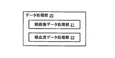
- FIG. 3 shows a configuration example of the data processing unit 20 of this embodiment.
- the data processing unit 20 of this example includes an ocular image data processing unit 21 and an ocular blood flow data processing unit 22.
- the eye image data processing unit 21 may include, for example, a processor that operates according to a program created at least based on the medical knowledge as described above.
- the ocular image data processing unit 21 generates information about the patient's circulatory system by processing the image data (ocular image data) acquired from the fundus of the patient by the data acquisition unit 10 with at least this processor. be able to.
- the eye image data input to the processor may be, for example, optical coherence stromography image data, color fundus image data, or the like.
- the information output from the processor includes, for example, information on thrombosis tendency, information on thrombotic symptoms, information on the state of the circulatory system associated with infectious disease, information on the state on septicemia, and state on DIC. It may be any of the information shown, the information indicating the state related to thrombus, and the information indicating the state related to vascular occlusion.
- the ocular image data processing unit 21 may include, for example, a trained model constructed by machine learning based on at least medical knowledge as described above.
- the ocular image data processing unit 21 relates to the patient's circulatory system by processing the image data (ocular image data) acquired from the fundus of the patient by the data acquisition unit 10 using at least this trained model. Information can be generated.
- the eye image data input to the trained model may be, for example, optical coherence stromography image data, color fundus image data, or the like.
- the information output from the trained model is, for example, as described above, information on thrombosis tendency, information on thrombotic symptoms, information on the state of the circulatory system associated with infectious disease, information on the state of septicemia, and DIC. It may be any of information indicating a condition, information indicating a condition relating to a thrombus, and information indicating a condition relating to vascular occlusion.
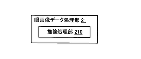
- FIG. 4 shows an example of the eye image data processing unit 21 configured by using machine learning.
- the eye image data processing unit 21 of this example includes an inference processing unit 210.
- the inference processing unit 210 uses a trained model constructed by machine learning using training data including clinical data (eye image data and diagnosis result data), and uses a trained model to obtain a patient from the eye image data acquired by the data acquisition unit 10. It is configured to perform inference processing that derives information about the circulatory system of.
- the eye image data included in the training data is, for example, image data acquired using the same optical modality as the optical modality of the data acquisition unit 10, but may be another modality.
- Other modality includes an optical modality different from the optical modality of the data acquisition unit 10, an ultrasonic modality, an electrical modality, a magnetic modality, an electromagnetic modality, and the like.
- the diagnostic result data included in the training data may be, for example, data obtained by a doctor or another inference model (trained model) based on the relevant ocular image data.
- a trained model that inputs eye image data acquired by the data acquisition unit 10 by machine learning (supervised learning) based on such training data and outputs estimated diagnostic data related to the circulatory system.
- the training data used for machine learning may include computer-generated data based on clinical data.
- Machine learning may include transfer learning.
- the inference processing unit 210 includes the trained model obtained in this way, inputs the eye image data acquired by the data acquisition unit 10 into the trained model, and estimates diagnostic data output from the trained model. Is sent to the output unit 30.
- Machine learning algorithms that can be used in exemplary embodiments are not limited to supervised learning, but may be any algorithm such as unsupervised learning, semi-supervised learning, enhanced learning, transduction, multitasking learning, and the like. Also, it may be a combination of any two or more algorithms.
- the machine learning techniques that can be used in the exemplary embodiments are arbitrary, such as neural networks, support vector machines, decision tree learning, correlation rule learning, genetic programming, clustering, Bayesian networks, expression learning, extreme learning machines, etc. It may be any technique, or it may be a combination of any two or more techniques.
- FIG. 5 shows an example of the configuration of the inference processing unit 210.
- the inference processing unit 210 of this example includes a first trained model 211 and a second trained model 212.
- the inference processing unit 210 may include only one of the first trained model 211 and the second trained model 212.
- the first trained model 211 is constructed by machine learning using training data including eye image data and diagnosis result data.
- the first trained model 211 includes a convolutional neural network (CNN).
- This convolutional neural network has, for example, an input layer into which eye image data is input, a convolutional layer in which a feature map is created by applying filtering (convolution) to the input eye image data, and features obtained by the convolutional layer.
- a pooling layer that compresses data while retaining the data, a fully connected layer that extracts characteristic findings from all the data obtained by the pooling layer and makes a judgment, and an output layer that outputs the data obtained by the fully connected layer. Includes.
- the features considered by the first trained model 211 may include, for example, features related to the drawing state, features related to the drawing target, and the like.
- Features related to the drawing state include color tone and brightness.
- the characteristics of the object to be visualized include the characteristics of the fundus blood vessels, the characteristics of the optic disc, and the characteristics of the macula.
- features relating to the fundus blood vessels are specifically considered in order to generate information about the circulatory system.
- Features of the fundus blood vessels include distribution, thickness (blood vessel diameter), tortuosity (running), and bleeding.
- features such as disruption of microvessels in the retina, bleeding, and abnormal running may be detected from ocular image data of patients with sepsis or DIC.
- the first trained model 211 includes, for example, a convolutional neural network constructed by machine learning using training data including optical coherence entomography angiographic image data and diagnostic result data.
- the training data may include arbitrary image data such as fluorescence contrast-enhanced fundus image data.
- the convolutional neural network of this embodiment is, for example, a feature relating to a vascular structure by applying filtering (convolution) to an input layer into which optical coherent stomography angiographic image data is input and optical coherent stomography angiographic image data.
- a convolutional layer that creates a map, a pooling layer that compresses data while preserving the characteristics of the vascular structure obtained by the convolutional layer, and a pooling layer that extracts and judges characteristic findings of the vascular structure from all the data obtained by the pooling layer. It includes a fully connected layer for performing the above and an output layer for outputting the data obtained by the fully connected layer.
- the first trained model 211 includes, for example, a convolutional neural network constructed by machine learning using training data including color fundus image data and diagnosis result data.
- the convolutional neural network of this embodiment for example, the input layer into which the color fundus image data is input and the color information (for example, R value, G value, B) by applying filtering (convolution) to the input color fundus image data are applied.
- a convolutional layer that creates a feature map for the value
- a pooling layer that compresses data while preserving the characteristics of the color information obtained in the convolutional layer, and characteristic findings of color information from all the data obtained in the pooling layer.
- It includes a fully connected layer that extracts and determines the data, and an output layer that outputs the data obtained by the fully connected layer.
- the second trained model 212 is constructed by machine learning using training data including data generated by processing eye image data acquired from the fundus of the eye using a predetermined modality and diagnosis result data. be.
- the second trained model 212 includes, for example, a convolutional neural network.
- This convolutional neural network is, for example, an input layer in which image data generated by processing eye image data is input, and a convolutional layer in which filtering (convolution) is applied to the input eye image data to create a feature map.
- a pooling layer that compresses data while retaining the characteristics obtained in the convolutional layer, a fully connected layer that extracts and determines characteristic findings from all the data obtained in the pooling layer, and a fully connected layer. It includes an output layer that outputs the obtained data.
- the second trained model 212 By inputting the data generated by processing the eye image data acquired by the data acquisition unit 10 into the second trained model 212, information on the circulatory system considering a predetermined feature is generated.
- the features considered by the second trained model 212 may be the same as or different from the features considered by the first trained model 211.
- the data input to the second trained model 212 is not limited to image data, and may be, for example, numerical data, distribution data, time series data, or the like.
- the second trained model 212 is constructed according to the form of the input data and the characteristics to be considered.
- the second trained model 212 may include a recurrent neural network (RNN).
- the trained model for processing the moving image data may have, for example, a structure in which a convolutional neural network and a recurrent neural network are combined.
- the ocular blood flow data processing unit 22 may include, for example, a processor that operates according to a program created at least based on the medical knowledge as described above. In this case, the ocular blood flow data processing unit 22 generates information on the patient's circulatory system by processing at least the data (ocular blood flow data) acquired from the patient's fundus by the data acquisition unit 10 with this processor. can do.
- the ocular blood flow data input to the processor may be, for example, data acquired by optical coherence tomography blood flow measurement.
- the data acquired by optical coherence stromography blood flow measurement is, for example, waveform image data showing time-series changes in blood flow dynamics (blood flow velocity, blood flow volume, etc.), and map images showing the spatial distribution of blood flow dynamics.
- Data image data showing both the spatial distribution of blood flow dynamics and time-series changes, a series of pairs (series of pairs) of numerical values and time representing the time-series changes of blood flow dynamics, and the spatial distribution of blood flow dynamics.
- the information output from the processor includes, for example, information on thrombosis tendency, information on thrombotic symptoms, information on the state of the circulatory system associated with infectious disease, information on the state on septicemia, and state on DIC. It may be any of the information shown, the information indicating the state related to thrombus, and the information indicating the state related to vascular occlusion.
- the ocular blood flow data processing unit 22 may include, for example, a trained model constructed by machine learning based on at least medical knowledge as described above. In this case, the ocular blood flow data processing unit 22 processes the data (ocular blood flow data) acquired from the fundus of the patient by the data acquisition unit 10 at least using this trained model, so that the circulatory system of the patient Can generate information about.
- the ocular blood flow data input to the trained model may be, for example, data acquired by optical coherence stromography blood flow measurement as in the case of the above-mentioned processor.
- the information output from the trained model may be, for example, the same information as in the case of the processor described above.
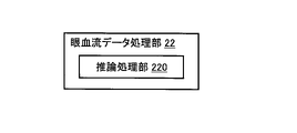
- FIG. 6 shows an example of the ocular blood flow data processing unit 22 configured by using machine learning.
- the ocular blood flow data processing unit 22 of this example includes an inference processing unit 220.
- the inference processing unit 220 uses the trained model constructed by machine learning using training data including clinical data (ocular blood flow data and diagnosis result data), and the ocular blood flow data acquired by the data acquisition unit 10. It is configured to perform inference processing that derives information about the patient's cardiovascular system from.
- the ocular blood flow data included in the training data is, for example, data acquired using the same optical modality as the optical modality of the data acquisition unit 10, but may be another modality.
- Other modality includes an optical modality different from the optical modality of the data acquisition unit 10, an ultrasonic modality, an electrical modality, a magnetic modality, an electromagnetic modality, and the like.
- the diagnostic result data included in the training data may be, for example, data obtained by a physician or other inference model (trained model) based on the relevant ocular blood flow data.
- a trained model that inputs ocular blood flow data acquired by the data acquisition unit 10 by machine learning (supervised learning) based on such training data and outputs estimated diagnostic data related to the circulatory system.
- Model can be created.
- the training data used for machine learning may include computer-generated data based on clinical data.
- Machine learning may include transfer learning.
- the machine learning algorithm and the machine learning technique may be the same as in the case of the ocular image data processing unit 21.
- the inference processing unit 220 includes the trained model obtained in this way, inputs the ocular blood flow data acquired by the data acquisition unit 10 into the trained model, and makes an estimation diagnosis output from the trained model. The data is sent to the output unit 30.
- FIG. 7 shows an example of the configuration of the inference processing unit 220.
- the inference processing unit 220 of this example includes a first trained model 221 and a second trained model 222.
- the inference processing unit 220 may include only one of the first trained model 221 and the second trained model 222.
- various matters concerning the learning model provided in the inference processing unit 220 may be the same as the corresponding matters in the learning model provided in the inference processing unit 210.
- the first trained model 221 was constructed by machine learning using training data including ocular blood flow data and diagnosis result data.
- the first trained model 221 includes, for example, a model according to the type (aspect) of the input data and the type (aspect) of the output data.
- the first trained model 221 may include a convolutional neural network similar to the first trained model 211 of the eye image data processing unit 21.
- the features considered by the first trained model 221 may be the same as or different from the features considered by the first trained model 211 of the eye image data processing unit 21.
- the second trained model 222 is constructed by machine learning using training data including data generated by processing data acquired from the fundus using a predetermined modality and diagnosis result data.
- the type of data generated by processing the data acquired from the fundus may be arbitrary, and may be, for example, image data, numerical data, distribution data, time series data, or the like.
- the second trained model 222 includes, for example, a model according to the type (aspect) of the input data and the type (aspect) of the output data.
- Information about the circulatory system is generated by inputting the data obtained by processing the data acquired by the data acquisition unit 10 into the second trained model 222.
- the features considered by the second trained model 222 may be the same as or different from the features considered by the second trained model 212 of the eye image data processing unit 21.
- the data input to the second trained model 222 may be, for example, one of the following types: an eye generated by processing ocular blood flow data acquired from the fundus of the eye using a predetermined modality. Blood flow data; data of a type other than ocular blood flow data generated by processing ocular blood flow data acquired from the fundus of the eye using a predetermined modality; eye blood acquired from the fundus of the eye using a predetermined modality. Ocular blood flow data generated by processing types of data other than flow data.
- the output unit 30 outputs the result of the processing executed by the data processing unit 20.
- the mode of output processing is arbitrary, and may be, for example, transmission, display, recording, and printing.
- the information output by the output unit 30 may be the result of the processing executed by the data processing unit 20 itself (information about the patient's circulatory system), information including the processing result, or processing the processing result.
- the information obtained may be used.
- the medical system 1 may further include a report creation unit (not shown) that creates a report based on the information about the circulatory system obtained by the data processing unit 20. In this case, the output unit 30 can output the created report.
- the output unit 30 illustrated in FIG. 1 includes a transmission unit 31.
- the transmission unit 31 transmits the result of the processing executed by the data processing unit 20 to the doctor terminal 3.
- the doctor terminal 3 is located at a remote position with respect to the data acquisition unit 10.
- the data transmission from the output unit 30 to the doctor terminal 3 may be direct transmission or indirect transmission.
- the direct transmission is an embodiment in which the result of processing (information about the circulatory system, a report, etc.) is transmitted from the output unit 30 to the doctor terminal 3.
- the indirect transmission is an embodiment in which the processing result is transmitted to a device (server, database, etc.) other than the doctor terminal 3 and the processing result is provided to the doctor terminal 3 via the device.
- the doctor terminal 3 is arranged at a remote position with respect to the data acquisition unit 10, and the information generated by the data processing unit 20 based on the data acquired by the data acquisition unit 10 from the patient's fundus (or to this).
- the doctor terminal 3 By configuring the doctor terminal 3 to provide (based information), it is possible to secure a social distance between the doctor (medical worker) and the patient, and reduce the risk of infection of the doctor (medical worker). It becomes possible.
- FIG. 8 shows an example of a usage pattern of the medical system 1. This example uses a trained model, but in the example that does not use the trained model, it is not necessary to build and load the trained model (steps S1 and S2). Installation is done.
- a trained model used in the data processing unit 20 is constructed.
- the process performed at this stage may be the update (adjustment / update of parameters) of the trained model that has already been operated.
- step S2 Install the trained model in the data processing unit
- the trained model constructed in step S1 is mounted on the data processing unit 20.
- the trained model constructed in step S1 is transmitted to the medical system 1 through the communication line.
- the subject may be, for example, a patient with a definitive diagnosis of a new coronavirus infection (COVID-19) or a suspected patient with a new coronavirus infection (COVID-19).
- the data acquisition unit 10 of the medical system 1 acquires data from the fundus of the patient using at least one optical modality.
- the data acquisition unit 10 can apply, for example, optical coherence tomography and / or color fundus photography to the fundus.
- the data acquired by optical coherence stromography may be, for example, one of three-dimensional image data, projection image data, optical coherence stromography angiographic image data, and optical coherence stromography blood flow data.
- the data acquired by color fundus photography may be, for example, color front image data representing the morphology of the fundus.
- At least a part of the inspection performed in this step may be a remote inspection using the operating device 2.
- step S4 Input data to the data processing unit
- the data acquired in step S3 is sent to the data processing unit 20.
- at least a part of the data input to the data processing unit 20 is input to the trained model constructed in step S1.
- the data processing unit 20 processes the data input in step S4 to generate information about the patient's circulatory system.
- This provides, for example, at least one of the following information: information on blood clot formation tendency (information on blood properties and / or information indicating changes in blood properties due to increased blood coagulation / fibrinolysis); thrombotic symptoms.
- Information about (methods of showing blood flow velocity distribution in blood vessels and / or information about structures formed in blood vessels); information about the state (and / or state change) of the circulatory system associated with an infectious disease; Information indicating the state related to septicemia; information indicating the state related to DIC; information indicating the state related to thrombosis; information indicating the state related to vascular obstruction.
- the information generated by the data processing unit 20 is recorded, for example, according to the data structure 100 of FIG. This provides a data package for the patient's circulatory system.
- the transmission unit 31 of the output unit 30 transmits the report created in step S6 to the doctor terminal 3 located at a remote position from the data acquisition unit 10 or to a computer capable of providing information to the medical terminal 3. .
- the doctor terminal 3 is not limited to the computer used by the doctor, and may be a computer (medical worker terminal) used by a medical worker other than the doctor.
- the medical system 1 may use non-invasive optical modalities such as optical coherence stromography and color fundus photography to acquire data from the patient's fundus and generate information about the patient's circulatory system from this data. It is possible to provide a technique for non-invasively detecting the condition of the patient's cardiovascular system. The condition of the circulatory system detected thereby is based on, for example, the medical findings described above and / or other medical findings, such as symptoms, signs of aggravation, risk of aggravation, and the like. be.
- FIG. 9 shows a configuration example of the medical system according to this embodiment.
- the medical system 1A of this example includes a data acquisition unit 10A, a data processing unit 20A, and an output unit 30.
- the output unit 30 and the transmission unit 31 are the same as the output unit 30 and the transmission unit 31 in the medical system 1, respectively. The same applies to the operating device 2 and the doctor terminal 3.
- the data acquisition unit 10A is an example of the data acquisition unit 10 of the medical system 1 described above, and includes an optical coherence tomography (OCT) device 11 and a calculation unit 12.
- OCT optical coherence tomography
- the optical coherence tomography apparatus 11 applies a scan for optical coherence tomography blood flow measurement to the fundus of the patient.
- the calculation unit 12 obtains ocular blood flow data based on the data collected by the optical coherence tomography apparatus 11 by this scan.
- Ocular blood flow data includes blood flow velocity and vessel diameter at the location where the scan was applied.
- the scanning method executed by the optical coherence tomography apparatus 11 and the calculation method executed by the calculation unit 12 may be any known method, and for example, the method described in Japanese Patent Application Laid-Open No. 2020-48730 may be used. Can be done.
- the data processing unit 20A processes the ocular blood flow data acquired by the data acquisition unit 10A in order to generate information on the blood coagulation / fibrinolysis system.
- FIG. 10 shows a configuration example of the data processing unit 20A.
- the data processing unit 20A of this example includes a wall shear rate (WALL Shear Rate; WSR) calculation unit 231, a storage unit 232, a wall shear stress (Wall Stress; WSS) calculation unit 233, and an information generation unit 234. include.
- the WSR calculation unit 231 calculates the wall shear rate (WSR) based on the blood flow velocity and the blood vessel diameter calculated by the calculation unit 12 of the data acquisition unit 10A.
- the method of calculating the wall shear rate from the blood flow velocity and the blood vessel diameter is arbitrary, and for example, the method described in the following document can be used: Taiji Nagaoka and Akitoshi Yoshida "Noninvasive Evolution of Wall Shear Retinal Humans ”, IOVS. 2006, Vol. 47, 1113-1119.
- the blood flow velocity and the blood vessel diameter are measured by using the laser Doppler velocity measurement (LDV), but the blood flow velocity and the blood vessel diameter obtained by using the optical coherence stromography as in the present embodiment. It is clear to those skilled in the art that a similar wall shear rate calculation method can be applied to.
- LDV laser Doppler velocity measurement
- the optical coherence tomography apparatus 11 of the data acquisition unit 10A applies a scan over at least one cardiac cycle to collect data.
- the calculation unit 12 calculates the time average ( Vmean ) of the (center line) blood flow velocity in one cardiac cycle as the blood flow velocity.
- the calculation unit 12 calculates the blood vessel diameter (D) from the cross-sectional image data constructed from the data collected in the above scan over one cardiac cycle.
- the storage unit 232 stores blood viscosity information 232a.
- the blood viscosity information 232a includes a blood viscosity value ⁇ .
- the blood viscosity value ⁇ may be an actually measured value or a standard value.
- the blood viscosity is measured, for example, using a conical plate viscometer.
- blood viscosity may be estimated from data obtained by a blood test such as hematocrit (Ht), red blood cell count, and red blood cell constant (mean corpuscular volume (MCV), mean corpuscular hemorrhage (MCH), etc.).
- the blood viscosity may be estimated by substituting a specified value for a blood parameter such as plasma viscosity.
- a normal value or a disease value determined from a range of blood viscosity values obtained from clinical data or experimental data can be used.
- the WSS calculation unit 233 calculates the wall shear stress (WSS) based on at least the wall shear rate calculated by the WSR calculation unit 231 and the blood viscosity value included in the blood viscosity information 232a.
- the information generation unit 234 generates information on the patient's circulatory system at least based on the wall shear stress calculated by the WSS calculation unit 233.
- information about the blood coagulation / fibrinolysis system can be generated as information about the patient's circulatory system.
- the WSR value and the WSS value are There is a one-to-one correspondence. Therefore, in this case, it is not necessary to provide the storage unit 232 and the WSS calculation unit 233, and the information generation unit 234 is the patient's circulatory system based on the value of the wall shear rate calculated by the WSR calculation unit 231. It may be configured to generate information about (information about the blood coagulation / fibrinolysis system).
- the data acquisition unit 10 performs optical coagulation stromography blood flow measurement, and generates information on the blood coagulation / fibrinolysis system from the acquired ocular blood flow data. , Generate information about the structures formed in the blood vessels.
- the medical system of this embodiment may have the same configuration as the medical system 1 and / or 1A described above.
- FIG. 11 shows a configuration example of the medical system according to this embodiment.
- the medical system 1B of this example includes a data acquisition unit 10B, a data processing unit 20B, and an output unit 30.
- the output unit 30 and the transmission unit 31 are the same as the output unit 30 and the transmission unit 31 in the medical system 1, respectively. The same applies to the operating device 2 and the doctor terminal 3.
- the data acquisition unit 10B is an example of the data acquisition unit 10 of the medical system 1 described above, and includes an optical coherence tomography (OCT) device 13 and a blood flow information generation unit 14.
- OCT optical coherence tomography
- the optical coherence tomography apparatus 13 applies a scan for optical coherence tomography blood flow measurement to the fundus of a patient.
- the optical coherence tomography apparatus 13 repeatedly applies an optical coherence tomography scan to a predetermined area of the patient's fundus to collect time-series data.
- This optical coherence tomography scan includes an A scan for at least one position (A line), and may be, for example, an A scan, a B scan, a circle scan, or the like for a plurality of positions. As a result, time series data corresponding to each scan application position can be obtained.
- the blood flow information generation unit 14 generates ocular blood flow data based on the time series collected by the optical coherence tomography apparatus 13.
- Ocular blood flow data includes blood flow information representing the spatial distribution and temporal variation of blood flow velocity.
- the space in which the distribution of blood flow velocity is defined may be one-dimensional space, two-dimensional space, or three-dimensional space.
- the time variation of blood flow velocity is defined for each point (each position) in space.
- the blood flow information obtained by the blood flow information generation unit 14 is the blood at each point in the one-dimensional space, the two-dimensional space, or the three-dimensional space inside the blood vessel to which the optical coherence stromography blood flow measurement is applied. It expresses the time change of the flow velocity.
- Such blood flow information is, for example, Robert S. et al. Reneman and Arnold P.M. G. Hoeks “Wall shear stress as measured in vivo: consequences for the design of the arterial system", Med Biol Eng Comput (2008) 46: 499.
- the data processing unit 20B can generate information on the blood coagulation / fibrinolysis system by processing the ocular blood flow data (blood flow information) acquired by the data acquisition unit 10A. Further, the data processing unit 20B can generate information on the structure formed in the blood vessel by processing the ocular blood flow data (blood flow information) acquired by the data acquisition unit 10A.
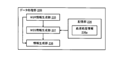
- FIG. 12 shows a configuration example of the data processing unit 20B.
- the data processing unit 20B of this example includes a wall shear rate (WSR) information generation unit 235, a storage unit 236, a wall shear stress (WSS) information generation unit 237, and an information generation unit 238.
- WSR wall shear rate
- WSS wall shear stress
- the WSR information generation unit 235 generates WSR information representing the spatial distribution and temporal change of the wall shear rate (WSR) based on at least the blood flow information generated by the blood flow information generation unit 14 of the data acquisition unit 10B.
- the method of generating the WSR information representing the spatial distribution and the temporal change of the wall shear rate from the blood flow information representing the spatial distribution and the temporal change of the blood flow velocity is arbitrary, and for example, the above-mentioned document (Robert S. Reneman and Arnold P. The method described in G. Hoeks) can be used.
- the storage unit 236 stores blood viscosity information 236a.
- the blood viscosity information 236a may be a single value ( ⁇ ) like the blood viscosity information 232a of the first embodiment, or at least blood in a target space (a space in which the distribution of blood flow velocity is defined). It may be a distribution of viscosity values.
- the single blood viscosity value ( ⁇ ) corresponds to the case where the blood viscosity distribution in the target space is uniform (constant).
- the WSS information generation unit 237 generates WSS information representing the spatial distribution and temporal change of the wall shear stress (WSS) based on at least the WSR information generated by the WSR information generation unit 235 and the blood viscosity distribution information 236a. ..
- the method of generating WSS information may be arbitrary.
- the WSS information generation unit 237 can generate WSS information by multiplying the value of the wall shear rate and the value of blood viscosity for each point in the target space, as in the first embodiment.
- the information generation unit 238 has a structure formed in the blood vessel based on at least the blood flow information generated by the blood flow information generation unit 14 of the data acquisition unit 10B and the WSS information generated by the WSS information generation unit 237. It can generate information about things (thrombus, thrombus formation tendency, etc.). Further, the information generation unit 238 is formed in the blood vessel based on at least the blood flow information generated by the blood flow information generation unit 14 of the data acquisition unit 10B and the WSR information generated by the WSR information generation unit 235. It is possible to generate information about the structure.
- the information generation unit 238 was generated by the blood flow information generated by the blood flow information generation unit 14 of the data acquisition unit 10B, the WSR information generated by the WSR information generation unit 235, and the WSS information generation unit 237.
- Information about the blood coagulation / fibrinolytic system can be generated based on any (and other) of the WSS information.
- the exemplary medical information processing apparatus 5 shown in FIG. 13 includes a data receiving unit 51, a data processing unit 52, and an output unit 53.
- a data acquisition device 6, an operation device 7, a communication device 8, and a doctor terminal 9 are provided outside the medical information processing device 5 of this embodiment.
- the data acquisition device 6 acquires data from the fundus of the patient using at least one optical method.
- the operation device 7 is used by a medical worker to operate the data acquisition device 6 (inspection device).
- the operating device 7 is located away from the data acquisition device 6 and is used to remotely control the data acquisition device 6.
- the communication device 8 transmits the data acquired by the data acquisition device 6 to the medical information processing device 5.
- the physician terminal 9 is located remotely with respect to the data acquisition device 6.
- the data receiving unit 51 of the medical information processing apparatus 5 receives data acquired from the fundus of the patient using at least one optical method.
- the data receiving unit 51 receives the data acquired by the data acquisition device 6 and transmitted by the communication device 8.
- data is transmitted from the data acquisition device 6 to the data reception unit 51 via the communication device 8, but the data input mode for the medical information processing device 5 is not limited to this.
- the data acquired by the data acquisition device 6 may be stored in a database or the like, and the data may be sent from this database to the data receiving unit 51.
- the data receiving unit 51 may include, for example, a communication device for connecting to a communication line, a drive device for reading data recorded on a recording medium, and the like.
- the data processing unit 52 is configured to process the data received by the data receiving unit 51 in order to generate information about the patient's circulatory system.
- the output unit 53 outputs information about the patient's circulatory system generated by the data processing unit 52.
- the output unit 53 of this example includes a transmission unit 54.
- the transmission unit 54 can transmit the information about the patient's circulatory system generated by the data processing unit 52 to the doctor terminal 9 located at a remote position with respect to the data acquisition device 6.
- a medical information processing device 5 and a medical system including the medical information processing device 5 it is possible to secure a social distance between a medical worker and a patient and reduce the risk of infection from the patient to the medical worker.
- data is acquired from the patient's fundus using a non-invasive optical modality such as optical coherence stromography and color fundus photography, and the patient is obtained from this data. Since it is configured to generate information about the circulatory system of the patient, it is possible to provide a technique for non-invasively detecting the condition of the patient's circulatory system.
- the condition of the circulatory system detected thereby is based on, for example, the medical findings described above and / or other medical findings, such as symptoms, signs of aggravation, risk of aggravation, and the like. be.
- the techniques according to the present disclosure are prone to vascular inflammation using non-invasive optical ophthalmic modalities such as optical coherence stromography blood flow measurement, optical coherence stromography angiography, and color fundus photography.
- non-invasive optical ophthalmic modalities such as optical coherence stromography blood flow measurement, optical coherence stromography angiography, and color fundus photography.
- changes in blood properties in the blood coagulation tendency associated with infectious diseases can be detected based on the wall shear rate and wall shear stress obtained from the blood flow velocity and blood vessel diameter obtained by optical coherence stromography blood flow measurement.
- the technique according to the present disclosure detects non-invasive blood flow dynamics (blood flow velocity, blood flow volume, shape of blood flow waveform, etc.) of retinal blood vessels, and detects wall shear stress, thrombus (peripheral vascular occlusion), etc. from the data. By evaluating, the risk of aggravation of the new coronavirus infection (COVID-19) is detected at an early stage.
- non-invasive blood flow dynamics blood flow velocity, blood flow volume, shape of blood flow waveform, etc.
- thrombus peripheral vascular occlusion
- the combination of input data and output data is not limited to this example, and can be arbitrarily determined based on medical knowledge and background.
- the input data may be color fundus image data, optical coherence stromography angiography image data, other optical coherence stromography image data (eg, morphological image data and / or functional image data).
- the output data may be the severity, the magnitude of the severity risk, the numerical value of a test other than the blood test, and the like.
- the technique according to the present disclosure can detect abnormalities in microvessels and blood flow at an early stage by using a non-invasive modality for diseases that cause systemic angiopathy / blood circulation disorder.
Landscapes
- Health & Medical Sciences (AREA)
- Life Sciences & Earth Sciences (AREA)
- Engineering & Computer Science (AREA)
- Medical Informatics (AREA)
- Public Health (AREA)
- Biomedical Technology (AREA)
- General Health & Medical Sciences (AREA)
- Physics & Mathematics (AREA)
- Biophysics (AREA)
- Molecular Biology (AREA)
- Surgery (AREA)
- Animal Behavior & Ethology (AREA)
- Heart & Thoracic Surgery (AREA)
- Veterinary Medicine (AREA)
- Pathology (AREA)
- Physiology (AREA)
- Epidemiology (AREA)
- Primary Health Care (AREA)
- Ophthalmology & Optometry (AREA)
- Artificial Intelligence (AREA)
- Hematology (AREA)
- Cardiology (AREA)
- Data Mining & Analysis (AREA)
- Databases & Information Systems (AREA)
- Nuclear Medicine, Radiotherapy & Molecular Imaging (AREA)
- Psychiatry (AREA)
- Radiology & Medical Imaging (AREA)
- Signal Processing (AREA)
- Computer Vision & Pattern Recognition (AREA)
- Vascular Medicine (AREA)
- Business, Economics & Management (AREA)
- General Business, Economics & Management (AREA)
- Evolutionary Computation (AREA)
- Fuzzy Systems (AREA)
- Mathematical Physics (AREA)
- Measuring Pulse, Heart Rate, Blood Pressure Or Blood Flow (AREA)
- Eye Examination Apparatus (AREA)
- Medical Treatment And Welfare Office Work (AREA)
Abstract
Description
例示的な態様の医療システムの構成について幾つかの例を説明する。図1に示す例示的な医療システム1は、データ取得部10と、データ処理部20と、出力部30とを含んでいる。医療システム1は、更に操作装置2を含んでいてもよい。
例示的な態様に係る医療システム1の使用形態について説明する。図8のフローチャートは、医療システム1の使用形態の例を示す。本例は学習済みモデルを利用しているが、学習済みモデルを利用しない例では学習済みモデルの構築及び搭載(ステップS1及びS2)は不要であり、例えば、それらの代わりに処理プログラムの作成及び搭載が行われる。
医療システム1の運用の準備として、データ処理部20において使用される学習済みモデルを構築する。なお、この段階で行われる処理は、既に運用されている学習済みモデルの更新(パラメータの調整・更新)であってもよい。
医療システム1の運用の更なる準備として、ステップS1で構築された学習済みモデルをデータ処理部20に搭載する。この工程では、例えば、ステップS1で構築された学習済みモデルが、通信回線を通じて医療システム1に送信される。
対象は、例えば、新型コロナウイルス感染症(COVID-19)の確定診断がなされた患者、又は、新型コロナウイルス感染症(COVID-19)の疑い患者であってよい。医療システム1のデータ取得部10は、少なくとも1つの光学的モダリティを用いて患者の眼底からデータを取得する。
ステップS3で取得されたデータは、データ処理部20に送られる。本例では、データ処理部20に入力されたデータの少なくとも一部は、ステップS1で構築された学習済みモデルに入力される。
データ処理部20は、ステップS4で入力されたデータを処理することで、患者の循環器系に関する情報を生成する。これにより、例えば、以下の情報のうちの少なくとも1つが得られる:血栓形成傾向に関する情報(血液性状に関する情報、及び/又は、血液凝固線溶系の亢進による血液性状の変化を示す情報);血栓症状に関する情報(血管内における血流速度分布を示す方法、及び/又は、血管内に形成された構造物に関する情報);感染症に随伴する循環器系の状態(及び/又は状態変化)に関する情報;敗血症に関する状態を示す情報;DICに関する状態を示す情報;血栓に関する状態を示す情報;血管閉塞に関する状態を示す情報。
医療システム1(前述した図示しないレポート作成部)は、ステップS5で生成された患者の循環器系に関する情報に基づいてレポートを作成する。
出力部30の送信部31は、ステップS6で作成されたレポートを、データ取得部10に対して遠隔位置にある医師端末3に、又は、医療端末3に対する情報提供が可能なコンピュータに、送信する。医師端末3は、医師が使用するコンピュータに限定されず、医師以外の医療従事者が使用するコンピュータ(医療従事者端末)であってもよい。
以上に説明した医療システム1の例示的な実施態様について説明する。本実施態様では、データ取得部10が光コヒーレンストモグラフィを行う場合、特に光コヒーレンストモグラフィ血流計測を行う場合について説明する。前述した医学的知見に基づき、本実施態様は、光コヒーレンストモグラフィ血流計測で取得された眼血流データから血液凝固線溶系に関する情報を生成するように構成されている。特に言及しない限り、本実施態様の医療システムは、上記医療システム1と同様の構成を備えていてよい。
医療システム1の他の例示的な実施態様について説明する。本実施態様は、第1の実施態様と同様に、データ取得部10は光コヒーレンストモグラフィ血流計測を実行し、それにより取得された眼血流データから血液凝固線溶系に関する情報を生成するとともに、血管内に形成された構造物に関する情報を生成する。特に言及しない限り、本実施態様の医療システムは、上記した医療システム1及び/又は1Aと同様の構成を備えていてよい。
例示的な態様の医療情報処理装置及びそれを含む医療システムの例を説明する。特に言及しない限り、以下の態様に係る要素は、上記した医療システム1、1A及び1Bのいずれかの要素と同様であってよい。
以上に説明したように、本開示に係る技術は、光コヒーレンストモグラフィ血流計測、光コヒーレンストモグラフィ血管造影、カラー眼底撮影などの非侵襲な光学的眼科モダリティを用いて、血管の炎症の傾向、血栓形成の傾向、敗血症の傾向、DICの傾向など、循環器系に関する状態を検知するものである。例えば、感染症などに伴う血液凝固傾向における血液性状の変化を、光コヒーレンストモグラフィ血流計測で得られた血流速や血管径から求められる壁せん断速度や壁せん断応力に基づき検知することや、血管断面における血流速の空間分布及び時間変化(血流速プロファイルの時間変化)に基づき検知することが可能である。また、機械学習により構築された学習済みモデルにこれらのデータを入力して、感染症の重症化などに関する指標を出力することが可能である。これにより、病状変化の非侵襲での早期検出や、各種診断支援情報の提供が可能となる。
2 操作装置
3 医師端末
10、10A、10B データ取得部
11、13 光コヒーレンストモグラフィ装置
12 算出部
14 血流情報生成部
20、20A、20B データ処理部
21 眼画像データ処理部
210 推論処理部
211 第1学習済みモデル
212 第2学習済みモデル
22 眼血流データ処理部
220 推論処理部
221 第1学習済みモデル
222 第2学習済みモデル
231 WSR算出部
232 記憶部
232a 血液粘度情報
233 WSS算出部
234 情報生成部
235 WSR情報生成部
236 記憶部
236a 血液粘度情報
237 WSS情報生成部
238 情報生成部
30 出力部
31 送信部
Claims (29)
- 少なくとも1つの光学的方法を用いて患者の眼底からデータを取得するデータ取得部と、
前記患者の循環器系に関する情報を生成するために、前記データ取得部により取得された前記データを処理するデータ処理部と
を含む、医療システム。 - 前記循環器系に関する情報は、血栓形成傾向に関する情報を含む、
請求項1の医療システム。 - 前記血栓形成傾向に関する情報は、血液性状に関する情報を含む、
請求項2の医療システム。 - 前記血液性状に関する情報は、血液凝固線溶系の亢進による血液性状の変化を示す情報を含む、
請求項3の医療システム。 - 前記循環器系に関する情報は、血栓症状に関する情報を含む、
請求項1の医療システム。 - 前記血栓症状に関する情報は、血管内における血流速度の分布を示す情報を含む、
請求項5の医療システム。 - 前記血栓症状に関する情報は、血管内に形成された構造物に関する情報を含む、
請求項5の医療システム。 - 前記循環器系に関する情報は、感染症に随伴する循環器系の状態に関する情報を含む、
請求項1の医療システム。 - 前記循環器系に関する情報は、敗血症に関する状態を示す情報、播種性血管内凝固症候群(DIC)に関する状態を示す情報、血栓に関する状態を示す情報、及び血管閉塞に関する状態を示す情報のうちの少なくとも1つを含む、
請求項1の医療システム。 - 前記少なくとも1つの光学的手法は、光コヒーレンストモグラフィ血流計測(OCT血流計測)、光コヒーレンストモグラフィ血管造影(OCT-A)、及びカラー眼底撮影のうちの少なくとも1つを含む、
請求項1の医療システム。 - 前記少なくとも1つの光学的手法は、前記OCT血流計測を含み、
前記データ処理部は、前記OCT血流計測で取得された血流データに少なくとも基づいて、血液凝固線溶系に関する情報を生成する、
請求項10の医療システム。 - 前記データ取得部は、
前記眼底に光コヒーレンストモグラフィ(OCT)スキャンを適用してデータを収集するOCT装置と、
前記OCT装置により収集された前記データに少なくとも基づいて血流速度及び血管径を算出する算出部と
を含み、
前記データ処理部は、前記算出部により算出された前記血流速度及び前記血管径に少なくとも基づいて前記血液凝固線溶系に関する情報を生成する、
請求項11の医療システム。 - 前記データ処理部は、前記血流速度及び前記血管径に少なくとも基づいて壁せん断速度(WSR)を算出するWSR算出部を含む、
請求項12の医療システム。 - 前記データ処理部は、
予め取得された血液粘度情報を記憶する記憶部と、
前記壁せん断速度及び前記血液粘度情報に少なくとも基づいて壁せん断応力(WSS)を算出するWSS算出部と
を更に含む、
請求項13の医療システム。 - 前記データ取得部は、
前記眼底の所定領域に光コヒーレンストモグラフィ(OCT)スキャンを繰り返し適用して時系列データを収集するOCT装置と、
前記OCT装置により収集された前記時系列データに少なくとも基づいて、血流速度の空間分布及び時間変化を表す血流情報を生成する血流情報生成部と
を含み、
前記データ処理部は、前記血流情報生成部により生成された前記血流情報に少なくとも基づいて前記血液凝固線溶系に関する情報を生成する、
請求項11の医療システム。 - 前記データ処理部は、前記血流情報に少なくとも基づいて、血管内に形成された構造物に関する情報を生成する、
請求項15の医療システム。 - 前記データ処理部は、前記血流情報に少なくとも基づいて、壁せん断速度(WSR)の空間分布及び時間変化を表すWSR情報を生成するWSR情報生成部を含む、
請求項15の医療システム。 - 前記データ処理部は、前記血流情報及び前記WSR情報に少なくとも基づいて、血管内に形成された構造物に関する情報を生成する、
請求項17の医療システム。 - 前記データ処理部は、
予め取得された血液粘度分布情報を記憶する記憶部と、
前記WSR情報及び前記血液粘度分布情報に少なくとも基づいて、壁せん断応力(WSS)の空間分布及び時間変化を表すWSS情報を生成するWSS情報生成部と
を更に含む、
請求項17の医療システム。 - 前記データ処理部は、前記血流情報及び前記WSS情報に少なくとも基づいて、血管内に形成された構造物に関する情報を生成する、
請求項19の医療システム。 - 前記データ処理部は、前記少なくとも1つの光学的方法を用いて眼底から取得された第1データと診断結果データとを含む第1訓練データを用いた機械学習によって構築された第1学習済みモデルを用いて、前記データ取得部により前記患者の前記眼底から取得された前記データを入力とし前記患者の循環器系に関する情報を出力とする推論処理を実行する第1推論処理部を含む、
請求項1の医療システム。 - 前記データ処理部は、前記少なくとも1つの光学的方法を用いて眼底から取得された第1データを処理して生成された第2データと診断結果データとを含む第2訓練データを用いた機械学習によって構築された第2学習済みモデルを用いて、前記データ取得部により前記患者の前記眼底から取得された前記データを処理して生成されたデータを入力とし前記患者の循環器系に関する情報を出力とする推論処理を実行する第2推論処理部を含む、
請求項1の医療システム。 - 前記データ取得部に対して遠隔位置にある医師端末に向けて、前記データ処理部により生成された前記循環器系に関する情報を送信する送信部を更に含む、
請求項1の医療システム。 - 前記医師端末を更に含む、
請求項23の医療システム。 - 前記データ取得部を遠隔操作するための操作部を更に含む、
請求項1の医療システム。 - 少なくとも1つの光学的方法を用いて患者の眼底から取得されたデータを受け付けるデータ受付部と、
前記患者の循環器系に関する情報を生成するために、前記データ受付部により受け付けられた前記データを処理するデータ処理部と
を含む、医療情報処理装置。 - 前記データの取得が行われた場所に対して遠隔位置にある医師端末に向けて、前記データ処理部により生成された前記循環器系に関する情報を送信する第1送信部を更に含む、
請求項26の医療情報処理装置。 - 請求項27の医療情報処理装置と、
前記医師端末と
を含む、医療システム。 - 前記少なくとも1つの光学的方法を用いて前記患者の前記眼底からデータを取得するデータ取得装置と、
前記データ取得装置により取得された前記データを前記医療情報処理装置に送信する第2送信部と
を更に含み、
前記データ受付部は、前記第2送信部により送信された前記データを受け付け、
前記データ処理部は、前記第2送信部により送信され前記データ受付部により受け付けられた前記データを、前記患者の循環器系に関する情報を生成するために処理する、
請求項28の医療システム。
Priority Applications (4)
| Application Number | Priority Date | Filing Date | Title |
|---|---|---|---|
| EP21872393.0A EP4220653A4 (en) | 2020-09-28 | 2021-09-21 | MEDICAL SYSTEM AND MEDICAL INFORMATION PROCESSING DEVICE |
| CN202180066272.XA CN116457888A (zh) | 2020-09-28 | 2021-09-21 | 医疗系统以及医疗信息处理装置 |
| US18/028,755 US12521012B2 (en) | 2020-09-28 | 2021-09-21 | Medical system and medical information processing apparatus |
| US19/336,561 US20260013721A1 (en) | 2020-09-28 | 2025-09-23 | Optical coherence tomography for eye fundus imaging to determine blood coagulation and fibrinolytic from ocular blood flow data |
Applications Claiming Priority (2)
| Application Number | Priority Date | Filing Date | Title |
|---|---|---|---|
| JP2020161990A JP7710669B2 (ja) | 2020-09-28 | 2020-09-28 | 医療システム及び医療情報処理装置 |
| JP2020-161990 | 2020-09-28 |
Related Child Applications (2)
| Application Number | Title | Priority Date | Filing Date |
|---|---|---|---|
| US18/028,755 A-371-Of-International US12521012B2 (en) | 2020-09-28 | 2021-09-21 | Medical system and medical information processing apparatus |
| US19/336,561 Continuation US20260013721A1 (en) | 2020-09-28 | 2025-09-23 | Optical coherence tomography for eye fundus imaging to determine blood coagulation and fibrinolytic from ocular blood flow data |
Publications (1)
| Publication Number | Publication Date |
|---|---|
| WO2022065264A1 true WO2022065264A1 (ja) | 2022-03-31 |
Family
ID=80846487
Family Applications (1)
| Application Number | Title | Priority Date | Filing Date |
|---|---|---|---|
| PCT/JP2021/034457 Ceased WO2022065264A1 (ja) | 2020-09-28 | 2021-09-21 | 医療システム及び医療情報処理装置 |
Country Status (5)
| Country | Link |
|---|---|
| US (2) | US12521012B2 (ja) |
| EP (1) | EP4220653A4 (ja) |
| JP (2) | JP7710669B2 (ja) |
| CN (1) | CN116457888A (ja) |
| WO (1) | WO2022065264A1 (ja) |
Cited By (1)
| Publication number | Priority date | Publication date | Assignee | Title |
|---|---|---|---|---|
| US20250342450A1 (en) * | 2024-05-03 | 2025-11-06 | Schlumberger Technology Corporation | Systems and methods for product circularity |
Families Citing this family (3)
| Publication number | Priority date | Publication date | Assignee | Title |
|---|---|---|---|---|
| US20230404411A1 (en) * | 2020-10-09 | 2023-12-21 | Biofluid Technology, Inc. | Rapid Profile Viscometer Devices And Methods |
| US20230414123A1 (en) * | 2020-11-17 | 2023-12-28 | Case Western Reserve University | System and method for measuring blood flow velocity on a microfluidic chip |
| JP2024014482A (ja) * | 2022-07-22 | 2024-02-01 | キヤノン株式会社 | 撮影システム、撮影制御方法、及びプログラム |
Citations (12)
| Publication number | Priority date | Publication date | Assignee | Title |
|---|---|---|---|---|
| JP2002153487A (ja) | 2000-11-17 | 2002-05-28 | Topcon Corp | 顕微鏡 |
| JP2014226156A (ja) | 2013-05-17 | 2014-12-08 | 株式会社トプコン | 走査型レーザ検眼鏡 |
| US20150348287A1 (en) * | 2014-04-28 | 2015-12-03 | Northwestern University | Devices, methods, and systems of functional optical coherence tomography |
| JP2016123605A (ja) | 2014-12-26 | 2016-07-11 | 積水化学工業株式会社 | 感染症リスク判定システム |
| US20180289253A1 (en) * | 2015-05-22 | 2018-10-11 | Indiana University Research & Technology Corporation | Methods and systems for patient specific identification and assessmentof ocular disease risk factors and treatment efficacy |
| JP2019054994A (ja) | 2017-09-21 | 2019-04-11 | 株式会社トプコン | 眼科撮影装置、眼科情報処理装置、プログラム、及び記録媒体 |
| JP2019058495A (ja) | 2017-09-27 | 2019-04-18 | 株式会社トプコン | 眼科装置、眼科画像処理方法、プログラム、及び記録媒体 |
| US20190272631A1 (en) * | 2018-03-01 | 2019-09-05 | Carl Zeiss Meditec, Inc. | Identifying suspicious areas in ophthalmic data |
| JP2019154988A (ja) | 2018-03-16 | 2019-09-19 | 株式会社トプコン | 眼科撮影装置、その制御方法、プログラム、及び記録媒体 |
| JP2019213734A (ja) | 2018-06-13 | 2019-12-19 | 株式会社トプコン | スリットランプ顕微鏡及び眼科システム |
| JP2020044027A (ja) | 2018-09-18 | 2020-03-26 | 株式会社トプコン | 眼科装置、その制御方法、プログラム、及び記録媒体 |
| JP2020048730A (ja) | 2018-09-26 | 2020-04-02 | 株式会社トプコン | 血流計測装置 |
Family Cites Families (13)
| Publication number | Priority date | Publication date | Assignee | Title |
|---|---|---|---|---|
| EP0903144A4 (en) * | 1996-04-17 | 2001-10-31 | Sumitomo Pharma | MEDICINE AGAINST RETAL SKIN NEUROPATHY |
| JP4803520B2 (ja) | 2006-12-01 | 2011-10-26 | 株式会社産学連携機構九州 | 血流速度画像化装置 |
| JP5627260B2 (ja) * | 2009-05-22 | 2014-11-19 | キヤノン株式会社 | 撮像装置および撮像方法 |
| EP2441390B1 (en) * | 2009-06-09 | 2017-03-01 | National Institute of Advanced Industrial Science And Technology | Device for examining vascular function |
| US20130271728A1 (en) * | 2011-06-01 | 2013-10-17 | Tushar Mahendra Ranchod | Multiple-lens retinal imaging device and methods for using device to identify, document, and diagnose eye disease |
| EP3331427A1 (en) | 2015-08-07 | 2018-06-13 | Northwestern University | Systems and methods for functional optical coherence tomography |
| WO2017062484A1 (en) * | 2015-10-09 | 2017-04-13 | Silbermann Emeric | Method for arriving at diagnoses of illnesses through comparison of retina images |
| KR101746763B1 (ko) * | 2016-02-01 | 2017-06-14 | 한국과학기술원 | 망막 또는 맥락막 내 혈관조영 광가간섭 단층촬영 장치 및 이를 이용한 질병 진단방법 |
| US10307050B2 (en) * | 2017-04-11 | 2019-06-04 | International Business Machines Corporation | Early prediction of hypertensive retinopathy |
| CN108771530B (zh) * | 2017-05-04 | 2021-03-30 | 深圳硅基智能科技有限公司 | 基于深度神经网络的眼底病变筛查系统 |
| CN111787843B (zh) | 2018-01-31 | 2023-07-21 | 株式会社拓普康 | 血流测量装置 |
| CN111428070A (zh) * | 2020-03-25 | 2020-07-17 | 南方科技大学 | 眼科案例的检索方法、装置、服务器及存储介质 |
| US20220039654A1 (en) | 2020-08-10 | 2022-02-10 | Welch Allyn, Inc. | Eye imaging devices |
-
2020
- 2020-09-28 JP JP2020161990A patent/JP7710669B2/ja active Active
-
2021
- 2021-09-21 WO PCT/JP2021/034457 patent/WO2022065264A1/ja not_active Ceased
- 2021-09-21 EP EP21872393.0A patent/EP4220653A4/en active Pending
- 2021-09-21 US US18/028,755 patent/US12521012B2/en active Active
- 2021-09-21 CN CN202180066272.XA patent/CN116457888A/zh active Pending
-
2025
- 2025-06-26 JP JP2025108071A patent/JP2025129243A/ja active Pending
- 2025-09-23 US US19/336,561 patent/US20260013721A1/en active Pending
Patent Citations (12)
| Publication number | Priority date | Publication date | Assignee | Title |
|---|---|---|---|---|
| JP2002153487A (ja) | 2000-11-17 | 2002-05-28 | Topcon Corp | 顕微鏡 |
| JP2014226156A (ja) | 2013-05-17 | 2014-12-08 | 株式会社トプコン | 走査型レーザ検眼鏡 |
| US20150348287A1 (en) * | 2014-04-28 | 2015-12-03 | Northwestern University | Devices, methods, and systems of functional optical coherence tomography |
| JP2016123605A (ja) | 2014-12-26 | 2016-07-11 | 積水化学工業株式会社 | 感染症リスク判定システム |
| US20180289253A1 (en) * | 2015-05-22 | 2018-10-11 | Indiana University Research & Technology Corporation | Methods and systems for patient specific identification and assessmentof ocular disease risk factors and treatment efficacy |
| JP2019054994A (ja) | 2017-09-21 | 2019-04-11 | 株式会社トプコン | 眼科撮影装置、眼科情報処理装置、プログラム、及び記録媒体 |
| JP2019058495A (ja) | 2017-09-27 | 2019-04-18 | 株式会社トプコン | 眼科装置、眼科画像処理方法、プログラム、及び記録媒体 |
| US20190272631A1 (en) * | 2018-03-01 | 2019-09-05 | Carl Zeiss Meditec, Inc. | Identifying suspicious areas in ophthalmic data |
| JP2019154988A (ja) | 2018-03-16 | 2019-09-19 | 株式会社トプコン | 眼科撮影装置、その制御方法、プログラム、及び記録媒体 |
| JP2019213734A (ja) | 2018-06-13 | 2019-12-19 | 株式会社トプコン | スリットランプ顕微鏡及び眼科システム |
| JP2020044027A (ja) | 2018-09-18 | 2020-03-26 | 株式会社トプコン | 眼科装置、その制御方法、プログラム、及び記録媒体 |
| JP2020048730A (ja) | 2018-09-26 | 2020-04-02 | 株式会社トプコン | 血流計測装置 |
Non-Patent Citations (9)
Cited By (1)
| Publication number | Priority date | Publication date | Assignee | Title |
|---|---|---|---|---|
| US20250342450A1 (en) * | 2024-05-03 | 2025-11-06 | Schlumberger Technology Corporation | Systems and methods for product circularity |
Also Published As
| Publication number | Publication date |
|---|---|
| JP2022054784A (ja) | 2022-04-07 |
| EP4220653A1 (en) | 2023-08-02 |
| JP7710669B2 (ja) | 2025-07-22 |
| CN116457888A (zh) | 2023-07-18 |
| US20260013721A1 (en) | 2026-01-15 |
| US20230218165A1 (en) | 2023-07-13 |
| US12521012B2 (en) | 2026-01-13 |
| EP4220653A4 (en) | 2024-10-02 |
| JP2025129243A (ja) | 2025-09-04 |
Similar Documents
| Publication | Publication Date | Title |
|---|---|---|
| US12521012B2 (en) | Medical system and medical information processing apparatus | |
| US12458298B2 (en) | Information processing apparatus, information processing method, information processing system, and program | |
| Galassi et al. | Ocular hemodynamics and glaucoma prognosis: a color Doppler imaging study | |
| Richter et al. | Telemedicine for retinopathy of prematurity diagnosis: evaluation and challenges | |
| JP6661603B2 (ja) | プラークの進行及び退行曲線に基づく治療計画のためのシステム及び方法 | |
| JP7089233B2 (ja) | 血管閉塞診断方法、機器及びシステム | |
| Hillard et al. | Retinal arterioles in hypo-, normo-, and hypertensive subjects measured using adaptive optics | |
| JP7820744B2 (ja) | 医療システム及び医療情報処理装置 | |
| Aschauer et al. | Identification of subclinical microvascular biomarkers in coronary heart disease in retinal imaging | |
| KR102306279B1 (ko) | 안저 영상판독 지원방법, 안저 영상판독 지원장치 및 이를 포함하는 안저 영상판독 지원 시스템 | |
| CA2884606A1 (en) | Systems and methods for diagnosing strokes | |
| JP2025029122A (ja) | 脳磁気共鳴血管造影検査の血管信号強度グラジエントを用いた血流算出法、並びにそれを用いた脳血管疾患及び脳卒中危険度の分析システム | |
| Theodoropoulos et al. | The current status of noninvasive intracranial pressure monitoring: a literature review | |
| Jeremic et al. | Severity stratification of coronary artery disease using novel inner ellipse-based foveal avascular zone biomarkers | |
| WO2021220910A1 (ja) | 医療システム | |
| Zhou et al. | Early diagnosis of coronary heart disease based on retinal microvascular parameters of OCTA | |
| Zhu et al. | Quantifying discordance between structure and function measurements in the clinical assessment of glaucoma | |
| WO2024057942A1 (ja) | 眼底画像処理装置および眼底画像処理プログラム | |
| Luzhnov et al. | Using nonlinear dynamics for signal analysis in transpalpebral rheoophthalmography | |
| RU2742917C1 (ru) | Способ определения модуля сдвига для стенки кровеносного сосуда на основе интраваскулярной оптической когерентной томографии | |
| CN121265007B (zh) | 一种基于影像特征变化趋势的脑梗风险预警方法及系统 | |
| US12632956B2 (en) | Fundus image processing device and non-transitory computer-readable storage medium storing computer-readable instructions | |
| US20220284577A1 (en) | Fundus image processing device and non-transitory computer-readable storage medium storing computer-readable instructions | |
| Stephens | AI Can Now Detect COVID-19 in Lung Ultrasound Images | |
| CN121729185A (zh) | 血管造影导出的微血管阻塞确定 |
Legal Events
| Date | Code | Title | Description |
|---|---|---|---|
| 121 | Ep: the epo has been informed by wipo that ep was designated in this application |
Ref document number: 21872393 Country of ref document: EP Kind code of ref document: A1 |
|
| WWE | Wipo information: entry into national phase |
Ref document number: 202180066272.X Country of ref document: CN |
|
| WWE | Wipo information: entry into national phase |
Ref document number: 2021872393 Country of ref document: EP |
|
| NENP | Non-entry into the national phase |
Ref country code: DE |
|
| ENP | Entry into the national phase |
Ref document number: 2021872393 Country of ref document: EP Effective date: 20230428 |
|
| WWG | Wipo information: grant in national office |
Ref document number: 18028755 Country of ref document: US |