WO2022085680A1 - ヒト角膜内皮細胞及び/又はヒト角膜内皮前駆細胞の保存方法 - Google Patents
ヒト角膜内皮細胞及び/又はヒト角膜内皮前駆細胞の保存方法 Download PDFInfo
- Publication number
- WO2022085680A1 WO2022085680A1 PCT/JP2021/038609 JP2021038609W WO2022085680A1 WO 2022085680 A1 WO2022085680 A1 WO 2022085680A1 JP 2021038609 W JP2021038609 W JP 2021038609W WO 2022085680 A1 WO2022085680 A1 WO 2022085680A1
- Authority
- WO
- WIPO (PCT)
- Prior art keywords
- cells
- corneal endothelial
- human corneal
- cell
- human
- Prior art date
- Legal status (The legal status is an assumption and is not a legal conclusion. Google has not performed a legal analysis and makes no representation as to the accuracy of the status listed.)
- Ceased
Links
Images

















Classifications
-
- C—CHEMISTRY; METALLURGY
- C12—BIOCHEMISTRY; BEER; SPIRITS; WINE; VINEGAR; MICROBIOLOGY; ENZYMOLOGY; MUTATION OR GENETIC ENGINEERING
- C12N—MICROORGANISMS OR ENZYMES; COMPOSITIONS THEREOF; PROPAGATING, PRESERVING, OR MAINTAINING MICROORGANISMS; MUTATION OR GENETIC ENGINEERING; CULTURE MEDIA
- C12N5/00—Undifferentiated human, animal or plant cells, e.g. cell lines; Tissues; Cultivation or maintenance thereof; Culture media therefor
- C12N5/06—Animal cells or tissues; Human cells or tissues
- C12N5/0602—Vertebrate cells
- C12N5/0618—Cells of the nervous system
- C12N5/0621—Eye cells, e.g. cornea, iris pigmented cells
-
- A—HUMAN NECESSITIES
- A01—AGRICULTURE; FORESTRY; ANIMAL HUSBANDRY; HUNTING; TRAPPING; FISHING
- A01N—PRESERVATION OF BODIES OF HUMANS OR ANIMALS OR PLANTS OR PARTS THEREOF; BIOCIDES, e.g. AS DISINFECTANTS, AS PESTICIDES OR AS HERBICIDES; PEST REPELLANTS OR ATTRACTANTS; PLANT GROWTH REGULATORS
- A01N1/00—Preservation of bodies of humans or animals, or parts thereof
- A01N1/10—Preservation of living parts
- A01N1/12—Chemical aspects of preservation
- A01N1/122—Preservation or perfusion media
- A01N1/126—Physiologically active agents, e.g. antioxidants or nutrients
-
- A—HUMAN NECESSITIES
- A01—AGRICULTURE; FORESTRY; ANIMAL HUSBANDRY; HUNTING; TRAPPING; FISHING
- A01N—PRESERVATION OF BODIES OF HUMANS OR ANIMALS OR PLANTS OR PARTS THEREOF; BIOCIDES, e.g. AS DISINFECTANTS, AS PESTICIDES OR AS HERBICIDES; PEST REPELLANTS OR ATTRACTANTS; PLANT GROWTH REGULATORS
- A01N1/00—Preservation of bodies of humans or animals, or parts thereof
- A01N1/10—Preservation of living parts
- A01N1/12—Chemical aspects of preservation
- A01N1/122—Preservation or perfusion media
- A01N1/125—Freeze protecting agents, e.g. cryoprotectants or osmolarity regulators
-
- A—HUMAN NECESSITIES
- A01—AGRICULTURE; FORESTRY; ANIMAL HUSBANDRY; HUNTING; TRAPPING; FISHING
- A01N—PRESERVATION OF BODIES OF HUMANS OR ANIMALS OR PLANTS OR PARTS THEREOF; BIOCIDES, e.g. AS DISINFECTANTS, AS PESTICIDES OR AS HERBICIDES; PEST REPELLANTS OR ATTRACTANTS; PLANT GROWTH REGULATORS
- A01N1/00—Preservation of bodies of humans or animals, or parts thereof
- A01N1/10—Preservation of living parts
- A01N1/16—Physical preservation processes
- A01N1/162—Temperature processes, e.g. following predefined temperature changes over time
-
- A—HUMAN NECESSITIES
- A61—MEDICAL OR VETERINARY SCIENCE; HYGIENE
- A61K—PREPARATIONS FOR MEDICAL, DENTAL OR TOILETRY PURPOSES
- A61K35/00—Medicinal preparations containing materials or reaction products thereof with undetermined constitution
- A61K35/12—Materials from mammals; Compositions comprising non-specified tissues or cells; Compositions comprising non-embryonic stem cells; Genetically modified cells
- A61K35/28—Bone marrow; Haematopoietic stem cells; Mesenchymal stem cells of any origin, e.g. adipose-derived stem cells
-
- A—HUMAN NECESSITIES
- A61—MEDICAL OR VETERINARY SCIENCE; HYGIENE
- A61K—PREPARATIONS FOR MEDICAL, DENTAL OR TOILETRY PURPOSES
- A61K35/00—Medicinal preparations containing materials or reaction products thereof with undetermined constitution
- A61K35/12—Materials from mammals; Compositions comprising non-specified tissues or cells; Compositions comprising non-embryonic stem cells; Genetically modified cells
- A61K35/48—Reproductive organs
- A61K35/54—Ovaries; Ova; Ovules; Embryos; Foetal cells; Germ cells
- A61K35/545—Embryonic stem cells; Pluripotent stem cells; Induced pluripotent stem cells; Uncharacterised stem cells
-
- C—CHEMISTRY; METALLURGY
- C12—BIOCHEMISTRY; BEER; SPIRITS; WINE; VINEGAR; MICROBIOLOGY; ENZYMOLOGY; MUTATION OR GENETIC ENGINEERING
- C12N—MICROORGANISMS OR ENZYMES; COMPOSITIONS THEREOF; PROPAGATING, PRESERVING, OR MAINTAINING MICROORGANISMS; MUTATION OR GENETIC ENGINEERING; CULTURE MEDIA
- C12N1/00—Microorganisms; Compositions thereof; Processes of propagating, maintaining or preserving microorganisms or compositions thereof; Processes of preparing or isolating a composition containing a microorganism; Culture media therefor
- C12N1/04—Preserving or maintaining viable microorganisms
-
- C—CHEMISTRY; METALLURGY
- C12—BIOCHEMISTRY; BEER; SPIRITS; WINE; VINEGAR; MICROBIOLOGY; ENZYMOLOGY; MUTATION OR GENETIC ENGINEERING
- C12N—MICROORGANISMS OR ENZYMES; COMPOSITIONS THEREOF; PROPAGATING, PRESERVING, OR MAINTAINING MICROORGANISMS; MUTATION OR GENETIC ENGINEERING; CULTURE MEDIA
- C12N5/00—Undifferentiated human, animal or plant cells, e.g. cell lines; Tissues; Cultivation or maintenance thereof; Culture media therefor
- C12N5/06—Animal cells or tissues; Human cells or tissues
-
- A—HUMAN NECESSITIES
- A61—MEDICAL OR VETERINARY SCIENCE; HYGIENE
- A61K—PREPARATIONS FOR MEDICAL, DENTAL OR TOILETRY PURPOSES
- A61K35/00—Medicinal preparations containing materials or reaction products thereof with undetermined constitution
- A61K35/12—Materials from mammals; Compositions comprising non-specified tissues or cells; Compositions comprising non-embryonic stem cells; Genetically modified cells
- A61K35/30—Nerves; Brain; Eyes; Corneal cells; Cerebrospinal fluid; Neuronal stem cells; Neuronal precursor cells; Glial cells; Oligodendrocytes; Schwann cells; Astroglia; Astrocytes; Choroid plexus; Spinal cord tissue
-
- A—HUMAN NECESSITIES
- A61—MEDICAL OR VETERINARY SCIENCE; HYGIENE
- A61P—SPECIFIC THERAPEUTIC ACTIVITY OF CHEMICAL COMPOUNDS OR MEDICINAL PREPARATIONS
- A61P27/00—Drugs for disorders of the senses
- A61P27/02—Ophthalmic agents
-
- C—CHEMISTRY; METALLURGY
- C12—BIOCHEMISTRY; BEER; SPIRITS; WINE; VINEGAR; MICROBIOLOGY; ENZYMOLOGY; MUTATION OR GENETIC ENGINEERING
- C12N—MICROORGANISMS OR ENZYMES; COMPOSITIONS THEREOF; PROPAGATING, PRESERVING, OR MAINTAINING MICROORGANISMS; MUTATION OR GENETIC ENGINEERING; CULTURE MEDIA
- C12N2501/00—Active agents used in cell culture processes, e.g. differentation
- C12N2501/10—Growth factors
- C12N2501/11—Epidermal growth factor [EGF]
-
- C—CHEMISTRY; METALLURGY
- C12—BIOCHEMISTRY; BEER; SPIRITS; WINE; VINEGAR; MICROBIOLOGY; ENZYMOLOGY; MUTATION OR GENETIC ENGINEERING
- C12N—MICROORGANISMS OR ENZYMES; COMPOSITIONS THEREOF; PROPAGATING, PRESERVING, OR MAINTAINING MICROORGANISMS; MUTATION OR GENETIC ENGINEERING; CULTURE MEDIA
- C12N2501/00—Active agents used in cell culture processes, e.g. differentation
- C12N2501/70—Enzymes
-
- C—CHEMISTRY; METALLURGY
- C12—BIOCHEMISTRY; BEER; SPIRITS; WINE; VINEGAR; MICROBIOLOGY; ENZYMOLOGY; MUTATION OR GENETIC ENGINEERING
- C12N—MICROORGANISMS OR ENZYMES; COMPOSITIONS THEREOF; PROPAGATING, PRESERVING, OR MAINTAINING MICROORGANISMS; MUTATION OR GENETIC ENGINEERING; CULTURE MEDIA
- C12N2501/00—Active agents used in cell culture processes, e.g. differentation
- C12N2501/70—Enzymes
- C12N2501/72—Transferases [EC 2.]
- C12N2501/727—Kinases (EC 2.7.)
-
- C—CHEMISTRY; METALLURGY
- C12—BIOCHEMISTRY; BEER; SPIRITS; WINE; VINEGAR; MICROBIOLOGY; ENZYMOLOGY; MUTATION OR GENETIC ENGINEERING
- C12N—MICROORGANISMS OR ENZYMES; COMPOSITIONS THEREOF; PROPAGATING, PRESERVING, OR MAINTAINING MICROORGANISMS; MUTATION OR GENETIC ENGINEERING; CULTURE MEDIA
- C12N2533/00—Supports or coatings for cell culture, characterised by material
- C12N2533/50—Proteins
- C12N2533/54—Collagen; Gelatin
Definitions
- the present invention relates to a method for preserving human corneal endothelial cells and / or human corneal endothelial progenitor cells, which can differentiate and mature by being stored and then recultured to express human corneal endothelial cell functional characteristics.
- Non-Patent Document 1 As a method for culturing human corneal endothelial cells in vitro, as shown in Patent Document 1, Non-Patent Document 1 and Non-Patent Document 2, epidermal growth factor (EGF) is given and human corneal endothelial cells are given by giving EGF.
- EGF epidermal growth factor
- a method of culturing by further giving another factor that suppresses the adverse effect on the maturation and differentiation of the cornea is known.
- Patent Documents 2 and 3 the present inventors have developed a method for culturing human corneal endothelial cells with as little epidermal growth factor (EGF) as possible.
- EGF epidermal growth factor
- the culture method described in Patent Document 2 or Patent Document 3 the contaminating cells having no human corneal endothelial functional characteristics as compared with the culture methods described in Patent Document 1, Non-Patent Document 1 and Non-Patent Document 2.
- a cell population in which the development is suppressed as much as possible and the content of functional human corneal endothelial cells (hereinafter, also referred to as effector cells) having the same corneal endothelial functional characteristics as those existing in the human body is improved. Obtainable.
- the cell population is described.
- the functional human corneal endothelial cells be stored in a suspended state instead of in a monolayer culture state for at least several days. Therefore, the present inventor has investigated a method of suspending a cell population or functional human corneal endothelial cells cultured by the method described in Patent Document 2 or Patent Document 3 and storing them for at least several days.
- cryopreservation is beneficial for long-term storage of cells for several years or more and cell banking, and we also investigated the establishment of a method for cryopreserving functional human corneal endothelial cells.
- human corneal endothelial cells are proliferated into a cell population having a content of functional human corneal endothelial cells (hereinafter, also referred to as E-ratio) higher than 90%. It is necessary to incubate for 4 to 5 weeks or more in order to mature and differentiate until it becomes mature. Therefore, first, a cell population that has been cultured for 4 weeks or more by the method described in Patent Document 2 or Patent Document 3 and matured and differentiated until the content of functional human corneal endothelial cells becomes a cell population higher than 90% is collected. Attempted to suspend and store.
- E-ratio functional human corneal endothelial cells
- the present inventor stored human functional corneal endothelial cells (cultured by the method described in Patent Document 2 or Patent Document 3) in a suspended state while maintaining an advantageously high survival rate.
- a method for suppressing the development of contaminating cells As a result, the present inventor maintains a high survival rate if the cell population is collected and suspended immediately after the fate of differentiation into functional human corneal endothelial cells is determined and at a time when the proliferative power is high.
- a new and improved method of preserving human corneal endothelial cells and / or human corneal endothelial progenitor cells was established.
- the present inventor if the cell population can be collected and stored immediately after the fate of differentiation into functional human corneal endothelial cells is determined and the cell population is high, the cells can be collected at the same time. We believe that it is possible to expand and cultivate functional human corneal endothelial cells in a very short period of time by collecting and repeating subculture, and we have also established a method for expanding and culturing functional human corneal endothelial cells. I let you.
- the present invention if a cell is destined to become a functional human corneal endothelial cell by being cultured under appropriate culture conditions, the cell is collected and stored in an immature state or stored. It was completed only after the present inventor found that functional human corneal endothelial cells having the objective properties of improving survival rate and reducing contaminating cells could be obtained by subculturing.
- the method for preserving human corneal endothelial cells and / or human corneal endothelial progenitor cells is as follows.
- [Item 1] Human corneal endothelial cells and / or humans grown using a medium containing a ROCK inhibitor and containing no epithelial growth factor (EGF) or having an EGF content of less than a concentration that causes transformation.
- the morphology of the human corneal endothelial cell and / or the human corneal endothelial progenitor cell has a major-minor axis ratio of 1 (for example,) from a spindle-shaped fibroblast-like morphology having irregular elongated protrusions to the length ratio including the protrusions. , About 2: 1 to about 1: 2) Immediately after turning into a paving stone-like (for example, polygonal or circular shape) morphology, and immediately before the boundaries between cells become unclear.
- B The period from when the expression level of CD44 becomes less than half of the maximum value observed after the most recent passage until the plateau is reached.
- the cell density of the human corneal endothelial cell and / or the human corneal endothelial progenitor cell is about 900 cells / mm 2 or more and about 2500 cells / mm 2 or less.
- the number of culture days after sowing is about 4 days or more and about 14 days or less.
- [Item 3] The storage method according to [Item 1] or [Item 2], wherein the human corneal endothelial cells and / or the human corneal endothelial progenitor cells are stored at a temperature of about -30 degrees or less.
- [Item 4] The storage according to any one of [Item 1] to [Item 3], wherein the human corneal endothelial cells and / or the human corneal endothelial progenitor cells are stored in a suspended state for about 24 hours or more. Method.
- [Item 5] The method according to any one of [Item 1] to [Item 4], wherein the transformation comprises epithelial-mesenchymal transformation.
- the human corneal endothelial cells and / or the human corneal endothelial precursor cells are collected from corneal endothelial cell-derived cells, pluripotent stem cells, mesenchymal stem cells, corneal endothelial precursor cells collected from the corneal endothelium, and corneal endothelium.
- a medium in which the human corneal endothelial cells and / or the human corneal endothelial precursor cells after storage contain a ROCK inhibitor and the content of epithelial growth factor (EGF) is less than the concentration that causes transformation.
- the cell population of human corneal endothelial cells obtained by proliferating and / or differentiating and maturing using the above (item 1) satisfies one or more of the following items (1) to (8).
- the cell viability is 70% or more with trypan blue staining.
- PDGF-BB is 100 pg / mL or more in the purity test of the cell supernatant by ELISA.
- the results of the purity test by FACS of cells satisfy all of the following ranges.
- the content rate (E-ratio) of effector cells having human corneal endothelial function is higher than 90%.
- the pump function Na + / K + ATPase
- the barrier function (ZO-1) is positive.
- the human corneal endothelial cell density (ECD) is 1500 cells / mm 2 or more.
- [Item 8] The storage method according to [Item 7], wherein the effector cells do not undergo endothelial mesenchymal transfer.
- the effector cells express the functional protein necessary for exerting the corneal endothelial functional characteristics, or the inhibitory protein that inhibits the corneal endothelial functional characteristics is not expressed or the expression of the inhibitory protein is reduced.
- the functional protein is selected from the group consisting of Na + / K + ATPase, ZO-1, sodium / hydrogen exchanger 1 (NHE1), aquaporin 1 (AQP-1) and carbonic anhydrase 5B (CA5B).
- the functional proteins include citrate synthase (CS), aconitase 2 (ACO2), isocitrate dehydrogenase 2 (IDH2), malic acid dehydrogenase 2 (MDH2), malic acid enzyme 3 (ME3), ACSS1, and acetyl-CoA. It further comprises one or more metabolism-related enzymes selected from the group consisting of acetyl transferase 1 (ACAT1), pyruvate dehydrogenase (PDH), BCAT2, and branched chain keto acid dehydrogenase 2 (BCKDH2).
- ACAT1 acetyl transferase 1
- PDH pyruvate dehydrogenase
- BCAT2 branched chain keto acid dehydrogenase 2
- BCKDH2 branched chain keto acid dehydrogenase 2
- the inhibitory protein is ATP citrate lyase (ACLY), aconitase 1 (ACO1), isocitrate dehydrogenase 1 (IDH1), malate dehydrogenase 1 (MDH1), malate enzyme 1 (ME1), ACSS2, acetyl.
- ACLY ATP citrate lyase
- ACO1 aconitase 1
- IDH1 isocitrate dehydrogenase 1
- MDH1 malate dehydrogenase 1
- ME1 malate enzyme 1
- ACSS2 ACSS2
- the present invention also provides the following inventions.
- [Item 13] Human intracorneal cells and / or human corneal endothelial progenitor cells preserved by the storage method according to any one of [Item 1] to [Item 12].
- [Item 14] A suspension of human intracorneal cells and / or human corneal endothelial progenitor cells preserved by the storage method according to any one of [Item 1] to [Item 12].
- [Item 15 Human corneal endothelial cells and / or human corneal endothelial progenitor cells conserved by the storage method according to any one of [Item 1] to [Item 12] containing a ROCK inhibitor and epithelial.
- a method for preparing human corneal endothelial cells which comprises growing and / or differentiating and maturing using a medium having a growth factor (EGF) content of less than a concentration that causes transformation.
- EGF growth factor
- Human corneal endothelial cells and / or human corneal endothelial precursor cells grown using a medium containing a ROCK inhibitor and having an epithelial growth factor (EGF) content of less than a concentration that causes transformation.
- Human corneal endothelial cells and / or human corneal endothelial precursor cells which are collected at a time satisfying any one or more of the following conditions (a) to (d) and put into a suspended state. Suspension preparation method.
- the morphology of the human corneal endothelial cells and / or the human corneal endothelial progenitor cells has a ratio of major axis and minor axis including the projections from a spindle-shaped fibroblast-like morphology having irregular elongated projections of 1. It is the period from immediately after the transition to a paving stone-like morphology close to that until just before the boundary between cells becomes unclear.
- B The period from when the expression level of CD44 becomes less than half of the maximum value observed after the most recent passage until the plateau is reached.
- the cell density of the human corneal endothelial cell and / or the human corneal endothelial progenitor cell is 900 cells / mm 2 or more and 2500 cells / mm 2 or less.
- the number of culture days from the most recent passage is 4 days or more and 14 days or less.
- EGF epithelial growth factor
- C The cell density of the human corneal endothelial cell and / or the human corneal endothelial progenitor cell is 900 cells / mm 2 or more and 2500 cells / mm 2 or less.
- D The number of culture days from the most recent passage is 4 days or more and 14 days or less.
- human corneal endothelial cells and / or human corneal endothelial precursor cells which can obtain a cell population having a content of functional human corneal endothelial cells higher than about 90% by re-culturing after storage, are high. It can be stored for at least several days while maintaining viability. As a result, by culturing again, it becomes possible to internationally supply a cell population having a content of functional human corneal endothelial cells higher than about 90% as a product for regenerative medicine and the like. Furthermore, cryopreservation makes it possible to create a master cell bank, which makes it possible to more efficiently and systematically manufacture products such as regenerative medicine.
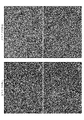
- 3 is a phase-contrast micrograph showing the difference in cell morphology depending on the culture conditions of cultured human corneal endothelial cells (Passage 3).
- 3 is a phase-contrast micrograph of cultured human corneal endothelial cells subcultured up to Passage 7 under culture condition 2. It is a graph which shows the analysis result of the cell surface antigen of the cell of Passage 3 cultured under culture condition 1 and the cell of Passage 3 or Passage 7 cultured under culture condition 2. It is a photograph showing the result of examining the expression of Na + / K + ATPase, ZO-1 and N-Cadherin in the cells of Passage 7 cultured under the culture condition 2 by an immunostaining method.
- phase-contrast micrograph of the cell cultured under the culture condition 2 from Passage 0 in Passage 7. It is a photograph and the graph which shows the state of the expression of the cell surface antigen and the like about the cell of FIG. It is a photograph which shows the result of trypan blue staining of the cell after cryopreservation which concerns on one Example of this invention.
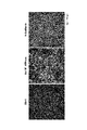
- 3 is a phase-contrast micrograph showing changes in cell morphology of cultured human corneal endothelial cells cultured under culture condition 2 after cryopreservation.
- 3 is a phase-contrast micrograph comparing the morphology of cryopreserved cells and cells that have been continuously cultured without cryopreservation.
- FIG. 12 is a phase-contrast micrograph showing cell morphology at each collection time in FIG. 12. It is a figure graph and a photograph which show the viability of the cell after cryopreservation at each collection time.
- FIG. 14 is a phase-contrast micrograph comparing the morphology of cells thawed after cryopreservation in FIG. 14 with cells cultured for 35 days under culture condition 2 and cells subcultured without cryopreservation.
- FIG. 14 is a graph comparing the surface antigens of cells thawed after cryopreservation in FIG. 14 and cells cultured for 35 days under culture condition 2 and cells subcultured without cryopreservation.
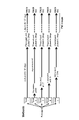
- FIG. 14 is a photograph comparing the results of immunostaining of cells thawed after cryopreservation in FIG. 14 between cells cultured under culture condition 2 for 35 days and cells subcultured without cryopreservation. It is a figure explaining the procedure of comparative analysis of the collection time suitable for cell preservation.
- 6 is a fluorescence micrograph confirming the expression of Na + / K + ATPase, ZO-1 and N-Cadherin in cells collected and cryopreserved at each collection time shown in FIG. 18.
- 6 is a fluorescence micrograph confirming the expression of Na + / K + ATPase, ZO-1 and N-Cadherin in cells collected and cryopreserved at each collection time shown in FIG. 18.
- 6 is a fluorescence micrograph confirming the expression of Na + / K + ATPase, ZO-1 and N-Cadherin in cells collected and cryopreserved at each collection time shown in FIG. 18.
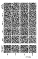
- phase contrast micrograph showing the diurnal change of the cell morphology when culturing under culture condition 2. It is a phase contrast micrograph showing the diurnal change of the cell morphology when culturing under culture condition 2. It is a graph which shows the diurnal change of the expression level of CD44 when culturing under culture condition 2. It is a graph which shows the diurnal change of ECD when culturing under culture condition 2. It is a phase contrast micrograph of cells cultured under culture condition 2 after cryopreservation, a graph showing the analysis result of surface antigen, and a photograph showing the result of immunostaining.
- FIG. 9 is a graph showing a phase-contrast micrograph of cells in which Passage 9 subcultured with the cells of FIG. 29 was cultured under culture condition 2 for 34 days and analysis results of surface antigens. It is a figure explaining the cell culture condition in Example 13. It is a graph which shows the change of the cell yield and the culture scale under each culture condition of FIG. 39.
- FIG. 39 is a phase-contrast micrograph of cell yield and cultured cells under each culture condition of FIG. 39.
- FIG. 39 is a phase-contrast micrograph of cell yield and cultured cells under each culture condition of FIG. 39.
- FIG. 39 is a phase-contrast micrograph of cell yield and cultured cells under each culture condition of FIG. 39.
- FIG. 39 is a phase-contrast micrograph of cell yield and cultured cells under each culture condition of FIG. 39.
- FIG. 39 is a phase-contrast micrograph of cell yield and cultured cells under each culture condition of FIG. 39.
- the preservation method according to the present embodiment is a method for preserving human corneal endothelial cells and / or human corneal endothelial progenitor cells.
- These human corneal endothelial cells and / or human corneal endothelial progenitor cells contain, for example, a ROCK inhibitor and the content of epithelial growth factor (EGF) is transformed according to the method described in Patent Document 2 or Patent Document 3. It was cultivated using a medium having a concentration lower than that causing the above.
- the description of Japanese Patent Application No. 2020-032139 mentioned as Patent Document 2 and the description of International Publication No. 2017/110094 mentioned as Patent Document 3 shall be appropriately referred to and incorporated.
- the human corneal endothelial cell or the human corneal endothelial precursor cell is a human corneal endothelial cell-derived cell, a pluripotent stem cell, a mesenchymal stem cell, a corneal endothelial precursor cell collected from the corneal endothelium, a cell collected from the corneal endothelium, and a direct. It is preferable that the cells are prepared from cells selected from the group consisting of corneal endothelial precursor cells and corneal endothelium-like cells prepared by a programming method.
- the human corneal endothelial progenitor cell is a concept representing a cell destined to become a human corneal endothelial cell by differentiation and maturation, and includes a cell in the middle of determining the differentiation fate.
- a basic medium used for culturing human corneal endothelial cells for example, Opti-MEM-I, Essential 6, DMEM / F12, or the like, to which an additive is appropriately added can be used.
- ROCK inhibitor also referred to as Rho-kinase inhibitor
- examples of the ROCK inhibitor include the following documents: US Pat. 076976, International Publication 02/076977, International Publication 2002/083175, International Publication 02/100833, International Publication 03/059913, International Publication 03/062227, International Publication 2004/09555, International Publication 2004/022541, International Publication 2004/108724, International Publication 2005/003101, International Publication 2005/039564, International Publication 2005/034866, International Publication 2005/037197, International Publication 2005/037198, International Publication 2005/035501, International Publication 2005/035553, International Publication 2005/035556, International Release Examples thereof include the compounds disclosed in 2005/08394, International Publication 2005/1003050, International Publication 2006/057270, International Publication 2007/0266664.
- R)-(+)-Trans- (4-pyridyl) -4- (1-aminoethyl) -cyclohexanecarboxamide dihydrochloride monohydrate and the like can be mentioned. These compounds are Wako Pure Chemical Industries, Ltd. It is also possible to purchase and obtain it from Asahi Kasei Pharma.
- the concentration of the ROCK inhibitor in the medium is, for example, preferably 1 to 100 ⁇ M, more preferably 5 to 20 ⁇ M, and particularly preferably 10 ⁇ M.
- a concentration below the concentration that causes transformation means that the concentration of epidermal growth factor (EGF) is lower than the concentration at which transformation of the corneal endothelial cells of interest (eg, endothelial mesenchymal transition) occurs or the EGF is It means that it is not included at all.
- the EGF concentration lower than the concentration that causes transformation is, for example, about 0 ng / ml to about 5 ng / ml, about 1 ng / ml to about 4 ng / ml, about 2 ng / ml to about 3 ng.
- the concentration less than the concentration that causes EGF transformation is, for example, less than 2 ng / mL, preferably less than 1 ng / mL, more preferably less than 0.5 ng / mL, and the human corneal endothelium due to the addition of EGF. From the viewpoint that adverse effects on the differentiation and maturation process of cells can be completely eliminated, 0 ng / mL (without addition) is particularly preferable.
- the medium is also free of other growth factors that mimic the EGF effect, eg, by acting on upstream or downstream signaling agents.
- transformation also referred to as phase transition
- transformation means that the trait of a cell changes to a state different from the previous state (unusual), and canceration that causes the cell to divide indefinitely. Or it includes dynamic metaplasias that change beyond the walls of the basic form of the tissue.
- transformation include cell state phase transfer (CST) such as epithelial mesenchymal transfer (EMT), fibrosis, aging, endothelial mesenchymal transfer and the like.
- CST cell state phase transfer
- EMT epithelial mesenchymal transfer
- fibrosis fibrosis
- aging endothelial mesenchymal transfer and the like.
- the method for preserving human corneal endothelial cells is human corneal endothelial cells or human corneal endothelial cells grown using a medium containing a ROCK inhibitor and having an EGF content of less than a concentration that causes transformation.
- a cell population containing human corneal endothelial precursor cells is collected at a time specified by one or more of the following indicators (a) to (d) (for example, before a differentiation event occurs). It is characterized in that it is stored in a suspended state. It is preferable that all of these indicators (a) to (d) are satisfied at the time of collecting cells, but it is not necessary to satisfy all of these, and one or more of them (for example, only (a)). It may be one that satisfies.
- the morphology of the human corneal endothelial cells and / or the human corneal endothelial progenitor cells has a ratio of major axis and minor axis including the projections from a spindle-shaped fibroblast-like morphology having irregular elongated projections of 1. It is the period from immediately after the transition to a paving stone-like morphology close to that until just before the boundary between cells becomes unclear.
- B The period from when the expression level of CD44 becomes less than half of the maximum value observed after the most recent passage until the plateau is reached.
- the cell density of the human corneal endothelial cell and / or the human corneal endothelial progenitor cell is 900 cells / mm 2 or more and 2500 cells / mm 2 or less.
- the number of culture days from the most recent passage is 4 days or more and 14 days or less.
- each index (also referred to as an evaluation standard) described in (a) to (d) will be described below.
- (a) will be described.
- human corneal endothelial cells are grown using a medium containing a ROCK inhibitor as described above and the EGF content is less than the concentration that causes transformation, the following cell morphology is obtained during the process of maturation and differentiation. Change.
- human corneal endothelial cells and / or corneal endothelial progenitor cells have a spindle-shaped morphology with irregular elongated protrusions like fibroblasts.
- the ratio of the major axis to the minor axis including the projections was close to 1 (for example, about 1.5: 1, 1.25: 1, 1.1).
- paving stones similar to corneal endothelial cells in human organisms eg hexagonal
- Etc. polygonal shape or circular shape
- the cell morphology that changes depending on the number of days of culture from passage has the above-mentioned period (a), that is, a morphology similar to that of corneal endothelial cells in vivo, but is completely differentiated.
- a a morphology similar to that of corneal endothelial cells in vivo, but is completely differentiated.
- One of the characteristic points of the preservation method according to the present embodiment is that cells are collected before maturity. If cells are collected at this time and stored in a suspended state as described herein, they can be recultured after storage to contain functional human corneal endothelial cells (hereinafter, also referred to as effector cells).
- Human corneal endothelial cells and / or human corneal endothelial precursor cells capable of obtaining a cell population having a rate (hereinafter, also referred to as E-ratio) of about 90% or more are maintained at a high survival rate for at least several days. The above can be saved.
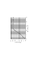
- the expression level of CD44 can be quantified by, for example, flow cytometric analysis using an antibody against CD44. For example, in the case of staining with the antibody-cell ratio of Example 3 using a PE-Cy7 binding CD44 antibody and then analyzing with FACSCanto TM II, the above-mentioned time period is the time when the cells were cultured for 35 days using the medium.
- the relative value of the average fluorescence intensity indicating the expression level of CD44 is larger than 1. It can be said that it is a period of 60 or less, more preferably 50 or less. If cells are collected at this time and stored in a suspended state, a cell population having a content of functional human corneal endothelial cells higher than about 90% can be obtained by culturing again after storage. / Or human corneal endothelial precursor cells can be stored for at least several days or more while maintaining high viability.
- the expression level of CD44 can also be measured by a method other than flow cytometric analysis such as quantitative PCR. However, it should be noted that absolute quantification is not required in all embodiments, but rather the expression pattern can be qualitatively evaluated.
- a cell population having a functional human corneal endothelial cell content of more than 90% can be obtained by culturing again after storage, and / or human corneal endothelial cells and / or Human corneal endothelial precursor cells can be stored for at least several days while maintaining high viability. It is preferable to collect the ECD value at the time of (c) described above (about 900 cells / mm 2 or more and about 2500 cells / mm 2 or less), but about 1000 cells / mm 2 or more and about 2400 cells / mm 2 or less, about 1200 cells /.
- the density of corneal endothelial cells at the start of culture is not particularly limited, but is preferably in the range of, for example, 300 cells / mm 2 or more and 500 cells / mm 2 or less.
- ECD corneal endothelial cell density
- cells are detached from a culture vessel with a cell exfoliating solution such as TrypLE to prepare a cell suspension, and a part of the cells is used to prepare a cell suspension using a blood cell counter.
- ECD corneal endothelial cell density
- Human corneal endothelial cells and / or human corneal endothelial progenitor cells collected at the above-mentioned time may be stored at an appropriate optimum temperature, but for example, low temperature storage such as about 10 ° C or lower, about 4 ° C or lower. It is preferable to store the ice temperature at about 0 ° C. or lower, about ⁇ 10 ° C. or lower, about ⁇ 30 ° C. or lower, about ⁇ 80 ° C. or lower, and freeze storage in a liquid nitrogen tank or the like. The temperature during storage may fluctuate slightly during storage, but for example, it is preferable to keep the temperature within the above-mentioned temperature range from freezing to thawing for use. ..
- the period for storing human corneal endothelial cells in a suspended state can be appropriately set according to the transportation time from the production base to the treatment institution, etc., but regardless of low temperature storage, ice temperature storage, or cryopreservation, 24 It is preferably stored for an hour or longer, and is preferably stored for a long period of time such as 2 days or longer, 3 days or longer, 4 days or longer, or about 1 week. In particular, in the case of cryopreservation, it is considered possible to store for several months to several decades.
- the conditioned medium used for culturing is preferable in the case of ice temperature storage, but the suspension is not limited to this, and for example, cultured cells are generally stored. You can use the preservative solution that is used when you do this.
- a suspension containing a cryoprotectant which is generally widely used for cryopreservation of animal cells, can be used. Examples of such a cryoprotectant include the following, which are cell membrane-permeable cryoprotectants.
- ethylene glycol ethylene glycol
- PG propylene glycol
- 1,2-propanediol 1,2-PD
- 1,3-propanediol 1,3-PD
- butylene glycol BG
- isoprene One or more cryoprotectants selected from the group consisting of glycol (IPG), dipropylene glycol (DPG) and glycerin.
- human corneal endothelial cells and / or human corneal endothelial precursor cells are preserved for a long period of time while maintaining a higher survival rate than expected. can.
- One of the reasons why it can be stored for a long period of time is that cells are collected and stored at a time when ERK (p44 / 42 MAPK) activation (phosphorylation) is enhanced compared to after maturation and differentiation. It is thought that it is one.
- the preserved cells can be administered to a patient as they are, for example.
- the preserved cells or cells recultured after preservation may be suitable for delivery to the eyes of a subject (patient) for the treatment of eye disease or injury, for example by administration or transplantation. ..
- these cells may be administered as they are, or may be used for preparing a drug containing these cells.
- the disease or injury is preferably related to the cornea.
- the preserved human corneal endothelial cells and / or human corneal endothelial progenitor cells are returned from the state of being preserved at a low temperature (for example, about -20 ° C or lower) to about room temperature to about 37 ° C such as 25 ° C. Then, after seeding in the medium again and culturing for a predetermined period (hereinafter, also referred to as after recovery), whether or not a cell population having a content of functional human corneal endothelial cells higher than 90% is obtained is listed below. It can be confirmed by whether or not one or a plurality of conditions (also referred to as evaluation criteria) among (1) to (8) are satisfied.
- the predetermined period may be a period during which human corneal endothelial cells and / or human corneal endothelial progenitor cells are sufficiently differentiated and matured, and is, for example, 4 weeks (for example, 28 days) or more after seeding. Is preferable, and more preferably 5 weeks (for example, 35 days) or more after sowing.
- the functional human corneal endothelial cell is a human corneal endothelial cell having the same corneal endothelial characteristics as the human corneal endothelial cell existing in a human body, or a human anterior chamber.
- the effector cell is at least one of the above-mentioned conditions (1) to (8), particularly CD166 + and CD24 - and CD44 neg to low , which are the conditions of (4). Refers to cells that satisfy. Of these indicators, it has already been confirmed in Patent Document 2 that CD26 + ⁇ 5% is always achieved in a cell population having CD44 high ⁇ 5%.
- Can elicit human corneal function means that it can elicit corneal endothelial properties (eg, improvement of corneal opacity and hydration edema, resulting in sustained long-term maintenance of corneal endothelial cell density, leading to improved visual acuity. Etc.) It is a concept including things.
- the expression of functional proteins linked to corneal endothelial properties is observed in functional human corneal endothelial cells having corneal endothelial properties.
- the functional protein include Na + / K + ATPase, ZO-1, sodium / hydrogen exchanger 1 (NHE1) and / or aquaporin 1 (AQP-1), carbonic anhydrase 5B (CA5B) and the like. ..
- human corneal endothelial cells proliferated and / or differentiated and matured in the presence of a ROCK inhibitor and in the presence of EGF below a concentration that causes transformation are cytoplasmic.
- these functional human corneal endothelial cells have citrate synthase (CS), aconitase 2 (ACO2), isocitrate dehydrogenase 2 (IDH2), pyruvate dehydrogenase 2 (MDH2), and malic acid enzymes 3 in their mitochondria.
- CS citrate synthase
- ACO2 aconitase 2
- IDH2 isocitrate dehydrogenase 2
- MDH2 pyruvate dehydrogenase 2
- malic acid enzymes 3 in their mitochondria.
- Expression of one or more metabolism-related enzymes selected from the group consisting of ME3 ACSS1, acetyl CoA acetyltransferase 1 (ACAT1), pyruvate dehydrogenase (PDH), BCAT2, and branched chain keto acid dehydrogenase 2 (BCKDH2).
- ACAT1 acetyl CoA acetyltransferas
- ATP citrate lyase ACLY
- ACO1 aconitase 1
- IDH1 isocitrate dehydrogenase 1
- MDH1 malate dehydrogenase 1
- ME1 malate enzyme 1
- ACSS2 acetyl-CoA acetyltransferase 2
- the present invention is not limited to that described above.
- the medium used does not necessarily have the composition described in the above embodiment, and human corneal endothelial cells and / or human corneal endothelial precursor cells satisfying the above-mentioned evaluation criteria at the time of cell collection and reculture are obtained. It goes without saying that anything that can be done is good. In addition, various modifications and combinations of embodiments may be made as long as it does not contradict the gist of the present invention.
- Example 1 Confirmation of the effect of culture conditions on the appearance rate of phase-transferred cells
- functional human corneal endothelial cells effector
- a medium containing a ROCK inhibitor and having an EGF content below the concentration that causes transformation It was confirmed that a cell population containing a very high proportion of cells) could be cultured.
- Corneal endothelial cells collected from the same donor-derived cornea were cultured in two types of culture solutions having different additive compositions, and the effects on cell quality such as the content of functional human corneal endothelial cells (effector cells) were investigated.
- the donor cornea used was that of an 18 year old male.
- the medium used was Opti-MEM-I + 8 mass% FBS + 200 mg / ml CaCl 2 + 0.08 mass% chondroitin sulfate + 20 ⁇ g / ml ascorbic acid + 50 ⁇ g / ml gentamicin.
- a medium obtained by adding 0.5 ng / ml EGF, 10 ⁇ M SB 203580, and 10 ⁇ M Y-27632 as an additive to the medium was used.
- a medium in which only 10 ⁇ M Y-27632 was added as an additive and EGF and SB203580 were not added to the medium was used.
- Corneal endothelial cells were exfoliated together with the Descemet's membrane from the donor cornea obtained from Seattle Eye Bank, treated overnight at 37 ° C. with collagenase, suspended in the medium of culture condition 1, and placed in a 6-well plate coated with type I collagen. The seeds were sown at a rate of 1 well per eye. This well was placed in a CO 2 incubator and cultured for 34 days. Medium exchange was performed twice a week. After exfoliating the cells from the culture dish by 10 ⁇ TrypLE TM Select (Thermo Fisher Scientific), the number of recovered cells was measured by a hemocytometer.
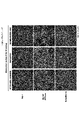
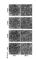
- FIG. 1 shows a phase difference photograph of cells of Passage 3 (third generation after being divided into culture condition 1 and culture condition 2) cultured under culture condition 1 or culture condition 2. From FIG. 1, it can be seen that under the culture condition 1, a large number of cell-sized phase transition cells appear. On the other hand, under culture condition 2, it can be seen that the cell size is maintained uniformly even after repeated passages, and the appearance and increase of phase-transferred cells are suppressed.
- FIG. 2 is a phase difference photograph of cells subcultured up to Passage 7 under culture condition 2, but the cell size is maintained uniformly at this stage as in the case of FIG. 1, and the phase is phased. It can be seen that the appearance and increase of transferred cells are suppressed, and there are no foreign substances, discoloration, or other abnormalities.
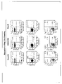
- FIG. 3 shows the analysis results of the cell surface antigens of the cells of Passage 3 cultured under culture condition 1 or the cells of Passage 3 or Passage 7 cultured under culture condition 2 by flow cytometry.
- the analysis of the cell surface antigen was carried out in the same manner as in Example 4 described later. From the results of FIG. 3, even if the subculture is repeated up to Passage 7 under the culture condition 2, the surface antigens of CD166 + CD24 - CD44 neg to low in human corneal endothelial cells or CD44 neg to low CD90 neg to low It was found that a cell population with a high proportion of effector cells showing the characteristics of the above and a high content of the target functional human corneal endothelial cells was cultured.
- the middle graph shows the CD44.
- the index of CD44 neg to low CD90 neg to low is examined in the lower graph of FIG. 3 independently of the upper and middle graphs of FIG. This also applies to FIGS. 6, 10, 16, 20, 20, 21, 22, 22, 30, 38 and the like.
- the expression level of these surface antigens is defined as follows. By "-" or “neg” is meant that expression is substantially non-observed, i.e. negative.
- the “+” means the above-mentioned non-negative one, that is, the one in which the expression is significantly observed, and the positive one.
- “Low” corresponds to weak positive when the expression is classified into weak positive, medium positive, and strong positive, and the expression level less than this weak positive is negative.
- “High” corresponds to a strong positive when the expression is classified into three stages: weak positive, medium positive, and strong positive.
- the expression intensity of surface antigens such as the above-mentioned CD marker is difficult to define numerically because the fluorescence intensity differs depending on the type of fluorescence of the label and the device setting. Each can be defined in the following range of fluorescence intensity.
- a frame for distinguishing three areas (also referred to as a gate) is described.
- the percentage in the figure indicates the percentage of cells belonging to each of the above-mentioned areas.
- ⁇ -elemen as compounds exhibiting ROCK-inhibiting properties, AT-13148, BA-210, ⁇ -elemen, chromane 1, DJ4, faszil, GSK-576371, GSK492286A, H-1152, hydroxyfaszil, ibuprofen, Selected from the group consisting of LX-7101 [24], Netaluszil, RKI-1447, Ripasudil, TCS-7001, Thiazovibin, Veloszil (AR-12286), Y-27632, Y-30141, Y-33973, and Y-39983. It may contain one or more combinations.
- Example 2 Confirmation of culture conditions that suppress phase transfer
- the cell population cultured from the primary culture (Passage 0) from the donor cornea under the above-mentioned culture condition 2 contained a high proportion of functional human corneal endothelial cells.
- the donor cornea used was that of a 27-year-old man.
- the medium used was the same as that used in the culture condition 2 of Example 1.
- Corneal endothelial cells were exfoliated from the donor cornea obtained from Seattle Eye Bank together with the Descemet's membrane, treated with collagenase at 37 ° C. overnight, suspended in the medium of the culture condition 2, and placed in a 6-well plate coated with type I collagen. The seeds were sown at a rate of 1 well per eye. This well was placed in a CO 2 incubator and cultured for 40 days. Medium exchange was performed once a week. After exfoliating the cells from the culture dish by 10 ⁇ TrypLE TM Select (Thermo Fisher Scientific), the number of recovered cells was measured by a hemocytometer.
- This cell suspension was seeded in a new culture vessel containing a medium of culture condition 2 at a cell density of 400 cells / mm 2 , and cultured again in a CO 2 incubator for 32 days. After exfoliating the cells from the culture dish by 10 ⁇ TrypLE TM Select (Thermo Fisher Scientific), the number of recovered cells was measured with a blood cell calculator, seeded at a cell density of 400 cells / mm 2 , and again in the CO 2 incubator 38. The operation of culturing for ⁇ 44 days was repeated under each culture condition, and the subculture was repeated.
- FIG. 5 shows a phase difference photograph of the cells of Passage 7 cultured as described above. From the phase difference photograph of FIG. 5, it can be seen that in the cells of Passage 7 cultured in this example, the cell size is maintained uniformly, and the appearance and increase of phase-transferred cells are suppressed.
- the graph of FIG. 6B shows the results of surface antigen analysis of the cells of Passage 7 of this example in the same procedure as in Example 1. From this result, the characteristics of the surface antigen in the functional human corneal endothelial cells were shown in most of the cells of Passage 7 of this example. From this result, it was confirmed that in this example as well, under the main culture conditions, a cell population having a high content of the target functional human corneal endothelial cells can be cultured while maintaining a high level even after long-term passage up to Passage 7. did it.
- a ROCK inhibitor such as that used in culture condition 2 from the earliest possible stage such as Passage 0 or Passage 1 is contained, and EGF is contained. It is preferable to culture in a medium in which the content of is less than the concentration that causes transformation.
- Example 3 Cell cryopreservation and confirmation of survival rate
- functional human corneal endothelial cells can be cryopreserved at a high survival rate and thawed by recovering the cell population cultured under the above-mentioned culture condition 2 at a specific culture time after passage. Later, it was confirmed that a cell population having a high content of functional human corneal endothelial cells could be obtained by culturing under the culture condition 2.
- the donor cornea used was that of a 29 year old female.
- the medium used was the same as that used in the culture condition 2 of Example 1.
- Example 3 From the results of Example 3, when the cells were seeded in the medium of the culture condition 2 at a cell density of 400 cells / mm 2 and then collected and cryopreserved on the 5th day, the survival rate was high even after the cryopreservation. It was confirmed that the maintenance was maintained. On the other hand, in the same control example in which cells were collected and stored after maturation and differentiation (after culturing for 136 days), it was found that the survival rate was significantly reduced. In addition, even when the matured and differentiated cells collected on the 35th day after subculture using a cornea derived from a male donor at the age of 14 (Passage 5) are cryopreserved by the same method, the survival rate after thawing. Was 49.5% when stored in liquid nitrogen and 63% when stored at -80 ° C, which were the same levels as the control examples shown in Table 1.
- Example 4 Evaluation of cells recovered after cryopreservation
- Example 4 it was investigated whether or not the content of functional human corneal endothelial cells was maintained at a high content in the cell population obtained by thawing and recovering the cells cryopreserved in Example 3.
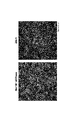
- FIG. 8 The observed changes in cell morphology over time are shown in FIG. From the results shown in FIG. 8, it was observed that the cells after cryopreservation proliferated and became a paving stone-like (polygonal shape, circular shape, etc.) morphology having a uniform size. Further, the cells that were cultured until the 35th day without cryopreservation in Example 3 and the cells that were continuously cultured until the 35th day by continuing the medium exchange without collecting the cells on the 4th day in Example 3 were placed. The result of comparison with the phase contrast microscope is shown in FIG. From the results shown in FIG. 9, no morphological difference was observed between the cells cryopreserved and the cells that were continuously cultured without cryopreservation.
- the cells cultured up to the 35th day without cryopreservation in Example 3 and the cells cultured for 35 days in this example were collected from the culture plate by 10 ⁇ TripLE TM Select (Thermo Fisher Scientific) and collected from the culture plate, and FACS buffer (PBS + 0). It was suspended in 5% by mass BSA + 0.05% by mass NaN 3 ) so as to be 4 ⁇ 10 6 cells / ml. 20 ⁇ L of this cell suspension was mixed with 20 ⁇ L of antibody solution 1 or antibody solution 2 and incubated at 4 ° C. for 1.5 to 2 hours under shading.
- the antibody solutions used are as follows. All antibodies not described by the manufacturer were obtained from BD Biosciences.
- mouse anti-Na + / K + ATTase antibody and rabbit anti-ZO-1 antibody were used as the primary antibody
- AlexaFluor488-labeled anti-rabbit IgG antibody and AlexaFluor555-labeled anti-mouse IgG antibody were used as the secondary antibody.
- the results are shown in FIG. 11 after observing with a fluorescence microscope. From this result, it was confirmed that the expression of Na + / K + ATPase and ZO-1, which are functional proteins of human corneal endothelial cells, is normally expressed even in the cryopreserved cells by recovery culture after thawing.
- Example 3 From the above results, it can be confirmed that the cell population thawed and recovered after cryopreservation in Example 3 has a high content of functional human corneal endothelial cells as in the cells that continued to be cultured without cryopreservation. rice field.
- Example 5 cryopreservation and confirmation of survival rate when other media are used
- Example 5 the effect of medium type on survival after cryopreservation was investigated.
- the donor corneas used were those of 18 years old, male and 10 years old, male.
- the medium used was Essential 6 + 8 mass% FBS + 50 ⁇ g / ml gentamicin + 10 ⁇ M Y-27632.
- the Essential 6 medium is based on DMEM / F12 medium widely used for cell culture, to which ascorbic acid, insulin, transferrin, selenium and the like are added, and its composition is open to the public.
- Example 6 Optimization of cell collection time
- Example 6-a Examination of cell collection time
- the donor cornea used was from a 41 year old female.
- the medium used was the same as that used in the culture condition 2 of Example 1.
- the donor cornea obtained from Seattle Eye Bank was cultured to Passage 2 under the same conditions as in Example 2.
- the cell suspension was divided into 5 flasks, one continued to be cultured as it was, and the remaining 4 flasks were cells on days 3, 7, 10 and 14 from the passage, respectively.
- the recovered cells were suspended in CELLBANKER I to 1,000,000 cells per ml. This cell suspension was placed in the air layer portion of a liquid nitrogen tank, frozen, and stored frozen in a liquid nitrogen tank.
- the cells of the first flask which had been continuously cultured, were subcultured to a type I collagen coated 24-well plate on the 35th day after subculture at a cell density of 400 cells / mm 2 .
- the cells that had been cryopreserved in the liquid nitrogen tank were also thawed on the same day as the cells that had been cultured.
- a ThrowSTAR frozen cell thawing station (Model CFTS, Astero Bio) was used.
- the cells washed with the medium of the culture condition 2 were seeded on a type I collagen-coated 24-well plate so as to have a cell density of 400 cells / mm 2 . This was cultured at 37 ° C.
- FIG. 13 shows a phase-contrast micrograph of cell morphology on the day of recovery of each cell lot for cryopreservation.
- FIGS. 14 the photographs of the survival rate after thawing of the cryopreserved cells and the results of trypan blue staining are shown in FIGS. 14, and the results of phase-contrast micrographs, FACS analysis and immuno-cell staining on the 37th day after recovery culture are shown in FIGS. 15 to 15. 17 is shown.
- FACS analysis and immune cell staining were performed in the same procedure as in Example 4.
- the survival rate is about 70%, and the cells collected and cryopreserved on the 7th or 10th day show a higher survival rate, and the survival rate may differ greatly depending on the recovery time after passage. It could be confirmed. From the results shown in FIGS. 15 to 17, the cells collected and cryopreserved on the 7th, 10th or 14th day were equivalent to the cells cultured without cryopreservation by the recovery culture after thawing. It was shown that the cell group contained functional human corneal endothelial cells in proportion. From this result, in order to obtain a cell group having a high survival rate even after cryopreservation and containing functional human corneal endothelial cells at the same ratio as cells cultured without cryopreservation by recovery culture after thawing. It has been found that it is preferable to collect and store cells for a period of 4 days or more and 14 days or less from the latest passage.
- Example 6-b Optimization of cell collection time
- the cell collection time clarified in Example 6-a was further optimized.
- the donor cornea used was from an 8-year-old man.
- the medium used was the same as that used in the culture condition 2 of Example 1.
- the donor cornea obtained from Seattle Eye Bank was cultured up to Passage 4 under the same conditions as in Example 2.
- the cell suspension was divided into 5 flasks, and cells were collected on days 6, 7, 8, 9, and 10 from the passage, respectively.
- Collected cells were suspended in STEMCELL-BANKER, 10% DMSO / 90% FBS, or BAMBANKER hRM (8th day only) to 1,000,000 cells per ml.
- This cell suspension was placed in the air layer portion of a liquid nitrogen tank, frozen, and stored frozen in a liquid nitrogen tank.
- the thawing operation was performed 200 days after the collection and cryopreservation on the 10th day (204 days after the collection and freezing on the 6th day).
- a water bath at 37 ° C. was used for thawing. After thawing, the cells washed with the medium of the culture condition 2 were seeded on a type I collagen-coated 24-well plate so as to have a cell density of 400 cells / mm 2 . This was cultured at 37 ° C. in a humidified atmosphere containing 5% v / v CO 2 . The medium was changed twice a week. Protein expression was comparatively analyzed by FACS analysis and immune cell staining on the 35th day after seeding on a 24-well plate. The procedure is outlined in FIG. The survival rate of the cryopreserved cells after thawing is shown in FIG.
- FIGS. 20 to 25 The survival rate was confirmed by trypan blue staining as in the other examples. FACS analysis and immune cell staining were performed in the same procedure as in Example 4.
- the bar graph on the right side of FIG. 19 shows the survival rate of the cells collected on the 8th day when either STEMCELL-BANKER, 10% DMSO / 90% FBS, or BAMBANKER hRM was used as a cryopreservation solution. ..
- Example 7 About other indicators indicating the time of cell collection
- Example 6 the number of days after seeding (passage) on the plate was examined as an index of the cell collection time.
- Example 7 by examining the characteristics of the cells at the time specified in Example 6, other indicators for specifying the time for collecting human corneal endothelial cells and / or human corneal endothelial progenitor cells were examined.
- Example 7-a Index by cell morphology
- the donor cornea used was that of a 29-year-old man.
- the medium used was the same as that used in the culture condition 2 of Example 1.
- Corneal endothelial cells were exfoliated together with Descemet's membrane from the donor cornea obtained from Seattle Eye Bank, treated overnight at 37 ° C with collagenase, suspended in the medium, and placed on a 6-well plate coated with type I collagen per eye. The seeds were sown at a rate of 1 well.
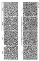
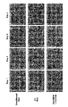
- the cells were cultured up to the 4th passage in the same procedure as in Example 2 described above. Medium exchange was performed twice a week. After passage to the 5th passage, the cells were cultured under the same conditions, and the cell morphology of each day was photographed with a phase-contrast microscope. The result is shown in FIG. 26, and a partially enlarged view of FIG. 26 is shown in FIG. 27 for further clarification.
- the cell size becomes uniform, and from the 3rd week, the boundary between cells becomes unclear from the state where the boundary between individual cells is clear, and the boundary between cells becomes unclear. It is considered that the cells are in the mature stage where the adhesion is formed and the cells become denser. From the above observations, it is presumed that under the main culture conditions, redifferentiation occurs within 2 weeks (14th day) after sowing, and then the process shifts to the maturation process. In light of the results of Example 6-a, the time when the desired human corneal endothelial cells and / or human corneal endothelial precursor cells can be collected is "the cell morphology is spindle-shaped with irregular elongated protrusions.
- Example 6-a Immediately after changing from a fibroblast-like morphology to a shape similar to the so-called paving stone-like (polygonal or circular) in-vivo corneal endothelial cells having only fine protrusions and a major-to-cell ratio close to 1, cell-cell-to-cell-to-cell It is the period until just before the adhesion of cells is formed and the boundary between cells becomes unclear. "
- the number of days in Example 6-a is used as a guide, but it is considered that the number of culture days required for the cells to mature may vary depending on the cell density at the time of passage and the culture conditions. Even in such a case, it is considered that the cells can be collected at an appropriate time by collecting the cells using the cell morphology obtained in this example as an index.
- Example 7-b Index of collection time based on the expression level of CD44
- the cell collection time can also be confirmed by the expression level of CD44.
- the expression level of CD44 on each day was measured by a flow cytometer for the cells cultured under the same conditions.
- Example 7 (Material used in the experiment) -The same cells / FACS buffer and antibody solution as in Example 7-a were used as in Example 4.
- Example 7-a After passage to the 5th passage of Example 7-a, the cells cultured for a predetermined number of days under the same conditions were washed with PBS (-) and then treated with 10 ⁇ TrypLE TM Select (Thermo Fisher Scientific) to obtain a culture plate. And suspended in FACS buffer to 4 ⁇ 106 cells / ml. 20 ⁇ L of this cell suspension was antibody-stained by the same method as in Example 4, and then analyzed by FACSCanto TM II (BD Biosciences). The results are shown in FIG.
- the average fluorescence intensity of CD44 increased to 15,000 or more immediately after sowing (passage) and then decreased logarithmically by the 11th day. It is considered that the plateau is almost reached on the 17th day, the differentiation is completed about 2 weeks after the subculture, and the maturation process is started.
- the time when the target human corneal endothelial cells and / or human corneal endothelial progenitor cells can be collected is the time when the expression level of CD44 is 1 to 2 days after passage. It is considered that it is the period from the time when it becomes less than half of the maximum value seen in the above to the time when the plateau is reached.
- Example 6-a the number of days in Example 6-a is used as a guide, but it is considered that the number of culture days required for the cells to mature may vary depending on the cell density at the time of passage and the culture conditions. Even in such a case, it is considered that the cells can be collected at an appropriate time by collecting the cells using the expression level of CD44 obtained in this example as an index.
- Example 7-c Index of collection time based on cell density
- ECD corner endothelial cell density
- Example 7-a subcultured to the 5th passage at a density of 400 cells / mm 2 were subjected to PBS (-" at each time point on the 2, 4, 6, 9, 12, 17, and 35 days after the passage. ) Washed and photographed with a phase contrast microscope. The resulting images were analyzed with KSSE-400EB software. The result is shown in FIG.
- ECD corneal endothelial cell density
- Example 6-a the number of days in Example 6-a is used as a guide, but it is considered that the number of culture days required for the cells to mature may vary depending on the cell density at the time of passage and the culture conditions. Even in such a case, it is considered that the cells can be collected at an appropriate time by collecting the cells using the cell density obtained in this example as an index.
- Example 8 Confirmation of viability and quality after recovery culture of cells cryopreserved for a long period of time
- the survival rate and the quality after recovery culture were investigated when the cells cryopreserved under the conditions optimized in Examples 6 and 7 were stored for a long period of time.
- the donor cornea used was from an 8-year-old man.
- the medium used was the same as that used in the culture condition 2 of Example 1.
- Corneal endothelial cells were exfoliated together with Descemet's membrane from the donor cornea obtained from Seattle Eye Bank, treated overnight at 37 ° C with collagenase, suspended in the medium, and placed on a 6-well plate coated with type I collagen per eye. It was sown at a rate of 1 well.
- the cells were cultured up to the second passage in the same procedure as in Example 2 described above. Medium exchange was performed twice a week. The cells were collected on the 7th day after seeding, frozen by the same method as in Example 6, and stored in a liquid nitrogen cell storage container.
- the cells were thawed in a hot water bath on the 99th, 160th, and 171st days after storage, and the survival rate was examined by the trypan blue staining method in the same procedure as in Example 3.
- the recovery rate (the number of cells after cryopreservation / the number of cells subjected to cryopreservation), which is the ratio to the number of cells in the cell, was also investigated. The results are shown in Table 3.
- FIGS. 30A to 30C FACS analysis and immune cell staining were performed in the same procedure as in Example 4.
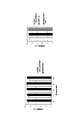
- the cell density analyzed by KSS-400EB software was 3344 ⁇ 150.8 cells / mm 2
- the PDGF-BB concentration in the culture supernatant on day 32 was 217.5 ⁇ 1.53 pg / mL.
- a cell population capable of obtaining a cell population having a high content of functional human corneal endothelial cells after recovery culture is maintained at a survival rate of 90% or more for a period of 5 months or more. It was confirmed that it can be cryopreserved.
- Example 9 Preservation of cell ice temperature and confirmation of survival rate
- the donor cornea used was from an 8-year-old man.
- the medium used was the same as that used in the culture condition 2 of Example 1.
- Each sample is as follows. Sample 1: Cells cultured in the same procedure as in Example 2 and cultured in Passage 3 for 94 days without cryopreservation. Sample 2: Cells cultured in the same procedure as in Example 2, collected on the 7th day of Passage 2, cryopreserved in the same procedure as in Example 3, thawed, and cultured in Passage 3 for 33 days.
- Sample 3 Cultivate in the same procedure as in Example 2, collect on the 7th day of Passage 2, freeze and store in the same procedure as in Example 3, repeat subculture until Passage 5 after thawing, and incubate in Passage 6 for 9 days. Cells.
- samples 1 to 3 were exfoliated from the culture vessel by 10 ⁇ TypLETM Select (Thermo Fisher Scientific) to prepare a cell suspension. After staining a part (10 ⁇ l) of each cell suspension with trypan blue, the number of live cells and the number of dead cells were measured using a hemocytometer. Based on this measurement result, the cells were suspended in each conditioned medium so as to have a cell density of 3.33 ⁇ 10 6 cells / ml, placed in a commercially available microtube of 1.5 mL, and stored in ice.
- FIG. 31 shows the results of trypan blue staining
- FIG. 32 shows the survival rate calculated from the results of FIG. 31. From the results shown in FIG. 32, the survival rate of sample 1 was lower than 70% on the first day and sample 2 was lower than 70% on the second day, whereas the survival rate of sample 3 was maintained at more than 80% even on the fourth day. I found that it was done.
- Example 10 Effect of preservation solution on preservation of cells at ice temperature
- the influence on the survival rate by the type of the preservation solution used for the preservation at ice temperature was investigated.
- the cells used in the experiment are sample 3 of Example 9.
- the preservation solution used in the experiment is as follows.
- Preservative solution A Acclimated medium
- Preservative solution B Medium of culture condition 2 of Example 1 prepared on the same day
- Preservative solution C Opti-MEM + 100 ⁇ M Y-27632 prepared on the same day
- Example 11 Evaluation of differentiation and maturation period of cells stored at ice temperature into functional human corneal endothelial cells
- Example 11 when cells stored at ice temperature are seeded at a cell density similar to that of transplantation into humans, do they differentiate and mature into functional human corneal endothelial cells at an early stage as in the case of transplantation into humans? It was confirmed.
- Example 9 Evaluation of cell morphology
- 300 ⁇ l of a suspension with a cell density of 3.33 ⁇ 10 6 cells / ml is injected into the anterior chamber. Therefore, in Example 9, a 24-well plate having a cell suspension (having a cell density equivalent to that at the time of transplantation into humans) stored at ice temperature for 4 days using the preservation solution A having an area close to the human endothelial surface. 300 ⁇ l was seeded (diameter about 7.78 mm, bottom area 190 mm 2 , type I collagen coated). After sowing, the cells were cultured under the culture condition 2 of Example 1 for 7 days. The phase difference micrograph at this time is shown in FIG. 35. From FIG. 35, it can be seen that even cells stored at ice temperature for 4 days are in the form of functional human corneal endothelial cells one week after seeding when seeded at the same cell density as transplanted into humans. ..
- FIG. 35 The cells of FIG. 35 were immobilized with ice-cold methanol, and the expression of the functional proteins Na + / K + ATPase and ZO-1 of human corneal endothelial cells was confirmed by immunostaining. The results are shown in FIG. From the results shown in FIG. 36, the expression of Na + / K + ATPase and ZO-1 was confirmed in almost all cells.
- Example 12 Short-term expansion culture of human corneal endothelial cells after freezing storage
- FIG. 38A the phase-contrast micrographs of the cells cultured under culture condition 2 for 34 days are shown in FIG. 38A, and the analysis results of the surface antigen by FACS analysis are shown in FIG. 38B. show.
- the FACS analysis was performed in the same procedure as in Example 4. Cells that were repeatedly passaged at intervals of 8 to 10 days after seeding, which was before differentiation and maturation, also used a medium that finally contained the ROCK inhibitor and the EGF content was less than the concentration that caused transformation. By further culturing, it was confirmed that a cell population having a sufficiently high content of functional human corneal endothelial cells could be obtained.
- Example 13 Short-term expansion culture of human corneal endothelial cells
- the donor corneas used were 29 years old, male, and 8 years old, female.
- FIG. 40 shows the yield and culture scale calculated from the growth rate. It was shown that the short-term expansion culture method of this example, in which passages were repeated at intervals of about 8 days, produced 2 billion cells within 80 days, or 500 flasks of cells when converted to T-25 flasks. .. Further, a photograph of cells cultured up to Passage 4 by the short-term expansion culture method described above and cultured in Passage 5 for about 5 weeks, cells of Control Example 2 cultured for about the same number of days, and cells of Control Example 2 cultured up to Passage 5. And a comparison of yields is shown in FIGS. 41 and 42.
Landscapes
- Health & Medical Sciences (AREA)
- Life Sciences & Earth Sciences (AREA)
- Engineering & Computer Science (AREA)
- Biomedical Technology (AREA)
- Zoology (AREA)
- Chemical & Material Sciences (AREA)
- Wood Science & Technology (AREA)
- Biotechnology (AREA)
- General Health & Medical Sciences (AREA)
- Cell Biology (AREA)
- Organic Chemistry (AREA)
- Bioinformatics & Cheminformatics (AREA)
- Genetics & Genomics (AREA)
- Developmental Biology & Embryology (AREA)
- Medicinal Chemistry (AREA)
- Virology (AREA)
- Microbiology (AREA)
- Biochemistry (AREA)
- General Engineering & Computer Science (AREA)
- Ophthalmology & Optometry (AREA)
- Neurology (AREA)
- Neurosurgery (AREA)
- Immunology (AREA)
- Environmental Sciences (AREA)
- Dentistry (AREA)
- Pharmacology & Pharmacy (AREA)
- Animal Behavior & Ethology (AREA)
- Public Health (AREA)
- Veterinary Medicine (AREA)
- Epidemiology (AREA)
- Reproductive Health (AREA)
- Tropical Medicine & Parasitology (AREA)
- Gynecology & Obstetrics (AREA)
- Hematology (AREA)
- Micro-Organisms Or Cultivation Processes Thereof (AREA)
- Medicines Containing Material From Animals Or Micro-Organisms (AREA)
- Materials For Medical Uses (AREA)
- Biophysics (AREA)
- Physiology (AREA)
- Agricultural Chemicals And Associated Chemicals (AREA)
Abstract
Description
そこで、本発明者は、特許文献2又は特許文献3に記載の方法で培養された細胞集団又は機能性ヒト角膜内皮細胞を懸濁状態にして少なくとも数日間保存する方法について検討した。
加えて、細胞の数年以上にわたる長期保存や、細胞のバンク化には凍結保存が有益であり、機能性ヒト角膜内皮細胞を凍結保存する方法の確立についても検討した。
そこで、まずは、特許文献2又は特許文献3に記載の方法で4週間以上培養し機能性ヒト角膜内皮細胞の含有率が90%より高い細胞集団になるまで成熟分化させた細胞集団を採取して懸濁状態にし、保存することを試みた。
その結果、機能性ヒト角膜内皮細胞の含有率が90%より高い細胞集団になるまで成熟分化させた後の細胞集団を懸濁状態にして保存すると、保存後の生存率が低く、保存後の培養において夾雑細胞の発生割合が高くなってしまうことが分かった。
その結果、本発明者は、機能性ヒト角膜内皮細胞に分化する運命が決定した直後であり、かつ増殖力が高い時期に細胞集団を採取して懸濁状態にすれば、高い生存率を維持したまま保存し、かつ保存後の培養において夾雑細胞の発生割合を小さく抑えることができることを見出して、ヒト角膜内皮細胞及び/又はヒト角膜内皮前駆細胞の新規、かつ改良された保存方法(およびそれに使用する関連組成物)を確立させた。
また本発明者は、機能性ヒト角膜内皮細胞に分化する運命が決定した直後であり、かつ増殖力が高い時期に細胞集団を採取して保存することができるのであれば、同時期に細胞を採取して継代培養を繰り返すことによって、従来に比べて非常に短い期間での機能性ヒト角膜内皮細胞の拡大培養が可能であると考え、機能性ヒト角膜内皮細胞の拡大培養方法をも確立させた。
このように、本発明は、適切な培養条件で培養されることによって機能性ヒト角膜内皮細胞になることが運命付けられた細胞であれば、その細胞を未成熟の状態で採取して保存又は継代することによって、生存率向上及び夾雑細胞低減という目的の性質を有する機能性ヒト角膜内皮細胞を得ることができることを本発明者が見出して初めて完成したものである。
〔項目1〕ROCK阻害剤を含有し、かつ上皮成長因子(EGF)が非添加若しくはEGFの含有量が形質転換を引き起こす濃度未満である培地を用いて増殖させたヒト角膜内皮細胞及び/又はヒト角膜内皮前駆細胞を、以下に挙げる(a)~(d)のいずれか1つ又は複数の条件を満たす時期に採取し、懸濁状態にして保存することを特徴とするヒト角膜内皮細胞及び/又はヒト角膜内皮前駆細胞の保存方法。
(a)前記ヒト角膜内皮細胞及び/又は前記ヒト角膜内皮前駆細胞の形態が、不規則な細長い突起を持つ紡錘状の線維芽細胞様の形態から前記突起を含めた長短径比が1(例えば、約2:1~約1:2)に近い敷石状(例えば、多角形状又は円形状)の形態に転じた直後から、細胞間の境界が不明瞭になる直前までの期間である。
(b)CD44の発現量が直近の継代後にみられる最大値の半分以下になった時からプラトーに達するまでの期間である。
(c)前記ヒト角膜内皮細胞及び/又は前記ヒト角膜内皮前駆細胞の細胞密度が、約900cells/mm2以上約2500cells/mm2以下である。
(d)播種してからの培養日数が、約4日以上約14日以下である。
〔項目2〕前記ヒト角膜内皮細胞及び/又は前記ヒト角膜内皮前駆細胞をおよそ10度以下保存する、〔項目1〕に記載の保存方法。
〔項目3〕前記ヒト角膜内皮細胞及び/又は前記ヒト角膜内皮前駆細胞をおよそ-30度以下で保存する、〔項目1〕又は〔項目2〕に記載の保存方法。
〔項目4〕前記ヒト角膜内皮細胞及び/又は前記ヒト角膜内皮前駆細胞を懸濁状態にしてから約24時間以上保存する、〔項目1〕~〔項目3〕の何れか一項に記載の保存方法。
〔項目5〕前記形質転換が、内皮間葉転換を含む、〔項目1〕~〔項目4〕の何れか一項に記載の方法。
〔項目6〕前記ヒト角膜内皮細胞及び/又は前記ヒト角膜内皮前駆細胞が、角膜内皮細胞由来細胞、多能性幹細胞、間葉系幹細胞、角膜内皮から採取した角膜内皮前駆細胞、角膜内皮から採取した細胞、並びにダイレクトプログラミング法で作製される角膜内皮前駆細胞及び角膜内皮様細胞からなる群から選択される細胞を起源として作成されたものである、〔項目1〕~〔項目5〕の何れか一項に記載の保存方法。
〔項目7〕保存後の前記ヒト角膜内皮細胞及び/又は前記ヒト角膜内皮前駆細胞を、ROCK阻害剤を含有し、かつ上皮成長因子(EGF)の含有量が形質転換を引き起こす濃度未満である培地を用いて増殖及び/又は分化・成熟させて得たヒト角膜内皮細胞の細胞集団が、以下の(1)~(8)のうち、1又は複数の項目を満たすものである、〔項目1〕~〔項目6〕の何れか一項に記載の保存方法。
(1)位相差画像による外観検査で、線維芽細胞、異物、変色、又は他の異常がない。
(2)細胞生存率がトリパンブルー染色で70%以上である。
(3)細胞上清のELISAによる純度試験で、PDGF-BBが100pg/mL以上である。
(4)細胞のFACSによる純度試験結果が以下の範囲を全て満たすものである。
CD166+>99%
CD24+<5%
CD44high<5%
CD44neg~low>90%
CD90+<5%
(5)ヒト角膜内皮機能を有するエフェクター細胞の含有率(E-ratio)が90%より高い。
(6)ポンプ機能(Na+/K+ ATPase)が陽性である。
(7)バリア機能(ZO-1)が陽性である。
(8)ヒト角膜内皮細胞密度(ECD)が1500細胞/mm2以上である。
〔項目8〕前記エフェクター細胞が、内皮間葉系移行が起きていないものである、〔項目7〕に記載の保存方法。
〔項目9〕
前記エフェクター細胞が、前記角膜内皮機能特性を発揮するために必要な機能蛋白を発現しているか、又は前記角膜内皮機能特性を阻害する阻害蛋白が発現されない若しくは阻害蛋白の発現が低下しているものである、〔項目7〕又は〔項目8〕に記載の保存方法。
〔項目10〕前記機能蛋白がNa+/K+ ATPase、ZO-1、ナトリウム/水素交換体1(NHE1)、アクアポリン1(AQP-1)及び炭酸脱水酵素5B(CA5B)からなる群より選ばれる一つ又は複数を含む、〔項目9〕に記載の保存方法。
〔項目11〕前記機能蛋白が、クエン酸シンターゼ(CS)、アコニターゼ2(ACO2)、イソクエン酸デヒドロゲナーゼ2(IDH2)、リンゴ酸デヒドロゲナーゼ2(MDH2)、リンゴ酸酵素3(ME3)、ACSS1、アセチルCoAアセチルトランスフェラーゼ1(ACAT1)、ピルビン酸デヒドロゲナーゼ(PDH)、BCAT2、及び分岐鎖ケト酸デヒドロゲナーゼ2(BCKDH2)からなる群から選択される1又は複数の代謝関連酵素をさらに含む、〔項目9〕又は〔項目10〕に記載の保存方法。
〔項目12〕前記阻害蛋白が、ATPクエン酸リアーゼ(ACLY)、アコニターゼ1(ACO1)、イソクエン酸デヒドロゲナーゼ1(IDH1)、リンゴ酸デヒドロゲナーゼ1(MDH1)、リンゴ酸酵素1(ME1)、ACSS2、アセチルCoAアセチルトランスフェラーゼ2(ACAT2)及び乳酸デヒドロゲナーゼ(LDH)からなる群から選択される少なくとも1種以上の代謝関連酵素を含む、〔項目9〕~〔項目11〕のいずれか一項に記載の保存方法。
〔項目13〕〔項目1〕~〔項目12〕の何れか一項に記載の保存方法で保存されたヒト角膜内細胞及び/又はヒト角膜内皮前駆細胞。
〔項目14〕〔項目1〕~〔項目12〕の何れか一項に記載の保存方法で保存されたヒト角膜内細胞及び/又はヒト角膜内皮前駆細胞の懸濁液。
〔項目15〕〔項目1〕~〔項目12〕の何れか一項に記載の保存方法で保存されたヒト角膜内皮細胞及び/又はヒト角膜内皮前駆細胞を、ROCK阻害剤を含有し、かつ上皮成長因子(EGF)の含有量が形質転換を引き起こす濃度未満である培地を用いて増殖及び/又は分化・成熟させることを特徴とする、ヒト角膜内皮細胞の調製方法。
〔項目16〕ROCK阻害剤を含有し、かつ上皮成長因子(EGF)の含有量が形質転換を引き起こす濃度未満である培地を用いて増殖させたヒト角膜内皮細胞及び/又はヒト角膜内皮前駆細胞を、以下に挙げる(a)~(d)のいずれか1つ又は複数の条件を満たす時期に採取し、懸濁状態にすることを特徴とする、ヒト角膜内皮細胞及び/又はヒト角膜内皮前駆細胞の懸濁液調製方法。
(a)前記ヒト角膜内皮細胞及び/又は前記ヒト角膜内皮前駆細胞の形態が、不規則な細長い突起を持つ紡錘状の線維芽細胞様の形態から前記突起を含めた長径短径の比が1に近い敷石状の形態に転じた直後から、細胞間の境界が不明瞭になる直前までの期間である。
(b)CD44の発現量が直近の継代後にみられる最大値の半分以下になった時からプラトーに達するまでの期間である。
(c)前記ヒト角膜内皮細胞及び/又は前記ヒト角膜内皮前駆細胞の細胞密度が、900cells/mm2以上2500cells/mm2以下である。
(d)直近の継代からの培養日数が、4日以上14日以下である。
〔項目17〕ROCK阻害剤を含有し、かつ上皮成長因子(EGF)の含有量が形質転換を引き起こす濃度未満である培地を用いて増殖させたヒト角膜内皮細胞及び/又はヒト角膜内皮前駆細胞を、以下に挙げる(a)~(d)のいずれか1つ又は複数の条件を満たす時期に採取し、継代することを特徴とする、ヒト角膜内皮細胞及び/又はヒト角膜内皮前駆細胞の培養方法。
(a)前記ヒト角膜内皮細胞及び/又は前記ヒト角膜内皮前駆細胞の形態が、不規則な細長い突起を持つ紡錘状の線維芽細胞様の形態から、長径短径の比が1に近い敷石状の形態に転じた直後から、細胞間の境界が不明瞭になる直前までの期間である。
(b)CD44の発現量が直近の継代後にみられる最大値の半分以下になった時からプラトーに達するまでの期間である。
(c)前記ヒト角膜内皮細胞及び/又は前記ヒト角膜内皮前駆細胞の細胞密度が、900cells/mm2以上2500cells/mm2以下である。
(d)直近の継代からの培養日数が、4日以上14日以下である。
その結果、再度培養することによって機能性ヒト角膜内皮細胞の含有率がおよそ90%より高い細胞集団を、再生医療等製品として、国際的に供給することが可能となる。
さらには、凍結保存によりマスターセルバンク化が可能となり、再生医療等製品としての製造をより効率的かつ計画的に行うことができるようになる。
本実施形態に係る保存方法は、ヒト角膜内皮細胞及び/又はヒト角膜内皮前駆細胞を保存する方法である。
これらヒト角膜内皮細胞及び/又はヒト角膜内皮前駆細胞は、例えば、特許文献2又は特許文献3に記載の方法に従って、ROCK阻害剤を含有し、かつ上皮成長因子(EGF)の含有量が形質転換を引き起こす濃度未満である培地を用いて培養されたものである。
なお、本願明細書では、この特許文献2として挙げた特願2020-032139の記載及び特許文献3として挙げた国際公開2017/110094号公報の記載を適宜参酌して援用するものとする。
前記培地中のROCK阻害剤の濃度は、例えば、1~100μMであることが好ましく、より好ましくは5~20μM、特に好ましくは10μMである。
細胞を採取する時期は、これら(a)~(d)の全ての指標を満たしていることが好ましいが、これらすべてを満たしている必要はなく、何れか1つ以上(例えば(a)のみ)を満たすものであっても良い。
(b)CD44の発現量が直近の継代後にみられる最大値の半分以下になった時からプラトーに達するまでの期間である。
(c)前記ヒト角膜内皮細胞及び/又は前記ヒト角膜内皮前駆細胞の細胞密度が、900cells/mm2以上2500cells/mm2以下である。
(d)直近の継代からの培養日数が、4日以上14日以下である。
まず(a)について説明する。
ヒト角膜内皮細胞を前述したようなROCK阻害剤を含有し、EGFの含有量が形質転換を引き起こす濃度未満である培地を使用して増殖させると、成熟分化の過程で以下のような細胞形態が変化する。
前述した培地を入れた培養プレートに播種した直後のヒト角膜内皮細胞及び/又は角膜内皮前駆細胞は、線維芽細胞様の不規則な細長い突起を有する紡錘状の形態である。培養日数が経つにつれて、細長い突起が小さくなり、突起を含めた長径と短径の比(長短径比)が1に近い(例えば、約1.5:1、1.25:1、1.1:1、1:1.1、1:1.25、1:1.5、およびこれらの間の任意の比率)、ヒトの生体内での角膜内皮細胞に類似の敷石状(例えば6角形状等の多角形状又は円形状)の形態になる。その後、細胞の大きさがより均一になり、十分に分化成熟した機能性角膜内皮細胞になると、細胞同士が接着して細胞間の境界線が不明瞭になる。
このように継代からの培養日数によって変遷する細胞の形態が、前述した(a)である時期、すなわち、生体内での角膜内皮細胞に類似の形態を有してはいるが、完全に分化成熟する前の時期に細胞を採取する点が本実施形態に係る保存方法の特徴点の一つである。
この時期に細胞を採取し、本明細書に記載されているように懸濁状態で保存すれば、保存後に再度培養することによって機能性ヒト角膜内皮細胞(以下、エフェクター細胞ともいう。)の含有率(以下、E-ratioともいう。)がおよそ90%より高い細胞集団を得ることができるヒト角膜内皮細胞及び/又はヒト角膜内皮前駆細胞を、高い生存率を維持したままで、少なくとも数日以上保存することができる。
ヒト角膜内皮細胞を前述したようなROCK阻害剤を含有し、EGFの含有量が形質転換を引き起こす濃度未満である培地を使用して増殖させると、成熟分化の過程で表面抗原であるCD44の発現量が以下のように変化する。
CD44の発現量は、ヒト角膜内皮細胞を前述した培地を入れた培養プレートに播種した直後(1~2日目)に大きく増大し、培養日数を経るにつれて対数的に減少し、ある程度細胞が分化成熟すると一定量に落ち着く(プラトーに達する)ことが観察されている。
このCD44の発現量が(b)のようになっている時期、すなわち、CD44の発現量が継代直後よりは減少しているが、十分に分化成熟しCD44の発現量がプラトーに達した細胞よりは多い時期に細胞を採取する点が本実施形態の特徴点の一つである。なお、このCD44の発現量は、たとえば、CD44に対する抗体を使用したフローサイトメトリー解析などによって定量することができる。例えば、PE-Cy7結合CD44抗体を用いて実施例3の抗体-細胞比率で染色後、FACSCantoTM IIで解析した場合であると、上述の時期は、前記培地を使用して35日間培養した時点の前記ヒト角膜内皮細胞及び/又は前記ヒト角膜内皮前駆細胞のCD44の発現量を示す平均蛍光強度の値を1とした場合の、CD44の発現量を示す平均蛍光強度の相対値が1より大きく60以下、より好ましくは50以下である時期であるとも言える。この時期に細胞を採取して懸濁状態で保存すれば、保存後に再度培養することによって機能性ヒト角膜内皮細胞の含有率がおよそ90%より高い細胞集団を得ることができるヒト角膜内皮細胞及び/又はヒト角膜内皮前駆細胞を、高い生存率を維持したままで、少なくとも数日以上保存することができる。このCD44の発現量は、例えば、定量PCR等のフローサイトメトリー解析以外の方法によって測定することも可能である。ただし、すべての実施形態において絶対的な定量は必要なく、むしろ発現パターンを定性的に評価することができることに留意されたい。
ヒト角膜内皮細胞は、前述した培地を入れた培養プレートに播種した後、ほぼ直線的に増加し、十分に分化成熟すると角膜内皮細胞密度(以下、ECDともいう。)の値が一定値に落ち着くことが分かっている。
このECDの値が(c)のようになっている時期、すなわち、細胞密度が増えている増殖期ではあるが、すでに機能性ヒト角膜内皮細胞に分化する運命が決定した時期に細胞を採取する点が本実施形態の特徴点の一つである。この時期に細胞を採取して懸濁状態にすることによって、保存後に再度培養すれば機能性ヒト角膜内皮細胞の含有率が90%より高い細胞集団を得ることができるヒト角膜内皮細胞及び/又はヒト角膜内皮前駆細胞を、高い生存率を維持したままで、少なくとも数日以上保存することができる。
ECDの値が、前述した(c)の時期(約900cells/mm2以上約2500cells/mm2以下)に採取するのが好ましいが、約1000cells/mm2以上約2400cells/mm2以下、約1200cells/mm2以上約2000cells/mm2以下、約1200cells/mm2以上約1800cells/mm2未満などの時期に採取するものとしても良い。いくつかの実施形態では、上記の範囲の組み合わせの間にあるECD値も用いることができる。
培養開始時の角膜内皮細胞密度は、特に限定されるものではないが、例えば、300cells/mm2以上500cells/mm2以下の範囲であることが好ましい。
なお、角膜内皮細胞密度(ECD)は、例えば、細胞を培養容器からTrypLEなどの細胞剥離液により剥離して細胞懸濁液を調製し、その一部を用いて血球計算盤を用いて細胞数を計測することや、位相差顕微鏡で撮像した写真等に基づいて解析ソフトウェア(例えば、KSSE-400EB等)によって求めることができる。
前述した培地を入れた培養プレートに播種してから、約4日以上約14日以下の時期、より好ましくは約6日以上約11日以下の時期に採取すれば、以上に述べた(a)~(c)の時期にヒト角膜内皮細胞及び/又はヒト角膜内皮前駆細胞を採取することができるので好ましい。
保存している間の温度は、保存中に多少変動しても構わないが、例えば、凍結してから使用のために解凍するまでの間は、前述した温度範囲内に保っておくことが好ましい。
凍結保存の場合は、一般的に広く動物細胞の凍結保存に使用される凍結保護剤を含む懸濁液を使用することができる。
このような凍結保護剤としては、細胞膜透過性の凍結保護剤である下記のものが挙げられる。ジメチルスルホキシド、エチレングリコール(EG)、プロピレングリコール(PG)、1,2-プロパンジオール(1,2-PD)、1,3-プロパンジオール(1,3-PD)、ブチレングリコール(BG)、イソプレングリコール(IPG)、ジプロピレングリコール(DPG)およびグリセリンからなる群から選択される1種または2種以上である凍結保護剤。
さらに、機能性角膜内皮細胞を得るためには易相転移性を克服する必要があるが、上記時期であることに加え、前述した(a)~(d)の各指標によって規定された時期に採取したヒト角膜内皮細胞又はヒト角膜内皮前駆細胞は、すでに機能性ヒト角膜細胞に分化する運命が決定した後に採取しているため、懸濁状態で保存した後に培養を再開しても相転移が抑えられ、機能性ヒト角膜内皮細胞の含有率が90%より高い細胞集団を有利に得ることができるものと考える。さらに言えば、機能性ヒト角膜内皮細胞に分化する運命が既に決定している細胞を採取しているので、保存した細胞を例えば、そのまま患者に投与することも可能である。
前記保存した細胞又は保存後に再培養した細胞は、眼の疾患または傷害を治療するために対象者(患者)の眼に、例えば投与や移植などによって送達されるに適したものとすることができる。
前記投与の際には、これら細胞をそのまま投与するものとしても良いし、これら細胞を含有する医薬の調製に使用するものとしても良い。なお、前記疾患または傷害は、角膜に関するものであることが好ましい。
なお、前記所定期間とは、ヒト角膜内皮細胞及び/又はヒト角膜内皮前駆細胞が十分に分化成熟する期間であれば良いが、例えば、播種から及び4週間(例えば、28日)以上であることが好ましく、さらに好ましくは播種から及び5週間(例えば、35日)以上である。
(2)細胞生存率がトリパンブルー染色で70%以上である。
(3)細胞上清のELISAによる純度試験で、PDGF-BBが少なくとも約100pg/mL以上である。
(4)細胞のFACSによる純度試験結果が以下の範囲を全て満たすものである。
CD166+>99%
CD24+<5%
CD26+<5%
CD44high<5%
CD44neg~low>90%
CD90+<5%
(5)エフェクター細胞(E-ratio)>およそ90%である。
(6)ポンプ機能(Na+/K+ ATPase)が陽性である。
(7)バリア機能(ZO-1)が陽性である。
(8)ECDが1500細胞/mm2以上である。
ヒト角膜機能を惹起し得るとは、角膜内皮特性を惹起し得る(例えば、角膜混濁や水和浮腫の改善その結果持続的に長期にわたり角膜内皮細胞密度を保持させ、視力改善に繋がるような効能を有する等)ものを含む概念である。
前記機能蛋白としては、Na+/K+ ATPaseやZO-1、ナトリウム/水素交換体1(NHE1)及び/又はアクアポリン1(AQP-1)、炭酸脱水酵素5B(CA5B)等を挙げることができる。
例えば、使用する培地は必ずしも前記実施形態において説明した組成のものである必要はなく、細胞採取時や再培養時に前述した各評価基準を満たすヒト角膜内皮細胞及び/又はヒト角膜内皮前駆細胞を得ることができるものであれば良いことは言うまでもない。
その他、本発明の趣旨に反しない限りにおいて、種々の変形や実施形態の組合せを行ってもかまわない。
以下の実施例では、ヘルシンキ宣言などの医療的な倫理規定、GCH等の規定、ならびに京都府立医科大学その他で規定される規定を遵守し、発明者が所属する組織に関係する倫理委員会の承認のもとで行った。必要なインフォームドコンセントを取得した上で、以下の実験を行った。
この実施例1では、ROCK阻害剤を含有し、かつEGFの含有量が形質転換を引き起こす濃度未満である培地を用いて増殖及び/又は分化・成熟させることによって、機能性ヒト角膜内皮細胞(エフェクター細胞)を非常に高い割合で含有する細胞集団を培養できることを確認した。同一ドナー由来角膜から採取した角膜内皮細胞を添加物組成の異なる2種類の培養液で培養し、機能性ヒト角膜内皮細胞(エフェクター細胞)含有率等の細胞品質への影響を調べた。
使用したドナー角膜は、18歳、男性のものである。
使用した培地は、Opti-MEM-I+8質量%FBS+200mg/ml CaCl2+0.08質量%コンドロイチン硫酸+20μg/mlアスコルビン酸+50μg/mlゲンタマイシンである。
以下実験の培養条件1では、前記培地に対して、0.5ng/ml EGF、10μM SB203580、10μM Y-27632を添加物として添加した培地を用いた。
一方、培養条件2では、前記培地に対して、10μM Y-27632のみを添加物として添加した、EGFおよびSB203580非添加の培地を用いた。
シアトルアイバンクから入手したドナー角膜より、角膜内皮細胞をデスメ膜ごと剥離し、コラゲナーゼにより37℃で一晩処理後、培養条件1の培地に懸濁し、I型コラーゲンコート済みの6ウェルプレートに1眼あたり1ウェルの割合で播種した。このウェルをCO2インキュベータ内に設置し34日間培養した。培地交換は1週間あたり2回の頻度で行った。
10×TrypLETM Select(Thermo Fisher Scientific)により、細胞を培養皿から剥離後、回収細胞数を血球計算板により測定した。細胞懸濁液を2つに分け、一方を培養条件1の培地に、他方を培養条件2の培地を入れた新しい培養容器に、それぞれ400cells/mm2の細胞密度で播種し、再度CO2インキュベータ内で42日間培養した。
10×TrypLETM Select(Thermo Fisher Scientific)により、細胞を培養皿から剥離後、回収細胞数を血球計算板により測定して、400cells/mm2の細胞密度で播種し、再度CO2インキュベータ内で29~42日間培養する操作をそれぞれの培養条件のもと繰り返して、継代培養を繰り返した。
培養条件1又は培養条件2で培養したPassage 3(培養条件1と培養条件2に分けてから3代目)の細胞の位相差写真を図1に示す。
この図1から、培養条件1では、多数の細胞の大きさが明らかに大きな相転移細胞が出現していることが分かる。一方で、培養条件2では、継代を重ねても細胞の大きさが均一に維持されており、相移転細胞の出現や増加が抑制されていることがわかる。
図2は、培養条件2において、Passage 7まで継代培養した細胞の位相差写真であるが、この段階でも図1の場合とほとんど変わらず、細胞の大きさが均一に維持されており、相移転細胞の出現や増加が抑制され、異物、変色、又は他の異常がないことがわかる。
図3は、培養条件1で培養したPassage 3の細胞又は培養条件2で培養したPassage 3若しくはPassage 7の細胞の細胞表面抗原のフローサイトメトリーによる解析結果を示すものである。細胞表面抗原の解析は、後述する実施例4と同様にして行った。
この図3の結果からも、培養条件2ではPassage 7まで継代培養を繰り返しても、ヒト角膜内皮細胞における表面抗原のCD166+CD24-CD44neg~lowの、あるいはCD44neg~lowCD90neg~lowの特徴を示すエフェクター細胞の割合が高く、目的の機能性ヒト角膜内皮細胞の含有率が高い細胞集団が培養されていることがわかった。
なお、CD166+CD24-CD44neg~lowであるという指標については、図3の上段のグラフでCD166+CD24-のゲートAに含まれている細胞集団を抽出した上で、中段のグラフでCD44の発現量を調べている。また、CD44neg~lowCD90neg~lowであるという指標については、図3の上段、中段のグラフとは独立して、図3の下段のグラフで調べている。これは、図6、図10、図16、図20、図21、図22、図30、図38等においても同様である。
これら表面抗原の発現量については、以下のように定義されるものである。
「-」又は「neg」とは、発現が実質的に観察されない、すなわち陰性を意味する。
「+」とは、前述した陰性でないもの、すなわち発現が有意に観察されるもの、陽性を意味する。
「low」とは、発現が認められるものを弱陽性、中陽性、強陽性と3段階に区別した場合の弱陽性にあたり、この弱陽性に満たない発現量の物は陰性となる。
「high」とは、発現が認められるものを弱陽性、中陽性、強陽性と3段階に区別した場合の強陽性にあたる。
前述したCDマーカー等の表面抗原の発現強度は、標識の蛍光の種類、機器設定により蛍光強度が異なるため数値での定義は難しいが、例えば、以下の条件における弱陽性、中陽性、強陽性はそれぞれ以下のような蛍光強度の範囲で定義することができる。
条件:PE-Cy7標識抗ヒトCD44抗体(BD Biosciences)を使用して、FACSCantoTM IIのArea Scaling Factorの設定値をBlue laser=0.75、voltage設定値をPE-Cy7=495とした場合。
弱陽性:蛍光強度が、およそ3800未満である。
中陽性:蛍光強度が、およそ3800以上~27500未満である。
強陽性:蛍光強度が、およそ27500以上である。
例えば、図3において、陰性か陽性かを判断するマーカーについてはグラフに陰性と陽性の領域(ゲートともいう。)を分ける実線を引いてある。
また、図3において、弱陽性、中陽性、強陽性の3段階で区別するマーカーについては、3つのエリア(ゲートともいう。)を区別する枠を記載するようにしてある。
なお図中の百分率は前述した各エリアに属する細胞の割合を示すものである。
さらに、培養条件2で培養したPassage 7の細胞について、Na+/K+ ATPase及びZO-1の発現を免疫染色法によって調べた。Passage 7に継代後、同じ培地で週2回の頻度で培地交換しながら40日間培養した後、培養上清を除いて氷冷メタノールまたは4%パラホルムアルデヒド/リン酸緩衝液で細胞を固定した。PBS(-)で洗浄後、PBS(-)+0.02質量% Triton X-100で10分間透過処理をした。その後、PBS(-)+1質量% ウシ血清アルブミンで室温、一時間以上ブロッキングを行った。ブロッキング後、マウス抗Na+/K+ ATPase抗体、またはマウス抗ZO-1抗体、またはマウス抗N-Cadherin抗体と4℃で一晩反応させた。PBS(-)で4回洗浄した後、AlexaFluor555標識抗マウスIgG抗体を室温で一時間反応させた。PBS(-)で洗浄した後、5μg/mlのDAPIで10分間反応させて核を染色し、さらにPBS(-)で3回洗浄後、蛍光顕微鏡で観察した。その結果を図4に示す。
図4の結果からも、培養条件2で培養したPassage 7のほとんどすべての細胞において、機能性ヒト角膜内皮細胞における重要な機能蛋白であるNa+/K+ ATPase、ZO-1、およびN-Cadherinを発現していることが確かめられた。
培養条件2で培養したPassage 7の継代後34日目の培養上清を回収し、培養上清中に分泌されたPDGF-BBの濃度をHuman PDGF-BB ELISA Kit(Abcam、♯ab184860)によって調べた。その結果、培養上清中のPDGF-BB濃度は328.4±19.6 pg/mLであり、規格値100pg/mL以上を満たしていることが確かめられた。
以上の結果から、本明細書で開示するいくつかの実施形態によれば、ROCK阻害剤を含有し、かつEGFの含有量が形質転換を引き起こす濃度未満である培地を用いて増殖及び/又は分化・成熟させることによって、相移転細胞の出現を抑え、機能性ヒト角膜内皮細胞を非常に高い割合で含有する細胞集団を培養できることを確認できた。また、本発明者らの経験から、Y-27632以外にも、ROCK阻害の特性を示す化合物が同様に、またはそれ以上に効果的であることが理解できる。いくつかの実施形態では、ROCK阻害の特性を示す化合物として、AT-13148、BA-210、β-エレメン、クロマン1、DJ4、ファスジル、GSK-576371、GSK429286A、H-1152、ヒドロキシファスジル、イブプロフェン、LX-7101[24]、ネタルスジル、RKI-1447、リパスジル、TCS-7001、チアゾビビン、ベロスジル(AR-12286)、Y-27632、Y-30141、Y-33075、及びY-39983からなる群から選ばれる1つまたは複数の組み合わせを含んでいてもよい。
実施例2では、ドナー角膜からの初代培養(Passage 0)から前述の培養条件2で培養した細胞集団が、機能性ヒト角膜内皮細胞を高い割合で含有するものであることを確認した。
使用したドナー角膜は、27歳、男性のものである。
培地は、実施例1の培養条件2と同じものを使用した。
シアトルアイバンクから入手したドナー角膜より、角膜内皮細胞をデスメ膜ごと剥離し、コラゲナーゼにより37℃で一晩処理後、前記培養条件2の培地に懸濁し、I型コラーゲンコート済みの6ウェルプレートに1眼あたり1ウェルの割合で播種した。このウェルをCO2インキュベータ内に設置し40日間培養した。培地交換は1週間あたり1回の頻度で行った。
10×TrypLETM Select(Thermo Fisher Scientific)により、細胞を培養皿から剥離後、回収細胞数を血球計算板により測定した。この細胞懸濁液を培養条件2の培地を入れた新しい培養容器に、それぞれ400cells/mm2の細胞密度で播種し、再度CO2インキュベータ内で32日間培養した。
10×TrypLETM Select(Thermo Fisher Scientific)により、細胞を培養皿から剥離後、回収細胞数を血球計算板により測定して、400cells/mm2の細胞密度で播種し、再度CO2インキュベータ内で38~44日間培養する操作をそれぞれの培養条件のもと繰り返して、継代培養を繰り返した。
前述したようにして培養したPassage 7の細胞の位相差写真を図5に示す。
この図5の位相差写真から、本実施例で培養したPassage 7の細胞は、細胞の大きさが均一に維持されており、相移転細胞の出現や増加が抑制されていることがわかる。
実施例1と同様の手順で、本実施例のPassage 7の細胞について、表面抗原を解析した結果を図6Bのグラフに示す。この結果から、本実施例のPassage 7のほとんどの細胞において、機能性ヒト角膜内皮細胞における表面抗原の特徴が示された。
この結果から、本実施例においても、本培養条件では、Passage 7まで長期継代しても目的の機能性ヒト角膜内皮細胞の含有率が高い細胞集団を高水準で維持したまま培養できることが確認できた。
さらに、本実施例で培養したPassage 7の細胞について、Na+/K+ ATPase、ZO-1及びN-Cadherinの発現を、実施例1と同様にして、免疫染色法によって調べた結果を図6Aに示す。
この結果からも、本実施例で培養したPassage 7のほとんどすべての細胞において、ヒト角膜内皮細胞における重要な機能蛋白であるNa+/K+ ATPase、ZO-1およびN-Cadherinを発現していることが確かめられた。
本実施例で培養したPassage 0~7の培養上清について、培養上清中に分泌されたPDGF-BBの濃度をHuman PDGF-BB ELISA Kit(Abcam、♯ab184860)によって調べた結果を図6Cに示す。いずれのPassageにおいても規格値100pg/mL以上を満たしていることが確かめられた。
この実施例2の結果から、初代培養(Passage 0)からでも前記培養条件2の培地(ROCK阻害剤を含有し、かつEGFの含有量が形質転換を引き起こす濃度未満である培地)を用いることにより、Passage 7という長期間にわたる培養でも相転移を抑制し、目的の機能性ヒト角膜内皮細胞を非常に高い割合で含有する細胞集団を得られることが確認できた。
ヒト角膜内皮細胞の相転移は不可逆的なものであると考えられており、実際に一度相転移してしまった細胞を培養条件2の培地で培養しても機能性ヒト角膜内皮細胞に戻すことはできていない。そのため機能性ヒト角膜内皮細胞の含有率の高い細胞集団を得るためには、Passage 0又はPassage 1等のできるだけ早い段階から培養条件2で使用しているようなROCK阻害剤を含有し、かつEGFの含有量が形質転換を引き起こす濃度未満である培地で培養することが好ましい。
実施例3では、前述の培養条件2で培養した細胞集団を継代後の特定の培養時期に回収することにより、機能性ヒト角膜内皮細胞を高い生存率で凍結保存することができ、かつ解凍後に培養条件2で培養することにより機能性ヒト角膜内皮細胞の含有率が高い細胞集団が得られることを確かめた。
使用したドナー角膜は、29歳、女性のものである。
培地は、実施例1の培養条件2と同じものを使用した。
前者の播種した細胞は、播種後4日目に同じ培地で培地交換を行い、5日目に10×TrypLETM Select(Thermo Fisher Scientific)によって培養プレートから細胞を回収(採取)した。回収した細胞を上記と同様の方法で凍結・保存した(実施例3)。
このようにして凍結保存した各々の細胞をチューブごと37℃に保温した湯浴中で解凍し、前記培養条件2の培地4mlで1回洗浄した。洗浄後の細胞を前記培養条件2の培地400μlに懸濁した。
この懸濁液10μlと0.4質量%トリパンブルー溶液(Sigma Aldrich Corp.)10μlとを混合し、血球計算盤上で生細胞、死細胞をそれぞれ計測した。結果を図7及び表1に示す。図7中、白く見えている細胞が生細胞を、青く染色された細胞が死細胞を表している。

この実施例3の結果から、400cells/mm2の細胞密度で前記培養条件2の培地に播種してから5日目に回収して凍結保存した場合、凍結保存の後であっても高い生存率を維持することが確認できた。一方で、同様にして成熟分化後(136日培養後)に細胞を採取して保存した対照例においては、生存率が大きく低下していることが分かった。また、14歳、男性ドナー由来の角膜を用いて継代播種後35日目(Passage 5)に回収した成熟分化後の細胞を同様の方法で凍結保存をした場合においても、融解後の生存率は、液体窒素保存したもので49.5%、-80℃保存したもので63%と表1に記載の対照例と同レベルであった。
実施例4では、実施例3で凍結保存した細胞を融解・回復させて得た細胞集団について、機能性ヒト角膜内皮細胞の含有率が高いまま維持されているかどうかについて調べた。
実施例3と同じドナー由来の細胞(Passage 3)を同様の方法で凍結保存した細胞懸濁液380μlを、実施例3と同様の方法で融解操作を行い、前記培養条件2の培地で2mlにメスアップし、I型コラーゲンでコーティングされた6ウェル細胞培養プレートの1ウェルに播種した。5%v/vのCO2を含む加湿大気下、37℃で培養を行った。培地は前述と同じ培養条件2の培地で週に2回交換し、定期的に位相差顕微鏡で観察を行った。観察された細胞形態の経時変化を図8に示す。
図8の結果から、凍結保存後の細胞は、増殖して、大きさが均一な敷石状(多角形状又は円形状等)の形態になることが観察できた。
さらに、実施例3で凍結保存せずに35日目まで培養した細胞と、実施例3で4日目に回収せずに、培地交換を続けて35日目まで培養しつづけた細胞とを位相差顕微鏡で比較した結果が、図9である。
図9の結果から、凍結保存した細胞と凍結保存せずに培養し続けた細胞との間の形態の差はみられなかった。
実施例3で冷凍保存せずに35日目まで培養した細胞と、本実施例で35日間培養した細胞を10×TrypLETM Select(Thermo Fisher Scientific)によって、培養プレートから回収し、FACSバッファ(PBS+0.5質量%BSA+0.05質量% NaN3)中に4×10⁶cells/mlとなるように懸濁させた。この細胞懸濁液20μLを、20μLの抗体溶液1又は抗体溶液2と混合し、遮光下、4℃で1.5~2時間インキュベートした。
使用した抗体溶液は以下の通りである。なお、メーカの記載がない抗体は全てBD Biosciencesから入手したものである。
(抗体溶液1)FITC結合抗ヒトCD90 mAb(4μL)、PE結合抗ヒトCD166 mAb(4μL)、PerCP-Cy5.5結合抗ヒトCD24 mAb(1μL)、PE-Cy7結合抗ヒトCD44 mAb(0.25μL)及びAPC結合抗ヒトCD105(eBioscience、1μL)をFACSバッファで20μLにメスアップしたもの。
FACSバッファで洗浄した後、細胞をFACSCantoTM II(BD Biosciences、S/N=V33896101710)で解析した。結果を図10に示す。尚、FACSCantoTM IIの各蛍光のvoltage設定値は以下の通りであった。FSC=270、 SSC=380、 FITC=290、 PE=290、 PerCP-Cy5.5=410、 PE-Cy7=495、 APC=430。また、Area Scaling Factorの設定値は下記の通りであった。FSC=0.5、 Blue Lasor=0.75、 Red Lasor=0.8。
図10の表面抗原の解析結果から、凍結保存した細胞と凍結保存せずに培養し続けた細胞との間にCD166+CD24-CD44neg~low(ゲート1)のあるいはCD44neg~lowCD90neg~low(下段パネル左下のゲート)の特徴を示すエフェクター細胞の含有率に差はみられなかった。
本実施例で35日間培養した細胞を10×TrypLETM Select(Thermo Fisher Scientific)によって回収し、前記培養条件2の培地に懸濁し、I型コラーゲンでコーティングされた培養プレートに400cells/mm2の細胞密度となるように播種した。
同じ培地で週2回の頻度で培地交換しながら5週間培養した後、実施例1と同様の方法で免疫染色した。但し、抗体は1次抗体としてマウス抗Na+/K+ ATPase抗体及びウサギ抗ZO-1抗体、2次抗体としてAlexaFluor488標識抗ウサギIgG抗体及びAlexaFluor555標識抗マウスIgG抗体を用いた。蛍光顕微鏡で観察し結果を図11に示す。
この結果から、凍結保存した細胞においても、融解後に回復培養することによりヒト角膜内皮細胞の機能蛋白であるNa+/K+ ATPase及びZO-1の発現が正常に発現することが確認できた。
以上の結果から、実施例3で凍結保存した後、融解・回復した細胞集団は、凍結保存せずに培養をつづけた細胞と同じく、機能性ヒト角膜内皮細胞の含有率が高いことが確認できた。
実施例5では、凍結保存後の生存率に対する培地の種類の影響を調べた。
使用したドナー角膜は、18歳、男性及び10歳、男性のものである。
使用した培地は、Essential 6+8質量%FBS+50μg/mlゲンタマイシン+10μM Y-27632である。
なお、Essential 6培地は、広く細胞培養に用いられているDMEM/F12培地を基本として、アスコルビン酸、インシュリン、トランスフェリン、セレニウム等を添加したものであり、その組成が公開されているものである。
前述した実施例3と同様の手順でトリパンブルー染色法によって生存率を調べた。結果を以下の表2に示す。

実施例3とほとんど同じ生存率が観察されたことから、異なる基本培地を使用した場合であっても、本発明の効果を奏することができることが確認できた。
実施例6では、凍結保存する際の細胞の採取時期について最適化するための検討を行った。
(実験に使用した材料)
使用したドナー角膜は、41歳、女性のものである。
培地は、実施例1の培養条件2と同じものを使用した。
シアトルアイバンクより入手した前記ドナー角膜を、実施例2と同様の条件でPassage 2まで培養した。Passage 3への継代時に細胞懸濁液を5つのフラスコに分けて、1つはそのまま培養を続け、残りの4つのフラスコについては、継代から3、7、10、14日目にそれぞれ細胞を回収した。回収した細胞をCELLBANKER Iに1mlあたり1,000,000細胞になるように懸濁した。
この細胞懸濁液を液体窒素タンクの気層部分に入れて凍結し液体窒素タンク内で凍結保存した。
培養を続けていた1つ目のフラスコの細胞を継代後35日目にI型コラーゲンコート済み24ウェルプレートへ400cells/mm2の細胞密度で継代した。
液体窒素タンク内で凍結保存していた細胞についても、培養を続けていた細胞と同日にそれぞれ解凍操作を行った。解凍には、ThowSTAR凍結細胞融解ステーション(Model CFTS、Astero Bio)を使用した。
解凍後、前記培養条件2の培地で洗浄した細胞をI型コラーゲンコート済み24ウェルプレートに、400cells/mm2の細胞密度となるように播種した。
これを5%v/vのCO2を含む加湿大気下、37℃において培養した。培地は週に2回の頻度で交換した。24ウェルプレートへの播種から35日目にFACS解析及び免疫細胞染色により蛋白質の発現を比較解析した。手順については、図12に概要を記載した。各細胞ロットの凍結保存のために回収した日の細胞形態の位相差顕微鏡写真を図13に示す。また、凍結保存した細胞の融解後の生存率およびトリパンブルー染色結果の写真を図14に、回復培養後37日目の位相差顕微鏡写真、FACS解析及び免疫細胞染色の結果を、図15~図17に示す。なお、FACS解析及び免疫細胞染色については、実施例4と同様の手順で行った。
図13の結果から、継代播種後14日目までに細胞サイズ、形態ともに大きな変化がみられ、14日目には35日目の機能性ヒト角膜内皮細胞と類似の形状になっていることから、この時期までは細胞は分化段階にあることが推測できる。
図14の結果から、継代から3日で回収して冷凍保存した細胞は、融解・回復後の生存率が50%を切っていたが、14日目に回収・凍結保存した細胞はこれよりも高く生存率が約70%であり、さらに7日目又は10日目に回収・凍結保存した細胞はさらに高い生存率を示し、継代後の回収時期の違いにより生存率が大きく異なることが確認できた。
また、図15~17の結果から、また、7日目、10日目又は14日目に回収・凍結保存した細胞は、融解後の回復培養により、凍結保存せずに培養した細胞と同等の割合で機能性ヒト角膜内皮細胞を含有する細胞群となることが示された。
この結果から、冷凍保存後も生存率が高く、かつ融解後の回復培養により凍結保存せずに培養した細胞と同等の割合で機能性ヒト角膜内皮細胞を含有する細胞群を得るためには、直近の継代から4日以上14日以下の期間に細胞を採取して保存することが好ましいことが分かった。
この実施例は、実施例6-aで明らかになった細胞の採取時期について、さらに最適化を図るべく行ったものである。
使用したドナー角膜は、8歳、男性のものである。
培地は、実施例1の培養条件2と同じものを使用した。
シアトルアイバンクより入手した前記ドナー角膜を、実施例2と同様の条件でPassage 4まで培養した。Passage 5への継代時に細胞懸濁液を5つのフラスコに分けて、継代から6、7、8、9、10日目にそれぞれ細胞を回収した。回収した細胞をSTEMCELL-BANKER、10%DMSO/90%FBS、又はBAMBANKER hRM(8日目のみ)に1mlあたり1,000,000細胞になるように懸濁した。
この細胞懸濁液を液体窒素タンクの気層部分に入れて凍結し液体窒素タンク内で凍結保存した。
10日目の回収・凍結保存してから200日後、(6日目に回収・凍結したものでは204日後)にそれぞれ解凍操作を行った。解凍には、37℃の湯浴を使用した。
解凍後、前記培養条件2の培地で洗浄した細胞をI型コラーゲンコート済み24ウェルプレートに、400cells/mm2の細胞密度となるように播種した。
これを5%v/vのCO2を含む加湿大気下、37℃において培養した。培地は週に2回の頻度で交換した。24ウェルプレートへの播種から35日目にFACS解析及び免疫細胞染色により蛋白質の発現を比較解析した。手順については、図18に概要を記載した。凍結保存した細胞の融解後の生存率を図19に、回復培養後35日目のFACS解析及び免疫細胞染色の結果を、図20~図25に示す。生存率の確認は、他の実施例と同様にトリパンブルー染色によって行った。また、FACS解析及び免疫細胞染色については、実施例4と同様の手順で行った。図19右側の棒グラフは、8日目に採取した細胞について、STEMCELL-BANKER、10%DMSO/90%FBS、又はBAMBANKER hRMのいずれかを凍結保存液として使用した場合の生存率をそれぞれ示している。
図19の結果から、直近の継代から6日以上10日以下で回収して冷凍保存した細胞は、融解・回復後の生存率がいずれも80%を超える高い生存率を示し、この時期に回収することが、より望ましいことが示された。また、図19の右側に示したグラフから、前述した期間に採取して保存した細胞であれば、凍結保存液の種類を変えた場合であっても十分に高い生存率を示すことが分かった。
また、図20~25の結果から、この時期に回収・凍結保存した細胞は、融解後の回復培養により、高い機能性ヒト角膜内皮細胞を維持する細胞群となることが示された。また、保存液の種類を変えた場合であっても保存後の生存率や回復培養後の機能性ヒト角膜内皮細胞の含有割合に大きな変化がないことが確認できた。
実施例6では、細胞の採取時期の指標として、プレートに播種(継代)してからの日数について検討した。実施例7においては、実施例6で特定された時期の細胞が有する特徴を調べることにより、ヒト角膜内皮細胞及び/又はヒト角膜内皮前駆細胞を採取する時期を特定する他の指標について検討した。
(実験に使用した材料)
使用したドナー角膜は、29歳、男性のものである。
培地は、実施例1の培養条件2と同じものを使用した。
シアトルアイバンクから入手した上記ドナー角膜より、角膜内皮細胞をデスメ膜ごと剥離し、コラゲナーゼにより37℃で一晩処理後、上記培地に懸濁し、I型コラーゲンコート済みの6ウェルプレートに1眼あたり1ウェルの割合で播種した。前述した実施例2と同様の手順で、第4継代まで培養した。培地交換は1週間あたり2回の頻度で行った。
第5継代に継代後、同条件で培養し、各日の細胞形態を位相差顕微鏡で撮影した。その結果を図26、より明確にするために図26の一部の拡大図を図27に示す。
この図26及び図27の結果から、継代播種後1日目、2日目は不規則な細長い突起を持つ紡錘状の線維芽細胞様の形態になるが、3日目から長径と短径が近似値へと変化し、5日目には長短径比が1に近いいわゆる敷石状(多角形状又は円形状)の生体内角膜内皮細胞に類似の形状に転じることが観察できた。このように大きな形態変化は播種後5日目までにみられた。
播種後2週目には、細胞の大きさが均質になり、3週目からは、個々の細胞の境界が明瞭である状態から細胞間の境界が不明瞭になり、細胞-細胞間同士の接着が形成されてやや細胞が密になっていく成熟段階にあるものと考えられる。
以上の観察から、本培養条件では、播種後2週間(14日目)までに再分化がおこり、その後成熟過程に移行するものと推測される。
実施例6-aの結果と照らし合わせて、目的のヒト角膜内皮細胞及び/又はヒト角膜内皮前駆細胞を採取することができる時期は、「細胞形態が、不規則な細長い突起を持つ紡錘状の線維芽細胞様の形態から微細な突起のみを有する長短径比が1に近いいわゆる敷石状(多角形状又は円形状)の生体内角膜内皮細胞に類似の形状に転じた直後から細胞-細胞間同士の接着が形成され細胞間の境界が不明瞭になる直前までの期間である。」と定義することができる。
ここでは、実施例6-aの日数を目安としているが、継代時の細胞密度や培養条件などによっては細胞が成熟するまでに必要な培養日数が変動する可能性があると考えられる。そのような場合であっても、この実施例で得た細胞形態を指標として細胞を採取すれば、適切な時期に細胞を採取することができると考えられる。
細胞の採取時期は、CD44の発現量によっても確認できる。
実施例7-aの第5継代に継代後、同条件で培養した細胞について、各日のCD44の発現量をフローサイトメーターにより計測した。
・実施例7-aと同じ細胞
・FACSバッファおよび抗体溶液は実施例4と同じものを使用した。
実施例7-aの第5継代に継代後、同条件で所定日数培養した細胞をPBS(-)で洗浄後、10×TrypLETM Select(Thermo Fisher Scientific)で処理することにより、培養プレートから回収し、FACSバッファ中に4×10⁶細胞/mlとなるように懸濁させた。この細胞懸濁液20μLを実施例4と同様の方法で抗体染色した後、FACSCantoTM II(BD Biosciences)で解析した。結果を図28に示す。
CD44の平均蛍光強度は播種(継代)直後に15000以上に上昇したのち11日目までに対数的に減少していった。17日目にはほぼプラトーに達しており、継代播種後およそ2週間で分化が終了し、成熟過程に移行するものであると考えられる。
実施例6-aの結果及び図28の結果から、目的のヒト角膜内皮細胞及び/又はヒト角膜内皮前駆細胞を採取することができる時期は、CD44の発現量が継代後1~2日目にみられる最大値の半分以下になった時からプラトーに達するまでの期間であると考えられる。
ここでは、実施例6-aの日数を目安としているが、継代時の細胞密度や培養条件などによっては細胞が成熟するまでに必要な培養日数が変動する可能性があると考えられる。そのような場合であっても、この実施例で得たCD44の発現量を指標として細胞を採取すれば、適切な時期に細胞を採取することができると考えられる。
実施例7-aの細胞について継代後からのECD(角膜内皮細胞密度)経日変化をKSSE-400EBにより計測した。
・実施例7-aと同じ細胞
・解析ソフトウェア:KSSE-400EB(KONAN Medical)
400cells/mm2の密度で第5継代へ継代播種した実施例7-aの細胞を、継代後2、4、6、9、12、17、35日目の各時点でPBS(-)洗浄し、位相差顕微鏡で写真撮影した。得られた画像をKSSE-400EBソフトウェアで解析した。その結果を、図29に示す。
ECD(角膜内皮細胞密度)は、第5継代への継代播種直後から17日目までに直線的に増加していき、17日目以降にプラトーに達した。この結果より、継代播種後の増殖期は成熟過程に移行する17日目頃まで継続すると考えられる。この増殖期の中でもヒト成熟分化細胞に分化する運命が決定した時期~成熟過程に移行する前という限られた時期に採取するには、実施例6-aの結果と照らし合わせて、ECDの値が900cells/mm2以上であり、2500cells/mm2以下の時期に採取するのが良いと考えられる。
ここでは、実施例6-aの日数を目安としているが、継代時の細胞密度や培養条件などによっては細胞が成熟するまでに必要な培養日数が変動する可能性があると考えられる。そのような場合であっても、この実施例で得た細胞密度を指標として細胞を採取すれば、適切な時期に細胞を採取することができると考えられる。
実施例8では、実施例6及び7で最適化した条件で凍結保存した細胞を長期間保存した場合の生存率、および回復培養後の品質について調べた。
(実験に使用した材料)
使用したドナー角膜は、8歳、男性のものである。
培地は、実施例1の培養条件2と同じものを使用した。
シアトルアイバンクから入手した上記ドナー角膜より、角膜内皮細胞をデスメ膜ごと剥離し、コラゲナーゼにより37℃で一晩処理後、上記培地に懸濁し、I型コラーゲンコート済みの6ウェルプレートに1眼あたり1ウェルの割合で播種した。前述した実施例2と同様の手順で、第2継代まで培養した。培地交換は1週間あたり2回の頻度で行った。播種から7日目に細胞を回収し、実施例6と同様の方法により凍結し、液体窒素細胞保存容器中で保存した。
保存から、99日目、160日目、171日目に細胞を湯浴中で融解し、実施例3と同様の手順でトリパンブルー染色法によって生存率を調べた。また、死細胞が破壊されてしまって見かけの生存率が上がってしまっているのではないかという疑いを解消するために、冷凍保存に供した細胞数と、冷凍保存後に生存率を調べたときの細胞数との比である回収率(冷凍保存後の細胞数/冷凍保存に供した細胞数)についても調べた。その結果を表3に示す。

このうち、99日間液体窒素保存した細胞について、培養条件2で33日間培養し、細胞形態、FACS解析、免疫細胞染色による機能タンパク発現を評価した結果を図30A~Cに示す。FACS解析及び免疫細胞染色については、実施例4と同様の手順で行った。KSS-400EBソフトウェアで解析した細胞密度は3344±150.8 cells/mm2、32日目の培養上清のPDGF-BBの濃度は217.5±1.53 pg/mLであった。以上のように、本発明により、回復培養後に機能性ヒト角膜内皮細胞の含有率が高い細胞集団を得ることができる細胞集団を、5か月以上の期間にわたって生存率を90%以上維持したまま凍結保存できることが確認できた。
実施例9では、細胞を懸濁液状態で氷温保存した場合の生存率に対する継代後回収時期の影響について調べた。
(実験に使用した材料)
使用したドナー角膜は、8歳、男性のものである。
培地は、実施例1の培養条件2と同じものを使用した。
各サンプルは以下の通りである。
サンプル1:実施例2と同じ手順で培養し、凍結保存せずにPassage 3で94日間培養した細胞。
サンプル2:実施例2と同じ手順で培養し、Passage 2の7日目に回収して実施例3と同じ手順で凍結保存し、解凍後Passage 3で33日間培養した細胞。
サンプル3:実施例2と同じ手順で培養し、Passage 2の7日目に回収して実施例3と同じ手順で凍結保存し、解凍後Passage 5まで継代を繰り返し、Passage 6で9日間培養した細胞。
前述したサンプル1~3を、10×TrypLETM Select(Thermo Fisher Scientific)によって培養容器から剥離し、細胞懸濁液を調製した。
各細胞懸濁液の一部(10μl)をトリパンブルー染色した後、生細胞数及び死細胞数を、血球計算盤を用いて計測した。この計測結果を基に、3.33×10⁶cells/mlの細胞密度になるようにそれぞれの馴化培地で懸濁し、1.5mL容の市販のマイクロチューブに入れて氷中で保存した。
保存開始から、1、2、3及び4日目に各細胞懸濁液の一部(10μl)を採取し、トリパンブルー染色後、血球計算盤を用いて生細胞数及び死細胞数を計測し、細胞の生存率を算出した。図31には、トリパンブルー染色の結果を、図32には図31の結果から算出された生存率をそれぞれ示す。
図32の結果から、サンプル1は1日目、サンプル2では2日目で、生存率が70%を下回っているのに対し、サンプル3では、4日目でも80%を超える生存率を維持できていることがわかった。
以上の結果から、凍結保存と同様に直近の継代後4日以上14日以下の時期に回収して保存すれば、成熟後に回収した場合に比べ、氷温保存においても生存率を高いまま維持できることが確かめられた。
実施例10では、氷温保存する際に使用する保存液の種類による生存率への影響を調べた。
(実験に使用した材料)
実験に使用した細胞は、実施例9のサンプル3である。
実験に使用した保存液は以下の通りである。
保存液A:馴化培地
保存液B:当日調製した実施例1の培養条件2の培地
保存液C:当日調製したOpti-MEM+100μM Y-27632
実施例9のサンプル3の細胞を、前述した保存液A、B又はCに3.33×10⁶cells/mlの細胞密度になるように懸濁し、氷中で保存した。
保存開始から、1、2、3及び4日目に各細胞懸濁液の一部(10μl)を採取し、トリパンブルー染色後、血球計算盤を用いて生細胞数及び死細胞数を計測し、細胞の生存率を算出した。図33には、トリパンブルー染色の結果を、図34には図33の結果から算出された生存率をそれぞれ示す。
図34の結果から、いずれの保存液を使用した場合であっても、少なくとも2日間は70%以上の生存率を維持できることが分かった。
また、馴化培地であれば4日目以降であっても80%以上の高い生存率を維持できることが分かった。
以上の結果から、氷温保存において使用できる保存液は多岐にわたることが推論できる。
また、より適した保存液を使用することによって氷温保存であっても、4日を上回る長期間にわたって、ヒト角膜内皮細胞の生存率を80%以上と高いまま維持できることが分かった。
実施例11では、氷温保存した細胞が、ヒトへの移植と同様の細胞密度で播種した場合に、ヒトに移植した場合と同様に、早期に機能性ヒト角膜内皮細胞へと分化成熟するかを確認した。
ヒトへの移植では、3.33×10⁶cells/mlの細胞密度の懸濁液300μlを前房内に注入している。そこで、実施例9において、保存液Aを使用して4日間氷温保存した細胞懸濁液(ヒトへの移植時と同等の細胞密度を有する)をヒト内皮面に近い面積を有する24ウェルプレート(直径約7.78mm、底面積190mm2、I型コラーゲンコーティング済み)に300μl播種した。播種後、実施例1の培養条件2で7日間培養した。この時の位相差顕微鏡写真を図35に示す。
図35から、4日間氷温保存した細胞であっても、ヒトへの移植と同様の細胞密度で播種した場合、播種後1週間で機能性ヒト角膜内皮細胞の形態になっていることが分かる。
図35の細胞を氷冷メタノールで固定化し、ヒト角膜内皮細胞の機能蛋白であるNa+/K+ ATPase及びZO-1の発現を免疫染色法によって確認した。結果を図36に示す。
図36の結果から、ほとんどすべての細胞において、Na+/K+ ATPase及びZO-1の発現が確認できた。
以上の結果から、氷温で4日間保存した細胞であっても、ヒトへの移植時と同等の細胞密度で播種した場合、ヒトへの移植時と同様に、1週間で分化成熟し、ヒト角膜内皮細胞機能タンパクを発現することが確認できた。
この実施例11において、播種後1週間という短期間で機能性ヒト角膜内皮細胞の集団を得ることができる理由の一つとしては、増殖期間が不要な高密度で播種したため、播種後すぐに分化成熟に向かったためだと考えられる。
以上の実施例1~11において、直近の継代から14日以内に細胞を回収した場合であっても、保存後の培養で相転移が起こらないことが確認できた。そこで、実施例12では、14日以内に細胞を回収して継代を繰り返えす方法によって従来よりも短期間でヒト角膜内皮細胞を増殖させることを試みた。
実施例2と同様の手順で、Passage 2の7日目まで培養し、このPassage 2の7日目の細胞を回収して実施例3と同じ手順で凍結保存した。
凍結保存後の細胞懸濁液を解凍し、培養開始時の細胞密度が400cells/mm2の細胞密度となるように播種し8日から10日で継代することをPassage 8まで繰り返して細胞を増殖させた。
この結果を図37及び表4に示す。なお、図37の位相差顕微鏡写真は、Passage 8の10日目のものである。
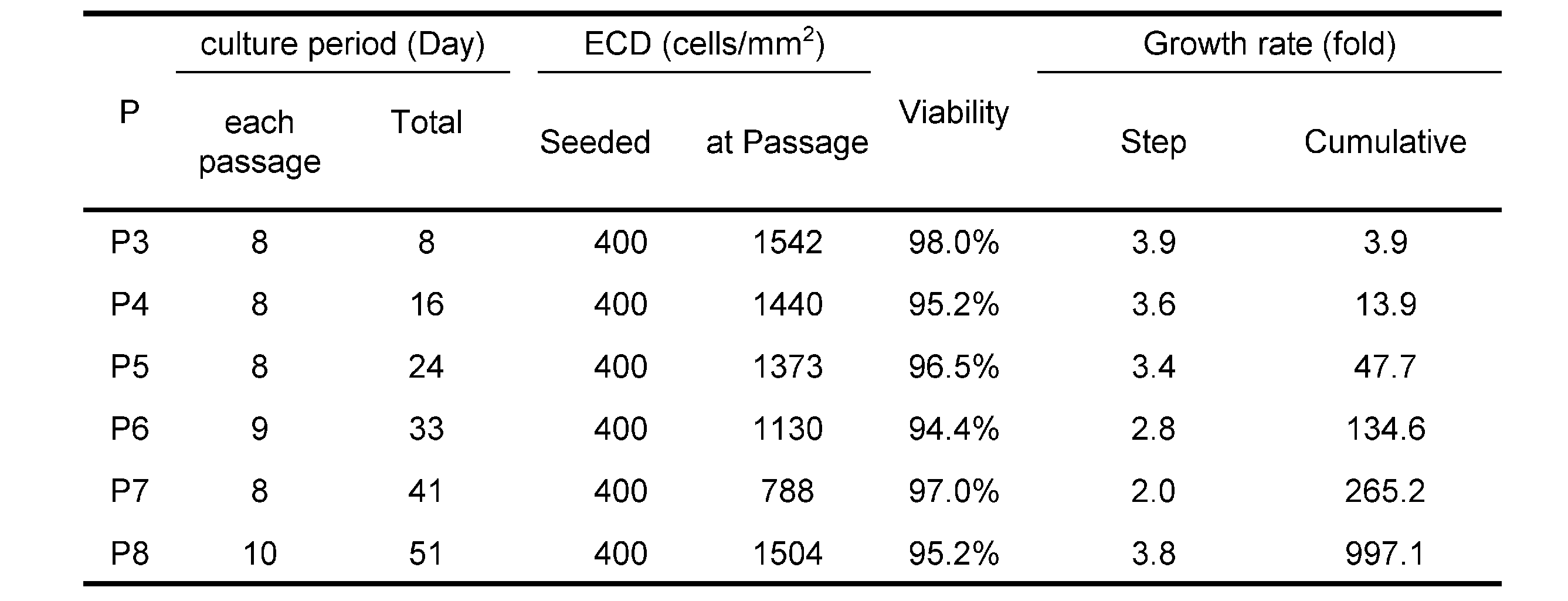
前述した実施例12においては、凍結保存後の細胞懸濁液を解凍し短期拡大培養した例を記載したが、本実施例においては冷凍保存していない細胞についての短期拡大培養を試みた。
使用したドナー角膜は、29歳、男性のもの、および8歳、女性のものである。
同一ドナー角膜のうち1眼を実施例1の培養条件1で35日間隔で継代して培養した(対照例1)。もう1眼を実施例1の培養条件2で培養し、Passage 1への継代時に2つに分け、一方を実施例1の培養条件2で35日間隔で継代して培養し(対照例2)、他方を実施例12と同様に8日前後(5日~11日)の間隔で継代を繰り返して培養した(実施例13)。手順の概要を図39に示す。
図40に増殖率から算出した収量と培養スケールを示した。8日前後の間隔で継代を繰り返した本実施例の短期拡大培養法では80日以内に20億個の細胞、T-25フラスコに換算すると500フラスコ分の細胞が得られることが示された。
また、Passage 4まで前述した短期拡大培養法で培養し、Passage 5で約5週間培養した細胞と、およそ同日数培養した対照例2の細胞、Passage 5まで培養した対照例2の細胞との写真および収量の比較を図41及び図42に示す。
同様にPassage 6まで短期拡大培養法で培養し、Passage 7で約5週間培養した細胞と、およそ同日数培養した対照例2の細胞、Passage 7まで培養した対照例2の細胞との写真および収量の比較を図43及び図44に示す。
図41~44の結果から、本実施例における短期拡大培養法によっても、機能性ヒト角膜内皮細胞の含有率が十分に高い細胞集団を短期間かつ高収量で得られることが示された。
以上の各実施例の結果から、適切な培養条件で培養されることによって機能性ヒト角膜内皮細胞になることが運命付けられた細胞であれば、その細胞を未成熟の状態で採取して保存又は継代しても、目的の機能性ヒト角膜内皮細胞を得ることができることが分かった。
Claims (31)
- ROCK阻害剤を含有し、かつ上皮成長因子(EGF)の含有量が形質転換を引き起こす濃度未満である培地を用いて増殖させたヒト角膜内皮細胞及び/又はヒト角膜内皮前駆細胞を、以下に挙げる(a)~(d)のいずれか1つ又は複数の条件を満たす時期に採取し、懸濁状態にして保存することを特徴とするヒト角膜内皮細胞及び/又はヒト角膜内皮前駆細胞の保存方法。
(a)前記ヒト角膜内皮細胞及び/又は前記ヒト角膜内皮前駆細胞の形態が、紡錘状の形態から長径短径の比が1に近い多角形状又は円形状の形態に転じた直後から、細胞間の境界が不明瞭になる直前までの期間である。
(b)CD44の発現量が直近の継代後にみられる最大値の半分以下になった時からプラトーに達するまでの期間である。
(c)前記ヒト角膜内皮細胞及び/又は前記ヒト角膜内皮前駆細胞の細胞密度が、900cells/mm2以上2500cells/mm2以下である。
(d)直近の継代からの培養日数が、4日以上14日以下である。 - 前記ヒト角膜内皮細胞及び/又は前記ヒト角膜内皮前駆細胞を10度以下で保存する、請求項1に記載の保存方法。
- 前記ヒト角膜内皮細胞及び/又は前記ヒト角膜内皮前駆細胞を-30度以下で保存する、請求項1に記載の保存方法。
- 前記ヒト角膜内皮細胞及び/又は前記ヒト角膜内皮前駆細胞を懸濁状態にしてから24時間以上保存する、請求項1~3の何れか一項に記載の保存方法。
- 前記形質転換が、内皮間葉転換を含む、請求項1~4の何れか一項に記載の保存方法。
- 前記ヒト角膜内皮細胞及び/又は前記ヒト角膜内皮前駆細胞が、角膜内皮細胞由来細胞、多能性幹細胞、間葉系幹細胞、角膜内皮から採取した角膜内皮前駆細胞、角膜内皮から採取した細胞、並びにダイレクトプログラミング法で作製される角膜内皮前駆細胞及び角膜内皮様細胞からなる群から選択される細胞を起源として作成されたものである、請求項1~5の何れか一項に記載の保存方法。
- 保存後の前記ヒト角膜内皮細胞及び/又は前記ヒト角膜内皮前駆細胞を、ROCK阻害剤を含有し、かつ上皮成長因子(EGF)の含有量が形質転換を引き起こす濃度未満である培地を用いて増殖及び/又は分化・成熟させて得たヒト角膜内皮細胞の細胞集団が、以下の(1)~(8)のうち、1又は複数の項目を満たすものである、請求項1~6の何れか一項に記載の保存方法。
(1)位相差画像による外観検査で、線維芽細胞、異物、変色、又は他の異常がない。
(2)細胞生存率がトリパンブルー染色で70%以上である。
(3)細胞上清のELISAによる純度試験で、PDGF-BBが100pg/mL以上である。
(4)細胞のFACSによる純度試験結果が以下の範囲を全て満たすものである。
CD166+>99%
CD24+<5%
CD44high<5%
CD44neg~low>90%
CD90+<5%
(5)ヒト角膜内皮機能を備えているエフェクター細胞の含有率(E-ratio)が90%より高い。
(6)ポンプ機能(Na+/K+ ATPase)が陽性である。
(7)バリア機能(ZO-1)が陽性である。
(8)ヒト角膜内皮細胞密度(ECD)が1500細胞/mm2以上である。 - 前記エフェクター細胞が、内皮間葉系移行が起きていないものである、請求項7に記載の保存方法。
- 前記エフェクター細胞が、角膜内皮機能特性を発揮するために必要な機能蛋白を発現しているか、又は前記角膜内皮機能特性を阻害する阻害蛋白が発現されない若しくは阻害蛋白の発現が低下しているものである、請求項7又は8に記載の保存方法。
- 前記機能蛋白がNa+/K+ ATPase、ZO-1、ナトリウム/水素交換体1(NHE1)、アクアポリン1(AQP-1)及び炭酸脱水酵素5B(CA5B)からなる群より選ばれる一つ又は複数を含む、請求項9に記載の保存方法。
- 前記機能蛋白が、クエン酸シンターゼ(CS)、アコニターゼ2(ACO2)、イソクエン酸デヒドロゲナーゼ2(IDH2)、リンゴ酸デヒドロゲナーゼ2(MDH2)、リンゴ酸酵素3(ME3)、ACSS1、アセチルCoAアセチルトランスフェラーゼ1(ACAT1)、ピルビン酸デヒドロゲナーゼ(PDH)、BCAT2、及び分岐鎖ケト酸デヒドロゲナーゼ2(BCKDH2)からなる群から選択される1又は複数の代謝関連酵素をさらに含む、請求項9又は10に記載の保存方法。
- 前記阻害蛋白が、ATPクエン酸リアーゼ(ACLY)、アコニターゼ1(ACO1)、イソクエン酸デヒドロゲナーゼ1(IDH1)、リンゴ酸デヒドロゲナーゼ1(MDH1)、リンゴ酸酵素1(ME1)、ACSS2、アセチルCoAアセチルトランスフェラーゼ2(ACAT2)及び乳酸デヒドロゲナーゼ(LDH)からなる群から選択される少なくとも1種以上の代謝関連酵素を含む、請求項9~11のいずれか一項に記載の保存方法。
- 請求項1~12の何れか一項に記載の保存方法で保存されたヒト角膜内細胞及び/又はヒト角膜内皮前駆細胞。
- 請求項1~12の何れか一項に記載の保存方法で保存されたヒト角膜内細胞及び/又はヒト角膜内皮前駆細胞の懸濁液。
- 請求項1~12の何れか一項に記載の保存方法で保存されたヒト角膜内皮細胞及び/又はヒト角膜内皮前駆細胞を、ROCK阻害剤を含有し、かつ上皮成長因子(EGF)の含有量が形質転換を引き起こす濃度未満である培地を用いて増殖及び/又は分化・成熟させることを特徴とする、ヒト角膜内皮細胞の調製方法。
- ROCK阻害剤を含有し、かつ上皮成長因子(EGF)の含有量が形質転換を引き起こす濃度未満である培地を用いて増殖させたヒト角膜内皮細胞及び/又はヒト角膜内皮前駆細胞を、以下に挙げる(a)~(d)のいずれか1つ又は複数の条件を満たす時期に採取し、懸濁状態にすることを特徴とする、ヒト角膜内皮細胞及び/又はヒト角膜内皮前駆細胞の懸濁液調製方法。
(a)前記ヒト角膜内皮細胞及び/又は前記ヒト角膜内皮前駆細胞の形態が、不規則な細長い突起を持つ紡錘状の線維芽細胞様の形態から前記突起を含めた長径短径の比が1に近い敷石状の形態に転じた直後から、細胞間の境界が不明瞭になる直前までの期間である。
(b)CD44の発現量が直近の継代後にみられる最大値の半分以下になった時からプラトーに達するまでの期間である。
(c)前記ヒト角膜内皮細胞及び/又は前記ヒト角膜内皮前駆細胞の細胞密度が、900cells/mm2以上2500cells/mm2以下である。
(d)直近の継代からの培養日数が、4日以上14日以下である。 - ROCK阻害剤を含有し、かつ上皮成長因子(EGF)の含有量が形質転換を引き起こす濃度未満である培地を用いて増殖させたヒト角膜内皮細胞及び/又はヒト角膜内皮前駆細胞を、以下に挙げる(a)~(d)のいずれか1つ又は複数の条件を満たす時期に採取し、継代することを特徴とする、ヒト角膜内皮細胞及び/又はヒト角膜内皮前駆細胞の培養方法。
(a)前記ヒト角膜内皮細胞及び/又は前記ヒト角膜内皮前駆細胞の形態が、不規則な細長い突起を持つ紡錘状の線維芽細胞様の形態から、長径短径の比が1に近い敷石状の形態に転じた直後から、細胞間の境界が不明瞭になる直前までの期間である。
(b)CD44の発現量が直近の継代後にみられる最大値の半分以下になった時からプラトーに達するまでの期間である。
(c)前記ヒト角膜内皮細胞及び/又は前記ヒト角膜内皮前駆細胞の細胞密度が、900cells/mm2以上2500cells/mm2以下である。
(d)直近の継代からの培養日数が、4日以上14日以下である。 - 培養したヒト角膜内皮細胞および/またはヒト角膜内皮前駆細胞を保存する方法であって、
複数のヒト角膜内皮細胞および/またはヒト角膜内皮前駆細胞を得る工程と、
前記複数の細胞の少なくとも一部を分離して、評価用の細胞集団と保存用の細胞集団を生成する工程と、
前記細胞集団の少なくとも一部を、以下の(a)、(b)、(c)及び(d)の評価基準うちの1つ以上について評価する工程と、
(a)ヒト角膜内皮細胞および/またはヒト角膜内皮前駆細胞の形態が、紡錘形から、(i)多角形または(ii)長軸と短軸の比が1に近い楕円形のいずれかに変化し、かつ細胞間の境界がはっきりしている細胞を特定するものである、
(b)CD44の発現量が、直前の継代後に観察された最大値の半分以下のレベルでCD44を発現した後、一定量のCD44を発現する細胞を特定するものである、
(c)ヒト角膜内皮細胞および/またはヒト角膜内皮前駆細胞の細胞密度が900個/mm2以上2500個/mm2以下であることを識別するものである、及び
(d)前記評価から直前の継代までの期間が4日以上14日以下であるかどうかを特定するものである。
評価用の細胞集団が、前記(a)、(b)、(c)及び(d)のいずれかの評価基準を1つ以上満たしている場合に、細胞集団から少なくとも一部の細胞を保存するために保存媒体に配置する工程と、を備える保存方法。 - 前記ヒト角膜内皮細胞および/またはヒト角膜内皮前駆細胞を10度以下で保存する、請求項18に記載の保存方法。
- 前記ヒト角膜内皮細胞及び/又は前記ヒト角膜内皮前駆細胞を24時間以上保存する、請求項18又は19に記載の保存方法。
- 前記ヒト角膜内皮細胞及び/又は前記ヒト角膜内皮前駆細胞が、角膜内皮細胞由来細胞、多能性幹細胞、間葉系幹細胞、角膜内皮から採取した角膜内皮前駆細胞、角膜内皮から採取した細胞、並びにダイレクトプログラミング法で作製される角膜内皮前駆細胞及び角膜内皮様細胞からなる群から選択される細胞を起源として作成されたものである、請求項18~20の何れか一項に記載の保存方法。
- 培養したヒト角膜内皮細胞および/またはヒト角膜内皮前駆細胞を処理する方法であって、
複数のヒト角膜内皮細胞および/またはヒト角膜内皮前駆細胞を、培養液を含む培養容器内で培養する工程と
前記複数のヒト角膜内皮細胞および/またはヒト角膜内皮前駆細胞の少なくとも一部を分離して、評価用の細胞集団と残りの細胞集団とを生成する工程と、
細胞集団の少なくとも一部を、以下の(a)、(b)、(c)及び(d)の評価基準の1つ以上について評価する工程と、
(a)ヒト角膜内皮細胞および/またはヒト角膜内皮前駆細胞の形態が、紡錘形から、(i)多角形または(ii)長軸と短軸の比が1に近い楕円形のいずれかに変化し、かつ細胞間の境界がはっきりしている細胞を特定するものである、
(b)CD44の発現量が、直前の継代後に観察された最大値の半分以下のレベルでCD44を発現した後、一定量のCD44を発現する細胞を特定するものである、
(c)ヒト角膜内皮細胞および/またはヒト角膜内皮前駆細胞の細胞密度が900個/mm2以上2500個/mm2以下であることを識別するものである、及び
(d)前記評価から直前の継代までの期間が4日以上14日以下であるかどうかを特定するものである。
前記評価用の細胞集団が前記(a)、(b)、(c)及び(d)の評価基準のうち1つ以上を満たしている場合に、細胞集団から細胞の少なくとも一部を保存するために保存媒体に配置する工程、または前記評価用の細胞集団が(a)、(b)、(c)及び(d)の各評価基準を満たさない場合に、培養液を含む培養容器に前記残りの細胞集団から少なくとも一部の細胞を入れて、さらに一定期間培養する工程と、を含む処理方法。 - 前記培養液がROCK阻害剤を含み、培養液が細胞の形質転換を引き起こす濃度以下の上皮成長因子(EGF)を含んでいる、請求項22に記載の処理方法。
- 前記培養液がROCK阻害剤を含み、前記培養液が上皮成長因子(EGF)の濃度が2ng/ml以下である、請求項22又は23に記載の処理方法。
- 前記培養液がEGFを含有しない、請求項24に記載の処理方法。
- 前記ヒト角膜内皮細胞及び/又は前記ヒト角膜内皮前駆細胞が、角膜内皮細胞由来細胞、多能性幹細胞、間葉系幹細胞、角膜内皮から採取した角膜内皮前駆細胞、角膜内皮から採取した細胞、並びにダイレクトプログラミング法で作製される角膜内皮前駆細胞及び角膜内皮様細胞からなる群から選択される細胞を起源として作成されたものである、請求項22~25の何れか一項に記載の処理方法。
- 請求項18~26のいずれか1項に記載の保存方法又は処理方法によって保存された細胞を保存状態から取り出し、追加培養のために新たな培養容器に戻す工程をさらに含み、追加培養に供された細胞が以下の(1)~(8)の評価基準のうち1つ以上を満たす方法。
(1)位相差画像による外観検査で、線維芽細胞、異物、変色、又は他の異常がない。
(2)細胞生存率がトリパンブルー染色で70%以上である。
(3)細胞上清のELISAによる純度試験で、PDGF-BBが100pg/mL以上である。
(4)細胞のFACSによる純度試験結果が以下の範囲を全て満たすものである。
CD166+>99%
CD24+<5%
CD44high<5%
CD44neg~low>90%
CD90+<5%
(5)ヒト角膜内皮機能を備えているエフェクター細胞の含有率(E-ratio)が90%より高い。
(6)ポンプ機能(Na+/K+ ATPase)が陽性である。
(7)バリア機能(ZO-1)が陽性である。
(8)ヒト角膜内皮細胞密度(ECD)が1500細胞/mm2以上である。 - 請求項18~26のいずれか1項に記載の保存方法又は処理方法によって保存され、該保存状態から取り出された細胞が、眼の疾患または傷害を治療するために対象者の眼に送達するのに適している方法。
- 請求項18~26のいずれか1項に記載の保存方法又は処理方法によって保存され、該保存状態から取り出された細胞が、眼の疾患または傷害を治療するために対象者の眼に送達する医薬品の調製に適している保存方法。
- 請求項18~26のいずれか1項に記載の保存方法又は処理方法によって保存され、該保存状態から取り出された細胞が、眼の疾患または傷害の治療に使用するのに適している保存方法。
- 請求項28~30のいずれか1項に記載の方法であって、前記眼の疾患または前記傷害が角膜の疾患または損傷である方法。
Priority Applications (13)
| Application Number | Priority Date | Filing Date | Title |
|---|---|---|---|
| JP2022557558A JP7361431B2 (ja) | 2020-10-22 | 2021-10-19 | ヒト角膜内皮細胞及び/又はヒト角膜内皮前駆細胞の保存方法 |
| NZ799955A NZ799955B2 (en) | 2021-10-19 | A storage method of human corneal endothelial cells and/or human corneal endothelial precursor cells | |
| CA3196110A CA3196110A1 (en) | 2020-10-22 | 2021-10-19 | A storage method of human corneal endothelial cells and/or human corneal endothelial precursor cells |
| CN202180072227.5A CN116583593A (zh) | 2020-10-22 | 2021-10-19 | 人角膜内皮细胞和/或人角膜内皮前体细胞的保存方法 |
| IL302168A IL302168A (en) | 2020-10-22 | 2021-10-19 | Storage method of human corneal endothelial cells and/or human corneal endothelial progenitor cells |
| AU2021366805A AU2021366805B2 (en) | 2020-10-22 | 2021-10-19 | A storage method of human corneal endothelial cells and/or human corneal endothelial precursor cells |
| EP21882821.8A EP4234682A4 (en) | 2020-10-22 | 2021-10-19 | METHOD FOR PRESERVING HUMAN CORNEAL ENDOTHELIAL CELLS AND/OR HUMAN CORNEAL ENDOTHELIAL PROGENITOR CELLS |
| US18/032,726 US12550891B2 (en) | 2020-10-22 | 2021-10-19 | Storage method of human corneal endothelial cells and/or human corneal endothelial precursor cells |
| MX2023004594A MX2023004594A (es) | 2020-10-22 | 2021-10-19 | Un metodo de almacenamiento de celulas endoteliales de la cornea humanas y/o celulas precursoras endoteliales de la cornea humanas. |
| KR1020237014236A KR20230091107A (ko) | 2020-10-22 | 2021-10-19 | 인간 각막 내피 세포 및/또는 인간 각막 내피 전구세포의 보존 방법 |
| MX2025003724A MX2025003724A (es) | 2020-10-22 | 2023-04-20 | Un metodo de almacenamiento de celulas endoteliales de la cornea humanas y/o celulas precursoras endoteliales de la cornea humanas |
| JP2023119210A JP2023139164A (ja) | 2020-10-22 | 2023-07-21 | ヒト角膜内皮細胞及び/又はヒト角膜内皮前駆細胞の保存方法 |
| AU2025206385A AU2025206385A1 (en) | 2020-10-22 | 2025-07-21 | A storage method of human corneal endothelial cells and/or human corneal endothelial precursor cells |
Applications Claiming Priority (2)
| Application Number | Priority Date | Filing Date | Title |
|---|---|---|---|
| JP2020177634 | 2020-10-22 | ||
| JP2020-177634 | 2020-10-22 |
Related Child Applications (2)
| Application Number | Title | Priority Date | Filing Date |
|---|---|---|---|
| US18/032,726 A-371-Of-International US12550891B2 (en) | 2020-10-22 | 2021-10-19 | Storage method of human corneal endothelial cells and/or human corneal endothelial precursor cells |
| US19/447,258 Division US20260137076A1 (en) | 2020-10-22 | 2026-01-13 | Storage method of human corneal endothelial cells and/or human corneal endotheliam precursor cells |
Publications (1)
| Publication Number | Publication Date |
|---|---|
| WO2022085680A1 true WO2022085680A1 (ja) | 2022-04-28 |
Family
ID=81290627
Family Applications (1)
| Application Number | Title | Priority Date | Filing Date |
|---|---|---|---|
| PCT/JP2021/038609 Ceased WO2022085680A1 (ja) | 2020-10-22 | 2021-10-19 | ヒト角膜内皮細胞及び/又はヒト角膜内皮前駆細胞の保存方法 |
Country Status (10)
| Country | Link |
|---|---|
| US (1) | US12550891B2 (ja) |
| EP (1) | EP4234682A4 (ja) |
| JP (2) | JP7361431B2 (ja) |
| KR (1) | KR20230091107A (ja) |
| CN (1) | CN116583593A (ja) |
| AU (2) | AU2021366805B2 (ja) |
| CA (1) | CA3196110A1 (ja) |
| IL (1) | IL302168A (ja) |
| MX (2) | MX2023004594A (ja) |
| WO (1) | WO2022085680A1 (ja) |
Families Citing this family (1)
| Publication number | Priority date | Publication date | Assignee | Title |
|---|---|---|---|---|
| WO2025145016A1 (en) * | 2023-12-29 | 2025-07-03 | Aurion Biotech, Inc. | Corneal endothelial cell (cec) culturing methods and compositions |
Citations (32)
| Publication number | Priority date | Publication date | Assignee | Title |
|---|---|---|---|---|
| US4678783A (en) | 1983-11-04 | 1987-07-07 | Asahi Kasei Kogyo Kabushiki Kaisha | Substituted isoquinolinesulfonyl compounds |
| JPH0276977A (ja) | 1988-09-12 | 1990-03-16 | Matsushita Electric Ind Co Ltd | 湯水混合装置 |
| JPH0276976A (ja) | 1988-09-12 | 1990-03-16 | Matsushita Electric Ind Co Ltd | 制御弁 |
| JPH02100833A (ja) | 1988-10-07 | 1990-04-12 | Tokai T R W Kk | 転造ダイスの加工方法 |
| JPH0359913A (ja) | 1989-07-28 | 1991-03-14 | Yazaki Corp | 金属溶射による回路体の製造方法 |
| JPH0362227A (ja) | 1989-07-31 | 1991-03-18 | Nec Corp | 高速再翻訳処理方式 |
| JPH04620A (ja) | 1990-04-18 | 1992-01-06 | Nec Off Syst Ltd | キーボード |
| JPH081403A (ja) | 1994-06-22 | 1996-01-09 | Fukui Seisakusho:Kk | Nc装置付き工作機械によるネジ端部バリ取り方法 |
| JP2002083175A (ja) | 2000-09-07 | 2002-03-22 | Cosmo Oil Co Ltd | 競合見積管理システムおよび方法、並びに購買ウエブサーバ |
| JP3421217B2 (ja) | 1995-11-20 | 2003-06-30 | 麒麟麦酒株式会社 | Rho標的タンパク質Rhoキナーゼ |
| JP2004009555A (ja) | 2002-06-07 | 2004-01-15 | Futec Inc | 印刷欠点の検出装置および検出方法 |
| JP2004022541A (ja) | 2002-06-12 | 2004-01-22 | Eastman Kodak Co | 有機発光ダイオード表示装置 |
| JP2004108724A (ja) | 2002-09-20 | 2004-04-08 | Toyotomi Co Ltd | 窓用空気調和機の構造 |
| JP2005003101A (ja) | 2003-06-12 | 2005-01-06 | Toyota Motor Corp | 無段変速機の制御装置 |
| JP2005037198A (ja) | 2003-07-18 | 2005-02-10 | Kawasaki Heavy Ind Ltd | 内側炉心槽の挿入・引き抜き方法及びその装置と内側炉心槽引き抜き時の燃料集合体の拘束支持方法及びその装置 |
| JP2005035506A (ja) | 2003-07-17 | 2005-02-10 | Masuo Kato | パイプを使用した水上、水中液体貯蔵装置及び水上輸送方法 |
| JP2005037197A (ja) | 2003-07-18 | 2005-02-10 | Ricoh Co Ltd | 接触式表面形状測定装置及び測定方法 |
| JP2005034866A (ja) | 2003-07-18 | 2005-02-10 | Yamazaki Mazak Corp | レーザ加工機 |
| JP2005035501A (ja) | 2003-07-14 | 2005-02-10 | Takahisa Tsutsui | 油タンカー船の積付け計算をボタン操作によって調整かつ、連動して海面を表示するプログラム |
| JP2005039564A (ja) | 2003-07-16 | 2005-02-10 | Nec Saitama Ltd | 画像配信システム、サーバ装置、携帯端末装置及びそれに用いる画像配信方法 |
| JP2005035503A (ja) | 2003-07-15 | 2005-02-10 | Okubo Tomio | 自転車等の防寒用ハンドルカバー |
| JP2005080394A (ja) | 2003-08-29 | 2005-03-24 | Rohm Co Ltd | 電源装置 |
| JP2005103050A (ja) | 2003-09-30 | 2005-04-21 | Nohmi Bosai Ltd | 消防用設備の開閉弁装置及びその制御弁 |
| JP2006057270A (ja) | 2004-08-18 | 2006-03-02 | Akimori Taniguchi | 巨大水母類の漂着による、取水口スクリ−ン吸着閉塞防止システム。 |
| JP2007026664A (ja) | 1995-04-14 | 2007-02-01 | Toshiba Corp | 光ディスクからビデオ及びオーディオを再生する再生システム、その再生方法及び光ディスクの記録方法 |
| WO2009028631A1 (ja) | 2007-08-29 | 2009-03-05 | Senju Pharmaceutical Co., Ltd. | 角膜内皮細胞接着促進剤 |
| WO2014038639A1 (ja) * | 2012-09-07 | 2014-03-13 | 日本ケミカルリサーチ株式会社 | 間葉系幹細胞の馴化培地を含有する角膜内皮細胞培養用培地 |
| JP2015155400A (ja) * | 2014-01-16 | 2015-08-27 | Jcrファーマ株式会社 | 角膜内皮細胞を含有する移植用組成物 |
| WO2017110094A1 (ja) | 2015-12-24 | 2017-06-29 | 学校法人同志社 | カスパーゼ阻害剤を含む、TGF-βに起因する障害を治療または予防するための医薬およびその応用 |
| WO2017141926A1 (en) * | 2016-02-15 | 2017-08-24 | Kyoto Prefectural Public University Corporation | Human functional corneal endothelial cell and application thereof |
| WO2020032139A1 (en) | 2018-08-09 | 2020-02-13 | Sharp Kabushiki Kaisha | Integrated Access and Backhaul Node Recognition in Integrated Access and Backhaul Network |
| JP2020032139A (ja) | 2018-08-31 | 2020-03-05 | 株式会社三洋物産 | 遊技機 |
Family Cites Families (29)
| Publication number | Priority date | Publication date | Assignee | Title |
|---|---|---|---|---|
| EP1195372A1 (en) | 1994-04-18 | 2002-04-10 | Mitsubishi Pharma Corporation | N-heterocyclic substituted benzamide derivatives with antihypertensive activity |
| WO1999020620A1 (en) | 1997-10-22 | 1999-04-29 | Nippon Shinyaku Co Ltd | Isoquinoline derivative and drug |
| DE69931051D1 (de) | 1998-05-25 | 2006-06-01 | Santen Pharmaceutical Co Ltd | Neue vinylbenzolderivate |
| JP4329003B2 (ja) | 2001-03-23 | 2009-09-09 | バイエル コーポレイション | Rhoキナーゼ阻害剤 |
| TWI336328B (en) | 2001-03-23 | 2011-01-21 | Bayer Healthcare Llc | Rho-kinase inhibitors |
| CA2707827A1 (en) | 2001-04-11 | 2002-10-24 | Senju Pharmaceutical Co., Ltd. | Visual function disorder improving agents containing rho kinase inhibitors |
| US7199147B2 (en) | 2001-06-12 | 2007-04-03 | Dainippon Sumitomo Pharma Co., Ltd. | Rho kinase inhibitors |
| JP4505228B2 (ja) | 2002-01-10 | 2010-07-21 | バイエル・シェーリング・ファルマ・アクチェンゲゼルシャフト | Rho−キナーゼ阻害剤 |
| MXPA04007196A (es) | 2002-01-23 | 2005-06-08 | Bayer Pharmaceuticals Corp | Inhibidores de rho-quinasa. |
| JPWO2004009555A1 (ja) | 2002-07-22 | 2005-11-17 | 旭化成ファーマ株式会社 | 5−置換イソキノリン誘導体 |
| CA2400996A1 (en) | 2002-09-03 | 2004-03-03 | Lisa Mckerracher | 1,4-substituted cyclohexane derivatives |
| US7160894B2 (en) | 2003-06-06 | 2007-01-09 | Asahi Kasei Pharma Corporation | Tricyclic compound |
| EP1644365A2 (en) | 2003-07-02 | 2006-04-12 | Biofocus Discovery Ltd | Pyrazine and pyridine derivatives as rho kinase inhibitors |
| WO2005039564A1 (en) | 2003-10-02 | 2005-05-06 | Vertex Pharmaceuticals Incorporated | Phthalimide compounds useful as protein kinase inhibitors |
| JP4764823B2 (ja) | 2003-10-06 | 2011-09-07 | グラクソ グループ リミテッド | キナーゼ阻害剤としての1,6−二置換アザベンゾイミダゾールの調製 |
| WO2005034866A2 (en) | 2003-10-06 | 2005-04-21 | Glaxo Group Limited | Preparation of 1, 6, 7- trisubstituted azabenzimidazoles as kinase inhibitors |
| WO2005037198A2 (en) | 2003-10-06 | 2005-04-28 | Glaxo Group Limited | Preparation of 1,7-disubstituted azabenzimidazoles as kinase inhibitors |
| CN1898377A (zh) * | 2003-10-10 | 2007-01-17 | 细胞生物工程公司 | 人角膜内皮细胞以及获得和培养用于角膜细胞移植的细胞的方法 |
| JP2007008816A (ja) | 2003-10-15 | 2007-01-18 | Ube Ind Ltd | 新規イソキノリン誘導体 |
| JP2007015928A (ja) | 2003-10-15 | 2007-01-25 | Ube Ind Ltd | 新規オレフィン誘導体 |
| EP2468729B1 (en) | 2003-10-15 | 2013-12-25 | Ube Industries, Ltd. | Novel indazole derivative |
| WO2005080394A1 (en) | 2004-02-24 | 2005-09-01 | Bioaxone Therapeutique Inc. | 4-substituted piperidine derivatives |
| WO2005103050A2 (en) | 2004-04-02 | 2005-11-03 | Vertex Pharmaceuticals Incorporated | Azaindoles useful as inhibitors of rock and other protein kinases |
| US20060101035A1 (en) | 2004-11-11 | 2006-05-11 | Mustakallio Minna M | System and method for blog functionality |
| JPWO2006057270A1 (ja) | 2004-11-26 | 2008-06-05 | 旭化成ファーマ株式会社 | 含窒素3環化合物 |
| EP1932841B1 (en) | 2005-08-30 | 2014-01-01 | Asahi Kasei Pharma Corporation | Sulfonamide compound |
| DE202015009826U1 (de) | 2015-02-13 | 2020-07-29 | P&L Gmbh & Co. Kg | Werkzeugmaschine zur Lagebestimmung eines Werkstücks |
| JPWO2020045642A1 (ja) * | 2018-08-31 | 2021-08-12 | 学校法人同志社 | 眼細胞を保存または培養するための組成物および方法 |
| US20230257700A1 (en) | 2020-02-27 | 2023-08-17 | Kyoto Prefectural Public University Corporation | Functional human corneal endothelial cells and application thereof |
-
2021
- 2021-10-19 JP JP2022557558A patent/JP7361431B2/ja active Active
- 2021-10-19 KR KR1020237014236A patent/KR20230091107A/ko active Pending
- 2021-10-19 MX MX2023004594A patent/MX2023004594A/es unknown
- 2021-10-19 US US18/032,726 patent/US12550891B2/en active Active
- 2021-10-19 IL IL302168A patent/IL302168A/en unknown
- 2021-10-19 AU AU2021366805A patent/AU2021366805B2/en active Active
- 2021-10-19 CN CN202180072227.5A patent/CN116583593A/zh active Pending
- 2021-10-19 WO PCT/JP2021/038609 patent/WO2022085680A1/ja not_active Ceased
- 2021-10-19 EP EP21882821.8A patent/EP4234682A4/en active Pending
- 2021-10-19 CA CA3196110A patent/CA3196110A1/en active Pending
-
2023
- 2023-04-20 MX MX2025003724A patent/MX2025003724A/es unknown
- 2023-07-21 JP JP2023119210A patent/JP2023139164A/ja active Pending
-
2025
- 2025-07-21 AU AU2025206385A patent/AU2025206385A1/en active Pending
Patent Citations (33)
| Publication number | Priority date | Publication date | Assignee | Title |
|---|---|---|---|---|
| US4678783A (en) | 1983-11-04 | 1987-07-07 | Asahi Kasei Kogyo Kabushiki Kaisha | Substituted isoquinolinesulfonyl compounds |
| US4678783B1 (en) | 1983-11-04 | 1995-04-04 | Asahi Chemical Ind | Substituted isoquinolinesulfonyl compounds |
| JPH0276977A (ja) | 1988-09-12 | 1990-03-16 | Matsushita Electric Ind Co Ltd | 湯水混合装置 |
| JPH0276976A (ja) | 1988-09-12 | 1990-03-16 | Matsushita Electric Ind Co Ltd | 制御弁 |
| JPH02100833A (ja) | 1988-10-07 | 1990-04-12 | Tokai T R W Kk | 転造ダイスの加工方法 |
| JPH0359913A (ja) | 1989-07-28 | 1991-03-14 | Yazaki Corp | 金属溶射による回路体の製造方法 |
| JPH0362227A (ja) | 1989-07-31 | 1991-03-18 | Nec Corp | 高速再翻訳処理方式 |
| JPH04620A (ja) | 1990-04-18 | 1992-01-06 | Nec Off Syst Ltd | キーボード |
| JPH081403A (ja) | 1994-06-22 | 1996-01-09 | Fukui Seisakusho:Kk | Nc装置付き工作機械によるネジ端部バリ取り方法 |
| JP2007026664A (ja) | 1995-04-14 | 2007-02-01 | Toshiba Corp | 光ディスクからビデオ及びオーディオを再生する再生システム、その再生方法及び光ディスクの記録方法 |
| JP3421217B2 (ja) | 1995-11-20 | 2003-06-30 | 麒麟麦酒株式会社 | Rho標的タンパク質Rhoキナーゼ |
| JP2002083175A (ja) | 2000-09-07 | 2002-03-22 | Cosmo Oil Co Ltd | 競合見積管理システムおよび方法、並びに購買ウエブサーバ |
| JP2004009555A (ja) | 2002-06-07 | 2004-01-15 | Futec Inc | 印刷欠点の検出装置および検出方法 |
| JP2004022541A (ja) | 2002-06-12 | 2004-01-22 | Eastman Kodak Co | 有機発光ダイオード表示装置 |
| JP2004108724A (ja) | 2002-09-20 | 2004-04-08 | Toyotomi Co Ltd | 窓用空気調和機の構造 |
| JP2005003101A (ja) | 2003-06-12 | 2005-01-06 | Toyota Motor Corp | 無段変速機の制御装置 |
| JP2005035501A (ja) | 2003-07-14 | 2005-02-10 | Takahisa Tsutsui | 油タンカー船の積付け計算をボタン操作によって調整かつ、連動して海面を表示するプログラム |
| JP2005035503A (ja) | 2003-07-15 | 2005-02-10 | Okubo Tomio | 自転車等の防寒用ハンドルカバー |
| JP2005039564A (ja) | 2003-07-16 | 2005-02-10 | Nec Saitama Ltd | 画像配信システム、サーバ装置、携帯端末装置及びそれに用いる画像配信方法 |
| JP2005035506A (ja) | 2003-07-17 | 2005-02-10 | Masuo Kato | パイプを使用した水上、水中液体貯蔵装置及び水上輸送方法 |
| JP2005034866A (ja) | 2003-07-18 | 2005-02-10 | Yamazaki Mazak Corp | レーザ加工機 |
| JP2005037197A (ja) | 2003-07-18 | 2005-02-10 | Ricoh Co Ltd | 接触式表面形状測定装置及び測定方法 |
| JP2005037198A (ja) | 2003-07-18 | 2005-02-10 | Kawasaki Heavy Ind Ltd | 内側炉心槽の挿入・引き抜き方法及びその装置と内側炉心槽引き抜き時の燃料集合体の拘束支持方法及びその装置 |
| JP2005080394A (ja) | 2003-08-29 | 2005-03-24 | Rohm Co Ltd | 電源装置 |
| JP2005103050A (ja) | 2003-09-30 | 2005-04-21 | Nohmi Bosai Ltd | 消防用設備の開閉弁装置及びその制御弁 |
| JP2006057270A (ja) | 2004-08-18 | 2006-03-02 | Akimori Taniguchi | 巨大水母類の漂着による、取水口スクリ−ン吸着閉塞防止システム。 |
| WO2009028631A1 (ja) | 2007-08-29 | 2009-03-05 | Senju Pharmaceutical Co., Ltd. | 角膜内皮細胞接着促進剤 |
| WO2014038639A1 (ja) * | 2012-09-07 | 2014-03-13 | 日本ケミカルリサーチ株式会社 | 間葉系幹細胞の馴化培地を含有する角膜内皮細胞培養用培地 |
| JP2015155400A (ja) * | 2014-01-16 | 2015-08-27 | Jcrファーマ株式会社 | 角膜内皮細胞を含有する移植用組成物 |
| WO2017110094A1 (ja) | 2015-12-24 | 2017-06-29 | 学校法人同志社 | カスパーゼ阻害剤を含む、TGF-βに起因する障害を治療または予防するための医薬およびその応用 |
| WO2017141926A1 (en) * | 2016-02-15 | 2017-08-24 | Kyoto Prefectural Public University Corporation | Human functional corneal endothelial cell and application thereof |
| WO2020032139A1 (en) | 2018-08-09 | 2020-02-13 | Sharp Kabushiki Kaisha | Integrated Access and Backhaul Node Recognition in Integrated Access and Backhaul Network |
| JP2020032139A (ja) | 2018-08-31 | 2020-03-05 | 株式会社三洋物産 | 遊技機 |
Non-Patent Citations (4)
| Title |
|---|
| ANONYMOUS: "ATCC® ANIMAL CELL CULTURE GUIDE - tips and techniques for continuous cell lines", ATCC ANIMAL CELL CULTURE BASICS, 1 December 2011 (2011-12-01), US, pages 1 - 40, XP009536304 * |
| OKUMURA N ET AL., PLOS ONE, vol. 14, no. 6, 2019, pages e0218431 |
| See also references of EP4234682A4 |
| VIANNA L.M.M. ET AL., ARQ BRAS OFTALMOL., vol. 79, no. 1, 2016, pages 37 |
Also Published As
| Publication number | Publication date |
|---|---|
| MX2023004594A (es) | 2023-05-08 |
| MX2025003724A (es) | 2025-05-02 |
| US12550891B2 (en) | 2026-02-17 |
| JP7361431B2 (ja) | 2023-10-16 |
| AU2025206385A1 (en) | 2025-08-07 |
| IL302168A (en) | 2023-06-01 |
| CN116583593A (zh) | 2023-08-11 |
| KR20230091107A (ko) | 2023-06-22 |
| US20230380417A1 (en) | 2023-11-30 |
| JP2023139164A (ja) | 2023-10-03 |
| CA3196110A1 (en) | 2022-04-28 |
| JPWO2022085680A1 (ja) | 2022-04-28 |
| NZ799955A (en) | 2025-05-30 |
| AU2021366805A1 (en) | 2023-06-22 |
| EP4234682A4 (en) | 2024-11-27 |
| EP4234682A1 (en) | 2023-08-30 |
| AU2021366805B2 (en) | 2025-04-24 |
Similar Documents
| Publication | Publication Date | Title |
|---|---|---|
| US10104881B2 (en) | Composition comprising plant-derived recombinant human serum albumin, lipids, and plant protein hydrolysates as active ingredients for cryopreservation of stem cells or primary cells | |
| CN105796602A (zh) | 利用分离的胎盘细胞治疗中风 | |
| JP2020516257A (ja) | 凍結保存方法 | |
| Proulx et al. | Methods being developed for preparation, delivery and transplantation of a tissue-engineered corneal endothelium | |
| TW201821610A (zh) | 哺乳動物細胞凍結保存液 | |
| CN104662148A (zh) | 含有间充质干细胞的条件培养基的角膜内皮细胞培养用培养基 | |
| CN106359368A (zh) | 细胞冻存液及冻存方法 | |
| JP2011501960A (ja) | 月経血幹細胞と共に臍帯血由来細胞を共培養する方法 | |
| CN102226169B (zh) | 山羊毛囊外根鞘细胞系的构建方法 | |
| Baylis et al. | An investigation of donor and culture parameters which influence epithelial outgrowths from cultured human cadaveric limbal explants | |
| Romanov et al. | Isolation of multipotent mesenchymal stromal cells from cryopreserved human umbilical cord tissue | |
| Li et al. | Enriching new transplantable RGC-like cells from retinal organoids for RGC replacement therapy | |
| CN117701505B (zh) | 肉瘤组织类器官培养基、构建肉瘤组织类器官和肉瘤组织类器官库的方法、构建动物疾病模型的方法 | |
| AU2025206385A1 (en) | A storage method of human corneal endothelial cells and/or human corneal endothelial precursor cells | |
| Schiozer et al. | An outcome analysis and long-term viability of cryopreserved cultured epidermal allografts: assessment of the conservation of transplantable human skin allografts | |
| EP3252143A1 (en) | Method for producing a platelet-lysate-containing gel | |
| CN106417253A (zh) | 一种骨骼肌干细胞的冻存液和冻存方法 | |
| CN109430251A (zh) | 一种脂肪组织的保存液及其制备方法与脂肪组织的保存方法 | |
| Menè et al. | Isolation and propagation of glomerular mesangial cells | |
| JP7706874B2 (ja) | 切除脂肪から間葉系幹細胞を製造する方法 | |
| CN116941606B (zh) | 脐带组织的细胞组织库的构建方法及其相关产品和应用 | |
| CN107312744A (zh) | 一种含血清的口腔黏膜上皮细胞培养液 | |
| US20250263673A1 (en) | Mammalian cell cryopreservation liquid | |
| US20260137076A1 (en) | Storage method of human corneal endothelial cells and/or human corneal endotheliam precursor cells | |
| WO2023282337A1 (ja) | 角膜内皮代替細胞を含有する治療用組成物 |
Legal Events
| Date | Code | Title | Description |
|---|---|---|---|
| 121 | Ep: the epo has been informed by wipo that ep was designated in this application |
Ref document number: 21882821 Country of ref document: EP Kind code of ref document: A1 |
|
| DPE1 | Request for preliminary examination filed after expiration of 19th month from priority date (pct application filed from 20040101) | ||
| ENP | Entry into the national phase |
Ref document number: 2022557558 Country of ref document: JP Kind code of ref document: A |
|
| ENP | Entry into the national phase |
Ref document number: 3196110 Country of ref document: CA |
|
| WWE | Wipo information: entry into national phase |
Ref document number: 202180072227.5 Country of ref document: CN |
|
| ENP | Entry into the national phase |
Ref document number: 20237014236 Country of ref document: KR Kind code of ref document: A |
|
| REG | Reference to national code |
Ref country code: BR Ref legal event code: B01A Ref document number: 112023007365 Country of ref document: BR |
|
| WWE | Wipo information: entry into national phase |
Ref document number: 202317035201 Country of ref document: IN |
|
| NENP | Non-entry into the national phase |
Ref country code: DE |
|
| ENP | Entry into the national phase |
Ref document number: 2021882821 Country of ref document: EP Effective date: 20230522 |
|
| ENP | Entry into the national phase |
Ref document number: 112023007365 Country of ref document: BR Kind code of ref document: A2 Effective date: 20230419 |
|
| ENP | Entry into the national phase |
Ref document number: 2021366805 Country of ref document: AU Date of ref document: 20211019 Kind code of ref document: A |
|
| WWG | Wipo information: grant in national office |
Ref document number: MX/A/2023/004594 Country of ref document: MX |
|
| WWD | Wipo information: divisional of initial pct application |
Ref document number: 820790 Country of ref document: NZ |
|
| WWG | Wipo information: grant in national office |
Ref document number: 799955 Country of ref document: NZ |