WO2022114128A1 - 立体的細胞組織の製造方法及び立体的細胞組織 - Google Patents
立体的細胞組織の製造方法及び立体的細胞組織 Download PDFInfo
- Publication number
- WO2022114128A1 WO2022114128A1 PCT/JP2021/043393 JP2021043393W WO2022114128A1 WO 2022114128 A1 WO2022114128 A1 WO 2022114128A1 JP 2021043393 W JP2021043393 W JP 2021043393W WO 2022114128 A1 WO2022114128 A1 WO 2022114128A1
- Authority
- WO
- WIPO (PCT)
- Prior art keywords
- gel
- cell tissue
- dimensional cell
- mixture
- extracellular matrix
- Prior art date
- Legal status (The legal status is an assumption and is not a legal conclusion. Google has not performed a legal analysis and makes no representation as to the accuracy of the status listed.)
- Ceased
Links
Images


Classifications
-
- C—CHEMISTRY; METALLURGY
- C12—BIOCHEMISTRY; BEER; SPIRITS; WINE; VINEGAR; MICROBIOLOGY; ENZYMOLOGY; MUTATION OR GENETIC ENGINEERING
- C12N—MICROORGANISMS OR ENZYMES; COMPOSITIONS THEREOF; PROPAGATING, PRESERVING, OR MAINTAINING MICROORGANISMS; MUTATION OR GENETIC ENGINEERING; CULTURE MEDIA
- C12N5/00—Undifferentiated human, animal or plant cells, e.g. cell lines; Tissues; Cultivation or maintenance thereof; Culture media therefor
- C12N5/0062—General methods for three-dimensional culture
-
- C—CHEMISTRY; METALLURGY
- C12—BIOCHEMISTRY; BEER; SPIRITS; WINE; VINEGAR; MICROBIOLOGY; ENZYMOLOGY; MUTATION OR GENETIC ENGINEERING
- C12N—MICROORGANISMS OR ENZYMES; COMPOSITIONS THEREOF; PROPAGATING, PRESERVING, OR MAINTAINING MICROORGANISMS; MUTATION OR GENETIC ENGINEERING; CULTURE MEDIA
- C12N5/00—Undifferentiated human, animal or plant cells, e.g. cell lines; Tissues; Cultivation or maintenance thereof; Culture media therefor
- C12N5/06—Animal cells or tissues; Human cells or tissues
- C12N5/0602—Vertebrate cells
- C12N5/069—Vascular Endothelial cells
-
- C—CHEMISTRY; METALLURGY
- C07—ORGANIC CHEMISTRY
- C07K—PEPTIDES
- C07K14/00—Peptides having more than 20 amino acids; Gastrins; Somatostatins; Melanotropins; Derivatives thereof
- C07K14/435—Peptides having more than 20 amino acids; Gastrins; Somatostatins; Melanotropins; Derivatives thereof from animals; from humans
- C07K14/745—Blood coagulation or fibrinolysis factors
- C07K14/75—Fibrinogen
-
- C—CHEMISTRY; METALLURGY
- C12—BIOCHEMISTRY; BEER; SPIRITS; WINE; VINEGAR; MICROBIOLOGY; ENZYMOLOGY; MUTATION OR GENETIC ENGINEERING
- C12N—MICROORGANISMS OR ENZYMES; COMPOSITIONS THEREOF; PROPAGATING, PRESERVING, OR MAINTAINING MICROORGANISMS; MUTATION OR GENETIC ENGINEERING; CULTURE MEDIA
- C12N5/00—Undifferentiated human, animal or plant cells, e.g. cell lines; Tissues; Cultivation or maintenance thereof; Culture media therefor
- C12N5/06—Animal cells or tissues; Human cells or tissues
- C12N5/0602—Vertebrate cells
- C12N5/0652—Cells of skeletal and connective tissues; Mesenchyme
- C12N5/0656—Adult fibroblasts
-
- C—CHEMISTRY; METALLURGY
- C12—BIOCHEMISTRY; BEER; SPIRITS; WINE; VINEGAR; MICROBIOLOGY; ENZYMOLOGY; MUTATION OR GENETIC ENGINEERING
- C12N—MICROORGANISMS OR ENZYMES; COMPOSITIONS THEREOF; PROPAGATING, PRESERVING, OR MAINTAINING MICROORGANISMS; MUTATION OR GENETIC ENGINEERING; CULTURE MEDIA
- C12N2501/00—Active agents used in cell culture processes, e.g. differentation
- C12N2501/90—Polysaccharides
- C12N2501/91—Heparin
-
- C—CHEMISTRY; METALLURGY
- C12—BIOCHEMISTRY; BEER; SPIRITS; WINE; VINEGAR; MICROBIOLOGY; ENZYMOLOGY; MUTATION OR GENETIC ENGINEERING
- C12N—MICROORGANISMS OR ENZYMES; COMPOSITIONS THEREOF; PROPAGATING, PRESERVING, OR MAINTAINING MICROORGANISMS; MUTATION OR GENETIC ENGINEERING; CULTURE MEDIA
- C12N2513/00—3D culture
-
- C—CHEMISTRY; METALLURGY
- C12—BIOCHEMISTRY; BEER; SPIRITS; WINE; VINEGAR; MICROBIOLOGY; ENZYMOLOGY; MUTATION OR GENETIC ENGINEERING
- C12N—MICROORGANISMS OR ENZYMES; COMPOSITIONS THEREOF; PROPAGATING, PRESERVING, OR MAINTAINING MICROORGANISMS; MUTATION OR GENETIC ENGINEERING; CULTURE MEDIA
- C12N2533/00—Supports or coatings for cell culture, characterised by material
- C12N2533/50—Proteins
- C12N2533/54—Collagen; Gelatin
-
- C—CHEMISTRY; METALLURGY
- C12—BIOCHEMISTRY; BEER; SPIRITS; WINE; VINEGAR; MICROBIOLOGY; ENZYMOLOGY; MUTATION OR GENETIC ENGINEERING
- C12N—MICROORGANISMS OR ENZYMES; COMPOSITIONS THEREOF; PROPAGATING, PRESERVING, OR MAINTAINING MICROORGANISMS; MUTATION OR GENETIC ENGINEERING; CULTURE MEDIA
- C12N2533/00—Supports or coatings for cell culture, characterised by material
- C12N2533/50—Proteins
- C12N2533/56—Fibrin; Thrombin
Definitions
- the present invention relates to a method for producing a three-dimensional cell tissue and a three-dimensional cell tissue.
- the present application claims priority with respect to Japanese Patent Application No. 2020-196310 filed in Japan on November 26, 2020, the contents of which are incorporated herein by reference.
- the inventors of the present application have previously obtained a mixture in which cells are suspended in a solution containing at least a cationic buffer, an extracellular matrix component and a polymer electrolyte, and the cells are obtained from the obtained mixture.
- a technique for producing a three-dimensional cell tissue which includes a step of collecting cells to form a cell aggregate on a substrate and a step of culturing the cells to obtain a three-dimensional cell tissue (for example, Patent Document 1). reference).
- the three-dimensional cell tissue can be used for various assays such as drug screening by using it for, for example, a biological tissue model and a solid cancer model.
- an object of the present invention is to provide a technique for suppressing a decrease in the thickness of a three-dimensional cell tissue over time.
- the present invention includes the following aspects.
- a step of obtaining a mixture containing cells, a cationic substance, an extracellular matrix component, and a polymer electrolyte a step of gelling the mixture to obtain a gel-like composition, and an incubation of the gel-like composition.
- a method for producing a steric cell tissue which comprises a step of obtaining a steric cell tissue.
- the gel-like composition contains a gel-like component in which at least one selected from the group consisting of the extracellular matrix component, agarose, pectin and fibrin monomer is gelled.
- the steps for obtaining the gel-like composition include a step of adding the thrombin to the mixture and a step of adding the fibrinogen to the mixture to which the thrombin is added, and as a result, the fibrin gel is formed and the mixture is formed.
- the polyelectrolyte is selected from the group consisting of glycosaminoglycan, dextran sulfate, ramnan sulfate, fucoidan, caraginan, polystyrene sulfonic acid, polyacrylamide-2-methylpropane sulfonic acid, polyacrylic acid and combinations thereof.
- the production method according to any one of [1] to [6].
- [8] The production method according to any one of [1] to [7], wherein the content of the polyelectrolyte in the mixture is 0.005 mg / mL or more.
- a step of applying an external force to the mixture to obtain a cell aggregate containing the cells, the cationic substance, the extracellular matrix component and the polymer electrolyte Prior to the step of obtaining the gel-like composition, a step of applying an external force to the mixture to obtain a cell aggregate containing the cells, the cationic substance, the extracellular matrix component and the polymer electrolyte.
- Method [10] A steric cell tissue containing cells, a cationic substance, an extracellular matrix component, a polyelectrolyte and a gel-like component.
- the gel-like component is a gel-like component in which at least one selected from the group consisting of a first extracellular matrix component, agarose, pectin and a fibrin monomer is gelled.
- Cellular tissue [13] Further, a second extracellular matrix component is contained, and the gel-like component is a gel-like component in which at least one selected from the group consisting of agarose, pectin and fibrin monomer is gelled [11].
- the maximum value of the thickness of the section obtained along the line passing through the center of gravity when viewed from the upper surface of the three-dimensional cell tissue on the 8th day after production is from the upper surface of the three-dimensional cell tissue immediately after production.
- the present invention comprises a step of obtaining a mixture containing cells, a cationic substance, an extracellular matrix component and a polymer electrolyte, a step of gelling the mixture to obtain a gel-like composition, and the above-mentioned gel state.
- a method for producing a steric cell tissue comprising a step of incubating the composition to obtain a steric cell tissue.
- the production method of the present embodiment it is possible to suppress a decrease in the thickness of the three-dimensional cell tissue over time.
- the maximum value of the thickness of the section obtained along the line passing through the center of gravity when viewed from the upper surface of the three-dimensional cell tissue on the 8th day after production is immediately after production. It is possible to maintain 80% or more of the maximum value of the thickness of the section obtained along the line passing through the center of gravity when viewed from the upper surface of the three-dimensional cell tissue.
- the maximum value of the three-dimensional cell tissue on the eighth day after production is a line passing through the center of gravity when the three-dimensional cell tissue 8 days after the start of incubation of the gel-like composition is viewed from above.
- the maximum value of the thickness of the three-dimensional cell tissue measured in the section obtained by cutting the three-dimensional cell tissue is obtained by cutting the three-dimensional cell tissue along a line passing through the center of gravity when the three-dimensional cell tissue immediately after the start of incubation of the gel-like composition is viewed from above. It can also be said to be the maximum value of the thickness of the three-dimensional cell tissue measured in the obtained section.
- immediately after the production of the three-dimensional cell tissue may be 5 minutes to 72 hours after the start of incubation of the gel-like composition, and 1 day (preferably 24 hours) after the start of incubation. May be good.
- the 8th day from the production may be the 8th day (preferably 192 hours) after the incubation of the gelled composition is started.
- the thickness of the section obtained along the line passing through the center of gravity when viewed from the upper surface of the three-dimensional cell tissue is the thickness of the section in the substantially central part of the three-dimensional cell tissue.
- the shape of the three-dimensional cell tissue varies depending on the container used for producing the three-dimensional cell tissue. For example, when the three-dimensional cell tissue is produced using a cylindrical cell culture insert, the shape becomes a columnar shape. In this case, the shape of the three-dimensional cell tissue when viewed from above is a circle, and the center of gravity when viewed from above is the center of the circle.
- the shape of the three-dimensional cell tissue is not limited to the cylindrical shape, and can be any shape depending on the purpose. Specifically, for example, a polygonal prism shape such as a triangular prism shape and a quadrangular prism shape can be exemplified.
- the thickness of the section obtained along the line passing through the center of gravity when viewed from the upper surface of the three-dimensional cell tissue is measured as follows.
- Each steric cell tissue is fixed to the cultured cell aggregate using formarin buffer (for example, model number "062-01661", Fujifilm Wako Pure Chemical Industries, Ltd.).
- the center of gravity when viewed from the upper surface of the three-dimensional cell tissue (in the case of producing the three-dimensional cell tissue using the cell culture insert, the upper surface of the cell culture insert).
- the sliced sections are stained with hematoxylin and eosin (also referred to as HE) and observed under a microscope to measure the maximum thickness of the three-dimensional cell tissue.
- HE hematoxylin and eosin
- three-dimensional cell tissue means a collection of three-dimensional cells.
- Applications of the three-dimensional cell tissue include, but are not limited to, a biological tissue model and a solid cancer model.
- biological tissue models include models of skin, hair, bone, cartilage, teeth, cornea, blood vessels, lymph vessels, heart, liver, pancreas, nerves, and esophagus.
- solid cancer model include models of gastric cancer, esophageal cancer, colon cancer, colon cancer, rectal cancer, pancreatic cancer, breast cancer, ovarian cancer, prostate cancer, renal cell cancer, and liver cancer.
- the morphology of the three-dimensional cell tissue is not particularly limited, and may be, for example, a three-dimensional cell tissue formed by culturing cells inside a container such as a cell culture insert, or a natural biopolymer such as collagen. It may be a three-dimensional cell tissue formed by culturing cells in a scaffold composed of a synthetic polymer or a cell aggregate (also referred to as a spheroid), or a sheet-like cell structure. It may be a body.
- the method for producing a three-dimensional cell tissue of the present embodiment is a step (a) of obtaining a mixture containing cells, a cationic substance, an extracellular matrix component and a polymer electrolyte, and gelling the mixture to obtain a gel-like composition.
- the step (b) of obtaining and the step (c) of incubating the gel-like composition to obtain a steric cell tissue are included.
- each step will be described.
- step (a) a mixture containing cells, a cationic substance, an extracellular matrix component and a polyelectrolyte is obtained.
- Mixing of cells, cationic substances, extracellular matrix components and polyelectrolytes may be carried out in an aqueous solvent.
- aqueous solvents include, but are not limited to, water, buffers and media.
- the cells are not particularly limited, and cells derived from mammals such as humans, monkeys, dogs, cats, rabbits, pigs, cows, mice and rats can be used.
- the origin of the cell is not particularly limited, and may be a somatic cell derived from bone, muscle, internal organs, nerve, brain, skin, blood, etc., a germ cell, or a cancer cell. ..
- somatic cells derived from blood include immune cells such as lymphocytes, neutrophils, macrophages and dendritic cells.
- cancer cells include gastric cancer, esophageal cancer, colon cancer, colon cancer, rectal cancer, pancreatic cancer, breast cancer, ovarian cancer, prostate cancer, renal cell cancer, liver cancer and the like.
- the cells may be pluripotent stem cells such as induced pluripotent stem cells (iPS cells) and embryonic stem cells (ES cells), or may be tissue stem cells.
- pluripotent stem cells such as induced pluripotent stem cells (iPS cells) and embryonic stem cells (ES cells)
- ES cells embryonic stem cells
- the cell may be a primary cell, or may be a cultured cell such as a subcultured cell or a cell line cell.
- one type of cell may be used alone, or a plurality of types may be mixed and used.
- Cationic substance As the cationic substance, a substance having an arbitrary positive charge can be used as long as it does not adversely affect the growth of cells and the formation of cell aggregates described later.
- Cationic substances include cationic buffers such as tris-hydrochloride, tris-maleic acid, bis-tris, and HEPES, ethanolamine, diethanolamine, triethanolamine, polyvinylamine, polyallylamine, polylysine, polyhistidine, polyarginine, and the like. However, it is not limited to these. Of these, a cationic buffer is preferable, and Tris-hydrochloric acid is more preferable.
- the concentration of the cationic substance in the mixture in the step (a) is not particularly limited as long as it does not adversely affect the growth of cells and the formation of cell aggregates.
- the concentration of the cationic substance used in this embodiment is preferably 10 to 100 mM with respect to the total volume of the mixture, for example, 20 to 90 mM, for example, 30 to 80 mM, for example, 40. It may be ⁇ 70 mM, for example 45-60 mM.
- the pH of the cationic buffer is not particularly limited as long as it does not adversely affect the growth of cells and the formation of cell aggregates.
- the pH of the cationic buffer used in this embodiment is preferably 6.0 to 8.0.
- the pH of the cationic buffer used in this embodiment is 7.0, 7.1, 7.2, 7.3, 7.4, 7.5, 7.6, 7.7, 7. It may be 8, 7.9 or 8.0.
- the pH of the cationic buffer used in this embodiment is more preferably 7.2 to 7.6, and even more preferably about 7.4.
- Extracellular matrix component any component constituting the extracellular matrix (also referred to as ECM) can be used as long as it does not adversely affect the growth of cells and the formation of cell aggregates described later.
- the extracellular matrix component include, but are not limited to, collagen, laminin, fibronectin, vitronectin, elastin, tenascin, entactin, fibrin, proteoglycan, and combinations thereof.
- the extracellular matrix component may be a variant or variant of the above-mentioned one.
- one type may be used alone, or two or more types may be used in combination.
- proteoglycans examples include chondroitin sulfate proteoglycan, heparan sulfate proteoglycan, keratan sulfate proteoglycan, and dermatan sulfate proteoglycan.
- extracellular matrix component collagen, laminin and fibronectin are preferable, and collagen is particularly preferable.
- the total content of extracellular matrix components in the mixture in step (a) is not particularly limited as long as it does not adversely affect the growth of cells and the formation of cell aggregates.
- the total content of extracellular matrix components relative to the total volume of the mixture may be 0.005 mg / mL or more and 1.5 mg / mL or less, or 0.005 mg / mL or more and 1.0 mg / mL or less. It may be 0.01 mg / mL or more and 1.0 mg / mL or less, 0.025 mg / mL or more and 1.0 mg / mL or less, and 0.025 mg / mL or more and 0.1 mg / mL or less. May be.
- the extracellular matrix component can be used by dissolving it in a suitable solvent. Examples of the solvent include, but are not limited to, water, a buffer solution, acetic acid and the like. Of these, a buffer solution or acetic acid is preferable.
- polymer electrolyte means a polymer having a dissociable functional group in the polymer chain.
- any polyelectrolyte can be used as long as it does not adversely affect the growth of cells and the formation of cell aggregates.
- polymer electrolyte examples include glycosaminoglycans such as heparin, chondroitin sulfate (for example, chondroitin 4-sulfate and chondroitin 6-sulfate), heparan sulfate, dermatane sulfate, keratane sulfate and hyaluronic acid, dextran sulfate, ramnan sulfate, and fucoidan. Examples thereof include, but are not limited to, caraginan, polystyrene sulfonic acid, polyacrylamide-2-methylpropanesulfonic acid, polyacrylic acid and combinations thereof.
- the polyelectrolyte may be a derivative of the above-mentioned one. These polymer electrolytes may be used alone or in combination of two or more.
- the polymer electrolyte is preferably glycosaminoglycan.
- glycosaminoglycan preferably glycosaminoglycan.
- heparin, chondroitin sulfate and dermatan sulfate are preferable, and heparin is particularly preferable.
- the concentration of the polyelectrolyte in the mixture in the step (a) is not particularly limited as long as it does not adversely affect the growth of cells and the formation of cell aggregates. Unlike the extracellular matrix component, the polyelectrolyte is effective at any concentration as long as it is below the lysis limit, and does not inhibit the effect of the extracellular matrix component.
- the concentration of the polymer electrolyte is preferably 0.005 mg / mL or more, preferably 0.005 mg / mL or more and 1.0 mg / mL or less, and 0.01 mg / mL or more 1 with respect to the total volume of the mixture. It may be 0.0 mg / mL or less, 0.025 mg / mL or more and 1.0 mg / mL or less, or 0.025 mg / mL or more and 0.1 mg / mL or less.
- the polyelectrolyte can be used by dissolving it in an appropriate solvent.
- solvents include, but are not limited to, water and buffers.
- a cationic buffer solution is used as the above-mentioned cationic substance, the polyelectrolyte may be dissolved in the cationic buffer solution and used.
- the compounding ratio (final concentration ratio) of the polyelectrolyte in the mixture in the step (a) and the extracellular matrix component is preferably 1: 2 to 2: 1, preferably 1: 1.5 to 1.5: 1. It may be 1: 1 or 1.
- step (a) the mixing of cells, cationic substances, extracellular matrix components and polyelectrolytes can be carried out in a suitable container such as a dish, tube, flask, bottle, well plate or cell culture insert. These mixings may be carried out in the container used in the step (b).
- the mixture in the step (a) may contain other components other than cells, cationic substances, extracellular matrix components and polyelectrolytes.
- other components include a gelling agent necessary for obtaining a gel-like composition in step (b), a cell culture medium, and the like.
- gelling agent examples include extracellular matrix components; agarose; pectin; a combination of fibrinogen and thrombin.
- the gelling agent may be contained in the mixture in the step (a) in advance, or may be added to the mixture in the step (a) in the step (b) described below.
- the mixture obtained in the step (a) is gelled to obtain a gel-like composition.
- the method of gelation depends on the gelling agent used, and may be, for example, placing the mixture obtained in step (a) under gelling conditions. Alternatively, after adding a gelling agent to the mixture obtained in step (a), it may be placed under gelling conditions.
- the gelation conditions include, for example, the condition that the mixture obtained in the step (a) is allowed to stand at about 37 ° C.
- an additive such as a calcium salt may be added to the mixture obtained in the step (a).
- the extracellular matrix component contained in the mixture in step (a) gels, and a gel-like composition is obtained.
- the extracellular matrix component may be further added to the mixture obtained in step (a) and then allowed to stand at about 37 ° C. for gelation.
- agarose When agarose is used as the gelling agent, agarose is added to the mixture obtained in step (a) to dissolve the agarose under a temperature condition equal to or higher than the melting point of the agarose to be used, and then the freezing point of the agarose to be used or less. It may be left to stand under the above temperature conditions to gel.
- the temperature at which agarose is melted is 50 ° C to 90 ° C.
- the temperature at which the dissolved agarose is gelled is 37 ° C to 40 ° C.
- the addition ratio of agarose to the mixture may be 0.5% to 4% with respect to the total volume of the mixture.
- pectin When pectin is used as the gelling agent, pectin may be added to the mixture obtained in step (a). As a result, pectin is gelled by divalent ions such as calcium ions contained in the mixture, and a gel-like composition is obtained.
- the addition ratio of pectin to the mixture may be 0.3 to 1.0% with respect to the total volume of the mixture.
- fibrinogen and thrombin may be used as the gelling agent.
- Thrombin a type of serine protease, cleaves fibrinogen to form fibrin monomers.
- the fibrin monomers polymerize with each other by the action of calcium ions to form a sparingly soluble fibrin polymer.
- factor XIII fibrin stabilizing factor
- a gel-like composition gelled by forming a fibrin polymer may be referred to as a fibrin gel.
- the fibrin monomer polymerizes to form a fibrin polymer, which means that the fibrin monomer gels.
- the step (b) for obtaining the gel-like composition may include a step of mixing thrombin and fibrinogen with the mixture obtained in the step (a).
- the gel-like composition in the step (b) may contain fibrin gel as a gel-like component.
- the step (b) for obtaining the gel-like composition is a step (b1) of adding thrombin to the mixture obtained in step (a) and fibrinogen is added to the mixture to which thrombin is added, and as a result, fibrin gel is added. It is preferable to include a step (b2) in which the mixture is formed and the mixture is gelled.
- the fibrinogen concentration in the mixture may be 0.5 mg / mL or more and 70 mg / mL or less, and 0.5 mg / mL or more and 45 mg in step (b). It is preferably / mL or less, and more preferably 0.5 mg / mL or more and 25 mg / mL or less.
- the fibrin monomer is easily gelled by being mixed with thrombin.
- it is 25 mg / mL or less, it becomes easy to dissolve in the mixture.
- thrombin is preferably dissolved or dispersed in the mixture in the step (b).
- the thrombin concentration in the mixture is preferably 1.0 mg / mL or more, and more preferably 5.0 mg / mL or more.
- the fibrin monomer tends to gel.
- the upper limit of the thrombin concentration in the mixture is not particularly limited, and examples thereof include 60 mg / mL.
- the upper and lower limits of the thrombin concentration in the mixture can be arbitrarily combined.
- the thrombin concentration in the mixture may be 1.0 mg / mL or more and 60 mg / mL or less, or 5.0 mg / mL or more and 60 mg / mL or less.
- the thrombin concentration and the fibrinogen concentration in the mixture in the step (b) are determined according to the mutual concentration.
- the fibrinogen concentration in the mixture is 0.5 mg / mL or more and 5.0 mg / mL or less
- the thrombin concentration is preferably 1.0 mg / mL or more and 60 mg / mL or less.
- the fibrinogen concentration in the mixture is 10 mg / mL or more and 70 mg / mL or less
- the thrombin concentration is preferably 5 mg / mL or more and 60 mg / mL or less.
- the mixture contains a substance having an anticoagulant effect (typically, when heparin is selected as the polyelectrolyte), it is considered that fibrin does not polymerize. Therefore, normally, a substance having an anticoagulant effect and fibrin are not used at the same time. On the other hand, in the present embodiment, the reason is unknown whether it is due to the combination of materials or the low concentration, but the polymerization of fibrin is not inhibited.
- a substance having an anticoagulant effect typically, when heparin is selected as the polyelectrolyte
- step (a) if fibrinogen is first added to the mixture obtained in the step (a), gelation may occur by itself depending on the conditions. It is presumed that this is because fibrinogen is cleaved by some protease contained in the mixture obtained in step (a) to form a fibrin monomer. Therefore, when thrombin and fibrinogen are mixed with the mixture obtained in step (a), it is preferable to mix thrombin first and then fibrinogen.
- step (b) a gel-like composition can be obtained by adding only fibrinogen as a gelling agent.
- the gel-like composition formed in the step (b) may contain a gel-like component in which at least one of the extracellular matrix component, agarose, pectin, and fibrin monomer is gelled.
- step (c) the gel-like composition obtained in step (b) is incubated to obtain a three-dimensional cell tissue.
- the time for culturing the gel-like composition to obtain a three-dimensional cell tissue may be 5 minutes to 192 hours, 12 hours to 144 hours, or 24 hours to 72 hours. ..
- the step (c) has the effect that the cells contained in the gel-like composition are promoted to adhere to each other and become stable as a three-dimensional cell tissue.
- the cells in the step (c) can be cultured under culture conditions suitable for the cells to be cultured.
- One of ordinary skill in the art can select an appropriate medium according to the cell type and desired function.
- the medium is not particularly limited, and for example, a medium such as DMEM, EMEM, MEM ⁇ , RPMI-1640, McCoy's 5A, Ham's F-12, and a medium obtained by adding about 1 to 20% by volume of serum to these are used.
- serum include fetal bovine serum (CS), fetal bovine serum (FBS), and fetal bovine serum (HBS).
- CS fetal bovine serum
- FBS fetal bovine serum
- HBS fetal bovine serum
- Various conditions such as the temperature of the culture environment and the atmospheric composition may be adjusted to conditions suitable for the cells to be cultured.
- Examples of the container used in the step (c) include the same containers as those used in the step (b). In the step (c), the container used in the step (b) may be used as it is, or may be transferred to another container.
- a substance for suppressing deformation of the obtained three-dimensional cell tissue for example, tissue contraction, tissue terminal detachment, etc.
- a substance for suppressing deformation of the obtained three-dimensional cell tissue for example, tissue contraction, tissue terminal detachment, etc.
- a substance for suppressing deformation of the obtained three-dimensional cell tissue include, but are not limited to, Y-27632, which is a Rho-associated coiled-coil forming kinase / Rho-binding kinase (ROCK) inhibitor.
- Y-27632 which is a Rho-associated coiled-coil forming kinase / Rho-binding kinase (ROCK) inhibitor.
- the step (c) may be performed after the steps (a) and (b) are performed twice or more.
- a three-dimensional cell tissue having a plurality of layers can be produced. That is, it is possible to produce a three-dimensional cell tissue having a large thickness.
- the steps (a) and (b) may be repeated, and each time the steps (b) are repeated, a different cell population may be used to stack three-dimensional cell tissues composed of different types of cells.
- the second step (a) is performed using a cell population different from that of the first step (a).
- a layer containing the cell population used in the second step (a) is formed on the layer containing the cell population used in the first step (a). can do.
- step (b) of obtaining the gel-like composition an external force is applied to the mixture obtained in the step (a) to apply an external force to cells, a cationic substance, an extracellular matrix component and a polymer electrolyte.
- step (b) for obtaining a gel-like composition further comprising the step (a') for obtaining a cell aggregate containing the above, the cell aggregate obtained in the step (a') is replaced with the mixture obtained in the step (a). May be gelled to obtain a gelled composition.
- the method for producing a three-dimensional cell tissue of the present embodiment is a step (a) of obtaining a mixture containing cells, a cationic substance, an extracellular matrix component and a polymer electrolyte, and applying an external force to the mixture to obtain cells.
- the "cell aggregate” means a structure in which cells are aggregated and integrated.
- the cell aggregate also includes a cell precipitate obtained by centrifugation, filtration, or the like.
- the cell aggregate is a slurry-like viscous body.
- “Slurry-like viscous body” means Akihiro Nishiguchi et al., Cell-cell crosslinking by bio-molecular recognition of heparin-based layer-by-layer nanofilms, Macromol Biosci., 15 (3), 312-317, Refers to a gel-like cell aggregate as described in 2015.
- Cell aggregates can also be formed by placing the mixture obtained in step (a) in a suitable container and allowing it to stand, or by placing the mixture obtained in step (a) in a suitable container, for example.
- Cells may be aggregated to form cell aggregates by centrifugation, magnetic separation, filtration, or the like. That is, in the step (a'), applying an external force may mean allowing gravity to act on the subject, centrifuging, magnetic separation, filtration, or the like. When cells are collected by standing, centrifugation, magnetic separation, filtration, etc., the liquid portion may or may not be removed.
- Examples of the container used in the step (a') include a culture container for use in culturing cells.
- the culture container may be a container having a material and a shape usually used for culturing cells and microorganisms.
- Examples of the material of the culture container include, but are not limited to, glass, stainless steel, plastic and the like.
- Examples of the culture vessel include, but are not limited to, dishes, tubes, flasks, bottles, well plates, cell culture inserts and the like. It is preferable that at least a part of the container is made of a material that does not allow cells in the liquid to pass through and allows the liquid to pass through.
- Examples of such containers include cell culture inserts such as Transwell® inserts, Netwell® inserts, Falcon® cell culture inserts and Millicell® cell culture inserts. Not limited.
- the conditions for centrifugation are not particularly limited as long as they do not adversely affect the growth of cells.
- cells can be collected and cell aggregates obtained by placing the mixture in a cell culture insert and subjecting it to centrifugation at 10 ° C., 400 xg for 1 minute.
- the step (b') of gelling the cell aggregate to obtain a gel-like composition is the same as the above-mentioned step (b) of gelling the mixture obtained in the step (a) to obtain a gel-like composition. Can be done.
- the cell aggregate obtained in the step (a') may be allowed to stand at about 37 ° C., or the cell aggregate obtained in the step (a') may be used. After further adding the extracellular matrix component, the cells may be allowed to stand at about 37 ° C. for gelation. Further, an additive such as a calcium salt may be added to the mixture obtained in the step (a').
- agarose When agarose is used as the gelling agent, agarose is added to the cell aggregate obtained in step (a') to dissolve the agarose under temperature conditions equal to or higher than the melting point of the agarose to be used, and then the agarose to be used. It may be gelled by allowing it to stand still under the temperature condition below the freezing point of.
- the temperature at which agarose is melted is 50 ° C to 90 ° C.
- the temperature at which the dissolved agarose is gelled is 37 ° C to 40 ° C.
- pectin When pectin is used as the gelling agent, pectin may be added to the cell aggregate obtained in step (a'). As a result, pectin is gelled by divalent ions such as calcium ions contained in the mixture, and a gel-like composition is obtained.
- the step (b') for obtaining the gel-like composition may include a step of mixing thrombin and fibrinogen with the cell aggregate obtained in the step (a').
- the gel-like composition in the step (b') may contain fibrin gel as a gel-like component.
- the step (b') for obtaining the gel-like composition is the step (b1') of adding thrombin to the cell aggregate obtained in the step (a'), and the step of adding fibrinogen to the cell aggregate to which thrombin is added.
- step (c) the gel-like composition obtained in step (b') is incubated to obtain a three-dimensional cell tissue.
- step (c) is the same as that described above.
- the invention provides a steric cell tissue comprising cells, cationic material, extracellular matrix components, polyelectrolytes and gel-like components.
- the invention provides a steric cell tissue comprising cells, cationic substances, polyelectrolytes and gel-like components. The three-dimensional cell tissue of the present embodiment is suppressed from decreasing in thickness over time.
- the maximum value of the thickness of the section obtained along the line passing through the center of gravity when viewed from the upper surface of the three-dimensional cell tissue on the 8th day after production is the above-mentioned immediately after production. It is preferably 80% or more, preferably 90% or more, and 95% or more of the maximum value of the thickness of the section obtained along the line passing through the center of gravity when viewed from the upper surface of the three-dimensional cell tissue. It is preferably 100% or more.
- “8th day after production” and “immediately after production” are the same as those described above.
- the cells, cationic substances, extracellular matrix components, polyelectrolytes and gel-like components are the same as those described above.
- the gel-like component is a gel-like component in which at least one of the extracellular matrix component, agarose, pectin and fibrin monomer is gelled.
- the steric cell tissue is a cell, a cationic substance, a polymer electrolyte and a gel. It may contain a state component. Further, when the gel-like component is a gel-like component in which at least one of agarose, pectin and fibrin monomer is gelled, the steric cell tissue is a cell, an extracellular matrix component, a cationic substance, and a polymer electrolyte. And may contain a gel-like component.
- the content of the gel-like component contained in the steric cell tissue is 0.5% to 4% with respect to the total volume of the steric cell tissue. You may.
- the content of the gel-like component contained in the steric cell tissue is 0.3 to 1.0% with respect to the total volume of the steric cell tissue. There may be.
- the content of the gel-like component contained in the steric cell tissue is 0.5 mg / mL or more with respect to the total volume of the steric cell tissue. It may be 70 mg / mL or less, preferably 0.5 mg / mL or more and 45 mg / mL or less, and more preferably 0.5 mg / mL or more and 25 mg / mL or less.
- the present invention includes the following aspects.
- a method for producing a three-dimensional cell tissue which comprises a step of incubating an object to obtain a three-dimensional cell tissue.
- step of obtaining the gel-like composition includes a step of adding the thrombin to the mixture and a step of adding the fibrinogen to the mixture to which the thrombin is added.
- Method. [4] The production method according to any one of [1] to [3], wherein the content of the fibrinogen in the mixture is 0.5 mg / mL or more and 60 mg / mL or less.
- the maximum value of the thickness of the section obtained along the line passing through the center of gravity when viewed from the upper surface of the three-dimensional cell tissue on the 8th day after production is from the upper surface of the three-dimensional cell tissue immediately after production.
- Example 1 Manufacturing of three-dimensional cell tissue
- a three-dimensional cell tissue containing no fibrin gel and a three-dimensional cell tissue containing fibrin gel were produced.
- human neonatal-derived skin fibroblasts NHDF (model number "CC-2509", Lonza) were used.
- DMEM medium model number "043-30085", Fujifilm Wako Pure Chemical Industries, Ltd.
- FBS fetal bovine serum
- antibiotic solution penicillin, streptomycin
- a medium was prepared by mixing a general-purpose medium and a medium for vascular cells (product name "EGM-2MV", model number "CC-3202", Lonza) in a ratio of 1: 1 (volume ratio) (hereinafter, "" It may be called “dedicated medium”).
- each cell suspension was centrifuged at room temperature at 1,000 ⁇ g (gravitational acceleration) for 1 minute, the supernatant was removed, and each cell suspension was resuspended in an appropriate amount of a special medium. Subsequently, each cell suspension was seeded in a 24-well cell culture insert (model number "3470", Corning).
- the cell culture insert was centrifuged at 400 ⁇ g for 1 minute at room temperature to obtain a cell aggregate. Subsequently, an appropriate amount of a special medium was added to the cell culture insert, and the cells were cultured in a CO 2 incubator (37 ° C., 5% CO 2 ) for 8 days. During that time, the medium was changed as appropriate.
- a thrombin solution was prepared by dissolving thrombin (model number "T4648-10KU", Sigma Co., Ltd.) in the above-mentioned general-purpose medium so as to have a final concentration of 10 Unit / mL.
- fibrinogen (model number "F8630-5G", Sigma Co., Ltd.) was dissolved in DMEM medium so as to have a final concentration of 10 mg / mL to prepare a fibrinogen solution. Further, a medium was prepared by mixing a fibrinogen solution and a general-purpose medium in a ratio of 1: 1 (volume ratio) (hereinafter, may be referred to as “fibrinogen-added medium”).
- each cell suspension was centrifuged at 1,000 ⁇ g at room temperature for 1 minute to remove the supernatant, and then resuspended in a thrombin solution.
- the thrombin solution in which the cells were lysed and the fibrinogen-added medium were mixed in a ratio of 1: 2 (volume ratio), and 100 ⁇ L of this mixed solution was placed in a 24-well cell culture insert (model number “3470”, Corning). Sown.
- the cell culture inserts were allowed to stand in a CO 2 incubator (37 ° C., 5% CO 2 ) until gelling. Subsequently, an appropriate amount of a special medium was added to the cell culture insert, and the cells were cultured in a CO 2 incubator (37 ° C., 5% CO 2 ) for 8 days. During that time, the medium was changed as appropriate.
- each steric cell tissue was fixed using a formalin buffer (model number "062-01661", Wako Pure Chemical Industries, Ltd.). Subsequently, each three-dimensional cell tissue is taken out from the cell culture insert, embedded in paraffin, and then thinned along a line passing through the center of gravity when viewed from the upper surface of the three-dimensional cell tissue (in other words, the upper surface of the cell culture insert). Cut sections were prepared. Subsequently, the sliced sections were stained with hematoxylin and eosin (also referred to as HE) and observed under a microscope to measure the maximum thickness of the three-dimensional cell tissue.
- HE hematoxylin and eosin
- FIG. 1A-1D are representative micrographs of sliced sections of each three-dimensional cell tissue.
- FIG. 1A is a micrograph of a tissue section one day after the start of culture of a three-dimensional cell tissue (manufactured with 2 ⁇ 10 6 cells) containing no fibrin gel.
- FIG. 1B is a micrograph of a tissue section 8 days after the start of culturing of a three-dimensional cell tissue (manufactured with 2 ⁇ 10 6 cells) containing no fibrin gel.
- FIG. 1C is a micrograph of a tissue section one day after the start of culturing of a three-dimensional cell tissue containing fibrin gel (manufactured with 2 ⁇ 10 6 cells).
- FIG. 1D is a photomicrograph of a tissue section 8 days after the start of culture of a three-dimensional cell tissue containing fibrin gel (manufactured with 2 ⁇ 10 6 cells).
- Table 1 below shows the results of measuring the maximum thickness of each three-dimensional cell tissue that does not contain fibrin gel.
- Table 2 shows the results of measuring the maximum thickness of each three-dimensional cell tissue containing fibrin gel.
- “thickness after 1 day” is the maximum value ( ⁇ m) of the thickness of the section obtained along the line passing through the center of gravity when viewed from the upper surface of the three-dimensional cell tissue 1 day after the start of culture.
- “thickness after 8 days” means the maximum value ( ⁇ m) of the thickness of the section obtained along the line passing through the center of gravity when viewed from the upper surface of the three-dimensional cell tissue 8 days after the start of culture.
- unmeasurable means that the cells were detached from the cell culture insert and the three-dimensional cell tissue could not be produced, and “unmeasurable” means that the measurement could not be performed.
- Thickness maintenance rate is a value calculated based on the following formula (1).
- Thickness maintenance rate (%) Three-dimensional cell tissue 8 days after the start of culture Maximum value of the thickness of the section obtained along the line passing through the center of gravity when viewed from the upper surface / Three-dimensional one day after the start of culture Maximum value of the thickness of the section obtained along the line passing through the center of gravity when viewed from the upper surface of the cell tissue ⁇ 100... (1)
- Thickness maintenance rate calculated based on the above formula (1) is 80% or more ⁇ ... Thickness maintenance rate calculated based on the above formula (1) is less than 80%
- the three-dimensional cell tissue containing fibrin gel was able to achieve a thickness maintenance rate of 80% or more under any cell number condition, and was able to suppress a decrease in the thickness of the three-dimensional cell tissue over time. It was confirmed that.
- the three-dimensional cell tissue containing no fibrin gel could not be prepared under the condition of using 8 ⁇ 10 6 cells. Further, even under other cell number conditions, the thickness maintenance rate was less than 80%, and it was confirmed that the thickness of the three-dimensional cell tissue decreased with time.
- the steric cell tissue containing fibrin gel can form a thicker tissue than the steric cell tissue not containing fibrin gel.
- Example 2 Manufacturing of three-dimensional cell tissue 1
- Human neonatal-derived skin fibroblasts NHDF model number "CC-2509", manufactured by Ronza
- human umbilical vein endothelial cells model number: cAP-0001GFP, manufactured by Funakoshi
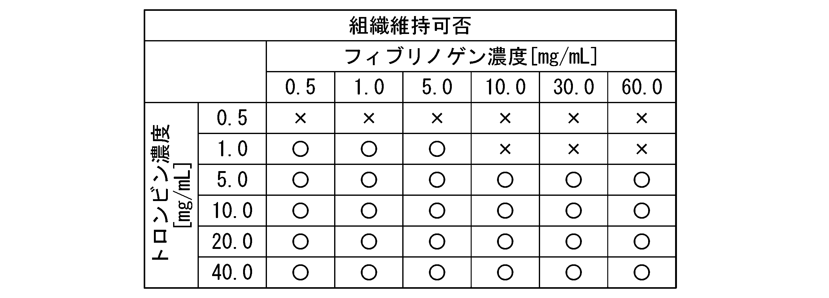
- a thrombin solution is prepared by dissolving thrombin (model number "T4648-10KU", Sigma Co., Ltd.) in the above-mentioned general-purpose medium so that the final concentrations are 0.5, 1, 5, 10, 20, and 40 Unit / mL, respectively. did.
- fibrinogen (model number "F8630-5G", Sigma) was dissolved in DMEM medium so that the final concentrations were 0.5, 1, 5.10, 30, and 60 mg / mL, respectively, to prepare a fibrinogen solution. Further, a medium was prepared by mixing a fibrinogen solution and a general-purpose medium in a ratio of 1: 1 (volume ratio) (hereinafter, may be referred to as “fibrinogen-added medium”).
- each cell suspension was centrifuged at 1,000 ⁇ g at room temperature for 1 minute to remove the supernatant, and then resuspended in a thrombin solution.
- the thrombin solution in which the cells were lysed and the fibrinogen-added medium were mixed in a ratio of 1: 2 (volume ratio), and 100 ⁇ L of this mixed solution was placed in a 24-well cell culture insert (model number “3470”, Corning). Sown.
- the cell culture inserts were allowed to stand in a CO 2 incubator (37 ° C., 5% CO 2 ) until gelling. Subsequently, an appropriate amount of the above-mentioned special medium was added to the cell culture insert, and the cells were cultured in a CO 2 incubator (37 ° C., 5% CO 2 ) for 8 days. During that time, the medium was changed as appropriate.
- each steric cell tissue was fixed using a formalin buffer (model number "062-1661", Wako Pure Chemical Industries, Ltd.). Subsequently, each three-dimensional cell tissue is taken out from the cell culture insert, embedded in paraffin, and then thinned along a line passing through the center of gravity when viewed from the upper surface of the three-dimensional cell tissue (in other words, the upper surface of the cell culture insert). Cut sections were prepared. Subsequently, the sliced sections were stained with hematoxylin and eosin (HE) and observed under a microscope to measure the maximum thickness of the three-dimensional cell tissue.
- HE hematoxylin and eosin
- Table 3 below shows the "maintenability" evaluation results of each three-dimensional cell tissue evaluated using the same evaluation criteria as in Experimental Example 1.
- the thickness maintenance rate of the three-dimensional cell tissue is high in the three-dimensional cell tissue produced under any condition where the final concentration of fibrinogen is 0.5 mg / mL or more. It was confirmed that it was possible to achieve 80% or more and suppress the decrease in the thickness of the steric cell tissue over time.
- the final thrombin concentration is 1 mg / mL
- Example 3 Manufacturing of three-dimensional cell tissue 2
- Human neonatal-derived skin fibroblasts NHDF model number "CC-2509", manufactured by Ronza
- human umbilical vein endothelial cells model number: cAP-0001GFP, manufactured by Funakoshi
- each cell suspension was centrifuged at room temperature at 1,000 ⁇ g (gravitational acceleration) for 1 minute, the supernatant was removed, and each cell suspension was resuspended in an appropriate amount of the above-mentioned special medium. Subsequently, each cell suspension was seeded in a 24-well cell culture insert (model number "3470", Corning).
- the cell culture insert was centrifuged at 400 ⁇ g for 1 minute at room temperature to obtain a cell aggregate.
- an appropriate amount of the above-mentioned special medium was added to the cell culture insert, and the cells were cultured in a CO 2 incubator (37 ° C., 5% CO 2 ) for 8 days. During that time, the medium was changed as appropriate.
- each cell suspension is centrifuged at 1,000 ⁇ g at room temperature for 1 minute, the supernatant is removed, and then collagen (CeIlamtrix Type IA, Nitta Gelatin Co., Ltd.) is prepared according to the procedure prescribed by the manufacturer. After mixing, 100 ⁇ L of this mixed solution was seeded in a 24-well cell culture insert (model number “3470”, Corning).
- the cell culture insert was allowed to stand in a CO 2 incubator (37 ° C., 5% CO 2 ) until the mixed solution gelled. Subsequently, an appropriate amount of the above-mentioned special medium was added to the cell culture insert, and the cells were cultured in a CO 2 incubator (37 ° C., 5% CO 2 ) for 8 days. During that time, the medium was changed as appropriate.
- each steric cell tissue was fixed using a formalin buffer (model number "062-1661", Wako Pure Chemical Industries, Ltd.). Subsequently, each three-dimensional cell tissue is taken out from the cell culture insert, embedded in paraffin, and then thinned along a line passing through the center of gravity when viewed from the upper surface of the three-dimensional cell tissue (in other words, the upper surface of the cell culture insert). Cut sections were prepared. Subsequently, the sliced sections were stained with hematoxylin and eosin (HE) and observed under a microscope to measure the maximum thickness of the three-dimensional cell tissue.
- HE hematoxylin and eosin
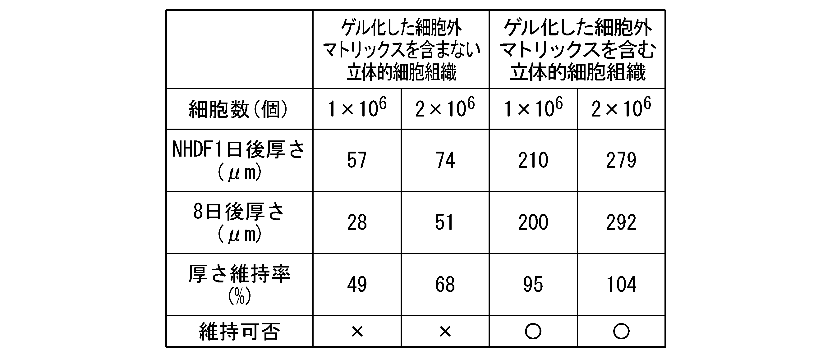
- Table 4 shows the results of measuring the maximum thickness of each three-dimensional cell tissue containing the gelled extracellular matrix and the three-dimensional cell tissue containing the gelled extracellular matrix. The "maintenability" evaluation result of each three-dimensional cell tissue evaluated by the same evaluation criteria as above is shown.
- the three-dimensional cell tissue containing the gelled extracellular matrix can achieve a thickness maintenance rate of 80% or more under any cell number condition, and the thickness of the three-dimensional cell tissue decreases with time. It was confirmed that it was possible to suppress.
- the thickness maintenance rate is less than 80% under any cell number condition, and the thickness of the three-dimensional cell tissue decreases with time. Was confirmed.
- the steric cell tissue containing the gelled extracellular matrix can form a thicker tissue than the steric cell tissue not containing the gelled extracellular matrix.
Landscapes
- Health & Medical Sciences (AREA)
- Engineering & Computer Science (AREA)
- Life Sciences & Earth Sciences (AREA)
- Biomedical Technology (AREA)
- Chemical & Material Sciences (AREA)
- Organic Chemistry (AREA)
- Zoology (AREA)
- Genetics & Genomics (AREA)
- Wood Science & Technology (AREA)
- Bioinformatics & Cheminformatics (AREA)
- Biotechnology (AREA)
- Biochemistry (AREA)
- General Health & Medical Sciences (AREA)
- General Engineering & Computer Science (AREA)
- Microbiology (AREA)
- Cell Biology (AREA)
- Rheumatology (AREA)
- Vascular Medicine (AREA)
- Biophysics (AREA)
- Gastroenterology & Hepatology (AREA)
- Toxicology (AREA)
- Medicinal Chemistry (AREA)
- Hematology (AREA)
- Proteomics, Peptides & Aminoacids (AREA)
- Molecular Biology (AREA)
- Micro-Organisms Or Cultivation Processes Thereof (AREA)
- Immobilizing And Processing Of Enzymes And Microorganisms (AREA)
- Materials For Medical Uses (AREA)
Abstract
Description
本願は、2020年11月26日に日本に出願された特願2020-196310号について優先権を主張し、その内容をここに援用する。
[1]細胞、カチオン性物質、細胞外マトリックス成分及び高分子電解質を含む混合物を得る工程と、前記混合物をゲル化させてゲル状組成物を得る工程と、前記ゲル状組成物をインキュベートして立体的細胞組織を得る工程と、を含む、立体的細胞組織の製造方法。
[2]前記ゲル状組成物が、前記細胞外マトリックス成分、アガロース、ペクチン及びフィブリン・モノマーからなる群より選択される少なくとも1種がゲル化したゲル状成分を含む、[1]に記載の製造方法。
[3]前記ゲル状成分が、フィブリン・モノマーがゲル化したフィブリンゲルであり、前記ゲル状組成物を得る工程が、前記混合物に、トロンビン及びフィブリノゲンを混合する工程を含む、[2]に記載の製造方法。
[4]前記ゲル状組成物を得る工程が、前記混合物に前記トロンビンを添加する工程と、前記トロンビンを添加した前記混合物に前記フィブリノゲンを添加し、その結果、前記フィブリンゲルが形成されて前記混合物がゲル化する工程と、を含む、[3]に記載の製造方法。
[5]前記細胞外マトリックス成分が、コラーゲン、ラミニン、フィブロネクチン、ビトロネクチン、エラスチン、テネイシン、エンタクチン、フィブリン、プロテオグリカン及びそれらの組み合わせからなる群より選択される、[1]~[4]のいずれか1つに記載の製造方法。
[6]前記混合物における、前記細胞外マトリックス成分の含有量の合計が、0.005mg/mL以上1.5mg/mL以下である、[1]~[5]のいずれか1つに記載の製造方法。
[7]前記高分子電解質が、グリコサミノグリカン、デキストラン硫酸、ラムナン硫酸、フコイダン、カラギナン、ポリスチレンスルホン酸、ポリアクリルアミド-2-メチルプロパンスルホン酸、ポリアクリル酸及びそれらの組み合わせからなる群より選択される、[1]~[6]のいずれか1つに記載の製造方法。
[8]前記混合物における、前記高分子電解質の含有量が、0.005mg/mL以上である、[1]~[7]のいずれか1つに記載の製造方法。
[9]前記ゲル状組成物を得る工程の前に、前記混合物に外力を加えて、前記細胞、前記カチオン性物質、前記細胞外マトリックス成分及び前記高分子電解質を含む細胞集合体を得る工程を更に含み、前記ゲル状組成物を得る工程において、前記混合物に代えて前記細胞集合体をゲル化させてゲル状組成物を得る、[1]~[8]のいずれか1つに記載の製造方法。
[10]細胞、カチオン性物質、細胞外マトリックス成分、高分子電解質及びゲル状成分を含む、立体的細胞組織。
[11]細胞、カチオン性物質、高分子電解質及びゲル状成分を含む、立体的細胞組織。
[12]前記ゲル状成分は、第1の細胞外マトリックス成分、アガロース、ペクチン及びフィブリン・モノマーからなる群より選択される少なくとも1種がゲル化したゲル状成分である[11]に記載の立体的細胞組織。
[13]さらに、第2の細胞外マトリックス成分を含み、前記ゲル状成分は、アガロース、ペクチン及びフィブリン・モノマーからなる群より選択される少なくとも1種がゲル化したゲル状成分である[11]に記載の立体的細胞組織。
[14]製造から8日目の前記立体的細胞組織の上面から見たときの重心を通る線に沿って取得した切片の厚さの最大値が、製造直後の前記立体的細胞組織の上面から見たときの重心を通る線に沿って取得した切片の厚さの最大値の80%以上である、[10]~[13]のいずれか1つに記載の立体的細胞組織。
一実施形態において、本発明は、細胞、カチオン性物質、細胞外マトリックス成分及び高分子電解質を含む混合物を得る工程と、前記混合物をゲル化させてゲル状組成物を得る工程と、前記ゲル状組成物をインキュベートして立体的細胞組織を得る工程と、を含む、立体的細胞組織の製造方法を提供する。
本実施形態の製造方法において、細胞としては特に限定されず、ヒト、サル、イヌ、ネコ、ウサギ、ブタ、ウシ、マウス及びラット等の哺乳動物に由来する細胞を使用することができる。細胞の由来部位も特に限定されず、骨、筋肉、内臓、神経、脳、皮膚又は血液等に由来する体細胞であってもよく、生殖細胞であってもよく、癌細胞であってもよい。
カチオン性物質としては、細胞の生育及び後述する細胞集合体の形成に悪影響を及ぼさない限り、任意の正電荷を有する物質を用いることができる。カチオン性物質には、トリス-塩酸、トリス-マレイン酸、ビス-トリス、HEPES等のカチオン性緩衝剤、エタノールアミン、ジエタノールアミン、トリエタノールアミン、ポリビニルアミン、ポリアリルアミン、ポリリシン及びポリヒスチジン、ポリアルギニン等が挙げられるが、これらに限定されない。なかでもカチオン性緩衝剤が好ましく、トリス-塩酸がより好ましい。
細胞外マトリックス成分としては、細胞の生育及び後述する細胞集合体の形成に悪影響を及ぼさない限り、細胞外マトリックス(ECMともいう)を構成する任意の成分を用いることができる。細胞外マトリックス成分としては、コラーゲン、ラミニン、フィブロネクチン、ビトロネクチン、エラスチン、テネイシン、エンタクチン、フィブリン、プロテオグリカン及びこれらの組み合わせ等が挙げられるが、これらに限定されない。細胞外マトリックス成分は、上述したものの改変体又はバリアント等であってもよい。細胞外マトリックス成分は、1種を単独で用いてもよく、2種以上を組み合わせて用いてもよい。
本明細書において、高分子電解質とは、高分子鎖中に解離可能な官能基を有する高分子を意味する。本実施形態で用いられる高分子電解質としては、細胞の生育及び細胞集合体の形成に悪影響を及ぼさない限り、任意の高分子電解質を用いることができる。高分子電解質としては、ヘパリン、コンドロイチン硫酸(例えば、コンドロイチン4-硫酸及びコンドロイチン6-硫酸)、ヘパラン硫酸、デルマタン硫酸、ケラタン硫酸及びヒアルロン酸等のグリコサミノグリカン、デキストラン硫酸、ラムナン硫酸、フコイダン、カラギナン、ポリスチレンスルホン酸、ポリアクリルアミド-2-メチルプロパンスルホン酸、ポリアクリル酸及びこれらの組み合わせ等が挙げられるが、これらに限定されない。高分子電解質は、上述したものの誘導体であってもよい。これらの高分子電解質は、1種を単独で用いてもよく、2種以上を組み合わせて用いてもよい。
ゲル化剤としては、細胞外マトリックス成分;アガロース;ペクチン;フィブリノゲン及びトロンビンの組み合わせ等が挙げられる。ゲル化剤は、予め工程(a)における混合物中に含有させていてもよいし、以下に説明する工程(b)において、工程(a)における混合物に添加してもよい。
本実施形態の製造方法は、ゲル状組成物を得る工程(b)の前に、工程(a)で得た混合物に外力を加えて、細胞、カチオン性物質、細胞外マトリックス成分及び高分子電解質を含む細胞集合体を得る工程(a’)を更に含み、ゲル状組成物を得る工程(b)において、工程(a)で得た混合物に代えて工程(a’)で得た細胞集合体をゲル化させてゲル状組成物を得るものであってもよい。
一実施形態において、本発明は、細胞、カチオン性物質、細胞外マトリックス成分、高分子電解質及びゲル状成分を含む、立体的細胞組織を提供する。もう一つの態様として、本発明は、細胞、カチオン性物質、高分子電解質及びゲル状成分を含む、立体的細胞組織を提供する。本実施形態の立体的細胞組織は、経時的な厚さの減少が抑制されている。
[1]細胞、カチオン性物質、細胞外マトリックス成分及び高分子電解質を含む混合物を得る工程と、前記混合物にトロンビン及びフィブリノゲンの少なくとも一方を混合しゲル状組成物を得る工程と、前記ゲル状組成物をインキュベートして立体的細胞組織を得る工程と、を含む、立体的細胞組織の製造方法。
[2]前記ゲル状組成物を得る工程が、前記混合物に前記フィブリノゲンを混合し、ゲル状組成物を得る工程である、[1]に記載の立体的細胞組織の製造方法。
[3]前記ゲル状組成物を得る工程が、前記混合物に前記トロンビンを添加する工程と、前記トロンビンを添加した前記混合物に前記フィブリノゲンを添加する工程と、を含む、[1]に記載の製造方法。
[4]前記混合物における、前記フィブリノゲンの含有量が、0.5mg/mL以上60mg/mL以下である、[1]~[3]のいずれか1つに記載の製造方法。
[5]前記混合物における、前記トロンビンの含有量が、1.0mg/mL以上40mg/mL以下である、[1]~[4]のいずれか1つに記載の製造方法。
[6]前記細胞外マトリックス成分が、コラーゲン、ラミニン及びフィブロネクチンからなる群より選択される少なくとも一つの細胞外マトリックスである、[1]~[5]のいずれか1つに記載の製造方法。
[7]前記細胞外マトリックス成分が、コラーゲンである、[1]~[6]のいずれか1つに記載の製造方法。
[8]前記混合物における、前記細胞外マトリックス成分の含有量の合計が、0.005mg/mL以上1.5mg/mL以下である、[1]~[7]のいずれか1つに記載の製造方法。
[9]前記高分子電解質が、グリコサミノグリカンである、[1]~[8]のいずれか1つに記載の製造方法。
[10]前記高分子電解質が、ヘパリン、コンドロイチン硫酸及びデルマタン硫酸からなる群から選択される少なくとも一つのグリコサミノグリカンである、[9]に記載の製造方法。
[11]前記混合物における、前記高分子電解質の含有量が、0.005mg/mL以上である、[1]~[10]のいずれか1つに記載の製造方法。
[12]前記ゲル状組成物を得る工程の前に、前記混合物に外力を加えて、前記細胞、前記カチオン性物質、前記細胞外マトリックス成分及び前記高分子電解質を含む細胞集合体を得る工程を更に含み、前記ゲル状組成物を得る工程において、前記混合物に代えて前記細胞集合体をゲル化させてゲル状組成物を得る、[1]~[11]のいずれか1つに記載の製造方法。
[13]細胞、カチオン性物質、細胞外マトリックス成分、高分子電解質及びゲル化したフィブリン・モノマーを含む、立体的細胞組織。
[14]前記立体細胞組織における、前記ゲル化したフィブリン・モノマーの含有量が、0.5mg/mL以上60mg/mL以下である、[13]に記載の立体的細胞組織。
[15]製造から8日目の前記立体的細胞組織の上面から見たときの重心を通る線に沿って取得した切片の厚さの最大値が、製造直後の前記立体的細胞組織の上面から見たときの重心を通る線に沿って取得した切片の厚さの最大値の80%以上である、[13]又は[14]に記載の立体的細胞組織。
(立体的細胞組織の製造)
フィブリンゲルを含まない立体的細胞組織、及び、フィブリンゲルを含む立体的細胞組織をそれぞれ製造した。細胞としてヒト新生児由来皮膚線維芽細胞NHDF(型番「CC-2509」、ロンザ社)を使用した。
10%ウシ胎児血清(FBS)及び1%抗生物質溶液(ペニシリン、ストレプトマイシン)を含むDMEM培地(型番「043-30085」、富士フイルム和光純薬)を調製した(以下、「汎用培地」という場合がある。)。また、汎用培地と血管細胞用培地(製品名「EGM-2MV」、型番「CC-3202」、ロンザ社)を1:1(体積比)となるように混合した培地を調製した(以下、「専用培地」という場合がある。)。
上述した汎用培地に、終濃度10Unit/mLとなるようにトロンビン(型番「T4648-10KU」、シグマ社)を溶解し、トロンビン溶液を調製した。
細胞集合体の培養開始から1日後及び8日後に、ホルマリン緩衝液(型番「062-01661」、富士フイルム和光純薬)を用いて各立体的細胞組織を固定した。続いて、各立体的細胞組織をセルカルチャーインサートから取り出し、パラフィンで包埋後、立体的細胞組織の上面(言い換えると、セルカルチャーインサートの上面)から見たときの重心を通る線に沿って薄切切片を作製した。続いて、薄切切片をヘマトキシリン・エオシン(HEともいう)染色して顕微鏡で観察し、立体的細胞組織の厚さの最大値を測定した。
厚さ維持率(%)=培養開始から8日後の立体的細胞組織の上面から見たときの重心を通る線に沿って取得した切片の厚さの最大値/培養開始から1日後の立体的細胞組織の上面から見たときの重心を通る線に沿って取得した切片の厚さの最大値×100 …(1)
(評価基準)
〇…上記式(1)に基づいて算出した厚さ維持率が80%以上
×…上記式(1)に基づいて算出した厚さ維持率が80%未満


(立体的細胞組織の製造1)
フィブリンゲルを含む立体的細胞組織を製造した。細胞としてヒト新生児由来皮膚線維芽細胞NHDF(型番「CC-2509」、ロンザ社)とヒト臍帯静脈内皮細胞(GFP-HUVEC)(型番:cAP-0001GFP、フナコシ社製)を使用した。
細胞集合体の培養開始から1日後及び8日後に、ホルマリン緩衝液(型番「062-01661」、富士フイルム和光純薬)を用いて各立体的細胞組織を固定した。続いて、各立体的細胞組織をセルカルチャーインサートから取り出し、パラフィンで包埋後、立体的細胞組織の上面(言い換えると、セルカルチャーインサートの上面)から見たときの重心を通る線に沿って薄切切片を作製した。続いて、薄切切片をヘマトキシリン・エオシン(HE)染色して顕微鏡で観察し、立体的細胞組織の厚さの最大値を測定した。
(立体的細胞組織の製造2)
細胞外マトリックスをゲル化した立体的細胞組織を製造した。細胞としてヒト新生児由来皮膚線維芽細胞NHDF(型番「CC-2509」、ロンザ社)とヒト臍帯静脈内皮細胞(GFP-HUVEC)(型番:cAP-0001GFP、フナコシ社製)を使用した。
1×10⁶個のNHDF細胞及び2×10⁵個のGFP-HUVECを0.05mg/mLヘパリン(型番「H3149-100KU」、シグマ社)を含む、50mMトリス-塩酸緩衝溶液(pH7.4)に懸濁した。同様に、2×106個のNHDF細胞及び4×105個のGFP-HUVECを上述の50mMトリス-塩酸緩衝溶液に懸濁した。
1×10⁶個のNHDF細胞及び2×10⁵個のGFP-HUVECを0.05mg/mLヘパリン(型番「H3149-100KU」、シグマ社)を含む50mMトリス-塩酸緩衝溶液(pH7.4)に懸濁した。同様に、2×106個のNHDF細胞4×105個のGFP-HUVECを0.05mg/mLヘパリン(型番「H3149-100KU」、シグマ社)を含む50mMトリス-塩酸緩衝溶液に懸濁した。
細胞集合体の培養開始から1日後及び8日後に、ホルマリン緩衝液(型番「062-01661」、富士フイルム和光純薬)を用いて各立体的細胞組織を固定した。続いて、各立体的細胞組織をセルカルチャーインサートから取り出し、パラフィンで包埋後、立体的細胞組織の上面(言い換えると、セルカルチャーインサートの上面)から見たときの重心を通る線に沿って薄切切片を作製した。続いて、薄切切片をヘマトキシリン・エオシン(HE)染色して顕微鏡で観察し、立体的細胞組織の厚さの最大値を測定した。
Claims (14)
- 細胞、カチオン性物質、細胞外マトリックス成分及び高分子電解質を含む混合物を得る工程と、
前記混合物をゲル化させてゲル状組成物を得る工程と、
前記ゲル状組成物をインキュベートして立体的細胞組織を得る工程と、
を含む、立体的細胞組織の製造方法。 - 前記ゲル状組成物が、前記細胞外マトリックス成分、アガロース、ペクチン及びフィブリン・モノマーからなる群より選択される少なくとも1種がゲル化したゲル状成分を含む、請求項1に記載の製造方法。
- 前記ゲル状成分が、フィブリン・モノマーがゲル化したフィブリンゲルであり、前記ゲル状組成物を得る工程が、前記混合物に、トロンビン及びフィブリノゲンを混合する工程を含む、請求項2に記載の製造方法。
- 前記ゲル状組成物を得る工程が、
前記混合物に前記トロンビンを添加する工程と、
前記トロンビンを添加した前記混合物に前記フィブリノゲンを添加し、その結果、前記フィブリンゲルが形成されて前記混合物がゲル化する工程と、
を含む、請求項3に記載の製造方法。 - 前記細胞外マトリックス成分が、コラーゲン、ラミニン、フィブロネクチン、ビトロネクチン、エラスチン、テネイシン、エンタクチン、フィブリン、プロテオグリカン及びそれらの組み合わせからなる群より選択される、請求項1~4のいずれか一項に記載の製造方法。
- 前記混合物における、前記細胞外マトリックス成分の含有量の合計が、0.005mg/mL以上1.5mg/mL以下である、請求項1~5のいずれか一項に記載の製造方法。
- 前記高分子電解質が、グリコサミノグリカン、デキストラン硫酸、ラムナン硫酸、フコイダン、カラギナン、ポリスチレンスルホン酸、ポリアクリルアミド-2-メチルプロパンスルホン酸、ポリアクリル酸及びそれらの組み合わせからなる群より選択される、請求項1~6のいずれか一項に記載の製造方法。
- 前記混合物における、前記高分子電解質の含有量が、0.005mg/mL以上である、請求項1~7のいずれか一項に記載の製造方法。
- 前記ゲル状組成物を得る工程の前に、前記混合物に外力を加えて、前記細胞、前記カチオン性物質、前記細胞外マトリックス成分及び前記高分子電解質を含む細胞集合体を得る工程を更に含み、
前記ゲル状組成物を得る工程において、前記混合物に代えて前記細胞集合体をゲル化させてゲル状組成物を得る、請求項1~8のいずれか一項に記載の製造方法。 - 細胞、カチオン性物質、細胞外マトリックス成分、高分子電解質及びゲル状成分を含む、立体的細胞組織。
- 細胞、カチオン性物質、高分子電解質及びゲル状成分を含む、立体的細胞組織。
- 前記ゲル状成分は、第1の細胞外マトリックス成分、アガロース、ペクチン及びフィブリン・モノマーからなる群より選択される少なくとも1種がゲル化したゲル状成分である請求項11に記載の立体的細胞組織。
- さらに、第2の細胞外マトリックス成分を含み、前記ゲル状成分は、アガロース、ペクチン及びフィブリン・モノマーからなる群より選択される少なくとも1種がゲル化したゲル状成分である[11]に記載の立体的細胞組織。
- 製造から8日目の前記立体的細胞組織の上面から見たときの重心を通る線に沿って取得した切片の厚さの最大値が、製造直後の前記立体的細胞組織の上面から見たときの重心を通る線に沿って取得した切片の厚さの最大値の80%以上である、請求項10~13のいずれか一項に記載の立体的細胞組織。
Priority Applications (4)
| Application Number | Priority Date | Filing Date | Title |
|---|---|---|---|
| EP21898100.9A EP4253528A4 (en) | 2020-11-26 | 2021-11-26 | METHOD FOR PRODUCING THREE-DIMENSIONAL CELL TISSUE AND THREE-DIMENSIONAL CELL TISSUE |
| JP2022541798A JP7239068B2 (ja) | 2020-11-26 | 2021-11-26 | 立体的細胞組織の製造方法及び立体的細胞組織 |
| CN202180079294.XA CN116457458A (zh) | 2020-11-26 | 2021-11-26 | 立体性细胞组织的制造方法及立体性细胞组织 |
| US18/319,691 US20230287356A1 (en) | 2020-11-26 | 2023-05-18 | Methods of producing three-dimensional cell tissue, and three-dimensional cell tissues |
Applications Claiming Priority (2)
| Application Number | Priority Date | Filing Date | Title |
|---|---|---|---|
| JP2020196310 | 2020-11-26 | ||
| JP2020-196310 | 2020-11-26 |
Related Child Applications (1)
| Application Number | Title | Priority Date | Filing Date |
|---|---|---|---|
| US18/319,691 Continuation US20230287356A1 (en) | 2020-11-26 | 2023-05-18 | Methods of producing three-dimensional cell tissue, and three-dimensional cell tissues |
Publications (1)
| Publication Number | Publication Date |
|---|---|
| WO2022114128A1 true WO2022114128A1 (ja) | 2022-06-02 |
Family
ID=81754428
Family Applications (1)
| Application Number | Title | Priority Date | Filing Date |
|---|---|---|---|
| PCT/JP2021/043393 Ceased WO2022114128A1 (ja) | 2020-11-26 | 2021-11-26 | 立体的細胞組織の製造方法及び立体的細胞組織 |
Country Status (5)
| Country | Link |
|---|---|
| US (1) | US20230287356A1 (ja) |
| EP (1) | EP4253528A4 (ja) |
| JP (1) | JP7239068B2 (ja) |
| CN (1) | CN116457458A (ja) |
| WO (1) | WO2022114128A1 (ja) |
Cited By (3)
| Publication number | Priority date | Publication date | Assignee | Title |
|---|---|---|---|---|
| WO2024014497A1 (ja) * | 2022-07-14 | 2024-01-18 | オリヅルセラピューティクス株式会社 | 細胞移植用のフィブリンゲルシート |
| WO2025023216A1 (ja) * | 2023-07-27 | 2025-01-30 | Toppanホールディングス株式会社 | 立体的細胞組織の製造方法及び立体的細胞組織 |
| WO2025143240A1 (ja) * | 2023-12-28 | 2025-07-03 | Toppanホールディングス株式会社 | 細胞構造体を製造する方法 |
Citations (5)
| Publication number | Priority date | Publication date | Assignee | Title |
|---|---|---|---|---|
| WO2017146124A1 (ja) * | 2016-02-22 | 2017-08-31 | 国立大学法人大阪大学 | 立体的細胞組織の製造方法 |
| JP2020031548A (ja) * | 2018-08-28 | 2020-03-05 | 国立大学法人大阪大学 | 三次元心筋組織の製造方法 |
| WO2020203579A1 (ja) * | 2019-04-01 | 2020-10-08 | 凸版印刷株式会社 | 三次元組織体及びその製造方法並びに細胞含有組成物の製造方法 |
| JP2020196310A (ja) | 2019-05-31 | 2020-12-10 | 市光工業株式会社 | 車両用灯具 |
| WO2021100709A1 (ja) * | 2019-11-19 | 2021-05-27 | 凸版印刷株式会社 | 細胞構造体及びその製造方法並びに被験物質の肝毒性の評価方法 |
Family Cites Families (4)
| Publication number | Priority date | Publication date | Assignee | Title |
|---|---|---|---|---|
| US20100255065A1 (en) * | 2007-08-01 | 2010-10-07 | Regenprime Co., Ltd. | Method for differenciating mesenchymal stem cell and culturing chondrocytes using alginate coated fibrin/ha composite scaffold |
| KR100971271B1 (ko) * | 2008-04-28 | 2010-07-20 | 한양대학교 산학협력단 | 헤파린이 결합된 피브린젤, 그 제조방법 및 키트 |
| US11045500B2 (en) * | 2011-02-14 | 2021-06-29 | Technion Research Development Foundation Ltd. | Tissue engineering construct comprising fibrin |
| WO2023017800A1 (ja) * | 2021-08-11 | 2023-02-16 | 凸版印刷株式会社 | 立体的細胞組織の製造方法及び立体的細胞組織 |
-
2021
- 2021-11-26 WO PCT/JP2021/043393 patent/WO2022114128A1/ja not_active Ceased
- 2021-11-26 JP JP2022541798A patent/JP7239068B2/ja active Active
- 2021-11-26 EP EP21898100.9A patent/EP4253528A4/en active Pending
- 2021-11-26 CN CN202180079294.XA patent/CN116457458A/zh active Pending
-
2023
- 2023-05-18 US US18/319,691 patent/US20230287356A1/en active Pending
Patent Citations (5)
| Publication number | Priority date | Publication date | Assignee | Title |
|---|---|---|---|---|
| WO2017146124A1 (ja) * | 2016-02-22 | 2017-08-31 | 国立大学法人大阪大学 | 立体的細胞組織の製造方法 |
| JP2020031548A (ja) * | 2018-08-28 | 2020-03-05 | 国立大学法人大阪大学 | 三次元心筋組織の製造方法 |
| WO2020203579A1 (ja) * | 2019-04-01 | 2020-10-08 | 凸版印刷株式会社 | 三次元組織体及びその製造方法並びに細胞含有組成物の製造方法 |
| JP2020196310A (ja) | 2019-05-31 | 2020-12-10 | 市光工業株式会社 | 車両用灯具 |
| WO2021100709A1 (ja) * | 2019-11-19 | 2021-05-27 | 凸版印刷株式会社 | 細胞構造体及びその製造方法並びに被験物質の肝毒性の評価方法 |
Non-Patent Citations (6)
| Title |
|---|
| AKIHIRO NISHIGUCHI ET AL.: "Cell- cell crosslinking by bio-molecular recognition of heparin-based layer-by-layer nanofilms", MACROMOL BIOSCI, vol. 15, no. 3, 2015, pages 312 - 317, XP055540432, DOI: 10.1002/mabi.201400415 |
| BONHOME-ESPINOSA ANA BELÉN, CAMPOS FERNANDO, DURAND-HERRERA DANIEL, SÁNCHEZ-LÓPEZ JOSÉ DARÍO, SCHAUB SÉBASTIEN, DURÁN JUAN D.G., L: "In vitro characterization of a novel magnetic fibrin-agarose hydrogel for cartilage tissue engineering", JOURNAL OF THE MECHANICAL BEHAVIOR OF BIOMEDICAL MATERIALS, ELSEVIER, AMSTERDAM, NL, vol. 104, no. 103619, 1 April 2020 (2020-04-01), AMSTERDAM, NL, XP055934501, ISSN: 1751-6161, DOI: 10.1016/j.jmbbm.2020.103619 * |
| HEO DONG NYOUNG; HOSPODIUK MONIKA; OZBOLAT IBRAHIM T.: "Synergistic interplay between human MSCs and HUVECs in 3D spheroids laden in collagen/fibrin hydrogels for bone tissue engineering", ACTA BIOMATERIALIA, ELSEVIER, AMSTERDAM, NL, vol. 95, 1 March 2019 (2019-03-01), AMSTERDAM, NL, pages 348 - 356, XP085782347, ISSN: 1742-7061, DOI: 10.1016/j.actbio.2019.02.046 * |
| INO, KOSUKE: "Bioorganic Construction by Biofabrication", KAGAKU TO KOGYO - CHEMISTRY AND CHEMICAL INDUSTRY, NIPPON KAGAKKAI, TOKYO, JP, vol. 73, no. 10, 1 October 2020 (2020-10-01), JP , pages 742 - 743, XP009537043, ISSN: 0022-7684 * |
| RAMESHWAR R. RAO; ALEXIS W. PETERSON; JACOB CECCARELLI; ANDREW J. PUTNAM; JAN P. STEGEMANN: "Matrix composition regulates three-dimensional network formation by endothelial cells and mesenchymal stem cells in collagen/fibrin materials", ANGIOGENESIS, KLUWER ACADEMIC PUBLISHERS, DO, vol. 15, no. 2, 2 March 2012 (2012-03-02), Do , pages 253 - 264, XP035048393, ISSN: 1573-7209, DOI: 10.1007/s10456-012-9257-1 * |
| See also references of EP4253528A4 |
Cited By (3)
| Publication number | Priority date | Publication date | Assignee | Title |
|---|---|---|---|---|
| WO2024014497A1 (ja) * | 2022-07-14 | 2024-01-18 | オリヅルセラピューティクス株式会社 | 細胞移植用のフィブリンゲルシート |
| WO2025023216A1 (ja) * | 2023-07-27 | 2025-01-30 | Toppanホールディングス株式会社 | 立体的細胞組織の製造方法及び立体的細胞組織 |
| WO2025143240A1 (ja) * | 2023-12-28 | 2025-07-03 | Toppanホールディングス株式会社 | 細胞構造体を製造する方法 |
Also Published As
| Publication number | Publication date |
|---|---|
| JP7239068B2 (ja) | 2023-03-14 |
| EP4253528A4 (en) | 2024-10-30 |
| EP4253528A1 (en) | 2023-10-04 |
| JPWO2022114128A1 (ja) | 2022-06-02 |
| US20230287356A1 (en) | 2023-09-14 |
| CN116457458A (zh) | 2023-07-18 |
Similar Documents
| Publication | Publication Date | Title |
|---|---|---|
| JP6427836B2 (ja) | 立体的細胞組織の製造方法 | |
| US20230287356A1 (en) | Methods of producing three-dimensional cell tissue, and three-dimensional cell tissues | |
| Hwang et al. | Response of zonal chondrocytes to extracellular matrix-hydrogels | |
| US20240174982A1 (en) | Methods of producing three-dimensional cellular tissues, and three-dimensional cellular tissues | |
| Demange et al. | Survival of cord blood haematopoietic stem cells in a hyaluronan hydrogel for ex vivo biomimicry | |
| US20220356442A1 (en) | Method of producing three-dimensional cell structure | |
| US20230323310A1 (en) | Method of producing three-dimensional tissue body and three-dimensional tissue body | |
| JP7819502B2 (ja) | 立体的細胞組織の製造方法 | |
| US20230193211A1 (en) | Method for producing three-dimensional cell structure, and three-dimensional cell structure | |
| JP7480476B2 (ja) | 立体的細胞組織の培養方法及びキット | |
| JP2023156998A (ja) | 立体的細胞組織の製造方法及び立体的細胞組織 | |
| WO2025023216A1 (ja) | 立体的細胞組織の製造方法及び立体的細胞組織 | |
| JP7107450B1 (ja) | 立体的細胞構造体の製造方法 | |
| JP2024090483A (ja) | 立体的細胞組織および立体的細胞組織の評価方法 | |
| EP4455268A1 (en) | Method for culturing three-dimensional cell tissue | |
| US20230203452A1 (en) | Method of producing conditioned medium for culturing patient-derived cancer cells | |
| WO2025143240A1 (ja) | 細胞構造体を製造する方法 | |
| JP2024136114A (ja) | 立体的細胞組織の製造方法及び培養容器 | |
| Vento | ADHESION LIGAND-PRESENTING HYDROGELS FOR STUDY-ING STEM CELL BEHAVIOURS IN THREE DIMENSIONS |
Legal Events
| Date | Code | Title | Description |
|---|---|---|---|
| ENP | Entry into the national phase |
Ref document number: 2022541798 Country of ref document: JP Kind code of ref document: A |
|
| 121 | Ep: the epo has been informed by wipo that ep was designated in this application |
Ref document number: 21898100 Country of ref document: EP Kind code of ref document: A1 |
|
| WWE | Wipo information: entry into national phase |
Ref document number: 202180079294.X Country of ref document: CN |
|
| NENP | Non-entry into the national phase |
Ref country code: DE |
|
| ENP | Entry into the national phase |
Ref document number: 2021898100 Country of ref document: EP Effective date: 20230626 |