WO2022186311A1 - 化合物及び放射性標識化合物 - Google Patents
化合物及び放射性標識化合物 Download PDFInfo
- Publication number
- WO2022186311A1 WO2022186311A1 PCT/JP2022/009003 JP2022009003W WO2022186311A1 WO 2022186311 A1 WO2022186311 A1 WO 2022186311A1 JP 2022009003 W JP2022009003 W JP 2022009003W WO 2022186311 A1 WO2022186311 A1 WO 2022186311A1
- Authority
- WO
- WIPO (PCT)
- Prior art keywords
- compound
- formula
- psma
- atomic group
- binding
- Prior art date
- Legal status (The legal status is an assumption and is not a legal conclusion. Google has not performed a legal analysis and makes no representation as to the accuracy of the status listed.)
- Ceased
Links
Images


Classifications
-
- C—CHEMISTRY; METALLURGY
- C07—ORGANIC CHEMISTRY
- C07D—HETEROCYCLIC COMPOUNDS
- C07D257/00—Heterocyclic compounds containing rings having four nitrogen atoms as the only ring hetero atoms
- C07D257/02—Heterocyclic compounds containing rings having four nitrogen atoms as the only ring hetero atoms not condensed with other rings
-
- A—HUMAN NECESSITIES
- A61—MEDICAL OR VETERINARY SCIENCE; HYGIENE
- A61K—PREPARATIONS FOR MEDICAL, DENTAL OR TOILETRY PURPOSES
- A61K31/00—Medicinal preparations containing organic active ingredients
- A61K31/33—Heterocyclic compounds
- A61K31/395—Heterocyclic compounds having nitrogen as a ring hetero atom, e.g. guanethidine or rifamycins
-
- A—HUMAN NECESSITIES
- A61—MEDICAL OR VETERINARY SCIENCE; HYGIENE
- A61K—PREPARATIONS FOR MEDICAL, DENTAL OR TOILETRY PURPOSES
- A61K51/00—Preparations containing radioactive substances for use in therapy or testing in vivo
- A61K51/02—Preparations containing radioactive substances for use in therapy or testing in vivo characterised by the carrier, i.e. characterised by the agent or material covalently linked or complexing the radioactive nucleus
- A61K51/04—Organic compounds
-
- A—HUMAN NECESSITIES
- A61—MEDICAL OR VETERINARY SCIENCE; HYGIENE
- A61K—PREPARATIONS FOR MEDICAL, DENTAL OR TOILETRY PURPOSES
- A61K51/00—Preparations containing radioactive substances for use in therapy or testing in vivo
- A61K51/02—Preparations containing radioactive substances for use in therapy or testing in vivo characterised by the carrier, i.e. characterised by the agent or material covalently linked or complexing the radioactive nucleus
- A61K51/04—Organic compounds
- A61K51/0402—Organic compounds carboxylic acid carriers, fatty acids
-
- A—HUMAN NECESSITIES
- A61—MEDICAL OR VETERINARY SCIENCE; HYGIENE
- A61K—PREPARATIONS FOR MEDICAL, DENTAL OR TOILETRY PURPOSES
- A61K51/00—Preparations containing radioactive substances for use in therapy or testing in vivo
- A61K51/02—Preparations containing radioactive substances for use in therapy or testing in vivo characterised by the carrier, i.e. characterised by the agent or material covalently linked or complexing the radioactive nucleus
- A61K51/04—Organic compounds
- A61K51/0404—Lipids, e.g. triglycerides; Polycationic carriers
- A61K51/0406—Amines, polyamines, e.g. spermine, spermidine, amino acids, (bis)guanidines
-
- A—HUMAN NECESSITIES
- A61—MEDICAL OR VETERINARY SCIENCE; HYGIENE
- A61K—PREPARATIONS FOR MEDICAL, DENTAL OR TOILETRY PURPOSES
- A61K51/00—Preparations containing radioactive substances for use in therapy or testing in vivo
- A61K51/02—Preparations containing radioactive substances for use in therapy or testing in vivo characterised by the carrier, i.e. characterised by the agent or material covalently linked or complexing the radioactive nucleus
- A61K51/04—Organic compounds
- A61K51/0474—Organic compounds complexes or complex-forming compounds, i.e. wherein a radioactive metal (e.g. 111In3+) is complexed or chelated by, e.g. a N2S2, N3S, NS3, N4 chelating group
- A61K51/0482—Organic compounds complexes or complex-forming compounds, i.e. wherein a radioactive metal (e.g. 111In3+) is complexed or chelated by, e.g. a N2S2, N3S, NS3, N4 chelating group chelates from cyclic ligands, e.g. DOTA
-
- A—HUMAN NECESSITIES
- A61—MEDICAL OR VETERINARY SCIENCE; HYGIENE
- A61K—PREPARATIONS FOR MEDICAL, DENTAL OR TOILETRY PURPOSES
- A61K51/00—Preparations containing radioactive substances for use in therapy or testing in vivo
- A61K51/02—Preparations containing radioactive substances for use in therapy or testing in vivo characterised by the carrier, i.e. characterised by the agent or material covalently linked or complexing the radioactive nucleus
- A61K51/04—Organic compounds
- A61K51/0497—Organic compounds conjugates with a carrier being an organic compounds
-
- C—CHEMISTRY; METALLURGY
- C07—ORGANIC CHEMISTRY
- C07B—GENERAL METHODS OF ORGANIC CHEMISTRY; APPARATUS THEREFOR
- C07B59/00—Introduction of isotopes of elements into organic compounds ; Labelled organic compounds per se
- C07B59/004—Acyclic, carbocyclic or heterocyclic compounds containing elements other than carbon, hydrogen, halogen, oxygen, nitrogen, sulfur, selenium or tellurium
-
- C—CHEMISTRY; METALLURGY
- C07—ORGANIC CHEMISTRY
- C07D—HETEROCYCLIC COMPOUNDS
- C07D213/00—Heterocyclic compounds containing six-membered rings, not condensed with other rings, with one nitrogen atom as the only ring hetero atom and three or more double bonds between ring members or between ring members and non-ring members
- C07D213/02—Heterocyclic compounds containing six-membered rings, not condensed with other rings, with one nitrogen atom as the only ring hetero atom and three or more double bonds between ring members or between ring members and non-ring members having three double bonds between ring members or between ring members and non-ring members
- C07D213/04—Heterocyclic compounds containing six-membered rings, not condensed with other rings, with one nitrogen atom as the only ring hetero atom and three or more double bonds between ring members or between ring members and non-ring members having three double bonds between ring members or between ring members and non-ring members having no bond between the ring nitrogen atom and a non-ring member or having only hydrogen or carbon atoms directly attached to the ring nitrogen atom
- C07D213/60—Heterocyclic compounds containing six-membered rings, not condensed with other rings, with one nitrogen atom as the only ring hetero atom and three or more double bonds between ring members or between ring members and non-ring members having three double bonds between ring members or between ring members and non-ring members having no bond between the ring nitrogen atom and a non-ring member or having only hydrogen or carbon atoms directly attached to the ring nitrogen atom with hetero atoms or with carbon atoms having three bonds to hetero atoms with at the most one bond to halogen, e.g. ester or nitrile radicals, directly attached to ring carbon atoms
- C07D213/78—Carbon atoms having three bonds to hetero atoms, with at the most one bond to halogen, e.g. ester or nitrile radicals
- C07D213/79—Acids; Esters
-
- C—CHEMISTRY; METALLURGY
- C07—ORGANIC CHEMISTRY
- C07D—HETEROCYCLIC COMPOUNDS
- C07D487/00—Heterocyclic compounds containing nitrogen atoms as the only ring hetero atoms in the condensed system, not provided for by groups C07D451/00 - C07D477/00
- C07D487/02—Heterocyclic compounds containing nitrogen atoms as the only ring hetero atoms in the condensed system, not provided for by groups C07D451/00 - C07D477/00 in which the condensed system contains two hetero rings
- C07D487/04—Ortho-condensed systems
-
- C—CHEMISTRY; METALLURGY
- C07—ORGANIC CHEMISTRY
- C07B—GENERAL METHODS OF ORGANIC CHEMISTRY; APPARATUS THEREFOR
- C07B2200/00—Indexing scheme relating to specific properties of organic compounds
- C07B2200/05—Isotopically modified compounds, e.g. labelled
Definitions
- the present invention relates to compounds and radiolabeled compounds.
- Radiolabeled compounds with radionuclides in their structures are used as reagents for the detection of target molecules, diagnostic agents, or pharmaceuticals for the treatment of diseases.
- studies are being conducted to improve specific accumulation in target tissues and sites and to reduce accumulation in non-target tissues and sites.
- Patent Document 1 describes a derivative (HTK01169) in which an iodophenylbutyryl group, which is an albumin binding site, is added so as to branch from the structure of PSMA-617. It has also been described that this derivative binds to prostate specific membrane antigen (PSMA) and can be used for the detection and treatment of prostate cancer.
- PSMA prostate specific membrane antigen
- Patent Document 2 also describes an albumin-binding PSMA inhibitor. It is also described that this inhibitor also binds to PSMA as a target and can be used for the purpose of detecting and treating prostate cancer, as in Patent Document 1.
- the present invention relates to a compound and a radiolabeled compound capable of improving accumulation in target tissues and reducing accumulation in non-target tissues, particularly reducing accumulation in the kidney.
- the present invention has in its structure a chelate moiety capable of coordinating with a radioactive metal ion, a first atomic group containing an albumin binding moiety, and a second atomic group containing a binding moiety with a PSMA molecule, A compound is provided in which the first atomic group and the second atomic group are bonded via the chelate moiety.
- the present invention provides a compound represented by the following general formula (2).
- A2 is a chelate moiety capable of coordinating with a radioactive metal ion
- B is an atomic group containing an albumin binding moiety
- C is an atomic group containing a binding moiety with a PSMA molecule
- A2 is Neunpa or Octapa or a derivative thereof
- L c is a linker structure, When A2 is Octapa , Lc contains a polyethylene glycol structure.
- the present invention provides a radiolabeled compound in which the compound is coordinated to a radioactive metal ion.
- the present invention provides a method for producing a radiolabeled compound, wherein the compound is coordinated with a radioactive metal ion to obtain a radiolabeled compound.
- FIG. 1 is a graph showing the results of cell binding experiments in Example 1-1 ([ 111 In]In-PSMA-DA1) and Comparative Example 1-1 ([ 111 In]In-PSMA-DB).
- FIG. 2 is a graph showing the results of albumin binding experiments on [ 111 In]In-PSMA-DA1 and [ 111 In]In-PSMA-DB.
- FIG. 3 is a graph showing the results of internal radioactivity distribution experiments for [ 111 In]In-PSMA-DA1 and [ 111 In]In-PSMA-DB.
- FIG. 4 shows SPECT/CT images in [ 111 In]In-PSMA-DA1 and [ 111 In]In-PSMA-DB.
- FIG. 5 is a graph showing changes in tumor volume and mouse body weight in Example 1-2 ([ 90 Y]Y-PSMA-DA1) and Comparative Example 1-2 ([ 90 Y]Y-PSMA-DB).
- FIG. 6 is a graph showing changes in tumor volume and mouse body weight in Example 1-3 ([ 225 Ac]Ac-PSMA-DA1) and Comparative Example 1-3 ([ 225 Ac]Ac-PSMA-DB).
- FIG. 7 is an HPLC chart showing the results of plasma stability in Example 2 ([ 111 In]In-PtDA).
- FIG. 8 is a graph showing the results of cell binding experiments with [ 111 In]In-PtDA.
- FIG. 9 is a graph showing the results of albumin binding experiments on [ 111 In]In-PtDA.
- FIG. 10 is a SPECT/CT image in [ 111 In]In-PtDA.
- FIG. 11 shows Example 3-1 ([ 111 In] In-Octapa-2), Example 3-2 ([ 111 In] In-Octapa-3), Example 3-3 ([ 111 In] In- Neunpa-2), Comparative Example 2-1 ([ 111 In]In-Octapa-1) and Comparative Example 2-2 ([ 111 In]In-Neunpa-1) are graphs showing the results of cell binding experiments. .
- FIG. 12 is a graph showing the results of albumin binding experiments in Examples 3-1 to 3-3 and Comparative Examples 2-1 to 2-2.
- FIG. 11 shows Example 3-1 ([ 111 In] In-Octapa-2), Example 3-2 ([ 111 In] In-Octapa-3), Example 3-3 ([ 111 In] In- Neunpa-2), Comparative Example 2-1 ([ 111 In]In-Octapa-1) and Comparative Example 2-2 ([ 111 In
- FIG. 13 is a graph showing the results of internal radioactivity distribution experiments in Examples 3-1 to 3-3 and Comparative Examples 2-1 to 2-2.
- FIG. 14 is a SPECT/CT image in Example 3-1 ([ 111 In]In-Octapa-2).
- FIG. 15 is a graph showing the results of cell binding experiments in Example 4-1 ([ 111 In]In-PSMA-NAT-DA1).
- FIG. 16 is a graph showing the results of albumin binding experiments in Example 4-1 ([ 111 In]In-PSMA-NAT-DA1).
- FIG. 17 is a SPECT/CT image in Example 4-1 ([ 111 In]In-PSMA-NAT-DA1).
- T to U [V] T and U are arbitrary numbers and [V] is a unit.
- T [V] or more U [ V] means “less than or equal to”.
- each independently may be in the S configuration or the R configuration unless otherwise specified.
- the chemical structures of the compounds of the present invention are roughly classified into a chelate moiety capable of coordinating with radioactive metal ions, a first atomic group containing an albumin-binding moiety, and a prostate specific membrane antigen (hereinafter also referred to as PSMA). It has three atomic groups in its structure with the second atomic group including the bond with the molecule.
- PSMA prostate specific membrane antigen
- the radiolabeled compound in which the radioactive metal ion is coordinated to the compound of the present invention has improved accumulation in the target tissue expressing the PSMA molecule and non-target tissue, especially It is compatible with the reduction of accumulation in the kidney.
- the compounds of the invention are precursor compounds used for labeling with radioisotopes such as radiometals, ie compounds preferably used as labeling precursors. A description of radioactive metals will be given later.
- the first atomic group containing the albumin binding moiety and the second atomic group containing the binding moiety for the PSMA molecule are bound via a chelate moiety.
- Such compounds are preferably represented by the following general formula (1).
- A1 is a chelating moiety capable of coordinating with a radioactive metal ion
- B is an atomic group containing an albumin binding moiety
- C is an atomic group containing a binding moiety with a PSMA molecule.
- La is a linker structure.
- Lb is a linker structure that is the same as or different from La.
- m and n are each independently 0 (zero) or 1; B or La is attached to any site of A1.
- C or Lb binds to A1 at a site different from the site where B or La binds to A1.
- the chelate moiety represented by symbol A1 in general formula ( 1 )
- the first atomic group containing the albumin binding portion represented by the symbol B in the general formula (1)
- the second atomic group containing the bonding portion of the PSMA molecule represented by the symbol C in the general formula (1)
- between A 1 and B, and between A 1 and C may each independently have a linker structure and be indirectly bonded, or have a linker structure. may be directly connected without
- the linker structures when there is a linker structure between A 1 and B and between A 1 and C, the linker structures may be the same or different. good. Details of the linker structure will be described later.
- a 1 has a cyclic structure, the cyclic structure has two or more nitrogen atoms, and each nitrogen atom is linked with two or more adjacent carbon atoms separated, or , A 1 preferably have a chain structure, the chain structure has two or more nitrogen atoms, and each nitrogen atom is preferably linked with two or more adjacent carbon atoms interposed therebetween.
- the cyclic structure when A 1 has a cyclic structure, the cyclic structure may have a skeleton composed only of nitrogen atoms and carbon atoms, and in addition to nitrogen atoms and carbon atoms, oxygen atoms It may be configured to include Bonds between carbon atoms in the cyclic structure may be chain-like or form a cyclic structure. Further, in the above formula ( 1 ), when A1 has a chain structure, the bonds between carbon atoms in the chain structure may be separated by nitrogen atoms. Bonds between carbon atoms in the chain structure may be chain or may form a ring structure.
- a 1 when A 1 has a cyclic structure or a chain structure, A 1 has a nitrogen-bonded atomic group directly bonded to a nitrogen atom constituting the cyclic structure or the chain structure. is preferred.
- Specific examples of the nitrogen-bonded atomic group are preferably atomic groups containing one or more of a carboxy group, a phosphoric acid group, an amide group, a benzene ring and a pyridine ring, and more preferably a chain-like atomic group.
- a 1 has a cyclic structure or a chain structure
- B binds to any site of A 1
- C is any site of A 1 different from the binding site of B with the proviso that B is bound to the above nitrogen-bonded group through L a or not through L a
- B is bound through L a or L a It is preferably bonded to a site other than the nitrogen-bonded atomic group that is bonded without intervening.
- a compound represented by any one of the following formulas (A1) to (A9) as a chelate moiety capable of coordinating with the radioactive metal represented by symbol A1 It preferably has a structure derived from it, and more preferably has a structure derived from a compound represented by the following formula (A1). That is, the compound of the present invention is preferably a derivative of a compound represented by any one of formulas (A1) to (A9) below, and is a derivative of a compound represented by formula (A1) below. is more preferred.
- These structures can be appropriately selected according to the type of radioactive metal described later. Regardless of the structure of the chelate moiety, both improved accumulation in PSMA-expressing target tissues and reduced accumulation in non-target tissues, particularly kidneys, are compatible.
- the chelate moiety represented by symbol A1 includes, for example, structures derived from the following compounds, but is not limited thereto.
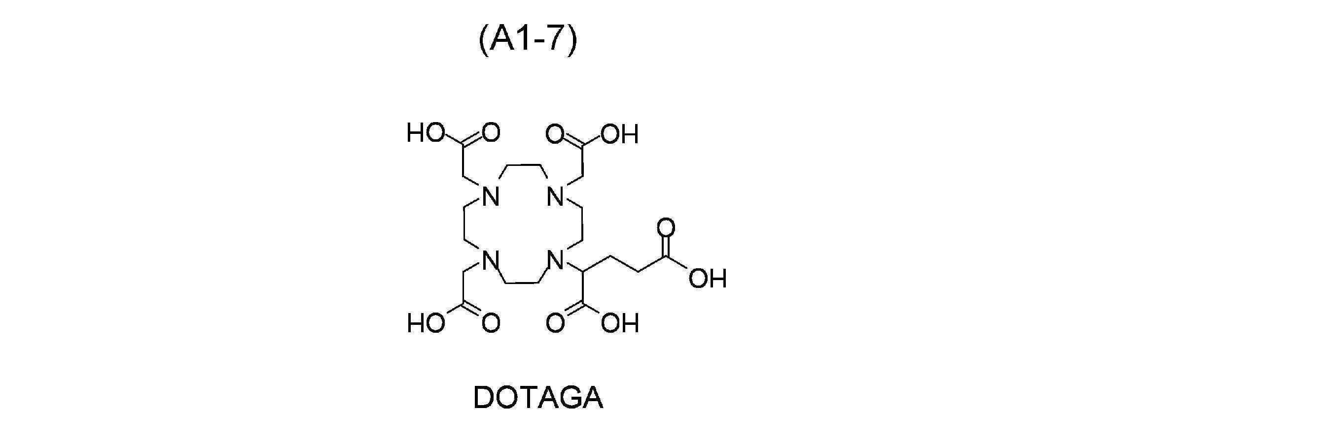
- DOTA ⁇ DOTA or its derivative> ⁇ 1,4,7,10-tetraazacyclododecane-1,4,7,10-tetraacetic acid (DOTA) ⁇ 1,4,7,10-tetraazacyclododecane-1,4,7,10-tetrapropionic acid (DOTPA) ⁇ 1,4,7,10-tetraazacyclododecane-1,4,7,10-tetramethylene phosphate (DOTMP) ⁇ Hydroxypropyltetraazacyclododecanetriacetic acid (HP-DO3A) ⁇ (1R,4R,7R,10R)- ⁇ , ⁇ ', ⁇ ", ⁇ '"-tetramethyl-1,4,7,10-tetraazacyclododecane-1,4,7,10-tetraacetic acid ( DOTAMA) ⁇ 1,4,7,10-tetrakis(carbamoylmethyl)-1,4,7,10-tetraazacycl
- TETA ⁇ 1,4,8,11-tetraazacyclotetradecane-1,4,8,11-tetraacetic acid
- TETPA ⁇ 1,4,8,11-tetraazacyclotetradecane-1,4,8,11-tetrapropionic acid
- PEPA PEPA
- NOTA ⁇ 2-[4,7-bis(carboxymethyl)-1,4,7-triazonan-1-yl]acetic acid
- R 11 , R 12 , R 13 and R 14 are each independently -(CH 2 ) p COOH, -(CH 2 ) p C 5 H 5 N, -(CH 2 ) p PO 3 H 2 , —(CH 2 ) p CONH 2 , —(CHCOOH)(CH 2 ) p COOH, where p is an integer of 0 or more and 3 or less.
- R 21 , R 22 , R 23 and R 24 are each independently a carboxy group or a carboxyalkyl group having 2 or 3 carbon atoms.
- R 31 , R 32 , R 33 and R 34 each independently have a hydrogen atom and 2 or more and 10 or less carbon atoms, and may contain a nitrogen atom or an oxygen atom.
- R 35 is a hydrogen atom, a carboxy group, or a carboxyalkyl group having 2 or 3 carbon atoms.
- R 41 , R 42 , R 43 and R 44 each independently have a hydrogen atom and 2 or more and 10 or less carbon atoms, and may contain a nitrogen atom or an oxygen atom.
- R 45 is a hydrogen atom, a carboxy group, or a carboxyalkyl group having 2 or 3 carbon atoms.
- R 48 and R 49 are each independently an atomic group having a hydrogen atom and 2 or more and 10 or less carbon atoms and optionally containing a nitrogen atom or an oxygen atom.
- each of R 51 , R 52 , R 53 , R 54 and R 55 independently has a hydrogen atom and 2 or more and 10 or less carbon atoms and contains a nitrogen atom or an oxygen atom. It is an atomic group that can be
- R 61 , R 62 , R 63 , R 64 , R 65 and R 66 each independently have a hydrogen atom and 2 or more and 10 or less carbon atoms, a nitrogen atom or an oxygen It is an atomic group that may contain atoms, and R 67 is a hydrogen atom, a carboxy group, or a carboxyalkyl group having 2 or 3 carbon atoms.
- R 71 , R 72 and R 73 are each independently an atomic group having a hydrogen atom and 2 or more and 10 or less carbon atoms and optionally containing a nitrogen atom or an oxygen atom. is.
- R 81 and R 82 are each independently an alkyl group having 1 to 5 carbon atoms, and the terminal of the alkyl group is substituted with a pyridyl group substituted with one or more carboxy groups.
- R 83 and R 84 are substituted or unsubstituted pyridinyl groups
- R 85 and R 86 are each independently is -COO-R a
- R a is an alkyl group having 1 to 5 carbon atoms.
- formula (A1) include, for example, structures represented by the following formulas (A1-1) to (A1-7).
- formula (A2) include, for example, structures represented by the following formulas (A2-1) and (A2-2).
- formula (A3) include, for example, structures represented by the following formulas (A3-1) to (A3-7).
- formula (A4) include structures represented by the following formulas (A4-1) and (A4-2).
- formula (A5) include, for example, structures represented by the following formulas (A5-1) to (A5-3).
- formula (A6) include, for example, structures represented by the following formula (A6-1).
- formula (A7) include, for example, structures represented by the following formulas (A7-1) and (A7-2).
- formula (A8) include, for example, structures represented by the following formulas (A8-1) to (A8-3).
- formula (A9) include, for example, structures represented by the following formulas (A9-1) to (A9-4).
- the chelate portion is bound so as to branch.
- the first atomic group containing the albumin-binding portion and the second atomic group containing the binding portion with the PSMA molecule are bound in a branched manner via the linker structure bound to the chelate portion.
- Such compounds are preferably represented by the following general formula (2).
- A2 is a chelating moiety capable of coordinating with a radioactive metal ion
- B is an atomic group containing an albumin binding moiety
- C is an atomic group containing a binding moiety with a PSMA molecule.
- A2 is Neunpa or Octapa or a derivative thereof.
- Lc is a linker structure. When A2 is Octapa , Lc contains a polyethylene glycol structure.
- a 2 preferably has a chain structure, the chain structure has two or more nitrogen atoms, and each nitrogen atom is linked with two or more adjacent carbon atoms separated from each other.
- A2 when A2 has a chain structure, the bonds between carbon atoms in the chain structure may be separated by nitrogen atoms. Bonds between carbon atoms in the chain structure may be chain or may form a ring structure.
- A2 is more preferably a compound represented by formula (A3) or formula (A4) or a derivative thereof.
- the site represented by symbol B has affinity for albumin, preferably serum albumin, more preferably human serum albumin, and is reversible with albumin. It is an atomic group containing an albumin binding site, which is a chemical structure capable of binding to By including such a structure in the compound of the present invention, when the compound is labeled with a radioactive metal and administered to the living body, the retention in the blood is increased and the accumulation in the kidney is reduced. be able to.
- a compound having an albumin-binding portion in its molecule easily binds to albumin in the blood, and the compound bound to albumin does not undergo glomerular filtration in the kidney, thus reducing excretion and migration to the kidney and urine. and improved retention in the blood.
- the accumulation in the kidney, which is a normal tissue is reduced, and the migration of the compound to target tissues such as tumor tissues expressing PSMA can be further enhanced.
- the distance between the albumin-binding site and the PSMA molecule-binding site can be appropriately secured.
- the affinity of the albumin binding moiety and the PSMA molecule are arranged at one structural end of the compound of the invention and the PSMA molecule binding moiety is located at the other structural end of the compound of the invention. Since a sufficient distance can be ensured at the binding site with the PSMA molecule, it is further advantageous in that both the affinity for albumin and the affinity for the PSMA molecule can be achieved at a high level.
- the linker structure represented by Lc ensures an appropriate distance between the albumin binding site and the PSMA molecule binding site. can combine affinity for albumin with that of the PSMA molecule.
- the linker structure represented by Lc in formula (2) by adopting a structure containing a polyethylene glycol group and not containing a cyclohexyl group and a naphthyl group as the linker structure represented by Lc in formula (2), the albumin binding site and the PSMA molecule can be It is further advantageous in that it is possible to ensure an appropriate distance at the binding site and achieve both high levels of affinity for albumin and affinity for the PSMA molecule.
- the structure of the albumin binding portion includes, for example, ⁇ -glutamic acid, substituted or unsubstituted phenylbutyric acid, lipid, hematin, bilirubin, clofibric acid, clofibrate, carotenoid, and steroid.
- albumin eg, peptides described in WO 2007/106120, etc.
- substituted or unsubstituted phenylbutyric acid applicable as the albumin-binding portion include structures represented by the following formula (B1).
- R is a hydrogen atom, a halogen atom, or an alkyl group having 1 to 5 carbon atoms, and the portion indicated by the wavy line is the bonding portion with another structure.
- R is preferably a hydrogen atom, an iodine atom, a bromine atom or a methyl group.
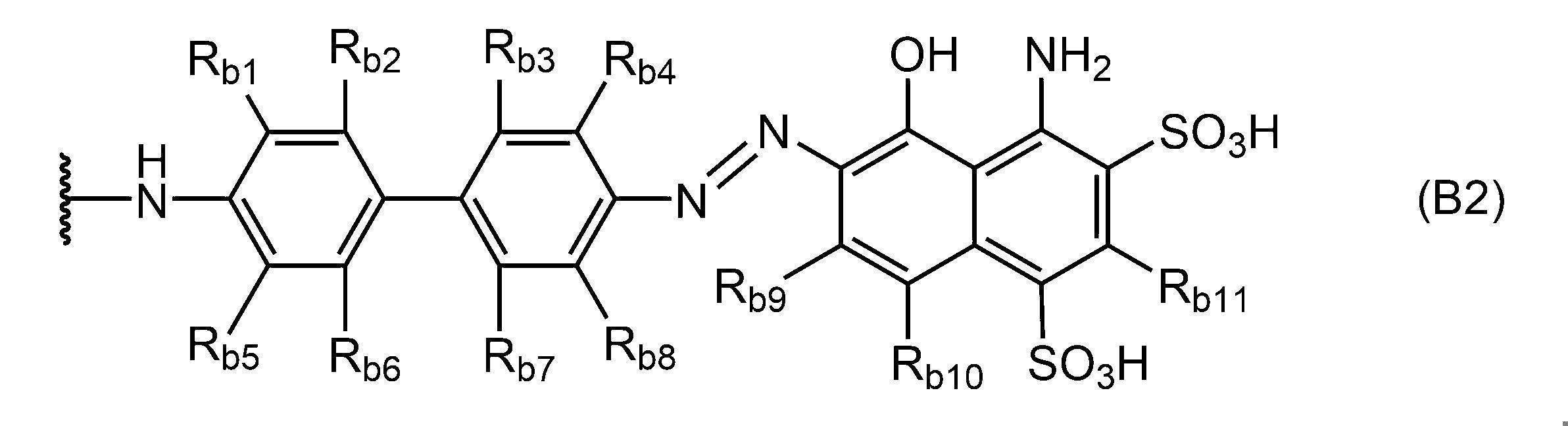
- Examples of Evans blue and its derivatives applicable as the albumin-binding portion include structures represented by the following formula (B2).
- R b1 to R b11 each independently represent a hydrogen atom, a halogen atom, a hydroxyl group, a cyano group, a substituted or unsubstituted alkyl group having 1 to 6 carbon atoms, or 1 carbon atom. It is a substituted or unsubstituted alkoxy group of 6 or less, and the portion indicated by the wavy line is the bonding portion with another structure.
- R b1 and R b4 are both methyl groups
- R b2 and R b3 and R b5 to R b11 are all preferably hydrogen atoms.
- immunoglobulins having classes of IgG, IgA, IgM, IgD and IgE may be used as long as they have affinity for albumin. good too. From the viewpoint of reducing accumulation in unintended tissues such as the liver, when an antibody capable of binding to albumin is used as the albumin-binding moiety, it is preferable to use a Fab fragment with a low molecular weight.
- Peptides capable of binding to albumin include, for example, peptides containing the sequences shown in WO 2007/106120, and more specifically peptides containing the following peptide sequences, but are limited to these sequences: can't In the following peptide sequences, amino acids are represented by single-letter code, the left side of the page indicates the N-terminus, and the right side of the page indicates the C-terminus.
- the binding portion to the PSMA molecule represented by symbol C has affinity for the PSMA molecule expressed in tissues that cause diseases such as cancer. It is an atomic group containing a bond with a PSMA molecule, which is a chemical structure capable of reversibly binding to a PSMA molecule.
- PSMA is a membrane-bound protein whose expression is particularly enhanced in prostate cancer. Although the expression level of PSMA is low in normal tissues including prostate, the expression level of PSMA increases as the degree of malignancy of prostate cancer increases. Therefore, PSMA is one of the useful target molecules in the present invention and is particularly useful as a target for diagnosis and treatment of prostate cancer.
- PSMA molecules that express PSMA molecules include, for example, prostate cancer.
- Prostate cancer can be primary or metastatic.
- the chemical structure of the binding site with the PSMA molecule can be appropriately selected according to the target tissue and the amount of PSMA molecules expressed in the tissue.
- a structure that has an affinity for PSMA molecules can be employed as a binding site for PSMA molecules.
- Structures that have affinity for PSMA molecules include, for example, one or more of low-molecular-weight compounds, peptides, antibodies, and antibody fragments such as Fab fragments.
- the PSMA molecule-binding portion preferably has a structure represented by the following formula (C1), or is an antibody or peptide capable of binding to the PSMA molecule.
- a and b are each independently an integer of 1 or more and 7 or less. Further, in formula (C1), the part indicated by the wavy line is the part that connects with A 1 or L a in formula (1) or with L c in formula (2).
- immunoglobulins having classes of IgG, IgA, IgM, IgD and IgE may be used as long as they have affinity for PSMA molecules, and antibody fragments (eg, Fab fragments) may be used. may be used. From the viewpoint of reducing accumulation in unintended tissues such as the liver, when an antibody capable of binding to a PSMA molecule is used as a binding site with a PSMA molecule, it is preferable to use a Fab fragment with a low molecular weight.
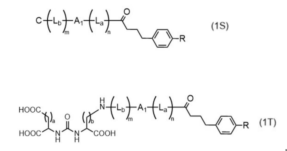
- the compound of the present invention has the structure of the above formula (1), it more preferably has a structure represented by the following general formula (1S), and is represented by the following general formula (1T). It is also more preferable to have a structure with By having such a structure, the structure has sufficient heteroatoms capable of coordinating the radioactive metal. can increase In addition, since the freedom of movement of the molecules at the albumin-binding site and the PSMA molecule-binding site is increased, it is possible to achieve both high levels of affinity for albumin and affinity for PSMA molecules. Furthermore, unintended accumulation in normal tissues such as liver and kidney is reduced.
- A1 is each independently a chelating moiety capable of coordinating with a radioactive metal ion.
- C is an atomic group containing a bond with the PSMA molecule.
- La is a linker structure.
- L b is a linker structure that is the same as or different from La.
- m and n are each independently 0 (zero) or 1.
- the compound of the present invention has the structure of formula (1) above, it is more preferable to have the structure of general formula (3) below.
- the structure has sufficient heteroatoms capable of coordinating the radioactive metal. can increase
- the freedom of movement of the molecules at the albumin-binding site and the PSMA molecule-binding site is increased, it is possible to achieve both high levels of affinity for albumin and affinity for PSMA molecules.
- unintended accumulation in normal tissues such as liver and kidney is reduced.
- one of R B1 and R B2 is an atomic group containing an albumin binding site, the other is a hydrogen atom, a hydroxyl group or a carboxy group, and one of R C1 and R C2 is a PSMA molecule. It is an atomic group containing a bond, and the other is a hydrogen atom, a hydroxyl group, or a carboxy group.
- R B1 is an atomic group containing an albumin binding site
- R C1 is an atomic group containing a binding site with a PSMA molecule
- both R B2 and R C2 are hydroxyl groups. is preferred.
- R B2 is the group containing the albumin binding site
- R C2 is the group containing the binding site to the PSMA molecule
- both R B1 and R C1 are hydrogen atoms. or each independently a carboxyalkyl group having 1 to 5 carbon atoms.
- the compound of the present invention is characterized by having the albumin binding moiety located at one structural end of the compound and the PSMA molecule binding moiety located at the other structural end of the compound. macroscopically, the chelate portion, the albumin-binding portion, and the PSMA molecule-binding portion are preferably arranged substantially linearly.
- the atomic group containing the albumin binding moiety is preferably an atomic group containing the structure represented by formula (B1) or formula (B2) described above as the albumin binding moiety.
- formulas (3-1) to (3-4) Specific examples of such chemical structures are shown in formulas (3-1) to (3-4) below.
- L 1 is each independently an alkyl group having 1 to 8 carbon atoms and containing a carboxy group.
- g is an integer of 1 or more and 5 or less
- h is 0 or 1.
- R is a hydrogen atom, a halogen atom or an alkyl group having 1 to 5 carbon atoms, preferably a hydrogen atom, an iodine atom, a bromine atom or a methyl group.
- R b1 to R b11 each independently represent a hydrogen atom, a halogen atom, a hydroxyl group, a cyano group, a substituted or unsubstituted alkyl group having 1 to 6 carbon atoms, or a substituted or unsubstituted alkyl group having 1 to 6 carbon atoms. It is an unsubstituted alkoxy group, preferably both R b1 and R b4 are methyl groups, and R b2 and R b3 and R b5 to R b11 are all preferably hydrogen atoms.
- One of R C1 and R C2 is an atomic group containing a bond with a PSMA molecule, and the other is a hydrogen atom, a hydroxyl group, or a carboxyalkyl group having 1 to 5 carbon atoms.
- the structure of the following formula (2S) has sufficient heteroatoms capable of coordinating the radioactive metal. can increase
- the freedom of movement of the molecules at the albumin-binding site and the PSMA molecule-binding site is increased, it is possible to achieve both high levels of affinity for albumin and affinity for PSMA molecules.
- unintended accumulation in normal tissues such as liver and kidney is reduced.
- the production time required for complex formation is reduced, and the production efficiency and radiochemical yield of the radiolabeled compound are further improved.
- A2 is Neunpa or Octapa or a derivative thereof.
- Lc is a linker structure.
- R is a hydrogen atom, a halogen atom or an alkyl group having 1 to 5 carbon atoms.
- a and b are each independently an integer of 1 or more and 7 or less.
- Lc contains a polyethylene glycol structure.
- the compound of the present invention is preferably dissolved in an aqueous solution such as a solvent or buffer solution, and reacted with a radioactive metal ion to coordinate the compound of each of the above embodiments to the radioactive metal ion, A radiolabeled compound can be obtained.
- This radiolabeled compound is a radiometal complex in which the chelate portion of the compound is coordinated to a radiometal ion.
- B in formula (1) has a structure represented by formula (B1) above.
- a compound represented by formula (1S) it is more preferable to use a compound represented by formula (1S). This can increase the efficiency of complex formation.
- the radioactive metal to be reacted with the compound is preferably used in the form of an ionizable radioactive metal compound, more preferably in the form of a radioactive metal ion (hereinafter, these forms are collectively referred to as (also referred to as a “radioactive metal source”).
- a radioactive metal source for example, a radioactive metal ion-containing liquid in which radioactive metal ions are dissolved or dispersed in a solvent mainly composed of water can be used.
- the order of addition of the compound and the radioactive metal source does not matter as long as the compound can form a complex with the radioactive metal ion.
- Either one of the compound and the radioactive metal source may be added and then the other may be added and reacted, or the other may be added to a solution in which one of the compound and the radioactive metal source is dissolved in a solvent and reacted. Alternatively, these may be simultaneously added to a reaction vessel containing a solvent and reacted.
- the reaction conditions for obtaining a radiolabeled compound can be, for example, the following conditions.
- Solvents used in this step include, for example, water, physiological saline, sodium acetate buffer, ammonium acetate buffer, phosphate buffer, phosphate buffered saline, Tris buffer, HEPES buffer, or tetramethyl A buffer such as an ammonium acetate buffer can be used.
- the reaction temperature may be, for example, room temperature (25° C.) or under heating conditions.
- radioactive metal source for example, a solution in which radioactive metal ions are dispersed in a solvent mainly composed of water can be used.
- the amount of the reaction liquid in this step is not particularly limited, but from the viewpoint of practicality in the manufacturing process, 0.01 mL to 100 mL is realistic at the start of this step.
- the concentrations of the compound and the radioactive metal ion in the reaction solution are each independently preferably 1 ⁇ M to 100 ⁇ M at the start of this step from the viewpoint of yield of the target radiolabeled compound.
- the obtained radiolabeled compound may be used as it is, or may be purified using filtration filters, membrane filters, columns filled with various fillers, chromatography, and the like.
- a solvent mainly composed of water and other pharmaceutically acceptable ingredients are added to the radiolabeled compound to obtain a radiopharmaceutical composition containing the radiolabeled compound as an active ingredient.
- the radiopharmaceutical composition can be produced, for example, by dissolving the radiolabeled compound produced by the method described above in a solvent that is mainly composed of water and that is approximately isotonic with the living body.
- a radiopharmaceutical composition is administered to a living body orally or parenterally such as intravenously, subcutaneously, intraperitoneally or intramuscularly, and is used for disease treatment, disease diagnosis, lesion detection, and the like.
- the radiometal ionically coordinated to the radiolabeled compound can be a metal nuclide that emits alpha, beta or gamma radiation or a combination thereof.
- radioactive metal nuclides include radioactive isotopes of alkali metals, alkaline earth metals, lanthanides, actinides, transition metals, and metals other than these metals.
- These radioactive metals can be produced according to conventional methods. These radionuclides are preferably obtained as a solution containing the radiometal in an ionized state.
- ⁇ -ray emitting nuclide When a radiolabeled compound is used for the purpose of treating a disease, it is preferable to use an ⁇ -ray emitting nuclide or a ⁇ - ray emitting nuclide as the radioactive metal from the viewpoint of enhancing the therapeutic effect.
- the ⁇ -ray emitting nuclide may be any nuclide that emits ⁇ -rays during the decay process of the radioactive metal. 225 Ac, more preferably 225 Ac.
- the ⁇ - ray emitting nuclide may be any nuclide that emits ⁇ - rays during the decay process of the radioactive metal, and specific examples include 59 Fe, 60 Co, 64 Cu, 67 Cu, 89 Sr, 90 Y, 99m Tc, 103 Ru, 153 Sm, 165 Dy, 166 Ho, 177 Lu, 186 Re, 188 Re, 198 Au, 203 Hg, 212 Pb, 212 Bi or 213 Bi are preferably used, and more preferably 64 Cu and 67 Cu. , 89 Sr, 90 Y, 177 Lu, 186 Re or 188 Re, and more preferably 90 Y is used.
- ⁇ + -ray emitting nuclides when using radiolabeled compounds for the purpose of diagnosing diseases or detecting lesions, it is recommended to use ⁇ + -ray emitting nuclides, electron capture decay nuclides, or ⁇ -ray emitting nuclides as radiometals from the viewpoint of enhancing diagnostic performance. is preferred.
- the ⁇ + -ray emitting nuclide may be any nuclide that emits positrons in the decay process of the radioactive metal, and 44 Sc, 58 Co, 68 Ga, 64 Cu or 89 Zr is preferably used, more preferably 64 Cu or 89 is Zr.
- the electron capture decay nuclide may be a nuclide that emits Auger electrons or characteristic X-rays in the decay process of a radioactive metal, 51 Cr, 57 Co, 58 Co, 64 Cu, 67 Ga, 68 Ga, 89 Zr, 111 In , 186 Re, 197 Hg, 201 Tl, etc. are preferably used.
- the ⁇ -ray emitting nuclide may be any nuclide that emits ⁇ -rays by ⁇ -decay, and 68 Ga, 99m Tc or 201 Tl is preferably used as the nuclide that emits ⁇ -rays by ⁇ -decay.
- the radioactive metals having an ionic radius of about 70 to 130 pm are 64 Cu, 67 Cu, 67 Ga, 68 Ga, 89Zr , 90Y , 99mTc , 103Ru , 111In , 153Sm , 165Dy , 166Ho , 177Lu , 186Re , 188Re , 198Au , 201Tl , 197Hg , 203Hg , 212Pb , 212Bi . _
- the compound of the present invention when a radiolabeled compound is used for the treatment of a disease and 225 Ac is used as the radioactive metal, the compound of the present invention is preferably ), and more preferably a compound having a chelate moiety having a structure represented by the formula (A1), (A3) or (A4).
- the compound of the present invention preferably has a chelate moiety having a structure represented by any of the formulas (A1) to (A3) or (A8), and further A compound having a chelate moiety having a structure represented by the formula (A1) is preferably used.
- the compound of the present invention when a radiolabeled compound is used for the purpose of diagnosing a disease or detecting a lesion, when 89 Zr is used as a radioactive metal, the compound of the present invention is preferably ), and more preferably a compound having a chelate moiety having the structure represented by the above formula (A1). Further, when 68 Ga or 111 In is used as the radioactive metal, the compound of the present invention preferably has a chelate portion having a structure represented by any of the above formulas (A1) to (A4) or (A9). More preferably, a compound having a chelate moiety having the structure represented by formula (A1) is used.
- the bond between the chelate moiety and the atomic group containing the albumin-binding moiety may be such that the chelate moiety and the albumin-binding moiety are directly bonded without a linker structure to be described later, or the chelate moiety and the albumin-binding moiety are bonded together. may be indirectly bonded via a linker structure to be described later.
- the bond between the chelate portion and the atomic group containing the binding portion to the PSMA molecule may be such that the chelate portion and the binding portion to the PSMA molecule are directly bonded without via a linker structure described later, Alternatively, the chelate moiety and the atomic group containing the albumin-binding moiety may be indirectly bonded via a linker structure described below. In any of the above-described “directly” or “indirectly” embodiments, the bonding via an amide bond is compatible with the ease of synthesis and the stability of the chemical structure. preferable.
- the linker structure of L a or L b in formula (1) or L c in formula (2) is preferably a structure derived from a compound capable of forming an amide bond or an ether bond independently.
- Specific examples thereof include acidic amino acids such as glutamic acid and aspartic acid, L- or D-amino acids such as basic amino acids such as lysine, dicarboxylic acids such as oxalic acid and malonic acid, diamines such as ethylenediamine, polyethylene glycol groups,
- the structure can be derived from an amino acid or the like having a C5-C10 cycloaliphatic or C6-C14 aromatic structure. These can be employed singly or by linking a plurality of them via an amide bond or an ether bond.
- Each of the above structures may also independently be unsubstituted or substituted with various substituents.
- a structure derived from an amino acid or the like is included as the linker structure described above, for example, for the purpose of controlling in vivo kinetics, WO 2017/150549, WO 2019/065774, WO 2019/221269 No., International Publication No. 2020/075746, International Publication No. 2020/145227, International Publication No. 2020/145228, etc. can be used.
- linker structure When a structure derived from ethylene glycol is included as the above-mentioned linker structure, it is also preferable that it is indirectly linked by a linker structure represented by the following formula (P).
- the structure is an ethylene glycol-derived structure, and in formula (P), k is preferably an integer of 2 or more and 10 or less, more preferably an integer of 2 or more and 8 or less, and still more preferably an integer of 2 or more and 5 or less. .
- linker structures may be composed of a single linker structure, or a single linker structure may be repeated, or multiple types of linker structures may be combined to form a linear or branched chain. You can use it with
- the linker structure in L c preferably has a polyethylene glycol structure represented by formula (P) and does not contain a cyclohexyl group or a naphthyl group. .
- the chemical structure is easy to move, and the distance between the albumin-binding portion and the PSMA molecule-binding portion can be appropriately secured. Affinity can be effectively combined.
- the chelate moiety and the albumin-binding moiety are "indirectly” bound, or the chelate moiety and the PSMA molecule-binding moiety are "indirectly” bound, known It may be linked by a coupling method, for example, it can be linked using a click reaction. Structures linked by these methods are also included in the linker structure herein. An example in which the click reaction is used to bind the chelate portion and the binding portion of the PSMA molecule will be described below.
- the chelate portion and the binding portion with the PSMA molecule each have an atomic group capable of a click reaction, and these atomic groups react with each other to bind the chelate portion and the binding portion with the PSMA molecule to each other. can be combined. In other words, it is carried out between the first atomic group of the chelate portion and the second atomic group of the binding portion to the PSMA molecule.
- an appropriate combination of click-reactive atomic groups is selected according to the type of click reaction. and the like.
- the first atomic group has one of the above atomic groups and the second atomic group has an atomic group that is a combination of the first atomic groups.
- the first atomic group is alkyne and the second atomic group is azide, or the first atom It is preferred that the group is 1,2,4,5-tetrazine and the second group is alkene.
- Specific examples of the click reaction by such a combination of atomic groups include Huisgen cycloaddition reaction, inverse electron demand type Diels-Alder reaction, and the like.
- a combination with an atomic group (formula (12b)) containing trans-cyclooctene (TCO) can be mentioned as the diatomic alkene.
- R1 represents a chelate moiety
- R2 represents a binding site with a PSMA molecule.
- R 3 and R 4 represents a chelate moiety or a binding site with a PSMA molecule, the other represents a hydrogen atom, a methyl group, a phenyl group, or a pyridyl group, and in formula (12b), R5 represents the chelating portion or the binding portion to the PSMA molecule.
- the order of addition of these components is irrelevant as long as the click reaction can proceed. and one of the bonding portion with the PSMA molecule may be added and then the other may be added to react, or one of the chelate portion and the bonding portion with the PSMA molecule may be dispersed in a solvent and the other added to the dispersion. You may let Alternatively, these may be simultaneously added to a reaction vessel containing a solvent and reacted.
- substituents that can be substituted in each atomic group, each structure, and each chemical structure of a compound and a radiolabeled compound include, for example, a halogen atom, a saturated or unsaturated alkyl group, a hydroxy group, and an aldehyde group. , carboxy group, acyl group, amino group, nitro group, ester group, isothiocyanate group, thioxy group, cyano group, amide group, imide group, phosphoric acid group, phenyl group, benzyl group, pyridyl group, naphthyl group, etc. be done.
- substituents may be one type alone or a group in which two or more types of substituents are combined.
- the present invention has been described above based on its preferred embodiments, the present invention is not limited to the above-described embodiments.
- compounds having one chelate moiety, one albumin-binding moiety, and one PSMA molecule-binding moiety were described. may have multiple sites in one chemical structure.
- Examples 1-1 to 1-4 and Comparative Examples 1-1 to 1-3 In this example, two types of compounds (PSMA-DA1 and PSMA-DB) targeting PSMA were synthesized. Then, radiolabeled compounds were obtained by coordinating 111 In ions, 90 Y ions or 225 Ac ions as radioactive metals to each compound. Details are shown below.
- PSMA-DA1 used in Examples 1-1 to 1-4 as shown in the above general formula (1), the PSMA molecule-binding portion and the albumin-binding portion are linearly linked via the chelate portion.
- PSMA-DA1 It has a chemical structure containing In PSMA-DA1, the chelate portion and the binding portion of the PSMA molecule are directly bound by an amide bond, and the atomic group containing the chelate portion and the albumin binding portion are linked via a lysine-derived linker structure. indirectly connected.
- PSMA-DB used in Comparative Examples 1-1 to 1-3 contains a chelate portion and a PSMA molecule-binding portion in its structure, but does not contain an albumin-binding portion.
- Examples 1-1 to 1-4 Synthetic routes in Examples 1-1 to 1-4 are outlined below as synthetic routes (V-1) and (V-2).
- Example 1 (2) 111 In labeling (Example 1-1) A [ 111 In]InCl 3 solution (3.7 MBq, 100 ⁇ L) and a DMSO solution of PSMA-DA1 (1 mM, 10 ⁇ L) were added to an acetate buffer (0.1 M, pH 5.5, 200 ⁇ L) at 90°C. It was allowed to stand for 30 minutes. Thereafter, the reaction solution was purified by reverse-phase HPLC under the following conditions to obtain the desired radiolabeled compound ([ 111 In]In-PSMA-DA1). The radioactivity of the resulting radiolabeled compound was measured with a Curie meter, and the percentage of the radioactivity of the [ 111 In]InCl 3 solution used in the reaction was defined as the radiochemical yield (%).
- a compound in which PSMA-DA1 is coordinated to non-radioactive In can be produced, for example, by the following method, and the resulting In complex was used to identify the HPLC retention time of the radiolabeled compound.
- PSMA-DA1 (1 mg) and indium (III) chloride anhydrate (2 mg) were dissolved in dimethylsulfoxide (DMSO) (100 ⁇ L), and 2-(N-morpholino)ethanesulfonic acid buffer (0.1 M, pH 5.6 , 900 ⁇ L) was added. After stirring the reaction mixture at 60° C. for 12 hours, the solution was purified by reverse phase HPLC by the following method to obtain a compound having the following MS.
- Radiochemical yields were determined by the following method. The radioactivity of the resulting radiolabeled compound was measured with a gamma ray spectrometer, and the radiochemical yield (%) was defined as a percentage of the radioactivity of the [ 225 Ac]AcCl 3 solution used in the reaction. The radiochemical purity of the obtained radiolabeled compound was measured by the following method.
- a compound in which PSMA-DB is coordinated to non-radioactive In can be produced, for example, by the following method.
- the resulting In complex was used to identify the HPLC retention times of radiolabeled compounds.
- Indium(III) chloride anhydrate (10 eq) was added to a solution of PSMA-DB (1 eq) in H 2 O/MeCN/TFA (49.95/49.95/0.1, 300 ⁇ L). After stirring for 18 hours at room temperature, the solution was purified by reverse phase HPLC. Purification conditions: Cosmosil 5C 18 -AR-II column (4.6 ⁇ 150 mm), mobile phase: MeCN/H 2 O/TFA [5/95/0.1 (0 to 10 minutes), 5/95/0. 1 (10 minutes) to 35/65/0.1 (40 minutes)], flow rate: 1 mL/min.
- LNCaP cells PSMA positive, human prostate cancer
- PC-3 cells PSMA negative, human prostate cancer
- LNCaP cells and PC-3 cells were each seeded in a 12-well plate at 4.0 ⁇ 10 5 cells/well and allowed to stand at 37° C. under 5% CO 2 for 48 hours.
- the culture medium was removed, and assay medium (0.5% FBS-containing RPMI1640 medium) solution (1 mL) containing [ 111 In]In-PSMA-DA1 or [ 111 In]In-PSMA-DB (37 kBq) was added. . After that, the plate was left at 37° C. under 5% CO 2 for 1 hour.
- Each radioactivity of assay medium and cell lysate was measured with a gamma counter. Separately, the total protein concentration in the cell lysate was calculated using the Thermo Fisher Scientific BCA Protein Assay Kit. A value (%ID/mg protein) obtained by dividing the percentage (%ID) of the amount of radioactivity in the sample with respect to the amount of added radioactivity by the amount of total protein (%ID/mg protein) was calculated for each sample. Data are expressed as mean ⁇ standard deviation. A significant difference test was performed using Student's t-test and one-way analysis of variance (ANOVA) test with Dunnet's post-hoc test, and p ⁇ 0.05 was considered significant.
- ANOVA analysis of variance
- FIG. 1 shows the results of evaluation of binding to cultured cells. A higher value indicates a higher abundance of the radiolabeled compound and a higher accumulation of the compound.
- [ 111 In]In-PSMA-DA1 and [ 111 In]In-PSMA-DB showed higher binding to LNCaP cells compared to PC-3 cells, and the binding was reduced by an excess of PSMA inhibitor (2-PMPA ) was significantly reduced by the addition of These results indicated that [ 111 In]In-PSMA-DA1 and [ 111 In]In-PSMA-DB specifically bind to PSMA-highly expressing cells.
- mice were then bred for 40-60 days.
- Mice were euthanized 1, 4, 24, 48, 96, and 192 hours after dosing. After that, the blood and each organ were collected, and the mass and radioactivity of the organ were measured. It is shown as a value (%ID/g) obtained by dividing the percentage of radioactivity (%ID) with respect to the injected dose by the mass of blood or organ (g). A higher value of %ID/g indicates a higher abundance of the radiolabeled compound and a higher accumulation of the compound in the target organ.
- [ 111 In]In-PSMA-DB showed lower tumor accumulation than [ 111 In]In-PSMA-DA1 at all time points, and showed a lower tumor/kidney ratio.
- SPECT/CT using LNCaP tumor-implanted mice > [ 111 In] In-PSMA-DA1 or [ 111 In] In-PSMA-DB in physiological saline solution (1.9-3.0 MBq, 150 ⁇ L) was added to the LNCaP tumor-implanted mice prepared by the above method through the tail vein.
- SPECT/CT was performed using FX3300 pre-clinical imaging system manufactured by Gamma Medica-Ideas. Under isoflurane anesthesia, imaging was performed using a pinhole collimator with a diameter of 1.0 mm at a radius of gyration of 35 mm, a projection time of 70 seconds, and the number of projections of 32 times.
- CT tube voltage: 60 kV, tube current: 350 ⁇ A
- Image reconstruction was performed on the SPECT projection data by a three-dimensional ordered subset expectation maximization method (8 subsets, 5 iterations).
- FIG. 4 shows the results of SPECT/CT.
- the portion indicated by the arrow is the location of the tumor
- the portion indicated by the circle is the location of the kidney.
- SPECT/CT imaging using [ 111 In]In-PSMA-DA1 showed significant radioactivity accumulation in LNCaP tumors (arrows in the figure) 24 and 48 hours after administration. A high radioactivity accumulation was also observed in the kidneys (indicated by circles in the figure), but 48 hours after administration, the radioactivity signal was comparable to that of the tumor.
- the tumor volume on the day of the start of administration of the 90 Y-labeled compound was 63.1 ⁇ 8.7 and 66 in the [ 90 Y]Y-PSMA-DA1, [ 90 Y]Y-PSMA-DB, and saline-administered groups, respectively. .1 ⁇ 30.7, and 66.7 ⁇ 25.7 mm 3 .
- Tumor volumes and body weights were measured twice a week after administration of 225 Ac-labeled compounds.
- the tumor volume on the first day of administration of the 225 Ac-labeled compound was 75.3 ⁇ 26.3 ⁇ 26.3 in the [ 225 Ac]Ac-PSMA-DA1, [ 225 Ac]Ac-PSMA-DB, and 5% ethanol-containing acetate buffer administration groups, respectively. 0, 80.6 ⁇ 20.8 , and 91.1 ⁇ 15.9 mm3.
- PSMA is the target molecule (((S)-5-amino-1-carboxypentyl)carbamoyl)-L-glutamic acid (a structure in which a is 2 and b is 4 in the above formula (C1) ) in the structure as a binding portion with the PSMA molecule, and a compound (PtDA) using a click reaction between an azide group and dibenzylcyclooctyne (DBCO) for binding the chelate portion and the binding portion with the PSMA molecule. Synthesized. Then, radiolabeled compounds were obtained by coordinating 111 In ions to these compounds as radioactive metals.
- DBCO dibenzylcyclooctyne
- Synthetic routes are outlined below as synthetic routes (VIII-1) to (VIII-4).
- the compound used in Example 2 contains a chelate portion, a PSMA molecule-binding portion and an albumin-binding portion in its structure.
- There are two synthetic routes for coordination with the 111 In ion see synthetic route (VIII-4)), but the radiolabeled compound obtained is the same regardless of the route.
- the chelate portion and the binding portion of the PSMA molecule are indirectly bound via the chemical structure derived from the click reaction and the polyethylene glycol group, and the atomic group containing the chelate portion and the albumin binding portion are indirectly linked via a lysine-derived linker structure.
- Radiochemical yield and radiochemical purity were determined in the same manner as described in Example 1-1.
- the HPLC conditions used for radiochemical purity determination were the same as the purification conditions described above.
- the radiochemical conversion rates (ratio of [ 111 In]In-ADA radioactivity to [ 111 In]InCl 3 radioactivity) and radiochemical yields are shown in Table 5 below.
- Radiochemical yield and radiochemical purity were determined in the same manner as described in Example 1-1.
- the HPLC conditions used for radiochemical purity determination were the same as the purification conditions described above.
- the radiochemical conversion rates (ratio of [ 111 In]In-PtDA radioactivity to [ 111 In]In-ADA radioactivity) and radiochemical yields are shown in Table 5 below.
- Radiochemical yield and radiochemical purity were determined in the same manner as described in Example 1-1.
- the HPLC conditions used for radiochemical purity determination were the same as the purification conditions described above.
- Radiochemical purities ratio of [ 111 In]In-PtDA radioactivity to [ 111 In]InCl 3 radioactivity
- radiochemical yields are shown in Table 5 below.
- In-ADA and In-PtDA in which non-radioactive In is coordinated can be produced by the following method.
- the resulting In complex was used to identify the HPLC retention times of radiolabeled compounds.
- ⁇ Synthesis of non-radioactive In-ADA> Compound 78 (1 eq) was dissolved in acetate buffer (1.0 M, pH 5.0, 100 ⁇ L) and indium(III) chloride anhydrate (10 eq) was added. After stirring the reaction at 90° C. for 5 minutes, the solution was purified by reverse phase HPLC.
- Example 1-1 LNCaP cells and PC-3 cells were cultured and cell binding experiments were performed in the same manner as in Example 1-1 above. Evaluation was performed by the same experimental procedure and statistical method as in Example 1-1, except that RPMI1640 medium (1 mL) containing 0.5% FBS containing [ 111 In]In-PtDA (37 kBq) was used.
- mice Male CB17/IcrJcl-Prkdc scid mice bred in the same manner as in Example 1-1 were added with LNCaP cells (1.0 ⁇ 10 7 cells/mouse) in a mixture of PBS and Matrigel (1:1, 150 ⁇ L). It was suspended and implanted subcutaneously into the right shoulder of mice under isoflurane anesthesia. In addition, the same mice were suspended with PC-3 cells (1.0 ⁇ 10 7 cells/mouse) in a mixture of PBS and Matrigel (1:1, 150 ⁇ L) under isoflurane anesthesia. Mice were implanted subcutaneously into the left shoulder. The mice were then bred for 40-60 days. Thus, tumor-implanted mice were obtained in which LNCaP cells and PC-3 cells were simultaneously implanted in one mouse.
- SPECT/CT was performed using FX3300 pre-clinical imaging system manufactured by Gamma Medica-Ideas. Under isoflurane anesthesia, images were taken using a pinhole collimator with a diameter of 1.0 mm at a radius of gyration of 35 mm, a projection time of 70 seconds, and a projection frequency of 32 times.
- CT tube voltage: 60 kV, tube current: 350 ⁇ A
- Image reconstruction was performed on the SPECT projection data by a three-dimensional ordered subset expectation maximization method (8 subsets, 5 iterations).
- FIG. 10 shows the results of SPECT/CT.
- SPECT/CT imaging using [ 111 In]In-PtDA showed high radioactivity accumulation in LNCaP tumors at 24 and 48 hours after administration, but almost no radioactivity signal was observed in PC-3 tumors. rice field. This result indicates that [ 111 In]In-PtDA enables clear visualization of PSMA-positive tumors.
- Examples 3-1 to 3-3 and Comparative Examples 2-1 to 2-2 In this example, a compound was synthesized with PSMA as the target molecule. Then, radiolabeled compounds were obtained by coordinating 111 In ions to each compound as a radioactive metal. Synthetic routes (X-1) to (X-3) are outlined below.
- the compounds (Octapa-2, Octapa-3 and Neunpa-2) used in Examples 3-1 to 3-3 have a chelate portion and a binding portion with a PSMA molecule as shown in the general formula (2). and an albumin-binding portion each have a chemical structure in which they are branched and bound via a linker structure.
- the compounds (Octapa-1 and Neunpa-1) used in Comparative Examples 2-1 and 2-2 had a chelate portion and a PSMA molecule-binding portion, but did not have an albumin-binding portion in their structures. rice field.
- Example 1-1 LNCaP cells and PC-3 cells cultured in the same manner as in Example 1-1 above were each seeded in a 12-well plate at 4.0 ⁇ 10 5 cells/well, 37 ° C., 5% CO 2 under 48 hours.
- the culture medium was removed, and the radiolabeled compounds of Examples 3-1 to 3-3 or Comparative Examples 2-1 to 2-2 (each 37 kBq) containing assay medium (RPMI1640 medium containing 0.5% FBS) solution (1 mL) ) was added. After that, the plate was left at 37° C. under 5% CO 2 for 1 hour.
- assay medium (1 mL) containing the radiolabeled compound described above and 2-(phosphonmethyl)pentanedioic acid (2-PMPA) (PSMA inhibitor, final concentration 100 ⁇ M) was added. . After that, the plate was left at 37° C. under 5% CO 2 for 1 hour. After removing the assay medium, each well was washed with assay medium (1 mL), and the cells were dissolved with 1N sodium hydroxide aqueous solution (200 ⁇ L ⁇ 2). Each radioactivity of the cell lysate was measured with a gamma counter. Separately, the total protein concentration in the cell lysate was calculated using the Thermo Fisher Scientific BCA Protein Assay Kit.
- a value (%ID/mg protein) obtained by dividing the percentage (%ID) of the amount of radioactivity in the sample with respect to the amount of added radioactivity by the amount of total protein (%ID/mg protein) was calculated for each sample. Data are expressed as mean ⁇ standard deviation. A significant difference test was performed using Student's t-test and one-way analysis of variance (ANOVA) test with Dunnet's post-hoc test, and p ⁇ 0.05 was considered significant.
- FIG. 11 shows the results of evaluation of binding to cultured cells. A higher value indicates a higher abundance of the radiolabeled compound and a higher accumulation of the compound.
- the radiolabeled compounds of Examples 3-1 to 3-3 and Comparative Examples 2-1 to 2-2 showed higher binding to PSMA-positive LNCaP cells than to PSMA-negative PC-3 cells, Its binding was significantly reduced by the addition of excess PSMA inhibitor (2-PMPA). Therefore, it was shown that the radiolabeled compounds of Examples and Comparative Examples specifically bind to PSMA-positive cells.
- Binding to albumin was evaluated using PBS and human albumin in the same manner as in the above examples.
- the same experimental procedure as in Example 1-1 except that the PBS solutions of the radiolabeled compounds of Examples 3-1 to 3-3 and Comparative Examples 2-1 to 2-2 (each 37 kBq, 50 ⁇ L) were used, and A statistical method was used for the evaluation.
- %ID/g a value obtained by dividing the percentage (%ID) of the amount of radioactivity to the injected dose by the mass of blood or organ (g).
- %ID/g a value obtained by dividing the percentage (%ID) of the amount of radioactivity to the injected dose by the mass of blood or organ (g).
- a higher value of %ID/g indicates a higher abundance of the radiolabeled compound and a higher accumulation of the compound in the target organ.
- Example 3-1 [ 111 In] In-Octapa-2) was added. , 150 ⁇ L was administered via the tail vein. 24 hours after administration, SPECT/CT was performed in the same manner as in Example 2, and the obtained images were reconstructed.
- FIG. 14 shows the results of SPECT/CT.
- SPECT/CT imaging using [ 111 In]In-Octapa-2 showed high radioactivity accumulation in LNCaP tumors (solid arrows in the figure) 24 hours after administration, but PC-3 tumors ( , dashed arrows) were hardly observed. Radioactivity was also accumulated in the kidneys (circles in the figure). This result indicates that [ 111 In]In-Octapa-2 is capable of clearly delineating PSMA-highly expressed tumors.
- PSMA is the target molecule (((S)-5-amino-1-carboxypentyl)carbamoyl)-L-glutamic acid (in the above formula (C1), a is 2, structure in which b is 4) as a binding portion with the PSMA molecule, and as shown in the general formula (1), the binding portion with the PSMA molecule and the albumin binding portion are linearly connected via the chelate portion.
- PSMA-NAT-DA1 contains a chelating portion, a PSMA molecule-binding portion and an albumin-binding portion in its structure.
- the chelate portion and the binding portion to the PSMA molecule are indirectly bound via a chemical structure derived from trans-4-aminocyclohexane-1-carboxylic acid and naphthylglycine, and The chelate moiety and the atomic group containing the albumin-binding moiety are indirectly bonded via a lysine-derived linker structure.
- N,N-dimethylformamide (DMF) (0.4 mL) solution of compound 1 (131 mg, 0.17 mmol) was added with N-[1-(cyano-2-ethoxy-2-oxoethylideneaminooxy)dimethylamino(morpholino)]uronium hexafluorophosphate ( COMU) (72.4 mg, 0.17 mmol) and N,N-diisopropylethylamine (DIPEA) (95 ⁇ L, 0.17 mmol) were added under ice-cooling and stirred for 15 minutes.
- DIPEA N,N-diisopropylethylamine
- the radiochemical purity of the obtained [ 111 In]In-PSMA-NAT-DA1 was measured by the following method. That is, a portion of the HPLC aliquot of [ 111 In]In-PSMA-NAT-DA1 was analyzed again under the following HPLC conditions, and [ 111 In]In-PSMA-NAT- The percentage of the area value of DA1 was defined as the radiochemical purity (%).
- Radiochemical yields were determined in the same manner as described in Example 1-1.
- the HPLC conditions used to measure radiochemical purity are shown below. As a result, the radiochemical yield was 9% and the radiochemical purity was 95% or more. Moreover, when the same synthesis was performed again, the radiochemical yield was improved to 60%.
- Purification conditions Cosmosil 5C 18 -AR-II column (4.6 ⁇ 150 mm), mobile phase: MeCN/H 2 O/TFA [10/90/0.1 (0 min) to 90/10/0.1 ( 40 min)], flow rate: 1 mL/min.
- Radiochemical yields were determined by the following method. The radioactivity of the resulting radiolabeled compound was measured with a gamma ray spectrometer, and the percentage of the radioactivity of the [ 225 Ac]AcCl 3 solution used in the reaction was defined as the radiochemical yield (%).
- ⁇ Binding evaluation using cultured cells > LNCaP cells and PC-3 cells cultured in the same manner as in Example 1-1 above were each seeded in a 12-well plate at 4.0 ⁇ 10 5 cells/well, 37 ° C., 5% CO 2 under 48 hours. The culture medium was removed and a solution of [ 111 In]In-PSMA-NAT-DA1 (8.5 kBq) in assay medium (RPMI1640 medium containing 0.5% FBS) (1 mL) was added. After that, the plate was left at 37° C. under 5% CO 2 for 1 hour.
- Inhibition experiments included [ 111 In]In-PSMA-NAT-DA1 (37 kBq) and 2-(phosphonomethyl)pentanedioic acid (2-PMPA) (PSMA inhibitor, final concentration 100 ⁇ M) after removing the culture medium. Media (1 mL) solution was added. After that, the plate was left at 37° C. under 5% CO 2 for 1 hour.
- 2-PMPA 2-(phosphonomethyl)pentanedioic acid
- each well was washed with assay medium (1 mL) containing no radiolabeled compound and 2-PMPA, and the cells were lysed with 1N sodium hydroxide aqueous solution (200 ⁇ L ⁇ 2).
- Each radioactivity of assay medium and cell lysate was measured with a gamma counter. Separately, the total protein concentration in the cell lysate was calculated using the Thermo Fisher Scientific BCA Protein Assay Kit.
- a value (%ID/mg protein) obtained by dividing the percentage (%ID) of the amount of radioactivity in the sample with respect to the amount of added radioactivity by the amount of total protein (%ID/mg protein) was calculated for each sample. Data are expressed as mean ⁇ standard deviation. A significant difference test was performed using Student's t-test and one-way analysis of variance (ANOVA) test with Dunnet's post-hoc test, and p ⁇ 0.05 was considered significant.
- FIG. 15 shows the results of evaluation of binding to cultured cells. A higher value indicates a higher abundance of the radiolabeled compound and a higher accumulation of the compound.
- [ 111 In]In-PSMA-NAT-DA1 exhibited higher binding to LNCaP cells compared to PC-3 cells, and the binding was significantly reduced by the addition of excess PSMA inhibitor (2-PMPA). . These results indicated that [ 111 In]In-PSMA-NAT-DA1 specifically bound to PSMA-highly expressing cells.
- mice were turbidized and implanted subcutaneously into the right shoulder of CB17/IcrJcl-Prkdc scid mice under isoflurane anesthesia. The mice were then bred for 40-60 days.
- [ 111 In]In-PSMA-NAT-DA1 showed high accumulation in LNCaP tumors (23.0-131.6% ID/g 1-48 hours after administration). In addition, it showed retention in blood (12.3% ID/g 48 hours after administration), and the tumor/kidney ratio exceeded 1 after 24 hours after administration. These results demonstrate that [ 111 In]In-PSMA-NAT-DA1 has high accumulation in PSMA-highly expressed tumors.
- SPECT/CT using LNCaP tumor-implanted mice A physiological saline solution of [ 111 In]In-PSMA-NAT-DA1 (1.57-2.59 MBq, 150 ⁇ L) was administered through the tail vein to LNCaP tumor-implanted mice prepared by the method described above. 24, 48 and 96 hours after administration, SPECT/CT was performed using FX3300 pre-clinical imaging system manufactured by Gamma Medica-Ideas. Under isoflurane anesthesia, images were taken using a pinhole collimator with a diameter of 1.0 mm at a radius of gyration of 35 mm, a projection time of 70 seconds, and the number of projections of 32 times. After SPECT, CT (tube voltage: 60 kV, tube current: 350 ⁇ A) was performed. Image reconstruction was performed on the SPECT projection data by a three-dimensional ordered subset expectation maximization method (8 subsets, 5 iterations).
- FIG. 17 shows the results of SPECT/CT.
- the portion indicated by the arrow is the location of the tumor, and the portion indicated by the circle is the location of the kidney.
- this SPECT/CT imaging significant radioactivity accumulation was observed in LNCaP tumors (arrows in the figure) 24 and 48 hours after administration. Radioactive accumulation was also observed in the kidneys (solid line circles in the figure) at 24 and 48 hours after administration, but was hardly confirmed at 96 hours after administration (dashed line circles in the figure). From these results, [ 111 In] In-PSMA-NAT-DA1 can clearly visualize PSMA-highly expressed tumors by SPECT, and is superior to [ 111 In] In-PSMA-DB. It has been shown.
Landscapes
- Chemical & Material Sciences (AREA)
- Health & Medical Sciences (AREA)
- Organic Chemistry (AREA)
- Life Sciences & Earth Sciences (AREA)
- Public Health (AREA)
- General Health & Medical Sciences (AREA)
- Medicinal Chemistry (AREA)
- Veterinary Medicine (AREA)
- Pharmacology & Pharmacy (AREA)
- Epidemiology (AREA)
- Animal Behavior & Ethology (AREA)
- Physics & Mathematics (AREA)
- Proteomics, Peptides & Aminoacids (AREA)
- Optics & Photonics (AREA)
- Biophysics (AREA)
- Molecular Biology (AREA)
- Medicines Containing Antibodies Or Antigens For Use As Internal Diagnostic Agents (AREA)
- Peptides Or Proteins (AREA)
- Medicines That Contain Protein Lipid Enzymes And Other Medicines (AREA)
Abstract
Description
第1原子団と第2原子団とは前記キレート部を介して結合している、化合物を提供するものである。

A2はNeunpa若しくはOctapa又はこれらの誘導体であり、
Lcは、リンカー構造であり、
A2がOctapaであるとき、Lcはポリエチレングリコール構造を含む。)
このような化学構造を有していることによって、本発明の化合物に放射性金属イオンを配位させた放射性標識化合物は、PSMA分子を発現した標的組織への集積の向上と、標的でない組織、特に腎臓への集積の低減とが両立したものとなる。本発明の化合物は、放射性金属などの放射性同位体によって標識するために用いられる前駆体化合物、すなわち、好ましくは標識前駆体として用いられる化合物である。放射性金属に関する説明は後述する。
このような化合物は、以下の一般式(1)で示されることが好ましい。

Laはリンカー構造である。
LbはLaと同一又は異なるリンカー構造である。
m及びnは、それぞれ独立して、0(ゼロ)又は1である。
B又はLaは、A1の任意の部位に結合している。
C又はLbは、B又はLaがA1に結合する部位とは異なる部位においてA1と結合している。
リンカー構造の詳細は後述する。
また前記式(1)中、A1が鎖状構造を有する場合、該鎖状構造における炭素原子どうしの結合は、窒素原子によって分断されていてもよい。鎖状構造における炭素原子どうしの結合は鎖状であってもよく、環状構造を形成していてもよい。
更に、前記式(1)中、A1が環状構造又は鎖状構造を有する場合、BはA1の任意の部位に結合し、且つCはBの結合部位とは異なるA1の任意の部位に結合していることを条件として、BがLaを介して又はLaを介さずに上述の窒素結合原子団に結合している場合、Cは、BがLaを介して又はLaを介さずに結合している窒素結合原子団以外の部位に結合していることが好ましい。
・1,4,7,10-テトラアザシクロドデカン-1,4,7,10-テトラ酢酸(DOTA)
・1,4,7,10-テトラアザシクロドデカン-1,4,7,10-テトラプロピオン酸(DOTPA)
・1,4,7,10-テトラアザシクロドデカン-1,4,7,10-テトラメチレンリン酸(DOTMP)
・ヒドロキシプロピルテトラアザシクロドデカントリ酢酸(HP-DO3A)
・(1R,4R,7R,10R)-α,α',α”,α'”-テトラメチル-1,4,7,10-テトラアザシクロドデカン-1,4,7,10-テトラ酢酸(DOTAMA)
・1,4,7,10-テトラキス(カルバモイルメチル)-1,4,7,10-テトラアザシクロドデカン(DOTAA)
・1,4,7,10-テトラアザシクロドデカン-1,4,7,10-テトラキス(アセタミドメチレン)ホスホン酸(DOTA-A-AMP)
・テトラアザシクロドデカンジメタンホスホン酸(DO2P)
・α-(2-カルボキシエチル)-1,4,7,10-テトラアザシクロドデカン-1,4,7,10-テトラ四酢酸(DOTAGA)
・N,N’,N”,N'”-テトラ(1,2-ジヒドロ-1-ヒドロキシ-2-オキソピリジン-6-カルボニル)-1,5,10,14-テトラアザテトラドデカン(1,2-HOPO)
・1,4,8,11-テトラアザシクロテトラデカン-1,4,8,11-テトラ酢酸(TETA)
・1,4,8,11-テトラアザシクロテトラデカン-1,4,8,11-テトラプロピオン酸(TETPA)
・1,4,7,10,13-ペンタアザシクロペンタドデカン-N,N’,N”,N'”,N””-ペンタ酢酸(PEPA)
・エチレンジアミン四酢酸(EDTA)
・6,6'-((エタン-1,2-ジイルビス((カルボキシメチル)アザンジイル))ビス(メチレン))ジピコリン酸 (H4octapa)
・6,6’-({9-ヒドロキシ-1,5-ビス(メトキシカルボニル)-2,4-ジ(ピリジン-2-イル)-3,7-ジアザビシクロ[3.3.1]ノナン-3,7-ジイル}ビス(-メチレン))ジピコリン酸(H2bispa2)
・1,2-[{6-(カルボキシ)-ピリジン-2-イル}-メチルアミノ]エタン(H2dedpa)
・6-(1,4,10,13-テトラオキサ-7,16-ジアザシクロオクタデカン-N,N’-、メチル)ピコリン酸(H2macropa)
・N,N”-ビス(6-カルボキシ-2-ピリジルメチル)-ジエチレントリアミン-N,N’,N”-トリ酢酸(H5decapa)
・N,N’-(メチレンホスホネート)-N,N’-[6-(メトキシカルボニル)ピリジン-2-イル]-メチル-1,2-ジアミノエタン(H6phospa)
・6,6'-(((((4-イソチアシアネートフェネチル)アザンジイル)ビス(エタン-2,1-ジイル))ビス((カルボキシメチル)アザンジイル))ビス(メチレン))ジピコリン酸(p-SCN-Bn-H4neunpa)
・6,6'-(((((4-ニトロフェネチル)アザンジイル)ビス(エタン-2,1-ジイル))ビス((カルボキシメチル)アザンジイル))ビス(メチレン))ジピコリン酸(p-NO2-Bn-H4neunpa)
・6,6'-(((アザンジイルビス(エタン-2,1-ジイル))ビス((カルボキシメチル)アザンジイル)ビス(メチレン))ジピコリン酸(H5neunpa)
・2-[4,7-ビス(カルボキシメチル)-1,4,7-トリアゾナン-1-イル]酢酸(NOTA)




具体的には、アルブミン結合部を含む第1原子団と、PSMA分子との結合部とを含む第2原子団とは、キレート部に結合されたリンカー構造を介して、分岐するように結合していることが好ましい。
このような化合物は、以下の一般式(2)で示されることが好ましい。

A2はNeunpa若しくはOctapa又はこれらの誘導体である。
Lcは、リンカー構造である。
A2がOctapaであるとき、Lcはポリエチレングリコール構造を含む。
特に断りのない限り、以下の説明は、一般式(1)及び一般式(2)に関する説明に適宜適用され、また適宜組み合わせて採用することも可能である。
また後述するように、式(2)中、Lcで示されるリンカー構造として、ポリエチレングリコール基を含み、シクロヘキシル基及びナフチル基を含まない構造を採用することで、アルブミン結合部及びPSMA分子との結合部における適切な距離を確保するとともに、アルブミンへの親和性とPSMA分子の親和性とを高いレベルで両立させることができる点で更に有利である。

式(B1)中、Rは水素原子、ヨウ素原子、臭素原子又はメチル基であることが好ましい。
式(B2)中、Rb1及びRb4はいずれもメチル基であり、Rb2及びRb3並びにRb5ないしRb11はすべて水素原子であることが好ましい。
以下のペプチド配列は、アミノ酸を一文字表記で示し、紙面左側がN末端、紙面右側がC末端を示す。
・LCLRDWGCLW(配列番号1)
・DICLPRWGCLWW(配列番号2)
・MEDICLPRWGCLWGD(配列番号3)
・QRLMEDICLPRWGCLWEDDE(配列番号4)
・QGLIGDICLPRWGCLWGRSV(配列番号5)
・QGLIGDICLPRWGCLWGRSVK(配列番号6)
・EDICLPRWGCLWEDD(配列番号7)
・RLMEDICLPRWGCLWEDD(配列番号8)
・MEDICLPRWGCLWEDD(配列番号9)
・MEDICLPRWGCLWED(配列番号10)
・RLMEDICLARWGCLWEDD(配列番号11)
・EVRSFCTRWPAEKSCKPLRG(配列番号12)
・RAPESFVCYWETICFERSEQ(配列番号13)

また、式(C1)中、波線で示す部分は式(1)におけるA1若しくはLa、又は式(2)におけるLcとの結合部分である。
このような構造を有していることによって、放射性金属を配位可能なヘテロ原子を構造中に十分に有したものとなるので、本発明の化合物に放射性金属を配位させる際に錯体形成効率を高めることができる。これに加えて、アルブミン結合部及びPSMA分子との結合部における分子の動きの自由度が高まるので、アルブミンへの親和性とPSMA分子の親和性とを高いレベルで両立させることができる。更に、肝臓や腎臓などの正常組織への意図しない集積が低減されたものとなる。
式(1S)中、CはPSMA分子との結合部を含む原子団である。
式(1S)及び式(1T)中、それぞれ独立して、Laはリンカー構造である。
式(1S)及び式(1T)中、それぞれ独立して、LbはLaと同一又は異なるリンカー構造である。
式(1S)及び式(1T)中、m及びnは、それぞれ独立して、0(ゼロ)又は1である。
式(1S)及び式(1T)中、上記式(B1)での説明と同様の説明が適宜適用される。
式(1S)及び式(1T)中、nが1である場合、Laは、A1の任意の部位に結合している。
式(1S)及び式(1T)中、nが0である場合、フェニルアルキル基に結合するカルボニル基の炭素原子は、A1の任意の部位に結合している。
式(1S)及び式(1T)中、mが1である場合、C又はLbは、LaがA1に結合する部位とは異なる部位においてA1と結合している。
式(1S)及び式(1T)中、mが0である場合、C又は窒素原子が、A1に結合する部位とは異なる部位においてA1と結合している。
このような構造を有していることによって、放射性金属を配位可能なヘテロ原子を構造中に十分に有したものとなるので、本発明の化合物に放射性金属を配位させる際に錯体形成効率を高めることができる。これに加えて、アルブミン結合部及びPSMA分子との結合部における分子の動きの自由度が高まるので、アルブミンへの親和性とPSMA分子の親和性とを高いレベルで両立させることができる。更に、肝臓や腎臓などの正常組織への意図しない集積が低減されたものとなる。

これらのうち、式(3)中、RB1はアルブミン結合部を含む原子団であり、RC1はPSMA分子との結合部を含む原子団であり、RB2及びRC2はともに水酸基であることが好ましい。
これに代えて、式(3)中、RB2はアルブミン結合部を含む原子団であり、RC2はPSMA分子との結合部を含む原子団であり、RB1及びRC1はともに水素原子であるか、又はそれぞれ独立して炭素数1~5のカルボキシアルキル基であることも好ましい。
上述したいずれの態様であっても、アルブミン結合部が化合物の一方の構造末端に配置され、且つPSMA分子との結合部が化合物の他方の構造末端に配置されていることによって、本発明の化合物の化学構造を巨視的に見た場合に、キレート部と、アルブミン結合部と、PSMA分子との結合部とが、略直線状に配置されていることが好ましい。
以下の式(3-1)及び式(3-2)中、L1は、それぞれ独立して、カルボキシ基を含む炭素数1以上8以下のアルキル基である。
以下の式(3-3)及び式(3-4)中、それぞれ独立して、gは1以上5以下の整数であり、hは0又は1である。
つまり、Rは、水素原子、ハロゲン原子又は炭素数1以上5以下のアルキル基であり、好ましくはRは水素原子、ヨウ素原子、臭素原子又はメチル基である。
Rb1ないしRb11は、それぞれ独立して、水素原子、ハロゲン原子、水酸基、シアノ基、炭素数が1以上6以下の置換若しくは無置換のアルキル基、又は炭素数が1以上6以下の置換若しくは無置換のアルコキシ基であり、好ましくはRb1及びRb4はいずれもメチル基であり、Rb2及びRb3並びにRb5ないしRb11はすべて水素原子であることが好ましい。
RC1及びRC2のうち一方はPSMA分子との結合部を含む原子団であり、他方は水素原子、水酸基、若しくは炭素数1~5のカルボキシアルキル基である。

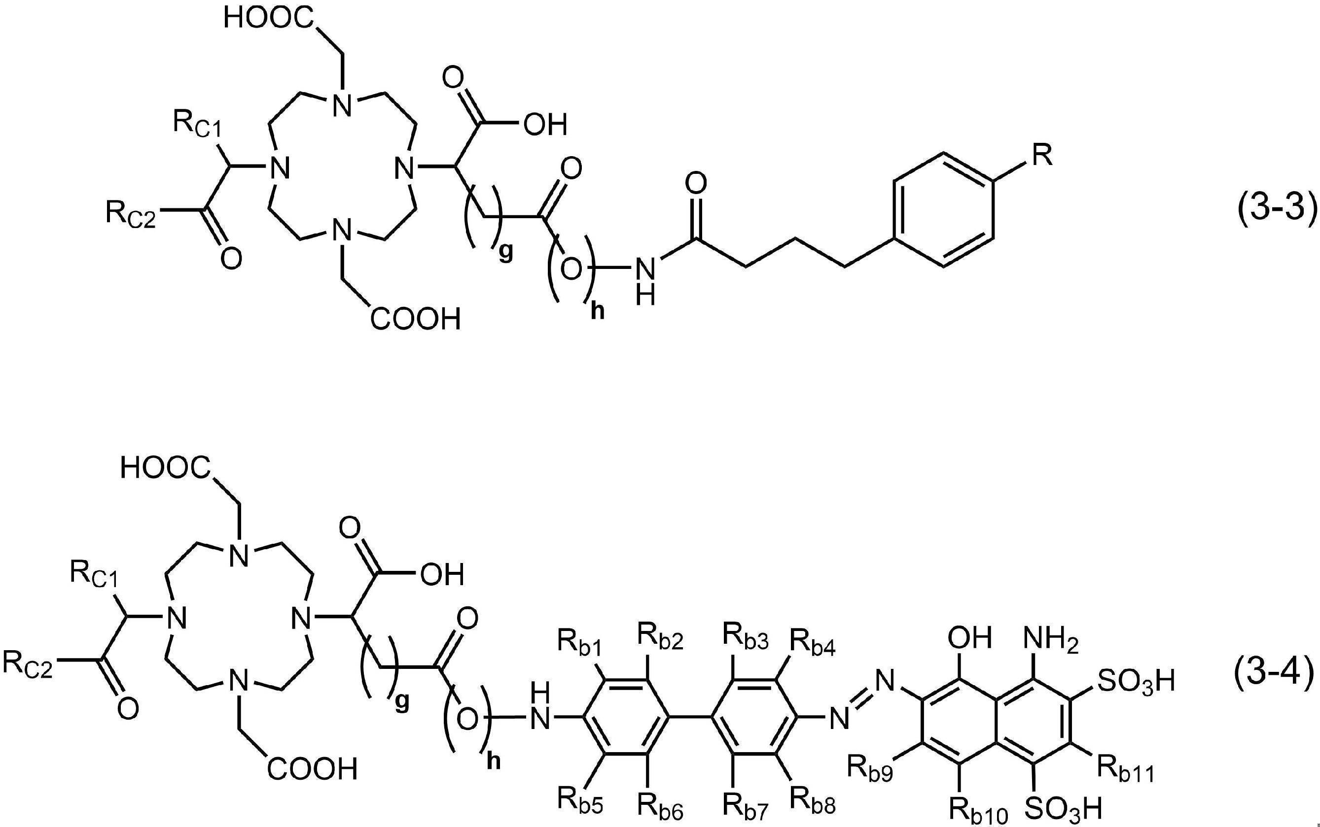
このような構造を有していることによって、放射性金属を配位可能なヘテロ原子を構造中に十分に有したものとなるので、本発明の化合物に放射性金属を配位させる際に錯体形成効率を高めることができる。これに加えて、アルブミン結合部及びPSMA分子との結合部における分子の動きの自由度が高まるので、アルブミンへの親和性とPSMA分子の親和性とを高いレベルで両立させることができる。更に、肝臓や腎臓などの正常組織への意図しない集積が低減されたものとなる。また錯体形成に要する製造時間が少なくなり、放射性標識化合物の製造効率及び放射化学的収率が更に向上する。

式(2S)中、Lcは、リンカー構造である。
式(2S)中、Rは、水素原子、ハロゲン原子又は炭素数1以上5以下のアルキル基である。
式(2S)中、a及びbは、それぞれ独立して、1以上7以下の整数である。
式(2S)中、A2がOctapaであるとき、Lcはポリエチレングリコール構造を含む。
また必要に応じて、以後の工程において、放射性標識化合物に、水を主体とする溶媒及び薬学的に許容される他の成分を加えて、放射性標識化合物を有効成分として含有する放射性医薬組成物とすることもできる。放射性医薬組成物は、例えば上述の方法で製造された放射性標識化合物を、水を主体とし、且つ生体と略等張の溶媒に溶解させて製造することができる。放射性医薬組成物は、経口的に、又は静脈内、皮下、腹腔内若しくは筋肉内等の非経口的に生体に投与され、疾患の治療、疾患の診断、あるいは病巣の検出等に用いられる。
これらのうち、商業利用可能であり且つ錯体形成性の向上を図る観点から、放射性金属の核種として、44Sc、51Cr、57Co、58Co、60Co、59Fe、64Cu、67Cu、67Ga、68Ga、89Sr、89Zr、90Y、99mTc、103Ru、111In、153Sm、165Dy、166Ho、177Lu、186Re、188Re、197Hg、198Au、201Tl、203Hg、212Pb、212Bi、213Bi、225Ac又は227Thを用いることが好ましい。これらの放射性金属は、常法に従って製造することができる。これらの放射性核種は、放射性金属が電離状態で含有される溶液として得ることが好ましい。
β-線放出核種は、放射性金属の壊変過程でβ-線を放出する核種であればよく、詳細には、59Fe、60Co、64Cu、67Cu、89Sr、90Y、99mTc、103Ru、153Sm、165Dy、166Ho、177Lu、186Re、188Re、198Au、203Hg、212Pb、212Bi又213Biは等が好ましく用いられ、より好ましくは64Cu、67Cu、89Sr、90Y、177Lu、186Re又は188Reが用いられ、更に好ましくは90Yが用いられる。
電子捕獲壊変核種は、放射性金属の壊変過程でオージェ電子又は特性X線を放出する核種であればよく、51Cr、57Co、58Co、64Cu、67Ga、68Ga、89Zr、111In、186Re、197Hg又は201Tl等が好ましく用いられる。
γ線放出核種は、γ崩壊によってγ線を放出する核種であればよく、γ崩壊によってγ線を放出する核種としては、68Ga、99mTc又は201Tlが好ましく用いられる。
また、放射性標識化合物を疾患の診断や病巣の検出を目的として用いる場合に、放射性金属として89Zrを用いる場合は、本発明の化合物として、好ましくは前記式(A1)、(A3)又は(A4)のいずれかで示される構造のキレート部を有する化合物を用い、更に好ましくは前記式(A1)で示される構造のキレート部を有する化合物を用いる。また、放射性金属として68Ga又は111Inを用いる場合は、本発明の化合物として、好ましくは前記式(A1)~(A4)又は(A9)のいずれかで示される構造のキレート部を有する化合物を用い、更に好ましくは前記式(A1)で示される構造のキレート部を有する化合物を用いる。
同様に、キレート部とPSMA分子との結合部を含む原子団との結合は、キレート部とPSMA分子との結合部とが後述するリンカー構造を介さずに直接的に結合していてもよく、あるいは、キレート部とアルブミン結合部を含む原子団とが後述するリンカー構造を介して間接的に結合していてもよい。
上述した「直接的に」あるいは「間接的に」のいずれの態様であっても、アミド結合を介して結合していることが、合成の容易性と化学構造の安定性とを両立する観点から好ましい。
上述のリンカー構造としてアミノ酸等に由来する構造を含む場合、例えば、生体内での動態を制御する目的で、国際公開第2017/150549号、国際公開第2019/065774号、国際公開第2019/221269号、国際公開第2020/075746号、国際公開第2020/145227号、国際公開第2020/145228号等に記載のペプチドリンカーを用いることができる。

以下、キレート部とPSMA分子との結合部の結合にクリック反応を用いる場合を例に説明する。この場合、キレート部及びPSMA分子との結合部がクリック反応可能な原子団をそれぞれ有しており、これらの原子団が互いに反応して、キレート部と、PSMA分子との結合部と、を互いに結合できるようになっている。つまり、キレート部が備える第1原子団と、PSMA分子との結合部が備える第2原子団との間で実行されるものである。


以下の実施例において、特に断りのない限り、NMRはいずれも日本電子株式会社のJNM-AL400 FT-NMR装置にて、内部標準物質としてテトラメチルシラン(TMS)を用い、TMS共鳴を0.00ppmに設定した。すべての化学シフトはデルタスケール(δ)上のppmであり、そしてシグナルの微細分裂については、略号(s:シングレット、d:ダブレット、t:トリプレット、m:マルチプレット、br:ブロード)を用いて示した。
また質量分析においては、MSを行う場合はLCMS 2020(株式会社島津製作所製)、HRMSを行う場合LCMS-IT-TOF(株式会社島津製作所製)を用いて行った。
本実施例では、PSMAを標的分子とする二種類の化合物(PSMA-DA1及びPSMA-DB)を合成した。次いで、各化合物に放射性金属として111Inイオン、90Yイオン又は225Acイオンをそれぞれ配位させた放射性標識化合物を得た。以下に詳細を示す。
実施例1-1~1-4にて用いたPSMA-DA1は、上記一般式(1)にて示すように、PSMA分子との結合部及びアルブミン結合部を、キレート部を介して直線状に含む化学構造を有するものである。PSMA-DA1は、キレート部とPSMA分子との結合部とがアミド結合によって直接的に結合しており、且つ、キレート部とアルブミン結合部を含む原子団とは、リシン由来のリンカー構造を介して間接的に結合している。
比較例1-1~1-3で用いたPSMA-DBは構造中にキレート部及びPSMA分子との結合部を含むが、アルブミン結合部を含まないものである。
実施例1-1~1-4における合成経路の概略を合成経路(V-1)及び(V-2)とし以下に示す。


化合物1は、Chem Commun.2008,28,3248-3250の方法に従って、1,4,7,10-テトラアザシクロドデカンから3段階で合成した。この化合物1(20mg,0.026mmol)をN,N-ジメチルホルムアミド(DMF)(2mL)に溶解させ、メチル-N6-(4-(4-ヨードフェニル)ブタノイル)-L-リジネート(11mg,0.0254mmol)、1-エチル-3-(3-ジメチルアミノプロピル)-カルボジイミド(EDC)塩酸塩(7.0mg,0.037mmol)、1-ヒドロキシ-7-アザベンゾトリアゾール(HOAt)(5.0mg,0.037mmol)、およびトリエチルアミン(5μL,0.036mmol)を加え,室温で24時間撹拌した。その後,(S)-di-tert-butyl 2-(3-((S)-6-amino-1-tert-butoxy-1-oxohexan-2-yl)ureido)pentanedioate(13mg,0.0267mmol)・EDC塩酸塩(7.0mg,0.037mmol),HOAt(5.0mg,0.037mmol),およびトリエチルアミン(5μL,0.036mmol)を加え,室温で72時間撹拌した。溶媒を除去した後,残渣に6規定塩酸(3mL)を加え,40℃で24時間撹拌後,逆相HPLCで以下の条件で精製し、目的とする化合物(PSMA-DA1)を得た。収量及びMSは以下のとおりであった。
収量:1.0mg(収率:3%、化合物1の物質量に対する得られたPSMA-DA1の物質量から算出した)。
MS(ESI):m/z1250.5[M+H]+.
酢酸緩衝液(0.1M,pH5.5,200μL)に、[111In]InCl3溶液(3.7MBq,100μL)と、PSMA-DA1のDMSO溶液(1mM,10μL)とを加え,90℃で30分間静置した.その後,反応液を逆相HPLCによって以下の条件で精製し、目的とする放射性標識化合物([111In]In-PSMA-DA1)を得た。
得られた放射性標識化合物の放射能をキュリーメータで測定し、反応に用いた[111In]InCl3溶液の放射能に対する百分率を放射化学的収率(%)とした。
また、放射性標識化合物のHPLC分取液の一部を精製条件と同じHPLC条件で分析し、検出された全てのピークの面積値に対する放射性標識化合物の面積値の百分率を放射化学的純度(%)とした。
その結果、放射化学的収率は61~90%,放射化学的純度は95%以上であった。
PSMA-DA1(1mg)と塩化インジウム(III)無水和物(2mg)をジメチルスルホキシド(DMSO)(100μL)に溶解させ、2-(N-morpholino)ethanesulfonic acid緩衝液(0.1M,pH5.6,900μL)を加えた。反応液を60℃で12時間撹拌後、溶液を逆相HPLCによって以下の方法で精製し、以下のMSを有する化合物を得た。
精製条件:Cosmosil 5C18-AR-II column(10×250mm),移動相:MeCN/H2O/TFA[10/90/0.1(0分)~100/0/0.1(90分)],流速:4mL/min.
MS(ESI):m/z1362.4[M+H]+.
酢酸緩衝液(0.1M,pH5.5,200μL)に[90Y]YCl3溶液(65-118MBq,10μL)とPSMA-DA1のDMSO溶液(1mM,10μL)とを加え,90℃で30分間静置した.その後,反応液を逆相HPLCによって以下の条件で精製し、目的とする放射性標識化合物([90Y]Y-PSMA-DA1)を得た。
放射化学的収率は実施例1-1と同様の方法で測定した。放射化学的純度の分析に用いたHPLC条件は以下に示す、90Y標識での精製条件を用いた。
その結果、放射化学的収率は49~79%,放射化学的純度は95%以上であった。
[225Ac]AcCl3の0.2M塩酸溶液(1.5MBq,10μL)に0.1M酢酸-酢酸アンモニウム緩衝液(pH5.5,170μL)とPSMA-DA1のDMSO溶液(2.0mM,10μL)を加え,70℃で1時間静置した.反応液にH2O(800μL)を加え,Oasis HLB Lightカラムに通液した.カラムにH2O(10mL)を通液後,70%EtOH(0.5mL)を通液し,精製溶液を得た。精製溶液を加熱留去にて乾固後、5%エタノール含有158mM酢酸-酢酸ナトリウム緩衝液(pH6.5)を加え、目的とする放射性標識化合物([225Ac]Ac-PSMA-DA1)の投与溶液を得た。
放射化学的収率は次の方法で測定した。得られた放射性標識化合物の放射能をガンマ線スペクトルメータで測定し、反応に用いた[225Ac]AcCl3溶液の放射能に対する百分率を放射化学的収率(%)とした。
得られた放射性標識化合物の放射化学的純度を次の方法で測定した。すなわち放射性標識化合物の溶液の一部をTLC(iTLC-SG、移動相:MeCN/H2O=1:1混液)で分析し、検出された全てのピークの面積値に対する放射性標識化合物の面積値の百分率を放射化学的純度(%)とした。
その結果、放射化学的収率は49%,放射化学的純度は87%であった。
TypeI Plusバイアル(2R)(SCHOTT社製)に[89Zr]Zr(Ox)2の1.0M塩酸溶液(2.2MBq、10μL)を分注し、110℃に加熱し、約40分間Ar気流下で溶媒留去した。バイアルに0.1mоl/L塩酸(100μL)と300mMゲンチジン酸-0.78M酢酸-酢酸ナトリウム緩衝液(pH5.5、50μL)とPSMA―DA1-DMSO溶液(2mM、75μL)とDMSO(75μL)を加え、70℃にて1時間静置し、目的とする放射性標識化合物([89Zr]Zr-PSMA-DA1)を得た。
得られた放射性標識化合物の放射化学的純度は実施例1-3と同様の方法で測定した。その結果、放射化学的純度は96%であった。
比較例1-1~2-3における合成経路の概略を合成経路(VI-1)及び(VI-2)として以下に示す。
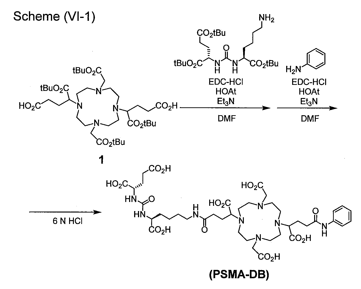

実施例1-1~1-4と同様の方法で合成した化合物1(35mg,0.045mmol)をDMF(2mL)に溶解させ,(S)-di-tert-butyl 2-(3-((S)-6-amino-1-tert-butoxy-1-oxohexan-2-yl)ureido)pentanedioate(22mg,0.045mmol),EDC塩酸塩(10mg,0.052mmol),HOAt(7.0mg,0.051mmol),トリエチルアミン(7μL,0.050mmol)を加え,室温で24時間撹拌した.その後,アニリン(5μL,0.055mmol),EDC塩酸塩(10mg,0.052mmol),HOAt(7.0mg,0.051mmol),トリエチルアミン(7μL,0.050mmol)を加え,室温で24時間撹拌した.溶媒を除去した後,残渣にTFA(1.8mL),トリイソプロピルシラン(100μL),およびH2O(100μL)を加え,室温で24時間撹拌した。溶媒を除去した後,残渣を逆相HPLCで以下の条件で精製し、目的とする化合物(PSMA-DB)を得た。収量及びMSは以下のとおりであった。
収量:5.0mg(収率:12%、化合物1の物質量に対する得られたPSMA-DBの物質量から算出した).
MS(ESI)m/z925.3[M+H]+.
PSMA-DA1に代えて、PSMA-DBを用いた以外は、実施例1-1と同様の方法によって、目的とする放射性標識化合物([111In]In-PSMA-DB)を得た。
放射化学的収率および放射化学的純度は実施例1-1に記載した方法と同様に測定した。精製条件および放射化学的純度の測定に用いたHPLC条件は以下に示す条件を用いた。
その結果、放射化学的収率は61~90%,放射化学的純度は95%以上であった。
PSMA-DB(1等量)のH2O/MeCN/TFA(49.95/49.95/0.1,300μL)溶液に塩化インジウム(III)無水和物(10等量)を加えた.室温で18時間撹拌後,溶液を逆相HPLCで精製した。
精製条件:Cosmosil 5C18-AR-II column(4.6×150mm),移動相:MeCN/H2O/TFA[5/95/0.1(0~10分),5/95/0.1(10分)~35/65/0.1(40分)],流速:1mL/min.
PSMA-DA1に代えて、PSMA-DBを用いた以外は、実施例1-2と同様の方法によって、目的とする放射性標識化合物([90Y]Y-PSMA-DB)を得た。
放射化学的収率及び放射化学的純度は実施例1-1に記載した方法と同様に測定した。放射化学的純度の測定は、上述したPSMA-DBを非放射性のInに配位させた化合物の精製条件にて示したHPLC条件で実施した。
その結果、放射化学的収率は49~79%,放射化学的純度は95%以上であった。
PSMA-DA1に代えて、PSMA-DBを用いた以外は、実施例1-3と同様の方法によって、目的とする放射性標識化合物([225Ac]Ac-PSMA-DB)を得た。
放射化学的収率及び放射化学的純度は実施例1-3に記載した方法と同様に測定した。
その結果、放射化学的収率は48%,放射化学的純度は83%であった。
マウス血漿(200μL)に[111In]In-PSMA-DA1(370kBq)または[90Y]Y-PSMA-DA1(3.7MBq)の生理食塩水(20μL)溶液を加え,37℃で24時間静置した(n=3)。その後、MeCN(200μL)を加え,10000×gで5分間遠心分離した.上清をろ過し,ろ液を逆相HPLCで以下の条件にて分析した。
(分析条件:Cosmosil 5C18-PAQ column(4.6×250mm),移動相:MeCN/H2O/TFA[20/80/0.1(0分)~50/50/0.1(30分)],流速:1mL/min.)
その結果、いずれの標識化合物もマウス血漿中37℃で24時間静置後も95%以上が安定して存在した。
LNCaP細胞(PSMA陽性,ヒト前立腺がん)およびPC-3細胞(PSMA陰性,ヒト前立腺がん)を用いた。これらの細胞はそれぞれAmerican Type Culture Collection社およびDS Biomedical社より購入した。各細胞は抗生物質(ペニシリンおよびストレプトマイシン)と非働化ウシ胎児血清10%を含むナカライテスク株式会社製RPMI1640中,37℃,5%CO2下で培養した。
培養培地を除去し,[111In]In-PSMA-DA1または[111In]In-PSMA-DB(37kBq)を含むアッセイ用培地(0.5%FBS含有RPMI1640培地)溶液(1mL)を加えた。その後,プレートを37℃,5%CO2下で1時間静置した。
阻害実験では,培養培地を除去後,[111In]In-PSMA-DA1または[111In]In-PSMA-DB(37kBq)と2-(phosphonomethyl)pentanedioic acid(2-PMPA)(PSMA阻害剤、終濃度100μM)を含むアッセイ用培地(1mL)溶液を加えた.その後,プレートを37℃,5%CO2下で1時間静置した。
アッセイ用培地を除去後,各ウェルを放射性標識化合物及び2-PMPAを含まないアッセイ用培地(1mL)で洗浄し,1規定水酸化ナトリウム水溶液(200μL×2)で細胞を溶解させた。
アッセイ用培地及び細胞溶解液の各放射能をガンマカウンターで測定した。これとは別に、細胞溶解液中の総タンパク質濃度をThermo Fisher Scientific社製BCA Protein Assay Kitを用いて算出した。添加放射能量に対するサンプルの放射能量の百分率(%ID)を、総タンパク質量で除した値(%ID/mg protein)をサンプルごとに算出した。
データは平均値±標準偏差で表した.有意差検定はStudent’s t-testおよびone-way analysis of variance (ANOVA) test with Dunnet’s post-hoc testを用いて行い,p<0.05を有意差ありとした。
[111In]In-PSMA-DA1および[111In]In-PSMA-DBはPC-3細胞と比較してLNCaP細胞に対する高い結合性を示し,その結合は過剰量のPSMA阻害剤(2-PMPA)の添加によって有意に低減した.これらの結果から,[111In]In-PSMA-DA1および[111In]In-PSMA-DBはPSMA高発現細胞に特異的に結合することが示された。
[111In]In-PSMA-DA1または[111In]In-PSMA-DBのPBS溶液(37kBq,50μL)を、200μLのPBS,マウス血漿,ヒト血漿,またはヒト血清アルブミン(HSA)のPBS溶液(45mg/mL)にそれぞれ加え,37℃で10分間静置した。その後,反応液をスピンカラム(サイティバ社製Sephadex G-100)に添加し,1500×g,4℃で2分間遠心分離した.分離後,カラム及び溶出液の放射能をそれぞれガンマカウンターで測定した。
データは平均値±標準偏差で表した。有意差検定はStudent’s t-testおよびone-way analysis of variance (ANOVA) test with Dunnet’s post-hoc testを用いて行い,p<0.05を有意差ありとした。
評価対象となる化合物がアルブミンに結合し複合体を形成すると,分子サイズの増加によってカラムを通過するが,アルブミンに結合しなかった際には,カラム内のゲルに保持される。
[111In]In-PSMA-DA1および[111In]In-PSMA-DBをPBS中で静置後,カラムに付したところ,溶出液において顕著な放射能は観測されなかった.一方,マウス血漿,ヒト血漿,およびHSA溶液中で静置した際には,[111In]In-PSMA-DBと比較して[111In]In-PSMA-DA1の溶出液における放射能は有意に高く,[111In]In-PSMA-DA1が血漿アルブミンに結合することが示された。
動物実験は京都大学動物実験委員会の指針を遵守して行った。雄性CB17/IcrJcl-Prkdcscidマウスは日本クレア株式会社より購入した。動物は12時間/12時間の昼夜サイクル条件下で飼育し,飼料と水は自由に与えた。PBSとCorning Life Sciences社製Matrigelの混合液(1:1,150μL)にLNCaP細胞(1.0×10⁷cells/mouse)またはPC-3細胞(1.0×107cells/mouse)を懸濁させ,イソフルラン麻酔下,マウスの右肩に皮下移植した。その後、当該マウスを40~60日間飼育した。
LNCaPまたはPC-3腫瘍移植マウスに、[111In]In-PSMA-DA1、または[111In]In-PSMA-DBの生理食塩水溶液(185kBq,100μL)を尾静脈より投与した(各n=3)。投与1,4,24,48,96,および192時間後にマウスを安楽死させた。その後,血液及び各臓器を回収し,臓器の質量及び放射能を測定した。
投与放射能量(injected dose)に対する放射能量の百分率(%ID)を血液質量又は臓器質量(g)で除した値(%ID/g)として示す。%ID/gの値が高いほど、放射性標識化合物の存在量が多く、該化合物の標的臓器への集積が高いことを示す。
LNCaP腫瘍移植マウス及び[111In]In-PSMA-DBにおける結果(平均±標準偏差、各n=3)を以下の表2及び図3に示す。
[111In]In-PSMA-DA1はLNCaP腫瘍への高い集積を示した(投与1~24時間後に9.41~12.6%ID/g)。また,血液での滞留性を示し(投与24時間後に14.0%ID/g),腫瘍/腎臓比は投与48時間後以降で1を上回った。一方,[111In]In-PSMA-DBはいずれのタイムポイントにおいても,[111In]In-PSMA-DA1より低い腫瘍集積が認められるとともに、低い腫瘍/腎臓比が示された。
これらの結果から,[111In]In-PSMA-DA1はPSMA高発現腫瘍に対する高い集積性を有するとともに、[111In]In-PSMA-DBと比較して優れた体内分布を有することが明らかとなった.

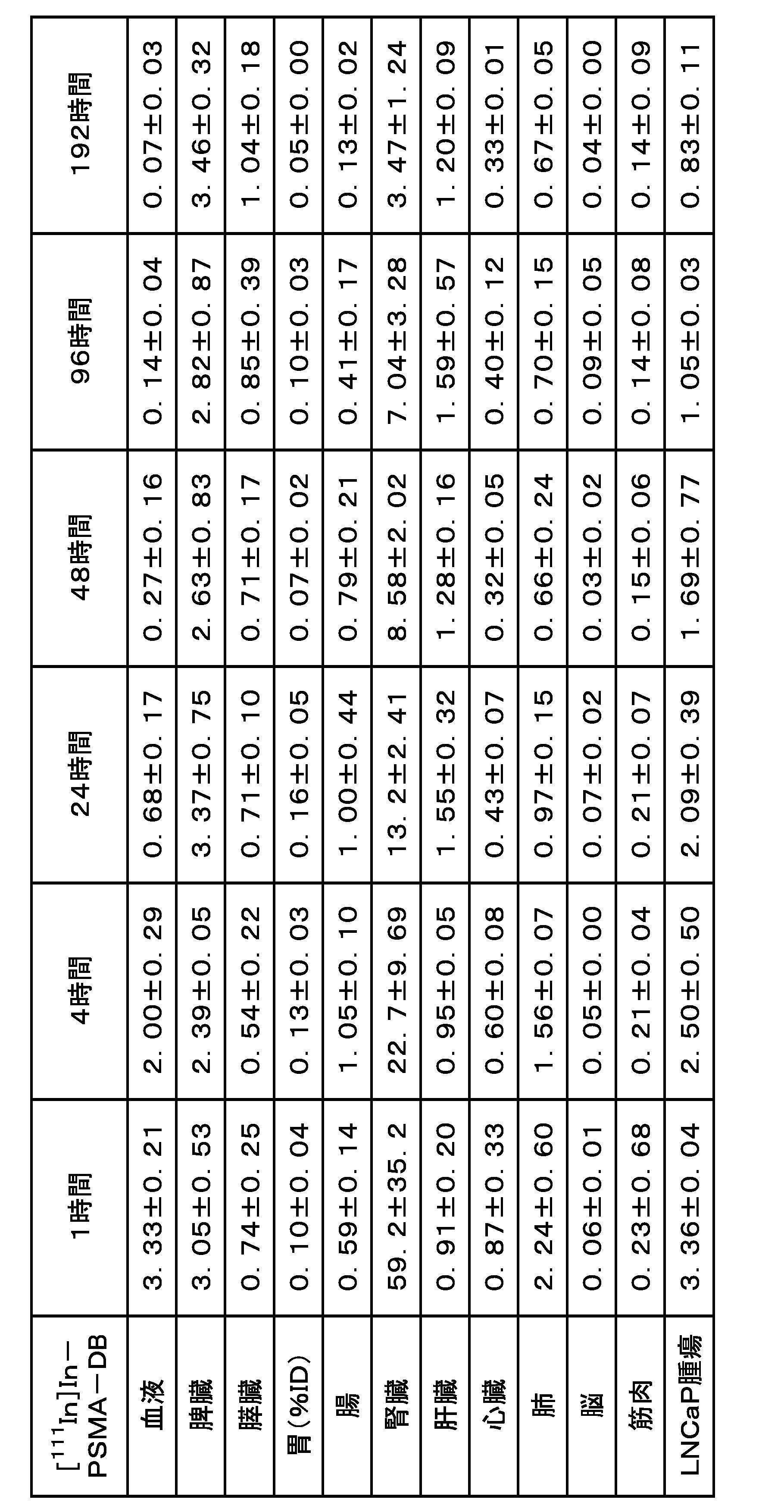
PC-3腫瘍移植マウス及び[111In]In-PSMA-DBにおける結果(平均±標準偏差、各n=3)を以下の表4に示す。
[111In]In-PSMA-DA1および[111In]In-PSMA-DBともに,PC-3腫瘍への集積は同タイムポイントにおけるLNCaP腫瘍への集積の結果よりも低いものであり、両放射性標識化合物がPSMA陽性腫瘍へ選択的に集積することが示された。

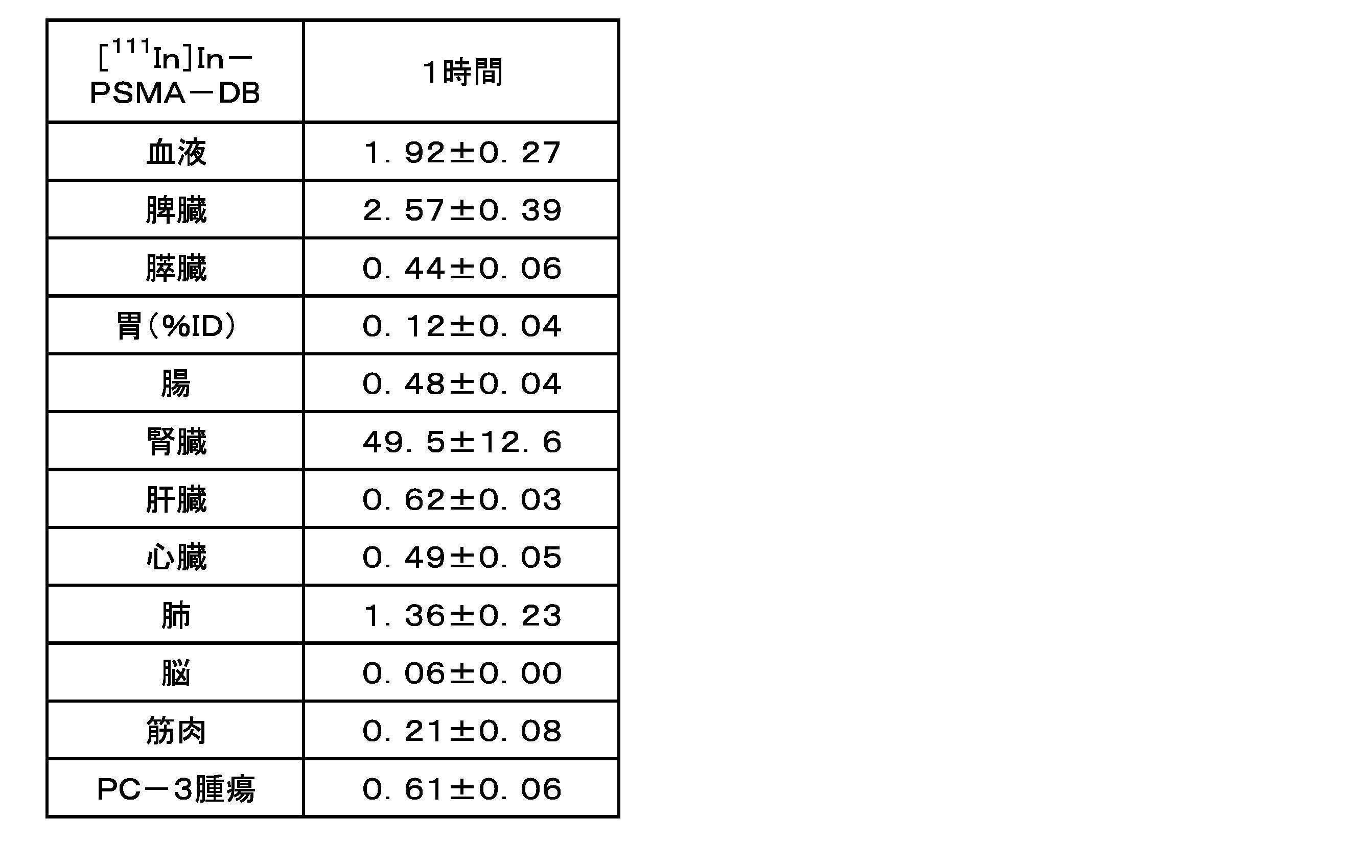
上述の方法で作製したLNCaP腫瘍移植マウスに、[111In]In-PSMA-DA1または[111In]In-PSMA-DBの生理食塩水溶液(1.9~3.0MBq,150μL)を尾静脈から投与した。投与24および48時間後にGamma Medica-Ideas社製FX3300 pre-clinical imaging systemにてSPECT/CTを行った。イソフルラン麻酔下,回転半径35mm,投影時間70秒,投影回数32回にて直径1.0mmのピンホールコリメーターを用いて撮像した。SPECT後,CT(管電圧:60kV,管電流:350μA)を行った。SPECTの投影データについて3次元ordered subset expectation maximization法(8subsets,5iterations)による画像再構成を行った。
[111In]In-PSMA-DA1を用いたSPECT/CT撮像では,投与24および48時間後においてLNCaP腫瘍(図中矢印)に顕著な放射能集積が認められた。腎臓(図中円)にも高い放射能集積が認められたが,投与48時間後には腫瘍と同程度な放射能シグナルであった。一方,[111In]In-PSMA-DBを用いたSPECT/CT撮像においてもLNCaP腫瘍(図中矢印)に放射能集積が認められたが,その集積は腎臓(図中円)と比較して低かった。
これらの結果から,[111In]In-PSMA-DA1はPSMA高発現腫瘍をSPECTによって明瞭に描出することが可能であるとともに,[111In]In-PSMA-DBよりも優れていることが示された。
上述の方法で得られた[90Y]Y-PSMA-DA1(3.7MBq)または[90Y]Y-PSMA-DB(3.7MBq)の生理食塩水溶液(100μL)をLNCaP腫瘍移植マウスに尾静脈から投与した(n=7)。対照群として,100μLの生理食塩水をLNCaP腫瘍移植マウスに尾静脈から投与した(n=7)。
90Y標識化合物を投与後,腫瘍容積及び体重を1週間あたり3回測定した。腫瘍容積は「(腫瘍容積)=[(長辺)×(短辺)2/2]」の算出式に基づいて算出した。
90Y標識化合物の投与開始日における腫瘍容積は[90Y]Y-PSMA-DA1,[90Y]Y-PSMA-DB,および生理食塩水投与群において,それぞれ63.1±8.7,66.1±30.7,および66.7±25.7mm3であった。
LNCaP腫瘍移植マウスに[90Y]Y-PSMA-DA1および[90Y]Y-PSMA-DBを投与したところ,生理食塩水投与群と比較して,それぞれ投与9日後および33日後以降に腫瘍容積に有意な差が認められた。[90Y]Y-PSMA-DBと比較して、[90Y]Y-PSMA-DA1は腫瘍成長を抑制する傾向が観察された。[90Y]Y-PSMA-DA1の投与によってマウスの体重がわずかに減少したが,その後,生理食塩水投与群と同等な値まで回復した。
上述の方法で得られた[225Ac]Ac-PSMA-DA1(20kBq)または[225Ac]Ac-PSMA-DB(20kBq)を5%エタノール含有酢酸緩衝液(158mM,pH6.5,100μL)に溶解させ,LNCaP腫瘍移植マウスに尾静脈より投与した(n=6または5).対照群として,100μLの5%エタノール含有酢酸緩衝液(158mM,pH6.5)をLNCaP腫瘍移植マウスに尾静脈より投与した(n=4)。
225Ac標識化合物を投与後,腫瘍容積及び体重を1週間あたり2回測定した。225Ac標識化合物投与開始日における腫瘍容積は[225Ac]Ac-PSMA-DA1,[225Ac]Ac-PSMA-DB,および5%エタノール含有酢酸緩衝液投与群において,それぞれ75.3±26.0,80.6±20.8,および91.1±15.9mm3であった。
LNCaP腫瘍移植マウスに[225Ac]Ac-PSMA-DA1および[225Ac]Ac-PSMA-DBを投与したところ,生理食塩水投与群と比較して腫瘍成長が顕著に抑制された。特に[225Ac]Ac-PSMA-DA1投与群では,腫瘍はほとんど成長せず,投与後6週間まで継続的な成長抑制が認められた。
[225Ac]Ac-PSMA-DA1の投与の影響と思われる体重の減少は一過性のものであった。[225Ac]Ac-PSMA-DBの投与の影響と思われる体重の減少は一時的に加速したが,その後回復する様子は認められなかった。
本実施例では、PSMAを標的分子とする(((S)-5-amino-1-carboxypentyl)carbamoyl)-L-glutamic acid(上記式(C1)中、aが2、bが4である構造)をPSMA分子との結合部として構造中に含み、キレート部とPSMA分子との結合部との結合に、アジド基とジベンジルシクロオクチン(DBCO)とのクリック反応を用いた化合物(PtDA)を合成した。次いで、これらの化合物に放射性金属として111Inイオンを配位させた放射性標識化合物をそれぞれ得た。合成経路の概略を合成経路(VIII-1)~(VIII-4)として以下に示す。
実施例2で用いた化合物は、構造中にキレート部、PSMA分子との結合部及びアルブミン結合部を含むものである。111Inイオンに配位させるにあたり2つの合成経路(合成経路(VIII-4)参照)があるが、いずれの経路であっても、得られる放射性標識化合物は同一である。
PtDAは、キレート部とPSMA分子との結合部とが、クリック反応由来の化学構造とポリエチレングリコール基とを介して間接的に結合しており、且つ、キレート部とアルブミン結合部を含む原子団とは、リシン由来のリンカー構造を介して間接的に結合している。

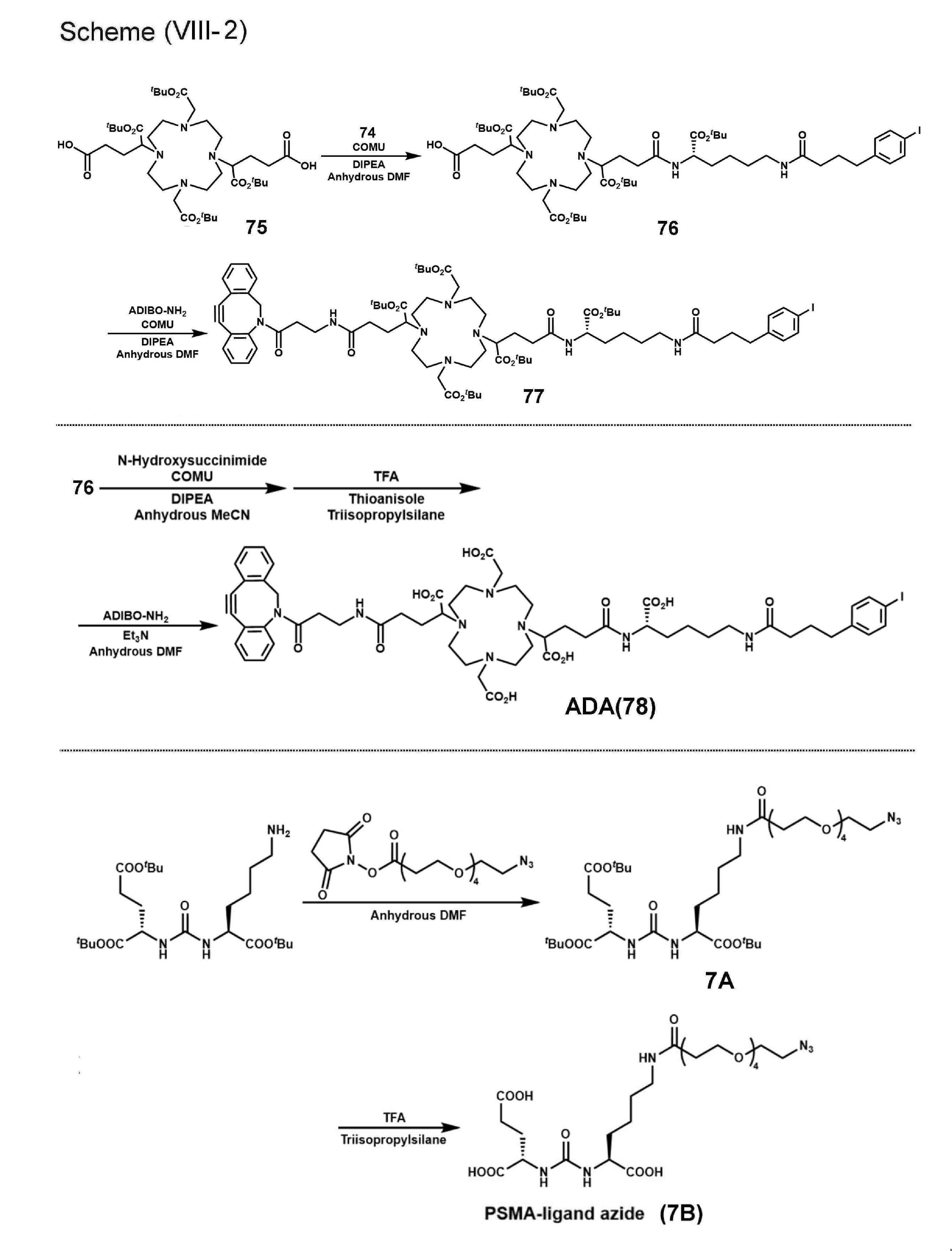


N2-[(9H-fluoren-9-ylmethoxy)carbonyl]-N6-[(4-methylphenyl)diphenylmethyl]-L-lysine(1000mg,1.6mmol)をジクロロメタン(10mL)に溶解させ,tert-butyl trichloroacetimidate(699.5mg,3.2mmol)とBF3・OEt2(25μL)を加えた.反応溶液を室温で42時間撹拌した.溶媒を除去した後,残渣をH2O(100mL)で洗い,酢酸エチル/ヘキサン(1/5,100mL×2)で抽出した.有機層を硫化ナトリウムで乾燥させ,ろ過した.ろ液を減圧留去した後,残渣を中圧カラムクロマトグラフィーによって精製した(酢酸エチル/ヘキサン)。収量、NMRスペクトル及びMSは以下のとおりであった。
1H-NMR(400MHz,CDCl3)δ7.69(d,J=7.3Hz,2H),7.56(d,J=7.8Hz,2H),7.45(d,J=7.8Hz,4H),7.33(d,J=8.2Hz,4H),7.23(m,6H),7.13(m,2H),7.04(d,J=7.8Hz,2H),5.39(d,J=8.2Hz,1H),4.35(d,J=6.9Hz,2H),4.25(m,1H),4.18(t,J=6.9Hz,1H),2.26(s,3H),2.12(m,2H),1.77(m,1H),1.59(m,1H),1.49(m,4H),1.44(s,9H).13C-NMR(100MHz,CDCl3)δ171.6,155.7,146.3(2C),143.7(2C),143.2,141.1(2C),135.4,128.4-128.3(8C),127.6-127.5(6C),126.9(2C),126.0(2C),125.0(2C),119.8(2C),81.7,70.5,66.7,60.2,47.1,43.2,32.7,30.4,27.8(3C),22.8,20.7.
HRMS(ESI):m/z681.3677[M+H]+.
化合物71(569mg,0.84mmol)をトリフルオロ酢酸(TFA)(100μL),トリイソプロピルシラン(250μL)、ジクロロメタン(4.65mL)の混合液に溶解させ,室温で6時間撹拌した.溶媒を除去した後,残渣を中圧カラムクロマトグラフィーによって精製した(メタノール/クロロホルム)。収量、NMRスペクトル及びMSは以下のとおりであった。
1H-NMR(400MHz,CDCl3)δ7.71(d,J=7.6Hz,2H),7.56(d,J=7.1Hz,2H),7.35(t,J=7.6Hz,2H),7.27(t,J=7.6Hz,2H),5.72(d,J=8.0Hz,1H),4.33(d,J=7.1Hz,2H),4.16(t,J=6.6Hz,2H),3.38(s,2H),1.77-1.56(m,4H),1.43(s,9H),1.27-1.13(m,2H).13C-NMR(100MHz,CDCl3)δ171.5,156.2,143.6(2C),141.1(2C),127.6(2C),127.0(2C),125.0(2C),119.8(2C),82.3,66.9,54.1,50.0,46.9,39.5,31.1,27.7(3C),26.8,22.1.
HRMS(ESI):m/z425.2437[M+H]+.
4-(4-ヨードフェニル)ブタン酸(364mg,1.25mmol)をN,N-ジメチルホルムアミド(DMF)(3mL)に溶解させ,1-エチル-3-(3-ジメチルアミノプロピル)-カルボジイミド(EDC)塩酸塩(320mg,1.7mmol),1-ヒドロキシ-7-アザベンゾトリアゾール(HOAt)(228mg,1.7mmol)を加え0℃で15分間攪拌した.その後,化合物72(355mg,0.84mmol)とトリエチルアミン(169mg,1.7mmol)を加え,0℃で攪拌した.15時間後,反応溶液をH2O(100mL)で洗い,酢酸エチル/ヘキサン(1/5,100mL×2)で抽出した.有機層を硫化ナトリウムで乾燥させ,ろ過した.ろ液を減圧留去した後,残渣を中圧カラムクロマトグラフィーによって精製した(酢酸エチル/ヘキサン)。収量、NMRスペクトル及びMSは以下のとおりであった。
1H-NMR(400MHz,CDCl3)δ7.73(d,J=7.3Hz,2H),7.60-7.50(m,6H),7.37(t,J=7.3Hz,2H),7.28(t,J=7.3Hz,2H),6.83(d,J=8.2Hz,2H),4.38-4.27(m,2H),4.25-4.16(m,2H),3.21-3.14(m,2H),2.48(m,2H),2.08(t,J=7.3Hz,2H),1.91-1.84(m,2H),1.73-1.57(m,2H),1.47-1.29(m,13H).13C-NMR(100MHz,CDCl3)δ172.8,171.5,156.0,143.5(2C),141.0-140.9(3C),137.1(2C),130.3(2C),127.5(2C),126.9(2C),124.9(2C),119.8(2C),90.8,82.0,66.8,53.9,46.9,38.9,35.4,34.4,32.1,28.6,27.9(3C),26.7,22.2.
HRMS(ESI):m/z697.2130[M+H]+.
化合物73のDMF(4mL)溶液にピペリジン(1mL)を加えた.室温で2時間撹拌後,溶液をH2O(100mL)で洗い,酢酸エチル/ヘキサン(1/5,100mL×2)で抽出した.有機層を硫化ナトリウムで乾燥させ,ろ過した.ろ液を減圧留去した後,残渣を中圧カラムクロマトグラフィーによって精製した(メタノール/クロロホルム)。収量、NMRスペクトル及びMSは以下のとおりであった。
1H-NMR(400MHz,CDCl3)δ7.58(d,J=8.2Hz,2H),6.92(d,J=8.2Hz,2H),3.31-3.27(m,1H),3.23(m,2H),2.58(m,2H),2.13(m,2H),1.96-1.88(m,2H),1.74-1.65(m,2H),1.56-1.48(m,2H),1.44(s,9H),1.43-1.40(m,2H).13C-NMR(100MHz,CDCl3)δ175.2,172.3,141.1,137.3(2C),130.5(2C),90.9,80.9,54.7,39.1,35.6,34.6,34.3,29.2,28.0(3C),26.8,22.9.
HRMS(ESI):m/z475.1453[M+H]+.
上述の実施例と同様に、1,4,7,10-テトラアザシクロドデカンから3段階で合成した(Chem Commun.2008,28,3248-3250)。
化合物75(317mg,0.41mmol)のDMF(2mL)溶液にN-[1-(cyano-2-ethoxy-2-oxoethylideneaminooxy)dimethylamino(morpholino)]uronium hexafluorophosphate(COMU)(176mg,0.41mmol)を加え,0℃で15分間撹拌した.反応液にN,N-ジイソプロピルエチルアミン(DIPEA)(53mg,0.41mmol)を加え,0℃でさらに15分間撹拌した後,化合物74(163mg,0.34mmol)を加えた.室温で12時間撹拌後,溶液を逆相HPLCで以下の条件で精製した。収量及びMSは以下のとおりであった。
精製条件:Cosmosil 5C18-AR-II column(20×250mm),移動相:MeCN/H2O/TFA[30/70/0.1(0分)~90/10/0.1(40分)],流速:5mL/min.
収量:229mg(収率:55%、化合物75の物質量に対する得られた化合物76の物質量から算出した)。
HRMS(ESI)m/z1229.6182[M+H]+.
化合物76(106mg,0.086mmol)のDMF(0.6mL)溶液にCOMU(147mg,0.34mmol)を加え,0℃で15分間撹拌した.反応液にDIPEA(89mg,0.69mmol)を加え,0℃でさらに15分間撹拌した後,ジベンゾシクロオクチンアミン(ADIBO-NH2)(36mg,0.22mmol)を加えた.室温で12時間撹拌後,溶液を逆相HPLCで以下の条件で精製した。収量及びMSは以下のとおりであった。
精製条件:Cosmosil 5C18-AR-II column(20×250mm),移動相:MeCN/H2O/TFA[20/80/0.1(0分)~90/10/0.1(35分)],流速:5mL/min.
収量:51mg(収率:40%、化合物76の物質量に対する得られた化合物77の物質量から算出した)。
HRMS(ESI):m/z1487.7351[M+H]+.
化合物76(50mg,0.041mmol)のMeCN(0.4mL)溶液にCOMU(70mg,0.16mmol)を加え,0℃で15分間撹拌した.反応液にDIPEA(89mg,0.69mmol)を加え,0℃でさらに15分間撹拌した後,N-ヒドロキシスクシンイミド(19mg,0.16mmol)を加えた.室温で24時間撹拌後,溶液をH2O(100mL)で洗い,酢酸エチル/ヘキサン(1/5,100mL×2)で抽出した.有機層を硫化ナトリウムで乾燥させ,ろ過した.ろ液を減圧留去した後,残渣の半量をTFA/チオアニソール/トリイソプロピルシラン混合液(95/3/2,2mL)に溶解させ,室温で11時間撹拌した.溶媒を除去後,残渣をDMF(0.4mL)とトリエチルアミン(10μL)に溶解させ,ADIBO-NH2(6.2mg,0.023mmol)を加えた.反応液を室温で24時間撹拌後,溶液を逆相HPLCで以下の条件で精製した。収量及びMSは以下のとおりであった。
精製条件:Cosmosil 5C18-AR-II column(4.6×150mm),移動相:MeCN/H2O/TFA[10/90/0.1(0分)~90/10/0.1(40分)],流速:1mL/min.
収量:6.7mg(収率:27%、化合物76の物質量に対する得られた化合物78の物質量から算出した).
HRMS(ESI):m/z1207.4213[M+H]+.
(S)-di-tert-butyl 2-(3-((S)-6-amino-1-tert-butoxy-1-oxohexan-2-yl)ureido)pentanedioate(31mg,0.064mmol)をDMF(0.5mL)に溶解させ,2,5-dioxopyrrolidin-1-yl 1-azido-3,6,9,12-tetraoxapentadecan-15-oate1(25mg,0.064mmol)を加えた.室温で12時間撹拌後,溶液を逆相HPLCで以下の条件で精製した。収量、NMRスペクトル及びMSは以下のとおりであった。
収量:35mg(収率:72%、(S)-di-tert-butyl 2-(3-((S)-6-amino-1-tert-butoxy-1-oxohexan-2-yl)ureido)pentanedioateの物質量に対する得られた化合物7Aの物質量から算出した)。
1H-NMR(400MHz,CDCl3)δ4.26-4.23(m,2H),3.75(t,J=5.2Hz,2H),3.69-3.61(m,14H),3.41(t,J=5.2Hz,2H),3.38-3.16(m,2H),2.59(t,J=5.2Hz,2H),2.34(dt,J=2.3,7.5Hz,2H),2.11-1.60(m,4H),1.54(t,J=7.0Hz,2H),1.48-1.40(m,27H),1.39-1.26(m,2H).13C-NMR(100MHz,CDCl3)δ174.3(2C),172.8(2C),158.4,82.7,82.4,81.2,70.4(2C),70.3,70.1,70.0(2C),69.9,66.8,53.7, 53.3,50.5,39.2,35.8,31.5,31.3,28.2,27.9(3C),27.8(7C),21.9.
HRMS(ESI):m/z761.4656[M+H]+.
化合物7A(23mg,0.030mmol)をTFA(950μL)とトリイソプロピルシラン(50μL)に溶解させ,室温で4時間撹拌した.溶媒を除去した後,残渣を逆相HPLCで以下の条件で精製した。収量、NMRスペクトル及びMSは以下のとおりであった。
精製条件:Cosmosil 5C18-AR-II column(10×250mm),移動相:MeCN/H2O/TFA[10/90/0.1(0分)~40/60/0.1(30分)],流速:4mL/min.
収量:11mg(収率:63%、化合物7Aの物質量に対する得られた化合物7Bの物質量から算出した)。
1H-NMR(400MHz,CDCl3)δ7.52(s,1H),6.37(s,2H),4.39(s,1H),4.31(s,1H),3.66(m,16H),3.40(s,2H),2.53-1.23(m,14H).
HRMS(ESI):593.2779[M+H]+.
化合物77(20mg,0.013mmol)をDMF(0.6mL)に溶解させ,化合物7A(35mg,0.046mmol)を加えた.室温で12時間撹拌後,溶媒を除去した.残渣にTFA(1.9mL),チオアニソール(60μL),トリイソプロピルシラン(20μL),およびH2O(20μL)を加え,室温で10時間撹拌した.溶媒を除去した後,残渣を逆相HPLCで以下の条件で精製した。収量及びMSは以下のとおりであった。
精製条件:Cosmosil 5C18-AR-II column(4.6×150mm),移動相:MeCN/H2O/TFA[10/90/0.1(0分)~90/10/0.1(40分)],流速:1mL/min.
収量:1.0mg(収率:4.2%、化合物77の物質量に対する得られた化合物7C(PtDA)の物質量から算出した)。
HRMS(ESI):m/z900.3488[M+2H]2+.
2-(N-morpholino)ethanesulfonic acid(MES)緩衝液(0.1M,pH5.7,100μL)に[111In]InCl3溶液(9.2MBq,100μL)と化合物78のジメチルスルホキシド(DMSO)溶液(0.60mM,7μL)を加え,90℃で5分間静置した.その後,反応溶液を逆相HPLCによって以下の条件で精製した。
精製条件:Cosmosil 5C18-AR-II column(4.6×150mm),移動相:MeCN/H2O/TFA[10/90/0.1(0分)~70/30/0.1(30分)],流速:1mL/min.
放射化学的収率及び放射化学的純度は実施例1-1に記載した方法と同様に測定した。放射化学的純度の測定に用いたHPLC条件は、前記精製条件と同様とした。
放射化学的転換率([111In]InCl3の放射能に対する[111In]In-ADAの放射能の割合)及び放射化学的収率を以下の表5に示す。
リン酸緩衝生理食塩水(PBS)/DMSO混合液(9/1,200μL)またはDMSO(200μL)に[111In]In-ADA(0.80MBq)を加え,化合物7B(0,2mg)をさらに加えた.37℃で10または30分間静置後,反応溶液を逆相HPLCによって以下の条件で精製した。
精製条件:Cosmosil 5C18-AR-II column(4.6×150mm),移動相:MeCN/H2O/TFA[10/90/0.1(0分)~70/30/0.1(30分)],流速:1mL/min.
放射化学的収率及び放射化学的純度は実施例1-1に記載した方法と同様に測定した。放射化学的純度の測定に用いたHPLC条件は、前記精製条件と同様とした。
放射化学的転換率([111In]In-ADAの放射能に対する[111In]In-PtDAの放射能の割合)及び放射化学的収率を以下の表5に示す。
MES緩衝液(0.1M,pH5.7,150μL)に[111In]InCl3溶液(2.1MBq,100μL)と化合物7CのDMSO溶液(0.56mM,2μL)を加え,90℃で5分間静置した.その後,反応溶液を逆相HPLCによって以下の条件で精製した.
精製条件:Cosmosil 5C18-AR-II column(4.6×150mm),移動相:MeCN/H2O/TFA[10/90/0.1(0分)~70/30/0.1(30分)],流速:1mL/min.
放射化学的収率及び放射化学的純度は実施例1-1に記載した方法と同様に測定した。放射化学的純度の測定に用いたHPLC条件は、前記精製条件と同様とした。
放射化学的純度([111In]InCl3の放射能に対する[111In]In-PtDAの放射能の割合)及び放射化学的収率を以下の表5に示す。

<非放射性In-ADAの合成>
化合物78(1等量)を酢酸緩衝液(1.0M,pH5.0,100μL)に溶解させ,塩化インジウム(III)無水和物(10等量)を加えた.反応液を90℃で5分間撹拌後,溶液を逆相HPLCで精製した.
精製条件:Cosmosil 5C18-AR-II column(4.6×150mm),移動相:MeCN/H2O/TFA[10/90/30.1(0分)~90/10/0.1(40分)],流速:1mL/min.
MS(ESI):m/z1319.3[M+H]+.
化合物7C(0.5mg,1.25mmol)のH2O/MeCN/TFA(49.95/49.95/0.1,300μL)溶液に塩化インジウム(III)無水和物(0.62mg,2.8μmol)を加えた.室温で18時間撹拌後,溶液を逆相HPLCで精製した.
精製条件:Cosmosil 5C18-AR-II column(4.6×150mm),移動相:MeCN/H2O/TFA[10/90/0.1(0分)~90/10/0.1(40分)],流速:1mL/min.
収量:0.05mg(収率:9.4%、化合物7Cの物質量に対する得られた非放射性In-PtDAの物質量から算出した)。
HRMS(ESI):m/z956.2893[M+2H]2+.
PBS(pH7.4,3mL)および1-オクタノール(3mL)の混合液に,[111In]In-PtDA(111kBq)を加え,2分間ボルテックスによる分散を行った後,4000×gで5分間遠心分離した。1-オクタノール層およびPBS層から1mLずつ回収し,各層の放射能をガンマカウンターで測定した(n=3)。
計算式は、「(分配係数)=Log10[(1-オクタノール層の放射能[kBq])/(PBS層の放射能[kBq])]」を用いた。
その結果、[111In]In-PtDAのLogPは「-3.08±0.02」であった。
マウス血漿(200μL)に[111In]In-PtDA(259kBq)の生理食塩水(20μL)溶液を加え,37℃で24時間静置した(n=3).MeCN(400μL)を加え,10000×gで5分間遠心した.上清をろ過し,ろ液を逆相HPLCで以下の条件で分析した。
分析条件:Cosmosil 5C18-AR-II column(4.6×150mm),移動相:MeCN/H2O/TFA[10/90/0.1(0分)~70/30/0.1(30分)],流速:1mL/min.
その結果、図7に示すように、[111In]In-PtDAはマウス血漿中37℃で24時間静置後も95%以上が安定に存在した。
上述の実施例1-1と同様に、LNCaP細胞およびPC-3細胞を培養し、細胞結合実験を行った。[111In]In-PtDA(37kBq)を含む0.5%FBS含有RPMI1640培地(1mL)を用いた以外は、実施例1-1と同様の実験手順及び統計手法で評価を行った。
[111In]In-PtDAはPC-3細胞(0.15%ID/mg protein)と比較してLNCaP細胞(15%ID/mg protein)に対する高い結合性を示し,その結合は過剰量のPSMA阻害剤(2-PMPA)の添加によって有意に低減した(0.36%ID/mg protein).これらの結果から,[111In]In-PtDAはPSMA陽性細胞に特異的に結合することが示された。
上述の実施例1-1と同様に、PBS,マウス血漿、ヒト血漿及びヒトアルブミンを用いて、アルブミンへの結合評価を行った。[111In]In-PtDAのPBS溶液(37kBq,50μL)を用いた以外は、実施例1-1と同様の実験手順及び統計手法で評価を行った。
[111In]In-PtDAをPBS中で静置後,カラムに付したところ,溶出液において放射能があまり検出されなかった(6.9%).一方,マウス血漿,ヒト血漿,およびヒトアルブミン溶液中で静置した際には溶出液に高い放射能が観測され(それぞれ67.2,79.0,および92.4%),[111In]In-PtDAが血漿アルブミンに結合することが示された。
実施例1-1と同様の方法で作製したLNCaP腫瘍移植マウスに、[111In]In-PtDAの生理食塩水溶液(241kBq/100μL)を尾静脈から投与し(n=3)、投与1,24,および48時間後にマウスを安楽死させた。その後,血液及び各臓器を回収し,臓器の質量及び放射能を測定した。
投与放射能量(injected dose)に対する放射能量の百分率(%ID)を血液質量又は臓器質量(g)で除した値(%ID/g)として示す。%ID/gの値が高いほど、放射性標識化合物の存在量が多く、該化合物の標的臓器への集積が高いことを示す。
[111In]In-PtDAはLNCaP腫瘍への高い集積を示し(投与24および48時間後にそれぞれ16.0および18.7%ID/g),血液での滞留性を示した(投与1~48時間後に6.33~19.8%ID/g).また,投与1時間後における腎集積は37.2%ID/gであり,既知のPSMAを標的分子とした放射性標識化合物である[111In]In-PSMA-I&T(腎臓:191%ID/g)、および[68Ga]Ga-PSMA-11(腎臓:139%ID/g)よりも顕著に低かった(例えば、EJNMMI Res,2012,2,23,J Nucl Med,2017,58,235-242)。
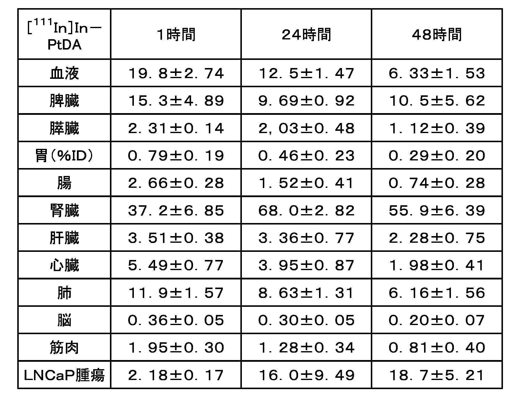
実施例1-1と同様の方法で飼育した雄性CB17/IcrJcl-Prkdcscidマウスに、PBSとMatrigelの混合液(1:1,150μL)にLNCaP細胞(1.0×107cells/mouse)を懸濁させ,イソフルラン麻酔下,マウスの右肩に皮下移植した。これに加えて、同一のマウスに、PBSとMatrigelの混合液(1:1,150μL)にPC-3細胞(1.0×107cells/mouse)を懸濁させたものをイソフルラン麻酔下,マウスの左肩に皮下移植した。その後、当該マウスを40~60日間飼育した。このようにして、1匹のマウスにLNCaP細胞とPC-3細胞とが同時に移植された腫瘍移植マウスを得た。
[111In]In-PtDAを用いたSPECT/CT撮像では投与24および48時間後において,LNCaP腫瘍に高い放射能集積が認められたが,PC-3腫瘍からはほとんど放射能シグナルが認められなかった.この結果から,[111In]In-PtDAはPSMA陽性腫瘍の明瞭な描出が可能であることが示された。
本実施例では、PSMAを標的分子とする化合物を合成した。次いで、各化合物に放射性金属として111Inイオンを配位させた放射性標識化合物を得た。合成経路の概略を合成経路(X-1)~(X-3)として以下に示す。
実施例3-1~3-3にて用いた化合物(Octapa-2、Octapa-3およびNeunpa-2)は、上記一般式(2)にて示すように、キレート部、PSMA分子との結合部及びアルブミン結合部がそれぞれ、リンカー構造を介して分枝状に結合した化学構造を有するものである。
比較例2-1および2-2にて用いた化合物(Octapa-1及びNeunpa-1)は、キレート部およびPSMA分子との結合部を有するが、アルブミン結合部を構造中に有しないものであった。


L-フェニルアラニンアミド(985mg,6.0mmol)をテトラヒドロフラン(THF)(20mL)に溶解させ,LiAlH4(1139mg,30mmol)のTHF(10mL)溶液を0℃でゆっくり加えた.反応液を60℃で24時間撹拌後,H2O(1.2mL),15%水酸化ナトリウム水溶液(1.2mL),およびH2O(3.6mL)を順に加えた.室温で1時間撹拌後,セライトろ過し,ろ液を減圧留去した.残渣に濃硫酸(5mL)を加えた後,濃硝酸(400μL)と濃硫酸(400μL)の混合液を加えた.室温で5時間撹拌後に中和し,クロロホルム(50mL×3)で抽出した.有機層を硫化ナトリウムで乾燥させ,ろ過した.ろ液を減圧留去した後,残渣を中圧カラムクロマトグラフィーによって精製した(メタノール/クロロホルム)。
収量:830mg(収率:71%、L-フェニルアラニンアミドの物質量に対する得られた化合物101の物質量から算出した)。
MS(ESI)m/z:196.1[M+H]+.
既報の方法に従い,化合物101から3段階で合成した(J Am Chem Soc.2013,135,12707-12721,J Chem Soc Dalt Trans.2014,43,7176-7190)。
化合物102(288mg,0.36mmol)をメタノール(5mL)に溶解させ,パラジウム炭素(30mg)を加えた.反応液を水素雰囲気下で3時間撹拌後,セライトろ過し,ろ液を減圧留去した.残渣を中圧カラムクロマトグラフィーによって精製した(メタノール/クロロホルム)。
収量:150mg(収率:54%、化合物102の物質量に対する得られた化合物103の物質量から算出した)。
1H-NMR(400MHz,CDCl3)δ7.82(d,J=6.9Hz,2H),7.70-7.60(m,4H),6.86(d,J=7.6Hz,2H),6.54(d,J=7.6Hz,2H),4.07(m,5H),3.40-3.23(m,5H),2.91-2.89(m,2H),2.73-2.69(m,1H),2.53-2.50(m,2H),1.62(s,18H),1.40(s,18H)。
MS(ESI)m/z:806.4[M+H]+.
既報の方法に従い合成した(Bioconjug.Chem.2017,28,2145-2159)。
化合物104(729mg,1.17mmol)のMeCN(40mL)溶液にt-ブチルブロモ酢酸(396μL,2.69mmol)と炭酸ナトリウム(285mg,2.69mmol)を加え,60℃で24時間撹拌した.室温に戻した後,反応液をろ過し,ろ液を減圧留去した.残渣を中圧カラムクロマトグラフィーによって精製した(ヘキサン/酢酸エチル)。
収量:566mg(収率:57%、化合物104の物質量に対する得られた化合物105の物質量から算出した)。
1H-NMR(400MHz,CDCl3)δ8.14(d,J=8.7Hz,2H),8.05-8.02(m,2H),7.71-7.67(m,4H),7.62-7.58(m,2H),7.37(d,J=8.7Hz,2H),4.08(s,4H),3.41(t,J=7.0Hz,4H),2.86-2.77(m,8H),1.34(s,18H)。
MS(ESI)m/z:851.2[M+H]+.
化合物105(566mg,0.67mmol)のTHF(20mL)溶液にチオフェノール(253μL,1.53mmol)と炭酸カリウム(212mg,1.53mmol)を加え,50℃で50時間撹拌した.室温に戻した後,反応液をろ過し,ろ液を減圧留去した.残渣を中圧カラムクロマトグラフィーによって精製した(メタノール/クロロホルム)。
収量:213mg(収率:66%、化合物105の物質量に対する得られた化合物106の物質量から算出した)。
1H-NMR(400MHz,CDCl3)δ8.14(d,J=8.2Hz,2H),7.42-7.37(m,2H),3.26(s,4H),2.91-2.65(m,12H),1.47(s,18H)。
MS(ESI)m/z:481.3[M+H]+.
化合物106(213mg,0.44mmol)のN,N-ジメチルホルムアミド(DMF)(15mL)溶液にt-ブチル-6-(ブロモメチル)ピコリネート(264mg,0.97mmol)と炭酸ナトリウム(103mg,0.97mmol)を加え,60℃で24時間撹拌した.室温に戻した後,反応液をろ過し,ろ液を減圧留去した.残渣を中圧カラムクロマトグラフィーによって精製した(メタノール/クロロホルム)。
収量:224mg(収率:60%、化合物106の物質量に対する得られた化合物107の物質量から算出した)。
1H-NMR(400MHz,CDCl3)δ8.06(d,J=8.5Hz,2H),7.88-7.86(m,2H),7.75-7.73(m,4H),7.23(d,J=8.7Hz,2H),3.99(s,4H),3.30(s,4H),2.74-2.63(m,12H),1.62(s,18H),1.45(s,18H)。
MS(ESI)m/z:863.5[M+H]+.
化合物103の合成と同様の反応を行い,化合物107から50.7mg(収率23%、化合物107の物質量に対する得られた化合物108の物質量から算出した)の化合物108を得た。
1H-NMR(400MHz,CDCl3)δ7.86(d,J=7.1Hz,2H),7.78-7.72(m,4H),6.85(d,J=8.2,2H),6.56(d,J=8.2,2H),4.00(s,4H),3.32(s,4H),2.76-2.55(m,12H),1.62(s,18H),1.44(s,18H).MS(ESI)m/z:833.4[M+H]+.
6-(Fmoc-アミノ)ヘキサン酸(31.1mg,0.088mmol)のジクロロメタン(7mL)溶液に化合物103(56.6mg,0.073mmol),1-エチル-3-(3-ジメチルアミノプロピル)-カルボジイミド(EDC)塩酸塩(28.8mg,0.15mmol),1-ヒドロキシ-7-アザベンゾトリアゾール(HOAt)(20.4mg,0.15mmol),およびトリエチルアミン(21μL,0.15mmol)を加え,室温で3時間撹拌した.溶媒を減圧留去した後,残渣を中圧カラムクロマトグラフィーによって精製した(メタノール/クロロホルム)。
収量:30.2mg(収率:37%、化合物103の物質量に対する得られた化合物109の物質量から算出した)。
HRMS(ESI)m/z:1111.6170[M+H]+.
化合物109の合成と同様の反応を行い,化合物108から17.7mg(収率25%、化合物108の物質量に対する得られた化合物110の物質量から算出した)の化合物110を得た。
HRMS(ESI)m/z:1168.6372[M+H]+.
化合物109(30.2mg,0.027mmol)をピペリジン(1mL)とDMF(4mL)の混合液に溶解させ,室温で2.5時間撹拌した.反応液に酢酸エチルとH2Oを加えた後,有機層を硫化ナトリウムで乾燥させ,ろ過した.ろ液を減圧留去した後,残渣にTHF(5mL)を加えた.無水コハク酸(11mg,0.11mmol)とおよびN,N-ジイソプロピルエチルアミン(DIPEA)(19μL,0.11mmol)を加え,室温で3時間撹拌した.溶媒を減圧留去した後,残渣を中圧カラムクロマトグラフィーによって精製した(メタノール/クロロホルム)。
収量:9.7mg(収率:37%、化合物109の物質量に対する得られた化合物111の物質量から算出した)。
HRMS(ESI)m/z:989.5600[M+H]+.
化合物111の合成と同様の反応を行い,化合物110から17.7mg(収率95%)の化合物112を得た。
HRMS(ESI)m/z:1046.6139[M+H]+.
化合物111(6.2mg,6.3μmol)のジクロロメタン(5mL)溶液に(S)-di-tert-butyl 2-(3-((S)-6-amino-1-tert-butoxy-1-oxohexan-2-yl)ureido)pentanedioate(6.2mg,13μmol),1-[bis(dimethylamino)methylene]-1H-benzotriazolium 3-oxide hexafluorophosphate(HBTU)(5.0mg,13μmol),およびDIPEA(10μL,0.57μmol)を加え,室温で26時間撹拌した.溶媒を減圧留去し,残渣を中圧カラムクロマトグラフィーによって精製した(メタノール/クロロホルム)。
収量:4.5mg(収率:49%、化合物111の物質量に対する得られた化合物113の物質量から算出した)。
HRMS(ESI)m/z:729.9404[M+2H]+.
化合物112(19.3mg,0.018mmol)のジクロロメタン(5mL)溶液に(S)-di-tert-butyl 2-(3-((S)-6-amino-1-tert-butoxy-1-oxohexan-2-yl)ureido)pentanedioate(17.6mg,0.036mmol),EDC塩酸塩(6.9mg,0.036mmol),HOAt(4.9mg,0.036mmol),およびトリエチルアミン(5μL,0.036mmol)を加えた.反応液を室温で6時間撹拌後,溶媒を減圧留去した.残渣を中圧カラムクロマトグラフィーによって精製した(メタノール/クロロホルム)。
収量:15.6mg(収率:57%、化合物112の物質量に対する得られた化合物114の物質量から算出した)。
HRMS(ESI)m/z:758.4704[M+2H]+.
化合物113(11.4mg,7.8μmol)をトリフルオロ酢酸(TFA)/H2O/トリイソプロピルシランの混合液(95/2.5/2.5,2mL)に溶解させ,室温で3時間撹拌した.溶媒を除去した後,残渣を逆相HPLCで以下の条件で精製した。
精製条件:Cosmosil 5C18-AR-II column(4.6×150mm),移動相:MeCN/H2O/TFA[10/90/0.1(0分)~30/70/0.1(30分)],流速:1mL/min.
収量:2.2mg(収率:27%、化合物113の物質量に対する得られた化合物115の物質量から算出した)。
HRMS(ESI)m/z:1066.4325[M+H]+.
化合物115の合成と同様の反応を行い,化合物114から1.8mg(収率43%)の化合物116を得た。
HRMS(ESI)m/z:1123.4924[M+H]+.
N2-[(9H-fluoren-9-ylmethoxy)carbonyl]-N6-[(4-methylphenyl)diphenylmethyl]-L-lysine(40.9mg,0.065mmol)のジクロロメタン(5mL)溶液に化合物103(42.3mg,0.055mmol),EDC塩酸塩(21.1mg,0.11mmol),HOAt(15.0mg,0.11mmol),およびトリエチルアミン(15μL,0.11mmol)を加えた.反応液を室温で3時間撹拌後,溶媒を減圧留去した.残渣を中圧カラムクロマトグラフィーによって精製した(メタノール/クロロホルム)。
収量:69.3mg(収率:91%、N2-[(9H-fluoren-9-ylmethoxy)carbonyl]-N6-[(4-methylphenyl)diphenylmethyl]-L-lysineの物質量に対する得られた化合物117の物質量から算出した)。
HRMS(ESI)m/z:1382.7418[M+H]+.
化合物117の合成と同様の反応を行い,化合物108から123mg(収率77%、化合物108の物質量に対する得られた化合物118の物質量から算出した)の化合物118を得た。
HRMS(ESI)m/z:1439.8029[M+H]+.
化合物117(115mg,0.083mmol)をピペリジン(1mL)とDMF(4mL)の混合液に溶解させ,室温で2.5時間撹拌した.反応液に酢酸エチルとH2Oを加えた後,有機層を硫化ナトリウムで乾燥させ,ろ過した.ろ液を減圧留去した後,残渣をジクロロメタン(5mL)に溶解させ,4-(4-ヨードフェニル)ブタン酸(49.3mg,0.17mmol),EDC塩酸塩(32.6mg,0.17mmol),HOAt(23.1mg,0.17mmol),およびトリエチルアミン(20μL,0.17mmol)を加えた.反応液を室温で7時間撹拌後,溶媒を減圧留去した.残渣を中圧カラムクロマトグラフィーによって精製した(メタノール/クロロホルム)。
収量:62.4mg(収率:52%、化合物117の物質量に対する得られた化合物119の物質量から算出した)。
HRMS(ESI)m/z:1432.6529[M+H]+.
化合物119の合成と同様の反応を行い,化合物118から84.2mg(収率66%、化合物118の物質量に対する得られた化合物120の物質量から算出した)の化合物120を得た。
HRMS(ESI)m/z:1489.7260[M+H]+.
化合物119(62.4mg,0.044mmol)を1%TFA含有ジクロロメタン(5mL)に溶解させ,室温で2時間撹拌した.溶媒を除去した後,残渣をTHF(5mL)に溶解させ,無水コハク酸(8.1mg,0.088mmol)とDIPEA(15μL,0.088mmol)を加えた.溶液を室温で4時間撹拌した後,溶媒を除去した.残渣を中圧カラムクロマトグラフィーによって精製した(メタノール/クロロホルム)。
収量:65mg(収率:100%、化合物119の物質量に対する得られた化合物121の物質量から算出した)。
HRMS(ESI)m/z:1276.5478[M+H]+.
化合物121の合成と同様の反応を行い,化合物120から39.1mg(収率35%、化合物120の物質量に対する得られた化合物122の物質量から算出した)の化合物122を得た。
HRMS(ESI)m/z:1333.5464[M+H]+.
化合物121(65mg,0.051mmol)のジクロロメタン(5mL)溶液に(S)-di-tert-butyl 2-(3-((S)-6-amino-1-tert-butoxy-1-oxohexan-2-yl)ureido)pentanedioate(37.3mg,0.077mmol),EDC塩酸塩(19.2mg,0.10mmol),HOAt(13.6mg,0.10mmol),およびトリエチルアミン(15μL,0.10mmol)を加えた.反応液を室温で5時間撹拌後,溶媒を減圧留去した.残渣を中圧カラムクロマトグラフィーによって精製した(メタノール/クロロホルム)。
収量:54.3mg(収率:61%、化合物121の物質量に対する得られた化合物123の物質量から算出した)。
HRMS(ESI)m/z:873.4248[M+2H]+.
化合物123の合成と同様の反応を行い,化合物122から22.3mg(収率25%、化合物122の物質量に対する得られた化合物124の物質量から算出した)の化合物124を得た。
HRMS(ESI)m/z:902.4312[M+2H]+.
化合物123(9.6mg,6.6μmol)をTFA/H2O/トリイソプロピルシランの混合液(95/2.5/2.5,1mL)に溶解させ,室温で2時間撹拌した。溶媒を除去した後,残渣を逆相HPLCで以下の条件で精製した。
精製条件:Cosmosil 5C18-AR-II column(4.6×150mm),移動相:MeCN/H2O/TFA[20/80/0.1(0分)~50/50/0.1(30分)],流速:1mL/min.
収量:1.3mg(収率:17%、化合物123の物質量に対する得られた化合物125の物質量から算出した)。
HRMS(ESI)m/z:677.2144[M+2H]+.
化合物125の合成と同様の反応を行い,化合物124から2.0mg(収率47%、化合物124の物質量に対する得られた化合物126の物質量から算出した)の化合物126を得た。
HRMS(ESI)m/z:705.74021[M+2H]+.
N2-[(9H-fluoren-9-ylmethoxy)carbonyl]-N6-[(4-methylphenyl)diphenylmethyl]-L-lysine(625mg,1.0mmol)のジクロロメタン(20mL)溶液にtert-butyl trichloroacetimidate(437mg,2.0mmol)とBF3・OEt2(20μL,0.16mmol)を加えた。反応液を室温で終夜撹拌した後,溶媒を減圧留去した.残渣を中圧カラムクロマトグラフィーによって精製した(ヘキサン/酢酸エチル)。
収量:322mg(収率:47%、N2-[(9H-fluoren-9-ylmethoxy)carbonyl]-N6-[(4-methylphenyl)diphenylmethyl]-L-lysineの物質量に対する得られた化合物127の物質量から算出した)。
1H-NMR(400MHz,CDCl3)δ7.71-7.69(m,2H),7.56(d,J=7.2Hz,2H),7.46(d,J=8.1Hz,4H),7.35-7.30(m,4H),7.26-7.21(m,6H),7.15-7.11(m,2H),7.04(d,J=8.1Hz,2H),5.38(d,J=8.7Hz,1H),4.35(d,J=6.4Hz,2H),4.28-4.23(m,1H),4.18(t,J=7.5Hz,1H),2.26(s,3H),2.12-2.11(m,2H),1.81-1.75(m,1H),1.59-1.44(m,14H)。
MS(ESI)m/z:681.4[M+H]+.
化合物127(200mg,0.47mmol)を5%TFA含有ジクロロメタン(10mL)に溶解させ,室温で3時間撹拌した.溶媒を除去した後,残渣を中圧カラムクロマトグラフィーによって精製した(メタノール/クロロホルム).得られたアミンをDMF(10mL)に溶解させ,4-(4-ヨードフェニル)ブタン酸(273mg,0.94mmol),EDC塩酸塩(180mg,0.94mmol),HOAt(123mg,0.94mmol),およびトリエチルアミン(130μL,0.94mmol)を加えた.反応液を室温で終夜撹拌後,酢酸エチルとH2Oを加えた.有機層を硫化ナトリウムで乾燥させ,ろ過した.ろ液を減圧留去した後,残渣を中圧カラムクロマトグラフィーによって精製した(メタノール/クロロホルム)。
1H-NMR(400MHz,CDCl3)δ7.76(d,J=7.5Hz,2H),7.60-7.53(m,4H),7.39(t,J=7.5Hz,2H),7.30(t,J=7.5Hz,2H),6.92-6.86(m,2H),4.40-4.09(m,4H),3.27-3.19(m,2H),2.52(t,J=7.8Hz,2H),1.93-1.80(m,3H),1.71-1.62(m,1H),1.52-1.36(m,13H)。
MS(ESI)m/z:697.2[M+H]+.
化合物128(473mg,0.68mmol)をピペリジン(2mL)とDMF(8mL)の混合液に溶解させ,室温で1時間撹拌した.反応液に酢酸エチルとH2Oを加えた後,有機層を硫化ナトリウムで乾燥させ,ろ過した.ろ液を減圧留去した後,残渣を中圧カラムクロマトグラフィーによって精製した(メタノール/クロロホルム).得られたアミンをDMF(10mL)に溶解させ,3,6,9-trioxaundecanedioic acid(91.1mg,0.41mmol),EDC塩酸塩(78.6mg,0.41mmol),HOAt(55.8mg,0.41mmol),およびトリエチルアミン(57μL,0.41mmol)を加えた.反応液を室温で5時間撹拌後,酢酸エチルとH2Oを加えた.有機層を硫化ナトリウムで乾燥させ,ろ過した.ろ液を減圧留去した後,残渣を中圧カラムクロマトグラフィーによって精製した(メタノール/クロロホルム)。
1H-NMR(400MHz,CD3OD)δ7.58(d,J=8.7Hz,2H),6.96(d,J=7.5,2H)4.41-4.29(m,2H),4.09-4.02(m,2H),3.85-3.73(m,8H),3.60(q,J=7.2Hz,2H),3.35(s,2H),3.10(t,J=4.0Hz,2H),2.55(t,J=7.5Hz,2H),2.16-2.14(m,2H),1.89-1.81(m,2H),1.41(s,9H),1.32-1.27(m,3H),1.18(t,J=7.0Hz,2H)。
MS(ESI)m/z:701.2[M+Na]+.
化合物117(400mg,0.29mmol)をピペリジン(1mL)とDMF(4mL)の混合液に溶解させ,室温で2.5時間撹拌した.反応液に酢酸エチルとH2Oを加えた後,有機層を硫化ナトリウムで乾燥させ,ろ過した.ろ液を減圧留去した後,残渣を中圧カラムクロマトグラフィーによって精製した(メタノール/クロロホルム).得られたアミンをDMF(10mL)に溶解させ,化合物129(170mg,0.25mmol),EDC塩酸塩(95.9mg,0.50mmol),HOAt(68.1mg,0.50mmol),およびトリエチルアミン(69μL,0.50mmol)を加えた.反応液を室温で2時間撹拌後,酢酸エチルとH2Oを加えた.有機層を硫化ナトリウムで乾燥させ,ろ過した.ろ液を減圧留去した後,残渣を中圧カラムクロマトグラフィーによって精製した(メタノール/クロロホルム)。
収量:272mg(収率:60%、化合物117の物質量に対する得られた化合物130の物質量から算出した)。
HRMS(ESI)m/z:911.4302[M+2H]+.
化合物130(272mg,0.15mmol)を1%TFA含有ジクロロメタン(3mL)に溶解させ,室温で1.5時間撹拌した。溶媒を除去した後,残渣をTHF(5mL)に溶解させ,無水コハク酸(30mg,0.30mmol)およびDIPEA(52μL,0.30mmol)を加えた.反応液を室温で2時間撹拌後,溶媒を減圧留去した.残渣を中圧カラムクロマトグラフィーによって精製した(メタノール/クロロホルム)。
収量:230mg(収率:92%、化合物130の物質量に対する得られた化合物131の物質量から算出した)。
HRMS(ESI)m/z:832.8701[M+2H]+.
化合物131(186mg,0.11mmol)のジクロロメタン(5mL)溶液に(S)-di-tert-butyl 2-(3-((S)-6-amino-1-tert-butoxy-1-oxohexan-2-yl)ureido)pentanedioate(63.4mg,0.13mmol),HBTU(83.4mg,0.22mmol),およびDIPEA(38μL,0.22mmol)を加え,室温で1.5時間撹拌した.溶媒を減圧留去し,残渣を中圧カラムクロマトグラフィーによって精製した(メタノール/クロロホルム)。
収量:80.4mg(収率:34%、化合物131の物質量に対する得られた化合物132の物質量から算出した)。
HRMS(ESI)m/z:1068.0419[M+2H]+.
化合物132(10mg,4.7μmol)をTFA/H2O/トリイソプロピルシランの混合液(95/2.5/2.5,2mL)に溶解させ,室温で終夜撹拌した.溶媒を除去した後,残渣を逆相HPLCで以下の条件で精製した。
精製条件:Cosmosil 5C18-AR-II column(4.6×150mm),移動相:MeCN/H2O/TFA[20/80/0.1(0分)~50/50/0.1(30分)],流速:1mL/min.
収量:2.3mg(収率:29%、化合物132の物質量に対する得られた化合物133の物質量から算出した)。
HRMS(ESI)m/z:843.2900[M+2H]+.
3種類のOctapa誘導体(Octapa-1~3)についてそれぞれ,酢酸緩衝液(pH5.5,10mM,350μL)に[111In]InCl3溶液(100μL)を混和し,標識前駆体(化合物115,25,または33)の水溶液(<10% DMSO,10μM,50μL)を加えた。室温で15分間静置した後,反応液を逆相HPLCにて以下の条件で精製した。
精製条件:Cosmosil 5C18-MS-II column(4.6×150mm),移動相:MeCN/酢酸緩衝液(pH4.5,10mM)[10/90(0分)~30/70(30分)または20/80(0分)~50/50(30分)],流速:1mL/min。
放射化学的収率及び放射化学的純度は実施例1-1に記載した方法と同様に測定した。放射化学的純度の測定に用いたHPLC条件は前記精製条件と同様である。
・比較例2-1:[111In]In-Octapa-1
・実施例3-1:[111In]In-Octapa-2
・実施例3-2:[111In]In-Octapa-3
2種類のNeunpa誘導体(Neunpa-1及び2)について,酢酸緩衝液(pH4.0,10mM,350μL)に[111In]InCl3溶液(100μL)を混和し,標識前駆体(化合物116または26)の水溶液(<10% DMSO,10μM,50μL)を加えた.室温で15分間静置した後,反応液を逆相HPLCにて以下の条件で精製した。
精製条件:Cosmosil 5C18-AR-II column(4.6×150mm),移動相:MeCN/H2O/TFA[10/90/0.1(0分)~30/70/0.1(30分)または20/80/0.1(0分)~50/50/0.1(30分)],流速:1mL/min。
放射化学的収率及び放射化学的純度は実施例1-1に記載した方法と同様に測定した。放射化学的純度の測定に用いたHPLC条件は前記精製条件と同様である。
・比較例2-2:[111In]In-Neunpa-1
・実施例3-3:[111In]In-Neunpa-2
上述の実施例1-1と同様の方法で培養したLNCaP細胞と、PC-3細胞とをそれぞれ、4.0×10⁵cells/wellにて12ウェルプレートに播種し,37℃,5%CO2下で48時間静置した。
培養培地を除去し,実施例3-1~3-3又は比較例2-1~2-2の放射性標識化合物(各37kBq)含有アッセイ用培地(0.5%FBS含有RPMI1640培地)溶液(1mL)を加えた。その後,プレートを37℃,5%CO2下で1時間静置した。
阻害実験では,培養培地を除去後,上述の放射性標識化合物と2-(phosphonomethyl)pentanedioic acid(2-PMPA)(PSMA阻害剤、終濃度100μM)とを含有するアッセイ用培地(1mL)を加えた.その後,プレートを37℃,5%CO2下で1時間静置した。
アッセイ用培地を除去後,各ウェルをアッセイ用培地(1mL)で洗浄し,1規定水酸化ナトリウム水溶液(200μL×2)で細胞を溶解させた。
細胞溶解液の各放射能をガンマカウンターで測定した。これとは別に、細胞溶解液中の総タンパク質濃度をThermo Fisher Scientific社製BCA Protein Assay Kitを用いて算出した。添加放射能量に対するサンプルの放射能量の百分率(%ID)を、総タンパク質量で除した値(%ID/mg protein)をサンプルごとに算出した。
データは平均値±標準偏差で表した.有意差検定はStudent’s t-testおよびone-way analysis of variance (ANOVA) test with Dunnet’s post-hoc testを用いて行い,p<0.05を有意差ありとした。
実施例3-1~3-3並びに比較例2-1~2-2の放射性標識化合物は、PSMA陰性であるPC-3細胞と比較して、PSMA陽性のLNCaP細胞に対する高い結合性を示し,その結合は過剰量のPSMA阻害剤(2-PMPA)の添加によって有意に低下した。したがって、各実施例及び比較例の放射性標識化合物は,PSMA陽性細胞に特異的に結合することが示された.
上述の実施例と同様に、PBS及びヒトアルブミンを用いて、アルブミンへの結合評価を行った。実施例3-1~3-3並びに比較例2-1~2-2の放射性標識化合物のPBS溶液(各37kBq,50μL)をそれぞれ用いた以外は、実施例1-1と同様の実験手順及び統計手法で評価を行った。
実施例3~1~3-3([111In]In-Octapa-2、[111In]In-Octapa-3および[111In]In-Neunpa-2)を用いたHSA溶液における溶出液の放射能割合は,PBSにおける溶出液の放射能割合に比べて顕著に高かった。
一方、比較例2-1~2-2([111In]In-Octapa-1および[111In]In-Neunpa-1)のHSA溶液における溶出液の放射能割合は、PBSの場合と同様に低い値を示した.これらの結果から,実施例3~1~3-3で用いた各化合物および放射性標識化合物は、アルブミンに対する結合性を有することが示された。
実施例1-1と同様の方法で作製したLNCaP腫瘍移植マウスに、実施例3~1~3-3又は比較例2-1~2-2の各放射性標識化合物の生理食塩水溶液(各259kBq/100μL)を尾静脈から投与し(n=2~3)、投与1,4,24,48,96,および192時間後にマウスを安楽死させた。その後,血液及び各臓器を回収し,臓器の質量及び放射能を測定した。
投与放射能量(injected dose)に対する放射能量の百分率(%ID)を血液質量又は臓器質量(g)で除した値(%ID/g)として示す。%ID/gの値が高いほど、放射性標識化合物の存在量が多く、該化合物の標的臓器への集積が高いことを示す。
実施例3~1~3-3の各放射性標識化合物は、比較例2-1~2-2のものと比較して、腎集積が同程度に低減されながらも、高い血中滞留性及び腫瘍集積性を有することが確認された。このことは、実施例3-1([111In]In-Octapa-2)、実施例3-2([111In]In-Octapa-3)及び実施例3-3([111In]In-Neunpa-2)において特に顕著であった。
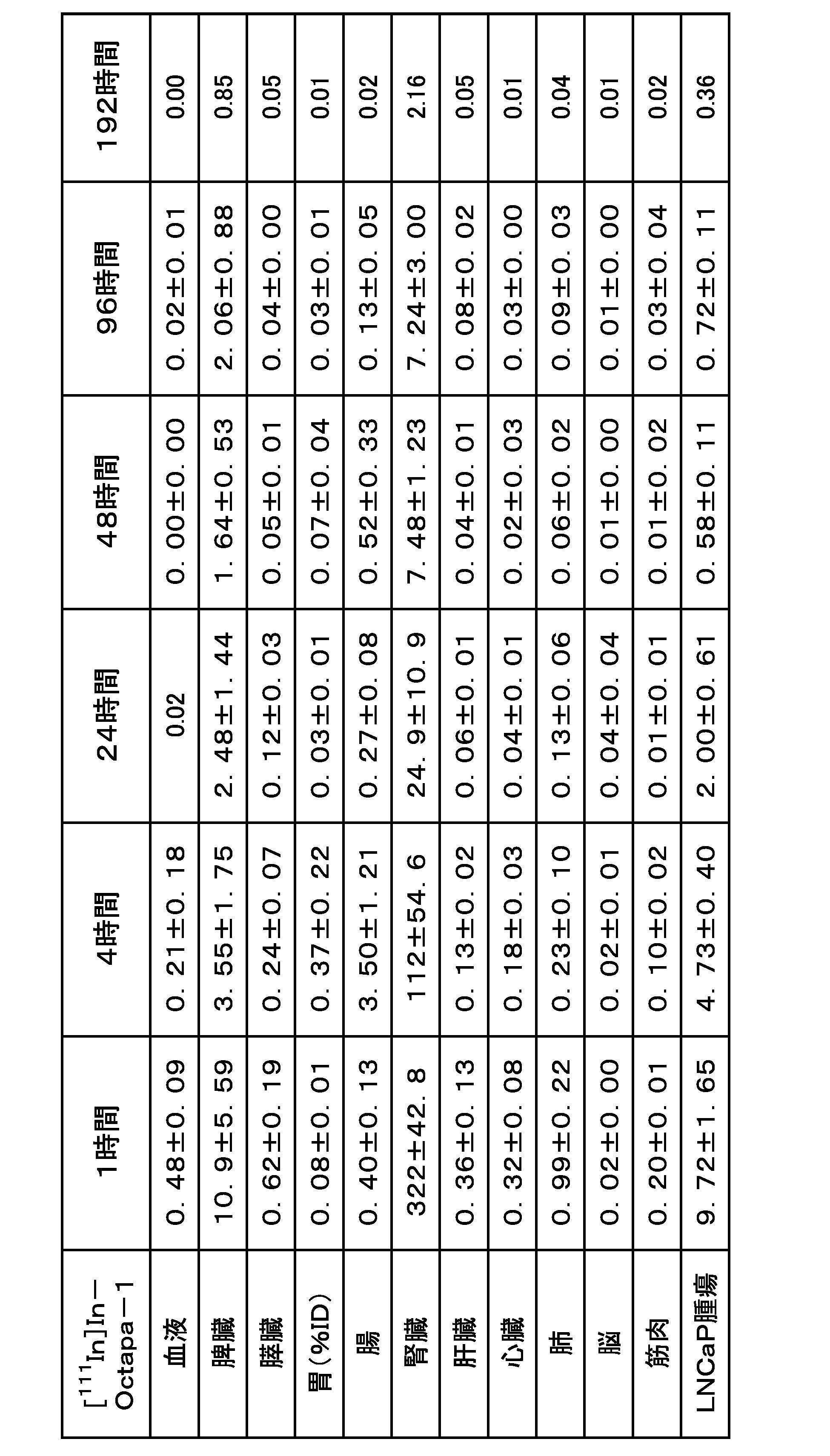
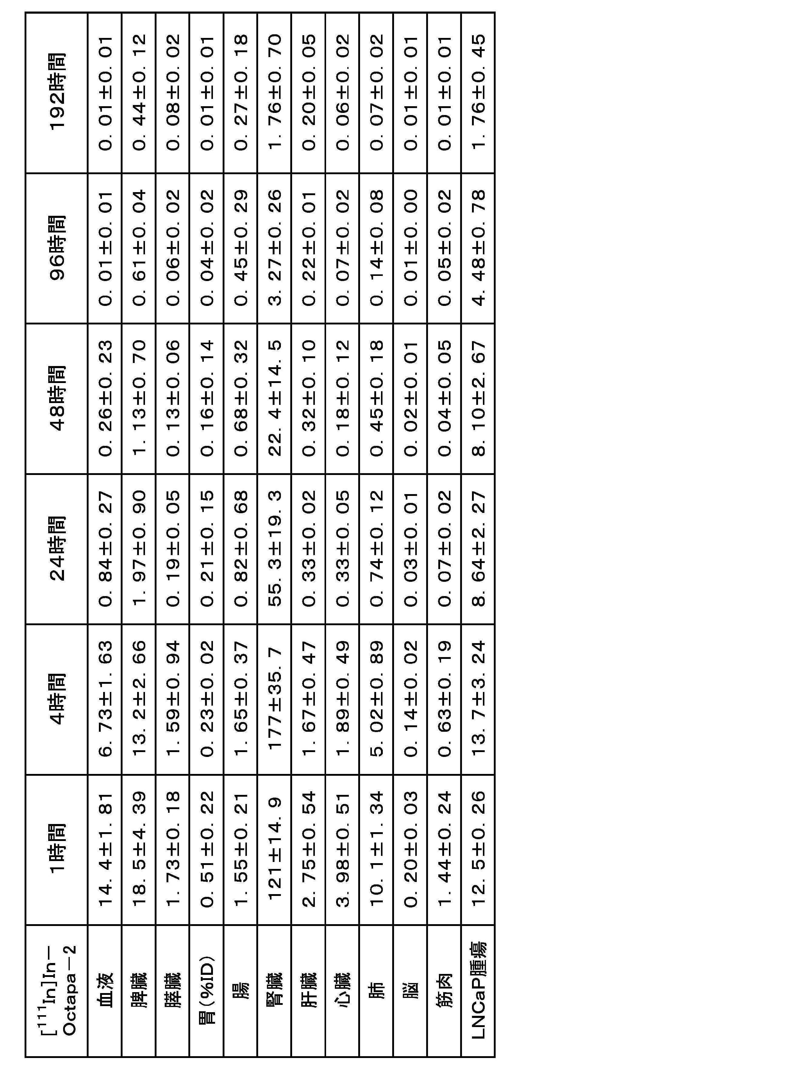
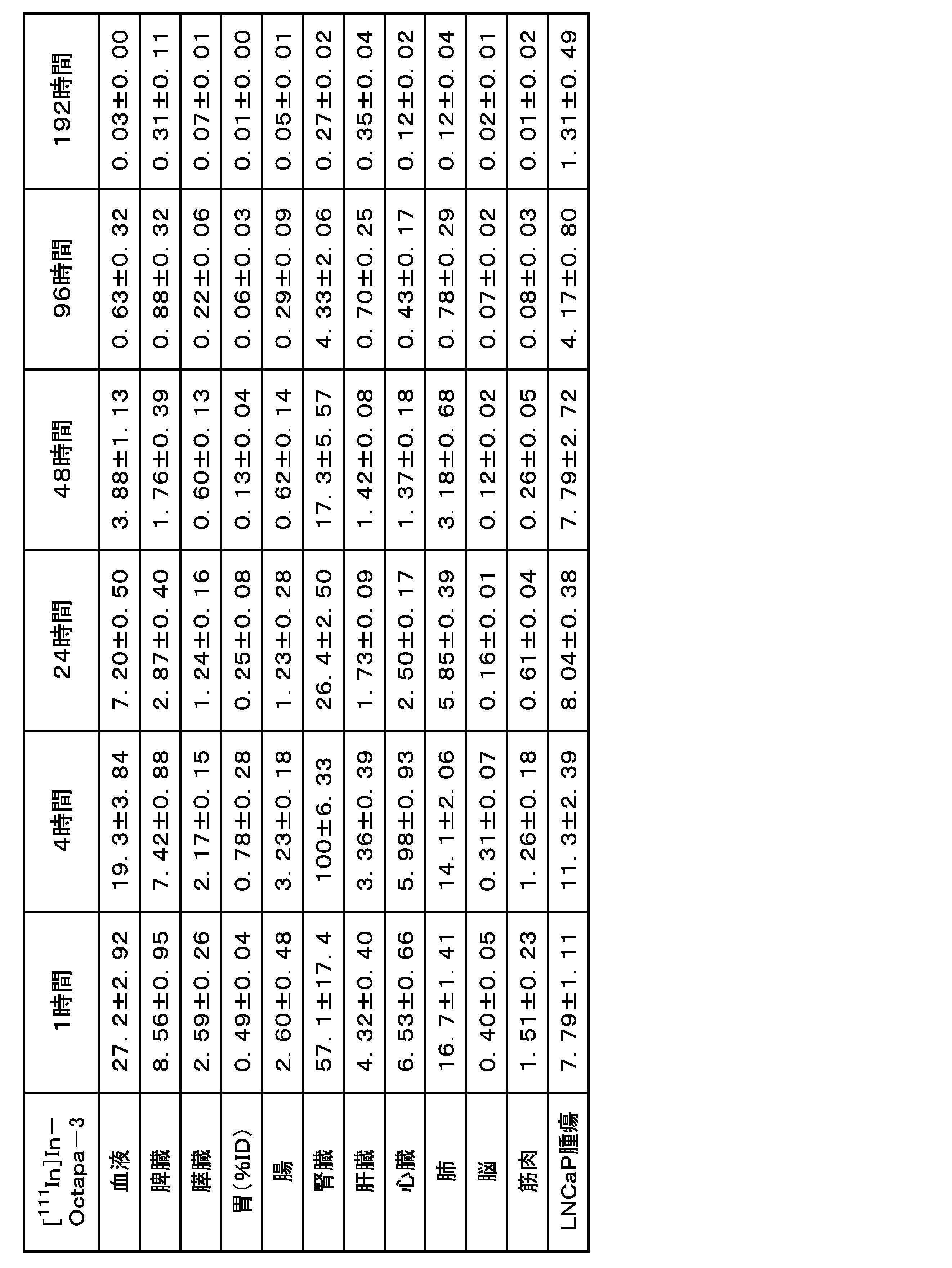


実施例2と同様の方法でLNCaP細胞とPC-3細胞との腫瘍同時移植マウスを得たあと、実施例3-1([111In]In-Octapa-2)の生理食塩水溶液(7.3MBq,150μL)を尾静脈から投与した。投与24時間後に、実施例2と同様の方法でSPECT/CTを行い、得られた画像の再構成を行った。
[111In]In-Octapa-2を用いたSPECT/CT撮像では投与24時間後において、LNCaP腫瘍(図中,実線矢印)に高い放射能集積が認められたが,PC-3腫瘍(図中,破線矢印)での放射能シグナルはほとんど観測されなかった。また、腎臓(図中,円)における放射能集積も認められた。この結果から、[111In]In-Octapa-2はPSMA高発現腫瘍の明瞭な描出が可能であることが示された。
実施例4-1~4-2では、PSMAを標的分子とする(((S)-5-amino-1-carboxypentyl)carbamoyl)-L-glutamic acid(上記式(C1)中、aが2、bが4である構造)をPSMA分子との結合部として構造中に含み、上記一般式(1)にて示すように、PSMA分子との結合部及びアルブミン結合部を、キレート部を介して直線状に含む化学構造を有する化合物(PSMA-NAT-DA1)を合成した。次いで、PSMA-NAT-DA1に放射性金属として111Inイオン又は225Acイオンをそれぞれ配位させた放射性標識化合物を得た。以下に詳細を示す。
PSMA-NAT-DA1は、構造中にキレート部、PSMA分子との結合部及びアルブミン結合部を含む。
PSMA-NAT-DA1は、キレート部とPSMA分子との結合部とが、trans-4-アミノシクロヘキサン-1-カルボン酸及びナフチルグリシン由来の化学構造を介して間接的に結合しており、且つ、キレート部とアルブミン結合部を含む原子団とは、リシン由来のリンカー構造を介して間接的に結合している。

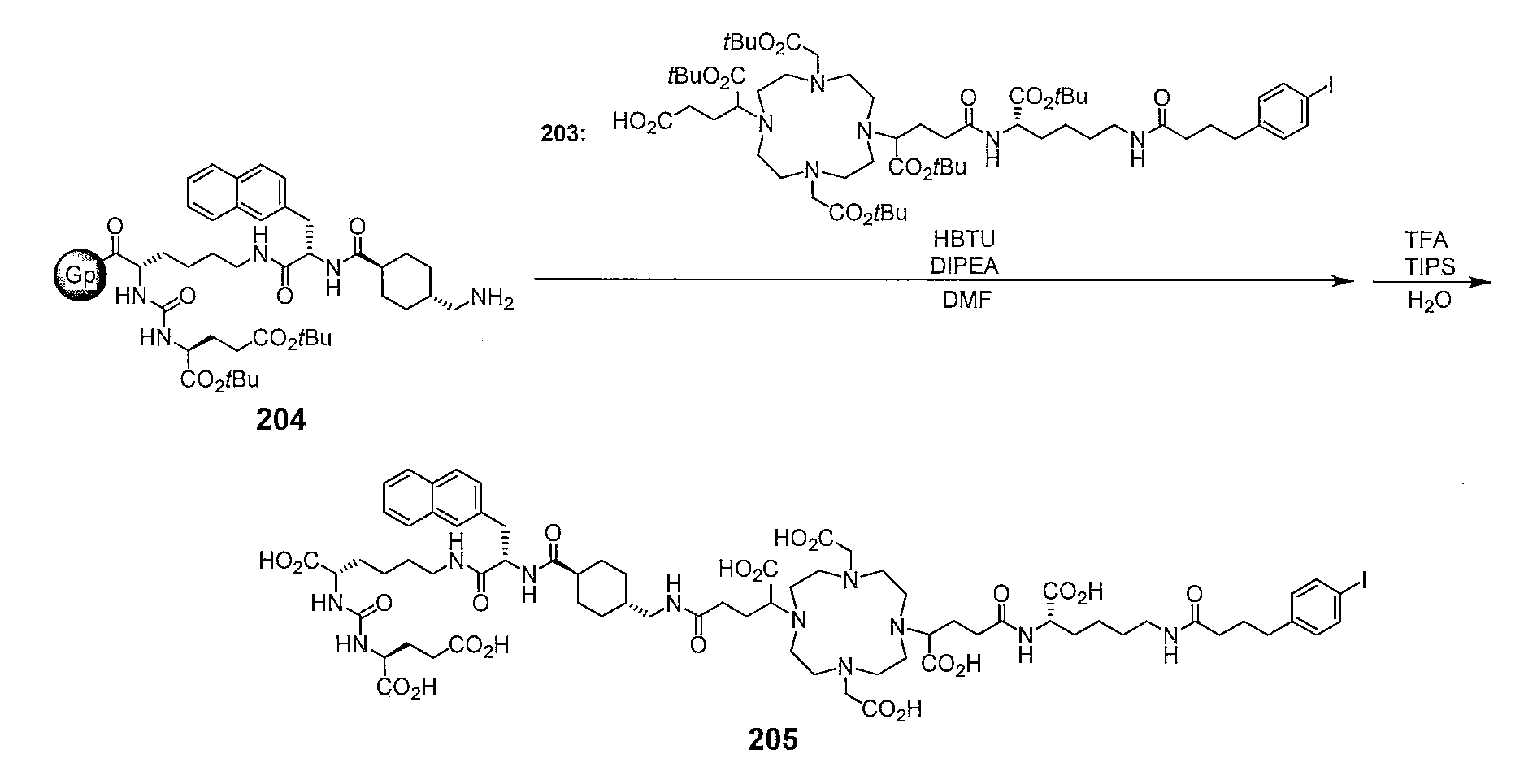
以下に示す手順で、PSMA-NAT-DA1を合成した。
化合物1は、実施例1-1に記載の方法と同様に合成した。
化合物202は、既報の方法に従い,N2-[(9H-fluoren-9-ylmethoxy)carbonyl]-N6-[(4-methylphenyl)diphenylmethyl]-L-lysineから4段階で合成した(Chem Commun.2021,57,6432-6435)。
化合物1(131mg,0.17mmol)のN,N-ジメチルホルムアミド(DMF)(0.4mL)溶液にN-[1-(cyano-2-ethoxy-2-oxoethylideneaminooxy)dimethylamino(morpholino)]uronium hexafluorophosphate(COMU)(72.4mg,0.17mmol)およびN,N-ジイソプロピルエチルアミン(DIPEA)(95μL,0.17mmol)を氷冷下で加え,15分間撹拌した。化合物202(66.8mg,0.14mmol)を加え、室温で5日間撹拌後、溶液を逆相HPLCで精製し、化合物203を得た。
精製条件:Cosmosil 5C18-AR-II column(10×250mm)、移動相:MeCN/H2O[3/7(0分)~9/1(60分)],流速:5mL/min。
MS(ESI):m/z1230[M+H]+。
既報(J.Med.Chem.2016,59,1761-1775,Mol.Pharm.2018,15,3502-3511)の方法を参考に,Fmoc-Lys(ivDde)-OHから9段階で化合物204を得た。
なお、化合物204の化学式中、符号Gpで表される化学構造は樹脂を示す。
樹脂が結合した化合物204に、化合物203(49.6mg,0.044mmol),1-[ビス(ジメチルアミノ)メチレン]-1H-ベンゾトリアゾリウム3-オキシドヘキサフルオロホスファート(HBTU)(16.7mg,0.44mmol),およびDIPEA(188μL,0.44mmol)を加え,DMF中で終夜振とうした。DMFで洗浄後,トリフルオロ酢酸(TFA)/トリイソプロピルシラン/H2O(95:2.5:2.5,2mL)を加え,3時間振とう後,溶液を逆相HPLCで以下の条件で精製し、目的とする化合物205(PSMA-NAT-DA1)を得た。
精製条件:Cosmosil 5C18-AR-II column(4.6×250mm)、移動相:MeCN/H2O/TFA[10/90/0.1(0分)~90/10/0.1(40分)],流速:5mL/min。
収量:11mg(16%、化合物203の物質量に対する得られた化合物205の物質量から算出した)
MS(ESI)m/z:MS(ESI)m/z793[M+2H]2+。
以下に示す手順で、目的とする放射性標識化合物([111In]In-PSMA-NAT-DA1)を得た。

その結果、放射化学的収率は9%,放射化学的純度は95%以上であった。また、再度同様な合成を行ったところ、放射化学的収率は60%に改善した。
精製条件:Cosmosil 5C18-AR-II column(4.6×150mm),移動相:MeCN/H2O/TFA[10/90/0.1(0分)~90/10/0.1(40分)],流速:1mL/min。
以下に示す手順で、目的とする放射性標識化合物([225Ac]Ac-PSMA―NAT-DA1)を得た。
Protein LoBind Tube1.5mL(EPPENDORF社製)に分注した[225Ac]AcCl3の0.2mоl/L塩酸溶液(1.0MBq、20μL)に、0.1M酢酸-酢酸アンモニウム緩衝液(pH5.5、170μL)とPSMA―NAT-DA1のDMSO溶液(2.0mM/L、10μL)を加え、70℃で1時間静置した。反応液にH2O(0.8mL)を加え、Oasis HLB Lightカラムに通液した。カラムにH2O(10mL)を通液後、70%EtOH(0.5mL)を通液し、目的とする放射性標識化合物([225Ac]Ac-PSMA―NAT-DA1)70%エタノール溶液を得た。

上述の実施例1-1と同様の方法で培養したLNCaP細胞と、PC-3細胞とをそれぞれ、4.0×10⁵cells/wellにて12ウェルプレートに播種し,37℃,5%CO2下で48時間静置した。
培養培地を除去し,[111In]In-PSMA-NAT-DA1(8.5kBq)を含むアッセイ用培地(0.5%FBS含有RPMI1640培地)溶液(1mL)を加えた。その後,プレートを37℃,5%CO2下で1時間静置した。
阻害実験では,培養培地を除去後,[111In]In-PSMA-NAT-DA1(37kBq)と2-(phosphonomethyl)pentanedioic acid(2-PMPA)(PSMA阻害剤、終濃度100μM)を含むアッセイ用培地(1mL)溶液を加えた。その後,プレートを37℃,5%CO2下で1時間静置した。
アッセイ用培地及び細胞溶解液の各放射能をガンマカウンターで測定した。これとは別に、細胞溶解液中の総タンパク質濃度をThermo Fisher Scientific社製BCA Protein Assay Kitを用いて算出した。添加放射能量に対するサンプルの放射能量の百分率(%ID)を、総タンパク質量で除した値(%ID/mg protein)をサンプルごとに算出した。
データは平均値±標準偏差で表した。有意差検定はStudent’s t-testおよびone-way analysis of variance (ANOVA) test with Dunnet’s post-hoc testを用いて行い,p<0.05を有意差ありとした。
[111In]In-PSMA―NAT-DA1はPC-3細胞と比較してLNCaP細胞に対する高い結合性を示し,その結合は過剰量のPSMA阻害剤(2-PMPA)の添加によって有意に低減した。これらの結果から,[111In]In-PSMA-NAT-DA1はPSMA高発現細胞に特異的に結合することが示された。
[111In]In-PSMA-NAT-DA1のリン酸緩衝生理食塩水(PBS)溶液(37kBq,50μL)を、200μLのヒト血清アルブミン(HSA)のPBS溶液(45mg/mL),マウス血漿,ヒト血漿,またはPBSに加え,37℃で10分間静置した。その後,反応液をスピンカラム(サイティバ社製Sephadex G-50)に添加し,1500×g,4℃で2分間遠心分離した。分離後,カラムと溶出液の放射能をガンマカウンターで測定した。
データは平均値±標準偏差で表した。有意差検定はStudent’s t-testおよびone-way analysis of variance (ANOVA) test with Dunnet’s post-hoc testを用いて行い,p<0.05を有意差ありとした。
評価対象となる化合物がアルブミンに結合し複合体を形成すると,分子サイズの増加によってカラムを通過するが,アルブミンに結合しなかった際には,カラム内のゲルに保持されると考えられる。
[111In]In-PSMA-NAT-DA1をPBS中で静置後,カラムに付したところ,溶出液において顕著な放射能は観測されなかった。一方,マウス血漿,ヒト血漿,およびHSA溶液中で静置した際には,[111In]In-PSMA-NAT-DA1の溶出液における放射能は有意に高く,[111In]In-PSMA-NAT-DA1が血漿アルブミンに結合することが示唆された。
動物実験は京都大学動物実験委員会の指針を遵守して行った。雄性CB17/IcrJcl-Prkdcscidマウスは日本クレア株式会社より購入した。動物は12時間/12時間の昼夜サイクル条件下で飼育し,飼料と水は自由に与えた。PBSとCorning Life Sciences社製Matrigelの混合液(1:1,150μL)にLNCaP細胞(4.3×10⁶cells/mouse)またはPC-3細胞(3.3×106cells/mouse)を懸濁させ,イソフルラン麻酔下,CB17/IcrJcl-Prkdcscidマウスの右肩に皮下移植した。その後、当該マウスを40~60日間飼育した。
LNCaPまたはPC-3腫瘍移植マウスに[111In]In-PSMA-NAT-DA1の生理食塩水溶液(259kBq,100μL)を尾静脈より投与した(各n=3)。投与1,4,24,48,96,および192時間後にマウスを安楽死させた。その後,血液及び各臓器を回収し,重量と放射能を測定した。


上述の方法で作製したLNCaP腫瘍移植マウスに、[111In]In-PSMA-NAT-DA1の生理食塩水溶液(1.57-2.59MBq,150μL)を尾静脈から投与した。投与24、48および96時間後にGamma Medica-Ideas社製FX3300 pre-clinical imaging systemにてSPECT/CTを行った。イソフルラン麻酔下,回転半径35mm,投影時間70秒,投影回数32回にて直径1.0mmのピンホールコリメーターを用いて撮像した。SPECT後,CT(管電圧:60kV,管電流:350μA)を行った。SPECTの投影データについて3次元ordered subset expectation maximization法(8subsets,5iterations)による画像再構成を行った。
本SPECT/CT撮像では,投与24および48時間後においてLNCaP腫瘍(図中矢印)に顕著な放射能集積が認められた。投与24および48時間後において腎臓(図中実線円)にも放射能集積が認められたが,投与96時間後(図中破線円)おいてはほとんど確認されなかった。これらの結果から,[111In]In-PSMA-NAT-DA1はPSMA高発現腫瘍をSPECTにより明瞭に描出することが可能であるとともに,[111In]In-PSMA-DBよりも優れていることが示された。
Claims (14)
- 放射性金属イオンと配位可能なキレート部、アルブミン結合部を含む第1原子団及びPSMA分子との結合部を含む第2原子団とを構造中に有し、
第1原子団と第2原子団とは前記キレート部を介して結合している、化合物。 - 下記一般式(1)で示される、請求項1に記載の化合物。

Laはリンカー構造であり、
LbはLaと同一又は異なるリンカー構造であり、
m及びnは、それぞれ独立して0又は1であり、
B又はLaは、A1の任意の部位に結合しており、
C又はLbは、B又はLaがA1に結合する部位とは異なる部位においてA1と結合している。) - 前記式(1)中、Cが下記式(C1)で表される、請求項1又は2に記載の化合物。

- 前記式(1)中、A1は環状構造を有し、該環状構造が窒素原子を2つ以上有し、各窒素原子が隣り合う2つ以上の炭素原子を隔てて連結されているか、又は、
A1は鎖状構造を有し、該鎖状構造が窒素原子を2つ以上有し、各窒素原子が隣り合う2つ以上の炭素原子を隔てて連結されており、
A1は、前記環状構造又は前記鎖状構造を構成する窒素原子に直接結合する窒素結合原子団を有し、
前記窒素結合原子団は、カルボキシ基、リン酸基、アミド基、ベンゼン環及びピリジン環のうち一種以上を含み、
Bが前記窒素結合原子団に結合している場合、CはBが結合している該原子団以外の前記窒素結合原子団に結合している、請求項1~3いずれか一項に記載の化合物。 - 前記式(1)中、A1は、DOTA、HOPO、Octapa、Macropa又はこれらの誘導体である、請求項1~4いずれか一項に記載の化合物。
- 前記式(1)中、前記第1原子団に含まれる前記アルブミン結合部は、γグルタミン酸、置換若しくは無置換のフェニル酪酸、脂質、ヘマチン、ビリルビン、クロフィブリン酸、クロフィブラート、カロテノイド、ステロイド骨格を有する化合物、イブプロフェン骨格を有する化合物、炭素数13以上20以下の直鎖若しくは分枝鎖且つ飽和若しくは不飽和である炭化水素、シアニン色素、スルホン酸基を有する染料、ジアゾ染料、ペンタメチンシアニン色素、ブルーデキストラン、ブロモクレゾールグリーン、及びエバンスブルー並びにこれらの誘導体のうち一種以上に由来する構造を有するか、又はアルブミンに結合可能な抗体若しくはペプチドである、請求項1~5のいずれか一項に記載の化合物。
- 前記式(1)中、前記第1原子団に含まれる前記アルブミン結合部は、下記式(B1)又は(B2)で表される構造を有する、請求項1~6のいずれか一項に記載の化合物。

式(B2)中、Rb1ないしRb11は、それぞれ独立して、水素原子、ハロゲン原子、水酸基、シアノ基、炭素数が1以上6以下の置換若しくは無置換のアルキル基、又は炭素数が1以上6以下の置換若しくは無置換のアルコキシ基であり、波線で示す部分は前記式(1)におけるA1又はLaとの結合部分である。) - 下記一般式(2)で示される、化合物。

A2はNeunpa若しくはOctapa又はこれらの誘導体であり、
Lcは、リンカー構造であり、
A2がOctapaであるとき、Lcはポリエチレングリコール構造を含む。) - 前記式(2)中、Bが下記式(B1)で表され、且つCが下記式(C1)で表される、請求項8に記載の化合物。


- 請求項1~9のいずれか一項に記載の化合物が、放射性金属のイオンに配位されてなる、放射性標識化合物。
- 前記放射性金属が、68Ga、64Cu、67Cu、89Zr、90Y、99mTc、111In、177Lu、186Re、188Re又は225Acである、請求項10に記載の放射性標識化合物。
- 請求項10又は11に記載の放射性標識化合物を有効成分とする放射性医薬組成物。
- 請求項1~9いずれか一項に記載の化合物を、放射性金属イオンに配位させて、放射性標識化合物を得る、放射性標識化合物の製造方法。
- 下記一般式(1S)で表される前記化合物を用いる、請求項13に記載の放射性標識化合物の製造方法。

Laはリンカー構造であり、
LbはLaと同一又は異なるリンカー構造であり、
m及びnはそれぞれ独立して0又は1であり、
Rは、水素原子、ハロゲン原子又は炭素数1以上5以下のアルキル基である。)
Priority Applications (7)
| Application Number | Priority Date | Filing Date | Title |
|---|---|---|---|
| CN202280017823.8A CN116940557A (zh) | 2021-03-04 | 2022-03-02 | 化合物及放射性标记化合物 |
| US18/548,716 US20240174622A1 (en) | 2021-03-04 | 2022-03-02 | Compound and radioactive labeling compound |
| EP22763364.1A EP4303213A4 (en) | 2021-03-04 | 2022-03-02 | Compound and radioactive labeling compound |
| JP2023503935A JPWO2022186311A1 (ja) | 2021-03-04 | 2022-03-02 | |
| KR1020237029291A KR20230154183A (ko) | 2021-03-04 | 2022-03-02 | 화합물 및 방사성 표지 화합물 |
| AU2022228911A AU2022228911A1 (en) | 2021-03-04 | 2022-03-02 | Compound and radioactive labeling compound |
| CA3210294A CA3210294A1 (en) | 2021-03-04 | 2022-03-02 | Compound and radioactive labeling compound |
Applications Claiming Priority (4)
| Application Number | Priority Date | Filing Date | Title |
|---|---|---|---|
| JP2021034026 | 2021-03-04 | ||
| JP2021-034026 | 2021-03-04 | ||
| JP2021-146833 | 2021-09-09 | ||
| JP2021146833 | 2021-09-09 |
Publications (1)
| Publication Number | Publication Date |
|---|---|
| WO2022186311A1 true WO2022186311A1 (ja) | 2022-09-09 |
Family
ID=83155351
Family Applications (1)
| Application Number | Title | Priority Date | Filing Date |
|---|---|---|---|
| PCT/JP2022/009003 Ceased WO2022186311A1 (ja) | 2021-03-04 | 2022-03-02 | 化合物及び放射性標識化合物 |
Country Status (8)
| Country | Link |
|---|---|
| US (1) | US20240174622A1 (ja) |
| EP (1) | EP4303213A4 (ja) |
| JP (1) | JPWO2022186311A1 (ja) |
| KR (1) | KR20230154183A (ja) |
| AU (1) | AU2022228911A1 (ja) |
| CA (1) | CA3210294A1 (ja) |
| TW (1) | TW202302542A (ja) |
| WO (1) | WO2022186311A1 (ja) |
Cited By (4)
| Publication number | Priority date | Publication date | Assignee | Title |
|---|---|---|---|---|
| CN116285684A (zh) * | 2023-05-15 | 2023-06-23 | 四川绚度眼视光科技有限公司 | 一种多功能超亲水涂层及其制备方法与应用 |
| WO2025026470A1 (zh) * | 2023-08-03 | 2025-02-06 | 浙江大学 | PSMA靶向的PSMA-PARPi偶联物及制备方法和应用 |
| WO2025220711A1 (ja) * | 2024-04-17 | 2025-10-23 | 日本メジフィジックス株式会社 | 化合物、放射性標識化合物及びその製造方法、並びに放射性医薬組成物 |
| WO2026014283A1 (ja) * | 2024-07-09 | 2026-01-15 | 日本メジフィジックス株式会社 | 化合物、放射性標識化合物およびその製造方法、並びに放射性医薬組成物 |
Families Citing this family (1)
| Publication number | Priority date | Publication date | Assignee | Title |
|---|---|---|---|---|
| EP4196175A4 (en) * | 2020-08-14 | 2025-07-30 | Clarity Pharmaceuticals Ltd | RADIOPHARMACEUTICAL COMPOUNDS, THEIR USES AND PROCESSES FOR THEIR PRODUCTION |
Citations (16)
| Publication number | Priority date | Publication date | Assignee | Title |
|---|---|---|---|---|
| WO2005117984A2 (en) | 2004-06-01 | 2005-12-15 | Celltech R & D Limited | Albumin-binding conjugates comprising a fatty acid and peg |
| WO2007106120A2 (en) | 2006-03-02 | 2007-09-20 | Genentech, Inc. | Serum albumin binding peptides for tumor targeting |
| WO2010127336A1 (en) | 2009-05-01 | 2010-11-04 | The Trustees Of The University Of Pennsylvania | Modified compstatin with peptide backbone and c-terminal modifications |
| WO2017150549A1 (ja) | 2016-03-01 | 2017-09-08 | 国立大学法人 千葉大学 | 放射性標識薬剤 |
| WO2018098390A1 (en) | 2016-11-23 | 2018-05-31 | Cancer Targeted Technology Llc | Albumin-binding psma inhibitors |
| WO2019065774A1 (ja) | 2017-09-26 | 2019-04-04 | 国立大学法人 千葉大学 | 放射性薬剤 |
| WO2019075583A1 (en) | 2017-10-22 | 2019-04-25 | British Columbia Cancer Agency Branch | NOVEL RADIOMETAL BINDING COMPOUNDS FOR THE DIAGNOSIS OR TREATMENT OF CANCER EXPRESSING A PROSTATE-SPECIFIC MEMBRANE ANTIGEN |
| WO2019221269A1 (ja) | 2018-05-17 | 2019-11-21 | アステラス製薬株式会社 | 抗ヒトMUC1抗体Fabフラグメント、ペプチドリンカー及び/又は配位子を含む複合体 |
| WO2020075746A1 (ja) | 2018-10-10 | 2020-04-16 | アステラス製薬株式会社 | 標識部-抗ヒト抗体Fabフラグメント複合体を含む医薬組成物 |
| WO2020106886A1 (en) * | 2018-11-20 | 2020-05-28 | Cornell University | Macrocyclic complexes of radionuclides and their use in radiotherapy of cancer |
| JP2020516611A (ja) * | 2017-04-05 | 2020-06-11 | コーネル ユニバーシティー | 画像化および抗腫瘍治療において有用な調整可能な薬物動態を有する三官能性構築物 |
| WO2020124237A1 (en) * | 2018-12-18 | 2020-06-25 | Provincial Health Services Authority | Dual mode 18f-labelled theranostic compounds and uses thereof |
| JP2020520902A (ja) * | 2017-05-24 | 2020-07-16 | イーテーエム イゾトーペン テクノロジエン ミュンヘン アーゲー | 新規psma結合剤及びその使用 |
| WO2020145227A1 (ja) | 2019-01-07 | 2020-07-16 | アステラス製薬株式会社 | 配位子、スペーサー、ペプチドリンカーおよび生物分子からなる複合体 |
| WO2020220023A1 (en) * | 2019-04-26 | 2020-10-29 | Five Eleven Pharma Inc. | Prostate-specific membrane antigen (psma) inhibitors as diagnostic and radionuclide therapeutic agents |
| WO2021177390A1 (ja) * | 2020-03-04 | 2021-09-10 | 日本メジフィジックス株式会社 | 化合物及び放射性標識化合物 |
Family Cites Families (1)
| Publication number | Priority date | Publication date | Assignee | Title |
|---|---|---|---|---|
| HRP20230604T1 (hr) * | 2018-03-30 | 2023-09-29 | Futurechem Co., Ltd. | Radiofarmaceutik usmjeren na psma, namijenjen dijagnosticiranju i liječenju raka prostate |
-
2022
- 2022-03-02 AU AU2022228911A patent/AU2022228911A1/en active Pending
- 2022-03-02 KR KR1020237029291A patent/KR20230154183A/ko active Pending
- 2022-03-02 US US18/548,716 patent/US20240174622A1/en active Pending
- 2022-03-02 CA CA3210294A patent/CA3210294A1/en active Pending
- 2022-03-02 JP JP2023503935A patent/JPWO2022186311A1/ja active Pending
- 2022-03-02 EP EP22763364.1A patent/EP4303213A4/en active Pending
- 2022-03-02 WO PCT/JP2022/009003 patent/WO2022186311A1/ja not_active Ceased
- 2022-03-03 TW TW111107807A patent/TW202302542A/zh unknown
Patent Citations (19)
| Publication number | Priority date | Publication date | Assignee | Title |
|---|---|---|---|---|
| WO2005117984A2 (en) | 2004-06-01 | 2005-12-15 | Celltech R & D Limited | Albumin-binding conjugates comprising a fatty acid and peg |
| WO2007106120A2 (en) | 2006-03-02 | 2007-09-20 | Genentech, Inc. | Serum albumin binding peptides for tumor targeting |
| WO2010127336A1 (en) | 2009-05-01 | 2010-11-04 | The Trustees Of The University Of Pennsylvania | Modified compstatin with peptide backbone and c-terminal modifications |
| WO2017150549A1 (ja) | 2016-03-01 | 2017-09-08 | 国立大学法人 千葉大学 | 放射性標識薬剤 |
| JP2019535754A (ja) * | 2016-11-23 | 2019-12-12 | キャンサー ターゲテッド テクノロジー エルエルシー | アルブミン結合psma阻害剤 |
| WO2018098390A1 (en) | 2016-11-23 | 2018-05-31 | Cancer Targeted Technology Llc | Albumin-binding psma inhibitors |
| JP2020516611A (ja) * | 2017-04-05 | 2020-06-11 | コーネル ユニバーシティー | 画像化および抗腫瘍治療において有用な調整可能な薬物動態を有する三官能性構築物 |
| JP2020520902A (ja) * | 2017-05-24 | 2020-07-16 | イーテーエム イゾトーペン テクノロジエン ミュンヘン アーゲー | 新規psma結合剤及びその使用 |
| WO2019065774A1 (ja) | 2017-09-26 | 2019-04-04 | 国立大学法人 千葉大学 | 放射性薬剤 |
| WO2019075583A1 (en) | 2017-10-22 | 2019-04-25 | British Columbia Cancer Agency Branch | NOVEL RADIOMETAL BINDING COMPOUNDS FOR THE DIAGNOSIS OR TREATMENT OF CANCER EXPRESSING A PROSTATE-SPECIFIC MEMBRANE ANTIGEN |
| JP2021503490A (ja) * | 2017-10-22 | 2021-02-12 | プロビンシャル・ヘルス・サービシーズ・オーソリティProvincial Health Services Authority | 前立腺特異的膜抗原発現癌の診断または治療のための新規放射性金属結合化合物 |
| WO2019221269A1 (ja) | 2018-05-17 | 2019-11-21 | アステラス製薬株式会社 | 抗ヒトMUC1抗体Fabフラグメント、ペプチドリンカー及び/又は配位子を含む複合体 |
| WO2020075746A1 (ja) | 2018-10-10 | 2020-04-16 | アステラス製薬株式会社 | 標識部-抗ヒト抗体Fabフラグメント複合体を含む医薬組成物 |
| WO2020106886A1 (en) * | 2018-11-20 | 2020-05-28 | Cornell University | Macrocyclic complexes of radionuclides and their use in radiotherapy of cancer |
| WO2020124237A1 (en) * | 2018-12-18 | 2020-06-25 | Provincial Health Services Authority | Dual mode 18f-labelled theranostic compounds and uses thereof |
| WO2020145227A1 (ja) | 2019-01-07 | 2020-07-16 | アステラス製薬株式会社 | 配位子、スペーサー、ペプチドリンカーおよび生物分子からなる複合体 |
| WO2020145228A1 (ja) | 2019-01-07 | 2020-07-16 | アステラス製薬株式会社 | 配位子及びCEACAM5抗体Fabフラグメントからなる複合体 |
| WO2020220023A1 (en) * | 2019-04-26 | 2020-10-29 | Five Eleven Pharma Inc. | Prostate-specific membrane antigen (psma) inhibitors as diagnostic and radionuclide therapeutic agents |
| WO2021177390A1 (ja) * | 2020-03-04 | 2021-09-10 | 日本メジフィジックス株式会社 | 化合物及び放射性標識化合物 |
Non-Patent Citations (10)
| Title |
|---|
| BIOCONJUG. CHEM., vol. 28, 2017, pages 2145 - 2159 |
| CHEM COMMUN., vol. 28, 2008, pages 3248 - 3250 |
| CHEM COMMUN., vol. 57, 2021, pages 6432 - 6435 |
| EJNMMI RES, vol. 2, 2012, pages 23 |
| J AM CHEM SOC., vol. 135, 2013, pages 12707 - 12721 |
| J CHEM SOC DALT TRANS., vol. 43, 2014, pages 7176 - 7190 |
| J NUCL MED, vol. 58, 2017, pages 235 - 242 |
| J. MED. CHEM., vol. 59, 2016, pages 1761 - 1775 |
| MOL. PHARM., vol. 15, 2018, pages 3502 - 3511 |
| See also references of EP4303213A4 |
Cited By (5)
| Publication number | Priority date | Publication date | Assignee | Title |
|---|---|---|---|---|
| CN116285684A (zh) * | 2023-05-15 | 2023-06-23 | 四川绚度眼视光科技有限公司 | 一种多功能超亲水涂层及其制备方法与应用 |
| CN116285684B (zh) * | 2023-05-15 | 2023-08-08 | 四川绚度眼视光科技有限公司 | 一种多功能超亲水涂层及其制备方法与应用 |
| WO2025026470A1 (zh) * | 2023-08-03 | 2025-02-06 | 浙江大学 | PSMA靶向的PSMA-PARPi偶联物及制备方法和应用 |
| WO2025220711A1 (ja) * | 2024-04-17 | 2025-10-23 | 日本メジフィジックス株式会社 | 化合物、放射性標識化合物及びその製造方法、並びに放射性医薬組成物 |
| WO2026014283A1 (ja) * | 2024-07-09 | 2026-01-15 | 日本メジフィジックス株式会社 | 化合物、放射性標識化合物およびその製造方法、並びに放射性医薬組成物 |
Also Published As
| Publication number | Publication date |
|---|---|
| TW202302542A (zh) | 2023-01-16 |
| JPWO2022186311A1 (ja) | 2022-09-09 |
| KR20230154183A (ko) | 2023-11-07 |
| CA3210294A1 (en) | 2022-09-09 |
| AU2022228911A1 (en) | 2023-09-21 |
| EP4303213A4 (en) | 2024-09-18 |
| EP4303213A1 (en) | 2024-01-10 |
| US20240174622A1 (en) | 2024-05-30 |
Similar Documents
| Publication | Publication Date | Title |
|---|---|---|
| WO2022186311A1 (ja) | 化合物及び放射性標識化合物 | |
| JP7340459B2 (ja) | 新規psma結合剤及びその使用 | |
| TWI908773B (zh) | 化合物及放射性標誌化合物 | |
| AU2018308699B2 (en) | Dual mode radiotracer and -therapeutics | |
| JP6966741B2 (ja) | 放射性標識薬剤 | |
| WO2019065774A1 (ja) | 放射性薬剤 | |
| CA3171753A1 (en) | Stable formulations for radionuclide complexes | |
| KR20230162589A (ko) | 이중 모드 방사성 트레이서 및 -치료제 | |
| JP5481673B2 (ja) | 放射性標識薬剤 | |
| CN116940557A (zh) | 化合物及放射性标记化合物 | |
| EP4337270A1 (en) | Radiopharmaceutical somatostatin receptor ligands and precursors thereof | |
| HK40092961A (zh) | 化合物及放射性标记化合物 | |
| HK40078050A (en) | Compound and radioactive labeling compound | |
| JP5604680B2 (ja) | 放射性標識薬剤 | |
| WO2025220711A1 (ja) | 化合物、放射性標識化合物及びその製造方法、並びに放射性医薬組成物 | |
| WO2026014283A1 (ja) | 化合物、放射性標識化合物およびその製造方法、並びに放射性医薬組成物 | |
| WO2024051794A1 (zh) | 放射性核素偶联药物及其药物组合物和应用 | |
| HK40022705A (en) | Dual mode radiotracer and -therapeutics | |
| HK40022705B (en) | Dual mode radiotracer and -therapeutics |
Legal Events
| Date | Code | Title | Description |
|---|---|---|---|
| 121 | Ep: the epo has been informed by wipo that ep was designated in this application |
Ref document number: 22763364 Country of ref document: EP Kind code of ref document: A1 |
|
| ENP | Entry into the national phase |
Ref document number: 2023503935 Country of ref document: JP Kind code of ref document: A |
|
| ENP | Entry into the national phase |
Ref document number: 3210294 Country of ref document: CA |
|
| WWE | Wipo information: entry into national phase |
Ref document number: 202280017823.8 Country of ref document: CN |
|
| WWE | Wipo information: entry into national phase |
Ref document number: 18548716 Country of ref document: US |
|
| WWE | Wipo information: entry into national phase |
Ref document number: 2022228911 Country of ref document: AU Ref document number: AU2022228911 Country of ref document: AU |
|
| ENP | Entry into the national phase |
Ref document number: 2022228911 Country of ref document: AU Date of ref document: 20220302 Kind code of ref document: A |
|
| WWE | Wipo information: entry into national phase |
Ref document number: 11202306328T Country of ref document: SG |
|
| WWE | Wipo information: entry into national phase |
Ref document number: 2022763364 Country of ref document: EP |
|
| NENP | Non-entry into the national phase |
Ref country code: DE |
|
| ENP | Entry into the national phase |
Ref document number: 2022763364 Country of ref document: EP Effective date: 20231004 |