WO2022250557A1 - A process for the isolation and culture of strains, the strains, use thereof, media for culturing thereof and a form of scytonemin - Google Patents
A process for the isolation and culture of strains, the strains, use thereof, media for culturing thereof and a form of scytonemin Download PDFInfo
- Publication number
- WO2022250557A1 WO2022250557A1 PCT/PL2022/050034 PL2022050034W WO2022250557A1 WO 2022250557 A1 WO2022250557 A1 WO 2022250557A1 PL 2022050034 W PL2022050034 W PL 2022050034W WO 2022250557 A1 WO2022250557 A1 WO 2022250557A1
- Authority
- WO
- WIPO (PCT)
- Prior art keywords
- scytonemin
- medium
- ppm
- stage
- culture
- Prior art date
- Legal status (The legal status is an assumption and is not a legal conclusion. Google has not performed a legal analysis and makes no representation as to the accuracy of the status listed.)
- Ceased
Links
Classifications
-
- C—CHEMISTRY; METALLURGY
- C07—ORGANIC CHEMISTRY
- C07D—HETEROCYCLIC COMPOUNDS
- C07D209/00—Heterocyclic compounds containing five-membered rings, condensed with other rings, with one nitrogen atom as the only ring hetero atom
- C07D209/56—Ring systems containing three or more rings
- C07D209/80—[b, c]- or [b, d]-condensed
- C07D209/94—[b, c]- or [b, d]-condensed containing carbocyclic rings other than six-membered
-
- C—CHEMISTRY; METALLURGY
- C12—BIOCHEMISTRY; BEER; SPIRITS; WINE; VINEGAR; MICROBIOLOGY; ENZYMOLOGY; MUTATION OR GENETIC ENGINEERING
- C12N—MICROORGANISMS OR ENZYMES; COMPOSITIONS THEREOF; PROPAGATING, PRESERVING, OR MAINTAINING MICROORGANISMS; MUTATION OR GENETIC ENGINEERING; CULTURE MEDIA
- C12N1/00—Microorganisms; Compositions thereof; Processes of propagating, maintaining or preserving microorganisms or compositions thereof; Processes of preparing or isolating a composition containing a microorganism; Culture media therefor
- C12N1/20—Bacteria; Culture media therefor
- C12N1/205—Bacterial isolates
-
- C—CHEMISTRY; METALLURGY
- C12—BIOCHEMISTRY; BEER; SPIRITS; WINE; VINEGAR; MICROBIOLOGY; ENZYMOLOGY; MUTATION OR GENETIC ENGINEERING
- C12N—MICROORGANISMS OR ENZYMES; COMPOSITIONS THEREOF; PROPAGATING, PRESERVING, OR MAINTAINING MICROORGANISMS; MUTATION OR GENETIC ENGINEERING; CULTURE MEDIA
- C12N1/00—Microorganisms; Compositions thereof; Processes of propagating, maintaining or preserving microorganisms or compositions thereof; Processes of preparing or isolating a composition containing a microorganism; Culture media therefor
- C12N1/02—Separating microorganisms from their culture media
-
- A—HUMAN NECESSITIES
- A61—MEDICAL OR VETERINARY SCIENCE; HYGIENE
- A61K—PREPARATIONS FOR MEDICAL, DENTAL OR TOILETRY PURPOSES
- A61K8/00—Cosmetics or similar toiletry preparations
- A61K8/18—Cosmetics or similar toiletry preparations characterised by the composition
- A61K8/30—Cosmetics or similar toiletry preparations characterised by the composition containing organic compounds
- A61K8/49—Cosmetics or similar toiletry preparations characterised by the composition containing organic compounds containing heterocyclic compounds
- A61K8/4906—Cosmetics or similar toiletry preparations characterised by the composition containing organic compounds containing heterocyclic compounds with one nitrogen as the only hetero atom
- A61K8/4913—Cosmetics or similar toiletry preparations characterised by the composition containing organic compounds containing heterocyclic compounds with one nitrogen as the only hetero atom having five membered rings, e.g. pyrrolidone carboxylic acid
- A61K8/492—Cosmetics or similar toiletry preparations characterised by the composition containing organic compounds containing heterocyclic compounds with one nitrogen as the only hetero atom having five membered rings, e.g. pyrrolidone carboxylic acid having condensed rings, e.g. indol
-
- A—HUMAN NECESSITIES
- A61—MEDICAL OR VETERINARY SCIENCE; HYGIENE
- A61K—PREPARATIONS FOR MEDICAL, DENTAL OR TOILETRY PURPOSES
- A61K8/00—Cosmetics or similar toiletry preparations
- A61K8/18—Cosmetics or similar toiletry preparations characterised by the composition
- A61K8/96—Cosmetics or similar toiletry preparations characterised by the composition containing materials, or derivatives thereof of undetermined constitution
- A61K8/99—Cosmetics or similar toiletry preparations characterised by the composition containing materials, or derivatives thereof of undetermined constitution from microorganisms other than algae or fungi, e.g. protozoa or bacteria
-
- A—HUMAN NECESSITIES
- A61—MEDICAL OR VETERINARY SCIENCE; HYGIENE
- A61Q—SPECIFIC USE OF COSMETICS OR SIMILAR TOILETRY PREPARATIONS
- A61Q17/00—Barrier preparations; Preparations brought into direct contact with the skin for affording protection against external influences, e.g. sunlight, X-rays or other harmful rays, corrosive materials, bacteria or insect stings
- A61Q17/04—Topical preparations for affording protection against sunlight or other radiation; Topical sun tanning preparations
-
- A—HUMAN NECESSITIES
- A61—MEDICAL OR VETERINARY SCIENCE; HYGIENE
- A61Q—SPECIFIC USE OF COSMETICS OR SIMILAR TOILETRY PREPARATIONS
- A61Q19/00—Preparations for care of the skin
- A61Q19/004—Aftersun preparations
-
- C—CHEMISTRY; METALLURGY
- C07—ORGANIC CHEMISTRY
- C07D—HETEROCYCLIC COMPOUNDS
- C07D209/00—Heterocyclic compounds containing five-membered rings, condensed with other rings, with one nitrogen atom as the only ring hetero atom
- C07D209/56—Ring systems containing three or more rings
- C07D209/58—[b]- or [c]-condensed
- C07D209/70—[b]- or [c]-condensed containing carbocyclic rings other than six-membered
-
- C—CHEMISTRY; METALLURGY
- C09—DYES; PAINTS; POLISHES; NATURAL RESINS; ADHESIVES; COMPOSITIONS NOT OTHERWISE PROVIDED FOR; APPLICATIONS OF MATERIALS NOT OTHERWISE PROVIDED FOR
- C09B—ORGANIC DYES OR CLOSELY-RELATED COMPOUNDS FOR PRODUCING DYES, e.g. PIGMENTS; MORDANTS; LAKES
- C09B61/00—Dyes of natural origin prepared from natural sources, e.g. vegetable sources
-
- C—CHEMISTRY; METALLURGY
- C09—DYES; PAINTS; POLISHES; NATURAL RESINS; ADHESIVES; COMPOSITIONS NOT OTHERWISE PROVIDED FOR; APPLICATIONS OF MATERIALS NOT OTHERWISE PROVIDED FOR
- C09B—ORGANIC DYES OR CLOSELY-RELATED COMPOUNDS FOR PRODUCING DYES, e.g. PIGMENTS; MORDANTS; LAKES
- C09B67/00—Influencing the physical, e.g. the dyeing or printing properties of dyestuffs without chemical reactions, e.g. by treating with solvents grinding or grinding assistants, coating of pigments or dyes; Process features in the making of dyestuff preparations; Dyestuff preparations of a special physical nature, e.g. tablets, films
- C09B67/0025—Crystal modifications; Special X-ray patterns
-
- C—CHEMISTRY; METALLURGY
- C09—DYES; PAINTS; POLISHES; NATURAL RESINS; ADHESIVES; COMPOSITIONS NOT OTHERWISE PROVIDED FOR; APPLICATIONS OF MATERIALS NOT OTHERWISE PROVIDED FOR
- C09B—ORGANIC DYES OR CLOSELY-RELATED COMPOUNDS FOR PRODUCING DYES, e.g. PIGMENTS; MORDANTS; LAKES
- C09B67/00—Influencing the physical, e.g. the dyeing or printing properties of dyestuffs without chemical reactions, e.g. by treating with solvents grinding or grinding assistants, coating of pigments or dyes; Process features in the making of dyestuff preparations; Dyestuff preparations of a special physical nature, e.g. tablets, films
- C09B67/0071—Process features in the making of dyestuff preparations; Dehydrating agents; Dispersing agents; Dustfree compositions
- C09B67/0083—Solutions of dyes
-
- C—CHEMISTRY; METALLURGY
- C12—BIOCHEMISTRY; BEER; SPIRITS; WINE; VINEGAR; MICROBIOLOGY; ENZYMOLOGY; MUTATION OR GENETIC ENGINEERING
- C12N—MICROORGANISMS OR ENZYMES; COMPOSITIONS THEREOF; PROPAGATING, PRESERVING, OR MAINTAINING MICROORGANISMS; MUTATION OR GENETIC ENGINEERING; CULTURE MEDIA
- C12N1/00—Microorganisms; Compositions thereof; Processes of propagating, maintaining or preserving microorganisms or compositions thereof; Processes of preparing or isolating a composition containing a microorganism; Culture media therefor
- C12N1/20—Bacteria; Culture media therefor
-
- C—CHEMISTRY; METALLURGY
- C12—BIOCHEMISTRY; BEER; SPIRITS; WINE; VINEGAR; MICROBIOLOGY; ENZYMOLOGY; MUTATION OR GENETIC ENGINEERING
- C12P—FERMENTATION OR ENZYME-USING PROCESSES TO SYNTHESISE A DESIRED CHEMICAL COMPOUND OR COMPOSITION OR TO SEPARATE OPTICAL ISOMERS FROM A RACEMIC MIXTURE
- C12P1/00—Preparation of compounds or compositions, not provided for in groups C12P3/00 - C12P39/00, by using microorganisms or enzymes
- C12P1/04—Preparation of compounds or compositions, not provided for in groups C12P3/00 - C12P39/00, by using microorganisms or enzymes by using bacteria
-
- C—CHEMISTRY; METALLURGY
- C12—BIOCHEMISTRY; BEER; SPIRITS; WINE; VINEGAR; MICROBIOLOGY; ENZYMOLOGY; MUTATION OR GENETIC ENGINEERING
- C12P—FERMENTATION OR ENZYME-USING PROCESSES TO SYNTHESISE A DESIRED CHEMICAL COMPOUND OR COMPOSITION OR TO SEPARATE OPTICAL ISOMERS FROM A RACEMIC MIXTURE
- C12P17/00—Preparation of heterocyclic carbon compounds with only O, N, S, Se or Te as ring hetero atoms
- C12P17/10—Nitrogen as only ring hetero atom
-
- C—CHEMISTRY; METALLURGY
- C12—BIOCHEMISTRY; BEER; SPIRITS; WINE; VINEGAR; MICROBIOLOGY; ENZYMOLOGY; MUTATION OR GENETIC ENGINEERING
- C12P—FERMENTATION OR ENZYME-USING PROCESSES TO SYNTHESISE A DESIRED CHEMICAL COMPOUND OR COMPOSITION OR TO SEPARATE OPTICAL ISOMERS FROM A RACEMIC MIXTURE
- C12P17/00—Preparation of heterocyclic carbon compounds with only O, N, S, Se or Te as ring hetero atoms
- C12P17/16—Preparation of heterocyclic carbon compounds with only O, N, S, Se or Te as ring hetero atoms containing two or more hetero rings
- C12P17/165—Heterorings having nitrogen atoms as the only ring heteroatoms
-
- A—HUMAN NECESSITIES
- A61—MEDICAL OR VETERINARY SCIENCE; HYGIENE
- A61K—PREPARATIONS FOR MEDICAL, DENTAL OR TOILETRY PURPOSES
- A61K2800/00—Properties of cosmetic compositions or active ingredients thereof or formulation aids used therein and process related aspects
- A61K2800/40—Chemical, physico-chemical or functional or structural properties of particular ingredients
- A61K2800/42—Colour properties
- A61K2800/43—Pigments; Dyes
-
- C—CHEMISTRY; METALLURGY
- C12—BIOCHEMISTRY; BEER; SPIRITS; WINE; VINEGAR; MICROBIOLOGY; ENZYMOLOGY; MUTATION OR GENETIC ENGINEERING
- C12R—INDEXING SCHEME ASSOCIATED WITH SUBCLASSES C12C - C12Q, RELATING TO MICROORGANISMS
- C12R2001/00—Microorganisms ; Processes using microorganisms
- C12R2001/01—Bacteria or Actinomycetales ; using bacteria or Actinomycetales
Definitions
- the object of the invention is a process for the isolation and culture of strains, the strains, use thereof, media for culturing thereof and a form of scytonemin.
- the invention is applicable to biotechnological and cosmetic industry.
- Standard isolation and culture methods are known (Rippka et al., 1979; Anahas and Muralitharan, 2015; Singh et al., 2014) which involve collecting a biological material from the endolytic microenvironment, for example pores in stone, either directly into a culture medium (BG11) (Singh et al., 2014) or else placing an isolated biological material on plates with 2% agar and the BG11 growth medium for the selection of monoclonal Cyanobacterium (according to Wolk, 1998), and subsequently transferring the colonies directly into the BG11 liquid medium (Anahas and Muralitharan, 2015).
- BG11 culture medium
- the fist object of the invention is a process for the isolation and culture of Cyanobacteria strains, in particular that deposited in Banco Espanol de Algas Universidad de Las Palmas de GC under number BEA_IDA_0068B or BEA_IDA_0075B, characterized in that it comprises: a) preparation of a growth medium by enriching it in micro- and macronutrients found in natural sandstone originating from Nubian formations with the following contents in mass percentages: 97.6% quartz, 0.4% muscovite-biotite 1.2% apatite and 0.8 % other minerals in trace quantities in the amount of 200 g per 1000 mL of an aqueous medium solution having the following composition per 1000 mL of the medium: 1.5 g NaNCb, 0.04 g K 2 HPO 4 , 0.075 g MgSO 4 x 7H 2 O, 0.036 g CaCl 2 x 2H 2 O, 6.0 mg citric acid, 6.0 mg ammoni
- Table 1 b) collection of bacteria from the environment; c) passaging the biological material collected in stage b) in the liquid medium obtained in stage a), i.e., according to Table 1, enriched with stone, with additional agar with ultimate contents between 2% in the beginning and 0.5% by weight in the end, preferably in three intermediate stages of 4 weeks each of the five stages, i.e. two ultimate stages (initial, final) and three intermediate stages, wherein the growth media in the intermediate stages contain the following quantities of additional agar: 1.75%, 1.5%, 1% by weight, respectively, with respect to the medium obtained in stage a); d) dissolving the culture solution from final stage c), i.e. containing 0.5% agar, in the aqueous medium solution whose composition is disclosed in stage a) but without addition of the stone and incubation at 25°C for 2 weeks with stirring.
- Table 1 b) collection of bacteria from the environment; c) passaging the biological material collected in stage b) in the liquid medium obtained in stage
- the second object of the invention is a bacterial strain deposited in Banco Espanol de Algas Universidad de Las Palmas de GC under number BEA_IDA_0068B.
- the third object of the invention is a bacterial strain deposited in Banco Espanol de Algas Universidad de Las Palmas de GC under number BEA_IDA_0075B.
- the fourth object of the invention is the use of the strain of the invention defined as the second object of the invention for the preparation of a pigment with UV absorption properties, in particular scytonemin or derivatives thereof.
- the use of the invention comprises application of the resulting pigment, in particular scytonemin or derivatives thereof, for the manufacture of cosmetic products, in particular for sunscreens.
- the fifth object of the invention is a medium for culturing Cyanobacteria, containing in 1000 mL of the aqueous medium solution 1.5 g NaNO 3 , 0.04 g K 2 HPO 4 , 0.075 g MgSO 4 x 7H 2 O, 0.036 g CaCl 2 x 2H 2 O, 6.0 mg citric acid, 6.0 mg ammonium ferric citrate, 1 mg EDTA, 0.02 g Na 2 CO 3 , 1 mL of the A5 blend of trace metals with the following composition per 1000 mL of the aqueous A5 blend solution: 2.86 g H 3 BO 3 , 1.81 g MnCl 2 x 4H 2 O, 0.222 g ZnSO 4 x 7H 2 O, 0.39 g Na 2 MoO 4 x 2H 2 O, 0.079 g CuSO 4 x 5H 2 O, 49.4 mg Co (NO 3 ) 2 x 6H 2 O, characterized in that it contains natural Nubian
- the sixth object of the invention is scytonemin crystals having at least one property selected from the following:
- thermogravimetric/differential thermal analysis (heating/cooling rate: 15/20°C/min)
- the taxonomic characteristics of the Cyanobacterium strain were determined based on optical microscopy analysis and on the latest guidelines published in Komerek et al. (2014) and the literature reports found in that paper. Micromorphological characteristics of the test strain show it belongs to the family Chroococcidiopsidaceae (Komerek et al., 2014) and the genus
- Cyanobacterium cells obtained in a culture of the invention had the following features: a) color: blue-green, yellow to light brown; b) form: single and spherical cells with a diameter of between 1.5 and 5 pm, clustered in colonies - from several to less than twenty cells or else forming aggregates, typically surrounded by a distinct sheath; c) division: cells divide along two or more planes. After division cell coats typically extend and include daughter cells, which is seen as layering of a colony sheath; d) thylakoid arrangement: arranged circularly near the cell wall.
- Fig. 1 shows comparative results of absorbance (A and B) and transmittance (C and D) tests for selected commercially available creams with SPF 30 and 50 (samples 1-3) and samples (no. 4) with scytonemin added, wherein the tests were performed using a thin-layer material to simulate artificial skin (3M ® surgical tape), and Fig. la shows absorbance curves for the samples in a range between UVB (280-320 nm), UVA (320-400 nm) and up to 800 nm and IB in a range between UVB (280-320 nm) and UVA (320-400 nm), and Fig.
- 1C shows transmittance curves for the samples in a range between UVB (280-320 nm), UVA (320-400 nm) and up to 800 nm and ID in a range between UVB (280-320 nm) and UVA (320-400 nm), wherein curve symbols: continuous line "bolt"
- Fig. 2 illustrates the FTIR spectrum of the SCY sample
- Fig. 3 shows the weight loss curve depending on sample heating temperature (black curve) and the heat flow curve (gray curve) in a temperature range of 350-520 °C with maximum decomposition temperature at 380.3 °C
- Fig. 4 shows an X-ray diffractogram obtained using PXRD (polycrystalline X-ray diffraction method) of the scytonemin form of the invention with the major peaks marked with "*”
- Fig. 5 and 5a present a proton nuclear magnetic resonance ( 1 H NMR) spectrum of the scytonemin sample recorded in pyridine-d 5 , in the d scale [ppm], wherein Fig.
- Fig. 5 contains a complete spectrum (range: -0.5 to 10.5 ppm), and Fig. 5a shows an extended range of 7.1 - 9.1 ppm, while Fig. 6 presents results of investigation into the degree of scytonemin dispersion in selected solutions used in cosmetics disclosed in Example 2.2, Fig. 7a and 7b show the crystal described in Example 8, Fig. 8a-8d show graphic models of the SCY structure obtained using MERCURY software (Macrae et al., 2020): asymmetric unit (Fig. 8a), general view of unit cell packing (Fig. 8b), and unit cell packing, view along the [001] direction ( Figure 8c) and along the [100] direction ( Figure 8d)
- Preparation of the growth medium involved enrichment in micro- and macronutrients found in sandstone originating from the Nubian formation from which Cyanobacteria are sourced. Therefore, 200 g of sterilized stone was ground in an agate mortar and added per each 1000 mL of a pure BG11 medium according to Table 1. The resulting mixture was subsequently stirred for 24 hours at 25°C, subjected to final 5-hour sedimentation and filtered through a filter with a diameter of 25 mm and pore size of 0.2 pm (Cyclopore Track-Etch Membranes, Whatman). The resulting BG11 medium enriched in micro- and macronutrients from sandstone was heated to 60°C.
- Stones with endolytic microorganisms originate from the Nubian Sandstone formation.
- X-day diffraction (XRD) was used for the quantitative analysis of the mineralogical composition of five samples of the stones with a total weight of 52 g.
- the sandstone had mean contents of: quartz 97.6%, muscovite-biotite 0.4%, apatite 1.2% and other minerals in trace quantities of 0.8%.
- Gradual passaging of the biological material from stage 1.2 was performed on solid media obtained in stage 1.1 and was conducted from the additional agar content of initially 2% by weight of the medium to 0.5% finally, preferably in three intermediate stages with agar contents of 1.75%, 1.5%, 1%.
- the passaging time was 4 weeks for each intermediate and final stages at 25°C with continuous PAR (400 - 700 nm) irradiation at 35 ⁇ mol photons m- 2 s -1 .
- the resulting colonies from stage 1.3 for two strains from the medium with an agar content of 0.5% by weight were dissolved in an aqueous solution of the medium from stage 1.1 whose composition is disclosed in Table 1, which had not been modified using the addition of micro- and macronutrients from the stone and were incubated at 25°C for 2 weeks with simultaneous continuous orbital shaking (20 rpm) using an IKA KS 501 Orbital Shaker and with continuous PAR irradiation (400-700 nm) at 35 m mol photons m -2 s -1 and two separate cultures for the two strains being the object of the invention were further maintained.
- Cyanobacteria colonies isolated under the microscope were placed in an aqueous solution of the medium of stage 1.1 whose composition is listed in Table 1 without the addition of the stone at pH 8.2; temp. 25°C and PAR light intensity of 20 ⁇ mol photons m -2 s -1 and were shaken at certain intervals for resuspension.
- Part (2 mL) of the culture was added every two weeks to 100 mL fresh standard medium of stage 1.1 without the addition of the stone to maintain a fresh culture.
- the photoperiod during cyanobacterial culture in the liquid medium was 10-12 hours of light and 12-14 hours of dark in a continuous or mixed mode.
- the culture solutions containing Cyanobacteria from stage 1.4 after the end of culture were filtered using a 0.2 pm filter.
- the filters were placed on a BG-11 solid agar medium (2%), The dishes with the filters were subjected to PAR irradiation at 65 ⁇ mol photons m -2 s -1 (or 40 W/m 2 ) and UVA irradiation at 1.8 W/m 2 .
- Some filters with irradiated Cyanobacteria were analyzed for scytonemin content every three days. Therefore, a spectrophotometry technique was used as shown below:
- Absorption spectra of extracted (methanol/ethyl acetate (v/v 1:1)) scytonemin (with other pigments) were obtained using an HP 8452A Diode Array single-beam spectrophotometer (Hewlett- Packard, Tokyo, Japan), Absorbance values for the specific wavelength (maximum peaks for respective pigments) were selected for the semi-quantitative assay of scytonemin [mg/g dry weight (DW)] using trichromatic equations and extinction coefficients (Lichtenthaler, 1987).
- the resulting scytonemin productivity was at least 1.75% for the bacterial strain deposited in Banco Espanol de Algas Universidad de Las Palmas de GC under number BEA_IDA_0075B and for the bacterial strain deposited in Banco Espanol de Algas Universidad de Las Palmas de GC under numberBEA_IDA_0068B as per dry weight of Cyanobacterium, that is, much higher than in the art in which it was between 0.03 - 0.09% scytonemin per dry weight of Cyanobacteria (DW) (Balskus et al ., 2011); therefore, productivity was between 19 and 58 times as high.
- DW Cyanobacteria
- the biomass obtained according to the description in the items above suspended in the culture liquid is separated by centrifugation (or filtration).
- the resulting biomass is subjected to preliminary purification in a chloroform :hexane mixture (v/v 1:1).
- the biomass with the mixture of solvents is shaken for 10 minutes and sonicated, also for 10 minutes.
- This is subsequently centrifuged (6000 rpm for 10 min) and the supernatant is collected from above the sediment.
- Another fresh portion of the mixture of solvents is added to the sediment and the procedure is repeated.
- the supernatant from both centrifugations is merged and may be purified using a vacuum evaporator for reuse.
- the biomass after the first stage of purification is subsequently subjected to primary extraction in an ethyl acetate : methanol mixture (v/v 1:1) or in acetone. Centrifugation and sonication in 10-minute cycles is also used at this stage. Centrifugation follows every cycle and the supernatant is collected. Extraction is repeated with further fresh portions of the solvent until the supernatant starts to lose color (typically 3 to 5 times). The collected supernatant is subsequently evaporated using a vacuum evaporator (40°C) for reuse. The dried residue after evaporation is subjected to the final purification procedure. The chloroform:hexane (v/v 1:1) is also used at this stage, with shaking, sonication and centrifugation.
- the number of purification stages depends on the degree of sample contamination and it is repeated until a clear colorless supernatant is obtained after centrifugation.
- the sediment scytonemin
- the sediment is additionally washed with hexane twice. After the last centrifugation and collection of the supernatant from above the sediment, it is dried in a vacuum dryer (40°C) and then weighed. The dried sediment is assayed by HPLC to asses the purity of the resulting product.
- FIG. 1 The efficiency of sun protection of the scytonemin product of the invention is shown in Figure 1 which presents comparative results of testing absorbance (A and B) and transmittance (C and D) for selected commercially available products with SPF (Sun Protection Factor) (Greiter, 1974) of 30 and 50 (samples 1-3) and sample 4 with scytonemin added.
- SPF Silicon Protection Factor
- Samples 1-3 sample 4 with scytonemin added.
- the tests were conducted using a thin-layer material that simulated artificial skin (3M® surgical tape).
- Fig. 1A shows absorbance curves for the samples in a range between UVB (280-320 nm), UVA (320-400 nm) and up to 800 nm and IB in a range between UVB (280-320 nm) and UVA (320— 400 nm), and Fig.
- 1C shows transmittance curves for the samples in a range between UVB (280-320 nm), UVA (320-400 nm) and up to 800 nm and ID in a range between UVB (280-320 nm) and UVA (320— 400 nm), wherein curve symbols: continuous line "bolt" - formulation 4 with scytonemin added; sample 1 - ; sample 2 and sample 3 - - -
- UVA range at a level similar to commercially available creams with SPF 30 and 50.
- the sample was mixed using a shaker for approx. 1 min and maintained for 10 min in an ultrasonic bath to achieve a higher dispersion level.
- Example 3 Comparative example - state of the art - standard isolation and culture method
- a sample, hereinafter referred to as SCY, obtained from the strain being the object of the invention with deposit number BEA_IDA_0075B was prepared for further analysis using techniques described below: differential thermal analysis/thermogravimetry (TG/DTA) and Fourier transform infrared spectroscopy (FTIR) according to the specific procedures described below. No prior sample preparation was necessary for the analysis. Subsequently repeated experiments for a sample of strain BEA_IDA_0068B provided similar results. All results presented in the examples refer to the same substance obtained from two strains being the object of the invention.
- Shimadzu FTIR 8400 spectrophotometer (Shimadzu, Kyoto, Japan).
- PIKE press (Pike Technologies, Madison, USA).
- Weight losses in successive stages were calculated based on the thermogravimetric curve, and the presence of exothermic and endothermic processes during sample heating were determined from the heat flow curve.
- Figure 3 shows the weight loss curve depending on sample heating temperature (black curve) and the heat flow curve (gray curve) in a temperature range of 350°C to 520°C.
- a distinct weight loss of SCY (black curve) was seen in this temperature range.
- Thermal decomposition of a substance is an exothermic process (heat flow value increment on the gray curve), starts at 365.4°C (vertical dashed line in Fig. 3) and achieves its maximum at 380.3°C (vertical solid line in Fig. 3). It was found that a weight loss of 4.32% of SCY occurred in a temperature range of 365.4°C to 380.3°C related to an exothermic process, which confirmed decomposition temperature of SCY in this temperature range with a distinct maximum at 380.3°C.
- Weight loss of SCY was still seen above this temperature, associated with an endothermic process (decreasing values on the gray curve), which confirmed a process of gas release and restructuring of SCY decomposition products except for the temperature range of 405-412°C, in which an exothermic process was seen, associated with secondary decomposition of SCY decomposition products.
- PXRD powder analysis was performed in a crystalline material (form of the invention) composed of SCY, obtained from the two strains being the object of the invention, for which two similar PXRD diffractograms were recorded.
- the PXRD measurement was performed using a Bruker D8-Discover polycrystalline diffractometer. Powder diffractograms were obtained at room temp, with an X-ray tube as the X-ray source (Cu anode, at 50 kV, 30 mA and collimator with a slit of 2 mm). Measurements were recorded in a continuous operating mode; 2 Theta angle scanning range between 2 and 60 degrees, measurement step of 0.02 degree, scanning rate of 0.7 sec/measurement step.
- Diffrac.EVA v5.1 software was used for the analysis of the resulting diffractogram data.
- the PXRD diffraction pattern contains approx. 35 significant diffraction peaks in the 2 Theta angle range of 2 to 43 degrees. Because the quality of the diffraction pattern was poorer and it was not possible to unambiguously determine (identify) peak parameters above this value, analysis was not performed.
- the recorded diffraction peaks are specific for SCY and may be used to identify the substance. Based on the available crystallographic databases of polycrystalline data, no other known substance with this diffraction pattern was found.
- Figures 5 and 5a show the proton spectrum ( 1 H NMR) of a scytonemin sample recorded in pyridine-d 5 in the d scale [ppm], wherein Figure 5 contains a complete spectrum (range of -0.5 to 10.5 ppm), and Figure 5a contains an extended range of 7.1 - 9.1 ppm.
- Example 1 The compound prepared in Example 1 was stored in room conditions (temp. 25°C) for 10 months. Absorbance spectra before and after the storage test are identical, which confirms stability of the compound. In addition, the high stability of scytonemin was confirmed in papers (Fleming and Castenholz 2007)and (Rastogi and Incharoensakdi 2014) in which it was shown that scytonemin still had practically unchanged characteristic absorbance spectra after 2 months of continuous UVA irradiation (5 W/m2) or heating to 60°C for 2 months. The crystalline form of scytonemin of the invention is stable.
- a monocrystalline sample of scytonemin for analysis using X-ray diffraction (XRD) was prepared by crystallization in the tetrahydrofuran (THF)-ethanol (EtOH) system in a 2:1 volumetric ratio. Approx. 30 mg of the compound and 12 mL of the THF-EtOH mixture was used in the process. The sample was initially dissolved in 8 mL THF, and subsequently, after 4 mL EtOH was added, the resulting solution was slowly (approx. 7 days) concentrated by free evaporation at room temperature.
- XRD X-ray diffraction
- Diffraction data for the selected SCY crystal were collected at 100 K using a Rigaku Oxford Diffraction Synergy-S four-cycle diffractometer equipped with a radiation source (1.54184 A), graphite monochromator and an Oxford CryoStream 800 sample cooling system for low-temperature measurements. Refinement of cell parameters and data reduction were performed using software from the diffractometer manufacturer (Rigaku Oxford Diffraction, 2018).
- phase problem was solved by intrinsic phasing and atom positions in the structure model were determined using SHELXT (Sheldrick, 2015- Section A) .
- SHELXT Strick, 2015- Section A
- F 2 (hkl) structure factor squares
- To improve structure refinement and correct molecular geometry parameters geometric constraints for benzene rings (AFIX 66) and terminal five-members rings having carbonyl groups (AFIX 56) were used.
- Parameters of the diffraction measurement, crystal lattice and structure model refinement for SCY are listed in Table 3.
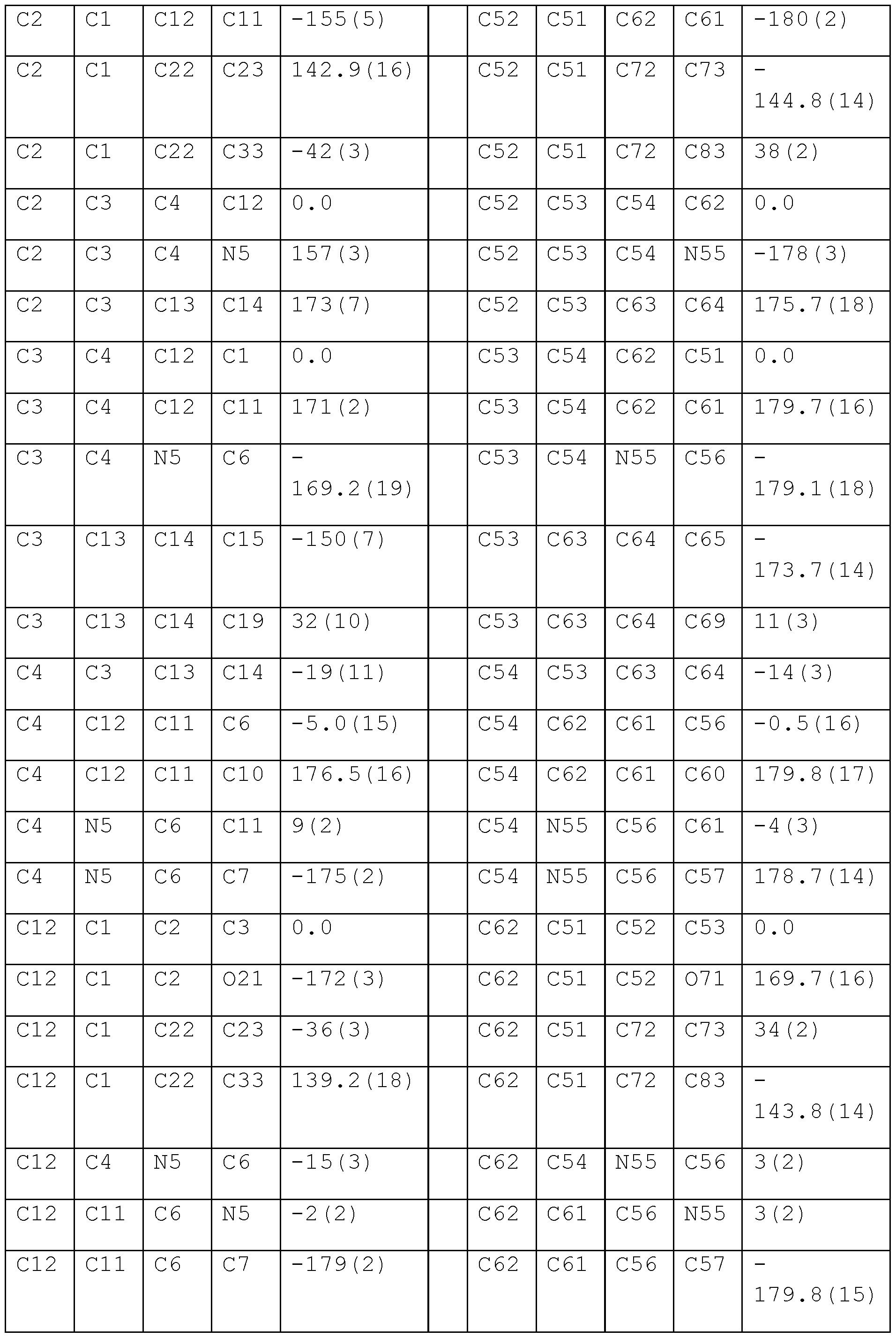
- Parameters of the geometrically determined structure model of SCY are listed in Tables 4-7. These are, respectively: atomic coordinates expressed as fractions of unit cell parameters ( ⁇ 10 4 ) and equivalent isotropic atomic displacement parameters U eq ( ⁇ 2 ⁇ 10 3 ) for SCY, wherein U eq values are defined as 1/3 of the trace of the orthogonalized Uu tensor (Table 4), bond lengths (Table 5) as well as valence (Table 6) and torsion angles (Table 7).
- a scytonemin sample obtained according to Example 1 was dissolved in DMSO (dimethylsulfoxide) to a concentration of 1% by weight and subsequently, with a spectrophotometer used according to the standard procedures (manufacturer: Varian, model: CARY 100 Scan) absorbance was measured for two wavelengths (305 and 393 nm) in a cuvette with 1 cm thickness. The following results were obtained : UV absorption and extinction coefficients:
Landscapes
- Chemical & Material Sciences (AREA)
- Organic Chemistry (AREA)
- Life Sciences & Earth Sciences (AREA)
- Health & Medical Sciences (AREA)
- Engineering & Computer Science (AREA)
- Zoology (AREA)
- Wood Science & Technology (AREA)
- General Health & Medical Sciences (AREA)
- Biotechnology (AREA)
- Genetics & Genomics (AREA)
- Bioinformatics & Cheminformatics (AREA)
- Microbiology (AREA)
- Biochemistry (AREA)
- General Engineering & Computer Science (AREA)
- Public Health (AREA)
- Animal Behavior & Ethology (AREA)
- Veterinary Medicine (AREA)
- Chemical Kinetics & Catalysis (AREA)
- Tropical Medicine & Parasitology (AREA)
- Medicinal Chemistry (AREA)
- Biomedical Technology (AREA)
- Virology (AREA)
- Birds (AREA)
- Epidemiology (AREA)
- General Chemical & Material Sciences (AREA)
- Dermatology (AREA)
- Mycology (AREA)
- Preparation Of Compounds By Using Micro-Organisms (AREA)
- Micro-Organisms Or Cultivation Processes Thereof (AREA)
- Cosmetics (AREA)
Abstract
Description


Claims
Priority Applications (13)
| Application Number | Priority Date | Filing Date | Title |
|---|---|---|---|
| BR112023024934A BR112023024934A2 (en) | 2021-05-28 | 2022-05-27 | PROCESS FOR ISOLATION AND CULTURE OF STRAINS, THE STRAINS, USE THEREOF, MEDIUM FOR CULTURE THEM AND FORM OF CYTONEMINE |
| ES202390033U ES1311217Y (en) | 2021-05-28 | 2022-05-27 | Scytonemin crystals, pigments and cosmetic products derived therefrom |
| DE212022000216.4U DE212022000216U1 (en) | 2021-05-28 | 2022-05-27 | Medium for cultivation, crystalline form of scytonemin and uses thereof |
| EP22811716.4A EP4347788A4 (en) | 2021-05-28 | 2022-05-27 | A process for the isolation and culture of strains, the strains, use thereof, media for culturing thereof and a form of scytonemin |
| AU2022280611A AU2022280611A1 (en) | 2021-05-28 | 2022-05-27 | A process for the isolation and culture of strains, the strains, use thereof, media for culturing thereof and a form of scytonemin |
| KR1020237044891A KR20240031237A (en) | 2021-05-28 | 2022-05-27 | Method for isolating and cultivating the strain, the strain, its use, its culture medium and sushitonemin form |
| JP2023573379A JP2024520094A (en) | 2021-05-28 | 2022-05-27 | Method for isolating and culturing the strain, the strain, its use, a medium for culturing it and a form of skinetmin |
| MX2023014056A MX2023014056A (en) | 2021-05-28 | 2022-05-27 | A process for the isolation and culture of strains, the strains, use thereof, media for culturing thereof and a form of scytonemin. |
| CN202280052766.7A CN118019839A (en) | 2021-05-28 | 2022-05-27 | Method for isolating and culturing a strain, use thereof, medium for culturing same and form of pseudocladophyllin |
| CA3220326A CA3220326A1 (en) | 2021-05-28 | 2022-05-27 | A process for the isolation and culture of strains, the strains, use thereof, media for culturing thereof and a form of scytonemin |
| IL308938A IL308938A (en) | 2021-05-28 | 2022-05-27 | Process for isolation and culture of strains, their uses, growth media for their culture and configuration of SCYTONEMIN |
| US18/564,630 US20240263133A1 (en) | 2021-05-28 | 2022-05-27 | Process for the isolation and culture of strains, the strains, use thereof, media for culturing thereof and a form of scytonemin |
| ZA2023/11099A ZA202311099B (en) | 2021-05-28 | 2023-11-30 | A process for the isolation and culture of strains, the strains, use thereof, media for culturing thereof and a form of scytonemin |
Applications Claiming Priority (2)
| Application Number | Priority Date | Filing Date | Title |
|---|---|---|---|
| PL437991A PL437991A1 (en) | 2021-05-28 | 2021-05-28 | Method of isolation and cultivation of strains, strains, their application, medium for their cultivation and form of scytonemin |
| PLP.437991 | 2021-05-28 |
Publications (1)
| Publication Number | Publication Date |
|---|---|
| WO2022250557A1 true WO2022250557A1 (en) | 2022-12-01 |
Family
ID=84230143
Family Applications (1)
| Application Number | Title | Priority Date | Filing Date |
|---|---|---|---|
| PCT/PL2022/050034 Ceased WO2022250557A1 (en) | 2021-05-28 | 2022-05-27 | A process for the isolation and culture of strains, the strains, use thereof, media for culturing thereof and a form of scytonemin |
Country Status (16)
| Country | Link |
|---|---|
| US (1) | US20240263133A1 (en) |
| EP (1) | EP4347788A4 (en) |
| JP (1) | JP2024520094A (en) |
| KR (1) | KR20240031237A (en) |
| CN (1) | CN118019839A (en) |
| AU (1) | AU2022280611A1 (en) |
| BR (1) | BR112023024934A2 (en) |
| CA (1) | CA3220326A1 (en) |
| CL (1) | CL2023003527A1 (en) |
| DE (1) | DE212022000216U1 (en) |
| ES (1) | ES1311217Y (en) |
| IL (1) | IL308938A (en) |
| MX (1) | MX2023014056A (en) |
| PL (1) | PL437991A1 (en) |
| WO (1) | WO2022250557A1 (en) |
| ZA (1) | ZA202311099B (en) |
Citations (2)
| Publication number | Priority date | Publication date | Assignee | Title |
|---|---|---|---|---|
| WO2000024369A1 (en) * | 1998-10-23 | 2000-05-04 | Nouvab Inc | Solar radiation protection composition |
| ES2668420A1 (en) * | 2016-11-17 | 2018-05-18 | Universidad De Las Palmas De Gran Canaria | Procedure for obtaining escitonemine and dihydroscitonemine, antioxidant and regulatory substances of redox homeostasis |
-
2021
- 2021-05-28 PL PL437991A patent/PL437991A1/en unknown
-
2022
- 2022-05-27 BR BR112023024934A patent/BR112023024934A2/en unknown
- 2022-05-27 JP JP2023573379A patent/JP2024520094A/en active Pending
- 2022-05-27 CA CA3220326A patent/CA3220326A1/en active Pending
- 2022-05-27 EP EP22811716.4A patent/EP4347788A4/en active Pending
- 2022-05-27 DE DE212022000216.4U patent/DE212022000216U1/en active Active
- 2022-05-27 WO PCT/PL2022/050034 patent/WO2022250557A1/en not_active Ceased
- 2022-05-27 US US18/564,630 patent/US20240263133A1/en active Pending
- 2022-05-27 AU AU2022280611A patent/AU2022280611A1/en active Pending
- 2022-05-27 CN CN202280052766.7A patent/CN118019839A/en active Pending
- 2022-05-27 MX MX2023014056A patent/MX2023014056A/en unknown
- 2022-05-27 IL IL308938A patent/IL308938A/en unknown
- 2022-05-27 ES ES202390033U patent/ES1311217Y/en active Active
- 2022-05-27 KR KR1020237044891A patent/KR20240031237A/en active Pending
-
2023
- 2023-11-27 CL CL2023003527A patent/CL2023003527A1/en unknown
- 2023-11-30 ZA ZA2023/11099A patent/ZA202311099B/en unknown
Patent Citations (2)
| Publication number | Priority date | Publication date | Assignee | Title |
|---|---|---|---|---|
| WO2000024369A1 (en) * | 1998-10-23 | 2000-05-04 | Nouvab Inc | Solar radiation protection composition |
| ES2668420A1 (en) * | 2016-11-17 | 2018-05-18 | Universidad De Las Palmas De Gran Canaria | Procedure for obtaining escitonemine and dihydroscitonemine, antioxidant and regulatory substances of redox homeostasis |
Non-Patent Citations (3)
| Title |
|---|
| BALSKUS EMILY P., CASE REBECCA J., WALSH CHRISTOPHER T.: "The biosynthesis of cyanobacterial sunscreen scytonemin in intertidal microbial mat communities : Scytonemin biosynthesis in intertidal microbial mats", FEMS MICROBIOLOGY ECOLOGY, ELSEVIER, NL, vol. 77, no. 2, 1 August 2011 (2011-08-01), NL , pages 322 - 332, XP093012175, ISSN: 0168-6496, DOI: 10.1111/j.1574-6941.2011.01113.x * |
| GAO XIANG, JING XIN, LIU XUFENG, LINDBLAD PETER: "Biotechnological Production of the Sunscreen Pigment Scytonemin in Cyanobacteria: Progress and Strategy", MARINE DRUGS, vol. 19, no. 3, 1 January 2021 (2021-01-01), pages 129, XP093012173, DOI: 10.3390/md19030129 * |
| See also references of EP4347788A4 * |
Also Published As
| Publication number | Publication date |
|---|---|
| CL2023003527A1 (en) | 2024-07-12 |
| PL437991A1 (en) | 2022-12-05 |
| DE212022000216U1 (en) | 2024-04-08 |
| ES1311217U (en) | 2024-10-21 |
| JP2024520094A (en) | 2024-05-21 |
| KR20240031237A (en) | 2024-03-07 |
| IL308938A (en) | 2024-01-01 |
| AU2022280611A1 (en) | 2024-01-04 |
| MX2023014056A (en) | 2024-03-15 |
| EP4347788A4 (en) | 2025-05-07 |
| CA3220326A1 (en) | 2022-12-01 |
| US20240263133A1 (en) | 2024-08-08 |
| CN118019839A (en) | 2024-05-10 |
| BR112023024934A2 (en) | 2024-02-15 |
| EP4347788A1 (en) | 2024-04-10 |
| ZA202311099B (en) | 2025-05-28 |
| ES1311217Y (en) | 2025-01-14 |
Similar Documents
| Publication | Publication Date | Title |
|---|---|---|
| Iqbal et al. | Biogenic synthesis of green and cost effective iron nanoparticles and evaluation of their potential biomedical properties | |
| Kashyap et al. | Screening of microalgae for biosynthesis and optimization of Ag/AgCl nano hybrids having antibacterial effect | |
| Cui et al. | Living yeast cells as a controllable biosynthesizer for fluorescent quantum dots | |
| Xie et al. | Comparison of ultrasonic vs mechanochemistry methods for fabrication of mixed-ligand Zn-based MOFs for electrochemical determination of luteolin | |
| Avilés et al. | Structures, semisyntheses, and absolute configurations of the antiplasmodial α-substituted β-lactam monamphilectines B and C from the sponge Svenzea flava | |
| US20240263133A1 (en) | Process for the isolation and culture of strains, the strains, use thereof, media for culturing thereof and a form of scytonemin | |
| US20040053375A1 (en) | Microbiological method of the biosynthesis of natural blue-violet colorants violacein and deoxyviolacein and the utilization thereof | |
| CN110040717B (en) | Method for preparing high-purity multicolor carbon dots by regulating and controlling carbon nanocrystalline morphology and application | |
| Ozkan | Screening diatom strains belonging to Cyclotella genus for chitin nanofiber production under photobioreactor conditions: Chitin productivity and characterization of physicochemical properties | |
| EA050967B1 (en) | METHOD OF ISOLATION AND CULTIVATION OF STRAINS, STRAINS, THEIR USE, MEDIA FOR THEIR CULTIVATION AND FORM OF SCITONEMIN | |
| HK40110022A (en) | Process for isolation and culture of strains, the strains, use thereof, media for culturing thereof and a form of scytonemin | |
| Kato et al. | Crystal structure of 1, 7-dihydroxyxanthone from Weddellina squamulosa Tul | |
| CN115403496B (en) | Compound, preparation method thereof and application thereof in oxidative damage repair | |
| Lim et al. | Synthesis and Characterization of the Large Single Crystal of Fully K+-exchanged Zeolite X (FAU),| K 80|[Si 112 Al 80 O 384]-FAU (Si/Al= 1.41) | |
| WO2000053792A1 (en) | Substances wk-5344a and wk-5344b and process for producing the same | |
| Luo et al. | Synthesis of Eu, Sm and Dy metal-organic framework nanosheets based on pyridyl carboxylic acid and their cytotoxic mechanism in vitro | |
| RU2488586C2 (en) | Solid forms of ortataxel | |
| CN117045666B (en) | Nanometer aluminum preparation of doxorubicin/zoledronic acid, and preparation method and application thereof | |
| DE3687188T2 (en) | SCYTOPHYCINE. | |
| CN117414436B (en) | A black phosphorus-methylene blue nanocomposite for precise targeted treatment of Tau phosphorylation and its synthesis method | |
| Djamaan et al. | Biotransformation of Microcrystalline Cellulose from Rice Straw Waste using TGJPC-120 Bacteria and its Characterization | |
| CN116478121B (en) | New compound extracted from endophytic metabolites of Camellia chrysantha and preparation method thereof | |
| CN108659037A (en) | The polymorph and preparation method of valproic acid phospholipid derivative | |
| WO2004065413A1 (en) | Novel substances k01-b0171 and process for producing the same | |
| Kazemi | Isolation, purification and characterization of bacteriochlorophyll c for engineering of novel photonic materials |
Legal Events
| Date | Code | Title | Description |
|---|---|---|---|
| 121 | Ep: the epo has been informed by wipo that ep was designated in this application |
Ref document number: 22811716 Country of ref document: EP Kind code of ref document: A1 |
|
| WWE | Wipo information: entry into national phase |
Ref document number: MX/A/2023/014056 Country of ref document: MX Ref document number: 3220326 Country of ref document: CA |
|
| WWE | Wipo information: entry into national phase |
Ref document number: 2023573379 Country of ref document: JP Ref document number: U202390033 Country of ref document: ES Ref document number: P6003076/2023 Country of ref document: AE |
|
| WWE | Wipo information: entry into national phase |
Ref document number: 12023553244 Country of ref document: PH Ref document number: 18564630 Country of ref document: US Ref document number: 308938 Country of ref document: IL Ref document number: 2301007793 Country of ref document: TH |
|
| REG | Reference to national code |
Ref country code: BR Ref legal event code: B01A Ref document number: 112023024934 Country of ref document: BR |
|
| WWE | Wipo information: entry into national phase |
Ref document number: 806559 Country of ref document: NZ Ref document number: 2022280611 Country of ref document: AU Ref document number: AU2022280611 Country of ref document: AU |
|
| WWE | Wipo information: entry into national phase |
Ref document number: 202347089001 Country of ref document: IN Ref document number: 202393505 Country of ref document: EA |
|
| WWE | Wipo information: entry into national phase |
Ref document number: 2022811716 Country of ref document: EP |
|
| ENP | Entry into the national phase |
Ref document number: 2022280611 Country of ref document: AU Date of ref document: 20220527 Kind code of ref document: A |
|
| WWE | Wipo information: entry into national phase |
Ref document number: 523451703 Country of ref document: SA |
|
| WWE | Wipo information: entry into national phase |
Ref document number: 202280052766.7 Country of ref document: CN |
|
| ENP | Entry into the national phase |
Ref document number: 2022811716 Country of ref document: EP Effective date: 20240102 |
|
| ENP | Entry into the national phase |
Ref document number: 112023024934 Country of ref document: BR Kind code of ref document: A2 Effective date: 20231128 |
|
| WWE | Wipo information: entry into national phase |
Ref document number: 523451703 Country of ref document: SA |
|
| WWE | Wipo information: entry into national phase |
Ref document number: 523451703 Country of ref document: SA |
|
| WWE | Wipo information: entry into national phase |
Ref document number: 523451703 Country of ref document: SA |
|
| WWG | Wipo information: grant in national office |
Ref document number: 523451703 Country of ref document: SA |