WO2023002890A1 - 針状体を利用した細胞内反応の制御手段 - Google Patents
針状体を利用した細胞内反応の制御手段 Download PDFInfo
- Publication number
- WO2023002890A1 WO2023002890A1 PCT/JP2022/027506 JP2022027506W WO2023002890A1 WO 2023002890 A1 WO2023002890 A1 WO 2023002890A1 JP 2022027506 W JP2022027506 W JP 2022027506W WO 2023002890 A1 WO2023002890 A1 WO 2023002890A1
- Authority
- WO
- WIPO (PCT)
- Prior art keywords
- needle
- cell
- cells
- particles
- shaped
- Prior art date
- Legal status (The legal status is an assumption and is not a legal conclusion. Google has not performed a legal analysis and makes no representation as to the accuracy of the status listed.)
- Ceased
Links
Images




Classifications
-
- C—CHEMISTRY; METALLURGY
- C12—BIOCHEMISTRY; BEER; SPIRITS; WINE; VINEGAR; MICROBIOLOGY; ENZYMOLOGY; MUTATION OR GENETIC ENGINEERING
- C12M—APPARATUS FOR ENZYMOLOGY OR MICROBIOLOGY; APPARATUS FOR CULTURING MICROORGANISMS FOR PRODUCING BIOMASS, FOR GROWING CELLS OR FOR OBTAINING FERMENTATION OR METABOLIC PRODUCTS, i.e. BIOREACTORS OR FERMENTERS
- C12M35/00—Means for application of stress for stimulating the growth of microorganisms or the generation of fermentation or metabolic products; Means for electroporation or cell fusion
-
- G—PHYSICS
- G01—MEASURING; TESTING
- G01N—INVESTIGATING OR ANALYSING MATERIALS BY DETERMINING THEIR CHEMICAL OR PHYSICAL PROPERTIES
- G01N33/00—Investigating or analysing materials by specific methods not covered by groups G01N1/00 - G01N31/00
- G01N33/48—Biological material, e.g. blood, urine; Haemocytometers
- G01N33/50—Chemical analysis of biological material, e.g. blood, urine; Testing involving biospecific ligand binding methods; Immunological testing
- G01N33/5005—Chemical analysis of biological material, e.g. blood, urine; Testing involving biospecific ligand binding methods; Immunological testing involving human or animal cells
- G01N33/5008—Chemical analysis of biological material, e.g. blood, urine; Testing involving biospecific ligand binding methods; Immunological testing involving human or animal cells for testing or evaluating the effect of chemical or biological compounds, e.g. drugs, cosmetics
- G01N33/502—Chemical analysis of biological material, e.g. blood, urine; Testing involving biospecific ligand binding methods; Immunological testing involving human or animal cells for testing or evaluating the effect of chemical or biological compounds, e.g. drugs, cosmetics for testing non-proliferative effects
- G01N33/5041—Chemical analysis of biological material, e.g. blood, urine; Testing involving biospecific ligand binding methods; Immunological testing involving human or animal cells for testing or evaluating the effect of chemical or biological compounds, e.g. drugs, cosmetics for testing non-proliferative effects involving analysis of members of signalling pathways
-
- C—CHEMISTRY; METALLURGY
- C12—BIOCHEMISTRY; BEER; SPIRITS; WINE; VINEGAR; MICROBIOLOGY; ENZYMOLOGY; MUTATION OR GENETIC ENGINEERING
- C12N—MICROORGANISMS OR ENZYMES; COMPOSITIONS THEREOF; PROPAGATING, PRESERVING, OR MAINTAINING MICROORGANISMS; MUTATION OR GENETIC ENGINEERING; CULTURE MEDIA
- C12N15/00—Mutation or genetic engineering; DNA or RNA concerning genetic engineering, vectors, e.g. plasmids, or their isolation, preparation or purification; Use of hosts therefor
- C12N15/09—Recombinant DNA-technology
- C12N15/87—Introduction of foreign genetic material using processes not otherwise provided for, e.g. co-transformation
- C12N15/89—Introduction of foreign genetic material using processes not otherwise provided for, e.g. co-transformation using microinjection
-
- C—CHEMISTRY; METALLURGY
- C12—BIOCHEMISTRY; BEER; SPIRITS; WINE; VINEGAR; MICROBIOLOGY; ENZYMOLOGY; MUTATION OR GENETIC ENGINEERING
- C12Q—MEASURING OR TESTING PROCESSES INVOLVING ENZYMES, NUCLEIC ACIDS OR MICROORGANISMS; COMPOSITIONS OR TEST PAPERS THEREFOR; PROCESSES OF PREPARING SUCH COMPOSITIONS; CONDITION-RESPONSIVE CONTROL IN MICROBIOLOGICAL OR ENZYMOLOGICAL PROCESSES
- C12Q1/00—Measuring or testing processes involving enzymes, nucleic acids or microorganisms; Compositions therefor; Processes of preparing such compositions
- C12Q1/02—Measuring or testing processes involving enzymes, nucleic acids or microorganisms; Compositions therefor; Processes of preparing such compositions involving viable microorganisms
-
- C—CHEMISTRY; METALLURGY
- C12—BIOCHEMISTRY; BEER; SPIRITS; WINE; VINEGAR; MICROBIOLOGY; ENZYMOLOGY; MUTATION OR GENETIC ENGINEERING
- C12N—MICROORGANISMS OR ENZYMES; COMPOSITIONS THEREOF; PROPAGATING, PRESERVING, OR MAINTAINING MICROORGANISMS; MUTATION OR GENETIC ENGINEERING; CULTURE MEDIA
- C12N15/00—Mutation or genetic engineering; DNA or RNA concerning genetic engineering, vectors, e.g. plasmids, or their isolation, preparation or purification; Use of hosts therefor
- C12N15/09—Recombinant DNA-technology
- C12N15/87—Introduction of foreign genetic material using processes not otherwise provided for, e.g. co-transformation
Definitions
- the present invention relates to a method for easily and efficiently introducing an intracellular introduction factor immobilized on a needle-shaped body into a large number of cells, and a means for controlling intracellular reactions using the method.
- the present invention provides needle-shaped particles on which a conjugate capable of binding to an intracellular molecule is immobilized, which is used for controlling intracellular reactions, and a composite substrate for cell treatment comprising the needle-shaped particles, and methods for controlling intracellular reactions using them.
- Patent Document 1 discloses a needle-shaped material for cell insertion in which an antibody against an intracellular or intercellular protein antigen is immobilized. By doing so, intracellular or intercellular proteins can be quantified, evaluated, etc. in living cells.
- Patent Documents 2 and 3 disclose a cell insertion member in which a large number of nanoneedles are arranged on a support, which is formed using photolithography, dry etching, and wet etching.
- a large number of nanoneedles are inserted into a large number of cells arranged on a substrate at the same time, and in a large number of cells , describes that the state of gene expression can be analyzed and target cells can be selectively lifted while the cells are alive.
- An object of the present invention is to provide new techniques and methods for controlling intracellular reactions that enable introduction of a given factor into a large number of cells with simple and efficient manipulation.
- the present invention provides a new technique for controlling intracellular reactions, which makes it possible to treat a large number of cells with simple manipulations and does not allow the introduced factors to remain in the cells after manipulation.
- the purpose is to provide methods and methods.
- a needle-shaped body on which an intracellularly-introduced factor is immobilized is brought into contact with a cell to which a weight is bound, and a part of the needle-shaped body is inserted into the cell to introduce the intracellularly-introduced factor. introducing into the cell, and extracting a portion of the needle-shaped body from the cell;
- a method for producing a modified cell comprising: [2] The method of [1], wherein the weight is one or more substances selected from the group consisting of resins, metals, magnetic substances, and combinations thereof. [3] The method of [1] or [2], wherein the weight has a bead-like form.
- [4] one or more selected from the group consisting of centrifugal force, magnetic force, water flow, water pressure, and electrostatic interaction in the step of inserting a portion of the needle-shaped body into the cell and/or extracting it from the cell;
- the intracellularly-introduced factor is a conjugate capable of binding to an intracellular molecule, and the needle-like structure is extracted from the cell, and the intracellular molecule bound to the conjugate in the cell is transferred to the needle.
- [6] The method of [5], wherein the conjugate is a nucleic acid, protein, peptide, or low-molecular-weight compound. [7] The method of [5] or [6], wherein the conjugate is an antibody or fragment thereof. [8] The method of [1] to [4], wherein the intracellularly-introduced factor is a physiologically active substance, and the needle-shaped body is extracted from the cell to allow the intracellularly-introduced factor to remain in the cell. either way.
- the physiologically active substance includes nucleic acids, peptides, proteins, sugars, polysaccharides, fatty acids, cholesterol, lipids, signaling substances, ligand substances, hormone substances, cytokines, ions, metal particles, magnetic fine particles, inorganic compounds, and quantum dots. , one or more substances selected from the group consisting of organic compounds and drugs.
- the method of [8] or [9], wherein the bond between the physiologically active substance and the needle-shaped structure is a bond that can be separated in cells.
- the bond between the intracellularly introduced factor and the needle-shaped structure is an electrostatic bond, a bond due to hydrophobic interaction, a chelate bond, a covalent bond that is cleaved in cells, or a bond via a photocleavable linker.
- the method of [10] which is at least one selected from the group consisting of binding via an enzyme-cleavable linker.
- the method of [12], wherein the acicular portion of the acicular particles has a length of 1 to 50 ⁇ m.
- the method of [12] or [13], wherein the acicular particles are zinc oxide.
- [15] The method according to any one of [1] to [14], wherein the needle-like structure is immobilized on a substrate.
- the substrate has a binder on its surface, and a portion of the needle-like structure is fixed to the substrate by the binder.
- the binder is made of a protein non-adsorptive material.
- the needle-like structure is manufactured using one or more selected from the group consisting of photolithography, dry etching, wet etching, and combinations thereof.
- a method for analyzing the function of the intracellular factor comprising the step of analyzing the state of the modified cell produced by the method of any one of [1] to [19].
- the step of analyzing the state of the modified cells analyzes one or more selected from the group consisting of cell growth rate, survival rate, metabolism, quantification of reactive oxygen species, and gene expression of [20] Method.
- needle-shaped particles in which a conjugate capable of binding to an intracellular molecule is immobilized and a composite substrate for cell treatment comprising the needle-shaped particles
- a composite substrate for cell treatment comprising the needle-shaped particles
- the present invention is based on these new findings and includes the following inventions.
- a composite base material for cell treatment comprising needle-like particles immobilized on a base material.
- the composite base material for cell treatment of [6] wherein the acicular portion of the acicular particles has a length of 1 to 50 ⁇ m.
- the composite substrate for cell treatment of [13], wherein the conjugate is a nucleic acid, protein, peptide, or low-molecular-weight compound.
- [16] a step of inserting a portion of the acicular particles of any one of [1] to [5] or the acicular particles of the composite substrate for cell treatment of any one of [13] to [15] into cells; a step of binding an intracellular molecule with the conjugate immobilized on the acicular particles; and extracting the acicular particles from the cell together with the intracellular molecule bound via the conjugate; including, A method for producing a modified cell in which the function of the intracellular molecule is removed or reduced. [17] The method of [16], wherein the modified cell is used to analyze the function of the intracellular molecule.
- the step of inserting a portion of the acicular particles into and/or withdrawing from the cell is one or more selected from the group consisting of centrifugal force, magnetic force, water flow, water pressure, and electrostatic interaction.
- the method of [16] or [17] performed using an external force.
- [20] a step of inserting a portion of the acicular particles of any one of [1] to [5] or the acicular particles of the composite substrate for cell treatment of any one of [13] to [15] into cells; the step of binding the intracellular molecule with the conjugate immobilized on the acicular particles; extracting the acicular particles from the cell together with the intracellular molecule bound via the conjugate; analyzing the state of the cell; including, A method for analyzing the function of the intracellular molecule.
- the method of [20] wherein the step of analyzing the state of cells analyzes one or more selected from the group consisting of cell growth rate, viability, metabolism, quantification of reactive oxygen species, and gene expression.
- the step of inserting a portion of the acicular particles into and/or extracting from the cell is one or more selected from the group consisting of centrifugal force, magnetic force, water flow, water pressure, and electrostatic interaction; The method of [20] or [21] performed using an external force. [23] The method according to any one of [20] to [22], wherein a spindle is bound to the cell.
- This specification is the basis of the priority of this application, and the contents described in the specifications of Japanese Patent Application No. 2021-119463 and Japanese Patent Application No. 2021-119466 filed on July 20, 2021. contain. All publications, patents and patent applications cited herein are hereby incorporated by reference in their entirety.
- INDUSTRIAL APPLICABILITY it is possible to provide new techniques and methods for controlling intracellular reactions that enable introduction of a given factor into a large number of cells with simple and efficient manipulation. .
- a new method for controlling intracellular reactions which enables treatment of a large number of cells with a simple operation, and which does not allow the introduced factors to remain in the cells after the operation. technology and methods can be provided.
- FIG. 1 is a schematic diagram showing a method for producing a composite base material for cell treatment on which acicular particles are immobilized.
- FIG. 2 is a photographic view of acicular particles (Panatetra) immobilized on the composite base material for cell treatment.
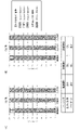
- FIG. 3 is a graph showing the percentage of activated cells (CD25 + or CD137 + ) in PBMCs obtained by inserting and removing needles of Panatetra coated with anti-Cbl antibody. (A) shows the results 1 day after the start of culture, and (B) shows the results 3 days after the start of culture.
- FIG. 1 is a schematic diagram showing a method for producing a composite base material for cell treatment on which acicular particles are immobilized.
- FIG. 2 is a photographic view of acicular particles (Panatetra) immobilized on the composite base material for cell treatment.
- FIG. 3 is a graph showing the percentage of activated cells (CD25 + or CD137 + ) in PBMCs obtained by inserting and removing
- FIG. 4 is a graph showing the viability of cells (that is, the efficiency of inserting needles of Pana-tetra into cells) when cells are subjected to repeated treatments with the composite base material for cell treatment.
- FIG. 5 shows differentiated cells (neuronal cells with a CD56 + phenotype) in MSCs (mesenchymal stem cells) obtained by inserting and removing needle-like parts of Panatetra coated with anti-GSK3 ⁇ antibody. It is a graph showing the ratio of .
- the "needle-shaped body” means a fine structure having at least one needle-shaped part.
- the "needle-shaped part” has a shape that is long and narrow, has a sufficiently small tip and width, and causes little or no damage, preferably no damage, to the cell when inserted into the cell.
- the needle-like part may have, for example, a cylindrical shape, a conical shape, a cylindrical shape (such as a prismatic shape), a pyramidal shape, etc. (but not limited to these), and the tip thereof is sharp. It may or may not be sharp. Cylindrical or conical shapes are preferred from the viewpoints of low invasiveness to cells and insertion efficiency.
- the length of the acicular part is 0.5 to 100 ⁇ m, preferably 1 to 50 ⁇ m, more preferably 5 to 40 ⁇ m, still more preferably 10 to 30 ⁇ m (eg, 10 ⁇ m, 20 ⁇ m, etc.).
- the aspect ratio of the needle-like portion is not particularly limited, but can be about 5:1 to 50:1, preferably 10:1 to 40:1.
- the "needle” is made of a material that has little or no toxicity to cells, preferably no toxicity at all. , quartz, nickel oxide, silica, alumina, diamond, titania, zirconia, etc.), metals (e.g., gold, silver, copper, platinum, aluminum, etc.), metal crystals (e.g., tungsten, titanium, silicon crystals, zirconium, etc.), Glass, resin (e.g., polyethylene, polypropylene, cyclic polyolefin, cyclic olefin copolymer, polyester, polystyrene, polymethyl acrylate, polylactic acid, polyether ether ketone, fluororesin, etc.), silicon nitride, silicon, etc. (these include is not limited), needle-like solid materials can be preferably used.
- quartz, nickel oxide, silica, alumina, diamond, titania, zirconia, etc. metals (e.g., gold, silver, copper, platinum,
- the form of the "needle” is not particularly limited as long as it allows the needle to be inserted into the cell and then removed (also referred to as “withdrawal”).
- the "needle-shaped body” can have a form of needle-shaped particles or immobilized on a substrate.
- needle-like particles means fine grains with at least one needle-like part.
- the needle-like particles have a shape in which one or more needle-like portions are arranged/bonded at the center, and may be, for example, rod-like, L-shaped, V-shaped, T-shaped, Y-shaped, or radial (for example, It can take a shape such as tripod, quattropod, tetrapod, etc., but is not limited to these.
- each needle-like part may have the same shape and/or length, or may have a different shape and/or length.
- the central portion may have a structure in which a plurality of needle-like portions are directly connected to each other, or may have a structure in which one or a plurality of needle-like portions are connected to a core member.
- the size of the needle-like particles may vary depending on the length and number of needle-like portions provided, but may be of a size that fits in a sphere with a radius of 0.5 ⁇ m to 100 ⁇ m.
- the acicular particles in the present invention are made of zinc oxide having a plurality of needle-like portions, and more preferably needle-like portions made of zinc oxide single crystals with a length of 10 to 20 ⁇ m are arranged in a tetrapod shape. It has a shape As such acicular particles, for example, Panatetra (registered trademark) WZ-0501 (manufactured by Amtech) and the like can be preferably used.
- the “form fixed to the base material” means a base material having a plurality of needle-like bodies (or needle-like parts) on its surface.
- the needle-shaped bodies or needle-shaped particles immobilized on the surface of the substrate mean a plurality of needle-shaped bodies or needle-shaped particles.
- the “substrate” is not particularly limited as long as it can function as a support by immobilizing the needle-shaped structure on its surface.
- LLDPE Linear Low Density Polyethylene
- VLDPE Very Low Density Polyethylene/ULDPE, Ultra Low Density Polyethylene
- LDPE Low Density Polypropylene
- Polyethylene polyacrylonitrile, styrene-butadiene copolymer, (meth)acrylic acid ester polymer, fluorine resin, etc.), silica gel, crosslinked dextran, polysaccharide, polysaccharides such as agarose, glass, metal, magnetic substances, and combinations thereof etc.
- the shape of the substrate is not particularly limited as long as it can immobilize the needle-shaped body. , tubes, microplates, microtubes, cells, cuvettes, dishes, flasks, bags), fibers, gels and the like. Specific examples include, but are not limited to, polyethylene films, polyethylene plates, magnetic beads, and the like.
- the fixation of the needle-shaped body to the surface of the base material can be performed using any means as long as the binding between the two can be maintained even after the needle-shaped part is inserted into and removed from the cell.
- the needle-shaped structure can be immobilized on the surface of the substrate by, for example, a physical adsorption method, a covalent bonding method, an ionic bonding method, embedding in the substrate, or using a binder (adhesive). It is not limited to these.
- the "binder” that can be used in the present invention is not particularly limited as long as it can fix the needle-shaped structure to the surface of the base material.
- -Hydroxyethyl methacrylate) Poly HEMA
- polyvinylpyrrolidone polyvinylpyrrolidone
- vinyl acetate resin urethane resin
- vinyl chloride resin epoxy resin
- vinyl chloride/vinyl acetate copolymer resin modified silicone resin
- ethyl 2-cyanoacrylate polystyrene, chloroprene rubber, nitrile rubber, styrene-butadiene rubber, nitrocellulose, starch, dextrin, alginic acid, agarose, gelatinous proteins (gelatin, elastin, fibrin, etc.), etc., and a combination of one or more of these can be used.
- the binder is applied to a predetermined region on the surface of the base material, and when the applied binder is solidified, the needle-like structure partly in contact with the binder, preferably partly embedded, is adhered to the binder. Immobilize on the surface of the substrate.
- the binder has low binding property and adsorptivity to the "intracellularly introduced factor" described in detail below, and examples of such binders include PVA, Poly HEMA, and the like.
- Binders can also include molecules that interact with cell surface proteins. Such molecules include, for example, fibronectin, laminin, collagen, cadherin, or fragments thereof, and the like, and combinations of one or more of these can be utilized. Such molecules may for example be pre-mixed with the binder component and then coated together on the substrate, or may be coated against the binder on the substrate and may be included in any convenient manner. . By using such a molecule, it is possible to suppress deterioration of the cell state due to a change from the normal culture environment when treating the cells.
- the fixation of the needle-shaped structure to the surface of the base material can be performed by manufacturing the base material and the needle-like structure (or the needle-shaped part) by integral molding or integral processing.
- the surface of the base material may have a fine uneven structure capable of accommodating cells together with the immobilized needle-shaped bodies.
- the microrelief structure accommodates and retains cells by side walls (e.g., inner walls of recesses) and bottoms, restricts lateral movement of cells (e.g., horizontal movement relative to the surface of a substrate), and prevents cells from This can reduce or prevent spicule damage (eg, cutting, tearing, etc.) that can occur with lateral movement of the needle.
- the shape of the "fine uneven structure” is not particularly limited as long as it is a shape capable of exhibiting the above effects. It has a concave shape (concave portion) such as a columnar shape, a prismatic shape, or the like.
- a needle-shaped body is arranged and fixed at the bottom of the recess.
- the size of the concave portion is not particularly limited as long as it is a size capable of exhibiting the above effects. can be done.
- Each recess may accommodate a single cell or a plurality of cells.
- needle-shaped bodies in a form "immobilized on a base material” may be referred to as a "composite base material for cell treatment”.
- the "intracellular factor” is immobilized on the needle-shaped body.
- intracellular factors include conjugates capable of binding intracellular molecules.
- a "conjugate capable of binding to an intracellular molecule” is capable of binding, preferably selectively binding, more preferably specifically binding to a target intracellular molecule in the introduced cell to form a complex. It can be selected as appropriate according to the target intracellular molecule.
- conjugates include nucleic acids (DNA, RNA, DNA-RNA hybrids, etc.), proteins (antibodies, antigens, enzymes, substrates, coenzymes, ligands, receptors, subunits of complexes, or fragments thereof).
- the "conjugate capable of binding to an intracellular molecule” is preferably one that itself does not directly affect the expression of any gene.
- “does not directly affect” means that any intracellular signal transduction pathway is not directly activated due to the introduction of the conjugate.
- the "conjugate capable of binding to an intracellular molecule” is an antibody or fragment thereof capable of binding, preferably selectively binding, more preferably specifically binding to a target intracellular molecule. is.
- “Fragments” of antibodies include Fab, Fab', F(ab') 2 , Fv, scFv, dsFv, diabodies, sc(Fv) 2 and the like, and multimers of these fragments (e.g., dimers, trimers, , tetramers, polymers) can also be utilized in the present invention.
- intracellular factors include those that exhibit any function in the introduced cell, or those that are predicted to exhibit any function.
- physiologically active substances include nucleic acids (DNA, RNA, DNA-RNA hybrids, etc.), plasmids, virus particles, chromosomes, etc.; proteins, amino acids, oligopeptides, polypeptides, multi-subunit proteins; saccharides, polysaccharides, one or more substances selected from the group consisting of fatty acids, cholesterol, lipids, signaling substances, ligand substances, hormone substances, cytokines, ions, metal particles, magnetic fine particles, inorganic compounds, quantum dots, organic compounds, drugs, etc. Examples include, but are not limited to.
- the method of binding (immobilizing) the "needle” and the "intracellularly introduced factor” can be appropriately selected according to the type of "intracellularly introduced factor” to be used.
- the "intracellular transfer factor” to be used is the above-described "conjugate capable of binding to an intracellular molecule”
- the binding (immobilization) of the needle-shaped structure and the conjugate is performed by binding (immobilizing) the needle-shaped structure to the cell.
- a method well known to those skilled in the art may be used as long as there is sufficient strength to maintain the bond even when it is inserted and then pulled out.
- the bonding method examples include a physical adsorption method, a covalent bonding method, an ionic bonding method and the like, and the covalent bonding method is preferred.
- the conjugate is a protein or peptide
- the covalent bonding method is a functional group (e.g., hydroxy group, amino group, N-hydroxysuccinimidyl group, sulfhydryl group, epoxy group, vinyl group) on the surface of the needle. group, etc.) and the carboxy terminus of the protein or peptide to form an ester bond, an amide bond, or the like.
- the surface of the needles can be treated to introduce functional groups such as polylysine, polyethyleneiminevinyltrialkoxysilane, 3-mercaptopropyltrialkoxysilane, aminoalkyltrialkoxysilane.
- functional groups such as polylysine, polyethyleneiminevinyltrialkoxysilane, 3-mercaptopropyltrialkoxysilane, aminoalkyltrialkoxysilane.
- Alkoxysilanes, epoxy-containing alkyltrialkoxysilanes, and the like can be used.
- the binding (immobilization) between the needle-shaped particles and the conjugates capable of binding to intracellular molecules may be directly bound to each other, or may be indirectly bound via a linker.
- the binding (immobilization) between the two is maintained at least until the needle-like portion is inserted into the cell. Bonds that are separated/cleaved within the nucleus and allow release of the factor into the cell are preferred.
- binding methods include electrostatic binding; binding through hydrophobic interactions; chelate binding; intracellularly cleavable covalent bonds such as disulfide bonds (S—S bonds); Examples include (but are not limited to) binding via a linker (enzyme-cleavable linker) having an enzyme-recognition sequence such as esterase, and can be performed using a method well known to those skilled in the art (JP-A-2006-166884). publications, etc.).
- All of the intracellularly introduced factors immobilized on one needle may be the same, or multiple different intracellularly introduced factors may be immobilized.
- the same intracellular factor may be immobilized on all of the needle-shaped structures, or a different intracellular factor may be immobilized on each needle-shaped structure.
- a predetermined intracellularly introduced factor may be previously immobilized on the needle-shaped body, or, when used, the desired intracellularly-introduced factor is immobilized on the needle-shaped body as described above. (That is, the needle-shaped structure at the time of provision may not have a predetermined intracellular transfer factor immobilized in advance).
- the needle-shaped body on which intracellular factors are immobilized can be used to control intracellular reactions, and can be used in a method for producing modified cells in which intracellular reactions are controlled.
- a needle means a plurality of needles.
- This manufacturing method is Contacting the needle-shaped body on which the intracellularly introduced factor is immobilized with the cell to which the weight is bound, thereby inserting a portion of the needle-shaped body into a cell to introduce the intracellular factor into the cell; extracting a portion of the needle-shaped body from the cell; including.
- Cells to be targeted in the present invention are not particularly limited, and may be either adherent cells or suspension cells.
- the origin of the cells is not particularly limited, and mammalian cells such as human, mouse, rat, monkey, dog, pig, bovine, guinea pig, and hamster cells can be preferably used.
- pluripotent stem cells refer to embryonic stem cells (ES cells) and similar pluripotent cells, that is, various tissues of the body (endoderm, mesoderm, ectoderm). refers to cells that potentially have the ability to differentiate into Cells having pluripotency similar to ES cells include "induced pluripotent stem cells” (sometimes referred to as “iPS cells”).
- pluripotent stem cells are human pluripotent stem cells.
- Induced pluripotent stem cells refer to reprogramming mammalian somatic cells or undifferentiated stem cells by introducing specific factors (nuclear reprogramming factors) such as Oct3/4, Sox2, Klf4 and c-Myc.
- pluripotent stem cells mean cells characterized by predetermined phenotypes and marker expression obtained by differentiation-inducing pluripotent stem cells.
- Marker means a cell antigen or its gene that is specifically expressed by a given cell type, such as “marker protein” and “marker gene”.
- cells may be cells collected from a living body, cultured cells, or freeze-thawed cells. It is preferable to use cells in a dissociated or dispersed state.
- the cells are bound with spindles.
- the weight of the cell is increased, which enhances the impact of contact between the cell and the spicule, facilitating the insertion of the spicule into the cell (i.e., insertion increase the probability).
- the weight one or more selected from the group consisting of resins, metals, magnetic substances, combinations thereof, and the like can be used.
- the shape of the weight is not particularly limited, it can be, for example, a bead shape.
- magnetic beads such as Dynabeads (registered trademark, Veritas Co., Ltd.) can be suitably used as the "consumer” of cells. Binding of cells to spindles can be performed using methods well known to those skilled in the art, for example, using one or more of antibodies or fragments thereof, avidin-biotin, streptavidin-biotin, and the like.
- the needle-shaped part of the needle-shaped body can be inserted into the cell by bringing the cell and the needle-shaped body into contact.
- the contact between the cells and the needle-shaped bodies is achieved by adding the needle-shaped bodies (in the form of needle-shaped particles) to the cells (or by adding the cells to the needle-shaped bodies), or by immobilizing them on a substrate. It can be carried out by adding cells to a needle-shaped body (composite base material for cell treatment) in the form of a sliver. If necessary, an external force may be applied to the contact between the cells and the needle-like structure.
- the "external force” means a force applied to cells and/or needle-shaped bodies, and includes centrifugal force, magnetic force, electrostatic interaction, water flow (water pressure), etc. One or more selected from these can be used. Centrifugation may be performed under conditions that do not damage the cells, for example, at 100 to 30000 g for 30 to 600 seconds. Water flow (water pressure) can be provided by a pipetting operation. By using an external force, it is possible to increase the impact at the time of contact between the cell and the spicule, thereby facilitating the insertion of the spicule of the spicule into the cell (that is, increasing the probability of insertion). can.
- the insertion of the needle-shaped part of the needle-shaped particles into the cells may be performed in a culture medium, preferably in a cell culture environment.
- the culture conditions are maintained at 37°C and 5% CO 2 . can be done while If the insertion time is short, it can be performed at room temperature or in an atmospheric environment.
- the intracellularly introduced factor immobilized on the needle-shaped body is the above-mentioned "conjugate capable of binding to an intracellular molecule"
- insertion of the needle-shaped part of the needle-shaped body into the cell is It is sufficient that the conjugate immobilized on the inserted needle-shaped body in the cell is maintained for a time sufficient for binding to the target intracellular molecule in the cell.
- a person skilled in the art can appropriately set the time depending on the type of intracellular molecule to be used.
- the needle-shaped portion is extracted from the cell together with the desired intracellular molecule bound to the conjugate, thereby reducing or eliminating the amount of the desired intracellular molecule within the cell.
- a modified cell in which the function of the intracellular molecule is eliminated or reduced can be obtained.
- the intracellular introduction factor immobilized on the needle-shaped body is the "physiologically active substance”
- the insertion of the needle-shaped body into the cell is It is sufficient that the physiologically active substance immobilized in the body is maintained for a sufficient period of time to be released from the needle-shaped structure (the bond is severed if necessary), and this time is sufficient for the physiologically active substance, etc.
- a person skilled in the art can appropriately set the time according to the type of binding, but the time can be, for example, about 1 to 120 minutes, preferably 5 to 60 minutes, more preferably about 30 minutes.
- the needle-shaped portion is extracted from the cell, and the physiologically active substance is allowed to remain in the cell, thereby increasing the amount of the physiologically active substance in the cell, thereby introducing the function of the physiologically active substance. , or enhanced modified cells can be obtained.
- the needle-shaped part can be extracted from the cell by applying an external force to separate the contacting cell and the needle-shaped body.
- an external force can be used as defined above.
- the steps of inserting the needle-shaped portion of the needle-shaped body into the cell and extracting the needle-shaped portion from the cell can be performed once or multiple times for the same cell.
- “multiple times” means 2 or more times, 3 or more times, 4 or more times, or 5 or more times, and the upper limit is not particularly limited. 15 times or less, or 10 times or less is preferable from the viewpoint of maintaining good conditions.
- the term “performed multiple times” as used herein means not only the case where the above steps are continuously performed multiple times, but also the case where the above steps are performed multiple times at regular time intervals.
- the resulting modified cells vary depending on the function of the introduced intracellular factors.
- the introduced intracellular factor is the above-mentioned "conjugate capable of binding to an intracellular molecule", for example, the intracellular molecule that binds to the above-mentioned conjugate is a factor that has the function of suppressing or inhibiting cell activation.
- the function of the factor is removed , or by reducing it, a modified cell in which the intracellular reaction of the factor is controlled, that is, activated can be obtained.
- the intracellular molecule that binds to the conjugate is a factor that has the function of suppressing or inhibiting differentiation induction of cells
- removal or reduction of the function of the factor can reduce the effect of the factor on cells. It is possible to obtain modified cells whose internal reaction is controlled, that is, differentiation-induced (examples of modified cells are not limited to these).
- the introduced intracellular factor is the aforementioned "physiologically active substance", for example, a factor having a function of promoting cell activation, by introducing or enhancing the function of the factor , it is possible to obtain modified cells in which the intracellular reaction of the factor is controlled, that is, in which activation is promoted.
- the physiologically active substance is a factor having a function of promoting cell differentiation induction
- the intracellular reaction of the factor is controlled by introducing or enhancing the function of the factor. That is, a modified cell induced to differentiate can be obtained (examples of modified cells are not limited to these).
- the modified cells obtained can be used to analyze the function of the introduced intracellular factors.
- Analysis of the function of the introduced intracellular factors can be performed by analyzing the state of the modified cells obtained by the above method.
- the state of the modified cells to be analyzed includes, but is not limited to, cell growth rate, viability, metabolism, reactive oxygen species, gene expression, etc., and one or more selected from these Quantify and analyze the state.
- the introduced intracellular factor is the above-mentioned "conjugate capable of binding to an intracellular molecule”
- the growth rate, survival rate, metabolism, reactive oxygen species, or gene expression in the resulting modified cells When compared to unmodified cells, the conjugate has such a function, i.e., the intracellular molecule targeted by the conjugate, is reduced, inhibited, or abolished. , metabolism, reactive oxygen species, or maintenance or promotion of gene expression.
- the conjugate when growth rate, viability, metabolism, reactive oxygen species, or gene expression is increased or induced in the resulting modified cells compared to unmodified cells has such a function, i.e., the intracellular molecule targeted by the conjugate has the function of suppressing or inhibiting growth rate, viability, metabolism, reactive oxygen species, or gene expression.
- the introduced intracellular factor is the "physiologically active substance”
- the growth rate, viability, metabolism, reactive oxygen species, or gene expression in the resulting modified cells are higher than those in unmodified cells. If the physiologically active substance is reduced, suppressed, or eliminated, the physiologically active substance has a function that contributes to the reduction, suppression, or elimination of cell growth rate, survival rate, metabolism, reactive oxygen species, or gene expression. Then you can judge.
- the physiological activity Substances can be determined to have the ability to increase or induce growth rate, viability, metabolism, reactive oxygen species, or gene expression.
- any gene to be analyzed and the protein encoded by it can be used. It is preferred that the molecule, the conjugate, and the physiologically active substance to be introduced are different.
- the present invention provides acicular particles on which a conjugate capable of binding to an intracellular molecule is immobilized, and a composite for cell treatment comprising the acicular particles immobilized on a substrate It relates to the base material.
- acicular particles on which a conjugate capable of binding to an intracellular molecule is immobilized
- a composite for cell treatment comprising the acicular particles immobilized on a substrate It relates to the base material.
- “needle-shaped particles”, “conjugates capable of binding to intracellular molecules”, “composite substrates for cell treatment” and various “immobilization” methods refer to those described above and are defined as above. It is.
- the "needle particles" and the “composite substrate for cell treatment” may be provided in a form in which the predetermined conjugate is previously immobilized on the acicular particles, or Alternatively, the desired conjugate may be immobilized on the acicular particles as described above (that is, the desired conjugate may not be previously immobilized on the acicular particles at the time of provision).
- needle-shaped particles on which a conjugate capable of binding to the intracellular molecule is immobilized and a complex for cell treatment comprising needle-shaped particles on which a conjugate capable of binding to the intracellular molecule is immobilized
- Substrates can be used in methods for producing modified cells.
- all needle-like particles mean a plurality of needle-like particles unless otherwise specified.
- the manufacturing method includes the following steps: For cell treatment comprising needle-shaped particles on which a conjugate capable of binding to the intracellular molecule is immobilized, or needle-shaped particles to which a conjugate capable of binding to the intracellular molecule is immobilized. inserting the needle-shaped portion of the needle-shaped particles in the composite substrate into cells; binding the target intracellular molecule to the conjugate immobilized on the needle-shaped particles in the cell; withdrawing the needle from the cell together with the intracellular molecule bound via the conjugate.
- the “cell”, the “insertion” of the acicular portion of the acicular particle into the cell, the “extraction” of the acicular portion from the cell, and various operations are as described above and are defined as above. It follows.
- the insertion of the needle-like portion of the needle-like particles into the cell allows the conjugate immobilized on the needle-like portion inserted in the cell to bind to the desired intracellular molecule in the cell.
- a sufficient period of time may be maintained, and this period of time can be appropriately set by those skilled in the art depending on the type of the conjugate and the target intracellular molecule. For example, 1 to 120 minutes, preferably It can be 5 to 60 minutes, more preferably about 30 minutes.
- the needle-shaped portion is extracted from the cell together with the desired intracellular molecule bound to the conjugate, thereby reducing or eliminating the amount of the desired intracellular molecule within the cell.
- a modified cell in which the function of the intracellular molecule is eliminated or reduced can be obtained. According to this embodiment, it is possible to treat a large number of cells with a simple operation, and to control intracellular reactions without the introduced factors remaining in the cells after the operation.
- the above-described "consumer” may be bound to cells as necessary.
- the weight of the cells increases, which enhances the impact of contact between the cells and the acicular particles, facilitating the insertion of the acicular part of the acicular particles into the cells (i.e., insertion increase the probability).
- the resulting modified cell is as described above and conforms to the above definition, assuming that the introduced intracellular factor is a "conjugate capable of binding to an intracellular molecule".
- the resulting modified cells can be used to analyze the functions of intracellular molecules of interest.
- the analysis of the function of the intracellular molecule of interest can be performed by analyzing the state of the modified cell in which the function of the intracellular molecule is removed or reduced, obtained by the above method. .
- the state of the modified cells to be analyzed includes, but is not limited to, cell growth rate, viability, metabolism, reactive oxygen species, gene expression, etc., and one or more selected from these The condition can be quantified and analyzed and determined as described above.
- FIG. 1 shows an outline of the method for producing a composite substrate for cell treatment on which acicular particles are immobilized in this experiment.
- a linear low-density polyethylene (LLDPE) film is covered with an LLDPE film (masking) with a hole of ⁇ 4 mm, and a water-soluble photosensitive resin (BIOSURFINE (registered trademark) AWP (manufactured by Toyo Gosei)) is applied. After adding the 1% aqueous solution, excess liquid was scraped out with a polystyrene cylinder.
- LLDPE linear low-density polyethylene
- AWP water-soluble photosensitive resin
- Needle-shaped particles (Pana Tetra (registered trademark) WZ-0501 (manufactured by Amtech)) are attached to a sponge puff (107 (NBR) for Emulsion Pact (manufactured by Shiseido)), and a photosensitive resin is added to the substrate surface using this. Pana-tetra was coated by tapping multiple times.
- the photosensitive resin was UV-crosslinked with a low-pressure mercury lamp (TUV15W/G15T8 (manufactured by Philips)), and the panatetra was adhered.
- the UV irradiation conditions were 30 minutes at a distance of 12 cm. Then, it was washed with water to wash away excess panatetra with insufficient adhesion.
- a ring-shaped member made of LDPE (inner diameter 4 mm) and a panatetra-coated portion ( ⁇ 4 mm) on the base material were aligned with each other, superimposed, and heat-sealed to produce a container-shaped composite base material for cell treatment.
- PBMC peripheral blood mononuclear cells, manufactured by HameCare
- ALyS505N-7 manufactured by Cell Science Laboratory
- fetal bovine serum manufactured by Thermo Fisher
- the cells were floated by pipetting, peeled off from the cell treatment composite substrate, collected in a 96-well plate, added with 150 ⁇ L of the medium, and cultured for 1 day or 3 days in a 37° C., 5% CO 2 environment ( Example 1).
- a composite substrate for cell treatment prepared in the same manner as the above composite substrate for cell treatment, except that neither anti-Cbl antibody nor panatetra was coated, or panatetra was coated with anti-Cbl antibody.
- Cells were treated in the same manner as above using a cell treatment composite base material prepared in the same manner as the cell treatment composite base material except that it was not coated (the former cell treatment composite base material was used). Cells treated with the substrate are referred to as “Comparative Example 1”, and cells treated with the latter composite substrate for cell treatment are referred to as “Comparative Example 2”).
- each cell culture was collected and treated with anti-CD3 antibody (fluorescence: APC-Cy7, manufactured by Biolegend), anti-CD25 antibody (fluorescence: APC, manufactured by Biolegend). ) and anti-CD137 antibody (fluorescence: Brilliant Violet 421, manufactured by Biolegend), the staining intensity of each antibody was analyzed with a flow cytometer CytoFLES S (manufactured by Beckmancoulter). For the analysis, the CD3-positive population was sorted, and the positive rate of CD25 and/or CD137 was calculated therefrom. In addition, unstained cells were used as a criterion for positive/negative determination.
- PBMC are known to be activated through an intracellular signaling cascade based on CD3 stimulation, and the expression of activation markers CD25 and CD137 is promoted (Endocrine Journal 2005, 52 (5) , 635-641; J Immunol Methods. 2008 Nov 30;339(1):23-37.).
- part of the intracellular signaling cascade based on this CD3 stimulation is inhibited by the ubiquitin ligase Cbl and cell activation is suppressed, that is, the expression of CD25 and CD137 is suppressed (Science Signaling 05 Feb 2019: Vol.12, Issue 567, eaav4373).
- Anti-Cbl antibody-coated panatetra needle insertion, removed cells showed less activated cells (CD25 ⁇ and CD137 ⁇ ) compared to Comparative Examples 1 and 2. and a significant increase in the percentage of activated cells exhibiting the active CD25 + and CD137 ⁇ , CD25 + and CD137 + , and CD25 + and CD137 ⁇ phenotypes, respectively. (Fig. 3).
- Example 1 Comparative Example 1, and Comparative Example 2
- the ratio of activated cells was higher after 3 days of culture than after 1 day of culture, and the ratio of activated cells in Example 1 was Since it is higher than Comparative Examples 1 and 2, it was confirmed that in Example 1, the progress of activation was faster than in Comparative Examples 1 and 2, and the strength was also increased (Fig. 3 (A) vs. (B)).
- Experiment 2 Efficiency improvement of treatment with composite base material for cell treatment by binding spindles to cells 2-1.
- Preparation of cells and binding of spindle Jurkat E6.1 (purchased from DS Pharma Biomedical) was cultured in ALyS505N-0 (manufactured by Cell Science Research Institute) supplemented with 2% fetal bovine serum (manufactured by Thermo Fisher).
- the cells were suspended by pipetting and mixed with an equal amount of trypan blue solution (manufactured by Fujifilm Wako Pure Chemical Industries). The cells were set on a hemocytometer and observed under a microscope, and the stained cells were counted as dead cells and the unstained cells were counted as viable cells, and the viable cell rate was calculated from these values (Example E).
- Example E the treatment was performed in the same manner as in Example E above, except that a composite substrate for cell treatment prepared in the same manner as the composite substrate for cell treatment was used, except that it was not coated with panatetra.
- Cell viability (Comparative Example F) was used.
- the survival rate is about the same as when cells that are bound to spindles are used. confirmed to be obtained.
- the treatment efficiency may decrease in practice, which may not be preferable.
- Experiment 3 Modification of Mesenchymal Stem Cells by Treatment with Composite Substrate for Cell Treatment 1-1.
- Preparation of composite base material for cell treatment A linear low-density polyethylene (LLDPE) film was covered with an LLDPE film (masking) with ⁇ 4 mm holes, and a water-soluble photosensitive resin (BIOSURFINE (registered trademark) After adding a 2% aqueous solution of AWP (manufactured by Toyo Gosei), excess liquid was scraped out with a silicon spatula.
- LLDPE linear low-density polyethylene
- BIOSURFINE water-soluble photosensitive resin
- a 5% ethanol dispersion of acicular particles (Pana-tetra (registered trademark) WZ-0501L (manufactured by Amtech); average needle length 20 ⁇ m) is dropped onto the above base material, and a bar coater (No. 20) is used to coat Pana-tetra. did the coating.
- the photosensitive resin was UV-crosslinked with a low-pressure mercury lamp (TUV15W/G15T8 (manufactured by Philips)), and the panatetra was adhered.
- the UV irradiation conditions were 30 minutes at a distance of 12 cm. Then, it was washed with water to wash away excess panatetra with insufficient adhesion.
- a ring-shaped member made of LDPE (inner diameter 4 mm) and a panatetra-coated portion ( ⁇ 4 mm) on the base material were aligned with each other, superimposed, and heat-sealed to produce a container-shaped composite base material for cell treatment.
- Antibody coating on composite base material for cell treatment 3-1 25 ⁇ L of an anti-GSK3 ⁇ antibody (manufactured by Biolegend) at a concentration of 40 ⁇ g/mL was added to the composite base material for cell treatment obtained in 1. and allowed to stand at 37° C. for 3 hours to coat panatetra with the antibody.
- the obtained antibody-coated composite substrate for cell treatment was washed once with 60 ⁇ L of PBS (manufactured by Nacalai Tesque) and then used for the following cell treatment.
- each cell culture was collected and stained with an anti-CD56 antibody (fluorescence: BV421, manufactured by Biolegend), followed by a flow cytometer CytoFLES S (manufactured by Beckmancoulter). Staining intensity was analyzed. In the analysis, the positive rate of CD56 was calculated. In addition, as a criterion for positive/negative determination, cells treated with an isotype antibody (fluorescence: BV421, manufactured by Biolegend) under the same conditions were used.
Landscapes
- Health & Medical Sciences (AREA)
- Life Sciences & Earth Sciences (AREA)
- Engineering & Computer Science (AREA)
- Chemical & Material Sciences (AREA)
- Genetics & Genomics (AREA)
- Organic Chemistry (AREA)
- Biomedical Technology (AREA)
- Biotechnology (AREA)
- Bioinformatics & Cheminformatics (AREA)
- Wood Science & Technology (AREA)
- Zoology (AREA)
- Molecular Biology (AREA)
- Immunology (AREA)
- Biochemistry (AREA)
- General Engineering & Computer Science (AREA)
- General Health & Medical Sciences (AREA)
- Microbiology (AREA)
- Physics & Mathematics (AREA)
- Urology & Nephrology (AREA)
- Hematology (AREA)
- Cell Biology (AREA)
- Analytical Chemistry (AREA)
- Biophysics (AREA)
- Proteomics, Peptides & Aminoacids (AREA)
- Food Science & Technology (AREA)
- Tropical Medicine & Parasitology (AREA)
- Plant Pathology (AREA)
- Toxicology (AREA)
- Medicinal Chemistry (AREA)
- General Physics & Mathematics (AREA)
- Pathology (AREA)
- Sustainable Development (AREA)
- Apparatus Associated With Microorganisms And Enzymes (AREA)
- Immobilizing And Processing Of Enzymes And Microorganisms (AREA)
- Micro-Organisms Or Cultivation Processes Thereof (AREA)
Abstract
Description
また、本発明は、細胞内反応の制御に利用される、細胞内分子に結合可能な結合体が固定化されている針状粒子、及び、当該針状粒子を備える細胞処理用複合基材、ならびにそれらを利用する細胞内反応の制御方法に関する。
また、本発明は、簡便な操作で多数の細胞を処理することが可能であり、かつ導入された因子が操作後の細胞に残留することのない、細胞内反応を制御するための新たな技術や手法を提供することを目的とする。
[1] 細胞内導入因子が固定化されている針状体と、錘を結合させた細胞とを接触させ、前記針状体の一部分を前記細胞内に挿入して、前記細胞内導入因子を前記細胞内に導入する工程、ならびに、
前記針状体の一部分を前記細胞より抜き出す工程、
を含む、改変細胞の製造方法。
[2] 前記錘が、樹脂、金属、磁性物質、及びそれらの組み合わせからなるから群から選択される一又は複数の物質である、[1]の方法。
[3] 前記錘がビーズ状の形態を有する、[1]又は[2]の方法。
[4] 前記針状体の一部分を細胞内へ挿入する、及び/又は細胞より抜き出す工程が、遠心力、磁力、水流、水圧、及び静電相互作用からなる群から選択される一又は複数の外力を用いて行われる、[1]~[3]のいずれかの方法。
[5] 前記細胞内導入因子が、細胞内分子に結合可能な結合体であり、前記針状体を前記細胞より抜き出して、前記細胞内において前記結合体に結合した細胞胞内分子を前記針状体と共に、前記細胞より抜き出すことを含む、[1]~[4]のいずれかの方法。
[6] 前記結合体が、核酸、タンパク質、ペプチド、又は低分子化合物である、[5]の方法。
[7] 前記結合体が抗体又はその断片である、[5]又は[6]の方法。
[8] 前記細胞内導入因子が生理活性物質であり、前記針状体を前記細胞より抜き出して、前記細胞内導入因子を前記細胞内に残留させることを含む、[1]~[4]のいずれかの方法。
[9] 前記生理活性物質が、核酸、ペプチド、タンパク質、糖類、多糖、脂肪酸、コレステロール、脂質、シグナル伝達物質、リガンド物質、ホルモン物質、サイトカイン、イオン、金属粒子、磁性微粒子、無機化合物、量子ドット、有機化合物及び薬剤からなる群から選択される一又は複数の物質である、[8]の方法。
[10] 前記生理活性物質と前記針状体との間の結合が、細胞内で分離可能な結合である、[8]又は[9]の方法。
[11] 前記細胞内導入因子と前記針状体との間の結合が、静電気的結合、疎水性相互作用による結合、キレート結合、細胞内で切断される共有結合、光切断リンカーを介した結合、酵素切断リンカーを介した結合からなる群より選択される少なくとも1種である、[10]の方法。
[12] 前記針状体が針状粒子である、[1]~[11]のいずれかの方法。
[13] 前記針状粒子の針状部の長さが1~50μmである、[12]の方法。
[14] 前記針状粒子が酸化亜鉛である、[12]又は[13]の方法。
[15] 前記針状体が基材に固定化された形態を有する、[1]~[14]のいずれかの方法。
[16] 前記基材がその表面にバインダーを有し、前記針状体の一部分が前記バインダーにより前記基材に固定化されている、[15]の方法。
[17] 前記バインダーが、タンパク非吸着性材料からなる、[16]の方法。
[18] 前記針状体が、フォトリソグラフィー、ドライエッチング、ウェットエッチング、及びそれらの組み合わせからなるから群から選択される一又は複数を用いて製造されたものである、[15]の方法。
[19] 前記基材の前記針状体を有する面に、細胞を収容できる微細凹凸構造を有する、[1]~[18]のいずれかの方法。
[20] [1]~[19]のいずれかの方法により製造された改変細胞の状態を解析する工程を含む、前記細胞内導入因子の機能を解析する方法。
[21] 前記改変細胞の状態を解析する工程が、細胞の増殖速度、生存率、代謝、活性酸素種の定量、遺伝子発現からなる群から選択される一又は複数について解析する、[20]の方法。
〔1〕 細胞内分子に結合可能な結合体が固定化されている針状粒子。
〔2〕 前記針状粒子の針状部の長さが1~50μmである、〔1〕の針状粒子。
〔3〕 前記針状粒子が酸化亜鉛である、〔1〕又は〔2〕の針状粒子。
〔4〕 前記結合体が、核酸、タンパク質、ペプチド、又は低分子化合物である、〔1〕~〔3〕のいずれかの針状粒子。
〔5〕 前記結合体が抗体又はその断片である、〔4〕の針状粒子。
〔7〕 前記針状粒子の針状部の長さが1~50μmである、〔6〕の細胞処理用複合基材。
〔8〕 前記針状粒子が酸化亜鉛である、〔6〕又は〔7〕の細胞処理用複合基材。
〔9〕 前記基材が樹脂からなる、〔6〕~〔8〕のいずれかの細胞処理用複合基材。
〔10〕 前記基材がその表面にバインダーを有し、前記針状粒子の一部分が前記バインダーにより前記基材に固定化されている、〔6〕~〔9〕のいずれかの細胞処理用複合基材。
〔11〕 前記バインダーが、タンパク非吸着性材料からなる、〔10〕の細胞処理用複合基材。
〔12〕 前記基材の前記針状粒子が固定化されている面に、細胞を収容できる微細凹凸構造が形成されている、〔6〕~〔11〕のいずれかの細胞処理用複合基材。
〔13〕 前記針状粒子に細胞内分子に結合可能な結合体が固定化されている、〔6〕~〔12〕のいずれかの細胞処理用複合基材。
〔14〕 前記結合体が、核酸、タンパク質、ペプチド、又は低分子化合物である、〔13〕の細胞処理用複合基材。
〔15〕 前記結合体が抗体又はその断片である、〔14〕の細胞処理用複合基材。
細胞内分子と前記針状粒子に固定化されている前記結合体とを結合させる工程、ならびに、
前記針状粒子を、前記結合体を介して結合している前記細胞内分子と共に、前記細胞より抜き出す工程、
を含む、
前記細胞内分子の機能が除去、又は低減された改変細胞の製造方法。
〔17〕 前記改変細胞が、前記細胞内分子の機能を解析するために用いられる、〔16〕の方法。
〔18〕 前記針状粒子の一部分を細胞内へ挿入する、及び/又は細胞より抜き出す工程が、遠心力、磁力、水流、水圧、及び静電相互作用からなる群から選択される一又は複数の外力を用いて行われる、〔16〕又は〔17〕の方法。
〔19〕 前記細胞に錘が結合されている、〔16〕~〔18〕のいずれかの方法。
細胞内分子と前記針状粒子に固定化されている前記結合体とを結合させる工程、
前記針状粒子を、前記結合体を介して結合している前記細胞内分子と共に、前記細胞より抜き出す工程、ならびに、
前記細胞の状態を解析する工程、
を含む、
前記細胞内分子の機能を解析する方法。
〔21〕 細胞の状態を解析する工程が、細胞の増殖速度、生存率、代謝、活性酸素種の定量、遺伝子発現からなる群から選択される一又は複数について解析する、〔20〕の方法。
〔22〕 前記針状粒子の一部分を細胞内へ挿入する、及び/又は細胞より抜き出す工程が、遠心力、磁力、水流、水圧、及び静電相互作用からなる群から選択される一又は複数の外力を用いて行われる、〔20〕又は〔21〕の方法。
〔23〕 前記細胞に錘が結合されている、〔20〕~〔22〕のいずれかの方法。
本明細書は本願の優先権の基礎である2021年7月20日に出願された、日本国特許出願2021-119463号ならびに日本国特許出願2021-119466号の明細書等に記載される内容を包含する。
本明細書で引用した全ての刊行物、特許および特許出願をそのまま参考として本明細書にとりいれるものとする。
また、本発明によれば、簡便な操作で多数の細胞を処理することが可能であり、かつ導入された因子が操作後の細胞に残留することのない、細胞内反応を制御するための新たな技術や手法を提供することができる。
細胞内導入因子が固定化されている針状体と、錘を結合させた細胞とを接触させること、
それによって前記針状体の一部分を細胞内に挿入して、前記細胞内導入因子を細胞内に導入すること、その後、
前記針状体の一部分を前記細胞より抜き出すこと、
を含む。
前記細胞内分子に結合可能な結合体が固定化されている針状粒子の針状部、又は、前記細胞内分子に結合可能な結合体が固定化されている針状粒子を備える細胞処理用複合基材における当該針状粒子の針状部を、細胞内に挿入すること、
細胞内において目的とする細胞内分子と前記針状粒子に固定化されている前記結合体とを結合させること、その後、
前記針状部を、前記結合体を介して結合している前記細胞内分子と共に細胞より抜き出すこと、を含む。
本実施形態に関し、「細胞」、細胞内への針状粒子の針状部の「挿入」、及細胞からの針状部の「抜き出し」、ならびに各種操作は、上記したとおりであり上記定義にしたがうものである。特に、細胞内への針状粒子の針状部の挿入は、細胞内において挿入した針状部に固定化された前記結合体と、細胞内の目的とする細胞内分子とが結合するのに十分な時間維持されればよく、この時間は前記結合体や目的とする細胞内分子の種類に応じて、当業者が適宜設定することが可能であるが、例えば、1~120分間、好ましくは5~60分間、より好ましくは30分間程度とすることができる。その後、針状部を前記結合体に結合した目的とする細胞内分子と共に、細胞より抜き出すことによって、当該細胞内において目的とする細胞内分子の量を低減、又は消失させることができ、これにより当該細胞内分子の機能が除去、又は低減された改変細胞を得ることができる。本実施形態によれば、簡便な操作で多数の細胞を処理することが可能であり、かつ導入された因子が操作後の細胞に残留することなく、細胞内反応を制御することができる。
1-1.細胞処理用複合基材の作製
本実験における、針状粒子が固定化された細胞処理用複合基材の作製方法の概要を図1に示す。直鎖状低密度ポリエチレン(LLDPE)製フィルムの上に、φ4mmの穴を開けたLLDPE製フィルム(マスキング)をかぶせ、水溶性の感光性樹脂(BIOSURFINE(登録商標)AWP(東洋合成製))の1%水溶液を添加した後、ポリスチレン製の円筒で余剰液を掻き出した。
上記1-1.で得られた細胞処理用複合基材に10μg/mLの濃度にて50μLの抗Cbl抗体(Biolegend製)を入れ、37℃にて2時間静置して、パナテトラを抗体でコーティングした。得られた抗体コーティング細胞処理用複合基材は、60μLのPBS(ナカライテスク製)で2回洗浄した後に、以下の細胞処理に用いた。
PBMC(末梢血単核細胞,HameCare製)を解凍し、1日培養した。培地はALyS505N-7(細胞科学研究所製)に10%ウシ胎児血清(Thermo Fisher製)を添加したものを使用した。
上記1-2.で得られた抗体コーティング細胞処理用複合基材に、上記1-3.で刺激したPBMCを、培地と共に2.5×10⁵細胞個加えて、遠心(21880g、23℃、3分)し、抗Cbl抗体でコーティングされたパナテトラの針状部を当該細胞に挿入して、そのまま23℃にて30分間静置した。
一般的に、PBMCはCD3刺激に基づく細胞内シグナルカスケードを経て活性化され、活性化マーカーであるCD25及びCD137の発現が促進されることが知られている(Endocrine Journal 2005,52(5),635-641;J Immunol Methods.2008 Nov 30;339(1):23-37.)。一方、このCD3刺激に基づく細胞内シグナルカスケードの一部はユビキチンリガーゼであるCblにより阻害され細胞の活性化が抑制される、すなわちCD25及びCD137の発現が抑制されることが知られている(Science Signaling 05 Feb 2019:Vol.12,Issue 567,eaav4373)。
2-1.細胞の調製・錘の結合
Jurkat E6.1(DSファーマバイオメディカルより購入)を、に2%ウシ胎児血清(Thermo Fisher製)を添加したALyS505N-0(細胞科学研究所製)で培養した。
上記1-1.で得られた細胞処理用複合基材に、上記2-1.で調製した錘結合Jurkatを、培地(50μL)と共に2.5×10⁵細胞個加えた。これを遠心処理(2000g、23℃、1分)してパナテトラの針状部を当該細胞に挿入、ピペッティングで剥がした後、再び遠心処理に付すという操作を繰り返した。遠心処理は合計で10回行った。
繰り返しの遠心処理後の、各細胞の生存率を図4に示す。
錘を結合させた細胞を遠心処理により細胞処理用複合基材と作用させた場合の細胞の生存率(実施例E、81%)は、当該細胞をパナテトラがコーティングされていない細胞処理用複合基材と作用させた場合の生存率(比較例F、95.9%)と比べて低いことが確認された。この生存率の低下は、繰り返しの遠心処理によりパナテトラの針状部が細胞に繰り返し挿入されたことによるものであるといえる。すなわち、当該生存率の低下は、細胞へのパナテトラの針状部の挿入が成功したことを示すものであり、当該生存率が低いほど、細胞へのパナテトラの針状部の挿入効率が高いことを意味する。
1-1.細胞処理用複合基材の作製
直鎖状低密度ポリエチレン(LLDPE)製フィルムの上に、φ4mmの穴を開けたLLDPE製フィルム(マスキング)をかぶせ、水溶性の感光性樹脂(BIOSURFINE(登録商標)AWP(東洋合成製))の2%水溶液を添加した後、シリコン製のヘラで余剰液を掻き出した。
上記3-1.で得られた細胞処理用複合基材に40μg/mLの濃度にて25μLの抗GSK3β抗体(Biolegend製)を入れ、37℃にて3時間静置して、パナテトラを抗体でコーティングした。得られた抗体コーティング細胞処理用複合基材は、60μLのPBS(ナカライテスク製)で1回洗浄した後に、以下の細胞処理に用いた。
MSC(間葉系幹細胞、PromoCell製)を解凍し、15日間培養した。培地はCellartis(登録商標) MSC Xeno-Free Culture Medium(タカラバイオ製)を使用した。
上記3-2.で得られた抗体コーティング細胞処理用複合基材に、上記3-3.で用意したMSCを、培地と共に1.0×10⁵細胞個加えて、遠心(2000g、4℃、3分)し、抗GSK3β抗体でコーティングされたパナテトラの針状部を当該細胞に挿入して、そのまま37℃にて30分間静置した。
詳細な機序は明確でないが、MSCは市販の神経細胞分化培地を用いることで神経細胞への分化が進行し、神経細胞マーカーであるCD56の発現が促進されることが知られている。一方、体性幹細胞の神経細胞分化に関わる細胞内シグナルカスケードの一部はGSK3βにより阻害され細胞の分化が抑制される、すなわちCD56の発現が抑制されることが知られている(Cell Biology International 2012 36, 967-972)。
Claims (42)
- 細胞内導入因子が固定化されている針状体と、錘を結合させた細胞とを接触させ、前記針状体の一部分を前記細胞内に挿入して、前記細胞内導入因子を前記細胞内に導入する工程、ならびに、
前記針状体の一部分を前記細胞より抜き出す工程、
を含む、改変細胞の製造方法。 - 前記錘が、樹脂、金属、磁性物質、及びそれらの組み合わせからなるから群から選択される一又は複数の物質である、請求項1に記載の方法。
- 前記錘がビーズ状の形態を有する、請求項1又は2に記載の方法。
- 前記針状体の一部分を細胞内へ挿入する、及び/又は細胞より抜き出す工程が、遠心力、磁力、水流、水圧、及び静電相互作用からなる群から選択される一又は複数の外力を用いて行われる、請求項1~3のいずれか一項に記載の方法。
- 前記細胞内導入因子が、細胞内分子に結合可能な結合体であり、前記針状体を前記細胞より抜き出して、前記細胞内において前記結合体に結合した細胞胞内分子を前記針状体と共に、前記細胞より抜き出すことを含む、請求項1~4のいずれか一項に記載の方法。
- 前記結合体が、核酸、タンパク質、ペプチド、又は低分子化合物である、請求項5に記載の方法。
- 前記結合体が抗体又はその断片である、請求項5又は6に記載の方法。
- 前記細胞内導入因子が生理活性物質であり、前記針状体を前記細胞より抜き出して、前記細胞内導入因子を前記細胞内に残留させることを含む、請求項1~4のいずれか一項に記載の方法。
- 前記生理活性物質が、核酸、ペプチド、タンパク質、糖類、多糖、脂肪酸、コレステロール、脂質、シグナル伝達物質、リガンド物質、ホルモン物質、サイトカイン、イオン、金属粒子、磁性微粒子、無機化合物、量子ドット、有機化合物及び薬剤からなる群から選択される一又は複数の物質である、請求項8に記載の方法。
- 前記生理活性物質と前記針状体との間の結合が、細胞内で分離可能な結合である、請求項8又は9に記載の方法。
- 前記細胞内導入因子と前記針状体との間の結合が、静電気的結合、疎水性相互作用による結合、キレート結合、細胞内で切断される共有結合、光切断リンカーを介した結合、酵素切断リンカーを介した結合からなる群より選択される少なくとも1種である、請求項10に記載の方法。
- 前記針状体が針状粒子である、請求項1~11のいずれか一項に記載の方法。
- 前記針状粒子の針状部の長さが1~50μmである、請求項12に記載の方法。
- 前記針状粒子が酸化亜鉛である、請求項12又は13に記載の方法。
- 前記針状体が基材に固定化された形態を有する、請求項1~14のいずれか一項に記載の方法。
- 前記基材がその表面にバインダーを有し、前記針状体の一部分が前記バインダーにより前記基材に固定化されている、請求項15に記載の方法。
- 前記バインダーが、タンパク非吸着性材料からなる、請求項16に記載の方法。
- 前記針状体が、フォトリソグラフィー、ドライエッチング、ウェットエッチング、及びそれらの組み合わせからなる群から選択される一又は複数を用いて製造されたものである、請求項15に記載の方法。
- 前記基材の前記針状体を有する面に、細胞を収容できる微細凹凸構造を有する、請求項1~18のいずれか一項に記載の方法。
- 請求項1~19のいずれか一項に記載の方法により製造された改変細胞の状態を解析する工程を含む、前記細胞内導入因子の機能を解析する方法。
- 前記改変細胞の状態を解析する工程が、細胞の増殖速度、生存率、代謝、活性酸素種の定量、遺伝子発現からなる群から選択される一又は複数について解析する、請求項20に記載の方法。
- 細胞内分子に結合可能な結合体が固定化されている針状粒子。
- 前記針状粒子の針状部の長さが1~50μmである、請求項22に記載の針状粒子。
- 前記針状粒子が酸化亜鉛である、請求項22又は23に記載の針状粒子。
- 前記結合体が、核酸、タンパク質、ペプチド、又は低分子化合物である、請求項22~24のいずれか一項に記載の針状粒子。
- 前記結合体が抗体又はその断片である、請求項25に記載の針状粒子。
- 針状粒子が基材に固定化されてなる細胞処理用複合基材。
- 前記針状粒子の針状部の長さが1~50μmである、請求項27に記載の細胞処理用複合基材。
- 前記針状粒子が酸化亜鉛である、請求項27又は28に記載の細胞処理用複合基材。
- 前記基材が樹脂からなる、請求項27~29のいずれか一項に記載の細胞処理用複合基材。
- 前記基材がその表面にバインダーを有し、前記針状粒子の一部分が前記バインダーにより前記基材に固定化されている、請求項27~30のいずれか一項に記載の細胞処理用複合基材。
- 前記バインダーが、タンパク非吸着性材料からなる、請求項31に記載の細胞処理用複合基材。
- 前記基材の前記針状粒子が固定化されている面に、細胞を収容できる微細凹凸構造が形成されている、請求項27~32のいずれか一項に記載の細胞処理用複合基材。
- 前記針状粒子に細胞内分子に結合可能な結合体が固定化されている、請求項27~33のいずれか一項に記載の細胞処理用複合基材。
- 前記結合体が、核酸、タンパク質、ペプチド、又は低分子化合物である、請求項34に記載の細胞処理用複合基材。
- 前記結合体が抗体又はその断片である、請求項35に記載の細胞処理用複合基材。
- 請求項22~26のいずれか一項に記載の針状粒子、又は請求項34~36のいずれか一項に記載の細胞処理用複合基材における針状粒子の一部分を細胞内に挿入する工程、
細胞内分子と前記針状粒子に固定化されている前記結合体とを結合させる工程、ならびに、
前記針状粒子を、前記結合体を介して結合している前記細胞内分子と共に、前記細胞より抜き出す工程、
を含む、
前記細胞内分子の機能が除去、又は低減された改変細胞の製造方法。 - 前記改変細胞が、前記細胞内分子の機能を解析するために用いられる、請求項37に記載の方法。
- 前記針状粒子の一部分を細胞内へ挿入する、及び/又は細胞より抜き出す工程が、遠心力、磁力、水流、水圧、及び静電相互作用からなる群から選択される一又は複数の外力を用いて行われる、請求項37又は38に記載の方法。
- 請求項22~26のいずれか一項に記載の針状粒子、又は請求項34~36のいずれか一項に記載の細胞処理用複合基材における針状粒子の一部分を細胞内に挿入する工程、
細胞内分子と前記針状粒子に固定化されている前記結合体とを結合させる工程、
前記針状粒子を、前記結合体を介して結合している前記細胞内分子と共に、前記細胞より抜き出す工程、ならびに、
前記細胞の状態を解析する工程、
を含む、
前記細胞内分子の機能を解析する方法。 - 細胞の状態を解析する工程が、細胞の増殖速度、生存率、代謝、活性酸素種の定量、遺伝子発現からなる群から選択される一又は複数について解析する、請求項40に記載の方法。
- 前記針状粒子の一部分を細胞内へ挿入する、及び/又は細胞より抜き出す工程が、遠心力、磁力、水流、水圧、及び静電相互作用からなる群から選択される一又は複数の外力を用いて行われる、請求項40又は41に記載の方法。
Priority Applications (2)
| Application Number | Priority Date | Filing Date | Title |
|---|---|---|---|
| EP22845824.6A EP4375356A4 (en) | 2021-07-20 | 2022-07-13 | INTRACELLULAR REACTION CONTROL MEANS USING A NEEDLE-SHAPED BODY |
| US18/290,702 US20240272144A1 (en) | 2021-07-20 | 2022-07-13 | Means for controlling intracellular reaction utilizing needle-shaped body |
Applications Claiming Priority (4)
| Application Number | Priority Date | Filing Date | Title |
|---|---|---|---|
| JP2021119463A JP7767755B2 (ja) | 2021-07-20 | 2021-07-20 | 針状粒子を利用した細胞内反応の制御手段 |
| JP2021-119463 | 2021-07-20 | ||
| JP2021119466A JP7767756B2 (ja) | 2021-07-20 | 2021-07-20 | 針状体を利用した細胞内反応の制御手段 |
| JP2021-119466 | 2021-07-20 |
Publications (1)
| Publication Number | Publication Date |
|---|---|
| WO2023002890A1 true WO2023002890A1 (ja) | 2023-01-26 |
Family
ID=84979152
Family Applications (1)
| Application Number | Title | Priority Date | Filing Date |
|---|---|---|---|
| PCT/JP2022/027506 Ceased WO2023002890A1 (ja) | 2021-07-20 | 2022-07-13 | 針状体を利用した細胞内反応の制御手段 |
Country Status (4)
| Country | Link |
|---|---|
| US (1) | US20240272144A1 (ja) |
| EP (1) | EP4375356A4 (ja) |
| TW (1) | TW202308704A (ja) |
| WO (1) | WO2023002890A1 (ja) |
Citations (10)
| Publication number | Priority date | Publication date | Assignee | Title |
|---|---|---|---|---|
| JP2003088383A (ja) * | 2001-09-19 | 2003-03-25 | Tokyo Inst Of Technol | 生細胞からの生体分子の採取方法 |
| JP2003325161A (ja) * | 2002-03-06 | 2003-11-18 | National Institute Of Advanced Industrial & Technology | 細胞操作装置及び方法 |
| WO2004092369A1 (ja) * | 2003-04-11 | 2004-10-28 | Riken | マイクロインジェクション方法および装置 |
| JP2006166884A (ja) | 2004-12-10 | 2006-06-29 | National Institute Of Advanced Industrial & Technology | 細胞に対して物質を導入する方法 |
| JP2006246731A (ja) | 2005-03-09 | 2006-09-21 | National Institute Of Advanced Industrial & Technology | 免疫力学測定による生細胞タンパク質の検出方法 |
| JP2011182761A (ja) * | 2010-03-11 | 2011-09-22 | National Institute Of Advanced Industrial Science & Technology | 細胞の分離法および分離装置 |
| JP2013183706A (ja) | 2012-03-09 | 2013-09-19 | National Institute Of Advanced Industrial Science & Technology | ナノニードルアレイ |
| JP2018076223A (ja) * | 2016-10-29 | 2018-05-17 | 公立大学法人奈良県立医科大学 | ケイ酸ストロンチウムアパタイトおよびこれを含む細胞培養基材と生体活性インプラント |
| JP2021119463A (ja) | 2020-03-30 | 2021-08-12 | ベイジン バイドゥ ネットコム サイエンス アンド テクノロジー カンパニー リミテッド | ナレッジグラフの生成方法、関係マイニング方法、装置、機器及び媒体 |
| JP2021119466A (ja) | 2020-06-29 | 2021-08-12 | ベイジン バイドゥ ネットコム サイエンス アンド テクノロジー カンパニー リミテッド | 3次元モデリングのための方法及び装置、電子機器、記憶媒体並びにコンピュータプログラム |
Family Cites Families (3)
| Publication number | Priority date | Publication date | Assignee | Title |
|---|---|---|---|---|
| US9394547B2 (en) * | 2012-01-03 | 2016-07-19 | City University Of Hong Kong | Method and apparatus for delivery of molecules to cells |
| JP6449057B2 (ja) * | 2015-03-10 | 2019-01-09 | 国立研究開発法人産業技術総合研究所 | ナノニードルアレイを用いた細胞への物質導入法 |
| WO2019113396A1 (en) * | 2017-12-08 | 2019-06-13 | President And Fellows Of Harvard College | Micro-and nanoneedles for plant and other cell penetration |
-
2022
- 2022-07-13 EP EP22845824.6A patent/EP4375356A4/en active Pending
- 2022-07-13 WO PCT/JP2022/027506 patent/WO2023002890A1/ja not_active Ceased
- 2022-07-13 US US18/290,702 patent/US20240272144A1/en active Pending
- 2022-07-19 TW TW111126970A patent/TW202308704A/zh unknown
Patent Citations (10)
| Publication number | Priority date | Publication date | Assignee | Title |
|---|---|---|---|---|
| JP2003088383A (ja) * | 2001-09-19 | 2003-03-25 | Tokyo Inst Of Technol | 生細胞からの生体分子の採取方法 |
| JP2003325161A (ja) * | 2002-03-06 | 2003-11-18 | National Institute Of Advanced Industrial & Technology | 細胞操作装置及び方法 |
| WO2004092369A1 (ja) * | 2003-04-11 | 2004-10-28 | Riken | マイクロインジェクション方法および装置 |
| JP2006166884A (ja) | 2004-12-10 | 2006-06-29 | National Institute Of Advanced Industrial & Technology | 細胞に対して物質を導入する方法 |
| JP2006246731A (ja) | 2005-03-09 | 2006-09-21 | National Institute Of Advanced Industrial & Technology | 免疫力学測定による生細胞タンパク質の検出方法 |
| JP2011182761A (ja) * | 2010-03-11 | 2011-09-22 | National Institute Of Advanced Industrial Science & Technology | 細胞の分離法および分離装置 |
| JP2013183706A (ja) | 2012-03-09 | 2013-09-19 | National Institute Of Advanced Industrial Science & Technology | ナノニードルアレイ |
| JP2018076223A (ja) * | 2016-10-29 | 2018-05-17 | 公立大学法人奈良県立医科大学 | ケイ酸ストロンチウムアパタイトおよびこれを含む細胞培養基材と生体活性インプラント |
| JP2021119463A (ja) | 2020-03-30 | 2021-08-12 | ベイジン バイドゥ ネットコム サイエンス アンド テクノロジー カンパニー リミテッド | ナレッジグラフの生成方法、関係マイニング方法、装置、機器及び媒体 |
| JP2021119466A (ja) | 2020-06-29 | 2021-08-12 | ベイジン バイドゥ ネットコム サイエンス アンド テクノロジー カンパニー リミテッド | 3次元モデリングのための方法及び装置、電子機器、記憶媒体並びにコンピュータプログラム |
Non-Patent Citations (6)
| Title |
|---|
| CELL BIOLOGY INTERNATIONAL, vol. 36, 2012, pages 967 - 972 |
| ENDOCRINE JOURNAL, vol. 52, no. 5, 2005, pages 635 - 641 |
| J IMMUNOL METHODS, vol. 339, no. 1, 30 November 2008 (2008-11-30), pages 23 - 37 |
| MEI SUEN KONG ET AL., SCIENCE SIGNALING, vol. 12, 5 February 2019 (2019-02-05), pages 4373 |
| See also references of EP4375356A4 |
| YOSUKE AMEMIYA; KEIKO KAWANO; MICHIYA MATSUSAKI; MITSURU AKASHI; NORIYUKI NAKAMURA; CHIKASHI NAKAMURA;: "Formation of nanofilms on cell surfaces to improve the insertion efficiency of a nanoneedle into cells", BIOCHEMICAL AND BIOPHYSICAL RESEARCH COMMUNICATIONS, ELSEVIER, AMSTERDAM NL, vol. 420, no. 3, Amsterdam NL , pages 662 - 665, XP028406436, ISSN: 0006-291X, DOI: 10.1016/j.bbrc.2012.03.062 * |
Also Published As
| Publication number | Publication date |
|---|---|
| US20240272144A1 (en) | 2024-08-15 |
| EP4375356A4 (en) | 2025-07-16 |
| TW202308704A (zh) | 2023-03-01 |
| EP4375356A1 (en) | 2024-05-29 |
Similar Documents
| Publication | Publication Date | Title |
|---|---|---|
| Poudineh et al. | Three-dimensional nanostructured architectures enable efficient neural differentiation of mesenchymal stem cells via mechanotransduction | |
| JP4148897B2 (ja) | 胚性幹細胞培養用基材および培養方法 | |
| US9440004B2 (en) | Method for preparing biological tissue | |
| US20100184182A1 (en) | Method for preparing biological tissue | |
| CN105849254A (zh) | 用于纯化视网膜色素上皮细胞的方法 | |
| Kim et al. | Novel streptavidin-functionalized silicon nanowire arrays for CD4+ T lymphocyte separation | |
| KR20140125662A (ko) | 세포배양 기판 | |
| JP2023055320A (ja) | 低浸透圧液の併用による針状体を利用した細胞内反応の制御手段 | |
| JP2015211688A (ja) | 胚性幹細胞或いは人工多能性幹細胞の分化誘導方法 | |
| Nazbar et al. | Molecular imprinting as a simple way for the long-term maintenance of the stemness and proliferation potential of adipose-derived stem cells: an in vitro study | |
| JP2025090847A (ja) | 細胞培養基材及びその製造方法、多能性幹細胞の分化誘導方法、並びに細胞培養キット | |
| Li et al. | Magnetically driven hydrogel surfaces for modulating macrophage behavior | |
| JP2010158206A (ja) | ヒト心筋前駆細胞の選別方法 | |
| Shahrokhtash et al. | Nanoscale Cellular Traction Force Quantification: CRISPR-Cas12a Supercharged DNA Tension Sensors in Nanoclustered Ligand Patterns | |
| Mattioli et al. | Stemness characteristics and osteogenic potential of sheep amniotic epithelial cells | |
| WO2023002890A1 (ja) | 針状体を利用した細胞内反応の制御手段 | |
| JP7767755B2 (ja) | 針状粒子を利用した細胞内反応の制御手段 | |
| JP7767756B2 (ja) | 針状体を利用した細胞内反応の制御手段 | |
| US20220275444A1 (en) | Methods for generating high-density magnetic devices | |
| WO2022114074A1 (ja) | 細胞塊の製造方法 | |
| US20030165483A1 (en) | Method for the modification of biological cells | |
| Woo et al. | Lossless immunocytochemistry based on large-scale porous hydrogel pellicle for accurate rare cell analysis | |
| US10787657B2 (en) | Methods for efficient intracellular delivery using anisotropic magnetic particles | |
| JP6277684B2 (ja) | 細胞積層体の製造方法 | |
| Fu et al. | Genetic approach to track neural cell fate decisions using human embryonic stem cells |
Legal Events
| Date | Code | Title | Description |
|---|---|---|---|
| 121 | Ep: the epo has been informed by wipo that ep was designated in this application |
Ref document number: 22845824 Country of ref document: EP Kind code of ref document: A1 |
|
| WWE | Wipo information: entry into national phase |
Ref document number: 18290702 Country of ref document: US |
|
| WWE | Wipo information: entry into national phase |
Ref document number: 2022845824 Country of ref document: EP |
|
| NENP | Non-entry into the national phase |
Ref country code: DE |
|
| ENP | Entry into the national phase |
Ref document number: 2022845824 Country of ref document: EP Effective date: 20240220 |