WO2023114868A1 - Use of hyperpolarizing agents alone and in combination with other therapeutic agents for treating cancers including glioblastoma - Google Patents
Use of hyperpolarizing agents alone and in combination with other therapeutic agents for treating cancers including glioblastoma Download PDFInfo
- Publication number
- WO2023114868A1 WO2023114868A1 PCT/US2022/081589 US2022081589W WO2023114868A1 WO 2023114868 A1 WO2023114868 A1 WO 2023114868A1 US 2022081589 W US2022081589 W US 2022081589W WO 2023114868 A1 WO2023114868 A1 WO 2023114868A1
- Authority
- WO
- WIPO (PCT)
- Prior art keywords
- cancer
- subject
- administering
- cells
- effective amount
- Prior art date
- Legal status (The legal status is an assumption and is not a legal conclusion. Google has not performed a legal analysis and makes no representation as to the accuracy of the status listed.)
- Ceased
Links
Classifications
-
- A—HUMAN NECESSITIES
- A61—MEDICAL OR VETERINARY SCIENCE; HYGIENE
- A61K—PREPARATIONS FOR MEDICAL, DENTAL OR TOILETRY PURPOSES
- A61K31/00—Medicinal preparations containing organic active ingredients
- A61K31/21—Esters, e.g. nitroglycerine, selenocyanates
- A61K31/27—Esters, e.g. nitroglycerine, selenocyanates of carbamic or thiocarbamic acids, meprobamate, carbachol, neostigmine
-
- A—HUMAN NECESSITIES
- A61—MEDICAL OR VETERINARY SCIENCE; HYGIENE
- A61K—PREPARATIONS FOR MEDICAL, DENTAL OR TOILETRY PURPOSES
- A61K31/00—Medicinal preparations containing organic active ingredients
- A61K31/16—Amides, e.g. hydroxamic acids
- A61K31/165—Amides, e.g. hydroxamic acids having aromatic rings, e.g. colchicine, atenolol, progabide
- A61K31/166—Amides, e.g. hydroxamic acids having aromatic rings, e.g. colchicine, atenolol, progabide having the carbon of a carboxamide group directly attached to the aromatic ring, e.g. procainamide, procarbazine, metoclopramide, labetalol
-
- A—HUMAN NECESSITIES
- A61—MEDICAL OR VETERINARY SCIENCE; HYGIENE
- A61K—PREPARATIONS FOR MEDICAL, DENTAL OR TOILETRY PURPOSES
- A61K31/00—Medicinal preparations containing organic active ingredients
- A61K31/16—Amides, e.g. hydroxamic acids
- A61K31/17—Amides, e.g. hydroxamic acids having the group >N—C(O)—N< or >N—C(S)—N<, e.g. urea, thiourea, carmustine
-
- A—HUMAN NECESSITIES
- A61—MEDICAL OR VETERINARY SCIENCE; HYGIENE
- A61K—PREPARATIONS FOR MEDICAL, DENTAL OR TOILETRY PURPOSES
- A61K31/00—Medicinal preparations containing organic active ingredients
- A61K31/185—Acids; Anhydrides, halides or salts thereof, e.g. sulfur acids, imidic, hydrazonic or hydroximic acids
- A61K31/19—Carboxylic acids, e.g. valproic acid
- A61K31/195—Carboxylic acids, e.g. valproic acid having an amino group
- A61K31/196—Carboxylic acids, e.g. valproic acid having an amino group the amino group being directly attached to a ring, e.g. anthranilic acid, mefenamic acid, diclofenac, chlorambucil
-
- A—HUMAN NECESSITIES
- A61—MEDICAL OR VETERINARY SCIENCE; HYGIENE
- A61K—PREPARATIONS FOR MEDICAL, DENTAL OR TOILETRY PURPOSES
- A61K31/00—Medicinal preparations containing organic active ingredients
- A61K31/21—Esters, e.g. nitroglycerine, selenocyanates
- A61K31/215—Esters, e.g. nitroglycerine, selenocyanates of carboxylic acids
- A61K31/216—Esters, e.g. nitroglycerine, selenocyanates of carboxylic acids of acids having aromatic rings, e.g. benactizyne, clofibrate
-
- A—HUMAN NECESSITIES
- A61—MEDICAL OR VETERINARY SCIENCE; HYGIENE
- A61K—PREPARATIONS FOR MEDICAL, DENTAL OR TOILETRY PURPOSES
- A61K31/00—Medicinal preparations containing organic active ingredients
- A61K31/33—Heterocyclic compounds
- A61K31/395—Heterocyclic compounds having nitrogen as a ring hetero atom, e.g. guanethidine or rifamycins
- A61K31/41—Heterocyclic compounds having nitrogen as a ring hetero atom, e.g. guanethidine or rifamycins having five-membered rings with two or more ring hetero atoms, at least one of which being nitrogen, e.g. tetrazole
- A61K31/42—Oxazoles
- A61K31/422—Oxazoles not condensed and containing further heterocyclic rings
-
- A—HUMAN NECESSITIES
- A61—MEDICAL OR VETERINARY SCIENCE; HYGIENE
- A61K—PREPARATIONS FOR MEDICAL, DENTAL OR TOILETRY PURPOSES
- A61K31/00—Medicinal preparations containing organic active ingredients
- A61K31/33—Heterocyclic compounds
- A61K31/395—Heterocyclic compounds having nitrogen as a ring hetero atom, e.g. guanethidine or rifamycins
- A61K31/41—Heterocyclic compounds having nitrogen as a ring hetero atom, e.g. guanethidine or rifamycins having five-membered rings with two or more ring hetero atoms, at least one of which being nitrogen, e.g. tetrazole
- A61K31/42—Oxazoles
- A61K31/423—Oxazoles condensed with carbocyclic rings
-
- A—HUMAN NECESSITIES
- A61—MEDICAL OR VETERINARY SCIENCE; HYGIENE
- A61K—PREPARATIONS FOR MEDICAL, DENTAL OR TOILETRY PURPOSES
- A61K31/00—Medicinal preparations containing organic active ingredients
- A61K31/33—Heterocyclic compounds
- A61K31/395—Heterocyclic compounds having nitrogen as a ring hetero atom, e.g. guanethidine or rifamycins
- A61K31/435—Heterocyclic compounds having nitrogen as a ring hetero atom, e.g. guanethidine or rifamycins having six-membered rings with one nitrogen as the only ring hetero atom
- A61K31/4353—Heterocyclic compounds having nitrogen as a ring hetero atom, e.g. guanethidine or rifamycins having six-membered rings with one nitrogen as the only ring hetero atom ortho- or peri-condensed with heterocyclic ring systems
- A61K31/436—Heterocyclic compounds having nitrogen as a ring hetero atom, e.g. guanethidine or rifamycins having six-membered rings with one nitrogen as the only ring hetero atom ortho- or peri-condensed with heterocyclic ring systems the heterocyclic ring system containing a six-membered ring having oxygen as a ring hetero atom, e.g. rapamycin
-
- A—HUMAN NECESSITIES
- A61—MEDICAL OR VETERINARY SCIENCE; HYGIENE
- A61K—PREPARATIONS FOR MEDICAL, DENTAL OR TOILETRY PURPOSES
- A61K31/00—Medicinal preparations containing organic active ingredients
- A61K31/33—Heterocyclic compounds
- A61K31/395—Heterocyclic compounds having nitrogen as a ring hetero atom, e.g. guanethidine or rifamycins
- A61K31/435—Heterocyclic compounds having nitrogen as a ring hetero atom, e.g. guanethidine or rifamycins having six-membered rings with one nitrogen as the only ring hetero atom
- A61K31/44—Non condensed pyridines; Hydrogenated derivatives thereof
- A61K31/4427—Non condensed pyridines; Hydrogenated derivatives thereof containing further heterocyclic ring systems
- A61K31/4439—Non condensed pyridines; Hydrogenated derivatives thereof containing further heterocyclic ring systems containing a five-membered ring with nitrogen as a ring hetero atom, e.g. omeprazole
-
- A—HUMAN NECESSITIES
- A61—MEDICAL OR VETERINARY SCIENCE; HYGIENE
- A61K—PREPARATIONS FOR MEDICAL, DENTAL OR TOILETRY PURPOSES
- A61K31/00—Medicinal preparations containing organic active ingredients
- A61K31/33—Heterocyclic compounds
- A61K31/395—Heterocyclic compounds having nitrogen as a ring hetero atom, e.g. guanethidine or rifamycins
- A61K31/495—Heterocyclic compounds having nitrogen as a ring hetero atom, e.g. guanethidine or rifamycins having six-membered rings with two or more nitrogen atoms as the only ring heteroatoms, e.g. piperazine or tetrazines
-
- A—HUMAN NECESSITIES
- A61—MEDICAL OR VETERINARY SCIENCE; HYGIENE
- A61K—PREPARATIONS FOR MEDICAL, DENTAL OR TOILETRY PURPOSES
- A61K31/00—Medicinal preparations containing organic active ingredients
- A61K31/33—Heterocyclic compounds
- A61K31/395—Heterocyclic compounds having nitrogen as a ring hetero atom, e.g. guanethidine or rifamycins
- A61K31/495—Heterocyclic compounds having nitrogen as a ring hetero atom, e.g. guanethidine or rifamycins having six-membered rings with two or more nitrogen atoms as the only ring heteroatoms, e.g. piperazine or tetrazines
- A61K31/505—Pyrimidines; Hydrogenated pyrimidines, e.g. trimethoprim
- A61K31/506—Pyrimidines; Hydrogenated pyrimidines, e.g. trimethoprim not condensed and containing further heterocyclic rings
-
- A—HUMAN NECESSITIES
- A61—MEDICAL OR VETERINARY SCIENCE; HYGIENE
- A61K—PREPARATIONS FOR MEDICAL, DENTAL OR TOILETRY PURPOSES
- A61K31/00—Medicinal preparations containing organic active ingredients
- A61K31/33—Heterocyclic compounds
- A61K31/395—Heterocyclic compounds having nitrogen as a ring hetero atom, e.g. guanethidine or rifamycins
- A61K31/53—Heterocyclic compounds having nitrogen as a ring hetero atom, e.g. guanethidine or rifamycins having six-membered rings with three nitrogens as the only ring hetero atoms, e.g. chlorazanil, melamine
-
- A—HUMAN NECESSITIES
- A61—MEDICAL OR VETERINARY SCIENCE; HYGIENE
- A61K—PREPARATIONS FOR MEDICAL, DENTAL OR TOILETRY PURPOSES
- A61K31/00—Medicinal preparations containing organic active ingredients
- A61K31/56—Compounds containing cyclopenta[a]hydrophenanthrene ring systems; Derivatives thereof, e.g. steroids
- A61K31/57—Compounds containing cyclopenta[a]hydrophenanthrene ring systems; Derivatives thereof, e.g. steroids substituted in position 17 beta by a chain of two carbon atoms, e.g. pregnane or progesterone
- A61K31/573—Compounds containing cyclopenta[a]hydrophenanthrene ring systems; Derivatives thereof, e.g. steroids substituted in position 17 beta by a chain of two carbon atoms, e.g. pregnane or progesterone substituted in position 21, e.g. cortisone, dexamethasone, prednisone or aldosterone
-
- A—HUMAN NECESSITIES
- A61—MEDICAL OR VETERINARY SCIENCE; HYGIENE
- A61K—PREPARATIONS FOR MEDICAL, DENTAL OR TOILETRY PURPOSES
- A61K31/00—Medicinal preparations containing organic active ingredients
- A61K31/70—Carbohydrates; Sugars; Derivatives thereof
- A61K31/7042—Compounds having saccharide radicals and heterocyclic rings
- A61K31/7048—Compounds having saccharide radicals and heterocyclic rings having oxygen as a ring hetero atom, e.g. leucoglucosan, hesperidin, erythromycin, nystatin, digitoxin or digoxin
-
- A—HUMAN NECESSITIES
- A61—MEDICAL OR VETERINARY SCIENCE; HYGIENE
- A61P—SPECIFIC THERAPEUTIC ACTIVITY OF CHEMICAL COMPOUNDS OR MEDICINAL PREPARATIONS
- A61P35/00—Antineoplastic agents
Definitions
- the field of the invention relates to methods and compositions for treating cell proliferative diseases and disorders such as cancer.
- the field of the invention relates to methods and compositions for treating brain cancers such as glioblastoma in a subject by administering to the subject one or more therapeutic agents that modulate the polarity of glioblastoma cells.
- Glioblastoma is a highly lethal cancer that can reoccur after the initial resection of the tumor due to a population of glioblastoma stem cells just outside the border of the main mass. It is thus critical to identify ways of keeping these cancer cells from proliferating by inducing cell cycle arrest. Depolarized resting membrane potentials have been associated with a proliferative state, suggesting that forced hyperpolarization could be a strategy for targeting glioblastoma cell overgrowth.
- NG108-15 cells and U87 cells in high serum media as a model to screen ion channel-modulating drugs, by themselves and in combination with each other or with compounds known to be somewhat effective at reducing GBM proliferation.
- the inventors tested whether drugs or drug combinations that could efficiently hyperpolarize cells would be capable of significantly reducing the proliferation of NG108-15 cells, and terminally differentiating NG108- 15 cells.
- compositions for treating cell proliferative diseases and disorders such as glioblastoma.
- the disclosed compositions comprise and the methods utilize one or more therapeutic agents that modulate the polarity of a cancer cell and may include therapeutic agents that hyperpolarize the cell membrane and modulate cancer differentiation and/or growth.
- Cancers that may be treated by the disclosed compositions and methods include glioblastomas.
- methods for treating a cell proliferative disease or disorder in a subject in need thereof include administering an effective amount of one or more potassium channel activators to the subject.
- the potassium channel activator may be administered in combination with one or more of a mTOR inhibitor, an alkylating agent, a corticosteroid, a proton pump inhibitor, a sodium channel inhibitor, a calcium channel inhibitor or a peroxisome proliferator-activated receptor alpha (PPARa) activator.
- a mTOR inhibitor an alkylating agent
- corticosteroid a proton pump inhibitor
- PPARa peroxisome proliferator-activated receptor alpha
- methods for treating a cell proliferative disease or disorder in a subject in need thereof which include (i) administering to the subject an effective amount of a sodium channel inhibitor (e.g., a sodium channel blocker); and (ii) administering to the subject an effective amount of a mTOR inhibitor are provided.
- a sodium channel inhibitor e.g., a sodium channel blocker
- a mTOR inhibitor e.g., a mTOR inhibitor
- methods for treating a cell proliferative disease or disorder in a subject in need thereof which include (i) administering to the subject an effective amount of a sodium channel inhibitor (e.g., a sodium channel blocker); and (ii) administering to the subject an effective amount of a proton pump inhibitor are provided.
- a sodium channel inhibitor e.g., a sodium channel blocker
- a proton pump inhibitor e.g., a proton pump inhibitor
- methods for treating a cell proliferative disease or disorder in a subject in need thereof which includes (i) administering to the subject an effective amount of a calcium channel inhibitor (e.g., a calcium channel blocker); and (ii) administering to the subject an effective amount of a mTOR inhibitor are provided.
- a calcium channel inhibitor e.g., a calcium channel blocker
- the calcium channel inhibitor is administered to the subject before, concurrently with, or after the mTOR inhibitor is administered to the subj ect.
- methods for treating a cell proliferative disease or disorder in a subject in need thereof which include (i) administering to the subject an effective amount of a proton pump inhibitor; and (ii) administering to the subject an effective amount of an alkylating agent are provided.
- the proton pump inhibitor is administered to the subject before, concurrently with, or after the alkylating agent is administered to the subject
- Figure 1 NG108-15 Proliferation is Significantly Lowered with Bioelectric Treatment and Show Changes in Cell Cycle Ratios.
- A Fold change to start cell counts (cells at day6/cells at dayO) after 6 days of treatment. Low values indicate less cell growth. Colors indicate treatments followed up for further analysis. Red shaded treatments correspond to positive controls that cannot be used clinically. Only treatments with significant values are shown out of 33 treatments compared to DMSO control. 0.0001 0.001, **:q ⁇ 0.01, *:q ⁇ 0.05 (one-way
- Figure 2 Combinations of Pantoprazole with Bioelectric Compounds Significantly Decrease Proliferation Compared to Pantoprazole Alone and Show Changes in Cell Cycle Ratio.
- FIG. 3 Recovery Test of Hyperpolarizing Treatments in Combination with Pantoprazole in NG108-15 FUCCI Cells.
- the log2 of the fold change in cell counts to Day 0 were recorded for 10 days. Dotted line marks the day on which drug treatment was removed and replaced with control media (n > 3 biological replicates). Combination drug treatment slopes from Day 6 to Day 10 were compared to pantoprazole alone, significance is shown with grey stars next to the corresponding line **:p ⁇ 0.01, *:p ⁇ 0.05 (one-way ANOVA with Dunnett post hoc analysis n > 3 biological replicates).
- FIG. 4 Hyperpolarizing Drugs in Combination with Each Other, Pantoprazole, and TMZ Reduced Proliferation in U87 cells and Changed the Cell Cycle Ratio Compared to Control.
- A Fold change to start cell counts (cells at day6/cells at dayO) after 6 days of treatment. Low values indicate less cell growth. Colors indicate treatments followed up for further analysis. Red shaded treatment corresponds to positive control that cannot be used clinically. Only treatments with significant values are shown out of 42 treatments compared to DMSO control. **** : q ⁇ 0.0001, ***.q ⁇ 0.001, **:q ⁇ 0.01, *:q ⁇ 0.05 (one-way ANOVA with FDR post hoc analysis n > 3 biological replicates).
- B FUCCI cell cycle data at day 6. Increased red and orange fractions indicate cell cycle arrest at G1 or G1 to S transition.
- FIG. 5 Treatments with Hyperpolarizing Compounds and Pantoprazole or TMZ were Significantly Better than TMZ Alone at Reducing Proliferation in U87 cells and Changed the Cell Cycle Ratio Compared to Control.
- A Percent reduction in cells compared to control after 6 days of treatment. Treatments that were significantly more effective than TMZ alone are shown out of 42 treatments. Red shaded treatment corresponds to positive control that cannot be used clinically.
- Statistical analysis was done on the log2 of the fold change in cell number to control on day 6. *** q ⁇ 0.001, **: ⁇ 7 ⁇ 0.01, *.q ⁇ 0.05 (one-way ANOVA with FDR/wVAoc analysis n >3 biological replicates).
- B FUCCI cell cycle data at day 6. Increased red and orange fractions indicate cell cycle arrest at G1 or G1 to S transition.
- FIG. 6 Combination of Hyperpolarizing Drugs with Pantoprazole or TMZ or Pantoprazole with TMZ Showed the Most Significant Decrease in Cell Proliferation and Changed the Cell Cycle Ratio Compared to Control
- A Percent reduction in cells compared to control after 6 days of treatment. Treatments that were significantly more effective than Pantoprazole alone are shown out of 42 treatments. Statistical analysis was done on the log2 of the fold change in cell numberto control on day 6. *** q ⁇ 0.001, **: ⁇ 0.01, *.q ⁇ 0.05 (one-way ANOVA with FDR post hoc analysis n >3 biological replicates).
- B FUCCI cell cycle data at day 6. Increased red and orange fractions indicate cell cycle arrest at G1 or G1 to S transition.
- Figure 7 Recovery Test of Hyperpolarizing Treatments in Combination with Pantoprazole or TMZ.
- the log2 of the fold change in cell counts to Day 0 were recorded for 10 days. Dotted line marks the day on which drug treatment was removed and replaced with control media (n >3 biological replicates). Combination drug treatment slopes from Day 6 to Day 10 were compared to pantoprazole or TMZ alone, but no significance was found (one-way ANOVA with Dunnett post hoc analysis n >3 biological replicates).
- FIG. 9 Differentiation Analysis of NG108-15 Cells Reveals that Treatments with Pantoprazole Increased Neuronal Markers after 6 days. Immunofluorescence of cells was analyzed with CellProfiler and quantified for integrated fluorescence intensity.
- A Stain of Microtubule Associated Protein 2 (MAP2).
- B Stain of Neuron-Specific Class III beta-Tubulin (Tuj I).
- C Stain of Neural Filament Medium Chain (NFM).
- NSE Neuron-Specific Enolase
- FIG. 10 Differentiation Analysis of NG108-15 Cells Reveals that Treatments with Pantoprazole Increased Astrocytic and Differentiation Markers after 6 days. Immunofluorescence of cells was done and analyzed with CellProfiler and measured for integrated fluorescence intensity.
- A Stain of SI 00 calcium binding protein B (SIOOB).
- B Stain Glial Fibrillary Acidic Protein (GFAP).
- C Stain of the phosphorylated cAMP-Response Element Binding Protein (Phospho CREB).
- D Stain of Connexin 43 (Cx43).
- Immunofluorescence of cells was done and analyzed with CellProfiler for integrated fluorescence intensity or presence or absence of a cellular signal.
- A Stain of senescence associated beta-galactosidase stain (SA-Beta Gal).
- B Stain of bromodeoxyuridine incorporation (BrdU).
- C Stain of the microtubule-associated protein light chain 3 II (LC3-II).
- D Stain of cleaved caspase 3 (Casp 3).
- E Stain of cyclin-dependent kinase inhibitor IB (p27Kip).
- F Size of Nuclei, determined by area of the Hoechst stain.
- MAP2 Microtubule Associated Protein 2
- B Stain of Neuron-Specific Class III beta-Tubulin
- C Stain of Neural Filament Medium Chain
- D Stain of Neuron-Specific Enolase (NSE). The log of the fold change in intensity was compared between single treatments and combined treatments, except the positive control, with significant values shown in the color of the treatment compared.
- FIG. 13 Differentiation Analysis of U87 Cells Reveals that Treatments with Pantoprazole Increased Astrocytic and Differentiation Markers after 6 days. Immunofluorescence of cells was done and analyzed with CellProfiler for integrated fluorescence intensity.
- A Stain of Vimentin.
- B Stain of the phosphorylated cAMP -Response Element Binding Protein (CREB).
- C Stain of SI 00 calcium binding protein B (SIOOB).
- D Stain Glial Fibrillary Acidic Protein (GFAP). The log of the fold change in intensity was compared between single treatments and combined treatments, except the positive control, with significant values shown in the color of the treatment compared.
- FIG. 14 Differentiation Analysis of U87 Cells Reveals that Treatments with Pantoprazole Increased Oligodendrocyte Markers after 6 days. Immunofluorescence of cells was done and analyzed with CellProfiler for integrated fluorescence intensity.
- A Stain of oligodendrocyte marker 04.
- B Stain of the Sry-related HMg-Box gene 10 (SOX10). The log of the fold change in intensity was compared between single treatments and combined treatments, except the positive control, with significant values shown in the color of the treatment compared.
- FIG. 15 Senescence and Proliferation Analysis of U87 Cells Reveals that Treatments with Pantoprazole or NS 164 with TMZ Increased Senescence, Decreased BrdU Incorporation, and Increased a p27 Kipl after 6 days
- a senescence associated beta-galactosidase stain was done and scored by eye. Immunofluorescence of cells was done and analyzed with CellProfiler for integrated fluorescence intensity or presence or absence of a cellular signal.
- A Stain of senescence associated beta-galactosidase stain (SA-Beta Gal).
- B Stain of bromodeoxyuridine incorporation (BrdU).
- C Stain of the microtubule-associated protein light chain 3 II (LC3-II).
- D Stain of cleaved caspase 3 (Casp 3).
- E Stain of cyclin-dependent kinase inhibitor IB (p27Kip).
- F Size of Nuclei, determined by area of the Hoechst stain. The log of the fold change in intensity was compared between single treatments and combined treatments, except the positive control, with significant values shown in the color of the treatment compared. The initial fluorescence intensities were compared to their corresponding control, with significant values shown under the bars. The logit of the percent positive cells was compared between single treatments and control, in cases of 0 values, the arcsine transformation was used.
- Figure 16 Voltage Dyes Showed that U87 Cells Treated with NS1643 and a Combination of NS1643 and Pantoprazole for 6 Days Showed a Hyperpolarization and YAP Increases its Translocation to the Cytoplasm in NS1643 or Pantoprazole with TMZ, and also Pantoprazole with NS1643 Treatment. Immunofluorescence of cells was done and analyzed with CellProfiler for integrated fluorescence intensity.
- Dye assays were analyzed for mean intensity, except for LysoSensor Green which was analyzed for integrated intensity.
- A Stain of lysosomal pH with LysoSensor Green, low levels indicate alkalization.
- Dye indicator of membrane voltage, DiBAC4(3) low levels indicate hyperpolarization.
- C Dye indicator of cytoplasmic pH, pHRodo Green, low levels indicate alkalization.
- D Dye indicator of cytoplasmic calcium, Fluo-4AM, high levels indicate an increase in calcium.
- E The ratio of nuclear to cytoplasmic Yes-associated protein (YAP), lower levels indicate translocation to the cytoplasm.
- the log of the fold change in intensity was compared between single treatments and combined treatments, except the positive control, with significant values shown in the color of the treatment compared.
- the initial fluorescence intensities were compared to their corresponding control, with significant values shown under the bars as, *** p ⁇ 0.001, **:/? ⁇ 0.01, *p ⁇ 0.05 (one-way ANOVA with Tukey post hoc analysis n >3 technical replicates).
- FIG. 17 Live/Dead assay and Senescence assay of Human Neuronal Cells After 3 Day Treatment Shows Low Level of Toxicity. Low values indicate less death or senescent cells.
- A Live/Dead assay done on human neuronal cells cultured with drug for 3 days.
- B Senescence assay results of senescence associated beta-galactosidase staining on human neuronal cells cultured with drug for 3 days. Treatments with best reduction of proliferation in NG108-15 or U87 cells are shown out of a 24-sample toxicity screen, with significant values shown. *** q ⁇ 0.001, **: ⁇ / ⁇ 0.01, *:q ⁇ 0.05 (one-way ANOVA with FDR post hoc analysis n >3 technical replicates). Increase in percent dead or senescent cells indicative of toxic treatment.
- Figure 18 NG108-15 Initial Screen of Compounds Part A. Percent reduction in cells compared to control after 6 days of treatment, n >5 technical replicates. Colored plots indicate compounds that were analyzed later in NG108-15 or U87 cells.
- Figure 19 NG108-15 Initial Screen of Compounds Part B. Percent reduction in cells compared to control after 6 days of treatment, n >5 technical replicates. Colored plots indicate compounds that were analyzed later inNG108-15 orU87 cells. Red shaded treatments are positive controls used in later analysis.
- Figure 20 U87 Initial Screen of Compounds. Percent reduction in cells compared to control after 6 days of treatment, n >5 technical replicates. Colored plots indicate compounds that were analyzed later in NG108-15 or U87 cells. Red shaded treatments are positive controls used in later analysis.
- Figures 21-51 show results of the tumor tissue cytotoxicity assay for various drug treatments on both colorectal cancer and breast cancer tissues.
- Figures 21-22 show the overall impact scores based on viability and morphology of cancer cells in the treated tissues on a scale of 0-100 as assessed by various parameters, including by nuclear details, tissue cohesiveness, cytoplasmic changes, and immunohistochemistry staining. On this scale, a higher score correlates with improved clinical responses and scores lower than 30 are considered as no response.
- the treatments tested were six days of (1) 100 pM pantoprazole combined with 10 pM retigabine, (2) 100 pM pantoprazole combined with 100 pM of Lamotrigine, (3) 200 nM rapamycin combined with 10 pM retigabine, (4) 200 nM rapamycin combined with 30 pM minoxidil, (5) 200 nM rapamycin combined with 10 pM Zolmitriptan, (6) 200 nM rapamycin combined with 50 pM NS1643, and (7) 50 pM of NS1643.
- FIG. 21 “CRC” refers to colorectal cancer cell tissues and “breast” refers to breast cancer cell tissues.
- Figure 22 “Median” refers to the median impact score of all drug treatments tested.
- Figures 23-51 show histological results of the tumor tissue cytotoxicity assay for the various drug treatments on both colorectal cancer and breast cancer tissues as detailed below.
- Figure 23 shows hematoxylin and eosin (H&E) staining histological results of the tumor tissue cytotoxicity assay for colorectal cancer tissue treated with various treatments as labeled, including an untreated control (bottom, far right).
- H&E hematoxylin and eosin
- Figure 25 H&E staining histological results of the tumor tissue cytotoxicity assay for colorectal cancer tissue treated with various treatments including an untreated control (bottom, far right).
- Figure 26 H&E staining histological results of the tumor tissue cytotoxicity assay for breast cancer tissue treated with various treatments including an untreated control (bottom, far right).
- Figure 29 H&E staining histological results of the tumor tissue cytotoxicity assay for colorectal cancer tissue treated with pantoprazole combined with retigabine (top) or pantoprazole combined with lamotrigine (bottom).
- Figure 30 H&E staining histological results of the tumor tissue cytotoxicity assay for colorectal cancer tissue treated with rapamycin combined with retigabine (top) or rapamycin combined with minoxidil (bottom).
- Figure 31 H&E staining histological results of the tumor tissue cytotoxicity assay for colorectal cancer tissue treated with rapamycin combined with zolmitriptan (top) or rapamycin combined with NS 1643 (bottom).
- Figure 32 H&E staining histological results of the tumor tissue cytotoxicity assay for colorectal cancer tissue treated with NS1643 alone (top) or untreated as a control (bottom).
- Figure 33 H&E staining histological results of the tumor tissue cytotoxicity assay for colorectal cancer tissue treated with pantoprazole combined with retigabine (top) or pantoprazole combined with lamotrigine (bottom).
- Figure 34 H&E staining histological results of the tumor tissue cytotoxicity assay for colorectal cancer tissue treated with rapamycin combined with retigabine (top) or rapamycin combined with minoxidil (bottom).
- Figure 36 H&E staining histological results of the tumor tissue cytotoxicity assay for colorectal cancer tissue treated with NS1643 alone (top) or untreated as a control (bottom).
- Figure 37 H&E staining histological results of the tumor tissue cytotoxicity assay for colorectal cancer tissue treated with pantoprazole combined with retigabine (top) or pantoprazole combined with lamotrigine (bottom).
- Figure 38 H&E staining histological results of the tumor tissue cytotoxicity assay for colorectal cancer tissue treated with rapamycin combined with retigabine (top) or rapamycin combined with minoxidil (bottom).
- Figure 39 H&E staining histological results of the tumor tissue cytotoxicity assay for colorectal cancer tissue treated with rapamycin combined with zolmitriptan (top) or rapamycin combined with NS 1643 (bottom).
- Figure 40 H&E staining histological results of the tumor tissue cytotoxicity assay for colorectal cancer tissue treated with NS1643 alone (top) or untreated as a control (bottom).
- Figure 41 H&E staining histological results of the tumor tissue cytotoxicity assay for breast cancer tissue treated with pantoprazole combined with retigabine (top) or pantoprazole combined with lamotrigine (bottom).
- Figure 42 H&E staining histological results of the tumor tissue cytotoxicity assay for breast cancer tissue treated with rapamycin combined with retigabine (top) or rapamycin combined with minoxidil (bottom).
- Figure 43 H&E staining histological results of the tumor tissue cytotoxicity assay for breast cancer tissue treated with rapamycin combined with zolmitriptan (top) or rapamycin combined with NS 1643 (bottom).
- Figure 44 H&E staining histological results of the tumor tissue cytotoxicity assay for breast cancer tissue treated with NS1643 alone (top) or untreated as a control (bottom).
- Figure 45 H&E staining histological results of the tumor tissue cytotoxicity assay for breast cancer tissue treated with pantoprazole combined with retigabine (top) or pantoprazole combined with lamotrigine (bottom).
- Figure 46 H&E staining histological results of the tumor tissue cytotoxicity assay for breast cancer tissue treated with rapamycin combined with retigabine (top) or rapamycin combined with minoxidil (bottom).
- Figure 49 H&E staining histological results of the tumor tissue cytotoxicity assay for breast cancer tissue treated with pantoprazole combined with retigabine (top) or pantoprazole combined with lamotrigine (bottom).
- Figure 50 H&E staining histological results of the tumor tissue cytotoxicity assay for breast cancer tissue treated with rapamycin combined with retigabine (top) or rapamycin combined with minoxidil (bottom).
- Figure 51 H&E staining histological results of the tumor tissue cytotoxicity assay for breast cancer tissue treated with rapamycin combined with NS 1643.
- the terms “include” and “including” have the same meaning as the terms “comprise” and “comprising.”
- the terms “comprise” and “comprising” should be interpreted as being “open” transitional terms that permit the inclusion of additional components further to those components recited in the claims.
- the terms “consist” and “consisting of’ should be interpreted as being “closed” transitional terms that do not permit the inclusion additional components other than the components recited in the claims.
- the term “consisting essentially of’ should be interpreted to be partially closed and allowing the inclusion only of additional components that do not fundamentally alter the nature of the claimed subject matter.
- the modal verb "may” refers to the preferred use or selection of one or more options or choices among the several described embodiments or features contained within the same. Where no options or choices are disclosed regarding a particular embodiment or feature contained in the same, the modal verb "may” refers to an affirmative act regarding how to make or use and aspect of a described embodiment or feature contained in the same, or a definitive decision to use a specific skill regarding a described embodiment or feature contained in the same. In this latter context, the modal verb "may” has the same meaning and connotation as the auxiliary verb "can.”
- the phrase "effective amount” shall mean that drug dosage that provides the specific pharmacological response for which the drug is administered in a significant number of patients in need of such treatment. An effective amount of a drug that is administered to a particular patient in a particular instance will not always be effective in treating the conditions/diseases described herein, even though such dosage is deemed to be a therapeutically effective amount by those of skill in the art.
- a subject may be a human subject.
- a subject may refer to a human subject having or at risk for acquiring a cell proliferative disease or disorder such as cancers including, but not limited to brain cancer (e.g., glioblastoma multiforme (GBM)), prostate cancer, breast cancer, lung cancer (e.g., non-small cell lung cancer (NSCLC)), colorectal cancer, bladder cancer, kidney cancer, uterine cancer, melanoma, lymphoma (e.g., Non-Hodgkin lymphoma), leukemia, pancreatic cancer, ovarian cancer, liver and intrahepatic bile duct cancer, oral cavity cancer, and esophageal cancer, and in particular, brain cancers such as glioblastoma.
- GBM glioblastoma multiforme
- NSCLC non-small cell lung cancer
- colorectal cancer bladder cancer
- kidney cancer e.g., uterine cancer
- melanoma lympho
- compositions and methods for treating cell proliferative diseases or disorders in a subject in need thereof comprise and the methods utilize one or more therapeutic agents that modulate the polarity of a cancers cell and may include therapeutic agents that hyperpolarize the cell membrane and modulate cancer differentiation and/or growth.
- the disclosed subject matter relates to methods for treating a cell proliferative disease or disorder in a subject in need thereof.
- the treatment methods may include administering to the subject an effective amount of one or more potassium channel activators.
- Suitable potassium channel activators for use in the disclosed treatment methods may include, but are not limited to, agents that activate the KCNQ/Kv7 channel.
- Suitable potassium channel activators may include, but are not limited to retigabine having the following chemical structure or suitable pharmaceutical salts thereof:
- Suitable potassium channel activators for use in the disclosed treatment methods may include, but are not limited to, agents that activate the K(ATP) channel.
- Suitable potassium channel activators may include, but are not limited to, minoxidil having the following chemical structure or suitable pharmaceutical salts thereof:
- Suitable potassium channel activators for use in the disclosed treatment methods may include, but are not limited to, agents that activate the hERG channel.
- Suitable potassium channel activators may include, but are not limited to, NS 1643 having the following chemical structure or suitable pharmaceutical salts thereof:
- Suitable potassium channel activators for use in the disclosed treatment methods may include, but are not limited to, agents that activate the KCNK3 channel.
- Suitable potassium channel activators may include, but are not limited to ONO-RS-082 having the following chemical structure or suitable pharmaceutical salts thereof:
- Suitable potassium channel activators for use in the disclosed treatment methods may include a combination of potassium channel activators.
- Suitable combinations of potassium channel activators may include, but are not limited to, a combination comprising ONO-RS-082 and NS 1643.
- Suitable potassium channel activators for use in the disclosed treatment methods may include, but are not limited to, agents that activate a BK potassium channel and/or SK potassium channel.
- Suitable potassium channel activators for use in the disclosed methods may include, but are not limited to, chlorzoxazone having the following chemical structure or suitable pharmaceutical salts thereof:
- Suitable therapeutic agents for use in the disclosed treatment methods may include a combination of potassium channel activators.
- Suitable combinations of potassium channel activators may include, but are not limited to, a combination comprising chlorzoxazone and NS 1643.
- Suitable therapeutic agents for use in the disclosed treatment methods may include a combination of one or more potassium channel activators and an mTOR inhibitor.
- the mTOR inhibitor may be administered before, concurrently with, or after administering the one or more potassium channel activators.
- Suitable therapeutic agents for use in the disclosed treatment methods may include a combination of one or more potassium channel activators and rapamycin having the following formula or suitable pharmaceutical salts thereof:
- the rapamycin may be administered before, concurrently with, or after administering the one or more potassium channel activators.
- Suitable therapeutic agents for use in the disclosed treatment methods may include a combination of one or more potassium channel activators and an alkylating agent.
- the alkylating agent may be administered before, concurrently with, or after administering the one or more potassium channel activators.
- Suitable therapeutic agents for use in the disclosed treatment methods may include a combination of one or more potassium channel activators and temozolomide having the following formula or suitable pharmaceutical salts thereof:
- temozolomide may be administered before, concurrently with, or after administering the one or more potassium channel activators.
- Suitable therapeutic agents for use in the disclosed treatment methods may include a combination of one or more potassium channel activators and a corticosteroid, which may include but is not limited to dexamethasone, betamethasone, triamcinolone acetonide, fluoromethoIone, cortisone, hydrocortisone, fludrocortisone acetate, prednisolone, prednisone, methylprednisolone, triamcinolone.
- the corticosteroid may be administered before, concurrently with, or after administering the one or more potassium channel activators.
- Suitable therapeutic agents for use in the disclosed treatment methods may include a combination of one or more potassium channel activators and dexamethasone or a derivative thereof having the following chemical structure and suitable pharmaceutical salts thereof:
- the dexamethasone or a derivative thereof may be administered before, concurrently with, or after administering the one or more potassium channel activators.
- Suitable therapeutic agents for use in the disclosed treatment methods may include a combination of one or more potassium channel activators and a proton pump inhibitor, which may include but is not limited to pantoprazole, omeprazole, lansoprazole, dexlansoprazole, esomeprazole, rabeprazole, and ilaprazole.
- the proton pump inhibitor may be administered before, concurrently with, or after administering the one or more potassium channel activators.
- Suitable therapeutic agents for use in the disclosed treatment methods may include a combination of one or more potassium channel activators and pantoprazole having the following chemical structure or suitable pharmaceutical salts thereof:
- pantoprazole may be administered before, concurrently with, or after administering the one or more potassium channel activators.
- Suitable therapeutic agents for use in the disclosed treatment methods may include a combination of one or more potassium channel activators and one or more sodium channel inhibitors (e.g., a sodium channel blocker).
- the sodium channel inhibitor may be administered before, concurrently with, or after administering the one or more potassium channel activators.
- Suitable therapeutic agents for use in the disclosed treatment methods may include a combination of one or more potassium channel activators and lamotrigine having the following chemical structure or suitable pharmaceutical salts thereof:
- the lamotrigine may be administered before, concurrently with, or after administering the one or more potassium channel activators.
- Suitable therapeutic agents for use in the disclosed treatment methods may include a combination of one or more potassium channel activators and cariporide having the following chemical structure or pharmaceutical salts thereof:
- the cariporide may be administered before, concurrently with, or after administering the one or more potassium channel activators.
- Suitable therapeutic agents for use in the disclosed treatment methods may include a combination of one or more potassium channel activators and topiramate having the following chemical structure or suitable pharmaceutical salts thereof:
- the topiramate may be administered before, concurrently with, or after administering the one or more potassium channel activators.
- Suitable therapeutic agents for use in the disclosed treatment methods may include a combination of one or more potassium channel activators and one or more calcium channel inhibitors (e.g., a calcium channel blocker).
- the calcium channel inhibitor may be administered before, concurrently with, or after administering the one or more potassium channel activators.
- Suitable therapeutic agents for use in the disclosed treatment methods may include a combination of one or more potassium channel activators and gabapentin having the following chemical structure or suitable pharmaceutical salts thereof:
- the gabapentin may be administered before, concurrently with, or after administering the one or more potassium channel activators.
- Suitable therapeutic agents for use in the disclosed treatment methods may include a combination of one or more potassium channel activators and zolmitriptan having the following chemical structure or suitable pharmaceutical salts thereof:
- the zolmitriptan may be administered before, concurrently with, or after administering the one or more potassium channel activators.
- Suitable therapeutic agents for use in the disclosed treatment methods may include a combination of one or more potassium channel activators and a peroxisome proliferator-activated receptor alpha (PPARa) activator (e.g., a PPARa agonist).
- PPARa activator may be administered before, concurrently with, or after administering the one or more potassium channel activators.
- Suitable therapeutic agents for use in the disclosed treatment methods may include a combination of one or more potassium channel activators and fenofibrate having the following chemical structure or suitable pharmaceutical salts thereof:
- the fenofibrate may be administered before, concurrently with, or after administering the one or more potassium channel activators.
- the disclosed subject matter relates to methods for treating a cell proliferative disease or disorder in a subject in need thereof.
- the treatment methods may include administering to the subject: (i) an effective amount of a sodium channel inhibitor (e.g., a sodium channel blocker); and (ii) an effective amount of a mTOR inhibitor, where the sodium channel inhibitor is administered to the subject before, concurrently with, or after the mTOR inhibitor is administered to the subject.
- a sodium channel inhibitor e.g., a sodium channel blocker
- Suitable sodium channel inhibitors for use in the disclosed treatment methods may include, but are not limited to, agents that inhibit a voltage-gated sodium channel.
- Suitable sodium channel inhibitors may include but are not limited to lamotrigine and topiramate.
- Suitable sodium channel inhibitors for use in the disclosed treatment methods may include, but are not limited to, agents that inhibit Na(+)/H(+) exchanger type 1 (NHE1).
- Suitable sodium channel inhibitors may include but are not limited to cariporide.
- Suitable therapeutic agents for use in the disclosed methods include agents that inhibitor mTOR.
- Suitable mTOR inhibitors may include but are not limited to rapamycin.
- the disclosed subject matter relates to methods for treating a cell proliferative disease or disorder in a subject in need thereof.
- the treatment methods may include administering to the subject: (i) an effective amount of a sodium channel inhibitor (e.g., a sodium channel blocker); and (ii) an effective amount of a proton pump inhibitor, where the sodium channel inhibitor is administered to the subject before, concurrently with, or after the proton pump inhibitor is administered to the subject.
- a sodium channel inhibitor e.g., a sodium channel blocker
- Suitable sodium channel inhibitors for use in the disclosed treatment methods may include, but are not limited to, agents that inhibit a voltage-gated sodium channel. Suitable sodium channel inhibitors may include but are not limited to lamotrigine and topiramate. [133] Suitable sodium channel inhibitors for use in the disclosed treatment methods may include, but are not limited to, agents that inhibit Na(+)/H(+) exchanger type 1 (NHE1). Suitable sodium channel inhibitors may include but are not limited to cariporide.
- Suitable therapeutic agents for use in the disclosed methods include agents that are proton pump inhibitors.
- Suitable proton pump inhibitors may include but are not limited to pantoprazole.
- the disclosed subject matter relates to methods for treating a cell proliferative disease or disorder in a subject in need thereof.
- the treatment methods may include administering to the subject: (i) an effective amount of a calcium channel inhibitor (e.g., a calcium channel blocker); and (ii) an effective amount of a mTOR inhibitor, wherein the calcium channel inhibitor is administered to the subject before, concurrently with, or after the mTOR inhibitor is administered to the subject.
- a calcium channel inhibitor e.g., a calcium channel blocker
- Suitable calcium channel inhibitors for use in the disclosed treatment methods may include, but are not limited to, agents that inhibit a voltage-gated calcium channel.
- Suitable calcium channel inhibitors may include but are not limited to zolmitriptan.
- Suitable therapeutic agents for use in the disclosed methods include agents that inhibitor mTOR.
- Suitable mTOR inhibitors may include but are not limited to rapamycin.
- the disclosed subject matter relates to methods for treating a cell proliferative disease or disorder in a subject in need thereof.
- the treatment methods may include administering to the subject: (i) an effective amount of a proton pump inhibitor; and (ii) an effective amount of an alkylating agent, where the proton pump inhibitor is administered to the subject before, concurrently with, or after the alkylating agent is administered to the subject.
- Suitable proton pump inhibitors may include, but are not limited to, pantoprazole.
- Suitable alkylating agents may include, but are not limited to, temozolomide.
- Suitable cell proliferative diseases and disorders treated by the disclosed methods may include cancers, for example, cancers that express a potassium channel (e.g., a KCNQ/Kv7 channel, a K(ATP) channel, a hERG channel, or a KCNK3 channel), a sodium channel (e.g. a voltage-gated sodium channel or a sodium/proton exchanger such as NHE1), or a calcium channel (e.g., a voltage-gated calcium channel).
- a potassium channel e.g., a KCNQ/Kv7 channel, a K(ATP) channel, a hERG channel, or a KCNK3 channel
- a sodium channel e.g. a voltage-gated sodium channel or a sodium/proton exchanger such as NHE1
- a calcium channel e.g., a voltage-gated calcium channel
- Cancer treated by the disclosed methods may include, but are not limited to, brain cancer (e.g., glioblastoma multiforme (GBM)), prostate cancer, breast cancer, lung cancer (e.g., non-small cell lung cancer (NSCLC)), colorectal cancer, bladder cancer, kidney cancer, uterine cancer, melanoma, lymphoma (e.g., Non-Hodgkin lymphoma), leukemia, pancreatic cancer, ovarian cancer, liver and intrahepatic bile duct cancer, oral cavity cancer, and esophageal cancer.
- GBM glioblastoma multiforme
- NSCLC non-small cell lung cancer
- colorectal cancer bladder cancer
- kidney cancer uterine cancer
- melanoma melanoma
- lymphoma e.g., Non-Hodgkin lymphoma
- leukemia pancreatic cancer
- ovarian cancer liver and intrahepatic bile duct cancer
- the compounds utilized in the methods disclosed herein may be formulated as anti-cancer therapeutics, including therapeutics for malignancies including cancers such as brain cancer (e.g., glioblastoma multiforme (GBM)), prostate cancer, breast cancer, lung cancer (e.g., non-small cell lung cancer (NSCLC)), colorectal cancer, bladder cancer, kidney cancer, uterine cancer, melanoma, lymphoma (e.g., Non-Hodgkin lymphoma), leukemia, pancreatic cancer, ovarian cancer, liver and intrahepatic bile duct cancer, oral cavity cancer, and esophageal cancer.
- cancers such as brain cancer (e.g., glioblastoma multiforme (GBM)), prostate cancer, breast cancer, lung cancer (e.g., non-small cell lung cancer (NSCLC)), colorectal cancer, bladder cancer, kidney cancer, uterine cancer, melanoma, lymphoma (e.g.,
- the compounds utilized in the methods disclosed herein may be formulated as pharmaceutical compositions that include: (a) a therapeutically effective amount of one or more compounds as disclosed herein; and (b) one or more pharmaceutically acceptable carriers, excipients, or diluents.
- the pharmaceutical composition may include the compound in a range of about 0.1 to 2000 mg (preferably about 0.5 to 500 mg, and more preferably about 1 to 100 mg).
- the pharmaceutical composition may be administered to provide the compound at a daily dose of about 0.1 to about 1000 mg/kg body weight (preferably about 0.5 to about 500 mg/kg body weight, more preferably about 50 to about 100 mg/kg body weight).
- the concentration of the compound at the site of action may be within a concentration range bounded by end-points selected from 0.001 pM, 0.005 pM, 0.01 pM, 0.5 pM, 0.1 pM, 1.0 pM, 10 pM, and 100 pM (e.g., 0.1 pM-1.0 pM).
- the concentration of the compound at the site of action may be within a concentration range bounded by end-points selected from 0.01 pM, 0.5 pM, 0.1 pM, 1.0 pM, 10 pM, 100 pM, 200 pM, 400 pM, 800 pM, and 1000 pM (e.g., 1.0 pM-800 pM).
- a subject in need thereof may include a subject having a cell proliferative disease, disorder, or condition such as cancer (e.g., cancers such as brain cancer (e.g., glioblastoma multiforme (GBM)), prostate cancer, breast cancer, lung cancer (e.g., non- small cell lung cancer (NSCLC)), colorectal cancer, bladder cancer, kidney cancer, uterine cancer, melanoma, lymphoma (e.g., Non-Hodgkin lymphoma), leukemia, pancreatic cancer, ovarian cancer, liver and intrahepatic bile duct cancer, oral cavity cancer, and esophageal cancer).
- cancers such as brain cancer (e.g., glioblastoma multiforme (GBM)), prostate cancer, breast cancer, lung cancer (e.g., non- small cell lung cancer (NSCLC)), colorectal cancer, bladder cancer, kidney cancer, uterine cancer, melanoma, lymphoma (
- the subject may be administered a dose of a compound as low as 1.25 mg, 2.5 mg, 5 mg, 7.5 mg, 10 mg, 12.5 mg, 15 mg, 17.5 mg, 20 mg, 22.5 mg, 25 mg, 27.5 mg, 30 mg, 32.5 mg, 35 mg, 37.5 mg, 40 mg, 42.5 mg, 45 mg, 47.5 mg, 50 mg, 52.5 mg, 55 mg, 57.5 mg, 60 mg, 62.5 mg, 65 mg, 67.5 mg, 70 mg, 72.5 mg, 75 mg, 77.5 mg, 80 mg, 82.5 mg, 85 mg, 87.5 mg, 90 mg, 100 mg, 200 mg, 500 mg, 1000 mg, or 2000 mg once daily, twice daily, three times daily, four times daily, once weekly, twice weekly, or three times per week in order to treat the disease or disorder in the subject.
- a compound as low as 1.25 mg, 2.5 mg, 5 mg, 7.5 mg, 10 mg, 12.5 mg, 15 mg, 17.5 mg, 20 mg, 22.5 mg, 25 mg, 27.5 mg, 30 mg, 32.5 mg, 35
- the subject may be administered a dose of a compound as high as 1.25 mg, 2.5 mg, 5 mg, 7.5 mg, 10 mg, 12.5 mg, 15 mg, 17.5 mg, 20 mg, 22.5 mg, 25 mg, 27.5 mg, 30 mg, 32.5 mg, 35 mg, 37.5 mg, 40 mg, 42.5 mg, 45 mg, 47.5 mg, 50 mg, 52.5 mg, 55 mg, 57.5 mg, 60 mg, 62.5 mg, 65 mg, 67.5 mg, 70 mg, 72.5 mg, 75 mg, 77.5 mg, 80 mg, 82.5 mg, 85 mg, 87.5 mg, 90 mg, 100 mg, 200 mg, 500 mg, 1000 mg, or 2000 mg, once daily, twice daily, three times daily, four times daily, once weekly, twice weekly, or three times per week in order to treat the disease or disorder in the subject.
- Minimal and/or maximal doses of the compounds may include doses falling within dose ranges having as endpoints any of these disclosed doses (e.g., 2.5-200 mg).
- a minimal dose level of a compound for achieving therapy in the disclosed methods of treatment may be at least about 10, 20, 30, 40, 50, 60, 70, 80, 90, 100, 150, 200, 250, 300, 350, 400, 450, 500, 550, 600, 650, 700, 750, 800, 850, 900, 950, 1000, 1200, 1400, 1600, 1800, 1900, 2000, 3000, 4000, 5000, 6000, 7000, 8000, 9000, 10000, 15000, or 20000 ng/kg body weight of the subject.
- a maximal dose level of a compound for achieving therapy in the disclosed methods of treatment may not exceed about 10, 20, 30, 40, 50, 60, 70, 80, 90, 100, 150, 200, 250, 300, 350, 400, 450, 500, 550, 600, 650, 700, 750, 800, 850, 900, 950, 1000, 1200, 1400, 1600, 1800, 1900, 2000, 3000, 4000, 5000, 6000, 7000, 8000, 9000, 10000, 15000, or 20000 ng/kg body weight of the subject.
- Minimal and/or maximal dose levels of the compounds for achieving therapy in the disclosed methods of treatment may include dose levels falling within ranges having as end-points any of these disclosed dose levels (e.g., 500-2000 ng/kg body weight of the subject).
- the compounds utilized in the methods disclosed herein may be formulated as a pharmaceutical composition in solid dosage form, although any pharmaceutically acceptable dosage form can be utilized.
- Exemplary solid dosage forms include, but are not limited to, tablets, capsules, sachets, lozenges, powders, pills, or granules, and the solid dosage form can be, for example, a fast melt dosage form, controlled release dosage form, lyophilized dosage form, delayed release dosage form, extended release dosage form, pulsatile release dosage form, mixed immediate release and controlled release dosage form, or a combination thereof.
- the compounds utilized in the methods disclosed herein may be formulated as a pharmaceutical composition that includes a carrier.
- the carrier may be selected from the group consisting of proteins, carbohydrates, sugar, talc, magnesium stearate, cellulose, calcium carbonate, and starch-gelatin paste.
- the compounds utilized in the methods disclosed herein may be formulated as a pharmaceutical composition that includes one or more binding agents, filling agents, lubricating agents, suspending agents, sweeteners, flavoring agents, preservatives, buffers, wetting agents, disintegrants, and effervescent agents.
- Filling agents may include lactose monohydrate, lactose anhydrous, and various starches;
- binding agents are various celluloses and crosslinked polyvinylpyrrolidone, microcrystalline cellulose, such as Avicel® PHI 01 and Avicel® PHI 02, microcrystalline cellulose, and silicified microcrystalline cellulose (ProSolv SMCCTM).
- Suitable lubricants may include colloidal silicon dioxide, such as Aerosil®200, talc, stearic acid, magnesium stearate, calcium stearate, and silica gel.
- colloidal silicon dioxide such as Aerosil®200, talc, stearic acid, magnesium stearate, calcium stearate, and silica gel.
- sweeteners may include any natural or artificial sweetener, such as sucrose, xylitol, sodium saccharin, cyclamate, aspartame, and acsulfame.
- sweeteners may include any natural or artificial sweetener, such as sucrose, xylitol, sodium saccharin, cyclamate, aspartame, and acsulfame.
- flavoring agents are Magnasweet® (trademark of MAFCO), bubble gum flavor, and fruit flavors, and the like.
- preservatives may include potassium sorbate, methylparaben, propylparaben, benzoic acid and its salts, other esters of parahydroxybenzoic acid such as butylparaben, alcohols such as ethyl or benzyl alcohol, phenolic compounds such as phenol, or quaternary compounds such as benzalkonium chloride.
- Suitable diluents may include pharmaceutically acceptable inert fillers, such as microcrystalline cellulose, lactose, dibasic calcium phosphate, saccharides, and mixtures of any of the foregoing.
- examples of diluents include microcrystalline cellulose, such as Avicel®. PHI 01 and Avicel® PH 102; lactose such as lactose monohydrate, lactose anhydrous, and Pharmatose® DCL21; dibasic calcium phosphate such as Emcompress. ®; mannitol; starch; sorbitol; sucrose; and glucose.
- Suitable disintegrants include lightly crosslinked polyvinyl pyrrolidone, com starch, potato starch, maize starch, and modified starches, croscarmellose sodium, cross-povidone, sodium starch glycolate, and mixtures thereof.
- effervescent agents are effervescent couples such as an organic acid and a carbonate or bicarbonate.
- Suitable organic acids include, for example, citric, tartaric, malic, fumaric, adipic, succinic, and alginic acids and anhydrides and acid salts.
- Suitable carbonates and bicarbonates include, for example, sodium carbonate, sodium bicarbonate, potassium carbonate, potassium bicarbonate, magnesium carbonate, sodium glycine carbonate, L-lysine carbonate, and arginine carbonate.
- only the sodium bicarbonate component of the effervescent couple may be present.
- the compounds utilized in the methods disclosed herein may be formulated as a pharmaceutical composition for delivery via any suitable route.
- the pharmaceutical composition may be administered via oral, intravenous, intramuscular, subcutaneous, topical, and pulmonary route.
- Examples of pharmaceutical compositions for oral administration include capsules, syrups, concentrates, powders and granules.
- the compounds are formulated as a composition for administration orally (e.g., in a solvent such as 5% DMSO in oil such as vegetable oil).
- the compounds utilized in the methods disclosed herein may be administered in conventional dosage forms prepared by combining the active ingredient with standard pharmaceutical carriers or diluents according to conventional procedures well known in the art. These procedures may involve mixing, granulating and compressing or dissolving the ingredients as appropriate to the desired preparation.
- compositions comprising the compounds may be adapted for administration by any appropriate route, for example by the oral (including buccal or sublingual), rectal, nasal, topical (including buccal, sublingual or transdermal), vaginal or parenteral (including subcutaneous, intramuscular, intravenous or intradermal) route.
- Such formulations may be prepared by any method known in the art of pharmacy, for example by bringing into association the active ingredient with the carrier(s) or excipient(s).
- compositions adapted for oral administration may be presented as discrete units such as capsules or tablets; powders or granules; solutions or suspensions in aqueous or non- aqueous liquids; edible foams or whips; or oil-in-water liquid emulsions or water-in-oil liquid emulsions.
- compositions adapted for transdermal administration may be presented as discrete patches intended to remain in intimate contact with the epidermis of the recipient for a prolonged period of time.
- the active ingredient may be delivered from the patch by iontophoresis.
- compositions adapted for topical administration may be formulated as ointments, creams, suspensions, lotions, powders, solutions, pastes, gels, impregnated dressings, sprays, aerosols or oils and may contain appropriate conventional additives such as preservatives, solvents to assist drug penetration and emollients in ointments and creams.
- the pharmaceutical compositions are preferably applied as a topical ointment or cream.
- the compound When formulated in an ointment, the compound may be employed with either a paraffinic or a water- miscible ointment base. Alternatively, the compound may be formulated in a cream with an oil- in-water cream base or a water-in-oil base.
- Pharmaceutical compositions adapted for topical administration to the eye include eye drops where the active ingredient is dissolved or suspended in a suitable carrier, especially an aqueous solvent.
- compositions adapted for nasal administration where the carrier is a solid include a coarse powder having a particle size (e.g., in the range 20 to 500 microns) which is administered in the manner in which snuff is taken (i.e., by rapid inhalation through the nasal passage from a container of the powder held close up to the nose).
- Suitable formulations where the carrier is a liquid, for administration as a nasal spray or as nasal drops, include aqueous or oil solutions of the active ingredient.
- compositions adapted for parenteral administration include aqueous and non-aqueous sterile injection solutions which may contain anti-oxidants, buffers, bacteriostats and solutes which render the formulation isotonic with the blood of the intended recipient; and aqueous and non-aqueous sterile suspensions which may include suspending agents and thickening agents.
- the formulations may be presented in unit-dose or multi-dose containers, for example sealed ampoules and vials, and may be stored in a freeze-dried (lyophilized) condition requiring only the addition of the sterile liquid carrier, for example water for injections, immediately prior to use.
- Extemporaneous injection solutions and suspensions may be prepared from sterile powders, granules and tablets.
- Tablets and capsules for oral administration may be in unit dose presentation form, and may contain conventional excipients such as binding agents, for example syrup, acacia, gelatin, sorbitol, tragacanth, or polyvinylpyrrolidone; fillers, for example lactose, sugar, maize-starch, calcium phosphate, sorbitol or glycine; tableting lubricants, for example magnesium stearate, talc, polyethylene glycol or silica; disintegrants, for example potato starch; or acceptable wetting agents such as sodium lauryl sulphate.
- the tablets may be coated according to methods well known in normal pharmaceutical practice.
- Oral liquid preparations may be in the form of, for example, aqueous or oily suspensions, solutions, emulsions, syrups or elixirs, or may be presented as a dry product for reconstitution with water or other suitable vehicle before use.
- Such liquid preparations may contain conventional additives, such as suspending agents, for example sorbitol, methyl cellulose, glucose syrup, gelatin, hydroxyethyl cellulose, carboxymethyl cellulose, aluminum stearate gel or hydrogenated edible fats, emulsifying agents, for example lecithin, sorbitan monooleate, or acacia; non-aqueous vehicles (which may include edible oils), for example almond oil, oily esters such as glycerine, propylene glycol, or ethyl alcohol; preservatives, for example methyl or propyl p-hydroxybenzoate or sorbic acid, and, if desired, conventional flavoring or coloring agents.
- suspending agents for example sorbitol, methyl cellulose, glucose syrup, gelatin, hydroxyethyl cellulose, carboxymethyl cellulose, aluminum stearate gel or hydrogenated edible fats, emulsifying agents, for example lecithin, sorbitan monooleate, or aca
- the present inventors have determined that various hyperpolarizing and serotonergic drugs may be utilized alone or in combination with rapamycin and other small drug-like molecules in order to suppress growth and differentiation of glioblastoma cells in vitro.
- the inventors examined several known bioelectric drugs (/. ⁇ ., candidate ionoceuticals) - alone, in combinations, and in combinations with established drug therapies, for the ability to reduce proliferation of mammalian glioblastoma cell lines in vitro.
- the inventors studied hyperpolarizing drugs including retigabine, minoxidil, NS 1643, lamotrigine, and zolmitriptan. The inventors tested these compounds either alone or in combination with other drugs including temozolomide (TMZ), pantoprazole, and rapamycin.
- TMZ temozolomide
- pantoprazole pantoprazole
- rapamycin temozolomide
- cAMP has been used in combination with rapamycin to differentiate and arrest the cell cycle of mouse-rat hybrid NG108-15 cells (glioblastoma and neuroblastoma). Rapamycin is known to induce an autophagic response while cAMP hyperpolarizes the cells.
- NG108-15 cells were transfected with a fluorescent cell cycle reporter and fluorescent marker for the cell membrane.
- Cells were plated in 96 well plates at a low confluence and drugs were added fresh every other day for 6 days. Some studies removed the drug at 6 days and added media with no drug for an additional 4 days to test for proliferation. Images of the cells were taken every day and a cell profiler pipeline was used to count the cells and determine the ratio of cells in each part of the cell cycle.
- Rapamycin at 100 nM, 150 nM, and 200 nM showed a very significant decrease in proliferation and an increase in cells arrested at Gl, confirming what other studies have found in human derived glioblastoma cells.
- Pantoprazole at 100 pM also showed a very significant decrease in cell proliferation and an increase in cells arrested at late S phase, also confirming what other studies have found in human derived glioblastoma cells.
- Temozolomide at 50 pM in combination with these two compounds was used as a comparison for the efficacy of novel combinations because temozolomide is currently the leading drug in the treatment of glioblastoma.
- Combinations of zolmitriptan at 10 pM, or retigabine at 10 pM, or lamotrigine at 100 pM, or minoxidil at 30 pM with rapamycin at 200 nM showed significant decreases in cell proliferation as compared to rapamycin alone.
- Combinations of lamotrigine at 100 pM, or retigabine at 10 pM with pantoprazole at 100 pM also showed significant decreases in cell proliferation as compared to pantoprazole alone.
- Retigabine in combination with pantoprazole also showed a very significant increase in the number of cells arrested at late S phase with recovery of cell proliferation.
- Glioblastoma is a highly lethal brain cancer that commonly recurs after tumor resection and chemotherapy treatment. Depolarized resting membrane potentials and an acidic intertumoral extracellular pH have been associated with a proliferative state and drug resistance, suggesting that forced hyperpolarization and disruption of proton pumps in the plasma membrane could be a successful strategy for targeting glioblastoma overgrowth. Screening of 139 compounds, concentrations, and combinations of ion-modulating drugs was conducted in the NG108-15 rodent neuroblastoma/glioma cell line. A subset of these were then tested in the U87 human glioblastoma cell line.
- a FUCCI cell cycle reporter was stably integrated into both cell lines and live imaging monitored the effects on proliferation and cell cycle response. Immunocytochemistry, electrophysiology, and a panel of physiological dyes reporting voltage and pH were used to characterize responses. The most effective treatments on proliferation in U87 cells were combinations of NS1643 and pantoprazole; retigabine and pantoprazole; and pantoprazole or NS1643 with temozolomide. Marker analysis and physiological dye signatures suggest that exposure to bioelectric drugs significantly reduces proliferation, makes the cells senescent, and promotes differentiation. These results, along with the observed low toxicity in human neurons, show the high efficacy of electroceuticals utilizing combinations of repurposed FDA approved drugs.
- Vmem is an important regulator of proliferation in terminally differentiated cells or cancer [39-41] and this has been confirmed in recent work linking depolarization with a plastic, undifferentiated, highly proliferative state.
- depolarizing drugs inhibited their differentiation into adipocytes or osteoblasts, suggesting that depolarized Vmem is not only correlated with sternness, but that hyperpolarization is required for differentiation[42-46].
- glioblastoma stem cells [168] The location of many ion channels in the outer cell membrane also makes them attractive targets. Agonists or antagonists used to modulate Vmem can be chosen for binding sites on the outer side of the membrane, bypassing the challenges posed by increased drug efflux transporters in glioblastoma stem cells (GSCs).
- GSCs glioblastoma stem cells
- ion channel transcripts have been shown to be upregulated in GSCs, including SCN8A which encodes a sodium channel, KCNB1 which encodes a voltage-gated potassium channel, and GRIA3 which encodes an ionotropic glutamate receptor that is non-selective for monovalent cations [53],
- GBM glioblastoma
- GBM glioblastoma
- NG108-15 cell-line was first used to screen for Vmem-modifying compounds that could potentially promote differentiation in glioblastoma cells [59], NG108-15 cells are a hybrid formed from mouse N18TG2 neuroblastoma cells with rat C6-BU-1 glioma cells and is a popular model system in neuronal differentiation studies.
- NG108-15 cells ATCC
- passage 7-10 were cultured in growth media containing DMEM medium with high glucose and no phenol red or sodium pyruvate (31053-028, ThermoFisher), which was supplemented with 2 mM Glutamax, 10% FBS, HAT supplement, and 10 U/mL penicillin/streptomycin.
- Cells were disassociated with Accutase and passaged when 70% confluent and maintained at 37°C with 5% CO2. Media was changed every two days.
- U87 (ATCC) cells were cultured under the same conditions as the NG108-15 cells minus the HAT supplement and used at passage 7-10 for experiments. All cells plated for antibody staining and senescence assays were cultured on polyethyleneimine (181978, Sigma) coated plates at 25 pg/mL in 150 mM NaCl solution for 1 hour at room temperature followed by a rinse of PBS and an additional coating of 1/50 Matrigel as described above.
- Human neuronal cells were differentiated from human induced neural stem cells (hiNSCs) (passage 7-10), a generous gift from David Kaplan, as described previously [68], Briefly, hiNSCs were grown on mouse embryonic fibroblast (MEF) feeder cells until ready to differentiate into neurons. Cells were then seeded on Poly-D-Lysine (A3890401, ThermoFisher) and laminin (L2020, Sigma) coated 96 well plates at a density of 128,000 cells/mL.
- hiNSCs human induced neural stem cells
- MEF mouse embryonic fibroblast
- Cells were differentiated in Neurobasal (12348017, ThermoFisher) media supplemented with 2% B27 (17504044, Gibco), 1% Glutamax, and 1% antibiotic-antimycotic for 7 days with media changes every 2 days. At the end of the 7 days the treatments were added in the differentiation media for 3 days.
- CAG pPalmitoyl-mTurquoise2 construct was subcloned from the plasmid pPalmitoyl- mTurquoise2 using BamHI and Notl, which was a gift from Dorus Gadella (Addgene plasmid # 36209 ; http://n2t.nct/addgene:36209 ; RRID:Addgene_36209) [69] .
- the resulting pENTRlA ES-FUCCI was then Gateway LR clonased (11791020, ThermoFisher) into the hyperactive piggyBac transposase-based, helper-independent, and self- inactivating delivery system, pmhyGENIE-3, a gift from Stefan Moisyad [70,71],
- the pENTRl A CAG pPalmitoyl-mTurquoise2 was cloned into a pmhyGENIE-3 containing a neomycin resistance gene in the backbone.
- the resulting plasmids, HypG3 Hygro ES-FUCCI, and HypG3 NeoBB CAG pPalmitoyl-mTurquoise2 were used for subsequent transfections.
- NG108-15 cells were plated on PEI and Matrigel coated plates at 15,000 or 30,000 cells/mL, U87 cells at 10,000 cells/mL and grown in imaging media for 6 days with drug treatment. Fresh media was added every 2 days. On day 6, cells were fixed with 4% formaldehyde in PBS for 30 minutes for all antibody staining, except for BrdU which was fixed for 15 minutes, and for the senescence assay which was fixed for 20 min using the Senescence Beta-Galactosidase Staining Kit (9860, Cell Signaling) and thereafter stained according to manufacturer’s protocol.
- Senescence Beta-Galactosidase Staining Kit 9860, Cell Signaling
- Antibodies were diluted in their respective blocking buffer and incubated overnight at 4°C. The next day, cells were washed with TBS-T or TBS for 3x for 5 min each and secondary antibody was added as follows: 1 :1000 donkey anti-mouse 647 (A-31571, ThermoFisher) or 1 : 1000 donkey anti-rabbit 647 (A-31573, Thermo-Fisher). Each were diluted in blocking buffer along with 2.5 pg/mL Hoechst 33342 and incubated for 1 hour at room temperature. Cells were then washed again with TBS-T or TBS for 3x for 5min each and covered with Gelvatol.
- Cells were imaged with an EVOS M7000 system, with at least 50% of each well in a 96 well plate imaged and analyzed using a CellProfiler pipeline for measuring integrated intensity, mean intensity, or nuclear to cytoplasmic ratio of mean intensities [73],
- HBSS Hank’s Balanced Salt Solution
- staining solution consisting of dye buffer with 2 pM DiBAC4(3) was added to the cells and allowed to incubate for 30 min at 37°C.
- the staining solution was then removed and fresh staining solution containing the treatments were added to the cells.
- Fluo-4 AM was used for cytoplasmic calcium staining.
- the staining solution consisted of dye buffer with 4 pM Fluo-4 AM and a 1: 1 ratio of Pluronic F-127 (20% in DMSO) and allowed to incubate for 30 min at room temperature. The staining solution was then removed, and fresh dye buffer was added and allowed to incubate for 20 min at 37°C. Then dye buffer containing the treatments were added to the cells.
- pHrodo Green was used for cytoplasmic pH staining.
- the staining solution consisted of dye buffer with a 1 : 1000 dilution of the pHrodo Green stock and a 1 :100 dilution of the PowerLoad concentrate (P35373, ThermoFisher). The cells were allowed to incubate for 30 min at 37°C.
- Staining solution was removed and washed once with dye buffer then fresh dye buffer containing the treatments were added and allowed to incubate for 5 min at 37°C. Calibration curve was done by instead adding the components of the intracellular pH calibration kit (P35379 ThermoFisher).
- LysoSensor Green was used for lysosomal pH staining.
- the staining solution consisted of dye buffer with a 1 pM dilution of LysoSensor Green DND-187 (L7535 ThermoFisher). The cells were incubated as above and staining solution was removed and replaced with dye buffer containing 2.5 pg/mL Hoechst 33342 for 10 min.
- Hoechst was removed and cells were washed with dye buffer one time before adding dye buffer containing the treatments.
- Cells were imaged using an EVOS M7000 system outfitted with a GFP filter cube for stains and a DAPI filter for Hoechst. Images were then analyzed using a CellProfiler pipeline.
- the supernatant was discarded, and cells are re-suspended in serum-free DMEM medium with high glucose and no phenol red or sodium pyruvate (31053-028, ThermoFisher), and physiological extracellular solution (pECS) 50% (v:v).
- the cells were kept until the moment of the experiment in a temperature controlled dedicated reservoir at 10° C and shaken at 200 rpm as described [74], The experiments were performed within one hour after the harvesting process.
- the assays were carried in single-hole chips with resistances between 4-5 M after priming the chip with the following solutions (in mM), physiological extracellular solution (pECS) 10 HEPES, 140 NaCl, 5 Glucose, 4 KC1, 2 CaCh, 1 MgCh, 295-305 mOsm pH 7.4 (NaOH).
- Internal recording solution (in mM) 20 EGTA, 50 KC1, 10 NaCl, 60 KF, 10 HEPES at pH 7.2, and 285 mOsm. 15 pL of the cell suspension (50% v/v pECS/Medium no serum) was added to each well to a final density of 50-80K cells/mL.
- Cell capture was promoted by holding a negative pressure of -100 mbar for 20 s. After successive hyperpolarization steps from -30 mV to -100 mV enhanced the capture the seal followed by the transient addition of a high Ca2+ extracellular solution (80mM NaCl, 3mM KC1, 35mM CaC12, lOmM MgC12, lOmM HEPES at pH 7.4 and 298 mOsm. High Ca2+ solution is washed out by successive external exchanges replacing half of the volume of the well each time with the external recording solution. All recording solutions were prepared with ultrapure MilliQ water (18 MQ- cm). After the formation of the Giga-seal, a 250 mbar pressure was applied to break the membrane patch.
- a high Ca2+ extracellular solution 80mM NaCl, 3mM KC1, 35mM CaC12, lOmM MgC12, lOmM HEPES at pH 7.4 and 298 mOsm.
- Bioelectric compounds and combinations with the proton pump inhibitor, pantoprazole stop proliferation of NG108-15 cells in high serum and shift proportion of cells in Late S, G2, M. NG108-15 cells containing the FUCCI cell cycle reporter and palmitoyl-mTurquoise2 fluorescent membrane tag were incubated with compounds by themselves and in combination, in media containing high serum (known to be prohibitive for differentiation in this line), to test for their ability to suppress proliferation (Figure 18-19).
- the compounds chosen for testing included compounds known to alter the membrane potential of the cell by decreasing proton efflux, increasing potassium efflux, or decreasing sodium influx, and others chosen for their potential combinatorial effects with the ion modulating drugs, including cell cycle specific disruptors and autophagy-inducing compounds (Table 1).
- Table 1 shows a list of the most effective compounds tested in experiments using the mouse/rat neuroblastoma/glioma NG108-15 cells containing a FUCCI cell cycle reporter and palmitoyl- mTurquise2 fluorescent membrane tag, human glioblastoma U87 cells containing the FUCCI cell cycle reporter and palmitoyl-mTurquoise2 fluorescent membrane tag, and/or induced neural human stem cells
- NS1643 at 50 pM also significantly lowered proliferation when combined with rapamycin (autophagy inducer), with a 4.6-fold decrease and worked better than rapamycin or NS1643 alone.
- Retigabine which opens the voltage-activated potassium channel Kv7 [80,81], significantly lowered cell proliferation by about 1.6-fold decrease compared to control, but its combination with rapamycin or pantoprazole worked better than any of those compounds alone with a 2.9- and 6-fold decrease as compared to control, respectively.
- Pantoprazole at 100 pM was the most effective compound alone (5.1 -fold decrease) or in combination with lamotrigine which blocks voltage gated sodium channels [82], or NS1643, or rapamycin, with fold decreases compared to control of 6.8, 7.2, and 9.3 respectively.
- These three combinations worked better than one of the positive controls which consisted of a treatment of 1 mM cAMP with rapamycin at 200 nM in full serum media (5.1 -fold decrease). This same positive control treatment is known to terminally differentiate these cells when in low serum [83], which was confirmed (Figures 9-10).
- pantoprazole with lamotrigine at 100 pM, NS 1643 at 20 pM, and rapamycin at 100 nM were the only combinations that showed significantly more efficacy than pantoprazole alone at reducing cell proliferation after 6 days of treatment, with the combination with rapamycin showing the most significant difference (Figure 2-A).
- the reductions in cell number for these combinations compared to control were 85%, 86%, and 90% respectively.
- the proportion of cells in Gl and early S only slightly increased for pantoprazole treatments in combination with lamotrigine and NS 1643, but the combination with rapamycin did increase the Gl proportion (Figure 2-B).
- Pantoprazole in combination with retigabine was the only treatment combination that was significantly different, besides the positive control, which showed no recovery, indicative of cells that had terminally differentiated.
- pantoprazole with retigabine although not significantly different than pantoprazole at day 6, did show less cells than the treatment of pantoprazole with lamotrigine at day 10. Therefore, we decided to perform further analysis on this combination.
- Pantoprazole also worked very well in this cell line with a significant percent reduction in cells to control of 54% and showed very significant differences in cell proliferation as compared to control when combined with rapamycin, retigabine, NS1643, lamotrigine, and TMZ with a percent reduction in cells of 60%, 72%, 72%, 61%, and 61% respectively (Figure 5-A).
- TMZ was very effective at reducing cell number as compared to control in U87 cells (43% decrease), but combinations with rapamycin, pantoprazole, or NS 1643 significantly increased the effectiveness up to 55%, 61%, 61% reduction in cells compared to control respectively (Figure 5-A).
- Pantoprazole showed its characteristic increase in the early S proportion of cells and rapamycin an increase in the G1 proportion seen in the NG108-15 cells.
- TMZ and NS1643 treatment did not show a large change in proportion of cells in each stage of the cell cycle as compared to control.
- TMZ in combination with rapamycin increased the proportion of cells in G1 and combination with NS1643 increased the proportion of cells in early S as compared to TMZ alone ( Figure 5-B).
- Pantoprazole combinations consistently showed a larger proportion of cells in early S as compared to TMZ alone with a complimentary decrease of cells in late S, G2, and M (Figure 5-B).
- pantoprazole in NG108-15 cells were also significant in U87 cells along with the added combinations of TMZ or retigabine (Figure 6- A).
- Pantoprazole in combination with rapamycin, lamotrigine, NS 1643, TMZ or retigabine were all significantly better than pantoprazole alone.
- the most significant combinations were with NS 1643 or retigabine, 1.6-fold decrease than pantoprazole alone.
- the combination of pantoprazole with NS 1643 was so effective that cutting the pantoprazole concentration by half and combining it with NS 1643 at 50 pM was significantly more effective than pantoprazole at 100 pM alone (1.2- fold decrease).
- Electrophysiology of NG108-15 Cells Show Changes in Resting Membrane Potential Induced by Treatment. Electrophysiological measurements were used to determine the change in resting membrane potential caused by treating NG108-15 cells with the compounds that significantly reduced cell proliferation as compared to control immediately after application. Untreated cells were patched, baseline measurements were taken and then the drugs were added and the changes in the cell Vmem were recorded.
- Rapamycin, retigabine, NS1643, TMZ, and lamotrigine all significantly hyperpolarized the cells as compared to the control ( Figure 8).
- Retigabine, lamotrigine, and NS1643 are known hyperpolarizing agents, but TMZ has been published to depolarize glioma cells [86] - an effect opposite to what was observed in NG108-15 cells.
- Rapamycin also hyperpolarized the NG108-15 cells, a novel effect suggesting that existing cancer drugs could have bioelectric mechanisms of action that are not yet recognized.
- pantoprazole did not have an immediate effect on the membrane potential of the cells, its combination with retigabine and rapamycin, which both individually hyperpolarize the cells, instead depolarized the cells.
- the combination of pantoprazole with lamotrigine and NS1643 did not observably change the membrane potential as compared to control.
- NG108-15 cells express neuronal markers with drug treatments after 6 days. The next question was whether, in addition to the effects on proliferation, such treatments also exerted a differentiating influence, which could be beneficial with respect to future behavior of treated cells in vivo. Differentiation markers for neuronal lineage were used to stain NG108-15 cells incubated for six days with the most effective treatments observed in the proliferation data (Figure 9).
- pantoprazole at 100 pM Treatment with pantoprazole at 100 pM combined with NS1643 at 50 pM or with rapamycin at 100 nM consistently showed a significant increase in neuronal differentiation markers, including Microtubule Associated Protein 2 (MAP2) [87] , Neuron-Specific Class III beta-Tubulin (TujI) [88] , Neuron-Specific Enolase (NSE) [89] , and Neural Filament Medium Chain (NFM) [90] .
- MAP2 Microtubule Associated Protein 2
- TujI Neuron-Specific Class III beta-Tubulin
- NSE Neuron-Specific Enolase
- NMF Neural Filament Medium Chain
- the size of the nuclei were investigated and found to be significantly larger in the pantoprazole, NS 1643, and pantoprazole in combination with NS1643, retigabine, or rapamycin treated groups (Figure 11-F).
- the cells were tested for cleaved caspase 3 ( Figure 11-D), a marker of apoptosis and LC3-II ( Figure 11-C), a marker of autophagy.
- Proliferation in NG108-15 cells as measured by BrdU incorporation showed a significant decrease with treatments of pantoprazole alone, retigabine alone, and combinations of pantoprazole with NS 1643, retigabine, and rapamycin after 6 days (Figure 11-B), agreeing with the live cell counts obtained in Figure 1-A.
- the combinatorial treatments that resulted in the lowest cellular proliferation also showed increased markers for senescence and did not show a large fraction of apoptotic or autophagic cells.
- Pantoprazole did not show an alkalinization of the lysosome in the U87 cells, which has previously been reported when pantoprazole is delivered in neutral cell culture media at pH 7.4 [113], In fact, pantoprazole and its combination with TMZ showed a significant increase in lysosomes, which agrees with another study done in neutral pH media [114], In addition to the dyes, the ratio of cytoplasmic to nuclear YAP was also tested (Figure 16-E).
- Pantoprazole has been found to decease YAP activity in ovarian cancer and in the liver [115,116], Since YAP has been found to be a master regulator of the cell cycle, especially in cancer, the effect of such treatments on this protein have been investigated [116-119], Pantoprazole treatment alone was found not to have a significant effect on the cytoplasmic to nuclear ratio of YAP but when combined with NS1643 or TMZ it showed a significant decrease indicative of less YAP in the nucleus as compared to the cytoplasm. This significant decrease in the YAP nucleus to cytoplasmic ratio was also evident for the NS1643 in combination with TMZ treatment.
- Neuronal cell toxicity was minimal after a three-day treatment with the top performing drugs and drug combinations.
- these compounds were tested on human induced pluripotent stem cells derived from fibroblasts and made to commit to a neuronal stem cell lineage.
- These hiNSCs were differentiated for 7 days in neuronal media and then treated with electroceuticals for 3 days. The short treatment time was necessary in order to be able to perform a Live/Dead assay without too much cell detachment.
- Figure 9 shows the percent of cells that died after treatment and compares to control.
- Bioelectric drugs Drugs were selected based on their predicted effects on Vmem, which has been shown in amphibian models in vivo to prevent and reverse tumorigenesis and metastatic behavior [120,121], Indeed, a number of drugs with bioelectric targets, such as ivermectin (a chloride channel drug) [122-125], salinomycin and monensin (ionophores) [122,126], a variety of potassium channel drugs [55,127-131], and drugs targeting proton pumps [132] have been discovered to have anti-cancer activity in various screens [133-137], Thus, these combinations of compounds represent novel entries to the field of electroceuticals: the repurposing of known ion channel-targeting drugs to manipulate complex cell outcomes [138,139], This approach has already been used for the design of interventions to repair birth defects of the brain [140,141], It is likely that a better understanding of the control of cell behavior, alone and in tissues, will enable much more precisely targeted electroceutical interventions in cancer as part of the goal of normalizing cells as
- NG108-15 hybrid cell line used in this study shows cancer stem cell characteristics, can be easily transfected and selected, and has been used to study neuronal differentiation for many years.
- all selected compounds were screened in high serum media, which is usually prohibitive for NG108-15 differentiation [83, 143, 144],
- the high serum media provided an abundance of growth factors that have been shown to be secreted in the peripheral zone of resected GBM tumors and are thought to drive the migration and proliferation of GBM stem cells in the area[9].
- Differentiation therapy for GBM is an alternative treatment strategy that could possibly overcome the issue of reoccurrence after resection of the tumor [14].
- treatment needs to be effective at clinically relevant concentrations and differentiation needs to be permanent, without cell cycle re-entry after treatment is stopped.
- hyperpolarizing compounds in combination with pantoprazole can be used to arrest the cell cycle of proliferating cells and drive them towards partial differentiation and senescence.
- NG108-15 cells treated with the most successful drug combinations showed neuronal, astrocytic, and oligodendrocyte differentiation markers as well as senescence markers.
- the mixed nature of differentiation markers in NG108-15 cells has been reported previously [145] and might be due to their hybrid neuroblastoma/glioma status.
- NS 1643 a hERG channel opener and potassium modulator [149]
- NS 1643 a hERG channel opener and potassium modulator [149]
- pantoprazole a hERG channel opener and potassium modulator
- NS 1643 with TMZ was also very effective at reducing proliferation in U87 cells, although according to voltage dye assays it did not hyperpolarize U87 cells.
- the cells did significantly express a variety of differentiation markers after 6 days of treatment.
- the cells treated with NS1643 and TMZ also showed, like the other NS1643 containing treatments, an increase in lysosomal and cytoplasmic pH, an increase in p27 kipl , and a significant decrease in the YAP nuclear to cytoplasmic ratio. It is possible that the voltage dye did not correctly report the membrane potential with this combination due to an interaction with the dye molecule itself, as has been reported for other compounds [152,153],
- NS1643 has also been shown to induce senescence in melanoma and breast cancer [55,127,154], This senescent phenotype is thought to occur through elevated internal calcium levels (a response to hyperpolarization) which trigger the activation of calcineurin which in turn dephosphorylates NF AT and results in its translocation to the nucleus [131], In the nucleus, it is possible that translocated NF AT and activated calcineurin, could be increasing the expression of p21 WAF1/CIP also found caused by NS1643 [131], by a mechanism like that found in differentiating keratinocytes where calcineurin increases Spl/Sp3 -dependent transcription and p21 promoter activity in combination with NF AT [155], That same study done on keratinocytes showed that calcineurin inactivation resulted in less p27 kipl as well [155], Interestingly, mitochondrial ROS production has been found to be increased
- Another successful combination included the KCNQ channel opener and FDA approved epilepsy treatment, retigabine [80], Although the application of retigabine alone hyperpolarized NG108-15 cells only slightly and reduced proliferation only marginally by itself, its ability to stop NG108-15 proliferation was increased most significantly with pantoprazole. Pantoprazole, a proton-pump inhibitor, worked well on its own at reducing proliferation in these two cell lines. However, when treatment was removed, the cells immediately re-entered the cell cycle, illustrating that treatment with pantoprazole alone does not arrest the cell cycle long enough to allow for terminal differentiation.
- Pantoprazole alone has been reported to increase the alkalinity of the lysosomes by inhibiting V-ATPase channels [79], However, no alkalization of the lysosomes was observed, and the lysosomal signal was increased.
- Pantoprazole has been shown to be effective against glioblastoma in vitro [77], but to our knowledge this is the first study that has tested its efficacy in combination with NS 1643, lamotrigine, retigabine, rapamycin, or temozolomide.
- TMZ The leading GBM treatment, TMZ was not significantly effective at reducing proliferation in NG108-15 cells. This has also been observed for some GBM cases as well.
- the combination of TMZ with pantoprazole in NG108-15 cells did significantly lower their proliferation as compared to pantoprazole alone, but did not terminally differentiate them, as evidenced by the recovery assay.
- TMZ by itself was not effective in NG108-15 cells but was significantly effective in U87 cells. This effectiveness in U87 cells might explain why the combination of pantoprazole or NS 1643 with TMZ was so effective at reducing proliferation and increasing differentiation markers in these cells and not in the NG108-15 line. It should be noted that these combinations also dramatically increased senescence markers in these cells and caused a significant decrease of the YAP nuclear to cytoplasmic ratio.
- Rapamycin an FDA approved drug for immunosuppression, was used at a dose of 100 nM for immunological studies. This concentration is under the highest dosage of 40 mg/day of a nano- amorphous oral formulation in a fasted state, with a Cmax of 219 ng/mL or about 239 nM and toxicity at this level was deemed manageable [164], Also, rapamycin has been used in a phase 1 clinical trial for glioblastoma and was found to cross the blood brain barrier effectively [165],
- Pantoprazole an FDA approved proton pump inhibitor, was used at a dose of 100 pM.
- the highest dosage of pantoprazole, given for Zollinger-Ellison syndrome is 240 mg/day and results in a Cmax of 42 mg/L or about 110 pM if the Cmax is proportional to that given for a 30 mg/day dose (as it is for dosages up to 80 mg/day) [166],
- pantoprazole has poor blood brain barrier penetrance of only 2% [167], Therefore, its use in glioblastoma therapy will have to rely on novel methods for delivery across the blood-brain barrier, of which there are many new strategies being developed [168], It should be noted however, that all tests done on pantoprazole in this study were done at a neutral pH, so pantoprazole’ s efficacy may increase in an in vivo setting where the tumor microenvironment is more acidic [169],
- NS 1643 a hERG activator
- FDA a commercially approved by the FDA.
- NS 1643 a hERG activator
- it has recently been used in a breast cancer xenograft model in immunodeficient mice and did not show any overt toxicity on the heart or on normal breast epithelial cells [127],
- the lack of hyperpolarization in the NG108-15 cells with the combination treatments points to the fact the hyperpolarization of the membrane potential is not necessarily needed for ion modulating drugs to have an effect.
- the depolarization of NG108- lS cells seen in the combination of pantoprazole with retigabine or rapamycin could have effects on the ability of the cell cycle to proceed if the needed level of hyperpolarization to proceed through S phase is not reached, an effect seen specifically in OPCs [170-172], However, this is not the case for the combination of pantoprazole with NS 1643 or lamotrigine, which did not have any significant change in resting membrane potential as compared to control in NG108-15 cells.
- glioblastoma cells use oligodendrocytes progenitor cells (GAOs) and microglia to acquire stem cell-like features.
- GEOs oligodendrocytes progenitor cells
- Antibodies for a variety of cell markers for differentiation, proliferation, and apoptosis were used to stain both NG108-15 and U87 cells that were treated with the most promising drug combinations for 6 days and then fixed. Senescence assays and BrdU incorporation assays were done on cells treated the same way. To confirm the mechanism of action of these treatments (control of bioelectric state in the cells), automated patch clamping data were obtained on NG108-15 cells treated with some of the most promising drug combinations immediately after application to investigate effects on resting membrane potential. Functional maturation determination was also performed by examining calcium signaling for some of the best performing compounds and combinations. Finally, a live/dead assay was performed on human induced neural stem cells that were differentiated for 7 days prior to a three-day treatment with the most promising compound combinations in both NG108-15 and U87 cells.
- Figure 1 shows the best of the individual and combination treatments when compared to control at day 6.
- higher e.g., control bars
- lower represents reduction in cancer cell proliferation (the effect we’re looking for).
- the stacked bar graphs show the ratio of cells in different parts of the cell cycle.
- Increased red (Gl) and orange (early S) fractions indicate cell cycle arrest.
- a reduction of the yellow fraction (actively dividing) and an increase in either the red or orange (in transition to start dividing) fraction is desired.
- Figure 1-A shows that in NG108-15 cells treated with NS1643 (hERG activator) at 20 pM and 50 pM alone were able to lower proliferation significantly. These two concentrations of NS 1643 were very effective at lowing cell proliferation when combined with pantoprazole (a proton pump inhibitor) and worked better than pantoprazole or NS1643 alone. NS1643 at 50 pM also significantly lowered proliferation when combined with rapamycin (autophagy inducer) and worked better than rapamycin or NS1643 alone. Retigabine (voltage activated potassium channel opener) by itself significantly lowered cell proliferation, but the combination of retigabine with rapamycin or pantoprazole worked better than any of the compounds alone.
- NS1643 hERG activator
- Pantoprazole at 100 pM was the most effective compound alone or in combination with lamotrigine (blocks voltage gated sodium channels), NS1643, and rapamycin. These three combinations worked better than one of the positive controls, cAMP with rapamycin at 200 nM in full serum media, which is known to terminally differentiate these cells when in low serum (the other positive control). However, cAMP cannot be used clinically due to its many off-target effects.
- the cell cycle data in Figure 1-B reveal that pantoprazole increases the proportion of cells in early S and that its combinations can also increase the proportion of cells in Gl. Rapamycin treatment increased the proportion of cells in Gl, while NS1643 treatment did not seem to affect the cell cycle proportion.
- Figure 2 shows the pantoprazole combinations that worked significantly better than pantoprazole alone in NG108-15 cells.
- Figure 2-A shows that the combination of pantoprazole with NS 1643 at 20 pM, lamotrigine at 100 pM and rapamycin at 100 nM were the only combinations that showed significantly more efficacy than pantoprazole alone at reducing cell proliferation after 6 days of treatment, with the combination with rapamycin showing the most significant difference.
- Figure 2-B shows that that the proportion of cells in Gl and early S only slightly increased for pantoprazole treatments in combination with NS 1643 and lamotrigine, but that the combination with rapamycin did increase the Gl proportion by quite a bit.
- FIG. 3 shows that although the three combinations with pantoprazole showed significantly less cell proliferation after 6 days of treatment, they still showed some recovery after treatment was removed, but that it was less than pantoprazole alone, but not significantly so. However, the positive control showed no recovery and was more indicative of cells that had terminally differentiated or were dying after treatment.
- Figure 4 shows the results of U87 human glioblastoma cells analyzed the same way as NG108-15 cells above.
- Figure 4-A shows that in human glioblastoma U87 cells the same drug treatments that were significantly effective in NG108-15 cells were also significantly effective compared to control after 6 days.
- NS1643 at 50 pM significantly decreased cell proliferation as compared to control, but was much more effective when combined with pantoprazole, TMZ, and rapamycin.
- Pantoprazole treatment also worked very well in this cell line and showed very significant differences in cell proliferation as compared to control when combined with rapamycin, retigabine, NS1643, lamotrigine, or TMZ.
- TMZ was very effective at reducing cell number as compared to control, but combinations with minoxidil, lamotrigine, pantoprazole, rapamycin, retigabine, and NS 1643 increased the effectiveness of TMZ.
- the cell cycle data in Figure 4-B shows that some of the most effective combinations increased the G1 and early S proportion of cells but not all.
- Pantoprazole showed its characteristic increase in the early S proportion of cells and rapamycin an increase in the G1 proportion seen in the NG108-15 cells.
- TMZ and NS1643 treatment did not show much change in proportion of cells in each stage of the cell cycle as compared to control.
- FIG. 5A showed that pantoprazole alone was significantly more effective at reducing cell proliferation after 6 days of treatment than TMZ alone. All combinations that were more effective than TMZ alone were those that were with TMZ or with pantoprazole. Rapamycin, NS 1643, and pantoprazole all increased the effectiveness of TMZ significantly, with NS 1643 and pantoprazole being the most significant of these combinations with TMZ.
- Figure 5-B showed that TMZ in combination with rapamycin increased the proportion of cells in G1 and that combinations with NS1643 increased the proportion of cells in early S as compared to TMZ alone. Pantoprazole combinations consistently showed a larger proportion of cells in early S as compared to TMZ alone with a complimentary decrease of cells in late S, G2, and M.
- Figure 6 takes a closer look at the significance between the different pantoprazole concentration and pantoprazole alone in U87 cells.
- Figure 6-A shows that, in U87 cells, the same combinations that were significantly better than pantoprazole in NG108-15 cells were also significant along with the added combinations of TMZ or retigabine.
- Pantoprazole in combination with rapamycin, lamotrigine, NS1643, TMZ or retigabine were all significantly better than pantoprazole alone. The most significant combinations were with NS 1643 and retigabine.
- pantoprazole with NS 1643 was so effective that cutting the pantoprazole concentration by half and combining it with NS 1643 at 50pM was significantly more effective than pantoprazole at 100 pM alone.
- Figure 6-B shows the characteristic increase in G1 when pantoprazole was combined with rapamycin and that the combination with NS 1643 increased the proportion of the cells in early S, with a complimentary decrease in late S, G2, and M.
- FIG. 8 shows that rapamycin, retigabine, NS 1643, TMZ, and lamotrigine all significantly hyperpolarized the cells as compared to the control.
- pantoprazole did not have an immediate effect on the membrane potential of the cells, its combination with retigabine and rapamycin depolarized the cells instead of hyperpolarized them.
- the combination of pantoprazole with lamotrigine and NS 1643 did not significantly change the membrane potential as compared to control.
- this electrophysiological analysis shows what immediate changes the compounds have on membrane potential and do not show what the changes in potential are over time.
- Pantoprazole is known to have an inhibitory effect on the transcription of V-ATPases in cancer cells and could result in changes to membrane potential over longer periods of time.
- the sustained depolarization of cells seen in the combination of pantoprazole with retigabine and rapamycin could have effects on the ability of the cell cycle to proceed if the needed level of hyperpolarization to proceed through S phase is not reached.
- the FUCCI data showed that pantoprazole and its combinations did increase the proportion of cells in early S and the proliferation data showed that the cells do slow their proliferation significantly.
- Toxicity analysis in hiNSCs differentiated to neurons for 7 days then treated with drugs for 3 days shows that only three treatments showed some toxicity.
- Pantoprazole showed a slight increase in toxicity that was significant compared to control, and pantoprazole with lamotrigine also showed a slight increase but was more significant than pantoprazole alone when compared to control.
- NS1643 at 50 pM in combination with TMZ showed the most toxicity when compared to control. However, the difference between the control toxicity and the most toxic combination of NS1643 at 50 pM and TMZ was still only 5.7% higher than control. Considering the high death percentage of the control cells and the delicate nature of these cells, the toxicity appears to be negligible. The rest of the treatments showed no significance compared to control.
- a tumor tissue cytotoxicity assay was used to evaluate the sensitivities of different tumor tissues to different drugs and drug combinations.
- This tumor tissue cytotoxicity assay uses a tissue platform which preserves the cancer tissue architecture, including vasculature and immune tissues, thereby more accurately reflecting cancer growth in the body (see e.g. Ben-Hamo, R. et al., Predicting and affecting response to cancer therapy based on pathway-level biomarkers. Nature Communications 11 (1), 3296 (2020)).
- This tumor tissue cytotoxicity assay is capable of providing a quantitative read out of the functional effects of different drugs on specific tumors in the context of the tumor tissue’s unique microenvironment.
- the tumor microenvironment can influence the efficacy of drugs, for example, via hypoxia responses, extracellular matrix changes, and immune responses.
- the tumor microenvironment is capable of conferring drug resistance in vivo to tumor cells that otherwise are drug sensitive in vitro.
- this tumor tissue cytotoxicity assay allows for evaluating anti-tumor drugs whose mechanism of action occurs via microenvironment and/or tissue features, such as vasculature or immune tissues. Further, this assay can maintain tumor tissue structural morphology even after 10 days in culture, which allows for evaluating drugs which require many days to achieve cytotoxicity.
- the drugs tested were pantoprazole, retigabine, lamotrigine, rapamycin, minoxidil, zolmitriptan, and NS 1643, either alone or in combination as shown in Table 3.
- Table 3 Drug Treatments Tested in the Tumor Tissue Cytotoxicity Assay
- FIGs 21-51 Results of the tumor tissue cytotoxicity assay for the drug treatments on either colorectal cancer or breast cancer tissues are shown in Figures 21-51.
- Figures 23-51 show examples of histological results of the tumor tissue cytotoxicity assay for the various drug treatments on both colorectal cancer and breast cancer tissues. Each tissue tested responded best (higher score) to a different drug or drug combination ( Figures 21-22). There was no single combination that demonstrated strong responses on all samples ( Figures 21-22). The combination of rapamycin and minoxidil scored 50 and above in four out of six tissues and was above median in five out of six samples ( Figures 21-22). One colorectal cancer tumor sample and breast cancer tumor sample were very resistant, and no combination demonstrated meaningful impact on these tissues (see Figures 21-22). These two samples were resistant to the treatment combining rapamycin and minoxidil (see Figure 22).
Landscapes
- Health & Medical Sciences (AREA)
- Life Sciences & Earth Sciences (AREA)
- General Health & Medical Sciences (AREA)
- Veterinary Medicine (AREA)
- Medicinal Chemistry (AREA)
- Pharmacology & Pharmacy (AREA)
- Animal Behavior & Ethology (AREA)
- Chemical & Material Sciences (AREA)
- Public Health (AREA)
- Epidemiology (AREA)
- Emergency Medicine (AREA)
- Molecular Biology (AREA)
- Chemical Kinetics & Catalysis (AREA)
- General Chemical & Material Sciences (AREA)
- Nuclear Medicine, Radiotherapy & Molecular Imaging (AREA)
- Organic Chemistry (AREA)
- Pharmaceuticals Containing Other Organic And Inorganic Compounds (AREA)
- Medicines That Contain Protein Lipid Enzymes And Other Medicines (AREA)
- Acyclic And Carbocyclic Compounds In Medicinal Compositions (AREA)
Abstract
Description

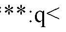
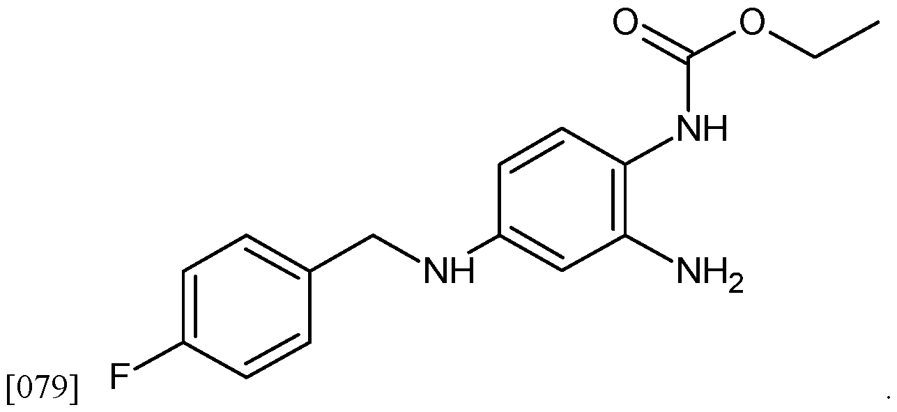


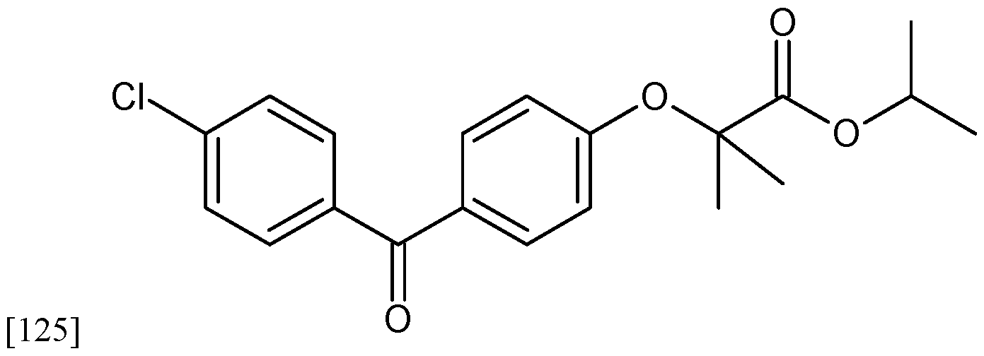


Claims
Priority Applications (5)
| Application Number | Priority Date | Filing Date | Title |
|---|---|---|---|
| JP2024535723A JP2025500876A (en) | 2021-12-14 | 2022-12-14 | Use of hyperpolarizing agents alone and in combination with other therapeutic agents for the treatment of cancer, including glioblastoma - Patent Application 20070123333 |
| AU2022414087A AU2022414087A1 (en) | 2021-12-14 | 2022-12-14 | Use of hyperpolarizing agents alone and in combination with other therapeutic agents for treating cancers including glioblastoma |
| EP22908674.9A EP4447955A4 (en) | 2021-12-14 | 2022-12-14 | USE OF HYPERPOLARIZING AGENTS ALONE AND IN COMBINATION WITH OTHER THERAPEUTIC AGENTS FOR THE TREATMENT OF CANCER, INCLUDING GLIOBLASTOM |
| CA3242726A CA3242726A1 (en) | 2021-12-14 | 2022-12-14 | USE OF HYPERPOLARIZING AGENTS ALONE AND IN COMBINATION WITH OTHER THERAPEUTIC AGENTS FOR THE TREATMENT OF CANCERS INCLUDING A GLIOBLASTOMA |
| US18/719,825 US20250049747A1 (en) | 2021-12-14 | 2022-12-14 | Use of hyperpolarizing agents alone and in combination with other therapeutic agents for treating cancers including glioblastoma |
Applications Claiming Priority (2)
| Application Number | Priority Date | Filing Date | Title |
|---|---|---|---|
| US202163265403P | 2021-12-14 | 2021-12-14 | |
| US63/265,403 | 2021-12-14 |
Publications (1)
| Publication Number | Publication Date |
|---|---|
| WO2023114868A1 true WO2023114868A1 (en) | 2023-06-22 |
Family
ID=86773652
Family Applications (1)
| Application Number | Title | Priority Date | Filing Date |
|---|---|---|---|
| PCT/US2022/081589 Ceased WO2023114868A1 (en) | 2021-12-14 | 2022-12-14 | Use of hyperpolarizing agents alone and in combination with other therapeutic agents for treating cancers including glioblastoma |
Country Status (6)
| Country | Link |
|---|---|
| US (1) | US20250049747A1 (en) |
| EP (1) | EP4447955A4 (en) |
| JP (1) | JP2025500876A (en) |
| AU (1) | AU2022414087A1 (en) |
| CA (1) | CA3242726A1 (en) |
| WO (1) | WO2023114868A1 (en) |
Citations (4)
| Publication number | Priority date | Publication date | Assignee | Title |
|---|---|---|---|---|
| US20070293553A1 (en) * | 2004-12-17 | 2007-12-20 | Dahl Bjarne H | Diphenylurea Derivatives Useful As Potassium Channel Activators |
| US20110053947A1 (en) * | 2008-02-07 | 2011-03-03 | Sanofi-Aventis | Arylchalcogenoarylalkyl-substituted imidazolidine-2,4-diones, process for preparation thereof, medicaments comprising these compounds and use thereof |
| WO2018209022A2 (en) * | 2017-05-10 | 2018-11-15 | University Of Rochester | Methods of treating neuropsychiatric disorders |
| US20210369677A1 (en) * | 2020-05-27 | 2021-12-02 | Northwestern University | Small molecule activators of polycystin-2 (pkd2) and uses thereof |
Family Cites Families (8)
| Publication number | Priority date | Publication date | Assignee | Title |
|---|---|---|---|---|
| US7705010B2 (en) * | 2005-02-22 | 2010-04-27 | Cedars-Sinai Medical Center | Use of minoxidil sulfate as an anti-tumor drug |
| DE102005047616A1 (en) * | 2005-10-05 | 2007-05-31 | Bayer Healthcare Ag | combination |
| CN102626416B (en) * | 2012-05-11 | 2013-04-17 | 江苏省人民医院 | Application of tamoxifen combined with rapamycin and ginsenoside Rg3 complex in the preparation of drugs for the treatment of liver cancer |
| CA2961894C (en) * | 2014-09-19 | 2023-12-12 | Memorial Sloan-Kettering Cancer Center | Methods for treating brain metastatis using gap junction inhibitors |
| CN104887672A (en) * | 2015-05-29 | 2015-09-09 | 许东武 | Broad-spectrum anticancer and cancer prevention formula medicine |
| JP2021513556A (en) * | 2017-12-21 | 2021-05-27 | ザ アンジオジェネシス ファウンデーション | Compositions and methods for treating neoplasia |
| CN111166756B (en) * | 2018-11-12 | 2024-01-30 | 上海交通大学医学院附属新华医院 | 20 Use of (S) -ginsenoside-Rg 3 in reversing drug resistance of glioma cells to chemotherapeutic drugs |
| CN114828853A (en) * | 2019-10-24 | 2022-07-29 | 赛立斯肿瘤学创新有限公司 | Combination therapy for cancer |
-
2022
- 2022-12-14 AU AU2022414087A patent/AU2022414087A1/en active Pending
- 2022-12-14 JP JP2024535723A patent/JP2025500876A/en active Pending
- 2022-12-14 EP EP22908674.9A patent/EP4447955A4/en active Pending
- 2022-12-14 CA CA3242726A patent/CA3242726A1/en active Pending
- 2022-12-14 WO PCT/US2022/081589 patent/WO2023114868A1/en not_active Ceased
- 2022-12-14 US US18/719,825 patent/US20250049747A1/en active Pending
Patent Citations (4)
| Publication number | Priority date | Publication date | Assignee | Title |
|---|---|---|---|---|
| US20070293553A1 (en) * | 2004-12-17 | 2007-12-20 | Dahl Bjarne H | Diphenylurea Derivatives Useful As Potassium Channel Activators |
| US20110053947A1 (en) * | 2008-02-07 | 2011-03-03 | Sanofi-Aventis | Arylchalcogenoarylalkyl-substituted imidazolidine-2,4-diones, process for preparation thereof, medicaments comprising these compounds and use thereof |
| WO2018209022A2 (en) * | 2017-05-10 | 2018-11-15 | University Of Rochester | Methods of treating neuropsychiatric disorders |
| US20210369677A1 (en) * | 2020-05-27 | 2021-12-02 | Northwestern University | Small molecule activators of polycystin-2 (pkd2) and uses thereof |
Non-Patent Citations (3)
| Title |
|---|
| BURG ET AL.: "Potassium channels in the regulation of pulmonary artery smooth muscle cell proliferation and apoptosis: pharmacotherapeutic implications", BRITISH JOURNAL OF PHARMACOLOGY, vol. 153, no. S1, 29 January 2009 (2009-01-29), pages S99 - S111, XP071136502, DOI: 10.1038/sj.bjp.0707635 * |
| See also references of EP4447955A4 * |
| ZHENG ET AL.: "Suppression of KCNQIM (Kv7) potassium channels in dorsal root ganglion neurons contributes to the development of bone cancer pain in a rat model", PAIN, vol. 154, no. 3, 2013, pages 434 - 448, XP028983602, DOI: 10.1016/j.pain.2012.12.005 * |
Also Published As
| Publication number | Publication date |
|---|---|
| US20250049747A1 (en) | 2025-02-13 |
| EP4447955A4 (en) | 2026-04-08 |
| CA3242726A1 (en) | 2023-06-22 |
| EP4447955A1 (en) | 2024-10-23 |
| AU2022414087A1 (en) | 2024-08-01 |
| JP2025500876A (en) | 2025-01-15 |
Similar Documents
| Publication | Publication Date | Title |
|---|---|---|
| AU2017211858B2 (en) | Differentiation of cortical neurons from human pluripotent stem cells | |
| Charkhkar et al. | Amyloid beta modulation of neuronal network activity in vitro | |
| US20180161335A1 (en) | Methods to treat neurological diseases | |
| CN101420980A (en) | Compounds and methods for treating stroke | |
| CA2994544A1 (en) | Compositions and methods for treating cancers associated with etbr activation | |
| Zhu et al. | EVs-mediated delivery of CB2 receptor agonist for Alzheimer's disease therapy | |
| Del Giovane et al. | Smoothened/AMP-activated protein kinase signaling in oligodendroglial cell maturation | |
| Obi et al. | Extracellular vesicles released from skeletal muscle post‐chronic contractile activity increase mitochondrial biogenesis in recipient myoblasts | |
| US20200188402A1 (en) | Compounds and methods for treatment of diamond blackfan anemia | |
| CA2797797C (en) | Methods of use of tetrahydroberberine (thb) | |
| US20250049747A1 (en) | Use of hyperpolarizing agents alone and in combination with other therapeutic agents for treating cancers including glioblastoma | |
| de Oca Balderas et al. | NMDA Receptors in Astroglia: Chronology, Controversies, and Contradictions from | |
| Mathews et al. | Ion Channel Drugs Suppress Cancer Phenotype in NG108-15 and U87 Cells: Toward Novel Electroceuticals for Glioblastoma. Cancers 2022, 14, 1499 | |
| CN114786661A (en) | Pharmaceutical combination of an artemisinin compound, 5-aminolevulinic acid or methyl-5-aminolevulinic acid and a chemotherapeutic agent | |
| US10576069B2 (en) | Treatment for chemotherapy-induced cognitive impairment | |
| ES3028375T3 (en) | 1,2-dihydroquinoline-2-ones for their use in the treatment of limb-girdle muscular dystrophy | |
| EP4356932A1 (en) | Method and drug for treating neuronal ceroid lipofuscinosis | |
| WO2026030225A1 (en) | Use of glycolysis-inhibiting compounds to boost neural stem cell function in the brain | |
| Ferraro et al. | GET73 modulates rat hippocampal glutamate transmission: evidence for a functional interaction with mGluR5 | |
| Li | Isoginkgetin Protects Against Motor Neuron Degeneration in Amyotrophic Lateral Sclerosis Through PINK1-Parkin Dependent Mitophagy | |
| Terceiro | Psychostimulants-induced neuroinflammation: the role of Rho GTPases in microglia | |
| Papandreou | Investigation of Disease Mechanisms and Screening for Treatments in Betapropeller Protein-Associated Neurodegeneration (BPAN) | |
| Liu | The study of Murine double minute-2 (Mdm2) in regulating neuronal activity-dependent protein translation | |
| Joshi | Utilizing Human Induced Pluripotent Stem Cells and Derived Models to Study Neurodevelopment and Neurodegenerative Disease | |
| Kramer | Functional characterisation of aromatic L-amino acid decarboxylase (AADC) deficiency using a patient-derived dopaminergic cell model |
Legal Events
| Date | Code | Title | Description |
|---|---|---|---|
| 121 | Ep: the epo has been informed by wipo that ep was designated in this application |
Ref document number: 22908674 Country of ref document: EP Kind code of ref document: A1 |
|
| WWE | Wipo information: entry into national phase |
Ref document number: 3242726 Country of ref document: CA |
|
| WWE | Wipo information: entry into national phase |
Ref document number: 2024535723 Country of ref document: JP |
|
| WWE | Wipo information: entry into national phase |
Ref document number: AU2022414087 Country of ref document: AU |
|
| WWE | Wipo information: entry into national phase |
Ref document number: 2022908674 Country of ref document: EP |
|
| NENP | Non-entry into the national phase |
Ref country code: DE |
|
| ENP | Entry into the national phase |
Ref document number: 2022908674 Country of ref document: EP Effective date: 20240715 |
|
| ENP | Entry into the national phase |
Ref document number: 2022414087 Country of ref document: AU Date of ref document: 20221214 Kind code of ref document: A |