WO2023190820A1 - 抗CK2α抗体又はその断片 - Google Patents
抗CK2α抗体又はその断片 Download PDFInfo
- Publication number
- WO2023190820A1 WO2023190820A1 PCT/JP2023/013069 JP2023013069W WO2023190820A1 WO 2023190820 A1 WO2023190820 A1 WO 2023190820A1 JP 2023013069 W JP2023013069 W JP 2023013069W WO 2023190820 A1 WO2023190820 A1 WO 2023190820A1
- Authority
- WO
- WIPO (PCT)
- Prior art keywords
- amino acid
- acid sequence
- seq
- sequence shown
- cancer
- Prior art date
- Legal status (The legal status is an assumption and is not a legal conclusion. Google has not performed a legal analysis and makes no representation as to the accuracy of the status listed.)
- Ceased
Links
Images


Classifications
-
- G—PHYSICS
- G01—MEASURING; TESTING
- G01N—INVESTIGATING OR ANALYSING MATERIALS BY DETERMINING THEIR CHEMICAL OR PHYSICAL PROPERTIES
- G01N33/00—Investigating or analysing materials by specific methods not covered by groups G01N1/00 - G01N31/00
- G01N33/48—Biological material, e.g. blood, urine; Haemocytometers
- G01N33/50—Chemical analysis of biological material, e.g. blood, urine; Testing involving biospecific ligand binding methods; Immunological testing
- G01N33/53—Immunoassay; Biospecific binding assay; Materials therefor
-
- C—CHEMISTRY; METALLURGY
- C07—ORGANIC CHEMISTRY
- C07K—PEPTIDES
- C07K16/00—Immunoglobulins [IG], e.g. monoclonal or polyclonal antibodies
- C07K16/40—Immunoglobulins [IG], e.g. monoclonal or polyclonal antibodies against enzymes
-
- G—PHYSICS
- G01—MEASURING; TESTING
- G01N—INVESTIGATING OR ANALYSING MATERIALS BY DETERMINING THEIR CHEMICAL OR PHYSICAL PROPERTIES
- G01N33/00—Investigating or analysing materials by specific methods not covered by groups G01N1/00 - G01N31/00
- G01N33/48—Biological material, e.g. blood, urine; Haemocytometers
- G01N33/50—Chemical analysis of biological material, e.g. blood, urine; Testing involving biospecific ligand binding methods; Immunological testing
- G01N33/53—Immunoassay; Biospecific binding assay; Materials therefor
- G01N33/575—Immunoassay; Biospecific binding assay; Materials therefor for cancer
-
- G—PHYSICS
- G01—MEASURING; TESTING
- G01N—INVESTIGATING OR ANALYSING MATERIALS BY DETERMINING THEIR CHEMICAL OR PHYSICAL PROPERTIES
- G01N33/00—Investigating or analysing materials by specific methods not covered by groups G01N1/00 - G01N31/00
- G01N33/48—Biological material, e.g. blood, urine; Haemocytometers
- G01N33/50—Chemical analysis of biological material, e.g. blood, urine; Testing involving biospecific ligand binding methods; Immunological testing
- G01N33/68—Chemical analysis of biological material, e.g. blood, urine; Testing involving biospecific ligand binding methods; Immunological testing involving proteins, peptides or amino acids
-
- G—PHYSICS
- G01—MEASURING; TESTING
- G01N—INVESTIGATING OR ANALYSING MATERIALS BY DETERMINING THEIR CHEMICAL OR PHYSICAL PROPERTIES
- G01N33/00—Investigating or analysing materials by specific methods not covered by groups G01N1/00 - G01N31/00
- G01N33/48—Biological material, e.g. blood, urine; Haemocytometers
- G01N33/50—Chemical analysis of biological material, e.g. blood, urine; Testing involving biospecific ligand binding methods; Immunological testing
- G01N33/68—Chemical analysis of biological material, e.g. blood, urine; Testing involving biospecific ligand binding methods; Immunological testing involving proteins, peptides or amino acids
- G01N33/6893—Chemical analysis of biological material, e.g. blood, urine; Testing involving biospecific ligand binding methods; Immunological testing involving proteins, peptides or amino acids related to diseases not provided for elsewhere
-
- A—HUMAN NECESSITIES
- A61—MEDICAL OR VETERINARY SCIENCE; HYGIENE
- A61P—SPECIFIC THERAPEUTIC ACTIVITY OF CHEMICAL COMPOUNDS OR MEDICINAL PREPARATIONS
- A61P35/00—Antineoplastic agents
-
- C—CHEMISTRY; METALLURGY
- C07—ORGANIC CHEMISTRY
- C07K—PEPTIDES
- C07K2317/00—Immunoglobulins specific features
- C07K2317/50—Immunoglobulins specific features characterized by immunoglobulin fragments
- C07K2317/56—Immunoglobulins specific features characterized by immunoglobulin fragments variable (Fv) region, i.e. VH and/or VL
- C07K2317/565—Complementarity determining region [CDR]
-
- G—PHYSICS
- G01—MEASURING; TESTING
- G01N—INVESTIGATING OR ANALYSING MATERIALS BY DETERMINING THEIR CHEMICAL OR PHYSICAL PROPERTIES
- G01N2333/00—Assays involving biological materials from specific organisms or of a specific nature
- G01N2333/90—Enzymes; Proenzymes
- G01N2333/91—Transferases (2.)
- G01N2333/912—Transferases (2.) transferring phosphorus containing groups, e.g. kinases (2.7)
Definitions
- the present invention relates to an anti-CK2 ⁇ antibody or a fragment thereof, a kit for predicting the prognosis of cancer patients, a method for predicting the prognosis of cancer patients, and the like.
- cancer is the most common cause of death, accounting for approximately 30%.
- breast cancer is the leading cause of death for women aged 30 to 64, with approximately 14,000 deaths in 2018.
- the survival rate of cancer patients has improved due to advances in cancer detection and/or treatment methods, there are still some patients with a poor prognosis who are at high risk of recurrence, metastasis, or death. Therefore, in order to improve the quality of cancer treatment including breast cancer, it is very important to predict the prognosis of cancer patients and to perform individual management of cancer patients according to the results.
- CK2 ⁇ (Casein kinase 2 ⁇ ) protein as a new biomarker that can predict the prognosis of cancer patients including breast cancer patients with high accuracy (Patent Document 1).
- CK2 ⁇ protein is present throughout the cell in normal cells, whereas in cancer cells it is highly expressed in the nucleus and localized to the nucleolus, which is associated with poor prognosis.
- the present inventors have revealed that the nuclear expression level and nucleolar staining level of CK2 ⁇ protein are strongly correlated with poor prognosis of cancer patients, and are also strongly associated with recurrence risk (Patent Document 1).
- CK2 ⁇ protein in the nucleolus as a biomarker is a technological technology that has made it possible to predict the prognosis of cancer patients with extremely high accuracy.
- existing anti-CK2 ⁇ antibodies used for predicting prognosis do not have sufficient specificity or sensitivity for CK2 ⁇ protein.
- the purpose of the present invention is to provide a new anti-CK2 ⁇ antibody with improved specificity and sensitivity.
- CK2 ⁇ protein together with CK2 ⁇ ' protein and CK2 ⁇ protein, constitutes CK2 (Casein Kinase 2), which is a serine-threonine kinase.
- CK2 ⁇ protein has a similar structure to CK2 ⁇ ' protein, and conventional anti-CK2 ⁇ antibodies could not distinguish between the two.
- mAb (6A3) as a new antibody that exhibits extremely high specificity for CK2 ⁇ protein.
- the present inventor succeeded in developing mAb (6A3) with extremely high specificity for CK2 ⁇ protein by applying the criterion that it binds only to CK2 ⁇ protein and not to CK2 ⁇ ' protein.
- mAb (6A3) showed significantly higher specificity and sensitivity than conventional antibodies in Western blotting and immunoprecipitation against CK2 ⁇ protein.
- mAb (6A3) detects CK2 ⁇ protein localized in the nucleolus with extremely high specificity and sensitivity in cancer tissues derived from cancer patients with poor prognosis, providing an extremely useful tool for biomarker detection.
- the present invention is based on the above findings and provides the following.
- the anti-CK2 ⁇ antibody or fragment thereof according to (1) which comprises a light chain variable region consisting of the following sequence.
- (3) A kit for predicting the prognosis of cancer patients, comprising the anti-CK2 ⁇ antibody or fragment thereof according to (1) or (2).
- the cancer is selected from the group consisting of breast cancer, uterine cancer, esophageal cancer, stomach cancer, biliary tract cancer, pancreatic cancer, liver cancer, kidney cancer, colon cancer, bladder cancer, lung cancer, thyroid cancer, and glioma; The kit according to (3) or (4).
- a method for predicting the prognosis of a cancer patient comprising: A step of detecting CK2 ⁇ protein or a fragment thereof in the nucleolus in cancer cells or tissues obtained from a cancer patient, and a step of detecting CK2 ⁇ protein or a fragment thereof in the nucleolus to a high degree compared to other cell fractions. and/or predicting a good prognosis when CK2 ⁇ protein or its fragments are not detected in the nucleolus to a high degree compared to other cell fractions.
- the method described above, wherein the protein or fragment thereof is detected using the anti-CK2 ⁇ antibody or fragment thereof according to (1) or (2).
- the prognosis includes recurrence risk.
- the cancer is selected from the group consisting of breast cancer, uterine cancer, esophageal cancer, stomach cancer, biliary tract cancer, pancreatic cancer, liver cancer, kidney cancer, colon cancer, bladder cancer, lung cancer, thyroid cancer, and glioma; The method described in (6) or (7).
- the cancer is breast cancer, and the presence or absence of detection of CK2 ⁇ protein or a fragment thereof is determined by at least one of the following: classification by stage, classification by hormone receptor expression status, and classification by HER2 gene and/or protein expression status.
- the method according to (8) which predicts the prognosis of breast cancer patients in combination.
- This specification includes the disclosure content of Japanese Patent Application No. 2022-055606, which is the basis of the priority of this application.
- the present invention provides a new anti-CK2 ⁇ antibody with improved specificity and sensitivity.
- FIG. 1 is a diagram showing the results of Western blotting using mouse antiserum and hybridoma culture supernatant.
- FIG. 1A shows the results of Western blotting for recombinant CK2 ⁇ protein ( ⁇ ) and recombinant CK2 ⁇ ' protein ( ⁇ ') using antisera from mice immunized with antigenic polypeptides.
- FIG. 1B shows the results of Western blotting for recombinant CK2 ⁇ protein ( ⁇ ) and recombinant CK2 ⁇ protein ( ⁇ ′) using culture supernatants of clones 6A, 6B, 6C, and 7A.
- FIG. 2 shows HEK293 cells analyzed by Western blotting using culture supernatants of clones 6A1, 6A2, and 6A3 and a commercially available mouse anti-CK2 ⁇ monoclonal antibody (ab70774, Abcam, UK; shown as "control antibody” in the figure).
- This shows the results of detecting CK2 ⁇ protein in the cytoplasmic lysate of. With the control antibody, a non-specific band indicated by an arrow was detected.
- Figure 3 shows the results of detecting endogenous CK2 ⁇ protein in HEK293 cells by Western blotting using mAb (6A3) and a control antibody.
- Figure 3A shows the results of Western blot.
- FIG. 3B shows the results of quantifying the staining intensity of the band corresponding to CK2 ⁇ protein.
- Figure 4 shows HEK293 cells expressing Flag-CK2 ⁇ protein (Flag-CK2 ⁇ (+) lane) and HEK293 cells not expressing Flag-CK2 ⁇ protein by Western blotting using mAb (6A3) and control antibody. The results of detecting Flag-CK2 ⁇ protein and endogenous CK2 ⁇ protein in the cytoplasmic lysate of cells (Flag-CK2 ⁇ (-) lane) are shown.
- FIG. 5 is a diagram showing the results of Western blotting on immunoprecipitates. Lysates prepared from the cytoplasm (C) and nucleus (N) of HEK293 cells were immunoprecipitated using mAb (6A3) or control antibody. The results of Western blotting using this method are shown. "No immunoprecipitation” indicates a sample in which cytoplasmic (C) and nuclear (N) lysates were loaded without undergoing immunoprecipitation.
- FIG. 6 is a diagram showing the results of Western blotting on immunoprecipitates.
- FIG. 7 shows the results of Western blotting on immunoprecipitates.
- the results of Western blotting using mAb (6A3) or anti-Flag antibody on the immunoprecipitate obtained by immunoprecipitation using mAb (6A3) or control antibody from the lysate of Flag-CK2 ⁇ -expressing RPE cells are shown below. show.
- FIG. 8 is a diagram showing the results of Western blotting on immunoprecipitates.
- Figure 7 shows the results of quantifying the staining intensity of bands (two bands indicated by arrows in Figure 7) when mAb (6A3) or a control antibody was used as the antibody for immunoprecipitation.
- Figure 9 shows a representative image of immunohistochemical staining in the cancer invasion area (lesion area) of invasive ductal carcinoma.
- Figure 9A is an image (magnification x 400) of a cancer invasion area (lesion area) stained using 0.1 ⁇ g/mL mAb (6A3). An enlarged image of the area indicated by the black frame in the figure is shown at the bottom left.
- Figure 9B is an image (magnification x 400) of a cancer invasion area (lesion area) stained with 2 ⁇ g/mL control antibody. An enlarged image of the area indicated by the black frame in the figure is shown at the bottom left.
- Figure 10 shows the results of Western blotting of IgG purified from multiple CK2 antibody clones established in addition to 6A3 on immunoprecipitates from either the MCF-7 cell line or HEK293 cells expressing Flag-CK2 ⁇ protein. .
- the upper panel shows the results of detecting endogenous CK2 ⁇ protein in the MCF-7 cell line.
- the lower panel shows the results of detecting endogenous CK2 ⁇ protein and Flag-CK2 ⁇ protein in HEK293 cells expressing Flag-CK2 ⁇ protein.
- FIG. 11 shows the results of ChIP-qPCR targeting the HMGB2 gene locus on fractions subjected to chromatin immunoprecipitation with IgG purified from multiple CK2 antibody clones established in addition to 6A3.
- “Control” indicates a lane loaded with a sample without the addition of IgG for the equally fractionated chromatin fraction.
- FIG. 12 shows the results of Western blotting of the purified recombinant CK2 ⁇ protein.
- Figure 13 shows a representative image of immunohistochemical staining at the cancer invasion area (lesion area) of invasive ductal carcinoma.
- Figure 13A is an image (magnification x 400) of a cancer infiltrated area (lesion area) stained using 0.1 ⁇ g/mL mAb (21B1).
- Figure 13B is an image (magnification x 400) of a cancer invasion area (lesion area) stained with 2 ⁇ g/mL control antibody.
- Figure 14 shows the CK2 ⁇ nucleolus stain-positive group (“CK2-NO(+)” in the figure) based on the results of histochemical staining using the 6A3 clone in FFPE samples of primary lung adenocarcinoma patients who underwent resection surgery. ) and the CK2 ⁇ nucleolus staining negative group (“CK2-NO(-)” in the figure).
- FIG. 15 shows the results of univariate and multivariate analyzes based on the results of histochemical staining using the 6A3 clone on FFPE samples of primary lung adenocarcinoma patients.
- Figure 15A shows the results of univariate analysis.
- Figure 15B shows the results of multivariate analysis.
- FIG. 15C shows the results of analyzing the effects that define recurrence-free survival in multivariate analysis.
- FIG. 16 shows the results of an analysis of variables that define the period until recurrence using two recurrence prediction models.
- FIG. 16A shows the results of recurrence prediction model 1 based on six types of variables.
- FIG. 16B shows the results of recurrence prediction model 2 based on seven types of variables with the addition of age.
- FIG. 17 shows the classification of stage I to III primary lung adenocarcinoma patients based on stage and CK2 ⁇ staining evaluation, and the presence or absence of recurrence
- CK2 ⁇ protein refers to the ⁇ subunit of casein kinase 2 (CK2).
- CK2 ⁇ protein is also referred to as casein kinase 2 alpha 1 or casein kinase II subunit alpha, CK2 ⁇ 1 protein, or CSNK2A1 protein.
- Casein kinase 2 is a type of serine-threonine kinase and is known to be involved in pro-survival pathways. Casein kinase 2 is known to function as a tetramer composed of an ⁇ subunit (CK2 ⁇ protein), an ⁇ ′ subunit (CK2 ⁇ ′ protein), and two ⁇ subunits (CK2 ⁇ protein). The ⁇ subunit (CK2 ⁇ protein) and ⁇ ' subunit (CK2 ⁇ ' protein) are known to function as catalytic subunits of casein kinase 2.
- CK2 ⁇ protein or a fragment thereof can serve as a biomarker for predicting the prognosis of cancer patients.
- the patient is a human
- human CK2 ⁇ protein or a fragment thereof can be the biomarker.
- CK2 ⁇ protein in the nucleolus can serve as a biomarker for predicting the prognosis of cancer patients such as breast cancer patients.
- immunohistochemical staining of CK2 ⁇ protein was performed on formalin-fixed, paraffin-embedded breast cancer tissue specimens excised from primary breast cancer patients who underwent radical resection.
- CK2 ⁇ protein is present throughout the cell, but in breast cancer cells, it is often observed that it is highly expressed in the nucleus, and in some breast cancer patients (about 40%), CK2 ⁇ protein is present throughout the cell.
- CK2 ⁇ is localized in the nucleolus within the nucleus, and that the higher the nuclear expression level and nucleolar staining level of CK2 ⁇ protein, the higher the proportion of cases with high stage breast cancer. Furthermore, as a result of evaluating the prognosis of primary breast cancer patients, it was found that nucleolar localization of CK2 ⁇ protein has a high relative risk for both recurrence and life prognosis, and that the presence or absence of nucleolar localization of CK2 ⁇ protein has a high relative risk. It was revealed that this is a significant predictor of recurrence.
- CK2 ⁇ protein can be localized to the nucleolus in all cancers, including bladder cancer, lung cancer (lung adenocarcinoma and lung squamous cell carcinoma), thyroid cancer, and glioma.
- the term "marker” refers to a biomarker consisting of the CK2 ⁇ protein or a fragment thereof, or a biomarker consisting of the CK2 ⁇ protein or a fragment thereof in the nucleolus, unless otherwise specified.
- the type of “cancer” is not limited, but includes, for example, adenocarcinoma, squamous cell carcinoma, small cell carcinoma, and large cell carcinoma.
- the types of cancer include, for example, malignant melanoma, oral cavity cancer, laryngeal cancer, pharyngeal cancer, thyroid cancer, lung cancer, breast cancer, esophageal cancer, stomach cancer, colorectal cancer (including colon cancer and rectal cancer), Small intestine cancer, bladder cancer, prostate cancer, testicular cancer, endometrial cancer, cervical cancer, endometrial cancer, ovarian cancer, stomach cancer, kidney cancer, liver cancer, pancreatic cancer, biliary tract cancer (including gallbladder cancer and bile duct cancer), brain tumor , head and neck cancer, pediatric tumors including mesothelioma, osteosarcoma, glioma, and neuroblastoma, leukemia, and lymphoma.
- the cancer is preferably breast cancer, uterine cancer, esophageal cancer, stomach cancer, pancreatic cancer, liver cancer, biliary tract cancer (e.g. gallbladder cancer or bile duct cancer), kidney cancer, colorectal cancer (e.g. rectal cancer or colon cancer), bladder cancer, lung cancer. (eg, lung adenocarcinoma or lung squamous cell carcinoma), thyroid cancer, or glioma (eg, astrocytoma), and more preferably breast cancer.
- biliary tract cancer e.g. gallbladder cancer or bile duct cancer
- kidney cancer e.g. rectal cancer or colon cancer
- bladder cancer e.g, lung adenocarcinoma or lung squamous cell carcinoma
- thyroid cancer eg, astrocytoma
- glioma eg, astrocytoma
- breast cancer is not limited, and includes, for example, ductal carcinoma in situ, invasive ductal carcinoma, invasive lobular carcinoma, lobular carcinoma in situ, and special types of breast cancer such as medullary carcinoma and mucinous carcinoma. Examples include cancer, tubular cancer, and the like.
- prognosis refers to the predicted course of a cancer patient (eg, presence or absence of recurrence or survival or death).
- Prediction of prognosis refers to recurrence risk (e.g., recurrence-free survival rate), survival period, or after a certain period of time after surgery (e.g., 1 year, 2 years, 3 years, 4 years, 5 years, 10 years, 15 years or It may be a prediction of survival (20 years or more), relapse-free survival (RFS), or disease-free survival (DFS).
- predicting prognosis includes predicting recurrence risk (eg, recurrence-free survival).
- the recurrence-free survival rate is the proportion of patients who do not develop recurrent cancer such as cancer associated with the first cancer
- the disease-specific survival rate is the proportion of patients who do not die related to the first cancer.
- Prediction of prognosis can also be referred to as determination, evaluation, or diagnosis of prognosis, or assistance thereof.
- CK2 ⁇ protein includes a human-derived CK2 ⁇ (human CK2 ⁇ ) protein that includes or consists of the amino acid sequence shown by SEQ ID NO: 2.
- CK2 ⁇ protein also includes CK2 ⁇ variants and CK2 ⁇ orthologs of other species that have functionally equivalent activity to the CK2 ⁇ protein shown by SEQ ID NO: 2.
- the amino acid sequence shown in SEQ ID NO: 2 has one or several amino acids deleted, substituted, or added, or 80% or more, 90% of the amino acid sequence shown in SEQ ID NO: 2.
- the above includes CK2 ⁇ proteins having amino acid identity of 95% or more, 97% or more, 98% or more, or 99% or more.
- “several” refers to, for example, 2 to 10, 2 to 7, 2 to 5, 2 to 4, or 2 to 3. Moreover, conservative amino acid substitutions are preferable for amino acid substitutions. "Conservative amino acid substitution” refers to a substitution between amino acids with similar properties such as charge, side chain, polarity, aromaticity, etc. Amino acids with similar properties include, for example, basic amino acids (arginine, lysine, histidine), acidic amino acids (aspartic acid, glutamic acid), uncharged polar amino acids (glycine, asparagine, glutamine, serine, threonine, cysteine, tyrosine), and nonpolar amino acids.
- amino acid identity means that when two amino acid sequences are aligned and gaps are introduced as necessary to achieve the highest degree of amino acid identity between the two, SEQ ID NO. It refers to the proportion (%) of identical amino acid residues between two amino acid sequences to the total amino acid residues of the CK2 ⁇ protein containing the amino acid sequence shown in 2.
- Amino acid identity can be calculated using a protein search system such as BLAST or FASTA. For details on how to determine identity, see, for example, Altschul et al, Nuc. Acids. Res. 25, 3389-3402, 1977 and Altschul et al, J. Mol. Biol. 215, 403-410, 1990. .
- CK2 ⁇ protein is encoded by the CK2 ⁇ gene.
- a specific example of the CK2 ⁇ gene is the human CK2 ⁇ gene that encodes the human CK2 ⁇ protein containing the amino acid sequence shown by SEQ ID NO:2. More specifically, the CK2 ⁇ gene includes a gene containing or consisting of the base sequence shown in SEQ ID NO: 1.
- the CK2 ⁇ gene also includes CK2 ⁇ variants that have functionally equivalent activity to the CK2 ⁇ protein encoded by the CK2 ⁇ gene shown in SEQ ID NO: 1, and CK2 ⁇ genes that encode CK2 ⁇ orthologs from other species. Specifically, 80% or more, 90% of the base sequence shown in SEQ ID NO: 1, where one or several bases have been deleted, substituted, or added, or the base sequence shown in SEQ ID NO: 1. The above includes CK2 ⁇ genes having base identity of 95% or more, 97% or more, 98% or more, or 99% or more.
- nucleotide sequence that hybridizes under high stringency conditions with a nucleic acid fragment that includes a part of the nucleotide sequence complementary to the nucleotide sequence shown in SEQ ID NO: 1, and is functionally equivalent to the CK2 ⁇ protein.
- Genes encoding active proteins are included.
- base identity means that when two base sequences are aligned and gaps are introduced as necessary to maximize the degree of base identity between the two, SEQ ID NO. It refers to the proportion (%) of identical bases between two base sequences to the total bases of the CK2 ⁇ gene, including the base sequence shown by 2.
- hybridizing under high stringency conditions refers to hybridization and washing under conditions of low salt concentration and/or high temperature. For example, incubate with the probe at 65°C to 68°C in 6x SSC, 5x Denhardt reagent, 0.5% SDS, 100 ⁇ g/mL denatured fragmented salmon sperm DNA, followed by a wash solution of 2x SSC, 0.1% SDS. An example is to start at room temperature, reduce the salt concentration in the wash solution to 0.1 ⁇ SSC, and increase the temperature to 68° C. until no background signal is detected. High stringency hybridization conditions are described in Green, M.R. and Sambrook, J., 2012, Molecular Cloning: A Laboratory Manual Fourth Ed., Cold Spring Harbor Laboratory Press, Cold Spring Harbor, New York. It can be used as a reference.
- nucleotide sequence information of the CK2 ⁇ gene can be searched from public databases (GenBank, EMBL, DDBJ). For example, based on the known base sequence information of the CK2 ⁇ gene shown in SEQ ID NO: 1, genes with high base identity can be searched from a database and obtained.
- a "fragment" of the CK2 ⁇ protein is a peptide fragment that includes or consists of a part of the amino acid sequence that constitutes the CK2 ⁇ protein, and is a fragment of the CK2 ⁇ protein from the amino acid sequence that constitutes the fragment. refers to something that can be identified.
- a "fragment” refers to 5 or more, 8 or more, 10 or more, 20 or more, 30 or more, 40 or more, or 50 or more consecutive amino acid residues of the full-length amino acid sequence of the CK2 ⁇ protein. It may also be a peptide consisting of 200 or less, 150 or less, 120 or less, 100 or less, or 80 or less consecutive amino acid residues.
- a "fragment” may be a peptide consisting of 5 to 200, 10 to 120, or 50 to 80 contiguous amino acid residues.
- nucleolus refers to a region that exists in the nucleus of eukaryotic cells and has a high molecular density, where rRNA transcription and ribosome production occur. Nucleoli can generally be observed with a light microscope. Usually one nucleolus is observed within the nucleus, but multiple nucleoli may be observed in some cases.
- the invention relates to an anti-CK2 ⁇ antibody or fragment thereof.
- the anti-CK2 ⁇ antibody or fragment thereof of the present invention can predict the prognosis of cancer patients by detecting the CK2 ⁇ protein or its peptide fragment that can be localized in the nucleolus in highly malignant cancers.
- Anti-CK2 ⁇ antibody refers to an antibody that exhibits immunoreactivity to CK2 ⁇ protein or a peptide fragment thereof.
- the biological species from which the anti-CK2 ⁇ antibody of the present invention is derived is not particularly limited. Antibodies derived from birds and mammals are preferred. Examples include chicken, ostrich, mouse, rat, guinea pig, rabbit, goat, donkey, sheep, camel, horse, or human.
- the anti-CK2 ⁇ antibody of the present invention is a monoclonal antibody.
- a “monoclonal antibody” includes a framework region (hereinafter referred to as "FR") and a complementarity determining region (hereinafter referred to as "CDR"), and A single type of immunoglobulin that can specifically bind to and recognize it, or at least one set of light chain variable region (V L region) and heavy chain variable region (V H region) contained in the immunoglobulin Refers to recombinant or synthetic antibodies that include.
- the immunoglobulins can be of any class (e.g., IgG, IgE, IgM, IgA, IgD, and IgY) or of any subclass (e.g., IgG1, IgG2, IgG3, IgG4). , IgA1, and IgA2).
- the epitope of the CK2 ⁇ protein or peptide fragment thereof recognized by the anti-CK2 ⁇ antibody of the present invention is an epitope specifically contained in the CK2 ⁇ protein. Preferably this epitope is not included in the CK2 ⁇ ' protein.
- mAb (6A3) mouse anti-CK2 ⁇ monoclonal antibody clone 6A3 (herein referred to as "mAb (6A3)") in the Examples described below.
- mAb (6A3) the heavy chain variable region consists of the amino acid sequence shown in SEQ ID NO: 11, and the light chain variable region consists of the amino acid sequence shown in SEQ ID NO: 12.
- Kabat's rule Kabat's rule (Kabat E.A., et al., 1991, Sequences of proteins of immunological interest, Vol.1, eds.
- CDR1 (HCDR1) consists of the amino acid sequence shown in SEQ ID NO: 5
- CDR2 (HCDR2) consists of the amino acid sequence shown in SEQ ID NO: 6
- CDR3 (HCDR3) consists of the amino acid sequence (FV) shown in SEQ ID NO: 7.
- CDR1 (LCDR1) consists of the amino acid sequence shown in SEQ ID NO: 8
- CDR2 (LCDR2) consists of the amino acid sequence shown in SEQ ID NO: 9
- CDR3 (LCDR3) consists of the amino acid sequence shown in SEQ ID NO: 9. It consists of the amino acid sequence shown in 10.
- the amino acid sequences of SEQ ID NOs: 5-12 are shown in Table 1 below.
- the heavy chain variable region consists of the amino acid sequence shown in SEQ ID NO: 15, and the light chain variable region consists of the amino acid sequence shown in SEQ ID NO: 16.
- CDR1 consists of the amino acid sequence shown in SEQ ID NO: 17
- CDR2 consists of the amino acid sequence shown in SEQ ID NO: 18
- CDR3 HCDR3
- CDR1 consists of the amino acid sequence shown in SEQ ID NO: 20
- CDR2 consists of the amino acid sequence shown in SEQ ID NO: 21
- CDR3 consists of the amino acid sequence shown in SEQ ID NO: 21. It consists of the amino acid sequence shown in 22.
- the heavy chain variable region consists of the amino acid sequence shown in SEQ ID NO: 23, and the light chain variable region consists of the amino acid sequence shown in SEQ ID NO: 24.
- CDR1 consists of the amino acid sequence shown in SEQ ID NO: 25
- CDR2 consists of the amino acid sequence shown in SEQ ID NO: 26
- CDR3 consists of the amino acid sequence shown in SEQ ID NO: 27.
- CDR1 consists of the amino acid sequence shown in SEQ ID NO: 28
- CDR2 consists of the amino acid sequence shown in SEQ ID NO: 29
- CDR3 consists of the amino acid sequence shown in SEQ ID NO: 29. It consists of the amino acid sequence shown in 30.
- the heavy chain variable region consists of the amino acid sequence shown in SEQ ID NO: 31, and the light chain variable region consists of the amino acid sequence shown in SEQ ID NO: 32.
- CDR1 consists of the amino acid sequence shown in SEQ ID NO: 33
- CDR2 consists of the amino acid sequence shown in SEQ ID NO: 34
- CDR3 consists of the amino acid sequence shown in SEQ ID NO: 35.
- CDR1 consists of the amino acid sequence shown in SEQ ID NO: 36
- CDR2 consists of the amino acid sequence shown in SEQ ID NO: 37
- CDR3 consists of the amino acid sequence shown in SEQ ID NO: 37. It consists of the amino acid sequence shown in 38.
- the heavy chain variable region consists of the amino acid sequence shown in SEQ ID NO: 39
- the light chain variable region consists of the amino acid sequence shown in SEQ ID NO: 40.
- CDR1 (HCDR1) consists of the amino acid sequence shown in SEQ ID NO: 41
- CDR2 (HCDR2) consists of the amino acid sequence shown in SEQ ID NO: 42
- CDR3 ( HCDR3) consists of the amino acid sequence shown in SEQ ID NO: 43.
- CDR1 LCDR1
- CDR2 LCDR2
- CDR3 LCDR3
- the heavy chain variable region consists of the amino acid sequence shown in SEQ ID NO: 47
- the light chain variable region consists of the amino acid sequence shown in SEQ ID NO: 48.
- CDR1 (HCDR1) consists of the amino acid sequence shown in SEQ ID NO: 49
- CDR2 (HCDR2) consists of the amino acid sequence shown in SEQ ID NO: 50
- CDR3 ( HCDR3) consists of the amino acid sequence shown in SEQ ID NO: 51.
- CDR1 consists of the amino acid sequence shown in SEQ ID NO: 52
- CDR2 consists of the amino acid sequence shown in SEQ ID NO: 53
- CDR3 consists of the amino acid sequence shown in SEQ ID NO: 53. It consists of the amino acid sequence shown in 54.
- nucleic acid (nucleotide) encoding the amino acid sequence shown in SEQ ID NO: 11, which corresponds to the heavy chain variable region of mAb (6A3) is a nucleic acid consisting of the base sequence shown in SEQ ID NO: 13.
- examples of the nucleic acid encoding the amino acid sequence shown in SEQ ID NO: 12, which corresponds to the light chain variable region of mAb (6A3) include a nucleic acid consisting of the base sequence shown in SEQ ID NO: 14.
- nucleic acid (nucleotide) encoding the amino acid sequence shown in SEQ ID NO: 15, which corresponds to the heavy chain variable region of mAb (10B2) include a nucleic acid consisting of the base sequence shown in SEQ ID NO: 55.
- examples of the nucleic acid encoding the amino acid sequence shown in SEQ ID NO: 16 corresponding to the light chain variable region of mAb (10B2) include a nucleic acid consisting of the base sequence shown in SEQ ID NO: 56.
- nucleic acid (nucleotide) encoding the amino acid sequence shown by SEQ ID NO: 23, which corresponds to the heavy chain variable region of mAb (15C1) is the nucleic acid consisting of the base sequence shown by SEQ ID NO: 57.
- examples of the nucleic acid encoding the amino acid sequence shown in SEQ ID NO: 24, which corresponds to the light chain variable region of mAb (15C1) include a nucleic acid consisting of the base sequence shown in SEQ ID NO: 58.
- nucleic acid (nucleotide) encoding the amino acid sequence shown by SEQ ID NO: 31, which corresponds to the heavy chain variable region of mAb (16C2) is the nucleic acid consisting of the base sequence shown by SEQ ID NO: 59.
- examples of the nucleic acid encoding the amino acid sequence shown in SEQ ID NO: 32, which corresponds to the light chain variable region of mAb (16C2) include a nucleic acid consisting of the base sequence shown in SEQ ID NO: 60.
- nucleic acid (nucleotide) encoding the amino acid sequence shown by SEQ ID NO: 39, which corresponds to the heavy chain variable region of mAb (19C2) is the nucleic acid consisting of the base sequence shown by SEQ ID NO: 61.
- examples of the nucleic acid encoding the amino acid sequence shown in SEQ ID NO: 40, which corresponds to the light chain variable region of mAb (19C2) include a nucleic acid consisting of the base sequence shown in SEQ ID NO: 62.
- nucleic acid (nucleotide) encoding the amino acid sequence shown by SEQ ID NO: 47, which corresponds to the heavy chain variable region of mAb (21B1) is the nucleic acid consisting of the base sequence shown by SEQ ID NO: 63.
- examples of the nucleic acid encoding the amino acid sequence shown in SEQ ID NO: 48, which corresponds to the light chain variable region of mAb (21B1) include a nucleic acid consisting of the base sequence shown in SEQ ID NO: 64.
- Recombinant antibody refers to a chimeric antibody or a humanized antibody.
- a “chimeric antibody” is an antibody produced by combining the amino acid sequences of antibodies derived from different animals, and is an antibody in which the constant region (C region) of one antibody is replaced with the C region of another antibody. For example, this is an antibody in which the C region of a mouse monoclonal antibody is replaced with the C region of a human antibody.
- the heavy chain variable region of a human antibody against an arbitrary antigen is replaced with the heavy chain variable region consisting of the amino acid sequence shown in SEQ ID NO: 11 of the aforementioned mAb (6A3), and the light chain of a human antibody Examples include antibodies in which the variable region is replaced with a light chain variable region consisting of the amino acid sequence shown by SEQ ID NO: 12. This can reduce the immune response to the antibody in the human body.
- a "humanized antibody” is a mosaic antibody in which the CDRs in a human antibody are replaced with the CDRs in an antibody derived from a non-human mammal.
- variable region (V region) of an immunoglobulin molecule consists of four FRs (FR1, FR2, FR3, and FR4) and three CDRs (CDR1, CDR2, and CDR3) starting from the N-terminus: FR1-CDR1-FR2-CDR2-FR3- It is composed of CDR3-FR4 concatenated in the order.
- FRs are relatively conserved regions that constitute the backbone of the variable region, and CDRs directly contribute to the antigen-binding specificity of antibodies.
- a humanized antibody is, for example, a set of CDR1, CDR2, and CDR3 in the light chain or heavy chain of a mouse-derived mAb (6A3), and a set of CDR1, CDR2, and CDR2 in the light chain or heavy chain of a human antibody against any antigen.
- CDR3 and CDR3 it is possible to construct a human antibody that inherits the antigen-binding specificity of mAb (6A3), a mouse antibody.
- CDR1 consists of the amino acid sequence shown in SEQ ID NO: 5 derived from the heavy chain of mAb (6A3) mentioned above
- CDR2 consists of the amino acid sequence shown in SEQ ID NO: 6
- CDR2 consists of the amino acid sequence shown in SEQ ID NO: 7.
- CDR1 consisting of the amino acid sequence shown in SEQ ID NO: 8 derived from the light chain of the aforementioned mAb (6A3)
- CDR1 consisting of the amino acid sequence shown in SEQ ID NO: 9 derived from the light chain of the above-mentioned mAb (6A3).
- Examples include antibodies in which CDR2 consisting of the amino acid sequence shown in SEQ ID NO: 10 and CDR3 consisting of the amino acid sequence shown in SEQ ID NO: 10 are substituted with the light chain CDR1, CDR2, and CDR3 of a human antibody, respectively. Since such humanized antibodies are derived from human antibodies except for CDRs, they can reduce the immune reaction against the antibodies in the human body more than chimeric antibodies.
- “Synthetic antibody” refers to an antibody that is synthesized chemically or by using recombinant DNA methods. Examples include antibodies newly synthesized using recombinant DNA methods. Specific examples include scFv (single chain fragment of variable region: single chain antibody), diabody, triabody, and tetrabody. In immunoglobulin molecules, a pair of variable regions (light chain variable region V L and heavy chain variable region V H ) that form a functional antigen-binding site are located on separate polypeptide chains, the light chain and the heavy chain. do.
- scFv is a synthetic antibody with a molecular weight of about 35 kDa or less, which has a structure in which V L and V H are linked by a sufficiently long flexible linker and included in one polypeptide chain in an immunoglobulin molecule.
- V L and V H are linked by a sufficiently long flexible linker and included in one polypeptide chain in an immunoglobulin molecule.
- a set of variable regions can self-assemble together to form a functional antigen binding site.
- scFv can be obtained by incorporating recombinant DNA encoding it into a vector using known techniques and expressing it.
- Diabodies are molecules whose structure is based on the dimeric structure of scFv (Holliger et al., 1993, Proc. Natl. Acad. Sci. USA 90:6444-6448).
- the two variable regions within the scFv cannot self-assemble, but by allowing the two scFvs to interact to form a diabody, one scFv
- the V L of one scFv can assemble with the V H of the other scFv to form two functional antigen binding sites.
- a cysteine residue to the C-terminus of scFv, it is possible to form a disulfide bond between two scFvs, thereby forming a stable diabody.
- Diabodies are thus bivalent antibody fragments.
- Triabodies and tetrabodies are trivalent and tetravalent antibodies, respectively, which are based on scFv structures and have trimeric and tetrameric structures, like diabodies.
- Diabodies, triabodies, and tetrabodies may be multispecific antibodies.
- the term "multispecific antibody” refers to a multivalent antibody, that is, an antibody having multiple antigen-binding sites in one molecule, in which each antigen-binding site binds to a different epitope.
- diabodies include bispecific antibodies in which each antigen-binding site binds to a different epitope.
- one antigen-binding site is a heavy chain variable region consisting of the amino acid sequence shown in SEQ ID NO: 11 and a light chain variable region consisting of the amino acid sequence shown in SEQ ID NO: 12. This applies to diabodies that contain a diabody and whose other antigen-binding site binds to a different epitope.
- the anti-CK2 ⁇ antibody of the present invention can also be modified.
- modification includes functional modifications necessary for antigen-specific binding activity such as glycosylation, and modifications on labels necessary for antibody detection.
- Glycosylation modifications on anti-CK2 ⁇ antibodies are performed to adjust the affinity of anti-CK2 ⁇ antibodies for the target CK2 ⁇ protein or peptide fragment thereof.
- modifications can be made such as introducing substitutions into amino acid residues that constitute glycosylation and removing the glycosylation site, thereby causing loss of glycosylation at that site. .
- Labeling of anti-CK2 ⁇ antibodies includes, for example, fluorescent dyes (FITC, rhodamine, Texas Red, Cy3, Cy5), fluorescent proteins (e.g., PE, APC, GFP), enzymes (e.g., horseradish peroxidase, alkaline phosphatase, glucose oxidase). ), radioisotopes (eg 3 H, 14 C, 35 S) or labeling with biotin or (strept)avidin.
- fluorescent dyes e.g., rhodamine, Texas Red, Cy3, Cy5
- fluorescent proteins e.g., PE, APC, GFP
- enzymes e.g., horseradish peroxidase, alkaline phosphatase, glucose oxidase
- radioisotopes eg 3 H, 14 C, 35 S
- the anti-CK2 ⁇ antibody of the present invention preferably has a dissociation constant with CK2 ⁇ protein of 10 ⁇ 7 M or less, for example, preferably has a high affinity of 10 ⁇ 8 M or less, more preferably 10 ⁇ 9 M It is particularly preferably 10 ⁇ 10 M or less.
- the above dissociation constant can be measured using techniques known in the art. For example, it may be measured using the Biacore system (GE Healthcare) using speed evaluation kit software.
- diabodies single chain antibody molecules (scFv), dimeric scFv (bivalent diabodies), multispecific antibodies, camelized single domain antibodies (camelized antibodies; VHH antibodies), etc.
- Fab is an antibody fragment produced when an IgG molecule is cleaved by papain on the N-terminal side of the disulfide bond in the hinge region, and constitutes the H chain constant region (heavy chain constant region: hereinafter referred to as C H ). It consists of three domains (C H 1, C H 2, C H 3), which are adjacent to V H , and the full-length L chain .
- F(ab') 2 is a dimer of Fab' produced when an IgG molecule is cleaved by pepsin at the C-terminal side of the disulfide bond in the hinge region.
- Fab' has a slightly longer H chain than Fab, including the hinge region, but has substantially the same structure as Fab.
- Fab' can be obtained by reducing F(ab') 2 under mild conditions and cleaving the disulfide linkage in the hinge region. Since all of these antibody fragments contain an antigen-binding site, they have the ability to specifically bind to an antigen epitope.
- anti-CK2 ⁇ antibody of the present invention can be obtained by a conventional method in the art. Furthermore, if the amino acid sequence of a monoclonal antibody is known, it can also be prepared using chemical synthesis methods or recombinant DNA technology based on the amino acid sequence. Furthermore, monoclonal antibodies can also be obtained from hybridomas that produce the antibodies.
- the antigen polypeptide or antigen peptide that can be used as an immunogen for the anti-CK2 ⁇ antibody of the present invention is any part or full length of the CK2 ⁇ protein (hereinafter referred to as "CK2 ⁇ antigen polypeptide").
- an example of an antigen polypeptide that can be used as an immunogen for the anti-CK2 ⁇ antibody of the present invention is the full-length human CK2 ⁇ protein consisting of the amino acid sequence shown in SEQ ID NO:2.
- CK2 ⁇ antigenic peptides can be prepared using, for example, chemical synthesis methods or DNA recombination techniques.
- the present invention provides an anti-CK2 ⁇ antibody or a fragment thereof for predicting the prognosis of cancer patients.
- CK2 ⁇ protein can be specifically detected.
- the anti-CK2 ⁇ antibody of the present invention or a fragment thereof can be used for immunohistochemical staining, enzyme immunoassay (including ELISA and EIA), Western blotting, radioimmunoassay (RIA), immunoprecipitation, and chromatin immunoprecipitation. Highly sensitive and specific detection is possible by using this method (CHIP) or flow cytometry method.
- the present invention relates to a kit for predicting the prognosis of cancer patients.
- the kit for predicting the prognosis of cancer patients of the present invention contains the above-mentioned anti-CK2 ⁇ antibody or its immunoreactive fragment as an essential component, and detects a biomarker for predicting the prognosis of cancer patients consisting of CK2 ⁇ protein. can do.
- the kit for predicting the prognosis of cancer patients of the present invention is an antibody (hereinafter referred to as "other biomarker") against a biomarker for predicting the prognosis of cancer patients other than the CK2 ⁇ protein or its peptide fragment (hereinafter referred to as “other biomarker for predicting prognosis”). or immunoreactive fragments thereof (referred to as “prognostic antibodies”) or immunoreactive fragments thereof.
- the kit for predicting the prognosis of cancer patients of the present invention contains the above-mentioned anti-CK2 ⁇ antibody or fragment thereof as an essential component.
- the anti-CK2 ⁇ antibody contained in the kit for predicting the prognosis of cancer patients of the present invention may be a single type or multiple types.
- the kit for predicting the prognosis of cancer patients of the present invention may further include one or more types of other prognosis prediction antibodies or fragments thereof having immunoreactivity as the selection component.
- the other antibody for predicting prognosis may be any antibody that can improve the accuracy of predicting the prognosis of cancer patients when used in combination with the anti-CK2 ⁇ antibody, and may be any antibody that can predict the prognosis of cancer patients.
- Antibodies against biomarkers can be used. Such biomarkers can be selected from known cancer markers.
- the kit for predicting the prognosis of cancer patients of the present invention includes other reagents necessary for predicting the prognosis of cancer patients, such as known immunohistochemical staining, ELISA, and Western blotting reagents.
- it may contain labeling reagents, buffers, chromogenic substrates, secondary antibodies, blocking agents, equipment and control buffers necessary for testing, and instructions used for detection and determination of results.
- the invention relates to a method for predicting the prognosis of cancer patients.
- the cancer is selected from the group consisting of breast cancer, uterine cancer, esophageal cancer, gastric cancer, pancreatic cancer, liver cancer, biliary tract cancer, renal cancer, colon cancer, bladder cancer, lung cancer, thyroid cancer, and glioma. .
- the present invention predicts the prognosis of cancer patients by combining a marker consisting of CK2 ⁇ protein or a fragment thereof with factors such as stage classification, tumor diameter, presence or absence of lymph node metastasis, and histological grade.
- the cancer is breast cancer
- the marker is classified into at least one, preferably two, of classification by stage, classification by hormone receptor expression status, and classification by HER2 gene and/or protein expression status. combined with all three to predict the prognosis of breast cancer patients.
- the prognosis of breast cancer patients is predicted by combining the marker with other factors such as tumor diameter, presence or absence of lymph node metastasis, and histological grade, in addition to or separately from the above classification. . Combining with other classifications or factors may have the effect of enabling better prognosis prediction.
- TNM classification by stage is performed based on the TNM classification of the International Union against Cancer (UICC International Code, L.H. Sobin, M.K. Gospodarowicz and Ch. Wittekind, TNM Classification of Malignant Tumours, 7th edition).
- UICC-TNM classification The above-mentioned TNM classification of the International Union against Cancer (UICC) is referred to herein as the UICC-TNM classification.
- UICC-TNM classification breast cancer is classified into stages 0, I, II, III, and IV in descending order of progression.
- the UICC-TNM classification classifies the degree of progression of cancer lesions based on three factors: lump size and spread within the breast (T classification), lymph node metastasis (N classification), and distant metastasis (M classification). Determination of the stage based on the UICC-TNM classification can be performed according to the common knowledge of those skilled in the art.
- stage 0 is when the breast cancer remains within the breast duct
- stage I is when the breast cancer tumor diameter is within 2 cm and there is no axillary lymph node metastasis or micrometastasis within 0.2 mm
- stage I is when the tumor diameter is over 2 cm.
- stage II with no axillary lymph node metastasis, or with a tumor diameter of 5 cm or less and 3 or fewer axillary lymph node metastases; (including those with clinically obvious sternal lymph node metastasis even without cancer), or if the tumor diameter is more than 5 cm and there are no more than 9 axillary lymph node metastases, or the tumor has invaded the chest wall regardless of the tumor diameter.
- stage III regardless of lymph node metastasis. Any tumor status is considered stage III if there are 10 or more axillary lymph node metastases, axillary lymph node and sternal lymph node metastases, or ipsilateral supraclavicular lymph node metastasis. If there is distant metastasis, it is classified as stage IV. In the examples described below, all cases are expressed as a stage (p stage) after a post-operative pathological diagnosis.
- Classification based on the expression status of hormone receptors is classification based on the expression status of estrogen receptors (ER) and/or progesterone receptors (PgR), such as the presence or absence of expression (positive or negative) or the level of expression.
- ER estrogen receptors
- PgR progesterone receptors
- the expression status of ER and PgR may be the expression status of genes encoding these proteins, but is preferably the expression status of these proteins.
- the classification based on the expression status of the HER2 gene and/or protein may be based on the presence or absence of expression (positive or negative) or the level of expression of the HER2 gene and/or protein.
- Methods for measuring the expression status of ER, PgR, and HER2 are known to those skilled in the art and are not limited, but for example, for protein detection methods, immunological detection methods such as immunohistochemical staining, Examples of methods for detecting nucleic acids include a nucleic acid amplification method using primers or a hybridization method using probes (eg, FISH (Fluorescence In Situ Hybridization) method).
- FISH Fluorescence In Situ Hybridization
- ER, PgR, and HER2 When classifying ER, PgR, and HER2 in combination, it can be classified into the following three groups: (1) Hormone receptor positive/HER2 negative where ER and/or PgR are expressed and HER2 is not expressed; (2) HER2 positive, in which HER2 is expressed regardless of whether ER and PgR are expressed; (3) Triple negative, in which neither ER, PgR, nor HER2 is expressed.
- the presence or absence or level of expression of CK2 ⁇ protein or its fragment in the nucleolus is used as a marker for predicting the prognosis of cancer patients.
- the presence or absence or level of expression will be described in detail below.
- the present invention relates to a method for predicting the prognosis of cancer patients.
- This method includes the steps of detecting CK2 ⁇ protein or a fragment thereof in the nucleolus in cancer cells or tissues obtained from a cancer patient, and predicting a poor prognosis when the CK2 ⁇ protein or fragment thereof is detected. and/or predicting that the prognosis is good when CK2 ⁇ protein or a fragment thereof is not detected.
- the detection step can be performed in vitro.
- detection of the CK2 ⁇ protein or its fragment in the nucleolus can be performed using the above-mentioned anti-CK2 ⁇ antibody or its immunoreactive fragment.
- the present invention relates to a method for predicting the prognosis of cancer patients.
- This method includes the steps of detecting CK2 ⁇ protein or a fragment thereof in the nucleolus in cancer cells or tissues obtained from a cancer patient, and CK2 ⁇ protein or a fragment thereof being highly concentrated in the nucleus compared to other cell fractions. Predicting that the prognosis is poor when detected in the nucleolus, and/or predicting that the prognosis is good when the CK2 ⁇ protein or its fragment is not detected in the nucleolus to a high degree compared to other cell fractions. including.
- the "other cell fraction” is not limited as long as it is a cell fraction other than the nucleolus, and may be, for example, the cytoplasm or nucleoplasm (nuclear fluid).
- "when CK2 ⁇ protein or its fragments are not detected in the nucleolus to a high degree compared to other cell fractions” means that CK2 ⁇ protein or its fragments are detected in the nucleolus to the same extent as other cell fractions. (including cases where CK2 ⁇ protein or its fragments are detected uniformly throughout the cell) or when CK2 ⁇ protein or its fragments are detected in other cell fractions to a higher degree than in the nucleolus. Including cases etc.
- the detection step can be performed in vitro.
- detection of the CK2 ⁇ protein or its fragment in the nucleolus can be performed using the above-mentioned anti-CK2 ⁇ antibody or its immunoreactive fragment.
- stage of cancer suffered by patients targeted by the present invention is not limited.
- the breast cancer afflicted by the subject subject of the present invention may be stage I-IV, eg, stage I-III or stage III breast cancer.
- the cancer patient in the present invention is, for example, a mammal, preferably a primate, and more preferably a human.
- Cancer cells or tissues used in the present invention are not particularly limited, but can be obtained from cancer patients by, for example, biopsy or surgical resection.
- the cells or tissues may be used as they are for marker detection, or may be pretreated as appropriate for measurement.
- paraffin-embedded sections may be prepared from patient-derived samples.
- a protein extract may be prepared by separating nuclei or nucleoli from a patient-derived sample.
- the marker detected by this method may be either the CK2 ⁇ protein or a fragment thereof. Detection includes measurement of the presence or absence of expression, the amount of expression, or the level of expression concentration. As used herein, the term “detection” includes measurement, qualitative, quantitative, and semi-quantitative detection.
- the method for detecting the CK2 ⁇ protein or a fragment thereof may be any known protein detection method, and is not particularly limited, but includes, for example, an immunological detection method.
- Immunological detection method is a method of measuring the amount of a target molecule using an antibody or antibody fragment that specifically binds to the target molecule, which is an antigen. Specifically, the above-mentioned CK2 ⁇ protein or a fragment thereof can be used in this step.
- immunological detection methods include immunohistochemical staining, enzyme immunoassay (including ELISA and EIA), Western blotting, radioimmunoassay (RIA), immunoprecipitation, and chromatin immunoprecipitation. (CHIP) or flow cytometry method.
- immunohistochemical staining method a known method can be adopted.
- a patient-derived sample may be fixed in formalin, embedded in paraffin, sliced into tissue pieces, and attached to a glass slide to be used as a section sample.
- the antigen is activated by heat treatment if necessary, and then a primary antibody that recognizes the CK2 ⁇ protein or its fragment (specifically, the above-mentioned CK2 ⁇ protein or its fragment) and a labeled antibody that recognizes the primary antibody are used. This may be performed on section samples using a secondary antibody.
- expression of the CK2 ⁇ protein or its fragments in the nucleolus can be confirmed by performing this on a sample from which the nucleolus has been separated in advance.
- each of the above-mentioned measurement methods is a technique known in the art. Therefore, the specific measuring method may be carried out according to a known method. For example, the method described in Green, M.R. and Sambrook, J., 2012 (mentioned above) can be referred to.
- this step the prognosis of the cancer patient is predicted based on the measurement results obtained in the measurement step.
- this step includes determining whether the cancer cell or tissue is positive or negative for the marker from the results obtained in the detection step. If cancer cells or tissues are negative for the marker, a cancer patient's prognosis may be predicted to be good. On the other hand, if cancer cells or tissues are positive for the marker, the cancer patient's prognosis may be predicted to be poor.
- an immunohistochemical staining method for example, if one or more cells or cell clusters are stained, it can be determined to be positive, and if there are no stained tumor cells, it can be determined to be negative. Alternatively, if the number of stained tumor cells exceeds a certain percentage (e.g. 10%, 15% or 20%) of the total number of tumor cells, it is determined to be positive, and the number of stained tumor cells compared to the total number of tumor cells is determined to be positive. If the percentage is below the certain percentage, it may be determined to be negative.
- sections can be classified into the following five stages: I, II, III, IV, and V. I: There is staining of the whole cell, but nuclear staining is not clear.
- the prediction step includes determining whether the expression level of the marker in the cancer cell or tissue obtained in the detection step is higher or lower (eg, than a predetermined threshold). If the expression level of the marker in cancer cells or tissues is lower (e.g., statistically significantly lower) than a predetermined threshold, the prognosis of a cancer patient (e.g., for a population with an expression level higher than a predetermined threshold) can be predicted to be good. On the other hand, if the expression level of the biomarker in cancer cells or tissues is higher (e.g., statistically significantly higher) than a predetermined threshold, then cancer patients (e.g., for a population with expression levels lower than a predetermined threshold) The prognosis can be predicted to be poor.
- the predetermined threshold value may be a control amount measured in a control sample (control cells or tissues, eg, control mammary gland cells or mammary gland tissue).
- the control sample may be derived from a healthy individual (eg, a healthy person), a benign tumor of the mammary gland, or a breast cancer patient (eg, a stage II breast cancer patient).
- a "healthy individual” refers to a healthy individual who is of the same species as the test individual and is not suffering from cancer.
- the expression level in these individuals or the median value, average value, upper limit level, lower limit level, or value in a certain range of expression levels in a plurality of individuals can be used as the predetermined threshold.
- the threshold value can be set as appropriate depending on the accuracy of prediction, etc., and can be determined by, for example, ROC (receiver operating characteristic curve) analysis.
- statically significant means that the risk rate (significance level) of the obtained value is small, specifically p ⁇ 0.05 (less than 5%), p ⁇ 0.01 (less than 1%). ) or p ⁇ 0.001 (less than 0.1%).
- the statistical test method is not particularly limited, and any known test method capable of determining the presence or absence of significance may be used as appropriate. For example, Student's t-test, multiple comparison test, and log-rank test can be used.
- poor prognosis means that the clinical outcome (e.g., after surgical resection) is poor (e.g., the risk or rate of recurrence of cancer such as breast cancer is high, the recurrence-free survival rate is poor). low disease (cancer)-specific survival rate, or low overall survival rate). If the prognosis is poor, the 5-year recurrence-free survival or disease-specific survival rate may be less than 95%, less than 90%, less than 85%, less than 80%, less than 75%, or less than 70%. In the present invention, survival rate means cumulative survival rate.
- good prognosis refers to a good clinical outcome. If the prognosis is good, the 5-year recurrence-free survival rate or survival rate after surgery to remove cancer may be 90% or more, 95% or more, or 100%.
- the prognosis of a cancer patient can be predicted, and based on the prediction, a treatment strategy (e.g., type of anticancer drug, dosage, administration interval, etc.) can be determined, or cancer recurrence and metastasis can be examined. The interval between can be determined.
- a treatment strategy e.g., type of anticancer drug, dosage, administration interval, etc.
- the present invention when a cancer patient's prognosis is predicted to be poor, drug therapy and/or radiotherapy may be administered to the patient in order to prevent cancer recurrence, improve prognosis, or improve survival rate. Also good. Therefore, the present invention also provides methods for preventing cancer recurrence or improving prognosis, including administering at least one of drug therapy and radiotherapy to cancer patients predicted to have a poor prognosis by the method of the present invention; or provide a method for improving survival rates. Furthermore, if a cancer patient's prognosis is predicted to be poor, the frequency of testing can be increased in order to detect cancer recurrence early.
- Drugs include, but are not limited to, anticancer agents such as doxorubicin, cyclophosphamide, 5-fluorouracil (5-FU), capecitabine, oxaliplatin, and irinotecan; antiestrogens (e.g., tamoxifen), LH-RH agonist preparations; (eg, Leuprin), aromatase inhibitors (eg, anastrozole), and progesterone preparations; antibody drugs such as HER2 antibodies (eg, trastuzumab). Drugs can be used alone or in combination. Drugs can be administered by injection, intravenous administration, oral administration, and other routes.
- anticancer agents such as doxorubicin, cyclophosphamide, 5-fluorouracil (5-FU), capecitabine, oxaliplatin, and irinotecan
- antiestrogens e.g., tamoxifen
- LH-RH agonist preparations
- the method described herein combines the presence or absence of detection of CK2 ⁇ protein or its fragments with factors such as stage classification, tumor diameter, presence or absence of lymph node metastasis, and histological grade to detect cancer patients. Predict the prognosis.
- the cancer in the method described herein, is breast cancer, and the presence or absence of detection of CK2 ⁇ protein or a fragment thereof is classified by stage, hormone receptor expression status, and HER2 gene and/or
- the prognosis of breast cancer patients is predicted in combination with at least one classification based on protein expression status. Classification by stage, classification by hormone receptor expression status, and HER2 gene and/or protein expression status are as described in the section (Use as a marker for predicting prognosis).
- the method described herein detects the presence or absence of detection of CK2 ⁇ protein or a fragment thereof, in addition to or separately from the above classification, tumor size, presence or absence of lymph node metastasis, histological Predict the prognosis of breast cancer patients in combination with other factors such as grade. Combining with other classifications or factors may have the effect of enabling better prognosis prediction.
- Figure 1A shows the results of Western blotting of the reactivity of antisera obtained from individual mice after immunization with recombinant CK2 ⁇ protein and recombinant CK2 ⁇ ' protein.
- Western blots were performed using anti-mouse IgG-HRP antibody (Abcam, #6789) as a secondary antibody and detected using chemiluminescent detection reagents (Thermo Scientific, #32209).
- Antiserum obtained from individual mice showed reactivity with both CK2 ⁇ protein and the structurally similar CK2 ⁇ ' protein. It was suggested that many anti-CK2 ⁇ antibodies produced in mice immunized with human CK2 ⁇ protein exhibit cross-reactivity with CK2 ⁇ ' protein.
- Figure 1B shows the results of Western blotting for recombinant CK2 ⁇ protein and recombinant CK2 ⁇ ' protein using culture supernatants of clones 6A, 6B, 6C, and 7A. Clones 6A, 6B, 6C, and 7A all reacted selectively to CK2 ⁇ protein and showed no reactivity to CK2 ⁇ ' protein.
- FIG. 1 shows the results of detecting CK2 ⁇ protein in the cytoplasmic lysate of HEK293 cells expressing Flag-CK2 ⁇ using culture supernatants of clones 6A1, 6A2, and 6A3.
- Figure 2 also shows the detection results using a commercially available mouse anti-CK2 ⁇ monoclonal antibody (ab70774, Abcam, UK) as a control ( Figure 2, "Control antibody”). With the control antibody, in addition to the CK2 ⁇ protein band, a nonspecific band on the high molecular weight side was detected. No such band was detected in clones 6A1, 6A2, and 6A3. Clones 6A1, 6A2, and 6A3 were shown to be antibodies with extremely high specificity.
- Example 2 Western blotting and immunoprecipitation using anti-CK2 ⁇ monoclonal antibody mAb (6A3)> (the purpose) Western blotting and immunoprecipitation are performed using the anti-CK2 ⁇ monoclonal antibody mAb (6A3) prepared in Example 1. The performance of mAb (6A3) and a commercially available mouse anti-CK2 ⁇ monoclonal antibody (ab70774, Abcam, UK; hereinafter referred to as "control antibody”) is compared.
- mAb (6A3) at 0.5 ⁇ g/mL was able to detect endogenous CK2 ⁇ protein with greater than 3.5-fold sensitivity compared to the control antibody at the same concentration. Additionally, mAb (6A3) at 0.1 ⁇ g/mL was able to detect endogenous CK2 ⁇ protein with more than twice the sensitivity compared to the control antibody at a concentration of 0.5 ⁇ g/mL. These results showed that mAb (6A3) can detect endogenous CK2 ⁇ protein with dramatically higher sensitivity than conventional antibodies.
- Flag-CK2 ⁇ protein band was detected only in cells that expressed Flag-CK2 ⁇ protein (Flag-CK2 ⁇ (+) lane), indicating that Flag-CK2 ⁇ protein was not expressed. In cells that were not treated (Flag-CK2 ⁇ (-) lane), no non-specific band was detected at the same position as the Flag-CK2 ⁇ protein (Fig. 4, left side, arrow position).
- mAb (6A3) can detect CK2 ⁇ protein with higher specificity compared to conventional antibodies. Furthermore, mAb (6A3) was shown to be able to specifically detect both endogenous CK2 ⁇ and CK2 ⁇ expressed as an exogenous gene in cell lysates.
- the immunoprecipitate obtained by centrifugation was treated with SDS and subjected to electrophoresis.
- 4 ⁇ g/mL mAb (6A3) or mouse anti-CK2 ⁇ monoclonal antibody (ab70774, Abcam, UK) was used.
- Example 3 Immunohistochemistry on breast cancer tissue using anti-CK2 ⁇ monoclonal antibody mAb (6A3)> (the purpose) Perform immunostaining of breast cancer tissue using anti-CK2 ⁇ monoclonal antibody mAb (6A3).
- mAb (6A3) and a commercially available mouse anti-CK2 ⁇ monoclonal antibody (ab70774, Abcam, UK; hereinafter referred to as “control antibody”), we detected the CK2 ⁇ protein localized in the nucleolus in cancer cells. Compare antibody performance.
- the formalin block was cut into 4 ⁇ m thick pieces and mounted on a glass plate. Deparaffinize and rehydrate using Tissue Tech Prisma 6120 (Sakura Fine Tech Japan Co., Ltd.) according to a conventional method, and autoclave at 105°C for 10 minutes in 10 mM sodium bicarbonate buffer (pH 8.0). The antigen was activated. Sections were blocked with a 200-fold dilution of goat serum in 10 mM phosphate-buffered saline (PBS) containing 1% bovine serum albumin (BSA) for 30 minutes at room temperature.
- PBS phosphate-buffered saline
- BSA bovine serum albumin
- anti-CK2 ⁇ monoclonal antibody mAb (6A3) was used at a 1,000-fold dilution (0.1 ⁇ g/mL).
- mouse anti-CK2 ⁇ monoclonal antibody (ab70774, Abcam, UK) was used at a 1,000-fold dilution (2 ⁇ g/mL).
- Figure 9 shows the staining results for the cancer invasion area (lesion area) of invasive ductal carcinoma.
- the nucleolus an intranuclear structure
- the background staining was low ( Figure 9A).
- the nucleolus where CK2 ⁇ protein is localized, could be clearly identified in cancer cells.
- the staining in the nucleolus was weak and the difference from the background was not clear (Figure 9B).
- Example 4 CDR sequencing of anti-CK2 ⁇ monoclonal antibody mAb (6A3)> (the purpose) The heavy chain and light chain variable region sequences and CDR sequences of anti-CK2 ⁇ monoclonal antibody mAb (6A3) are determined.
- Example 5 Production of further anti-CK2 ⁇ monoclonal antibody> (the purpose)
- the heavy chain and light chain variable region sequences and CDR sequences are determined for each clone.
- mice were immunized using human CK2 ⁇ protein as an antigen according to the method described in Example 1, "(1) Immunization with antigen polypeptide.” Furthermore, after immunization according to the method described in “(2) Cell fusion and screening”, “(3) Analysis of selected clones”, and “(4) Further cloning targeting clone 6A" of Example 1, Lymphocytes were isolated from individual mice, and antibody-producing hybridomas were produced by fusing the lymphocytes and myeloma cells, and five clones, clones 10B2, 15C1, 16C2, 19C2, and 21B1, were obtained.
- the culture supernatant of each clone was purified according to the method described in "(5) IgG purification from clone 6A3" in Example 1, and the purified monoclonal antibodies were "mAb (10B2)” and “mAb (15C1)”. ),” “mAb (16C2),” “mAb (19C2),” and “mAb (21B1)” were obtained.
- the heavy chain and light chain variable region sequences and CDR sequences of each of the obtained anti-CK2 ⁇ monoclonal antibodies were determined. The results of determining the sequences of each variable region and each CDR of the heavy chain and light chain are shown below. The CDRs were identified according to Kabat's antibody numbering system.
- the obtained anti-CK2 ⁇ monoclonal antibody is ⁇ mAb (6A3),'' ⁇ mAb (10B2),'' ⁇ mAb (15C1),'' and ⁇ mAb (16C2), all of which are IgG2b, but ⁇ mAb (19C2)''. ” and “mAb (21B1)” were also found to be IgG1.
- Example 6 Western blotting and chromatin immunoprecipitation using anti-CK2 ⁇ monoclonal antibody> (the purpose) Anti-CK2 ⁇ monoclonal antibody mAb (6A3) prepared in Example 1, anti-CK2 ⁇ monoclonal antibody mAb (10B2), mAb (15C1), mAb (16C2), and mAb (19C2) prepared in Example 5 and commercially available Western blots and immunoprecipitations are performed to compare the performance of a mouse anti-CK2 ⁇ monoclonal antibody (ab70774, Abcam, UK; referred to as "control antibody”).
- Anti-CK2 ⁇ monoclonal antibody mAb (6A3) prepared in Example 1
- anti-CK2 ⁇ monoclonal antibody mAb (10B2), mAb (15C1), mAb (16C2), and mAb (19C2) prepared in Example 5 and commercially available Western blots and immunoprecipitations are performed to compare the performance of a mouse anti-CK2 ⁇ monoclonal antibody (ab7077
- Figure 10 shows the results of Western blotting after immunoprecipitation for the MCF-7 cell line or HEK293 cells expressing Flag-CK2 ⁇ protein.
- Western blotting using a control antibody in addition to the endogenous CK2 ⁇ band in the MCF-7 cell line, a nonspecific band that was equally or more intense was detected, and in HEK293 cells expressing Flag-CK2 ⁇ protein, a nonspecific band was detected.
- a non-specific band that was equally or more intense was detected.
- this non-specific band was not detected with mAb (6A3), mAb (10B2), mAb (15C1), mAb (16C2), and mAb (19C2) of the present invention, so the specificity for CK2 ⁇ was was found to be high.
- HMGB2 High Mobility Group Box 2 gene locus in the nucleus (inventors, unpublished). Based on this, the performance of the antibody of the present invention will be verified by performing chromatin immunoprecipitation targeting the HMGB2 gene locus. Specifically, lysates were prepared from human retinal pigment epithelial cells (human RPE cells) and human RPE cells (RPE-ko) in which the CK2 gene had been knocked out using the CRISPR-Cas9 method, and the chromatin fraction was prepared using a standard method for chromatin immunoprecipitation. Prepared.
- Example 7 Immunohistochemistry on breast cancer tissue using anti-CK2 ⁇ monoclonal antibody mAb (21B1)> (the purpose) Using the anti-CK2 ⁇ monoclonal antibody mAb (21B1) prepared in Example 5 and a control antibody, breast cancer tissue is immunostained to detect CK2 ⁇ protein localized in the nucleolus in cancer cells.
- Figure 9 shows the staining results for the cancer invasion area (lesion area) of invasive ductal carcinoma.
- the nucleolus In staining with 0.1 ⁇ g/mL mAb (21B1), the nucleolus, an intranuclear structure, was strongly stained, while the background staining was low ( Figure 13A).
- the nucleolus where CK2 ⁇ protein is localized, could be clearly identified in cancer cells.
- the staining using a 2 ⁇ g/mL control antibody the staining of the nucleolus was weak and the difference from the background was not clear (Figure 13B).
- Example 8 Prognosis evaluation after resection surgery of lung adenocarcinoma patients> (the purpose) Using the monoclonal anti-CK2 ⁇ antibody mAb (6A3) of the present invention, we evaluated the expression and localization of CK2 ⁇ protein in lung adenocarcinoma tissues excised from lung adenocarcinoma patients, and determined the prognosis after resection surgery of lung adenocarcinoma patients. Evaluate.
- the formalin block was cut into 4 ⁇ m thick pieces and mounted on a glass plate. Deparaffinize and rehydrate using Tissue Tech Prisma 6120 (Sakura Fine Tech Japan Co., Ltd.) according to a conventional method, and autoclave at 105°C for 10 minutes in 10 mM sodium bicarbonate buffer (pH 8.0). The antigen was activated. Sections were blocked with a 200-fold dilution of goat serum in 10 mM phosphate-buffered saline (PBS) containing 1% bovine serum albumin (BSA) for 30 minutes at room temperature.
- PBS phosphate-buffered saline
- BSA bovine serum albumin
- Immunohistochemistry slides with CK2 ⁇ antibody were evaluated by two independent pathologists blinded to patient information using the following criteria in five grades: I, II, III, IV, and V.
- I There is staining of the whole cell, but nuclear staining is not clear.
- II Nuclear staining (+), nuclear staining is clearer than cytoplasmic staining III: Nuclear staining (++), nuclear staining is at a higher level than II IV: Nuclear staining (+, ++), and further nucleolar staining (+)
- V Nuclear staining (-), nucleolar staining (+)
- univariate and multivariate analyzes were performed on 120 patients with primary lung adenocarcinoma.
- multivariate analysis select four variables that showed a P value of less than 0.05 in univariate analysis, and determine whether they mutually influence recurrence-free survival or define the objective variable as an independent variable. was analyzed.
- Figures 15A and 15B show the results of calculating the hazard ratio (indicated as "HR” in the figure, which indicates the relative likelihood of recurrence) and its 95% confidence interval in univariate and multivariate analysis. Furthermore, in order to analyze the effect of defining recurrence-free survival time in multivariate analysis, the Partition Tree of JMP Pro version 14 was used to compare the degree of contribution to the target variable. The results are shown in FIG. 15C. These results showed that the indicator of positive CK2 ⁇ nucleolar staining was strongly associated with future recurrence, as indicated by the statistical significance of the independent variable. Furthermore, in multivariate analysis, the indicator of positive CK2 ⁇ nucleolar staining had the highest Wald value, which is a test statistic indicating statistical significance.
- Figure 16 shows the results of an analysis of variables that define the period until recurrence using two recurrence prediction models.
- Figure 16A shows the results of recurrence prediction model 1 based on six types of variables.
- the median time to recurrence was 1397 days, but the top variable was 717 days for patients with peripheral lymph node metastases at the time of surgery, compared with 1496 days for patients without peripheral lymph node metastases, whereas CK2 ⁇ nucleolus In cases with positive staining, it was 1326 days, but among those with positive CK2 ⁇ nucleolar staining and lymphovascular invasion, it was 745 days.
- Figure 16B shows the results of recurrence prediction model 2 based on seven types of variables, which are the six types of variables in model 1 plus age. If the top variable is patients with peripheral lymph node metastases at the time of surgery (717 days compared to the median time to recurrence of 1397 days) and no peripheral lymph node metastases (1496 days), age is 80 years. On the other hand, in cases of age younger than 80 years (1566 days), cases with positive CK2 ⁇ nucleolar staining were 1370 days; The number of active cases was 745 days.
- Figure 17 shows the classification of stage I to III primary lung adenocarcinoma patients based on the stage and CK2 ⁇ staining evaluation, and the presence or absence of recurrence. Especially in clinical cancer stages, even in cases classified as early stages due to the size of the initial cancer and low or no metastasis to surrounding lymph nodes, positive CK2 ⁇ nucleolar staining indicates the possibility of future recurrence. It was statistically suggested that it is effective as an indicator of this (Figure 17).
Landscapes
- Health & Medical Sciences (AREA)
- Life Sciences & Earth Sciences (AREA)
- Chemical & Material Sciences (AREA)
- Immunology (AREA)
- Engineering & Computer Science (AREA)
- Molecular Biology (AREA)
- Biomedical Technology (AREA)
- Hematology (AREA)
- Urology & Nephrology (AREA)
- General Health & Medical Sciences (AREA)
- Biochemistry (AREA)
- Medicinal Chemistry (AREA)
- Food Science & Technology (AREA)
- Pathology (AREA)
- Physics & Mathematics (AREA)
- Analytical Chemistry (AREA)
- Cell Biology (AREA)
- Biotechnology (AREA)
- General Physics & Mathematics (AREA)
- Microbiology (AREA)
- Proteomics, Peptides & Aminoacids (AREA)
- Organic Chemistry (AREA)
- Biophysics (AREA)
- Genetics & Genomics (AREA)
- Peptides Or Proteins (AREA)
- Hospice & Palliative Care (AREA)
- Oncology (AREA)
Abstract
Description
核小体におけるCK2αタンパク質をバイオマーカーとして用いる方法は、癌患者の予後を極めて高い精度で予測することを可能とした画期的な技術である。しかしながら、予後予測に用いる既存の抗CK2α抗体は、CK2αタンパク質に対する特異性や感度が十分ではなかった。
(i)配列番号5で示すアミノ酸配列からなるCDR1、
配列番号6で示すアミノ酸配列からなるCDR2、及び
アミノ酸配列FVからなるCDR3
を含む重鎖可変領域、並びに
配列番号8で示すアミノ酸配列からなるCDR1、
配列番号9で示すアミノ酸配列からなるCDR2、及び
配列番号10で示すアミノ酸配列からなるCDR3
を含む軽鎖可変領域、
(ii)配列番号17で示すアミノ酸配列からなるCDR1、
配列番号18で示すアミノ酸配列からなるCDR2、及び
アミノ酸配列FVからなるCDR3
を含む重鎖可変領域、並びに
配列番号20で示すアミノ酸配列からなるCDR1、
配列番号21で示すアミノ酸配列からなるCDR2、及び
配列番号22で示すアミノ酸配列からなるCDR3
を含む軽鎖可変領域、
(iii)配列番号25で示すアミノ酸配列からなるCDR1、
配列番号26で示すアミノ酸配列からなるCDR2、及び
アミノ酸配列FVからなるCDR3
を含む重鎖可変領域、並びに
配列番号28で示すアミノ酸配列からなるCDR1、
配列番号29で示すアミノ酸配列からなるCDR2、及び
配列番号30で示すアミノ酸配列からなるCDR3
を含む軽鎖可変領域、
(iv)配列番号33で示すアミノ酸配列からなるCDR1、
配列番号34で示すアミノ酸配列からなるCDR2、及び
アミノ酸配列FVからなるCDR3
を含む重鎖可変領域、並びに
配列番号36で示すアミノ酸配列からなるCDR1、
配列番号37で示すアミノ酸配列からなるCDR2、及び
配列番号38で示すアミノ酸配列からなるCDR3
を含む軽鎖可変領域、
(v)配列番号41で示すアミノ酸配列からなるCDR1、
配列番号42で示すアミノ酸配列からなるCDR2、及び
配列番号43で示すアミノ酸配列からなるCDR3
を含む重鎖可変領域、並びに
配列番号44で示すアミノ酸配列からなるCDR1、
配列番号45で示すアミノ酸配列からなるCDR2、及び
配列番号46で示すアミノ酸配列からなるCDR3
を含む軽鎖可変領域、又は
(vi)配列番号49で示すアミノ酸配列からなるCDR1、
配列番号50で示すアミノ酸配列からなるCDR2、及び
配列番号51で示すアミノ酸配列からなるCDR3
を含む重鎖可変領域、並びに
配列番号52で示すアミノ酸配列からなるCDR1、
配列番号53で示すアミノ酸配列からなるCDR2、及び
配列番号54で示すアミノ酸配列からなるCDR3
を含む軽鎖可変領域
を含む、前記抗CK2α抗体又はその断片。
(2)前記抗CK2α抗体又はその断片が、
(a)配列番号11で示すアミノ酸配列からなる重鎖可変領域、及び
配列番号12で示すアミノ酸配列からなる軽鎖可変領域、
(b)配列番号15で示すアミノ酸配列からなる重鎖可変領域、及び
配列番号16で示すアミノ酸配列からなる軽鎖可変領域
(c)配列番号23で示すアミノ酸配列からなる重鎖可変領域、及び
配列番号24で示すアミノ酸配列からなる軽鎖可変領域
(d)配列番号31で示すアミノ酸配列からなる重鎖可変領域、及び
配列番号32で示すアミノ酸配列からなる軽鎖可変領域
(e)配列番号39で示すアミノ酸配列からなる重鎖可変領域、及び
配列番号40で示すアミノ酸配列からなる軽鎖可変領域、又は
(f)配列番号47で示すアミノ酸配列からなる重鎖可変領域、及び
配列番号48で示すアミノ酸配列からなる軽鎖可変領域
を含む、(1)に記載の抗CK2α抗体又はその断片。
(3)(1)又は(2)に記載の抗CK2α抗体又はその断片を含む、癌患者の予後予測用キット。
(4)予後が再発リスクを含む、(3)に記載のキット。
(5)前記癌が、乳癌、子宮癌、食道癌、胃癌、胆道癌、膵癌、肝癌、腎癌、大腸癌、膀胱癌、肺癌、甲状腺癌、及び神経膠腫からなる群から選択される、(3)又は(4)に記載のキット。
(6)癌患者の予後を予測するための方法であって、
癌患者から得られた癌細胞又は組織において、核小体中のCK2αタンパク質又はその断片を検出する工程、並びに
CK2αタンパク質又はその断片が他の細胞画分と比して高度に核小体に検出された場合に予後が悪いと予測する、及び/又はCK2αタンパク質又はその断片が他の細胞画分と比して高度に核小体に検出されない場合に予後が良いと予測する工程
を含み、CK2αタンパク質又はその断片が(1)又は(2)に記載の抗CK2α抗体又はその断片を用いて検出される、前記方法。
(7)予後が再発リスクを含む、(6)に記載の方法。
(8)前記癌が、乳癌、子宮癌、食道癌、胃癌、胆道癌、膵癌、肝癌、腎癌、大腸癌、膀胱癌、肺癌、甲状腺癌、及び神経膠腫からなる群から選択される、(6)又は(7)に記載の方法。
(9)前記癌が乳癌であり、CK2αタンパク質又はその断片の検出の有無を、ステージによる分類、ホルモン受容体の発現状況による分類、並びにHER2遺伝子及び/又はタンパク質の発現状況による分類の少なくとも一つと組み合わせて乳癌患者の予後を予測する、(8)に記載の方法。
本明細書は本願の優先権の基礎となる日本国特許出願番号2022-055606号の開示内容を包含する。
本明細書において、「CK2αタンパク質」は、カゼインキナーゼ2(Casein kinase 2;CK2)のαサブユニットを意味する。CK2αタンパク質は、カゼインキナーゼ2アルファ1又はカゼインキナーゼIIサブユニットアルファ、CK2α1タンパク質、又はCSNK2A1タンパク質とも称される。CK2αタンパク質は、CK2α'タンパク質及びCK2βタンパク質と共に、4量体であるCK2を構成する。
一態様において、本発明は抗CK2α抗体又はその断片に関する。本発明の抗CK2α抗体又はその断片は、悪性度の高い癌において核小体に局在し得るCK2αタンパク質又はそのペプチド断片を検出することによって、癌患者の予後を予測することができる。
本明細書において「抗CK2α抗体」とは、CK2αタンパク質又はそのペプチド断片に対して免疫応答性を示す抗体をいう。
本発明の抗CK2α抗体の由来生物種は、特に限定しない。好ましくは鳥類及び哺乳動物由来の抗体である。例えば、ニワトリ、ダチョウ、マウス、ラット、モルモット、ウサギ、ヤギ、ロバ、ヒツジ、ラクダ、ウマ、又はヒト等が挙げられる。
本発明の抗CK2α抗体が認識するCK2αタンパク質又はそのペプチド断片のエピトープは、CK2αタンパク質に特異的に含まれるエピトープである。好ましくはこのエピトープはCK2α'タンパク質には含まれない。

本明細書において「その断片」とは、抗CK2α抗体の一部からなり、かつ抗CK2α抗体と同様にCK2αタンパク質又はその断片に対して免疫応答性を示す抗体断片をいう。例えば、Fab、F(ab')2、Fab'、Fvフラグメント、ジスルフィド結合により安定化したFvフラグメント(dsFv)、(dsFv)2、二重特異性dsFv(dsFv-dsFv')、ジスルフィド結合により安定化したディアボディ(dsディアボディ)、単鎖抗体分子(scFv)、二量体scFv(2価ディアボディ)、多重特異性抗体、ラクダ化シングルドメイン抗体(ラクダ化抗体;VHH抗体)等の重鎖抗体、ナノボディ、ドメイン抗体、及び2価ドメイン抗体等が該当する。
本発明の抗CK2α抗体は、当該分野の常法によって得ることができる。また、モノクローナル抗体のアミノ酸配列が明らかであれば、そのアミノ酸配列に基づいて、化学的合成法や組換えDNA技術を用いることによって調製することもできる。さらに、モノクローナル抗体は、その抗体を産生するハイブリドーマから得ることもできる。
一態様において、本発明は癌患者の予後予測用キットに関する。本発明の癌患者の予後予測用キットは、上述の抗CK2α抗体又はその免疫応答性を有する断片を必須の構成要素として含み、CK2αタンパク質からなる癌患者の予後を予測するためのバイオマーカーを検出することができる。本発明の癌患者の予後予測用キットは、CK2αタンパク質又はそのペプチド断片以外の癌患者の予後予測用バイオマーカー(以下、「他の予後予測用バイオマーカー」と称する)に対する抗体(以下、「他の予後予測用抗体」と称する)又は免疫応答性を有するその断片を選択構成物として含む。
本発明の癌患者の予後予測用キットは、上述の抗CK2α抗体又はその断片を必須の構成要素として含む。本発明の癌患者の予後予測用キットに含まれる抗CK2α抗体は単一種であってもよいし、複数種であってもよい。
本発明の癌患者の予後予測用キットは、選択構成物として、1種類又は複数種類の他の予後予測用抗体又は免疫応答性を有するその断片をさらに含んでもよい。ここで、他の予後予測用抗体は、上記の抗CK2α抗体と組み合わせて用いた場合に癌患者の予後予測の精度を向上し得るものであれば任意の抗体でよく、癌の任意の予後予測用バイオマーカーに対する抗体を使用することができる。このようなバイオマーカーは、公知の癌マーカーから選択することができる。
一態様において、本発明は、癌患者の予後を予測するための方法に関する。
(1)検出工程
本発明の対象となる患者が患う癌のステージは限定しない。例えば、乳癌の場合には、本発明の対象となる患者が患う乳癌は、ステージI~IV、例えばステージI~III又はステージIIIの乳癌であってよい。
本発明における癌患者は、例えば哺乳動物、好ましくは霊長類、より好ましくはヒトである。
本工程では、前記測定工程で得られた測定結果に基づいて、癌患者の予後を予測する。 一実施形態において、本工程は、前記検出工程で得られた結果から、前記癌細胞又は組織がマーカーについて陽性であるか又は陰性であるか判定することを含む。癌細胞又は組織がマーカーについて陰性である場合、癌患者の予後は良いと予測し得る。一方、癌細胞又は組織がマーカーについて陽性である場合、癌患者の予後は悪いと予測し得る。
I:細胞全体の染色があるが、核染色が明瞭でない
II:核染色(+)、細胞質より核染色が明瞭である
III:核染色(++)、核染色がIIより高いレベルである
IV:核染色(+、++)、さらに核小体染色(+)
V:核染色(-)、核小体染色(+)
上記分類において、IV及びVを核小体におけるCK2αタンパク質又はその断片が陽性であると判定することができる。
(目的)
CK2αタンパク質を特異的かつ高感度で検出可能なモノクローナル抗体(抗CK2αモノクローナル抗体)を開発する。
(1)抗原ポリペプチドによる免疫
抗CK2αモノクローナル抗体の作製は、その作業の一部を株式会社免疫生物研究所に委託して実施した。具体的には、GSTタグを結合させた配列番号2で示すヒトCK2αタンパク質を大腸菌(DE3)で発現させた後、トロンビン処理によりGSTタグを切断して精製したポリペプチドを抗原として作製した。その後、株式会社免疫生物研究所に委託してマウスの免疫を行った。免疫後、試験採血を行ってELISAにて抗体価を確認した。
免疫後のマウス個体からリンパ細胞を単離し、リンパ細胞とミエローマ細胞とを融合した後、培養上清中の抗体産生をELISAを用いてスクリーニングした。次いで良好なウェルを対象として限界希釈法によりハイブリドーマのクローニングとELISAによるスクリーニングを実施した。CK2α'タンパク質には反応せず、CK2αタンパク質に対して選択的に反応性を示すハイブリドーマのクローンとして、クローン6A、6B、6C、及び7Aの4つのクローンが得られた。
図1Bは、クローン6A、6B、6C、及び7Aの培養上清を用いた、組換えCK2αタンパク質及び組換えCK2α'タンパク質に対するウェスタンブロットの結果を示す。クローン6A、6B、6C、及び7Aは、いずれもCK2αタンパク質に選択的に反応し、CK2α'タンパク質に対しては全く反応性を示さなかった。
クローン6Aを対象として、さらに限界希釈法によるクローニングを実施することにより、クローン6A1、6A2、及び6A3が得られた。クローン6A1、6A2、及び6A3の培養上清を用いて、Flag-CK2α発現HEK293細胞の細胞質ライセートにおいてCK2αタンパク質を検出した結果を図2に示す。図2は、市販のマウス抗CK2αモノクローナル坑体(ab70774, Abcam, UK)を用いた検出結果も対照として示す(図2、「対照抗体」)。対照抗体では、CK2αタンパク質のバンドに加えて、高分子量側に非特異的なバンドが検出された。クローン6A1、6A2、及び6A3では、このようなバンドは検出されなかった。クローン6A1、6A2、及び6A3は、特異性が極めて高い抗体であることが示された。
クローン6A3を血清添加培養により増殖させた後、無血清培地によりさらに培養し、培養上清を回収した。回収した培養上清から、プロテインAカラムを用いてIgGを精製した。以下では、精製後のモノクローナル抗体を「mAb (6A3)」という。
(目的)
実施例1で作製した抗CK2αモノクローナル坑体mAb (6A3)を用いてウェスタンブロットと免疫沈降を行う。mAb (6A3)と市販のマウス抗CK2αモノクローナル坑体(ab70774, Abcam, UK;以下、「対照抗体」という)の性能を比較する。
(1)細胞内在性CK2αタンパク質に対するウェスタンブロット
0.1 μg/mL若しくは0.5 μg/mLのmAb (6A3)を用いて、HEK293細胞の細胞質ライセートに対してウェスタンブロットを行い、内在性CK2αタンパク質を検出した。
結果を図3に示す。図3では、0.5 μg/mLのマウス抗CK2αモノクローナル坑体(ab70774, Abcam, UK)を用いた検出結果も対照として示す(図3、「対照抗体」)。図3Bに示す通り、0.5 μg/mLのmAb (6A3)は、同じ濃度の対照抗体と比較して3.5倍を超える感度で内在性CK2αタンパク質を検出することができた。また、0.1 μg/mLのmAb (6A3)は、0.5 μg/mLの濃度の対照抗体と比較して2倍を超える感度で内在性CK2αタンパク質を検出することができた。
この結果から、mAb (6A3)は、従来の抗体と比べて飛躍的に高い感度で内在性のCK2αタンパク質を検出できることが示された。
0.1 μg/mLのmAb (6A3)及び0.5 μg/mLの対照抗体を用いて、Flag-CK2αタンパク質を発現するHEK293細胞の細胞質ライセートに対してウェスタンブロットを行った。
結果を図4に示す。対照抗体では、Flag-CK2αタンパク質を発現していない細胞(Flag-CK2α(-)のレーン)においても、Flag-CK2αタンパク質と同じ位置に非特異的なバンドが検出された(図4、右側、矢印で示す2つのバンド)。対照的に、mAb (6A3)による検出では、Flag-CK2αタンパク質を発現させた細胞(Flag-CK2α(+)のレーン)のみにおいてFlag-CK2αタンパク質のバンドが検出され、Flag-CK2αタンパク質を発現していない細胞(Flag-CK2α(-)のレーン)では、Flag-CK2αタンパク質と同じ位置に非特異的なバンドは検出されなかった(図4、左側、矢印の位置)。
HEK293細胞の細胞質及び核の各々からライセートを調製した。細胞質と核のライセートの各々から、1 μgのmAb (6A3)又はマウス抗CK2αモノクローナル坑体(ab70774, Abcam, UK)を用いて免疫沈降を行った。具体的には、培養したHEK293細胞をPBSで洗浄後プレートから回収し、常法にしたがって細胞質画分と核画分を部分精製し、抗CK2α抗体を加えて4℃にて2時間温和に混合することにより、CK2α免疫複合体を形成させた。次いで、遠心分離して得られた免疫沈降物をSDS処理後に電気泳動した。ウェスタンブロットには、4 μg/mLのmAb (6A3)又はマウス抗CK2αモノクローナル坑体(ab70774, Abcam, UK)を用いた。
Flag-CK2α発現RPE細胞の細胞質ライセートを調製した。このライセートから、1 μgのmAb (6A3)又はマウス抗CK2αモノクローナル坑体(ab70774, Abcam, UK)を用いて免疫沈降を行った。得られた免疫沈降物に対して0.1 μg/mLのmAb (6A3)又は2 μg/mLの抗Flag抗体(Sigma、#1805)を用いてウェスタンブロットを行った。
(目的)
抗CK2αモノクローナル坑体mAb (6A3)を用いて、乳癌組織の免疫染色を行う。mAb (6A3)と市販のマウス抗CK2αモノクローナル坑体(ab70774, Abcam, UK;以下、「対照抗体」という)を用いて、癌細胞において核小体に局在するCK2αタンパク質を検出し、2つの抗体の性能を比較する。
星総合病院で2007年から2014年の間に初発乳癌摘出術を受けた原発性乳癌患者(浸潤性乳管癌)から切除された乳癌組織における癌浸潤部(病変部)のホルマリン固定パラフィン包埋標本を用いた。腫瘍のステージは悪性腫瘍のTNM分類(UICC国際規約、L.H. Sobin, M.K. Gospodarowicz and Ch. Wittekind, TNM Classification of Malignant Tumours, 7th edition)に従って、p Stage IIBであった。なお本研究は、星総合病院及び福島県立医科大学の審査委員会の承認を受けた。
(目的)
抗CK2αモノクローナル坑体mAb (6A3)の重鎖及び軽鎖の可変領域配列並びにCDR配列を決定する。
クローン6A3を対象として、株式会社ファスマックに抗体アミノ酸配列の決定を委託した。重鎖及び軽鎖の各可変領域及び各CDRの配列を決定した結果を以下に示す。なお、CDRの同定はKabatの抗体ナンバリングシステムに従った。また、mAb (6A3)はIgG2bであることも判明した。
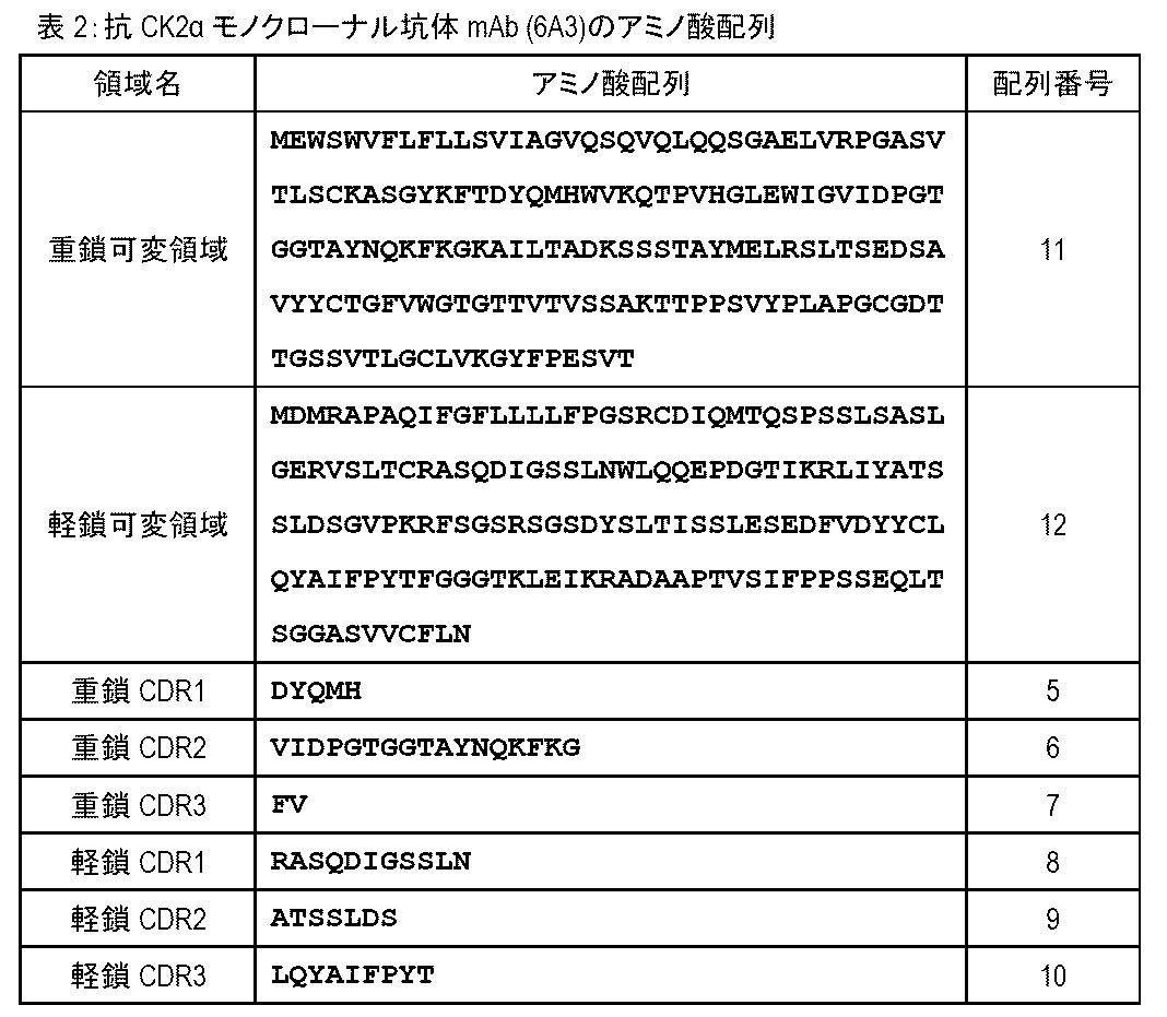
(目的)
CK2αタンパク質を特異的かつ高感度で検出可能なモノクローナル抗体(抗CK2αモノクローナル抗体)をさらに開発する。さらに、各クローンについて重鎖及び軽鎖の可変領域配列並びにCDR配列を決定する。
実施例1の「(1)抗原ポリペプチドによる免疫」に記載の方法に準じてヒトCK2αタンパク質を抗原としてマウスを免疫した。さらに実施例1の「(2)細胞融合及びスクリーニング」、「(3)選抜されたクローンの解析」、及び「(4)クローン6Aを対象とするさらなるクローニング」に記載の方法に準じて免疫後のマウス個体からリンパ細胞を単離し、リンパ細胞とミエローマ細胞とを融合して、抗体産生ハイブリドーマを作製し、クローン10B2、15C1、16C2、19C2、及び21B1の5つのクローンが得られた。さらに実施例1の「(5)クローン6A3からのIgG精製」に記載の方法に準じて各クローンの培養上清を精製し、精製後のモノクローナル抗体として「mAb (10B2)」、「mAb (15C1)」、「mAb (16C2)」、「mAb (19C2)」、及び「mAb (21B1)」を得た。
次に実施例4と同様の方法により、得られた抗CK2αモノクローナル坑体の各々について、重鎖及び軽鎖の可変領域配列並びにCDR配列を決定した。重鎖及び軽鎖の各可変領域及び各CDRの配列を決定した結果を以下に示す。なお、CDRの同定はKabatの抗体ナンバリングシステムに従った。また、得られた抗CK2αモノクローナル坑体は,「mAb(6A3)」、「mAb (10B2)」、「mAb (15C1)」、「mAb (16C2)はすべてIgG2bであるが、「mAb (19C2)」、及び「mAb (21B1)」はIgG1であることも判明した。
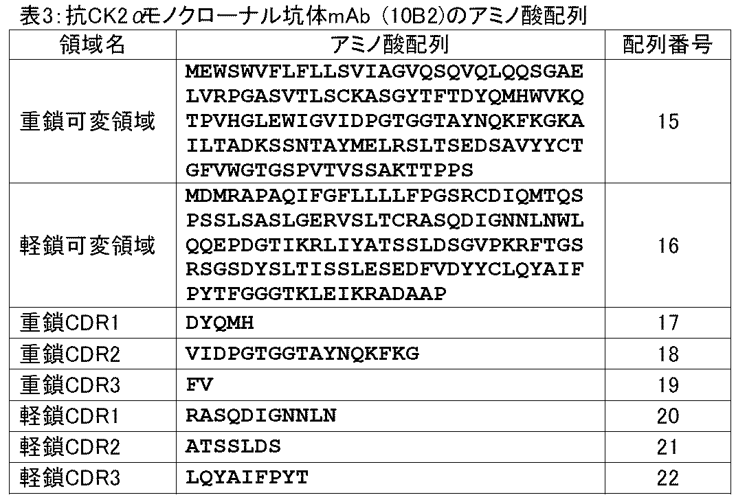
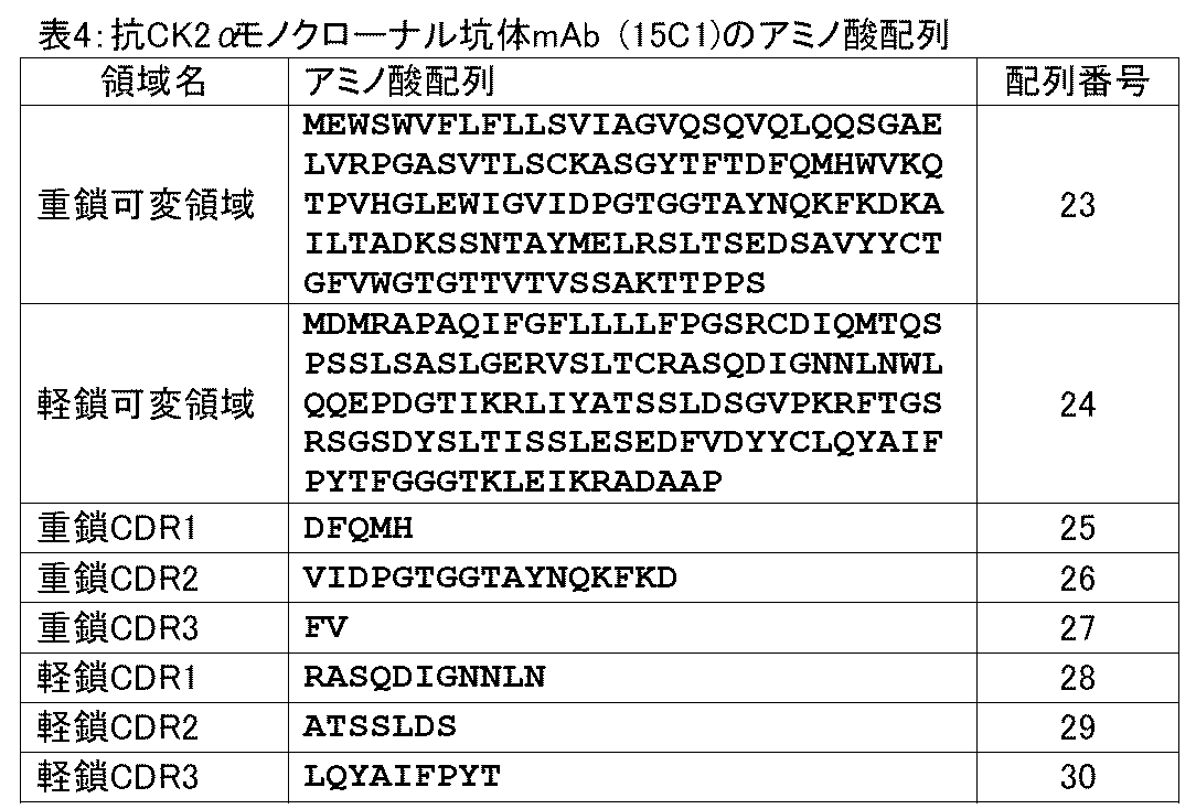


(目的)
実施例1で作製した抗CK2αモノクローナル坑体mAb (6A3)、及び実施例5で作製した抗CK2αモノクローナル坑体mAb (10B2)、mAb (15C1)、mAb (16C2)、及びmAb (19C2)と市販のマウス抗CK2αモノクローナル坑体(ab70774, Abcam, UK;「対照抗体」という)の性能を比較するために、ウェスタンブロットと免疫沈降を行う。
(1)細胞ライセートからの免疫沈降物に対するウェスタンブロット
乳癌細胞株MCF-7(JCRBセルバンク、#JCRB0134)、及びFlag-CK2αタンパク質を発現するHEK293細胞からライセートを調製した。培養したMCF-7細胞株又はFlag-CK2αタンパク質を発現するHEK293細胞をPBSで洗浄後プレートから回収し、常法にしたがって細胞ライゼートから可溶性画分を部分精製し、抗CK2α抗体(1 μgの本発明の6種類の抗体又は対照抗体)を加えて4℃にて2時間温和に混合した後、Protein G-agaroseビーズを添加して遠心分離することにより、CK2α免疫複合体を形成させた。次いで、遠心分離して得られた免疫沈降物をSDS処理後に電気泳動した。ウェスタンブロットには、0.1 μg/mLの本発明の抗体又は対照抗体を用いた。
CK2αタンパク質は、核内においてHMGB2(High Mobility Group Box 2)遺伝子座を含むヒトゲノム上の広範な部位にリクルートされる(本発明者ら、未発表)。このことに基づいて、HMGB2遺伝子座を対象としてクロマチン免疫沈降を実施することによって、本発明の抗体の性能を検証する。
具体的には、ヒト網膜色素上皮細胞(ヒトRPE細胞)ならびにCRISPR-Cas9法によってCK2遺伝子をノックアウトしたヒトRPE細胞(RPE-ko)からクロマチン免疫沈降の常法に従いライセートを作製しクロマチン画分を調製した。このクロマチン画分に対して抗CK2α抗体(1 μgのmAb (6A3)、mAb (10B2)、mAb (15C1)、又はmAb (16C2))を加えて4℃にて一晩ローテーターを用いて温和に混合した後、Protein G-agaroseビーズを添加して遠心分離することによってクロマチン免疫複合体の画分を取得した。得られた画分に含まれる核酸を鋳型として、配列番号65及び66で示すプライマー対を用いてヒトHMGB2遺伝子座の塩基配列をPCR増幅した。PCRの増幅条件(温度及び時間)は以下の通りであった:95℃で3秒、60℃で20秒(1サイクル)を40サイクル繰り返した。HMGB2に対応するDNA増幅量は、KAPA SYBR Fast qPCRキット(KAPA Biosystems)を用いApplied Biosystems StepOne(Life Technologies)によって測定した。
本発明のmAb (6A3)、mAb (10B2)、mAb (15C1)、mAb (16C2)、mAb (19C2)、及びmAb (21B1)を使用して、精製品のリコンビナントCK2αタンパク質をウェスタンブロットによって検出した。リコンビナントCK2αタンパク質は、pGEX2T-CK2α遺伝子を大腸菌に導入して発現させたヒト由来CK2αタンパク質を精製したものを使用した。0.05 μg、0.1 μg、又は0.2 μgのリコンビナントCK2αタンパク質をSDS処理して電気泳動し、0.1 μg/mLの本発明の6種類の抗体を用いて検出を行った。
(目的)
実施例5で作製した抗CK2αモノクローナル坑体mAb (21B1)及び対照抗体を用いて、乳癌組織の免疫染色を行い、癌細胞において核小体に局在するCK2αタンパク質を検出する。
実施例3に記載の方法に準じて、乳癌組織における癌浸潤部(病変部)のホルマリン固定パラフィン包埋標本を対象として、抗CK2αモノクローナル坑体mAb (21B1)による免疫染色を実施した。なお、抗CK2αモノクローナル坑体mAb (21B1)は1,000倍希釈(0.1 μg/mL)で使用した。また、マウス抗CK2αモノクローナル坑体(ab70774, Abcam, UK)は1,000倍希釈(2 μg/mL)で使用した。
(目的)
本発明のモノクローナル抗CK2α坑体mAb (6A3)を用いて、肺腺癌患者から切除された肺腺癌組織におけるCK2αタンパク質の発現及び局在を評価し、肺腺癌患者の切除手術後の予後を評価する。
(方法)
福島県立医科大学呼吸器外科学講座で2007年から2014年の間に根治的切除術を受けた原発性肺腺癌患者120人の患者から切除された肺腺癌組織ホルマリン固定パラフィン包埋標本を用いた。腫瘍のステージは悪性腫瘍のTNM分類(UICC国際規約、L.H. Sobin, M.K. Gospodarowicz and Ch. Wittekind, TNM Classification of Malignant Tumours, 8th edition) に従って決定した。なお本研究は、福島県立医科大学の審査委員会の承認を受けた。
I:細胞全体の染色があるが、核染色が明瞭でない
II:核染色(+)、細胞質より核染色が明瞭である
III:核染色(++)、核染色がIIより高いレベルである
IV:核染色(+、++)、さらに核小体染色(+)
V:核染色(-)、核小体染色(+)
無再発生存率の結果を図14に示す。CK2α核小体染色陽性の肺腺癌患者は、核小体染色陰性患者と比較して有意に低い無再発生存率を示した。このことはCK2α核小体染色陽性症例の再発リスクが陰性症例と比較して有意に高いことを示している(N=118、P=0.0031、ログランク検定)。
単変量解析及び多変量解析においてハザード比(図中「HR」として示し、再発の起こりやすさを相対的に示す)及びその95%信頼区間を算出した結果を図15A及び図15Bに示す。さらに多変量解析において無再発生存期間を規定する効果を解析するために、JMP Pro version 14のPartition Treeを使用して目的変数への寄与度を比較した結果を図15Cに示す。この結果から、CK2α核小体染色陽性という指標が、将来の再発と強く関連することが独立変数の統計的有意性として示された。また、多変量解析ではCK2α核小体染色陽性の指標は、統計的有意性を示す検定統計量であるWald値が最大になった。
Claims (9)
- 抗CK2α抗体又はその断片であって、
(1)配列番号5で示すアミノ酸配列からなるCDR1、
配列番号6で示すアミノ酸配列からなるCDR2、及び
アミノ酸配列FVからなるCDR3
を含む重鎖可変領域、並びに
配列番号8で示すアミノ酸配列からなるCDR1、
配列番号9で示すアミノ酸配列からなるCDR2、及び
配列番号10で示すアミノ酸配列からなるCDR3
を含む軽鎖可変領域、
(2)配列番号17で示すアミノ酸配列からなるCDR1、
配列番号18で示すアミノ酸配列からなるCDR2、及び
アミノ酸配列FVからなるCDR3
を含む重鎖可変領域、並びに
配列番号20で示すアミノ酸配列からなるCDR1、
配列番号21で示すアミノ酸配列からなるCDR2、及び
配列番号22で示すアミノ酸配列からなるCDR3
を含む軽鎖可変領域、
(3)配列番号25で示すアミノ酸配列からなるCDR1、
配列番号26で示すアミノ酸配列からなるCDR2、及び
アミノ酸配列FVからなるCDR3
を含む重鎖可変領域、並びに
配列番号28で示すアミノ酸配列からなるCDR1、
配列番号29で示すアミノ酸配列からなるCDR2、及び
配列番号30で示すアミノ酸配列からなるCDR3
を含む軽鎖可変領域、
(4)配列番号33で示すアミノ酸配列からなるCDR1、
配列番号34で示すアミノ酸配列からなるCDR2、及び
アミノ酸配列FVからなるCDR3
を含む重鎖可変領域、並びに
配列番号36で示すアミノ酸配列からなるCDR1、
配列番号37で示すアミノ酸配列からなるCDR2、及び
配列番号38で示すアミノ酸配列からなるCDR3
を含む軽鎖可変領域、
(5)配列番号41で示すアミノ酸配列からなるCDR1、
配列番号42で示すアミノ酸配列からなるCDR2、及び
配列番号43で示すアミノ酸配列からなるCDR3
を含む重鎖可変領域、並びに
配列番号44で示すアミノ酸配列からなるCDR1、
配列番号45で示すアミノ酸配列からなるCDR2、及び
配列番号46で示すアミノ酸配列からなるCDR3
を含む軽鎖可変領域、又は
(6)配列番号49で示すアミノ酸配列からなるCDR1、
配列番号50で示すアミノ酸配列からなるCDR2、及び
配列番号51で示すアミノ酸配列からなるCDR3
を含む重鎖可変領域、並びに
配列番号52で示すアミノ酸配列からなるCDR1、
配列番号53で示すアミノ酸配列からなるCDR2、及び
配列番号54で示すアミノ酸配列からなるCDR3
を含む軽鎖可変領域
を含む、前記抗CK2α抗体又はその断片。 - 前記抗CK2α抗体又はその断片が、
(a)配列番号11で示すアミノ酸配列からなる重鎖可変領域、及び
配列番号12で示すアミノ酸配列からなる軽鎖可変領域、
(b)配列番号15で示すアミノ酸配列からなる重鎖可変領域、及び
配列番号16で示すアミノ酸配列からなる軽鎖可変領域
(c)配列番号23で示すアミノ酸配列からなる重鎖可変領域、及び
配列番号24で示すアミノ酸配列からなる軽鎖可変領域
(d)配列番号31で示すアミノ酸配列からなる重鎖可変領域、及び
配列番号32で示すアミノ酸配列からなる軽鎖可変領域
(e)配列番号39で示すアミノ酸配列からなる重鎖可変領域、及び
配列番号40で示すアミノ酸配列からなる軽鎖可変領域、又は
(f)配列番号47で示すアミノ酸配列からなる重鎖可変領域、及び
配列番号48で示すアミノ酸配列からなる軽鎖可変領域
を含む、請求項1に記載の抗CK2α抗体又はその断片。 - 請求項1又は2に記載の抗CK2α抗体又はその断片を含む、癌患者の予後予測用キット。
- 予後が再発リスクを含む、請求項3に記載のキット。
- 前記癌が、乳癌、子宮癌、食道癌、胃癌、胆道癌、膵癌、肝癌、腎癌、大腸癌、膀胱癌、肺癌、甲状腺癌、及び神経膠腫からなる群から選択される、請求項3又は4に記載のキット。
- 癌患者の予後を予測するための方法であって、
癌患者から得られた癌細胞又は組織において、核小体中のCK2αタンパク質又はその断片を検出する工程、並びに
CK2αタンパク質又はその断片が他の細胞画分と比して高度に核小体に検出された場合に予後が悪いと予測する、及び/又はCK2αタンパク質又はその断片が他の細胞画分と比して高度に核小体に検出されない場合に予後が良いと予測する工程
を含み、CK2αタンパク質又はその断片が請求項1又は2に記載の抗CK2α抗体又はその断片を用いて検出される、前記方法。 - 予後が再発リスクを含む、請求項6に記載の方法。
- 前記癌が、乳癌、子宮癌、食道癌、胃癌、胆道癌、膵癌、肝癌、腎癌、大腸癌、膀胱癌、肺癌、甲状腺癌、及び神経膠腫からなる群から選択される、請求項6又は7に記載の方法。
- 前記癌が乳癌であり、CK2αタンパク質又はその断片の検出の有無を、ステージによる分類、ホルモン受容体の発現状況による分類、並びにHER2遺伝子及び/又はタンパク質の発現状況による分類の少なくとも一つと組み合わせて乳癌患者の予後を予測する、請求項8に記載の方法。
Priority Applications (3)
| Application Number | Priority Date | Filing Date | Title |
|---|---|---|---|
| EP23780828.2A EP4403581A4 (en) | 2022-03-30 | 2023-03-30 | ANTI-CK2 ALPHA ANTIBODY OR FRAGMENT THEREOF |
| JP2024512766A JPWO2023190820A1 (ja) | 2022-03-30 | 2023-03-30 | |
| US18/579,662 US20240376227A1 (en) | 2022-03-30 | 2023-03-30 | ANTI-CK2a ANTIBODY OR A FRAGMENT THEREOF |
Applications Claiming Priority (2)
| Application Number | Priority Date | Filing Date | Title |
|---|---|---|---|
| JP2022055606 | 2022-03-30 | ||
| JP2022-055606 | 2022-03-30 |
Publications (1)
| Publication Number | Publication Date |
|---|---|
| WO2023190820A1 true WO2023190820A1 (ja) | 2023-10-05 |
Family
ID=88202747
Family Applications (1)
| Application Number | Title | Priority Date | Filing Date |
|---|---|---|---|
| PCT/JP2023/013069 Ceased WO2023190820A1 (ja) | 2022-03-30 | 2023-03-30 | 抗CK2α抗体又はその断片 |
Country Status (4)
| Country | Link |
|---|---|
| US (1) | US20240376227A1 (ja) |
| EP (1) | EP4403581A4 (ja) |
| JP (1) | JPWO2023190820A1 (ja) |
| WO (1) | WO2023190820A1 (ja) |
Citations (7)
| Publication number | Priority date | Publication date | Assignee | Title |
|---|---|---|---|---|
| JP2003093056A (ja) * | 2001-09-26 | 2003-04-02 | Japan Science & Technology Corp | p53のSer46をリン酸化する酵素 |
| WO2004017997A1 (ja) * | 2002-08-06 | 2004-03-04 | Toray Industries, Inc. | 腎疾患治療又は予防剤及び腎疾患の診断方法 |
| EP2472263A1 (en) * | 2011-01-03 | 2012-07-04 | Commissariat À L'Énergie Atomique Et Aux Énergies Alternatives | Methods for the prognostic assessment of breast cancer |
| JP2013506836A (ja) * | 2009-10-02 | 2013-02-28 | サイリーン ファーマシューティカルズ インコーポレーティッド | プロテインキナーゼck2介在性疾患のck2阻害剤に対する感受性および反応を予測するバイオマーカー |
| US20130230852A1 (en) * | 2010-07-01 | 2013-09-05 | The Regents Of The University Of California | Protein kinase ck2 gene mutations, amplifications and polymorphisms in human cancers and methods of use |
| WO2021132544A1 (ja) | 2019-12-25 | 2021-07-01 | 公立大学法人福島県立医科大学 | 癌の予後バイオマーカー |
| JP2022055606A (ja) | 2020-09-29 | 2022-04-08 | 株式会社 Mts雪氷研究所 | プログラム及び開口部装置 |
-
2023
- 2023-03-30 WO PCT/JP2023/013069 patent/WO2023190820A1/ja not_active Ceased
- 2023-03-30 JP JP2024512766A patent/JPWO2023190820A1/ja active Pending
- 2023-03-30 US US18/579,662 patent/US20240376227A1/en active Pending
- 2023-03-30 EP EP23780828.2A patent/EP4403581A4/en active Pending
Patent Citations (7)
| Publication number | Priority date | Publication date | Assignee | Title |
|---|---|---|---|---|
| JP2003093056A (ja) * | 2001-09-26 | 2003-04-02 | Japan Science & Technology Corp | p53のSer46をリン酸化する酵素 |
| WO2004017997A1 (ja) * | 2002-08-06 | 2004-03-04 | Toray Industries, Inc. | 腎疾患治療又は予防剤及び腎疾患の診断方法 |
| JP2013506836A (ja) * | 2009-10-02 | 2013-02-28 | サイリーン ファーマシューティカルズ インコーポレーティッド | プロテインキナーゼck2介在性疾患のck2阻害剤に対する感受性および反応を予測するバイオマーカー |
| US20130230852A1 (en) * | 2010-07-01 | 2013-09-05 | The Regents Of The University Of California | Protein kinase ck2 gene mutations, amplifications and polymorphisms in human cancers and methods of use |
| EP2472263A1 (en) * | 2011-01-03 | 2012-07-04 | Commissariat À L'Énergie Atomique Et Aux Énergies Alternatives | Methods for the prognostic assessment of breast cancer |
| WO2021132544A1 (ja) | 2019-12-25 | 2021-07-01 | 公立大学法人福島県立医科大学 | 癌の予後バイオマーカー |
| JP2022055606A (ja) | 2020-09-29 | 2022-04-08 | 株式会社 Mts雪氷研究所 | プログラム及び開口部装置 |
Non-Patent Citations (7)
| Title |
|---|
| ALTSCHUL ET AL., J. MOL. BIOL., vol. 215, 1990, pages 403 - 410 |
| ALTSCHUL ET AL., NUC. ACIDS. RES., vol. 25, 1977, pages 3389 - 3402 |
| GREEN, M.R.SAMBROOK, J.: "Molecular Cloning: A Laboratory Manual", 2012, COLD SPRING HARBOR LABORATORY PRESS |
| HOLLIGER ET AL., PROC. NATL. ACAD. SCI. USA, vol. 90, 1993, pages 6444 - 6448 |
| KABAT E.A. ET AL.: "Sequences of proteins of immunological interest", vol. 1, 1991, NIH |
| L.H. SOBINM.K. GOSPODAROWICZCH. WITTEKIND: "TNM Classification of Malignant Tumours" |
| See also references of EP4403581A4 |
Also Published As
| Publication number | Publication date |
|---|---|
| EP4403581A4 (en) | 2025-06-18 |
| JPWO2023190820A1 (ja) | 2023-10-05 |
| EP4403581A1 (en) | 2024-07-24 |
| US20240376227A1 (en) | 2024-11-14 |
Similar Documents
| Publication | Publication Date | Title |
|---|---|---|
| RU2651513C2 (ru) | Новое антитело для диагностики и (или) прогнозирования рака | |
| US10273308B2 (en) | Methods of producing antibodies specific for p95 | |
| EP3149042B1 (en) | Pd-l1 antibodies and uses thereof | |
| US7521543B2 (en) | A34 and A33-like 3 DNA, proteins, antibodies thereto and methods of treatment using same | |
| JP6940505B2 (ja) | 組成物および癌の発生リスクの評価方法 | |
| CA3087779A1 (en) | Agents and methods for predicting response to therapy | |
| JP7695672B2 (ja) | 癌の予後バイオマーカー | |
| WO2022154037A1 (ja) | がんの予後バイオマーカー | |
| CN113677994B (zh) | 用于评估t细胞功能和预测对疗法的应答的方法和药剂 | |
| EP3203238A1 (en) | Reagents for detecting or diagnosing cancer cells having high invasive capacity | |
| JP2022526131A (ja) | 肝細胞癌診断用バイオマーカーセレブロンと、これに特異的な新規なモノクローナル抗体 | |
| US20240376227A1 (en) | ANTI-CK2a ANTIBODY OR A FRAGMENT THEREOF | |
| CN111303289B (zh) | 抗人Tn型糖基化MUC1抗体及其用途 | |
| WO2022214432A1 (en) | Immunohistochemistry methods and kir3dl2-specific reagents | |
| KR101415927B1 (ko) | 항―tmap/ckap2 항체를 포함하는 암의 예후 진단용 조성물 | |
| CA3009768C (en) | Compositions and methods for assessing the risk of cancer occurrence | |
| WO2023196882A1 (en) | Claudin 18.2 immunohistochemistry assay and use thereof | |
| KR20250166247A (ko) | Pro-c17 분석법 | |
| WO2023002943A1 (ja) | がん患者の予後を予測するためのバイオマーカー、がん患者の予後を予測するための方法、がん患者における、がん治療薬の効果を予測するための方法、及び、がん患者の予後を予測するためのキット | |
| HK1262633B (zh) | 用於评估癌症发生的风险的组合物和方法 | |
| HK1183311A (en) | Novel antibody for the diagnosis and/or prognosis of cancer |
Legal Events
| Date | Code | Title | Description |
|---|---|---|---|
| 121 | Ep: the epo has been informed by wipo that ep was designated in this application |
Ref document number: 23780828 Country of ref document: EP Kind code of ref document: A1 |
|
| WWE | Wipo information: entry into national phase |
Ref document number: 18579662 Country of ref document: US |
|
| WWE | Wipo information: entry into national phase |
Ref document number: 2023780828 Country of ref document: EP |
|
| ENP | Entry into the national phase |
Ref document number: 2023780828 Country of ref document: EP Effective date: 20240123 |
|
| ENP | Entry into the national phase |
Ref document number: 2024512766 Country of ref document: JP Kind code of ref document: A |
|
| NENP | Non-entry into the national phase |
Ref country code: DE |