WO2023191384A1 - 항염증 및 항섬유화 활성을 갖는 펩타이드 및 이의 용도 - Google Patents
항염증 및 항섬유화 활성을 갖는 펩타이드 및 이의 용도 Download PDFInfo
- Publication number
- WO2023191384A1 WO2023191384A1 PCT/KR2023/003862 KR2023003862W WO2023191384A1 WO 2023191384 A1 WO2023191384 A1 WO 2023191384A1 KR 2023003862 W KR2023003862 W KR 2023003862W WO 2023191384 A1 WO2023191384 A1 WO 2023191384A1
- Authority
- WO
- WIPO (PCT)
- Prior art keywords
- peptide
- lung
- inflammatory
- cells
- treatment
- Prior art date
- Legal status (The legal status is an assumption and is not a legal conclusion. Google has not performed a legal analysis and makes no representation as to the accuracy of the status listed.)
- Ceased
Links
Images





Classifications
-
- A—HUMAN NECESSITIES
- A23—FOODS OR FOODSTUFFS; TREATMENT THEREOF, NOT COVERED BY OTHER CLASSES
- A23L—FOODS, FOODSTUFFS OR NON-ALCOHOLIC BEVERAGES, NOT OTHERWISE PROVIDED FOR; PREPARATION OR TREATMENT THEREOF
- A23L33/00—Modifying nutritive qualities of foods; Dietetic products; Preparation or treatment thereof
- A23L33/10—Modifying nutritive qualities of foods; Dietetic products; Preparation or treatment thereof using additives
- A23L33/17—Amino acids, peptides or proteins
- A23L33/18—Peptides; Protein hydrolysates
-
- A—HUMAN NECESSITIES
- A61—MEDICAL OR VETERINARY SCIENCE; HYGIENE
- A61K—PREPARATIONS FOR MEDICAL, DENTAL OR TOILETRY PURPOSES
- A61K38/00—Medicinal preparations containing peptides
- A61K38/04—Peptides having up to 20 amino acids in a fully defined sequence; Derivatives thereof
- A61K38/10—Peptides having 12 to 20 amino acids
-
- A—HUMAN NECESSITIES
- A61—MEDICAL OR VETERINARY SCIENCE; HYGIENE
- A61K—PREPARATIONS FOR MEDICAL, DENTAL OR TOILETRY PURPOSES
- A61K9/00—Medicinal preparations characterised by special physical form
- A61K9/0087—Galenical forms not covered by A61K9/02 - A61K9/7023
- A61K9/0095—Drinks; Beverages; Syrups; Compositions for reconstitution thereof, e.g. powders or tablets to be dispersed in a glass of water; Veterinary drenches
-
- A—HUMAN NECESSITIES
- A61—MEDICAL OR VETERINARY SCIENCE; HYGIENE
- A61K—PREPARATIONS FOR MEDICAL, DENTAL OR TOILETRY PURPOSES
- A61K9/00—Medicinal preparations characterised by special physical form
- A61K9/14—Particulate form, e.g. powders, Processes for size reducing of pure drugs or the resulting products, Pure drug nanoparticles
- A61K9/16—Agglomerates; Granulates; Microbeadlets ; Microspheres; Pellets; Solid products obtained by spray drying, spray freeze drying, spray congealing,(multiple) emulsion solvent evaporation or extraction
- A61K9/1605—Excipients; Inactive ingredients
- A61K9/1617—Organic compounds, e.g. phospholipids, fats
- A61K9/1623—Sugars or sugar alcohols, e.g. lactose; Derivatives thereof; Homeopathic globules
-
- A—HUMAN NECESSITIES
- A61—MEDICAL OR VETERINARY SCIENCE; HYGIENE
- A61K—PREPARATIONS FOR MEDICAL, DENTAL OR TOILETRY PURPOSES
- A61K9/00—Medicinal preparations characterised by special physical form
- A61K9/14—Particulate form, e.g. powders, Processes for size reducing of pure drugs or the resulting products, Pure drug nanoparticles
- A61K9/16—Agglomerates; Granulates; Microbeadlets ; Microspheres; Pellets; Solid products obtained by spray drying, spray freeze drying, spray congealing,(multiple) emulsion solvent evaporation or extraction
- A61K9/1605—Excipients; Inactive ingredients
- A61K9/1629—Organic macromolecular compounds
- A61K9/1652—Polysaccharides, e.g. alginate, cellulose derivatives; Cyclodextrin
-
- A—HUMAN NECESSITIES
- A61—MEDICAL OR VETERINARY SCIENCE; HYGIENE
- A61K—PREPARATIONS FOR MEDICAL, DENTAL OR TOILETRY PURPOSES
- A61K9/00—Medicinal preparations characterised by special physical form
- A61K9/14—Particulate form, e.g. powders, Processes for size reducing of pure drugs or the resulting products, Pure drug nanoparticles
- A61K9/16—Agglomerates; Granulates; Microbeadlets ; Microspheres; Pellets; Solid products obtained by spray drying, spray freeze drying, spray congealing,(multiple) emulsion solvent evaporation or extraction
- A61K9/1605—Excipients; Inactive ingredients
- A61K9/1664—Compounds of unknown constitution, e.g. material from plants or animals
-
- A—HUMAN NECESSITIES
- A61—MEDICAL OR VETERINARY SCIENCE; HYGIENE
- A61K—PREPARATIONS FOR MEDICAL, DENTAL OR TOILETRY PURPOSES
- A61K9/00—Medicinal preparations characterised by special physical form
- A61K9/20—Pills, tablets, discs, rods
- A61K9/2004—Excipients; Inactive ingredients
- A61K9/2013—Organic compounds, e.g. phospholipids, fats
- A61K9/2018—Sugars, or sugar alcohols, e.g. lactose, mannitol; Derivatives thereof, e.g. polysorbates
-
- A—HUMAN NECESSITIES
- A61—MEDICAL OR VETERINARY SCIENCE; HYGIENE
- A61K—PREPARATIONS FOR MEDICAL, DENTAL OR TOILETRY PURPOSES
- A61K9/00—Medicinal preparations characterised by special physical form
- A61K9/20—Pills, tablets, discs, rods
- A61K9/2004—Excipients; Inactive ingredients
- A61K9/2022—Organic macromolecular compounds
- A61K9/205—Polysaccharides, e.g. alginate, gums; Cyclodextrin
- A61K9/2059—Starch, including chemically or physically modified derivatives; Amylose; Amylopectin; Dextrin
-
- A—HUMAN NECESSITIES
- A61—MEDICAL OR VETERINARY SCIENCE; HYGIENE
- A61K—PREPARATIONS FOR MEDICAL, DENTAL OR TOILETRY PURPOSES
- A61K9/00—Medicinal preparations characterised by special physical form
- A61K9/48—Preparations in capsules, e.g. of gelatin, of chocolate
- A61K9/4816—Wall or shell material
- A61K9/4825—Proteins, e.g. gelatin
-
- A—HUMAN NECESSITIES
- A61—MEDICAL OR VETERINARY SCIENCE; HYGIENE
- A61K—PREPARATIONS FOR MEDICAL, DENTAL OR TOILETRY PURPOSES
- A61K9/00—Medicinal preparations characterised by special physical form
- A61K9/48—Preparations in capsules, e.g. of gelatin, of chocolate
- A61K9/4841—Filling excipients; Inactive ingredients
- A61K9/4858—Organic compounds
-
- A—HUMAN NECESSITIES
- A61—MEDICAL OR VETERINARY SCIENCE; HYGIENE
- A61K—PREPARATIONS FOR MEDICAL, DENTAL OR TOILETRY PURPOSES
- A61K9/00—Medicinal preparations characterised by special physical form
- A61K9/48—Preparations in capsules, e.g. of gelatin, of chocolate
- A61K9/4841—Filling excipients; Inactive ingredients
- A61K9/4866—Organic macromolecular compounds
-
- A—HUMAN NECESSITIES
- A61—MEDICAL OR VETERINARY SCIENCE; HYGIENE
- A61P—SPECIFIC THERAPEUTIC ACTIVITY OF CHEMICAL COMPOUNDS OR MEDICINAL PREPARATIONS
- A61P11/00—Drugs for disorders of the respiratory system
-
- A—HUMAN NECESSITIES
- A61—MEDICAL OR VETERINARY SCIENCE; HYGIENE
- A61P—SPECIFIC THERAPEUTIC ACTIVITY OF CHEMICAL COMPOUNDS OR MEDICINAL PREPARATIONS
- A61P29/00—Non-central analgesic, antipyretic or antiinflammatory agents, e.g. antirheumatic agents; Non-steroidal antiinflammatory drugs [NSAID]
-
- A—HUMAN NECESSITIES
- A23—FOODS OR FOODSTUFFS; TREATMENT THEREOF, NOT COVERED BY OTHER CLASSES
- A23V—INDEXING SCHEME RELATING TO FOODS, FOODSTUFFS OR NON-ALCOHOLIC BEVERAGES AND LACTIC OR PROPIONIC ACID BACTERIA USED IN FOODSTUFFS OR FOOD PREPARATION
- A23V2002/00—Food compositions, function of food ingredients or processes for food or foodstuffs
-
- A—HUMAN NECESSITIES
- A23—FOODS OR FOODSTUFFS; TREATMENT THEREOF, NOT COVERED BY OTHER CLASSES
- A23V—INDEXING SCHEME RELATING TO FOODS, FOODSTUFFS OR NON-ALCOHOLIC BEVERAGES AND LACTIC OR PROPIONIC ACID BACTERIA USED IN FOODSTUFFS OR FOOD PREPARATION
- A23V2200/00—Function of food ingredients
- A23V2200/30—Foods, ingredients or supplements having a functional effect on health
- A23V2200/314—Foods, ingredients or supplements having a functional effect on health having an effect on lung or respiratory system
-
- A—HUMAN NECESSITIES
- A23—FOODS OR FOODSTUFFS; TREATMENT THEREOF, NOT COVERED BY OTHER CLASSES
- A23V—INDEXING SCHEME RELATING TO FOODS, FOODSTUFFS OR NON-ALCOHOLIC BEVERAGES AND LACTIC OR PROPIONIC ACID BACTERIA USED IN FOODSTUFFS OR FOOD PREPARATION
- A23V2200/00—Function of food ingredients
- A23V2200/30—Foods, ingredients or supplements having a functional effect on health
- A23V2200/324—Foods, ingredients or supplements having a functional effect on health having an effect on the immune system
Definitions
- the present invention relates to peptides with anti-inflammatory and anti-fibrotic activity and their uses. More specifically, the present invention relates to peptides having activity in the treatment, prevention, or amelioration of pulmonary inflammatory diseases or pulmonary fibrosis and their uses.
- the human lungs occupy 6% of the body's volume and are composed of many small gas sacs, the alveoli.
- the primary purpose of the lungs is to facilitate gas exchange with the systemic circulation.
- cytokines and growth factors are released from the immune cells in the lungs, such as neutrophils, macrophages, neutrophils, and mast cells, and various lung cells. (growth factors) are secreted.
- Inflammatory cytokines are known to be important mediators of inflammatory lung diseases.
- Lung epithelial cells are stimulated by cytokines secreted by inflammatory cells, including interleukins, chemokines, colony stimulating factors and growth factors ( It plays an important role in this cytokine network at local inflammation sites by producing and secreting growth factors, etc.
- COPD chronic obstructive pulmonary disease
- the mucosa and submucosa of the airways are invaded by eosinophils, mast cells, and CD4+T cells, and these cells produce and secrete IL-4, IL-5, and IL-13. there is.
- pulmonary fibrosis is known to be a very important cause of inflammatory response in the lungs. Most organs go through inflammation and healing processes after tissue damage. If the damage is minor, normal structure and function are maintained, but if there is continued damage, the tissue undergoes fibrosis during the healing process.
- corticosteroids are most commonly used as anti-inflammatory drugs to reduce inflammation.
- drugs that target immune responses or cytokines activate Th1 responses or suppress Th2 responses.
- cytokine inhibitor, anti-IL-2, anti-IL-4, anti-IL-5, anti-IL-9, anti-IL-13, anti-PGD2, anti-TNF- ⁇ , mast cell inhibitor, PPAR- ⁇ promoter, anti-PGD2 -Treatments such as IgE and anti-CD23 are being developed.
- these drugs cause side effects in actual clinical trials or have weak actual therapeutic effects, so the development of new drugs that are safer, show higher efficacy, and are more economical in cost is urgently needed.
- Patent Document 1 WO 2011/029931
- the present inventors have made research efforts to develop a peptide with anti-inflammatory activity, especially an activity capable of suppressing inflammation in the lung and inhibiting the progression of lung fibrosis.
- the peptide of the present invention significantly alleviates the inflammatory response in the lung.
- the present invention was completed by proving through lung cell and animal experiments that it effectively inhibits the progression of lung fibrosis.
- the purpose of the present invention is to provide a pharmaceutical composition for treating or preventing pulmonary inflammatory diseases.
- Another object of the present invention is to provide a functional food composition for improving or preventing lung inflammation.
- Another object of the present invention is to provide a functional food composition for improving or preventing pulmonary fibrosis.
- one aspect of the present invention provides a pharmaceutical composition for the treatment or prevention of pulmonary inflammatory disease, which contains a peptide containing the amino acid sequence of SEQ ID NO: 1 as an active ingredient.
- Another aspect of the present invention provides a functional food composition for improving or preventing lung inflammation comprising a peptide containing the amino acid sequence of SEQ ID NO: 1 as an active ingredient.
- Another aspect of the present invention provides a functional food composition for improving or preventing lung fibrosis comprising a peptide containing the amino acid sequence of SEQ ID NO: 1 as an active ingredient.
- a peptide containing the amino acid sequence disclosed in SEQ ID NO: 1 is provided.
- peptide refers to a linear molecule formed by linking amino acid residues to each other through peptide bonds.
- the peptide containing the amino acid sequence of SEQ ID NO: 1 of the present invention can be used without modification, but deletion or insertion of amino acid residues is possible within the range that does not affect the original activities of the peptide, such as anti-diabetic and anti-obesity activities. , substitution, or a combination of these can be used as a variant or fragment of an amino acid having a different sequence.
- the peptide of the present invention can be modified by phosphorylation, sulfation, acrylation, glycosylation, methylation, farnesylation, etc., to the extent that its activity is not changed. .
- the peptide of the present invention includes a peptide containing an amino acid sequence substantially identical to the peptide containing the amino acid sequence of SEQ ID NO: 1, and a variant or active fragment thereof.
- the substantially identical amino acid sequence refers to an amino acid sequence having sequence identity of at least 75%, for example, at least 80%, at least 85%, at least 90%, at least 95%, and at least 97%, respectively, with the amino acid sequence of SEQ ID NO: 1.
- the peptide may additionally include a targeting sequence, a tag, a labeled residue, and an amino acid sequence prepared for the specific purpose of increasing half-life or peptide stability.
- the peptide of the present invention may have N-terminal and/or C-terminal modifications induced to select a portion of the amino acid sequence and increase its activity. Through these N-terminal and/or C-terminal modifications, the stability of the peptide of the present invention can be significantly improved, for example, the half-life of the peptide can be increased when administered in vivo.
- the term "stability" is meant to include not only in vivo stability, which protects the peptide of the present invention from attack by protein-cleaving enzymes in vivo, but also storage stability (e.g., storage stability at room temperature).
- the N-terminal modification includes an acetyl group, a fluoreonylmethoxycarbonyl group, a formyl group, a palmitoyl group, and a myristyl group at the N-terminus of the peptide. ), a stearyl group, and polyethylene glycol (PEG).
- the C-terminal modification may be the binding of a hydroxyl group (-OH), amino group (-NH2), azide (-NHNH2), etc. to the C-terminus of the peptide, but is not limited thereto. .
- Pheptide of the present invention can be produced by various methods widely known in the technical field to which the present invention pertains.
- the peptide of the present invention can be synthesized using chemical synthesis methods known in the art, especially solid-phase synthesis techniques (Merrifield, J. Amer. Chem. Soc. 85:2149-54 (1963); Stewart, et al. , Solid Phase Peptide Synthesis, 2nd. ed., Pierce Chem. Co.: Rockford, 111 (1984)) or liquid phase synthesis technology (US Patent No. 5,516,891).
- the peptide of the present invention has the activity of suppressing the expression of inflammatory cytokine genes, inflammatory cytokine proteins, or proteins related to inflammatory cytokine signaling in lung cells where an inflammatory response is induced.
- the inflammatory cytokine gene whose expression is suppressed may be one or more selected from the group consisting of TNF ⁇ , IL-1 ⁇ , IL-6, IL-8, and COX-2 genes.
- the inflammatory cytokine proteins or proteins involved in inflammatory cytokine signaling whose expression is suppressed include IL-1 ⁇ , IL-6, COX2, IL-8, p-JNK, p-ERK, p-PI3K, It may be one or more selected from the group consisting of p-AKT, p-ERK, and Myeloperoxidase.
- Pheptide of the present invention inhibits the expression of fibrogenic genes in lung cells where an inflammatory response is induced.
- the fibrosis gene whose expression is suppressed may be one or more selected from the group consisting of ⁇ -SMA, Collagen1A1, Fibronectin1, and Tenascin C genes.
- the peptide of the present invention can reduce the number of neutrophils in lung tissue where an inflammatory response is induced.
- the peptide of the present invention described above has the above-mentioned activity, it can exhibit excellent efficacy in treating, preventing, or improving inflammatory diseases of the lung.
- composition for treating or preventing pulmonary inflammatory diseases 1.
- a pharmaceutical composition for the treatment or prevention of pulmonary inflammatory disease comprising a peptide containing the amino acid sequence of SEQ ID NO: 1 as an active ingredient.
- the peptide of the present invention inhibits the expression of inflammatory cytokine genes, proteins, or signaling-related proteins in lung cells that induce an inflammatory response, and thus has excellent activity in the treatment or prevention of lung inflammatory diseases.
- the lung inflammatory disease is asthma, allergic asthma, bronchitis, chronic bronchitis, acute bronchitis, bronchiolitis, diffuse panbronchiolitis, bronchiectasis, pleurisy, alveolitis, vasculitis, pneumonia, hypersensitivity pneumonitis, chronic obstructive pulmonary disease. It may be a disease selected from the group consisting of lung disease, pulmonary fibrosis, idiopathic pulmonary fibrosis, cystic fibrosis, acute respiratory distress syndrome, acute lung injury, smoke inhalation lung injury, and heat-induced lung injury, but is not limited thereto.
- the peptide inhibits the expression of inflammatory cytokine genes, inflammatory cytokine proteins, or proteins related to inflammatory cytokine signaling in lung cells in which an inflammatory response is induced. You can.
- the inflammatory cytokine gene may be one or more selected from the group consisting of TNF ⁇ , IL-1 ⁇ , IL-6, IL-8, and COX-2 genes.
- the inflammatory cytokine proteins or proteins involved in inflammatory cytokine signaling include IL-1 ⁇ , IL-6, COX2, IL-8, p-JNK, p-ERK, p-PI3K, p-AKT, p-ERK, and It may be one or more selected from the group consisting of myeloperoxidase.
- the peptide in the pharmaceutical composition for treating or preventing pulmonary inflammatory diseases of the present invention, can inhibit the expression of fibrosis genes in lung cells in which an inflammatory response is induced.
- the fibrosis gene may be one or more selected from the group consisting of ⁇ -SMA, Collagen1A1, Fibronectin1, and Tenascin C genes.
- the peptide in the pharmaceutical composition for treating or preventing pulmonary inflammatory diseases of the present invention, can reduce the number of neutrophils in lung tissue where an inflammatory response is induced.
- the pharmaceutical composition of the present invention may contain a therapeutically effective amount of the above-described peptide and a pharmaceutically acceptable carrier.
- terapéuticaally effective amount refers to an amount sufficient for the peptide, which is an active ingredient of the pharmaceutical composition of the present invention, to achieve its activity or efficacy, for example, to achieve the efficacy of treating or preventing inflammatory diseases of the lung. It means sufficient amount.
- the pharmaceutically acceptable carriers are commonly used in formulations and include lactose, dextrose, sucrose, sorbitol, mannitol, starch, gum acacia, calcium phosphate, alginate, gelatin, calcium silicate, microcrystalline cellulose, Includes, but is not limited to, polyvinylpyrrolidone, cellulose, water, syrup, methyl cellulose, methylhydroxybenzoate, propylhydroxybenzoate, talc, magnesium stearate, and mineral oil.
- the pharmaceutical composition of the present invention may further include, but is not limited to, lubricants, wetting agents, sweeteners, flavoring agents, emulsifiers, suspending agents, preservatives, etc.
- Suitable pharmaceutically acceptable carriers and formulations are described in detail in Remington: The Science and Practice of Pharmacy, (19th ed., 1995, Williams & Wilkins).
- the pharmaceutical composition of the present invention can be administered by any route suitable for treating inflammatory diseases of the lung, for example, orally or parenterally, and in the case of parenteral administration, intravenous injection, subcutaneous injection, It can be administered by intramuscular injection, intraperitoneal injection, topical administration, or transdermal administration.
- the dosage of the pharmaceutical composition may be 0.0001 ⁇ g to 100 mg, 0.001 ⁇ g to 100 mg, 0.01 ⁇ g to 100 mg, 0.1 ⁇ g to 100 mg, or 1.0 ⁇ g to 1000 mg per day, but is not limited thereto. It can be prescribed in various ways depending on factors such as formulation method, administration method, patient's age, weight, sex, pathological condition, food, administration time, administration route, excretion rate, and reaction sensitivity.
- the pharmaceutical composition of the present invention is prepared in unit dosage form by formulating it using a pharmaceutically acceptable carrier and/or excipient according to a method that can be easily performed by those skilled in the art. Alternatively, it can be manufactured by placing it in a multi-capacity container. At this time, the formulation may be in the form of a solution, suspension, or emulsion in an oil or aqueous medium, or may be in the form of an extract, powder, granule, tablet, or capsule, and may additionally contain a dispersant or stabilizer.
- a functional food composition for improving or preventing lung inflammation comprising a peptide containing the amino acid sequence of SEQ ID NO: 1 as an active ingredient.
- the lung inflammation includes asthma, allergic asthma, bronchitis, chronic bronchitis, acute bronchitis, bronchiolitis, diffuse panbronchiolitis, bronchiectasis, pleurisy, alveolitis, vasculitis, pneumonia, hypersensitivity pneumonitis, chronic obstructive pulmonary disease, pulmonary fibrosis, and idiopathic pulmonary fibrosis. , cystic fibrosis, acute respiratory distress syndrome, acute lung injury, smoke inhalation lung injury, and heat-induced lung injury, but is not limited to this.
- a functional food composition for improving or preventing pulmonary fibrosis comprising a peptide containing the amino acid sequence of SEQ ID NO: 1 as an active ingredient.
- the peptide inhibits the expression of inflammatory cytokine genes, inflammatory cytokine proteins, or proteins related to inflammatory cytokine signaling in lung cells in which an inflammatory response is induced. It can be suppressed.
- the inflammatory cytokine gene may be one or more selected from the group consisting of TNF ⁇ , IL-1 ⁇ , IL-6, IL-8, and COX-2 genes.
- the inflammatory cytokine proteins or proteins involved in inflammatory cytokine signaling include IL-1 ⁇ , IL-6, COX2, IL-8, p-JNK, p-ERK, p-PI3K, p-AKT, p-ERK, and It may be one or more selected from the group consisting of myeloperoxidase.
- the peptide in the functional food composition for improving or preventing lung inflammation or lung fibrosis of the present invention, can inhibit the expression of fibrosis genes in lung cells in which an inflammatory response is induced.
- the fibrosis gene may be one or more selected from the group consisting of ⁇ -SMA, Collagen1A1, Fibronectin1, and Tenascin C genes.
- the peptide in the functional food composition for improving or preventing lung inflammation or lung fibrosis of the present invention, can reduce the number of neutrophils in lung tissue where an inflammatory response is induced.
- the peptide may be included in an appropriate amount within the range of 0.0001% by weight to 10% by weight based on the total weight of the composition, but is not limited thereto.
- the functional food composition of the present invention may include a foodologically effective amount of the peptide and a foodologically acceptable carrier.
- the food composition of the present invention includes not only the peptide as the active ingredient, but also ingredients commonly added during food production, and may include, for example, proteins, carbohydrates, fats, nutrients, seasonings, and flavoring agents.
- examples of the above-mentioned carbohydrates include monosaccharides such as glucose, fructose, etc.; Disaccharides such as maltose, sucrose, oligosaccharides, etc.; and polysaccharides, such as common sugars such as dextrin and cyclodextrin, and sugar alcohols such as xylitol, sorbitol, and erythritol.
- flavoring agents natural flavoring agents, thaumatin, stevia extract (e.g., rebaudioside A, glycyrrhizin, etc.) and synthetic flavoring agents (saccharin, aspartame, etc.) can be used.
- the ratio of the carbohydrate may generally be about 1 to 20 g, preferably about 5 to 12 g, per 100 g of the food composition of the present invention, but is not limited thereto.
- the functional food composition of the present invention contains various nutrients, vitamins, minerals (electrolytes), flavoring agents such as synthetic and natural flavors, colorants and thickening agents (cheese, chocolate, etc.), pectic acid, and salts thereof. , alginic acid and its salts, organic acids, protective colloidal thickeners, pH adjusters, stabilizers, preservatives, glycerin, alcohol, carbonating agents used in carbonated beverages, etc. In addition, it may contain pulp for the production of natural fruit juice, fruit juice drinks, and vegetable drinks.
- the functional food composition of the present invention when manufactured as a drink, in addition to the peptide, which is the active ingredient of the present invention, citric acid, high fructose corn syrup, sugar, glucose, acetic acid, malic acid, fruit juice, Eucommia extract, jujube extract, licorice extract, etc. are added. May be included.
- treatment of pulmonary inflammatory disease comprising administering a therapeutically effective amount of a peptide containing the amino acid sequence of SEQ ID NO: 1 or a pharmaceutical composition containing the peptide to a patient with pulmonary inflammatory disease.
- preventive, or improvement methods are provided.
- the use of the peptide containing the amino acid sequence of SEQ ID NO: 1 is provided for treating, preventing, or improving pulmonary inflammatory disease.
- a peptide comprising the amino acid sequence of SEQ ID NO: 1 described above for preparing a medicament or functional food composition for treating, preventing, or improving pulmonary inflammatory diseases.
- the peptide of the present invention has the activity of suppressing the expression of inflammatory cytokine genes, inflammatory cytokine proteins, or proteins related to inflammatory cytokine signaling in lung cells in which an inflammatory response is induced. Additionally, the peptide can reduce the number of neutrophils in lung tissue where an inflammatory response is induced. Therefore, the peptide of the present invention can be used as an active substance useful for treating, preventing, or improving inflammatory diseases of the lung.
- Figure 1 shows the effect of treatment with the peptide of Preparation Example 1 on gene expression of inflammatory cytokines TNF- ⁇ , IL-1 ⁇ , IL-6, and IL-8 in MRC-5 cells induced an inflammatory response by LPS treatment. This is the result of measurement.
- Figure 2 shows the effect of treatment with the peptide of Preparation Example 1 on the gene expression of inflammatory cytokines TNF- ⁇ , IL-1 ⁇ , IL-6, IL-8, and COX-2 in A549 cells in which an inflammatory response was induced by LPS treatment. This is the result of measurement.
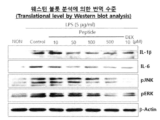
- Figure 3 shows the results of measuring the effect of treatment with the peptide of Preparation Example 1 on the expression of IL-1 ⁇ , IL-6, pJNK, and pERK in MRC-5 cells in which an inflammatory response was induced by LPS treatment.
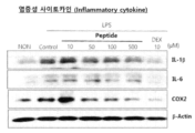
- Figures 4a and 4b show the treatment of the peptide of Preparation Example 1 on the expression of Phospho-PI3K, Phospho-AKT, Phospho-ERK, IL-1 ⁇ , IL-6, and COX2 in A549 cells in which an inflammatory response was induced by LPS treatment. This is the result of measuring the impact.
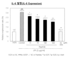
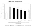
- Figures 5a and 5b show the results of measuring the effect of treatment with the peptide of Preparation Example 1 on the expression of IL-6 and IL-8 in MRC-5 cells in which an inflammatory response was induced by LPS treatment.
- Figure 6 shows the results of measuring the effect of treatment with the peptide of Preparation Example 1 on the expression levels of the fibrogenic genes ⁇ -SMA and Collagen1A1 genes in MRC-5 cells in which an inflammatory response was induced by LPS treatment.
- Figure 7 shows the results of measuring the effect of treatment with the peptide of Preparation Example 1 on the expression levels of the fibrotic genes ⁇ -SMA, Fibronectin1, Collagen1A1, and Tenascin C in MRC-5 cells treated with TGF- ⁇ 1.
- Figure 8 shows the effect of treatment with the peptide of Preparation Example 1 on the gene expression levels of inflammatory cytokines TNF- ⁇ , IL-1 ⁇ , IL-6, IL-8, and COX-2 in A549 cells treated with IL-1 ⁇ . This is the result of measurement.
- Figures 9A, 9B, and 9C measure the effect of treatment with the peptide of Preparation Example 1 on the expression levels of inflammatory cytokines IL-1 ⁇ , IL-6, and IL-8 in A549 cells treated with IL-1 ⁇ . This is one result.
- Figures 10a and 10b show the results of measuring the effect of treatment with the peptide of Preparation Example 1 on the total number of cells, number of neutrophils, and number of macrophages in bronchial lavage fluid of mice in which lung inflammation was induced by LPS. .
- Figures 11a to 11d measure the effect of treatment with the peptide of Preparation Example 1 on the expression levels of TNF- ⁇ , IL-1 ⁇ , IL-6, and Myeloperoxidase (MPO) in bronchial lavage fluid of mice in which lung inflammation was induced by LPS. This is one result.
- Figures 12a and 12b show the results of observation after H&E staining of lung tissue of mice in which lung inflammation was induced by LPS, the results of measuring the thickness of the alveolar wall, the results of measuring the total number of cells, and the number of alveoli. This is one result.
- Figure 13a shows the results of measuring the body weight of a mouse with pulmonary fibrosis induced by BLM (Bleomycin), and Figure 13b shows the peptide of Preparation Example 1 for the number of macrophages and lymphocytes in the bronchial lavage fluid of the mouse. This is the result of measuring the effect of treatment, and Figures 13c and 13d show the results of observing the cells under a microscope in the bronchial lavage fluid of the mouse and measuring the total number of cells.
- BLM Bleomycin
- Figure 14a shows the results of observing H&E staining of lung tissue of mice in which pulmonary fibrosis was induced by BLM (Bleomycin), and Figure 14b shows the results of measuring the number of alveoli in each treatment group in the H&E-stained lung tissue photo. These are the results of measuring the total number of cells and the thickness of the alveolar walls.
- Figure 15a shows the results observed by Trichrome staining of the lung tissue of mice with pulmonary fibrosis induced by BLM (Bleomycin), and Figure 15b shows the change in the amount of collagen in the remaining treatment groups with the amount of collagen in the normal group set to 100%. is expressed in %.
- a peptide having the amino acid sequence of SEQ ID NO: 1 shown in Table 1 below was synthesized using an automatic peptide synthesizer (Milligen 9050, Millipore, USA), followed by C18 reversed-phase high-performance liquid chromatography (HPLC) (Waters Associates, USA). These synthesized peptides were purified using .
- the column used was ACQUITY UPLC BEH300 C18 (2.1 mm ⁇ 100 mm, 1.7 ⁇ m, Waters Co, USA).
- MRC-5 cells lung fibroblast cells
- LPS Sigma Aldrich
- LPS 5 ⁇ g/ml treated group negative control
- LPS 5 ⁇ g/ml the peptide of Preparation Example 1 (10, 50, 100, 500 ⁇ M) treated group
- LPS 5 ⁇ g/ml the peptide of Preparation Example 1 (10, 50, 100, 500 ⁇ M) treated group
- LPS 5 ⁇ g/ml the peptide of Preparation Example 1 (10, 50, 100, 500 ⁇ M) treated group
- LPS 5 ⁇ g/ml And for the Dexamethasone (DEX) 10 ⁇ M treatment group positive control group
- RNA was extracted 24 hours after treatment with the active substances and drugs and RT-PCR was performed.
- A549 cells (human alveolar basal epithelial cells) were seeded at 3 x 10 5 cells/well in a 6-well plate and used in the experiment. After culturing for 1 day in medium containing 10% FBS, the medium was replaced with medium without FBS and starvation was induced for 4 hours.
- MRC-5 cells lung fibroblast cells
- LPS Sigma Aldrich
- LPS 5 ⁇ g/ml treated group negative control
- LPS 5 ⁇ g/ml and the peptide of Preparation Example 1 (10, 50, 100, 500 ⁇ M) treated group
- LPS 5 ⁇ g/ml ml and Dexamethasone (DEX) 10 ⁇ M treated group positive control
- the cultured cells of each treatment group and control group were lysed by adding lysis buffer and then centrifuged at 4°C at 12,000 rpm for 30 minutes.
- the obtained protein was quantified using the BCA Protein Assay kit (Sigma). Proteins were subjected to SDS-PAGE (sodium dodecyl sulfate-polyacrylamide gel electrophoresis) and then electrotransferred to a membrane.
- SDS-PAGE sodium dodecyl sulfate-polyacrylamide gel electrophoresis
- the secondary antibody was reacted at room temperature for 1 hour, washed again with PBS-T, and Western detection reagent (Elpis Biotech, Daejeon, Korea).
- the antibodies used in the experiment were as follows: anti-IL-1 ⁇ antibody (Cell signaling technology (CST), USA), anti-IL-6 antibody (Santa Cruz Biotechnology, USA), anti-pJNK antibody (Santa Cruz Biotechnology, USA) ), anti-pERK antibody (Cell signaling technology (CST), USA), anti- ⁇ -Actin antibody (Santa Cruz Biotechnology, USA).
- A549 cells (human alveolar basal epithelial cells) were seeded at 3 x 10 5 cells/well in a 6-well plate and used in the experiment. After culturing for 1 day with medium containing 10% FBS, the medium was replaced with medium without FBS and starvation was induced for 4 hours. Normal group where cells were not treated with anything, LPS (Sigma Aldrich) (5 ⁇ g/ml) treated group (negative control), LPS (5 ⁇ g/ml) and peptide of Preparation Example 1 (10, 50, 100, 500 ⁇ M) treated group.
- LPS (5 ⁇ g/ml) and Dexamethasone (DEX) (10 ⁇ M) treatment group (positive control) cells were treated with the above-mentioned active substances and drugs and cultured for 24 hours.
- the cultured cells of each treatment group and control group were lysed by adding lysis buffer and then centrifuged at 4°C at 12,000 rpm for 30 minutes.
- the obtained protein was quantified using the BCA Protein Assay kit (Sigma). Proteins were subjected to SDS-PAGE (sodium dodecyl sulfate-polyacrylamide gel electrophoresis) and then electrotransferred to a membrane.
- SDS-PAGE sodium dodecyl sulfate-polyacrylamide gel electrophoresis
- the membrane to which the protein was attached was treated with 5% skim milk for blocking, and then the primary antibody was reacted overnight at 4°C. After washing with PBS-T, the secondary antibody was reacted at room temperature for 1 hour, washed again with PBS-T, and Western detection reagent (Elpis Biotech, Daejeon, Korea).
- the antibodies used in the experiment were as follows: anti-IL-1 ⁇ antibody: (Cell signaling technology (CST), USA), anti-IL-6 antibody: (Santa Cruz Biotechnology, USA), anti-COX2 antibody: (Santa Cruz Biotechnology , USA), anti-Phospho-PI3K antibody: (Cell signaling technology (CST), USA), anti-Phospho-AKT antibody: (Cell signaling technology (CST), USA), anti-Phospho-ERK antibody: (Cell signaling technology (CST), USA), anti- ⁇ -Actin antibody: (Santa Cruz Biotechnology, USA).
- MRC-5 cells lung fibroblast cells
- LPS Sigma Aldrich
- LPS 5 ⁇ g/ml treated group negative control
- LPS 5 ⁇ g/ml and the peptide of Preparation Example 1 (10, 50, 100, 500 ⁇ M) treated group
- the culture medium was centrifuged at 4°C and 3,000 rpm for 10 minutes, and the obtained supernatant was used using the IL-6 Quantikine ELISA Kit (R&D systems) and IL-8 Quantikine ELISA kit (R&D systems) according to the instructions in the kit instructions. After performing ELISA analysis, absorbance was measured at 450 nm using a microplate reader. As a result of the experiment, as shown in FIGS. 5A and 5B, when treated with LPS, the expression levels of IL-6 and IL-8 increased, but treatment with the peptide of Preparation Example 1 reduced IL-6 and IL-8. The expression level decreased in a concentration-dependent manner.
- PCR was performed using a PCR machine (Eppendorf, Germany). The mRNA expression pattern was then determined by agarose gel electrophoresis. The base sequences of the primers used in the experiment are listed in Table 3 below.
- PCR was performed using a PCR machine (Eppendorf, Germany). The mRNA expression pattern was then determined by agarose gel electrophoresis. The base sequences of the primers used in the experiment are listed in Table 4 below.
- IL-1 ⁇ (2ng/ml) treated group negative control, NC
- IL-1 ⁇ (2ng/ml) and the peptide of Preparation Example 1 10, 50, 100, Cells from the 500 ⁇ M) treatment group
- IL-1 ⁇ (2 ng/ml) and Dexamethasone (DEX) (10 ⁇ M) treatment group positive control group
- the culture medium was centrifuged at 4°C and 3,000 rpm for 10 minutes, and the obtained supernatant was assayed with IL-6 Quantikine ELISA Kit (R&D systems), IL-8 Quantikine ELISA Kit (R&D systems), and IL-1 ⁇ Quantikine ELISA Kit (R&D Systems) was used to perform ELISA analysis according to the instructions in the kit instructions, and the absorbance was measured at 450 nm using a microplate reader. As a result of the experiment, as shown in FIGS.
- BALB/c mice Female/7W were used in animal experiments, and an acute lung inflammation model was established by injecting LPS.
- the experiment was performed on 7-week-old BALB/c female mice, divided into 5 groups of 5 mice each, as shown in [Table 5] below.
- LPS (10 ⁇ g/50 ⁇ l/mouse) was administered nasally to the G2-G5 groups, and 1 hour after LPS administration, the G3 and G4 groups were administered the peptide (0.5 mg, 1 mg) of Preparation Example 1 intranasally.
- G5 positive control group was intraperitoneally administered Dexamethasone (100 ⁇ g). 24 hours after administration of the active substance and drug, the mouse was sacrificed to obtain bronchial lavage fluid and lung tissue, which were used in the next analysis.
- peptide Peptide of Preparation Example 1
- DEX Dexamethasone
- IN intranasal administration
- IP intraperitoneal administration.
- BALF bronchial lavage fluid
- bronchoalveolar lavage fluid BALF was collected by repeating the injection and withdrawal of 1ml of PBS twice through an IV catheter (BD, USA). did.
- the collected BALF was centrifuged at 3000 rpm for 10 minutes to separate the cells.
- the separated cells were resuspended in 0.5 ml of PBS, diluted with trypan blue solution at a 1:1 ratio, and the total was measured using a hematocytometer. Cells were counted.
- BALF bronchial lavage fluid
- ELISA analysis was performed on pro-inflammatory proteins on the collected BALF.
- the collected BALF was centrifuged at 3000 rpm for 10 minutes, and the supernatant was assayed with TNF- ⁇ Quantikine ELISA kit (R&D systems), IL-1 ⁇ Quantikine ELISA kit (R&D systems), IL-6 Quantikine ELISA kit (R&D systems), and Myeloperoxidase ( MPO) Quantikine ELISA Kit (R&D systems) was used to perform the analysis according to the instructions, and the absorbance was measured at 450 nm using a Microplate reader. As a result of the experiment, as can be seen in FIGS.
- the expression levels of TNF- ⁇ , IL-1 ⁇ , IL-6, and Myeloperoxidase (MPO) increased by LPS administration, and the expression levels of the cytokines increased. It was reduced in a concentration-dependent manner by treatment with the peptide of Preparation Example 1.
- Lung tissue obtained from mice subjected to animal experiments was washed with PBS at room temperature and then fixed with 4% paraformaldehyde (PBS dilution).
- the fixed tissue was washed three times with PBS and dehydrated using an ethanol series with a concentration gradient of 70% to 100%.
- the lung tissue samples were then embedded and cut into 4 ⁇ m thick slices.
- the 4- ⁇ m-sized sliced specimen placed on a slide glass was dewaxed and hydrated in a gradient ethanol series, and then stained in hematoxlin solution for 1 minute and in Eosin solution for 10 seconds. After staining, the tissue slides were sequentially immersed in 90% and 100% ethanol, and finally, they were dipped in xylene twice for 5 minutes before mounting the tissue slides.
- the graph in Figure 12b shows the results of measuring the thickness of the alveolar walls (the alveolar wall thickness of the normal group is set as 100%, and the degree of increase in thickness of the remaining treatment groups is expressed in %), and the results of measuring the number of cells in the stained lung photograph ( The number of cells in the normal group is set to 100%, and the increase in the number of cells in the remaining treatment group is expressed as a percentage), and the number of alveoli in the lung photograph was measured (the number of alveoli in the normal group was set to 100%) The increase and decrease in the number of alveoli in the remaining treatment groups are expressed as %, respectively.
- mice Eight-week-old BALB/c male mice were divided into four groups of five, as shown in [Table 6] below.
- BLM was induced once by administering Bleomycin solution (10ng/50 ⁇ l/mouse) through the oropharynx, and then from day 7, the peptide of Preparation Example 1 (1 mg/mouse) was administered intranasally three times a week for 14 days.
- Pirfeninone (2mg/mouse) a positive control group, was administered orally three times a week for 14 days.
- mice On the 21st day of BLM induction, mice were sacrificed to obtain bronchial lavage fluid and lung tissue, which were used in the following analysis.
- BALF bronchial lavage fluid
- bronchoalveolar lavage fluid BALF
- the collected BALF was centrifuged at 3000 rpm for 10 minutes to separate the cells.
- the separated cells were resuspended in 0.5 ml of PBS, diluted with trypan blue solution at a 1:1 ratio, and the total was measured using a hematocytometer. Cells were counted.
- Lung tissue obtained from mice subjected to animal experiments was washed with PBS at room temperature and then fixed with 4% paraformaldehyde (PBS dilution). The fixed tissue was washed three times with PBS and dehydrated using a 70% to 100% gradient ethanol series. The lung tissue samples were then embedded and cut into 4 ⁇ m thick slices. Sectioned specimens of 4 ⁇ m size placed on glass slides were dewaxed and hydrated in a gradient ethanol series and sequentially stained in hematoxlin solution for 1 minute and Eosin solution for 10 seconds. After staining, the tissue slides were sequentially immersed in 90% and 100% ethanol, and finally, they were dipped in xylene twice for 5 minutes before mounting the tissue slides.
- PBS dilution paraformaldehyde
- Figure 14a shows the results of observing H&E staining of lung tissue.
- Figure 14b shows the results of measuring the number of alveoli in each treatment group in the stained lung tissue photograph, and the total number of cells in the stained lung tissue photograph (the total number of cells in the normal group was set as 100%, and the rest were treated The degree of increase in the total number of cells in the group is expressed as a percentage), and the thickness of the alveolar wall of each treatment group was measured in the stained lung tissue photo (the thickness of the alveolar wall in the normal group was set as 100%, and the thickness of the remaining treatment groups increased) degree is expressed in %).
- Lung tissue obtained from mice subjected to animal experiments was washed with PBS at room temperature and then fixed with 4% paraformaldehyde (PBS dilution). The fixed tissue was washed three times with PBS and dehydrated using a 70% to 100% gradient ethanol series. The lung tissue samples were then embedded and cut into 4 ⁇ m thick slices. Thin sections of 4 ⁇ m size placed on glass slides were dewaxed and hydrated in a gradient ethanol series. Staining was performed for 10 minutes each in Weigert's iron hematoxlin, Biebrich scarlet-acid fuchsin solution, Phosphomolybdic-phosphotungstic acid solution, and aniline blue solution.
- FIG. 15a shows the results of collagen staining using Trichrome staining.
- Figure 15b shows the change in the amount of collagen in the remaining treatment groups as a percentage, taking the amount of collagen in the normal group as 100%.
- tablets were manufactured by tableting according to a conventional tablet manufacturing method.
- a capsule was prepared by filling a gelatin capsule according to a typical capsule manufacturing method.
- Vitamin A 70 mg
- Vitamin E 1.0mg
- Vitamin B6 0.5mg
- Vitamin B12 0.2mg
- composition ratio of the above vitamin and mineral mixture is a mixture of components relatively suitable for health food in a preferred embodiment, but the mixing ratio may be modified arbitrarily.
- the above ingredients are mixed according to a typical health food manufacturing method, and then , granules can be manufactured and used to manufacture health food compositions according to conventional methods.
- composition ratio is a preferred embodiment of mixing ingredients that are relatively suitable for beverages of preference, but the mixing ratio can be arbitrarily modified depending on regional and ethnic preferences such as demand class, country of demand, and intended use.
Landscapes
- Health & Medical Sciences (AREA)
- Life Sciences & Earth Sciences (AREA)
- Chemical & Material Sciences (AREA)
- Medicinal Chemistry (AREA)
- Pharmacology & Pharmacy (AREA)
- Animal Behavior & Ethology (AREA)
- General Health & Medical Sciences (AREA)
- Public Health (AREA)
- Veterinary Medicine (AREA)
- Engineering & Computer Science (AREA)
- Epidemiology (AREA)
- Bioinformatics & Cheminformatics (AREA)
- Organic Chemistry (AREA)
- Nuclear Medicine, Radiotherapy & Molecular Imaging (AREA)
- Proteomics, Peptides & Aminoacids (AREA)
- General Chemical & Material Sciences (AREA)
- Chemical Kinetics & Catalysis (AREA)
- Molecular Biology (AREA)
- Pulmonology (AREA)
- Gastroenterology & Hepatology (AREA)
- Immunology (AREA)
- Nutrition Science (AREA)
- Biophysics (AREA)
- Mycology (AREA)
- Food Science & Technology (AREA)
- Polymers & Plastics (AREA)
- Pain & Pain Management (AREA)
- Rheumatology (AREA)
- Botany (AREA)
- Zoology (AREA)
- Medicines That Contain Protein Lipid Enzymes And Other Medicines (AREA)
- Peptides Or Proteins (AREA)
- Coloring Foods And Improving Nutritive Qualities (AREA)
Abstract
Description
| 서열번호 | 펩타이드의 아미노산 서열 |
| 1 | KYLLVHRPYYRR |
| 유전자 | 프라이머 | 서열(5' -> 3') | 서열번호 |
| TNF-α | F | AACATCCAACCTTCCCAAACG | 2 |
| R | GACCCTAAGCCCCCAATTCTC | 3 | |
| IL-1β | F | TTCGACACATGGGATAACGA | 4 |
| R | TCTTTCAACACGCAGGACAG | 5 | |
| IL-6 | F | AAA GAG GCA CTG CCA GAA AA | 6 |
| R | ATC TGA GGT GCC CAT GCT AC | 7 | |
| IL-8 | F | GAAGGTGCAGTTTTGCCAAG | 8 |
| R | ACCCTCTGCACCCAGTTTTC | 9 | |
| COX-2 | F | ATCATTCACCAGGCAAATTGC | 10 |
| R | GGCTTCAGCATAAAGCGTTTG | 11 | |
| GAPDH | F | GTG ATG GCA TGG ACT GTG GT | 12 |
| R | GGA GCC AAA AGG GTC ATC AT | 13 |
| 유전자 | 프라이머 | 서열(5' -> 3') | 서열번호 |
| α-SMA | F | GGTGCTGTCTCTCTATGCCTCTGGA | 14 |
| R | CCCATCAGGCAACTCGTAACTCTTC | 15 | |
| Collagen1A1 | F | CATCACCTACCACTGCAAGAAC | 16 |
| R | ACGTCGAAGCCGAATTCC | 17 | |
| GAPDH | F | GTG ATG GCA TGG ACT GTG GT | 12 |
| R | GGA GCC AAA AGG GTC ATC AT | 13 |
| 유전자 | 프라이머 | 서열(5' -> 3') | 서열번호 |
| α-SMA | F | GGTGCTGTCTCTCTATGCCTCTGGA | 14 |
| R | CCCATCAGGCAACTCGTAACTCTTC | 15 | |
| Fibronectin1 | F | GGATGCTCCTGCTGTCAC | 18 |
| R | CTGTTTGATCTGGACCTGCAG | 19 | |
| Collagen1A1 | F | CATCACCTACCACTGCAAGAAC | 16 |
| R | ACGTCGAAGCCGAATTCC | 17 | |
| Tenascin C | F | CCAGCGACCATCAACGCAGC | 20 |
| R | GGGGCTTGTTCAGTGGATGCCT | 21 | |
| GAPDH | F | GTG ATG GCA TGG ACT GTG GT | 12 |
| R | GGA GCC AAA AGG GTC ATC AT | 13 |
| 그룹 | 성별 | 동물 수 및 번호 |
테스트 그룹 | LPS | 펩타이드 /약물 |
Dose (mg/head) |
Dose amount (mL/head) |
| G1 | Female | 5(1-5) | 정상군 | - | - | - | - |
| G2 | Female | 5(6-10) | 음성대조군 | + | - | - | - |
| G3 | Female | 5(11-15) | 실험군 1 | + | 펩타이드(IN) | 0.5mg | 0.05 mL |
| G4 | Female | 5(16-20) | 실험군 2 | + | 펩타이드(IN) | 1mg | 0.05 mL |
| G5 | Female | 5(21-25) | 양성대조군 | + | DEX(IP) | 100μg | 0.05 mL |
| 그룹 | 성별 | 동물의 수 및 번호 |
테스트 그룹 | BLM | 펩타이드 /약물 |
Dose (mg/head) |
Dose amount (mL/head) |
| G1 | Male | 5 (1-5) | 정상군 | - | - | - | - |
| G2 | Male | 5 (6-10) | 음성대조군 | + | - | - | - |
| G3 | Male | 5 (11-15) | 실험군 | + | 펩타이드(IN) | 1 mg | 0.05 mL |
| G4 | Male | 5 (16-20) | 양성대조군 | + | Pirfeninone (Oral) | 2 mg | 0.1 mL |
Claims (10)
- 서열번호 1의 아미노산 서열을 포함하는 펩타이드를 유효성분으로 포함하는 폐 염증성 질환의 치료 또는 예방용 약학적 조성물.
- 청구항 1에 있어서,상기 폐 염증성 질환은 천식, 알러지성 천식, 기관지염, 만성 기관지염, 급성 기관지염, 세기관지염, 미만성 범세기관지염, 기관지 확장증, 흉막염, 폐포염, 혈관염, 폐렴, 과민성 폐렴, 만성폐쇄성폐질환, 폐 섬유증, 특발성 폐 섬유증, 낭포성 섬유증, 급성 호흡곤란 증후군, 급성 폐손상, 연기 흡입 폐손상, 및 열에 의한 폐손상으로 이루어지는 군에서 선택되는 질환인 것인, 폐 염증성 질환의 치료 또는 예방용 약학적 조성물.
- 청구항 1에 있어서,상기 폡타이드는 염증 반응이 유도되는 폐 세포에서 염증성 사이토카인 유전자, 염증성 사이토카인 단백질 또는 염증성 사이토카인 신호전달에 관련된 단백질의 발현을 억제하는 것인, 폐 염증성 질환의 치료 또는 예방용 약학적 조성물.
- 청구항 3에 있어서,상기 염증성 사이토카인 유전자는 TNF α, IL-1β, IL-6, IL-8 및 COX-2 유전자로 이루어진 군에서 선택되는 하나 이상인 것인, 폐 염증성 질환의 치료 또는 예방용 약학적 조성물.
- 청구항 3에 있어서,상기 염증성 사이토카인 단백질 또는 염증성 사이토카인 신호전달에 관련된 단백질은 IL-1β, IL-6, COX2, IL-8, p-JNK, p-ERK, p-PI3K, p-AKT, p-ERK, 및 Myeloperoxidase으로 이루어진 군에서 선택되는 하나 이상인 것인, 폐 염증성 질환의 치료 또는 예방용 약학적 조성물.
- 청구항 1에 있어서,상기 폡타이드는 염증 반응이 유도되는 폐 세포에서 섬유화 유전자의 발현을 억제하는 것인, 폐 염증성 질환의 치료 또는 예방용 약학적 조성물.
- 청구항 6에 있어서,상기 섬유화 유전자는 α-SMA, Collagen1A1, Fibronectin1, 및 Tenascin C유전자로 이루어지는 군에서 선택되는 하나 이상인 것인, 폐 염증성 질환의 치료 또는 예방용 약학적 조성물.
- 청구항 1에 있어서,상기 폡타이드는 염증 반응이 유도되는 폐 조직에서 호중구의 수를 감소시키는 것인, 폐 염증성 질환의 치료 또는 예방용 약학적 조성물.
- 서열번호 1의 아미노산 서열을 포함하는 펩타이드를 유효성분으로 포함하는 폐 염증의 개선 또는 예방용 기능성 식품 조성물.
- 서열번호 1의 아미노산 서열을 포함하는 펩타이드를 유효성분으로 포함하는 폐 섬유증의 개선 또는 예방용 기능성 식품 조성물.
Priority Applications (5)
| Application Number | Priority Date | Filing Date | Title |
|---|---|---|---|
| EP23781240.9A EP4501344A4 (en) | 2022-03-31 | 2023-03-23 | PEPTIDE WITH ANTI-INFLAMMATORY AND ANTIFIBROTIC ACTIVITIES AND ITS USE |
| JP2024558150A JP2025511247A (ja) | 2022-03-31 | 2023-03-23 | 抗炎症及び抗線維化活性を有するペプチド並びにその用途 |
| CN202380032361.1A CN118973599A (zh) | 2022-03-31 | 2023-03-23 | 具有抗炎和抗纤维化活性的肽及其用途 |
| US18/850,815 US20250213649A1 (en) | 2022-03-31 | 2023-03-23 | Peptide having anti-inflammatory and anti-fibrotic activities and use thereof |
| MX2024011989A MX2024011989A (es) | 2022-03-31 | 2024-09-27 | Peptido que tiene actividades antiinflamatorias y antifibroticas y uso del mismo |
Applications Claiming Priority (2)
| Application Number | Priority Date | Filing Date | Title |
|---|---|---|---|
| KR1020220040544A KR102810165B1 (ko) | 2022-03-31 | 2022-03-31 | 항염증 및 항섬유화 활성을 갖는 펩타이드 및 이의 용도 |
| KR10-2022-0040544 | 2022-03-31 |
Publications (1)
| Publication Number | Publication Date |
|---|---|
| WO2023191384A1 true WO2023191384A1 (ko) | 2023-10-05 |
Family
ID=88202582
Family Applications (1)
| Application Number | Title | Priority Date | Filing Date |
|---|---|---|---|
| PCT/KR2023/003862 Ceased WO2023191384A1 (ko) | 2022-03-31 | 2023-03-23 | 항염증 및 항섬유화 활성을 갖는 펩타이드 및 이의 용도 |
Country Status (7)
| Country | Link |
|---|---|
| US (1) | US20250213649A1 (ko) |
| EP (1) | EP4501344A4 (ko) |
| JP (1) | JP2025511247A (ko) |
| KR (1) | KR102810165B1 (ko) |
| CN (1) | CN118973599A (ko) |
| MX (1) | MX2024011989A (ko) |
| WO (1) | WO2023191384A1 (ko) |
Cited By (1)
| Publication number | Priority date | Publication date | Assignee | Title |
|---|---|---|---|---|
| CN119857136A (zh) * | 2023-10-20 | 2025-04-22 | 天津济坤医药科技有限公司 | 一种抗cd84抗体在制备防治肺纤维化的药物中的应用 |
Families Citing this family (1)
| Publication number | Priority date | Publication date | Assignee | Title |
|---|---|---|---|---|
| CN120248012B (zh) * | 2025-04-03 | 2026-04-17 | 复美长青健康管理(珠海横琴)有限公司 | 一种治疗肺结节的肺干细胞肽及其制备方法和应用 |
Citations (5)
| Publication number | Priority date | Publication date | Assignee | Title |
|---|---|---|---|---|
| US5516891A (en) | 1992-06-16 | 1996-05-14 | Kinerton, Ltd. | Liquid phase synthesis of peptides and peptide derivatives |
| KR20010108002A (ko) * | 1998-12-03 | 2001-12-07 | 추후보정 | 소형 펩타이드 및 천식 및 염증을 치료하기 위한 방법 |
| WO2011029931A1 (en) | 2009-09-11 | 2011-03-17 | Protaffin Biotechnologie Ag | Composition for treatment of cxcl8-mediated lung inflammation |
| US10793596B2 (en) * | 2006-11-10 | 2020-10-06 | Cara Therapeutics, Inc. | Synthetic peptide amides |
| KR20220009924A (ko) * | 2020-07-16 | 2022-01-25 | (주)케어젠 | 제2형 중증급성호흡기증후군 코로나바이러스에 중화 활성을 갖는 펩타이드 |
Family Cites Families (2)
| Publication number | Priority date | Publication date | Assignee | Title |
|---|---|---|---|---|
| KR101831887B1 (ko) * | 2016-03-09 | 2018-02-27 | (주)케어젠 | 발모 촉진 활성 및 멜라닌 생성 촉진 활성을 나타내는 펩타이드 및 이의 용도 |
| KR101885847B1 (ko) | 2017-09-20 | 2018-08-07 | (주)케어젠 | 발모 촉진 활성 및 멜라닌 생성 촉진 활성을 나타내는 펩타이드 및 이의 용도 |
-
2022
- 2022-03-31 KR KR1020220040544A patent/KR102810165B1/ko active Active
-
2023
- 2023-03-23 WO PCT/KR2023/003862 patent/WO2023191384A1/ko not_active Ceased
- 2023-03-23 JP JP2024558150A patent/JP2025511247A/ja active Pending
- 2023-03-23 CN CN202380032361.1A patent/CN118973599A/zh active Pending
- 2023-03-23 US US18/850,815 patent/US20250213649A1/en active Pending
- 2023-03-23 EP EP23781240.9A patent/EP4501344A4/en active Pending
-
2024
- 2024-09-27 MX MX2024011989A patent/MX2024011989A/es unknown
Patent Citations (5)
| Publication number | Priority date | Publication date | Assignee | Title |
|---|---|---|---|---|
| US5516891A (en) | 1992-06-16 | 1996-05-14 | Kinerton, Ltd. | Liquid phase synthesis of peptides and peptide derivatives |
| KR20010108002A (ko) * | 1998-12-03 | 2001-12-07 | 추후보정 | 소형 펩타이드 및 천식 및 염증을 치료하기 위한 방법 |
| US10793596B2 (en) * | 2006-11-10 | 2020-10-06 | Cara Therapeutics, Inc. | Synthetic peptide amides |
| WO2011029931A1 (en) | 2009-09-11 | 2011-03-17 | Protaffin Biotechnologie Ag | Composition for treatment of cxcl8-mediated lung inflammation |
| KR20220009924A (ko) * | 2020-07-16 | 2022-01-25 | (주)케어젠 | 제2형 중증급성호흡기증후군 코로나바이러스에 중화 활성을 갖는 펩타이드 |
Non-Patent Citations (6)
| Title |
|---|
| "Remington: The Science and Practice of Pharmacy", 1995, WILLIAMS & WILKINS |
| ASRANI PURVA; HASSAN MD. IMTAIYAZ: "SARS-CoV-2 mediated lung inflammatory responses in host: targeting the cytokine storm for therapeutic interventions", MOLECULAR AND CELLULAR BIOCHEMISTRY, SPRINGER US, NEW YORK, vol. 476, no. 2, 16 October 2020 (2020-10-16), New York, pages 675 - 687, XP037363330, ISSN: 0300-8177, DOI: 10.1007/s11010-020-03935-z * |
| DATABASE Protein 10 October 2019 (2019-10-10), ANONYMOUS : "Sequence 2 from patent US 10344061", XP093093941, retrieved from NCBI Database accession no. QFO42108.1 * |
| MERRIFIELD, J., AMER. CHEM. SOC., vol. 85, 1963, pages 2149 - 54 |
| See also references of EP4501344A4 |
| STEWART ET AL.: "Solid Phase Peptide Synthesis", 1984, PIERCE CHEM. CO.: ROCKFORD, pages: 111 |
Cited By (1)
| Publication number | Priority date | Publication date | Assignee | Title |
|---|---|---|---|---|
| CN119857136A (zh) * | 2023-10-20 | 2025-04-22 | 天津济坤医药科技有限公司 | 一种抗cd84抗体在制备防治肺纤维化的药物中的应用 |
Also Published As
| Publication number | Publication date |
|---|---|
| EP4501344A1 (en) | 2025-02-05 |
| MX2024011989A (es) | 2024-11-08 |
| JP2025511247A (ja) | 2025-04-15 |
| CN118973599A (zh) | 2024-11-15 |
| EP4501344A4 (en) | 2025-07-09 |
| KR102810165B1 (ko) | 2025-05-22 |
| KR20230141227A (ko) | 2023-10-10 |
| US20250213649A1 (en) | 2025-07-03 |
Similar Documents
| Publication | Publication Date | Title |
|---|---|---|
| WO2020197359A1 (ko) | 섬유증의 예방, 개선 또는 치료를 위한 chp (사이클로-히스프로)의 용도 | |
| WO2017048028A1 (ko) | 마이시니딘 펩타이드로부터 유래한 신규 항균 펩타이드 및 이의 용도 | |
| WO2015026112A1 (ko) | 모노아세틸디아실글리세롤 화합물을 유효성분으로 함유하는 혈액암 또는 암전이 억제용 조성물 | |
| WO2017014502A1 (ko) | 해당화 꽃 추출물을 유효성분으로 포함하는 il-6 매개성 질환의 예방 또는 치료용 약학적 조성물 | |
| WO2021080129A1 (ko) | 하이드란제놀 또는 필로둘신을 유효성분으로 하는 피부장벽 강화 및 아토피 피부염 개선용 조성물 | |
| WO2023182567A1 (ko) | 항당뇨 활성을 갖는 펩타이드, 펩타이드 복합체 및 이의 용도 | |
| US20250213649A1 (en) | Peptide having anti-inflammatory and anti-fibrotic activities and use thereof | |
| WO2024090747A1 (ko) | 칸나비디올 및 타우린을 포함하는 치주염 예방 또는 치료용 조성물 | |
| WO2019078381A1 (ko) | 엔테로코커스 패칼리스, 이의 배양액 또는 이의 사균체를 유효성분으로 함유하는 근육감퇴, 약화 및 근위축 예방, 개선 또는 치료용 약학 조성물, 식품 조성물 및 식품첨가제 | |
| WO2016010340A1 (ko) | 오풍칠 추출물을 유효성분으로 함유하는 염증 또는 알러지 질환의 예방 및 치료용 조성물 및 이의 용도 | |
| WO2015026124A1 (ko) | 모노아세틸디아실글리세롤 화합물을 유효성분으로 함유하는 천식의 예방 또는 치료용 조성물 | |
| WO2018174424A1 (ko) | 능이버섯 추출물을 유효성분으로 함유하는 부인과 질환의 예방 또는 치료용 약학적 조성물 | |
| WO2014126285A1 (ko) | 디메틸푸마레이트를 유효성분으로 포함하는 신섬유증의 예방 또는 치료용 조성물 | |
| WO2024122660A1 (ko) | 혼합 생약 추출물을 유효성분으로 함유하는 면역 증강용 조성물 | |
| WO2015105373A1 (ko) | 화살나무 추출물 또는 이의 분획물을 포함하는 천식의 예방 또는 치료용 조성물 | |
| WO2022270760A1 (ko) | 커큐민 유도체와 tgf-β 수용체 억제제의 병용투여를 통한 비알코올성 지방간염 치료 방법 | |
| WO2023027540A1 (ko) | 진세노사이드 rc를 포함하는 근육 질환의 예방, 개선 또는 치료용 조성물 | |
| WO2017105081A1 (ko) | 폐질환 치료용 또는 증상완화용 약학조성물 | |
| WO2016204493A1 (en) | A novel compound (ks 513) isolated from pseudolysimachion rotundum var. subintegrum, the composition comprising the same as an active ingredient for preventing or treating allergy disease, inflammatory disease, asthma or chronic obstructive pulmonary disease and the use thereof | |
| WO2018105998A1 (ko) | 플라보노이드 유도체 화합물을 유효성분으로 함유하는 남성 불임증 예방 및 치료용 조성물 및 이의 용도 | |
| WO2020040522A1 (ko) | 더덕 추출물을 유효성분으로 함유하는 근육 질환의 예방 또는 치료용 약학적 조성물 | |
| WO2025226035A1 (ko) | 지방간 질환의 예방 또는 치료 활성을 갖는 펩타이드 복합체 및 이의 용도 | |
| WO2025216517A1 (ko) | 성장 촉진 활성을 갖는 펩타이드 및 이의 용도 | |
| WO2020242010A1 (ko) | 파파야 발효물을 포함하는 아토피 피부염의 예방 또는 치료용 약학적 조성물 | |
| WO2018174502A1 (ko) | 국화잎 추출물을 유효성분으로 포함하는 당뇨병 예방 또는 치료용 약학적 조성물 |
Legal Events
| Date | Code | Title | Description |
|---|---|---|---|
| 121 | Ep: the epo has been informed by wipo that ep was designated in this application |
Ref document number: 23781240 Country of ref document: EP Kind code of ref document: A1 |
|
| REG | Reference to national code |
Ref country code: BR Ref legal event code: B01A Ref document number: 112024018354 Country of ref document: BR |
|
| WWE | Wipo information: entry into national phase |
Ref document number: 18850815 Country of ref document: US |
|
| WWE | Wipo information: entry into national phase |
Ref document number: MX/A/2024/011989 Country of ref document: MX |
|
| WWE | Wipo information: entry into national phase |
Ref document number: 140350140003004544 Country of ref document: IR |
|
| WWE | Wipo information: entry into national phase |
Ref document number: 2024558150 Country of ref document: JP Ref document number: 202380032361.1 Country of ref document: CN |
|
| WWE | Wipo information: entry into national phase |
Ref document number: 2023781240 Country of ref document: EP Ref document number: 2024127295 Country of ref document: RU |
|
| ENP | Entry into the national phase |
Ref document number: 2023781240 Country of ref document: EP Effective date: 20241031 |
|
| NENP | Non-entry into the national phase |
Ref country code: DE |
|
| ENP | Entry into the national phase |
Ref document number: 112024018354 Country of ref document: BR Kind code of ref document: A2 Effective date: 20240905 |
|
| WWP | Wipo information: published in national office |
Ref document number: 140350140003004544 Country of ref document: IR |
|
| WWP | Wipo information: published in national office |
Ref document number: 18850815 Country of ref document: US |