WO2024143436A1 - 網膜シートの製造方法 - Google Patents
網膜シートの製造方法 Download PDFInfo
- Publication number
- WO2024143436A1 WO2024143436A1 PCT/JP2023/046832 JP2023046832W WO2024143436A1 WO 2024143436 A1 WO2024143436 A1 WO 2024143436A1 JP 2023046832 W JP2023046832 W JP 2023046832W WO 2024143436 A1 WO2024143436 A1 WO 2024143436A1
- Authority
- WO
- WIPO (PCT)
- Prior art keywords
- retinal
- transplantation
- sheet
- cells
- transplant
- Prior art date
- Legal status (The legal status is an assumption and is not a legal conclusion. Google has not performed a legal analysis and makes no representation as to the accuracy of the status listed.)
- Ceased
Links
Images





Classifications
-
- A—HUMAN NECESSITIES
- A61—MEDICAL OR VETERINARY SCIENCE; HYGIENE
- A61P—SPECIFIC THERAPEUTIC ACTIVITY OF CHEMICAL COMPOUNDS OR MEDICINAL PREPARATIONS
- A61P27/00—Drugs for disorders of the senses
- A61P27/02—Ophthalmic agents
-
- A—HUMAN NECESSITIES
- A61—MEDICAL OR VETERINARY SCIENCE; HYGIENE
- A61B—DIAGNOSIS; SURGERY; IDENTIFICATION
- A61B3/00—Apparatus for testing the eyes; Instruments for examining the eyes
- A61B3/10—Objective types, i.e. instruments for examining the eyes independent of the patients' perceptions or reactions
- A61B3/102—Objective types, i.e. instruments for examining the eyes independent of the patients' perceptions or reactions for optical coherence tomography [OCT]
-
- A—HUMAN NECESSITIES
- A61—MEDICAL OR VETERINARY SCIENCE; HYGIENE
- A61B—DIAGNOSIS; SURGERY; IDENTIFICATION
- A61B3/00—Apparatus for testing the eyes; Instruments for examining the eyes
- A61B3/10—Objective types, i.e. instruments for examining the eyes independent of the patients' perceptions or reactions
- A61B3/12—Objective types, i.e. instruments for examining the eyes independent of the patients' perceptions or reactions for looking at the eye fundus, e.g. ophthalmoscopes
- A61B3/1225—Objective types, i.e. instruments for examining the eyes independent of the patients' perceptions or reactions for looking at the eye fundus, e.g. ophthalmoscopes using coherent radiation
-
- A—HUMAN NECESSITIES
- A61—MEDICAL OR VETERINARY SCIENCE; HYGIENE
- A61K—PREPARATIONS FOR MEDICAL, DENTAL OR TOILETRY PURPOSES
- A61K35/00—Medicinal preparations containing materials or reaction products thereof with undetermined constitution
- A61K35/12—Materials from mammals; Compositions comprising non-specified tissues or cells; Compositions comprising non-embryonic stem cells; Genetically modified cells
- A61K35/48—Reproductive organs
- A61K35/54—Ovaries; Ova; Ovules; Embryos; Foetal cells; Germ cells
- A61K35/545—Embryonic stem cells; Pluripotent stem cells; Induced pluripotent stem cells; Uncharacterised stem cells
-
- A—HUMAN NECESSITIES
- A61—MEDICAL OR VETERINARY SCIENCE; HYGIENE
- A61L—METHODS OR APPARATUS FOR STERILISING MATERIALS OR OBJECTS IN GENERAL; DISINFECTION, STERILISATION OR DEODORISATION OF AIR; CHEMICAL ASPECTS OF BANDAGES, DRESSINGS, ABSORBENT PADS OR SURGICAL ARTICLES; MATERIALS FOR BANDAGES, DRESSINGS, ABSORBENT PADS OR SURGICAL ARTICLES
- A61L27/00—Materials for grafts or prostheses or for coating grafts or prostheses
- A61L27/36—Materials for grafts or prostheses or for coating grafts or prostheses containing ingredients of undetermined constitution or reaction products thereof, e.g. transplant tissue, natural bone, extracellular matrix
- A61L27/38—Materials for grafts or prostheses or for coating grafts or prostheses containing ingredients of undetermined constitution or reaction products thereof, e.g. transplant tissue, natural bone, extracellular matrix containing added animal cells
- A61L27/3804—Materials for grafts or prostheses or for coating grafts or prostheses containing ingredients of undetermined constitution or reaction products thereof, e.g. transplant tissue, natural bone, extracellular matrix containing added animal cells characterised by specific cells or progenitors thereof, e.g. fibroblasts, connective tissue cells, kidney cells
-
- A—HUMAN NECESSITIES
- A61—MEDICAL OR VETERINARY SCIENCE; HYGIENE
- A61L—METHODS OR APPARATUS FOR STERILISING MATERIALS OR OBJECTS IN GENERAL; DISINFECTION, STERILISATION OR DEODORISATION OF AIR; CHEMICAL ASPECTS OF BANDAGES, DRESSINGS, ABSORBENT PADS OR SURGICAL ARTICLES; MATERIALS FOR BANDAGES, DRESSINGS, ABSORBENT PADS OR SURGICAL ARTICLES
- A61L27/00—Materials for grafts or prostheses or for coating grafts or prostheses
- A61L27/36—Materials for grafts or prostheses or for coating grafts or prostheses containing ingredients of undetermined constitution or reaction products thereof, e.g. transplant tissue, natural bone, extracellular matrix
- A61L27/38—Materials for grafts or prostheses or for coating grafts or prostheses containing ingredients of undetermined constitution or reaction products thereof, e.g. transplant tissue, natural bone, extracellular matrix containing added animal cells
- A61L27/3804—Materials for grafts or prostheses or for coating grafts or prostheses containing ingredients of undetermined constitution or reaction products thereof, e.g. transplant tissue, natural bone, extracellular matrix containing added animal cells characterised by specific cells or progenitors thereof, e.g. fibroblasts, connective tissue cells, kidney cells
- A61L27/3813—Epithelial cells, e.g. keratinocytes, urothelial cells
-
- A—HUMAN NECESSITIES
- A61—MEDICAL OR VETERINARY SCIENCE; HYGIENE
- A61L—METHODS OR APPARATUS FOR STERILISING MATERIALS OR OBJECTS IN GENERAL; DISINFECTION, STERILISATION OR DEODORISATION OF AIR; CHEMICAL ASPECTS OF BANDAGES, DRESSINGS, ABSORBENT PADS OR SURGICAL ARTICLES; MATERIALS FOR BANDAGES, DRESSINGS, ABSORBENT PADS OR SURGICAL ARTICLES
- A61L27/00—Materials for grafts or prostheses or for coating grafts or prostheses
- A61L27/36—Materials for grafts or prostheses or for coating grafts or prostheses containing ingredients of undetermined constitution or reaction products thereof, e.g. transplant tissue, natural bone, extracellular matrix
- A61L27/38—Materials for grafts or prostheses or for coating grafts or prostheses containing ingredients of undetermined constitution or reaction products thereof, e.g. transplant tissue, natural bone, extracellular matrix containing added animal cells
- A61L27/3886—Materials for grafts or prostheses or for coating grafts or prostheses containing ingredients of undetermined constitution or reaction products thereof, e.g. transplant tissue, natural bone, extracellular matrix containing added animal cells comprising two or more cell types
- A61L27/3891—Materials for grafts or prostheses or for coating grafts or prostheses containing ingredients of undetermined constitution or reaction products thereof, e.g. transplant tissue, natural bone, extracellular matrix containing added animal cells comprising two or more cell types as distinct cell layers
-
- A—HUMAN NECESSITIES
- A61—MEDICAL OR VETERINARY SCIENCE; HYGIENE
- A61L—METHODS OR APPARATUS FOR STERILISING MATERIALS OR OBJECTS IN GENERAL; DISINFECTION, STERILISATION OR DEODORISATION OF AIR; CHEMICAL ASPECTS OF BANDAGES, DRESSINGS, ABSORBENT PADS OR SURGICAL ARTICLES; MATERIALS FOR BANDAGES, DRESSINGS, ABSORBENT PADS OR SURGICAL ARTICLES
- A61L27/00—Materials for grafts or prostheses or for coating grafts or prostheses
- A61L27/36—Materials for grafts or prostheses or for coating grafts or prostheses containing ingredients of undetermined constitution or reaction products thereof, e.g. transplant tissue, natural bone, extracellular matrix
- A61L27/38—Materials for grafts or prostheses or for coating grafts or prostheses containing ingredients of undetermined constitution or reaction products thereof, e.g. transplant tissue, natural bone, extracellular matrix containing added animal cells
- A61L27/3895—Materials for grafts or prostheses or for coating grafts or prostheses containing ingredients of undetermined constitution or reaction products thereof, e.g. transplant tissue, natural bone, extracellular matrix containing added animal cells using specific culture conditions, e.g. stimulating differentiation of stem cells, pulsatile flow conditions
-
- A—HUMAN NECESSITIES
- A61—MEDICAL OR VETERINARY SCIENCE; HYGIENE
- A61P—SPECIFIC THERAPEUTIC ACTIVITY OF CHEMICAL COMPOUNDS OR MEDICINAL PREPARATIONS
- A61P41/00—Drugs used in surgical methods, e.g. surgery adjuvants for preventing adhesion or for vitreum substitution
-
- C—CHEMISTRY; METALLURGY
- C12—BIOCHEMISTRY; BEER; SPIRITS; WINE; VINEGAR; MICROBIOLOGY; ENZYMOLOGY; MUTATION OR GENETIC ENGINEERING
- C12N—MICROORGANISMS OR ENZYMES; COMPOSITIONS THEREOF; PROPAGATING, PRESERVING, OR MAINTAINING MICROORGANISMS; MUTATION OR GENETIC ENGINEERING; CULTURE MEDIA
- C12N5/00—Undifferentiated human, animal or plant cells, e.g. cell lines; Tissues; Cultivation or maintenance thereof; Culture media therefor
- C12N5/06—Animal cells or tissues; Human cells or tissues
- C12N5/0602—Vertebrate cells
- C12N5/0618—Cells of the nervous system
- C12N5/0621—Eye cells, e.g. cornea, iris pigmented cells
-
- A—HUMAN NECESSITIES
- A61—MEDICAL OR VETERINARY SCIENCE; HYGIENE
- A61L—METHODS OR APPARATUS FOR STERILISING MATERIALS OR OBJECTS IN GENERAL; DISINFECTION, STERILISATION OR DEODORISATION OF AIR; CHEMICAL ASPECTS OF BANDAGES, DRESSINGS, ABSORBENT PADS OR SURGICAL ARTICLES; MATERIALS FOR BANDAGES, DRESSINGS, ABSORBENT PADS OR SURGICAL ARTICLES
- A61L2430/00—Materials or treatment for tissue regeneration
- A61L2430/16—Materials or treatment for tissue regeneration for reconstruction of eye parts, e.g. intraocular lens, cornea
-
- C—CHEMISTRY; METALLURGY
- C12—BIOCHEMISTRY; BEER; SPIRITS; WINE; VINEGAR; MICROBIOLOGY; ENZYMOLOGY; MUTATION OR GENETIC ENGINEERING
- C12N—MICROORGANISMS OR ENZYMES; COMPOSITIONS THEREOF; PROPAGATING, PRESERVING, OR MAINTAINING MICROORGANISMS; MUTATION OR GENETIC ENGINEERING; CULTURE MEDIA
- C12N2501/00—Active agents used in cell culture processes, e.g. differentation
- C12N2501/70—Enzymes
- C12N2501/72—Transferases [EC 2.]
- C12N2501/727—Kinases (EC 2.7.)
-
- C—CHEMISTRY; METALLURGY
- C12—BIOCHEMISTRY; BEER; SPIRITS; WINE; VINEGAR; MICROBIOLOGY; ENZYMOLOGY; MUTATION OR GENETIC ENGINEERING
- C12N—MICROORGANISMS OR ENZYMES; COMPOSITIONS THEREOF; PROPAGATING, PRESERVING, OR MAINTAINING MICROORGANISMS; MUTATION OR GENETIC ENGINEERING; CULTURE MEDIA
- C12N2506/00—Differentiation of animal cells from one lineage to another; Differentiation of pluripotent cells
- C12N2506/45—Differentiation of animal cells from one lineage to another; Differentiation of pluripotent cells from artificially induced pluripotent stem cells
-
- C—CHEMISTRY; METALLURGY
- C12—BIOCHEMISTRY; BEER; SPIRITS; WINE; VINEGAR; MICROBIOLOGY; ENZYMOLOGY; MUTATION OR GENETIC ENGINEERING
- C12N—MICROORGANISMS OR ENZYMES; COMPOSITIONS THEREOF; PROPAGATING, PRESERVING, OR MAINTAINING MICROORGANISMS; MUTATION OR GENETIC ENGINEERING; CULTURE MEDIA
- C12N2513/00—3D culture
Definitions
- the present invention relates to a method for producing a transplant sheet from a retinal organoid derived from pluripotent stem cells.
- a method for evaluating the quality of grafts excised from retinal organoids is known in which quantitative PCR is performed on the organoids, approximately 1-2 mm in size, on a portion (ring) considered to be equivalent to the product (cap), to confirm that the target retinal marker gene is expressed, and that the expression of non-retinal markers and anterior segment marker genes is below a preset standard value, thereby evaluating the quality of the graft (Patent Document 1).
- retinal organoids themselves are 1-2 mm in size and have individual differences, there is a problem that inspection of all items is inefficient and expensive.
- excising the cap-ring requires the visual judgment and technique of an expert, and therefore can only be performed by a limited number of technicians.
- the primary objective of the present invention is to provide a method for efficiently producing retinal sheets with quality suitable for transplantation, without relying on the sophisticated discrimination techniques of an expert.
- the method further comprises a step of performing bright field microscopy and/or phase contrast microscopy on the pluripotent stem cell-derived retinal organoid, The method according to any one of [1] to [5], wherein the OCT is used to identify a transplant-compatible area in an area in which differentiation into retina and homogeneity can be confirmed by the bright-field microscopy and/or phase-contrast microscopy.
- markers for extra-graft cells include one or more selected from CRYAB, EMX2, s100A10, Zic1, AQP1, and MSX1, preferably one or more selected from CRYAB, Zic1, and EMX2, and the markers for retinal cells include one or more selected from Crx, Recoverin, and Rxr-gamma.
- a method for treating a retinal degenerative disease comprising producing a plurality of transplantable retinal sheets from pluripotent stem cell-derived retinal organoids by a method according to any one of [1] to [7], [10], and [11], and transplanting the plurality of transplantable retinal sheets into the retina of a subject in need thereof.
- a method for treating a retinal degenerative disease comprising producing a composite sheet for retinal transplantation by the method described in [8] and transplanting the composite sheet for retinal transplantation into the retina of a subject in need thereof.
- FIG. 4 shows the reconstruction of a horizontal X-Y plane image from an OCT tomographic image (three-dimensional image).
- Figure 7 shows the results of quantitative PCR for 1) planar retinal organoids with a uniform brightness distribution and 2) a thickness of 70 ⁇ m or more (4,5,9,10), and for areas that do not meet either of 1) or 2) (1-3,6-8).
- expression of marker genes for outer retinal tissues is low, and markers for each group of photoreceptor precursor cells/photoreceptor precursor cells/inner retinal cells, which are components of retinal organoids, can be confirmed as a set, and the area can be determined to be suitable for transplantation.
- FIG. 8 shows an example of cutting out a retinal sheet for transplantation from a transplant-compatible region of a retinal organoid. A grid is aligned with the transplant-compatible region so as to maximize the effective number of sheets, and the retinal sheet for transplantation is cut out.
- FIG. 9 shows a state in which a plurality of retinal sheets for transplantation, each about 1 ⁇ 2 mm, are loaded into a 24 G cannula for transplantation.
- Figure 10 shows a composite sheet in which a retinal sheet for transplantation is laminated on a retinal pigment epithelial sheet using 7-fold diluted hyaluronic acid for intraocular surgery (Viscoat (registered trademark)).
- the present invention relates to a method for producing a retinal sheet for transplantation from a retinal organoid derived from pluripotent stem cells, comprising the steps of performing optical coherence tomography on the retinal organoid derived from the pluripotent stem cells to identify an area having a thickness of 70 ⁇ m or more and free of foreign body structures characterized by differences in brightness as a transplant-compatible area, and cutting out a small piece from the transplant-compatible area to form a retinal sheet for transplantation.
- pluripotent stem cells in the present invention refers to embryonic stem cells (hereinafter referred to as "ES cells”) and cells that potentially have the same pluripotency as ES cells.
- ES cells embryonic stem cells
- iPS cells induced pluripotent stem cells
- ES cells are pluripotent stem cells derived from early embryos, and various established ES cell lines can be used. Examples of ES cell lines that can be used include, but are not limited to, clinical human ES cell lines provided by the Human ES Cell Research Center, Institute of Medical Biology, Kyoto University, and human ES cell lines provided by RIKEN BRC (KhES-1, KhES-1_Crx::Venus, KhES-1_Rx::Venus).
- iPS cells not only iPS cells induced from the patient's own somatic cells, but also various iPS cell lines can be used.
- iPS cell lines that can be used include, but are not limited to, various iPS cell stock lines provided by the Kyoto University iPS Cell Research Foundation, iPS cell lines derived from healthy individuals provided by RIKEN BRC, KVs09, CLs23, etc.
- the method for producing retinal organoids from pluripotent stem cells is not particularly limited, and any method may be used, such as a suspension culture method such as the SFEBq method, or a known method such as those described in WO2015/025967A1, WO2019/217630A1, WO2013/077425A1, WO2022/239868A1, WO2020/249935A1, JP2013-128475A, WO2022/138803A1, JP2021-118733A, WO2017/183732A1, WO2020/249935, and WO2022/054925A1.
- a suspension culture method such as the SFEBq method
- a known method such as those described in WO2015/025967A1, WO2019/217630A1, WO2013/077425A1, WO2022/239868A1, WO2020/249935A1, JP2013-128475A, WO2022/138803A1, JP2021-118733A, WO2017/183732A1,
- the retinal organoid preferably has a diameter of about 1 cm or more so that multiple grafts can be excised.
- a diameter of 1 cm or more means the length of the part corresponding to the diameter when the retinal organoid is shaped like a circle, or the length of the part corresponding to the major axis when the retinal organoid is shaped like an ellipse.
- optical coherence tomography is performed on the above-mentioned retinal organoids to identify areas suitable for transplantation.
- OCT optical coherence tomography
- Optical coherence tomography is a technology that uses the coherence of light to image the fine structure inside the object being measured, and allows for high-speed, high-resolution, non-invasive, real-time observation of the structure of retinal organoids.
- OCT includes spectral domain OCT, swept-source OCT, full-field OCT, adaptive optics OCT, etc.
- the area suitable for transplantation must be an area that can be confirmed by OCT to have a certain thickness and to be free of foreign body structures.
- a confocal imaging system using infrared to near-infrared light, an ultrasound microscope, etc. can also be used as a means of observing the internal structure of retinal organoids at different depths.
- the term "transplantation-compatible region” refers to a region of a retinal organoid that is suitable for transplantation into the retina.
- the transplantation-compatible region is a region that has been confirmed by OCT to have a thickness of at least 70 ⁇ m or more, preferably 70 to 175 ⁇ m, or 80 ⁇ m or more, and more preferably 80 to 150 ⁇ m.
- the thickness of the retinal organoid can be confirmed by an OCT tomographic image (three-dimensional image) or a horizontal plane image reconstructed from an OCT tomographic image.
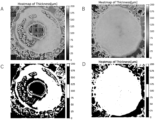
- the thickness of the retinal organoid can be easily confirmed by using a heat map created from an OCT tomographic image, and the compatibility range can be easily narrowed down by using a binary image.
- the transplant-suitable area must be an area that has been confirmed by OCT to be free of foreign body structures.
- "Foreign body structures" are characterized by high brightness in the brightness distribution of the OCT image that differs from the average brightness distribution in other uniform areas. Because retinal organoids have a layered structure, it is preferable to continuously extract and evaluate multiple horizontal X-Y plane images reconstructed from the OCT image in the Z direction, for example at intervals of approximately 10 ⁇ m or less (see Figure 4), to determine the presence or absence of foreign body structures.
- the term “about” refers to a value that varies by plus or minus 10%, 8%, 6%, 5%, 4%, 3%, 2%, or 1%, respectively, from the reference value.
- the term “about” refers to a range of plus or minus 10%, 5%, or 1%, respectively, from the reference value.
- the transplant-compatible areas can be efficiently identified by using bright-field and/or phase-contrast microscopy in addition to various types of OCT to exclude areas that are not suitable for transplantation (transplant-unsuitable areas) or retinal organoids that are not suitable for transplantation.
- “Bright-field microscopy” is ideal for understanding the overall condition of retinal organoids.
- organoids differentiate into the retina, they show the formation of characteristic retinal pigment cells (RPE) or spherical 3D organoid-like structures in the periphery, so the presence of these structures can be used to confirm whether they have differentiated properly into the retina.
- RPE retinal pigment cells
- thicker areas are observed in a different color (e.g., light brown) from thinner areas, so the overall uniformity and thickness can also be roughly confirmed.
- bright-field microscopy areas where differentiation into the retina is not observed or areas that are thin and non-uniform can be excluded from the OCT examination.
- by recording the differentiation state over time using bright-field microscopy and tracking the progress it can be confirmed that differentiation is occurring in a good state.
- the small pieces cut out are preferably about 1.0 to 10.0 mm long and 0.5 to 2.0 mm short so that they can be attached to a transplant cannula.
- the short length is about 0.5 to 1.0 mm, and the long length can be freely set.
- the short length can be extended up to about 2.0 mm.
- a low-viscosity viscoelastic substance such as 7-fold diluted hyaluronic acid for ophthalmic surgery (such as Viscoat)
- multiple transplant pieces can be attached.
- the retinal sheet for transplantation obtained by the method of the present invention is composed of retinal organoids in which the ratio of retinal cells (including retinal progenitor cells) to the total cells is about 90% or more, and more preferably about 95% or more.
- a retinal sheet for transplantation composed of such retinal organoids can be easily obtained by the method using the above-mentioned OCT, or OCT and bright-field microscopy and/or phase-contrast microscopy, without depending on the skills of an expert.
- cell non-adhesive substances examples include MPC (2-methacryloyloxyethyl phosphorylcholine) polymer; celluloses such as methylcellulose, hydroxyethylcellulose, hydroxypropylcellulose, hydroxypropylmethylcellulose, and sodium carboxymethylcellulose; polyethylene oxide; carboxyvinyl polymer; polyvinylpyrrolidone; polyethylene glycol; polyamides such as polyacrylamide and poly-N-isopropylacrylamide; polysaccharides such as chitin, chitosan, hyaluronic acid, alginic acid, starch, pectin, carrageenan, guar gum, gum arabic, and dextran; albumin and derivatives thereof.
- the cell non-adhesive substance is an MPC polymer.
- Region A and region B may be formed on the surface of the culture substrate by any method used for patterning. Region A and region B can be formed on the surface of the culture substrate by techniques such as soft lithography, photolithography, and 3D printing.
- the pluripotent stem cells collected and dispersed using a cell dispersion solution containing an enzyme e.g., trypsin, collagenase, hyaluronidase, elastase, pronase, DNase, papain
- an enzyme e.g., trypsin, collagenase, hyaluronidase, elastase, pronase, DNase, papain
- a chelating agent e.g., ethylenediaminetetraacetic acid (EDTA)
- EDTA ethylenediaminetetraacetic acid
- the cell dispersion solution for example, a commercially available product such as TrypLETM Select (Thermo Fisher Scientific) or TrypLETM Express (Thermo Fisher Scientific) may be used.
- the medium containing the BMP signal inhibitor or BMP signal agonist is a 1:1 mixture of F-12 medium and IMDM medium to which KnockOutTM Serum Replacement (Thermo Fisher Scientific) (e.g., 0.5% to 30%, 1% to 20%, or 10%), Chemically Defined Lipid Concentrate (Thermo Fisher Scientific), BSA, and 1-thioglycerol have been added.
- KnockOutTM Serum Replacement e.g., 0.5% to 30%, 1% to 20%, or 10%
- Chemically Defined Lipid Concentrate Thermo Fisher Scientific
- BSA 2-thioglycerol
- the retinal sheet for transplantation of the present invention can be evaluated as having the desired quality if, in quantitative PCR, the expression of the inner photoreceptor precursor cell marker among the above-mentioned retinal cell markers is high, the expression of the inner retinal layer cell marker and the retinal progenitor cell marker is confirmed, and the expression of the outer retinal tissue cell marker is low or below the detection limit.
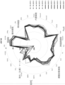
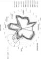
- the "relative expression pattern" of these genes can also be an important marker that defines the retina and its characteristics and quality. For example, by plotting each gene on a radar chart, it is possible to confirm the degree to which each group gene is expressed, the relative expression ratio of any two genes, etc. (see Example 1-4, Figure 11 below). In addition, for exclusion markers such as retinal extracellular tissue cell markers, and caution markers such as ciliary body markers, where contamination below a certain level is acceptable, by checking deviations from the pattern on the chart in addition to the values for individual genes, it is possible to determine whether the deviation is due to a single gene by chance, or as part of the overall expression pattern, which is expected to lead to more reliable judgments.
- exclusion markers such as retinal extracellular tissue cell markers
- caution markers such as ciliary body markers
- RNA is extracted from each sample using RNeasy Micro Kits (Qiagen), and cDNA is synthesized using SuperScript® III Reverse Transcriptase Kits (Invitrogen).
- the synthesized cDNA sample is diluted, and quantitative PCR is performed for each primer using TB Green® Premix DimerEraser(TM) (Takara).
- Evaluation is performed, for example, by averaging the ⁇ CT of each primer using an internal control (e.g., GAPDH and ARP) to determine the ⁇ CT of the corresponding primer, and then comparing the ⁇ CTs. Evaluation is performed according to certain criteria, such as a ⁇ CT value of 5 or less indicating strong expression, and a ⁇ CT value of 5 to 7 indicating expression.
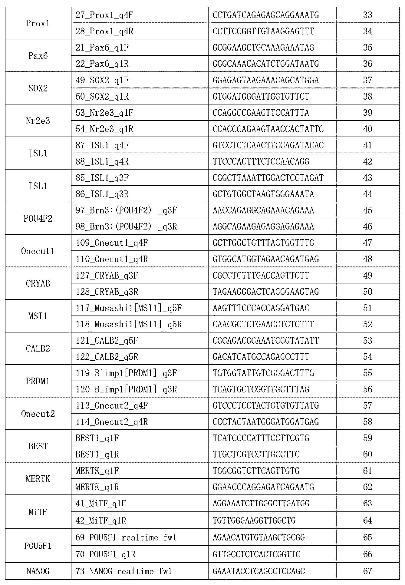
- Table 1 shows an example of primers used for quantitative PCR.
- the present invention also provides a drug for treating retinal degenerative disease, comprising a plurality of retinal sheets for transplantation and a transplantation medium (which also functions as a storage solution for the sheets).
- a drug for treating retinal degenerative disease comprising a plurality of retinal sheets for transplantation and a transplantation medium (which also functions as a storage solution for the sheets).
- the drug at least a portion of the plurality of retinal sheets for transplantation are cut from one planar retinal organoid derived from pluripotent stem cells, and each of them has a thickness of 70 ⁇ m or more and is confirmed to be free of foreign body structures by optical coherence tomography (OCT).
- OCT optical coherence tomography
- the "transplantation medium” include a diluted viscoelastic substance solution such as 7-fold diluted Viscoat.
- Each sheet is a small piece that is sized to fit into a cannula, a transplantation device, preferably an 18G to 24G cannula, for example, with a short axis length of 0.5 to 2.0 mm, preferably 0.5 to 1.0, and a long axis length of 1 mm or more, preferably 1 to 3 mm. Therefore, multiple sheets can be fitted into a 24G cannula for transplantation, and transplantation is possible without making a large incision in the retina.
- the multiple retinal sheets for transplantation contained in the pharmaceutical product of the present invention are also characterized by high expression of one or more photoreceptor precursor cell markers selected from Crx, Recoverin, and Rxr-gamma, and low or below the detection limit expression of one or more extraretinal tissue cell markers selected from CRYAB, EMX2, s100A10, Zic1, AQP1, and MSX1, preferably one or more selected from CRYAB, Zic1, and EMX2.
- a composite sheet can be used for transplantation in which a retinal sheet for transplantation and a retinal pigment epithelial cell sheet are loosely adhered together using a low-viscosity fluid gel, such as x7 Viscoat (a mixture of sodium chondroitin sulfate and sodium hyaluronate for ophthalmic surgery).
- a low-viscosity fluid gel such as x7 Viscoat (a mixture of sodium chondroitin sulfate and sodium hyaluronate for ophthalmic surgery).
- the present invention also provides a method for producing a composite sheet for retinal transplantation as described above, the method including the steps of producing a retinal sheet for transplantation from pluripotent stem cell-derived retinal organoids, and laminating the retinal sheet for transplantation on a retinal pigment epithelial cell sheet via a fluid gel to produce a composite sheet for retinal transplantation.
- the program may include, as necessary, a step of acquiring information from bright-field and/or phase-contrast microscopy; a step of inputting information from the bright-field and/or phase-contrast microscopy, a step of identifying a region (estimated region) that is estimated to be a transplant-compatible region according to the information from the bright-field and/or phase-contrast microscopy, and a step of issuing a command to perform OCT on the estimated region.
- Reference Example 1 Preparation of retinal organoids from pluripotent stem cells Planar retinal organoids were prepared by the following method. ⁇ Day 0 A PDMS sheet with a 1 cm diameter hole is placed on a 35 mm dish, and the inside of the hole is filled with 200 ⁇ l of 7 ⁇ l iMatrix-511/500 ⁇ l PBS dilution to coat the dish in a circular shape (1 hour at 37 °C). After removing the coating agent with an aspirator, 5x10 5 iPS cells are suspended in 200 ⁇ l StemFIT (registered trademark) AKO2N medium containing Y27632 at a final concentration of 10 ⁇ M and seeded at the same site.
- StemFIT registered trademark
- the thickness information from the OCT cross-sectional images was written out as a heat map (Figure 5). This makes it possible to identify the overall thickness trend using the heat map after confirming the internal structure using the OCT cross-sectional images. Furthermore, by converting the heat map into a binary image, it is possible to identify graft areas with appropriate thickness, such as areas with a thickness of 80 ⁇ m or more, or areas between 75 and 125 ⁇ m.
- transplant-compatible area of retinal organoids can be identified as an area with a certain thickness (70 ⁇ m or more) and no foreign body structure with different brightness distribution.
- the thickness and the presence or absence of foreign body structure can be identified by creating continuous XY plane images or heat maps from OCT tomographic images. By narrowing down the area to be evaluated by OCT using bright-field and phase-contrast microscopy, it is possible to identify the transplant-compatible area more efficiently.
- a region with uniformity suitable for transplantation is estimated using bright field/phase contrast, and 1 x 2 mm graft tissues are continuously cut out from the homogeneous region with a thickness of approximately 70 ⁇ m or more in the OCT tomographic image.
- thickness evaluation can be efficiently performed by using a heat map.
- a grid is set on the retinal organoid according to the size of the graft, and the grafts are aligned so that the number of usable sheets is maximized in the transplant-compatible region, and then cut out along the grid ( Figure 8).
- Multiple 1 x 2 mm grafts can be attached to a 24G transplant cannula, making it possible to transplant them without making a large incision in the retina ( Figure 9).
- a viscoelastic medium (35,000-60,000 mPa ⁇ s (25°C, shear rate 2 s-1) diluted two-fold) was placed on top of the retinal organoid, and a retinal pigment epithelial cell sheet was then placed on top of it so that the parietal side was in contact with the organoid.
- the sheets may be cut to an appropriate size beforehand and then layered, or they may be layered and then cut to the desired size. After layering, excess viscoelastic material is removed as much as possible using MQA (registered trademark) or similar.
- MQA registered trademark
- the method of the present invention makes it possible to efficiently cut out sheets suitable for transplantation from retinal organoids using an objective evaluation method, without relying on the special techniques of an expert.
- the present invention will greatly contribute to the practical application of regenerative medicine for retinal degenerative diseases.
Landscapes
- Health & Medical Sciences (AREA)
- Life Sciences & Earth Sciences (AREA)
- Engineering & Computer Science (AREA)
- Biomedical Technology (AREA)
- Chemical & Material Sciences (AREA)
- General Health & Medical Sciences (AREA)
- Veterinary Medicine (AREA)
- Public Health (AREA)
- Animal Behavior & Ethology (AREA)
- Cell Biology (AREA)
- Medicinal Chemistry (AREA)
- Zoology (AREA)
- Chemical Kinetics & Catalysis (AREA)
- Epidemiology (AREA)
- Bioinformatics & Cheminformatics (AREA)
- Botany (AREA)
- Transplantation (AREA)
- Oral & Maxillofacial Surgery (AREA)
- Organic Chemistry (AREA)
- Developmental Biology & Embryology (AREA)
- Dermatology (AREA)
- Ophthalmology & Optometry (AREA)
- Pharmacology & Pharmacy (AREA)
- Biotechnology (AREA)
- Nuclear Medicine, Radiotherapy & Molecular Imaging (AREA)
- Surgery (AREA)
- General Chemical & Material Sciences (AREA)
- Physics & Mathematics (AREA)
- Genetics & Genomics (AREA)
- Biophysics (AREA)
- Heart & Thoracic Surgery (AREA)
- Reproductive Health (AREA)
- Medical Informatics (AREA)
- Wood Science & Technology (AREA)
- Molecular Biology (AREA)
- Urology & Nephrology (AREA)
- Microbiology (AREA)
- Virology (AREA)
- Immunology (AREA)
- Gynecology & Obstetrics (AREA)
Abstract
Description
本出願は特願2022-210998(2022年12月28日出願)に基づく優先権を主張しており、この内容は本明細書に参照として取り込まれる。
技術分野:
本発明は、多能性幹細胞由来の網膜オルガノイドから移植用シートを製造する方法に関する。
[1]多能性幹細胞由来の網膜オルガノイドから移植用網膜シートを製造する方法であって、前記多能性幹細胞由来の網膜オルガノイドに対して光干渉断層撮影(OCT)、共焦点画像解析、及び超音波顕微鏡解析から選ばれるいずれか1または2以上(好ましくはOCT)を実施して、70μm以上(好ましくは70~175μm、場合により80μm以上、80~150μm)の厚さを有し、かつ、輝度の違いで特徴付けられる異物構造が存在しない領域を、移植適合領域として特定する工程、及び前記移植適合領域から小片を切り出し、移植用網膜シートとする工程を含む、前記方法。
[2]異物構造が存在しない領域が、OCT断層像から再構築した、水平方向のX-Y平面画像をZ方向に複数枚連続的に取り出して評価することにより特定される、[1]に記載の方法。
[3]70μm以上(好ましくは70~175μm、場合により80μm以上、80~150μm)の厚さを有する領域が、ヒートマップを用いて特定される、[1]又は[2]に記載の方法。
[4]切り出される小片が、18G~24Gカニューレに装着可能な大きさである、[1]~[3]のいずれかに記載の方法。
[5]切り出される小片が、長軸方向の長さが1.0~10.0mmであり、短軸方向の長さが0.5~2.0mmである、[1]~[4]のいずれかに記載の方法。
[6]前記方法が、多能性幹細胞由来の網膜オルガノイドに対して明視野顕微鏡撮影及び/又は位相差顕微鏡撮影を実施する工程をさらに含み、
前記明視野顕微鏡撮影及び/又は位相差顕微鏡撮影により、網膜への分化と均質性が確認できる領域を対象として、前記OCTによる移植適合領域の特定を実施する、[1]~[5]のいずれかに記載の方法。
[7]網膜オルガノイドが、直径1cm以上の平面状網膜オルガノイドである、[1]~[6]のいずれかに記載の方法。
[8][1]~[7]のいずれかに記載の方法で移植用網膜シートを製造する工程と、前記移植用網膜シートを流動性ゲル(例えば、ヒアルロン酸、コンドロイチン硫酸、及びこれらの誘導体から選ばれる1以上を含むゲル)を介して網膜色素上皮細胞シートに積層して、網膜移植用複合シートを製造する工程を含む、網膜移植用複合シートの製造方法。
[9]網膜変性疾患の治療用医薬品であって、多能性幹細胞由来の1つの平面状網膜オルガノイドから切り出された複数の網膜移植用細胞シートと移植媒体を含み、前記複数の網膜移植用細胞シートが、長軸方向の長さが1.0~10.0mmで、短軸方向の長さが0.5~2.0mmの大きさを有し、光干渉断層撮影(OCT)により、70μm以上(好ましくは70~175μm、場合により80μm以上、80~150μm)の厚さを有し、かつ、異物構造が存在しないことが確認されたものである、前記医薬品。
[10]切り出された小片に対して定量的PCRを行い、網膜組織外細胞のマーカーの発現が低く、網膜細胞のマーカーの発現が高いことを確認する工程をさらに含む、[1]~[8]のいずれかに記載の方法。前記確認工程は、例えば、レーダーチャート等を用いた絶対的または相対的な発現量パターン又は発現比の解析により実施できる。
[11]移植組織外細胞のマーカーがCRYAB、EMX2、s100A10、Zic1、AQP1、及びMSX1から選択される1つ以上、好ましくはCRYAB、Zic1、及びEMX2から選択される1つ以上を含み、網膜細胞のマーカーがCrx、Recoverin、及びRxr-gammaから選択される1つ以上を含む、[10]に記載の方法。
[12]網膜変性疾患の治療方法であって、[1]~[7]、[10]、及び[11]のいずれかに記載の方法により多能性幹細胞由来の網膜オルガノイドから複数の移植用網膜シートを製造し、必要とする対象の網膜に前記複数の移植用網膜シートを移植することを含む、治療方法。
[13]網膜変性疾患の治療方法であって、[8]に記載の方法により網膜移植用複合シートを製造し、必要とする対象の網膜に前記網膜移植用複合シートを移植することを含む、治療方法。
本発明は、多能性幹細胞由来の網膜オルガノイドから移植用網膜シートを製造する方法であって、前記多能性幹細胞由来の網膜オルガノイドに対して光干渉断層撮影を実施して、70μm以上の厚さを有し、かつ、輝度の違いで特徴付けられる異物構造が存在しない領域を、移植適合領域として特定する工程、及び、前記移植適合領域から小片を切り出し、移植用網膜シートとする工程を含む方法に関する。
多能性幹細胞からの網膜オルガノイドの作製方法として、細胞接着性を有する領域Aと、前記領域Aの少なくとも一部に隣接する前記領域Aよりも細胞接着性の低い領域Bとをその表面に有する培養基材上の領域Aにおいて、多能性幹細胞を網膜組織に分化誘導する方法を挙げることができる。以下、このような特定のパターンを有する基材上での培養をパターニング培養と呼ぶ。
(1) 多能性幹細胞を、BMPシグナル阻害薬を含む培地で培養すること、及び
(2) 工程(1)で得られた細胞を、BMPシグナル作用薬を含む培地で培養すること。
本発明の方法で調製した移植用網膜シートが所望の品質を有することは、既報(例えば、WO2020/184720等)にしたがい、定量的PCRを実施し、その遺伝子発現パターンを例えばレーダーチャート表示することにより確認することができる。または、免疫染色、シングルセル解析、フローサイトメトリーを用いた解析やこれらを教師データとして用いた機械学習、深層学習などを応用した解析などを行うことにより、適合部位が視細胞前駆細胞を含む網膜オルガノイドであることを確認できる。


本発明は、複数の移植用網膜シートと移植媒体(シートの保存液としても機能する)を含む、網膜変性疾患の治療用医薬品も提供する。前記医薬品において、複数の移植用網膜シートの少なくとも一部は、多能性幹細胞由来の1つの平面状網膜オルガノイドから切り出されたものであり、いずれも、光干渉断層撮影(OCT)により、70μm以上の厚さを有し、かつ、異物構造が存在しないことが確認されたものである。「移植媒体」としては、例えば、7倍希釈ビスコートなど希釈粘弾性物質液が挙げられる。
網膜オルガノイドはシート形状を生かして、網膜色素上皮細胞と積層して、複合シートを作成することも可能である。生体内において網膜と網膜色素上皮細胞は頭頂面同士で接触しているだけで、強固な接着は必要としない。そのため、粘度の低い流動性ゲル、例えば、x7ビスコート(眼科手術用コンドロイチン硫酸エステルナトリウム・ヒアルロン酸ナトリウム 混合材)などを用いて、移植用網膜シートと網膜色素上皮細胞シートを緩く接着させた複合シートとして、移植に使用することができる。
本発明は、1.に記載した方法にしたがい、多能性幹細胞由来の網膜オルガノイドから複数の移植用網膜シートを調製し、必要とする対象の網膜に前記複数の移植用網膜シートを(例えば、24Gカニューレに装着するなどして)移植することを含む、網膜変性疾患の治療方法も提供する。
本発明の移植用網膜シートを調製する方法は、その少なくとも一部を自動化することができる。
例えば、前記プログラムは、網膜オルガノイドのOCT情報を取得する工程;前記OCT情報をコンピューターに入力する工程;前記OCT情報からX-Y平面画像を連続的に生成し、異物構造を含まない領域を特定する工程;OCT情報からヒートマップを生成し、厚さが70μm以上、好ましくは70~175μm、特に好ましくは80~150μmの領域を選択する工程;厚さが70μm以上、かつ異物構造を含まない領域を移植適合領域として特定する工程;移植用網膜シートに合わせたサイズのグリッドを当該移植適合領域上に有効枚数が最大となるように設定し、グリッドに合わせて移植用網膜シートを切り出す指令を切り出し用機器に出力する工程を含む。前記プログラムは、必要に応じて、明視野顕微鏡撮影及び/又は位相差顕微鏡撮影の情報を取得する工程;前記明視野顕微鏡撮影及び/又は位相差顕微鏡撮影の情報を入力する工程、前記明視野顕微鏡撮影及び/又は位相差顕微鏡撮影の情報にしたがって、移植適合領域と推定される領域(推定領域)を特定する工程、前記推定領域に対して、OCTを実施するよう指令を出す工程を含んでいてもよい。
以下の方法で平面状網膜オルガノイドを作成した。
・Day0
直径1cmの穴のあいたPDMSシートを35 mm dishに設置し、穴の内部に7 μl iMatrix-511/500μl PBS 希釈液200μlで満たしてdishを円状にコーティングする(37 ℃で1時間)。コーティング剤をアスピレーターで除去後 5x10⁵ iPS細胞を200μl StemFIT(登録商標)AKO2NにY27632を最終濃度10μMで添加した培地に懸濁し、同部位に播種する。37℃インキュベータで30分静置後、滅菌鑷子を用いてPDMシートを取り外し、上述のStemFIT (登録商標)AKO2N+Y27632培地を300μl追加して5%CO2インキュベーターで培養する。
培地をStemFIT AKO2N培地(Y 27632無し、ペニシリン/ストレプトマイシン添加)500μlに置換する。
1ml gfCDM(10%KSR)で3回wash後 1ml gfCDM(10%KSR)に全量置換する。
[gfCDM培地の組成]
IMDM (31980-030) 125ml
F-12 (31765-092) 125ml
CD lipid (11950-031) 2.5ml
BSA (5g/20ml) 5ml
1-チオグリセロール(207-09232) 9.75μl
ペニシリン/ストレプトマイシン 2.5ml
KSR 25ml
100nM LDNの入ったgfCDM培地1mlに置換する。
・Day8
1mlのgfCDMで洗浄後、1mlのBMP4 (終濃度1.5nM)入り培地に置換する。
・Day11とDay14
gfCDM培地を半量交換する。
DMEM/F-12+Glutamaxで3回洗浄後下記培地1mlに置換する。
[培地の組成]
DMEM/F-12+Glutamax (10565-042) 5ml
N2 supplement (17502-048) 50μl
ペニシリン/ストレプトマイシン 50μl
以下のcontrol成熟培地 1mlに置換する。
[培地の組成]
DMEM/F-12+Glutamax (10565-042) 500ml
N2 supplement (17502-048) 5ml
非働化FBS 50ml
50mM タウリン (in PBS) 1.12ml
Antibiotic-Antimycotic (100x)(15240-062) 5ml
*以降2-3日に1回培地交換する。
分化60日前後の網膜平面オルガノイドにおいて、1)明視野顕微鏡撮影(全体の状態の把握)、2)位相差顕微鏡撮影(均質性の観察)、3)OCT断層増撮影(均一性、厚さ、異物情報)、4)OCT X-Y平面での観察を用いて、網膜オルガノイドの厚さと異物構造を含まない領域を特定した。
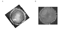
まず、明視野顕微鏡撮影にて全体の状態を把握した。オルガノイドは、網膜への分化が見られる場合、周辺部に特徴的なRPE又は3Dオルガノイド様構造物の形成が全例で見られ、これらの構造物が網膜への分化の傍証となる。また、厚みのある部位は薄茶色に観察され、大まかな全体の均一性を明視野で観察することができる(図1)。
オルガノイドに対し位相差顕微鏡撮影を実施すると、より内部構造が詳細に観察され、シート内に均一構造を持つ部分が確認できた(図2)。
Screen社の光干渉断層撮影(OCT)を用いて断層像を取得した。輝度の違いから組織内の異物の検出や均一性の確認が可能であり(図3)、輝度分布を調べることにより、異物の混在を同定することが可能と思われた。また、OCT断層像から水平方向のX-Y平面画像、Z方向に10μm以下の間隔で複数枚連続的に取り出し、X-Y水平面の連続的評価を行うことで、さらに効率よく輝度分布の異なる異物構造がないことを判定することができる(図4)。
推定網膜領域を確定後、その領域内から1x2mmのサイズの移植片を切り出し、その遺伝子発現パターンを、PCRパネルを使用して確認した。これにより、網膜外組織混入の判定、網膜細胞内の内層細胞/視細胞の分化60日時点での大まかな構成内容などがわかる。その結果、薄い網膜では毛様体/水晶体マーカーが高いことが確認できた(図6)。一方、厚みを持った均一な部分(4,5,9,10)では網膜外組織のマーカー遺伝子発現は低く、移植に向いた部位であることが確認できた(図7)。
以上の結果から、網膜オルガノイドの移植適合領域は、一定の厚さ(70μm以上)と輝度分布の異なる異物構造を有さない領域として同定しうることが確認された。厚さと異物構造の有無は、OCT断層像から、連続的X-Y平面画像やヒートマップを作製するなどして、特定することができる。明視野顕微鏡撮影や位相差顕微鏡撮影により、OCTによる評価対象領域を絞り込むことで、より効率的な移植適合領域の特定が可能となる。
Claims (9)
- 多能性幹細胞由来の網膜オルガノイドから移植用網膜シートを製造する方法であって、
前記多能性幹細胞由来の網膜オルガノイドに対して光干渉断層撮影(OCT)、共焦点画像解析、及び超音波顕微鏡解析から選ばれるいずれか1または2以上を実施して、70μm以上の厚さを有し、かつ、輝度の違いで特徴付けられる異物構造が存在しない領域を、移植適合領域として特定する工程、及び
前記移植適合領域から小片を切り出し、移植用網膜シートとする工程を含む、前記方法。 - 異物構造が存在しない領域が、OCT断層像から再構築した、水平方向のX-Y平面画像をZ方向に複数枚連続的に取り出して評価することにより特定される、請求項1に記載の方法。
- 70μm以上の厚さを有する領域が、ヒートマップを用いて特定される、請求項1に記載の方法。
- 切り出される小片が、18G~24Gカニューレに装着可能な大きさである、請求項1に記載の方法。
- 切り出される小片が、長軸方向の長さが1.0~10.0mmであり、短軸方向の長さが0.5~2.0mmである、請求項1に記載の方法。
- 前記方法が、多能性幹細胞由来の網膜オルガノイドに対して明視野顕微鏡撮影及び/又は位相差顕微鏡撮影を実施する工程をさらに含み、
前記明視野顕微鏡撮影及び/又は位相差顕微鏡撮影により、網膜への分化と均質性が確認できる領域を対象として、前記OCTによる移植適合領域の特定を実施する、請求項1に記載の方法。 - 網膜オルガノイドが、直径1cm以上の平面状網膜オルガノイドである、請求項1に記載の方法。
- 請求項1~7のいずれか1項に記載の方法で移植用網膜シートを製造する工程と、前記移植用網膜シートを流動性ゲルを介して網膜色素上皮細胞シートに積層して、網膜移植用複合シートを製造する工程を含む、網膜移植用複合シートの製造方法。
- 網膜変性疾患の治療用医薬品であって、多能性幹細胞由来の1つの平面状網膜オルガノイドから切り出された複数の網膜移植用細胞シートと移植媒体を含み、前記複数の網膜移植用細胞シートが、長軸方向の長さが1.0~10.0mmで、短軸方向の長さが0.5~2.0mmの大きさを有し、光干渉断層撮影(OCT)により、70μm以上の厚さを有し、かつ、異物構造が存在しないことが確認されたものである、前記医薬品。
Priority Applications (4)
| Application Number | Priority Date | Filing Date | Title |
|---|---|---|---|
| CN202380089743.8A CN120569227A (zh) | 2022-12-28 | 2023-12-27 | 视网膜片材的制备方法 |
| EP23912214.6A EP4643894A1 (en) | 2022-12-28 | 2023-12-27 | Method for manufacturing retinal sheet |
| AU2023416411A AU2023416411A1 (en) | 2022-12-28 | 2023-12-27 | Method for manufacturing retinal sheet |
| JP2024567903A JPWO2024143436A1 (ja) | 2022-12-28 | 2023-12-27 |
Applications Claiming Priority (2)
| Application Number | Priority Date | Filing Date | Title |
|---|---|---|---|
| JP2022-210998 | 2022-12-28 | ||
| JP2022210998 | 2022-12-28 |
Publications (1)
| Publication Number | Publication Date |
|---|---|
| WO2024143436A1 true WO2024143436A1 (ja) | 2024-07-04 |
Family
ID=91717774
Family Applications (1)
| Application Number | Title | Priority Date | Filing Date |
|---|---|---|---|
| PCT/JP2023/046832 Ceased WO2024143436A1 (ja) | 2022-12-28 | 2023-12-27 | 網膜シートの製造方法 |
Country Status (5)
| Country | Link |
|---|---|
| EP (1) | EP4643894A1 (ja) |
| JP (1) | JPWO2024143436A1 (ja) |
| CN (1) | CN120569227A (ja) |
| AU (1) | AU2023416411A1 (ja) |
| WO (1) | WO2024143436A1 (ja) |
Citations (14)
| Publication number | Priority date | Publication date | Assignee | Title |
|---|---|---|---|---|
| WO2013077425A1 (ja) | 2011-11-25 | 2013-05-30 | 住友化学株式会社 | 網膜組織及び網膜関連細胞の製造方法 |
| JP2013128475A (ja) | 2011-11-25 | 2013-07-04 | Sumitomo Chemical Co Ltd | 網膜組織の製造方法 |
| WO2015025967A1 (ja) | 2013-08-23 | 2015-02-26 | 住友化学株式会社 | 網膜組織及び網膜関連細胞の製造方法 |
| WO2016063986A1 (ja) * | 2014-10-24 | 2016-04-28 | 大日本住友製薬株式会社 | 網膜組織の製造方法 |
| WO2017183732A1 (ja) | 2016-04-22 | 2017-10-26 | 大日本住友製薬株式会社 | 網膜組織の製造法 |
| WO2019217630A1 (en) | 2018-05-09 | 2019-11-14 | The Regents Of The University Of Colorado, A Body Corporate | Stem cell-derived cell cultures, stem cell-derived three-dimensional tissue products, and methods of making and using the same |
| WO2020138430A1 (ja) * | 2018-12-28 | 2020-07-02 | 国立研究開発法人理化学研究所 | 網膜系細胞又は網膜組織の障害を伴う疾患の治療薬 |
| WO2020184720A1 (ja) | 2019-03-13 | 2020-09-17 | 大日本住友製薬株式会社 | 移植用神経網膜の品質を評価する方法及び移植用神経網膜シート |
| WO2020249935A1 (en) | 2019-06-10 | 2020-12-17 | Newcells Biotech Limited | Improved retinal organoids and methods of making the same |
| JP2021118733A (ja) | 2015-09-08 | 2021-08-12 | 大日本住友製薬株式会社 | 網膜組織の製造方法 |
| WO2022054925A1 (ja) | 2020-09-11 | 2022-03-17 | 国立研究開発法人理化学研究所 | 神経網膜を含む細胞凝集体とマトリクスとを含む複合体及びその製造方法 |
| WO2022138803A1 (ja) | 2020-12-24 | 2022-06-30 | 国立研究開発法人理化学研究所 | 視細胞を含む層状の網膜組織の促成製造方法 |
| WO2022239868A1 (ja) | 2021-05-14 | 2022-11-17 | 国立研究開発法人理化学研究所 | 網膜組織の製造方法 |
| WO2023003025A1 (ja) * | 2021-07-21 | 2023-01-26 | 国立大学法人京都大学 | 網膜組織の製造方法 |
-
2023
- 2023-12-27 JP JP2024567903A patent/JPWO2024143436A1/ja active Pending
- 2023-12-27 EP EP23912214.6A patent/EP4643894A1/en active Pending
- 2023-12-27 WO PCT/JP2023/046832 patent/WO2024143436A1/ja not_active Ceased
- 2023-12-27 CN CN202380089743.8A patent/CN120569227A/zh active Pending
- 2023-12-27 AU AU2023416411A patent/AU2023416411A1/en active Pending
Patent Citations (14)
| Publication number | Priority date | Publication date | Assignee | Title |
|---|---|---|---|---|
| WO2013077425A1 (ja) | 2011-11-25 | 2013-05-30 | 住友化学株式会社 | 網膜組織及び網膜関連細胞の製造方法 |
| JP2013128475A (ja) | 2011-11-25 | 2013-07-04 | Sumitomo Chemical Co Ltd | 網膜組織の製造方法 |
| WO2015025967A1 (ja) | 2013-08-23 | 2015-02-26 | 住友化学株式会社 | 網膜組織及び網膜関連細胞の製造方法 |
| WO2016063986A1 (ja) * | 2014-10-24 | 2016-04-28 | 大日本住友製薬株式会社 | 網膜組織の製造方法 |
| JP2021118733A (ja) | 2015-09-08 | 2021-08-12 | 大日本住友製薬株式会社 | 網膜組織の製造方法 |
| WO2017183732A1 (ja) | 2016-04-22 | 2017-10-26 | 大日本住友製薬株式会社 | 網膜組織の製造法 |
| WO2019217630A1 (en) | 2018-05-09 | 2019-11-14 | The Regents Of The University Of Colorado, A Body Corporate | Stem cell-derived cell cultures, stem cell-derived three-dimensional tissue products, and methods of making and using the same |
| WO2020138430A1 (ja) * | 2018-12-28 | 2020-07-02 | 国立研究開発法人理化学研究所 | 網膜系細胞又は網膜組織の障害を伴う疾患の治療薬 |
| WO2020184720A1 (ja) | 2019-03-13 | 2020-09-17 | 大日本住友製薬株式会社 | 移植用神経網膜の品質を評価する方法及び移植用神経網膜シート |
| WO2020249935A1 (en) | 2019-06-10 | 2020-12-17 | Newcells Biotech Limited | Improved retinal organoids and methods of making the same |
| WO2022054925A1 (ja) | 2020-09-11 | 2022-03-17 | 国立研究開発法人理化学研究所 | 神経網膜を含む細胞凝集体とマトリクスとを含む複合体及びその製造方法 |
| WO2022138803A1 (ja) | 2020-12-24 | 2022-06-30 | 国立研究開発法人理化学研究所 | 視細胞を含む層状の網膜組織の促成製造方法 |
| WO2022239868A1 (ja) | 2021-05-14 | 2022-11-17 | 国立研究開発法人理化学研究所 | 網膜組織の製造方法 |
| WO2023003025A1 (ja) * | 2021-07-21 | 2023-01-26 | 国立大学法人京都大学 | 網膜組織の製造方法 |
Non-Patent Citations (2)
| Title |
|---|
| AKIBA RYUTARO: "Toward establishment of regenerative cell therapy for retinitis pigmentosa using iPS cell derived retinal sheet", FOLIA PHARMACOLOGICA JAPONICA, vol. 155, 1 March 2020 (2020-03-01), pages 93 - 98, XP055868350 * |
| See also references of EP4643894A1 |
Also Published As
| Publication number | Publication date |
|---|---|
| CN120569227A (zh) | 2025-08-29 |
| EP4643894A1 (en) | 2025-11-05 |
| JPWO2024143436A1 (ja) | 2024-07-04 |
| AU2023416411A1 (en) | 2025-08-07 |
Similar Documents
| Publication | Publication Date | Title |
|---|---|---|
| JP6850456B2 (ja) | 網膜色素上皮細胞の純化方法 | |
| JP5552478B2 (ja) | 膜 | |
| CN111164205B (zh) | 包含视网膜组织的细胞聚集体及其制造方法 | |
| CA2828197C (en) | Method of producing retinal pigment epithelial cell sheet | |
| Kang et al. | Effects of flow-induced shear stress on limbal epithelial stem cell growth and enrichment | |
| KR20190112090A (ko) | 다능성줄기세포의 분화 제어 방법 | |
| Tian et al. | Polarized, cobblestone, human retinal pigment epithelial cell maturation on a synthetic PEG matrix | |
| WO2023090427A1 (ja) | シート状網膜組織の製造方法 | |
| CN120569227A (zh) | 视网膜片材的制备方法 | |
| WO2024185751A1 (ja) | 神経網膜前駆細胞を含む組織とその製造方法、該組織を含む網膜疾患治療用組成物 | |
| WO2024204596A1 (ja) | 網膜組織の製造方法 | |
| Kingston | Utilising Glaucomatous Extracellular Matrix Substrates and Novel Biomimetic 3D Polymer Scaffolds to Study Trabecular Meshwork (TM) Cells and TM Progenitors | |
| US20250290044A1 (en) | Method for producing cell sheet | |
| JP2025524999A (ja) | 分離ウェルマイクロプレートを使用する胚様体のヒドロゲルへの自動埋め込みの方法 | |
| CN120897992A (zh) | 视网膜组织的生产方法 | |
| Dimmock | Simulation and characterization of the corneal limbal epithelial stem cell niche by using new biotechnologies | |
| WO2024085251A1 (ja) | 網膜移植物の品質を評価する方法 | |
| Liu et al. | Development of a biodegradable smooth-surface microcarrier for retinal pigment epithelial cell expansion and maturation | |
| Savioja | Attachment and Differentiation of Human Embryonic Stem Cell Derived Retinal Pigment Epithelium Cells on Different Coating Materials | |
| HK40008770B (zh) | 多能干细胞的分化调控方法 |
Legal Events
| Date | Code | Title | Description |
|---|---|---|---|
| 121 | Ep: the epo has been informed by wipo that ep was designated in this application |
Ref document number: 23912214 Country of ref document: EP Kind code of ref document: A1 |
|
| WWE | Wipo information: entry into national phase |
Ref document number: 2024567903 Country of ref document: JP |
|
| WWE | Wipo information: entry into national phase |
Ref document number: 202380089743.8 Country of ref document: CN |
|
| WWE | Wipo information: entry into national phase |
Ref document number: AU2023416411 Country of ref document: AU |
|
| WWE | Wipo information: entry into national phase |
Ref document number: 2023912214 Country of ref document: EP |
|
| NENP | Non-entry into the national phase |
Ref country code: DE |
|
| ENP | Entry into the national phase |
Ref document number: 2023912214 Country of ref document: EP Effective date: 20250728 |
|
| ENP | Entry into the national phase |
Ref document number: 2023416411 Country of ref document: AU Date of ref document: 20231227 Kind code of ref document: A |
|
| ENP | Entry into the national phase |
Ref document number: 2023912214 Country of ref document: EP Effective date: 20250728 |
|
| WWP | Wipo information: published in national office |
Ref document number: 202380089743.8 Country of ref document: CN |
|
| WWP | Wipo information: published in national office |
Ref document number: 2023912214 Country of ref document: EP |