WO2024181580A1 - 脂質ナノ粒子、医薬組成物、および脂質ナノ粒子の製造方法 - Google Patents
脂質ナノ粒子、医薬組成物、および脂質ナノ粒子の製造方法 Download PDFInfo
- Publication number
- WO2024181580A1 WO2024181580A1 PCT/JP2024/008154 JP2024008154W WO2024181580A1 WO 2024181580 A1 WO2024181580 A1 WO 2024181580A1 JP 2024008154 W JP2024008154 W JP 2024008154W WO 2024181580 A1 WO2024181580 A1 WO 2024181580A1
- Authority
- WO
- WIPO (PCT)
- Prior art keywords
- mol
- lipid
- polymer
- antibody
- igg
- Prior art date
- Legal status (The legal status is an assumption and is not a legal conclusion. Google has not performed a legal analysis and makes no representation as to the accuracy of the status listed.)
- Ceased
Links
Classifications
-
- A—HUMAN NECESSITIES
- A61—MEDICAL OR VETERINARY SCIENCE; HYGIENE
- A61P—SPECIFIC THERAPEUTIC ACTIVITY OF CHEMICAL COMPOUNDS OR MEDICINAL PREPARATIONS
- A61P35/00—Antineoplastic agents
-
- A—HUMAN NECESSITIES
- A61—MEDICAL OR VETERINARY SCIENCE; HYGIENE
- A61K—PREPARATIONS FOR MEDICAL, DENTAL OR TOILETRY PURPOSES
- A61K9/00—Medicinal preparations characterised by special physical form
- A61K9/48—Preparations in capsules, e.g. of gelatin, of chocolate
- A61K9/50—Microcapsules having a gas, liquid or semi-solid filling; Solid microparticles or pellets surrounded by a distinct coating layer, e.g. coated microspheres, coated drug crystals
- A61K9/51—Nanocapsules; Nanoparticles
- A61K9/5107—Excipients; Inactive ingredients
- A61K9/5123—Organic compounds, e.g. fats, sugars
-
- A—HUMAN NECESSITIES
- A61—MEDICAL OR VETERINARY SCIENCE; HYGIENE
- A61K—PREPARATIONS FOR MEDICAL, DENTAL OR TOILETRY PURPOSES
- A61K9/00—Medicinal preparations characterised by special physical form
- A61K9/48—Preparations in capsules, e.g. of gelatin, of chocolate
- A61K9/50—Microcapsules having a gas, liquid or semi-solid filling; Solid microparticles or pellets surrounded by a distinct coating layer, e.g. coated microspheres, coated drug crystals
- A61K9/51—Nanocapsules; Nanoparticles
- A61K9/5107—Excipients; Inactive ingredients
- A61K9/513—Organic macromolecular compounds; Dendrimers
- A61K9/5146—Organic macromolecular compounds; Dendrimers obtained otherwise than by reactions only involving carbon-to-carbon unsaturated bonds, e.g. polyethylene glycol, polyamines, polyanhydrides
- A61K9/5153—Polyesters, e.g. poly(lactide-co-glycolide)
-
- A—HUMAN NECESSITIES
- A61—MEDICAL OR VETERINARY SCIENCE; HYGIENE
- A61P—SPECIFIC THERAPEUTIC ACTIVITY OF CHEMICAL COMPOUNDS OR MEDICINAL PREPARATIONS
- A61P43/00—Drugs for specific purposes, not provided for in groups A61P1/00-A61P41/00
-
- A—HUMAN NECESSITIES
- A61—MEDICAL OR VETERINARY SCIENCE; HYGIENE
- A61K—PREPARATIONS FOR MEDICAL, DENTAL OR TOILETRY PURPOSES
- A61K39/00—Medicinal preparations containing antigens or antibodies
- A61K39/395—Antibodies; Immunoglobulins; Immune serum, e.g. antilymphocytic serum
- A61K39/39591—Stabilisation, fragmentation
-
- C—CHEMISTRY; METALLURGY
- C07—ORGANIC CHEMISTRY
- C07K—PEPTIDES
- C07K16/00—Immunoglobulins [IG], e.g. monoclonal or polyclonal antibodies
- C07K16/18—Immunoglobulins [IG], e.g. monoclonal or polyclonal antibodies against material from animals or humans
-
- C—CHEMISTRY; METALLURGY
- C07—ORGANIC CHEMISTRY
- C07K—PEPTIDES
- C07K16/00—Immunoglobulins [IG], e.g. monoclonal or polyclonal antibodies
- C07K16/40—Immunoglobulins [IG], e.g. monoclonal or polyclonal antibodies against enzymes
-
- C—CHEMISTRY; METALLURGY
- C07—ORGANIC CHEMISTRY
- C07K—PEPTIDES
- C07K2317/00—Immunoglobulins specific features
- C07K2317/70—Immunoglobulins specific features characterized by effect upon binding to a cell or to an antigen
-
- C—CHEMISTRY; METALLURGY
- C07—ORGANIC CHEMISTRY
- C07K—PEPTIDES
- C07K2317/00—Immunoglobulins specific features
- C07K2317/80—Immunoglobulins specific features remaining in the (producing) cell, i.e. intracellular antibodies or intrabodies
Definitions
- the present disclosure relates to lipid nanoparticles.
- the present disclosure relates to lipid nanoparticles that can encapsulate proteins such as antibodies and deliver them into cells, including the cytosol, a pharmaceutical composition, and a method for producing lipid nanoparticles.
- antibody drugs have antigen-specific binding ability, they are not only used as research reagents but are also put to practical use as molecular targeted drugs, making them biopharmaceuticals that play an important role in modern medicine.
- antibodies are highly hydrophilic polymers (approximately 150 kDa), and due to the constraints of biological membrane permeability, their targets are limited to extracellular factors. In other words, if an innovative method for introducing antibodies into cells could be developed, the range of application of antibodies could be expanded from extracellular antigens to intracellular antigens, and new disease applications of antibody drugs could be expected.
- LNP Lipid Nanoparticle
- Comirnaty registered trademark
- Spikevax registered trademark
- LNPs are lipid nanoparticle formulations that efficiently encapsulate negatively charged molecules such as nucleic acids and enhance their delivery to the cytosol, and are also expected to be applied to antibody drugs.
- Non-Patent Document 1 describes that a liposome-based formulation, DOSP:MM27 (imidazole-based helper lipid MM27+aminoglycoside lipid dioleylsuccinylparomomycin (DOSP)), was able to deliver anti-cytokeratin 8 (K8) antibody intracellularly.
- Non-Patent Document 2 describes that antibodies against ⁇ -tubulin and ⁇ -actin could be delivered intracellularly using lipospermine DOGS (dioctadecylglycylspermine) dissolved in ethanol.
- DOGS dioctadecylglycylspermine
- Patent Document 1 describes a method for concentrating a protein having a surface charge by forming a complex between the protein and a polyamino acid in a buffer solution.
- Patent Document 2 describes lipids that form lipid nanoparticles for use in drug delivery systems, which are said to improve escape from endosomes after endocytosis.
- the present disclosure aims to deliver useful proteins into cells, particularly to deliver antibodies against functional proteins within cells, such as cancer cells.
- the inventors discovered that when encapsulating a protein, which is a polymer with a multivalent charge, in lipids, the coexistence of an acidic polymer such as an acidic polyamino acid produces lipid nanoparticles with smaller particle sizes, and thus completed the present disclosure.
- a lipid nanoparticle comprising a composition comprising a protein selected from an antibody and an antibody fragment, a polymer, and a lipid, wherein the protein and the polymer have opposite charges at a predetermined pH.
- the lipid nanoparticle according to (1) in which the protein is encapsulated in the lipid nanoparticle.
- the polymer is an anionic or cationic polymer.
- the cationic polymer is at least one selected from the group consisting of polylysine, polyarginine, polyhistidine, and water-soluble salts thereof.
- the molecular weight of the cationic or anionic polyamino acid is 0.5 kDa to 1000 kDa.
- a pharmaceutical composition comprising a composition comprising a protein selected from an antibody and an antibody fragment, a polymer, and a lipid, wherein the protein and the polymer have opposite charges at a predetermined pH.
- (12-1) A method for delivering an antibody, which is a pharmacologic active ingredient, comprising administering the pharmaceutical composition according to (12) above.
- (12-2) Use of the pharmaceutical composition according to (12) above.
- (13) a droplet formation step of mixing the protein selected from the antibody and the antibody fragment with the polymer to form droplets;
- a method for producing lipid nanoparticles comprising: an encapsulation step of mixing the droplets with the lipid to encapsulate the droplets in the lipid.
- 13-1) The method for producing lipid nanoparticles described in (13), wherein at least one step selected from the droplet formation step and the encapsulation step is a step using a flow channel of a microfluidic device.
- a lipid nanoparticle comprising a composition comprising a protein, a polymer, and a lipid, wherein the polymer has an opposite charge to the protein at a certain pH.
- a protein selected from an antibody or an antibody fragment can be delivered into a cell, including the cytoplasm or nucleus.
- IgG was most efficiently delivered to the cytosol with 1:200 IgG_polyE-LNP.
- FIG. 1 shows that IgG_polyE-LNP was able to deliver hIgG-AF488 to the cytosol in HeLa, HT1080, MDA-MB-231, and SW480 cells.
- IgG_polyE-LNP prepared with t-BuOH lipid solution and IgG_polyE-LNP prepared with 90% t-BuOH lipid solution were similarly capable of delivering antibody to the cytosol.
- FIG. 1 shows that the peak top of the lipid nanoparticle particle size is approximately 100 nm.
- FIG. 1 shows the results of particle size measurement of IgG_polyE-LNP determined by nanoparticle tracking analysis (NTA method).
- FIG. 13 shows images of each cell immunostained in Example 13.
- FIG. 13 shows images of each cell immunostained in Example 13.
- FIG. 13 shows the particle size of hIgG_polyE-LNP in Example 13.
- FIG. 13 shows the encapsulation rate of each antibody in hIgG_polyE-LNP in Example 13.
- FIG. 13 shows the values of hIgG_polyE-LNP in Example 14.
- FIG. 13 is a diagram of each cell immunostained in Example 14.
- FIG. 15 shows the formation of droplets in Example 15.
- FIG. 13 is a diagram of each cell immunostained in Example 16.
- the composition used in this disclosure is a composition comprising a protein, a polymer, and a lipid, characterized in that the polymer has an opposite charge to the protein at a certain pH.
- it is a composition comprising a protein, a polymer, and a lipid, characterized in that the protein and the polymer have an opposite charge at a certain pH.
- the numerical range of 1 to 50 includes 1, 2, 3, 4, 5, 6, 7, 8, 9, 10, 11, 12, 13, 14, 15, 16, 17, 18, 19, 20, 21, 22, 23, 24, 25, 26, 27, 28, 29, 30, 31, 32, 33, 34, 35, 36, 37, 38, 39, 40, 41, 42, 43, 44, 45, 46, 47, 48, 49, and 50.
- the protein used in the present disclosure is a protein selected from an antibody and an antibody fragment.
- An antibody is composed of immunoglobulin (Ig), a protein molecule that specifically binds to an antigen, and includes types such as IgG, IgA, IgM, IgD, and IgE.
- An antibody fragment is a partial fragment of an antibody. The isoelectric point of an antibody or an antibody fragment is generally about pH 7.5 to 9.5.
- the use of the pharmaceutical composition of the present disclosure enables the protein to be more stably transported and stored when applied to humans as a pharmaceutical preparation or diagnostic agent, and further, the protein can be easily administered to humans when in use, making the composition highly useful.
- the protein may have a sugar chain.
- high purity means pharmaceutical-grade purity, i.e., a level that can be used as a human medicine, while for other proteins, high purity means reagent-grade purity, for example, a threshold value of 0.1% by mass or less in terms of the total amount of impurities.
- Antigens to which the antibodies or antibody fragments used in the present disclosure specifically bind include, but are not limited to, polypeptides and peptides present in cells, including the cytoplasm and nucleus, genes including RNA (ribonucleic acid) including mRNA, rRNA, or tRNA, and DNA (deoxyribonucleic acid), lipids including phospholipids such as cardiolipin, or sugar chains bound to the above polypeptides, peptides, genes, or lipids.
- RNA ribonucleic acid
- lipids including phospholipids such as cardiolipin, or sugar chains bound to the above polypeptides, peptides, genes, or lipids.
- antibodies may be produced by the hybridoma method (Kohler and Milstein, Nature 256:495 (1975)) or recombinant method (U.S. Patent No. 4,816,567). They may also be isolated from phage antibody libraries (Clackson et al., Nature 352:624-628 (1991); Marks et al., J. Mol. Biol. 222:581-597 (1991)). They may also be isolated from single B cell clones (N. Biotechnol. 28(5):253-457 (2011)).
- an antibody delivered into the cytoplasm by receptor-dependent endocytosis and endosomal escape by cholic acid, or liposome-mediated endocytosis and endosomal escape by photosensitive benzoporphyrin is translocated into the nucleus by a nuclear localization signal inserted into the sequence (Mol. Pharmaceutics 2016, 13, 1915-1926 and Mol. Pharmaceutics 2015, 12, 9, 3272-3281).
- a nuclear localization signal inserted into the sequence
- the polymer used in the present invention is a polymer with a charge that cancels the charge of the protein to be delivered into the cell under the conditions of use.
- the conditions of use are determined either at the time of manufacture, in blood, or in the cell.
- An acidic or basic polymer anionic polymer, cationic polymer
- Polymers that are considered to have a charge include polymers with carboxyl groups or amino groups. Examples of such polymers include polyamino acids, polyesters, glycosaminoglycans, and polyamides.
- the polymers used in the present disclosure are selected to have opposite charges under the measurement conditions of the protein to be delivered into the cell.
- a combination is selected in which the polymer and protein have opposite charges at a specified pH. It is possible to confirm whether the polymer and protein have opposite charges at a specified pH by measuring the pI values of the polymer and protein. It is possible to determine the pI values of the polymer and protein using known methods such as measuring the zeta potential of a polymer solution in water or measuring using isoelectric focusing of a protein.
- known methods such as measuring the zeta potential of a polymer solution in water or measuring using isoelectric focusing of a protein.
- the polyamino acids used in the present disclosure are selected to have an opposite charge under the measurement conditions of the protein to be delivered into the cell.
- the isoelectric point of the polymer In order for the polymer to associate with the protein, the isoelectric point of the polymer must be different from the isoelectric point of the protein described above, and must be higher or lower than the isoelectric point of the protein.
- the isoelectric point of antibodies and antibody fragments which are examples of proteins, is pH 7.5 to 9.5 as described above, it is preferable that the isoelectric point of the polymer is outside this range, for example, pH 7.0 or lower or 10.0 or higher.
- polymer examples include cationic or anionic polyamino acids or salts thereof.
- anionic polyamino acids include sodium salts of polyglutamic acid and polyaspartic acid
- examples of the cationic polyamino acids include water-soluble salts (e.g., hydrochlorides) of polyarginine, polyhistidine, polylysine, polyornithine, polycitrulline, and the like.
- polyamino acids include sodium poly-L-glutamic acid and sodium poly-L-aspartic acid as anionic polyamino acids, and poly-L-arginine hydrochloride, poly-L-lysine hydrochloride, poly-L-arginine hydrobromide, and poly-L-lysine hydrobromide as cationic polyamino acids.
- the polyamino acids are homopolymers or copolymers, or mixtures thereof. It is preferable that the polyamino acids have a CH2 group between the main chain and the carboxyl group or amino group of the side chain.
- the degree of polymerization can be appropriately selected from the range of 20 to 3000.
- the degree of polymerization can be appropriately selected from the ranges of 60 to 3000, 60 to 2500, 60 to 2000, 60 to 1500, 60 to 1000, 60 to 500, 60 to 400, 60 to 300, and 60 to 200.
- the degree of polymerization can be appropriately selected from the ranges of 80 to 3000, 80 to 2500, 80 to 2000, 80 to 1500, 80 to 1000, 80 to 500, 80 to 400, 80 to 300, and 80 to 200.
- the polymerization degree can be appropriately selected from the ranges of 100 to 3000, 100 to 2500, 100 to 2000, 100 to 1500, 100 to 1000, 100 to 500, 100 to 400, 100 to 300, and 100 to 200.
- the polyglutamic acid may have a degree of polymerization having a molecular weight of 3000 (degree of polymerization 20) to 60,000 (degree of polymerization 408), and in another non-limiting embodiment, the polyglutamic acid may have a degree of polymerization of 3000 (degree of polymerization 20) to 50,000 (degree of polymerization 340), 3000 (degree of polymerization 20) to 40,000 (degree of polymerization 272), 3000 (degree of polymerization 20) to 20,000 (degree of polymerization 136), 3000 (degree of polymerization 20) to 15,000 (degree of polymerization 102), 3000 (degree of polymerization 20) to 10,000 (degree of polymerization 68), 4000 (degree of polymerization 27) to 50,000 (degree of polymerization 340), 4000 (degree of polymerization 2 7) to 40,000 (degree of polymerization 272), 4,000 (degree of polymerization 27) to 30,000 (degree of polymerization
- an antibody or antibody fragment thereof including human immunoglobulin is usually basic and positively charged at plasma pH 7.4, so an anionic polymer can be used.
- polyamino acids such as polyglutamic acid and polyaspartic acid can be preferably used.
- glycosaminoglycans such as hyaluronic acid and heparin can be preferably used as anionic polymers.
- acidic antibodies and antibody fragments that have been approved as therapeutic antibodies (Journal of Chromatography B, Volumes 1065-1066, 2017, Pages 119-128).
- a cationic polymer can be used in the composition of the present disclosure that includes such an antibody or its antibody fragment that is negatively charged at plasma pH 7.4, a cationic polymer can be used.
- polyamino acids such as polyarginine, polylysine, and polyornithine are preferable.
- the pKa, pI, size, shape, etc. of the protein to be delivered can be taken into consideration, and the pKa, pI, molecular weight, shape, etc. of the polymer to be selected can also be taken into consideration.
- the size of the lipid nanoparticles can be reduced.
- a protein including an antibody or an antibody fragment, etc. is stably associated with a polymer having an opposite charge through electrostatic bonding to form droplets.
- the form of association between a protein including an antibody or an antibody fragment, etc., and a polymer can be, for example, a form in which the protein is electrostatically bonded to a polymer having an opposite charge, or a form in which the protein is electrostatically bonded to a polymer having an opposite charge and stably encapsulated within the polymer.
- PPCs Protein-polyelectrolyte complexes
- the formation of the droplets of the present disclosure can be appropriately confirmed by observation under a microscope using differential interference contrast.
- the diameter of the formed droplets can be measured using an ocular micrometer under a transmission electron microscope to obtain an approximate value, a particle size distribution meter using a dynamic light scattering method, a nanoparticle tracking analyzer using the characteristics of both scattered light and Brownian motion, and the like.
- the particle size of the droplets of the present disclosure is determined by nanoparticle tracking analysis. By adjusting the production conditions, droplets of the present disclosure can also be produced in the range of 100 nm to 10 ⁇ m, 200 nm to 2 ⁇ m, or 300 nm to 1 ⁇ m.
- the ratio (mass ratio) of polymer to protein can be set appropriately. For example, it can be in the range of 1:100 to 1:10, or even in the range of 1:10 to 1:1.
- the temperature during mixing can also be set appropriately, for example, in the range of 15 to 40°C, or even in the range of 20°C to 30°C.
- the pH during mixing may be any pH at which the protein and polymer can electrostatically interact with each other, and is preferably, for example, a pH between the isoelectric point of the protein and the isoelectric point of the polymer.
- the droplets formed are separated using a process such as centrifugation, filtration, or deep bed filtration (for example, Patent No. 5361738, etc.), and the droplets reconstituted as an aqueous solution with an appropriate protein concentration can be provided to the next encapsulation process.
- lipid mixture As the lipid used in the present disclosure, at least one type of lipid capable of forming micelles in water is used as a mixture (hereinafter also referred to as lipid mixture). So-called amphipathic lipids having both hydrophilic and hydrophobic portions can be used. Such lipids can be known, and examples of such lipids include phosphatidylinositol, phosphatidylethanolamine, phosphatidylserine, phosphatidic acid, phosphatidylglycerol, phosphatidylcholine, and the like, which are phospholipids known to be components of biological membranes.
- PEG lipids such as DMG-PEG5k, and the like, which are used as surfactants, such as the amphoteric lipid 3-[(3-cholamidopropyl)dimethylammonio]propanesulfonate (CHAPS), the anionic lipid sodium cholic acid salt, and the nonionic lipid octylglycoside, can also be used as appropriate.
- CHAPS amphoteric lipid 3-[(3-cholamidopropyl)dimethylammonio]propanesulfonate
- anionic lipid sodium cholic acid salt e.glycoside
- neutral phospholipids such as 1,2-distearoyl-sn-glycero-3-phosphocholine (DSPC) and PEG lipids such as 1,2-distearoyl-sn-glycero-3-phosphoethanolamine-N-methoxy-polyethylene glycol-2000 (DSPEmPEG2000) may be used.
- DSPC 1,2-distearoyl-sn-glycero-3-phosphocholine
- PEG lipids such as 1,2-distearoyl-sn-glycero-3-phosphoethanolamine-N-methoxy-polyethylene glycol-2000 (DSPEmPEG2000) may be used.
- DSPEmPEG2000 1,2-distearoyl-sn-glycero-3-phosphoethanolamine-N-methoxy-polyethylene glycol-2000
- the cationic lipid comprises 20 mol% to 60 mol%, 20 mol% to 55 mol%, 20 mol% to 50 mol%, 20 mol% to 45 mol%, 20 mol% to 40 mol%, 25 mol% to 55 mol%, 25 mol% to 50 mol%, 25 mol% to 45 mol%, 25 mol% to 40 mol%, 30 mol% to 55 mol%, 30 mol% to 50 mol%, 30 mol% to 45 mol% of the lipid mixture (excluding PEG lipids) present in the lipid nanoparticles of the present disclosure.
- the phospholipids in the lipid mixture (excluding PEG lipids) present in the lipid nanoparticles may comprise 2 mol% to 20 mol%, 2 mol% to 15 mol%, 2 mol% to 12 mol%, 2 mol% to 10 mol%, 2 mol% to 8 mol%, 4 mol% to 15 mol%, 4 mol% to 12 mol%, or 4 mol% to 10 mol%, 4 mol% to 8 mol% (or any percentage or range thereof) of the lipid mixture (excluding PEG lipids) present in the lipid nanoparticles of the present disclosure.
- the phospholipids in the lipid mixture comprise 8 mol% to 15 mol%, 8 mol% to 12 mol%, 8 mol% to 10 mol%, or 5 mol%, 6 mol%, 7 mol%, 8 mol%, 9 mol%, 10 mol%, 11 mol%, or 12 mol% (or any percentage or range thereof) of the lipid mixture (excluding PEG lipids) present in the particle.
- the cholesterol or cholesterol derivatives in the lipid mixture are present in the lipid nanoparticles of the present disclosure in an amount of 20 mol% to 60 mol%, 20 mol% to 55 mol%, 20 mol% to 50 mol%, 20 mol% to 45 mol%, 20 mol% to 40 mol%, 25 mol% to 55 mol%, 25 mol% to 50 mol%, 25 mol% to 45 mol%, 25 mol% to 40 mol%, 30 mol% or more of the lipid mixture (excluding PEG lipids) present in the lipid nanoparticles of the present disclosure.
- the PEG lipid comprises 0.5 mol% to 5 mol%, 0.5 mol% to 8 mol%, 0.5 mol% to 10 mol%, 1.0 mol% to 5 mol%, 1.0 mol% to 8 mol%, 1.0 mol% to 10 mol%, 1.5 mol% to 5 mol%, 1.5 mol% to 8 mol%, 1.5 mol% to 10 mol%, 2.0 mol% to 5 mol%, 2.0 mol% to 8 mol%, 2.0 mol% to 10 mol%, 0.5 mol%, 1.0 mol%, 1.5 mol%, 5 mol%, 8 mol%, 10 mol% (or any percentage or range thereof) of the lipid mixture (excluding the PEG lipid) present in the lipid nanoparticles of the present disclosure.
- the percentage of PEG lipid present in the lipid nanoparticles of the present disclosure is the target amount during manufacturing, and the actual content of PEG lipid present in the formulation may vary, for example
- the composition containing cationic lipids can be 20 mol% to 60 mol% cationic lipids, 20 mol% to 60 mol% cholesterol, and 2 mol% to 20 mol% phospholipids with 0.5 mol% to 10 mol% PEG lipids.
- the composition containing cationic lipids is 40 mol% to 55 mol% cationic lipids, 40 mol% to 55 mol% cholesterol, and 8 mol% to 15 mol% phospholipids with 1.0 mol% to 5 mol% PEG lipids.
- intracellular environment-responsive lipids are also preferably used as cationic lipids in order to increase the efficiency of escape from endosomes after delivery to cells.
- Intracellular environment-responsive lipids are lipids that decompose or change their structure in response to environmental factors such as reducing conditions and pH in the cell.
- intracellular environment-responsive lipids having one or more properties selected from ionizable property, reducing environment responsive property, and self-degrading property can be appropriately used.
- Lipids with ionizable properties have no charge outside the cell, but after being taken up into the cell via endocytosis, they become positively charged under acidic conditions in the endosome and can fuse with the negatively charged endosomal membrane.
- Non-limiting examples of lipids with ionizable properties include a combination of 1,2-dioleoyl-sn-glycero-3-phosphoethanolamine (DOPE) and cholesterol hemisuccinic acid (CHEMS), and a combination of 1,2-dioleoyl-3-dimethylammoniumpropane (DODAP) and phosphatidylserine (PS), which are appropriately used.
- DOPE 1,2-dioleoyl-sn-glycero-3-phosphoethanolamine
- CHEMS cholesterol hemisuccinic acid
- DODAP 1,2-dioleoyl-3-dimethylammoniumpropane
- PS phosphatidylserine
- lipids with ionizable properties is 1,2-dilinoleyl-MC3-N,N-dimethyl-3-aminopropane (DLin-MC3-DMA), which has a tertiary amine in the hydrophilic portion of the amphipathic lipid.
- DLin-MC3-DMA 1,2-dilinoleyl-MC3-N,N-dimethyl-3-aminopropane
- Lipids that are responsive to reducing conditions have no charge outside the cell, but after being taken up into the cell via endocytosis, they become positively charged under acidic conditions in the endosome and can fuse with the negatively charged endosomal membrane.
- Non-limiting examples of lipids with such ionizable properties include a combination of 1,2-dioleoyl-sn-glycero-3-phosphoethanolamine (DOPE) and cholesterol hemisuccinic acid (CHEMS), and a combination of 1,2-dioleoyl-3-dimethylammoniumpropane (DODAP) and phosphatidylserine (PS), which are appropriately used.
- DOPE 1,2-dioleoyl-sn-glycero-3-phosphoethanolamine
- CHEMS cholesterol hemisuccinic acid
- DODAP 1,2-dioleoyl-3-dimethylammoniumpropane
- PS phosphatidylserine
- lipids with such ionizable properties is 1,2-dilinoleyl-MC3-N,N-dimethyl-3-aminopropane (DLin-MC3-DMA), which has a tertiary amine in the hydrophilic portion of the amphipathic lipid.
- DLin-MC3-DMA 1,2-dilinoleyl-MC3-N,N-dimethyl-3-aminopropane
- lipids listed in WO2019-188867 for example, the lipids listed in the following formula (1), can be used.
- R1a and R1b each independently represent an alkylene group having 1 to 6 carbon atoms
- Xa and Xb each independently represent a non-cyclic alkyl tertiary amino group having 1 to 6 carbon atoms and one tertiary amino group, or a cyclic alkylene tertiary amino group having 2 to 5 carbon atoms and one or two tertiary amino groups
- R2a and R2b each independently represent an alkylene group or an oxydialkylene group having 8 or less carbon atoms
- Ya and Yb each independently represent an ester bond, an amide bond, a carbamate bond, an ether bond, or a urea bond
- Za and Zb each independently represent a divalent group derived from an aromatic compound having 3 to 16 carbon atoms, at least one aromatic ring, and optionally having a heteroatom
- R3a and R3b each independently represent a residue derived from a reaction product of a fat-soluble vitamin having a hydroxy
- examples of such lipids include O-Ph-P3C1, O-Ph-P4C1, O-Ph-P4C2, O-BnP4C2, E-Ph-P4C2, L-Ph-P4C2, HD-Ph-P4C2, O-Ph-amide-P4C2, and O-Ph-C3M. These lipids respond to pH, redox activity, and the like, and can increase the efficiency of endosomal escape.
- lipid one or more lipids selected from the compounds described in the Examples below, that is, bis[[4-[2-[[4-(oleoyloxy)phenyl]acetoxy]ethyl]piperidino]ethyl]disulfane (SS-OP) as a lipid having ionizable properties, reducing condition responsive properties, and autolytic properties, 1,2-dioleoyl-sn-glycero-3-phosphoethanolamine (DOPE) as a neutral phospholipid, DMG-PEG5k as a PEG lipid, and ⁇ -sitosterol as cholesterol, can be suitably used.
- DOPE 1,2-dioleoyl-sn-glycero-3-phosphoethanolamine
- the antibody content (protein loading rate) in the lipid nanoparticles of the present disclosure can be appropriately adjusted from within the ranges of 5-95 mol%, 10-90 mol%, or 30-80 mol%.
- the lipid mixture of the present disclosure can be prepared by dispersing the cationic lipids of the present disclosure and other components (lipids, etc.) in an appropriate solvent or dispersion medium, for example, an aqueous solvent or an alcoholic solvent, and performing an operation to induce organization as necessary.
- amphiphilic substances are used in producing the lipid nanoparticles of the present disclosure.
- the amphiphilic substance is selected in consideration of the stability and uniformity of the lipid nanoparticles.
- amphiphilic substances used in the present disclosure include lipids such as phosphoglycerides, phosphatidylcholine, dipalmitoylphosphatidylcholine (DPPC), dioleylphosphatidylethanolamine (DOPE), dioleyloxypropyltriethylammonium (DOTMA), dioleoylphosphatidylcholine, cholesterol, cholesterol esters, diacylglycerol, diacylglycerol succinate, diphosphatidylglycerol (DPPG), hexanedecanol, and polyethylene glycol (PEG).
- lipids such as phosphoglycerides, phosphatidylcholine, dipalmitoylphosphatidylcholine (DPPC), dioleylphosphatidyl
- Aliphatic alcohols such as palmitic acid or oleic acid; fatty acids; fatty acid monoglycerides; fatty acid diglycerides; fatty acid amides; sorbitan trioleate (Span® 85); glycocholate; sorbitan monolaurate (Span® 20); polysorbate 20 (Tween® 20); polysorbate 60 (Tween® 60); polysorbate 65 (Tween® 65); polysorbate 80 (Tween® 65); (Tween® 85); polysorbate 85 (Tween® 85); polyoxyethylene monostearate; surfactin; poloxomer; sorbitan fatty acid esters such as sorbitan trioleate; lecithin; lysolecithin; phosphatidylserine; phosphatidylinositol; sphingomyelin; phosphatidylethanolamine (cephalin); cardiolipin; phosphatidic acid
- the lipid nanoparticles can be produced by the production method described below.
- the lipid nanoparticles have a structure in which droplets formed by the association of a protein containing an antibody or an antibody fragment with a polymer are encapsulated in lipid.
- the protein containing an antibody or an antibody fragment is stably associated with a polymer having an opposite charge through electrostatic bonding to form droplets.
- the form of association between the protein containing an antibody or an antibody fragment and the polymer is not limited to a specific theory, but examples of the form in which the protein is electrostatically bonded to a polymer having an opposite charge and the protein is electrostatically bonded to a polymer having an opposite charge and stably encapsulated in the polymer can be assumed.
- the particle size of the lipid nanoparticles can be measured using a particle size distribution meter using a dynamic light scattering method or a nanoparticle tracking analyzer that utilizes the characteristics of both scattered light and Brownian motion.
- the particle size of the lipid nanoparticles in the present disclosure is determined by nanoparticle tracking analysis and is produced in the range of 10 to 600 nm.
- the lipid nanoparticles can also be produced in the range of 50 to 500 nm or 70 to 400 nm.
- the average particle size distribution determined by nanoparticle tracking analysis is 300 nm or less.
- the peak top is produced at 150 nm or less, and can be produced at 120 nm or less, or even 100 nm or less by adjusting the production conditions. It is said that a particle size of less than 100 nm is easily endocytosed. Furthermore, in tumor cells, etc., it is said that a particle size of less than 100 nm is retained around the cancer tissue through the Enhanced Permeability and Retention effect, increasing selectivity.
- the antibody content (protein loading rate) in the lipid nanoparticles of the present disclosure can be appropriately adjusted within the following ranges: 0.1-10.0 mol%, 0.2-10.0 mol%, 0.3-10.0 mol%, 0.4-10.0 mol%, 0.5-10.0 mol%, 0.1-5.0 mol%, 0.2-5.0 mol%, 0.3-5.0 mol%, 0.4-5.0 mol%, 0.5-5.0 mol%, 0.1-2.0 mol%, 0.2-2.0 mol%, 0.3-2.0 mol%, 0.4-2.0 mol%, 0.5-2.0 mol%.
- compositions In order to formulate the lipid nanoparticles of the present disclosure into pharmaceutical compositions, the lipid nanoparticles can be combined with various pharma- ceutically acceptable additives and bases or carriers for dispersing the active agent(s).
- additives include pH adjusters such as arginine, sodium hydroxide, glycine, hydrochloric acid, citric acid, and mixtures thereof.
- additives include local anesthetics (e.g., benzyl alcohol), isotonicity agents (e.g., sodium chloride, mannitol, sorbitol), adsorption inhibitors (e.g., Tween 80), solubility enhancers (e.g., cyclodextrin and its derivatives), stabilizers (e.g., serum albumin), and reducing agents (e.g., glutathione).
- the pharmaceutical compositions comprising the lipid nanoparticles of the present disclosure can be dispersed in a base or vehicle, which may include a hydrophilic compound capable of dispersing the active agent and any desired additives.
- the base may be selected from a wide range of suitable carriers, including, but not limited to, copolymers of polycarboxylic acids or their salts, copolymers of carboxylic anhydrides (e.g., maleic anhydride) and other monomers (e.g., methyl (meth)acrylate, acrylic acid, etc.), hydrophilic vinyl polymers such as polyvinyl acetate, polyvinyl alcohol, polyvinylpyrrolidone, cellulose derivatives such as hydroxymethylcellulose, hydroxypropylcellulose, etc., and natural polymers such as chitosan, collagen, sodium alginate, gelatin, hyaluronic acid, and non-toxic metal salts thereof.
- suitable carriers including, but not limited to, copolymers of polycarboxylic acids or their salts, copolymers of carboxylic anhydrides (e.g., maleic anhydride) and other monomers (e.g., methyl (meth)acrylate, acrylic acid, etc.), hydrophil
- biodegradable polymers such as polylactic acid, poly(lactic acid-glycolic acid) copolymers, polyhydroxybutyric acid, poly(hydroxybutyric acid-glycolic acid) copolymers, and mixtures thereof, are selected as the base or carrier.
- synthetic fatty acid esters such as polyglycerin fatty acid esters, sucrose fatty acid esters, etc. may also be used as carriers.
- Hydrophilic polymers and other carriers can be used alone or in combination and can impart enhanced structural integrity to the carrier by partial crystallization, ionic bonding, crosslinking, etc.
- Carriers can be provided in a variety of forms including fluid or viscous solutions, gels, pastes, powders, microspheres, and films for direct application to the nasal mucosa. Use of selected carriers in this context can result in enhanced absorption of the biologically active agent.
- compositions of the present disclosure may contain pharma- ceutically acceptable carriers necessary to approximate physiological conditions, such as pH adjusting and buffering agents, tonicity adjusting agents, and wetting agents, e.g., sodium acetate, sodium lactate, sodium chloride, potassium chloride, calcium chloride, sorbitan monolaurate, triethanolamine oleate, and mixtures thereof.
- pharma- ceutically acceptable carriers necessary to approximate physiological conditions, such as pH adjusting and buffering agents, tonicity adjusting agents, and wetting agents, e.g., sodium acetate, sodium lactate, sodium chloride, potassium chloride, calcium chloride, sorbitan monolaurate, triethanolamine oleate, and mixtures thereof.
- conventional non-toxic pharma-ceutically acceptable carriers may be used, including, for example, pharmaceutical grades of mannitol, lactose, starch, magnesium stearate, sodium saccharin, talc, cellulose, glucose, sucrose, magnesium carbonate, and the like.
- Lipid nanoparticles can be produced based on known methods with reference to J. Chem. Phys. 150, 064903 (2019) and the like. Specifically, first, a protein containing an antibody or an antibody fragment and a polymer are mixed in an appropriate ratio to form droplets (droplet formation step).
- the form of association between a protein containing an antibody or an antibody fragment and a polymer is not limited to a specific theory, but examples of the form in which the protein is electrostatically bound to a polymer having an opposite charge, or the protein is electrostatically bound to a polymer having an opposite charge and stably encapsulated within the polymer, can be assumed.
- Mixing can be performed by mixing with a liquid mixer or pipetting, stirring with a stirrer or stirring blade in a mixing container, mixing with a high-speed impinging jet mixer or a multi-inlet vortex mixer, or contacting on a flow path of a microfluidic device (i.e., a microflow path).
- the ratio (mass ratio) of polymer to protein can be appropriately set, but can be, for example, within the range of 100:1 to 10:1, or even within the range of 10:1 to 1:1.
- the temperature during mixing can also be appropriately set, for example, within the range of 15 to 40°C, or 20 to 30°C.
- the pH during mixing may be any pH that allows electrostatic interaction between the protein and the polymer, and is preferably, for example, a pH between the isoelectric point of the protein and the isoelectric point of the polymer.
- the droplets formed are separated using a process such as centrifugation, filtration, or depth filtration (e.g., Patent 5361738, etc.), and the droplets reconstituted as an aqueous solution with an appropriate protein concentration can be provided to the subsequent encapsulation process.
- the droplets are mixed with lipids to encapsulate the droplets in the lipids (encapsulation process), producing lipid nanoparticles.
- This mixing can be performed by using a liquid mixer or pipetting, stirring in a mixing vessel with a stirrer or impeller, or using a high-speed confined impinging jets mixer or a multi-inlet vortex mixer.
- Other industrial production methods such as contacting the droplets on a flow channel of a microfluidic device, alcohol dilution (precipitation), hydration, and emulsification, have also been established.
- the ratio (mass ratio) of lipid to droplets can be set appropriately, and can be, for example, within the range of 1:100 to 10:1, or even within the range of 1:10 to 1:1.
- the temperature during mixing can also be set appropriately, and can be, for example, within the range of 15 to 40°C, or even within the range of 20°C to 30°C.
- the pH during mixing can be, for example, within the range of 5 to 9, and further within the range of h 6 to 8.
- solvents that can be used to dissolve lipids include alcohols such as ethanol, butanol, and tert-butanol, as well as ethylene glycol, glycerin, and polyethylene glycol. However, the solvent should be selected after comprehensively considering the particle formation properties, delivery properties to cells, and toxicity.
- effect The nanoparticles thus produced are evaluated in vitro and in vivo.
- the nanoparticles of the present disclosure have been observed to be localized intracellularly, including in the cytosol and nucleus.
- IgG used was normal human IgG, whole molecule, purified product (manufactured by Fujifilm Wako Pure Chemical Industries, Ltd.) In this specification, it is referred to as IgG or hIgG.
- the polyglutamic acids used had the following sizes (mol wt 3,000-15,000, Sigma).
- the particle diameter, PdI (particle size distribution, and zeta potential ( ⁇ -potential) were evaluated using a Zetasizer Nano ZS (Malvern). The collected samples were diluted 100-fold with PBS and measurements were performed.
- the encapsulation rate was calculated by centrifuging the prepared LNP at 4 ° C. and 20,000 g for 30 minutes using a tabletop ultracentrifuge, measuring the protein concentration of the supernatant. That is, IgG_polyE-LNP was adjusted with PBS (-) so that the antibody concentration was 2 ⁇ M (final sample volume 100 ⁇ L). This was centrifuged at 4 ° C., 20,000 ⁇ g, for 1 hour to precipitate the LNP fraction. 50 ⁇ L of the supernatant was collected, the antibody concentration was measured using Nanodrop, and the encapsulation rate was calculated back from the following formula.
- C IgG refers to the IgG concentration calculated from the supernatant obtained by centrifuging 2 ⁇ M IgG
- C LNP refers to the IgG concentration calculated from the supernatant obtained by centrifuging IgG_polyE-LNP.
- Encapsulation rate of IgG (%) (C IgG ⁇ C LNP )/C IgG ⁇ 100
- HeLa cells HeLa cells obtained from ATCC were cultured in ⁇ -MEM containing 10% bovine serum (BS).
- BS bovine serum
- Dialysis The dialysis membrane used was a 2-14k MWCO dialysis membrane (Spectrum Laboratories Inc.) After filling the dialysis membrane, dialysis was performed by stirring with a Corning Stirrer.
- hIgG-AF488 For the Alexa Fluor 488 modification, 345 ⁇ L of PBS( ⁇ ) was added to 100 ⁇ L of 10 mg/mL hIgG (normal human IgG, Fujifilm Wako Pure Chemical Corporation), and 50 ⁇ L of 1 M NaHCO 3 was added thereto to make the solution basic. The mixture was mixed with 1000 ⁇ g/mL Alexa488-SDP ester (Thermo Fisher Scientific) and mixed by inversion for 1 hour at 25°C in the dark. The antibody fraction was then separated using a PD10 column and purified with Alexa Fluor 488-labeled hIgG (hIgG-AF488). was purified.
- Anti-GFP antibodies were prepared using the Expi293TM Expression System (Thermo Fisher Scientific). Expi293FTM cells were prepared to a concentration of 3,000,000 cells/mL, and 25 mL was added to a 125 mL Erlenmeyer flask. 25 ⁇ g of a plasmid encoding Anti-GFP-IgG (Addgene, Anti-GFP [N86/38.1R]) was mixed with ExpiFectamineTM293 (Thermo Fisher Scientific) and transfected according to the recommended protocol.
- Expi293TM Expression System Thermo Fisher Scientific
- the fraction containing IgG was collected and dialyzed against PBS (pH 7.4) to obtain anti-GFP antibody (anti-GFP-IgG).
- the recovered anti-GFP antibody (anti-GFP-IgG) was stored at 4° C.
- an antibody in a citric acid solution pH 7.5
- the solution was replaced with a citric acid solution (pH 5) using a Micro Biospin (registered trademark) Column 30 (BIORAD).
- Example 1 (Production of LNPs) The lipid was dissolved in chloroform to prepare 20 mM SS-OP, 10 mM DOPE, 20 mM ⁇ -sitosterol, and 10 mM DMG-PEG5k.
- the composition of SS-OP/DOPE/ ⁇ -sitosterol/DMG-PEG5k (molar ratio 45/10/45/1.5) was mixed in a flask at room temperature, and 1 mL of t-BuOH (Fujifilm Wako Pure Chemical Corporation) was added and dissolved, and the chloroform was evaporated in a rotary evaporator to obtain a lipid solution.
- this lipid solution was freeze-dried for 18 to 24 hours, and then dissolved in t-BuOH to prepare a 10 mM lipid solution.
- a lipid solution dissolved in t-BuOH and a polyglutamic acid solution (0.75 mg/mL polyglutamic acid solution (polyE, mol wt 3,000-15,000) dissolved in 10 mM citric acid solution (pH 5) were separately incubated at 40° C. for 30 minutes.
- hIgG:polyE 5:1 (mass ratio) to form hIgG-polyE droplets
- the lipid solution incubated at 40°C for 30 minutes was mixed with the droplet solution by pipetting 15 times to give a 1:4 (volume ratio).
- the mixture was dialyzed against 1,000 times or more volume of PBS (-) (pH 7.4) using a 12-14k MWCO dialysis membrane (Spectrum Laboratories Inc.) to prepare lipid nanoparticles encapsulating hIgG (hIgG_polyE-LNPs).
- Example 2 (Production of LNPs) hIgG_polyE-LNP was produced in the same manner as in Example 1, except that a 90% tBuOH aqueous solution was used instead of tBuOH. (effect)
- Example 3 (Localization in HeLa cells) (Production of LNPs)
- IgG_polyE-LNP(a) to (e) with antibody:lipid ratios of (a) to (e) were prepared in the same manner as in Example 1, except that hIgG-AF488 was used instead of IgG.
- hIgG-AF488:polyE (a) 1:100 (b) 1:200 (c) 1:300 (d) 1:400 (e) 1:500
- the adjusted hIgG-AF488_polyE-LNP was added to serum medium of HeLa cells so that the IgG concentration was 1 ⁇ M, suspended, and incubated for 18 hours under conditions of 37°C and 5% CO2 , and the intracellular localization of hIgG-AF488 was observed.
- Figure 1 A the time course of (b) 1:200 IgG_polyE-LNP was observed, it was found that hIgG was delivered to the cytosol 6 hours after addition.
- Example 4 (Production of various LNPs) (Anti-GFP antibody_polyE-LNP)
- Anti-GFP antibody_polyE-LNP was produced in the same manner as in Example 1, except that anti-GFP antibody was used instead of hIgG.
- Example 5 (a) hIgG-AF488, (b) hIgG-AF488_PolyE, or (c) hIgG-AF488_PolyE-LNP was added to serum-containing medium at 1 ⁇ M to HeLa cells, and the cells were incubated at 37° C. and 5% CO2 for 18 hours to observe the intracellular localization of IgG (FIG. 2A).
- Example 6 In order to demonstrate the usefulness of adding polyE, LNPs (IgG-LNPs) (Reference Example 1) were prepared by mixing polyE-free hIgG with lipids, and a comparative test was carried out with hIgG_polyE-LNP (Example 1). To observe intracellular localization, hIgG-AF488 was used as the hIgG.
- HIgG-LNP and hIgG_polyE-LNP were added to HeLa cells in serum medium at an IgG concentration of 1 ⁇ M, suspended, and incubated at 37°C and 5% CO2 for 18 hours.
- IgG concentration 1 ⁇ M
- Example 7 LgBiT and HiBiT associate to reconstitute nanoluciferase, which emits light when luciferin is added.
- the amount of cytosolic transfer of hIgG_polyE-LNP (Production Example 1) and hIgG-LNP (Production Example 2) was compared.
- hIgG_polyE-LNPs or IgG-LNPs encapsulating hIgG-HiBiT which had a HiBiT sequence genetically engineered to the C-terminus of an anti-GFP antibody (anti-GFP-IgG) (Addgene; Anti-GFP [N86/38.1R], Plasmid #114492), were suspended in serum medium at an IgG concentration of 0.5 ⁇ M and administered to HeLa cells (HeLa-LgBiT) constitutively expressing LgBiT for 18 hours. After incubation, the cells were washed three times with PBS (pH 7.4) and harvested by trypsinization.
- anti-GFP-IgG anti-GFP antibody
- HRas is a member of the Ras protein family and is a protein that is localized in the cell membrane. GFP was localized in the cell membrane by using HeLa cells expressing HRas-GFP, which is a fusion of HRas with GFP.
- Anti-GFP-IgG (Addgene; Anti-GFP [N86/38.1R], Plasmid #114492) (anti-GFP antibody-AF594) labeled with Alexa Fluor 594 was prepared, converted into LNP, and suspended in serum medium to an IgG concentration of 1 ⁇ M in HRas-GFP-expressing HeLa cells for 18 hours, and the intracellular localization of IgG was observed. Co-localization of the fluorescent signals of GFP and anti-GFP-IgG-AF594 was confirmed near the cell membrane where HRas-GFP was present (Figure 5A).
- Non-fluorescently labeled anti-GFP antibodies were converted into LNPs, suspended in serum medium to an IgG concentration of 1 ⁇ M in HRas-GFP-expressing HeLa cells, and incubated for 18 hours. The cells were then fixed with paraformaldehyde and immunostained using an Alexa fluor 568-labeled secondary antibody (ThermoFisher Scientific) to observe the intracellular localization of IgG. As with the fluorescently labeled antibodies, it was confirmed that the fluorescent signal of HRas-GFP and the fluorescent signal of the secondary antibody Alexa fluor 568 were colocalized (Figure 5B). These results demonstrated that both fluorescently labeled and non-fluorescently labeled antibodies can be delivered to the cytosol by IgG_polyE-LNPs.
- Example 9 In order to introduce an antibody that recognizes an endogenous protein, an antibody against nuclear pore complexes (NPC), anti-NPC-IgG (Mab414, Cat#: 902902, BioLegend), was introduced.
- NPC nuclear pore complexes
- hIgG-AF488_polyE-LNP or hIgG-AF488_polyE sample was suspended in serum medium to an IgG concentration of 1 ⁇ M in HeLa cells, and administered for 18 hours. Thereafter, immunostaining was performed to observe the intracellular localization of IgG.
- Example 10 We carried out the delivery of IgG, which not only recognizes intracellular proteins but also exerts cellular functions by inhibiting the activity of the recognized proteins.
- Akt suppresses the activity of proteins that induce apoptosis in cancer cells. It has been reported that IgG (anti-pAKT1-IgG) (Cat#: 700392, Invitrogen), which recognizes phosphorylated Akt (pAkt), an active form of Akt, and inhibits its function, can induce apoptosis in cells by introducing it into HeLa cells. Therefore, we examined whether anti-pAkt1-IgG could be delivered to the cytosol by LNP and induce apoptosis.
- IgG_polyE-LNP encapsulating hIgG and anti-pAkt1-IgG as control IgG was suspended in serum medium to a hIgG concentration of 1 ⁇ M in HeLa cells, and administered for 24 hours. After that, Caspase-Glo (registered trademark) 3/7 Assay reagent (Promega) was added and incubated at room temperature for 1 hour, and the amount of luminescence was quantified. It was confirmed that apoptosis was significantly induced in the group delivered with anti-pAkt1 antibody (anti-pAkt1-IgG) compared to hIgG ( Figure 7A).
- IgG_polyE-LNP The versatility of IgG_polyE-LNP was examined using HT1080 cells, MDA-MB-231 cells, and SW480 cells in addition to HeLa cells.
- IgG_polyE-LNP encapsulating hIgG-AF488 was suspended in serum medium to an IgG concentration of 1 ⁇ M, administered to the cells, incubated for 24 hours, and observed under a confocal microscope.
- hIgGAlexa488 was efficiently delivered to the cytosol ( Figure 8).
- MDA-MB-231 cells and SW480 cells diffusion into the cytosol was confirmed, although the efficiency was not high ( Figure 8). Therefore, it was found that hIgG_polyE-LNP can be delivered to the cytosol in cancer cells other than HeLa cells.
- polyE poly-L-glutamic acid
- the lipid solution and droplet solution which had been prepared to 4 mM with 90% t-BuOH and incubated at 40 ° C. for 30 minutes, were mixed by pipetting 15 times to make the ratio 1: 4 (volume ratio). Thereafter, the mixture was dialyzed against 1,000 times or more volume of PBS (-) using a 12-14k MWCO dialysis membrane (Spectrum Laboratories Inc.) to prepare lipid nanoparticles (hIgG_polyE-LNP) encapsulating IgG. These were added to HeLa cells, and 18 hours later, the cells were fixed and permeabilized, immunostained with Alexa488-labeled anti-human IgG antibody, and the cells were observed using a confocal microscope.
- hIgG_polyE-LNP prepared with 90% t-BuOH lipid solution was also capable of delivering antibodies to the cytosol in the same way as hIgG_polyE-LNP prepared with t-BuOH lipid solution (FIG. 9).
- Lipid Nanoparticles (hIgG_polyE-LNP) was investigated using a microfluidic device.
- Human IgG (10 mg/mL) and polyE (0.75 mg/mL) were dissolved in 10 mM MES buffer (pH 5) at a ratio of 5:1 (w/w), droplets were formed, and the droplets were loaded into a 1 mL syringe (Terumo).
- a 8 mM or 16 mM lipid solution prepared with t-BuOH to give a 1.5 mol% PEG5k mixture of SS-OP:DOPE: ⁇ -sitosterol 45:10:45 was filled into another 1 mL syringe (Terumo). These were set in a syringe pump (kd scientific) and mixed at each flow rate, and the mixture was dialyzed overnight against PBS. The conditions for each sample are shown in the table below.
- Lipid Nanoparticles hIgG_polyE-LNP
- the Lipid Nanoparticles hIgG_polyE-LNP
- the Lipid Nanoparticles prepared under conditions of a lipid concentration of 8 mM and a flow rate of 100 ⁇ L/min had the highest delivery efficiency.
- the particle size of the prepared Lipid Nanoparticle (hIgG_polyE-LNP) was measured (FIG. 13), and the antibody encapsulation rate (FIG. 14) was calculated.
- the particle size of the Lipid Nanoparticle (hIgG_polyE-LNP) prepared under the condition of a flow rate of 500 ⁇ L/min showed a small tendency for the encapsulation rate to be low.
- lipid nanoparticles (hIgG_polyE-LNP) exhibiting desired properties by changing the mixing conditions of the microfluidic device.
- Lipid Nanoparticles (hIgG_polyE-LNP) prepared by adding serum-containing medium to the dialysate to make 200 ⁇ L (fill-up) at 37° C. for 18 hours, and the cells were washed with PBS, fixed with 4% formaldehyde, and permeabilized with 0.1% Triton X-100.
- Immunostaining with anti-Human IgG-Alexa488 suggested that the intracellular delivery efficiency of Lipid Nanoparticles (hIgG_polyE-LNP) prepared using droplets with 2.6k polyglutamic acid was lower than that of droplets with 3-15k and 39k polyglutamic acid (FIGS. 15 and 16).
- Lipid Nanoparticles (hIgG_polyE-LNP) exhibiting desired properties by examining the degree of polymerization of the polymer.
- Hyaluronic acid Example 15
- the efficiency of delivery into the cytoplasm of lipid nanoparticles (hIgG_polyE-LNP) prepared using droplets containing hyaluronic acid with a molecular weight of about 1,000,000 (approximate degree of polymerization of about 2500) and an antibody was confirmed.
- the mixture in which the droplets were formed was adjusted to 40 ⁇ L (fill-up) with 10 mM citrate buffer (pH 5) and then suspended, and 10 ⁇ L of 8 mM lipid solution was added, followed by dialysis overnight against PBS to produce Lipid Nanoparticles (hIgG_polyE-LNP).
- HeLa cells were treated with Lipid Nanoparticles (hIgG_polyE-LNP) prepared by adding serum-containing medium to the dialysate to make 200 ⁇ L (fill-up) at 37° C. for 18 hours, and the cells were washed with PBS, fixed with 4% formaldehyde, and permeabilized with 0.1% Triton X-100.
- Immunostaining with anti-Human IgG-Alexa488 confirmed that IgG contained in the Lipid Nanoparticles (hIgG_polyE-LNP) was delivered to the cytosol (FIG. 18).
- a nuclear molecule-specific antibody (IgG-NLS) is prepared by expressing a gene in which a nuclear localization signal sequence (PAAKRVKLD) is added to the C-terminus of an antibody capable of binding to a target molecule present in the nucleus.
- PAKRVKLD nuclear localization signal sequence
- HeLa cells were treated with lipid nanoparticles (IgG-NLS_polyE-LNP) prepared by adding serum-containing medium to the dialysis solution to make 200 ⁇ L (fill-up) at 37°C for 18 hours, after which the cells were washed with PBS, fixed with 4% formaldehyde, and permeabilized with 0.1% Triton X-100.
- Immunostaining was performed using anti-Human IgG-Alexa488, and the localization of IgG-NLS was observed using a confocal microscope.
- lipid nanoparticles By using charged proteins and polymers with the opposite charge to create lipid nanoparticles, it is possible to produce smaller lipid nanoparticles that are taken up by cells and delivered to the interior of the cell, including the cytosol and nucleus. By using this drug delivery system, it is now possible to introduce antibodies into cells, which was previously difficult, expanding the range of treatment methods.
Landscapes
- Health & Medical Sciences (AREA)
- Chemical & Material Sciences (AREA)
- Engineering & Computer Science (AREA)
- Bioinformatics & Cheminformatics (AREA)
- Life Sciences & Earth Sciences (AREA)
- Veterinary Medicine (AREA)
- Public Health (AREA)
- General Health & Medical Sciences (AREA)
- Medicinal Chemistry (AREA)
- Pharmacology & Pharmacy (AREA)
- Animal Behavior & Ethology (AREA)
- Biomedical Technology (AREA)
- Epidemiology (AREA)
- Optics & Photonics (AREA)
- Nanotechnology (AREA)
- Physics & Mathematics (AREA)
- Chemical Kinetics & Catalysis (AREA)
- General Chemical & Material Sciences (AREA)
- Nuclear Medicine, Radiotherapy & Molecular Imaging (AREA)
- Organic Chemistry (AREA)
- Medicinal Preparation (AREA)
Abstract
Description
非特許文献2には、エタノールに溶解させたリポスペルミンDOGS(ジオクタデシルグリシルスペルミン)を用いて、α-tubulinとβ-actinの抗体を細胞内送達できたことが記載されている。
特許文献2には、薬物送達システムに用いられる脂質ナノ粒子を形成する脂質について記載されている。この脂質を用いると、エンドサイトーシス後にエンドソームから脱出性が良好になるとされる。
(2)前記脂質ナノ粒子は、前記タンパク質が封入されている、(1)記載の脂質ナノ粒子。
(3)前記脂質ナノ粒子の粒径を測定したときに粒径分布のピークトップが80から120nmである、(1)または(2)記載の脂質ナノ粒子。
(4)前記抗体または抗体フラグメントが、ポリペプチド、ペプチド、遺伝子、または脂質もしくは前記ポリペプチド、前記ペプチド、前記遺伝子、または前記脂質に結合した糖鎖に特異的に結合する、(1)から(3)のいずれか1項に記載の脂質ナノ粒子。
(5)前記ポリマーが、アニオン性またはカチオン性のポリマーである、(1)から(4)のいずれか1項に記載の脂質ナノ粒子。
(6)前記カチオン性ポリマーが、ポリリジン、ポリアルギニン、ポリヒスチジンおよびこれらの水溶性塩からなる群から選択される少なくとも1つである、(5)記載の脂質ナノ粒子。
(7)前記アニオン性ポリマーが、ポリグルタミン酸、ポリアスパラギン酸からなる群より選択される少なくとも1つのポリアミノ酸またはヒアルロン酸、プルランからなる群より選択される少なくとも1つのグリコサミノグリカンである、(5)記載の脂質ナノ粒子。
(8)前記カチオン性または前記アニオン性ポリアミノ酸の分子量が、0.5kDa~1000kDaである、(5)記載の脂質ナノ粒子。
(9)前記脂質がカチオン性脂質である、前記(1)から(8)のいずれか1項に記載の脂質ナノ粒子。
(10)前記カチオン性脂質が、イオン化可能特性、還元条件応答特性、自己分解性特性から選択される一以上の特性を有する細胞内環境応答性脂質である、(9)記載の脂質ナノ粒子。
(11)さらに中性リン脂質、PEG脂質、およびコレステロールからなる群より選択される一以上の脂質を含む(1)から(10)のいずれか1項に記載の脂質ナノ粒子。。
(12)抗体および抗体フラグメントから選択されるタンパク質と、ポリマーと、脂質と、を含む組成物であって、所定のpHにおいて前記タンパク質と前記ポリマーとが反対の電荷を持つことを特徴とする組成物を含む医薬組成物。
(12-1)前記(12)記載の医薬組成物を投与することを含む薬学的有効成分である抗体の送達方法。
(12-2)前記(12)記載の医薬組成物の使用。
(13)前記抗体および抗体フラグメントから選択されるタンパク質と、前記ポリマーとを混合して液滴を形成させる液滴形成工程と、
前記液滴と前記脂質とを混合して前記脂質に前記液滴を内封させる内封工程と、を備えることを特徴とする脂質ナノ粒子の製造方法。
(13-1)前記液滴形成工程および前記内封工程から選択される少なくとも一つの工程がマイクロ流体デバイスの流路を用いる工程である、(13)記載の脂質ナノ粒子の製造方法。
(14)タンパク質、ポリマー、脂質からなる組成物であって、ポリマーはあるpHにおいてタンパク質と反対の電荷を持つことを特徴とする組成物からなる脂質ナノ粒子。
本開示に使用されるタンパク質は、抗体及び抗体フラグメントから選択されるタンパク質である。抗体は、抗原に特異的に結合する分子であるタンパク質である免疫グロブリン(Ig)で構成され、IgG、IgA、IgM、IgD、IgEなどの種類がある。抗体フラグメントは、抗体の一部の断片である。抗体や抗体フラグメントの等電点は、一般にpH7.5~9.5程度である。
高純度であるとは医療用タンパク質であれば、医薬品レベル、ヒトの医薬品として使用可能なレベルであり、その他のタンパク質では試薬級の高純度、例えば不純物の総量で0.1質量%以下という閾値が挙げられる。
受容体依存性のエンドサイトーシスおよびコール酸によるエンドソーム脱出、または、リポソーム介在性エンドサイトーシスおよび光感受性ベンゾポルフィリンによるエンドソーム脱出によって細胞質内に送達された抗体がその配列中に挿入された核移行シグナル(Nuclear Localization Signal)により核内に移行されることが示されている(Mol. Pharmaceutics 2016, 13, 1915-1926およびMol. Pharmaceutics 2015, 12, 9, 3272-3281)。本開示において、こうした核移行シグナルを抗体に付与することで、細胞質内に送達された本開示に係る抗体を核内に移行させることが可能となる。
発明に用いられるポリマーは、細胞内に送達すべきタンパク質と使用の条件においてタンパク質の電荷を打ち消す電荷のポリマーが用いられる。使用の条件とは、製造時、血液中、細胞内の条件のいずれかにおいて決定される。送達すべきタンパク質の等電点電気泳動の結果などを参考に、酸性または塩基性のポリマー(アニオン性ポリマー、カチオン性ポリマー)を用いることができる。電荷を持つと考えられるポリマーとしては、カルボキシル基やアミノ基を持つポリマーが考えられる。そのようなポリマーとしては、ポリアミノ酸やポリエステル、グリコサミノグリカン、ポリアミドなどを挙げることができる。本開示に使用されるポリマーは、細胞内に送達すべきタンパク質の測定条件において、反対の電荷を持つものが選択される。たとえば所定のpHにおいてポリマーとタンパク質が反対の電荷をもつ組合せが選択される。所定のpHにおいてポリマーとタンパク質が反対の電荷をもつかどうかは、ポリマーとタンパク質のpI値を測定することで確認することが可能である。水中のポリマー溶液のゼータ電位の測定やタンパク質の等電点電気泳動を用いた測定等、公知の方法を用いてポリマーやタンパク質のpI値を決定することが可能である。
本開示に使用されるタンパク質については、特許文献1や特開2022-122871号を参照することができる。
ポリマーの選択にあたっては、送達するタンパク質のpKa、pIや大きさ、形状なども考慮の上、選択するポリマーのpKa、pIや分子量、形状などを勘案して選択することができる。
適切なポリマーを選択すると、脂質ナノ粒子の粒径を小さくすることができる。
本開示に使用されるポリアミノ酸については、特許文献1を参照することができる。
本開示においては、抗体または抗体フラグメント等を含むタンパク質が、反対電荷を有するポリマーと静電結合を伴って安定的に会合し液滴を形成している。特定の理論に拘束されるものではないが、抗体または抗体フラグメント等を含むタンパク質とポリマーとの会合の形態は、タンパク質が反対電荷を有するポリマーと静電的に結合している形態や、タンパク質が反対電荷を有するポリマーと静電的に結合しポリマー内に安定的に包接される形態が一例として想定され得る。タンパク質の高密度形態の一つとして知られている、タンパク質と高分子電解質の複合体(PPC;Protein-polyelectrolyte complex)(Mashiro Mimura et. al.,J.Chem.Phys.150,064903(2019))も本開示の液滴の非限定の一形態として挙げられ、本開示の液滴の形成は、微分干渉コントラスト(Differential Interference Contrast)を利用した顕微鏡下での観察により適宜確認することが可能である。また、形成した液滴の直径は、透過型電子顕微鏡での検鏡下での接眼マイクロメーターを用いて概数値を求めるほか、動的光散乱法を用いた粒度分布計や散乱光とブラウン運動の両方の特性を利用したナノ粒子トラッキング解析計等を用いて測定することができる。本開示の液滴の粒子径は、ナノ粒子トラッキング解析により決定される。製造条件の調整により本開示の液滴は、100nm~10μm、200nm~2μmまたは300nm~1μmの範囲でも製造される。
本開示に用いられる脂質としては、水中でミセルを形成することが可能な少なくとも一種類以上の脂質が混合物(以下、脂質混合物ともいう)として用いられる。親水性と疎水性部分の両方を合わせ持ついわゆる両親媒性脂質を用いることができる。そのような脂質としては公知のものが使用できるが、そのような脂質としては、生体膜の構成成分としても知られているリン脂質であるホルファチジルイノシトール、ホスファチジルエタノールアミン、ホスファチジルセリン、ホスファチジン酸、ホルファチジルグリセロール、ホスファチジルコリン等を挙げることができる。さらに、DMG―PEG5k等のPEG脂質等、界面活性剤として利用されている、両極性脂質の3-[(3-コールアミドプロピル)ジメチルアンモニオ]プロパンスルホネート(CHAPS)、アニオン性脂質のコール酸ナトリウム塩、非イオン性脂質のオクチルグリコシド等も適宜利用され得る。さらに、たとえば、1,2-ジステアロイル-sn-グリセロ-3-ホスホコリン(DSPC)等の中性リン脂質や、1,2-ジステアロイル-sn-グリセロ-3-ホスホエタノールアミン-N-メトキシ-ポリエチレングリコール-2000(DSPEmPEG2000)等のPEG脂質を用いても良い。これらの脂質は、ポリエチレングリコール、タンパク質、ペプチド、糖が連結された複合体であっても良い。
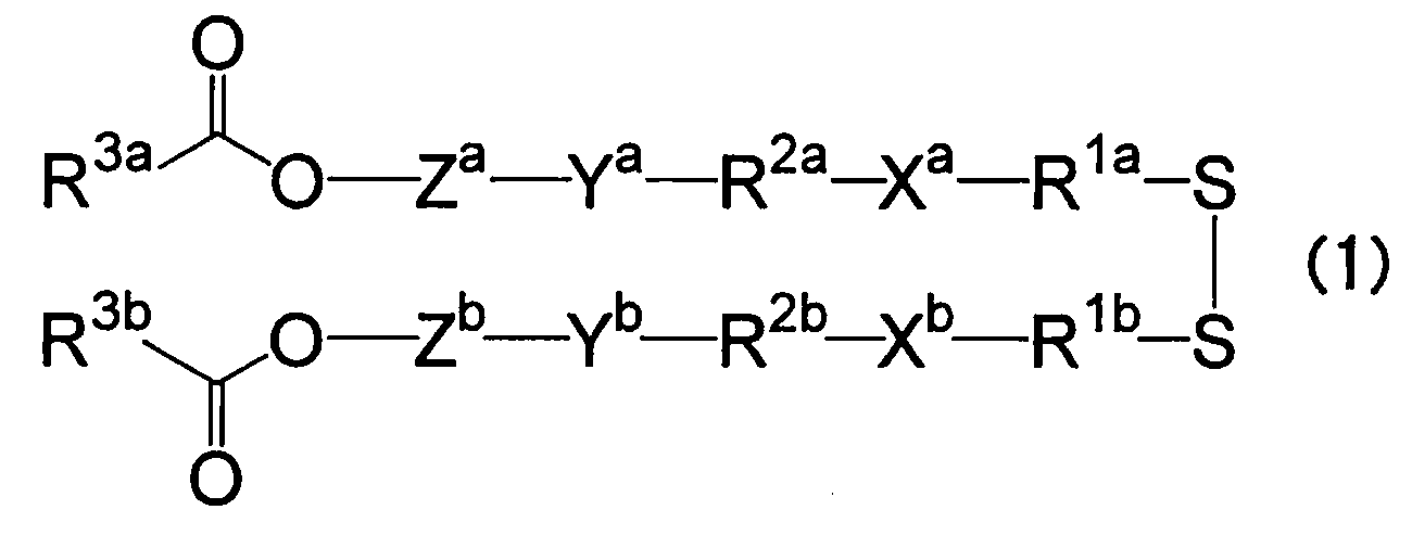
(式(1)中、
R1a及びR1bはそれぞれ独立して、炭素数1~6のアルキレン基を表し、
Xa及びXbはそれぞれ独立して、炭素数が1~6であり、かつ3級アミノ基の数が1の非環状のアルキル3級アミノ基、又は炭素数が2~5であり、かつ3級アミノ基の数が1~2の環状のアルキレン3級アミノ基を表し、
R2a及びR2bはそれぞれ独立して、炭素数8以下のアルキレン基又はオキシジアルキレン基を表し、
Ya及びYbはそれぞれ独立して、エステル結合、アミド結合、カーバメート結合、エーテル結合又はウレア結合を表し、
Za及びZbはそれぞれ独立して、炭素数が3~16であり、少なくとも1つの芳香環を有し、かつヘテロ原子を有していてもよい芳香族化合物から誘導される2価の基を表し、
R3a及びR3bはそれぞれ独立して、水酸基を有する脂溶性ビタミンとコハク酸無水物又はグルタル酸無水物との反応物由来の残基、又は水酸基を有するステロール誘導体とコハク酸無水物又はグルタル酸無水物との反応物由来の残基、又は炭素数12~22の脂肪族炭化水素基を表す。)
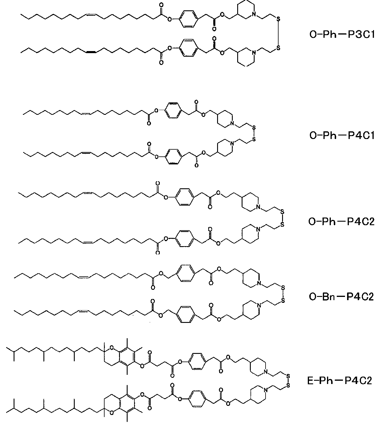
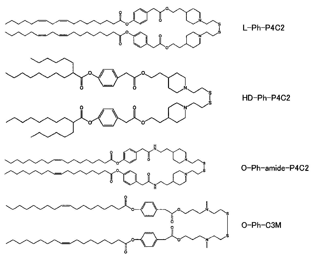
脂質としては、後述する実施例に記載されている化合物、すなわち、イオン化可能特性、還元条件応答特性、及び自己分解性特性を有する脂質としてビス[[4-[2-[[4-(オレオイルオキシ)フェニル]アセトキシ]エチル]ピペリジノ]エチル]ジスルファン(SS-OP)、中性リン脂質として1,2-ジオレオイル-sn-グリセロ-3-ホスホエタノールアミン(DOPE)、PEG脂質としてDMG-PEG5k、コレステロールとしてβ―シトステロールから選択される一つ以上の脂質などが好適に使用され得る。
本開示の脂質ナノ粒子を製造するにあたり、1つまたは2つ以上の両親媒性の物質を用いられる。両親媒性物質は、脂質ナノ粒子の安定性や、均一性などを考慮して選択される。本開示に用いられる両親媒性の例としては、ホスホグリセリド;ホスファチジルコリン;ジパルミトイルホスファチジルコリン(DPPC);ジオレイルホスファチジルエタノールアミン(DOPE);ジオレイルオキシプロピルトリエチルアンモニウム(DOTMA);ジオレオイルホスファチジルコリン;コレステロール;コレステロールエステル;ジアシルグリセロール;ジアシルグリセロールスクシネート;ジホスファチジルグリセロール(DPPG);ヘキサンデカノール;ポリエチレングリコール(PEG)などの脂肪族アルコール;ポリオキシエチレン-9-ラウリルエーテル;パルミチン酸またはオレイン酸などの表面活性脂肪酸;脂肪酸;脂肪酸モノグリセリド;脂肪酸ジグリセリド;脂肪酸アミド;ソルビタントリオレエート(Span(登録商標)85)グリココレート;ソルビタンモノラウレート(Span(登録商標)20);ポリソルベート20(Tween(登録商標)20);ポリソルベート60(Tween(登録商標)60);ポリソルベート65(Tween(登録商標)65);ポリソルベート80(Tween(登録商標)80);ポリソルベート85(Tween(登録商標)85);ポリオキシエチレンモノステアレート;サーファクチン;ポロキソマー(poloxomer);ソルビタントリオレエートなどのソルビタン脂肪酸エステル;レシチン;リゾレシチン;ホスファチジルセリン;ホスファチジルイノシトール;スフィンゴミエリン;ホスファチジルエタノールアミン(ケファリン);カルジオリピン;ホスファチジン酸;セレブロシド;ジセチルホスフェート;ジパルミトイルホスファチジルグリセロール;ステアリルアミン;ドデシルアミン;ヘキサデシルアミン;アセチルパルミテート;グリセロールリシノレエート;ヘキサデシルステアレート;イソプロピルミリステート;チロキサポール;ポリ(エチレングリコール)5000-ホスファチジルエタノールアミン;ポリ(エチレングリコール)400-モノステアレート;リン脂質;高い界面活性物質活性を有する合成および/または天然洗剤;デオキシコレート;シクロデキストリン;カオトロピック塩;イオンペア剤;および、これらの組み合わせが挙げられるが、これらに限定されない。他の添加剤として、脂質ナノ粒子の膜の安定性のために寄与する物質を加えることができる。そのような物質としては、コレステロール、フィトステロール、スティグマステロール、β-シトステロールを挙げることができる。
脂質ナノ粒子は、後述する製造方法にて製造することができる。脂質ナノ粒子は、抗体または抗体フラグメント等を含むタンパク質とポリマーとが会合して形成された液滴が、脂質内に内封された構造を有している。また、抗体または抗体フラグメント等を含むタンパク質が反対電荷を有するポリマーと静電結合を伴って安定的に会合し液滴を形成している。抗体または抗体フラグメント等を含むタンパク質とポリマーとの会合の形態は、特定の理論に拘束されるものではないが、タンパク質が反対電荷を有するポリマーと静電的に結合している形態や、タンパク質が反対電荷を有するポリマーと静電的に結合しポリマー内に安定的に包接される形態が一例として想定され得る。脂質ナノ粒子の粒径は、動的光散乱法を用いた粒度分布計や散乱光とブラウン運動の両方の特性を利用したナノ粒子トラッキング解析計等を用いて測定することができる。本開示における脂質ナノ粒子の粒子径は、ナノ粒子トラッキング解析により決定され、10~600nmの範囲で製造される。製造条件の調整により50~500nm、70~400nmでの範囲でも製造され得る。ナノ粒子トラッキング解析で決定される、平均粒径分布は300nm以下である。そのピークトップは150nm以下で製造され、製造条件の調整により120nm以下、または100nm以下でも製造され得る。粒径が100nmを下回るとエンドサイトーシスされやすいと言われている。さらに、腫瘍細胞などでは、100nmを下回るとEnhanced Permeability and Retention効果を通じてがん組織周辺に滞留し選択性が上がると言われている。
本開示の脂質ナノ粒子を医薬組成物として製剤化するために、脂質ナノ粒子を、様々な薬学的に許容される添加剤、並びに活性剤(複数可)の分散のための基剤又は担体と組み合わせることが可能である。添加剤の例としては、アルギニン、水酸化ナトリウム、グリシン、塩酸、クエン酸、及びこれらの混合物などのpH調整剤が挙げられる。他の添加剤としては、局所麻酔剤(例えば、ベンジルアルコール)、等張剤(例えば、塩化ナトリウム、マンニトール、ソルビトール)、吸着阻害剤(例えば、Tween 80)、溶解度増強剤(例えば、シクロデキストリン及びその誘導体)、安定剤(例えば、血清アルブミン)、及び還元剤(例えば、グルタチオン)が挙げられる。本開示の脂質ナノ粒子を含む医薬組成物は、活性剤及び任意の所望の添加剤を分散させる能力を有する親水性化合物を含み得る、基剤又はビヒクル中に分散され得る。基剤は、ポリカルボン酸又はその塩のコポリマー、カルボン酸無水物(例えば、無水マレイン酸)と他のモノマー(例えば、メチル(メタ)アクリレート、アクリル酸など)とのコポリマー、親水性ビニルポリマー、例えば、ポリ酢酸ビニル、ポリビニルアルコール、ポリビニルピロリドン、セルロース誘導体、例えば、ヒドロキシメチルセルロース、ヒドロキシプロピルセルロース等、並びに天然ポリマー、例えば、キトサン、コラーゲン、アルギン酸ナトリウム、ゼラチン、ヒアルロン酸、及びこれらの非毒性金属塩等を含むが、これらに限定されない広範な好適な担体から選択され得る。多くの場合、生分解性ポリマー、例えば、ポリ乳酸、ポリ(乳酸-グリコール酸)コポリマー、ポリヒドロキシ酪酸、ポリ(ヒドロキシ酪酸-グリコール酸)コポリマー、及びこれらの混合物は、基剤又は担体として選択される。基剤又は担体に代えて、若しくは基剤又は担体に加えて、ポリグリセリン脂肪酸エステル、スクロース脂肪酸エステル等の合成脂肪酸エステルを担体として用いることもできる。親水性ポリマー及び他の担体は、単独で又は組み合わせて使用することができ、部分的な結晶化、イオン結合、架橋等によって、増強された構造的一体性を担体に付与することができる。担体は、流体又は粘性溶液、ゲル、ペースト、粉末、微小球、及び鼻粘膜に直接適用するためのフィルムを含む様々な形態で提供され得る。この文脈における選択された担体の使用は、生物学的活性剤の吸収促進をもたらし得る。
脂質ナノ粒子は、J.Chem.Phys.150,064903(2019)などを参考にして公知の方法をもとに製造できる。
具体的には、まず抗体または抗体フラグメント等を含むタンパク質とポリマーとを適切な比率で混合して液滴を形成させる(液滴形成工程)。抗体または抗体フラグメント等を含むタンパク質とポリマーとの会合の形態は、特定の理論に拘束されるものではないが、タンパク質が反対電荷を有するポリマーと静電的に結合している形態や、タンパク質が反対電荷を有するポリマーと静電的に結合しポリマー内に静電的に結合し安定的に包接される形態等が、一例として想定され得る。混合は、液体混合機やピペッティングで混合する、混合容器中においてスターラーまたは攪拌翼で撹拌する、高速型衝突噴流(confined impinging jets)ミキサーやマルチインレットボルテックスミキサーを用いて混合する、マイクロ流体デバイスの流路(すなわち、マイクロ流路)上で接触させるなどの操作で行うことができる。ポリマーとタンパク質の比率(質量比)は、適宜設定することができるが、例えば、100:1~10:1の範囲内、さらには10:1~1:1の範囲内とすることができる。混合時の温度も適宜設定することができ、例えば15~40℃、または20℃から30℃の範囲内とすることができる。混合の際のpHは、タンパク質とポリマーが静電相互作用しうるpHであればよく、例えばタンパク質の等電点とポリマーの等電点の間のpHであることが好ましい。形成された液滴は、遠心分離、ろ過、または深層濾過等の工程を用いて分離され(例えば、特許5361738等)、適切なタンパク質の濃度の水溶液として再構成された液滴を次の内封工程に供与することが可能となる。
生成したナノ粒子は、in vitroおよびin vivoでの評価がなされる。本明細書では、本開示のナノ粒子とすることで、サイトゾルや核等を含む細胞内への局在が観察された。
(試薬)
pH応答性脂質であるss-cleavable and pH-activated lipid-like material(ssPalm)O-Phe(SS-OP 、 NOF Corporation)、1,2-dioleoyl-sn-glycero-3-phosphoethanolamine(DOPE、NOF Corporation)、β-sitosterol(Sigma Aldrich)、1,2-dimyristoyl-rac-glycero-3-methylpolyoxyethyleneglycol chain, molecular weight 5000(DMG-PEG5k、NOF Corporation)は10mMの濃度でクロロホルム(Fujifilm Wako Pure Chemical Corporation)に溶解し使用するまで-30度の冷凍庫にて保存した。
IgGは、正常ヒトIgG,全分子,精製品(富士フイルム和光純薬製)を用いた。本明細書においては、IgG、またはhIgGと表記する。
ポリグルタミン酸は、以下のサイズのものを用いた(mol wt3,000-15,000,sigma)。
粒径は、Zetasizer Nano ZS(Malvern社製)を使って粒子直径、PdI(粒径分布、ゼータ電位(ζ-電位)を評価した。回収したサンプルをPBSに100倍希釈して測定を実施した。
内包率は、調製したLNPを卓上超遠心機によって、4℃、20000gにて30分間遠心分離して、上清のタンパク質濃度を測定することで、タンパク質の内封率を算出した。すなわち、IgG_polyE-LNPをPBS(-)で抗体濃度が2μMとなるように調整した(最終サンプル量100μL)。これを4℃、20,000×g、1時間遠心分離することでLNP画分を沈殿させた。上清50μLを回収し、Nanodropにて抗体濃度を測定し、内封率を以下の式から逆算した。ただし、CIgGは2μM IgGを遠心した上清から算出したIgG濃度、CLNPはIgG_polyE-LNPを遠心した上清から算出したIgG濃度を指す。
IgGの内封率(%)=(CIgG-CLNP)/CIgG×100
ATCCから入手したHeLa細胞を10%牛血清(BS)含有α-MEMにて培養した。
透析膜には、2-14k MWCO透析膜(Spectrum Laboratories Inc.)を用いた。透析膜に充填した後に、Corning Stirrerにて攪拌して透析を実施した。
Alexa Fluor 488修飾のため、10mg/mL hIgG(正常ヒトIgG、Fujifilm Wako Pure Chemical Corporation)100μLにPBS(-)345μLを加え、そこに1M NaHCO3 50μLを添加し溶液を塩基性にした後、10mg/mL Alexa488-SDP ester(Thermo Fisher Scientific)と混合し、遮光下、25℃で1時間転倒混和した。その後PD10カラムにて抗体画分を分取し、Alexa Fluor 488標識hIgG(hIgG-AF488)を精製した。
抗GFP抗体の調製はExpi293TM Expression System(Thermo Fisher Scientific)にて行った。Expi293FTM細胞を3,000,000cells/mLとなるように準備し、125mL用三角フラスコに25mL加えた。Anti-GFP-IgGをコードするプラスミド(Addgene, Anti-GFP[N86/38.1R])を25μg、ExpiFectamineTM293(Thermo Fisher Scientific)と混合し、その推奨プロトコルに従いトランスフェクションした。トランスフェクション後の20時間後に、ExpiFectamineTM293 Transfectin Enhancer 1を150μLおよびEnhancer 2を1.5mL加え五日間インキュベートした。培養液を50mLチューブに移し、3,200×g、4℃、20分間遠心分離し、上清を回収した。抗体が含まれる上清を0.22μmのフィルターでろ過滅菌した。さらにこの溶液をHiTrap Protein A High Performance(Cytiva)に吸着させ、精製用緩衝液(0.1M Glycine HCl pH=2.7)によってカラムから溶出させた。IgGを含むフラクションを回収し、PBS(pH7.4)にて透析を行ない、抗GFP抗体(anti-GFP-IgG)を得た。回収した抗GFP抗体(anti-GFP-IgG)は4℃条件で保存した。クエン酸溶液(pH5)の抗体を用いる場合は、マイクロバイオスピン(登録商標) カラム30(BIORAD)を用いてクエン酸溶液(pH5)に置換した。
10mMクエン酸溶液(pH5)に溶解したポリ-L-グルタミン酸(polyE、mol wt 3,000-15,000)溶液を40℃の条件で30分間インキュベートした。その後、PBS(-)もしくはクエン酸溶液(pH5)に溶解されたIgG(抗体全て)と上記のpolyEをIgG:polyE=5:1(質量比)となるようにピペッティング10回で混合した。その後、12-14k MWCO透析膜(Spectrum Laboratories Inc.)に充填して、1,000倍以上の体積量のPBS(pH7.4)に浮かしてスターラーで攪拌して透析を実施した。
(LNPの製造)
脂質をクロロホルムで溶解し、20mM SS-OP、10mM DOPE、20mM β-sitosterol、10mM DMG-PEG5kを調製した。SS-OP/DOPE/β-sitosterol/DMG-PEG5k(モル比 45/10/45/1.5)の組成物をフラスコ内で室温にて混合し、1mLのt-BuOH(富士フィルム和光純薬 Fujifilm Wako Pure Chemical Corporation)を加えて溶解させた後、ロータリーエバポレーターにてクロロホルムを蒸発させ脂質溶液を得た。さらに、この脂質溶液を18から24時間凍結乾燥させ、その後t-BuOHで溶解させることで10mM脂質溶液を作製した。t-BuOHに溶解した脂質溶液と10mMクエン酸溶液(pH5)に溶解したポリグルタミン酸溶液(0.75mg/mLポリグルタミン酸溶液(polyE、mol wt 3,000-15,000)溶液を、別々に40度の条件で30分間インキュベートした。
(LNPの製造)
polyEを含まずにhIgGのみを用いた以外は、製造例1と同様に行いLNP化したサンプル(hIgG-LNP)を製造した。
実施例1で調製したhIgG_polyE―LNPと、比較例1で調整したhIgG-LNPをZetasizer Nano ZS(Malvern)を使って粒子直径、PdI、ζ-電位を評価した。結果を、図10a,図10bおよび表1に示す。調製したLNPを卓上超遠心機によって、4℃、20000gにて30分間遠心分離して、上清の抗体濃度を測定することで、抗体の内封率を算出した。結果を表1に示す。
結果を表1に示す。
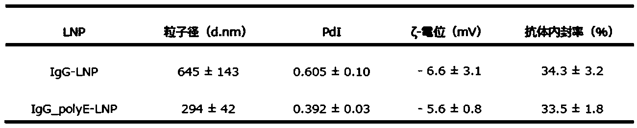
また、NTA法での粒子径の測定に当たっては、PBSにて100倍に希釈した上記LNPをNanoSight NS300を用いて測定した。その結果、粒子径は123±2nmであった。
(LNPの製造)
実施例1において、tBuOHの代わりに90%tBuOH水溶液を用いた以外は実施例1と同様な操作を行い、hIgG_polyE-LNPを製造した。
(効果)
(LNPの製造)
抗体と脂質の混合比によりhIgGのサイトゾルへの送達活性が変化するかどうか検討するために、IgGの代わりにhIgG-AF488を用いた以外は(実施例1)と同様な操作を行い、抗体:脂質比が(a)~(e)の混合比のIgG_polyE-LNP(a)~(e)を調整した。
hIgG-AF488:polyE
(a) 1:100
(b) 1:200
(c) 1:300
(d) 1:400
(e) 1:500
(各種LNPの製造)
(抗GFP抗体_polyE―LNP)
実施例1で、hIgGの代わりに抗GFP抗体を用いた以外は同様の操作を行い、抗GFP抗体_polyE―LNPを製造した。
HeLa細胞に(a)hIgG-AF488、(b)hIgG-AF488_PolyE、(c)hIgG-AF488_PolyE-LNPを1μMとなるように血清培地に添加し、37℃、5%CO2の条件下、18時間インキュベーションし、IgGの細胞内局在を観察した(図2A)。
polyEを添加することの有用性を示すために、polyEを含まないhIgGと脂質を混合し得られるLNP(IgG-LNP)(参考例1)を調製し、hIgG_polyE-LNP(実施例1)との比較試験を実施した。細胞内局在を観察するために、hIgGは、hIgG-AF488を用いた。
LgBiTとHiBiTは会合することでナノルシフェラーゼnanoLuciferaseを再構成し、ルシフェリンLuciferinを添加すると発光を呈する。この技術を利用してhIgG_polyE-LNP(製造例1)とhIgG-LNP(製造例2)のサイトゾル移行量を比較した。
次に、サイトゾルに存在するタンパク質を標的とするIgGをLNPにより送達することで、その分子を認識できるかどうか検討した。
HRasはRasタンパク質ファミリーの一種で、細胞膜に局在するタンパク質であり、これにGFPを融合したHRas-GFPを発現するHeLa細胞を用いることでGFPを細胞膜に局在させた。Alexa Fluor 594で標識したanti-GFP-IgG(Addgene; Anti-GFP[N86/38.1R]、Plasmid #114492)(抗GFP抗体-AF594)を作製し、LNP化し、HRas-GFP発現HeLa細胞にIgG濃度にして1μMとなるように血清培地に懸濁後、18時間投与を行い、IgGの細胞内局在を観察した。HRas-GFPが存在する細胞膜付近においてGFPとanti-GFP-IgG-AF594の蛍光シグナルの共局在が確認された(図5A)。
次に、内在性タンパク質を認識する抗体を導入するため、核膜孔複合体(nuclearpore complexes,NPC)に対する抗体anti-NPC-IgG(Mab414、Cat#: 902902、BioLegend)導入した。HeLa細胞にhIgG-AF488_polyE-LNPまたはhIgG-AF488_polyEサンプルをIgG濃度にして1μMとなるように血清培地に懸濁し、18時間投与を行った。その後、免疫染色を行い、IgGの細胞内局在を観察した。hIgG-AF488_polyEを添加した細胞では、点状のシグナルが確認されるのみであったが、hIgG-AF488_polyE-LNP添加において、核周囲にIgGのシグナルがリング状に確認された(yellow arrow heads)(図6A)。また、核を横切るようにラインプロットを作製したところ、hIgG-AF488_polyE droplet添加では、核シグナル(シアン色)から離れた位置に抗体シグナル(マゼンタ色)が確認されるのに対し、hIgG-AF488_polyE-LNP処理では核シグナルの両端と抗体シグナルが隣接ように検出された(図6B)。さらに、各サンプルで400個程度の細胞を観察し、核周囲に抗体由来のシグナルがリング状に観察される細胞をカウントし、その割合を算出したところ、IgG_polyE-LNP処理によって約20%の細胞に抗NPC抗体を核膜孔に送達でき蛍光イメージングできることが確認された(図6C)。
細胞内のタンパク質を認識する活性のみでなく、認識したタンパク質の活性を阻害することで細胞機能を発揮するIgGの送達を実施した。Aktはがん細胞においてアポトーシスを誘導するタンパク質の活性を抑制している。Aktの活性体であるリン酸化Akt(pAkt)を認識し、その機能を阻害するIgG(anti-pAKT1-IgG)(Cat#:700392、Invitrogen)を、HeLa細胞に導入することで、細胞のアポトーシスを誘発できることが報告されている。そこで、anti-pAkt1-IgGをLNPによりサイトゾルへ送達し、アポトーシスを誘導できるか検討した。
HeLa細胞にコントロールIgGとしてhIgGおよびanti-pAkt1-IgGを内封するIgG_polyE-LNPをhIgG濃度にして1μMとなるように血清培地にそれぞれ懸濁し、24時間投与を行った。その後、Caspase-Glo(登録商標) 3/7 Assay試薬(Promega)を加え1時間室温でインキュベート後、発光量を定量したところ、hIgGと比較して、抗pAkt1抗体(anti-pAkt1-IgG)を送達した群では、有意にアポトーシスを誘導されたことが確認された(図7A)。また、細胞生存率をWST-8 assayにより同様の実験条件で検討を行ったところ、抗pAkt1抗体(anti-pAkt1-IgG)を送達するLNP添加を行った群において有意な細胞増殖阻害効果を得た(図7B)。以上の結果から、hIgG_polyE-LNPはタンパク質の活性を阻害可能なIgGをサイトゾルにまで導入でき、細胞機能を制御できることが判明した。
IgG_polyE-LNPの汎用性についてHeLa細胞以外にHT1080細胞、MDA-MB-231細胞、SW480細胞を用いて検討した。hIgG-AF488を内封したIgG_polyE-LNPをIgG濃度にして1μMとなるように血清培地にそれぞれ懸濁し、上記細胞に投与して24時間インキュベートして共焦点顕微鏡にて観察したところ、HT1080細胞では効率良くサイトゾルにhIgGAlexa488が送達されていた(図8)。MDA-MB-231細胞およびSW480細胞では効率は高くないながらも、サイトゾルへの拡散が確認された(図8)。よって、hIgG_polyE-LNPはHeLa細胞以外のがん細胞においてもサイトゾル送達可能であることが判明した。
脂質溶液における溶媒を検討するために凍結乾燥後の脂質を90%t-BuOHに溶解した。脂質溶液と10mMクエン酸溶液(pH5)に溶解した0.75mg/mL poly-L-glutamic acid(polyE、mol wt 3,000-15,000)溶液を別々に40度の条件で30分間インキュベートした。その後、3μLのhIgG(10mg/mL)を8μLのpolyEに添加して[IgG:polyE=5:1(質量比)]、29.8μLのクエン酸溶液を添加して、ピペッティング10回で混合し、hIgG-polyE液滴を形成した。90% t-BuOHで4mMに調製し40℃で30分インキュベートした脂質溶液と液滴溶液を1:4(体積比)となるようにピペッティング15回で混合した。その後、1,000倍以上の体積量のPBS(-)に対して12-14k MWCO透析膜(Spectrum Laboratories Inc.)で透析し、IgGを内包するLipid Nanoparticle(hIgG_polyE-LNP)を調製した。これらをHeLa細胞に添加して18時間後に固定化・膜透過処理して、Alexa488標識されたanti-human IgG抗体にて免疫染色を実施して、共焦点顕微鏡によって細胞を観察したところ、90% t-BuOH脂質溶液によって調製したhIgG_polyE-LNPもt-BuOH脂質溶液で調製したhIgG_polyE-LNPと同様にサイトゾルに抗体を送達可能であることが判明した(図9)。
マイクロ流体デバイスを用いてLipid Nanoparticle(hIgG_polyE-LNP)の調製を検討した。
Human IgG(10mg/mL)とpolyE(0.75 mg/mL)を5:1(w/w)になるように10mM MES buffer(pH5)に溶解し、液滴を形成させ、1mLシリンジ(テルモ)に充填した。
SS-OP:DOPE:β-sitosterol=45:10:45の混合液に1.5mol%PEG5kとなるようにt-BuOHで調製した8mMもしくは16mMの脂質溶液を別の1mLシリンジ(テルモ)に充填した。これらをシリンジポンプ(kd scientific)にセットして各流速で混合した後、同混合液をPBSにて終夜透析した。各サンプルの条件を以下の表に示す。

上記のようにマイクロ流体デバイスの混合条件を変えることで所望の特性を示すLipid Nanoparticle(hIgG_polyE-LNP)を作製可能であることが示された。
分子量の異なるPolyEと抗体を含む液滴を用いて調製されたLipid Nanoparticle(hIgG_polyE-LNP)の細胞質内への送達効率を確認した。10mg/mLのIgG4μLを0.375mg/mL、0.75mg/mL、1.5mg/mL、3.0mg/mLの3-15k、39k、2.6kの分子量を有する各ポリグルタミン酸溶液8μLと混合した後、10mMクエン酸緩衝液(pH5)で40μLに調製(Fill-up)した後に懸濁し、さらに8mMの脂質溶液10μLを添加し、PBSで終夜透析してLipid Nanoparticle(hIgG_polyE-LNP)を作製した。
上記のようにポリマーの重合度を検討することで所望の特性を示すLipid Nanoparticle(hIgG_polyE-LNP)を作製可能であることが示された。
PolyE以外のアニオン性ポリマー、具体的には分子量約1,000,000(重合度概算で約2500)のヒアルロン酸と抗体を含む液滴を用いて調製されたLipid Nanoparticle(hIgG_polyE-LNP)の細胞質内への送達効率を確認した。10mg/mLのIgG3μLを0.09375mg/mL、0.1875mg/mL、0.375mg/mL、0.75mg/mL、1.5mg/mLの各ヒアルロン酸溶液8μLと混合したところ、ヒアルロン酸:IgG(w/w)を1:40~1:5となるように調製された混合液中で、液滴の形成が検鏡下で確認された(図17)。
透析液に血清含有培地を添加し200μLに調製(Fill-up)したLipid Nanoparticle(hIgG_polyE-LNP)でHeLa細胞を37℃で18時間処理した後、PBSを用いて洗浄された細胞を4%ホルムアルデヒドで固定し0.1%Triton X-100で膜透過処理を施した。anti-Human IgG-Alexa488によって免疫染色をおこなったところ、Lipid Nanoparticle(hIgG_polyE-LNP)に含まれるIgGがサイトゾルに送達されていることが確認された(図18)。
核内に存在する標的分子に結合し得る抗体のC末端に核移行シグナル配列(PAAKRVKLD)を付加した遺伝子を発現させて核内分子特異的抗体(IgG-NLS)を調製する。10mg/mLのIgG-NLSを3μLおよび各濃度の (0.375mg/mL、0.75mg/mL、1.5mg/mL、又は3.0mg/mL濃度のポリグルタミン酸溶液8μLを混合した後、10mMクエン酸緩衝液(pH5)で40μLに調製(Fill-up)した後に懸濁し、さらに8mMの脂質溶液10μLを添加し、PBSで終夜透析してLipid Nanoparticle(IgG-NLS_polyE-LNP)を作製する。
Claims (11)
- 抗体および抗体フラグメントから選択されるタンパク質と、ポリマーと、脂質と、を含む組成物であって、所定のpHにおいて前記タンパク質と前記ポリマーとが反対の電荷を持つことを特徴とする組成物からなる脂質ナノ粒子。
- 前記脂質ナノ粒子は、前記タンパク質が封入されている、請求項1記載の脂質ナノ粒子。
- 前記脂質ナノ粒子の粒径を測定したときに粒径分布のピークトップが80nmから120nmである、請求項1記載の脂質ナノ粒子。
- 前記ポリマーが、アニオン性またはカチオン性のポリマーである、請求項1記載の脂質ナノ粒子。
- 前記カチオン性ポリマーが、ポリリジン、ポリアルギニン、ポリヒスチジンおよびこれらの水溶性塩からなる群から選択される少なくとも1つである、請求項4記載の脂質ナノ粒子。
- 前記アニオン性ポリマーが、ポリグルタミン酸、ポリアスパラギン酸からなる群より選択される少なくとも1つのポリアミノ酸、またはヒアルロン酸、プルランからなる群より選択される少なくとも1つのグリコサミノグリカンである、請求項4記載の脂質ナノ粒子。
- 前記カチオン性または前記アニオン性ポリマーの分子量が、0.5kDa~1000kDaである請求項4記載の脂質ナノ粒子。
- 前記脂質がカチオン性脂質である、請求項1記載の脂質ナノ粒子。
- さらに中性リン脂質、PEG脂質、およびコレステロールからなる群より選択される一以上の脂質を含む請求項1から8のいずれか1項に記載の脂質ナノ粒子。
- 抗体および抗体フラグメントから選択されるタンパク質と、ポリマーと、脂質と、を含む組成物であって、所定のpHにおいて前記タンパク質と前記ポリマーとが反対の電荷を持つことを特徴とする組成物からなる医薬組成物。
- 前記抗体および抗体フラグメントから選択されるタンパク質と、前記ポリマーとを混合して液滴を形成させる液滴形成工程と、
前記液滴と前記脂質とを混合して前記脂質に前記液滴を内封させる内封工程と、を備えることを特徴とする脂質ナノ粒子の製造方法。
Priority Applications (3)
| Application Number | Priority Date | Filing Date | Title |
|---|---|---|---|
| JP2025504021A JPWO2024181580A1 (ja) | 2023-03-02 | 2024-03-04 | |
| CN202480016036.0A CN120826236A (zh) | 2023-03-02 | 2024-03-04 | 脂质纳米粒子、医药组合物以及脂质纳米粒子的制造方法 |
| EP24764057.6A EP4674431A1 (en) | 2023-03-02 | 2024-03-04 | Lipid nanoparticles, pharmaceutical composition, and production method for lipid nanoparticles |
Applications Claiming Priority (2)
| Application Number | Priority Date | Filing Date | Title |
|---|---|---|---|
| JP2023-031510 | 2023-03-02 | ||
| JP2023031510 | 2023-03-02 |
Publications (1)
| Publication Number | Publication Date |
|---|---|
| WO2024181580A1 true WO2024181580A1 (ja) | 2024-09-06 |
Family
ID=92589996
Family Applications (1)
| Application Number | Title | Priority Date | Filing Date |
|---|---|---|---|
| PCT/JP2024/008154 Ceased WO2024181580A1 (ja) | 2023-03-02 | 2024-03-04 | 脂質ナノ粒子、医薬組成物、および脂質ナノ粒子の製造方法 |
Country Status (4)
| Country | Link |
|---|---|
| EP (1) | EP4674431A1 (ja) |
| JP (1) | JPWO2024181580A1 (ja) |
| CN (1) | CN120826236A (ja) |
| WO (1) | WO2024181580A1 (ja) |
Citations (7)
| Publication number | Priority date | Publication date | Assignee | Title |
|---|---|---|---|---|
| US4816567A (en) | 1983-04-08 | 1989-03-28 | Genentech, Inc. | Recombinant immunoglobin preparations |
| JP2010536786A (ja) * | 2007-08-17 | 2010-12-02 | アムジエン・インコーポレーテツド | ポリカチオンを用いた抗体およびfc融合分子の製剤 |
| JP5361738B2 (ja) | 2007-01-22 | 2013-12-04 | ジェネンテック, インコーポレイテッド | 多価電解質沈殿物及びタンパク質の精製 |
| WO2015064591A1 (ja) | 2013-10-28 | 2015-05-07 | テルモ株式会社 | タンパク質水性懸濁剤 |
| WO2019188867A1 (ja) | 2018-03-27 | 2019-10-03 | 日油株式会社 | 細胞内動態を改善した新規カチオン性脂質 |
| JP2021519748A (ja) * | 2018-01-29 | 2021-08-12 | ザ・ジョンズ・ホプキンス・ユニバーシティ | タンパク質治療薬をカプセル製剤化および徐放性製剤化を可能にするポリマーナノ粒子組成物 |
| JP2022122871A (ja) | 2013-05-03 | 2022-08-23 | セレクタ バイオサイエンシーズ インコーポレーテッド | 特定の薬力学的有効期間および免疫寛容の誘発のための抗原を有する免疫抑制剤の送達 |
-
2024
- 2024-03-04 JP JP2025504021A patent/JPWO2024181580A1/ja active Pending
- 2024-03-04 WO PCT/JP2024/008154 patent/WO2024181580A1/ja not_active Ceased
- 2024-03-04 EP EP24764057.6A patent/EP4674431A1/en active Pending
- 2024-03-04 CN CN202480016036.0A patent/CN120826236A/zh active Pending
Patent Citations (7)
| Publication number | Priority date | Publication date | Assignee | Title |
|---|---|---|---|---|
| US4816567A (en) | 1983-04-08 | 1989-03-28 | Genentech, Inc. | Recombinant immunoglobin preparations |
| JP5361738B2 (ja) | 2007-01-22 | 2013-12-04 | ジェネンテック, インコーポレイテッド | 多価電解質沈殿物及びタンパク質の精製 |
| JP2010536786A (ja) * | 2007-08-17 | 2010-12-02 | アムジエン・インコーポレーテツド | ポリカチオンを用いた抗体およびfc融合分子の製剤 |
| JP2022122871A (ja) | 2013-05-03 | 2022-08-23 | セレクタ バイオサイエンシーズ インコーポレーテッド | 特定の薬力学的有効期間および免疫寛容の誘発のための抗原を有する免疫抑制剤の送達 |
| WO2015064591A1 (ja) | 2013-10-28 | 2015-05-07 | テルモ株式会社 | タンパク質水性懸濁剤 |
| JP2021519748A (ja) * | 2018-01-29 | 2021-08-12 | ザ・ジョンズ・ホプキンス・ユニバーシティ | タンパク質治療薬をカプセル製剤化および徐放性製剤化を可能にするポリマーナノ粒子組成物 |
| WO2019188867A1 (ja) | 2018-03-27 | 2019-10-03 | 日油株式会社 | 細胞内動態を改善した新規カチオン性脂質 |
Non-Patent Citations (19)
| Title |
|---|
| "Intracytoplasmic delivery of anionic proteins", MOL THER., vol. 9, no. 6, June 2004 (2004-06-01), pages 964 - 9 |
| CELL, vol. 185, no. 15, 2022, pages 2789 - 2805 |
| CHATIN BMEVEL MDEVALLIERE J ET AL.: "Liposome-based formulation for intracellular delivery of functional proteins", MOL THER NUCLEIC ACIDS. |
| CLACKSON E, NATURE, vol. 352, 1991, pages 624 - 628 |
| HIRAI YUSUKE, HIROSE HISAAKI, IMANISHI MIKI, ASAI TOMOHIRO, FUTAKI SHIROH: "Cytosolic protein delivery using pH-responsive, charge-reversible lipid nanoparticles", SCIENTIFIC REPORTS, NATURE PUBLISHING GROUP, US, vol. 11, no. 1, US , XP093205049, ISSN: 2045-2322, DOI: 10.1038/s41598-021-99180-5 * |
| HIRAI YUSUKE, KAWAGUCHI YOSHIMASA, KASAHARA CHISATO, HIROSE HISAAKI, FUTAKI SHIROH: "Liquid Droplet-Mediated Formulation of Lipid Nanoparticles Encapsulating Immunoglobulin G for Cytosolic Delivery", MOLECULAR PHARMACEUTICS, AMERICAN CHEMICAL SOCIETY, US, vol. 21, no. 4, 1 April 2024 (2024-04-01), US , pages 1653 - 1661, XP093205060, ISSN: 1543-8384, DOI: 10.1021/acs.molpharmaceut.3c00868 * |
| JOURNAL OF BIOMEDICAL SCIENCE, vol. 27 |
| JOURNAL OF CHROMATOGRAPHY B, vol. 1065, no. 1066, 2017, pages 119 - 128 |
| KOHLERMILSTEIN, NATURE, vol. 256, 1975, pages 495 |
| LU TIEMEI, LIESE SUSANNE, SCHOENMAKERS LUDO, WEBER CHRISTOPH A., SUZUKI HIROAKI, HUCK WILHELM T. S., SPRUIJT EVAN: "Endocytosis of Coacervates into Liposomes", JOURNAL OF THE AMERICAN CHEMICAL SOCIETY, AMERICAN CHEMICAL SOCIETY, vol. 144, no. 30, 3 August 2022 (2022-08-03), pages 13451 - 13455, XP093205052, ISSN: 0002-7863, DOI: 10.1021/jacs.2c04096 * |
| MARKS ET AL., J. MOL. BIOL., vol. 222, 1991, pages 581 - 597 |
| MASHIRO MIMURA, J. CHEM. PHYS., vol. 150, pages 064903 |
| MOL. PHARMACEUTICS, vol. 12, no. 9, 2015, pages 3272 - 3281 |
| MOL. PHARMACEUTICS, vol. 13, 2016, pages 1915 - 1926 |
| N. BIOTECHNOL., vol. 28, no. 5, 2011, pages 253 - 457 |
| See also references of EP4674431A1 |
| TSUMURA KEISUKE; HSU WEILIN; MIMURA MASAHIRO; HORIUCHI AIKO; SHIRAKI KENTARO: "Lowering the viscosity of a high-concentration antibody solution by protein–polyelectrolyte complex", JOURNAL OF BIOSCIENCE AND BIOENGINEERING, ELSEVIER, AMSTERDAM, NL, vol. 133, no. 1, 8 October 2021 (2021-10-08), NL , pages 17 - 24, XP086925102, ISSN: 1389-1723, DOI: 10.1016/j.jbiosc.2021.09.011 * |
| YUSUKE HIRAI, HISAAKI HIROSE, MIKI IMANISHI; TOMOHIRO ASAI, SHIROH FUTAKI: "29V09-pm03 Intracellular delivery of negatively charged protein using lipid-based carrier", ABSTRACTS OF THE 141ST ANNUAL MEETING OF THE PHARMACEUTICAL SOCIETY OF JAPAN, MARCH 26-29, 2021, ONLINE, PHARMACEUTICAL SOCIETY OF JAPAN, JP, 26 March 2021 (2021-03-26) - 29 March 2021 (2021-03-29), JP, pages 29V09 - pm03S, XP009559593 * |
| ZHANG YANWEN, CHEN YUFENG, YANG XIAOHAI, HE XIAOXIAO, LI MEI, LIU SONGYANG, WANG KEMIN, LIU JIANBO, MANN STEPHEN: "Giant Coacervate Vesicles As an Integrated Approach to Cytomimetic Modeling", JOURNAL OF THE AMERICAN CHEMICAL SOCIETY, AMERICAN CHEMICAL SOCIETY, vol. 143, no. 7, 24 February 2021 (2021-02-24), pages 2866 - 2874, XP093205055, ISSN: 0002-7863, DOI: 10.1021/jacs.0c12494 * |
Also Published As
| Publication number | Publication date |
|---|---|
| JPWO2024181580A1 (ja) | 2024-09-06 |
| EP4674431A1 (en) | 2026-01-07 |
| CN120826236A (zh) | 2025-10-21 |
Similar Documents
| Publication | Publication Date | Title |
|---|---|---|
| Rome et al. | Development of the vault particle as a platform technology | |
| CN116496193B (zh) | 一种氨基酸脂质形成的纳米递送系统及其应用 | |
| JPWO2007037444A1 (ja) | 目的物質を核内又は細胞内に送達するためのベクター | |
| JP2017502997A (ja) | ハイブリドソーム、それを含む組成物、それらの製造方法、およびその使用 | |
| JP2023543623A (ja) | 融合関連小膜貫通タンパク質が配合されたプロテオリピド小胞 | |
| CN103037840A (zh) | 具有核内转运性的脂质膜结构体 | |
| CN1863558A (zh) | 具有向核内转移能力的聚精氨酸修饰的脂质体 | |
| Kalra et al. | Virosomes: as a drug delivery carrier | |
| US20100166840A1 (en) | Liposome having lipid membrane containing bacterial cell component | |
| Zhang et al. | Macropinocytosis is the major pathway responsible for DNA transfection in CHO cells by a charge-reversal amphiphile | |
| Jing et al. | Delivery of siRNA complexed with palmitoylated α-peptide/β-peptoid cell-penetrating peptidomimetics: membrane interaction and structural characterization of a lipid-based nanocarrier system | |
| US20090305409A1 (en) | Liposome Capable of Effective Delivery of Given Substance Into Nucleus | |
| US20150140066A1 (en) | Carrier for intracellular delivery of functional protein | |
| KR101916941B1 (ko) | 플라스미드 디엔에이 전달용 고분자 나노입자 조성물 및 그의 제조방법 | |
| EP4442256A1 (en) | Lipid nanoparticle compositions | |
| Binici et al. | A comparative study of cationic lipid-enriched LNPs for mRNA vaccine delivery | |
| ES2640060T3 (es) | Proceso para producir una partícula fina revestida | |
| JP2006028030A (ja) | リポソーム封入物質がエンドソームから脱出可能なリポソーム | |
| JP5067733B2 (ja) | 目的物質をミトコンドリア内に送達可能な脂質膜構造体 | |
| KR20220117133A (ko) | 양이온성 분자 수송체 및 SARS-CoV-2 mRNA의 이온 복합체를 포함하는 코로나바이러스감염증-19 예방 백신 조성물 | |
| WO2024181580A1 (ja) | 脂質ナノ粒子、医薬組成物、および脂質ナノ粒子の製造方法 | |
| US9937127B2 (en) | Method of producing fine particles surface-modified with water-soluble substance | |
| US20090010999A1 (en) | Complex Particles and Coated Complex Particles | |
| Sun et al. | Enhanced in vivo gene expression mediated by listeriolysin O incorporated anionic LPDII: Its utility in cytotoxic T lymphocyte-inducing DNA vaccine | |
| Wang et al. | Spleen Targeting Nucleic Acid Delivery Vector Based on Metal–Organic Frameworks |
Legal Events
| Date | Code | Title | Description |
|---|---|---|---|
| 121 | Ep: the epo has been informed by wipo that ep was designated in this application |
Ref document number: 24764057 Country of ref document: EP Kind code of ref document: A1 |
|
| ENP | Entry into the national phase |
Ref document number: 2025504021 Country of ref document: JP Kind code of ref document: A |
|
| WWE | Wipo information: entry into national phase |
Ref document number: 2025504021 Country of ref document: JP |
|
| WWE | Wipo information: entry into national phase |
Ref document number: 202480016036.0 Country of ref document: CN |
|
| WWE | Wipo information: entry into national phase |
Ref document number: 2024764057 Country of ref document: EP |
|
| NENP | Non-entry into the national phase |
Ref country code: DE |
|
| WWP | Wipo information: published in national office |
Ref document number: 202480016036.0 Country of ref document: CN |
|
| ENP | Entry into the national phase |
Ref document number: 2024764057 Country of ref document: EP Effective date: 20251002 |
|
| ENP | Entry into the national phase |
Ref document number: 2024764057 Country of ref document: EP Effective date: 20251002 |
|
| ENP | Entry into the national phase |
Ref document number: 2024764057 Country of ref document: EP Effective date: 20251002 |
|
| WWP | Wipo information: published in national office |
Ref document number: 2024764057 Country of ref document: EP |