CN102725006A - Devices and methods for tissue engineering - Google Patents
Devices and methods for tissue engineering Download PDFInfo
- Publication number
- CN102725006A CN102725006A CN2010800448963A CN201080044896A CN102725006A CN 102725006 A CN102725006 A CN 102725006A CN 2010800448963 A CN2010800448963 A CN 2010800448963A CN 201080044896 A CN201080044896 A CN 201080044896A CN 102725006 A CN102725006 A CN 102725006A
- Authority
- CN
- China
- Prior art keywords
- fiber
- fibers
- scaffold
- tissue
- tissue support
- Prior art date
- Legal status (The legal status is an assumption and is not a legal conclusion. Google has not performed a legal analysis and makes no representation as to the accuracy of the status listed.)
- Granted
Links
Images




Classifications
-
- A—HUMAN NECESSITIES
- A61—MEDICAL OR VETERINARY SCIENCE; HYGIENE
- A61F—FILTERS IMPLANTABLE INTO BLOOD VESSELS; PROSTHESES; DEVICES PROVIDING PATENCY TO, OR PREVENTING COLLAPSING OF, TUBULAR STRUCTURES OF THE BODY, e.g. STENTS; ORTHOPAEDIC, NURSING OR CONTRACEPTIVE DEVICES; FOMENTATION; TREATMENT OR PROTECTION OF EYES OR EARS; BANDAGES, DRESSINGS OR ABSORBENT PADS; FIRST-AID KITS
- A61F2/00—Filters implantable into blood vessels; Prostheses, i.e. artificial substitutes or replacements for parts of the body; Appliances for connecting them with the body; Devices providing patency to, or preventing collapsing of, tubular structures of the body, e.g. stents
- A61F2/02—Prostheses implantable into the body
- A61F2/30—Joints
- A61F2/3094—Designing or manufacturing processes
-
- A—HUMAN NECESSITIES
- A61—MEDICAL OR VETERINARY SCIENCE; HYGIENE
- A61F—FILTERS IMPLANTABLE INTO BLOOD VESSELS; PROSTHESES; DEVICES PROVIDING PATENCY TO, OR PREVENTING COLLAPSING OF, TUBULAR STRUCTURES OF THE BODY, e.g. STENTS; ORTHOPAEDIC, NURSING OR CONTRACEPTIVE DEVICES; FOMENTATION; TREATMENT OR PROTECTION OF EYES OR EARS; BANDAGES, DRESSINGS OR ABSORBENT PADS; FIRST-AID KITS
- A61F2/00—Filters implantable into blood vessels; Prostheses, i.e. artificial substitutes or replacements for parts of the body; Appliances for connecting them with the body; Devices providing patency to, or preventing collapsing of, tubular structures of the body, e.g. stents
- A61F2/02—Prostheses implantable into the body
- A61F2/28—Bones
-
- A—HUMAN NECESSITIES
- A61—MEDICAL OR VETERINARY SCIENCE; HYGIENE
- A61L—METHODS OR APPARATUS FOR STERILISING MATERIALS OR OBJECTS IN GENERAL; DISINFECTION, STERILISATION OR DEODORISATION OF AIR; CHEMICAL ASPECTS OF BANDAGES, DRESSINGS, ABSORBENT PADS OR SURGICAL ARTICLES; MATERIALS FOR BANDAGES, DRESSINGS, ABSORBENT PADS OR SURGICAL ARTICLES
- A61L27/00—Materials for grafts or prostheses or for coating grafts or prostheses
- A61L27/02—Inorganic materials
-
- A—HUMAN NECESSITIES
- A61—MEDICAL OR VETERINARY SCIENCE; HYGIENE
- A61L—METHODS OR APPARATUS FOR STERILISING MATERIALS OR OBJECTS IN GENERAL; DISINFECTION, STERILISATION OR DEODORISATION OF AIR; CHEMICAL ASPECTS OF BANDAGES, DRESSINGS, ABSORBENT PADS OR SURGICAL ARTICLES; MATERIALS FOR BANDAGES, DRESSINGS, ABSORBENT PADS OR SURGICAL ARTICLES
- A61L27/00—Materials for grafts or prostheses or for coating grafts or prostheses
- A61L27/02—Inorganic materials
- A61L27/04—Metals or alloys
-
- A—HUMAN NECESSITIES
- A61—MEDICAL OR VETERINARY SCIENCE; HYGIENE
- A61L—METHODS OR APPARATUS FOR STERILISING MATERIALS OR OBJECTS IN GENERAL; DISINFECTION, STERILISATION OR DEODORISATION OF AIR; CHEMICAL ASPECTS OF BANDAGES, DRESSINGS, ABSORBENT PADS OR SURGICAL ARTICLES; MATERIALS FOR BANDAGES, DRESSINGS, ABSORBENT PADS OR SURGICAL ARTICLES
- A61L27/00—Materials for grafts or prostheses or for coating grafts or prostheses
- A61L27/02—Inorganic materials
- A61L27/04—Metals or alloys
- A61L27/06—Titanium or titanium alloys
-
- A—HUMAN NECESSITIES
- A61—MEDICAL OR VETERINARY SCIENCE; HYGIENE
- A61L—METHODS OR APPARATUS FOR STERILISING MATERIALS OR OBJECTS IN GENERAL; DISINFECTION, STERILISATION OR DEODORISATION OF AIR; CHEMICAL ASPECTS OF BANDAGES, DRESSINGS, ABSORBENT PADS OR SURGICAL ARTICLES; MATERIALS FOR BANDAGES, DRESSINGS, ABSORBENT PADS OR SURGICAL ARTICLES
- A61L27/00—Materials for grafts or prostheses or for coating grafts or prostheses
- A61L27/02—Inorganic materials
- A61L27/10—Ceramics or glasses
-
- A—HUMAN NECESSITIES
- A61—MEDICAL OR VETERINARY SCIENCE; HYGIENE
- A61L—METHODS OR APPARATUS FOR STERILISING MATERIALS OR OBJECTS IN GENERAL; DISINFECTION, STERILISATION OR DEODORISATION OF AIR; CHEMICAL ASPECTS OF BANDAGES, DRESSINGS, ABSORBENT PADS OR SURGICAL ARTICLES; MATERIALS FOR BANDAGES, DRESSINGS, ABSORBENT PADS OR SURGICAL ARTICLES
- A61L27/00—Materials for grafts or prostheses or for coating grafts or prostheses
- A61L27/02—Inorganic materials
- A61L27/12—Phosphorus-containing materials, e.g. apatite
-
- A—HUMAN NECESSITIES
- A61—MEDICAL OR VETERINARY SCIENCE; HYGIENE
- A61L—METHODS OR APPARATUS FOR STERILISING MATERIALS OR OBJECTS IN GENERAL; DISINFECTION, STERILISATION OR DEODORISATION OF AIR; CHEMICAL ASPECTS OF BANDAGES, DRESSINGS, ABSORBENT PADS OR SURGICAL ARTICLES; MATERIALS FOR BANDAGES, DRESSINGS, ABSORBENT PADS OR SURGICAL ARTICLES
- A61L27/00—Materials for grafts or prostheses or for coating grafts or prostheses
- A61L27/40—Composite materials, i.e. containing one material dispersed in a matrix of the same or different material
- A61L27/42—Composite materials, i.e. containing one material dispersed in a matrix of the same or different material having an inorganic matrix
-
- A—HUMAN NECESSITIES
- A61—MEDICAL OR VETERINARY SCIENCE; HYGIENE
- A61L—METHODS OR APPARATUS FOR STERILISING MATERIALS OR OBJECTS IN GENERAL; DISINFECTION, STERILISATION OR DEODORISATION OF AIR; CHEMICAL ASPECTS OF BANDAGES, DRESSINGS, ABSORBENT PADS OR SURGICAL ARTICLES; MATERIALS FOR BANDAGES, DRESSINGS, ABSORBENT PADS OR SURGICAL ARTICLES
- A61L27/00—Materials for grafts or prostheses or for coating grafts or prostheses
- A61L27/50—Materials characterised by their function or physical properties, e.g. injectable or lubricating compositions, shape-memory materials, surface modified materials
- A61L27/56—Porous materials, e.g. foams or sponges
-
- A—HUMAN NECESSITIES
- A61—MEDICAL OR VETERINARY SCIENCE; HYGIENE
- A61F—FILTERS IMPLANTABLE INTO BLOOD VESSELS; PROSTHESES; DEVICES PROVIDING PATENCY TO, OR PREVENTING COLLAPSING OF, TUBULAR STRUCTURES OF THE BODY, e.g. STENTS; ORTHOPAEDIC, NURSING OR CONTRACEPTIVE DEVICES; FOMENTATION; TREATMENT OR PROTECTION OF EYES OR EARS; BANDAGES, DRESSINGS OR ABSORBENT PADS; FIRST-AID KITS
- A61F2/00—Filters implantable into blood vessels; Prostheses, i.e. artificial substitutes or replacements for parts of the body; Appliances for connecting them with the body; Devices providing patency to, or preventing collapsing of, tubular structures of the body, e.g. stents
- A61F2/02—Prostheses implantable into the body
- A61F2/30—Joints
- A61F2/44—Joints for the spine, e.g. vertebrae, spinal discs
- A61F2/4455—Joints for the spine, e.g. vertebrae, spinal discs for the fusion of spinal bodies, e.g. intervertebral fusion of adjacent spinal bodies, e.g. fusion cages
- A61F2/4465—Joints for the spine, e.g. vertebrae, spinal discs for the fusion of spinal bodies, e.g. intervertebral fusion of adjacent spinal bodies, e.g. fusion cages having a circular or kidney shaped cross-section substantially perpendicular to the axis of the spine
-
- A—HUMAN NECESSITIES
- A61—MEDICAL OR VETERINARY SCIENCE; HYGIENE
- A61F—FILTERS IMPLANTABLE INTO BLOOD VESSELS; PROSTHESES; DEVICES PROVIDING PATENCY TO, OR PREVENTING COLLAPSING OF, TUBULAR STRUCTURES OF THE BODY, e.g. STENTS; ORTHOPAEDIC, NURSING OR CONTRACEPTIVE DEVICES; FOMENTATION; TREATMENT OR PROTECTION OF EYES OR EARS; BANDAGES, DRESSINGS OR ABSORBENT PADS; FIRST-AID KITS
- A61F2/00—Filters implantable into blood vessels; Prostheses, i.e. artificial substitutes or replacements for parts of the body; Appliances for connecting them with the body; Devices providing patency to, or preventing collapsing of, tubular structures of the body, e.g. stents
- A61F2/02—Prostheses implantable into the body
- A61F2/28—Bones
- A61F2002/2892—Tibia
-
- A—HUMAN NECESSITIES
- A61—MEDICAL OR VETERINARY SCIENCE; HYGIENE
- A61F—FILTERS IMPLANTABLE INTO BLOOD VESSELS; PROSTHESES; DEVICES PROVIDING PATENCY TO, OR PREVENTING COLLAPSING OF, TUBULAR STRUCTURES OF THE BODY, e.g. STENTS; ORTHOPAEDIC, NURSING OR CONTRACEPTIVE DEVICES; FOMENTATION; TREATMENT OR PROTECTION OF EYES OR EARS; BANDAGES, DRESSINGS OR ABSORBENT PADS; FIRST-AID KITS
- A61F2/00—Filters implantable into blood vessels; Prostheses, i.e. artificial substitutes or replacements for parts of the body; Appliances for connecting them with the body; Devices providing patency to, or preventing collapsing of, tubular structures of the body, e.g. stents
- A61F2/02—Prostheses implantable into the body
- A61F2/30—Joints
- A61F2002/30001—Additional features of subject-matter classified in A61F2/28, A61F2/30 and subgroups thereof
- A61F2002/30003—Material related properties of the prosthesis or of a coating on the prosthesis
- A61F2002/3006—Properties of materials and coating materials
- A61F2002/30062—(bio)absorbable, biodegradable, bioerodable, (bio)resorbable, resorptive
-
- A—HUMAN NECESSITIES
- A61—MEDICAL OR VETERINARY SCIENCE; HYGIENE
- A61F—FILTERS IMPLANTABLE INTO BLOOD VESSELS; PROSTHESES; DEVICES PROVIDING PATENCY TO, OR PREVENTING COLLAPSING OF, TUBULAR STRUCTURES OF THE BODY, e.g. STENTS; ORTHOPAEDIC, NURSING OR CONTRACEPTIVE DEVICES; FOMENTATION; TREATMENT OR PROTECTION OF EYES OR EARS; BANDAGES, DRESSINGS OR ABSORBENT PADS; FIRST-AID KITS
- A61F2/00—Filters implantable into blood vessels; Prostheses, i.e. artificial substitutes or replacements for parts of the body; Appliances for connecting them with the body; Devices providing patency to, or preventing collapsing of, tubular structures of the body, e.g. stents
- A61F2/02—Prostheses implantable into the body
- A61F2/30—Joints
- A61F2/30767—Special external or bone-contacting surface, e.g. coating for improving bone ingrowth
- A61F2002/3092—Special external or bone-contacting surface, e.g. coating for improving bone ingrowth having an open-celled or open-pored structure
-
- A—HUMAN NECESSITIES
- A61—MEDICAL OR VETERINARY SCIENCE; HYGIENE
- A61F—FILTERS IMPLANTABLE INTO BLOOD VESSELS; PROSTHESES; DEVICES PROVIDING PATENCY TO, OR PREVENTING COLLAPSING OF, TUBULAR STRUCTURES OF THE BODY, e.g. STENTS; ORTHOPAEDIC, NURSING OR CONTRACEPTIVE DEVICES; FOMENTATION; TREATMENT OR PROTECTION OF EYES OR EARS; BANDAGES, DRESSINGS OR ABSORBENT PADS; FIRST-AID KITS
- A61F2/00—Filters implantable into blood vessels; Prostheses, i.e. artificial substitutes or replacements for parts of the body; Appliances for connecting them with the body; Devices providing patency to, or preventing collapsing of, tubular structures of the body, e.g. stents
- A61F2/02—Prostheses implantable into the body
- A61F2/30—Joints
- A61F2/3094—Designing or manufacturing processes
- A61F2002/30968—Sintering
-
- A—HUMAN NECESSITIES
- A61—MEDICAL OR VETERINARY SCIENCE; HYGIENE
- A61F—FILTERS IMPLANTABLE INTO BLOOD VESSELS; PROSTHESES; DEVICES PROVIDING PATENCY TO, OR PREVENTING COLLAPSING OF, TUBULAR STRUCTURES OF THE BODY, e.g. STENTS; ORTHOPAEDIC, NURSING OR CONTRACEPTIVE DEVICES; FOMENTATION; TREATMENT OR PROTECTION OF EYES OR EARS; BANDAGES, DRESSINGS OR ABSORBENT PADS; FIRST-AID KITS
- A61F2/00—Filters implantable into blood vessels; Prostheses, i.e. artificial substitutes or replacements for parts of the body; Appliances for connecting them with the body; Devices providing patency to, or preventing collapsing of, tubular structures of the body, e.g. stents
- A61F2/02—Prostheses implantable into the body
- A61F2/30—Joints
- A61F2/44—Joints for the spine, e.g. vertebrae, spinal discs
- A61F2002/4495—Joints for the spine, e.g. vertebrae, spinal discs having a fabric structure, e.g. made from wires or fibres
-
- A—HUMAN NECESSITIES
- A61—MEDICAL OR VETERINARY SCIENCE; HYGIENE
- A61F—FILTERS IMPLANTABLE INTO BLOOD VESSELS; PROSTHESES; DEVICES PROVIDING PATENCY TO, OR PREVENTING COLLAPSING OF, TUBULAR STRUCTURES OF THE BODY, e.g. STENTS; ORTHOPAEDIC, NURSING OR CONTRACEPTIVE DEVICES; FOMENTATION; TREATMENT OR PROTECTION OF EYES OR EARS; BANDAGES, DRESSINGS OR ABSORBENT PADS; FIRST-AID KITS
- A61F2310/00—Prostheses classified in A61F2/28 or A61F2/30 - A61F2/44 being constructed from or coated with a particular material
- A61F2310/00005—The prosthesis being constructed from a particular material
- A61F2310/00329—Glasses, e.g. bioglass
Landscapes
- Health & Medical Sciences (AREA)
- Chemical & Material Sciences (AREA)
- Veterinary Medicine (AREA)
- Oral & Maxillofacial Surgery (AREA)
- Transplantation (AREA)
- Life Sciences & Earth Sciences (AREA)
- Animal Behavior & Ethology (AREA)
- General Health & Medical Sciences (AREA)
- Public Health (AREA)
- Dermatology (AREA)
- Medicinal Chemistry (AREA)
- Epidemiology (AREA)
- Inorganic Chemistry (AREA)
- Engineering & Computer Science (AREA)
- Vascular Medicine (AREA)
- Cardiology (AREA)
- Heart & Thoracic Surgery (AREA)
- Biomedical Technology (AREA)
- Orthopedic Medicine & Surgery (AREA)
- Dispersion Chemistry (AREA)
- Manufacturing & Machinery (AREA)
- Ceramic Engineering (AREA)
- Composite Materials (AREA)
- Materials Engineering (AREA)
- Materials For Medical Uses (AREA)
- Prostheses (AREA)
Abstract
Description
【技术领域】 【Technical field】
一般而言,本发明涉及多孔性医药植入物的领域。具体而言,本发明涉及一种生物惰性纤维植入物,当应用于活体内的环境中,其具有骨刺激性(osteostimulative)性质。 Generally, the present invention relates to the field of porous medical implants. In particular, the present invention relates to a bioinert fibrous implant having osteostimulative properties when applied in an in vivo environment.
【背景技术】 【Background technique】
在手术及矫形外科程序中通常需要人工替代装置以用于修复骨组织中的缺陷。在老化人口中,愈来愈需要人工替代物(prosthesis)以用于替换或修复患病或退化的骨组织,以及增进人体本身的机能以快速愈合因严重的外伤或退化性疾病所导致的肌肉与骨骼的伤害。 Artificial replacement devices are often required in surgical and orthopedic procedures for repairing defects in bone tissue. In the aging population, there is an increasing need for artificial substitutes (prosthesis) to replace or repair diseased or degenerated bone tissue, as well as to enhance the body's own functions to quickly heal muscles caused by severe trauma or degenerative diseases with bone injuries.
已发展自体移植术与异体移植术程序以修复骨缺陷。在自体移植术程序中,将得自一病患的供应部位的骨移植物(例如得自髂骨棱(iliac crest))移植至修复部位,以促进骨组织的再生。然而,自体移植术程序尤其具侵入性,造成获取部位的感染及非必要的疼痛与不适的风险。在异体移植术程序中,使用来自相同物种的捐赠者的骨移植物,但使用这些材料会升高感染、疾病传播、及免疫反应的风险,以及宗教反对。因此,已寻找用于移植人造材料的人造材料及方法以作为自体移植术及异体移植术的替代方案。 Autograft and allograft procedures have been developed to repair bone defects. In an autograft procedure, a bone graft from a patient's supply site (eg, from the iliac crest) is grafted to the repair site to promote regeneration of bone tissue. However, autograft procedures are particularly invasive, creating risks of infection at the harvest site and unnecessary pain and discomfort. In allograft procedures, bone grafts from donors of the same species are used, but the use of these materials increases the risk of infection, disease transmission, and immune response, as well as religious objections. Therefore, artificial materials and methods for grafting artificial materials have been sought as an alternative to autografting and allografting.
已发展用于修复骨组织中的缺陷的人造的人工替代装置,以试图提供一具有天然骨材料的机械性质的材料,同时促进骨组织生长以提供持续且永久的修复。对于骨的结构及生物机械性质的知识、以及骨治疗过程的了解,提供关于用于骨修复的理想的人造的人工替代装置所欲的性质及特性的指引。这些性质包括但不限于:骨刺激性及/或骨传导性,以在伤口愈合时促进骨组织向内生长进入所述装置;以及荷重承载或重量分担以支撑修复部位,使伤口愈合时不移动组织,以促进持续的修复。 Man-made artificial replacement devices for repairing defects in bone tissue have been developed in an attempt to provide a material with the mechanical properties of natural bone material while promoting bone tissue growth to provide a sustained and permanent repair. Knowledge of the structural and biomechanical properties of bone, as well as an understanding of the bone healing process, provides guidance on the desired properties and characteristics of an ideal artificial prosthetic device for bone repair. These properties include, but are not limited to: osteostimulatory and/or osteoconductive to promote ingrowth of bone tissue into the device as the wound heals; and load bearing or weight sharing to support the repair site so that it does not move as the wound heals tissue to facilitate ongoing repair.
至今已成功发展出达到至少部分所欲特性的材料,但几乎所有材料折损了至少部分关于理想的硬组织支架的生物机械需求。 Materials have been successfully developed to date that achieve at least some of the desired properties, but nearly all of them compromise at least some of the biomechanical requirements for an ideal hard tissue scaffold.
【发明内容】 【Content of invention】
本发明通过提供一骨刺激性、具有在植入部位与活体组织相配的机械性质的荷重承载的支架,达成用于修复骨缺陷的有效的人造骨的人工替代物的目的。本发明提供一种生物惰性金属纤维的组织支架,具有特定孔隙形态且经烧结以形成一具有一生物惰性组成的刚性三维多孔性基质。所述多孔性基质具有互连的孔隙空间,其孔隙尺寸分布是取决于在所述生物惰性金属纤维连结在一起之前所存在的挥发性组分。在一实施方案中,所述多孔性基质的孔隙尺寸分布为约50微米至约600微米。所述多孔性基质的孔隙度可为40%至85%,以在一旦移植进骨组织后提供骨传导性。本发明的实施方案包括具有双波型孔隙尺寸分布或多波型孔隙尺寸分布的孔隙空间。 The present invention achieves the object of an effective artificial bone substitute for repairing bone defects by providing an osteostimulatory, load-bearing scaffold having mechanical properties compatible with living tissue at the implantation site. The present invention provides a bioinert metal fiber tissue scaffold with specific pore morphology and sintered to form a rigid three-dimensional porous matrix with a bioinert composition. The porous matrix has interconnected pore spaces with a pore size distribution that is dependent on the volatile components that were present before the bioinert metal fibers were linked together. In one embodiment, the porous matrix has a pore size distribution of about 50 microns to about 600 microns. The porous matrix may have a porosity of 40% to 85% to provide osteoconductivity once implanted into bone tissue. Embodiments of the present invention include pore spaces having a bimodal pore size distribution or a multimodal pore size distribution.
在本发明的一方面,所述人造骨的人工替代支架为一处于互相缠绕关系的生物惰性纤维的多孔性支架,具有在重叠与相邻的纤维之间形成连结的生物惰性材料以形成一刚性三维基质。在刚性三维基质中互连的孔隙空间具有由挥发性组分而预订的孔隙尺寸分布。在一实施方案中,生物惰性材料在重叠与相邻的纤维之间所形成连结为玻璃连结、玻璃陶瓷连结、陶瓷连结、及金属连结的至少一者。其孔隙尺寸分布具有一约100微米至约500微米的波型,以在一旦移植进活体组织后促进骨传导性。在一实施方案中,所述生物惰性纤维的直径为约2微米至约200微米。在一可供选择的实施方案中,所述生物惰性纤维的直径为约25微米至约200微米。 In one aspect of the invention, the artificial bone replacement scaffold is a porous scaffold of bioinert fibers in intertwined relationship with bioinert material forming bonds between overlapping and adjacent fibers to form a rigid three-dimensional matrix. The interconnected pore spaces in the rigid three-dimensional matrix have a pore size distribution predetermined by volatile components. In one embodiment, the bonds formed by the bioinert material between overlapping and adjacent fibers are at least one of glass bonds, glass-ceramic bonds, ceramic bonds, and metal bonds. Its pore size distribution has a wave pattern of about 100 microns to about 500 microns to promote osteoconductivity once implanted in living tissue. In one embodiment, the bioinert fibers have a diameter of from about 2 microns to about 200 microns. In an alternative embodiment, the bioinert fibers have a diameter of about 25 microns to about 200 microns.
根据本发明还提供一种制造一种人造骨的人工替代物的方法,包括混合一生物惰性纤维与一包括一孔隙形成剂的挥发性组分、及一液体,以提供一塑性可成形的批料,并揉捏所述可成形的批料,以分散金属纤维成一互相缠绕及重叠的金属纤维的实质上均质团料。所述可成形的批料经干燥、加热,以去除所述挥发性组分,并在互相缠绕及重叠的生物惰性纤维之间形成连结。 According to the present invention there is also provided a method of making an artificial substitute for artificial bone comprising mixing a bioinert fiber with a volatile component comprising a pore forming agent and a liquid to provide a plastically formable batch and kneading the formable batch to disperse the metal fibers into a substantially homogeneous mass of intertwined and overlapping metal fibers. The formable batch is dried and heated to remove the volatile components and form bonds between intertwined and overlapping bioinert fibers.
本发明的这些及其他特征将参照以下描述而更清楚,且尤其可通过所附权利要求书中所指出的手段及组合而了解。 These and other features of the present invention will become more apparent with reference to the following description, and can be especially realized by means of the instruments and combinations pointed out in the appended claims.
【附图说明】 【Description of drawings】
本发明的前述及其他目的、特点、及优点将通过以下数个本发明的实施方案的详细描述而清楚;如所附附图中所示,其中相同的元件符号在不同视图中是指相同部分。附图并非一定按比例绘制,而是用以强调说明本发明的原则。 The foregoing and other objects, features, and advantages of the present invention will become clear from the following detailed description of several embodiments of the present invention; as shown in the accompanying drawings, wherein the same reference numerals refer to the same parts in different views . The drawings are not necessarily to scale, emphasis instead being placed upon illustrating the principles of the invention.
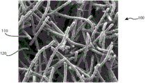
图1A为大约50X的放大倍率的光学显微镜图,显示根据本发明的组织支架的实施方案。 Figure 1A is an optical microscope image at approximately 50X magnification showing an embodiment of a tissue scaffold according to the present invention.
图1B为大约500X的放大倍率的光学显微镜图,显示根据本发明的组织支架的实施方案。 Figure IB is an optical microscope image at approximately 500X magnification showing an embodiment of a tissue scaffold according to the present invention.
图2为本发明方法的用于形成图1A及图1B的组织支架的实施方案的流程图。 2 is a flow diagram of an embodiment of the method of the present invention for forming the tissue scaffold of FIGS. 1A and 1B .
图3为根据图2发明的方法的固化步骤的实施方案的流程图。 FIG. 3 is a flow diagram of an embodiment of the curing step of the inventive method according to FIG. 2 .
图4为根据本发明方法而制造的物件的实施方案的示意图。 Figure 4 is a schematic illustration of an embodiment of an article produced according to the method of the present invention.
图5为图4的物件在完成本发明方法的挥发性组分去除步骤后的示意图。 Figure 5 is a schematic view of the article of Figure 4 after completion of the volatile component removal step of the method of the present invention.
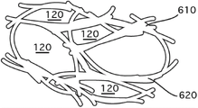
图6为图5的物件在完成本发明方法的连结形成步骤后的示意图。 FIG. 6 is a schematic view of the object of FIG. 5 after the link forming step of the method of the present invention is completed.
图7为评估本发明二例示性实施方案的应力-应变(stress-strain)关系的示意图。 Figure 7 is a schematic diagram for evaluating the stress-strain relationship of two exemplary embodiments of the present invention.
图8为光学显微镜图,显示一具有根据本发明的功能性材料的组织支架的实施方案。 Figure 8 is an optical micrograph showing an embodiment of a tissue scaffold with functional materials according to the invention.
图9为本发明用于形成图8的组织支架的方法的一可供选择的实施方案的流程图。 9 is a flowchart of an alternative embodiment of a method of the present invention for forming the tissue scaffold of FIG. 8 .
图10为根据本发明的组织支架制成的脊椎植入物的侧面立视图。 Figure 10 is a side elevational view of a spinal implant made from a tissue scaffold according to the present invention.
图11为植入椎间空间具有图10的脊椎植入物的脊椎的一部分的侧面透视图。 11 is a side perspective view of a portion of a vertebra having the spinal implant of FIG. 10 implanted in the intervertebral space.
图12为一示意图,显示根据本发明的组织支架制成的截骨术楔形物(osteotomy wedge)的等角视图。 Figure 12 is a schematic diagram showing an isometric view of an osteotomy wedge made from a tissue scaffold according to the present invention.
图13为一示意图,显示可经操作将图12的截骨术楔形物插入一骨中的截骨开口的展开视图。 13 is a schematic diagram showing an expanded view of an osteotomy opening operable to insert the osteotomy wedge of FIG. 12 into a bone.
如前述,除了上述附图说明目前揭露的实施方案,亦可考虑其它实施方案。这些揭露代表通过例示性的方式说明实施方案,并非用以限制。本领域的技艺人士可设计落入本发明揭露之实施方案的原则的范围与精神内的多种其他修饰及实施方案。 As previously stated, other implementations are contemplated in addition to the presently disclosed implementations illustrated in the above figures. These disclosures represent embodiments by way of illustration, not limitation. Numerous other modifications and embodiments can be devised by those skilled in the art which will fall within the scope and spirit of the principles of the disclosed embodiments.
【实施方式】 【Implementation】
本发明提供一种用于修复组织缺陷的人造的人工替代组织支架。当用于本文中,各种形式的「人造的人工替代组织支架(synthetic prosthetic tissue scaffold)」及「骨组织支架」及「组织支架」及「人造骨的人工替代物」等词于全文中可互换。在一实施方案中,人造的人工替代组织支架一旦植入活体组织后为生物惰性。在一实施方案中,人造的人工替代组织支架一旦植入活体组织后为骨传导性。在一实施方案中,人造的人工替代组织支架一旦植入活体组织后为骨刺激性。在一实施方案中,人造的人工替代组织支架一旦植入活体组织后为荷重承载。 The present invention provides an artificial artificial replacement tissue scaffold for repairing tissue defects. As used herein, the various forms of "synthetic prosthetic tissue scaffold" and "bone tissue scaffold" and "tissue scaffold" and "artificial substitute for artificial bone" may be used throughout exchange. In one embodiment, the artificial prosthetic tissue scaffold is biologically inert once implanted in living tissue. In one embodiment, the artificial prosthetic tissue scaffold is osteoconductive once implanted in living tissue. In one embodiment, the artificial prosthetic tissue scaffold is osteostimulatory once implanted in living tissue. In one embodiment, the artificial prosthetic tissue scaffold is load bearing once implanted in living tissue.
已发展用于组织工程应用的各种种类的人造植入物,以试图提供一人造的人工替代装置,其可模拟天然骨组织的性质及促进组织的修复及愈合。已发展金属及生物持久性结构的生物惰性材料,以在促进新组织生长的多孔性结构中提供高强度。然而,这些多孔性材料无法提供具有对于健康组织的向内生长最佳化的孔隙形态的孔隙度。先前技术的生物持久性金属及生物相容性的植入物的一缺点为高荷重承载能力并不会转移至围绕所述植入物的再生组织。当形成硬组织时,应力荷重导致较强壮的组织,但金属植入物屏蔽新形成的骨接收此应力。骨组织的应力屏蔽从而导致弱的骨组织,其确实会被身体再吸收,这是人工替代物松脱的起因。 Various types of artificial implants have been developed for tissue engineering applications in an attempt to provide an artificial artificial replacement device that mimics the properties of natural bone tissue and promotes tissue repair and healing. Bioinert materials of metals and biopersistent structures have been developed to provide high strength in porous structures that promote new tissue growth. However, these porous materials fail to provide a porosity with a pore morphology optimized for healthy tissue ingrowth. A disadvantage of prior art biopersistent metallic and biocompatible implants is that the high load bearing capacity is not transferred to the regenerating tissue surrounding the implant. When hard tissue is formed, the stress load results in stronger tissue, but the metal implant shields the newly formed bone from receiving this stress. The stress shielding of the bone tissue thus results in weak bone tissue, which is indeed resorbed by the body, which is the cause of the loosening of the artificial substitute.
植入物进入活体组织引起生物反应是取决于数种因素,如植入物的组成。生物惰性材料通常以纤维组织封装(encapsulated),以隔离主体与所述植入物。金属及大多数聚合物产生如近惰性陶瓷(如氧化铝或氧化锆)这类的介面反应。若植入物具有一足够的孔隙尺寸及孔隙尺寸分布的多孔性表面,活体组织将因生物体的自然愈合过程的功能而往内生长并连结至所述植入物。这种介面连结可造成一介面,其可稳定在骨床(bony bed)中的支架或植入物,并提供由支架跨过连结介面至骨组织中的应力。当修复处承受重量时,包括再生的骨组织的骨组织受到应力,限制因应力屏蔽所导致的骨组织再吸收。 The biological response of an implant into living tissue depends on several factors, such as the composition of the implant. Bioinert materials are usually encapsulated with fibrous tissue to isolate the subject from the implant. Metals and most polymers produce interfacial reactions like near-inert ceramics such as alumina or zirconia. If the implant has a porous surface of sufficient pore size and pore size distribution, living tissue will ingrow and bond to the implant as a function of the organism's natural healing process. This interfacing creates an interface that stabilizes the scaffold or implant in the bony bed and provides stress from the scaffold across the bonding interface into the bone tissue. When weight is applied to the repair, the bone tissue, including the regenerated bone tissue, is stressed, limiting bone tissue resorption due to stress shielding.
使用生物惰性材料发展组织支架的挑战是在具有足以促进骨组织生长的孔隙度下达到荷重承载强度、具有与周围的骨相似的弹性模数以使应力传送至新组织以确保在植入部位形成健康的骨。制备成具有足够的强度以成为荷重承载强度的传统生物惰性材料,并未提供具有所欲的孔隙尺寸及孔隙尺寸分布以促进健康组织的向内生长的开放且互连的孔隙,或展现显着超过天然骨的弹性模数而导致应力屏蔽。 The challenge in developing tissue scaffolds using bioinert materials is to achieve load-bearing strength with sufficient porosity to promote bone tissue growth, with a similar modulus of elasticity to the surrounding bone to enable stress transfer to new tissue to ensure formation at the implant site. healthy bones. Conventional bioinert materials prepared with sufficient strength to be load bearing strength do not provide open and interconnected pores with the desired pore size and pore size distribution to promote ingrowth of healthy tissue, or exhibit significant Exceeding the modulus of elasticity of native bone results in stress shielding.
一般认为以纤维为基质的结构可提供固有较高的强度对重量比率(strength to weight ratios),鉴于单一纤维的强度可显着大于相同组成的以粉末为基质或以颗粒为基质的材料。可生产具有相对低的不连续性的纤维,不连续性会促使形成造成失效传递(failure propagation)的应力集中。相反的,以粉末为基质或以颗粒为基质的材料需要在各个邻接的颗粒之间形成连结,各个连接介面可能产生应力集中。此外,提供以纤维为基质的结构以缓和应力,因而具较大的强度,当以纤维为基质的结构受到应变时,任何单一纤维的失效并不会透过相邻的纤维传递。因此,相较于一相同组成的以粉末为基质的材料,以纤维为基质的结构在相等尺寸及孔隙度的情况下展现较优异的机械强度性质。 It is generally believed that fiber-based structures can provide inherently higher strength-to-weight ratios, given that the strength of a single fiber can be significantly greater than that of a powder-based or particle-based material of the same composition. Fibers can be produced with relatively low discontinuities that promote the development of stress concentrations that cause failure propagation. In contrast, powder-based or particle-based materials require the formation of bonds between individual adjacent particles, and stress concentrations may arise at the interface of each connection. In addition, the fiber-based structure is provided for stress relaxation and thus greater strength, and when the fiber-based structure is strained, the failure of any single fiber is not propagated through adjacent fibers. Therefore, compared to a powder-based material of the same composition, the fiber-based structure exhibits superior mechanical strength properties at equal size and porosity.
本发明提供一种用于组织工程应用的材料,其为生物惰性、具有低弹性模数的荷重承载能力、及具有可受控制与最佳化的孔隙结构的骨刺激性,以促进骨的向内生长。 The present invention provides a material for tissue engineering applications that is biologically inert, has a low elastic modulus of load-carrying capacity, and has an osteostimulatory property with a controllable and optimized pore structure to promote bone orientation. Ingrown.
图1A为大约50X的放大倍率的光学显微镜图,显示本发明的一组织支架100的实施方案。组织支架100是一刚性三维基质110所形成一模拟骨结构的强度、弹性模数、与孔隙形态的结构。当用于本文中,「刚性」乙词意指结构在应力的施用下,直到以与天然骨相同的方式断裂前,其并未明显屈曲(yield),则可被认为是刚性结构。支架100为一多孔性材料,具有通常为互连的孔隙120网络。在一实施方案中,孔隙120的互连的网络提供骨传导性。当用于本文中,骨传导性乙词意指所述材料可促进骨组织的向内生长。典型人类的疏松骨具有为约4兆帕(MPa)至约12兆帕的压缩粉碎强度、约0.1吉帕(GPa)至1.0吉帕的弹性模数。如以下所示,本发明的组织支架100可在一钽材料中提供一多孔性骨刺激性结构,其具有大于约50%的孔隙度、大于4兆帕且高达且超过110兆帕的压缩粉碎强度、具有非常符合天然骨的弹性模数(例如,0.1至3.5吉帕)。
FIG. 1A is an optical micrograph at approximately 50X magnification showing a
在一实施方案中,三维基质110是由连结且熔合至一刚性结构的纤维所形成,具有生物惰性组成。相较于使用传统的以粉末为基质的原材料(包括由化学气相沉积所形成的材料),使用纤维作为创造三维基质110的原材料提供一明显的优点。在一实施方案中,在一给定的孔隙度下,相较于以粉末为基质的结构,以纤维为基质的原材料提供一具有更大强度的结构。在一实施方案中,以纤维为基质的原材料提供一具有一较传统结构低的弹性模数的结构。
In one embodiment, the three-
本发明的组织支架100提供所欲的机械及化学特性,结合孔隙形态以促进骨传导性。孔隙120网络为天然的互连的孔隙度,其是由模拟天然骨的结构中的互相缠绕的、不织纤维材料之间的孔隙所形成。此外,使用本文所描述的方法,可控制及最佳化孔隙尺寸,以在支架100及再生骨内增进血液及体液的流动。举例言之,孔隙尺寸及孔隙尺寸分布可通过选择在形成支架100期间挥发的孔隙形成剂及有机黏结剂而控制。孔隙尺寸及孔隙尺寸分布可由孔隙形成剂的颗粒尺寸及颗粒尺寸分布而决定,包含单波型(single mode)孔隙尺寸、双波型(bi-modal)孔隙尺寸分布、及/或多波型(multi-modal)孔隙尺寸分布。支架100的孔隙度可为40%至约85%。在一实施方案中,一旦植入活体组织,在此范围内的孔隙度可促进再生组织的骨诱导性的过程,且展现荷重承载强度。
The
支架100是使用纤维作为原材料而制造。所述纤维可由一生物惰性材料所组成。本文中所用的「纤维」乙词是用以描述连续性或不连续性的线、细丝、杆或晶须,具有大于一的长宽比,且是通过拉线(wire-drawing)或纤维-形成过程(如拉制、纺制、吹制、或其他典型用于形成纤维材料的类似过程)所形成。生物惰性线或纤维可由可形成线或纤维形式的生物惰性组合物而制造,如生物惰性材料如钽、钛、不锈钢、或这些材料的合金、或氧化铝、或其他生物惰性氧化物。可通过传统金属拉线方法(包括多次及/或连续拉制,以降低线直径至所欲的纤维直径,并切割或裁切长度)形成含有钛及钛合金的生物惰性材料。所述纤维可由生物惰性组合物的前驱物而制造,其可在形成三维基质110时形成一生物惰性组合物,而形成支架100。可使用生物惰性纤维组合物制造兼具荷重承载、骨传导性、及/或骨刺激性的支架100
The
再参照图1A,在三维基质110内的孔隙120网络具有一独特结构,其具有尤其有利于骨组织的向内生长而作为支架100的性质。孔隙空间120的特性可通过选择挥发性组分而控制,如以下所述。孔隙尺寸及孔隙尺寸分布为孔隙120网络的重要特性,其可经指定及控制,从而通过选择具有特定颗粒尺寸及分布的挥发性组分而预定,以提供骨传导性的结构,同时维持用于荷重承载应用的强度。此外,相较于现有技术材料,孔隙120网络展现改进的互连性,具有因黏结剂及孔隙形成剂造成的纤维位置所导致的在孔隙之间大的相对喉孔尺寸(throat sizes),进一步增进本发明的组织支架100的骨传导性。孔隙120网络是由纤维材料的天然的堆积密度所造成的空间、以及在形成支架100期间与纤维混合的挥发性组分所造成的纤维的位移所造成的空间而产生的。如以下进一步描述,形成三维基质110的生物惰性材料是以熔合及连结重叠且互相缠绕的纤维而制造的。
Referring again to FIG. 1A , the network of
参照图1B,在高放大倍率下显示本发明的一实施方案的连结且重叠互相缠绕的纤维的分解图。纤维110使用连结剂115而熔合且连结至重叠纤维110。连结剂115可补充并加强创造组织支架100的三维基质的纤维与纤维间的连结。纤维及连结剂为非挥发性组分,是通过形成一具有挥发性组分的均质混合物(如黏结剂及孔隙形成剂,例如包含有机材料)而预置(prepositioned),以预定在孔隙间所得孔隙尺寸、孔隙分布、及喉孔尺寸。此外,挥发性组分通过增加孔隙间的喉孔尺寸而有效地增加孔隙互连的数量,使得孔隙连接成多孔隙。松散纤维在混合物中去结块及分散,得到纤维材料在挥发性有机材料内呈重叠及互相缠绕关系的相对位置。在去除挥发性组分、且熔合及连结纤维以形成三维基质110时,孔隙120网络是由经挥发性组分所占的空间所产生。
Referring to Figure IB, an exploded view of linked and overlapping intertwined fibers of an embodiment of the present invention is shown at high magnification.
本发明的支架的一目的为促进原位(in situ)组织生成,作为活体组织内的植入物。有许多关于用于骨组织修复的理想支架的标准,一个重要特性为一高度互连的多孔性网络,兼具足以进行细胞迁移、液体交换及最终的组织的向内生长及血管新生(例如,血管的渗透)的大小的孔隙尺寸及孔隙互连。本发明的组织支架100为一具有孔隙尺寸及孔隙互连性的多孔性结构,其特别适用于骨组织的向内生长。孔隙120网络具有可通过选择用以制造组织支架100的挥发性组分而控制的孔隙尺寸,以提供至少100微米的平均孔隙尺寸。组织支架100的实施方案具有约50微米至约600微米的平均孔隙尺寸,或者,约100微米至约500微米的平均孔隙尺寸。所述挥发性组分(包含有机黏结剂及孔隙形成剂(形成孔隙))、及由一孔隙延伸至至少一相邻的孔隙的互相缠绕的纤维(取决于纤维因挥发性组分而预定的位置),确保在三维基质内具有大的孔隙喉孔尺寸的高程度的互连性。具有通过活体外(in vitro)分析而测定的双波型或多波型的孔隙尺寸分布是理想的。可通过选择展现相似的多波型颗粒尺寸分布的孔隙形成剂材料而提供多波型的孔隙尺寸分布。同样的,混合的各种特性(例如厚度或直径、长度、或截面形状)的纤维材料会影响孔隙的尺寸及尺寸分布。
One purpose of the scaffold of the present invention is to facilitate tissue generation in situ, as an implant within living tissue. There are many criteria for an ideal scaffold for bone tissue repair, an important property being a highly interconnected porous network, combined with sufficient cell migration, fluid exchange and ultimately tissue ingrowth and angiogenesis (e.g., vascular permeability) size of pore size and pore interconnection. The
参照图2,显示形成组织支架100的方法200的实施方案。一般而言,松散纤维210是与黏结剂230及液体250混合,形成一塑性可模塑材料,其接着经固化以形成组织支架100。固化步骤280选择性的去除混合物的挥发性成分,留下开放且互连的孔隙空间120,及将纤维210有效地熔合与连结成刚性三维基质110。
Referring to Figure 2, an embodiment of a method 200 of forming a
松散纤维210可以一松散形式,或细碎纤维而提供。纤维210的直径可为约3微米至约500微米,且典型为约25微米至约200微米。这类纤维210通常生产为具有相对窄的且经控制的纤维直径的分布,且可使用一给定直径的纤维、或具有一纤维直径范围的纤维的混合物。纤维210的直径会影响所得多孔性结构的孔隙尺寸及孔隙尺寸分布、以及三维基质110的尺寸及厚度,这不仅会影响支架100的骨传导性,也会影响其所得强度特性,包含压缩强度及弹性模数。纤维210通常经切割或裁切长度。纤维长度可为纤维直径的约3至约1000倍,且通常为纤维直径的约20至50倍。
当黏结剂230及液体250与纤维210混合时,产生一塑性可成形的批料混合物,其使纤维210均匀地分散在批料中,且提供生胚强度,以使批料材料在后续的形成步骤270中形成所欲的形状。可使用有机黏结剂材料作为黏结剂230,如甲基纤维素、羟丙基甲基纤维素(HPMC)、乙基纤维素、及前述的组合。黏结剂230可包含例如以下材料:聚乙烯、聚丙烯、聚丁烯、聚苯乙烯、聚乙烯乙酯、聚酯、等规聚丙烯(isotactic polypropylene)、无规聚丙烯(atactic polypropylene)、聚砜、聚甲醛聚合物、聚甲基丙烯酸甲酯、富马-茚共聚物(fumaron-indane copolymer)、乙烯醋酸乙烯共聚物、苯乙烯-丁二烯共聚物、压克力橡胶、聚乙烯缩丁醛、离子键树脂、环氧树脂、尼龙、酚甲醛、苯酚糠醛、石蜡、蜡乳液、微晶蜡、纤维素、糊精、氯化烃类、精炼藻酸盐(refined alginates)、淀粉、明胶、木质素、橡胶、丙烯酸树脂、沥青、酪蛋白、树胶(gum)、白蛋白、蛋白质、乙二醇、羟乙基纤维素、羧甲基纤维素钠、聚乙烯醇、聚乙烯吡咯烷酮、聚环氧乙烷、聚丙烯酰胺、聚醚酰亚胺、洋菜、洋菜糖、糖蜜、糊精、淀粉、木素磺酸盐、木质素液、海藻酸钠、阿拉伯树胶、黄原树胶、树胶黄蓍、刺梧桐树胶、刺槐豆树胶、爱尔兰苔藓、核菌多醣(scleroglucan)、丙烯酸、及阳离子半乳甘露聚糖、或前述的混合。虽然以上已列示数种黏结剂230,应理解亦可使用其他黏结剂。黏结剂230提供塑性批料材料所欲的流变性,以形成所欲的物件,并在所述物件形成时维持纤维210在混合物中的相对位置,同时保持对生物惰性材料的惰性。黏结剂230的物理性质会影响支架100的孔隙空间120的孔隙尺寸及孔隙尺寸分布。优选地,黏结剂230可热崩解(thermal
disintegration),或选择性的溶解,而不冲击生物惰性组分(包含纤维210)的化学组成。
When the binder 230 and the liquid 250 are mixed with the
视需要添加流体250,以使塑性批料材料达到所欲的流变性,使所述塑性批料材料在后续的形成步骤270中形成所欲的物件。通常使用水,虽然亦可使用各种种类的溶剂。可在混合步骤260期间进行流变测量,以在形成步骤270前评估混合物的可塑性及凝聚强度(cohesive strength)。 Fluid 250 is added as needed to bring the plastic batch material to the desired rheology for forming the desired article in a subsequent forming step 270 . Typically water is used, although various types of solvents can also be used. Rheological measurements may be taken during the mixing step 260 to assess the plasticity and cohesive strength of the mixture prior to the forming step 270 .
孔隙形成剂240可包含于混合物中,以增加支架100的孔隙空间120。孔隙形成剂为非反应性材料,其在混合步骤260及形成步骤270期间在塑性批料材料中占有体积。当使用孔隙形成剂240时,孔隙形成剂240的颗粒尺寸及尺寸分布影响所得支架100的孔隙空间120的孔隙尺寸及孔隙尺寸分布。颗粒尺寸通常为约25微米至小于约450微米或更大,或者,孔隙形成剂的颗粒尺寸可为纤维210直径的函数,纤维210直径的约0.1倍至约100倍。孔隙形成剂240必须可易于在固化步骤280期间移除,而不会明显破坏围绕的纤维210的相对位置。在本发明的一实施方案中,可通过在固化步骤280期间于上升的温度下的高温裂解或热分解,或挥发而去除孔隙形成剂240。举例言之,微蜡乳液、酚醛树脂颗粒、面粉、淀粉、或碳粒子可包含于混合物中作为孔隙形成剂240。其他孔隙形成剂240可包含炭黑、活性炭、石墨片、合成石墨、木粉、改质淀粉、纤维素、椰子壳荚、乳胶球、鸟饵、锯屑、可高温裂解的聚合物、聚(甲基丙烯酸烷酯)、聚甲基丙烯酸甲酯、聚甲基丙烯酸乙酯、聚甲基丙烯酸正丁酯、聚醚、聚四氢呋喃、聚(1,3-二氧戊环)、聚环氧烷、聚环氧乙烷、聚环氧丙烷、甲基丙烯酸酯共聚物、聚异丁烯、聚碳酸三亚甲酯、聚乙烯草酸、聚β-丙内酯、聚δ-戊内酯、聚碳酸伸乙酯、聚碳酸伸丙酯、乙烯基甲苯/α-甲基苯乙烯共聚物,苯乙烯/α-甲基苯乙烯共聚物、及烯烃二氧化硫共聚物。一般而言,孔隙形成剂240可定义为有机物或无机物,相较于无机物,有机物通常在较低的温度烧除。虽然以上列示数种孔隙形成剂240,应理解可使用其他孔隙形成剂240。然而孔隙形成剂240不须为完全生物可相容的,因为其在加工期间会自支架100去除。
A pore former 240 may be included in the mixture to increase the
连结剂220可视需要包含在混合物中,以促进所得支架100的连结形成及性能。连结剂220可包含与松散纤维210相同组成的以粉末为基质的材料,或可包含不同组成的以粉末为基质的材料。如以下进一步的详细叙述,以连结剂220为基质的添加物通过在相邻的及交叉的纤维210之间形成连结而增进形成三维基质110的互相缠绕的纤维210的连结强度。连结剂220可为生物惰性金属、玻璃、玻璃-陶瓷、陶瓷、或前述的前驱物。在一本发明的实施方案中,连结剂220为磷酸钙。在一可供选择的实施方案中,连结剂220为β-磷酸三钙。在又一可供选择的实施方案中,连结剂220为氢氧磷灰石。
A
个别材料的相对量(包含松散纤维210、黏结剂230、及液体250)是取决于组织支架100中所欲的整体孔隙度。举例言之,提供孔隙度为约60%的支架100,以混合物的体积计,其非挥发性组分275(如纤维210)的量为约40%。以混合物的体积计,挥发性组分285(如黏结剂230及液体250)的相对量为约60%,其中黏结剂与液体的相对量是取决于混合物所欲的流变性。此外,调整挥发性组分285的量以包含挥发性孔隙形成剂240,以产生具有由孔隙形成剂240所增加的孔隙度的支架100。相似地,可调整非挥发性组分275的量以包含非挥发性连结剂220,以产生具有因连结剂220所增加的强度的支架100。由于材料密度会因在固化步骤280期间的组分的反应而变化,故可理解非挥发性组分275及挥发性组分285的相对量以及所得支架100的孔隙度将会改变。以下提供特定实施例。
The relative amounts of the individual materials (including
在混合步骤260中,纤维210、黏结剂230、液体250、孔隙形成剂240、及/或连结剂220(若包含)是混合成一塑性可变形且均匀的混合物的均质团料。混合步骤260可包含干燥混合、湿混合、剪切混合、及揉合,其必须可均匀地将材料分散成一均质团料,且供给所需的剪切力以剪断与分散或去结块(de-agglomerate)纤维210与非纤维材料。这种混合过程的混合、剪切、及揉合的程度及持续的时间是取决于所选择的纤维210及非纤维材料,随在混合步骤260期间所选用的混合设备的种类而定,以得到材料均匀且一致分散于混合物内、所述混合物具有所欲的流变性质,以在后续的形成步骤270形成所述物件。可使用工业混合设备进行混合,例如批料混合机、剪切混合机、及/或揉捏机。
In mixing step 260,
形成步骤270使混合步骤260的混合物形成可成为组织支架100的物件。形成步骤270可包含挤制、滚卷、压力浇铸、或塑形为几乎任何所欲的外形,以提供一大致的成形物件,其可在固化步骤280中固化,以提供支架100。由于所述物件在固化步骤280期间可预期的收缩,可理解支架100的最终尺寸可与形成步骤270中的形成物件不同,因此可能需要进一步的机械加工及最终塑形,以符合特定的尺寸需求。一例示性的实施方案提供机械及活体外与活体内试验的样本,形成步骤270使用一活塞式挤制机将混合物推动通过一圆形模具,将混合物挤制成圆柱状杆。
Forming step 270 forms the mixture of mixing step 260 into an object that may become
接着在固化步骤280中将所述物件固化成组织支架100,如进一步参照图3的描述。在图3所述的一实施方案中,固化步骤280可依序进行以下三阶段:干燥步骤310;挥发性组分去除步骤320;及连结形成步骤330。在第一阶段的干燥310,通过使用略为升高的温度且具有或不具有强制对流(forced convection)的热以逐渐去除液体,而干燥所述形成物件。可使用各种方法加热所述物件,包含但不限于:热空气对流加热、真空冷冻干燥、溶剂萃取、微波、或电磁/无线电频率(RF)干燥方法。在形成物件内的液体优选为不要太快速的去除,以避免因收缩而造成的干燥裂缝。典型地,对于以水为基质的系统,可在形成物件曝露于约90oC至约150oC约一小时而干燥,然而确切的干燥时间可因物件的尺寸及形状而变化,愈大、愈重的物件则需要愈长的干燥时间。在微波或RF能量干燥的情况中,液体本身、及/或物件的其他组分吸附辐射能,以更均匀地在整体材料中产生热。在干燥步骤310期间,取决于选用的作为挥发性组分的材料,黏结剂230可冻凝(congeal)或冻胶(gel),以提供较大的生胚强度,提供物件在后续处理中所需的刚度及强度。
The article is then cured into
一旦所述物件通过干燥步骤310而干燥,或实质不含液体组分250后,进行固化步骤280的下一个阶段,挥发性组分去除步骤320。此阶段自物件去除挥发性组分(例如,黏结剂230及孔隙形成剂240),仅留下非挥发性组分,其形成组织支架100的三维基质110。可例如通过高温裂解、热分解、或溶剂萃取去除挥发性组分。当挥发性组分285经选择以使挥发性组分去除步骤320可依序去除所述组分,挥发性组分去除步骤320可进一步分成一连续的组分去除步骤,如黏结剂烧除步骤340,接着为孔隙形成剂去除步骤350。举例言之,使用HPMC作为黏结剂230可在约300oC下热分解。在氧气存在时加热至约600oC可将石墨孔隙形成剂220氧化成二氧化碳。相似地,当使用面粉或淀粉作为孔隙形成剂220时,可在约300oC至约600oC的温度下热分解。因此,由HPMC的黏结剂230及石墨颗粒的孔隙形成剂220所组成的形成物件可在挥发性组分去除步骤320中加工,通过使所述物件进行二步骤的燃烧程序而去除黏结剂230,并接着去除孔隙形成剂220。在此实施例中,黏结剂烧除步骤340可在至少约300oC但低于600oC的温度下进行一段时间。可接着将温度加热至至少约600oC的温度,在包含氧的加热室中进行孔隙形成剂去除步骤350。提供此热连续的(thermally-sequenced)挥发性组分去除步骤320以控制挥发性组分285的去除,并维持在形成物件中的非挥发性组分275的相对位置。
Once the article is dried by the drying step 310 , or substantially free of the liquid component 250 , the next stage of the curing
图4描绘一在挥发性组分去除步骤320之前的形成物件的各种组分的示意图。纤维210在黏结剂230及孔隙形成剂240的混合物内互相缠绕。连结剂220可视需要进一步分散于混合物中。图5描绘一在挥发性组分去除步骤320完成后的形成物件的示意图。纤维210维持其相对位置,如同由纤维210与挥发性组分285的混合物去除挥发性组分285时所决定的相对位置。当完成挥发性组分285的去除时,物件的机械强度可能相当脆弱,应小心处理此阶段的物件。在一实施方案中,固化步骤280的每一阶段皆在相同的烤炉或窑中进行。在一实施方案中,提供一处理盘,可在所述处理盘上加工所述物件,以减少处理的损害。
FIG. 4 depicts a schematic diagram of the various components forming the article prior to the volatile
图6描绘一在完成固化步骤280的最后步骤、连结形成330后的形成物件的示意图。孔隙空间120是在去除的黏结剂230及孔隙形成剂240的位置而创造的,且纤维210经熔合及连结成三维基质110。挥发性组分285的特性(包含孔隙形成剂240的尺寸、及/或孔隙形成剂240的颗粒尺寸的分布、及/或黏结剂230的相对量)综合起来以决定所得组织支架100的孔隙尺寸、孔隙尺寸分布、及孔隙互连性。连结剂220及在三维基质110的重叠节点610及相邻的节点620形成的连结提供所得三维基质110结构完整性。
FIG. 6 depicts a schematic view of the formed object after completion of the final step of curing
往回参照图3,连结形成步骤330将非挥发性组分275(包含松散纤维210)转换为组织支架100的刚性三维基质110,同时维持由去除挥发性组分275所创造的孔隙空间120。连结形成步骤330将非挥发性组分275在使松散纤维210连结至相邻及重叠的纤维210的环境中加热,并维持足以形成连结的时间,且不会熔化纤维210而破坏非挥发性组分275的相对位置。连结形成环境及其持续时间是取决于包含松散纤维210的非挥发性组分275的化学组成。举例言之,若使用以钛或钛合金为基质的纤维作微松散纤维210,可在一真空炉中于10-3托及约1,200ºC的温度下进行连结形成步骤330。若使用氧化铝纤维作为松散纤维210,可在一静态或经空气清洗的窑中于大气压力及约1,200ºC至约1,600ºC的温度下进行连结形成步骤330。其他可用以作为松散纤维210的材料可加热至使在纤维结构的交叉及重叠的节点发生固态质量转移的温度,或发生液态连结的温度,取决于非挥发性材料的组成、有助于此连结的形成的环境,包含但不限于如空气、氮气、氩气、或其他惰性气体、及真空环境。
Referring back to FIG. 3 ,
在连结形成步骤330中,将形成物件加热至连结形成温度,以在纤维结构的重叠节点610及相邻的结点620形成连结。若使用连结剂220,则通过非常接近纤维210的连结剂220的反应,与纤维210反应形成一连结,而在所述纤维结构的重叠节点610及相邻的结点620形成所述连结。在连结形成步骤330中,纤维210的材料可参与和连结剂220的化学反应,或纤维210在连结剂220的反应中保持惰性。此外,松散纤维210可为纤维组合物的混合物,其中一部分或全部纤维210参与形成连结的反应,以创造三维基质110。
In a
连结形成步骤330的持续时间是取决于在连结形成步骤330期间的温度轮廓(temperature profile),其中纤维210的连结形成温度下的时间是限于相对短的持续时间,以使非挥发性组分275(包含松散纤维210)的相对位置不会受到显着的改变。形成物件中的孔隙尺寸、孔隙尺寸分布、及孔隙间的互连性是由挥发性组分285的松散纤维210的相对位置决定。当形成物件的挥发性组分285可能因达到连结形成温度而烧除,纤维210及非挥发性组分275的相对位置不会受到显着的改变。形成物件可能在连结形成步骤330期间受到轻微或少部分致密化,但可维持孔隙尺寸及孔隙尺寸及分布的控制,因此可通过选择颗粒尺寸稍微过大的孔隙形成剂240或调整挥发性组分285的相对量,以因应预期的致密化。
The duration of the
在形成三维基质110的互相缠绕的纤维的重叠的及相邻的节点之间所形成的连结可为经烧结的连结,其具有实质上与松散纤维210的组成相同的组成。所述连结亦可为松散纤维210与连结剂220之间反应的结果,形成具有一实质上与松散纤维210的组成相同或不同的组成的连结相。由于对于用作医药装置或植入物材料的许可的控制需求,使用经许可的、不会因装置制造方法及过程而显着改变的材料组合物作为原材料是理想的。或者,使用一经许可的材料组合物的前驱物、其在装置制造方法及过程中形成所欲的组合物,作为原材料也是理想的。本发明提供的组织支架装置,其可使用多种医药上许可的材料而制造,或制造成一医药上许可的材料组合物。
The bonds formed between overlapping and adjacent nodes of the interentangled fibers forming three-
由于能够通过具体指定非挥发性组分275及挥发性组分285的特性来控制孔隙形貌,本发明的组织支架100展现经控制的孔隙互连性。举例言之,纤维长度分布可展现大于孔隙形成剂直径的模式,以增进孔隙互连性,其中展现此模式的纤维将由一孔隙延伸至另一孔隙,具有相邻的纤维之间的空间创造孔隙互连性。此外,小于孔隙形成剂颗粒尺寸的纤维直径可确保孔隙形成剂颗粒的紧密堆积,以提供改进的孔隙互连性。
Due to the ability to control the pore morphology by specifying the properties of the non-volatile component 275 and the volatile component 285, the
可通过操纵制造方法200中的各种参数、及/或通过操纵原材料(包括非挥发性组分275及挥发性组分285)的各种参数及特性,针对特定应用控制并调整或最佳化组织支架100的机械性质。举例言之,在荷重承载应用中,可以此处所描述的各种方式最佳化及控制组织支架100的弹性模数。
can be controlled and tuned or optimized for a particular application by manipulating various parameters in manufacturing process 200, and/or by manipulating various parameters and characteristics of raw materials, including non-volatile components 275 and volatile components 285 Mechanical Properties of
组织支架在荷重承载应用中优选为在一大面积上均匀地分布荷重,以使应力连续地传送至周围的组织,以促进健康的骨形成遍布介面。组织支架主要影响支架传送连续应力的效率的机械特性为弹性模数。当组织支架的弹性模数非常符合周围组织的弹性模数时,应力通过支架传送至周围的组织,刺激健康新组织的生长。若支架的弹性模数相对大于周围组织的弹性模数时,生长进入支架的再生的组织会有效地屏蔽应力,导致一根据沃尔夫定律(骨通过减少其质量(不论通过变得更多孔性或变得较薄)而使其自身适应应力降低)的称为骨再吸收的干扰现象。若支架的弹性模数极度地小于周围组织的弹性模数时,应力无法有效地传送至周围组织而不使支架变形及施加过度的应力及应变至新生长的组织上。 Tissue scaffolds in load-bearing applications preferably distribute load evenly over a large area so that stress is continuously transmitted to surrounding tissue to promote healthy bone formation throughout the interface. The mechanical property of a tissue scaffold that primarily affects the efficiency with which the scaffold transmits continuous stress is the modulus of elasticity. When the elastic modulus of the tissue scaffold closely matches that of the surrounding tissue, stress is transmitted through the scaffold to the surrounding tissue, stimulating the growth of healthy new tissue. If the modulus of elasticity of the scaffold is relatively greater than that of the surrounding tissue, the regenerated tissue growing into the scaffold will effectively shield the stress, resulting in an bone becomes thinner) and adapts itself to the reduced stress) in a disturbing phenomenon called bone resorption. If the elastic modulus of the stent is extremely smaller than that of the surrounding tissue, the stress cannot be effectively transmitted to the surrounding tissue without deforming the stent and applying excessive stress and strain to the newly grown tissue.
本发明的方法及装置允许通过针对一给定的材料组合物控制各种因素制造符合理想的弹性模数。一般而言,纤维210特性的变化、挥发性组分285的特性的变化、连结剂220特性的变化、及固化步骤280的环境的控制,可造成所得支架100的强度、孔隙度及弹性模数的最佳化。
The method and apparatus of the present invention allow the creation of a desired modulus of elasticity by controlling various factors for a given material composition. In general, changes in the properties of the
纤维特性(包括组成、直径、长度)直接影响支架的强度及可挠性。由纤维材料的固有的物理特性(例如抗拉强度及弹性模数)引起的组成影响包括例如材料的晶粒边界及脆性。纤维的直径可影响所得支架的强度及可挠性,其中较粗的纤维倾向更坚固及更坚硬。较长的纤维可提供增加的可挠性。此外,纤维的直径及长度,个别或共同地直接影响纤维材料的天然堆积密度。纤维的天然堆积密度愈大,所得支架中的纤维与纤维间的连接可能愈多。当纤维与纤维间的连接增加,支架的强度及模数通常会增加。 Fiber properties (including composition, diameter, length) directly affect the strength and flexibility of the scaffold. Compositional effects arising from inherent physical properties of fiber materials such as tensile strength and modulus of elasticity include, for example, grain boundaries and brittleness of the material. The diameter of the fibers can affect the strength and flexibility of the resulting scaffold, with thicker fibers tending to be stronger and more rigid. Longer fibers provide increased flexibility. In addition, the diameter and length of the fibers, individually or collectively, directly affect the natural bulk density of the fiber material. The greater the natural packing density of the fibers, the more fiber-to-fiber connections possible in the resulting scaffold. As the fiber-to-fiber connections increase, the strength and modulus of the scaffold generally increase.
连结剂220,当使用时,会影响所得支架的强度及可挠性。连结剂220可增加在基质中的纤维与纤维间的连接的数目,从而增加所得的强度并改变弹性模数。此外,连结剂220的相对量将会增加非挥发性组分相对于挥发性组分的量,这会影响孔隙度。一般而言,高孔隙度(在所有其他条件相同下)将导致强度降低。连结剂220的组成将影响所得支架的强度及可挠性,其中固有的物理特性(例如抗拉与压缩强度及弹性模数)会加诸至所得支架。连结剂220的颗粒尺寸可影响强度及模数,其中较大的颗粒具有坐落在纤维的交叉点的倾向,造成更多可桥接相邻的纤维并将其接合至经连结的基质的材料。较小的颗粒具有在连结剂烧除时维持在相同的相对位置的倾向,以使其黏附在纤维的表面,以改变纤维的化学及物理性质。此外,较小的颗粒及/或较小相对量的连结剂220会导致较少的纤维与纤维间的连结,这将降低所得支架的强度并降低其弹性模数。
The
挥发性组分的特性会影响所得支架的强度及可挠性。孔隙形成剂可控制整个支架的互连的孔隙的尺寸及分布,如以上所详述。至于对支架100的机械性质的影响,所有其他条件保持相同时,挥发性组分的量的增加(包括增加的孔隙形成剂的相对量)会影响支架的强度及降低其弹性模数。此外,存在具有与纤维直径及纤维长度相关的变数、与纤维材料的天然堆积密度有关的第二互相作用。当挥发性组分与非挥发性组分混合时,可增加纤维的捆扎,其中二或更多纤维长段将实质上与其他纤维相邻排列,并沿着纤维长度连结在一起,有效地增加形成支架的基质的「支柱」的截面积。以此方式捆扎的纤维的区域将有效地影响支架100的强度及弹性模数。
The nature of the volatile components can affect the strength and flexibility of the resulting scaffold. Pore formers can control the size and distribution of interconnected pores throughout the scaffold, as detailed above. As for the effect on the mechanical properties of the
在形成支架100的方法200期间所选择的加工参数可影响支架的机械性质。举例言之,固化步骤280环境参数包括加热速率、加热温度、固化时间、及加热环境,例如真空、惰性气体(氮气、氩气等)、成形气体(还原环境)或空气。各项或各项的组合皆可影响整个支架的纤维与纤维间的连结的数量及相对强度。
The processing parameters selected during the method 200 of forming the
用于控制及最佳化支架100的孔隙度/强度关系及弹性模数的其他因素包括原材料的特定特性,结合某些制造加工200步骤,这会影响纤维的整体排列。可调整混合步骤260及形成步骤270以提供一纤维实质上排列在一方向上的成形物件。举例言之,使用形成步骤270中的挤制加工会影响混合物的纤维在挤制的方向上的整体排列。所得支架100的物理特性可展现一弹性模数(其为所述装置的方向的一函数),其中在挤制方向上的压缩强度及弹性模数会相对高,而在与挤制方向垂直的方向上较低。可使用这些变数特性设计用于熔合椎骨的脊椎植入物,以最佳化支架的荷重承载及重量分配特点,以确保健康组织的生长。纤维定位在某些进入支架的血管生长是必须的应用中是理想的。经定位的纤维会诱发展现优选与纤维平行的方向的孔隙形态。在一应用中,支架100熔合骨组织,在邻接的骨之间联系的血管生长可有效地通过本发明的支架而桥接。
Other factors used to control and optimize the porosity/strength relationship and modulus of elasticity of the
此外,可进行上述参数的任一变化或任一组合变化以达到最佳化或所欲的强度及弹性模数、孔隙度、及孔隙尺寸分布,用于预期的应用。此外,可调整强度、弹性模数、孔隙度及孔隙尺寸分布、及其他机械及物理性质,用于其他应用,不限于此处所述的例子。 In addition, changes in any or any combination of the above parameters can be made to achieve an optimized or desired strength and elastic modulus, porosity, and pore size distribution for the intended application. In addition, strength, elastic modulus, porosity and pore size distribution, and other mechanical and physical properties can be tuned for other applications, not limited to the examples described here.
图7描绘由根据本发明的二例示性支架的压缩测试所得的应力-应变曲线720,其证实通过在制造期间添加连结剂而改变支架的强度及弹性模数的效果。二样品皆以上述方法200制造,使用平均直径为约63微米的钛6Al4V合金纤维。第一样品是通过混合3克切割成0.045英吋长度的纤维与1克切割成0.010英吋长度的纤维、0.25克作为有机黏结剂的HPMC、及1克颗粒尺寸为约100微米作为孔隙形成剂的PMMA、及视需要调整的约1.5克的去离子水,以提供一塑性可成形混合物。将所述混合物挤制成直径10毫米的杆并于一对流烤炉中干燥。烧除挥发性组分并接着将支架在1,400˚C于0.3托真空下热处理二小时,以形成一孔隙度为70%的支架。第二样品是以一相同的方式制造,改变仅在于添加0.25克颗粒尺寸小于325微米的作为连结剂220的钛粉末,所得孔隙度为67%。参照图7,第一样品730(无连结剂)的应力-应变曲线展现第一弹性模数735及第一波峰强度值740。第二样品750(含有连结剂)展现一小于第一弹性模数735约65%的第二弹性模数755,及一大于第一强度值740约34%的第二波峰强度值760。
FIG. 7 depicts stress-strain curves 720 resulting from compression testing of two exemplary scaffolds according to the present invention, demonstrating the effect of changing the strength and modulus of elasticity of the scaffolds by adding a bonding agent during fabrication. Both samples were fabricated using the method 200 described above, using titanium 6Al4V alloy fibers with an average diameter of about 63 microns. The first sample was formed by mixing 3 grams of fibers cut to a length of 0.045 inches with 1 gram of fibers cut to a length of 0.010 inches, 0.25 grams of HPMC as an organic binder, and 1 gram of a particle size of about 100 microns as pores PMMA, and about 1.5 grams of deionized water adjusted as needed, to provide a plastically formable mixture. The mixture was extruded into 10 mm diameter rods and dried in a convection oven. Volatile components were burned off and the scaffold was then heat-treated at 1,400°C for 2 hours under 0.3 Torr vacuum to form a scaffold with a porosity of 70%. A second sample was fabricated in the same manner, with the only change being the addition of 0.25 g of titanium powder as
图8描绘一本发明的可供选择的实施方案,显示具有功能性材料705选择性地沉积在整个支架表面的支架100。功能性材料705选择性地沉积以在支架中提供第二功能(例如增强支架100的骨传导性及血管质),以避免在植入物配置期间或之后活化病理性过程;以提供医疗试剂,包含但不限于抗生素、抗凝血剂、抗真菌剂、抗发炎剂、及免疫抑制剂;以提供放射性材料,其可提供用于侦测及定位所述植入物的追踪剂及/或其他功能性强化的功能。图9描绘以方法205制造经强化的多孔性支架100,以在支架中提供第二功能。方法205大致与上述参照图2的方法200相似,惟具有以下视需要的改变。在一实施方案中,功能性材料705可为一添加作为功能性原材料770的材料,作为与纤维210(及视需要与连结剂220)、与含有黏结剂230及孔隙形成剂240的挥发性组分285、以及液体250混合的非挥发性组分275。所述混合物经混合以分散所述包含分散在整个均质混合物的功能性材料705的材料。均质混合物接着形成物件270并在步骤280固化成多孔性支架,如上述参照图2及图3的描述。在此实施方案中,所述固化步骤形成纤维与纤维间的连结并使功能性材料黏附至所得支架100。在一第二实施方案中,在固化步骤期间添加功能性材料705,如显示为视需要的功能性材料注入步骤780。以此方式,功能性材料在连结形成步骤330期间注入至支架中(如以上参照图3的描述)。这可通过在一经控制的高温环境(例如,在真空熔炉中热处理操作)中的气相或电浆沉积进行。在一第三实施方案中,在后续的涂布步骤790(在形成支架100后进行)期间添加功能性材料705。在此实施方案中,功能性材料可通过将支架沉浸至一含有功能性材料705的溶液、化学气相沉积功能性材料的、阴极电弧沉积功能性材料、或其他用于材料沉积的相似方法而沉积。在又一实施方案中,功能性材料可应用在以下任何组合中:视需要的功能性原材料步骤770、视需要的功能性材料注入步骤780、及后续的涂布步骤790。
Figure 8 depicts an alternative embodiment of the present invention showing a
本发明的组织支架可用于以下程序:如截骨术(例如在髋、膝、手及下巴);修复脊柱的结构性失效(例如,椎间人工替代物、层板人工替代物、骶骨人工替代物、椎体人工替代物、及小面人工替代物);骨缺陷填充物;骨折修复手术;肿瘤切除手术;髋膝人工替代物;骨质增大;拔牙;长骨关节固定术;踝关节与足关节固定术,包含距骨下关节植入物、及固定螺丝引脚(fixation screws pins)。本发明的组织支架可用于长骨,包含但不限于肋骨、锁骨、股骨、胫骨、腿部的腓骨、肱骨、桡骨、手臂的尺骨、掌骨、手及脚的跖骨、手指及脚趾的指骨。本发明的组织支架可用于短骨,包含但不限于腕骨、跗骨、膝盖骨、以及其他籽骨(sesamoid bone)。本发明的组织支架可用于其他骨,包含但不限于头骨、下颌骨、胸骨、椎骨、及骶骨。在一实施方案中,相较于传统的装置,本发明的组织支架具有高的荷重承载能力。在一实施方案中,相较于传统的装置,本发明的组织支架需要较少的植入材料。此外,由于材料的强度,使用本发明的组织支架需要较少的辅助固定。以这种方式,用于植入此装置的手术程序侵略性较低、更容易进行、且不需后续的手术程序去除仪器及辅助固定。 The tissue scaffolds of the present invention can be used in procedures such as osteotomies (e.g., at the hip, knee, hand, and jaw); repair of structural failures in the spine (e.g., intervertebral artificial substitutes, laminar artificial substitutes, sacral artificial substitutes implants, vertebral artificial substitutes, and facet artificial substitutes); bone defect fillers; fracture repair surgery; tumor resection surgery; hip and knee artificial substitutes; bone augmentation; tooth extraction; long bone arthrodesis; ankle and Foot arthrodesis, including subtalar joint implants, and fixation screws pins. The tissue scaffolds of the present invention can be used in long bones, including but not limited to ribs, clavicles, femurs, tibias, fibulas of legs, humerus, radius, ulnas of arms, metacarpals, metatarsals of hands and feet, phalanges of fingers and toes. The tissue scaffolds of the present invention can be used on short bones, including but not limited to carpals, tarsals, patella, and other sesamoid bones. The tissue scaffolds of the present invention can be used on other bones including, but not limited to, the skull, mandible, sternum, vertebrae, and sacrum. In one embodiment, the tissue scaffold of the present invention has a high load bearing capacity compared to conventional devices. In one embodiment, the tissue scaffolds of the present invention require less implant material than conventional devices. In addition, due to the strength of the material, less auxiliary fixation is required to use the tissue scaffold of the present invention. In this way, the surgical procedure for implanting the device is less invasive, easier to perform, and does not require subsequent surgical procedures for instrumentation and auxiliary fixation.
在一特定的应用中,如以上所述而制造的本发明的组织支架可作为脊椎植入物800,如图10及图11所描绘。参照图10及图11,脊椎植入物800包含一具有壁820的主体810,其具有啮合在相邻的椎骨V之间的空间S内的尺寸,以维持空间S。装置800是由生物惰性纤维所形成,其可使用挤制方法而形成所欲的形状,以形成一圆柱型状,其可经切割或机械加工成所欲的尺寸。壁820具有与空间S的高度H相对应的高度h。在一实施方案中,壁820的高度h较椎间的空间S的高度H略大。壁820相邻且介于上方啮合表面840与下方啮合表面850之间,其经组态以用于啮合相邻的椎骨V,如图11所示。
In one particular application, the tissue scaffold of the present invention fabricated as described above can be used as a
在另一特定的应用中,本发明的如上所述而制造的组织支架可用作一截骨术楔形植入物1000,如图12及13所描绘。参照图12及图13,截骨术植入物1000通常描述为楔形,设计成符合如胫骨的解剖截面,从而提供大部分胫骨表面机械支撑。截骨术植入物是由生物惰性纤维连结及熔合至一多孔性材料而形成,其可由一经挤制的矩形块状物形成,并切割或机械加工成所欲尺寸及轮廓的楔形形状。植入物1000的近端面1010的特性为其曲线轮廓。远端面1020符合胫骨植入位置的形状。植入物1000的厚度可在约5毫米至约20毫米间变化,取决于病患尺寸及畸形程度。在楔形物的上方及下方表面之间的角度亦可变化。
In another specific application, the tissue scaffold of the present invention fabricated as described above can be used as an
图13说明一使用截骨术楔形植入物1000重新调整一异常的棱角膝盖的方法。在胫骨中间处产生一横切口,且保持胫骨侧面部分完整,并调整胫骨的上部分1050及下部分1040至一预定角度,以产生空间1030。将实质上为楔形形状的植入物1000插入空间1030,如本文所述,利用骨再生并生长进入植入物1000,可在愈合时稳定胫骨部位至所欲位置。可视需要施用固定引脚,以在骨再生以及愈合植入物的位置时稳定胫骨。
FIG. 13 illustrates a method of using an
一般而言,使用本发明的骨组织支架作为一骨移植物牵涉手术程序,其与使用自体移植或异体移植骨移植物的程序相似。若使用足够材料填充及稳定植入部位,通常可以单一程序进行骨移植。在一实施方案中,可将固定引脚插入围绕的天然骨、及/或插入且遍布骨组织支架。骨组织支架插入所述位置并固定位置。所述区域接着闭合,且在一定的愈合及熟成过程后,骨将再生并变得扎实地熔合至植入物内。 In general, using the bone tissue scaffold of the present invention as a bone graft involves surgical procedures similar to those using autograft or allograft bone grafts. Bone grafting can usually be performed in a single procedure if sufficient material is used to fill and stabilize the implant site. In one embodiment, fixation pins may be inserted into the surrounding natural bone, and/or inserted throughout the bone tissue scaffold. A bone tissue scaffold is inserted in place and fixed in place. The area is then closed and after a certain healing and maturation process the bone will regenerate and become solidly fused into the implant.
使用本发明的骨组织支架作为骨缺陷填充物牵涉手术过程,其可以单一程序进行或以修复步骤或阶段的多个程序进行。在一实施方案中,本发明的组织支架置于骨缺陷部位,并使用固定引脚或螺丝附着至骨。或者,组织支架可使用支撑物而在外部固定。所述区域接着闭合,且在一定的愈合及熟成过程后,骨将再生以修复所述缺陷。 The use of bone tissue scaffolds of the present invention as bone defect fillers involves surgical procedures, which can be performed in a single procedure or in multiple procedures with repair steps or stages. In one embodiment, a tissue scaffold of the invention is placed at a bone defect and attached to the bone using fixation pins or screws. Alternatively, the tissue scaffold can be secured externally using struts. The area is then closed and after a certain healing and maturation process the bone will regenerate to repair the defect.
一种填充骨中缺陷的方法,包含以一含有生物惰性纤维连结成多孔性基质的组织支架填充骨中的空间,所述多孔性基质的孔隙尺寸分布促进骨组织的向内生长;以及使组织支架附着至所述骨。 A method of filling a defect in bone comprising filling a space in bone with a tissue scaffold comprising biologically inert fibers linked into a porous matrix having a pore size distribution that promotes ingrowth of bone tissue; and causing the tissue A scaffold is attached to the bone.
一种处理截骨术的方法,包含以一含有生物惰性纤维连结成多孔性基质的组织支架填充骨中的空间,所述多孔性基质的孔隙尺寸分布促进骨组织的向内生长;以及使组织支架附着至所述骨。 A method of treating an osteotomy comprising filling a space in bone with a tissue scaffold comprising biologically inert fibers linked into a porous matrix having a pore size distribution that promotes ingrowth of bone tissue; and causing the tissue A scaffold is attached to the bone.
一种处理椎骨的结构性失效的方法,包含以一含有生物惰性纤维连结成多孔性基质的组织支架填充骨中的空间,所述多孔性基质的孔隙尺寸分布促进骨组织的向内生长;以及使组织支架附着至所述骨。 A method of treating structural failure of a vertebra comprising filling a space in bone with a tissue scaffold comprising biologically inert fibers linked into a porous matrix having a pore size distribution that promotes ingrowth of bone tissue; and A tissue scaffold is attached to the bone.
一种制造一人工人造骨的人工替代物的方法,包含混合生物惰性线或纤维、黏结剂、孔隙形成剂、及液体,以提供一塑性可成形的批料;揉捏所述可成形的批料以分散所述生物惰性线或纤维、孔隙形成剂、及黏结剂,形成互相缠绕及重叠的纤维的可成形的批料的均质团料;使所述可成形的批料形成所欲的形状,以提供成形外形;干燥所述成形外形以去除所述液体;加热所述成形外形以去除所述黏结剂及孔隙形成剂;加热所述成形外形至连结形成温度,以在互相缠绕及重叠的生物惰性玻璃纤维之间形成连结。 A method of making an artificial substitute for artificial artificial bone comprising mixing bioinert threads or fibers, binders, pore formers, and liquids to provide a plastically formable batch; kneading said formable batch material to disperse the bioinert threads or fibers, pore forming agent, and binder to form a homogeneous mass of formable batch of intertwined and overlapping fibers; forming the formable batch into desired shape to provide a shaped shape; dry the shaped shape to remove the liquid; heat the shaped shape to remove the binder and pore forming agent; heat the shaped shape to a bond forming temperature to intertwine and overlap Bonds are formed between the biologically inert glass fibers.
在一实施方案中,本发明揭露使用生物惰性纤维连结成多孔性基质,所述多孔性基质的孔隙尺寸分布促进骨组织的向内生长,用于处理骨缺陷。 In one embodiment, the present invention discloses the use of biologically inert fibers bonded into a porous matrix with a pore size distribution that promotes ingrowth of bone tissue for the treatment of bone defects.
在一实施方案中,本发明揭露使用生物惰性纤维连结成多孔性基质,所述多孔性基质的孔隙尺寸分布促进骨组织的向内生长,用于处理截骨术。 In one embodiment, the present invention discloses the use of biologically inert fibers bonded into a porous matrix with a pore size distribution that promotes ingrowth of bone tissue for the treatment of osteotomies.
在一实施方案中,本发明揭露使用生物惰性纤维连结成多孔性基质,所述多孔性基质的孔隙尺寸分布促进骨组织的向内生长,用于处理脊椎柱的各种部位的结构性失效。 In one embodiment, the present invention discloses the use of bioinert fibers bonded into a porous matrix with a pore size distribution that promotes ingrowth of bone tissue for the treatment of structural failures at various locations of the spinal column.
实施例 Example
以下实施例是提供以进一步说明及有助于了解本发明。这些特定的实施例是用于例示说明本发明,而非用于限制本发明。 The following examples are provided to further illustrate and facilitate the understanding of the present invention. These specific examples are used to illustrate the invention, not to limit the invention.
在第一例示性实施方案中,一支架是通过以下方法由钛纤维所形成:混合4克平均直径为约225微米、经裁切成长度约1至3毫米、松散形式的钛6Al4V合金纤维作为非挥发性组分;0.125克HPMC作为有机黏结剂;0.5克颗粒尺寸为25-30微米的PMMA作为孔隙形成剂;及约1.5克的去离子水,视需要调整以提供一塑性可成形混合物。将混合物挤制成直径为10毫米的杆,并于一对流烤炉中干燥。烧除挥发性组分并接着在1,400ºC下于0.3托真空热处理二小时。本实施例的孔隙度经测量为69.1%。 In a first exemplary embodiment, a scaffold was formed from titanium fibers by mixing 4 grams of titanium 6Al4V alloy fibers in loose form with an average diameter of about 225 microns and cut into lengths of about 1 to 3 mm as Non-volatile components; 0.125 grams of HPMC as an organic binder; 0.5 grams of PMMA with a particle size of 25-30 microns as a pore former; and about 1.5 grams of deionized water, adjusted as necessary to provide a plastically formable mixture. The mixture was extruded into 10 mm diameter rods and dried in a convection oven. Volatile components were burned off and then heat treated under vacuum at 0.3 Torr for two hours at 1,400°C. The porosity of this example was measured to be 69.1%.
在第二例示性实施方案中,一支架是通过以下方法由氧化铝纤维所形成:混合50克平均直径为约3至5微米的氧化铝纤维、30克氢氧磷灰石粉末及0.8克碳酸镁粉末作为非挥发性组分;65克平均颗粒尺寸为45微米的石墨粉末(Asbury Carbons A625石墨)作为孔隙形成剂;5克HPMC作为黏结剂;及约70克的去离子水,视需要调整以提供一塑性可成形混合物。将混合物挤制成直径为10毫米的杆,并于一对流烤炉中干燥。在一经空气清洗的烤炉中烧除挥发性组分并接着在1,600ºC、大气压力下于静态空气窑热处理二小时。所得支架的组成为氧化铝纤维与氢氧磷灰石陶瓷连结的经连结的多孔性结构,且本实施例的孔隙度经测量为68%。 In a second exemplary embodiment, a scaffold is formed from alumina fibers by mixing 50 grams of alumina fibers having an average diameter of about 3 to 5 microns, 30 grams of hydroxyapatite powder, and 0.8 grams of carbonic acid Magnesium powder as the non-volatile component; 65 grams of graphite powder (Asbury Carbons A625 graphite) with an average particle size of 45 microns as the pore former; 5 grams of HPMC as the binder; and approximately 70 grams of deionized water, adjusted as needed to provide a plastically formable mixture. The mixture was extruded into 10 mm diameter rods and dried in a convection oven. Volatile components were burned off in an air-purged oven followed by heat treatment in a static air kiln at 1,600ºC for two hours at atmospheric pressure. The composition of the obtained scaffold is an interconnected porous structure in which alumina fibers are interconnected with HA ceramics, and the porosity of this embodiment is measured to be 68%.
在第三例示性实施方案中,一支架是通过以下方法由氧化铝纤维所形成:混合50克平均直径为约3至5微米的氧化铝纤维、50克氢氧磷灰石粉末及0.8克碳酸镁粉末作为非挥发性组分;65克平均颗粒尺寸为250微米的石墨粉末(Asbury Carbons 4015石墨)作为孔隙形成剂;5克HPMC作为黏结剂;及约70克的去离子水,视需要调整以提供一塑性可成形混合物。将混合物挤制成直径为10毫米的杆,并于一对流烤炉中干燥。在一经空气清洗的烤炉中烧除挥发性组分并接着在1,400ºC、大气压力下于静态空气窑热处理二小时。所得支架的组成为氧化铝纤维与氢氧磷灰石陶瓷连结的经连结的多孔性结构,且本实施例的孔隙度经测量为68%。 In a third exemplary embodiment, a scaffold is formed from alumina fibers by mixing 50 grams of alumina fibers having an average diameter of about 3 to 5 microns, 50 grams of hydroxyapatite powder, and 0.8 grams of carbonic acid Magnesium powder as the non-volatile component; 65 grams of graphite powder (Asbury Carbons 4015 graphite) with an average particle size of 250 microns as the pore former; 5 grams of HPMC as the binder; and approximately 70 grams of deionized water, adjusted as needed to provide a plastically formable mixture. The mixture was extruded into 10 mm diameter rods and dried in a convection oven. Volatile components were burned off in an air-purged oven followed by heat treatment in a static air kiln at 1,400ºC for two hours at atmospheric pressure. The composition of the obtained scaffold is an interconnected porous structure in which alumina fibers are interconnected with HA ceramics, and the porosity of this embodiment is measured to be 68%.
在第四例示性实施方案中,一支架是通过以下方法由铝硅酸镁纤维所形成:混合50克平均直径为约10微米的ISOFRAX纤维(得自Unifrax LLC,Niagara Falls,纽约)、30克氢氧磷灰石粉末作为非挥发性组分;65克平均颗粒尺寸为250微米的石墨粉末(Asbury Carbons 4015石墨)作为孔隙形成剂;5克HPMC作为黏结剂;及约80克的去离子水,视需要调整以提供一塑性可成形混合物。将混合物挤制成直径为10毫米的杆,并于一对流烤炉中干燥。在一经空气清洗的烤炉中烧除挥发性组分并接着在1,200ºC、大气压力下于静态空气窑热处理二小时。所得支架的组成为铝硅酸镁纤维与氢氧磷灰石陶瓷连结的经连结的多孔性结构,且本实施例的孔隙度经测量为69%。 In a fourth exemplary embodiment, a scaffold is formed from magnesium aluminosilicate fibers by mixing 50 grams of ISOFRAX fibers having an average diameter of about 10 microns (obtained from Unifrax LLC, Niagara Falls, New York), 30 grams Hydroxyapatite powder as the non-volatile component; 65 grams of graphite powder (Asbury Carbons 4015 graphite) with an average particle size of 250 microns as the pore former; 5 grams of HPMC as the binder; and about 80 grams of deionized water , adjusted as necessary to provide a plastically formable mixture. The mixture was extruded into 10 mm diameter rods and dried in a convection oven. Volatile components were burned off in an air-purged oven followed by heat treatment in a static air kiln at 1,200ºC for two hours at atmospheric pressure. The composition of the obtained scaffold is a linked porous structure in which magnesium aluminosilicate fibers are linked with HA ceramics, and the porosity of this embodiment is measured to be 69%.
在第五例示性实施方案中,一支架是通过以下方法由钛纤维所形成:混合0.9克平均直径为约225微米、经裁切成长度约1至3毫米、松散形式的纯钛纤维作为非挥发性组分;0.3克HPMC作为有机黏结剂;0.5克颗粒尺寸为约50微米的马铃薯淀粉作为孔隙形成剂;及约2克的去离子水,视需要调整以提供一塑性可成形混合物。将混合物挤制成直径为10毫米的杆,并于一对流烤炉中干燥。烧除挥发性组分并接着在1,400ºC下于0.3托真空热处理二小时。本实施例的孔隙度经测量为69.1%。 In a fifth exemplary embodiment, a scaffold is formed from titanium fibers by mixing 0.9 grams of pure titanium fibers with an average diameter of about 225 microns, cut into lengths of about 1 to 3 mm, in loose form as non- Volatile components; 0.3 grams of HPMC as an organic binder; 0.5 grams of potato starch having a particle size of about 50 microns as a pore former; and about 2 grams of deionized water, adjusted as necessary to provide a plastically formable mixture. The mixture was extruded into 10 mm diameter rods and dried in a convection oven. Volatile components were burned off and then heat treated under vacuum at 0.3 Torr for two hours at 1,400°C. The porosity of this example was measured to be 69.1%.
在第六例示性实施方案中,一支架是通过以下方法由钛纤维所形成:混合2克平均直径为约65微米、经裁切成长度约1至2毫米、松散形式的的钛6Al4V合金纤维、及0.5克作为连结剂的颗粒尺寸小于44微米(-325筛目)的钛6Al4V合金粉末作为非挥发性组分;0.5克HPMC作为有机黏结剂;0.5克颗粒尺寸为约150微米的聚乙烯颗粒作为孔隙形成剂;及约2克的去离子水,视需要调整以提供一塑性可成形混合物。将混合物挤制成直径为10毫米的杆,并于一对流烤炉中干燥。在350˚C下烧除挥发性组分14小时,并接着在经氩气清洗的窑中使用每分钟5˚C的升温速率在1,400ºC下热处理,保持在1,400˚C二小时。本实施例的孔隙度经测量为88.1%。 In a sixth exemplary embodiment, a scaffold is formed from titanium fibers by mixing 2 grams of titanium 6Al4V alloy fibers in loose form having an average diameter of about 65 microns, cut into lengths of about 1 to 2 mm , and 0.5 grams of titanium 6Al4V alloy powder with a particle size of less than 44 microns (-325 mesh) as a non-volatile component as a binder; 0.5 grams of HPMC as an organic binder; 0.5 grams of polyethylene with a particle size of about 150 microns particles as a pore former; and about 2 grams of deionized water, adjusted as necessary to provide a plastically formable mixture. The mixture was extruded into 10 mm diameter rods and dried in a convection oven. Volatile components were burned off at 350°C for 14 hours, followed by heat treatment at 1,400°C in an argon-purged kiln using a ramp rate of 5°C per minute, held at 1,400°C for two hours. The porosity of this example was measured to be 88.1%.
在第七例示性实施方案中,一支架是通过以下方法由二种型态的钛纤维的混合物所形成:在此实施例中,混合2克平均直径为约65微米、经裁切成长度约1至2毫米的钛6Al4V合金纤维、2克平均直径为约225微米、经裁切成长度约1至3毫米的钛6Al4V合金纤维、及1.0克作为连结剂的颗粒尺寸小于44微米(-325筛目)的钛6Al4V合金粉末作为非挥发性组分;0.5克HPMC作为有机黏结剂;0.5克颗粒尺寸为约150微米的聚乙烯颗粒作为孔隙形成剂;及约2克的去离子水,视需要调整以提供一塑性可成形混合物。将混合物挤制成直径为10毫米的杆,并于一对流烤炉中干燥。在350˚C下烧除挥发性组分14小时,并接着在经氩气清洗的窑中使用每分钟5˚C的升温速率在1,400ºC下热处理,保持在1,400˚C二小时。 In a seventh exemplary embodiment, a scaffold is formed from a mixture of two types of titanium fibers by mixing, in this example, 2 grams of an average diameter of about 65 microns cut into lengths of about 1 to 2 mm of titanium 6Al4V alloy fiber, 2 grams of titanium 6Al4V alloy fiber with an average diameter of about 225 microns cut to a length of about 1 to 3 mm, and 1.0 g of a particle size less than 44 microns (-325 Mesh) titanium 6Al4V alloy powder as a non-volatile component; 0.5 grams of HPMC as an organic binder; 0.5 grams of polyethylene particles with a particle size of about 150 microns as a pore former; and about 2 grams of deionized water, depending on Adjustments are required to provide a plastically formable mixture. The mixture was extruded into 10 mm diameter rods and dried in a convection oven. Volatile components were burned off at 350°C for 14 hours, followed by heat treatment at 1,400°C in an argon-purged kiln using a ramp rate of 5°C per minute, held at 1,400°C for two hours.
本说明书已通过特定说明及特定实施方案详细描述本发明,然本发明不受限于此,可进行许多不背离本发明的权利要求书的精神及范围的修饰。 This description has described the present invention in detail through specific descriptions and specific implementations, but the present invention is not limited thereto, and many modifications can be made without departing from the spirit and scope of the claims of the present invention.
Claims (14)
Applications Claiming Priority (7)
| Application Number | Priority Date | Filing Date | Title |
|---|---|---|---|
| US24944909P | 2009-10-07 | 2009-10-07 | |
| US61/249,449 | 2009-10-07 | ||
| US30613610P | 2010-02-19 | 2010-02-19 | |
| US61/306,136 | 2010-02-19 | ||
| US38166610P | 2010-09-10 | 2010-09-10 | |
| US61/381,666 | 2010-09-10 | ||
| PCT/US2010/051555 WO2011044182A2 (en) | 2009-10-07 | 2010-10-06 | Devices and methods for tissue engineering |
Publications (2)
| Publication Number | Publication Date |
|---|---|
| CN102725006A true CN102725006A (en) | 2012-10-10 |
| CN102725006B CN102725006B (en) | 2014-10-22 |
Family
ID=43823816
Family Applications (1)
| Application Number | Title | Priority Date | Filing Date |
|---|---|---|---|
| CN201080044896.3A Expired - Fee Related CN102725006B (en) | 2009-10-07 | 2010-10-06 | Porosity tissue scaffold |
Country Status (9)
| Country | Link |
|---|---|
| US (2) | US20110082564A1 (en) |
| EP (1) | EP2485780A4 (en) |
| JP (1) | JP2013507184A (en) |
| KR (1) | KR20120095377A (en) |
| CN (1) | CN102725006B (en) |
| BR (1) | BR112012007489A2 (en) |
| CA (1) | CA2776169A1 (en) |
| IL (1) | IL218663A0 (en) |
| WO (1) | WO2011044182A2 (en) |
Cited By (3)
| Publication number | Priority date | Publication date | Assignee | Title |
|---|---|---|---|---|
| CN103300945A (en) * | 2013-06-06 | 2013-09-18 | 上海交通大学 | Medical porous composite material |
| CN113865817A (en) * | 2021-09-30 | 2021-12-31 | 电子科技大学 | Using method of phenolic aldehyde laminated cloth rod in osteotomy vibration test |
| CN115298304A (en) * | 2020-03-05 | 2022-11-04 | 国立大学法人大阪大学 | Method for controlling Young's modulus of three-dimensional structure, method for producing three-dimensional structure, and three-dimensional structure |
Families Citing this family (26)
| Publication number | Priority date | Publication date | Assignee | Title |
|---|---|---|---|---|
| US8567162B2 (en) | 2009-10-29 | 2013-10-29 | Prosidyan, Inc. | Dynamic bioactive bone graft material and methods for handling |
| EP2493519B1 (en) | 2009-10-29 | 2018-04-18 | Prosidyan, Inc. | Dynamic bioactive bone graft material having an engineered porosity |
| US20130103165A1 (en) * | 2010-12-06 | 2013-04-25 | Composite Materials Technology, Inc. | Biocompatible extremely fine tantalum fiber scaffolding for bone and soft tissue prosthesis |
| US20130004657A1 (en) * | 2011-01-13 | 2013-01-03 | CNano Technology Limited | Enhanced Electrode Composition For Li ion Battery |
| WO2012177759A1 (en) | 2011-06-20 | 2012-12-27 | Rdc Holdings, Llc | System and method for repairing joints |
| US8998925B2 (en) | 2011-06-20 | 2015-04-07 | Rdc Holdings, Llc | Fixation system for orthopedic devices |
| KR101901638B1 (en) * | 2011-07-27 | 2018-09-27 | 프라운호퍼 게젤샤프트 쭈르 푀르데룽 데어 안겐반텐 포르슝 에. 베. | Implant |
| US20140332731A1 (en) * | 2012-04-02 | 2014-11-13 | CNano Technology Limited | Electrode Composition for Battery |
| US20140050765A1 (en) * | 2012-08-14 | 2014-02-20 | Bio2 Technologies, Inc. | Devices and Methods for Tissue Engineering |
| US9031671B2 (en) | 2012-09-21 | 2015-05-12 | Composite Materials Technology, Inc. | Medical implantable lead and manufacture thereof |
| US9028584B2 (en) * | 2012-10-11 | 2015-05-12 | Composite Materials Technology, Inc. | System and method for fabrication of 3-D parts |
| US8883195B2 (en) | 2013-03-14 | 2014-11-11 | Prosidyan, Inc. | Bioactive porous bone graft implants |
| US9381274B2 (en) | 2013-03-14 | 2016-07-05 | Prosidyan, Inc. | Bone graft implants containing allograft |
| US8889178B2 (en) | 2013-03-14 | 2014-11-18 | Prosidyan, Inc | Bioactive porous bone graft compositions in synthetic containment |
| US9498337B2 (en) * | 2013-12-23 | 2016-11-22 | Metal Industries Research & Development Centre | Intervertebral implant |
| CN103751852B (en) * | 2014-01-24 | 2015-06-17 | 天津理工大学 | Preparation method of three-dimensional artificial random porous structure tissue engineering scaffold |
| US10160650B2 (en) | 2014-02-13 | 2018-12-25 | Morris Brothers And Company Holdings Limited | Method for making a three dimensional object |
| US9155605B1 (en) * | 2014-07-10 | 2015-10-13 | Composite Materials Technology, Inc. | Biocompatible extremely fine tantalum filament scaffolding for bone and soft tissue prosthesis |
| US9498316B1 (en) * | 2014-07-10 | 2016-11-22 | Composite Materials Technology, Inc. | Biocompatible extremely fine tantalum filament scaffolding for bone and soft tissue prosthesis |
| WO2016154063A1 (en) * | 2015-03-20 | 2016-09-29 | The Board Of Trustees Of The Leland Stanford Junior Univeristy | Customized load-bearing and bioactive functionally-graded implant for treatment of osteonecrosis |
| FR3038831B1 (en) | 2015-07-16 | 2017-07-21 | Spineart Sa | INTERVERTEBRAL CAGE FOR ARTHRODESIS |
| CN105147423B (en) * | 2015-08-04 | 2017-03-22 | 天津理工大学 | Preparation method of tissue engineering scaffold with three-dimensional composite porous structure |
| CN109715320A (en) | 2016-08-12 | 2019-05-03 | 复合材料技术公司 | Electrolytic capacitor and the method for improving electrolytic capacitor anode |
| KR20190077321A (en) | 2016-09-01 | 2019-07-03 | 컴포짓 매터리얼스 테크놀로지, 아이엔씨. | Nano-scale / nano-structured Si coating on valve metal substrate for LIB anode |
| PL4185238T3 (en) * | 2020-07-21 | 2025-03-24 | Bellaseno Gmbh | THREE-DIMENSIONAL IMPLANT FOR TISSUES RECONSTRUCTION |
| CN112690932B (en) * | 2020-12-25 | 2023-07-28 | 北京爱康宜诚医疗器材有限公司 | Processing method of vertebral body prosthesis |
Citations (10)
| Publication number | Priority date | Publication date | Assignee | Title |
|---|---|---|---|---|
| US3992725A (en) * | 1973-11-16 | 1976-11-23 | Homsy Charles A | Implantable material and appliances and method of stabilizing body implants |
| EP0178650A2 (en) * | 1984-10-17 | 1986-04-23 | Paul Ducheyne | Porous flexible metal fiber material for surgical implantation |
| CN1087279A (en) * | 1992-11-27 | 1994-06-01 | 中国科学院光电技术研究所 | Bioactive glass ceramic artificial bone and its preparation method |
| US6451059B1 (en) * | 1999-11-12 | 2002-09-17 | Ethicon, Inc. | Viscous suspension spinning process for producing resorbable ceramic fibers and scaffolds |
| US20020160033A1 (en) * | 2001-04-25 | 2002-10-31 | Noel Caplice | Implantable medical devices |
| US6679913B2 (en) * | 1998-04-14 | 2004-01-20 | Tranquil Prospects Ltd. | Implantable sheet material |
| CN1887361A (en) * | 2006-08-03 | 2007-01-03 | 复旦大学 | Mesoporous biological glass fiber material and its prepn and application |
| US7241486B2 (en) * | 2001-04-26 | 2007-07-10 | Inion Ltd. | Bone grafting materials |
| US20070162151A1 (en) * | 2006-01-11 | 2007-07-12 | Jiang Chang | Resorbable macroporous bioactive glass scaffold and method of manufacture |
| US20090035511A1 (en) * | 2007-07-31 | 2009-02-05 | Geo2 Technologies, Inc. | Fiber-Based Ceramic Substrate and Method of Fabricating the Same |
Family Cites Families (47)
| Publication number | Priority date | Publication date | Assignee | Title |
|---|---|---|---|---|
| US3906550A (en) * | 1973-12-27 | 1975-09-23 | William Rostoker | Prosthetic device having a porous fiber metal structure |
| JPS5832761A (en) * | 1981-08-22 | 1983-02-25 | 朝倉 由純 | Artificial bone |
| US4479271A (en) * | 1981-10-26 | 1984-10-30 | Zimmer, Inc. | Prosthetic device adapted to promote bone/tissue ingrowth |
| SE453183B (en) * | 1982-05-04 | 1988-01-18 | Svenska Silikatforskning | PROCEDURE FOR REDUCING TIME BY REMOVING TEMPORA BINDERS FROM A FORMATED BODY |
| JPS5957971A (en) * | 1982-10-30 | 1984-04-03 | 株式会社イナックス | Mineral fiber-apatite baked composite body |
| US4503157A (en) * | 1982-09-25 | 1985-03-05 | Ina Seito Co., Ltd. | Sintered apatite bodies and composites thereof |
| US5030233A (en) * | 1984-10-17 | 1991-07-09 | Paul Ducheyne | Porous flexible metal fiber material for surgical implantation |
| JPS62158175A (en) * | 1986-01-07 | 1987-07-14 | 住友大阪セメント株式会社 | Porous ceramic formed body for substitute bone and manufacture |
| JPH0251475A (en) * | 1988-04-27 | 1990-02-21 | Ibiden Co Ltd | Fiber-reinforced calcium phosphate-based compound ceramic and its production |
| US4978358A (en) * | 1988-10-06 | 1990-12-18 | Zimmer Inc. | Orthopaedic prosthetic device possessing improved composite stem design |
| EP0532582B1 (en) * | 1990-06-01 | 1995-12-13 | E.I. Du Pont De Nemours And Company | Composite orthopedic implant with modulus variations |
| US5104410A (en) * | 1990-10-22 | 1992-04-14 | Intermedics Orthopedics, Inc | Surgical implant having multiple layers of sintered porous coating and method |
| US5198308A (en) * | 1990-12-21 | 1993-03-30 | Zimmer, Inc. | Titanium porous surface bonded to a cobalt-based alloy substrate in an orthopaedic implant device |
| US5282861A (en) * | 1992-03-11 | 1994-02-01 | Ultramet | Open cell tantalum structures for cancellous bone implants and cell and tissue receptors |
| US5629186A (en) * | 1994-04-28 | 1997-05-13 | Lockheed Martin Corporation | Porous matrix and method of its production |
| US5879398A (en) * | 1995-02-14 | 1999-03-09 | Zimmer, Inc. | Acetabular cup |
| US5734959A (en) * | 1995-10-12 | 1998-03-31 | Zimmer, Inc. | Method of making an orthopaedic implant having a porous surface using an organic binder |
| JPH10167853A (en) * | 1996-12-11 | 1998-06-23 | Shigeharu Takagi | Porous ceramic compact for artificial bone material |
| US5961554A (en) * | 1996-12-31 | 1999-10-05 | Janson; Frank S | Intervertebral spacer |
| US6187329B1 (en) * | 1997-12-23 | 2001-02-13 | Board Of Regents Of The University Of Texas System | Variable permeability bone implants, methods for their preparation and use |
| CA2285149A1 (en) * | 1998-10-07 | 2000-04-07 | Isotis B.V. | Device for tissue engineering a bone equivalent |
| FI105116B (en) * | 1998-11-30 | 2000-06-15 | Valmet Corp Valmet Corp | Bendable beam for use in a paper or cardboard machine |
| US6673075B2 (en) * | 2001-02-23 | 2004-01-06 | Albert N. Santilli | Porous intervertebral spacer |
| EP1389978B1 (en) * | 2001-05-01 | 2009-01-07 | Amedica Corporation | Radiolucent bone graft |
| US7458991B2 (en) * | 2002-02-08 | 2008-12-02 | Howmedica Osteonics Corp. | Porous metallic scaffold for tissue ingrowth |
| JP3837502B2 (en) * | 2002-05-08 | 2006-10-25 | 独立行政法人産業技術総合研究所 | Biological porous composite, method for producing the same, and use thereof |
| US20030220696A1 (en) * | 2002-05-23 | 2003-11-27 | Levine David Jerome | Implantable porous metal |
| US6945448B2 (en) * | 2002-06-18 | 2005-09-20 | Zimmer Technology, Inc. | Method for attaching a porous metal layer to a metal substrate |
| US20060100716A1 (en) * | 2002-06-27 | 2006-05-11 | Reto Lerf | Open-pored metal coating for joint replacement implants and method for production thereof |
| JP2004067547A (en) * | 2002-08-02 | 2004-03-04 | Yoshinori Kuboki | Titanium fiber medical material |
| JP2004073401A (en) * | 2002-08-14 | 2004-03-11 | Toyo Kohan Co Ltd | Porous body, biological material using the same and manufacturing method for porous body |
| EP1418013B1 (en) * | 2002-11-08 | 2005-01-19 | Howmedica Osteonics Corp. | Laser-produced porous surface |
| US20070152364A1 (en) * | 2005-11-16 | 2007-07-05 | Bilal Zuberi | Process for extruding a porous substrate |
| CN101810882B (en) * | 2004-09-24 | 2013-07-24 | Hi-Lex株式会社 | Body hard tissue or soft tissue inductive scaffolding material |
| CN100591364C (en) * | 2004-10-28 | 2010-02-24 | 独立行政法人物质·材料研究机构 | Method for producing porous body containing apatite/collagen composite fiber |
| US9981063B2 (en) * | 2004-11-24 | 2018-05-29 | Mayo Foundation For Medical Education And Research | Biosynthetic composite for osteochondral defect repair |
| JP2006263445A (en) * | 2005-02-25 | 2006-10-05 | Yasuharu Noisshiki | Medical materials |
| WO2006090777A1 (en) * | 2005-02-23 | 2006-08-31 | Hi-Lex Corporation | Medical material, artificial tooth root and method of producing material for clinical use |
| US7635447B2 (en) * | 2006-02-17 | 2009-12-22 | Biomet Manufacturing Corp. | Method and apparatus for forming porous metal implants |
| US20090157194A1 (en) * | 2006-03-10 | 2009-06-18 | Takiron Co., Ltd. | Implant composite material |
| CN101646402A (en) * | 2007-01-19 | 2010-02-10 | 金文申有限公司 | Porous, non-degradable implants made by powder molding |
| US7582118B2 (en) * | 2007-02-06 | 2009-09-01 | Zimmer Technology, Inc. | Femoral trochlea prostheses |
| WO2008122595A2 (en) * | 2007-04-05 | 2008-10-16 | Cinvention Ag | Biodegradable therapeutic implant for bone or cartilage repair |
| US8318297B2 (en) * | 2007-06-25 | 2012-11-27 | Board Of Trustees Of The University Of Arkansas | Titanate nanowire, titanate nanowire scaffold, and processes of making same |
| EP2228042A1 (en) * | 2007-12-28 | 2010-09-15 | Takiron Co., Ltd. | Biomaterial for artificial cartilages |
| CA2712559C (en) * | 2008-01-30 | 2015-03-31 | Zimmer, Inc. | Orthopedic component of low stiffness |
| KR20120101021A (en) * | 2009-10-29 | 2012-09-12 | 프로시다이안 인코포레이티드 | Bone graft material |
-
2010
- 2010-10-06 EP EP20100822568 patent/EP2485780A4/en not_active Withdrawn
- 2010-10-06 CN CN201080044896.3A patent/CN102725006B/en not_active Expired - Fee Related
- 2010-10-06 US US12/898,797 patent/US20110082564A1/en not_active Abandoned
- 2010-10-06 KR KR1020127011634A patent/KR20120095377A/en not_active Withdrawn
- 2010-10-06 CA CA2776169A patent/CA2776169A1/en not_active Abandoned
- 2010-10-06 BR BR112012007489A patent/BR112012007489A2/en not_active IP Right Cessation
- 2010-10-06 JP JP2012533261A patent/JP2013507184A/en active Pending
- 2010-10-06 WO PCT/US2010/051555 patent/WO2011044182A2/en not_active Ceased
-
2011
- 2011-03-11 US US13/046,179 patent/US20110204537A1/en not_active Abandoned
-
2012
- 2012-03-15 IL IL218663A patent/IL218663A0/en unknown
Patent Citations (10)
| Publication number | Priority date | Publication date | Assignee | Title |
|---|---|---|---|---|
| US3992725A (en) * | 1973-11-16 | 1976-11-23 | Homsy Charles A | Implantable material and appliances and method of stabilizing body implants |
| EP0178650A2 (en) * | 1984-10-17 | 1986-04-23 | Paul Ducheyne | Porous flexible metal fiber material for surgical implantation |
| CN1087279A (en) * | 1992-11-27 | 1994-06-01 | 中国科学院光电技术研究所 | Bioactive glass ceramic artificial bone and its preparation method |
| US6679913B2 (en) * | 1998-04-14 | 2004-01-20 | Tranquil Prospects Ltd. | Implantable sheet material |
| US6451059B1 (en) * | 1999-11-12 | 2002-09-17 | Ethicon, Inc. | Viscous suspension spinning process for producing resorbable ceramic fibers and scaffolds |
| US20020160033A1 (en) * | 2001-04-25 | 2002-10-31 | Noel Caplice | Implantable medical devices |
| US7241486B2 (en) * | 2001-04-26 | 2007-07-10 | Inion Ltd. | Bone grafting materials |
| US20070162151A1 (en) * | 2006-01-11 | 2007-07-12 | Jiang Chang | Resorbable macroporous bioactive glass scaffold and method of manufacture |
| CN1887361A (en) * | 2006-08-03 | 2007-01-03 | 复旦大学 | Mesoporous biological glass fiber material and its prepn and application |
| US20090035511A1 (en) * | 2007-07-31 | 2009-02-05 | Geo2 Technologies, Inc. | Fiber-Based Ceramic Substrate and Method of Fabricating the Same |
Non-Patent Citations (3)
| Title |
|---|
| C. VITALE BROVARONE · E. VERN´E · P. APPENDINO: ""Macroporous bioactive glass-ceramic scaffolds for tissue engineering"", 《J MATER SCI: MATER MED》, vol. 17, no. 11, 30 November 2006 (2006-11-30), pages 1069 - 1078, XP019451044, DOI: doi:10.1007/s10856-006-0533-8 * |
| HAE-WON KIM ETAL.: ""Bioactive glass nanofiber–collagen nanocomposite as a novel bone regeneration matrix "", 《JOURNAL OF BIOMEDICAL MATERIALS RESEARCH PART A》, vol. 79, no. 3, 18 July 2006 (2006-07-18), pages 698 - 705, XP007904399 * |
| JULIAN R. JONES ETAL.: ""Optimising bioactive glass scaffolds for bone tissue engineering"", 《BIOMATERIALS》, vol. 27, no. 7, 18 August 2005 (2005-08-18), pages 964 - 973 * |
Cited By (4)
| Publication number | Priority date | Publication date | Assignee | Title |
|---|---|---|---|---|
| CN103300945A (en) * | 2013-06-06 | 2013-09-18 | 上海交通大学 | Medical porous composite material |
| CN115298304A (en) * | 2020-03-05 | 2022-11-04 | 国立大学法人大阪大学 | Method for controlling Young's modulus of three-dimensional structure, method for producing three-dimensional structure, and three-dimensional structure |
| CN113865817A (en) * | 2021-09-30 | 2021-12-31 | 电子科技大学 | Using method of phenolic aldehyde laminated cloth rod in osteotomy vibration test |
| CN113865817B (en) * | 2021-09-30 | 2023-06-27 | 电子科技大学 | Use method of phenolic aldehyde laminated cloth rod in osteotomy vibration test |
Also Published As
| Publication number | Publication date |
|---|---|
| WO2011044182A3 (en) | 2011-08-18 |
| JP2013507184A (en) | 2013-03-04 |
| EP2485780A2 (en) | 2012-08-15 |
| KR20120095377A (en) | 2012-08-28 |
| CA2776169A1 (en) | 2011-04-14 |
| IL218663A0 (en) | 2012-05-31 |
| US20110082564A1 (en) | 2011-04-07 |
| CN102725006B (en) | 2014-10-22 |
| BR112012007489A2 (en) | 2019-09-24 |
| US20110204537A1 (en) | 2011-08-25 |
| EP2485780A4 (en) | 2014-05-21 |
| WO2011044182A2 (en) | 2011-04-14 |
Similar Documents
| Publication | Publication Date | Title |
|---|---|---|
| CN102725006B (en) | Porosity tissue scaffold | |
| US8337876B2 (en) | Devices and methods for tissue engineering | |
| US8959741B2 (en) | Method of fabricating a porous orthopedic implant | |
| US20110206828A1 (en) | Devices and Methods for Tissue Engineering | |
| US20120239162A1 (en) | Devices and Methods for Tissue Engineering | |
| US20120219635A1 (en) | Devices and Methods for Tissue Engineering | |
| CA2823203C (en) | Devices and methods for tissue engineering | |
| US20120183429A1 (en) | Devices and Methods for Tissue Engineering | |
| US20130066427A1 (en) | Devices and Methods for Tissue Engineering |
Legal Events
| Date | Code | Title | Description |
|---|---|---|---|
| C06 | Publication | ||
| PB01 | Publication | ||
| C10 | Entry into substantive examination | ||
| SE01 | Entry into force of request for substantive examination | ||
| C14 | Grant of patent or utility model | ||
| GR01 | Patent grant | ||
| CP01 | Change in the name or title of a patent holder |
Address after: 12-r Cape Road, Woburn, Ma Patentee after: Noven Medical Co.,Ltd. Address before: 12-r Cape Road, Woburn, Ma Patentee before: BIO2 TECHNOLOGIES, Inc. |
|
| CP01 | Change in the name or title of a patent holder | ||
| CF01 | Termination of patent right due to non-payment of annual fee |
Granted publication date: 20141022 |
|
| CF01 | Termination of patent right due to non-payment of annual fee |