CN103403183A - Noninvasive testing of fetal genetic abnormalities - Google Patents
Noninvasive testing of fetal genetic abnormalities Download PDFInfo
- Publication number
- CN103403183A CN103403183A CN201180067286XA CN201180067286A CN103403183A CN 103403183 A CN103403183 A CN 103403183A CN 201180067286X A CN201180067286X A CN 201180067286XA CN 201180067286 A CN201180067286 A CN 201180067286A CN 103403183 A CN103403183 A CN 103403183A
- Authority
- CN
- China
- Prior art keywords
- content
- chromosome
- overburden depth
- chromosomal
- sample
- Prior art date
- Legal status (The legal status is an assumption and is not a legal conclusion. Google has not performed a legal analysis and makes no representation as to the accuracy of the status listed.)
- Granted
Links
Images






Classifications
-
- G—PHYSICS
- G16—INFORMATION AND COMMUNICATION TECHNOLOGY [ICT] SPECIALLY ADAPTED FOR SPECIFIC APPLICATION FIELDS
- G16B—BIOINFORMATICS, i.e. INFORMATION AND COMMUNICATION TECHNOLOGY [ICT] SPECIALLY ADAPTED FOR GENETIC OR PROTEIN-RELATED DATA PROCESSING IN COMPUTATIONAL MOLECULAR BIOLOGY
- G16B20/00—ICT specially adapted for functional genomics or proteomics, e.g. genotype-phenotype associations
-
- C—CHEMISTRY; METALLURGY
- C12—BIOCHEMISTRY; BEER; SPIRITS; WINE; VINEGAR; MICROBIOLOGY; ENZYMOLOGY; MUTATION OR GENETIC ENGINEERING
- C12Q—MEASURING OR TESTING PROCESSES INVOLVING ENZYMES, NUCLEIC ACIDS OR MICROORGANISMS; COMPOSITIONS OR TEST PAPERS THEREFOR; PROCESSES OF PREPARING SUCH COMPOSITIONS; CONDITION-RESPONSIVE CONTROL IN MICROBIOLOGICAL OR ENZYMOLOGICAL PROCESSES
- C12Q1/00—Measuring or testing processes involving enzymes, nucleic acids or microorganisms; Compositions therefor; Processes of preparing such compositions
- C12Q1/68—Measuring or testing processes involving enzymes, nucleic acids or microorganisms; Compositions therefor; Processes of preparing such compositions involving nucleic acids
- C12Q1/6844—Nucleic acid amplification reactions
-
- C—CHEMISTRY; METALLURGY
- C12—BIOCHEMISTRY; BEER; SPIRITS; WINE; VINEGAR; MICROBIOLOGY; ENZYMOLOGY; MUTATION OR GENETIC ENGINEERING
- C12Q—MEASURING OR TESTING PROCESSES INVOLVING ENZYMES, NUCLEIC ACIDS OR MICROORGANISMS; COMPOSITIONS OR TEST PAPERS THEREFOR; PROCESSES OF PREPARING SUCH COMPOSITIONS; CONDITION-RESPONSIVE CONTROL IN MICROBIOLOGICAL OR ENZYMOLOGICAL PROCESSES
- C12Q1/00—Measuring or testing processes involving enzymes, nucleic acids or microorganisms; Compositions therefor; Processes of preparing such compositions
- C12Q1/68—Measuring or testing processes involving enzymes, nucleic acids or microorganisms; Compositions therefor; Processes of preparing such compositions involving nucleic acids
- C12Q1/6876—Nucleic acid products used in the analysis of nucleic acids, e.g. primers or probes
- C12Q1/6883—Nucleic acid products used in the analysis of nucleic acids, e.g. primers or probes for diseases caused by alterations of genetic material
-
- G—PHYSICS
- G16—INFORMATION AND COMMUNICATION TECHNOLOGY [ICT] SPECIALLY ADAPTED FOR SPECIFIC APPLICATION FIELDS
- G16B—BIOINFORMATICS, i.e. INFORMATION AND COMMUNICATION TECHNOLOGY [ICT] SPECIALLY ADAPTED FOR GENETIC OR PROTEIN-RELATED DATA PROCESSING IN COMPUTATIONAL MOLECULAR BIOLOGY
- G16B20/00—ICT specially adapted for functional genomics or proteomics, e.g. genotype-phenotype associations
- G16B20/10—Ploidy or copy number detection
-
- G—PHYSICS
- G16—INFORMATION AND COMMUNICATION TECHNOLOGY [ICT] SPECIALLY ADAPTED FOR SPECIFIC APPLICATION FIELDS
- G16B—BIOINFORMATICS, i.e. INFORMATION AND COMMUNICATION TECHNOLOGY [ICT] SPECIALLY ADAPTED FOR GENETIC OR PROTEIN-RELATED DATA PROCESSING IN COMPUTATIONAL MOLECULAR BIOLOGY
- G16B30/00—ICT specially adapted for sequence analysis involving nucleotides or amino acids
-
- G—PHYSICS
- G16—INFORMATION AND COMMUNICATION TECHNOLOGY [ICT] SPECIALLY ADAPTED FOR SPECIFIC APPLICATION FIELDS
- G16B—BIOINFORMATICS, i.e. INFORMATION AND COMMUNICATION TECHNOLOGY [ICT] SPECIALLY ADAPTED FOR GENETIC OR PROTEIN-RELATED DATA PROCESSING IN COMPUTATIONAL MOLECULAR BIOLOGY
- G16B30/00—ICT specially adapted for sequence analysis involving nucleotides or amino acids
- G16B30/10—Sequence alignment; Homology search
-
- C—CHEMISTRY; METALLURGY
- C12—BIOCHEMISTRY; BEER; SPIRITS; WINE; VINEGAR; MICROBIOLOGY; ENZYMOLOGY; MUTATION OR GENETIC ENGINEERING
- C12Q—MEASURING OR TESTING PROCESSES INVOLVING ENZYMES, NUCLEIC ACIDS OR MICROORGANISMS; COMPOSITIONS OR TEST PAPERS THEREFOR; PROCESSES OF PREPARING SUCH COMPOSITIONS; CONDITION-RESPONSIVE CONTROL IN MICROBIOLOGICAL OR ENZYMOLOGICAL PROCESSES
- C12Q2600/00—Oligonucleotides characterized by their use
- C12Q2600/156—Polymorphic or mutational markers
-
- G—PHYSICS
- G16—INFORMATION AND COMMUNICATION TECHNOLOGY [ICT] SPECIALLY ADAPTED FOR SPECIFIC APPLICATION FIELDS
- G16B—BIOINFORMATICS, i.e. INFORMATION AND COMMUNICATION TECHNOLOGY [ICT] SPECIALLY ADAPTED FOR GENETIC OR PROTEIN-RELATED DATA PROCESSING IN COMPUTATIONAL MOLECULAR BIOLOGY
- G16B20/00—ICT specially adapted for functional genomics or proteomics, e.g. genotype-phenotype associations
- G16B20/20—Allele or variant detection, e.g. single nucleotide polymorphism [SNP] detection
Landscapes
- Life Sciences & Earth Sciences (AREA)
- Chemical & Material Sciences (AREA)
- Health & Medical Sciences (AREA)
- Engineering & Computer Science (AREA)
- Physics & Mathematics (AREA)
- Proteomics, Peptides & Aminoacids (AREA)
- Bioinformatics & Cheminformatics (AREA)
- Analytical Chemistry (AREA)
- Biophysics (AREA)
- Biotechnology (AREA)
- General Health & Medical Sciences (AREA)
- Genetics & Genomics (AREA)
- Organic Chemistry (AREA)
- Molecular Biology (AREA)
- Bioinformatics & Computational Biology (AREA)
- Evolutionary Biology (AREA)
- Theoretical Computer Science (AREA)
- Spectroscopy & Molecular Physics (AREA)
- Medical Informatics (AREA)
- Wood Science & Technology (AREA)
- Zoology (AREA)
- General Engineering & Computer Science (AREA)
- Immunology (AREA)
- Microbiology (AREA)
- Biochemistry (AREA)
- Pathology (AREA)
- Chemical Kinetics & Catalysis (AREA)
- Measuring Or Testing Involving Enzymes Or Micro-Organisms (AREA)
- Investigating Or Analysing Biological Materials (AREA)
Abstract
Description
技术领域 technical field
本发明涉及通过对来自孕妇的样品进行DNA测序来检测胎儿遗传异常的无创性方法。更具体而言,本发明涉及数据分析,以除去由DNA样品的扩增和测序引入的GC偏差。本发明还涉及统计分析,目的是检测胎儿遗传异常,例如包括非整倍性的染色体异常。 The present invention relates to a non-invasive method of detecting fetal genetic abnormalities by DNA sequencing of samples from pregnant women. More specifically, the invention relates to data analysis to remove GC bias introduced by amplification and sequencing of DNA samples. The invention also relates to statistical analysis for the purpose of detecting fetal genetic abnormalities, such as chromosomal abnormalities including aneuploidy. the
背景技术 Background technique
包含有创步骤的常规产前诊断方法例如绒膜绒毛取样和羊膜腔穿刺术对胎儿和母亲二者都有潜在的风险。使用产妇血清标记物和超声波来无创性筛查胎儿非整倍性是可行的,但敏感性和特异性有限(Kagan,et al.,Human Reproduction(2008)23:1968-1975;Malone,et al.,N Engl J Med(2005)353:2001-2011)。 Routine prenatal diagnostic methods involving invasive steps such as chorionic villus sampling and amniocentesis have potential risks to both the fetus and the mother. Noninvasive screening for fetal aneuploidy using maternal serum markers and ultrasound is feasible but has limited sensitivity and specificity (Kagan, et al., Human Reproduction (2008) 23:1968-1975; Malone, et al ., N Engl J Med (2005) 353:2001-2011). the
最近的研究已经证明,通过对孕妇血浆中的DNA分子进行大规模平行测序而无创性检测胎儿非整倍性是可行的。胎儿DNA已经在母体血浆和血清中被检测到并被定量(Lo,et al.,Lancet(1997)350:485487;Lo,et al.,Am.J. hum.Genet.(1998)62:768-775)。多种胎儿细胞类型在母体循环系统出现,包括胎儿粒细胞、淋巴细胞、有核红细胞、血液细胞和滋养细胞(Pertl and Bianchi,Obstetrics and Gynecology(2001)98:483-490)。胎儿DNA可以在妊娠第7周在血清中被检测到,并且随妊娠期增加。产妇血清和血浆中存在的胎儿DNA与从胎儿细胞分离方法得到的DNA浓度相当。 Recent studies have demonstrated the feasibility of noninvasive detection of fetal aneuploidy by massively parallel sequencing of DNA molecules in maternal plasma. Fetal DNA has been detected and quantified in maternal plasma and serum (Lo, et al., Lancet (1997) 350: 485487; Lo, et al., Am. J. hum. Genet. (1998) 62: 768 -775). Various fetal cell types occur in the maternal circulatory system, including fetal granulocytes, lymphocytes, nucleated erythrocytes, blood cells, and trophoblasts (Pertl and Bianchi, Obstetrics and Gynecology (2001) 98:483-490). Fetal DNA can be detected in serum from the 7th week of gestation and increases with gestational age. Fetal DNA is present in maternal serum and plasma at concentrations comparable to DNA obtained from fetal cell isolation methods. the
循环的胎儿DNA已经被用于确定胎儿的性别(Lo,et al.,Am.J.hum.Genet.(1998)62:768-775)。同时,通过胎儿DNA已经检测到胎儿恒河猴D基因型。然而,循环的胎儿DNA的诊断应用和临床应用限于存在于胎儿但不存在于母亲的基因(Pertl and Bianchi,Obstetrics and Gynecology(2001)98:483-490)。因此,仍存在这样的无创性方法的需求,即该无创性方法可以确定胎儿DNA序列并提供对胎儿染色体异常的确定性诊断。 Circulating fetal DNA has been used to determine the sex of the fetus (Lo, et al., Am. J. hum. Genet. (1998) 62:768-775). Meanwhile, fetal rhesus monkey D genotype has been detected by fetal DNA. However, the diagnostic and clinical applications of circulating fetal DNA are limited to genes present in the fetus but not in the mother (Pertl and Bianchi, Obstetrics and Gynecology (2001) 98:483-490). Therefore, there remains a need for a non-invasive method that can determine the sequence of fetal DNA and provide a definitive diagnosis of fetal chromosomal abnormalities. the
过去数十年在母体血液中发现胎儿细胞和无细胞胎儿核酸以及对母体血浆无细胞DNA应用高通量鸟枪测序使得如下是可行的:检测母体血浆样品中由非整倍体胎儿造 成的染色体呈现的小变化。无创检测13三体性、18三体性和21三体性妊娠已经实现。
The discovery of fetal cells and cell-free fetal nucleic acid in maternal blood over the past few decades and the application of high-throughput shotgun sequencing of cell-free DNA from maternal plasma have made it feasible to detect chromosomes caused by aneuploid fetuses in maternal plasma samples Small changes in presentation. Non-invasive detection of
然而,如一些研究显示的,通过扩增和测序引入的GC偏差对非整倍性检测的敏感性产生了操作限制。在不同条件例如试剂组成、簇密度和温度下,GC偏差可能在样品制备和测序过程中引入,这造成对不同GC组成的DNA分子的差异取样和富含GC或少含GC的染色体的测序数据的显著偏差。 However, GC bias introduced by amplification and sequencing creates operational limitations on the sensitivity of aneuploidy detection, as shown in several studies. Under different conditions such as reagent composition, cluster density, and temperature, GC bias may be introduced during sample preparation and sequencing, which results in differential sampling of DNA molecules with different GC compositions and sequencing data of GC-rich or GC-poor chromosomes significant deviation. the
为了提高敏感性,已经开发了用于去除GC偏差效应的方法。Fan and Quake开发了一种通过计算去除GC偏差的方法,该方法通过基于局部基因组GC含量对每个GC密度给予权重以计算去除GC偏差,从而通过乘以相应权重改善映射到每一区段(bin)中的读段(read)数目(Fan and Quake PLoS ONE(2010)5:e10439)。然而,该方法在处理性染色体病症特别是Y染色体相关病症方面有困难,原因是该方法可能造成数据轻微失真,这会干扰检测的准确性。 To improve sensitivity, methods have been developed to remove the effects of GC bias. Fan and Quake developed a method for computationally debiasing GC by weighting each GC density based on the local genomic GC content for computationally debiasing GC, thereby improving the mapping to each segment by multiplying the corresponding weights ( bin) (Fan and Quake PLoS ONE (2010) 5: e10439). However, this method has difficulties in dealing with sex chromosome disorders, especially Y chromosome-related disorders, because the method may introduce slight distortions in the data, which can interfere with the accuracy of the detection. the
在本文中,发明人描述了一种计算去除GC偏差的方法,从而在避免数据失真的同时还获得更高敏感性的胎儿遗传异常检测。该方法根据GC含量定义用于统计检验的参数。另外,发明人通过显示出更高的敏感性和特异性的二元假设将估计的胎儿分数引入诊断。发明人的方法还表明,对于含低胎儿DNA分数的母体样品,通过测序更多的多核苷酸片段,有可能将对胎儿遗传异常的无创性检测的敏感性增加至预设精度。在后面的孕周对母体血浆再次取样也可以增加诊断的敏感性。 In this paper, the inventors describe a method to computationally remove GC bias, resulting in higher sensitivity detection of fetal genetic abnormalities while avoiding data distortion. This method defines parameters for statistical testing in terms of GC content. In addition, the inventors introduced the estimated fetal fraction into the diagnosis through a binary hypothesis that showed higher sensitivity and specificity. The inventors' method also demonstrates that it is possible to increase the sensitivity of noninvasive detection of fetal genetic abnormalities to a preset precision by sequencing more polynucleotide fragments for maternal samples containing low fetal DNA fractions. Resampling of maternal plasma at later gestational weeks may also increase diagnostic sensitivity. the
发明内容 Contents of the invention
本发明涉及用于通过大规模对来自母体生物样品的核苷酸测序的胎儿遗传异常的无创性检测的方法。进一步提供了去除由于染色体GC含量差异而造成的测序结果GC偏差的方法。 The present invention relates to methods for the non-invasive detection of fetal genetic abnormalities by large-scale sequencing of nucleotides from maternal biological samples. Further provided is a method for removing GC bias in sequencing results caused by differences in chromosomal GC content. the
因此,在一方面,本文提供了一种用于建立染色体的覆盖深度和GC含量之间的关系的方法,所述方法包括:从多于一个样品获得涵盖所述染色体和另一染色体的多个多核苷酸片段的序列信息;基于所述序列信息将所述片段分配至染色体;基于每个样品的所述序列信息计算所述染色体的覆盖深度和GC含量;并确定所述染色体的所述覆盖深度和GC含量之间的关系。 Thus, in one aspect, provided herein is a method for establishing a relationship between coverage depth and GC content of a chromosome, the method comprising: obtaining a plurality of samples covering the chromosome and another chromosome from more than one sample sequence information for polynucleotide fragments; assigning the fragments to chromosomes based on the sequence information; calculating coverage depth and GC content for the chromosomes based on the sequence information for each sample; and determining the coverage for the chromosomes Relationship between depth and GC content. the
在一个实施例中,所述多核苷酸片段的长度区间是约10-约1000bp。在另一个实施例中,所述多核苷酸片段的长度区间是约15-约500bp。在又一个实施例中,所述多核苷酸片段的长度区间是约20-约200bp。在再一个实施例中,所述多核苷酸片段的长度区间是约25-约100bp。在另一个实施例中,所述多核苷酸片段的长度是约35bp。 In one embodiment, the polynucleotide fragments have a length ranging from about 10 to about 1000 bp. In another embodiment, the polynucleotide fragments have a length ranging from about 15 to about 500 bp. In yet another embodiment, the polynucleotide fragments have a length ranging from about 20 to about 200 bp. In yet another embodiment, the polynucleotide fragments have a length ranging from about 25 to about 100 bp. In another embodiment, the polynucleotide fragment is about 35 bp in length. the
在一个实施例中,所述序列信息通过平行基因组测序获得。在另一个实施例中,将所述片段分配至染色体是通过将所述片段的序列与人基因组参考序列比较进行的。人基因组参考序列可以是任何合适的和/或公布的人基因组版本(build),例如hg18或hg19。分配至多于一个染色体或未分配到任一染色体的片段可以被忽略。 In one embodiment, the sequence information is obtained by parallel genome sequencing. In another embodiment, assigning said fragments to chromosomes is performed by comparing the sequence of said fragments to a human genome reference sequence. The human genome reference sequence may be any suitable and/or published build of the human genome, eg hg18 or hg19. Fragments assigned to more than one chromosome or not assigned to any chromosome can be ignored. the
在一个实施例中,染色体的覆盖深度是分配至所述染色体的片段数目与所述染色体的参考独特读段数目之间的比值。在另一个实施例中,覆盖深度是标准化的。在又一个实施例中,标准化是相对于所有其他常染色体的所述覆盖进行计算的。在又一个实施例中,标准化是相对于所有其他染色体的所述覆盖进行计算的。 In one embodiment, the coverage depth of a chromosome is the ratio between the number of segments assigned to the chromosome and the number of reference unique reads for the chromosome. In another embodiment, the depth of coverage is normalized. In yet another embodiment, normalization is calculated relative to said coverage of all other autosomes. In yet another embodiment, normalization is calculated relative to said coverage of all other chromosomes. the
在一个实施例中,所述关系为以下公式: In one embodiment, the relationship is the following formula:
cri,j=f(GCi,j)+εi,j,j=1,2,…,22,X,Y, cr i, j = f(GC i, j )+ε i, j , j=1, 2,..., 22, X, Y,
其中f(GCi,j)代表样品i、染色体j的标准化覆盖深度和相应GC含量之间的关系,εi,j代表样品i、染色体i的残差。在一些实施例中,覆盖深度和GC含量之间的关系是通过局部多项式回归进行计算的。在一些实施例中,所述关系可以是非强线性关系。在一些实施例中,所述关系通过loess算法确定。 where f(GC i, j ) represents the relationship between the normalized coverage depth of sample i and chromosome j and the corresponding GC content, and ε i, j represents the residual of sample i and chromosome i. In some embodiments, the relationship between coverage depth and GC content is calculated by local polynomial regression. In some embodiments, the relationship may be a non-strongly linear relationship. In some embodiments, the relationship is determined by a loess algorithm.
在一些实施例中,所述方法进一步包括根据以下公式计算拟合覆盖深度: In some embodiments, the method further includes calculating the fitted coverage depth according to the following formula:
在一些实施例中,所述方法进一步包括根据以下公式计算标准差: In some embodiments, the method further comprises calculating the standard deviation according to the following formula:
其中ns代表参考样品的数目。 where ns represents the number of reference samples. the
在一些实施例中,所述方法进一步包括根据以下公式计算student t-统计量: In some embodiments, the method further comprises calculating the student t-statistic according to the following formula:
在一个实施例中,染色体的所述GC含量是分配至所述染色体的所有片段的平均GC含量。片段的所述GC含量可以通过用所述片段的G/C核苷酸数目除以所述片段的核苷酸总数目进行计算。在另一个实施例中,染色体的所述GC含量是所述染色体的参考独特读段的合计GC含量。 In one embodiment, said GC content of a chromosome is the average GC content of all segments assigned to said chromosome. The GC content of a fragment can be calculated by dividing the number of G/C nucleotides of the fragment by the total number of nucleotides of the fragment. In another embodiment, said GC content of a chromosome is the aggregate GC content of reference unique reads of said chromosome. the
在一些实施例中,使用至少2、5、10、20、50、100、200、500或1000个样品。在一些实施了中,所述染色体是染色体1、2、......、22、X或Y。
In some embodiments, at least 2, 5, 10, 20, 50, 100, 200, 500, or 1000 samples are used. In some implementations, the chromosome is
在一个实施例中,所述样品来自怀孕的女性受试者。在另一个实施例中,所述样品来自男性受试者。在又一个实施例中,所述样品来自怀孕的女性受试者和男性受试者二者。 In one embodiment, the sample is from a pregnant female subject. In another embodiment, the sample is from a male subject. In yet another embodiment, the samples are from both pregnant female subjects and male subjects. the
在一些实施例中,所述样品是生物样品。在一些实施例中,所述样品是外周血样。 In some embodiments, the sample is a biological sample. In some embodiments, the sample is a peripheral blood sample. the
本文还提供了一种检测胎儿遗传异常的方法,所述方法包括:a)从样品获得多个多核苷酸片段的序列信息;b)基于所述序列信息将所述片段分配至染色体;c)基于所述序列信息计算染色体的覆盖深度和GC含量;d)使用所述染色体的所述GC含量以及所确立的所述染色体的覆盖深度和GC含量之间的关系计算所述染色体的拟合覆盖深度;以及e)将所述拟合覆盖深度与所述染色体的覆盖深度进行比较,其中它们之间的差异表明胎儿遗传异常。 Also provided herein is a method for detecting genetic abnormalities in a fetus, the method comprising: a) obtaining sequence information of a plurality of polynucleotide fragments from a sample; b) assigning the fragments to chromosomes based on the sequence information; c) calculating a coverage depth and GC content of a chromosome based on said sequence information; d) calculating a fitted coverage of said chromosome using said GC content of said chromosome and the established relationship between coverage depth and GC content of said chromosome depth; and e) comparing said fitted coverage depth to said chromosome's coverage depth, wherein a difference between them indicates a fetal genetic abnormality. the
在一些实施例中,所述方法进一步包括步骤f)确定胎儿性别。所述胎儿性别可以根据以下公式确定: In some embodiments, the method further comprises the step f) determining the sex of the fetus. The sex of the fetus can be determined according to the following formula:
其中cr.ai,x和cr.ai,y分别是X和Y染色体的标准化的相对覆盖度。 where cr.a i, x and cr.a i, y are the normalized relative coverage of the X and Y chromosomes, respectively.
在一些实施例中,所述方法进一步包括步骤g)估计所述胎儿分数。所述胎儿分数可以根据以下的公式进行计算: In some embodiments, said method further comprises the step g) estimating said fetal fraction. The fetal fraction can be calculated according to the following formula:
其中

其中

其中是从来自怀有女性胎儿的孕妇的样品的所述X染色体覆盖深度和相应GC含量的所述关系计算的所述拟合覆盖深度,


在一个实施例中,所述遗传异常是染色体异常。在另一个实施例中,所述遗传异常是非整倍性。在又一个实施例中,所述胎儿非整倍性是选自由13三体性、18三体性和21三体性组成的组的常染色体病症。在再一个实施例中,所述胎儿非整倍性是选自由XO、XXX、XXY和XYY组成的组的性染色体病症。
In one embodiment, the genetic abnormality is a chromosomal abnormality. In another embodiment, the genetic abnormality is aneuploidy. In yet another embodiment, said fetal aneuploidy is an autosomal disorder selected from the group consisting of
在一些实施例中,将所述拟合覆盖深度与所述染色体的覆盖深度进行比较是通过统计假设检验进行的,其中一个假设是所述胎儿是整倍体(H0)且另一个假设是所述胎儿是非整倍体(H1)。可以对这两个假设计算统计量。在一些实施例中,根据以下公式分别对H0和H1计算student t-统计量:
在一个实施例中,所述胎儿性别是女性,student t-统计量根据以下公式进行计算:
在另一个实施例中,所述胎儿性别是男性,student t-统计量根据以下公式计算:
本文进一步提供了一种确定胎儿遗传异常的方法,所述方法包括:a)从多于一个正常样品获得覆盖染色体和另一染色体的多个多核苷酸片段的序列信息;b)基于所述序列信息将所述片段分配至染色体;c)基于所述正常样品的序列信息计算所述染色体的覆盖深度和 GC含量;d)确定所述染色体的所述覆盖深度和GC含量之间的所述关系;e)从生物样品获得多个多核苷酸片段的序列信息;f)基于来自所述生物样品的所述序列信息将所述片段分配至染色体;g)基于所述生物样品的所述序列信息计算所述染色体的覆盖深度和GC含量;h)使用所述染色体的所述GC含量以及所述染色体的覆盖深度和GC含量之间的所述关系计算所述染色体的拟合覆盖深度;以及i)将所述拟合覆盖深度与所述染色体的覆盖深度进行比较,其中它们之间的差异表明胎儿遗传异常。 Further provided herein is a method of determining a genetic abnormality in a fetus, the method comprising: a) obtaining sequence information of a plurality of polynucleotide fragments covering a chromosome and another chromosome from more than one normal sample; b) based on said sequence information assigning the fragments to chromosomes; c) calculating the coverage depth and GC content of the chromosome based on the sequence information of the normal sample; d) determining the relationship between the coverage depth and GC content of the chromosome e) obtaining sequence information of a plurality of polynucleotide fragments from a biological sample; f) assigning said fragments to chromosomes based on said sequence information from said biological sample; g) based on said sequence information of said biological sample calculating the coverage depth and GC content of the chromosome; h) calculating the fitted coverage depth of the chromosome using the GC content of the chromosome and the relationship between the coverage depth and GC content of the chromosome; and i ) comparing the fitted coverage depth to the coverage depth of the chromosome, wherein a difference between them indicates a fetal genetic abnormality. the
在另一方面中,本文提供了一种包含用于进行胎儿遗传异常的产前诊断的多条指令的计算机可读介质,其包括以下步骤:a)从样品接收多个多核苷酸片段的序列信息;b)基于所述序列信息将所述多核苷酸片段分配至染色体;c)基于所述序列信息计算染色体的覆盖深度和GC含量;d)使用所述染色体的所述GC含量以及所确立的所述染色体的覆盖深度和GC含量之间的关系计算所述染色体的拟合覆盖深度;以及e)将所述拟合覆盖深度与所述染色体的覆盖深度进行比较,其中它们之间的差异表明胎儿遗传异常。 In another aspect, provided herein is a computer readable medium comprising instructions for performing prenatal diagnosis of a fetal genetic abnormality comprising the steps of: a) receiving sequences of a plurality of polynucleotide fragments from a sample information; b) assigning the polynucleotide fragments to chromosomes based on the sequence information; c) calculating the coverage depth and GC content of the chromosome based on the sequence information; d) using the GC content of the chromosome and the established The relationship between the coverage depth of the chromosome and the GC content calculates the fitted coverage depth of the chromosome; and e) compares the fitted coverage depth with the coverage depth of the chromosome, wherein the difference between them Indicates a genetic abnormality in the fetus. the
在又一方面中,本文提供了一种用于确定胎儿遗传异常的系统,其包括:a)用于从样品获得多个多核苷酸片段的序列信息的手段;和b)包含用于进行胎儿遗传异常的产前诊断的多条指令的计算机可读介质。在一些实施例中,所述系统进一步包含从怀孕的女性受试者获得的生物样品,其中所述生物样品包含多个多核苷酸片段。 In yet another aspect, provided herein is a system for determining a fetal genetic abnormality comprising: a) means for obtaining sequence information of a plurality of polynucleotide fragments from a sample; and b) comprising means for performing fetal A computer readable medium of instructions for prenatal diagnosis of genetic abnormalities. In some embodiments, the system further comprises a biological sample obtained from a pregnant female subject, wherein the biological sample comprises a plurality of polynucleotide fragments. the
附图说明 Description of drawings
图1显示了通过使用多核苷酸片段的序列信息计算覆盖深度和GC含量的示意图。 Figure 1 shows a schematic diagram of calculating coverage depth and GC content by using sequence information of polynucleotide fragments. the
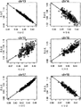
图2示出了通过使用来自300个参考案例的数据建立的标准化覆盖深度-GC含量相关性。将每个案例的标准化覆盖深度相对于序列的GC含量作图。十字表示怀有整倍体女性胎儿的案例,方块表示怀有整倍体男性胎儿的案例。实线是覆盖深度和GC含量的拟合线。 Figure 2 shows the normalized coverage depth-GC content correlation established by using data from 300 reference cases. The normalized depth of coverage for each case is plotted against the GC content of the sequence. Crosses indicate cases with euploid female fetuses and squares indicate cases with euploid male fetuses. Solid lines are fitted lines of coverage depth and GC content. the
图3示出了通过将染色体以其内在上升GC含量排列的标准化的覆盖深度和相应GC含量之间的趋势。这里每一个染色体的内在上升GC含量是指来自300个参考案例染色体的序列标签的平均GC含量。 Figure 3 shows the trend between normalized coverage depth and corresponding GC content by arranging chromosomes by their intrinsic ascending GC content. Here the intrinsic ascending GC content of each chromosome refers to the average GC content of the sequence tags from the 300 reference case chromosomes. the
图4显示了每个染色体的CG类的不同组成。对每个染色体计算参考独特读段的每35bp读段的GC含量,将GC含量分级成36个水平,并且每个水平的百分比被计算为每个染色体的GC组成。然后,将所述染色体通过热图作图并进行层次聚类。 Figure 4 shows the different compositions of CG classes for each chromosome. The GC content of each 35 bp read referenced to the unique reads was calculated for each chromosome, the GC content was graded into 36 levels, and the percentage of each level was calculated as the GC composition of each chromosome. Then, the chromosomes were mapped by heatmap and hierarchically clustered. the
图5显示了图2所示的通过人工模拟测序仪偏好的过程测序偏差引入的相关性。 Figure 5 shows the correlations shown in Figure 2 introduced by process sequencing bias by artificially simulating sequencer preferences. the
图6绘出相对于所测序的多核苷酸片段的总数目的标准差。在150个样品中,每一染色体的校正标准方差显示出与独特读段的数目的平方根的倒数的线性关系。 Figure 6 plots the standard deviation relative to the total number of polynucleotide fragments sequenced. Across 150 samples, the corrected standard deviation for each chromosome showed a linear relationship with the inverse square root of the number of unique reads. the
图7显示了对通过公式3计算的每一条染色体残差绘图,显示出具有正态分布的线性关系。
Figure 7 shows a plot of the residuals for each chromosome calculated by
图8显示了Y染色体覆盖深度的直方图。有两个峰值表示案例的性别可以通过Y染色体的覆盖深度区分。曲线是通过以Gaussian核函数进行核密度估计而得出的Y染色体的相对覆盖深度的分布。 Figure 8 shows the histogram of Y chromosome coverage depth. There are two peaks indicating that the sex of the cases can be distinguished by the coverage depth of the Y chromosome. The curve is the distribution of relative coverage depth of Y chromosome obtained by kernel density estimation with Gaussian kernel function. the
图9显示了用于对903个测试样品诊断胎儿染色体异常的方法的示意图。 Fig. 9 shows a schematic diagram of the method for diagnosing fetal chromosomal abnormalities on 903 test samples. the
图10显示了非整倍性的结果:13三体性、18三体性和21三体性以及XO、XXY、XYY案例和正常案例。图10A显示了对13号染色体、18号染色体和21号染色体的GC含量相对于标准化覆盖深度的绘制图。图10B显示了对X和Y染色体的绘制图。圆代表正常女性胎儿的相对覆盖度深度与GC含量,点代表正常男性胎儿。实线是相对覆盖度和GC含量的拟合线,虚线是t值绝对值是1,点线是t值绝对值是2,点虚线:t值绝对值是3。
Figure 10 shows the results of aneuploidy:
图11比较了不同诊断方法的置信度的值。 Figure 11 compares the values of confidence for different diagnostic methods. the
图12显示了胎儿DNA分数和孕龄之间的关系。母体血浆中胎儿DNA的分数与孕龄相关。胎儿DNA分数通过X和Y一起估计。在平均胎儿DNA分数和孕龄之间有统计显著的相关性(P<0.001)。注意,R2值表示相关系数的平方较小。分数最小值是3.49%。 Figure 12 shows the relationship between fetal DNA fraction and gestational age. The fraction of fetal DNA in maternal plasma correlates with gestational age. Fetal DNA fraction is estimated by X and Y together. There was a statistically significant correlation (P<0.001) between mean fetal DNA fraction and gestational age. Note that the R2 value indicates that the square of the correlation coefficient is small. The score minimum is 3.49%. the
图13显示了标准方差与检测所需的案例数目之间的关系。通过公式5计算每一染色体的标准方差随不同样品数目而变化。当样品数目超过100个时标准方差变得稳定。
Figure 13 shows the relationship between the standard deviation and the number of cases required for detection. The standard deviation of each chromosome was calculated by
图14显示了在无细胞血浆中用于胎儿非整倍性检测的独特读段数目的估计,其是胎儿DNA分数的函数。用于对各自具有不同长度的13号、18号、21号和X甚至Y染色体(从X和Y之间的关系)非整倍性的评估是基于t值置信度水平不小于3。随着胎儿DNA分数降低,需要的鸟枪序列的总数目增加。使用流动池(flowcell)上每通道4百万序列读段的测序通量,如果3.5%的无细胞DNA是胎儿的则可以检测到21三体性。当所述分数和独特读段数目较小例如4%和5百万读段时,不易检测到X染色体的非整倍性。不同染色体需要不同水平的胎儿DNA分数和独特读段数目,这可能是由于染色体的GC结构导致的。
Figure 14 shows estimates of the number of unique reads for fetal aneuploidy detection in cell-free plasma as a function of fetal DNA fraction. Evaluation for aneuploidy of
图15显示了针对每个孕周和每个数据量点的敏感性的恒值图,该恒值图是由用于检测女性胎儿13号染色体三体性的数据量和孕龄(周)绘制的。
Figure 15 shows the constant value diagram for the sensitivity of each gestational week and each data volume point, which is drawn by the data volume and gestational age (weeks) for detecting trisomy of
图16显示了针对每个孕周和每个数据量点的敏感性的恒值图,该恒值图是由用于检测女性胎儿18号染色体三体性的数据量和孕龄(周)绘制的。
Figure 16 shows the constant value diagram for the sensitivity of each gestational week and each data volume point, which is drawn by the data volume and gestational age (weeks) for detecting trisomy of
图17显示了针对每个孕周和每个数据量点的敏感性的恒值图,该恒值图是由用于检测女性胎儿21号染色体三体性的数据量和孕龄(周)绘制的。
Figure 17 shows the constant value diagram for the sensitivity of each gestational week and each data volume point, which is drawn by the data volume and gestational age (week) for detecting trisomy of
图18显示了针对每个孕周和每个数据量点的敏感性的恒值图,该恒值图是由用于检测 女性胎儿X染色体三体性的数据量和孕龄(周)绘制的。 Fig. 18 shows the constant value diagram of the sensitivity for each gestational week and each data volume point, which is drawn by the data volume and gestational age (weeks) used to detect female fetal X chromosome trisomy . the
图19显示了用于检测男性13号染色体三体性的数据量和孕龄(周)绘制的敏感性的恒值图。针对每个孕周和每个数据量点,发明人对给定数据量计算胎儿DNA分数和标准方差的经验分布,并比较通过XY或Y估计的分数,然后发明人计算了每一种类型非整倍性的敏感性。
Figure 19 shows a constant value plot of the sensitivity plotted against the amount of data and gestational age (weeks) for detecting
图20显示了用于检测男性18号染色体三体性的数据量和孕龄(周)绘制的敏感性的恒值图。
Figure 20 shows a constant value plot of the sensitivity plotted against the amount of data and gestational age (weeks) for detecting
图21显示了用于检测男性21号染色体三体性的数据量和孕龄(周)绘制的敏感性的恒值图。
Figure 21 shows a constant value plot of the sensitivity plotted against the amount of data and gestational age (weeks) for detecting
具体实施方式 Detailed ways
发明涉及用于通过大规模测序来自母体生物样品的多核苷酸片段而无创性检测胎儿遗传异常的方法。进一步提供了基于染色体的覆盖深度和相应GC含量之间的关系去除由于染色体GC含量差异造成的测序结果GC偏差的方法。因此,本文提供了一种方法,以通过局部加权的多项式回归从而对每个样品染色体的覆盖深度相对于所述多核苷酸片段的GC含量进行拟合,从而通过计算校正作为参考参数的用于student-t统计的GC含量。 The invention relates to methods for the non-invasive detection of fetal genetic abnormalities by large-scale sequencing of polynucleotide fragments from maternal biological samples. Further provided is a method for removing the GC bias of the sequencing result caused by the difference in the GC content of the chromosome based on the relationship between the coverage depth of the chromosome and the corresponding GC content. Therefore, this paper provides a method to fit the coverage depth of each sample chromosome with respect to the GC content of the polynucleotide fragment by locally weighted polynomial regression, thereby correcting the reference parameter used as GC content of student-t statistics. the
本文还提供了一种通过使用统计假设检验的统计分析来确定胎儿遗传异常的方法。另外,还提供了方法,以计算可用于确定对特定统计显著性水平所需要的临床样品量的数据质量控制(DQC)标准。 Also provided herein is a method for determining a genetic abnormality in a fetus by statistical analysis using statistical hypothesis testing. Additionally, methods are provided to calculate data quality control (DQC) criteria that can be used to determine the clinical sample size required for a particular level of statistical significance. the
I.定义I. Definition
除非另外定义,否则本文使用的所有技术术语和科学术语具有与本发明所属技术领域普通技术人员通常理解相同的含义。本文参考的所有专利、专利申请、公布的专利申请和其他出版物都通过引用的方式全文纳入本文。如果此部分中陈述的定义与通过引用纳入本文的所述专利、专利申请、公布的专利申请和其他出版物中陈述的定义相反或其他方面不一致,此部分中列出的定义优先于通过引用纳入本文中的定义。 Unless defined otherwise, all technical and scientific terms used herein have the same meaning as commonly understood by one of ordinary skill in the art to which this invention belongs. All patents, patent applications, published patent applications, and other publications referenced herein are hereby incorporated by reference in their entirety. To the extent that definitions set forth in this section are contrary to or otherwise inconsistent with definitions set forth in said patents, patent applications, published patent applications, and other publications incorporated herein by reference, the definitions set forth in this section take precedence over those incorporated by reference Definitions in this article. the
除非另外指明,否则本文使用的单数形式“一(a)”、“一个(an)”和“该(the)”包括复数的指代物。例如,“一”二聚体包括一个或多个二聚体。 As used herein, the singular forms "a," "an," and "the" include plural referents unless otherwise specified. For example, reference to "a" dimer includes one or more dimers. the
术语“染色体异常”是指受试者染色体和正常同源染色体的结构之间的偏差。术语“正常”是指特定物种健康个体中出现的占主导地位的核型或带型。染色体异常可以是数目的或结构的,并且包括但不限于非整倍性、多倍性、倒位、三体性、单体性、重复、缺失、部 分染色体缺失、增加、部分染色体增加、插入、染色体片段、染色体区、染色体重排和易位。染色体异常可能与存在病理状态或者与倾向于发生病理状态相关。本文定义的单核苷酸多态性(“SNP”)不是染色体异常。 The term "chromosomal abnormality" refers to a deviation between the structure of a subject's chromosome and the normal homologous chromosome. The term "normal" refers to the predominant karyotype or banding pattern that occurs in healthy individuals of a particular species. Chromosomal abnormalities can be numerical or structural, and include, but are not limited to, aneuploidy, polyploidy, inversion, trisomy, monosomy, duplication, deletion, partial chromosomal deletion, gain, partial chromosomal gain, Insertions, chromosomal fragments, chromosomal regions, chromosomal rearrangements and translocations. Chromosomal abnormalities may be associated with the presence of a pathological condition or with a predisposition to the development of a pathological condition. A single nucleotide polymorphism ("SNP") as defined herein is not a chromosomal abnormality. the
单体性X(XO,缺失整条X染色体)是最Turner综合征的常见类型,在每2500至3000个新生女婴中出现1例(Sybert and McCauley N Engl J Med(2004)351:1227-1238)。XXY综合征是人类男性具有一条额外X染色体的状态,在每1000名男性中大约出现1例(Bock,Understanding Klinefelter Syndrome:A Guide for XXY Males and Their Families.NIH Pub.No.93-3202(1993))。XYY综合征是人类男性具有额外一条Y染色体的性染色体非整倍性,共有47条染色体,而不是正常的46条,在1000个出生男性中有1例受到影响,并可能导致男性不育(Aksglaede,et al.,J Clin Endocrinol Metab(2008)93:169-176)。 Monosomy X (XO, loss of the entire X chromosome) is the most common form of Turner syndrome, occurring in 1 in 2500 to 3000 female births (Sybert and McCauley N Engl J Med (2004) 351:1227- 1238). XXY syndrome is a condition in which human males have an extra X chromosome, occurring in about 1 in 1000 males (Bock, Understanding Klinefelter Syndrome: A Guide for XXY Males and Their Families.NIH Pub.No.93-3202 (1993 )). XYY syndrome is a sex chromosome aneuploidy in which human males have an extra Y chromosome, with a total of 47 chromosomes instead of the normal 46, affecting 1 in 1000 male births and can lead to male infertility ( Aksglaede, et al., J Clin Endocrinol Metab (2008) 93:169-176). the
Turner综合征包括几种状态,其中单体性X(XO,缺少整条性染色体,巴氏小体)最常见。女性通常具有两条X染色体,但Turner综合征中,这些性染色体中的一条缺失。在2000至5000表型女性中出现1例,该综合征自身以多种方式显现。Klinefelter综合征是人类男性具有额外一条X染色体的状态。在人类中,Klinefelter综合征是最常见的性染色体病症,并且是由存在额外染色体引起的第二常见状态。该状态在每1000名男性中出现大约1例。XYY综合征是人类男性具有一条额外Y染色体的性染色体非整倍性,共有47条染色体,而不是正常的46条。这产生了47、XYY核型。该状态通常无症状,并且1000个出生男性中有1例受到影响,这有可能导致男性不育。 Turner syndrome includes several states, of which monosomy X (XO, lack of an entire sex chromosome, Barr bodies) is the most common. Females usually have two X chromosomes, but in Turner syndrome, one of these sex chromosomes is missing. Occurring in 1 in 2000 to 5000 women with the phenotype, the syndrome manifests itself in multiple ways. Klinefelter syndrome is a condition in which human males have an extra X chromosome. In humans, Klinefelter syndrome is the most common sex chromosome disorder and the second most common condition caused by the presence of an extra chromosome. This condition occurs in about 1 in 1000 men. XYY syndrome is a sex chromosome aneuploidy in which human males have an extra Y chromosome, for a total of 47 chromosomes instead of the normal 46. This produces a 47, XYY karyotype. The condition is usually asymptomatic and affects 1 in 1000 male births, potentially leading to male infertility. the
13三体性(Patau综合征)、18三体性(Edward综合征)和21三体性(Down综合征)是临床上最重要的常染色体三体性,而如何检测它们一直是热点。检测以上胎儿染色体畸变在产前诊断中具有重大意义(Ostler,Diseases of the eye and skin:a color atlas。Lippincott Williams & Wilkins.pp.72.ISBN9780781749992(2004);Driscoll and Gross N Engl J Med(2009)360:2556-2562;Kagan,et al.,Human Reproduction(2008)23:1968-1975)。 Trisomy 13 (Patau syndrome), trisomy 18 (Edward syndrome) and trisomy 21 (Down syndrome) are the most clinically important autosomal trisomies, and how to detect them has always been a hot spot. The detection of the above fetal chromosomal aberrations is of great significance in prenatal diagnosis (Ostler, Diseases of the eye and skin: a color atlas . Lippincott Williams & Wilkins.pp.72.ISBN9780781749992 (2004); Driscoll and Gross N Engl J Med (2009 ) 360: 2556-2562; Kagan, et al., Human Reproduction (2008) 23: 1968-1975).
术语“参考独特读段”是指具有独特序列的染色体片段。因此,这类片段可以被明确地分配至单染色体位置。染色体的参考独特读段可以基于发布的基因组参考序列例如hg18或hg19进行构建。 The term "reference unique read" refers to a segment of a chromosome that has a unique sequence. Thus, such fragments can be unambiguously assigned to single-chromosomal positions. Reference unique reads for a chromosome can be constructed based on published genome reference sequences such as hg18 or hg19. the
术语“多核苷酸”、“寡核苷酸”、“核酸”和“核酸分子”在本文中可互换使用,是指任意长度核苷酸的聚合体形式,并且可以包括核糖核苷酸、脱氧核糖核苷酸、其类似物或其混合物。该术语仅是指所述分子的一级结构。因此,所述术语包括三链、双链和单链脱氧核糖核酸(“DNA”)以及三链、双链和单链核糖核酸(“RNA”)。它还包括修饰形式的(例如通过烷基化和/或通过加帽)多核苷酸和非修饰形式的多核苷酸。更具体地,术语“多核苷酸”、 “寡核苷酸”、“核酸”和“核酸分子”包括多脱氧核糖核苷酸(含2-脱氧-D-核糖)、多核糖核苷酸(含D-核糖),包括tRNA、rRNA、hRNA和剪接或未剪接的mRNA,嘌呤或嘧啶碱基的N-糖苷或C-糖苷的任何其他类型多核苷酸,以及含非核苷酸(normucleotidic)骨架的其他多聚体,例如聚酰胺(例如核酸肽(“PNA”))和多吗啉代(可市购自Anti-Virals,Inc.,Corvallis,OR.,例如
“大规模平行测序”是指用于测序数百万核酸片段的技术,例如通过将随机片段化的基因组DNA附着于透光的平面上并进行固相扩增以形成具有数百万个簇的高密度测序流动池,每个簇在每平方厘米上包含约1000个拷贝的模板。将这些模板使用4色DNA边合成边测序技术进行测序。参见Illumina,Inc.,San Diego,Calif提供的产品。本发明使用的测序优选在无预扩增或克隆步骤的情况下进行,但可以与基于扩增的方法相结合,该方法在微流芯片具有可以用于PCR和基于显微模板测序的反应室。仅需要约30bp的随机序列信息鉴定属于特定人类的染色体序列。更长序列可以唯一地鉴定更具体的目标。在本例中,获得了大量的35bp读段。对大规模平行测序方法的进一步描述见Rogers and Ventner,Nature(2005)437:326-327。 "Massively parallel sequencing" refers to techniques used to sequence millions of nucleic acid fragments, for example by attaching randomly fragmented genomic DNA to a light-transmitting surface and performing solid-phase amplification to form clusters of millions of High-density sequencing flow cell, each cluster contains about 1000 copies of template per square centimeter. These templates were sequenced using 4-color DNA sequencing by synthesis technology. See offerings from Illumina, Inc., San Diego, Calif. Sequencing as used in the present invention is preferably performed without pre-amplification or cloning steps, but can be combined with amplification-based methods that have reaction chambers on a microfluidic chip that can be used for PCR and microscopic template-based sequencing . Only about 30 bp of random sequence information is required to identify chromosomal sequences belonging to a particular human. Longer sequences can uniquely identify more specific targets. In this example, a large number of 35bp reads were obtained. For a further description of massively parallel sequencing methods see Rogers and Ventner, Nature (2005) 437:326-327. the
本文使用的“生物样品”是指自活体或病毒来源或其他来源的大分子和生物分子获得的任何样品,并且包括受试者的任何细胞类型或组织,从所述受试者可获得核酸、蛋白质或其他大分子。生物样品可以是从生物源直接获得的样品,或者经过加工的样品。例如,扩增的分离核酸构成了生物样品。生物样品包括但不限于体液,例如血液、血浆、血清、脑脊液、滑膜液、尿和汗液;来自动植物的组织和器官样品及其经加工样品。 As used herein, "biological sample" refers to any sample obtained from a living organism or macromolecules and biomolecules of viral or other origin, and includes any cell type or tissue of a subject from which nucleic acids, proteins or other macromolecules. A biological sample may be a sample obtained directly from a biological source, or a processed sample. For example, amplified isolated nucleic acid constitutes a biological sample. Biological samples include, but are not limited to, bodily fluids such as blood, plasma, serum, cerebrospinal fluid, synovial fluid, urine, and sweat; tissue and organ samples from animals and plants, and processed samples thereof. the
应理解,本文描述的本发明的方面和实施方案包括“由......组成”和/或“基本由......组 成”的方面和实施方案。 It is to be understood that aspects and embodiments of the invention described herein include "consisting of" and/or "consisting essentially of" aspects and embodiments. the
结合附图的如下详细说明,本发明的其他目标、优点和特征将变得清晰。 Other objects, advantages and features of the present invention will become apparent from the following detailed description in conjunction with the accompanying drawings. the
II.建立覆盖深度和GC含量之间的关系II. Establishing the relationship between coverage depth and GC content
本文提供了一种用于建立染色体的覆盖深度和GC含量之间的关系的方法,所述方法包括:从多于一个样品获得涵盖所述染色体和另一染色体的多个多核苷酸片段的序列信息;基于所述序列信息将所述片段分配至染色体;基于每个样品的所述序列信息计算所述染色体的覆盖深度和GC含量;并确定所述染色体的所述覆盖深度和GC含量之间的关系。所述操作步骤可以以无特定顺序的方式进行。在一些实施例中,所述方法可以以如下顺序进行:a)从多于一个样品获得涵盖所述染色体和另一染色体的多个多核苷酸片段的序列信息;b)基于所述序列信息将所述片段分配至染色体;c)基于对于每个样品所述序列信息计算所述染色体的覆盖深度和GC含量;和d)确定所述染色体的所述覆盖深度和GC含量之间的关系。 Provided herein is a method for establishing a relationship between depth of coverage and GC content of a chromosome, the method comprising: obtaining sequences of a plurality of polynucleotide fragments covering the chromosome and another chromosome from more than one sample information; assign the fragments to chromosomes based on the sequence information; calculate the coverage depth and GC content of the chromosome based on the sequence information of each sample; and determine the distance between the coverage depth and GC content of the chromosome Relationship. The operational steps may be performed in no particular order. In some embodiments, the method may be performed in the following order: a) obtaining sequence information from more than one sample covering multiple polynucleotide fragments of the chromosome and another chromosome; b) obtaining sequence information based on the sequence information assigning the fragments to chromosomes; c) calculating a coverage depth and GC content of the chromosome based on the sequence information for each sample; and d) determining a relationship between the coverage depth and GC content of the chromosome. the
为了计算染色体定位的覆盖深度和GC含量,通过测序从样品获得的模板DNA来获得多核苷酸片段的序列信息。在一个实施例中,所述模板DNA包含母体DNA和胎儿DNA二者。在另一个实施例中,模板DNA获自孕妇的血液。血液可以使用用于取血的任何常规技术收集,包括但不限于静脉穿刺。例如,血液可以取自肘内侧或手背的静脉。血样可以在胎儿妊娠的任何时间从孕妇采集。例如,血样可以在胎儿妊娠的第1-4、4-8、8-12、12-16、16-20、20-24、24-28、28-32、32-36、36-40或40-44周,优选在胎儿妊娠的第8-28周,从人类女性采集。 To calculate the coverage depth and GC content of chromosomal mapping, the sequence information of the polynucleotide fragments was obtained by sequencing the template DNA obtained from the samples. In one embodiment, the template DNA comprises both maternal and fetal DNA. In another embodiment, the template DNA is obtained from the blood of a pregnant woman. Blood can be collected using any conventional technique for blood drawing, including but not limited to venipuncture. For example, blood can be taken from a vein on the inside of the elbow or the back of the hand. Blood samples can be collected from pregnant women at any time during fetal pregnancy. For example, blood samples can be taken on days 1-4, 4-8, 8-12, 12-16, 16-20, 20-24, 24-28, 28-32, 32-36, 36-40, or 40 days of fetal pregnancy. - 44 weeks, preferably between weeks 8-28 of fetal gestation, from a human female. the
所述多核苷酸片段基于所述序列信息被分配至染色体定位。基因组参考序列用于获得所述参考独特读段。本文使用的术语“参考独特读段”是指基于基因组参考序列已被分配至特异基因组定位的所有独特多核苷酸片段。在一些实施例中,参考独特读段具有相同长度,例如约10、12、15、20、25、30、35、40、50、100、200、300、500或1000bp。在其他一些实施例中,人基因组版本hg18或hg19可以被用作所述基因组参考序列。染色体定位可以是染色体上具有长度约为10、20、30、40、50、60、70、80、90、100、200、300、400、500、600、700、800、900、1000、2000、300、400、5000、6000、7000、8000、9000、10,000或更多kb的连续窗口。染色体定位还可以是单条染色体。 The polynucleotide fragments are assigned to chromosomal locations based on the sequence information. A genomic reference sequence is used to obtain the reference unique reads. As used herein, the term "reference unique reads" refers to all unique polynucleotide fragments that have been assigned to a specific genomic location based on a genomic reference sequence. In some embodiments, the reference unique reads are the same length, eg, about 10, 12, 15, 20, 25, 30, 35, 40, 50, 100, 200, 300, 500, or 1000 bp. In some other embodiments, human genome version hg18 or hg19 can be used as the genome reference sequence. The chromosomal location can be on the chromosome with a length of about 10, 20, 30, 40, 50, 60, 70, 80, 90, 100, 200, 300, 400, 500, 600, 700, 800, 900, 1000, 2000, Sequential windows of 300, 400, 5000, 6000, 7000, 8000, 9000, 10,000 or more kb. A chromosomal location can also be a single chromosome. the
本文使用的术语“覆盖深度”是指所述分配到染色体定位的片段数目和所述染色体定位的参考独特读段数目之间的比值,使用以下公式: As used herein, the term "depth of coverage" refers to the ratio between the number of fragments assigned to a chromosome map and the number of reference unique reads for that chromosome map, using the following formula:
Ci,j=ni,j/Nj,j=1,2,…,22,X,Y (1), C i,j =n i,j /N j ,j=1,2,...,22,X,Y (1),
其中ni,j是样品i中映射到染色体j上的独特序列读段数目;Ci,j是样品i中染色体i的所述覆盖深度;Nj是染色体j中参考独特读段数目。 where n i,j is the number of unique sequence reads in sample i mapped to chromosome j; C i,j is the coverage depth of chromosome i in sample i; N j is the number of reference unique reads in chromosome j.
在一些实施例中,将未分配至单个染色体定位或分配至多个染色体定位的多核苷酸片段被忽略。在一些实施例中,所述覆盖深度是基于另一染色体定位的覆盖深度、另一染色体的覆盖深度、所有其他常染色体的平均覆盖深度、所有其他染色体的平均覆盖深度或所有染色体的平均覆盖深度而标准化的。在一些实施例中,22条常染色体的平均覆盖深度被用作标准化常数以计算对不同样品获得的序列读段总数目的差异: In some embodiments, polynucleotide fragments that are not assigned to a single chromosomal location or that are assigned to multiple chromosomal locations are ignored. In some embodiments, the depth of coverage is the depth of coverage based on another chromosome location, the depth of coverage of another chromosome, the average depth of coverage of all other autosomes, the average depth of coverage of all other chromosomes, or the average depth of coverage of all chromosomes And standardized. In some embodiments, the average coverage depth of the 22 autosomes is used as a normalization constant to calculate the difference in the total number of sequence reads obtained for different samples:
其中cri,j代表样品i中染色体j的相对覆盖深度。从这个角度提出,每一条染色体的“相对覆盖深度”是指标准化的值,被用于比较不同的样品以及用于后续分析。 where cr i,j represents the relative coverage depth of chromosome j in sample i. From this point of view, the "relative depth of coverage" of each chromosome refers to the normalized value, which is used to compare different samples and for subsequent analysis.
染色体定位的GC含量可以通过染色体定位的所述平均GC百分比计算,其中所述染色体定位是基于所述染色体定位的所述独特参考读段,或基于被分配至染色体定位的经测序多核苷酸片段。染色体的GC含量可以使用以下公式进行计算: The GC content of a chromosome map can be calculated by the average GC percentage of a chromosome map based on the unique reference reads of the chromosome map, or based on the sequenced polynucleotide fragments assigned to the chromosome map . The GC content of a chromosome can be calculated using the following formula:
GCi,j=NGCi,j/BASEi,j (3), GC i, j = NGC i, j / BASE i, j (3),
其中i代表样品i,j代表染色体j,NGCi,j代表样品i中染色体j上的DNA碱基G和C数目,BASEi,j代表样品i中染色体j上的DNA碱基数目。 Where i represents sample i, j represents chromosome j, NGC i, j represents the number of DNA bases G and C on chromosome j in sample i, BASE i, j represents the number of DNA bases on chromosome j in sample i.
覆盖深度和GC含量可以基于从单个样品或者从多个样品获得的多核苷酸片段的所述序列信息。为了建立染色体定位的所述覆盖深度和GC含量之间的关系,所述计算可以基于从至少1、2、5、10、20、50、100、200、500或1000个样品中获得的多核苷酸片段的所述序列信息。 Depth of coverage and GC content can be based on the sequence information of polynucleotide fragments obtained from a single sample or from multiple samples. In order to establish the relationship between the coverage depth and GC content of chromosomal mapping, the calculation may be based on polynucleotides obtained from at least 1, 2, 5, 10, 20, 50, 100, 200, 500 or 1000 samples The sequence information of the acid fragment. the
在一些实施例中,覆盖深度和GC含量之间的所述关系是非强线性关系。 In some embodiments, the relationship between depth of coverage and GC content is a non-strong linear relationship. the
Loess算法或局部加权多项式回归可以用于评估数值对之间例如覆盖深度和GC含量之间的非线性关系(相关性)。 The Loess algorithm or locally weighted polynomial regression can be used to evaluate non-linear relationships (correlations) between pairs of values, eg, between coverage depth and GC content. the
III.确定胎儿遗传异常III. Determination of Fetal Genetic Abnormalities
本文还提供了一种确定胎儿遗传异常的方法,所述方法包括:a)从样品获得多个多核苷酸片段的序列信息;b)基于所述序列信息将所述片段分配至染色体;c)基于所述序列信息计算染色体的覆盖深度和GC含量;d)使用所述染色体的所述GC含量以及确立所述染 色体的覆盖深度和GC含量之间的关系计算所述染色体的拟合覆盖深度;并且e)将所述拟合覆盖深度和所述染色体的所述覆盖深度进行比较,其中它们之间的差异表明胎儿遗传异常。 Also provided herein is a method of determining a genetic abnormality in a fetus, the method comprising: a) obtaining sequence information of a plurality of polynucleotide fragments from a sample; b) assigning the fragments to chromosomes based on the sequence information; c) Calculating the coverage depth and GC content of a chromosome based on the sequence information; d) calculating the fitted coverage of the chromosome using the GC content of the chromosome and establishing a relationship between the coverage depth and GC content of the chromosome depth; and e) comparing said fitted coverage depth to said coverage depth of said chromosome, wherein a difference between them indicates a fetal genetic abnormality. the
所述方法可以用于检测胎儿染色体异常,并且特别适用于检测非整倍性、多倍性、单体性、三体性、21三体性、13三体性、14三体性、15三体性、16三体性、18三体性、22三体性、三倍性、四倍性,和性染色体异常包括XO、XXY、XYY和XXX。根据本方法还可以关注人类基因组中的特定区域以鉴定部分单体性和部分三体性。例如,所述方法可以涉及分析确定的染色体滑动“窗口”中的序列数据,例如分布在整个染色体上的连续的、不重叠的50kb区域。除其他之外,已经报道了部分三体性13q、8p(8p23.1)、7q、远端6p、5p、3q(3q25.1)、2q、1q(1q42.1和1q21-qter)、部分Xpand单体性4q35.1。例如,在18q21.1-qter重复的情况下,染色体18长臂的部分重复可导致爱德华兹综合征(Mewar,et al.,Am J Hum Genet.(1993)53:1269-78)。
The method can be used to detect fetal chromosomal abnormalities, and is particularly suitable for detecting aneuploidy, polyploidy, monosomy, trisomy, 21 trisomy, 13 trisomy, 14 trisomy, 15 trisomy Trisomy,
在一些实施例中,所述胎儿分数是基于来自样品的所述多核苷酸片段获得的所述序列信息而估计的。X和Y染色体的覆盖深度和GC含量可以用于估计所述胎儿分数。在一些实施例中,所述胎儿性别是基于来自样品的所述多核苷酸片段获得的所述序列信息而确定的。X和Y染色体的覆盖深度和GC含量可以用于确定所述胎儿性别。 In some embodiments, said fetal fraction is estimated based on said sequence information obtained from said polynucleotide fragments of a sample. The coverage depth and GC content of the X and Y chromosomes can be used to estimate the fetal fraction. In some embodiments, said fetal sex is determined based on said sequence information obtained from said polynucleotide fragments of a sample. The coverage depth and GC content of the X and Y chromosomes can be used to determine the sex of the fetus. the
在一些实施例中,所述拟合覆盖深度与所述染色体的所述覆盖深度的所述比较是通过统计假设检验进行的,其中一个假设是所述胎儿是整倍体(HO)且另一个假设是所述胎儿是非整倍体(H1)。在一些实施例中,分别对两个假设计算student t-统计量,作为t1和t2。在一些实施例中,计算t1和t2的对数似然比。在一些实施例中,对数似然比>1表明指示所述胎儿三体性。 In some embodiments, said comparison of said fitted coverage depth to said coverage depth of said chromosome is performed by statistical hypothesis testing, wherein one hypothesis is that said fetus is euploid (HO) and the other The hypothesis is that the fetus is aneuploid (H1). In some embodiments, student t-statistics are calculated for the two hypotheses, as t1 and t2, respectively. In some embodiments, the log-likelihood ratio of t1 and t2 is calculated. In some embodiments, a log likelihood ratio >1 is indicative of said fetal trisomy. the
IV.用于诊断胎儿遗传异常的计算机可读介质和系统IV. Computer Readable Medium and System for Diagnosing Fetal Genetic Abnormalities
在另一方面,本文提供了一种包含用于进行胎儿遗传异常的产前诊断的多条指令的计算机可读介质,其包含以下步骤:a)接收所述序列信息;b)基于所述序列信息将所述多核苷酸片段分配至染色体;c)基于所述序列信息计算染色体的覆盖深度和GC含量;d)使用所述染色体的所述GC含量以及所确立的所述染色体的覆盖深度和GC含量之间的关系计算所述染色体的拟合覆盖深度;以及e)将所述拟合覆盖深度与所述染色体的覆盖深度进行比较,其中它们之间的差异表明遗传异常。 In another aspect, provided herein is a computer readable medium comprising instructions for performing prenatal diagnosis of a fetal genetic abnormality, comprising the steps of: a) receiving said sequence information; b) based on said sequence information assigning said polynucleotide fragments to chromosomes; c) calculating a coverage depth and GC content of a chromosome based on said sequence information; d) using said GC content of said chromosome and the established coverage depth and calculating a fitted coverage depth for the chromosome from the relationship between the GC contents; and e) comparing the fitted coverage depth with the coverage depth for the chromosome, wherein a difference between them indicates a genetic abnormality. the
在又一方面中,本文提供了一种用于确定胎儿非整倍性的系统,其包括:a)用于所述多个多核苷酸片段获得序列信息的手段;和b)包含用于进行胎儿遗传异常的产前诊断的多 条指令的计算机可读介质。在一些实施例中,所述系统还包含从怀孕的女性受试者获得的生物样品,其中所述生物样品包含多个多核苷酸片段。 In yet another aspect, provided herein is a system for determining fetal aneuploidy comprising: a) means for obtaining sequence information for said plurality of polynucleotide fragments; and b) comprising means for performing A computer readable medium of instructions for prenatal diagnosis of fetal genetic abnormalities. In some embodiments, the system further comprises a biological sample obtained from a pregnant female subject, wherein the biological sample comprises a plurality of polynucleotide fragments. the
对本领域技术人员来说显而易见的是,可以使用若干不同的测序方法和变型。在一个实施例中,所述测序是使用大规模平行测序进行的。大规模平行测序,例如可在454平台(Roche)(Margulies,et al.,Nature(2005)437:376-380)、Illumina Genome Analyzer(或SolexaTM平台)或SOLiD System(Applied Biosystems)上或采用Helicos True Single Molecule DNA测序技术(Harris,et al.,Science(2008)320:106-109)、单分子、Pacific Biosciences的实时(SMRTTM)技术和纳米孔测序技术(Soni and Meller,Clin Chem(2007)53:1996-2001)实现的那些,允许以平行的方式以高次多路对从样本分离的许多核酸分子进行测序(Dear,Brief Funct Genomic Proteomic(2003)1:397-416)。这些平台各自均可测序克隆扩增的或者甚至未扩增的核酸片段单分子。市售可得的测序设备可以用于获得所述多核苷酸片段的所述序列信息。 It will be apparent to those skilled in the art that several different sequencing methods and variations can be used. In one embodiment, said sequencing is performed using massively parallel sequencing. Massively parallel sequencing, for example on the 454 platform (Roche) (Margulies, et al., Nature (2005) 437:376-380), Illumina Genome Analyzer (or Solexa TM platform) or SOLiD System (Applied Biosystems) or using Helicos True Single Molecule DNA sequencing technology (Harris, et al., Science (2008) 320: 106-109), single molecule, real-time (SMRT TM ) technology of Pacific Biosciences and nanopore sequencing technology (Soni and Meller, Clin Chem ( 2007) 53: 1996-2001 ), allowing high-order multiplexed sequencing of many nucleic acid molecules isolated from a sample in a parallel fashion (Dear, Brief Funct Genomic Proteomic (2003) 1: 397-416). Each of these platforms can sequence clone amplified or even single molecules of non-amplified nucleic acid fragments. Commercially available sequencing equipment can be used to obtain the sequence information of the polynucleotide fragments.
V.实施例V. Examples
提供以下实施例以举例说明本发明,但不限制本发明。 The following examples are provided to illustrate the invention, but not to limit it. the
实施例1分析影响检测敏感性的因素:GC偏差和性别Example 1 Analysis of Factors Affecting Detection Sensitivity: GC Bias and Gender
用于计算覆盖深度和GC含量的示意步骤框架显示于图1。发明人使用软件通过将hg18参考序列切割成1-mer(这里的1-mer是以与样品测序读段相同的长度“1”从人类序列参考人工分解的读段)而产生参考独特读段,并收集这些“独特”1-mer作为发明人的参考独特读段。第二,本发明人将他们的测序样品读段映射至每个染色体的参考独特读段。第三,发明人通过应用五分之一离群值截止法去除离群值,以得到干净的数据集。最后,发明人对每个样品计算每一条染色体的覆盖深度,并且对于每个样品计算映射到每一条染色体的测序独特读段的GC含量。 A schematic framework of steps used to calculate coverage depth and GC content is shown in Figure 1. The inventors used software to generate reference unique reads by cutting the hg18 reference sequence into 1-mers (here 1-mers are reads that were manually resolved from the human sequence reference with the same length "1" as the sample sequencing reads), And these "unique" 1-mers were collected as inventor's reference unique reads. Second, the inventors mapped their sequencing sample reads to reference unique reads for each chromosome. Third, the inventors removed outliers by applying a quintile outlier cutoff to obtain a clean dataset. Finally, the inventors calculated the coverage depth for each chromosome for each sample, and calculated for each sample the GC content of the sequenced unique reads that mapped to each chromosome. the
为了研究GC含量如何影响发明人的数据,发明人选择了有核型结果的300个整倍体案例,并将它们的测序读段的覆盖深度和相关GC含量散布至图中,该图显示了它们之间的强相关性,该现象以前未被报道过(图2)。在图2中,覆盖深度与GC-含量强相关,在一些染色体例如4、13等中显示明显的向下趋势,而在其他染色体例如19、22等中显示向上趋势。将所有染色体按它们的内在GC-含量升序排列,如图3中所示,向下趋势存在于较低GC-含量组染色体中,而向上趋势存在于较高GC-含量组染色体中。这可以解释为,如果对于一个样品被测序的多核苷酸片段具有比其他样品更高的GC-含量,则该样品呈现 的覆盖深度与其他样品的覆盖深度相比在较低GC-含量组染色体中将下降,而在较高GC-含量组染色体中将上升。 To investigate how GC content affects our data, the inventors selected 300 euploid cases with karyotype results and scattered the coverage depth and associated GC content of their sequencing reads into a plot showing A strong correlation between them, a phenomenon not previously reported (Fig. 2). In Figure 2, coverage depth is strongly correlated with GC-content, showing a clear downward trend in some chromosomes such as 4, 13, etc., and an upward trend in others such as 19, 22, etc. Arranging all chromosomes in ascending order of their intrinsic GC-content, as shown in Figure 3, a downward trend exists in the lower GC-content group chromosomes, while an upward trend exists in the higher GC-content group chromosomes. This can be interpreted as, if for one sample the sequenced polynucleotide fragments have a higher GC-content than other samples, the sample exhibits a coverage depth compared to the coverage depth of the other samples in the lower GC-content group of chromosomes will decrease in the chromosomes of the higher GC-content group and will increase in the chromosomes of the higher GC-content group. the
对于在不同GC-含量染色体中这种不同变化趋势的可能解释是,图4中所示的不同染色体中GC-含量组成的差异结合测序过程中引入的GC偏差。对于每一条染色体的每35-mer参考独特读段的GC含量被用于将GC含量分级成36个水平。计算作为每一条染色体的GC组成的每个水平的百分比,然后用于使用Heatmap2软件画热图。以染色体13为例,其大部分由较低GC-含量序列区段组成,但其小部分由较高GC-含量序列区段组成。如果测序或PCR过程中的条件有利于测序那些较高GC-含量的区段,相当大部分的具有低GC-含量的染色体13将难以被测序,这导致在该样品中染色体13的覆盖深度变得较低。相比之下,在较高GC-含量组例如染色体19中,在该样品中染色体19的覆盖深度变得较高,原因是大部分的染色体19具有较高的测序仪偏好GC-含量。不管在哪个染色体中,少含GC区段和富含GC区段都难以被测序,但GC偏差引起的影响对具有不同GC-含量组成的不同染色体是不同的。将每一条参考染色体分成1kb的区段,计算所述区段中每个独特参考读段的GC含量。将以合适间隔形式[0.3,0.6]存在的每个区段的GC含量除以0.001的步长,然后计算每个间隔的相对覆盖度。图5显示了对每条染色体的相对覆盖度和GC含量的作图。
A possible explanation for this different variation trend in different GC-content chromosomes is the difference in GC-content composition in different chromosomes shown in Fig. 4 combined with GC bias introduced during the sequencing process. The GC content of each 35-mer reference unique read for each chromosome was used to rank the GC content into 36 levels. The percentage of each level was calculated as the GC composition of each chromosome and then used to draw a heatmap using Heatmap2 software. Taking
使用两独立样本的t检验分析胎儿性别对数据的影响。对于相同GC含量除了性染色体之外的常染色体之间基本未发现显著差异,但在女性和男性之间UR%有明显差异(Chiu et a1.,(2008)Proc Natl Acad Sci USA105:20458-20463),暗示在检测常染色体非整倍性时不需要区分胎儿性别,但在检测性染色体非整倍性例如XO、XYY等时需要首先区分胎儿性别。 The effect of fetal sex on the data was analyzed using the t-test of two independent samples. For the same GC content, no significant difference was found between autosomes except sex chromosomes, but there was a significant difference in UR% between females and males (Chiu et a1., (2008) Proc Natl Acad Sci USA105: 20458-20463 ), implying that it is not necessary to distinguish the sex of the fetus when detecting autosomal aneuploidy, but it is necessary to first distinguish the sex of the fetus when detecting sex chromosome aneuploidy such as XO, XYY, etc. the
实施例2统计模型
使用上文讨论的该现象,发明人试图使用局部多项式来拟合覆盖深度和相应GC含量之间的关系。覆盖深度由如下的GC的函数和正态分布的残差组成: Using this phenomenon discussed above, the inventors attempted to fit the relationship between coverage depth and corresponding GC content using local polynomials. Coverage depth is composed of the following function of GC and normally distributed residuals:
cri,j=f(GCi,j)+εi,j,j=1,2,…,22,X,Y (4), cr i, j = f(GC i, j )+ε i, j , j=1, 2,..., 22, X, Y (4),
其中f(GCi,j)代表样品i、染色体j的覆盖深度和相应GC含量之间的关系的函数,εi,j代表样品i、染色体i的残差。 where f(GC i,j ) represents the function of the relationship between the coverage depth of sample i and chromosome j and the corresponding GC content, and ε i,j represents the residual of sample i and chromosome i.
在所述覆盖深度和相应GC含量之间有非强线性关系,因此发明人应用loess算法将所述覆盖深度与所述相应GC含量进行拟合,发明人从中计算了对发明人的模型来说重要的值,即拟合覆盖深度: There is a non-strong linear relationship between the depth of coverage and the corresponding GC content, so the inventors applied the loess algorithm to fit the depth of coverage to the corresponding GC content, from which the inventors calculated the Important values, namely fit coverage depth:
用所述拟合覆盖深度,根据以下公式6和公式7计算标准方差和student t:
Using the fitted coverage depths as described, the standard deviation and student t were calculated according to
实施例3胎儿分数估计Example 3 Fetal Fraction Estimation
由于胎儿分数对发明人的检测非常重要,在测试步骤之前发明人估计了胎儿分数。如前文指出的,发明人测序了19名成年男性,当将他们的覆盖深度与怀有女性胎儿的案例比较时,发明人发现男性的X染色体覆盖深度是女性X染色体覆盖深度接近1/2,男性的Y染色体覆盖深度比女性Y染色体覆盖深度大接近0.5倍。因此,发明人可以估计所述胎儿分数如公式8、公式9和公式10依赖于X和Y染色体的覆盖深度,并考虑GC相关性:
Since fetal fraction is so important to the inventor's assay, the inventor estimated the fetal fraction prior to the testing procedure. As pointed out above, the inventors sequenced 19 adult males, and when comparing their coverage depths with those of female fetuses, the inventors found that the X chromosome coverage depth of males was close to 1/2 of that of female X chromosomes, The coverage depth of Y chromosome in male is nearly 0.5 times larger than that in female. Therefore, the inventors can estimate the fetal fraction as
其中是通过对怀有女性胎儿案例的X染色体覆盖深度和相应GC含量进行回归关联得到的拟合覆盖深度,


实施例4计算每一条染色体的残差
图6显示了,在独特读段确定总数目下每一条染色体的标准差(参见公式3)受到参与的参考案例数目的影响。在对于每个案例测序170万的总独特读段数目的条件下,当选择的案例数目超过150个时,标准差几乎不增加。然而,对于不同染色体所述标准差是不同的。在考虑GC偏差后,发明人的方法对于以下染色体有适度的标准差:染色体13 (0.0063)、染色体18(0.0066)和染色体21(0.0072)。X染色体的标准差比以上提及的染色体高,它需要更多策略来进行准确的异常检测。 Figure 6 shows that the standard deviation per chromosome (see Equation 3) under the total number of unique read determinations is affected by the number of reference cases involved. With a total number of unique reads sequenced for each case of 1.7 million, the standard deviation hardly increases when the number of selected cases exceeds 150. However, the standard deviation is different for different chromosomes. After accounting for GC bias, the inventors' method had modest standard deviations for the following chromosomes: chromosome 13 (0.0063), chromosome 18 (0.0066), and chromosome 21 (0.0072). The standard deviation of the X chromosome is higher than that of the above-mentioned chromosomes, which requires more strategies for accurate anomaly detection. the
图7显示了Q-Q绘图,其中残差被编译成正态分布,正态分布表明所述student-t计算是合理的。 Figure 7 shows a Q-Q plot where the residuals are compiled into a normal distribution, which indicates that the student-t calculation is reasonable. the
实施例5区分胎儿性别
为了发现性染色体病症,最好区分胎儿性别。在发明人对300个案例中Y染色体覆盖深度的频率分布进行研究时,存在两个明显峰,这提示可通过Y染色体的覆盖深度区分性别。覆盖深度小于0.04的案例可以被看作怀有女性胎儿,而大于0.051被看作怀有男性胎儿,0.04和0.051之间被看作性别不确定,如图8。对于这些性别不确定和非整倍性案例,使用逻辑回归来预测他们的性别,如公式11(Fan,et al.,Proc Natl Acad Sci USA(2008)42:16266-16271): In order to detect sex chromosome disorders, it is best to differentiate the sex of the fetus. When the inventors studied the frequency distribution of the coverage depth of the Y chromosome in 300 cases, there were two obvious peaks, which suggested that genders could be distinguished by the coverage depth of the Y chromosome. Cases with a coverage depth of less than 0.04 can be regarded as pregnant with female fetuses, while cases greater than 0.051 are regarded as pregnant with male fetuses, and cases between 0.04 and 0.051 are regarded as indeterminate gender, as shown in Figure 8. For these gender indeterminate and aneuploidy cases, use logistic regression to predict their sex, as in Equation 11 (Fan, et al., Proc Natl Acad Sci USA (2008) 42: 16266-16271):
其中cr.ai,x和cr.ai,y分别是X和Y的标准化的相对覆盖度。 where cr.a i,x and cr.a i,y are the normalized relative coverage of X and Y, respectively.
与核型结果相比,发明人的区分胎儿性别的方法在其300个参考案例中进行的非常好,准确性100%,而在其901个案例组中进行时仅弄错一个案例,并且该错误案例的Y染色体覆盖深度在0.04和0.051之间。 Compared with the karyotype results, the inventor's method of distinguishing fetal sex performed very well in its 300 reference cases with 100% accuracy, while only getting one case wrong in its 901 case group, and the The depth of Y chromosome coverage for the error cases was between 0.04 and 0.051. the
实施例6GC-相关性t-检验方法的诊断性能The diagnostic performance of embodiment 6GC-correlation t-test method
样品募集Sample collection
903参加者预期地募集自深圳人民医院和深圳母婴保健中心,有其核型结果。从每个募集单位的公共审查部门获得许可,所有参加者签署知情同意书。在取血时记录母亲年龄和孕周。所述903个案例包括2个13三体性案例、15个18三体性案例、16个21三体性案例、3个XO案例、2个XXY案例和1个XYY案例。其核型结果分布如图9中所示。
903 participants were prospectively recruited from Shenzhen People's Hospital and Shenzhen Maternal and Child Health Center with their karyotype results. Permission was obtained from the public review department of each recruiting unit, and all participants signed informed consent. Maternal age and gestational age were recorded at the time of blood draw. The 903 cases included 2 cases of
母体血浆DNA测序Maternal plasma DNA sequencing
从每个参加孕妇采集外周静脉血(5毫升)至EDTA管中,在4小时内在1,600g下离心10分钟。将血浆转移至微量离心管中并在16,000g下离心10分钟以除去残留细胞。将无细胞血浆在80摄氏度下保存至DNA提取。每个血浆样品仅冻融一次。 Peripheral venous blood (5 mL) was collected from each participating pregnant woman into EDTA tubes and centrifuged at 1,600 g for 10 minutes over 4 hours. Plasma was transferred to microcentrifuge tubes and centrifuged at 16,000g for 10 minutes to remove residual cells. Cell-free plasma was stored at 80°C until DNA extraction. Each plasma sample was frozen and thawed only once. the
为了大规模平行基因组测序,根据改良的来自Illumina的方案使用从600微升母体血浆提取的DNA进行DNA文库构建。简而言之,使用T4DNA聚合酶、KlenowTM聚合酶和 T4多核苷酸激酶对母体血浆DNA片段进行末端配对。加入末端A残基后,将市购的接头(Illumina)连接至所述DNA片段。然后,将所述连接接头的DNA以常规多重引物使用17个循环的PCR另外进行扩增。使用Agencourt AMPureTM60毫升试剂盒(Beckman)纯化PCR产物。在2100BioanalyzerTM(Agilent)上以DNA1000试剂盒分析所述测序文库的大小分布,并以实时PCR进行定量。然后,将具有不同标签(index)的测序文库等量合并为一个,然后在Illumina GAIITM上进行cluster station(单末端测序)。 For massively parallel genome sequencing, DNA library construction was performed using DNA extracted from 600 microliters of maternal plasma according to a modified protocol from Illumina. Briefly, fragments of maternal plasma DNA were end-paired using T4 DNA polymerase, Klenow ™ polymerase, and T4 polynucleotide kinase. After addition of terminal A residues, commercially available adapters (Illumina) were ligated to the DNA fragments. The adapter-ligated DNA was then additionally amplified using 17 cycles of PCR with conventional multiplex primers. PCR products were purified using Agencourt AMPure ™ 60ml kit (Beckman). The size distribution of the sequencing library was analyzed with DNA1000 kit on 2100Bioanalyzer TM (Agilent), and quantified with real-time PCR. Then, equal amounts of sequencing libraries with different indexes (index) were merged into one, and then cluster station (single-end sequencing) was performed on Illumina GAII TM .
将19个男性整倍体样品进行测序,用于胎儿DNA分数估计的后续分析。发明人开发了一种新的GC相关性t检验方法,用于诊断13三体性、18三体性、21三体性和性染色体异常,发明人将该新方法与下文提及的其他三种方法在诊断性能方面进行了比较。
Nineteen male euploid samples were sequenced for subsequent analysis for fetal DNA fraction estimation. The inventors developed a new GC correlation t-test method for the diagnosis of
实施例7检测胎儿非整倍性例如13、18和21三体性Example 7 Detection of fetal aneuploidy such as
为了确定患者案例中的染色体拷贝数是否偏离正常,将染色体的覆盖深度与所有其他参考案例的覆盖深度进行比较。所有以前的研究仅有一个零假设。发明人通过使用两个零假设首次引入了二元假设。一个零假设(H0:所述胎儿是整倍性)是假定所述患者案例分布的平均覆盖深度和所有正常参考分布的平均覆盖深度相等,这意味着如果该零假设被接受则该患者案例是整倍体。使用student t检验,t1可以如公式12计算: To determine whether the chromosome copy number in the patient case deviated from normal, the coverage depth of the chromosome was compared to that of all other reference cases. All previous studies had only one null hypothesis. The inventors introduced a binary hypothesis for the first time by using two null hypotheses. A null hypothesis (H0: the fetus is euploid) is to assume that the mean depth of coverage of the patient case distribution is equal to the mean depth of coverage of all normal reference distributions, which means that if the null hypothesis is accepted then the patient case is euploid. Using student t test, t1 can be calculated as formula 12:
另一零假设(H1:所述胎儿是非整倍性)是具有不良胎儿分数的患者案例分布的平均覆盖深度等于具有相同胎儿分数的非整倍性案例分布的平均覆盖深度,这意味着如果该零假设被接受则该患者案例是非整倍体。student t-统计量,t2如公式13计算: Another null hypothesis (H1: The fetus is aneuploid) is that the mean depth of coverage of the distribution of cases of patients with adverse fetal fractions is equal to the mean depth of coverage of the distribution of aneuploidy cases with the same fetal fraction, which means that if the The null hypothesis is accepted and the patient case is aneuploid. Student t-statistics, t2 is calculated as formula 13:
|t1|>3和|t2|<3将表明在大多数情况下特别是当整倍体案例和非整倍体案例之间的分布被完全区分时的非整倍体案例,而在其他条件下例如精密度不足或胎儿分数不足等,|t1|可以小于3,但胎儿是异常的。t1和t2结合可以帮助发明人作更正确的决定,然后发明人应用如公式14的t1和t2的对数似然比: |t1|>3 and |t2|<3 will indicate aneuploid cases in most cases especially when the distribution between euploid and aneuploid cases is fully differentiated, while in other conditions Under conditions such as insufficient precision or insufficient fetal fraction, |t1| can be less than 3, but the fetus is abnormal. The combination of t1 and t2 can help the inventor to make a more correct decision, and then the inventor applies the log likelihood ratio of t1 and t2 such as formula 14:
Li,j=log(p(t1i,j,degree|D))/log(p(t2i,j,degree|T)) (14), L i,j =log(p(t1 i,j ,degree|D))/log(p(t2 i,j ,degree|T)) (14),
其中Li,j是对数似然比。如果所述比值大于1,发明人将推断所述胎儿可能是三体性。 where L i,j is the log-likelihood ratio. If the ratio is greater than 1, the inventors would infer that the fetus may be trisomy.
但对于怀有女性胎儿的案例,发明人难以估计其胎儿分数,因此不可能进行计算。然而,根据胎儿分数的经验分布,发明人可以得到7%的分数参考值(RV)。 However, in the case of a female fetus, the inventors had difficulty estimating the fetal fraction, so calculations were not possible. However, based on the empirical distribution of fetal fractions, the inventors could derive a fractional reference value (RV) of 7%. the
研究了903个案例,其中866个携带整倍体胎儿,再在其中随机选择300个案例来开发GC相关性student-t方法。另外,2个13三体性、12个18三体性、16个21三体性、4个XO(由3个XO案例和1个嵌合体45、xo/46、xx(27:23)案例组成)、2个XXY和1个XYY案例参加了发明人的研究。在比对后,发明人获得每个案例平均170万的数据(SD=306185)独特对齐的读段(无错配)。通过使用发明人新开发的CG相关性student t检验,所有T13案例(2个中的2个)被成功鉴定,而901个13非三体性案例中的901个被正确分类(图10A)。该方法的敏感性和特异性是100%和100%(表1)。
903 cases were studied, 866 of which carried euploid fetuses, of which 300 were randomly selected to develop the GC-associated student-t method. In addition, 2 cases of
对于18三体性,12个18三体性案例中的12个和891个18非三体性案例中的888个可以被正确地鉴定(图10A)。该方法的敏感性和特异性分别是100%和99.66%。对于21三体性,16个21三体性案例中的16个和16个21非三体性案例中的16个也可以被正确地检测(图10A)。该方法的敏感性和特异性分别是100%和100%。
For
实施例8检测XO、XXX、XXY、XYY
在上文发明人考虑了对常染色体三体性的检测,通过发明人的方法也可以检测性染色体病症例如XO、XXX、XXY和XYY。 Where the inventors above considered the detection of autosomal trisomies, sex chromosome disorders such as XO, XXX, XXY and XYY can also be detected by the inventors' method. the
首先,通过性别区分确认性别。如果测试案例被确认怀有女性胎儿,那么需要计算student-t值t1

如果测试样品被确认怀有男性胎儿,首先通过Y和X估计所述胎儿DNA分数。同时,发明人能够以仅通过Y染色体覆盖深度估计的胎儿DNA分数外推X染色体的拟合覆盖深度,并且可以计算t2。
在XO检测中,4个XO案例中的3个被检测,不能被鉴定的案例是嵌合体案例(图10B)。该方法的敏感性和特异性分别是75%(如果发明人忽略所述嵌合体案例则是100%)和99.55%。对于XXY案例,所有2个案例都被成功鉴定,而901个非XXY案例中的901 个被正确分类(图10B),敏感性100%且特异性100%。对于XYY案例,发明人将其正确地鉴定(图10B),敏感性和特异性分别是100%和100%。 In the XO assay, 3 out of 4 XO cases were detected and the cases that could not be identified were mosaic cases (Fig. 10B). The sensitivity and specificity of this method were 75% (100% if the inventors ignored the chimera cases) and 99.55%, respectively. For XXY cases, all 2 cases were successfully identified, while 901 of 901 non-XXY cases were correctly classified (Figure 10B), with a sensitivity of 100% and a specificity of 100%. For the XYY case, the inventors correctly identified it (Fig. 10B), with a sensitivity and specificity of 100% and 100%, respectively. the
为了评价在与其他两个已被报道的方法(z值和GC校正z值比较时)本发明的新方法是否有任何优点,发明人使用所有这3种方法分析发明人的900个案例,相同的300个案例作为参考组用于所有这些方法。测量的精度始终以置信度值(CV)体现。在发明人的研究中,标准的z值方法的CV在临床感兴趣的18和21染色体中大于其他方法(图11),导致对于18和21三体性的敏感性较低(表1)。
In order to evaluate whether the new method of the present invention has any advantages when compared with the other two reported methods (z-value and GC-corrected z-value), the inventors analyzed 900 cases of the inventors using all three methods, the same The 300 cases were used as a reference group for all these methods. The accuracy of the measurement is always expressed as a confidence value (CV). In the inventor's study, the CV of the standard z-score method was larger than other methods in clinically
表1不同方法的敏感性和特异性的比较 Table 1 Comparison of sensitivity and specificity of different methods
对于GC校正z值方法,13号染色体的CV值是0.0066,100%敏感性率和100%特异性率。对于本文讨论的新的GC相关性student t方法,13号染色体的CV值是0.0063,100%敏感性率和100%特异性率。在18号染色体中,这两种方法的CV分别是0.0062和0.0066, 都为100%敏感性且对它们的特异性率分别是99.89%和99.96%。对于21号染色体,在比较这两种方法的CV时性能相似:分别为0.0088和0.0072。二者在发明人的小案例组研究中都导致100%的相同敏感性率,并达到相同的100%特异性率。并且,这两种方法的性能都优于标准的z值方法。发明人新开发的GC相关性方法不但与GC校正方法相比具有好的性能,而且它在检测性染色体异常例如XO、XXY和XYY中有另一优点。发明人的数据显示,在进行GC校正方法时,难以用在通过乘以权重因子修复序列标签数目中引入的性染色体所呈现的数据偏差来区分胎儿性别,从而对性染色体病症的检测似乎很难。
For the GC-corrected z-score method, the CV value for
实施例9考虑数据大小、孕周和胎儿DNA分数的GC相关性t检验方法的理论性能Example 9 Theoretical performance of the GC correlation t-test method considering data size, gestational age and fetal DNA fraction
由于母体DNA的高背景(Fan,et al.,Proc Natl Acad Sci USA(2008)42:16266-16271)和任意小的胎儿分数是现今通过大规模平行基因组测序(MPGS)方法进行非整倍性检测的最重要限制因素,测量非整倍性仍然具有挑战性。然而,在MPGS检测之前在临床上确定最小胎儿DNA分数方面没有大的突破,特别针对女性胎儿,而仅有的与涉及胎儿DNA分数相关的临床线索是孕周。以前已经报道在胎儿DNA分数和孕龄之间有统计显著的相关性(Lo,et al.,Am.J.Human Genet.(1998)62:768-775)。在发明人的研究中,为了研究估计的胎儿DNA分数和孕龄之间的关系,发明人在图12中绘出了通过参考公式10估计的所有怀有男性胎儿的参与案例(共427个案例)的胎儿DNA分数。对每个样品估计的胎儿DNA分数与孕周相关(P小于0.0001)。还显示,即使在孕龄20周时,65个案例中仍有4个的胎儿DNA分数少于5%,这将不利地影响检测准确性。为了评价所述胎儿分数估计方法,发明人选择了估计的胎儿分数中层次分布的一些案例,然后用Q-PCR帮助计算另一相关胎儿分数。然后,发明人得到显示它们之间强相关性的相关性标准曲线,这证明了通过发明人的方法估计胎儿分数是可信的。
Due to the high background of maternal DNA (Fan, et al., Proc Natl Acad Sci USA (2008) 42: 16266-16271) and arbitrarily small fetal fractions, aneuploidy is currently detected by massively parallel genome sequencing (MPGS) methods The most important limitation of detection, measuring aneuploidy remains challenging. However, there has been no major breakthrough in clinically determining the minimum fetal DNA fraction prior to MPGS testing, especially for female fetuses, and the only clinical clue relevant to the involvement of fetal DNA fraction is gestational age. A statistically significant correlation between fetal DNA fraction and gestational age has been reported previously (Lo, et al., Am. J. Human Genet. (1998) 62:768-775). In the inventor's study, in order to study the relationship between the estimated fetal DNA fraction and gestational age, the inventor plotted all participating cases with male fetuses estimated by referring to
同时,测序深度(总独特读段的数目)是影响以标准差值体现的非整倍性检测的精确度的另一重要因素。当参考案例数目达到150个时,发明人的GC相关方法中采用的每条染色体的标准差可以被固定在某一测序深度水平下(图13)。为了研究测序深度如何影响每条染色体的标准差,发明人不但以本发明的170万水平,而且以总独特读段数目达到5百万(SD=170万)的另一测序深度水平测序了150个案例。依赖于这两个集,发明人发现标准方差与总独特读段数目的平方的倒数是线性相关的,如图6中所示。 Meanwhile, sequencing depth (the number of total unique reads) is another important factor affecting the precision of aneuploidy detection represented by the standard deviation value. When the number of reference cases reaches 150, the standard deviation of each chromosome used in the inventor's GC correlation method can be fixed at a certain level of sequencing depth ( FIG. 13 ). To investigate how sequencing depth affects the standard deviation per chromosome, the inventors sequenced 1.5 million reads not only at the 1.7 million level of the present invention, but also at another sequencing depth level of 5 million (SD=1.7 million) total unique reads. case. Relying on these two sets, the inventors found that the standard deviation is linearly related to the inverse of the square of the total number of unique reads, as shown in FIG. 6 . the
对于给定的胎儿DNA分数,发明人可以估计本发明方法所需要的总独特读段数目以检测在t1等于3时与正常的染色体拷贝数的偏差(图14)。已经表明,胎儿DNA分数越少,所需的测序深度越大。在本发明的170万独特读段集中,本发明方法能够检测针对胎儿DNA 分数多于4.5%的13号和X染色体的非整倍性胎儿,和针对超过4%的21号和18号染色体的非整倍性胎儿;而在本发明的5百万参考集中,本发明方法能够检测甚至所述胎儿DNA分数约3%的18三体性和21三体性。如果发明人想鉴定胎儿分数为约4%的X染色体异常例如XXX或XO的胎儿,在这些案例和相应参考案例中需要的总独特数目应达5百万。如果胎儿DNA少于3.5%,则测序深度需求将超过20M。并且,如果胎儿DNA分数更低,则检测将变得不可信且难以进行,因此发明人建议其他策略,即应在孕龄变大时再取样孕妇血浆、再进行本发明实验并再分析数据,因为在孕龄变大时有更大可能性胎儿DNA分数将随孕龄增加而升高。并且,该策略还可以应用于怀疑有少量胎儿DNA分数的样品。
For a given fraction of fetal DNA, the inventors can estimate the total number of unique reads required by the method of the present invention to detect deviations from normal chromosome copy number at t1 equal to 3 ( FIG. 14 ). It has been shown that the smaller the fraction of fetal DNA, the greater the depth of sequencing required. In the set of 1.7 million unique reads of the present invention, the method of the present invention is able to detect aneuploid fetuses targeting more than 4.5% of the fetal DNA fraction on
即使本发明的方法工作良好,但如果没有大量异常案例集合则没有说服力。为了估计本发明应用的该GC相关性student t方法的敏感性,发明人公开了考虑不同孕龄和不同测序深度的理论敏感性。 Even if the method of the present invention works well, it is not convincing without a large collection of abnormal cases. In order to estimate the sensitivity of the GC-correlation student t method applied in the present invention, the inventors disclosed theoretical sensitivities considering different gestational ages and different sequencing depths. the
发明人以以下步骤计算了非整倍性的理论敏感性。首先,发明人应用了回归分析来以孕龄拟合胎儿DNA分数

然而,发明人的推理是相对保守的,原因是难以得到无限接近胎儿DNA分数随孕龄-特别是小规模取样中的小孕龄-的真实分布的分布。 However, the inventors' reasoning is relatively conservative due to the difficulty of obtaining a distribution that is infinitely close to the true distribution of fetal DNA fraction with gestational age, especially small gestational ages in small samples. the
参考文献references
1.Virginia P.Sybert,Elizabeth McCauley(2004).Turner′s Syndrome.N Engl J Med2004;351:1227-1238. 1. Virginia P. Sybert, Elizabeth McCauley (2004). Turner's Syndrome. N Engl J Med2004; 351: 1227-1238.
2.Robert Bock(1993).Understanding Klinefelter Syndrome:A Guide for XXY Males and Their Families.NIH Pub.No.93-3202August1993. 2.Robert Bock(1993).Understanding Klinefelter Syndrome: A Guide for XXY Males and Their Families.NIH Pub.No.93-3202August1993.
3.Aksglaede,Lise;Skakkebaek,Niels E.;Juul,Anders(January2008).″Abnormal sex chromosome constitution and longitudinal growth:serum levels of insulin-like growth factor (IGF)-I,IGF binding protein-3,luteinizing hormone,and testosterone in109males with47,XXY,47,XYY,or sex-determining region of the Y chromosome(SRY)-positive46,XX karyotypes″.J Clin Endocrinol Metab93(1):169-176.doi:10.1210/jc.2007-1426.PMID17940117. 3. Aksglaede, Lise; Skakkebaek, Niels E.; Juul, Anders (January 2008). "Abnormal sex chromosome constitution and longitudinal growth: serum levels of insulin-like growth factor (IGF)-I, IGF binding protein-3, luteinizing hormone , and testosterone in109 males with 47, XXY, 47, XYY, or sex-determining region of the Y chromosome (SRY)-positive46, XX karyotypes″. J Clin Endocrinol Metab93 (1): 169-176. doi: 10.1210/jc.2007 -1426.PMID17940117.
4.H.Bruce Ostler(2004).Diseases of the eye and skin:a color atlas.Lippincott Williams&Wilkins.pp.72.ISBN9780781749992. 4.H.Bruce Ostler(2004).Diseases of the eye and skin: a color atlas.Lippincott Williams&Wilkins.pp.72.ISBN9780781749992.
5.Driscoll DA,Gross S(2009)Clinical practice.Prenatal screening for aneuploidy.N Engl J Med360:2556-2562. 5. Driscoll DA, Gross S(2009) Clinical practice. Prenatal screening for aneuploidy. N Engl J Med360: 2556-2562.
6.Karl O.Kagan,Dave Wright,Catalina Valencia etc(2008).Screening for trisomies21,18and13by maternal age,fetal nuchal translucency,fetal heart rate,free b-hCG and pregnancy-associated plasma protein-A.Human Reproduction Vol.23,No.9pp.1968-1975,2008doi:10.1093/humrep/den224. 6. Karl O. Kagan, Dave Wright, Catalina Valencia etc(2008). Screening for trisomies21, 18and13 by maternal age, fetal nuchal translucency, fetal heart rate, free b-hCG and pregnancy-associated plasma protein-A.Human Reproduction 23, No.9pp.1968-1975, 2008 doi: 10.1093/humrep/den224.
7.Malone FD,et al.(2005)First-trimester or second-trimester screening,or both,for Down’s svndrome.N Engl J Med353:2001-2011. 7. Malone FD, et al. (2005) First-trimester or second-trimester screening, or both, for Down’s svndrome.N Engl J Med353: 2001-2011.
8.Fan HC,Quake SR(2010)Sensitivity of Noninvasive Prenatal Detection of Fetal Aneuploidyfrom Maternal Plasma Using Shotgun Sequencing Is Limited Only by Counting Statistics.PLoS ONE5(5):e10439.doi:10.1371/journal.pone.0010439. 8. Fan HC, Quake SR(2010) Sensitivity of Noninvasive Prenatal Detection of Fetal Aneuploidy from Maternal Plasma Using Shotgun Sequencing Is Limited Only by Counting Statistics.PLoS ONE5(5):e10439.doi:10.1journ.0p/1371
9.Chiu RW,Chan KC,Gao Y,Lau VY,Zheng W,et al.(2008)Noninvasive prenatal diagnosis offetal chromosomal aneuploidy by massively parallel genomic sequencing of DNA in maternal plasma.Proc Natl Acad Sci USA 105:20458-20463.
9. Chiu RW, Chan KC, Gao Y, Lau VY, Zheng W, et al. (2008) Noninvasive prenatal diagnosis offetal chromosomal aneuploidy by massively parallel genomic sequencing of DNA in maternal plasma.Proc Natl Acad Sci5-20 45
10.McCullagh,P.and Nelder,J.~A.(1989),Generalized Linear Models,London,UK:Chapman&Hall/CRC. 10. McCullagh, P. and Nelder, J.~A. (1989), Generalized Linear Models, London, UK: Chapman&Hall/CRC.
11.Fan HC,Blumenfeld YJ,et al.(2008)Noninvasive diagnosis of fetal aneuploidy by shotgun sequencing DNA from maternal blood.Proe Natl Acad Sci USA42:16266-16271. 11. Fan HC, Blumenfeld YJ, et al. (2008) Noninvasive diagnosis of fetal aneuploidy by shotgun sequencing DNA from maternal blood. Proe Natl Acad Sci USA42: 16266-16271.
12.Melanie Birke.(2008)Shape constrained kernel density estimation.Journal of Statistical Planning and Inference Volume139,Issue8,1August2009,Pages2851-2862. 12.Melanie Birke.(2008)Shape constrained kernel density estimation.Journal of Statistical Planning and Inference Volume139, Issue8, 1August2009, Pages2851-2862.
13.Lo et al.,Lancet350:485487(1997). 13. Lo et al., Lancet 350: 485487 (1997).
14.Lo et al.,Am. J.hum.Genet.62:768-775(1998). 14. Lo et al., Am. J. hum. Genet. 62: 768-775 (1998).
15.Pertl and Bianchi,Obstetrics and Gynecology98:483-490(2001). 15. Pertl and Bianchi, Obstetrics and Gynecology 98: 483-490 (2001).
16.Rogers and Ventner,″Genomics:Massively parallel sequencing,″Nature,437,326-327(15Sep.2005). 16. Rogers and Ventner, "Genomics: Massively parallel sequencing," Nature, 437, 326-327 (15Sep.2005).
17.Mewar et al.,″Clinical and molecular evaluation of four patients with partial duplications of the long arm of chromosome18,″Am J Hum Genet.1993December;53(6):1269-78. 17. Mewar et al., "Clinical and molecular evaluation of four patients with partial duplications of the long arm of chromosome18," Am J Hum Genet. 1993 December; 53(6): 1269-78.
18.Margulies et al.,(2005)Nature437:376-380. 18.Margulies et al., (2005) Nature437: 376-380.
19.Harris et al.,(2008)Science,320:106-109. 19. Harris et al., (2008) Science, 320: 106-109.
20.Soni and Meller,(2007)Clin Chem53:1996-2001. 20. Soni and Meller, (2007) Clin Chem53: 1996-2001.
21.Dear,(2003)Brief Funct Genomic Proteomic1:397-416. 21. Dear, (2003) Brief Funct Genomic Proteomic1: 397-416.
Claims (67)



Priority Applications (1)
| Application Number | Priority Date | Filing Date | Title |
|---|---|---|---|
| CN201410339920.6A CN104120181B (en) | 2011-06-29 | 2011-06-29 | The method and device of GC corrections is carried out to chromosome sequencing result |
Applications Claiming Priority (1)
| Application Number | Priority Date | Filing Date | Title |
|---|---|---|---|
| PCT/CN2011/001070 WO2013000100A1 (en) | 2011-06-29 | 2011-06-29 | Noninvasive detection of fetal genetic abnormality |
Related Child Applications (1)
| Application Number | Title | Priority Date | Filing Date |
|---|---|---|---|
| CN201410339920.6A Division CN104120181B (en) | 2011-06-29 | 2011-06-29 | The method and device of GC corrections is carried out to chromosome sequencing result |
Publications (2)
| Publication Number | Publication Date |
|---|---|
| CN103403183A true CN103403183A (en) | 2013-11-20 |
| CN103403183B CN103403183B (en) | 2014-10-15 |
Family
ID=47392194
Family Applications (1)
| Application Number | Title | Priority Date | Filing Date |
|---|---|---|---|
| CN201180067286.XA Active CN103403183B (en) | 2011-06-29 | 2011-06-29 | Noninvasive testing of fetal genetic abnormalities |
Country Status (17)
| Country | Link |
|---|---|
| US (1) | US9547748B2 (en) |
| EP (1) | EP2561103B1 (en) |
| JP (1) | JP5659319B2 (en) |
| KR (1) | KR101489568B1 (en) |
| CN (1) | CN103403183B (en) |
| AU (1) | AU2012261664B2 (en) |
| BR (1) | BR112012033760B1 (en) |
| CA (2) | CA2948939C (en) |
| DK (1) | DK2561103T3 (en) |
| ES (1) | ES2512448T3 (en) |
| MY (1) | MY172864A (en) |
| PL (1) | PL2561103T3 (en) |
| RU (1) | RU2589681C2 (en) |
| SG (1) | SG191757A1 (en) |
| SI (1) | SI2561103T1 (en) |
| WO (1) | WO2013000100A1 (en) |
| ZA (1) | ZA201209583B (en) |
Cited By (17)
| Publication number | Priority date | Publication date | Assignee | Title |
|---|---|---|---|---|
| CN104156631A (en) * | 2014-07-14 | 2014-11-19 | 天津华大基因科技有限公司 | Triploid testing method for chromosomes |
| CN104789466A (en) * | 2015-05-06 | 2015-07-22 | 安诺优达基因科技(北京)有限公司 | Kit and device for detecting aneuploidy of chromosomes |
| CN105354443A (en) * | 2015-12-14 | 2016-02-24 | 孔祥军 | Noninvasive prenatal gene testing and analyzing software |
| CN106096330A (en) * | 2016-05-31 | 2016-11-09 | 北京百迈客医学检验所有限公司 | A kind of noninvasive antenatal biological information determination method |
| CN106164295A (en) * | 2014-02-25 | 2016-11-23 | 生物纳米基因公司 | Reduce genome and cover the deviation in measuring |
| US9547748B2 (en) | 2011-06-29 | 2017-01-17 | Bgi Health Service Co., Ltd. | Method for determining fetal chromosomal abnormality |
| CN106795551A (en) * | 2014-09-26 | 2017-05-31 | 深圳华大基因股份有限公司 | The CNV analysis methods and detection means of unicellular chromosome |
| CN107949845A (en) * | 2015-08-06 | 2018-04-20 | 伊万基因诊断中心有限公司 | The new method of sex of foetus and fetus sex chromosomal abnormality can be distinguished on multiple platforms |
| WO2018137141A1 (en) | 2017-01-24 | 2018-08-02 | 深圳华大基因研究院 | Exosomal dna-based method for performing non-invasive prenatal diagnosis and application thereof |
| CN109192243A (en) * | 2018-08-13 | 2019-01-11 | 成都凡迪医学检验所有限公司 | Modification method, device, the medium of chromosome ratio |
| CN110211654A (en) * | 2019-05-30 | 2019-09-06 | 湖南自兴智慧医疗科技有限公司 | A kind of the caryogram detection system and method for automatic hiding gender information |
| CN110914456A (en) * | 2017-03-31 | 2020-03-24 | 普莱梅沙有限公司 | Methods of detecting fetal chromosomal abnormalities |
| CN111627498A (en) * | 2020-05-21 | 2020-09-04 | 北京吉因加医学检验实验室有限公司 | A method and device for GC bias correction of sequencing data |
| CN112955960A (en) * | 2018-09-07 | 2021-06-11 | Illumina公司 | Method for determining whether circulating fetal cells isolated from a pregnant mother are from a current or past pregnancy |
| CN115273970A (en) * | 2016-02-12 | 2022-11-01 | 瑞泽恩制药公司 | Method and system for detecting abnormal karyotype |
| CN119936386A (en) * | 2025-01-21 | 2025-05-06 | 北京航空航天大学杭州创新研究院 | A single molecule immunoassay raw material and a detection method thereof |
| WO2025208288A1 (en) * | 2024-04-01 | 2025-10-09 | 深圳华大生命科学研究院 | Gene prediction method and apparatus, computer device, and computer readable storage medium |
Families Citing this family (48)
| Publication number | Priority date | Publication date | Assignee | Title |
|---|---|---|---|---|
| US11270781B2 (en) | 2011-01-25 | 2022-03-08 | Ariosa Diagnostics, Inc. | Statistical analysis for non-invasive sex chromosome aneuploidy determination |
| US20140235474A1 (en) | 2011-06-24 | 2014-08-21 | Sequenom, Inc. | Methods and processes for non invasive assessment of a genetic variation |
| US9984198B2 (en) | 2011-10-06 | 2018-05-29 | Sequenom, Inc. | Reducing sequence read count error in assessment of complex genetic variations |
| CA2850785C (en) | 2011-10-06 | 2022-12-13 | Sequenom, Inc. | Methods and processes for non-invasive assessment of genetic variations |
| US10196681B2 (en) | 2011-10-06 | 2019-02-05 | Sequenom, Inc. | Methods and processes for non-invasive assessment of genetic variations |
| US9367663B2 (en) | 2011-10-06 | 2016-06-14 | Sequenom, Inc. | Methods and processes for non-invasive assessment of genetic variations |
| US10424394B2 (en) | 2011-10-06 | 2019-09-24 | Sequenom, Inc. | Methods and processes for non-invasive assessment of genetic variations |
| WO2013109981A1 (en) | 2012-01-20 | 2013-07-25 | Sequenom, Inc. | Diagnostic processes that factor experimental conditions |
| US10504613B2 (en) | 2012-12-20 | 2019-12-10 | Sequenom, Inc. | Methods and processes for non-invasive assessment of genetic variations |
| US9920361B2 (en) | 2012-05-21 | 2018-03-20 | Sequenom, Inc. | Methods and compositions for analyzing nucleic acid |
| US10497461B2 (en) | 2012-06-22 | 2019-12-03 | Sequenom, Inc. | Methods and processes for non-invasive assessment of genetic variations |
| US10482994B2 (en) | 2012-10-04 | 2019-11-19 | Sequenom, Inc. | Methods and processes for non-invasive assessment of genetic variations |
| US20130309666A1 (en) | 2013-01-25 | 2013-11-21 | Sequenom, Inc. | Methods and processes for non-invasive assessment of genetic variations |
| EP2959015B1 (en) * | 2013-02-20 | 2020-11-04 | Bionano Genomics, Inc. | Characterization of molecules in nanofluidics |
| US10844424B2 (en) | 2013-02-20 | 2020-11-24 | Bionano Genomics, Inc. | Reduction of bias in genomic coverage measurements |
| WO2014133369A1 (en) * | 2013-02-28 | 2014-09-04 | 주식회사 테라젠이텍스 | Method and apparatus for diagnosing fetal aneuploidy using genomic sequencing |
| PL2981921T3 (en) * | 2013-04-03 | 2023-05-08 | Sequenom, Inc. | Methods and processes for non-invasive assessment of genetic variations |
| EP4604127A3 (en) * | 2013-05-24 | 2025-12-03 | Sequenom, Inc. | Methods and processes for non-invasive assessment of genetic variations |
| CN112037860B (en) * | 2013-06-13 | 2024-02-23 | 豪夫迈·罗氏有限公司 | Statistical analysis for non-invasive determination of sex chromosome aneuploidy |
| HUE042654T2 (en) * | 2013-06-21 | 2019-07-29 | Sequenom Inc | Method for non-invasive detection of genetic variants |
| IL304949B2 (en) | 2013-10-04 | 2025-09-01 | Sequenom Inc | Methods and processes for non-invasive assessment of genetic variations |
| EP3495496B1 (en) | 2013-10-07 | 2020-11-25 | Sequenom, Inc. | Methods and processes for non-invasive assessment of chromosome alterations |
| KR102429186B1 (en) | 2013-10-21 | 2022-08-03 | 베리나타 헬스, 인코포레이티드 | Method for improving the sensitivity of detection in determining copy number variations |
| CN103525939B (en) * | 2013-10-28 | 2015-12-02 | 博奥生物集团有限公司 | The method and system of Non-invasive detection foetal chromosome aneuploidy |
| CA2950596C (en) * | 2014-05-30 | 2023-10-31 | Verinata Health, Inc. | Detecting fetal sub-chromosomal aneuploidies and copy number variations |
| WO2016010401A1 (en) * | 2014-07-18 | 2016-01-21 | 에스케이텔레콘 주식회사 | Method for expecting fetal single nucleotide polymorphisms using maternal serum dna |
| WO2016019042A1 (en) | 2014-07-30 | 2016-02-04 | Sequenom, Inc. | Methods and processes for non-invasive assessment of genetic variations |
| EP3502273B1 (en) | 2014-12-12 | 2020-07-08 | Verinata Health, Inc. | Cell-free dna fragment |
| BE1022789B1 (en) * | 2015-07-17 | 2016-09-06 | Multiplicom Nv | Method and system for gender assessment of a fetus of a pregnant woman |
| KR101678962B1 (en) | 2015-08-21 | 2016-12-06 | 이승재 | Apparatus and Method for Non-invasive Prenatal Testing(NIPT) using Massively Parallel Shot-gun Sequencing(MPSS) |
| WO2017051996A1 (en) * | 2015-09-24 | 2017-03-30 | 에스케이텔레콤 주식회사 | Non-invasive type fetal chromosomal aneuploidy determination method |
| CN105483229B (en) * | 2015-12-21 | 2018-10-16 | 广东腾飞基因科技股份有限公司 | Method and system for detecting fetal chromosome aneuploidy |
| KR101817180B1 (en) * | 2016-01-20 | 2018-01-10 | 이원다이애그노믹스(주) | Method of detecting chromosomal abnormalities |
| US10095831B2 (en) * | 2016-02-03 | 2018-10-09 | Verinata Health, Inc. | Using cell-free DNA fragment size to determine copy number variations |
| JP6785068B2 (en) * | 2016-05-31 | 2020-11-18 | 富士フイルム株式会社 | Biological information analysis method |
| US11200963B2 (en) | 2016-07-27 | 2021-12-14 | Sequenom, Inc. | Genetic copy number alteration classifications |
| US11694768B2 (en) | 2017-01-24 | 2023-07-04 | Sequenom, Inc. | Methods and processes for assessment of genetic variations |
| JP7370862B2 (en) | 2017-03-17 | 2023-10-30 | セクエノム, インコーポレイテッド | Methods and processes for genetic mosaicism |
| US11342047B2 (en) | 2017-04-21 | 2022-05-24 | Illumina, Inc. | Using cell-free DNA fragment size to detect tumor-associated variant |
| ES2970286T3 (en) | 2018-01-05 | 2024-05-27 | Billiontoone Inc | Quality control templates to ensure validity of sequencing-based assays |
| WO2019195975A1 (en) * | 2018-04-09 | 2019-10-17 | 深圳华大生命科学研究院 | Construction method for gene library and use thereof |
| CN111918965A (en) * | 2018-04-28 | 2020-11-10 | 深圳华大生命科学研究院 | A kind of enrichment method of fetal cell-free nucleic acid and its application |
| WO2019227420A1 (en) * | 2018-05-31 | 2019-12-05 | 深圳华大临床检验中心 | Method and system for determining presence of triploids in male sample to be tested, and computer readable medium |
| KR20200106643A (en) | 2019-03-05 | 2020-09-15 | (주)인실리코젠 | High sensitive genetic variation detection and reporting system based on barcode sequence information |
| WO2020226528A1 (en) * | 2019-05-08 | 2020-11-12 | Общество с ограниченной ответственностью "ГЕНОТЕК ИТ" | Method for determining fetal karyotype in a pregnant woman |
| RU2752783C1 (en) * | 2020-12-18 | 2021-08-03 | Федеральное государственное бюджетное учреждение "Ивановский научно-исследовательский институт материнства и детства имени В.Н. Городкова" Министерства здравоохранения Российской Федерации | Method for prediction of embryo aneuploidy in the extracorporal fertilization program in women with endometriosis-associated infertility |
| JP2024534899A (en) * | 2021-09-03 | 2024-09-26 | アンスティチュ ナショナル ドゥ ラ サンテ エ ドゥ ラ ルシェルシュ メディカル | Methods and devices for non-invasive prenatal testing |
| JP2025509878A (en) | 2022-03-21 | 2025-04-11 | ビリオントゥーワン,インコーポレイテッド | Molecular counting of methylated cell-free DNA for treatment monitoring |
Citations (3)
| Publication number | Priority date | Publication date | Assignee | Title |
|---|---|---|---|---|
| US20080070792A1 (en) * | 2006-06-14 | 2008-03-20 | Roland Stoughton | Use of highly parallel snp genotyping for fetal diagnosis |
| WO2010033578A2 (en) * | 2008-09-20 | 2010-03-25 | The Board Of Trustees Of The Leland Stanford Junior University | Noninvasive diagnosis of fetal aneuploidy by sequencing |
| US20100216153A1 (en) * | 2004-02-27 | 2010-08-26 | Helicos Biosciences Corporation | Methods for detecting fetal nucleic acids and diagnosing fetal abnormalities |
Family Cites Families (27)
| Publication number | Priority date | Publication date | Assignee | Title |
|---|---|---|---|---|
| US10021A (en) * | 1853-09-13 | Screw-eastemtito- for boots and shoes | ||
| US20010051341A1 (en) | 1997-03-04 | 2001-12-13 | Isis Innovation Limited | Non-invasive prenatal diagnosis |
| GB9704444D0 (en) | 1997-03-04 | 1997-04-23 | Isis Innovation | Non-invasive prenatal diagnosis |
| USRE39920E1 (en) | 1997-05-30 | 2007-11-13 | Xenomics, Inc. | Methods for detection of nucleic acid sequences in urine |
| AU7703998A (en) | 1997-05-30 | 1998-12-30 | Lxr Biotechnology Inc. | Methods for detection of nucleic acid sequences in urine |
| US6492144B1 (en) | 1997-05-30 | 2002-12-10 | Diagen Corporation | Methods for detection of nucleic acid sequences in urine |
| US20020119478A1 (en) | 1997-05-30 | 2002-08-29 | Diagen Corporation | Methods for detection of nucleic acid sequences in urine |
| WO2005007869A2 (en) | 2003-07-10 | 2005-01-27 | Third Wave Technologies, Inc. | Assays for the direct measurement of gene dosage |
| DE60328193D1 (en) | 2003-10-16 | 2009-08-13 | Sequenom Inc | Non-invasive detection of fetal genetic traits |
| US20060046258A1 (en) | 2004-02-27 | 2006-03-02 | Lapidus Stanley N | Applications of single molecule sequencing |
| US20100216151A1 (en) | 2004-02-27 | 2010-08-26 | Helicos Biosciences Corporation | Methods for detecting fetal nucleic acids and diagnosing fetal abnormalities |
| TR201910868T4 (en) | 2006-02-02 | 2019-08-21 | Univ Leland Stanford Junior | Non-invasive fetal genetic screening with digital analysis. |
| US20100184043A1 (en) | 2006-02-28 | 2010-07-22 | University Of Louisville Research Foundation | Detecting Genetic Abnormalities |
| EP2351858B1 (en) | 2006-02-28 | 2014-12-31 | University of Louisville Research Foundation | Detecting fetal chromosomal abnormalities using tandem single nucleotide polymorphisms |
| US20100184044A1 (en) | 2006-02-28 | 2010-07-22 | University Of Louisville Research Foundation | Detecting Genetic Abnormalities |
| US20080038733A1 (en) | 2006-03-28 | 2008-02-14 | Baylor College Of Medicine | Screening for down syndrome |
| US20080090239A1 (en) | 2006-06-14 | 2008-04-17 | Daniel Shoemaker | Rare cell analysis using sample splitting and dna tags |
| EP2589668A1 (en) | 2006-06-14 | 2013-05-08 | Verinata Health, Inc | Rare cell analysis using sample splitting and DNA tags |
| US20080050739A1 (en) | 2006-06-14 | 2008-02-28 | Roland Stoughton | Diagnosis of fetal abnormalities using polymorphisms including short tandem repeats |
| WO2007147079A2 (en) | 2006-06-14 | 2007-12-21 | Living Microsystems, Inc. | Rare cell analysis using sample splitting and dna tags |
| WO2009035447A1 (en) | 2006-06-14 | 2009-03-19 | Living Microsystems, Inc. | Diagnosis of fetal abnormalities by comparative genomic hybridization analysis |
| US8137912B2 (en) | 2006-06-14 | 2012-03-20 | The General Hospital Corporation | Methods for the diagnosis of fetal abnormalities |
| US20080113358A1 (en) | 2006-07-28 | 2008-05-15 | Ravi Kapur | Selection of cells using biomarkers |
| ES2391212T3 (en) | 2006-12-07 | 2012-11-22 | Novartis Ag | Non-invasive prenatal genetic screening |
| US12180549B2 (en) | 2007-07-23 | 2024-12-31 | The Chinese University Of Hong Kong | Diagnosing fetal chromosomal aneuploidy using genomic sequencing |
| LT2557517T (en) | 2007-07-23 | 2023-01-10 | The Chinese University Of Hong Kong | Determining a nucleic acid sequence imbalance |
| DK2561103T3 (en) | 2011-06-29 | 2014-10-20 | Bgi Diagnosis Co Ltd | NON-INVASIVE DETECTION OF FETAL GENETIC ANOMALY |
-
2011
- 2011-06-29 DK DK11863253.8T patent/DK2561103T3/en active
- 2011-06-29 WO PCT/CN2011/001070 patent/WO2013000100A1/en not_active Ceased
- 2011-06-29 CA CA2948939A patent/CA2948939C/en active Active
- 2011-06-29 KR KR1020127034453A patent/KR101489568B1/en active Active
- 2011-06-29 CN CN201180067286.XA patent/CN103403183B/en active Active
- 2011-06-29 PL PL11863253T patent/PL2561103T3/en unknown
- 2011-06-29 BR BR112012033760-2A patent/BR112012033760B1/en active IP Right Grant
- 2011-06-29 JP JP2014517369A patent/JP5659319B2/en active Active
- 2011-06-29 US US13/641,080 patent/US9547748B2/en active Active
- 2011-06-29 MY MYPI2012005470A patent/MY172864A/en unknown
- 2011-06-29 CA CA2791118A patent/CA2791118C/en active Active
- 2011-06-29 SG SG2013049317A patent/SG191757A1/en unknown
- 2011-06-29 RU RU2012158107/15A patent/RU2589681C2/en active
- 2011-06-29 AU AU2012261664A patent/AU2012261664B2/en active Active
- 2011-06-29 EP EP11863253.8A patent/EP2561103B1/en active Active
- 2011-06-29 SI SI201130281T patent/SI2561103T1/en unknown
- 2011-06-29 ES ES11863253.8T patent/ES2512448T3/en active Active
-
2012
- 2012-12-18 ZA ZA2012/09583A patent/ZA201209583B/en unknown
Patent Citations (3)
| Publication number | Priority date | Publication date | Assignee | Title |
|---|---|---|---|---|
| US20100216153A1 (en) * | 2004-02-27 | 2010-08-26 | Helicos Biosciences Corporation | Methods for detecting fetal nucleic acids and diagnosing fetal abnormalities |
| US20080070792A1 (en) * | 2006-06-14 | 2008-03-20 | Roland Stoughton | Use of highly parallel snp genotyping for fetal diagnosis |
| WO2010033578A2 (en) * | 2008-09-20 | 2010-03-25 | The Board Of Trustees Of The Leland Stanford Junior University | Noninvasive diagnosis of fetal aneuploidy by sequencing |
Non-Patent Citations (3)
| Title |
|---|
| CHIU RW1, SUN H, AKOLEKAR R, CLOUSER C, LEE C, MCKERNAN K, ZHOU: "Maternal plasma DNA analysis with massively parallel sequencing by ligation for noninvasive prenatal diagnosis of trisomy 21", 《CLINICAL CHEMISTRY》, vol. 56, no. 3, 31 March 2010 (2010-03-31), pages 459 - 463, XP055026815, DOI: doi:10.1373/clinchem.2009.136507 * |
| FAN, H. C. ET AL.: "Noninvasive diagnosis of fetal aneuploidy by shotgun sequencing DNA from maternal blood.", 《PNAS》, vol. 105, no. 42, 21 October 2008 (2008-10-21), pages 16266 - 16271, XP002613056, DOI: doi:10.1073/pnas.0808319105 * |
| FAN, H. C. ET AL.: "Sensitivity of Noninvasive Prenatal Detection of Fetal Aneuploidy from Maternal Plasma Using Shotgun Sequencing Is Limited Only by Counting Statistics", 《PLOS ONE》, vol. 5, no. 5, 31 March 2010 (2010-03-31), pages 1 - 5, XP055026436, DOI: doi:10.1371/journal.pone.0010439 * |
Cited By (23)
| Publication number | Priority date | Publication date | Assignee | Title |
|---|---|---|---|---|
| US9547748B2 (en) | 2011-06-29 | 2017-01-17 | Bgi Health Service Co., Ltd. | Method for determining fetal chromosomal abnormality |
| CN106164295B (en) * | 2014-02-25 | 2020-08-11 | 生物纳米基因公司 | Reducing bias in genome coverage measurements |
| CN106164295A (en) * | 2014-02-25 | 2016-11-23 | 生物纳米基因公司 | Reduce genome and cover the deviation in measuring |
| CN104156631A (en) * | 2014-07-14 | 2014-11-19 | 天津华大基因科技有限公司 | Triploid testing method for chromosomes |
| CN104156631B (en) * | 2014-07-14 | 2017-07-18 | 天津华大基因科技有限公司 | The chromosome triploid method of inspection |
| CN106795551B (en) * | 2014-09-26 | 2020-11-20 | 深圳华大基因股份有限公司 | Single-cell chromosome CNV analysis method and detection device |
| CN106795551A (en) * | 2014-09-26 | 2017-05-31 | 深圳华大基因股份有限公司 | The CNV analysis methods and detection means of unicellular chromosome |
| CN104789466A (en) * | 2015-05-06 | 2015-07-22 | 安诺优达基因科技(北京)有限公司 | Kit and device for detecting aneuploidy of chromosomes |
| CN107949845A (en) * | 2015-08-06 | 2018-04-20 | 伊万基因诊断中心有限公司 | The new method of sex of foetus and fetus sex chromosomal abnormality can be distinguished on multiple platforms |
| CN105354443A (en) * | 2015-12-14 | 2016-02-24 | 孔祥军 | Noninvasive prenatal gene testing and analyzing software |
| CN115273970A (en) * | 2016-02-12 | 2022-11-01 | 瑞泽恩制药公司 | Method and system for detecting abnormal karyotype |
| CN106096330A (en) * | 2016-05-31 | 2016-11-09 | 北京百迈客医学检验所有限公司 | A kind of noninvasive antenatal biological information determination method |
| CN106096330B (en) * | 2016-05-31 | 2019-02-01 | 北京百迈客医学检验所有限公司 | A kind of noninvasive antenatal biological information determination method |
| WO2018137141A1 (en) | 2017-01-24 | 2018-08-02 | 深圳华大基因研究院 | Exosomal dna-based method for performing non-invasive prenatal diagnosis and application thereof |
| US11242520B2 (en) | 2017-01-24 | 2022-02-08 | Bgi Shenzhen | Method for non-invasive prenatal diagnosis based on exosomal DNA and application thereof |
| CN110914456A (en) * | 2017-03-31 | 2020-03-24 | 普莱梅沙有限公司 | Methods of detecting fetal chromosomal abnormalities |
| CN109192243A (en) * | 2018-08-13 | 2019-01-11 | 成都凡迪医学检验所有限公司 | Modification method, device, the medium of chromosome ratio |
| CN112955960A (en) * | 2018-09-07 | 2021-06-11 | Illumina公司 | Method for determining whether circulating fetal cells isolated from a pregnant mother are from a current or past pregnancy |
| CN110211654A (en) * | 2019-05-30 | 2019-09-06 | 湖南自兴智慧医疗科技有限公司 | A kind of the caryogram detection system and method for automatic hiding gender information |
| CN111627498A (en) * | 2020-05-21 | 2020-09-04 | 北京吉因加医学检验实验室有限公司 | A method and device for GC bias correction of sequencing data |
| CN111627498B (en) * | 2020-05-21 | 2022-10-04 | 北京吉因加医学检验实验室有限公司 | Method and device for correcting GC bias of sequencing data |
| WO2025208288A1 (en) * | 2024-04-01 | 2025-10-09 | 深圳华大生命科学研究院 | Gene prediction method and apparatus, computer device, and computer readable storage medium |
| CN119936386A (en) * | 2025-01-21 | 2025-05-06 | 北京航空航天大学杭州创新研究院 | A single molecule immunoassay raw material and a detection method thereof |
Also Published As
| Publication number | Publication date |
|---|---|
| RU2589681C2 (en) | 2016-07-10 |
| CA2948939C (en) | 2021-02-02 |
| EP2561103A1 (en) | 2013-02-27 |
| AU2012261664A1 (en) | 2013-01-17 |
| US20140099642A1 (en) | 2014-04-10 |
| CA2791118C (en) | 2019-05-07 |
| US9547748B2 (en) | 2017-01-17 |
| ES2512448T3 (en) | 2014-10-24 |
| DK2561103T3 (en) | 2014-10-20 |
| CN103403183B (en) | 2014-10-15 |
| HK1190758A1 (en) | 2014-07-11 |
| JP2014520509A (en) | 2014-08-25 |
| AU2012261664B2 (en) | 2014-07-03 |
| JP5659319B2 (en) | 2015-01-28 |
| WO2013000100A1 (en) | 2013-01-03 |
| KR101489568B1 (en) | 2015-02-03 |
| SG191757A1 (en) | 2013-08-30 |
| MY172864A (en) | 2019-12-13 |
| BR112012033760A2 (en) | 2018-02-27 |
| CA2948939A1 (en) | 2012-12-29 |
| EP2561103A4 (en) | 2013-08-07 |
| ZA201209583B (en) | 2014-01-29 |
| CA2791118A1 (en) | 2012-12-29 |
| EP2561103B1 (en) | 2014-08-27 |
| SI2561103T1 (en) | 2014-11-28 |
| BR112012033760B1 (en) | 2020-11-17 |
| RU2012158107A (en) | 2015-08-10 |
| KR20140023847A (en) | 2014-02-27 |
| PL2561103T3 (en) | 2015-02-27 |
Similar Documents
| Publication | Publication Date | Title |
|---|---|---|
| CN103403183B (en) | Noninvasive testing of fetal genetic abnormalities | |
| CN104120181B (en) | The method and device of GC corrections is carried out to chromosome sequencing result | |
| JP7168247B2 (en) | Mutation detection for cancer screening and fetal analysis | |
| US8682594B2 (en) | Noninvasive diagnosis of fetal aneuploidy by sequencing | |
| KR102018444B1 (en) | Method and device for determining fraction of cell-free nucleic acids in biological sample and use thereof | |
| US20210407623A1 (en) | Determining tumor fraction for a sample based on methyl binding domain calibration data | |
| HUE030510T2 (en) | Diagnosing fetal chromosomal aneuploidy using genomic sequencing | |
| TW201441618A (en) | Maternal plasma transcript analysis by large parallel RNA sequencing | |
| JP2015512264A (en) | Method and system for detecting copy number variation | |
| EP3018213A1 (en) | Method for determining the presence of a biological condition by determining total and relative amounts of two different nucleic acids | |
| EP3662479A1 (en) | A method for non-invasive prenatal detection of fetal sex chromosomal abnormalities and fetal sex determination for singleton and twin pregnancies | |
| EP3568472A1 (en) | Method for non-invasive prenatal screening for aneuploidy | |
| TWI489305B (en) | Non-invasive detection of fetus genetic abnormality | |
| HK1190758B (en) | Noninvasive detection of fetal genetic abnormality | |
| WO2025203031A2 (en) | Methods of cancer detection by discordant methylation in cfdna | |
| HK1224707A1 (en) | Method for determining the presence of a biological condition by determining total and relative amounts of two different nucleic acids | |
| HK1224707A (en) | Method for determining the presence of a biological condition by determining total and relative amounts of two different nucleic acids |
Legal Events
| Date | Code | Title | Description |
|---|---|---|---|
| C06 | Publication | ||
| PB01 | Publication | ||
| C10 | Entry into substantive examination | ||
| SE01 | Entry into force of request for substantive examination | ||
| REG | Reference to a national code |
Ref country code: HK Ref legal event code: DE Ref document number: 1190758 Country of ref document: HK |
|
| C53 | Correction of patent of invention or patent application | ||
| CB02 | Change of applicant information |
Address after: Yantian District of Shenzhen City, Guangdong province 518083 North Road No. 146 North Industrial Zone 11, floor 3, 2 Applicant after: Shenzhen BGI Medicine Co., Ltd. Address before: Yantian District of Shenzhen City, Guangdong province 518083 North Road No. 146 North Industrial Zone 11, floor 3, 2 Applicant before: Shenzhen Huada Gene Health Technology Co., Ltd. |
|
| COR | Change of bibliographic data |
Free format text: CORRECT: APPLICANT; FROM: SHENZHEN BEIJING GENOMICS INSTITUTE HEALTH TECHNOLOGY CO., LTD. TO: SHENZHEN BGI MEDICINE CO., LTD. |
|
| C14 | Grant of patent or utility model | ||
| GR01 | Patent grant | ||
| REG | Reference to a national code |
Ref country code: HK Ref legal event code: GR Ref document number: 1190758 Country of ref document: HK |
|
| C56 | Change in the name or address of the patentee |
Owner name: SHENZHEN BGI CORPORATION Free format text: FORMER NAME: SHENZHEN BGI MEDICINE CO., LTD. |
|
| CP03 | Change of name, title or address |
Address after: Yantian District of Shenzhen City, Guangdong province 518083 Hongan street No. 21 China Comprehensive Park 7 Building 7 layer -14 layer Patentee after: BGI SHENZHEN CO LTD Address before: Yantian District of Shenzhen City, Guangdong province 518083 North Road No. 146 North Industrial Zone 11, floor 3, 2 Patentee before: Shenzhen BGI Medicine Co., Ltd. |