Method for regulating and controlling directional differentiation of mesenchymal stem cells
Technical Field
The invention belongs to the technical field of stem cells, and particularly relates to a method for regulating and controlling directional differentiation of mesenchymal stem cells.
Background
Vascular endothelial cells and vascular smooth muscle cells are two major vascular cells constituting the vascular wall, and they can be differentiated from mesenchymal stem cells. Mesenchymal stem cells are adult stem cells derived from mesoderm and having self-replicating ability and multipotentiality, and can be differentiated into various adult cells such as adipocytes, chondrocytes, osteocytes and vascular cells. Mesenchymal stem cells are distributed in connective tissues and organs of the whole body, are in a physiological state with a rare number and in a resting dormant state, migrate to a lesion part (also called homing) after being stimulated, and differentiate into adult cells.
VEGF (vascular endothelial growth factor) is an important cytokine for inducing stem cell differentiation into vascular endothelial cell[1]The structure of the polypeptide is formed by connecting two homologous peptide chains through a disulfide bond. VEGF includes 7 subtypes, of which VEGF-A is the most widely used, and 5 isoforms of VEGF-A, all of which induce vascular endothelial cell proliferation. VEGFR (VEGF receptor) belongs to the tyrosine kinase receptor, and includes three subtypes, of which VEGFR-2 mainly mediates mitosis, survival and permeability of vascular endothelial cells, and is considered as a marker of angiogenesis[2]. VEGF is an important mitogen that promotes the division, proliferation, migration and chemotaxis of vascular endothelial cells[3]。
PDGF (platelet-derived growth factor) is an important cytokine for inducing stem cell differentiation into vascular smooth muscle cells[1]The PDGF is structurally formed by connecting two peptide chains through disulfide bonds, wherein the peptide chains comprise A, B, C and D, so that PDGF has multiple subtypes, namely PDGF-AA, PDGF-BB, PDGF-AB, PDGF-CC and PDGF-DD.PDGFR (PDGF receptor) consisting of α and β subunitsThe PDGF-AA and PDGF-CC are selectively combined with PDGFR α subunit, PDGF-BB and PDGF-AB can be combined with PDGFR α and β subunit, and PDGF-DD is only combined with PDGFR β subunit[4]。
Although the differentiation mechanism and the signal pathway for inducing differentiation of mesenchymal stem cells are not well understood, the microenvironment in which mesenchymal stem cells are located is an important factor determining their differentiation[5]. Under the condition of complex environment (such as the coexistence of a plurality of differentiation inducing factors such as VEGF, PDGF and the like)[6,7]) Indeed, the tendency of mesenchymal stem cells to differentiate is difficult to judge. At this time, how to adopt an effective method to regulate the directional differentiation of stem cells is a technical problem in the field.
HMGB1(high mobility group pbox 1) is a highly conserved small-molecule nucleoprotein, which structurally comprises two structural domains, namely an Abox domain and a B box domain, wherein the B box domain is a functional region of HMGB1 playing a role in promoting inflammatory response, and the Abox domain has an anti-inflammatory effect and is an antagonist for inhibiting HMGB1 from inducing inflammatory response. Both Abox and B box are capable of binding to DNA and are involved in conformational changes of the DNA duplex. HMGB1 is also a damage-associated molecular pattern (damage associated molecular pattern) molecule that is modified by acetylation of the lysine residue of HMGB1 in the nucleus when tissue is damaged, causing HMGB1 to translocate from the nucleus to the cytoplasm and subsequently be released outside the cell to transmit a damage signal. In addition, injury-induced cell death (necrossis) can release large amounts of HMGB1 directly out of the cell. The extracellular HMGB1 binds to a cell membrane receptor, and induces inflammatory response and immune response by activating downstream signal channels such as MAPK (mitogen-activated protein kinase) and NF-kB (nuclear factor kappa-light-chain-enhanced protein B cells)[8]。
Previous researches find that HMGB1 can be used as a chemokine to promote the homing of mesenchymal stem cells[9,10]The mesenchymal stem cells over-expressed by HMGB1 can secrete cytokines VEGF and PCNA (stimulating cell nuclear antigen)[11]. However, previous studies did not relate to the regulatory role of HMGB1 in the differentiation of mesenchymal stem cells into vascular endothelial cells, nor to the regulatory role of HMGB1 in the differentiation of mesenchymal stem cells into vascular smooth muscle cells. The references referred to above are as follows:
[1]Patsch C,Challet-Meylan L,Thoma EC,et al.Generation ofvascularendothelial and smooth muscle cells from human pluripotent stem cells[J].NatCell Biol,2015,17(8):994-1003.
[2]Kajdaniuk D,Marek B,Borgiel-Marek H,et al.Vascular endothelialgrowth factor(VEGF)-part 1:inphysiology andpathophysiology[J].Endokrynol Pol,2011,62(5):444-455.
[3]Wu X,Zhao Y,Tang C,et al.Re-endothelialization study onendovascular stents seeded by endothelial cells through up-or downregulationof VEGF[J].ACS Appl Mater Interfaces,2016,8(11):7578-7589.
[4]Spin JM,Maegdefessel L,Tsao PS.Vascular smooth muscle cellphenotypic plasticity:focus on chromatin remodelling[J].Cardiovasc Res,2012,95(2):147-155.
[5] the research on mesenchymal stem cells in the treatment of atherosclerosis [ J ] in bioengineering, 2013,29(11):1538-1547.
[6]Nykanen AI,Krebs R,Tikkanen JM,et al.Combined vascular endothelialgrowth factor and platelet-derived growth factor inhibition in rat cardiacallografts:beneficial effects on inflammation and smooth muscle cellproliferation[J].Transplantation,2005,79(2):182-189.
[7]Cagnin S,Biscuola M,Patuzzo C,et al.Reconstruction and functionalanalysis of altered molecularpathways in human atherosclerotic arteries[J].BMC Genomics,2009,10:13.
[8]Huebener P,Hernandez C,Schwabe RF.HMGB1and injury amplification[J].Oncotarget,2015,6(27):23048-23049.
[9]Lotfi R,Eisenbacher J,Solgi G,et al.Human mesenchymal stem cellsrespond to native but not oxidized damage associated molecular patternmolecules from necrotic(tumor)material[J].Eur J Immunol,2011,41(7):2021-2028.
[10]Xie HL,Zhang Y,Huang YZ,et al.Regulation of high mobility groupbox 1 and hypoxia in the migration ofmesenchymal stem cells[J].Cell Biol Int,2014,38(7):892-897.
[11]Niu S,Jian L,Zhang L.Protective effects of mesenchymal stem cellswith transient overexpression of hmgbl on balloon-induced carotid arteryinjury[J].European Journal of Inflammation,2012,10(3):347-356.
Disclosure of Invention
Aiming at the defects in the prior art, the invention aims to provide a method for regulating and controlling the directional differentiation of mesenchymal stem cells, which can promote the differentiation of the mesenchymal stem cells to vascular endothelial cells and simultaneously inhibit the differentiation of the mesenchymal stem cells to vascular smooth muscle cells.
The purpose of the invention is realized by the following technical scheme:
a method for regulating and controlling directional differentiation of mesenchymal stem cells comprises the following steps: up-regulating the expression of HMGB1 in mesenchymal stem cells.
The differentiation direction of the directional differentiation is as follows: promote the differentiation of the mesenchymal stem cells to vascular endothelial cells and inhibit the differentiation of the mesenchymal stem cells to vascular smooth muscle cells.
The directional differentiation method comprises the following steps: and transfecting the mesenchymal stem cells by using lentiviruses carrying and expressing the HMGB1 gene to promote the up-regulation of HMGB1 expression in the mesenchymal stem cells.
The construction process of the slow virus carrying and expressing HMGB1 gene includes designing two primers with Primer-blast software for RT-PCR amplification of HMGB1cDNA, inserting Flag protein coding sequence, termination sequence and restriction enzyme cutting recognition site in two ends, taking synthesized oligonucleotide segment, annealing, connecting to slow virus shuttle plasmid under the action of T4DNA ligase to obtain pLVTH-HMGB1-GFP-PURO, transforming competent DH-5 α colibacillus, screening and amplifying with kanamycin, extracting purified plasmid with plasmid miniprep kit, sequencing and identifying, verifying that the base sequence of the inserted segment in the shuttle plasmid is identical to the designed sequence, setting negative control recombinant plasmid, transfecting cell with shuttle plasmid pLVTH-HMGB1-GFP-PURO, packaging plasmid pCMV-dR8.2dVpr, envelope plasmid pCMV-VSV-G, packaging to obtain gene carrying HMGB, green purine resistance gene and green gene (HMGB 4656) gene, and controlling gene expression of green gene (HMGB) and fluorescent protein gene containing HMGB1 gene, and fluorescent protein controlling gene (HMGB) in PCR expression plasmid.
The method for transfecting the mesenchymal stem cells comprises the following steps: transfecting the lentivirus into mesenchymal stem cells, screening by puromycin to obtain positive transfected cells, and determining the up-regulation of the HMGB1 expression of the cells by protein quantitative detection.
Still further, the method for transfecting the mesenchymal stem cells comprises: the mesenchymal stem cells are cultured in DMEM culture solution in an adherent manner, and 10% fetal bovine serum and penicillin-streptomycin antibacterial solution are added into the culture solution. The mesenchymal stem cells were seeded into six-well culture plates each containing 2X 10 cells per well5Culturing the cells in a carbon dioxide incubator for 24 hours, adding pLV-HMGB1 (adding pLV-control in a negative control group), culturing for 8 hours, replacing a fresh culture solution, continuously culturing, adding puromycin on the 4 th day of culture, screening positive transfected clones, and observing the positive transfected cells (emitting green fluorescence) by an immunofluorescence microscope. Western blot is used for detecting the protein expression condition of mesenchymal stem cells, a primary antibody used for detecting the expression of HMGB1 is a rabbit anti-rat HMGB1 antibody, a secondary antibody is an HRP-labeled goat anti-rabbit antibody, HMGB1 overexpressed by virus transfection contains a Flag protein marker, and can be detected by using an anti-Flag antibody, the used primary antibody is a mouse anti-Flag antibody, and the secondary antibody is an HRP-labeled goat anti-mouse antibody.
The invention adopts a method for up-regulating the expression of HMGB1, and has the following beneficial effects:
the differentiation tendency of the mesenchymal stem cells is changed by adopting an HMGB1 expression up-regulation method, the differentiation of the mesenchymal stem cells to vascular endothelial cells under the induction of VEGF is promoted, and the differentiation of the mesenchymal stem cells to vascular smooth muscle cells under the induction of PDGF is inhibited.
Drawings
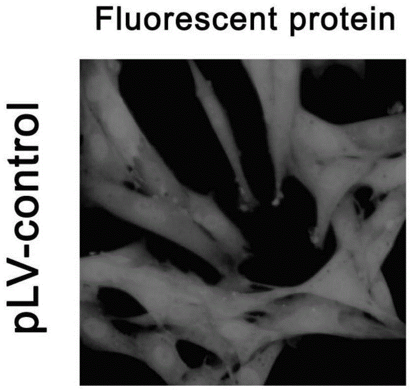
FIG. 1-1: pLV-control transfected mesenchymal stem cells expressed GFP (green fluorescent);
FIGS. 1-2: pLV-HMGB1 transfected mesenchymal stem cells expressed GFP (green fluorescent);
FIGS. 1 to 3: detecting the expression condition of mesenchymal stem cells HMGB1 transfected by pLV-control and pLV-HMGB1 by Western blot;
FIGS. 1 to 4: detecting that the mesenchymal stem cells transfected by pLV-HMGB1 express Flag-labeled HMGB1 by Western blot;
FIG. 2-1: detecting the differentiation condition of the mesenchymal stem cells transfected by pLV-control and pLV-HMGB1 to CD31 expression positive cells by flow cytometry;
FIG. 2-2 flow cytometry is used for detecting the differentiation condition of mesenchymal stem cells transfected by pLV-control and pLV-HMGB1 to α SMA expression positive cells;
FIG. 3-1: pLV-control rat transplanted arterial neovascular intimal GFP-labeled cells (fluorescing green) were examined under a fluorescence microscope;
FIG. 3-2: pLV-control rat transplanted arterial neovascular intima CD31 expression positive cells (red fluorescence) were examined under a fluorescence microscope;
FIGS. 3-3: pLV-HMGB1 rat transplanted arterial neovascular intimal GFP-labeled cells (fluorescing green) were examined under a fluorescence microscope;
FIGS. 3-4: pLV-HMGB1 rat transplanted arterial neovascular intima CD31 expression positive cells (red fluorescence) were examined under a fluorescence microscope;
FIGS. 3 to 5: pLV-control rat transplanted arterial neovascular intimal GFP-labeled cells (fluorescing green) were examined under a fluorescence microscope;
FIGS. 3-6 examination of pLV-control rat transplanted arterial neovascular intima α SMA expression positive cells (red fluorescence) under fluorescent microscope;
FIGS. 3 to 7: pLV-HMGB1 rat transplanted arterial neovascular intimal GFP-labeled cells (fluorescing green) were examined under a fluorescence microscope;
FIGS. 3-8 examination of pLV-HMGB1 rat transplanted arterial neointima α SMA expression positive cells (red fluorescence) under fluorescent microscope.
Detailed Description
The following will clearly and completely describe the regulation and control method adopted by the present invention in conjunction with the embodiments of the present invention, and it is obvious that the described embodiments are only a part of the embodiments of the present invention, and not all embodiments. All other embodiments, which can be derived by a person skilled in the art from the embodiments given herein without making any creative effort, shall fall within the protection scope of the present invention.
Example 1: preparation of HMGB1 overexpressed mesenchymal stem cells
1. Materials and reagents, see table 1 below:
TABLE 1
2. The method comprises the following steps:
the virus construction process includes designing two primers for RT-PCR amplification of HMGB1cDNA with Flag protein coding sequence, terminating sequence and restriction site inserted into two ends, slow annealing, connecting with slow virus shuttle plasmid under the action of T4DNA ligase to obtain connected product pLVTH-HMGB1-GFP-PURO, transforming competent DH-5 α colibacillus, kanamycin screening and amplification, extracting purified plasmid with small plasmid amount extracting kit, enzyme digestion, sequencing and identification, determining the base sequence of the inserted segment in the shuttle plasmid is identical to the designed sequence, setting negative control recombinant plasmid pLVTH-HMGB 1-PURO, packaging plasmid pCMV-dR8.2dVpr and envelope pCMV-VSV-G to transfect 293T cell together, and packaging to obtain slow virus plasmid carrying HMGB1 gene containing green purine gene (HMGB-1) and green gene (HMGB) expressing resistance gene 1.
And (3) virus transfection process: the mesenchymal stem cells are cultured in DMEM culture solution in an adherent manner, and 10% fetal calf serum and penicillin-streptomycin antibacterial liquid are added into the culture solution. The mesenchymal stem cells were seeded into six-well culture plates each containing 2X 10 cells per well5Culturing the cells in a carbon dioxide incubator for 24 hours, adding pLV-HMGB1 (adding pLV-control in a negative control group), culturing for 8 hours, replacing a fresh culture solution, continuously culturing, adding puromycin on the 4 th day of culture, screening positive transfected clones, and observing the positive transfected cells (emitting green fluorescence) by an immunofluorescence microscope. Western blot is used for detecting the protein expression condition of mesenchymal stem cells, a primary antibody used for detecting the expression of HMGB1 is a rabbit anti-rat HMGB1 antibody, a secondary antibody is an HRP-labeled goat anti-rabbit antibody, HMGB1 overexpressed by virus transfection contains a Flag protein marker, and can be detected by using an anti-Flag antibody, the used primary antibody is a mouse anti-Flag antibody, and the secondary antibody is an HRP-labeled goat anti-mouse antibody.
3. As a result: after viral transfection, more than 99.9% of mesenchymal stem cells express GFP and emit green fluorescence (see figures 1-1 and 1-2), the expression level of the mesenchymal stem cells HMGB1 transfected by pLV-HMGB1 is obviously higher than that of the mesenchymal stem cells pLV-control (figures 1-3), and the expression of the HMGB1 containing Flag protein markers is detected in the mesenchymal stem cells transfected by pLV-HMGB1 (figures 1-4). Therefore, the mesenchymal stem cells realize the up-regulation of HMGB1 expression by adopting a virus transfection method.
Example 2: evaluation of differentiation Capacity of mesenchymal Stem cells overexpressing HMGB1 in example 1 in vitro
1. Materials and reagents, see table 2 below:
TABLE 2
2. The method comprises the following steps: VEGF is added into the culture solution to induce the mesenchymal stem cells to differentiate into vascular endothelial cells, the concentration of the VEGF in the culture solution is 25ng/ml, and the in vitro culture is carried out for 14 days; PDGF is added into the culture solution to induce the differentiation of the mesenchymal stem cells to vascular smooth muscle cells, the concentration of the PDGF in the culture solution is 12.5ng/ml, and the PDGF is cultured in vitro for 14 days.
CD31 is a vascular endothelial cell specific protein marker, a PE marker CD31 antibody is used for detecting CD31 expression positive cells in the flow cytometry, the positive expression of cell CD31 indicates the successful differentiation into the vascular endothelial cells, α SMA is a vascular smooth muscle cell specific protein marker, a PE marker α SMA antibody is used for detecting α SMA expression positive cells in the flow cytometry, and the positive expression of cell α SMA indicates the successful differentiation into the vascular smooth muscle cells.
3. As a result, the rate of differentiation of pLV-HMGB 1-transfected mesenchymal stem cells into CD 31-expressing positive cells was increased compared to pLV-control (FIG. 2-1, the vertical line in the figure indicates the negative threshold using the isotype control antibody), and the rate of differentiation into α SMA-expressing positive cells was decreased compared to pLV-control (FIG. 2-2, the vertical line in the figure indicates the negative threshold using the isotype control antibody).
Example 3: evaluation of differentiation potency of HMGB 1-overexpressed mesenchymal Stem cells in vivo in example 1
1. Experimental animals, see table 3 below:
TABLE 3
2. The method comprises the following steps:
constructing a transplantation arteriosclerosis model: both F344 and LEWIS are inbred rats with a relatively close genotype, essentially identical Major Histocompatibility Complex (MHC) sites, but partially different on non-MHC, and therefore chronic rejection of the transplanted abdominal aorta between these two rat lines can occur, with pathomorphologically manifested as transplant arteriosclerosis. Firstly, the abdominal aorta of a LEWIS rat is taken as a graft, then an F344 rat is taken as a receptor rat, the abdominal aorta is cut after the blood flow is blocked, and the abdominal aorta of the LEWIS rat is bridged between the two broken ends, so that the patency of the abdominal aorta is recovered. Arteriosclerosis was observed in the transplanted abdominal aorta 90 days after the operation.
Animal grouping and treatment mode: the transplanted arteriosclerosis model rats 24 are divided into two groups of 8 rats, and the details are shown in Table 4. Two groups of rats were inoculated with the two lentivirus-transfected mesenchymal stem cells of example 1 by tail vein injection from 30 days after transplantation, respectively, at a cell amount of 2X 10⁶Each cell was inoculated every 15 days for a total of 4 times.
TABLE 4 animal Experimental groups
After 90 days, rat transplanted arteries are taken to prepare frozen sections, positive cells for expressing CD31 and positive cells for expressing α SMA are marked by an immunofluorescence technique, and the conditions of expressing CD31 and α SMA by homing cells (emitting green fluorescence) are analyzed and compared.
3. As a result, the proportion of homing cells in the neointimal neovasculature of rats in the pLV-HMGB1 group expressed CD31 (FIGS. 3-3 and 3-4 are the same visual field, note that the cells marked by the arrow) was higher than that of the pLV-control group (FIGS. 3-1 and 3-2 are the same visual field, note that the cells marked by the arrow) and the proportion of homing cells in the neointimal neovasculature of rats in the pLV-HMGB1 group expressed α SMA (FIGS. 3-8 and 3-7 are the same visual field, note that the cells marked by the arrow) was lower than that of the pLV-control group (FIGS. 3-5 and 3-6 are the same visual field, note that the cells marked by the arrow).