CN106974683B - Negative pressure wound closure device - Google Patents
Negative pressure wound closure device Download PDFInfo
- Publication number
- CN106974683B CN106974683B CN201610912564.1A CN201610912564A CN106974683B CN 106974683 B CN106974683 B CN 106974683B CN 201610912564 A CN201610912564 A CN 201610912564A CN 106974683 B CN106974683 B CN 106974683B
- Authority
- CN
- China
- Prior art keywords
- wound
- closure device
- stabilizer
- collapse
- filler
- Prior art date
- Legal status (The legal status is an assumption and is not a legal conclusion. Google has not performed a legal analysis and makes no representation as to the accuracy of the status listed.)
- Active
Links
Images




Classifications
-
- A—HUMAN NECESSITIES
- A61—MEDICAL OR VETERINARY SCIENCE; HYGIENE
- A61B—DIAGNOSIS; SURGERY; IDENTIFICATION
- A61B17/00—Surgical instruments, devices or methods
- A61B17/08—Wound clamps or clips, i.e. not or only partly penetrating the tissue ; Devices for bringing together the edges of a wound
-
- A—HUMAN NECESSITIES
- A61—MEDICAL OR VETERINARY SCIENCE; HYGIENE
- A61F—FILTERS IMPLANTABLE INTO BLOOD VESSELS; PROSTHESES; DEVICES PROVIDING PATENCY TO, OR PREVENTING COLLAPSING OF, TUBULAR STRUCTURES OF THE BODY, e.g. STENTS; ORTHOPAEDIC, NURSING OR CONTRACEPTIVE DEVICES; FOMENTATION; TREATMENT OR PROTECTION OF EYES OR EARS; BANDAGES, DRESSINGS OR ABSORBENT PADS; FIRST-AID KITS
- A61F13/00—Bandages or dressings; Absorbent pads
- A61F13/05—Bandages or dressings; Absorbent pads specially adapted for use with sub-pressure or over-pressure therapy, wound drainage or wound irrigation, e.g. for use with negative-pressure wound therapy [NPWT]
-
- A—HUMAN NECESSITIES
- A61—MEDICAL OR VETERINARY SCIENCE; HYGIENE
- A61B—DIAGNOSIS; SURGERY; IDENTIFICATION
- A61B17/00—Surgical instruments, devices or methods
-
- A—HUMAN NECESSITIES
- A61—MEDICAL OR VETERINARY SCIENCE; HYGIENE
- A61B—DIAGNOSIS; SURGERY; IDENTIFICATION
- A61B17/00—Surgical instruments, devices or methods
- A61B17/0057—Implements for plugging an opening in the wall of a hollow or tubular organ, e.g. for sealing a vessel puncture or closing a cardiac septal defect
-
- A—HUMAN NECESSITIES
- A61—MEDICAL OR VETERINARY SCIENCE; HYGIENE
- A61M—DEVICES FOR INTRODUCING MEDIA INTO, OR ONTO, THE BODY; DEVICES FOR TRANSDUCING BODY MEDIA OR FOR TAKING MEDIA FROM THE BODY; DEVICES FOR PRODUCING OR ENDING SLEEP OR STUPOR
- A61M1/00—Suction or pumping devices for medical purposes; Devices for carrying-off, for treatment of, or for carrying-over, body-liquids; Drainage systems
- A61M1/08—Cupping glasses, i.e. for enhancing blood circulation
-
- A—HUMAN NECESSITIES
- A61—MEDICAL OR VETERINARY SCIENCE; HYGIENE
- A61M—DEVICES FOR INTRODUCING MEDIA INTO, OR ONTO, THE BODY; DEVICES FOR TRANSDUCING BODY MEDIA OR FOR TAKING MEDIA FROM THE BODY; DEVICES FOR PRODUCING OR ENDING SLEEP OR STUPOR
- A61M1/00—Suction or pumping devices for medical purposes; Devices for carrying-off, for treatment of, or for carrying-over, body-liquids; Drainage systems
- A61M1/84—Drainage tubes; Aspiration tips
-
- A—HUMAN NECESSITIES
- A61—MEDICAL OR VETERINARY SCIENCE; HYGIENE
- A61M—DEVICES FOR INTRODUCING MEDIA INTO, OR ONTO, THE BODY; DEVICES FOR TRANSDUCING BODY MEDIA OR FOR TAKING MEDIA FROM THE BODY; DEVICES FOR PRODUCING OR ENDING SLEEP OR STUPOR
- A61M1/00—Suction or pumping devices for medical purposes; Devices for carrying-off, for treatment of, or for carrying-over, body-liquids; Drainage systems
- A61M1/90—Negative pressure wound therapy devices, i.e. devices for applying suction to a wound to promote healing, e.g. including a vacuum dressing
-
- A—HUMAN NECESSITIES
- A61—MEDICAL OR VETERINARY SCIENCE; HYGIENE
- A61B—DIAGNOSIS; SURGERY; IDENTIFICATION
- A61B17/00—Surgical instruments, devices or methods
- A61B2017/00535—Surgical instruments, devices or methods pneumatically or hydraulically operated
- A61B2017/00561—Surgical instruments, devices or methods pneumatically or hydraulically operated creating a vacuum
-
- A—HUMAN NECESSITIES
- A61—MEDICAL OR VETERINARY SCIENCE; HYGIENE
- A61B—DIAGNOSIS; SURGERY; IDENTIFICATION
- A61B17/00—Surgical instruments, devices or methods
- A61B17/0057—Implements for plugging an opening in the wall of a hollow or tubular organ, e.g. for sealing a vessel puncture or closing a cardiac septal defect
- A61B2017/00646—Type of implements
- A61B2017/00654—Type of implements entirely comprised between the two sides of the opening
-
- A—HUMAN NECESSITIES
- A61—MEDICAL OR VETERINARY SCIENCE; HYGIENE
- A61B—DIAGNOSIS; SURGERY; IDENTIFICATION
- A61B17/00—Surgical instruments, devices or methods
- A61B17/08—Wound clamps or clips, i.e. not or only partly penetrating the tissue ; Devices for bringing together the edges of a wound
- A61B2017/081—Tissue approximator
-
- A—HUMAN NECESSITIES
- A61—MEDICAL OR VETERINARY SCIENCE; HYGIENE
- A61F—FILTERS IMPLANTABLE INTO BLOOD VESSELS; PROSTHESES; DEVICES PROVIDING PATENCY TO, OR PREVENTING COLLAPSING OF, TUBULAR STRUCTURES OF THE BODY, e.g. STENTS; ORTHOPAEDIC, NURSING OR CONTRACEPTIVE DEVICES; FOMENTATION; TREATMENT OR PROTECTION OF EYES OR EARS; BANDAGES, DRESSINGS OR ABSORBENT PADS; FIRST-AID KITS
- A61F13/00—Bandages or dressings; Absorbent pads
- A61F2013/00089—Wound bandages
- A61F2013/0017—Wound bandages possibility of applying fluid
- A61F2013/00174—Wound bandages possibility of applying fluid possibility of applying pressure
-
- A—HUMAN NECESSITIES
- A61—MEDICAL OR VETERINARY SCIENCE; HYGIENE
- A61F—FILTERS IMPLANTABLE INTO BLOOD VESSELS; PROSTHESES; DEVICES PROVIDING PATENCY TO, OR PREVENTING COLLAPSING OF, TUBULAR STRUCTURES OF THE BODY, e.g. STENTS; ORTHOPAEDIC, NURSING OR CONTRACEPTIVE DEVICES; FOMENTATION; TREATMENT OR PROTECTION OF EYES OR EARS; BANDAGES, DRESSINGS OR ABSORBENT PADS; FIRST-AID KITS
- A61F13/00—Bandages or dressings; Absorbent pads
- A61F2013/00361—Plasters
- A61F2013/00365—Plasters use
- A61F2013/00536—Plasters use for draining or irrigating wounds
-
- A—HUMAN NECESSITIES
- A61—MEDICAL OR VETERINARY SCIENCE; HYGIENE
- A61F—FILTERS IMPLANTABLE INTO BLOOD VESSELS; PROSTHESES; DEVICES PROVIDING PATENCY TO, OR PREVENTING COLLAPSING OF, TUBULAR STRUCTURES OF THE BODY, e.g. STENTS; ORTHOPAEDIC, NURSING OR CONTRACEPTIVE DEVICES; FOMENTATION; TREATMENT OR PROTECTION OF EYES OR EARS; BANDAGES, DRESSINGS OR ABSORBENT PADS; FIRST-AID KITS
- A61F13/00—Bandages or dressings; Absorbent pads
- A61F2013/00361—Plasters
- A61F2013/00365—Plasters use
- A61F2013/0054—Plasters use for deep wounds
-
- A—HUMAN NECESSITIES
- A61—MEDICAL OR VETERINARY SCIENCE; HYGIENE
- A61F—FILTERS IMPLANTABLE INTO BLOOD VESSELS; PROSTHESES; DEVICES PROVIDING PATENCY TO, OR PREVENTING COLLAPSING OF, TUBULAR STRUCTURES OF THE BODY, e.g. STENTS; ORTHOPAEDIC, NURSING OR CONTRACEPTIVE DEVICES; FOMENTATION; TREATMENT OR PROTECTION OF EYES OR EARS; BANDAGES, DRESSINGS OR ABSORBENT PADS; FIRST-AID KITS
- A61F13/00—Bandages or dressings; Absorbent pads
- A61F2013/00361—Plasters
- A61F2013/00544—Plasters form or structure
-
- A—HUMAN NECESSITIES
- A61—MEDICAL OR VETERINARY SCIENCE; HYGIENE
- A61F—FILTERS IMPLANTABLE INTO BLOOD VESSELS; PROSTHESES; DEVICES PROVIDING PATENCY TO, OR PREVENTING COLLAPSING OF, TUBULAR STRUCTURES OF THE BODY, e.g. STENTS; ORTHOPAEDIC, NURSING OR CONTRACEPTIVE DEVICES; FOMENTATION; TREATMENT OR PROTECTION OF EYES OR EARS; BANDAGES, DRESSINGS OR ABSORBENT PADS; FIRST-AID KITS
- A61F13/00—Bandages or dressings; Absorbent pads
- A61F2013/00361—Plasters
- A61F2013/00544—Plasters form or structure
- A61F2013/00548—Plasters form or structure net
-
- A—HUMAN NECESSITIES
- A61—MEDICAL OR VETERINARY SCIENCE; HYGIENE
- A61M—DEVICES FOR INTRODUCING MEDIA INTO, OR ONTO, THE BODY; DEVICES FOR TRANSDUCING BODY MEDIA OR FOR TAKING MEDIA FROM THE BODY; DEVICES FOR PRODUCING OR ENDING SLEEP OR STUPOR
- A61M1/00—Suction or pumping devices for medical purposes; Devices for carrying-off, for treatment of, or for carrying-over, body-liquids; Drainage systems
- A61M1/90—Negative pressure wound therapy devices, i.e. devices for applying suction to a wound to promote healing, e.g. including a vacuum dressing
- A61M1/91—Suction aspects of the dressing
- A61M1/916—Suction aspects of the dressing specially adapted for deep wounds
Landscapes
- Health & Medical Sciences (AREA)
- Heart & Thoracic Surgery (AREA)
- Life Sciences & Earth Sciences (AREA)
- Public Health (AREA)
- General Health & Medical Sciences (AREA)
- Biomedical Technology (AREA)
- Veterinary Medicine (AREA)
- Engineering & Computer Science (AREA)
- Animal Behavior & Ethology (AREA)
- Surgery (AREA)
- Vascular Medicine (AREA)
- Hematology (AREA)
- Anesthesiology (AREA)
- Nuclear Medicine, Radiotherapy & Molecular Imaging (AREA)
- Medical Informatics (AREA)
- Molecular Biology (AREA)
- Cardiology (AREA)
- Media Introduction/Drainage Providing Device (AREA)
- Materials For Medical Uses (AREA)
- Surgical Instruments (AREA)
- Medicines Containing Material From Animals Or Micro-Organisms (AREA)
- External Artificial Organs (AREA)
Abstract
本发明涉及一种负压伤口闭合系统和用于这类系统的方法。本发明的优选实施方案通过优先收缩帮助伤口闭合而提供组织移动。优选实施方案可利用组织抓取元件以将伤口闭合力施加至组织。

The present invention relates to a negative pressure wound closure system and a method for such a system. Preferred embodiments of the present invention provide tissue movement by preferentially contracting to aid in wound closure. Preferred embodiments may utilize tissue grasping elements to apply wound closure forces to tissue.
Description
本申请是申请号为201280017448.3、申请日为2012年2月3日、名称为“负压伤口闭合装置”的专利申请的分案申请。This application is a divisional application of the patent application with the application number of 201280017448.3 and the filing date of February 3, 2012, with the title of "Negative Pressure Wound Closure Device".
相关申请的交叉引用CROSS-REFERENCE TO RELATED APPLICATIONS
本申请主张2011年2月4日提交的美国申请61/439,525的优先权。以上申请的全文以引用的方式并入本文。This application claims priority to US Application 61/439,525, filed February 4, 2011. The entire contents of the above applications are incorporated herein by reference.
技术领域technical field
本发明涉及负压伤口闭合装置。The present invention relates to negative pressure wound closure devices.
背景技术Background technique
已经发展用于处理包括由事故造成的伤口和由手术造成的伤口的伤口的许多技术。通常,使用缝线或缝合钉使伤口闭合。然而,插入这些机械闭合技术需要对皮肤做出额外刺破或伤口,这可能导致组织伤害且在过度肿胀的情况下可能造成缺血和组织损失。此外,诸如缝合钉和缝线的机械伤口闭合物可能在插入点造成阻碍且损害皮肤的正常伤口愈合过程的高度局部压力。Numerous techniques have been developed for treating wounds including wounds caused by accidents and wounds caused by surgery. Usually, the wound is closed with sutures or staples. However, inserting these mechanical closure techniques requires additional punctures or wounds to the skin, which can lead to tissue damage and, in the case of excessive swelling, can result in ischemia and tissue loss. Furthermore, mechanical wound closures such as staples and sutures can create high localized pressures at the point of insertion that hinder and impair the normal wound healing process of the skin.
近几年,已经越来越关注使用负压装置用于处理伤口。负压伤口处理通过对伤口施加负压抽吸而利用移除伤口积液的装置。据信这种负压会通过在伤口部位帮助形成肉芽组织并帮助身体正常炎症过程且同时移除过量积液(其可能含有不利细菌细胞因子)而促进伤口愈合。然而,需要进一步改善负压伤口治疗以完全实现处理的优点。In recent years, there has been increasing interest in the use of negative pressure devices for treating wounds. Negative pressure wound treatment utilizes a device to remove wound fluid by applying negative pressure suction to the wound. This negative pressure is believed to promote wound healing by helping to form granulation tissue at the wound site and aiding the body's normal inflammatory process while removing excess fluid, which may contain unfavorable bacterial cytokines. However, further improvements in negative pressure wound therapy are needed to fully realize the benefits of the treatment.
发明内容SUMMARY OF THE INVENTION
本发明涉及一种尤其在伤口边缘用力以帮助伤口闭合的负压伤口闭合装置。所述装置用于减少一般采用的伤口填充材料的重复移位的需要且可增快愈合的速度。所述装置同时使用负压移除伤口积液。The present invention relates to a negative pressure wound closure device that applies force, particularly at the edges of the wound, to aid in wound closure. The device serves to reduce the need for repeated displacement of commonly employed wound packing materials and to increase the rate of healing. The device simultaneously uses negative pressure to remove wound fluid.
在一个实施方案中,负压伤口闭合装置包括伤口填充材料,其被大小调整且塑形以配合在伤口开口内且在将负压施加至填充材料之后沿着至少一个第一维度收缩。所述填充材料因此被配置来在至少一个方向上优先收缩且在一个或多个额外方向上抑制收缩。先前负压装置无法辅助伤口闭合,而是用于排放积液。通过在愈合过程期间结合如结合本发明所述从伤口排放积液来提供组织的受控移动,可实现在愈合速度上的大幅改善。应注意,取决于伤口大小,可使用增加的负压。In one embodiment, the negative pressure wound closure device includes a wound filler material sized and shaped to fit within the wound opening and contracted along at least one first dimension after negative pressure is applied to the filler material. The filler material is thus configured to preferentially shrink in at least one direction and inhibit shrinkage in one or more additional directions. Previous negative pressure devices did not assist in wound closure, but were used to drain fluid. By providing controlled movement of tissue during the healing process in conjunction with drainage of fluid from the wound as described in connection with the present invention, substantial improvements in the speed of healing can be achieved. It should be noted that depending on the size of the wound, increased negative pressure may be used.
在另一优选实施方案中,组织抓取表面在伤口填充材料的外周边表面上延伸且包括多个向外突出的组织锚,所述组织锚在所述伤口开口的边缘接合组织。在施加负压之后,移位所述伤口边缘的组织以帮助所述伤口的闭合。将诸如真空泵的负压源耦接至伤口填充材料以提供负压。In another preferred embodiment, a tissue grasping surface extends over the outer peripheral surface of the wound packing material and includes a plurality of outwardly projecting tissue anchors that engage tissue at the edges of the wound opening. Following the application of negative pressure, the tissue at the edge of the wound is displaced to assist in the closure of the wound. A negative pressure source, such as a vacuum pump, is coupled to the wound packing material to provide negative pressure.
伤口填充材料通常包括多孔材料,诸如泡沫。对于采用组织锚的实施方案,这些可成一体地形成于填充材料中。在其它实施方案中,将组织锚提供在固定至填充材料的分开覆盖物或膜上。Wound filling materials typically include porous materials, such as foams. For embodiments employing tissue anchors, these may be integrally formed in the filler material. In other embodiments, the tissue anchor is provided on a separate covering or membrane secured to the filler material.
在优选实施方案中,填充材料包括稳定结构以实现至少一个第一方向上的塌缩并且抑制至少一个第二方向上的塌缩。稳定结构可包括由相对可压缩材料围绕的一个或多个相对刚性材料区。在优选实施方案中,稳定结构是由刚性和/或半刚性材料形成的内骨架。In a preferred embodiment, the filler material includes a stabilizing structure to effect collapse in at least one first direction and inhibit collapse in at least one second direction. The stabilizing structure may include one or more regions of relatively rigid material surrounded by relatively compressible material. In preferred embodiments, the stabilizing structure is an endoskeleton formed of rigid and/or semi-rigid materials.
在特定实施方案中,稳定结构抑制填充材料沿其高度维度塌缩,同时实现填充材料在由伤口边缘限定的平面内塌缩。这在腹部手术的情况中是有用的,例如其中手术切口沿着直线形成椭圆形伤口。这个大致椭圆形伤口可延伸通过具有可变机械特性的肌肉和脂肪组织。较佳通过使用被调适成优先朝向切口的原线塌缩的椭圆形结构而提供伤口愈合。在优选实施方案中,稳定结构促进填充器以实现伤口组织的缝接的方式塌缩。可使用本发明的实施方案成功处理筋膜伤口或其它伤口开裂,或任何开放性伤口。In certain embodiments, the stabilizing structure inhibits the collapse of the filler material along its height dimension, while enabling the filler material to collapse within the plane defined by the wound edge. This is useful in the case of abdominal surgery, for example, where the surgical incision follows a straight line to form an oval wound. This generally oval wound can extend through muscle and adipose tissue with variable mechanical properties. Wound healing is preferably provided through the use of an elliptical structure adapted to collapse preferentially towards the origin of the incision. In preferred embodiments, the stabilizing structure facilitates the collapse of the obturator in a manner that achieves suturing of the wound tissue. Fascial or other wound dehiscence, or any open wound, can be successfully treated using embodiments of the present invention.
伤口闭合装置可用来处理纵隔膜中的伤口、处理压迫溃疡、处理手足情况(手臂或腿)中的伤口。伤口闭合装置还可用来处理不同形状的伤口,诸如圆形、正方形、矩形或非常规形状伤口。多个伤口闭合元件可被大小调整来配合在伤口内且可附接在一起以优先在所要方向上使伤口闭合。不同元件可包括不同材料或具有不同特征,诸如孔大小和/或锚大小和分布以形成复合结构。Wound closure devices can be used to treat wounds in the mediastinum, pressure ulcers, wounds in hand-foot conditions (arms or legs). Wound closure devices can also be used to treat wounds of different shapes, such as round, square, rectangular or unconventional shaped wounds. The plurality of wound closure elements can be sized to fit within the wound and can be attached together to preferentially close the wound in a desired orientation. The different elements may comprise different materials or have different characteristics, such as pore size and/or anchor size and distribution to form a composite structure.
在一个实施方案中,内骨架稳定结构包括多个隔开且形成交叉影线构造的刚性部件。内骨架使填充材料能够沿其宽度维度塌缩且沿其长度维度伸长至更小程度。在特定实施方案中,举例而言,多个刚性部件沿着填充材料的高度延伸且抑制所述填充物在其高度上塌缩。根据特定实施方案,内骨架包括互连刚性部件网络,其可在所述填充材料的塌缩期间相对于彼此关节式连接。内骨架可包括桁架支撑件以抑制填充材料的倾斜动作。在一些实施方案中个,组织锚可成一体地形成在内骨架中。In one embodiment, the endoskeleton stabilization structure includes a plurality of rigid members spaced apart and forming a cross-hatched configuration. The inner skeleton enables the filler material to collapse along its width dimension and elongate to a lesser extent along its length dimension. In certain embodiments, for example, a plurality of rigid members extend along the height of the filler material and inhibit the filler from collapsing over its height. According to certain embodiments, the endoskeleton includes a network of interconnected rigid components that can be articulated relative to each other during collapse of the filler material. The inner skeleton may include truss supports to inhibit the tilting action of the fill material. In some embodiments, the tissue anchor can be integrally formed in the endoskeleton.
在特定实施方案中,伤口填充材料包括平滑底表面,其具有微孔以允许积液从伤口通过底表面且进入装置用于移除。微孔可具有可变孔大小和/或孔密度中以从负压源引导真空力的分配。在一些实施方案中,伤口填充材料可具有可变内孔大小和/或孔密度以引导真空力的分布。In certain embodiments, the wound packing material includes a smooth bottom surface with micropores to allow fluid accumulation from the wound to pass through the bottom surface and into the device for removal. The micropores may have variable pore size and/or pore density in order to direct the distribution of vacuum force from the negative pressure source. In some embodiments, the wound filler material may have variable inner pore size and/or pore density to direct the distribution of vacuum force.
在一个实施方案中,用于管理和/或移除积液的负压伤口处理组件耦接至伤口填充材料。单个负压源可用于伤口闭合和积液管理/排放。在伤口闭合物与积液管理组件之间的界面处提供有滑动表面。In one embodiment, a negative pressure wound treatment assembly for managing and/or removing fluid accumulation is coupled to the wound packing material. A single source of negative pressure can be used for wound closure and fluid management/drainage. A sliding surface is provided at the interface between the wound closure and the effusion management assembly.
在又另一实施方案中,填充材料包括可移除部分以调整伤口闭合装置的大小。填充材料可具有预定裂开线用于处理或切除材料的部分。在特定实施方案中,多组组织锚被嵌入填充材料中,且通过移除可移除部分而暴露。In yet another embodiment, the filler material includes a removable portion to adjust the size of the wound closure device. The filler material may have predetermined cleavage lines for processing or excising portions of the material. In certain embodiments, sets of tissue anchors are embedded in the filler material and exposed by removing the removable portion.
根据另一实施方案,组织锚具有可变力分布。所述力分布可基于接合的组织的深度或组织的类型而变化。在一些实施方案中,组织抓取表面的力分布绕伤口闭合装置的周边改变。力分布通过例如改变组织锚的长度、锚的形状、锚的材料和锚的密度中的一个或多个而变化。According to another embodiment, the tissue anchor has a variable force distribution. The force distribution may vary based on the depth of the engaged tissue or the type of tissue. In some embodiments, the force distribution of the tissue grasping surface varies around the perimeter of the wound closure device. The force distribution is varied by, for example, changing one or more of the length of the tissue anchor, the shape of the anchor, the material of the anchor, and the density of the anchor.
本发明还涉及使用如上所述伤口闭合装置使伤口闭合的方法。举例而言,腹部地层的皮肤中的线性切口提供对诸如人或动物身体的肠胃系统的手术部位的接达。在完成以后,必须通过负压治疗处理伤口以促进恢复。因此,插入根据本发明的优选实施方案的伤口闭合装置用于伤口闭合处理。The present invention also relates to a method of closing a wound using the wound closure device as described above. For example, linear incisions in the skin of the abdominal formation provide access to surgical sites such as the gastrointestinal system of a human or animal body. After completion, the wound must be treated with negative pressure therapy to promote recovery. Accordingly, a wound closure device according to a preferred embodiment of the present invention is inserted for wound closure treatment.
通过使用本发明的负压伤口闭合装置,具有大型或严重伤口的病患可免于或参加康复物理治疗,而变为在家且接着将其伤口恢复成简单地缝合闭合。通过改善伤口闭合处理且从而减少成本,这些装置有机会成为用于伤口护理设备的重要部分。By using the negative pressure wound closure device of the present invention, patients with large or severe wounds can be exempted from or enrolled in rehabilitation physical therapy, and become at home and then have their wounds restored to simple suture closure. By improving wound closure procedures and thereby reducing costs, these devices have the opportunity to become an important part of the equipment used for wound care.
附图说明Description of drawings
结合附图后,从本发明的以下详细描述中本发明的其它特征和优点将显而易见,其中:Other features and advantages of the present invention will become apparent from the following detailed description of the invention when taken in conjunction with the accompanying drawings, wherein:
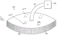
图1A是负压伤口闭合装置的透视示意图。Figure 1A is a schematic perspective view of a negative pressure wound closure device.
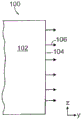
图1B是伤口闭合装置的组织抓取表面的截面图。Figure IB is a cross-sectional view of the tissue grasping surface of the wound closure device.
图1C是组织抓取表面的一个实施方案的侧视图。Figure 1C is a side view of one embodiment of a tissue grasping surface.
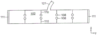
图1D是以虚线示出x-y稳定器的伤口闭合装置的俯视图。Figure ID is a top view of a wound closure device showing an x-y stabilizer in phantom.
图1E是示出x-y稳定器和z稳定器的填充材料的截面图。Figure IE is a cross-sectional view showing the fill material of the x-y stabilizer and the z stabilizer.
图1F是示出用于移除来自伤口部位的积液的光滑底表面和微孔的伤口闭合装置的仰视图。Figure IF is a bottom view of a wound closure device showing a smooth bottom surface and micropores for removing fluid from a wound site.
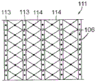
图1G是周边稳定器元件的正视图。Figure 1G is a front view of a peripheral stabilizer element.
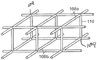
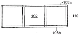
图2A和图2B分别是支撑内骨架的透视图和侧视图。2A and 2B are perspective and side views, respectively, of a support endoskeleton.
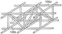
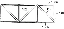
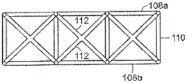
图3A和图3B分别是具有支撑桁架的支撑内骨架的透视图和侧视图。3A and 3B are perspective and side views, respectively, of a support endoskeleton with support trusses.
图3C是具有x形状支撑桁架的支撑内骨架的侧视图。Figure 3C is a side view of a support endoskeleton with an x-shaped support truss.
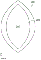
图4A至图4C图示使伤口闭合的本发明的伤口闭合装置。4A-4C illustrate a wound closure device of the present invention closing a wound.
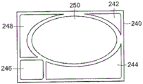
图4D至图4E图示用于不同形状的伤口的多个伤口闭合元件的使用。4D-4E illustrate the use of multiple wound closure elements for wounds of different shapes.
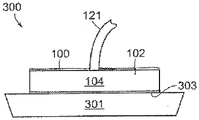
图5图示二级负压伤口处理和负压伤口闭合(NPWT/NPWC)装置。Figure 5 illustrates a secondary negative pressure wound treatment and negative pressure wound closure (NPWT/NPWC) device.
图6图示根据本发明的组织锚系统的优选实施方案的放大图。Figure 6 illustrates an enlarged view of a preferred embodiment of a tissue anchor system according to the present invention.
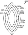
图7图示具有用于适应不同伤口大小的撕开和切除设计的伤口填充材料的实施方案,其中组织锚嵌在预定裂开点的填充材料内。Figure 7 illustrates an embodiment of a wound packing material with tear and resection designs for accommodating different wound sizes, wherein tissue anchors are embedded within the packing material at predetermined dehiscence points.
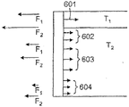
图8A是组织抓取表面的侧视图,其图示用于不同类型组织(T1、T2)的不同组织锚和锚的各自力分布,包括真空闭合(F1)期间施加的最大力和没有破坏组织的情况下从组织移除锚所需的力(F2)。8A is a side view of a tissue grasping surface illustrating different tissue anchors for different types of tissue (T 1 , T 2 ) and the respective force distributions of the anchors, including the maximum force applied during vacuum closure (F 1 ) and The force (F2 ) required to remove the anchor from the tissue without breaking the tissue.
图8B图示用于本发明的组织锚的不同设计。Figure 8B illustrates different designs of tissue anchors for use in the present invention.
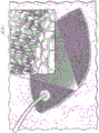
图8C图示椭圆形伤口闭合装置的周边表面的组织锚元件的放大图。Figure 8C illustrates an enlarged view of the tissue anchoring element of the peripheral surface of the oval wound closure device.
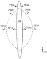
图9A是根据一个实施方案的位于伤口内的伤口闭合装置的示意图,其示出伤口边缘周围的不同力分布。Figure 9A is a schematic diagram of a wound closure device positioned within a wound showing different force distributions around the wound edge, according to one embodiment.
图9B图示在伤口闭合和愈合时间后的图9A的伤口闭合装置,其中伤口的原始构造和伤口闭合装置以虚线指示。9B illustrates the wound closure device of FIG. 9A after wound closure and healing time, with the original configuration of the wound and wound closure device indicated in dashed lines.
图10示意性图示使用根据本发明的伤口闭合装置的手术。Figure 10 schematically illustrates a procedure using a wound closure device according to the present invention.
具体实施方式Detailed ways
图1A至图1F图示本发明的伤口闭合装置100的实施方案。装置100包括被大小调整和塑形以配合于人或动物病患的伤口开口内的伤口填充材料102。在优选实施方案中,填充材料102是多孔、生物相容性材料,诸如开孔聚氨酯泡沫。填充材料102还可以优先是可塌缩的,此意味着可以通过将负压施加至填充材料102,同时在另一方向上抑制收缩或以较慢速率收缩而沿着至少一个维度(例如,长度、宽度、高度)减少其大小。1A-1F illustrate an embodiment of a
组织抓取表面104在填充材料102的至少一个表面上延伸,且优选地在填充材料102的外周界表面上延伸。在一个实施方案中,组织抓取表面104是柔韧性覆盖物(诸如网状膜),其固定至填充材料102的外周界表面且可以随着填充材料102的膨胀和收缩而膨胀和收缩。在一个实施方案中,组织抓取表面102是网状膜或复合聚酯网状膜,诸如来自Covidien(马萨诸塞州,曼斯菲尔德)的ParietexTM网。组织抓取表面104包括多个面向外组织锚元件106,其在优选实施方案中是可以成一体地形成于网状膜中的多个密集的倒钩、挂钩或组织抓取元件。The
图1B是示出从伤口填充材料102的周边上的组织抓取表面104突出的组织抓取元件106的装置100的边视图。图1C是一个实施方案的侧视图,其中组织抓取表面104由柔韧性材料(特定来说,网状材料)形成。抓取元件106从图1C中的页状物突出。组织抓取表面104的柔韧性、网状材料允许表面在必要时随着底层伤口填充材料102的膨胀和收缩而膨胀和收缩。FIG. 1B is a side view of the
在其它实施方案中,具有锚元件106的组织抓取表面104可以成一体地形成于填充材料102中。还可以使用可再吸收材料形成组织抓物表面和/或锚元件。In other embodiments, the
组织锚元件106优选地提供于填充材料102的整个外周界表面上。当填充材料102放置于伤口内时,锚元件106埋入于伤口边缘的组织内且将装置100固定于伤口开口内。组织锚元件106优选地在伤口边缘的整个表面上展开以在抓取力上提供足够强度。组织抓取表面104被优选地设计为允许伤口闭合装置100容易被放置,但是还容易被移除且根据需要(例如,2至7天后)被新装置100或者其它伤口敷料替换。抓取表面104可以被构造来在其表面的至少一部分上具有高抓取强度,但是可容易通过(例如)在边沿拉离而移除。组织抓取表面104被优选地设计为在不会损害周围组织下从伤口移除。锚元件106被优选地设计为适应不同组织应用(诸如肌肉、脂肪、皮肤和胶原蛋白)及其不同组合。在某些实施方案中,锚元件106还可以被设计为在所选时间段内保持固定地附接至特定组织。
在其中抓取表面104由填充材料102的外周边表面上的覆盖物形成的实施方案中,可以使用任何适当技术(诸如用粘合剂或机械固定系统)将抓取表面附接至填充材料102。在优选实施方案中,组织抓取表面104包括可以是倒钩的填充物抓取锚元件,其将抓取表面固定至填充材料。如图6的截面图中所示,抓取表面400包括薄网或膜,其具有两组倒钩或类似锚元件:第一组410面向外组织抓取元件412,其被设计为突出至组织中;和第二组404元件406,其突出至填充材料中以将抓取表面固定至填充材料。In embodiments in which the
返回至图1A至图1F,负压源120(诸如泵)通过适当耦接件或导管(诸如管子121)耦接至填充材料102。额外管子107还可以通过间隔的口105阵列连接以在空间上分配吸力,使得沿着侧壁104运用的力可以脱离积液吸力而受控。可以启动负压源120以将负压施加至填充材料102。一般而言,负压引起所得压力差,其引起填充材料102收缩或“塌缩”。随着填充材料102收缩,组织抓取表面104在相邻组织上抓住和拉动,这导致组织移位,从而帮助伤口的闭合。在优选实施方案中,填充材料102被设计为优先在至少一个方向上塌缩。例如,在图1A的实施方案中,填充材料102包括分别沿着y轴和x轴的长度和宽度维度以及沿着z轴的高度。为了将负压有效地传输至皮下或其它伤口边缘,优选的是填充材料102不在z方向上中心塌缩(类似煎饼),使得负压的作用主要在x-y方向上起作用,或更特定来说,在沿着伤口边缘的二维平面上(诸如在打开的腹部或筋膜中)起作用。应理解,在一些实施方案中,诸如当伤口围绕腹部或腿的曲线扩散时,伤口边缘的平面可以是弯曲的。Returning to FIGS. 1A-1F , a negative pressure source 120 (such as a pump) is coupled to the
此外,在优选的实施方案中,填充材料102被构造来优先在长度和/或宽度(即,沿着x轴和y轴)上塌缩以缝接伤口边缘的组织。应注意,可以在没有本文描述的锚元件下处理某些类型的伤口。Furthermore, in preferred embodiments, the
存在其中填充材料102被构造来展现优先塌缩特性的若干方法。例如,填充材料102的部分可以由比周围材料更具刚性的材料制造,这引起填充材料优先在特定方向上塌缩。在一个实施方案中,填充材料102可以包括由嵌入于“可塌缩”填充物(诸如开孔泡沫)内的适当刚性材料制造的稳定内骨架。应注意,施加的负压量可以取决于伤口的大小和形状而调整。125mm以上至多达250mm的压力可以用于帮助伤口闭合。压力可以随着伤口收缩,经过一段时间后减少。There are several approaches in which the
如图1D和图1E中所示,例如,填充材料102可以包括多个稳定器元件108(以虚线示出),其可使填充材料在某些方向上塌缩,同时在其它方向上进行抑制。在这个实施方案中,稳定器元件108包括由适当刚性或半刚性材料(诸如塑料)制造的多个稳定肋状物、弯曲部分或杆状物。肋状结构被构造来优先沿着特定轴塌缩以帮助伤口的适当闭合。在这个实施方案中,内部稳定器元件108形成如图1D中所示的交叉影线图案,但是将理解,可以利用其它构造。例如,“开放式”状态中的元件之间的间距可以在1至2cm的范围中。可以如图1E的截面图中所示,在填充材料内的不同深度提供稳定器元件108,这帮助在z方向上抑制塌缩。在一些实施方案中,可以利用z轴稳定器元件110以在这个方向上抑制塌缩。在图1E中,z轴稳定器元件110是从肋状物108垂直延伸的突出部分。在其它实施方案中,可以采用单独z轴稳定器,诸如棒状物或肋状物结构。As shown in FIGS. 1D and 1E , for example, the
在某些实施方案中,装置100可以包括图1E中所示的围绕填充材料102的外周边延伸的周边稳定器元件111。稳定器元件111可以包括肋状物结构,其强化填充材料102以在z方向上防止塌缩以及在z-y和z-x平面中抑制填充材料倾斜。因此,在施加负压之后,填充材料的优选实施方案优先在至少第一方向上相对于第二方向收缩。因此,例如,宽度将以较快速率相对于长度收缩,但是高度(伤口的深度)不会收缩一段实质性距离。In certain embodiments, the
在一些实施方案中,组织抓取锚元件106可以包括在周边稳定器元件111上,且从填充材料102的周边突出。这可以作为在单独网或膜上提供锚元件106的替代或补充。周边稳定器元件111优先被构造来在必要时随着伤口填充材料102的膨胀和收缩而膨胀和收缩。因此,在优选实施方案中,稳定器元件111具有足够柔韧性以在x和y方向上(即,围绕填充材料102的周边)收缩和膨胀,但是沿着z方向(即,沿着填充物的高度)具有足够刚性以在这个方向上抑制塌缩或倾斜。In some embodiments, tissue grabbing
周边稳定器元件111的实施方案示出于图1G的正视图中。稳定器元件111包括被定向以在z方向上抑制塌缩的多个稳定棒状物113。棒状物113由柔韧性材料114分离,其允许稳定器元件111随着底层填充材料的膨胀和收缩围绕伤口边缘膨胀和收缩。在这个实施方案中,组织锚元件106形成于周边稳定器元件111中且从页状物突出。An embodiment of a
本发明的伤口填充材料的内骨架的实施方案示出于图2A和图2B中。内骨架包括由多个z轴稳定器元件110连接的第一组x-y稳定器元件108a和第二组x-y稳定器元件108b。在填充材料102的塌缩期间,各自x-y稳定器元件108a、108b在x-y方向上是可塌缩的,但是z轴稳定器元件110在z方向上抑制塌缩。在优选实施方案中,稳定器元件可以在塌缩期间相对于彼此关节式连接。结构中的接点109可以被铰接或具有减少的厚度以适应系统的挠曲。接点之间的弯曲部分还可以弯曲以适应沿着第一或横向轴117(参见图4B)的所期望的压缩。一些膨胀可以随着装置压缩而沿着第二或纵向轴119发生。框架材料可以具有形状记忆特性,其与吸力25组合来定义施加至组织的力水平。Embodiments of the endoskeleton of the wound filler material of the present invention are shown in Figures 2A and 2B. The endoskeleton includes a first set of
在图3A和图3B中所示的另一实施方案中,内骨架包括在塌缩期间抑制填充材料102的倾斜的桁架稳定器112。随着填充材料102塌缩,桁架稳定器112使上108a和下108bx-y稳定器保持彼此对准。在一些实施方案中,桁架稳定器112可以在某些方向上具刚性且在其它方向上较不具刚性(例如,桁架稳定器可以为弓形)以在某些方向上促进塌缩。图3C图示具有呈“x”形图案的桁架稳定器112的替代实施方案。In another embodiment shown in FIGS. 3A and 3B , the inner skeleton includes
在某些实施方案中的稳定内骨架可以整体或部分由形状记忆材料制造。可以使用不同形状记忆材料,其从变形状态(暂时形状)返回至其原始(永久)形状。形状上的这个改变可以由外部刺激或触发引发。在一个实施方案中,内骨架的原始或“永久”形状是伤口闭合装置的“塌缩”构造,或将导致伤口缝接的形状。当伤口闭合装置最初插入于伤口开口中时,内骨架是呈变形或暂时状态且嵌入于填充材料内。内骨架可以优先恢复至其原始或“塌缩”状态,或者或是引起装置膨胀以接合组织。形状记忆内骨架的“塌缩”力可以是由负压源引发的真空力的补充或替代。在某些实施方案中,负压至伤口闭合装置的施加可以引起内骨架恢复至其原始状态。The stable endoskeleton in certain embodiments may be fabricated in whole or in part from a shape memory material. Different shape memory materials can be used, which return from a deformed state (temporary shape) to its original (permanent) shape. This change in shape can be triggered by an external stimulus or trigger. In one embodiment, the original or "permanent" shape of the endoskeleton is the "collapsed" configuration of the wound closure device, or the shape that will result in stitching of the wound. When the wound closure device is initially inserted into the wound opening, the endoskeleton is in a deformed or temporary state and embedded within the filler material. The endoskeleton can preferentially return to its original or "collapsed" state, or alternatively cause the device to expand to engage tissue. The "collapse" force of the shape memory endoskeleton can be in addition to or in lieu of the vacuum force induced by the negative pressure source. In certain embodiments, the application of negative pressure to the wound closure device can cause the endoskeleton to return to its original state.
图1F示出根据一个实施方案的伤口闭合装置100的底部。在这个实施方案中的装置100包括平滑底表面115。这个材料可以是与从Smith&Nephew购得的
在一些实施方案中,微孔116可以在不同区域可以具有不同大小和/或在不同区域中具有不同孔密度以将真空源的不同力水平引导至装置100的不同区域。类似地,填充材料102可以被设计为具有不同内部孔大小和/或孔密度以将力分配从真空源引导至装置100的不同区域。In some embodiments, the micropores 116 may have different sizes in different regions and/or have different hole densities in different regions to direct different force levels of the vacuum source to different regions of the
图4A至图4C图示使伤口200闭合的本发明100的使用。伤口200包括如图4A中所示的伤口开口201和伤口边缘203。在图4B中,伤口闭合装置100放置于伤口开口201内,使得组织抓取表面104接触伤口边缘203。在某些实施方案中,可以通过将填充材料102修整或撕开为适当大小,接着围绕填充材料102的周边附接组织抓取元件106来形成伤口闭合装置100。在一个实施方案中,通过将两侧倒钩网附接至填充材料102来附接抓取元件106,在填充材料102处,面向外叉状物被设计为抓取组织且面向内叉状物被设定为将网固定至填充材料102。管子121将填充材料102连接至负压源。可以通过密封悬垂物205覆盖包括填充材料102的伤口200的区域。4A-4C illustrate the use of the
在图4B的实施方案中,填充材料102包括给填充材料102提供优先塌缩特性的多个内部稳定器元件108(用虚线示出)。稳定器元件108在x方向和y方向上帮助控制填充材料102的塌缩和伤口边缘203周围的组织的所得移位。可以提供额外稳定器元件以沿着z方向控制或抑制塌缩。如上文连同图1D所描述,在这个实施方案中的稳定器元件108包括交叉影线构造。In the embodiment of FIG. 4B , the
图4C图示将负压施加至伤口闭合装置100之后的伤口200。组织锚元件106抓住组织边缘203且随着填充材料102塌缩而引起组织边缘203的移位。如图4C中所见,填充材料102按照以下方式在x方向和y方向上塌缩:在伤口边缘203缝接组织。在图4B和图4C的实施方案中,稳定器元件108的影线构造在塌缩期间帮助控制组织移位的方向。这个实施方案中的最大组织移位量是在伤口200的中心区域中,在此处,开口201最宽,且这个移位主要沿着x方向向内。在远离中心区域(例如,在如图4A和图4B中所示的伤口的顶部和底部),伤口边缘更紧密地在一起的地方,在x方向上不那么需要缝接组织。一般而言,不期望填充材料沿着y方向的向内塌缩。事实上,在组织缝接期间,伤口200将趋向于随着伤口边缘在x方向上闭合而在y方向伸长。在优选实施方案中,内部稳定器元件108以提供伤口缝接的方式促进填充材料的塌缩。例如,在图4-C的实施方案中,在填充物塌缩期间,交叉影线的稳定器元件108类似于可折叠门相对于彼此成直线。最大移位是沿着x方向,在填充物102的中心区域。稳定器102一般沿着y方向抑制向内塌缩。因为稳定器108成直线,所以其还帮助伤口在y方向上的伸长以允许适当组织缝接。在图4D至图4E中所示的是不同形状的伤口220、240,其中组合使用多个伤口闭合元件来填充伤口。在图4D中,元件222、224、226和228具有不同形状,这些形状被切割或修整为适当大小以便实质上填充在这个实例中,在形状上是圆形的伤口。当施加负压时,元件合作以使伤口在期望方向上闭合。图4E图示使用闭合元件242、244、246、248和250来填充伤口240的矩形伤口240。每个闭合元件的组织锚还可以附接至(若干)邻接闭合元件。在吸力施加至中心元件224、250的情况下,邻接元件朝着中心元件靠拢以使伤口闭合。FIG. 4C illustrates wound 200 after negative pressure is applied to wound
伤口闭合装置200可以这个构造保持几天或几周的一段时间以帮助伤口200的闭合和愈合。在一段愈合期之后,装置100可以被移除且视情况被较小装置替换。在使用本发明使伤口充分闭合之后,其可被缝合为闭合。The
图5图示二级负压伤口处理和负压伤口闭合(NPWT/NPWC)装置300。装置包括本技术中所知的负压排放/积液管理组件301,其与底层负压伤口闭合装置100连接。伤口闭合装置100包括实质上如上文所描述的可塌缩伤口填充材料102和组织抓取表面104。管子121将装置300连接至单个泵以用于将负压施加至伤口闭合和伤口处理组件。装置300可以取决于特定伤口应用的需求而包括可互换零件。在一个实施方案中,装置300用于腹部伤口,且还可以用于纵隔膜和筋膜伤口。FIG. 5 illustrates a secondary negative pressure wound treatment and negative pressure wound closure (NPWT/NPWC)
在优选实施方案中,填充材料102能够在整个NPWT/NPWC装置300内“滑动”。填充材料102包括在伤口闭合物和积液管理组件之间的界面的滑动表面303。滑动表面可以包括处理表面或单独材料层。滑动表面303在没有来自积液管理组件的干扰下帮助伤口闭合组件的自由收缩。因为肉芽会减慢或抑制“滑动”,所以底层积液管理组件301尤其被构造来只管理积液且并不产生肉芽。In a preferred embodiment, the
图6图示根据本发明的组织锚系统400的优选实施方案的放大图。材料402的一侧具有第一组锚元件404,其被调适来抓取填充材料。第一锚元件404可以被塑形以利用(诸如)远侧挂钩形状406抓取填充材料。因为材料402必须在某个抓取强度下附接至填充物以将足够拉力施加于组织上,所以必须被施加以从填充材料移除挂钩的指定力水平F超出施加至组织的拉力。类似地,因为将由材料402抓取的组织具有不同于填充材料的结构特性,所以被调适来抓取组织的第二组锚元件410可以具有不同于第一锚元件的形状和抓取力。在这个实施方案中,倒钩412将为双侧叉状物414,其趋向于在插入于组织之后塌缩且在相对方向上拉动时,仍膨胀,使得某个拉力可以施加至组织。但是,叉状物或圆锥形形状元件具有释放力,使得倒钩在不引起伤害下从组织手动拉开。6 illustrates an enlarged view of a preferred embodiment of a tissue anchor system 400 in accordance with the present invention. One side of the
图7图示具有用于适应不同伤口大小的撕掉或切除设计的伤口填充材料500的实施方案。填充材料500包括自然裂开线501、503、505,其允许材料的大小被调整以配合伤口闭合。材料500被设计为在裂开线撕开或切除以移除材料的一个或多个部分502a、502b、502c且调整材料的大小。组织锚506a、506b、506c、506d的组在预定裂开点嵌入于填充材料内且随着各自外部分502a、502b、502c移除而暴露。组织锚506a、506b、506c、506d可以与(诸如)上文连同图1至图4描述的稳定内骨架结构关联。在一些实施方案中,稳定内骨架结构可以包括预定义裂开点,其随着调整填充材料500的大小而移除稳定器结构的部分。FIG. 7 illustrates an embodiment of a
图8A是图示用于不同类型的组织(T1、T2)的不同组织锚601、602、603、604的组织抓取表面的侧视图。还图示的是用于锚的各自力分布的实例,其包括在真空闭合期间施加至组织的最大力(F1)和在不损害组织下从组织移除锚所需的力(F2)。在一个实施方案中,组织锚的特性改变以跨越伤口闭合装置和周围组织之间的界面提供不同力分布。例如,对于(若干)上组织层T1,锚601被设计为附接至(诸如)真皮中的胶原蛋白材料。如图8A中所示,锚601在(若干)上组织层T1上具有不同力分布(F1和F2)。在下组织层T2,锚602、603、604被设计为附接至皮下层的脂肪组织。一般而言,需要较小力分布来将锚固定至这个组织。Figure 8A is a side view illustrating the tissue grabbing surfaces of different tissue anchors 601, 602, 603, 604 for different types of tissue (T1, T2 ) . Also illustrated are examples of the respective force distributions for the anchors, including the maximum force applied to the tissue during vacuum closure (F 1 ) and the force required to remove the anchor from the tissue without damaging the tissue (F 2 ) . In one embodiment, the properties of the tissue anchor are altered to provide different force distributions across the interface between the wound closure device and surrounding tissue. For example, for upper tissue layer(s) T 1 ,
锚和其所得力分布的特性可以随着许多参数(诸如锚的长度、锚的形状、抓取特征的结构、用于锚的(若干)材料、锚的相对柔韧性/刚性和锚的间距/密度)而改变。例如,在图8A中,锚601明显比锚602、603长,锚602、603转而比锚604长。图8A还图示改变(诸如)用602、603和604所示的锚的密度。图8B图示不同类型的抓取特征的三个实例,其包括倒钩构造605、交错挂钩构造606和交错倒钩构造607。可以利用其它适当抓取特征,诸如图8C的放大透视图所示的锚元件620。可以通过缝补填充材料或将内骨架支撑至组织来增强锚定过程。还可以通过控制填充材料中的真空力分配(诸如通过改变填充物的孔大小和/或孔密度)来改变力分布。The properties of the anchor and its resulting force distribution can vary with many parameters such as the length of the anchor, the shape of the anchor, the structure of the grasping feature, the material(s) used for the anchor, the relative flexibility/rigidity of the anchor and the spacing/ density). For example, in Figure 8A,
本发明的伤口闭合装置可以套件提供以用于使不同类型的伤口(例如,腹部、筋膜等等)闭合。组织抓取表面可以取决于伤口部位的组织的结构被最优化以用于不同类型的组织(诸如胶原蛋白、脂肪组织和肌肉)。The wound closure devices of the present invention can be provided in kits for closing different types of wounds (eg, abdominal, fascia, etc.). The tissue grasping surface can be optimized for different types of tissue (such as collagen, adipose tissue and muscle) depending on the structure of the tissue at the wound site.
在某些实施方案中,伤口闭合装置的力分布在伤口周边周围是可变的。示例性实施方案图示于图9A中,其示出运用于伤口周边上的多个位置的伤口边缘上的力分布(f1)。在这个实施方案中,最大f1是在伤口填充物102的中心区域,在这个中心区域,伤口开口最宽且伤口闭合力完全或几乎完全在x方向上。朝着伤口的顶部区域和底部区域移动,闭合力(f1)小得多。此原因是因为伤口开口在这些区域中小得多,所以需要小得多的力来缝接组织。同样,运用于这些区域中的向内力包括在x方向和y方向两者上的分量。因此,较小力分布为优选的以避免组织在y方向上的向内塌缩。如图9B中所示,随着伤口从最初状态(由虚线指示)闭合和愈合至稍后状态(由实线指示),其在y方向上伸长。因此,组织锚701a和701b的移位专门在x方向和闭合力(f1)的方向上,但是组织锚703a、703b的移位在x方向上(在闭合力的方向上)向内和y方向(与闭合力相对的方向)上向外的两者中。因此,在这些区域中较小f1是优选的以在锚元件与周围组织之间提供更多“互动”。或者,伤口闭合装置被构造,使得其不伸长,而是不沿着长轴720改变其长度。In certain embodiments, the force distribution of the wound closure device is variable around the perimeter of the wound. An exemplary embodiment is illustrated in FIG. 9A , which shows the force distribution (f 1 ) on the wound edge applied to various locations on the wound perimeter. In this embodiment, the maximum fi is in the central region of the
在伤口闭合装置的周边周围的力分布的变化可以各种方式实现,诸如改变组织锚的间距/密度,锚的类型、锚的长度等等。例如,在图9A和图9B中,相比于锚703a、703b,锚701a、701b更长且更深地穿透至组织中。还可以通过控制填充材料中的真空力分配(诸如通过改变填充物的孔大小和/或孔密度)来改变力分布。Changes in force distribution around the perimeter of the wound closure device can be achieved in various ways, such as changing the spacing/density of the tissue anchors, the type of anchors, the length of the anchors, and the like. For example, in Figures 9A and 9B, anchors 701a, 701b penetrate longer and deeper into the tissue than
在一个实施方案中,制造本发明的伤口闭合装置的方法包括形成刚性或半刚性材料的稳定内骨架且在内骨架上形成可塌缩填充材料。稳定内骨架可以使用模制工艺形成且可以模制为整体单元或模制于接着被组装以形成内骨架的一个或多个组件中。内骨架的不同组件可以具有不同厚度和/刚性度以沿着不同方向提供不同级别的刚性和柔韧性。可以通过结合组件(诸如通过使用适当粘合剂或其它结合工艺)组装内骨架。在某些实施方案中,可以组装至少一些组件以提供关节式连接接点。在优选实施方案中,通过将适当计量量的组成物质(例如,聚氨酯泡沫的情况下的异氰酸酯、多元醇、催化剂、表面活性剂、发泡剂等等)混合在一起,将反应混合物施配至模型中,接着使材料固化和脱模来形成填充材料。视情况,接着将材料切割或修整为完成后形状。在优选实施方案中,内骨架支撑结构被组装且放置于模型中,且填充材料围绕内骨架模制。适合于本伤口闭合装置的可生物降级泡沫产品和制造这样的泡沫的方法的实例描述于Rolfes等人的美国公开申请2009/0093550中,其全部内容以引用的方式并入本文中。In one embodiment, a method of making a wound closure device of the present invention includes forming a stable endoskeleton of rigid or semi-rigid material and forming a collapsible filler material on the endoskeleton. The stabilizing endoskeleton can be formed using a molding process and can be molded as a unitary unit or in one or more components that are then assembled to form the endoskeleton. Different components of the endoskeleton may have different thicknesses and/or stiffnesses to provide different levels of rigidity and flexibility along different directions. The endoskeleton can be assembled by bonding components, such as by using an appropriate adhesive or other bonding process. In certain embodiments, at least some of the components may be assembled to provide articulating joints. In a preferred embodiment, the reaction mixture is formulated to In the mold, the material is then cured and demolded to form the fill material. The material is then cut or trimmed to the finished shape, as appropriate. In a preferred embodiment, the endoskeleton support structure is assembled and placed in a mold, and the filler material is molded around the endoskeleton. Examples of biodegradable foam products suitable for the present wound closure devices and methods of making such foams are described in US Published Application 2009/0093550 to Rolfes et al., which is incorporated herein by reference in its entirety.
使用根据本发明的优选实施方案的伤口闭合装置执行外科手术800的方法如图10中所示。在使进行手术的病患做好准备800之后,切开820切口以使手术部位(通常在腹部)暴露。在执行手术之后,伤口准备830闭合。选择840伤口闭合装置的适当大小和形状,其中周边组织附接部件位于装置的外周或外壁表面周围。装置插入850至伤口中且组织附接元件插入860至组织中。接着施加870负压以将闭合力运用于伤口边缘上。取决于特定应用,大伤口可以在移除第一较大装置之后要求较小第二闭合的安置880。最后,移除890装置且通常通过缝补使伤口闭合。A method of performing a
虽然已连同特定方法和装置描述本发明,但是所属领域技术人员将认识到本文的特定实施方案的其它等效物。将理解,描述是举例说明且不作为对本发明的范畴的限制且这些等效物旨在由下文提出的权利要求书涵盖。Although the present invention has been described in connection with specific methods and apparatus, those skilled in the art will recognize other equivalents to the specific embodiments herein. It is to be understood that the description is by way of illustration and not as limitation on the scope of the invention and that equivalents are intended to be covered by the claims presented below.
Claims (37)
Applications Claiming Priority (3)
| Application Number | Priority Date | Filing Date | Title |
|---|---|---|---|
| US201161439525P | 2011-02-04 | 2011-02-04 | |
| US61/439525 | 2011-02-04 | ||
| CN201280017448.3A CN103501709B (en) | 2011-02-04 | 2012-02-03 | Negative pressure wound closure device |
Related Parent Applications (1)
| Application Number | Title | Priority Date | Filing Date |
|---|---|---|---|
| CN201280017448.3A Division CN103501709B (en) | 2011-02-04 | 2012-02-03 | Negative pressure wound closure device |
Publications (2)
| Publication Number | Publication Date |
|---|---|
| CN106974683A CN106974683A (en) | 2017-07-25 |
| CN106974683B true CN106974683B (en) | 2020-02-21 |
Family
ID=46603322
Family Applications (2)
| Application Number | Title | Priority Date | Filing Date |
|---|---|---|---|
| CN201610912564.1A Active CN106974683B (en) | 2011-02-04 | 2012-02-03 | Negative pressure wound closure device |
| CN201280017448.3A Expired - Fee Related CN103501709B (en) | 2011-02-04 | 2012-02-03 | Negative pressure wound closure device |
Family Applications After (1)
| Application Number | Title | Priority Date | Filing Date |
|---|---|---|---|
| CN201280017448.3A Expired - Fee Related CN103501709B (en) | 2011-02-04 | 2012-02-03 | Negative pressure wound closure device |
Country Status (11)
| Country | Link |
|---|---|
| US (4) | US9226737B2 (en) |
| EP (2) | EP2670312B1 (en) |
| JP (4) | JP6158096B2 (en) |
| CN (2) | CN106974683B (en) |
| AU (5) | AU2012212070A1 (en) |
| BR (1) | BR112013019836B1 (en) |
| CA (1) | CA2828964A1 (en) |
| MX (2) | MX358022B (en) |
| RU (2) | RU2612529C2 (en) |
| WO (1) | WO2012106590A2 (en) |
| ZA (1) | ZA201306619B (en) |
Families Citing this family (86)
| Publication number | Priority date | Publication date | Assignee | Title |
|---|---|---|---|---|
| US11298453B2 (en) | 2003-10-28 | 2022-04-12 | Smith & Nephew Plc | Apparatus and method for wound cleansing with actives |
| US8529548B2 (en) | 2004-04-27 | 2013-09-10 | Smith & Nephew Plc | Wound treatment apparatus and method |
| US10413644B2 (en) | 2004-04-27 | 2019-09-17 | Smith & Nephew Plc | Wound treatment apparatus and method |
| US9820888B2 (en) | 2006-09-26 | 2017-11-21 | Smith & Nephew, Inc. | Wound dressing |
| GB0808376D0 (en) | 2008-05-08 | 2008-06-18 | Bristol Myers Squibb Co | Wound dressing |
| GB0817796D0 (en) | 2008-09-29 | 2008-11-05 | Convatec Inc | wound dressing |
| GB201020236D0 (en) | 2010-11-30 | 2011-01-12 | Convatec Technologies Inc | A composition for detecting biofilms on viable tissues |
| JP5833134B2 (en) | 2010-12-08 | 2015-12-16 | コンバテック・テクノロジーズ・インコーポレイテッドConvatec Technologies Inc | Method and system for removing exudate from a wound site |
| JP6151186B2 (en) | 2010-12-08 | 2017-06-21 | コンバテック・テクノロジーズ・インコーポレイテッドConvatec Technologies Inc | Wound exudate system attachment device |
| CN103347561B (en) | 2010-12-08 | 2016-09-07 | 康沃特克科技公司 | For assessing the integrated system of wound exudate |
| WO2012106590A2 (en) | 2011-02-04 | 2012-08-09 | University Of Massachusetts | Negative pressure wound closure device |
| US9421132B2 (en) | 2011-02-04 | 2016-08-23 | University Of Massachusetts | Negative pressure wound closure device |
| KR20140039024A (en) | 2011-06-07 | 2014-03-31 | 스미쓰 앤드 네퓨 피엘씨 | Wound contacting members and methods |
| BR112014001112A2 (en) * | 2011-07-19 | 2019-04-02 | Shieldheart Medtech Ab | stabilizer for stabilizing the position of a wound dressing and / or barrier disc; barrier disc or wound dressing for use in treating a wound; method for controlling the position of a wound dressing or barrier disc in therapy for negative pressure injuries; method for maintaining or facilitating wound pressure transduction or drainage from a wound dressing or barrier disc in negative pressure wound treatment |
| GB201115182D0 (en) | 2011-09-02 | 2011-10-19 | Trio Healthcare Ltd | Skin contact material |
| GB2497406A (en) | 2011-11-29 | 2013-06-12 | Webtec Converting Llc | Dressing with a perforated binder layer |
| GB201120693D0 (en) | 2011-12-01 | 2012-01-11 | Convatec Technologies Inc | Wound dressing for use in vacuum therapy |
| CN104661601B (en) | 2012-05-22 | 2018-06-22 | 史密夫及内修公开有限公司 | Apparatus and method for wound therapy |
| MX2014014265A (en) | 2012-05-22 | 2015-06-23 | Smith & Nephew | Wound closure device. |
| CN104736110B (en) | 2012-05-24 | 2019-05-31 | 史密夫和内修有限公司 | Apparatus and method for treating and closing wounds with negative pressure |
| RU2015104581A (en) * | 2012-07-16 | 2016-09-10 | Смит Энд Нефью, Инк. | DEVICE FOR CLOSING THE Wound USING NEGATIVE PRESSURE |
| WO2014014842A1 (en) * | 2012-07-16 | 2014-01-23 | University Of Massachusetts | Negative pressure wound closure device |
| WO2014096843A2 (en) | 2012-12-20 | 2014-06-26 | Convatec Technologies Inc. | Processing of chemically modified cellulosic fibres |
| DE102013004573A1 (en) * | 2013-03-11 | 2014-09-11 | Johnson & Johnson Medical Gmbh | Surgical implant |
| WO2014165275A1 (en) | 2013-03-13 | 2014-10-09 | Smith & Nephew Inc. | Negative pressure wound closure device and systems and methods of use in treating wounds with negative pressure |
| RU2015142877A (en) | 2013-03-14 | 2017-04-18 | СМИТ ЭНД НЕФЬЮ ПиЭлСи | COMPRESSIBLE WOUND FILLERS AND SYSTEMS AND WAYS OF THEIR APPLICATION IN THE TREATMENT OF THE RAS WITH APPLICATION OF NEGATIVE PRESSURE |
| EP2968648B1 (en) | 2013-03-15 | 2020-12-23 | Smith & Nephew plc | Wound dressing and method of treatment |
| US10010658B2 (en) | 2013-05-10 | 2018-07-03 | Smith & Nephew Plc | Fluidic connector for irrigation and aspiration of wounds |
| AU2014291873B2 (en) | 2013-07-16 | 2019-01-24 | Smith & Nephew Plc | Apparatus for wound therapy |
| AU2014340232B2 (en) * | 2013-10-21 | 2019-07-11 | Smith & Nephew Inc. | Negative pressure wound closure device |
| MX2016009476A (en) | 2014-01-21 | 2016-10-13 | Smith & Nephew | Wound treatment apparatuses. |
| CN110974539A (en) * | 2014-01-21 | 2020-04-10 | 史密夫及内修公开有限公司 | Collapsible dressing for negative pressure wound therapy |
| US9943394B2 (en) * | 2014-02-24 | 2018-04-17 | Boston Scientific Scimed, Inc. | Hemostasis and closure methods utilizing mesh |
| CA2947298C (en) * | 2014-05-09 | 2023-06-06 | Kci Licensing, Inc. | Debriding dressing for use with negative pressure and fluid instillation |
| AU2015255723B2 (en) * | 2014-05-09 | 2019-09-19 | Solventum Intellectual Properties Company | Dressing with contracting layer for linear tissue sites |
| EP3157484B1 (en) | 2014-06-18 | 2020-02-26 | Smith & Nephew plc | Wound dressing |
| AU2015302021B2 (en) * | 2014-08-11 | 2019-05-30 | 3M Innovative Properties Company | Protease modulating wound interface layer for use with negative pressure wound therapy |
| CN104188705B (en) * | 2014-09-11 | 2016-10-05 | 黄成� | Moulding of a kind of hemostasis |
| EP4209201A1 (en) | 2015-04-29 | 2023-07-12 | Smith & Nephew, Inc. | Negative pressure wound closure device |
| WO2016188968A1 (en) * | 2015-05-26 | 2016-12-01 | Smith & Nephew Plc | Compressible negative pressure source and methods of use |
| WO2017053872A1 (en) * | 2015-09-25 | 2017-03-30 | Steven Craig Anderson | Apparatus and method for adhesion |
| GB2543544A (en) | 2015-10-21 | 2017-04-26 | Brightwake Ltd | Wound dressing |
| US10575991B2 (en) | 2015-12-15 | 2020-03-03 | University Of Massachusetts | Negative pressure wound closure devices and methods |
| US10814049B2 (en) | 2015-12-15 | 2020-10-27 | University Of Massachusetts | Negative pressure wound closure devices and methods |
| WO2018237206A2 (en) * | 2017-06-21 | 2018-12-27 | University Of Massachusetts | NEGATIVE PRESSURE WOUND CLOSURE DEVICES AND METHODS |
| CA3019436A1 (en) | 2016-03-30 | 2017-10-05 | Qualizyme Diagnostics Gmbh & Co Kg | Detecting microbial infection in wounds |
| SG11201808488XA (en) | 2016-03-30 | 2018-10-30 | Convatec Technologies Inc | Detecting microbial infections in wounds |
| GB201608099D0 (en) | 2016-05-09 | 2016-06-22 | Convatec Technologies Inc | Negative pressure wound dressing |
| JP2019525799A (en) | 2016-07-08 | 2019-09-12 | コンバテック・テクノロジーズ・インコーポレイテッドConvatec Technologies Inc | Fluid collection device |
| EP3871645B1 (en) | 2016-07-08 | 2022-05-04 | ConvaTec Technologies Inc. | Flexible negative pressure system |
| ES2912094T3 (en) | 2016-07-08 | 2022-05-24 | Convatec Technologies Inc | Fluid flow detection |
| CN109640903A (en) | 2016-08-30 | 2019-04-16 | 史密夫及内修公开有限公司 | For applying the system of reduced pressure therapy |
| JP2019531846A (en) * | 2016-08-30 | 2019-11-07 | スミス アンド ネフュー インコーポレイテッド | Wound closure device |
| EP3506867A1 (en) | 2016-08-30 | 2019-07-10 | Smith&Nephew, Inc. | Negative pressure wound closure device |
| WO2018060144A1 (en) | 2016-09-27 | 2018-04-05 | Smith & Nephew Plc | Wound closure devices with dissolvable portions |
| US11617684B2 (en) * | 2016-11-02 | 2023-04-04 | Smith & Nephew, Inc. | Wound closure devices |
| EP3554573B1 (en) | 2016-12-16 | 2025-04-30 | Smith & Nephew PLC | Negative pressure wound closure device |
| CN106726146A (en) * | 2017-01-18 | 2017-05-31 | 柴家科 | For the negative pressure wound surface therapeutic system of Wound treating |
| WO2018134162A1 (en) * | 2017-01-23 | 2018-07-26 | Medela Holding Ag | Porous wound insert for use in negative pressure therapy |
| US20180303982A1 (en) * | 2017-04-22 | 2018-10-25 | Integrated Healing Technologies, LLC | Wound drain |
| EP3638168B1 (en) | 2017-06-13 | 2024-10-23 | Smith & Nephew PLC | Wound closure device and method of use |
| EP3638169B1 (en) | 2017-06-13 | 2024-11-13 | Smith & Nephew PLC | Collapsible structure and method of use |
| US11395873B2 (en) | 2017-06-14 | 2022-07-26 | Smith & Nephew, Inc. | Control of wound closure and fluid removal management in wound therapy |
| EP3638332A1 (en) | 2017-06-14 | 2020-04-22 | Smith & Nephew, Inc | Fluid removal management and control of wound closure in wound therapy |
| AU2018285239B2 (en) | 2017-06-14 | 2023-09-21 | Smith & Nephew Plc | Collapsible sheet for wound closure and method of use |
| WO2018229011A1 (en) | 2017-06-14 | 2018-12-20 | Smith & Nephew Plc | Collapsible structure for wound closure and method of use |
| EP3658090B1 (en) * | 2017-07-27 | 2021-11-10 | Smith & Nephew PLC | Customizable wound closure device |
| EP3664756B1 (en) | 2017-08-07 | 2024-01-24 | Smith & Nephew plc | Wound closure device with protective layer |
| WO2019042790A1 (en) | 2017-08-29 | 2019-03-07 | Smith & Nephew Plc | Systems and methods for monitoring wound closure |
| CN117138139A (en) * | 2017-10-06 | 2023-12-01 | 阿罗阿生物外科有限公司 | Fluid drainage or delivery devices for treatment sites |
| EP4360666A3 (en) | 2017-11-16 | 2024-05-08 | ConvaTec Limited | Fluid collection apparatus |
| RU182371U1 (en) * | 2018-01-31 | 2018-08-15 | федеральное государственное бюджетное образовательное учреждение высшего образования "Омский государственный медицинский университет" Министерства здравоохранения Российской Федерации (ФГБОУ ВО ОмГМУ Минздрава России) | Individual splint with support plane |
| WO2019157466A1 (en) | 2018-02-12 | 2019-08-15 | Healyx Labs, Inc. | Negative pressure wound therapy systems, devices, and methods |
| US11040127B2 (en) * | 2018-04-09 | 2021-06-22 | Kci Licensing, Inc. | Abdominal dressing with mechanism for fascial closure |
| USD878609S1 (en) | 2018-04-09 | 2020-03-17 | Kci Licensing, Inc. | Compressive layer for abdominal wound dressing |
| WO2019199798A1 (en) * | 2018-04-13 | 2019-10-17 | Kci Licensing, Inc. | Compression strain and negative pressure delivery indicator for a wound dressing |
| US11253401B2 (en) | 2018-08-03 | 2022-02-22 | Kci Licensing, Inc. | Wound therapy system with wound volume estimation |
| WO2020040816A1 (en) | 2018-08-21 | 2020-02-27 | Kci Licensing, Inc. | System and method for utilizing pressure decay to determine available fluid capacity in a negative pressure dressing |
| WO2020124038A1 (en) | 2018-12-13 | 2020-06-18 | University Of Massachusetts | Negative pressure wound closure devices and methods |
| SG11202112292QA (en) | 2019-06-03 | 2021-12-30 | Convatec Ltd | Methods and devices to disrupt and contain pathogens |
| WO2021044308A1 (en) * | 2019-09-05 | 2021-03-11 | Kci Licensing, Inc. | Long-term wear tissue interfaces for high-closure force negative- pressure therapy dressings |
| US11771819B2 (en) | 2019-12-27 | 2023-10-03 | Convatec Limited | Low profile filter devices suitable for use in negative pressure wound therapy systems |
| US11331221B2 (en) | 2019-12-27 | 2022-05-17 | Convatec Limited | Negative pressure wound dressing |
| WO2023172751A1 (en) | 2022-03-10 | 2023-09-14 | University Of Massachusetts | Devices and methods of treating wounds |
| AU2024225341A1 (en) | 2023-02-21 | 2025-09-04 | Osteal Therapeutics, Inc. | Methods of treating a localized infection with locally administered antibiotics |
| CN119367626B (en) * | 2024-11-18 | 2025-05-06 | 南通大学附属医院 | Negative pressure wound debridement and healing device for burns and plastic surgery |
Citations (5)
| Publication number | Priority date | Publication date | Assignee | Title |
|---|---|---|---|---|
| US5336219A (en) * | 1993-03-23 | 1994-08-09 | Medi-Flex Hospital Products, Inc. | Skin closure system |
| CN1438904A (en) * | 2000-05-09 | 2003-08-27 | Kci特许公司 | Abdominal wound dressing |
| CN1571682A (en) * | 2001-08-24 | 2005-01-26 | Kci特许公司 | Therapy system that uses vacuum to help tissue heal |
| WO2009158125A1 (en) * | 2008-05-30 | 2009-12-30 | Kci Licensing, Inc. | Reduced-pressure, linear-wound treatment systems |
| CN201519362U (en) * | 2009-09-17 | 2010-07-07 | 宋九宏 | Superficial wound surface negative-pressure close drainage system |
Family Cites Families (344)
| Publication number | Priority date | Publication date | Assignee | Title |
|---|---|---|---|---|
| US3194239A (en) | 1963-01-16 | 1965-07-13 | Cornelius J P Sullivan | Suture provided with radiopaque free metal |
| US3789851A (en) | 1971-07-01 | 1974-02-05 | H Leveen | Wound splints |
| JPS5636960A (en) | 1979-08-31 | 1981-04-10 | Matsuda Ika Kogyo | Suturing gut for medical treatment |
| US4467805A (en) | 1982-08-25 | 1984-08-28 | Mamoru Fukuda | Skin closure stapling device for surgical procedures |
| DK149601C (en) | 1984-01-23 | 1987-02-02 | Coloplast As | PRESSURELY BANDAGE |
| US4815468A (en) | 1987-01-09 | 1989-03-28 | Annand David S | Sutureless closure |
| US5409472A (en) | 1989-08-03 | 1995-04-25 | Smith & Nephew Plc | Adhesive polymeric foam dressings |
| US5264218A (en) | 1989-10-25 | 1993-11-23 | C. R. Bard, Inc. | Modifiable, semi-permeable, wound dressing |
| US5860978A (en) | 1990-09-25 | 1999-01-19 | Innovasive Devices, Inc. | Methods and apparatus for preventing migration of sutures through transosseous tunnels |
| US7229959B1 (en) | 1990-11-27 | 2007-06-12 | The American National Red Cross | Supplemented fibrin matrix delivery systems |
| US5636643A (en) | 1991-11-14 | 1997-06-10 | Wake Forest University | Wound treatment employing reduced pressure |
| US7198046B1 (en) | 1991-11-14 | 2007-04-03 | Wake Forest University Health Sciences | Wound treatment employing reduced pressure |
| FR2691923B1 (en) | 1992-06-04 | 1994-09-09 | Europ Propulsion | Honeycomb structure in thermostructural composite material and its manufacturing process. |
| US5584859A (en) | 1993-10-12 | 1996-12-17 | Brotz; Gregory R. | Suture assembly |
| US5423857A (en) | 1993-11-02 | 1995-06-13 | Ethicon, Inc. | Three piece surgical staple |
| US5695777A (en) | 1994-05-10 | 1997-12-09 | Medtronic, Inc. | Absorptive wound dressing for wound healing promotion |
| ATE172377T1 (en) | 1994-08-22 | 1998-11-15 | Kinetic Concepts Inc | WOUND DRAINAGE DEVICE |
| US5512041A (en) | 1994-10-07 | 1996-04-30 | Scott Health Care | Wound dressing for promoting moist wound healing |
| GB9523253D0 (en) | 1995-11-14 | 1996-01-17 | Mediscus Prod Ltd | Portable wound treatment apparatus |
| US6287322B1 (en) | 1995-12-07 | 2001-09-11 | Loma Linda University Medical Center | Tissue opening locator and everter and method |
| US7771402B2 (en) | 1996-07-11 | 2010-08-10 | PulseCare Medical | Wound irrigation containment arrangement |
| US5960497A (en) | 1997-08-22 | 1999-10-05 | Kci-Rik Acquisition, Corp. | Pressure relieving pad with graduated pillars |
| CA2306513C (en) | 1997-04-14 | 2008-06-17 | Biosurgical Corporation | Medical suctioning apparatus and methods of use |
| US7214202B1 (en) | 1997-07-28 | 2007-05-08 | Kci Licensing, Inc. | Therapeutic apparatus for treating ulcers |
| US6080168A (en) | 1997-08-28 | 2000-06-27 | Levin; John M. | Compression pad for laparoscopic/thorascopic surgery |
| GB9719520D0 (en) | 1997-09-12 | 1997-11-19 | Kci Medical Ltd | Surgical drape and suction heads for wound treatment |
| GB9822341D0 (en) | 1998-10-13 | 1998-12-09 | Kci Medical Ltd | Negative pressure therapy using wall suction |
| US6767334B1 (en) | 1998-12-23 | 2004-07-27 | Kci Licensing, Inc. | Method and apparatus for wound treatment |
| DE19901134C2 (en) | 1999-01-14 | 2002-11-21 | Wilhelm Fleischmann | dressing material |
| US6086591A (en) | 1999-01-29 | 2000-07-11 | Smith & Nephew, Inc. | Soft tissue anchor |
| JP5031144B2 (en) | 1999-03-25 | 2012-09-19 | メタボリックス,インコーポレイテッド | Medical devices and medical applications of polyhydroxyalkanoate polymers |
| EP1168997B1 (en) | 1999-04-02 | 2013-04-24 | KCI Licensing, Inc. | Vacuum assisted closure system with provision for introduction of agent |
| EP1726276B2 (en) | 1999-04-02 | 2015-10-21 | KCI Licensing, Inc. | Vacuum assisted closure system with heating and cooling provision |
| US6994702B1 (en) | 1999-04-06 | 2006-02-07 | Kci Licensing, Inc. | Vacuum assisted closure pad with adaptation for phototherapy |
| US6695823B1 (en) | 1999-04-09 | 2004-02-24 | Kci Licensing, Inc. | Wound therapy device |
| GB9909301D0 (en) | 1999-04-22 | 1999-06-16 | Kci Medical Ltd | Wound treatment apparatus employing reduced pressure |
| US6991643B2 (en) * | 2000-12-20 | 2006-01-31 | Usgi Medical Inc. | Multi-barbed device for retaining tissue in apposition and methods of use |
| GB9926538D0 (en) | 1999-11-09 | 2000-01-12 | Kci Medical Ltd | Multi-lumen connector |
| FR2801188B1 (en) | 1999-11-22 | 2002-11-08 | Didier Detour | DEVICE FOR THE NON-TRAUMATIC CLOSURE, WITHOUT SUTURE, OF THE OPEN EDGES OF A WOUND OF THE MAMMALIAN SKIN |
| US7153312B1 (en) | 1999-12-02 | 2006-12-26 | Smith & Nephew Inc. | Closure device and method for tissue repair |
| US6794554B2 (en) | 2000-02-01 | 2004-09-21 | Ferris Pharmaceuticals, Inc. | Wound packing material |
| US6566575B1 (en) | 2000-02-15 | 2003-05-20 | 3M Innovative Properties Company | Patterned absorbent article for wound dressing |
| US6548727B1 (en) | 2000-02-17 | 2003-04-15 | 3M Innovative Properties Company | Foam/film composite medical articles |
| US6977323B1 (en) | 2000-02-17 | 2005-12-20 | 3M Innovative Properties Company | Foam-on-film medical articles |
| US6712830B2 (en) | 2000-03-15 | 2004-03-30 | Esplin Medical Inventions, L.L.C. | Soft tissue anchor |
| US7172615B2 (en) | 2000-05-19 | 2007-02-06 | Coapt Systems, Inc. | Remotely anchored tissue fixation device |
| US20040010275A1 (en) | 2000-05-19 | 2004-01-15 | Daniel Jacobs | Multi-point tissue tension distribution device and method, a custom-fittable variation |
| US6485503B2 (en) | 2000-05-19 | 2002-11-26 | Coapt Systems, Inc. | Multi-point tissue tension distribution device, a brow and face lift variation, and a method of tissue approximation using the device |
| US6645226B1 (en) | 2000-05-19 | 2003-11-11 | Coapt Systems, Inc. | Multi-point tension distribution system device and method of tissue approximation using that device to improve wound healing |
| US7156862B2 (en) | 2000-05-19 | 2007-01-02 | Coapt Systems, Inc. | Multi-point tension distribution system device and method of tissue approximation using that device to improve wound healing |
| US20050119694A1 (en) | 2000-05-19 | 2005-06-02 | Jacobs Daniel I. | Remotely anchored tissue fixation device and method |
| AU2001264739B2 (en) | 2000-05-19 | 2006-06-15 | Microaire Surgical Instruments Llc | Tissue approximation device and a method using it |
| EP1607078A3 (en) | 2000-07-18 | 2006-03-22 | Coloplast A/S | A dressing |
| US6767356B2 (en) | 2000-09-01 | 2004-07-27 | Angiolink Corporation | Advanced wound site management systems and methods |
| US7066182B1 (en) | 2000-09-27 | 2006-06-27 | 3M Innovative Properties Company | Conformable adhesive wound closures |
| GB0025068D0 (en) | 2000-10-12 | 2000-11-29 | Browning Healthcare Ltd | Apparatus and method for treating female urinary incontinence |
| US20060205995A1 (en) | 2000-10-12 | 2006-09-14 | Gyne Ideas Limited | Apparatus and method for treating female urinary incontinence |
| US6855135B2 (en) | 2000-11-29 | 2005-02-15 | Hill-Rom Services, Inc. | Vacuum therapy and cleansing dressing for wounds |
| US6685681B2 (en) | 2000-11-29 | 2004-02-03 | Hill-Rom Services, Inc. | Vacuum therapy and cleansing dressing for wounds |
| US7700819B2 (en) | 2001-02-16 | 2010-04-20 | Kci Licensing, Inc. | Biocompatible wound dressing |
| US7070584B2 (en) | 2001-02-20 | 2006-07-04 | Kci Licensing, Inc. | Biocompatible wound dressing |
| US7645269B2 (en) | 2001-04-30 | 2010-01-12 | Kci Licensing, Inc. | Gradient wound treatment system and method |
| US7262174B2 (en) | 2001-05-09 | 2007-08-28 | Geron Corporation | Treatment for wounds |
| WO2002092783A2 (en) | 2001-05-15 | 2002-11-21 | Children's Medical Center Corporation | Methods and apparatus for application of micro-mechanical forces to tissues |
| AU2002327219B2 (en) | 2001-07-06 | 2009-04-23 | Syntach Ag | Anti-arrhythmia devices and methods of use |
| ES2252505T3 (en) | 2001-07-13 | 2006-05-16 | Janssen Pharmaceutica N.V. | CARDIOVASCULAR SAFETY TESTS. |
| US20030114818A1 (en) | 2001-08-03 | 2003-06-19 | Benecke Herman P. | Color masking component for use with feminine sanitary pad and the like |
| US6787682B2 (en) | 2001-11-05 | 2004-09-07 | Hollister Incorporated | Absorbent foam wound dressing |
| US20050048035A1 (en) | 2001-12-07 | 2005-03-03 | Fraser John K. | Methods of using regenerative cells in the treatment of stroke and related diseases and disorders |
| US6958432B2 (en) | 2001-12-14 | 2005-10-25 | Kimberly-Clark Worldwide, Inc. | Disposable absorbent article |
| US6726668B2 (en) | 2001-12-14 | 2004-04-27 | Kimberly-Clark Worldwide, Inc. | Disposable absorbent article |
| US20030120249A1 (en) | 2001-12-20 | 2003-06-26 | Wulz Andrea Susan | Absorbent article having an insert providing for improved fluid distribution |
| AU2002359830A1 (en) | 2001-12-26 | 2003-07-24 | Hill-Rom Services, Inc. | Wound vacuum therapy dressing kit |
| US6645330B2 (en) | 2002-01-03 | 2003-11-11 | Paragon Trade Brands, Inc. | Method of making disposable absorbent article having graphics using ultrasonic thermal imaging |
| DE10209122B4 (en) | 2002-03-01 | 2006-04-13 | Fleischmann, Wilhelm, Dr.med. | Instrument for tissue dilation of the skin |
| US8241308B2 (en) | 2002-04-24 | 2012-08-14 | Boston Scientific Scimed, Inc. | Tissue fastening devices and processes that promote tissue adhesion |
| JP4544991B2 (en) | 2002-06-14 | 2010-09-15 | ローマ リンダ ユニヴァーシティ メディカル センター | Apparatus and method for closing vascular wounds |
| GB2389794A (en) | 2002-06-19 | 2003-12-24 | Johnson & Johnson Medical Ltd | Wound dressing with variable shape |
| US7164360B2 (en) | 2002-08-14 | 2007-01-16 | Mark Schiebler | Multi-use linkage device |
| JP2005536275A (en) | 2002-08-21 | 2005-12-02 | ヒル−ロム サービシズ,インコーポレイテッド | Wound packing to prevent wound closure |
| US7381211B2 (en) | 2002-08-21 | 2008-06-03 | Kci Licensing, Inc. | Medical closure screen device and method |
| US7413571B2 (en) | 2002-08-21 | 2008-08-19 | Kci Licensing, Inc. | Flexible medical closure screen and method |
| US8062331B2 (en) | 2002-08-21 | 2011-11-22 | Kci Licensing, Inc. | Internal and external medical closure screen systems and methods |
| US7351250B2 (en) * | 2002-08-21 | 2008-04-01 | Kci Licensing, Inc. | Circumferential medical closure device and method |
| US7846141B2 (en) | 2002-09-03 | 2010-12-07 | Bluesky Medical Group Incorporated | Reduced pressure treatment system |
| US7815616B2 (en) | 2002-09-16 | 2010-10-19 | Boehringer Technologies, L.P. | Device for treating a wound |
| US7625362B2 (en) | 2003-09-16 | 2009-12-01 | Boehringer Technologies, L.P. | Apparatus and method for suction-assisted wound healing |
| US7189238B2 (en) | 2002-09-25 | 2007-03-13 | Linvatec Corporation | Soft tissue anchor |
| GB0224986D0 (en) | 2002-10-28 | 2002-12-04 | Smith & Nephew | Apparatus |
| US7367342B2 (en) | 2002-12-02 | 2008-05-06 | Life Support Technologies, Inc. | Wound management systems and methods for using the same |
| US7976519B2 (en) | 2002-12-31 | 2011-07-12 | Kci Licensing, Inc. | Externally-applied patient interface system and method |
| US6951553B2 (en) | 2002-12-31 | 2005-10-04 | Kci Licensing, Inc | Tissue closure treatment system and method with externally-applied patient interface |
| JP4002847B2 (en) | 2003-01-31 | 2007-11-07 | 松下電器産業株式会社 | Level conversion circuit with automatic delay adjustment function |
| US6838589B2 (en) | 2003-02-19 | 2005-01-04 | 3M Innovative Properties Company | Conformable wound dressing |
| US20070255167A1 (en) | 2004-03-01 | 2007-11-01 | Wolfe Tory Medical, Inc. | Apparatus for monitoring intra-abdominal pressure |
| WO2005004928A2 (en) | 2003-04-04 | 2005-01-20 | W.R. Grace & Co.-Conn. | Porous particulate collagen sponges |
| US20050020899A1 (en) | 2003-07-25 | 2005-01-27 | Rubicor Medical, Inc. | Post-biopsy cavity treatmetn implants and methods |
| US7942866B2 (en) | 2003-08-28 | 2011-05-17 | Boehringer Technologies, L.P. | Device for treating a wound |
| US7361184B2 (en) | 2003-09-08 | 2008-04-22 | Joshi Ashok V | Device and method for wound therapy |
| NL1025938C2 (en) * | 2003-09-17 | 2005-03-18 | Broockeville Corp N V | Wound drainage device. |
| GB0325130D0 (en) | 2003-10-28 | 2003-12-03 | Smith & Nephew | Apparatus with scaffold |
| GB0518804D0 (en) | 2005-09-15 | 2005-10-26 | Smith & Nephew | Exudialysis tissue cleanser |
| GB0325120D0 (en) | 2003-10-28 | 2003-12-03 | Smith & Nephew | Apparatus with actives |
| GB0325129D0 (en) * | 2003-10-28 | 2003-12-03 | Smith & Nephew | Apparatus in situ |
| US7252870B2 (en) | 2003-12-31 | 2007-08-07 | Kimberly-Clark Worldwide, Inc. | Nonwovens having reduced Poisson ratio |
| US7128735B2 (en) | 2004-01-02 | 2006-10-31 | Richard Scott Weston | Reduced pressure wound treatment appliance |
| US20050222613A1 (en) | 2004-02-05 | 2005-10-06 | Edwin Ryan | Wound closure device and method for vitrectomy |
| GB0403969D0 (en) | 2004-02-24 | 2004-03-31 | Huntleigh Technology Plc | Tissue treatment device |
| US8100887B2 (en) | 2004-03-09 | 2012-01-24 | Bluesky Medical Group Incorporated | Enclosure-based reduced pressure treatment system |
| US7754937B2 (en) | 2004-03-18 | 2010-07-13 | Boehringer Technologies, L.P. | Wound packing material for use with suction |
| US7909805B2 (en) | 2004-04-05 | 2011-03-22 | Bluesky Medical Group Incorporated | Flexible reduced pressure treatment appliance |
| US7708724B2 (en) | 2004-04-05 | 2010-05-04 | Blue Sky Medical Group Incorporated | Reduced pressure wound cupping treatment system |
| US7776028B2 (en) | 2004-04-05 | 2010-08-17 | Bluesky Medical Group Incorporated | Adjustable overlay reduced pressure wound treatment system |
| US8062272B2 (en) | 2004-05-21 | 2011-11-22 | Bluesky Medical Group Incorporated | Flexible reduced pressure treatment appliance |
| US7951124B2 (en) * | 2004-04-13 | 2011-05-31 | Boehringer Technologies, Lp | Growth stimulating wound dressing with improved contact surfaces |
| GB0508528D0 (en) | 2005-04-27 | 2005-06-01 | Smith & Nephew | SAI with macrostress |
| GB0424046D0 (en) | 2004-10-29 | 2004-12-01 | Smith & Nephew | Apparatus |
| EP1750790A1 (en) | 2004-05-17 | 2007-02-14 | Applied Tissue Technologies Llc | Wound chamber with remote access portal |
| US20060020269A1 (en) | 2004-07-20 | 2006-01-26 | Eric Cheng | Device to aid in stone removal and laser lithotripsy |
| US7364145B2 (en) * | 2004-09-08 | 2008-04-29 | Equipment Solutions, Inc | High stiffness flexure |
| US7455681B2 (en) | 2004-09-13 | 2008-11-25 | Wound Care Technologies, Llc | Wound closure product |
| US8491503B2 (en) | 2004-09-29 | 2013-07-23 | Covidien Lp | Intrauterine pressure catheter interface cable system |
| US8790632B2 (en) | 2004-10-07 | 2014-07-29 | Actamax Surgical Materials, Llc | Polymer-based tissue-adhesive form medical use |
| AU2004324086A1 (en) | 2004-10-11 | 2006-04-20 | Abviser Medical, Llc | Intra-abdominal pressure monitoring device and method |
| US20060257457A1 (en) | 2004-10-20 | 2006-11-16 | Gorman Anne J | Method for making a reinforced absorbable multilayered hemostatic wound dressing |
| US9358318B2 (en) | 2004-10-20 | 2016-06-07 | Ethicon, Inc. | Method of making a reinforced absorbable multilayered hemostatic wound dressing |
| US7315183B2 (en) | 2004-11-22 | 2008-01-01 | Texas Instruments Incorporated | Single-supply voltage translator input having low supply current |
| USD544092S1 (en) | 2004-12-03 | 2007-06-05 | Kci Licensing, Inc. | Wearable negative pressure wound care appliance |
| JP4560415B2 (en) | 2005-01-20 | 2010-10-13 | ユニ・チャーム株式会社 | Absorbent articles |
| AU2006210494B2 (en) | 2005-02-04 | 2011-01-06 | Ams Research Corporation | Needle design for male transobturator sling |
| GB2423019A (en) | 2005-02-09 | 2006-08-16 | Ind Ltd Ak | Wound dressing and wound treatment system including the wound dressing |
| DE102005007016A1 (en) | 2005-02-15 | 2006-08-24 | Fleischmann, Wilhelm, Dr.med. | Device for the treatment of wounds |
| US20060259074A1 (en) | 2005-02-22 | 2006-11-16 | Brian Kelleher | Methods and devices for anchoring to soft tissue |
| WO2006110197A2 (en) * | 2005-03-03 | 2006-10-19 | Icon Medical Corp. | Polymer biodegradable medical device |
| US7438705B2 (en) | 2005-07-14 | 2008-10-21 | Boehringer Technologies, L.P. | System for treating a wound with suction and method detecting loss of suction |
| US20110077605A1 (en) | 2005-07-14 | 2011-03-31 | Boehringer Technologies, L.P. | Pump system for negative pressure wound therapy |
| US7857806B2 (en) | 2005-07-14 | 2010-12-28 | Boehringer Technologies, L.P. | Pump system for negative pressure wound therapy |
| US20070027414A1 (en) | 2005-07-28 | 2007-02-01 | Integra Lifesciences Corporation | Laminar construction negative pressure wound dressing including bioabsorbable material |
| US20070032755A1 (en) | 2005-08-02 | 2007-02-08 | Medica-Rents Co., Ltd. | Method and apparatus for treating a wound |
| US7608066B2 (en) | 2005-08-08 | 2009-10-27 | Innovative Therapies, Inc. | Wound irrigation device pressure monitoring and control system |
| BRPI0616591A2 (en) | 2005-09-07 | 2011-06-28 | Tyco Healthcare | independent wound dressing apparatus |
| EP1922045B1 (en) | 2005-09-07 | 2012-11-07 | Tyco Healthcare Group LP | Self contained wound dressing with micropump |
| EP1922095A2 (en) | 2005-09-07 | 2008-05-21 | Tyco Healthcare Group LP | Wound dressing with vacuum reservoir |
| EP1965707B1 (en) | 2005-10-05 | 2011-03-02 | Loma Linda University Medical Center | Vascular wound closure device |
| DK2010245T3 (en) | 2005-11-21 | 2016-01-18 | Joshua David Smith | WOUND CARE SYSTEM |
| US7605299B2 (en) | 2005-12-23 | 2009-10-20 | Biosara Corporation | Wound guard bandage |
| US20100081983A1 (en) | 2005-12-23 | 2010-04-01 | Biosara Corporation | Wound guard bandage |
| CA2635617C (en) | 2006-02-06 | 2011-11-08 | Kci Licensing, Inc. | Systems and methods for improved connection to wound dressings in conjunction with reduced pressure wound treatment systems |
| MX348927B (en) | 2006-02-07 | 2017-07-04 | Smith & Nephew Inc | Surgical wound dressing. |
| AU2007224199B2 (en) | 2006-03-01 | 2013-09-12 | Boss Instruments, Ltd. | Surgical retractor frame system |
| EP2004070A4 (en) * | 2006-04-13 | 2015-07-22 | Kci Licensing Inc | Medical closure screen installation systems and methods |
| US20100028407A1 (en) | 2006-04-27 | 2010-02-04 | University Of Louisville Research Foundation, Inc. | Layered bio-adhesive compositions and uses thereof |
| US7779625B2 (en) | 2006-05-11 | 2010-08-24 | Kalypto Medical, Inc. | Device and method for wound therapy |
| US7615036B2 (en) * | 2006-05-11 | 2009-11-10 | Kalypto Medical, Inc. | Device and method for wound therapy |
| US7837706B2 (en) | 2006-05-31 | 2010-11-23 | Boston Scientific Scimed, Inc. | Tissue attachment device, system, and method |
| US7699831B2 (en) * | 2006-06-02 | 2010-04-20 | Surgical Design Solutions, Llc | Assemblies, systems, and methods for vacuum assisted internal drainage during wound healing |
| US8551075B2 (en) | 2006-06-02 | 2013-10-08 | Kci Medical Resources | Assemblies, systems, and methods for vacuum assisted internal drainage during wound healing |
| WO2007142688A1 (en) | 2006-06-02 | 2007-12-13 | Bengtson Bradley P | Assemblies, systems, and methods for vacuum assisted internal drainage during wound healing |
| EP2059204A4 (en) * | 2006-07-07 | 2010-12-01 | Boehringer Technologies Lp | Growth stimulating wound dressing with improved contact surfaces |
| WO2008021306A2 (en) | 2006-08-15 | 2008-02-21 | Bio-Innovative Operations, Inc. | Computer adjusted pressure wound care devices, systems & methods |
| US7491541B2 (en) | 2006-09-21 | 2009-02-17 | Kci Licensing, Inc. | Method for quantitation of collagen in tissue |
| US9820888B2 (en) | 2006-09-26 | 2017-11-21 | Smith & Nephew, Inc. | Wound dressing |
| GB0903032D0 (en) | 2009-02-24 | 2009-04-08 | Smith & Nephew | Drapeable wound dressing |
| EP2081528B1 (en) | 2006-09-26 | 2018-11-14 | T.J. Smith & Nephew, Limited | Lattice dressing |
| EP2081629A4 (en) | 2006-09-28 | 2014-06-25 | Puricore Inc | APPARATUS AND METHOD FOR TREATMENT OF INJURY, SURGICAL CAVITY OR BONE |
| CN102626529B (en) | 2006-10-13 | 2015-10-28 | 凯希特许有限公司 | For lower severity wound is provided treatment, there is the reduced pressure delivery system manually booting pump |
| KR100788356B1 (en) | 2006-10-26 | 2008-01-02 | 동부일렉트로닉스 주식회사 | Single-Supply Level Translator with Large Voltage Difference |
| US8353931B2 (en) | 2006-11-02 | 2013-01-15 | Covidien Lp | Long term bioabsorbable barbed sutures |
| US7931774B2 (en) | 2006-11-06 | 2011-04-26 | 3M Innovative Properties Company | Acrylic adhesives containing an amine plasticizer |
| US8030534B2 (en) | 2006-11-28 | 2011-10-04 | Boehringer Technologies, L.P. | Tunnel dressing for use with negative pressure wound therapy system |
| WO2008064502A1 (en) | 2006-11-30 | 2008-06-05 | Medela Holding Ag | Device for treating wounds |
| RU62504U1 (en) * | 2006-12-07 | 2007-04-27 | Юрий Павлович Савченко | MEDIUM DRAINING DEVICE |
| WO2008104609A1 (en) | 2007-03-01 | 2008-09-04 | Coloplast A/S | Pressure-distributing elements for use with negative pressure therapy |
| EP2152196A1 (en) | 2007-05-01 | 2010-02-17 | The Brigham and Women's Hospital, Inc. | Wound healing device |
| JP5243526B2 (en) | 2007-05-10 | 2013-07-24 | ケーシーアイ ライセンシング インコーポレイテッド | Depressurized wound dressing comprising a wound contact surface having columnar protrusions |
| US8062315B2 (en) | 2007-05-17 | 2011-11-22 | Portaero, Inc. | Variable parietal/visceral pleural coupling |
| JP5642539B2 (en) | 2007-05-24 | 2014-12-17 | アプライド ティッシュ テクノロジーズ リミテッド ライアビリティ カンパニー | Wound treatment device using negative pressure |
| US7858835B2 (en) | 2007-06-27 | 2010-12-28 | Tyco Healthcare Group Lp | Foam control for synthetic adhesive/sealant |
| GB0712737D0 (en) | 2007-07-02 | 2007-08-08 | Smith & Nephew | Apparatus |
| GB0712736D0 (en) | 2007-07-02 | 2007-08-08 | Smith & Nephew | Apparatus |
| GB0715259D0 (en) | 2007-08-06 | 2007-09-12 | Smith & Nephew | Canister status determination |
| GB0715211D0 (en) | 2007-08-06 | 2007-09-12 | Smith & Nephew | Apparatus |
| USD602583S1 (en) | 2007-07-02 | 2009-10-20 | Smith & Nephew Plc | Device for applying negative pressure to a wound |
| GB0712757D0 (en) | 2007-07-02 | 2007-08-08 | Smith & Nephew | Pressure control |
| WO2009019685A2 (en) | 2007-08-03 | 2009-02-12 | Nicast Ltd. | Fibrous surgically implantable mesh |
| US20090043268A1 (en) | 2007-08-06 | 2009-02-12 | Eddy Patrick E | Wound treatment system and suction regulator for use therewith |
| CN101112326A (en) | 2007-09-05 | 2008-01-30 | 李旭辉 | Skin wound stitching instrument |
| US20090099519A1 (en) | 2007-09-07 | 2009-04-16 | Albert Einstein Healthcare Network | Advanced abdominal dressing for the treatment of the postoperative hypothermic patients with an open abdomen |
| US9023001B2 (en) | 2007-09-12 | 2015-05-05 | Heal-Ex, Llc | Systems and methods for providing a debriding wound vacuum |
| US20090069904A1 (en) | 2007-09-12 | 2009-03-12 | Applied Medical Research | Biomaterial including micropores |
| US8197506B2 (en) | 2007-09-14 | 2012-06-12 | Kenneth Burke | Wound closing device |
| WO2009038783A1 (en) | 2007-09-19 | 2009-03-26 | Surmodics, Inc. | Biocompatible foams, systems, and methods |
| US8246590B2 (en) | 2007-10-11 | 2012-08-21 | Spiracur, Inc. | Closed incision negative pressure wound therapy device and methods of use |
| US20090105670A1 (en) | 2007-10-23 | 2009-04-23 | Boehringer Technologies, L.P. | Thin film wound cover, suction assisted wound treatment system using the same, method of using the thin film wound cover and method of making the same |
| US8746662B2 (en) | 2007-10-23 | 2014-06-10 | Elisna S.A.R.L. | Foam spring for pillows, cushions, mattresses or the like and method for manufacturing such a foam spring |
| US9408755B2 (en) | 2007-11-13 | 2016-08-09 | Medela Holding Ag | Wound drainage dressing |
| CA2706341C (en) * | 2007-11-19 | 2018-08-14 | Massachusetts Institute Of Technology | Adhesive articles |
| GB0722820D0 (en) | 2007-11-21 | 2008-01-02 | Smith & Nephew | Vacuum assisted wound dressing |
| ES2776709T3 (en) | 2007-11-21 | 2020-07-31 | Smith & Nephew | Wound dressing |
| MX2010005553A (en) | 2007-11-21 | 2010-06-01 | Smith & Nephew | Wound dressing. |
| GB0723872D0 (en) | 2007-12-06 | 2008-01-16 | Smith & Nephew | Apparatus for topical negative pressure therapy |
| US20130096518A1 (en) | 2007-12-06 | 2013-04-18 | Smith & Nephew Plc | Wound filling apparatuses and methods |
| GB0723876D0 (en) | 2007-12-06 | 2008-01-16 | Smith & Nephew | Apparatus and method for topical negative pressure therapy |
| GB0724044D0 (en) | 2007-12-08 | 2008-01-16 | Smith & Nephew | Wound packing members |
| US8273105B2 (en) | 2008-02-20 | 2012-09-25 | Tyco Healthcare Group Lp | Compound barb medical device and method |
| EP2254605A4 (en) | 2008-02-22 | 2012-06-20 | Celonova Biosciences Inc | MULTIFUNCTIONAL MATRICES FOR DRESSINGS AND ASSOCIATED METHODS |
| US8449508B2 (en) | 2008-03-05 | 2013-05-28 | Kci Licensing, Inc. | Dressing and method for applying reduced pressure to and collecting and storing fluid from a tissue site |
| US20090227938A1 (en) | 2008-03-05 | 2009-09-10 | Insitu Therapeutics, Inc. | Wound Closure Devices, Methods of Use, and Kits |
| US20110087261A1 (en) | 2008-03-11 | 2011-04-14 | Umc Utrecht Holding B.V. | Device and Method for Transseptal Puncturing |
| EP2257320A2 (en) | 2008-03-12 | 2010-12-08 | Bluesky Medical Group Inc. | Negative pressure dressing and method of using same |
| GB0804654D0 (en) | 2008-03-13 | 2008-04-16 | Smith & Nephew | Vacuum closure device |
| CA2723364A1 (en) | 2008-04-01 | 2009-11-12 | Ohio Medical Corporation | Wound treatment system |
| CN102076269B (en) | 2008-05-16 | 2015-01-28 | 伊西康有限责任公司 | Coded heterofunctional sutures and methods |
| HUE047281T2 (en) | 2008-05-27 | 2020-04-28 | Smith & Nephew Inc | Negative pressure wound therapy device |
| EP3556407B1 (en) | 2008-05-30 | 2023-03-15 | 3M Innovative Properties Company | Reduced-pressure, linear-wound treatment systems |
| CN103977459B (en) | 2008-05-30 | 2017-09-05 | 凯希特许有限公司 | Reduced-pressure, linear wound closing bolsters and system |
| GB2461048C (en) | 2008-06-18 | 2012-11-21 | Jake Timothy | Improved wound dressing |
| GB0811572D0 (en) | 2008-06-24 | 2008-07-30 | Smith & Nephew | Negitive pressure wound theraphy device |
| EP2140888A1 (en) | 2008-07-04 | 2010-01-06 | Bayer MaterialScience AG | Layered compound, intended as wound dressing, containing a polyurethane foam layer, an absorbent layer and a covering layer |
| EP2300069B1 (en) | 2008-07-08 | 2017-12-13 | Smith & Nephew, Inc | Portable negative pressure wound therapy device |
| US20100022990A1 (en) | 2008-07-25 | 2010-01-28 | Boehringer Technologies, L.P. | Pump system for negative pressure wound therapy and improvements thereon |
| US7777522B2 (en) | 2008-07-31 | 2010-08-17 | Freescale Semiconductor, Inc. | Clocked single power supply level shifter |
| US8454990B2 (en) | 2008-08-01 | 2013-06-04 | Milliken & Company | Composite article suitable for use as a wound dressing |
| US20100036333A1 (en) | 2008-08-06 | 2010-02-11 | Schenk Iii Albert A | Fluid level sensor for a container of a negative pressure wound treatment system |
| JP5502083B2 (en) | 2008-08-08 | 2014-05-28 | スミス アンド ネフュー インコーポレイテッド | Continuous fiber wound dressing |
| US9271706B2 (en) | 2008-08-12 | 2016-03-01 | Covidien Lp | Medical device for wound closure and method of use |
| CN102143770B (en) | 2008-09-18 | 2015-08-12 | 凯希特许有限公司 | For in the laminar flow dressing of applying reduced pressure at tissue site and system |
| EP2346414A2 (en) * | 2008-09-20 | 2011-07-27 | Steven Craig Anderson | Apparatus and method for tissue adhesion |
| EP2179749B1 (en) | 2008-10-23 | 2012-08-08 | Paul Hartmann AG | Polyurethane gel foams |
| WO2010051070A1 (en) | 2008-10-29 | 2010-05-06 | Kci Licensing, Inc. | Modular, reduced-pressure, wound-closure systems and methods |
| WO2010053870A1 (en) | 2008-11-07 | 2010-05-14 | Kci Licensing, Inc. | Reduced-pressure, wound-treatment dressings and systems |
| EP2355762B1 (en) | 2008-11-18 | 2019-09-25 | KCI Licensing, Inc. | Reduced-pressure, composite manifolds |
| EP3614114A1 (en) | 2008-11-25 | 2020-02-26 | KCI Licensing, Inc. | Device for delivery of reduced pressure to body surfaces |
| US8721629B2 (en) | 2008-12-02 | 2014-05-13 | Kci Licensing, Inc. | System and method for mechanical closure of wounds |
| US20100150991A1 (en) | 2008-12-15 | 2010-06-17 | Bernstein Brent H | Combination Wound Therapy |
| US8708984B2 (en) * | 2008-12-24 | 2014-04-29 | Kci Licensing, Inc. | Reduced-pressure wound treatment systems and methods employing manifold structures |
| US8529528B2 (en) | 2008-12-24 | 2013-09-10 | Kci Licensing, Inc. | Reduced-pressure wound treatment systems and methods employing microstrain-inducing manifolds |
| US8486032B2 (en) | 2008-12-24 | 2013-07-16 | Kci Licensing, Inc. | Reduced-pressure treatment systems and methods employing debridement mechanisms |
| CN102264405B (en) | 2008-12-31 | 2013-07-03 | 凯希特许有限公司 | System for providing fluid flow to nerve tissues |
| GB0900423D0 (en) | 2009-01-12 | 2009-02-11 | Smith & Nephew | Negative pressure device |
| EP2391322A1 (en) | 2009-02-02 | 2011-12-07 | BSN medical GmbH | Wound dressing, method for the production thereof, and use thereof for dressing wounds |
| WO2010091282A2 (en) | 2009-02-05 | 2010-08-12 | Tht Consulting, Llc | Flexible structural apparatus, spring, wound covering, and methods |
| GB0902368D0 (en) | 2009-02-13 | 2009-04-01 | Smith & Nephew | Wound packing |
| WO2010104983A2 (en) | 2009-03-10 | 2010-09-16 | The Johns Hopkins University | Biological tissue connection and repair devices and methods of using same |
| US20100256672A1 (en) | 2009-04-01 | 2010-10-07 | Weinberg Medical Physics Llc | Apparatus and method for wound weaving and healing |
| EP2905037B1 (en) | 2009-04-10 | 2017-05-17 | KCI Licensing, Inc. | Devices for applying closed incision negative pressure wound therapy |
| US8444614B2 (en) | 2009-04-10 | 2013-05-21 | Spiracur, Inc. | Methods and devices for applying closed incision negative pressure wound therapy |
| US9039783B2 (en) | 2009-05-18 | 2015-05-26 | Baxter International, Inc. | Method for the improvement of mesh implant biocompatibility |
| ES1070675Y (en) | 2009-05-20 | 2010-01-14 | Canada Juan Marquez | IMPROVED VACUUM TANK APPLICABLE AS POSTOPERATIVE COMPRESSION TREATMENT |
| US20120130327A1 (en) | 2009-05-20 | 2012-05-24 | Marquez Canada Juan | Perfecting of the improved vacuum dressing and the use thereof in vacuum-assisted therapy |
| SE533843C2 (en) | 2009-06-15 | 2011-02-08 | Moelnlycke Health Care Ab | Wound pad comprising a body of compressed open cell foam material |
| BRPI1012740A2 (en) | 2009-06-15 | 2016-03-22 | Damascus Armour Dev Pty Ltd | martensitic shielding steel alloy with high ballistic strength |
| US8469936B2 (en) * | 2009-07-15 | 2013-06-25 | Kci Licensing, Inc. | Reduced-pressure dressings, systems, and methods employing desolidifying barrier layers |
| US8447375B2 (en) * | 2009-08-13 | 2013-05-21 | J&M Shuler Medical, Inc. | Methods and dressing systems for promoting healing of injured tissue |
| DE102009039336B4 (en) | 2009-08-29 | 2023-07-13 | Paul Hartmann Ag | Vacuum therapy device with pressure sensor |
| FR2949688B1 (en) | 2009-09-04 | 2012-08-24 | Sofradim Production | FABRIC WITH PICOTS COATED WITH A BIORESORBABLE MICROPOROUS LAYER |
| US20110066096A1 (en) | 2009-09-11 | 2011-03-17 | Pal Svedman | Device for traction wound closure |
| US20110082480A1 (en) | 2009-10-01 | 2011-04-07 | Tyco Healthcare Group Lp | Wound closure device including pivotable claws |
| CN102596560B (en) | 2009-11-09 | 2015-11-25 | 3M创新有限公司 | Medical article and the method using miscible composition to be prepared |
| US9398943B2 (en) | 2009-11-30 | 2016-07-26 | Covidien Lp | Ventral hernia repair with barbed suture |
| DK2515961T3 (en) | 2009-12-22 | 2019-07-15 | Smith & Nephew Inc | APPARATUS AND METHODS FOR NEGATIVE PRESSURE WOUND THERAPY |
| WO2011090410A1 (en) | 2010-01-19 | 2011-07-28 | Sca Hygiene Products Ab | Absorbent article comprising an absorbent porous foam |
| CA2786101C (en) | 2010-01-20 | 2018-08-07 | Kci Licensing, Inc. | Foam wound inserts with regions of higher and lower densities, wound dressings, and methods |
| WO2011091169A1 (en) | 2010-01-20 | 2011-07-28 | University Of Rochester | Nerve and blood vessel repair systems |
| US8500776B2 (en) | 2010-02-08 | 2013-08-06 | Covidien Lp | Vacuum patch for rapid wound closure |
| US8791315B2 (en) * | 2010-02-26 | 2014-07-29 | Smith & Nephew, Inc. | Systems and methods for using negative pressure wound therapy to manage open abdominal wounds |
| US8721606B2 (en) | 2010-03-11 | 2014-05-13 | Kci Licensing, Inc. | Dressings, systems, and methods for treating a tissue site |
| US8454580B2 (en) | 2010-03-12 | 2013-06-04 | Kci Licensing, Inc. | Adjustable reduced-pressure wound coverings |
| US8882730B2 (en) | 2010-03-12 | 2014-11-11 | Kci Licensing, Inc. | Radio opaque, reduced-pressure manifolds, systems, and methods |
| US8814842B2 (en) | 2010-03-16 | 2014-08-26 | Kci Licensing, Inc. | Delivery-and-fluid-storage bridges for use with reduced-pressure systems |
| DE102010012521A1 (en) | 2010-03-17 | 2011-09-22 | Aesculap Ag | Medical kit for use in vacuum sealing therapy |
| US8758800B2 (en) | 2010-03-24 | 2014-06-24 | Covidien Lp | Therapeutic implant |
| US8758799B2 (en) | 2010-03-24 | 2014-06-24 | Covidien Lp | Therapeutic implant |
| KR101782188B1 (en) | 2010-03-24 | 2017-09-26 | 바스프 에스이 | Ultrathin fluid-absorbent cores |
| US9427506B2 (en) | 2010-03-31 | 2016-08-30 | Kci Licensing, Inc. | System and method for locating fluid leaks at a drape using sensing techniques |
| GB201008347D0 (en) | 2010-05-19 | 2010-07-07 | Smith & Nephew | Wound protection |
| GB201006988D0 (en) | 2010-04-27 | 2010-06-09 | Smith & Nephew | Suction port |
| US9061095B2 (en) | 2010-04-27 | 2015-06-23 | Smith & Nephew Plc | Wound dressing and method of use |
| GB201006985D0 (en) | 2010-04-27 | 2010-06-09 | Smith & Nephew | Wound dressing |
| US8623047B2 (en) | 2010-04-30 | 2014-01-07 | Kci Licensing, Inc. | System and method for sealing an incisional wound |
| US20110282365A1 (en) | 2010-05-14 | 2011-11-17 | Ahmad Robert Hadba | Surgical Implants |
| AU2011267977B2 (en) | 2010-06-14 | 2015-02-05 | Zipline Medical, Inc. | Methods and apparatus for inhibiting scar formation |
| US8585665B2 (en) | 2010-07-01 | 2013-11-19 | Prospera Technologies Llc | Transmissive dressing for negative pressure wound therapy |
| US9211175B2 (en) | 2010-07-08 | 2015-12-15 | Covidien Lp | Self-detachable medical devices |
| US8795257B2 (en) | 2010-07-19 | 2014-08-05 | Kci Licensing, Inc. | Systems and methods for electrically detecting the presence of exudate in dressings |
| US8679153B2 (en) | 2010-07-30 | 2014-03-25 | Andrew Dennis | Method and apparatus for surgically closing an open abdomen |
| US10751221B2 (en) | 2010-09-14 | 2020-08-25 | Kpr U.S., Llc | Compression sleeve with improved position retention |
| CN103200974B (en) | 2010-09-20 | 2018-04-27 | 史密夫及内修公开有限公司 | Negative pressure device |
| GB201015656D0 (en) | 2010-09-20 | 2010-10-27 | Smith & Nephew | Pressure control apparatus |
| EP2627296A1 (en) | 2010-10-15 | 2013-08-21 | The Procter and Gamble Company | Absorbent article having surface visual texture |
| US9220499B2 (en) | 2010-10-28 | 2015-12-29 | Covidien Lp | Wound closure device including barbed pins |
| US9364591B2 (en) | 2010-11-17 | 2016-06-14 | Kci Licensing, Inc. | Systems and methods for managing reduced pressure at a plurality of wound sites |
| DE102010052336A1 (en) | 2010-11-25 | 2012-05-31 | Paul Hartmann Ag | Wound dressing for the negative pressure therapy |
| JP6078472B2 (en) | 2010-11-25 | 2017-02-08 | スミス アンド ネフュー ピーエルシーSmith & Nephew Public Limited Company | Compositions I-II and Products and Their Uses |
| TW201223512A (en) | 2010-12-06 | 2012-06-16 | Kang Na Hsiung Entpr Co Ltd | Absorbent hygiene product |
| WO2012078556A2 (en) | 2010-12-07 | 2012-06-14 | Kci Licensing, Inc. | Wound healing apparatus for promoting granulation and epithelialisation at a tissue site |
| US8613733B2 (en) | 2010-12-15 | 2013-12-24 | Kci Licensing, Inc. | Foam dressing with integral porous film |
| US20120157826A1 (en) | 2010-12-15 | 2012-06-21 | Christopher Brian Locke | Method for the Remote Detection of Wound Interface Materials Inside the Body |
| EP2654821B1 (en) | 2010-12-22 | 2019-05-08 | Smith & Nephew, Inc. | Apparatuses for negative pressure wound therapy |
| US9375208B2 (en) | 2010-12-30 | 2016-06-28 | Covidien Lp | Wound closure device |
| US8637726B2 (en) | 2011-01-02 | 2014-01-28 | Omnitek Partners Llc | Shape and pressure adjustable dressing |
| US8608775B2 (en) | 2011-01-24 | 2013-12-17 | Covidien Lp | Two part tape adhesive for wound closure |
| US9421132B2 (en) | 2011-02-04 | 2016-08-23 | University Of Massachusetts | Negative pressure wound closure device |
| WO2012106590A2 (en) | 2011-02-04 | 2012-08-09 | University Of Massachusetts | Negative pressure wound closure device |
| US9107990B2 (en) | 2011-02-14 | 2015-08-18 | Kci Licensing, Inc. | Reduced-pressure dressings, systems, and methods for use with linear wounds |
| US8785714B2 (en) | 2011-02-28 | 2014-07-22 | Celanese International Corporation | Alkali neutralizing acquisition and distribution structures for use in personal care articles |
| US9278166B2 (en) | 2011-03-02 | 2016-03-08 | Medline Industries, Inc. | Method and apparatus pertaining to a medical drape having a suction port |
| DE102011001832A1 (en) | 2011-04-06 | 2012-10-11 | Birgit Riesinger | Wound care article with targeted deformable shell |
| GB2489947B (en) | 2011-04-11 | 2018-12-12 | Adv Med Solutions Ltd | Wound dressing |
| CA2833232C (en) | 2011-04-15 | 2018-05-22 | University Of Massachusetts | Surgical cavity drainage and closure system |
| US8968760B2 (en) | 2011-04-27 | 2015-03-03 | Covidien Lp | Attachment of a biomaterial to tissue |
| GB201108229D0 (en) | 2011-05-17 | 2011-06-29 | Smith & Nephew | Tissue healing |
| KR20140039024A (en) | 2011-06-07 | 2014-03-31 | 스미쓰 앤드 네퓨 피엘씨 | Wound contacting members and methods |
| AU2012282287B2 (en) | 2011-07-14 | 2017-06-01 | Smith & Nephew Plc | Wound dressing and method of treatment |
| BR112014001112A2 (en) | 2011-07-19 | 2019-04-02 | Shieldheart Medtech Ab | stabilizer for stabilizing the position of a wound dressing and / or barrier disc; barrier disc or wound dressing for use in treating a wound; method for controlling the position of a wound dressing or barrier disc in therapy for negative pressure injuries; method for maintaining or facilitating wound pressure transduction or drainage from a wound dressing or barrier disc in negative pressure wound treatment |
| DE102011120491A1 (en) | 2011-09-02 | 2013-03-07 | BLüCHER GMBH | Wound dressing useful for treating wound e.g. mechanical wound, comprises multilayer structure comprising at least one hydraulic colloid layer, preferably collagen-containing layer and at least one activated carbon-containing layer |
| DE102011112795A1 (en) | 2011-09-09 | 2013-03-14 | Paul Hartmann Ag | Device for simulating a wound on the open abdomen |
| EP2567682B2 (en) | 2011-09-09 | 2017-12-27 | Paul Hartmann AG | Abdominal wound dressing with application aid |
| WO2013043258A1 (en) | 2011-09-20 | 2013-03-28 | Kci Licensing, Inc. | Tissue treatment systems and methods having a non-tactile-stimulus-activated, macroscopically-deforming material |
| EP3351665B1 (en) | 2011-11-01 | 2020-06-03 | Brightwake Limited | Gelling wound dressings |
| US9084845B2 (en) | 2011-11-02 | 2015-07-21 | Smith & Nephew Plc | Reduced pressure therapy apparatuses and methods of using same |
| CA2850956C (en) * | 2011-11-11 | 2020-06-02 | Kci Licensing, Inc. | Reduced-pressure, tunnel-wound dressings, systems, and methods |
| CN103917255B (en) | 2011-11-18 | 2016-09-28 | 凯希特许有限公司 | There is tissue therapy system and the method for the perforated substrate of band constricted zone and extended region |
| US20150159066A1 (en) | 2011-11-25 | 2015-06-11 | Smith & Nephew Plc | Composition, apparatus, kit and method and uses thereof |
| GB201120693D0 (en) | 2011-12-01 | 2012-01-11 | Convatec Technologies Inc | Wound dressing for use in vacuum therapy |
| MX2014011030A (en) | 2012-03-12 | 2015-03-20 | Smith & Nephew | Reduced pressure apparatus and methods. |
| MX2014014265A (en) | 2012-05-22 | 2015-06-23 | Smith & Nephew | Wound closure device. |
| CN104661601B (en) | 2012-05-22 | 2018-06-22 | 史密夫及内修公开有限公司 | Apparatus and method for wound therapy |
| CN104736110B (en) | 2012-05-24 | 2019-05-31 | 史密夫和内修有限公司 | Apparatus and method for treating and closing wounds with negative pressure |
| CN104411340B (en) | 2012-05-29 | 2016-09-07 | 3M创新有限公司 | Absorbent article including polymer foam with superabsorbent and intermediate |
| RU2015104581A (en) | 2012-07-16 | 2016-09-10 | Смит Энд Нефью, Инк. | DEVICE FOR CLOSING THE Wound USING NEGATIVE PRESSURE |
| WO2014014842A1 (en) | 2012-07-16 | 2014-01-23 | University Of Massachusetts | Negative pressure wound closure device |
| MX2015001760A (en) | 2012-08-08 | 2015-06-03 | Smith & Nephew | Bespoke wound treatment apparatuses and methods for use in negative pressure wound therapy. |
| WO2014165275A1 (en) | 2013-03-13 | 2014-10-09 | Smith & Nephew Inc. | Negative pressure wound closure device and systems and methods of use in treating wounds with negative pressure |
| RU2015142877A (en) | 2013-03-14 | 2017-04-18 | СМИТ ЭНД НЕФЬЮ ПиЭлСи | COMPRESSIBLE WOUND FILLERS AND SYSTEMS AND WAYS OF THEIR APPLICATION IN THE TREATMENT OF THE RAS WITH APPLICATION OF NEGATIVE PRESSURE |
| WO2014158526A1 (en) | 2013-03-14 | 2014-10-02 | Kci Licensing, Inc. | Negative pressure therapy with dynamic profile capability |
| AU2014291873B2 (en) | 2013-07-16 | 2019-01-24 | Smith & Nephew Plc | Apparatus for wound therapy |
| CN203408163U (en) | 2013-08-14 | 2014-01-29 | 朱新生 | Novel compound foam material negative-pressure absorption device |
| AU2014340232B2 (en) | 2013-10-21 | 2019-07-11 | Smith & Nephew Inc. | Negative pressure wound closure device |
| MX2016009476A (en) | 2014-01-21 | 2016-10-13 | Smith & Nephew | Wound treatment apparatuses. |
| CN110974539A (en) | 2014-01-21 | 2020-04-10 | 史密夫及内修公开有限公司 | Collapsible dressing for negative pressure wound therapy |
| WO2015109359A1 (en) | 2014-01-24 | 2015-07-30 | Rmit University | Structured porous metamaterial |
| AU2015255723B2 (en) | 2014-05-09 | 2019-09-19 | Solventum Intellectual Properties Company | Dressing with contracting layer for linear tissue sites |
| EP4209201A1 (en) | 2015-04-29 | 2023-07-12 | Smith & Nephew, Inc. | Negative pressure wound closure device |
-
2012
- 2012-02-03 WO PCT/US2012/023754 patent/WO2012106590A2/en not_active Ceased
- 2012-02-03 US US13/365,615 patent/US9226737B2/en active Active
- 2012-02-03 BR BR112013019836-2A patent/BR112013019836B1/en not_active IP Right Cessation
- 2012-02-03 CN CN201610912564.1A patent/CN106974683B/en active Active
- 2012-02-03 EP EP12741902.6A patent/EP2670312B1/en active Active
- 2012-02-03 RU RU2013140635A patent/RU2612529C2/en active
- 2012-02-03 EP EP20207196.5A patent/EP3932327B1/en active Active
- 2012-02-03 AU AU2012212070A patent/AU2012212070A1/en not_active Abandoned
- 2012-02-03 CN CN201280017448.3A patent/CN103501709B/en not_active Expired - Fee Related
- 2012-02-03 JP JP2013552670A patent/JP6158096B2/en not_active Expired - Fee Related
- 2012-02-03 CA CA2828964A patent/CA2828964A1/en active Pending
- 2012-02-03 RU RU2017106796A patent/RU2756986C2/en active
- 2012-02-03 MX MX2013009034A patent/MX358022B/en active IP Right Grant
-
2013
- 2013-08-02 MX MX2018009361A patent/MX2018009361A/en unknown
- 2013-09-03 ZA ZA2013/06619A patent/ZA201306619B/en unknown
-
2014
- 2014-12-23 US US14/581,685 patent/US9301742B2/en active Active
-
2016
- 2016-03-29 US US15/083,675 patent/US10405861B2/en not_active Expired - Fee Related
-
2017
- 2017-03-17 AU AU2017201848A patent/AU2017201848A1/en not_active Abandoned
- 2017-06-06 JP JP2017111453A patent/JP6470349B2/en active Active
-
2018
- 2018-11-16 AU AU2018264109A patent/AU2018264109A1/en not_active Abandoned
-
2019
- 2019-01-17 JP JP2019006438A patent/JP7063519B2/en active Active
- 2019-08-13 US US16/539,801 patent/US20200038023A1/en not_active Abandoned
-
2020
- 2020-05-04 AU AU2020202961A patent/AU2020202961B2/en active Active
-
2021
- 2021-09-03 AU AU2021225250A patent/AU2021225250A1/en not_active Abandoned
-
2022
- 2022-01-11 JP JP2022002498A patent/JP2022046774A/en active Pending
Patent Citations (5)
| Publication number | Priority date | Publication date | Assignee | Title |
|---|---|---|---|---|
| US5336219A (en) * | 1993-03-23 | 1994-08-09 | Medi-Flex Hospital Products, Inc. | Skin closure system |
| CN1438904A (en) * | 2000-05-09 | 2003-08-27 | Kci特许公司 | Abdominal wound dressing |
| CN1571682A (en) * | 2001-08-24 | 2005-01-26 | Kci特许公司 | Therapy system that uses vacuum to help tissue heal |
| WO2009158125A1 (en) * | 2008-05-30 | 2009-12-30 | Kci Licensing, Inc. | Reduced-pressure, linear-wound treatment systems |
| CN201519362U (en) * | 2009-09-17 | 2010-07-07 | 宋九宏 | Superficial wound surface negative-pressure close drainage system |
Also Published As
Similar Documents
| Publication | Publication Date | Title |
|---|---|---|
| CN106974683B (en) | Negative pressure wound closure device | |
| US20220096085A1 (en) | Negative pressure wound closure device | |
| EP2877103B1 (en) | Negative pressure wound closure device |
Legal Events
| Date | Code | Title | Description |
|---|---|---|---|
| PB01 | Publication | ||
| PB01 | Publication | ||
| SE01 | Entry into force of request for substantive examination | ||
| SE01 | Entry into force of request for substantive examination | ||
| GR01 | Patent grant | ||
| GR01 | Patent grant |