CN108398409B - A method of fluorescence ratio detection of hypochlorite - Google Patents
A method of fluorescence ratio detection of hypochlorite Download PDFInfo
- Publication number
- CN108398409B CN108398409B CN201810116645.XA CN201810116645A CN108398409B CN 108398409 B CN108398409 B CN 108398409B CN 201810116645 A CN201810116645 A CN 201810116645A CN 108398409 B CN108398409 B CN 108398409B
- Authority
- CN
- China
- Prior art keywords
- hypochlorite
- fluorescence
- solution
- probe
- concentration
- Prior art date
- Legal status (The legal status is an assumption and is not a legal conclusion. Google has not performed a legal analysis and makes no representation as to the accuracy of the status listed.)
- Active
Links
Images


Classifications
-
- G—PHYSICS
- G01—MEASURING; TESTING
- G01N—INVESTIGATING OR ANALYSING MATERIALS BY DETERMINING THEIR CHEMICAL OR PHYSICAL PROPERTIES
- G01N21/00—Investigating or analysing materials by the use of optical means, i.e. using sub-millimetre waves, infrared, visible or ultraviolet light
- G01N21/62—Systems in which the material investigated is excited whereby it emits light or causes a change in wavelength of the incident light
- G01N21/63—Systems in which the material investigated is excited whereby it emits light or causes a change in wavelength of the incident light optically excited
- G01N21/64—Fluorescence; Phosphorescence
- G01N21/6428—Measuring fluorescence of fluorescent products of reactions or of fluorochrome labelled reactive substances, e.g. measuring quenching effects, using measuring "optrodes"
- G01N2021/6432—Quenching
Landscapes
- Health & Medical Sciences (AREA)
- Immunology (AREA)
- Chemical & Material Sciences (AREA)
- Physics & Mathematics (AREA)
- Nuclear Medicine, Radiotherapy & Molecular Imaging (AREA)
- Optics & Photonics (AREA)
- Chemical Kinetics & Catalysis (AREA)
- Life Sciences & Earth Sciences (AREA)
- Analytical Chemistry (AREA)
- Biochemistry (AREA)
- General Health & Medical Sciences (AREA)
- General Physics & Mathematics (AREA)
- Pathology (AREA)
- Investigating Or Analysing Materials By The Use Of Chemical Reactions (AREA)
- Investigating, Analyzing Materials By Fluorescence Or Luminescence (AREA)
Abstract
本发明提供了一种荧光比率检测次氯酸根的方法,是基于一种罗丹明‑萘酰亚胺缀合物(M1)比率检测次氯酸根的方法。具体检测方法是:将M1作为荧光试剂,在HEPES缓冲弄溶液中,通过测定两个不同波长(580 nm和534 nm)的荧光发射强度比率作为定量依据,实现对次氯酸根的比率检测。该检测方法,对次氯酸根显示了较高的灵敏性,检出限低至1.36×10‑8 mol/L,检测过程简便、抗干扰能力强、快速、灵敏,检测结果准确,在生物分子检测领域具有广阔的应用前景。
The invention provides a method for detecting hypochlorite by fluorescence ratio, which is a method for detecting hypochlorite based on a rhodamine-naphthalimide conjugate (M1). The specific detection method is as follows: M1 is used as a fluorescent reagent in HEPES buffer solution, and the ratio of hypochlorite can be detected by measuring the ratio of fluorescence emission intensity of two different wavelengths (580 nm and 534 nm) as a quantitative basis. The detection method shows high sensitivity to hypochlorite, the detection limit is as low as 1.36×10 ‑8 mol/L, the detection process is simple, the anti-interference ability is strong, fast and sensitive, and the detection results are accurate. The detection field has broad application prospects.
Description
技术领域technical field
本发明属于次氯酸根(ClO-)检测技术领域,具体涉及一种比率检测次氯酸根的方法。The invention belongs to the technical field of hypochlorite (ClO − ) detection, and in particular relates to a method for ratio detection of hypochlorite.
背景技术Background technique
次氯酸是生物体内起重要生理作用的一种氧化能力较强的活性氧,在生物体的免疫系统中具有重要的作用。在生理条件下,次氯酸可以解离出次氯酸根来完成抗菌消炎的重要生理防护使命。次氯酸根与我们的日常生活息息相关。例如生活中常见的漂白水、消毒液的主要成分就是次氯酸钠。日常生活中次氯酸(一般是其钠盐)被广泛用作漂白剂、除臭剂和消毒剂。作为一种重要的活性氧物种,次氯酸在维持细胞内的氧化还原平衡状态起着非常重要的作用,但是一旦细胞中次氯酸的浓度发生异常,就会引起包括风湿性关节炎、心血管疾病和癌症在内的多种疾病,检测生物体系中次氯酸的浓度已成为一个重要的课题。Hypochlorous acid is a kind of reactive oxygen species with strong oxidative ability that plays an important physiological role in the organism, and plays an important role in the immune system of the organism. Under physiological conditions, hypochlorous acid can dissociate hypochlorite to complete the important physiological protection mission of antibacterial and anti-inflammatory. Hypochlorite is closely related to our daily life. For example, the main component of bleach and disinfectant commonly used in life is sodium hypochlorite. Hypochlorous acid (usually its sodium salt) is widely used as a bleach, deodorant and disinfectant in daily life. As an important reactive oxygen species, hypochlorous acid plays a very important role in maintaining the redox balance in cells, but once the concentration of hypochlorous acid in cells is abnormal, it will cause diseases including rheumatoid arthritis, cardiac For various diseases including vascular disease and cancer, the detection of hypochlorous acid concentration in biological systems has become an important topic.
荧光探针是检测生物体内次氯酸根的有效手段之一。与传统的检测技术相比,荧光探针法作为一种非侵入性检测技术,具有作用前后荧光变化明显、对目标分子响应速度快、检测灵敏度高、可以实现原位检测等众多优点。目前报道的用于次氯酸根检测的荧光探针方法主要作用机制是基于荧光增强或荧光淬灭,这类探针易受环境中如pH、极性、温度等易变因素的干扰,还具有难以定量等缺点。因此迫切需要设计一种高效、高灵敏性、高选择性且不容易受到识别检测环境因素干扰且容易定量的快速检测方法,从而实现对次氯酸根的快速实时定量检测。Fluorescent probes are one of the effective means to detect hypochlorite in living organisms. Compared with traditional detection technology, fluorescent probe method, as a non-invasive detection technology, has many advantages, such as obvious fluorescence change before and after action, fast response to target molecules, high detection sensitivity, and in situ detection. The main mechanism of the currently reported fluorescent probe methods for the detection of hypochlorite is based on fluorescence enhancement or fluorescence quenching. Difficult to quantify and other disadvantages. Therefore, there is an urgent need to design a rapid detection method with high efficiency, high sensitivity, high selectivity, which is not easily disturbed by the identification and detection environmental factors, and is easy to quantify, so as to realize the rapid real-time quantitative detection of hypochlorite.
发明内容SUMMARY OF THE INVENTION
本发明的目的是针对上述问题,提供一种体系简单、操作方便、选择性高的比率识别检测次氯酸根的方法;本发明提供的比率识别检测次氯酸根的方法,是一种基于罗丹明-萘酰亚胺缀合物检测次氯酸根的方法。The object of the present invention is to aim at the above-mentioned problems, and provide a kind of method for identifying and detecting hypochlorite with a simple system, convenient operation and high selectivity; - Method for the detection of hypochlorite by naphthalimide conjugates.
一种荧光比率检测次氯酸根的方法,包括如下步骤:A method for detecting hypochlorite by fluorescence ratio, comprising the steps:
(1)将HEPES缓冲溶液与乙腈按体积比4:1混匀后制备混合溶液A,用乙腈配制浓度为1 mM的探针M1溶液;(1) Mix HEPES buffer solution and acetonitrile at a volume ratio of 4:1 to prepare mixed solution A, and use acetonitrile to prepare probe M1 solution with a concentration of 1 mM;
(2)用蒸馏水配置次氯酸根溶液,把3 mL的混合溶液A和30 μL的探针M1乙腈溶液加到干净的荧光比色皿中,逐渐加入体积分别为0,3 μL,6 μL,9 μL,12 μL,15 μL,18 μL,21 μL,24 μL,27 μL,30 μL,60 μL,90 μL的次氯酸根溶液,随着次氯酸根溶液的加入,以420 nm为激发波长,次氯酸根使得探针在534 nm处的荧光发射强度降低,在580 nm处的荧光发射强度增强,且随着次氯酸根浓度的增大,F580 nm/F534 nm的比值也增大;同时在荧光光谱仪上测定534 nm和580 nm处的荧光发射强度,以次氯酸根的浓度为横坐标,以荧光强度F580 nm/F534 nm的比值为纵坐标,得到次氯酸根浓度的工作曲线,线性回归方程为:F580 nm/F534 nm的比值=0.27466C+0.60854,C的单位为μmol/L;(2) Prepare hypochlorite solution with distilled water, add 3 mL of mixed solution A and 30 μL of probe M1 acetonitrile solution to a clean fluorescent cuvette, and gradually add 0, 3 μL, 6 μL, respectively, 9 μL, 12 μL, 15 μL, 18 μL, 21 μL, 24 μL, 27 μL, 30 μL, 60 μL, 90 μL of hypochlorite solution, with the addition of hypochlorite solution, with excitation wavelength of 420 nm , hypochlorite reduces the fluorescence emission intensity of the probe at 534 nm and increases the fluorescence emission intensity at 580 nm, and with the increase of hypochlorite concentration, the ratio of F 580 nm /F 534 nm also increases ; At the same time, measure the fluorescence emission intensity at 534 nm and 580 nm on a fluorescence spectrometer, take the concentration of hypochlorite as the abscissa and the ratio of the fluorescence intensity F 580 nm /F 534 nm as the ordinate to obtain the hypochlorite concentration. Working curve, the linear regression equation is: the ratio of F 580 nm /F 534 nm =0.27466C+0.60854, the unit of C is μmol/L;
(3)将3000 μL的混合溶液A和30 μL的探针M1乙腈溶液加到干净的荧光比色皿中,用微量进样器吸取V μL待测样品溶液,加入到此干净的荧光比色皿中,在荧光光谱仪上测试,将测得的F580 nm/F534 nm的比值代入步骤(3)的线性回归方程中,得到浓度C,待测样品的浓度C待测样=3000 *C*/V,C待测样的单位为μmol/L。(3) Add 3000 μL of mixed solution A and 30 μL of probe M1 acetonitrile solution to a clean fluorescence cuvette, and use a micro-injector to draw V μL of the sample solution to be tested and add it to this clean fluorescence colorimetric In a dish, test on a fluorescence spectrometer, and substitute the measured ratio of F 580 nm /F 534 nm into the linear regression equation of step (3) to obtain the concentration C, the concentration of the sample to be tested C to be tested = 3000 * C */V, C The unit to be tested is μmol/L.
所述步骤(1)中HEPES缓冲溶液的浓度为10 mM ,pH=7.4。The concentration of the HEPES buffer solution in the step (1) is 10 mM, pH=7.4.
所述步骤(2)中次氯酸根溶液的浓度为100 μmol/L。The concentration of the hypochlorite solution in the step (2) is 100 μmol/L.
所述步骤(3)中探针M1荧光法检测次氯酸根的浓度线性范围为0-3 µM,最低检出限为1.36×10⁻⁸ mol/L。In the step (3), the linear range of the concentration of hypochlorite detected by the probe M1 fluorescence method is 0-3 µM, and the minimum detection limit is 1.36×10 -8 mol/L.
所述探针M1为罗丹明-萘酰亚胺缀合物,探针M1的结构为:
所述探针M1的合成技术路线为:The synthetic technical route of the probe M1 is:


所述探针M1的制备步骤为:The preparation steps of the probe M1 are:
S1. 在50 mL单口烧瓶中,室温搅拌下,将3.0 mL的水合肼溶液逐滴滴加至含1.2g罗丹明B盐酸盐的30 mL乙醇溶液中,加热回流约两小时,TLC检测反应完毕,冷却至室温,减压除去溶剂得到红色固体,加入约50 mL二氯甲烷使固体完全溶解,体系用饱和NaCl溶液(10 mL)洗涤三次,有机相用无水硫酸钠干燥,过滤,减压除去溶剂得到1.03 g浅黄色的固体产物即为中间体1,产率90 %。S1. In a 50 mL single-necked flask, under stirring at room temperature, 3.0 mL of hydrazine hydrate solution was added dropwise to 30 mL of ethanol solution containing 1.2 g of rhodamine B hydrochloride, heated to reflux for about two hours, and the reaction was detected by TLC After completion, it was cooled to room temperature, and the solvent was removed under reduced pressure to obtain a red solid. About 50 mL of dichloromethane was added to completely dissolve the solid. The system was washed three times with saturated NaCl solution (10 mL), and the organic phase was dried with anhydrous sodium sulfate, filtered, and reduced in size. The solvent was removed under pressure to obtain 1.03 g of light yellow solid product, which was Intermediate 1, and the yield was 90%.
S2. 在50 mL单口烧瓶中,将商业购买的4-溴-1,8-萘酐(413.8 mg, 1.5 mmol)和吗啡啉(0.2 g, 2.3 mmol)溶解于25 mL的乙二醇单甲醚溶液中,氮气保护下回流反应12小时。反应完全后,冷却至室温,减压去除溶剂,柱层析分离(甲醇:二氯甲烷=1:10,体积比)得到0.55黄色固体(产率90%)即为中间体4。S2. In a 50 mL single-necked flask, dissolve commercially purchased 4-bromo-1,8-naphthalene anhydride (413.8 mg, 1.5 mmol) and morpholine (0.2 g, 2.3 mmol) in 25 mL of ethylene glycol monomethacrylate The ether solution was refluxed for 12 hours under nitrogen protection. After the reaction was completed, it was cooled to room temperature, the solvent was removed under reduced pressure, and separated by column chromatography (methanol:dichloromethane=1:10, volume ratio) to obtain 0.55% of a yellow solid (yield 90%), which was intermediate 4.
S3. 在100 mL的圆底烧瓶内,称取4-吗啡啉-1,8-萘酐(中间体4,0.1 g,0.35mmol)和对苯二胺(0.76 g, 0.70 mmol)溶于50 mL无水乙醇中,回流反应24小时,反应完全后将体系冷却至室温,将体系中析出的固体过滤并用少量乙醇(10 mL*3次)洗涤,得到黄色固体即为中间体3,产率73%。S3. In a 100 mL round-bottom flask, weigh 4-morpholine-1,8-naphthalene anhydride (Intermediate 4, 0.1 g, 0.35 mmol) and p-phenylenediamine (0.76 g, 0.70 mmol) and dissolve them in 50 mL anhydrous ethanol, refluxed for 24 hours, after the reaction was completed, the system was cooled to room temperature, and the solid precipitated in the system was filtered and washed with a small amount of ethanol (10 mL*3 times) to obtain a yellow solid, which is Intermediate 3. The yield is 73%.
S4. 在50 mL单口烧瓶中,将中间体3 (0.1 g, 0.26 mmol)溶解于10 mL的无水二氯甲烷中,并缓慢滴入含二氯硫化碳(75 μL, 0.1 mmol)和三乙胺(1 mL)的无水二氯甲烷(12 mL)溶液中,滴加完成后,反应体系室温搅拌5小时,反应完全后,体系用饱和NaCl溶液(10 mL)洗涤三次,有机相用无水硫酸钠干燥,过滤,减压下除去溶剂,采用柱层析分离的方法提纯 (二氯甲烷为洗脱剂),得到浅黄色固体即为中间体2,产率90%。S4. In a 50 mL single-necked flask, dissolve intermediate 3 (0.1 g, 0.26 mmol) in 10 mL of anhydrous dichloromethane, and slowly dropwise add the mixture containing carbon dichlorosulfide (75 μL, 0.1 mmol) and trichloromethane. Ethylamine (1 mL) in anhydrous dichloromethane (12 mL) solution, after the dropwise addition, the reaction system was stirred at room temperature for 5 hours. After the reaction was complete, the system was washed three times with saturated NaCl solution (10 mL), and the organic phase was washed with Dry over anhydrous sodium sulfate, filter, remove the solvent under reduced pressure, and purify by column chromatography (dichloromethane as the eluent) to obtain a light yellow solid, which is Intermediate 2, with a yield of 90%.
S5. 在100 mL的圆底烧瓶内,称取0.3 g (0.72 mmol)中间体2和0.457 g (1mmol) 中间体1溶于50 mL无水乙腈中,搅拌下升温加热回流12 h,通过TLC检测反应完全;冷却反应液,减压下除去溶剂,用二氯甲烷溶解残留物,再加入蒸馏水,在分液漏斗中提取,收集有机相,用无水硫酸钠干燥。过滤得滤液,减压下除去溶剂,得到粗产品,采用柱层析分离的方法提纯 (甲醇:二氯甲烷=1:30),得到橙红色泡沫状固体即为探针M1,产率65%。S5. In a 100 mL round-bottomed flask, weigh 0.3 g (0.72 mmol) of intermediate 2 and 0.457 g (1 mmol) of intermediate 1, dissolve them in 50 mL of anhydrous acetonitrile, heat under stirring under reflux for 12 h, pass TLC Check that the reaction is complete; cool the reaction solution, remove the solvent under reduced pressure, dissolve the residue with dichloromethane, add distilled water, extract in a separating funnel, collect the organic phase, and dry with anhydrous sodium sulfate. The filtrate was obtained by filtration, and the solvent was removed under reduced pressure to obtain a crude product, which was purified by column chromatography (methanol:dichloromethane=1:30) to obtain an orange-red foamy solid, which was probe M1, with a yield of 65%. .
产物探针M1的产品表征为:The product characterization of product probe M1 is:
核磁共振氢谱测定:1H NMR (CDCl3, 400 MHz) δ 1.18 (q, J = 5.3 Hz, 12 H),3.28 (t, J = 8.0 Hz, 4 H), 3.35 (q, J = 6.7 Hz, 8 H), 4.02 (d, J = 4.0 Hz, 4H), 6.34 (q, J = 4.0 Hz, 2 H), 6.46 (d, J = 4.0 Hz, 2 H), 6.52 (d, J = 8.0Hz, 2 H), 7.05 (s, 1 H), 7.11 (d, J = 8.0 Hz, 2 H), 7.24 (d, J = 8.0 Hz, 1H), 7.30 (m, 2 H), 7.70 (m, 3 H), 8.02 (s, 1 H), 8.45 (d, J = 4.0 Hz, 1 H),8.53 (d, J = 4.0 Hz, 1 H), 8.58 (t, J = 4.0 Hz, 1 H)。 1H NMR (CDCl 3 , 400 MHz) δ 1.18 (q, J = 5.3 Hz, 12 H), 3.28 (t, J = 8.0 Hz, 4 H), 3.35 (q, J = 6.7 Hz, 8 H), 4.02 (d, J = 4.0 Hz, 4H), 6.34 (q, J = 4.0 Hz, 2 H), 6.46 (d, J = 4.0 Hz, 2 H), 6.52 (d, J = 8.0Hz, 2 H), 7.05 (s, 1 H), 7.11 (d, J = 8.0 Hz, 2 H), 7.24 (d, J = 8.0 Hz, 1H), 7.30 (m, 2 H), 7.70 ( m, 3 H), 8.02 (s, 1 H), 8.45 (d, J = 4.0 Hz, 1 H), 8.53 (d, J = 4.0 Hz, 1 H), 8.58 (t, J = 4.0 Hz, 1 H).
核磁共振碳谱测定:13C NMR (CDCl3, 100 MHz) δ182.45, 167.17, 164.34,163.85, 162.56, 155.90, 154.32, 150.24, 149.38, 137.83, 134.31, 132.86,132.74, 131.50, 130.43, 130.21, 129.09, 129.06, 128.68, 128.08, 127.66,126.21, 125.89, 124.97, 124.76, 123.87, 123.41, 117.09, 114.98, 108.43,104.23, 98.37, 77.48, 77.16, 76.85, 67.16, 66.94, 53.43, 44.44, 12.63。核磁共振碳谱测定: 13 C NMR (CDCl 3 , 100 MHz) δ182.45, 167.17, 164.34,163.85, 162.56, 155.90, 154.32, 150.24, 149.38, 137.83, 134.31, 132.86,132.74, 131.50, 130.43, 130.21, 129.09, 129.06, 128.68, 128.08, 127.66,126.21, 125.89, 124.97, 124.76, 123.87, 123.41, 117.09, 114.98, 108.43,104.23, 98.37, 77.48, 77.16, 76.85, 67.16, 66.94, 53.43, 44.44, 12.63。
高分辨质谱测定:HR-ESI-MS calcd for C51H49N7O5S:871.3516, found 872.3522[M+H+]。High resolution mass spectrometry: HR-ESI-MS calcd for C 51 H 49 N 7 O 5 S: 871.3516, found 872.3522 [M+H + ].
所述探针M1荧光法检测次氯酸根(ClO-)时,其他共存的阴离子(Br-, Cl-, F-, I-,CO3 2-, HCO3 -, PO4 3-, HPO4 2-, H2PO4 -, S2-, SO4 2-, HSO4 -, NO3 -, MnO4 -)和活性物质(H2O2,KO2, TBHP,NO2 -)之一,在浓度等于次氯酸根时,对次氯酸根的测定无干扰。When the probe M1 fluorescence method detects hypochlorite (ClO - ), other coexisting anions (Br - , Cl - , F - , I - , CO 3 2- , HCO 3 - , PO 4 3- , HPO 4 2- , H 2 PO 4 - , S 2- , SO 4 2- , HSO 4 - , NO 3 - , MnO 4 - ) and one of active substances (H 2 O 2 , KO 2 , TBHP, NO 2 - ) , when the concentration is equal to hypochlorite, there is no interference with the determination of hypochlorite.
所述探针M1荧光法检测次氯酸根的浓度线性范围为0-3 µM,最低检出限为1.36×10⁻⁸ mol/L。The linear range of the concentration of hypochlorite detected by the probe M1 fluorescence method was 0-3 µM, and the minimum detection limit was 1.36×10 -8 mol/L.
本发明的有益效果:Beneficial effects of the present invention:
1、传统的荧光探针检测次氯酸根的方法是简单的荧光增强型,其作用机制是基于荧光增强或荧光淬灭,这类探针易受环境中如pH、极性、温度等易变因素的干扰,还具有难以定量等缺点。本发明制备的比率检测次氯酸根的方法中荧光探针M1为比率型荧光探针,比率检测次氯酸根的方法识别机理为荧光共振能量转移机理(FRET),该机理通过使用在两个不同波长出测定的荧光强度比值作为定量依据,可以进行探针分子内自校准,能够消除或者有效消除环境温度、酸碱度、检测器效率等外界因素对测量荧光强度的干扰,从而实现对痕量次氯酸根含量的实时定性定量检测。1. The traditional method for the detection of hypochlorite by fluorescent probes is a simple fluorescence enhancement type, and its mechanism of action is based on fluorescence enhancement or fluorescence quenching. Such probes are susceptible to changes in the environment such as pH, polarity, temperature, etc. It also has the disadvantage of being difficult to quantify due to the interference of factors. In the method for ratiometric detection of hypochlorite prepared by the present invention, the fluorescent probe M1 is a ratiometric fluorescent probe, and the identification mechanism of the method for ratiometric detection of hypochlorite is a fluorescence resonance energy transfer mechanism (FRET). The ratio of the fluorescence intensity measured by the wavelength out of the wavelength can be used as a quantitative basis, and the intramolecular self-calibration of the probe can be performed, which can eliminate or effectively eliminate the interference of external factors such as ambient temperature, pH, detector efficiency on the measured fluorescence intensity, so as to realize the detection of trace hypochlorite. Real-time qualitative and quantitative detection of acid radical content.
2、本申请制备的探针M1主要是由能量受体罗丹明基团和能量供体4-溴-1,8-萘酰亚胺组成;该探针结构中含有的罗丹明基团部分因具有高的吸收系数,荧光发射在可见光区和较好的荧光量子产率等优良性能;螺环形式的罗丹明衍生物荧光很弱且无色,但当环状结构打开后,就会产生强烈的荧光发射,并伴随着溶液的颜色变成粉红色,在该探针M1结构中选择罗丹明衍生物作为体系中能量的受体和金属离子的识别基团、选择4-溴-1,8-萘酰亚胺作为体系中能量的供体,是因为其为平面刚性结构,存在一个较大的共轭体系,一端连有强供电子基团 (4位被氨基取代),另一端连接有强吸电子基团,在这个系统中的电子就易受到光的照射而被激发后发生跃迁,产生很强的荧光,通过柔性链或共轭基团的方式将能量受体罗丹明的衍生物和能量供体萘酰亚胺连接起来,形成两个荧光团的二聚体,从而实现对次氯酸根的比率检测,并且将该探针M1成功应用于生物活体细胞内的次氯酸根的比率成像检测。2. The probe M1 prepared in this application is mainly composed of an energy acceptor rhodamine group and an energy donor 4-bromo-1,8-naphthalimide; the rhodamine group part contained in the probe structure has high high absorption coefficient, fluorescence emission in the visible region and good fluorescence quantum yield; the rhodamine derivatives in the spiro ring form are weak and colorless, but when the ring structure is opened, it will produce strong fluorescence Emission, and the color of the solution turns pink. In the structure of this probe M1, rhodamine derivatives are selected as the energy acceptor and the recognition group of metal ions in the system, and 4-bromo-1,8-naphthalene is selected. The imide is used as the energy donor in the system because it is a plane rigid structure, and there is a large conjugated system, one end is connected with a strong electron-donating group (the 4-position is substituted by an amino group), and the other end is connected with a strong attracting group. Electron group, in this system, the electrons in this system are easily irradiated by light and excited and undergo transitions, resulting in strong fluorescence, and the energy acceptor rhodamine derivatives and energy through flexible chains or conjugated groups. The donor naphthalimide is linked to form a dimer of two fluorophores, thereby realizing the ratiometric detection of hypochlorite, and the probe M1 was successfully applied to the ratiometric imaging detection of hypochlorite in living cells .
3、本发明所述比率检测次氯酸根的方法核心部分是探针分子和次氯酸根发生不可逆的化学反应,对次氯酸根表现出高选择性,离子选择性好,可以有效区分次氯酸根离子和其他活性物质离子、阴离子等。通过紫外、荧光光谱研究等实验结果,推测出探针M1识别次氯酸根的可能机理如附图8所示:单独的探针M1结构中罗丹明部分是闭环结构,不显示荧光,只有萘酰亚胺部分显示534 nm处的荧光,当探针溶液中加入次氯酸根,由于次氯酸根的强氧化作用导致探针M1结构中罗丹明部分螺环打开,释放出荧光(580 nm),同时由于发生分子内荧光共振能量转移导致探针溶液在萘酰亚胺部分的荧光(534 nm)明显减弱。探针M1在识别次氯酸根的过程中罗丹明基团和萘酰亚胺基团发生了FRET作用,生成了新的含1,2,4-恶二唑的化合物(计算分子量为838.3711)通过HR-MS对产物含1,2,4-恶二唑的化合物的结构进行了确证。实验结果表明,HR-MS结果显示为 838.3648。该数据确证了附图8所示作用机理。3. The core part of the method for ratio detection of hypochlorite according to the present invention is the irreversible chemical reaction between probe molecules and hypochlorite, which shows high selectivity to hypochlorite, good ion selectivity, and can effectively distinguish hypochlorite. ions and other active substances ions, anions, etc. Through the experimental results of ultraviolet and fluorescence spectroscopy, it is speculated that the possible mechanism of probe M1 to recognize hypochlorite is shown in Figure 8: the rhodamine part in the structure of the single probe M1 is a closed ring structure, does not show fluorescence, only naphthoyl The imine part shows fluorescence at 534 nm. When hypochlorite is added to the probe solution, the spiro ring of the rhodamine part of the probe M1 structure is opened due to the strong oxidation of hypochlorite, releasing fluorescence (580 nm), and at the same time Due to the intramolecular fluorescence resonance energy transfer, the fluorescence (534 nm) of the probe solution in the naphthalimide moiety was significantly weakened. In the process of recognizing hypochlorite, probe M1 undergoes FRET interaction between rhodamine group and naphthalimide group, and a new compound containing 1,2,4-oxadiazole (calculated molecular weight is 838.3711) is generated by HR -MS confirmed the structure of the product containing 1,2,4-oxadiazole. The experimental results showed that the HR-MS result showed 838.3648. This data confirms the mechanism of action shown in Figure 8.
4、本发明的比率检测次氯酸根的方法对次氯酸根的最低检测限为1.36×10⁻⁸mol/L,适合微量检测,可以应用于生物活细胞中检测次氯酸根,具备较强的实际应用价值。4. The method for detecting hypochlorite in the ratio of the present invention has a minimum detection limit of 1.36×10 -8 mol/L, which is suitable for micro-detection and can be applied to detect hypochlorite in living cells, and has a strong ability to detect hypochlorite. practical application value.
附图说明Description of drawings
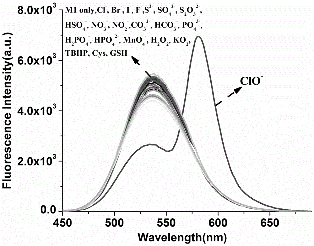
图1为本发明的荧光探针M1荧光选择性图,激发波长420 nm。Figure 1 is a graph of the fluorescence selectivity of the fluorescent probe M1 of the present invention, the excitation wavelength is 420 nm.
图2为本发明的荧光探针M1荧光选择性比率型柱状图,激发波长420 nm,发射波长580 nm和534 nm。Figure 2 is a ratiometric bar graph of the fluorescence selectivity of the fluorescent probe M1 of the present invention, the excitation wavelength is 420 nm, and the emission wavelengths are 580 nm and 534 nm.
图3为本发明的荧光探针M1紫外选择性图。FIG. 3 is a diagram of the ultraviolet selectivity of the fluorescent probe M1 of the present invention.
图4为本发明的荧光探针M1紫外选择性比率型柱状图,吸收波长566 nm和 398nm。Figure 4 is a ratiometric bar graph of the ultraviolet selectivity of the fluorescent probe M1 of the present invention, with absorption wavelengths of 566 nm and 398 nm.
图5为本发明的荧光探针M1识别OCl-的抗其他活性物质干扰性图(比率型柱状图),激发波长420 nm,发射波长580 nm和534 nm。Figure 5 is a graph showing the anti-interference ability of the fluorescent probe M1 of the present invention to identify OCl - (ratio bar graph), the excitation wavelength is 420 nm, and the emission wavelengths are 580 nm and 534 nm.
图6为本发明的荧光探针M1识别OCl-的荧光滴定图,激发波长420 nm。FIG. 6 is a fluorescence titration diagram of the recognition of OCl - by the fluorescent probe M1 of the present invention, and the excitation wavelength is 420 nm.
图7为本发明的荧光探针M1识别OCl-的工作曲线图(比率型线性图),激发波长420nm,发射波长580 nm和534 nm。Fig. 7 is a working curve diagram (ratiometric linear diagram) of the fluorescent probe M1 of the present invention for identifying OCl- , the excitation wavelength is 420 nm, and the emission wavelengths are 580 nm and 534 nm.
图8为本发明的荧光探针M1识别OCl-的识别机理图。Figure 8 is a diagram showing the recognition mechanism of the fluorescent probe M1 of the present invention to recognize OCl - .
图9为本发明的荧光探针M1应用于细胞中对外源性ClO-进行荧光成像图。FIG. 9 is a fluorescent imaging diagram of exogenous ClO - applied in cells using the fluorescent probe M1 of the present invention.
具体实施方式Detailed ways
下面将结合本发明实施例,对本发明的技术方案进行清楚、完整地描述,显然,所描述的实施例仅仅是本发明一部分实施例,而不是全部的实施例。基于本发明中的实施例,本领域普通技术人员在没有付出创造性劳动前提下所获得的所有其他实施例,都属于本发明保护的范围。The technical solutions of the present invention will be clearly and completely described below with reference to the embodiments of the present invention. Obviously, the described embodiments are only a part of the embodiments of the present invention, rather than all the embodiments. Based on the embodiments of the present invention, all other embodiments obtained by those of ordinary skill in the art without creative efforts shall fall within the protection scope of the present invention.
本申请所使用的探针M1的制备步骤为:The preparation steps of the probe M1 used in this application are:
S1. 在50 mL单口烧瓶中,室温搅拌下,将3.0 mL的水合肼溶液逐滴滴加至含1.2g罗丹明B盐酸盐的30 mL乙醇溶液中,加热回流约两小时,TLC检测反应完毕,冷却至室温,减压除去溶剂得到红色固体,加入约50 mL二氯甲烷使固体完全溶解,体系用饱和NaCl溶液(10 mL)洗涤三次,有机相用无水硫酸钠干燥,过滤,减压除去溶剂得到1.03 g浅黄色的固体产物即为中间体1,产率90 %。S1. In a 50 mL single-necked flask, under stirring at room temperature, 3.0 mL of hydrazine hydrate solution was added dropwise to 30 mL of ethanol solution containing 1.2 g of rhodamine B hydrochloride, heated to reflux for about two hours, and the reaction was detected by TLC After completion, it was cooled to room temperature, and the solvent was removed under reduced pressure to obtain a red solid. About 50 mL of dichloromethane was added to completely dissolve the solid. The system was washed three times with saturated NaCl solution (10 mL), and the organic phase was dried with anhydrous sodium sulfate, filtered, and reduced in size. The solvent was removed under pressure to obtain 1.03 g of light yellow solid product, which was Intermediate 1, and the yield was 90%.
S2. 在50 mL单口烧瓶中,将商业购买的4-溴-1,8-萘酐(413.8 mg, 1.5 mmol)和吗啡啉(0.2 g, 2.3 mmol)溶解于25 mL的乙二醇单甲醚溶液中,氮气保护下回流反应12小时。反应完全后,冷却至室温,减压去除溶剂,柱层析分离(甲醇:二氯甲烷=1:10,体积比)得到0.55黄色固体(产率90%)即为中间体4。S2. In a 50 mL single-necked flask, dissolve commercially purchased 4-bromo-1,8-naphthalene anhydride (413.8 mg, 1.5 mmol) and morpholine (0.2 g, 2.3 mmol) in 25 mL of ethylene glycol monomethacrylate The ether solution was refluxed for 12 hours under nitrogen protection. After the reaction was completed, it was cooled to room temperature, the solvent was removed under reduced pressure, and separated by column chromatography (methanol:dichloromethane=1:10, volume ratio) to obtain 0.55% of a yellow solid (yield 90%), which was intermediate 4.
S3. 在100 mL的圆底烧瓶内,称取4-吗啡啉-1,8-萘酐(中间体4,0.1 g,0.35mmol)和对苯二胺(0.76 g, 0.70 mmol)溶于50 mL无水乙醇中,回流反应24小时,反应完全后将体系冷却至室温,将体系中析出的固体过滤并用少量乙醇(10 mL*3次)洗涤,得到黄色固体即为中间体3,产率73%。S3. In a 100 mL round-bottom flask, weigh 4-morpholine-1,8-naphthalene anhydride (
S4. 在50 mL单口烧瓶中,将中间体3 (0.1 g, 0.26 mmol)溶解于10 mL的无水二氯甲烷中,并缓慢滴入含二氯硫化碳(75 μL, 0.1 mmol)和三乙胺(1 mL)的无水二氯甲烷(12 mL)溶液中,滴加完成后,反应体系室温搅拌5小时,反应完全后,体系用饱和NaCl溶液(10 mL)洗涤三次,有机相用无水硫酸钠干燥,过滤,减压下除去溶剂,采用柱层析分离的方法提纯 (二氯甲烷为洗脱剂),得到浅黄色固体即为中间体2,产率90%。S4. In a 50 mL single-necked flask, dissolve intermediate 3 (0.1 g, 0.26 mmol) in 10 mL of anhydrous dichloromethane, and slowly dropwise add the mixture containing carbon dichlorosulfide (75 μL, 0.1 mmol) and trichloromethane. Ethylamine (1 mL) in anhydrous dichloromethane (12 mL) solution, after the dropwise addition, the reaction system was stirred at room temperature for 5 hours. After the reaction was complete, the system was washed three times with saturated NaCl solution (10 mL), and the organic phase was washed with Dry over anhydrous sodium sulfate, filter, remove the solvent under reduced pressure, and purify by column chromatography (dichloromethane as the eluent) to obtain a light yellow solid, which is
S5. 在100 mL的圆底烧瓶内,称取0.3 g (0.72 mmol)中间体2和0.457 g (1mmol) 中间体1溶于50 mL无水乙腈中,搅拌下升温加热回流12 h,通过TLC检测反应完全;冷却反应液,减压下除去溶剂,用二氯甲烷溶解残留物,再加入蒸馏水,在分液漏斗中提取,收集有机相,用无水硫酸钠干燥。过滤得滤液,减压下除去溶剂,得到粗产品,采用柱层析分离的方法提纯 (甲醇:二氯甲烷=1:30),得到橙红色泡沫状固体即为探针M1,产率65%。S5. In a 100 mL round-bottomed flask, weigh 0.3 g (0.72 mmol) of intermediate 2 and 0.457 g (1 mmol) of intermediate 1, dissolve them in 50 mL of anhydrous acetonitrile, heat under stirring under reflux for 12 h, pass TLC Check that the reaction is complete; cool the reaction solution, remove the solvent under reduced pressure, dissolve the residue with dichloromethane, add distilled water, extract in a separating funnel, collect the organic phase, and dry with anhydrous sodium sulfate. The filtrate was obtained by filtration, and the solvent was removed under reduced pressure to obtain a crude product, which was purified by column chromatography separation (methanol:dichloromethane=1:30) to obtain an orange-red foamy solid, which was probe M1, with a yield of 65%. .
产物探针M1的产品表征为:The product characterization of product probe M1 is:
核磁共振氢谱测定:1H NMR (CDCl3, 400 MHz) δ 1.18 (q, J = 5.3 Hz, 12 H),3.28 (t, J = 8.0 Hz, 4 H), 3.35 (q, J = 6.7 Hz, 8 H), 4.02 (d, J = 4.0 Hz, 4H), 6.34 (q, J = 4.0 Hz, 2 H), 6.46 (d, J = 4.0 Hz, 2 H), 6.52 (d, J = 8.0Hz, 2 H), 7.05 (s, 1 H), 7.11 (d, J = 8.0 Hz, 2 H), 7.24 (d, J = 8.0 Hz, 1H), 7.30 (m, 2 H), 7.70 (m, 3 H), 8.02 (s, 1 H), 8.45 (d, J = 4.0 Hz, 1 H),8.53 (d, J = 4.0 Hz, 1 H), 8.58 (t, J = 4.0 Hz, 1 H)。 1H NMR (CDCl 3 , 400 MHz) δ 1.18 (q, J = 5.3 Hz, 12 H), 3.28 (t, J = 8.0 Hz, 4 H), 3.35 (q, J = 6.7 Hz, 8 H), 4.02 (d, J = 4.0 Hz, 4H), 6.34 (q, J = 4.0 Hz, 2 H), 6.46 (d, J = 4.0 Hz, 2 H), 6.52 (d, J = 8.0Hz, 2 H), 7.05 (s, 1 H), 7.11 (d, J = 8.0 Hz, 2 H), 7.24 (d, J = 8.0 Hz, 1H), 7.30 (m, 2 H), 7.70 ( m, 3 H), 8.02 (s, 1 H), 8.45 (d, J = 4.0 Hz, 1 H), 8.53 (d, J = 4.0 Hz, 1 H), 8.58 (t, J = 4.0 Hz, 1 H).
核磁共振碳谱测定:13C NMR (CDCl3, 100 MHz) δ182.45, 167.17, 164.34,163.85, 162.56, 155.90, 154.32, 150.24, 149.38, 137.83, 134.31, 132.86,132.74, 131.50, 130.43, 130.21, 129.09, 129.06, 128.68, 128.08, 127.66,126.21, 125.89, 124.97, 124.76, 123.87, 123.41, 117.09, 114.98, 108.43,104.23, 98.37, 77.48, 77.16, 76.85, 67.16, 66.94, 53.43, 44.44, 12.63。核磁共振碳谱测定: 13 C NMR (CDCl 3 , 100 MHz) δ182.45, 167.17, 164.34,163.85, 162.56, 155.90, 154.32, 150.24, 149.38, 137.83, 134.31, 132.86,132.74, 131.50, 130.43, 130.21, 129.09, 129.06, 128.68, 128.08, 127.66,126.21, 125.89, 124.97, 124.76, 123.87, 123.41, 117.09, 114.98, 108.43,104.23, 98.37, 77.48, 77.16, 76.85, 67.16, 66.94, 53.43, 44.44, 12.63。
高分辨质谱测定:HR-ESI-MS calcd for C51H49N7O5S:871.3516, found 872.3522[M+H+]。High resolution mass spectrometry: HR-ESI-MS calcd for C 51 H 49 N 7 O 5 S: 871.3516, found 872.3522 [M+H + ].
下面结合具体检测实施例对本发明的进行进一步的解释说明:The present invention is further explained below in conjunction with specific detection examples:
实施例1Example 1
配制浓度为pH=7.4, 10 mM的HEPES缓冲溶液,并用乙腈配置体积比为4:1的混合溶液A,用乙腈配制浓度为1 mM的探针M1溶液。用荧光光谱仪考察了探针M1对次氯酸根的选择性。如附图1所示,在激发波长为420 nm的激发条件下,单独的探针M1 (10 µM)在混合溶液A中只在534 nm处具有较强的荧光发射强度,当加入次氯酸根(100 µM)后,在534 nm处的荧光发射强度明显降低,在580 nm处的荧光发射强度明显增强,但是加入其它分析物 (100µM) 时,溶液体系的荧光发射强度与单独探针体系的荧光发射强度相比没有明显变化。附图2为上述结果以荧光发射强度比率(F580/F534)柱状图。以上实验结果表明,该探针对次氯酸根具有较好的专一选择性。A HEPES buffer solution with a concentration of pH=7.4 and 10 mM was prepared, and a mixed solution A with a volume ratio of 4:1 was prepared with acetonitrile, and a solution of probe M1 with a concentration of 1 mM was prepared with acetonitrile. The selectivity of probe M1 to hypochlorite was investigated by fluorescence spectrometer. As shown in Fig. 1, under the excitation condition of excitation wavelength of 420 nm, the single probe M1 (10 µM) in mixed solution A has a strong fluorescence emission intensity only at 534 nm. (100 µM), the fluorescence emission intensity at 534 nm was significantly reduced, and the fluorescence emission intensity at 580 nm was significantly enhanced, but when other analytes (100 µM) were added, the fluorescence emission intensity of the solution system was similar to that of the single probe system. The fluorescence emission intensity did not change significantly. FIG. 2 is a histogram of the above results in terms of the ratio of fluorescence emission intensity (F 580 /F 534 ). The above experimental results show that the probe has good specific selectivity for hypochlorite.
实施例2Example 2
配制浓度为pH=7.4, 10 mM的HEPES缓冲溶液,并用乙腈配置体积比为4:1的混合溶液A,用乙腈配制浓度为1 mM的探针M1溶液。用紫外-可见光谱仪考察了探针M1对次氯酸根的选择性。如附图3所示,单独的探针M1 (10 µM)在混合溶液A中只在398 nm处具有较强的紫外吸收强度,当加入次氯酸根(100 µM)后,在566 nm处的紫外吸收强度明显增强,但是加入其它分析物 (100 µM) 时,溶液体系的紫外吸收强度与单独探针体系的荧光发射强度相比没有明显变化。附图2为上述结果以紫外吸收强度比率(F566/F398)柱状图。以上实验结果表明,该探针对次氯酸根具有较好的专一选择性。A HEPES buffer solution with a concentration of pH=7.4 and 10 mM was prepared, and a mixed solution A with a volume ratio of 4:1 was prepared with acetonitrile, and a solution of probe M1 with a concentration of 1 mM was prepared with acetonitrile. The selectivity of probe M1 to hypochlorite was investigated by UV-Vis spectrometer. As shown in Fig. 3, the single probe M1 (10 µM) has a strong UV absorption intensity only at 398 nm in mixed solution A. When hypochlorite (100 µM) is added, the UV absorption intensity at 566 nm is strong. The UV absorption intensity was significantly enhanced, but with the addition of other analytes (100 µM), the UV absorption intensity of the solution system did not change significantly compared to the fluorescence emission intensity of the probe system alone. Figure 2 is a histogram of the above results in terms of UV absorption intensity ratio (F 566 /F 398 ). The above experimental results show that the probe has good specific selectivity for hypochlorite.
应用例1Application example 1
为了测试探针M1对次氯酸根识别作用在复杂体系中的抗干扰能力,在荧光发射光谱中分别测试了其抗常见活性物质的干扰性。配制浓度为pH=7.4, 10 mM的HEPES缓冲溶液,并用乙腈配置体积比为4:1的混合溶液A,用乙腈配制浓度为1 mM的探针M1溶液。在18个干净的荧光比色皿中,分别加入3000 μL的混合溶液A和30 μL的探针M1乙腈溶液,再分别加入10个摩尔当量的ClO-和10个摩尔当量的其他分析物(Br-, Cl-, F-, I-, CO3 2-, HCO3 -,PO4 3-, HPO4 2-, H2PO4 -, S2-, HSO4 -, NO2 -, NO3 -, MnO4 -, SO4 2-, H2O2, KO2, TBHP),在激发波长为420 nm的激发条件下,在其他活性物质存在时加入次氯酸根与单独加入次氯酸根时所得到的荧光发射强度比率(F580/F534)基本相同,得到荧光发射柱状图(附图5)。In order to test the anti-interference ability of the probe M1 on the recognition of hypochlorite in complex systems, its anti-interference ability against common active substances was tested in the fluorescence emission spectrum. A HEPES buffer solution with a concentration of pH=7.4 and 10 mM was prepared, and a mixed solution A with a volume ratio of 4:1 was prepared with acetonitrile, and a solution of probe M1 with a concentration of 1 mM was prepared with acetonitrile. In 18 clean fluorescence cuvettes, 3000 μL of mixed solution A and 30 μL of probe M1 acetonitrile solution were added, followed by 10 molar equivalents of ClO- and 10 molar equivalents of other analytes (Br - , Cl - , F - , I - , CO 3 2- , HCO 3 - , PO 4 3- , HPO 4 2- , H 2 PO 4 - , S 2- , HSO 4 - , NO 2 - , NO 3 - , MnO 4 - , SO 4 2- , H 2 O 2 , KO 2 , TBHP), under the excitation wavelength of 420 nm, with the addition of hypochlorite in the presence of other active substances and the addition of hypochlorite alone The obtained fluorescence emission intensity ratio (F 580 /F 534 ) was basically the same, and a fluorescence emission histogram was obtained (Fig. 5).
经实验证明,其他分析物不干扰体系对ClO-的测定。It has been proved by experiments that other analytes do not interfere with the determination of ClO - in the system.
应用例2Application example 2
配制浓度为pH=7.4, 10 mM的HEPES缓冲溶液,并用乙腈配置体积比为4:1的混合溶液A,用乙腈配制浓度为1 mM的探针M1溶液。固定探针M1浓度为10 µM,测定其对不同浓度的次氯酸根的响应强度,在激发波长为420 nm的激发条件下,随着次氯酸根浓度的增加,体系荧光发射强度在534 nm处不断降低,同时在580 nm处不断增强(附图6),研究发现溶液荧光发射强度比率(F580 nm/F534 nm)在次氯酸根浓度为0-3 µM间呈线性(R2 = 0.992)(附图7),经计算(3σ/k)得出该探针M1对次氯酸根的最低检出限为1.36×10-8 mol/L,该检出限可满足国家对城市自来水中次氯酸根含量的限量要求,表明该探针M1在城市自来水质量安全方面具有潜在的应用价值。A HEPES buffer solution with a concentration of pH=7.4 and 10 mM was prepared, and a mixed solution A with a volume ratio of 4:1 was prepared with acetonitrile, and a solution of probe M1 with a concentration of 1 mM was prepared with acetonitrile. The concentration of the immobilized probe M1 was 10 µM, and its response intensity to different concentrations of hypochlorite was determined. Under the excitation wavelength of 420 nm, with the increase of hypochlorite concentration, the fluorescence emission intensity of the system was at 534 nm. The ratio of fluorescence emission intensity in solution (F 580 nm /F 534 nm ) is linear between 0-3 µM hypochlorite concentration (R 2 = 0.992 ) (Fig. 7), after calculation (3σ/k), it is concluded that the probe M1 has a minimum detection limit of 1.36×10 -8 mol/L for hypochlorite, which can meet the national requirements for urban tap water. The limit requirement of hypochlorite content indicates that the probe M1 has potential application value in the quality and safety of urban tap water.
应用例3Application example 3
探针M1对细胞外源性次氯酸根荧光成像Fluorescence imaging of cellular exogenous hypochlorite with probe M1
将本发明探针M1应用于Eca109(食管癌细胞),对外源性的次氯酸根进行荧光成像,具体步骤如下:The probe M1 of the present invention is applied to Eca109 (esophageal cancer cells) to perform fluorescence imaging on exogenous hypochlorite, and the specific steps are as follows:
a) 将10 µM探针M1溶液加入到育有Eca109细胞的培养液(2 mL)中,在二氧化碳培养箱中培养20 min,用HEPES缓冲溶液洗涤2次,明场成像,如图(A),可以看到细胞大致的轮廓;a) 10 µM probe M1 solution was added to the culture medium (2 mL) of Eca109 cells, incubated in a carbon dioxide incubator for 20 min, washed twice with HEPES buffer solution, and imaged in bright field, as shown in Figure (A) , you can see the rough outline of the cell;
b) 将 a)中细胞用420 nm激光激发,绿光通道成像,得到具有较强绿色荧光的图(B),没有红色荧光;b) The cells in a) were excited with a 420 nm laser, and the green light channel was imaged to obtain a picture (B) with strong green fluorescence and no red fluorescence;
c) 将10 µM探针M1溶液加入到育有Eca109细胞的培养液(2 mL)中,在二氧化碳培养箱中培养20 min,加入20 µM的次氯酸钠水溶液后,在二氧化碳培养箱中培养20 min,用HEPES缓冲溶液洗涤2次,明场成像,如图(C),可以看到细胞大致的轮廓;c) Add 10 µM probe M1 solution to the culture medium (2 mL) of Eca109 cells, incubate in a carbon dioxide incubator for 20 min, add 20 µM sodium hypochlorite aqueous solution, and incubate in a carbon dioxide incubator for 20 min, Washed twice with HEPES buffer solution, and imaged in bright field, as shown in Figure (C), you can see the rough outline of the cells;
d) 将 c)中细胞用420 nm激光激发,红光通道成像,得到具有较强红色荧光的图(D),没有绿色荧光;d) The cells in c) were excited with a 420 nm laser and imaged in the red light channel to obtain a picture (D) with strong red fluorescence and no green fluorescence;
根据附图9所示,没有加入次氯酸钠溶液的细胞在420 nm的激发下在绿光通道有较强的绿色荧光发出。然而,另一份在加入次氯酸钠溶液后,在420 nm的激发下在红光通道具有较强的红色荧光发出。这说明本发明探针M1可以对细胞中外源性的次氯酸根进行荧光成像。As shown in Fig. 9, the cells without sodium hypochlorite solution emit strong green fluorescence in the green light channel under the excitation of 420 nm. However, the other one exhibited strong red fluorescence emission in the red channel under excitation at 420 nm after the addition of sodium hypochlorite solution. This shows that the probe M1 of the present invention can perform fluorescence imaging on the exogenous hypochlorite in cells.
以上所述仅为本发明的较佳实施例而已,并不用以限制本发明,凡在本发明的精神和原则之内,所作的任何修改、等同替换、改进等,均应包含在本发明的保护范围之内。The above descriptions are only preferred embodiments of the present invention, and are not intended to limit the present invention. Any modification, equivalent replacement, improvement, etc. made within the spirit and principle of the present invention shall be included in the scope of the present invention. within the scope of protection.
Claims (4)

Priority Applications (1)
| Application Number | Priority Date | Filing Date | Title |
|---|---|---|---|
| CN201810116645.XA CN108398409B (en) | 2018-02-06 | 2018-02-06 | A method of fluorescence ratio detection of hypochlorite |
Applications Claiming Priority (1)
| Application Number | Priority Date | Filing Date | Title |
|---|---|---|---|
| CN201810116645.XA CN108398409B (en) | 2018-02-06 | 2018-02-06 | A method of fluorescence ratio detection of hypochlorite |
Publications (2)
| Publication Number | Publication Date |
|---|---|
| CN108398409A CN108398409A (en) | 2018-08-14 |
| CN108398409B true CN108398409B (en) | 2020-10-27 |
Family
ID=63096237
Family Applications (1)
| Application Number | Title | Priority Date | Filing Date |
|---|---|---|---|
| CN201810116645.XA Active CN108398409B (en) | 2018-02-06 | 2018-02-06 | A method of fluorescence ratio detection of hypochlorite |
Country Status (1)
| Country | Link |
|---|---|
| CN (1) | CN108398409B (en) |
Families Citing this family (6)
| Publication number | Priority date | Publication date | Assignee | Title |
|---|---|---|---|---|
| CN109060746B (en) * | 2018-08-21 | 2021-12-21 | 复旦大学 | Ratio type fluorescence detection method and application |
| CN108896523B (en) * | 2018-08-31 | 2020-10-20 | 河南省农业科学院农业质量标准与检测技术研究所 | Fluorescence enhancement type hypochlorite detection method and application |
| CN110183458A (en) * | 2019-05-10 | 2019-08-30 | 安徽工业大学 | A kind of preparation and its application of 2- methylfuran rhodamine 6G amide |
| CN110903467B (en) * | 2019-09-29 | 2022-11-04 | 苏州大学 | Fluorescent conjugated polymer with side group containing rhodamine B unit, preparation method and application thereof |
| CN114685546B (en) * | 2021-12-22 | 2024-05-03 | 青海师范大学 | ICT and FRET mechanism-based hypochlorous acid fluorescence chemical sensor and synthesis method and application thereof |
| CN115684103A (en) * | 2022-09-15 | 2023-02-03 | 济南大学 | Method for quantitatively detecting pH value of cement by using ratio type fluorescent probe |
Citations (3)
| Publication number | Priority date | Publication date | Assignee | Title |
|---|---|---|---|---|
| CN106632363A (en) * | 2016-11-07 | 2017-05-10 | 山东大学 | Mitochondrial targeted ratio type hypochlorous acid fluorescence probe and application thereof |
| CN107021953A (en) * | 2017-05-15 | 2017-08-08 | 三峡大学 | A kind of coumarin fluorescent probe and preparation method and its application on detection hypochlorite ion |
| CN107056704A (en) * | 2016-05-16 | 2017-08-18 | 郑州大学 | A kind of hypochlorite ion's fluorescence probe of lysosome targeting and its application |
-
2018
- 2018-02-06 CN CN201810116645.XA patent/CN108398409B/en active Active
Patent Citations (3)
| Publication number | Priority date | Publication date | Assignee | Title |
|---|---|---|---|---|
| CN107056704A (en) * | 2016-05-16 | 2017-08-18 | 郑州大学 | A kind of hypochlorite ion's fluorescence probe of lysosome targeting and its application |
| CN106632363A (en) * | 2016-11-07 | 2017-05-10 | 山东大学 | Mitochondrial targeted ratio type hypochlorous acid fluorescence probe and application thereof |
| CN107021953A (en) * | 2017-05-15 | 2017-08-08 | 三峡大学 | A kind of coumarin fluorescent probe and preparation method and its application on detection hypochlorite ion |
Non-Patent Citations (4)
| Title |
|---|
| A highly selective fluorescent bioimaging probe for hypochlorite based on 1,8-naphthalimide derivative;Kangming Xiong 等;《Sensors and Actuators B:Chemical》;20150803;第221卷;第1508-1514页 * |
| A ratiometric rhodamine-naphthalimide pH selective probe built on the basis of a PAMAM light-harvesting architecture;Khalid A.Alamry 等;《Journal of Luminescence》;20140916;第158卷;第50-59页 * |
| 基于罗丹明结构的比率型pH、次氯酸根荧光探针的设计、合成及应用研究;张晓帆;《中国优秀硕士学位论文全文数据库 工程科技I辑》;20170215(第02期);正文第28-61页 * |
| 新型荧光/EPR双功能次氯酸探针的构建及性能;刘辉强 等;《高等学校化学学报》;20170930;第38卷(第9期);第1542-1548页 * |
Also Published As
| Publication number | Publication date |
|---|---|
| CN108398409A (en) | 2018-08-14 |
Similar Documents
| Publication | Publication Date | Title |
|---|---|---|
| Huang et al. | A near-infrared BODIPY-based fluorescent probe for ratiometric and discriminative detection of Hg2+ and Cu2+ ions in living cells | |
| Zhong et al. | A simple H2S fluorescent probe with long wavelength emission: Application in water, wine, living cells and detection of H2S gas | |
| Li et al. | A colorimetric and ratiometric fluorescent probe for hydrazine and its application in living cells with low dark toxicity | |
| CN109081836B (en) | Mercury ion near-infrared fluorescent probe based on hemicyanine structure and preparation method and application thereof | |
| Peng et al. | A novel fluorescent probe for selective detection of hydrogen sulfide in living cells | |
| Tian et al. | A novel turn-on Schiff-base fluorescent sensor for aluminum (III) ions in living cells | |
| CN106220640B (en) | A kind of mercury ion fluorescence probe and its preparation method and application | |
| Geng et al. | An aqueous methylated chromenoquinoline-based fluorescent probe for instantaneous sensing of thiophenol with a red emission and a large Stokes shift | |
| Zhang et al. | A BODIPY-based dual-responsive turn-on fluorescent probe for NO and nitrite | |
| CN108003869B (en) | Fluorescent probe for detecting hypochlorite with high sensitivity and synthesis method and application thereof | |
| CN108398409A (en) | A kind of method of ratio fluorescent detection hypochlorite | |
| Zhu et al. | A highly selective ratiometric fluorescent probe for hydrogen peroxide displaying a large emission shift | |
| CN110229147A (en) | A kind of cumarin-benzothiazolyl hydrazone compounds and its preparation method and application | |
| Han et al. | Colorimetric hydrazine detection and fluorescent hydrogen peroxide imaging by using a multifunctional chemical probe | |
| CN111073636B (en) | A kind of fluorescent probe for formaldehyde detection and its preparation method and application | |
| Wu et al. | A colorimetric and near-infrared ratiometric fluorescent probe for hydrazine detection and bioimaging | |
| Wu et al. | A red-to-near-infrared fluorescent probe for the detection of thiophenol based on a novel hydroxylflavone-quinoline-amino molecular system with large Stokes shift | |
| CN113354618B (en) | Hypochlorous acid fluorescent probe capable of targeting cell lysosome, preparation method and application | |
| CN114874142B (en) | Fluorescent probe based on 1,8-naphthalimide derivative and its preparation method and application | |
| CN110590753A (en) | A near-infrared SO2 derivative ratiometric fluorescent probe targeting mitochondria and its application | |
| Shi et al. | Turn on fluorescent detection of hydrazine with a 1, 8-naphthalimde derivative | |
| Erdemir et al. | Building an eco-friendly, biocompatible, and ratiometric NIR fluorescent sensor for the rapid detection of trace Pd2+ in real samples and living cells | |
| CN114249760B (en) | Synthesis and application of fluorescent probe capable of simultaneously distinguishing hypochlorous acid and hydrogen peroxide through three channels | |
| CN113527283B (en) | Small molecule fluorescent HClO probe, preparation method and application thereof | |
| CN106146526B (en) | Fluorescent probe compound and preparation method and application thereof |
Legal Events
| Date | Code | Title | Description |
|---|---|---|---|
| PB01 | Publication | ||
| PB01 | Publication | ||
| SE01 | Entry into force of request for substantive examination | ||
| SE01 | Entry into force of request for substantive examination | ||
| GR01 | Patent grant | ||
| GR01 | Patent grant | ||
| TR01 | Transfer of patent right | ||
| TR01 | Transfer of patent right |
Effective date of registration: 20251120 Address after: 450000 Henan Province, Zhengzhou City, Jinsui District, Dongfeng Road Street, Yuantian Road No. 16, Wenyi Community, Building 13, Unit 1, Room 401 Patentee after: Henan Liangjie Technology Co., Ltd. Country or region after: China Address before: No.6 Longzihu North Road, Longzihu University Park, Zhengdong New District, Zhengzhou City, Henan Province Patentee before: HENAN University OF ANIMAL HUSBANDRY AND ECONOMY Country or region before: China |