Method and device for generating ultrasonic radiation force field for in-vitro culture of engineering cartilage
Technical Field
The invention belongs to the field of ultrasonic regulation, articular cartilage repair and biomedical tissue engineering, and particularly relates to a method and a device for generating an ultrasonic radiation force field for in-vitro culture of an engineered cartilage.
Background
In joint motion, normal cartilage is an indispensable tissue. However, with the development of modern technology and the change of working and living habits, more and more people tend to maintain an action posture for a long time, and the problem of articular cartilage damage is easy to occur. Meanwhile, the articular cartilage has poor self-healing capability and can not be regenerated, so that the problems of joint stiffness, pain aggravation and the like are easily caused after the articular cartilage is damaged, and the life quality of a patient is seriously influenced. At present, the treatment schemes of articular cartilage damage comprise joint lavage, microfracture operation, periosteum transplantation, cartilage transplantation and the like, but all have defects. Clinical research shows that the engineering cartilage transplantation is a limited way for repairing articular cartilage damage and realizing functional reconstruction at present, and is expected to become a brand new treatment mode for repairing bone defects.
The engineering cartilage transplantation technology is a technology for culturing cartilage cells in vitro by a method of planting the cartilage cells on a degradable biological material bracket, constructing cartilage tissues and then implanting the bracket of composite cells into a defect to form bone tissues with physiological functions. The engineered cartilage transplantation combines the operability and the stability of tissue engineered cartilage suture, and the interface healing capacity is improved by using biological materials as cell carriers. However, the mechanical properties of the engineered cartilage produced by the prior art are far different from those of normal cartilage, and cannot meet the requirements of clinical transplantation. At present, the engineering cartilage tissue with good biocompatibility and mechanical property is difficult to obtain by an in vitro culture technology. How to construct normal tissue engineering cartilage under the in vitro condition is used for repairing cartilage damage, provides a better choice for the treatment of cartilage defect, and has good clinical significance.
Williamson et al have shown that the micro-environment of cartilage growth influences its mechanical properties. The cartilage is always in the mechanical environment of static and dynamic pressure alternate activity in vivo, so that the metabolic activity of extracellular matrix is continuously adjusted, collagen and pyridine crosslinking is promoted, and the mechanical property and shape structure of the cartilage are changed. When the engineering cartilage is statically cultured in vitro, the hollow phenomenon of the engineering cartilage is easy to form, and the mechanical property of the finally generated engineering cartilage is insufficient. Most of the existing bioreactors can only realize constant shearing force and dynamic loading pressure. The combination of the two acting forces is single, which is not favorable for culturing the articular cartilage with good structure and function. Therefore, when the engineered cartilage is cultured in vitro, in order to obtain the structural shape and the functional tissue similar to those of the natural cartilage, it is necessary to design a dynamic microenvironment suitable for the growth of the engineered cartilage tissue.
Research in recent years on dynamic culture of engineered cartilage shows that low-intensity ultrasonic waves can regulate and control the mechanical properties of the engineered cartilage by accelerating cartilage formation. The ultrasound waves, as an alternating pressure wave, can create a stable cavity and transmit beamlets within the tissue. Therapeutic intensity ultrasound can produce shear forces on cell membranes, and this effect may directly alter gene expression by affecting the cytoskeleton. Ultrasound also allows for the transport of transmembrane ions and subsequent cellular responses to be altered through transmembrane channels in the cell membrane. The ultrasonic radiation force changes the collagen correlation condition in the cells, so that the ultrasonic regulation and control of the mechanical property of the constructed engineering cartilage become possible. Therefore, in the process of culturing the engineering cartilage in vitro, the structural shape and the mechanical property of the engineering cartilage can be regulated and controlled through the ultrasonic radiation force, and the normal engineering cartilage is obtained.
The invention aims at the requirements, and designs a method and a device for generating an ultrasonic radiation force field for in vitro culture of engineering cartilage through innovative designs such as an ultrasonic transducer working mode, an up-down displacement module and the like. The ultrasonic radiation force field generated by the method and the device can perform mechanical regulation and control on the in vitro culture engineering cartilage to obtain the normal engineering cartilage with good biological property. Realizing the anisotropy of the engineering cartilage, carrying out the chondrocyte transplantation and finally realizing the articular cartilage repair.
Disclosure of Invention
The invention provides a method and a device for generating an ultrasonic radiation force field for in vitro culture of an engineering cartilage, aiming at the application requirements of the occasions such as anisotropy of tissue engineering, mechanical property regulation and control in the process of in vitro culture of the engineering cartilage and the like on specific ultrasonic radiation force. The method can also improve the mechanical property of the engineering cartilage and realize the rapid repair of the cartilage and the comprehensive healing of the bone.
An ultrasonic radiation force field generating device for in-vitro culture of engineering cartilage comprises an ultrasonic transducer (1), a connecting frame (2), a lifting table (3), a fixing base (4), a guide ring (5), an in-vitro culture device (6) of the engineering cartilage, an ultrasonic transmitting and receiving device (7), a signal acquisition and processing module (8) and an oscilloscope (9).
Elevating platform (3) include stiff end and motion end, elevating platform (3) are fixed on unable adjustment base (4) through the stiff end, unable adjustment base (4) are for the square platform that contains cylindrical indent, link (2) are fixed on the motion end of elevating platform (3), ultrasonic transducer (1) are fixed on link (2), guide ring (5) are fixed in the cylindrical indent of unable adjustment base (4), engineering cartilage in vitro culture device (6) are fixed in through the restriction location of guide ring (5) and unable adjustment base (4) fixing device in the cylindrical indent, ultrasonic transducer (1) is connected in ultrasonic emission receiving arrangement (7), signal acquisition processing module (8) are connected ultrasonic transducer (1), oscilloscope (9) are connected signal acquisition processing module (8).
The lifting platform (3) is a gear and rack meshing lifting platform, the fixed end is a T-shaped fixed plate, two symmetrical U-shaped grooves are formed in the length direction of a transverse plate of the T-shaped fixed plate, the U-shaped grooves are located in the middle of the width direction of the transverse plate and can be used for adjusting the position of the lifting platform (3) in a left-right mode within a certain range, the U-shaped grooves are matched with threaded holes in the fixed base plate (4) and fixedly lock the lifting platform (3) through threaded connection, a rack is installed at the vertical end of the T-shaped fixed plate, and the rack is installed at the central position of the vertical end of the T-shaped fixed plate; the movable end is a square block containing a built-in gear, the built-in gear is meshed with the rack at the fixed end, and the built-in gear is manually adjusted to realize the up-and-down movement of the square block.
The connecting frame (2) is a T-shaped connecting piece, the transverse end of the T-shaped connecting piece is installed on the moving end of the lifting platform (3) through a threaded structure, a cylindrical hole used for fixing the ultrasonic transducer (1) is formed in the vertical direction of the long end of the T-shaped connecting piece, a through groove is formed in the bottom surface of the cylindrical hole and the bottom surface of the long end of the T-shaped connecting piece, a threaded through hole is transversely formed in one side of the through groove, a matched threaded hole is formed in the other side of the through groove, the size of the cylindrical hole is slightly changed through threaded connection, and the ultrasonic transducer (1) is locked.
The ultrasonic transducer (1) is a line focus ultrasonic transducer.
The guide ring (5) is a hollow ring with a micro gap, guide teeth are arranged on an inner ring of the micro gap which is symmetrical about the circle center of the guide ring (5), an outer ring of the guide ring (5) is matched with the cylindrical inner recess on the fixed base (4), the inner ring of the guide ring (5) is matched with a culture dish of an engineering cartilage in-vitro culture device (6), and the guide teeth of the guide ring (5) are matched with a base structure of the culture dish to limit the rotation of the culture dish; the center of the micro gap of the guide ring (5) is in a straight line with the circle center of the guide ring (5) and the center of the rack of the lifting platform (3), and the guide ring (5) is fixed in the cylindrical inner recess of the fixed base (4) through fixing glue.
There is the fixed block at the both ends of unable adjustment base (4), and the symmetric center of fixed block is on same straight line with the axial center of cylindrical indent, the fixed block transversely has the relative screw hole that sets up, fixes through helicitic texture engineering cartilage in vitro culture apparatus (6), the axial center of the cylindrical indent of unable adjustment base (4) is on same straight line with the axial center in link cylinder hole.
The engineering cartilage in-vitro culture device (6) comprises a culture dish, a culture medium, engineering cartilage cells and a culture box, and is used for in-vitro culture of the engineering cartilage cells; the height of the culture solution in the engineering cartilage in-vitro culture device (6) is larger than the focal length of the ultrasonic transducer (1).
The ultrasonic transmitting and receiving device (7) is connected with the ultrasonic transducer (1) through a cable and is used for exciting the ultrasonic transducer (1).
The signal acquisition and processing module (8) is based on a high-speed operational amplifier, the specific model is LMH6629, and the signal acquisition and processing module (8) is matched with an oscilloscope (9) to determine the position of the transmitting head of the ultrasonic transducer (1).
A method for generating an ultrasonic radiation force field for in vitro culture of engineering cartilage comprises the following steps:
step (1), connecting an ultrasonic transducer (1) with an ultrasonic transmitting and receiving device (7) through a cable;
and (2) controlling the vertical distance between the emitting head of the ultrasonic transducer (1) and the culture device by manually adjusting the built-in gear of the lifting table (3) so that the emitting head of the ultrasonic transducer (1) is immersed in the culture medium of the engineered cartilage in-vitro culture device (6).
Determining the focal distance of the emitting head of the ultrasonic transducer (1) in the culture medium through a signal acquisition processing module (8), so that the engineering cartilage is positioned at the focal point of the emitting head of the ultrasonic transducer (1);
obtaining the distance L between the transmitting head of the ultrasonic transducer (1) and the engineering cartilage through an oscilloscope (9) according to the product of the distance equal to the period T of the waveform and the sound velocity c in the culture medium; the focus position at the peak maximum value is determined according to the peak amplitude by adjusting the displacement of the emitting head of the ultrasonic transducer (1) up and down;
step (4), the direction of an ultrasonic radiation force field is determined by rotating the ultrasonic transducer (1) to be matched with the guide ring (5) and the engineering cartilage culture device (6);
uploading the waveform of the oscilloscope (9) to a computer upper computer, calculating by the computer upper computer to obtain the sound intensity, and regulating and controlling the position of the transmitting head of the ultrasonic transducer (1) in real time by the obtained sound intensity;
and (6) setting corresponding frequency, emission energy and repetition frequency, starting an ultrasonic emission and receiving device (7), and exciting an ultrasonic transducer (1) to generate an ultrasonic radiation force field.
The invention has the following beneficial effects:
the invention can generate an ultrasonic radiation force field suitable for the in vitro culture of the engineering cartilage, and regulate and control the chondrocyte cultured in vitro to construct a normal engineering cartilage; the ultrasonic radiation force field generated by the invention can change the size of the ultrasonic radiation force by adjusting the distance of the focusing probe up and down. The method can be used for adjusting and controlling the mechanical property of the engineering cartilage, can also be used for realizing the anisotropy of the engineering cartilage tissue, greatly improves the mechanical property of the constructed engineering cartilage, and is used for repairing the articular cartilage.
Drawings
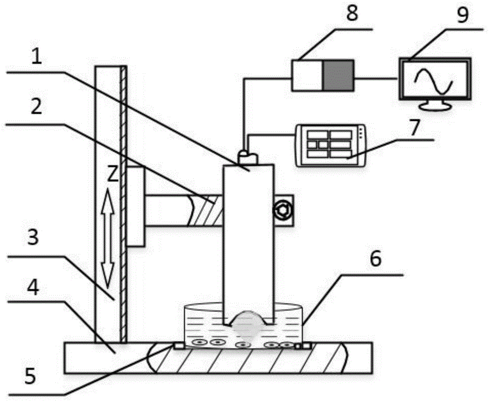
FIG. 1 is a schematic diagram of the structure of the apparatus of the present invention;
FIG. 2 is a flow chart of the method of the present invention;
figure 3 is a top view of the apparatus of the present invention.
In the figure: 1. ultrasonic transducer 2, link 3, elevating platform 4, unable adjustment base 5, guide ring 6, engineering cartilage in vitro culture device 7, ultrasonic emission receiving arrangement 8, signal acquisition processing module 9, oscilloscope.
Detailed Description
The invention is further illustrated with reference to the following figures and examples, without however being limited to the scope of the invention as described below.
As shown in fig. 1, an ultrasonic radiation force field generating device for in vitro culture of engineering cartilage comprises an ultrasonic transducer 1, a connecting frame 2, a lifting table 3, a fixing base 4, a guide ring 5, an engineering cartilage in vitro culture device 6, an ultrasonic transmitting and receiving device 7, a signal acquisition and processing module 8 and an oscilloscope 9.
Elevating platform 3 includes stiff end and motion end, elevating platform 3 passes through the stiff end to be fixed on unable adjustment base 4, unable adjustment base 4 is for the square platform who contains cylindrical indent, link 2 is fixed on elevating platform 3's motion end, ultrasonic transducer 1 is fixed on link 2, guide ring 5 is fixed in unable adjustment base 4's cylindrical indent, engineering cartilage in vitro culture apparatus 6 passes through the restriction location of guide ring 5 and unable adjustment base 4 fixing device fixes in cylindrical indent, ultrasonic transmitting and receiving device 7 connects ultrasonic transducer 1, ultrasonic transducer 1 is connected to signal acquisition processing module 8, oscilloscope 9 connects signal acquisition processing module 8.
Figure 3 is a top view of the apparatus of the present invention.
The lifting platform 3 is a gear rack meshing lifting platform, the fixed end is a T-shaped fixed plate, two symmetrical U-shaped grooves are formed in the transverse end of the T-shaped fixed plate in the length direction of the transverse plate, the U-shaped grooves are located in the middle of the transverse plate in the width direction, and the position of the lifting platform 3 can be adjusted left and right within a certain range. The diameter of the U-shaped groove is 3mm, the length of the U-shaped groove is 8mm, the U-shaped groove is matched with a threaded hole in the fixed bottom plate 4, the lifting platform 3 is fixedly locked through threaded connection, a rack is installed at the vertical end of the T-shaped fixed plate, and the rack is installed at the central position of the vertical end of the T-shaped fixed plate; the moving end is a square block containing a built-in gear, the built-in gear is meshed with the rack at the fixed end, and the built-in gear is manually adjusted to realize the up-and-down movement of the square block. The range of the lifting platform 3 is +/-40 mm, and the precision is 0.1 mm.
The link 2 is T shape connecting piece, the horizontal end of T shape connecting piece is installed on elevating platform 3's motion end through helicitic texture, the cylinder hole that is used for fixed ultrasonic transducer 1 is offered to the vertical direction of the long end of T shape connecting piece, the size of cylinder hole is 18mm the same with ultrasonic transducer 1 external diameter, logical groove has been offered with the bottom surface of the long end of T shape connecting piece to the cylinder hole, the size that leads to the groove is 2 x 5 x 3mm, logical groove one side transversely is provided with the screw through-hole, the screw through-hole diameter is 2mm, the opposite side is provided with supporting screw hole, the screw hole specification is M1.5, through the small size that changes the cylinder hole of threaded connection, locking ultrasonic transducer 1.
The ultrasonic transducer 1 is a line focus ultrasonic transducer, the line focus ultrasonic transducer is cylindrical, and the emitting head of the line focus ultrasonic transducer is a concave hemisphere. The outer diameter of the line focus ultrasonic transducer is 18mm, and the length of the line focus ultrasonic transducer is 78 mm. The piezoelectric material of the line focus ultrasonic transducer is PET crystal, the self frequency is 2.5MHz, the focal length is 18mm, and the size of the crystal is 14 x 14 mm.
The guide ring 5 is a hollow ring with a micro gap, guide teeth are arranged on an inner ring of the micro gap which is symmetrical about the circle center of the guide ring 5, an outer ring of the guide ring 5 is matched with a cylindrical inner recess on the fixed base 4, the inner ring of the guide ring 5 is matched with a culture dish of the engineering cartilage in-vitro culture device 6, and the guide teeth of the guide ring 5 are matched with a base structure of the culture dish to limit the rotation of the culture dish; the center of the micro gap of the guide ring 5, the center of the circle of the guide ring 5 and the center of the rack of the lifting platform 3 form a straight line, and the guide ring 5 is fixed in the cylindrical inner recess of the fixed base 4 through fixing glue.
There is the fixed block at unable adjustment base 4's both ends, and the symmetric center of fixed block and the axial center of cylindrical indent are on the same straight line, and the fixed block transversely has the relative screw hole that sets up, through the fixed engineering cartilage in vitro culture apparatus 6 of helicitic texture, and the axial center of the cylindrical indent of unable adjustment base 4 and the axial center of link cylinder hole are on the same straight line.
The engineering cartilage in-vitro culture device 6 comprises a culture dish, a culture medium, engineering cartilage cells and a culture box and is used for in-vitro culture of the engineering cartilage cells; the height of the culture solution in the engineered cartilage in-vitro culture device 6 is larger than the focal length of the ultrasonic transducer 1.
The ultrasonic transmitting and receiving device 7 is connected with the ultrasonic transducer 1 through a cable and is used for exciting the ultrasonic transducer 1.
The signal acquisition and processing module 8 is based on a high-speed operational amplifier, the specific model is LMH6629, and the signal acquisition and processing module 8 is matched with the oscilloscope 9 to determine the position of the transmitting head of the ultrasonic transducer 1.
As shown in fig. 2, a method for generating an ultrasonic radiation force field for in vitro culture of engineered cartilage:
step (1), connecting an ultrasonic transducer 1 with an ultrasonic transmitting and receiving device 7 through a cable;
and (2) controlling the vertical distance between the emitting head of the ultrasonic transducer 1 and the culture device by manually adjusting the built-in gear of the lifting platform 3, so that the emitting head of the ultrasonic transducer 1 is immersed in the culture medium of the engineered cartilage in-vitro culture device 6.
Step (3), determining the focal distance of the emitting head of the ultrasonic transducer 1 in the culture medium through the signal acquisition processing module 8, so that the engineering cartilage is positioned at the focal point of the emitting head of the ultrasonic transducer 1;
obtaining the distance L between the emitting head of the ultrasonic transducer 1 and the engineering cartilage through an oscilloscope 9 according to the product of the distance equal to the period T of the waveform and the sound velocity c in the culture medium; determining the focal position at the maximum value of the wave crest according to the amplitude of the wave crest by adjusting the displacement of the emitting head of the ultrasonic transducer 1 up and down;
step (4), the direction of an ultrasonic radiation force field is determined by rotating the ultrasonic transducer 1 to be matched with the guide ring 5 and the engineering cartilage culture device 6;
uploading the waveform of the oscilloscope 9 to a computer upper computer, calculating by the computer upper computer to obtain the sound intensity, and regulating and controlling the position of the transmitting head of the ultrasonic transducer 1 in real time through the obtained sound intensity;
and (6) setting corresponding frequency, emission energy and repetition frequency, starting the ultrasonic emission and receiving device 7, and exciting the ultrasonic transducer 1 to generate an ultrasonic radiation force field.
Example 1:
firstly, the lifting platform 3 is arranged at a designated position of the fixed base 1, the moving end of the lifting platform 3 is adjusted to a zero position, and the position of the lifting platform is manually locked. The connecting frame 2 is then mounted to the moving end of the lifting table 3 and secured using a threaded structure. And then the line focus ultrasonic transducer 1 is arranged on the connecting frame, and the line focus ultrasonic transducer 1 is locked on the connecting frame 2 by matching the fixing hole on the connecting frame 2 with the thread. The line focus ultrasonic transducer 1 is connected to the ultrasonic transmitting and receiving device 7 through a cable. The prepared engineering cartilage in-vitro culture device 6 is placed on the fixed base 4, the engineering cartilage in-vitro culture device 6 is rotated to determine the direction of the applied ultrasonic radiation force, and the engineering cartilage in-vitro culture device 6 is fixedly clamped through the fixed block of the fixed base 1. The up-down distance between the emitting head of the ultrasonic transducer 1 and the culture device is manually adjusted by using the lifting platform 3, so that the emitting head of the ultrasonic transducer 1 is immersed in the culture medium of the culture device. The focal distance of the emitting head of the ultrasonic transducer 1 in the culture medium is determined through the signal acquisition processing module 8, so that the engineering cartilage is positioned at the focus of the emitting head of the ultrasonic transducer 1. And displaying the distance L between the emitting head of the ultrasonic transducer 1 and the engineering cartilage through an oscilloscope 9 according to the product of the distance equal to the period T of the waveform and the sound velocity c in the culture medium. And determining the focal position at the maximum value of the wave crest according to the amplitude of the wave crest by adjusting the displacement of the emitting head of the ultrasonic transducer 1 up and down. And the final ultrasonic radiation force field direction is determined by the cooperation of the rotary ultrasonic transducer 1, the guide ring 5 and the engineering cartilage culture device 6. Uploading the waveform of the oscilloscope 9 to a computer upper computer, calculating by the computer upper computer to obtain the sound intensity, and regulating and controlling the position of the transmitting head of the ultrasonic transducer 1 in real time through the obtained sound intensity; the frequency of ultrasonic emission is set to be 2.5MHz, the energy emitted by a probe is 32uJ, the repetition frequency is 1KHz, and a pulse type ultrasonic emission receiver is adopted to generate an alternate ultrasonic sound field. The sound waves propagate in the culture medium, generating a certain pressure p. When encountering the engineering cartilage tissue, the engineering cartilage tissue collides with the engineering cartilage tissue to form ultrasonic radiation force F. The ultrasonic radiation force up-and-down displacement module and the engineering cartilage in-vitro culture device are placed in an incubator and used for in-vitro culture of the engineering cartilage. The ultrasonic transmitting and receiving device is of a pulse negative pressure type, can generate dynamic alternating ultrasonic radiation force F and simulates a tissue culture microenvironment in a human body. The entire device was placed in an incubator and stimulated continuously for 3 days. And taking out the engineering cartilage tissue, and testing the mechanical property of the engineering cartilage tissue. Observing the regulation and control of the mechanical property of the engineering cartilage by the ultrasonic radiation force field.