Gene editing nanocapsule and preparation method and application thereof
Technical Field
The invention relates to the technical field of gene editing, in particular to a gene editing nanocapsule and a preparation method and application thereof.
Background
Malignant tumors are the first diseases threatening human life and health. Most tumors are hidden, early symptoms are not obvious, the diagnosis is already at middle and late stages, and the chance of surgical excision is lost. Chemotherapy is currently one of the major approaches to tumor therapy. However, the traditional chemotherapy drugs have strong toxic and side effects, poor targeting and easy generation of drug resistance. The gene therapy can specifically change genetic materials, so that the purpose of treatment is achieved, the gene therapy has the characteristics of strong targeting property, small toxic and side effects and permanence in treatment, plays more and more important roles in tumor treatment, and has the defects of complex operation, low efficiency, insufficient application range and the like.
In recent years, gene editing techniques have provided new avenues for deficiencies in the field of gene therapy. The CRISPR-Cas9 genome editing system is high in efficiency, can edit multiple genes simultaneously, is simple to operate and wide in application range, and plays a significant role in the field of gene therapy. This system is part of the adaptive immune system in archaea and bacteria, defending against the invasiveness of bacteriophages and plasmids. Two key components, Cas9 nuclease and sgRNA, are critical for CRISPR/Cas9 activity. Wherein the sgRNA recognizes a target sequence in the genome and directs Cas9 nuclease to the target sequence, the Cas9 nuclease in the system then acts as scissors to cleave the double strand of DNA, and then through an in vivo double strand break repair system: nonhomologous end joining (NHEJ) or homologous recombination repair (HDR) results in changes in the target sequence, thereby effecting gene editing. Since the discovery, CRISPR-Cas9 has become the strongest eukaryotic gene editing engineering, and has attracted great interest in tumor therapy. Currently, this system has been widely used in tumor research.
Although the CRISPR-Cas9 technology is promising, some challenges still need to be addressed. The biggest challenge is how to safely and efficiently target the CRISPR-Cas9 genome editing system into tumor cells. One way of delivery is to encapsulate Cas9 and sgRNA as plasmids into viral vectors. However, in this way plasmid DNA can be randomly integrated into the genome, causing cancer or other genetic diseases. Furthermore, template-driven expression of genes limits control over the total amount of Cas9, off-target of this system mainly from overdosing.
An alternative delivery method is to deliver Cas9/sgRNA ribonucleoprotein complex, which allows better control of its intracellular concentration and limits the time frame over which gene editing can be performed. However, significant challenges remain in drug delivery. Most proteins such as enzymes, antibodies or transcription factors face problems of low stability and poor cell membrane permeability due to their fragile tertiary structure and large molecular size. RNA is strongly negatively charged, does not readily penetrate cell membranes, is nuclease sensitive, and usually requires chemical modification to prevent degradation. It is therefore desirable to design suitable vectors to prevent the destruction of proteins and RNA by the deleterious physiological environment, and to deliver both simultaneously into the target cell.
In recent years, rapid development of nanotechnology in the medical field brings new opportunity for tumor treatment. Nanoparticles range in size from tens to hundreds of nanometers, and can be designed for encapsulation and delivery of genetic material, and the like. The properties of high surface area ratio, small volume, loading of various drugs, modification and the like endow the nano material with incomparable advantages of other materials in tumor treatment, such as biocompatibility enhancement, tumor targeting, prolongation of circulation time of drugs in blood, drug release control, carrying of multiple drugs for combined treatment and the like. The damage to normal tissues is reduced by enhancing the penetration and retention (EPR) effect (namely, the permeability of blood vessels at the tumor part is larger than that of normal blood vessels, and the nano-drug is not easy to penetrate the normal blood vessels but can pass through the tumor blood vessels) to gather in the tumor tissues in a more targeted way.
Disclosure of Invention
The invention aims to provide a gene editing nanocapsule, a preparation method and an application thereof, wherein the gene editing nanocapsule comprises a Cas/sgRNA ribonucleoprotein compound and is supported and protected by a polymer, and the gene editing nanocapsule can be efficiently released in tumor cells and is expected to become a safe and efficient gene therapy medicament for tumors.
In order to achieve the above object, the present invention first provides a gene editing nanocapsule, comprising an inner core and an outer shell encapsulating the inner core; the inner core comprises a Cas/sgRNA ribonucleoprotein complex obtained by compounding a Cas nuclease and a sgRNA; the shell comprises a polymer formed by polymerizing a monomer material, the monomer material comprises a first monomer and a second monomer which can be mutually polymerized, the first monomer is a molecule which can be electrostatically combined with the Cas/sgRNA ribonucleoprotein complex, and the second monomer is a tumor microenvironment sensitive molecule.
Specifically, the particle size of the gene editing nanocapsule is 30-50nm, and the particle size is small, so that the gene editing nanocapsule has high endocytosis efficiency.
In some embodiments of the invention, the first monomer comprises at least one of guanidino acrylate, spermine acrylate, and N- (3-aminopropyl) methacrylamide;
the tumor microenvironment sensitive molecule comprises at least one of a reduction sensitive molecule, an acid sensitive molecule, and a ROS (reactive oxygen species) responsive molecule; preferably, the second monomer is a reduction-sensitive molecule;
optionally, the reduction-sensitive molecule is a disulfide bond-containing molecule; preferably, the reduction-sensitive molecule is N, N' -bis (acryloyl) cystamine.
In particular, the method comprises the following steps of,the structural formula of the N, N' -di (acryloyl) cystamine is shown in the specification
Specifically, an acrylate molecule in the acrylate Polyethylene glycol succinylcarboxymethyl ester contains a double bond and can participate in a polymerization reaction, the acrylate Polyethylene glycol succinylcarboxymethyl ester contains a Polyethylene glycol molecule, and Polyethylene glycol (PEG) is a low-toxicity water-soluble molecule, so that the surface charge of nanoparticles can be shielded, steric hindrance is provided, and nonspecific adsorption and aggregation can be reduced; the stability of the nanoparticles can be significantly enhanced and the half-life of the nanoparticle systemic circulation can be increased. The succinylcarboxymethyl ester molecule in the acrylate polyethylene glycol succinylcarboxymethyl ester can be connected with the target agent subjected to amination through a chemical bond, so that the connection between the polymer and the target agent is realized.
Specifically, the structural formula of the acrylate polyethylene glycol succinyl methyl carboxylate is shown in the specification
Wherein n is 22-120.
Specifically, N, N' -di (acryloyl) cystamine contains disulfide bonds, GSH can break the disulfide bonds, and the GSH content in a tumor tissue is far higher than that in a normal tissue, so that after the gene nanocapsule prepared by the invention enters a tumor cell, a polymer in the gene nanocapsule can be efficiently degraded by the GSH, the release efficiency of an effective component (Cas/sgRNA ribonucleoprotein complex) in the gene nanocapsule in the tumor cell is far higher than that of the effective component in the normal cell, the effect of directionally releasing the tumor cell is realized, and when a gene targeted by the sgRNA is a tumor treatment target point, the effect of dying the tumor cell and survival of the normal cell can be realized.
Specifically, the guanidinoacrylate is positively charged, so that the guanidinoacrylate can be electrostatically bonded with a Cas/sgRNA ribonucleoprotein complex with negative charge, a polymer is formed on the outer surface of the Cas/sgRNA ribonucleoprotein complex, the guanidinoacrylate can also improve the positive charge content of the whole nanocapsule, and the cell endocytosis efficiency of the nanocapsule can be improved by adding the guanidinoacrylate into the nanocapsule because the outer surface of a cell membrane is negatively charged.
Specifically, the structural formula of the guanidino acrylate is shown as
In some embodiments of the invention, the gene-editing nanocapsule further comprises a targeting agent modified on the outer surface of the shell; the monomer material further comprises a third monomer capable of being mutually polymerized with the first monomer and/or the second monomer, wherein the third monomer is a molecule capable of being connected with the targeting agent through a chemical bond;
optionally, the third monomer comprises at least one of acrylate polyethylene glycol succinyl carboxymethyl ester and acrylate polyethylene glycol maleimide;
optionally, the targeting agent comprises at least one of Angiopep-2, RGD peptide, apolipoprotein E, and transferrin; preferably, the targeting agent is Angiopep-2.
Specifically, the Angiopep-2 can be used as a targeting agent, specifically targets tumor cells, improves the endocytosis efficiency of the nanocapsule, has a mediation effect, can mediate the nanocapsule to cross BBB, and enables the nanocapsule to be efficiently phagocytosed by human brain glioma cells.
The presence of the Blood Brain Barrier (BBB) makes human brain gliomas one of the most problematic tumors in cancer treatment. BBB is the self-balance defense mechanism of brain, on one hand, it ensures that the central nervous system is free from the invasion of foreign substances, maintains high-efficiency steady state, and simultaneously inputs nutrient substances into the brain; on the other hand, the dense structure of the BBB also prevents therapeutic drugs from entering the brain by non-invasive administration. Since BBB endothelial cells and GBM (glioblastoma multiforme) tissues both highly express receptor-associated protein 1(LRP-1), when GBM is treated by using the gene editing nanocapsule provided by the invention, the targeting agent is preferably Angiopep-2, Angiopep-2 (NH)2) Modified gene editing nano glueThe bursa can be specifically combined with a low density Lipoprotein (LRP) receptor over-expressed on a brain endothelial cell and a brain glioma cell, and the BBB permeability of a Cas/sgRNA ribonucleoprotein complex can be obviously enhanced while the bursa targets the brain glioma cell.
Optionally, the targeting gene of the sgRNA is a tumor targeting therapeutic gene; optionally, the tumor targeted therapeutic gene comprises at least one of MTH1 gene and PLK1 gene; alternatively, the sequence of the target site of the sgRNA on the PLK1 gene is as shown in SEQ ID NO: 1 is shown in the specification;
optionally, the Cas nuclease is Cas9 nuclease.
In some embodiments of the invention, the Cas nuclease and the sgRNA are present in a molar ratio of 1:1 to 1.5, preferably 1: 1.2;
the monomer material comprises acrylate polyethylene glycol succinyl carboxymethyl ester, guanidyl acrylate and N, N '-bis (acryloyl) cystamine, wherein the molar ratio of the acrylate polyethylene glycol succinyl carboxymethyl ester to the guanidyl acrylate to the N, N' -bis (acryloyl) cystamine is 1-3: 1-3, and preferably 1:1: 1;
the molar ratio of the Cas nuclease to the guanidino acrylate is 1: 200-250, preferably 1: 220;
the targeting agent is Angiopep-2, and the molar ratio of the Angiopep-2 to the acrylate polyethylene glycol succinyl carboxymethyl ester is 1-5: 1, preferably 3: 1.
The invention also provides an application of the gene editing nanocapsule in preparing a medicament for treating tumors;
optionally, the tumor is brain glioma, non-small cell lung cancer or cervical cancer; preferably, the tumor is a brain glioma, more preferably a glioblastoma.
The invention also provides a medicine for treating tumor, which comprises the gene editing nanocapsule;
optionally, the tumor is brain glioma, non-small cell lung cancer or cervical cancer; preferably, the tumor is a brain glioma, more preferably a glioblastoma.
The invention also provides a preparation method of the gene editing nanocapsule, which comprises the following steps:
step 1, incubating a Cas nuclease and a sgRNA in a buffer solution to form a Cas/sgRNA ribonucleoprotein complex;
and 2, adding the monomer material and an initiator into the system obtained in the step 1, so that the monomer material is subjected to polymerization reaction to form a polymer coated on the outer surface of the Cas/sgRNA ribonucleoprotein complex.
In some embodiments of the invention, the preparation method further comprises: step 3, adding a targeting agent into the system obtained in the step 2, wherein the targeting agent is connected with the polymer through a chemical bond;
optionally, the targeting agent comprises at least one of Angiopep-2, RGD peptide, apolipoprotein E, and transferrin; preferably, the targeting agent is Angiopep-2;
optionally, in the step 3, after the targeting agent is added, stirring is carried out for 1-3 hours;
the monomer material added in the step 2 further comprises a third monomer capable of mutually polymerizing with the first monomer and/or the second monomer, wherein the third monomer is a molecule capable of being connected with the targeting agent through a chemical bond;
optionally, the third monomer comprises at least one of acrylate polyethylene glycol succinyl carboxymethyl ester and acrylate polyethylene glycol maleimide;
optionally, the initiator comprises ammonium persulfate and N, N, N ', N' -tetramethyl ethylenediamine, the ratio of the ammonium persulfate to the reaction system is 1-5 mg:500 μ L, the ratio of the N, N, N ', N' -tetramethyl ethylenediamine solution to the reaction system is 1-5 μ L:500 μ L, and the concentration of the N, N, N ', N' -tetramethyl ethylenediamine solution is 0.2% -0.8% w/v;
optionally, the step 1 is carried out at 10-30 ℃, and the incubation time is 3-8 minutes; the step 2 is carried out at the temperature of 0-5 ℃ in an oxygen-free environment along with stirring, and the reaction time is 60-120 minutes;
optionally, the preparation method of the gene editing nanocapsule further includes: removing impurities after the gene editing nanocapsule is prepared; optionally, an ultrafiltration centrifuge tube with a molecular weight cut-off of 10kDa is used for removing impurities.
It is understood that the reason why the step 2 is performed at 0 ℃ to 5 ℃ is to avoid the exothermic polymerization reaction causing the system temperature to be too high and further the inactivation of the Cas nuclease.
Optionally, step 2 is performed at 4 ℃.
Optionally, the stirring speed in the step 2 is 250-350 rpm.
It is understood that step 2 is performed in an oxygen-free environment because oxygen may hinder the progress of the polymerization reaction, resulting in poor polymerization effect.
Optionally, the oxygen-free environment is a nitrogen environment or an inert gas (helium, neon, argon, krypton, xenon) environment.
The invention has the beneficial effects that:
the gene editing nanocapsule has a core-shell structure, wherein the inner core comprises a Cas/sgRNA ribonucleoprotein compound, the outer shell comprises a polymer, the Cas/sgRNA ribonucleoprotein compound has a gene editing function, the polymer is used as a carrier of the Cas/sgRNA ribonucleoprotein compound and protects the Cas/sgRNA ribonucleoprotein compound, and the nanocapsule can be efficiently released in tumor cells because the polymer comprises tumor microenvironment sensitive molecules.
Furthermore, the surface of the shell can be modified with a targeting agent, so that the nanocapsule can specifically target tumor cells, and the endocytosis efficiency of the nanocapsule is improved.
The gene editing nanocapsule disclosed by the invention has good biocompatibility and biological safety, and is expected to become a safe and efficient gene therapy medicament for tumors.
The preparation method of the gene editing nanocapsule provided by the invention has the advantages of simple preparation process, high encapsulation efficiency, small particle size of the prepared nanocapsule and high endocytosis efficiency of cells.
Drawings
To more clearly illustrate the technical solutions of the embodiments of the present invention, the drawings that are required to be used in the embodiments will be briefly described below, and it should be understood that the following drawings only illustrate some embodiments of the present invention, and therefore should not be considered as limiting the scope of the present invention.
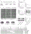
FIG. 1 shows ANCSS(Cas9/sgRNA) nanocapsules particle size distribution and morphology features, wherein a is ANCSS(Cas9/sgRNA) nanocapsule particle size distribution graph, b is ANCSS(Cas9/sgRNA) scanning electron micrographs of nanocapsules.
FIG. 2 is a partial in vitro cell assay result, wherein:
(d)ANCSS(Cas9/sgRNA) nanocapsule, ANC (Cas9/sgRNA) nanocapsule, NCSS(Cas9/sgRNA) nanocapsules, Free Cas9/sgRNA in U87MG-luc cells after 4h incubation flow cytogram;
(e)ANCSS(Cas9/sgRNA) nanocapsule, ANC (Cas9/sgRNA) nanocapsule, NCSS(Cas9/sgRNA) nanocapsules, Free Cas9/sgRNA in U87MG-luc cells after 4h incubation confocal microscopy;
(i) T7E1 experiment analysis U87MG-luc intracellular editing efficiency, the numerical value through imageJ analysis to get;
(j) WB analysis of expression of PLK1 in U87MG-luc cells;
(h) by ANCSS(Cas9/sgRNA) sequencing results compiled for PLK1 gene in nanocapsule treated U87MG-luc cells.
FIG. 3 is a graph showing the results of another in vitro cell assay, in which:
(a) the T7E1 experiment analyzes the editing efficiency in CSC2-luc cells, and the numerical value is obtained by imageJ analysis;
(b) WB analyzed expression of PLK1 in CSC2-luc cells.
Fig. 4A, 4B and 4C are in vivo experimental study results, wherein:
FIG. 4A is ANCSS(Cas9/sgRNA) nanocapsule, ANC (Cas9/sgRNA) nanocapsule, NCSS(Cas9/sgRNA) nanocapsules, Free Cas9/sgRNA pharmacokinetic studies;
FIG. 4B shows ANCSS(Cas9/sgRNA) nanocapsule, ANC (Cas9/sgRNA) nanocapsule, NCSS(Cas9/sgRNA) nanocapsules, Free Cas9/sgRNA in heart, liver, spleen, lung, kidney qualitative distribution; the enlarged view on the right shows the observation of ANC by laser scanning confocal microscopy (CLSM)SS(Cas9/sgRNA) and tumor penetration profile of control group;
FIG. 4C shows ANCSS(Cas9/sgRNA) nanocapsule, ANC (Cas9/sgRNA) nanocapsule, NCSS(Cas9/sgRNA) nanocapsules, Free Cas9/sgRNA in heart, liver, spleen, lung, kidney quantitative distribution.
FIG. 5 shows ANCSS(Cas9/sgRNA) nanocapsules for in vivo therapeutic effect on U87MG-luc tumor-bearing mice, wherein:
(a) a schematic showing an in situ tumor study schedule;
(b) bioluminescence of U87 MG-luc;
(c) h & E whole brain scan characterizes tumor size;
(d) relative photon mass for different nanoparticles;
(e) body weight change during treatment of mice;
(f) survival rate during treatment of mice;
(g) T7E1 experiment analysis U87MG-luc tumor tissue editing efficiency, the numerical value through imageJ analysis;
(h) WB analysis of PLK1 expression in U87MG-luc tumor tissue;
(i) WB quantification results;
(j) gene editing and sequencing results of U87MG-luc tumor tissue.
FIG. 6 shows ANCSS(Cas9/sgRNA) nanocapsules for in vivo therapeutic effect on CSC2-luc tumor-bearing mice, wherein:
(a) schematic diagram of PDX-originated GBM GSC in-situ model building;
(b) bioluminescence of CSC2-luc cells;
(c) h & E whole brain scan characterizes tumor size;
(d) relative photon mass for different nanoparticles;
(e) body weight change during treatment of mice;
(f) survival rate during treatment of mice;
(g) the editing efficiency of the CSC2-Luc tumor tissue is analyzed in a T7E1 experiment, and the numerical value is obtained through imageJ analysis;
(h) WB analysis of expression of PLK1 in CSC2-Luc tumor tissues;
(i) WB quantification results;
(j) CSC2-Luc tumor tissue gene editing and sequencing results.
FIG. 7 shows ANCSS(Cas9/sgRNA) nanocapsule, ANC (Cas9/sgRNA) nanocapsule, NCSS(Cas9/sgRNA) nanocapsules, Free Cas9/sgRNA injected into mice in vivo after different time points of fluorescence intensity.
Detailed Description
The terms as used herein:
"prepared from … …" is synonymous with "comprising". The terms "comprises," "comprising," "includes," "including," "has," "having," "contains," "containing," or any other variation thereof, as used herein, are intended to cover a non-exclusive inclusion. For example, a composition, process, method, article, or apparatus that comprises a list of elements is not necessarily limited to only those elements but may include other elements not expressly listed or inherent to such composition, process, method, article, or apparatus.
The conjunction "consisting of … …" excludes any unspecified elements, steps or components. If used in a claim, the phrase is intended to claim as closed, meaning that it does not contain materials other than those described, except for the conventional impurities associated therewith. When the phrase "consisting of … …" appears in a clause of the subject matter of the claims rather than immediately after the subject matter, it defines only the elements described in the clause; other elements are not excluded from the claims as a whole.
When an amount, concentration, or other value or parameter is expressed as a range, preferred range, or as a range of upper preferable values and lower preferable values, this is to be understood as specifically disclosing all ranges formed from any pair of any upper range limit or preferred value and any lower range limit or preferred value, regardless of whether ranges are separately disclosed. For example, when the range "1 ~ 5" is disclosed, the ranges described should be construed to include the ranges "1 ~ 4", "1 ~ 3", "1 ~ 2 and 4 ~ 5", "1 ~ 3 and 5", and the like. When a range of values is described herein, unless otherwise stated, the range is intended to include the endpoints thereof and all integers and fractions within the range.
In these examples, the parts and percentages are by mass unless otherwise indicated.
"part by mass" means a basic unit of measure indicating a mass ratio of a plurality of components, and 1 part may represent any unit mass, for example, 1g or 2.689 g. If we say that the part by mass of the component A is a part by mass and the part by mass of the component B is B part by mass, the ratio of the part by mass of the component A to the part by mass of the component B is a: b. alternatively, the mass of the A component is aK and the mass of the B component is bK (K is an arbitrary number, and represents a multiple factor). It is unmistakable that, unlike the parts by mass, the sum of the parts by mass of all the components is not limited to 100 parts.
"and/or" is used to indicate that one or both of the illustrated conditions may occur, e.g., a and/or B includes (a and B) and (a or B).
Embodiments of the present invention will be described in detail below with reference to specific examples, but those skilled in the art will appreciate that the following examples are only illustrative of the present invention and should not be construed as limiting the scope of the present invention. The examples, in which specific conditions are not specified, were conducted under conventional conditions or conditions recommended by the manufacturer. The reagents or instruments used are not indicated by the manufacturer, and are all conventional products available commercially.
Some of the reagents used in the examples of the invention were derived as follows:
guanidino acrylate is synthesized by the method described in the literature ROS-Responsive Polymeric siRNA Robust by Triple Interactions for the Robust silence combinatorial RNAi Therapy;
cysteamine propylene (sigma);
acrylate polyethylene glycol succinylcarboxymethyl ester (bond KeyI);
angiopep-2 (intense blaze).
In the present embodiment, the room temperature refers to a temperature condition of 10 ℃ to 30 ℃.
(I) Synthesis of nanocapsules
Example 1 ANCSSPreparation method of (Cas9/sgRNA) nanocapsule
Step 1, Cas9 and sgRNA were added to 500 μ l of 4- (2-hydroxyethyl) -1-piperazineethanesulfonic acid (HEPES buffer) (10mM PH7.4) at a molar ratio of 1:1.2 and incubated for 5 minutes at room temperature.
Specifically, the targeting gene of the sgRNA is PLK1 gene, and the sequence of the target site of the sgRNA on PLK1 gene is shown in SEQ ID NO: 1 is shown.
And 2, transferring the system to a 4 ℃ environment, adding acrylate polyethylene glycol succinylcarboxymethyl ester, stirring for 10 minutes, then adding guanidyl acrylate, stirring for 5 minutes, and then adding degradable N, N '-bis (acryloyl) cystamine, wherein the molar ratio of the acrylate polyethylene glycol succinylcarboxymethyl ester to the guanidyl acrylate to the N, N' -bis (acryloyl) cystamine is 1:1: 1. Adding 3mg of ammonium persulfate and 3 mu of LN, N, N ', N' -tetramethylethylenediamine solution, and immediately initiating polymerization reaction, wherein the polymerization reaction is carried out for 90 minutes at 4 ℃ under the protection of nitrogen, and the polymerization reaction process is always accompanied with mechanical stirring.
Specifically, the molar ratio of the Cas nuclease to the guanidinium acrylate is 1: 220.
And 3, adding the aminated Angiopep-2 into the system, and stirring at room temperature for 2 hours to finally form the nano capsule.
Specifically, the step 2 and the step 3 are both stirred by a magnetic stirrer, and the stirring speed is 250-350 rpm.
And 4, removing impurities by using a centrifugal filter tube with the molecular weight cutoff of 10kDa to remove unreacted monomers and an initiator, and diluting the concentrated solution by using PBS (phosphate buffer solution) (PH 7.4).
FIG. 1 is an ANC prepared in example 1SS(Cas9/sgRNA) nanocapsules DLS test results. As shown in fig. 1, the nanocapsule prepared in example 1 has an average particle size of 31nm and a relatively uniform particle size distribution.
Example 2 ANCSSPreparation method of (Cas9/sgRNA) nanocapsule
The only difference from the above embodiment 1 is that: the molar ratio of the Cas nuclease to the guanidinium acrylate is 1: 200.
Example 3 ANCSSPreparation method of (Cas9/sgRNA) nanocapsule
The only difference from the above embodiment 1 is that: the molar ratio of the Cas nuclease to the guanidinium acrylate is 1: 250.
Comparative example 1 preparation method of ANC (Cas9/sgRNA) nanocapsule
The only difference from the above example 1 is that: in the step 2, N' -bis (acryloyl) cystamine which can be degraded by GSH is replaced by hexamethylene diacrylate which cannot be degraded by GSH, so that the polymer in the prepared ANC (Cas9/sgRNA) nanocapsule cannot be specifically degraded by tumor cells, and thus the gene editing system (Cas9RNP) in the ANC (Cas9/sgRNA) nanocapsule cannot be efficiently released in the tumor cells, and therefore the gene editing efficiency is low.
Comparative example 2 NCSSPreparation method of (Cas9/sgRNA) nanocapsule
The difference from the above example 1 is that: step 3 was not included (i.e., no Angiopep-2 addition), while the acrylate polyethylene glycol succinylcarboxymethyl ester was replaced with methoxypolyethylene glycol amine (mPEG-NH)2) MW: 2000 (available from Peng), manufactured NCSS(Cas9/sgRNA) nanocapsules do not have the ability to cross the BBB and cannot be efficiently phagocytosed by human brain glioma cells.
Comparative example 3 ANCSSPreparation method of (Cas9/sgScr) nanocapsule
The only difference from the above example 1 is that: the sgRNA used in comparative example 3 was an ineffective sgRNA and had no targeting function.
Comparative example 4 preparation method of Free Cas9/sgRNA
Cas9 and sgRNA were coded at 1:1.2 molar ratio to 500. mu.l HEPES buffer, and incubated at room temperature for 5 minutes.
It is worth mentioning that the following two cell experiments and three animal experimentsANC usedSSThe (Cas9/sgRNA) nanocapsules are all the nanocapsules prepared in example 1, the ANC (Cas9/sgRNA) nanocapsules are prepared in comparative example 1, and NCSS(Cas9/sgRNA) nanocapsules were prepared from comparative example 2, ANCSS(Cas9/sgScr) nanocapsules were prepared from comparative example 3, and Free Cas9/sgRNA from comparative example 4.
(II) cell assay
Flow cytometry and confocal microscope characterization of endocytosis and intracellular release
In flow cytometry assays, U87MG-luc cells were seeded in 6-well cell culture plates (1X 10)6Cells/well) was cultured at 37 ℃ for 24 hours, and 150. mu.L of ANC was addedSS(Cas9/sgRNA) nanocapsule, ANC (Cas9/sgRNA) nanocapsule, NCSS(Cas9/sgRNA) nanocapsules, Free Cas9/sgRNA in PBS (Cas9 concentration 20nM), incubated for 4 hours, the sample aspirated, and the cells digested with 500. mu.L of trypsin. The resulting Cell suspension was centrifuged at 1000 Xg for 3 minutes, washed twice with PBS buffer, re-dispersed in 500. mu.L of PBS buffer, subjected to flow cytometry (BD FACS Calibur, Becton Dickinson, USA) for 1 hour, and harvested by circling 10000 cells using Cell Quest software.
The endocytosis and intracellular drug release behavior was observed by CLSM (focal laser scanning microscope) photographs. U87MG-luc cells were plated in 24-well cell culture plates (1X 10) containing microscope slides5Cells/well) for 24 hours, 50. mu.L of ANC was addedSS(Cas9/sgRNA) nanocapsule, ANC (Cas9/sgRNA) nanocapsule, NCSS(Cas9/sgRNA) nanocapsules, Free Cas9/sgRNA in PBS buffer (Cas9 concentration 20 nM). After 4 hours of incubation, the medium was removed and washed twice with PBS buffer. Cytoskeleton was washed twice with Phalloidin for 30 min, followed by washing twice with DAPI for 15 min. Fluorescent pictures were taken by CLSM (TCS SP 5).
② in vitro gene editing characterization cell editing efficiency
U87MG-luc/CSC2-luc cells were seeded in 24-well plates (5X 10)4One cell/well) and cultured for 24 hoursThen (c) is performed. Add 50. mu.L of ANCSS(Cas9/sgRNA) nanocapsules, ANCSS(Cas9/sgScr) nanocapsule, ANC (Cas9/sgRNA) nanocapsule, NCSS(Cas9/sgRNA) nanocapsules, Free Cas9/sgRNA in PBS (Cas9 concentration 20nM) were incubated overnight, then the medium was changed and the cells were incubated at 37 ℃ for another 48 h. Genomic DNA was extracted using a universal genomic DNA kit (China, CWBIO). Then, a DNA fragment containing the sgRNA target site was amplified with a high fidelity enzyme Kod-Plus-Neo (Japan, TOYOBO), and the PCR product was purified with a gel recovery kit (CWBIO, China). Finally, the T7E1 enzyme (NEB, USA) was tested for indel efficiency. PCR products with mutations indicated by the T7E1 assay were DNA sequenced and then subcloned into the T cloning vector (Vazyme Biotech, china). Colonies were randomly selected and further analyzed by DNA Sanger sequencing using M13F as primer (Sangon Biotech), and the sequencing results of partial clones are shown in fig. 2, h. Sanger sequencing showed that 12 out of 21 clones produced mutations at the target sequence, with the mutation pattern for 5 clones being a T single base insertion and the mutation pattern for 7 clones being a base deletion.
③ representing protein level expression quantity in cells by WB experiment in vitro
U87MG-luc/CSC2-luc cells were seeded in 6-well plates (1X 10)5Individual cells/well) and cultured for 24 hours. Add 150. mu.L of ANCSS(Cas9/sgRNA) nanocapsules, ANCSS(Cas9/sgScr) nanocapsule, ANC (Cas9/sgRNA) nanocapsule, NCSS(Cas9/sgRNA) nanocapsules, Free Cas9/sgRNA in PBS (Cas9 concentration 20nM) were incubated overnight, then the medium was changed and the cells were incubated at 37 ℃ for a further 72 h. Cells were treated with lysis buffer (Beyotime, china) and the concentration of the resulting protein was quantified with BCA kit (Beyotime, china). Lysates were separated by electrophoresis (SDS polyacrylamide gel) and transferred to PDVF membrane (Beyotime, china). PDVF membrane was mixed with primary antibody against PLK1 (mouse mAb 35-206, Abcam) at 1: 1000, and incubated overnight at 4 ℃. ECL secondary antibody incubation for 1h showed protein bands (Licor, USA). Protein bands were analyzed using Image J software.
(III) animal experiments
Study of pharmacokinetics
In an in vivo pharmacokinetic study, BALB/c mice were randomly grouped (3 mice per group in parallel) at 6-8 weeks, and 200. mu.L of ANC was injected from the tail veinSS(Cas9/sgRNA) nanocapsule, ANC (Cas9/sgRNA) nanocapsule, NCSS(Cas9/sgRNA) nanocapsules, Free Cas9/sgRNA (Cas9 dose is 30 μ g), and orbital bleeds were taken at predetermined time points. The blood sample is extracted by organic solvent to separate out the medicine, and the medicine is quantified by a multifunctional enzyme-labeling instrument. The pharmacokinetic parameters of the drug such as elimination half-life (t1/2), the area under the drug concentration-time curve (AUC), Clearance (CL) and the like in vivo can be calculated by software fitting.
② antitumor effect
The U87MG-Luc or CSC2-Luc brain colloidal tumor in-situ model is established by transplanting tumor tissues into the brain of BALB/c nude mice (18-20g, 6-8 weeks old). When the tumor volume is 20-30mm3Is used for treatment experiments; the tumor volume is 100-150mm3It is used for biodistribution experiments.
An in-situ model is established by using luciferase-labeled human brain glioma cells U87MG-Luc or human brain glioma stem cells CSC2-Luc, single-dose or multi-dose administration is carried out in a tail vein injection mode, and the growth condition of the tumor is qualitatively and quantitatively tracked by IVIS III. In the treatment process, the systemic toxic and side effects and the antitumor activity of the nano-drug are evaluated through the weight change and the survival rate of the mice. After the treatment is finished, the health condition of each normal organ and the apoptosis condition of tumor tissues of the mouse after the nano-drug treatment are analyzed by histological staining methods such as H & E, TUNEL and the like. Through treatment experiments, the system toxicity and the anti-tumor activity of the nano-drug to U87MG-Luc or CSC2-Luc tumor-bearing nude mice can be judged.
③ biodistribution
Tail vein injecting nanometer medicine into nude mouse with Henshi U87MG-Luc/CSC2-Luc, collecting main tissues of mouse heart, liver, spleen, lung, kidney, brain and tumor at different time points, and imaging with IVIS III in vitro. Then, after homogenizing each tissue, extracting with an organic solvent, and centrifuging to obtain a supernatant, the fluorescence spectrophotometer quantitatively analyzes the in vivo biodistribution of the drug at different time points. Through the experiment, the influence of in vivo stability, active targeting performance and enrichment, retention and permeation of the released Cas9 drug at a tumor part of the nano drug can be deduced.
Crossing effect and targeting property of BBB
Injecting nanometer medicine into nude mouse with in situ human brain glioma U87MG-luc via tail vein, tracking the distribution of nanometer medicine in vivo at different time points with small animal imaging instrument (IVIS III), and observing the accumulation and retention of brain tumor part, and comparing with non-targeting control group (NC)SS(Cas9/sgRNA) nanocapsule) and a non-sensitive control group (ANC (Cas9/sgRNA) nanocapsule) are qualitatively and quantitatively compared, and BBB crossing efficiency and nano-drug tumor targeting capability are examined.
(IV) results and discussion
(1) In vitro cell experiments
Flow cytometry experiments (FIG. 2, d) demonstrate ANCSS(Cas9/sgRNA) nanocapsules have high endocytosis efficiency, and the endocytosis efficiency of the nanocapsules is non-targeted control NCSS2.5 times of (Cas9/sgRNA) and 10.3 times of control group ANC (Cas 9/sgRNA).
ANC was observed in CLSM (laser scanning confocal microscope) (FIG. 2, e)SS(Cas9/sgRNA) nanocapsules were incubated for 4 hours, and strong fluorescence of Cas9 was observed in the U87MG-luc nucleus, which confirmed ANCSS(Cas9/sgRNA) nanocapsules can be rapidly degraded in cells, release Cas9 and transport to cell nucleus for function.
In vitro gene editing experiments (FIG. 2, i) demonstrate ANCSSThe (Cas9/sgRNA) nanocapsule has high gene editing efficiency on U87MG-luc cells, and the gene editing efficiency reaches 36.6%.
WB experiment (FIG. 2, j) demonstrated ANCSSThe (Cas9/sgRNA) nanocapsule can obviously reduce the expression of PLK1 in U87MG-luc cells, thereby achieving the effect of inhibiting the growth of tumors.
(2) In vivo experimental study on targeted nano-drug ANCSS(Cas9/sgRNA) nanocapsules pharmacokinetics and biodistribution
The results of in vivo pharmacokinetics (FIG. 4A) studies show that the targeted nano-drug ANCSS(Cas9/sgRNA) in vivoHas longer circulation time and is compared with ANC (Cas9/sgRNA) nanocapsule and NCSS(Cas9/sgRNA) nanocapsules were comparable, significantly longer than Free Cas 9/sgRNA. Thus showing that the biocompatibility of the nano capsule is better.
The results of in vivo biodistribution experiments (FIG. 4B, FIG. 4C) in BALB/C mice bearing U87MG-luc glioma brain cells showed that ANCSSThe enrichment amount of the (Cas9/sgRNA) nanocapsule at the tumor site is obviously improved compared with that of Free Cas9/sgRNA, and after 4 hours of injection, the enrichment amount of the Cas9 is 12% (the content of Cas9 in each gram of tissue accounts for the mass percent of the total injection amount).
(3)ANCSS(Cas9/sgRNA) nanocapsule tumor inhibition experiment and histological analysis
ANCSSThe results of the treatment experiments of (Cas9/sgRNA) nanocapsules in U87MG-luc tumor-bearing BALB/c nude mice are shown in FIG. 5:
in FIG. 5, ANCSS(Cas9/sgPLK1) shows that the targeting gene of sgRNA is PLK1, ANCSS(Cas9/sgScr) for negative control experiments, PBS for PBS buffer (control experiments);
as can be seen in FIG. 5, ANCSSThe (Cas9/sgRNA) nanocapsule can effectively inhibit tumor growth. ANCSS(Cas9/sgRNA) nanocapsule-treated mice showed slight weight changes, compared with a 20% decrease in body weight in PBS group at 8 days, indicating ANCSS(Cas9/sgRNA) nanocapsules have small toxic and side effects, and in addition, it is known to those skilled in the art that brain tumor growth causes weight loss, so the experimental data of the present application reflect ANC laterallySSThe (Cas9/sgRNA) nanocapsule has a good curative effect and can inhibit brain tumor.
ANCSSThe results of the treatment experiments of (Cas9/sgRNA) nanocapsules in CSC2-luc tumor-bearing BALB/c nude mice are shown in FIG. 6:
in FIG. 6, ANCSS(Cas9/sgPLK1) shows that the targeting gene of sgRNA is PLK1, ANCss(Cas9/sgScr) for negative control experiments, PBS for PBS buffer (control experiments);
as can be seen in FIG. 6, ANCSSThe (Cas9/sgRNA) nanocapsule can effectively inhibit tumor growth. ANCSS(Cas9/sgRNA) nanocapsule therapyThe slight change in body weight of the treated mice indicates ANCSS(Cas9/sgRNA) nanocapsules have small toxic and side effects, and in addition, it is known to those skilled in the art that brain tumor growth causes weight loss, so the experimental data of the present application reflect ANC laterallySSThe (Cas9/sgRNA) nanocapsule has a good curative effect and can inhibit brain tumor.
Remarkably, mice were on ANCSS(Cas9/sgRNA) nanocapsules dose 30 μ g treatment significantly extended the survival cycle. From H&Results of histological analysis of E staining demonstrated that the cells were not viable at ANCSSAfter the (Cas9/sgRNA) nanocapsule dose is 30 mu g, the damage to main organs such as heart, liver, spleen, lung and kidney is small. The results again illustrate ANCSS(Cas9/sgRNA) nanocapsules have very low systemic toxicity.
(4)ANCSSTumor targeting experiment of (Cas9/sgRNA) nanocapsules
The results of tracking the distribution of the nano-drug in vivo at different time points (fig. 7) by a small animal imager (IVIS III) show that ANCSSThe fluorescence intensity of the (Cas9/sgRNA) nanocapsule is obviously higher than that of a control group, which indicates that the nanocapsule has good tumor targeting capability.
Finally, it should be noted that: the above embodiments are only used to illustrate the technical solution of the present invention, and not to limit the same; while the invention has been described in detail and with reference to the foregoing embodiments, it will be understood by those skilled in the art that: the technical solutions described in the foregoing embodiments may still be modified, or some or all of the technical features may be equivalently replaced; and the modifications or the substitutions do not make the essence of the corresponding technical solutions depart from the scope of the technical solutions of the embodiments of the present invention.
Furthermore, those skilled in the art will appreciate that while some embodiments herein include some features included in other embodiments, rather than other features, combinations of features of different embodiments are meant to be within the scope of the invention and form different embodiments. For example, in the claims above, any of the claimed embodiments may be used in any combination. The information disclosed in this background section is only for enhancement of understanding of the general background of the invention and should not be taken as an acknowledgement or any form of suggestion that this information forms the prior art already known to a person skilled in the art.
Sequence listing
<110> university of Henan
<120> gene editing nanocapsule and preparation method and application thereof
<160> 1
<170> SIPOSequenceListing 1.0
<210> 1
<211> 20
<212> DNA
<213> Intelligent (Homo sapiens)
<400> 1
tacctacggc aaattgtgct 20