Disclosure of Invention
The invention aims to solve the defects of the existing research, provides a novel hot-pressing sterilization program, is successfully applied to the sterilization treatment of biological ink, and constructs a human liver 3D model by using the sterilized biological ink. The treatment method adopts a common high-pressure steam sterilizer, the model is constructed by gelatin-sodium alginate biological ink sterilized by a control program, the operation is simple, large-scale equipment is not needed, the ink printing capability is good, the cell growth is good, and the stable cell activity can be maintained for more than 20 days.
Compared with the traditional radiation sterilization and ethylene oxide sterilization, the biological ink treatment method can more quickly finish the sterilization step of the biological ink, and does not depend on special equipment and places. The biological ink sterilized by the method constructs a 3D model of the human liver, can well simulate the physiological conditions in the body, has flexible and changeable bracket size, can be matched with porous plates of various models, and meets the requirement of high-throughput screening. The sterilization method can be well used for processing the gelatin-sodium alginate bio-ink and also provides a reference for processing other formula bio-inks.
In order to solve the problems encountered in the sterilization treatment of the current biological ink, the invention establishes a novel biological ink sterilization method, does not need special instruments, has complete sterilization and simple, convenient and easy operation, can be directly applied to the treatment of the gelatin-sodium alginate biological ink with different concentrations, and provides reference for the sterilization of other formula biological inks.
In order to solve the technical problems, the invention provides a treatment method, a property investigation method and application of biological ink. The invention utilizes the sterilized gelatin-sodium alginate biological ink to establish an in vitro liver model which can simulate the growth environment of the liver under the physiological condition in vivo, the model is transferred to a perforated plate for culture after printing, other intervention conditions are not needed, cells are automatically proliferated and aggregated in a printed bracket, the operation is simple and convenient, and the invention can provide reference for in vitro tissue regeneration. And can also be applied to high-flux drug screening.
In order to achieve the purpose, the invention adopts the following technical scheme:
A3D culture system of human hepatocytes comprises a biological ink gel scaffold loaded with human hepatocytes and a special culture medium for 3D culture of the human hepatocytes. The special culture medium for 3D culture is a basic culture medium (MEM) added with 10% (v: v) fetal calf serum and 1% (v: v) non-essential amino acid.
Preferably, the non-essential amino acid is a mixed solution of 7 non-essential amino acids, such as 890mg/l L-alanine, 1320mg/l L-asparagine, 1150mg/l L-proline, 1330mg/l L-aspartic acid, 1050mg/l L-serine, 1470mg/l L-glutamic acid and/or 750mg/l L-glycine.
The culture components are the optimal components obtained by experiments, and after the concentration of serum or non-essential amino acid is reduced or reduced, the growth rate and the balling form of 3D cultured hepatocytes are negatively affected, a large amount of cell death phenomena occur, and long-term culture cannot be realized.
Preferably, the bio-ink consists of gelatin, sodium alginate and pure water. The gelatin is B type gelatin with the intensity of 250g Bloom gum, and the sodium alginate is analytically pure sodium alginate. The gelatin has good biocompatibility, plays a role similar to an extracellular matrix, provides a position for cell adhesion growth, and provides a space for cell proliferation in the culture process due to the degradability of the gelatin. Under the action of calcium ions, the sodium alginate can be reversibly crosslinked to form a stable structure, and the stability of the gel structure is kept.
Preferably, the concentration of gelatin in the bio-ink is 20% (w/v), the concentration of sodium alginate is 4% (w/v), and the sterilization time is 25 min.
The above concentration is the best composition obtained by the experiment, and after the component concentration is reduced or changed or the sterilization time is shortened, the printing capability of the biological ink is negatively affected, the phenomena of over-strong fluidity, collapse in the printing process and the like occur, and good printing fidelity and long-term culture cannot be realized.
The invention provides the investigation of the fluidity, extrusion characteristic, rheological property and swelling property of the biological ink treated by the treatment method.
The invention also provides application of the biological ink in constructing a human liver cell 3D culture system and detecting cell viability.
A3D culture method of human hepatocytes comprises the following steps of culturing hepatocytes by using a 3D culture system in which human hepatocytes are loaded with bio-ink.
Preferably, the biological ink consists of gelatin and sodium alginate, pure water is used as a solvent, the gelatin and the sodium alginate are dissolved in an open environment under the heating condition of a water bath at 37 ℃, and high-pressure steam sterilization is carried out after full stirring and mixing.
Further, the temperature rise rate of the high-pressure steam sterilization is 5.5 ℃/min, the sterilization condition is 121 ℃, the pressure is 0.1MPa, the sterilization time is 25min, after the sterilization is finished, the air is exhausted at the speed of 5L/min immediately, the temperature is reduced to be below 100 ℃ within 10min, the biological ink is taken out immediately, and the biological ink is placed at room temperature for cooling for standby.
Preferably, the liver cell is a human liver cancer cell HepG2 cell line, and is prepared from a HepG2 cell line subjected to planar amplification or cryopreservation recovery.
The processing method of the plane amplification cell line comprises the following steps: cells were digested with Trypsin digest in cell culture dishes after planar amplification, centrifuged at low speed and resuspended in fresh medium.
The cell line treatment method for cryopreservation resuscitation comprises the following steps: taking out the freezing tube filled with the human liver cells, immediately putting the freezing tube into a water bath at 37 ℃ for heating, immediately transferring the freezing tube into a 15ml centrifuge tube after the freezing solution is completely melted, adding 10ml of fresh culture medium for resuspension, and resuspending the freezing tube into the fresh culture medium after low-speed centrifugation.
Further, the method for culturing human hepatocytes uses a culture condition of 37 ℃ CO 2 The volume concentration was 5%.
Further, in the method for culturing human hepatocytes, the hepatocyte suspension after low-speed centrifugation is mixed with preheated biological ink at 37 ℃, loaded on a 1 mL-3D biological printer, printed with a multi-layer grid structure (such as 4 layers), and immersed in CaCl after printing 2 The solution was crosslinked, washed, transferred to a 12-well plate, and cultured by adding a culture medium. The density of the mixed liver cells is 1-5 multiplied by 10 6 Individual cells/ml; preferably 5X 10 6 Individual cells/ml. Too low initial cell density is detrimental to cell growth, and too high cell density affects balling efficiency and growth, exceeding 1X 10 7 Individual cells/ml, easily causing local accumulation of metabolic waste products, leading to cell death. The mixing volume ratio of the cell suspension to the biological ink is 1: 1-1: 4, preferably 1: 4. too high a proportion of cell suspension may affect the printability of the mixed ink, and is less than 1: 4, the cell suspension has too small volume and too high concentration, which easily causes the error of cell density and influences the repeatability of the experiment.
Further, in the method for culturing human hepatocytes, the printing environment temperature is 26 ℃, the printing speed is 3mm/s, and the printing platform temperature is 4 ℃. The temperature of the printing environment is too high, the ink fluidity is enhanced, and the structure collapse in the printing process is caused; too low a temperature of the printing environment may cause an increase in extrusion pressure, resulting in massive cell death. Too fast a printing speed may result in possible printing distortions; too low a printing speed can cause unevenness of line edges, prolong printing time and influence cell viability. Too low a temperature of the printing platform can result in reduced cell viability; excessive printing platform temperature can cause the gel structure to be unstable during printing, causing the structure to collapse.
Further, in the method for culturing human hepatocytes, CaCl 2 The solution concentration was 3% (w/v, i.e. 3 gCaCl) 2 Dissolved in 100ml of water) and the crosslinking time was 30 s. Modification of CaCl 2 Solution concentration, corresponding to crosslinking timeChanges need to be made. The crosslinking time is too short, the gel stability is insufficient, and the gel may disintegrate in the culture process; too long a cross-linking time can result in cell damage, leading to massive cell death.
The invention has the advantages that:
1. the invention provides a novel biological ink sterilization treatment method, which is used for 3D culture of human hepatocytes, is simple and easy to implement, does not depend on special instruments, and can be stored for a long time at 4 ℃ after being treated.
2. The 3D culture system based on the biological ink does not need external force, cells loaded by the biological ink are printed and then added with culture solution for culture, the cells grow in the bracket automatically, and possible mechanical damage caused by centrifugal force introduced by a suspension culture system is reduced. The scaffold is loaded with cells, so that possible cell loss in the culture process is greatly reduced, and cell loss caused by liquid change is reduced.
3. The invention introduces extracellular matrix, better simulates the physiological environment in vivo, and the degradability of the matrix reduces the limit of the scaffold on cell proliferation.
4. The 3D culture system constructed by the invention can be matched with multi-hole plates of various models, and meets the requirement of high-throughput screening.
5. Compared with a 2D model, the 3D model cultured by the invention can better simulate the physiological environment in vivo in vitro, has larger cell proliferation space, maintains longer and stable cell activity, and can meet the requirement of long-term administration. The model can be used for high-throughput drug screening and disease research, and provides more reliable and real data support for the model.
Detailed Description
The experimental procedures used in the following examples are all conventional procedures unless otherwise specified. Materials, reagents and the like used in the following examples are commercially available unless otherwise specified. The main reagents are as follows: MEM medium (KeygEN), PBS buffer (KeygEN), nonessential amino acids (Wisent), fetal bovine serum (Wisent), 0.25% Trypsin pancreatin-EDTA (Gibco), CCK-8 cell viability assay kit (MCE), Calcein-propidium iodide (Calcein-AM/PI) viable cell staining kit (Yeasen).
The main materials are as follows: gelatin and sodium alginate.
The main apparatus is as follows: LDZX-50KBS vertical pressure steam sterilizer (Shanghai Shenan), cell culture box (Thermo), rotation 5 cell imager (Biotek), ultra-clean bench (Sujing Andai), low-speed desk centrifuge, Discover HR-2 rheometer (TA).
Embodiments of the present invention will be described in further detail below with reference to the accompanying drawings.
Example 1:
gelatin and sodium alginate are selected as the constituent materials of the biological ink. The gelatin is type B gelatin and is derived from animal bone. The sodium alginate is analytically pure alginate, and the viscosity of the sodium alginate is 350-550 mPa & s (1% in H) 2 O, 20 ℃). Derived from another fish skinGelatin is also used for biological culture, but the mechanical property of gel formation is poor, so that the gelatin is not suitable for 3D biological printing, and animal source type B gelatin with better mechanical property and higher use rate is finally selected as a biological ink material for further research.
Firstly, preparing a biological ink precursor solution in an open environment, wherein the specific scheme is as follows:
step 1, adding water into gelatin for swelling at room temperature until no obvious liquid flows, heating at 37 ℃ for dissolving, adding a small amount of sodium alginate powder for multiple times, and fully stirring for dissolving.
And 2, placing the biological ink precursor solution in a vertical high-pressure steam sterilization pot for sterilization, wherein the heating rate is 5.5 ℃/min, the sterilization condition is 121 ℃, the pressure is 0.1MPa, the sterilization time is 25min, immediately exhausting at the rate of 5L/min after the sterilization is finished, exhausting within 10min, cooling to below 100 ℃, immediately taking out the biological ink, and standing at room temperature for cooling for later use.
And 3, loading 1mL of the biological ink on a 3D biological printer in the superclean bench, starting printing at a printing environment temperature of 26 ℃, a printing speed of 3mm/s and a printing platform temperature of 4 ℃ and manufacturing a 16-layer grid structure. Immersing in 3% (w/v) CaCl after printing 2 The solution was cross-linked for 30 s.
Particularly, the method for manufacturing the blank gel scaffold by using the biological ink and the printing parameters is simple, convenient and feasible, and has good printing fidelity.
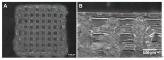
Fig. 1 is a schematic diagram of a 3D bioprinted grid structure model. As shown in fig. 7, the printed 3D gel scaffold has good fidelity, complete structure and no obvious fusion between different layers.
Example 2:
the difference from example 1 is that example 2 is a routine sterilization procedure, the sterilization time is 25min, and the specific scheme is as follows:
step 1, adding water into gelatin for swelling at room temperature until no obvious liquid flows, heating at 37 ℃ for dissolving, adding a small amount of sodium alginate powder for multiple times, and fully stirring for dissolving.
And 2, placing the biological ink precursor solution in a vertical high-pressure steam sterilization pot for sterilization for 25min, naturally cooling according to an instrument prefabrication program after sterilization is finished, and taking out the biological ink for later use.
Example 3:
the difference from the embodiment 1 is that the sterilization time is changed from 25min to 30min, and the air is exhausted immediately after the sterilization is finished, and the specific scheme is as follows:
step 1, adding water into gelatin for swelling at room temperature until no obvious liquid flows, heating at 37 ℃ for dissolving, adding a small amount of sodium alginate powder for multiple times, and fully stirring for dissolving.
And 2, placing the biological ink precursor solution in a vertical high-pressure steam sterilization pot for sterilization for 30min, immediately exhausting air after the sterilization is finished, immediately taking out the biological ink, and placing the biological ink at room temperature for later use.
Example 4:
the difference from the embodiment 1 is that the sterilization time is changed from 25min to 20min, and the air is exhausted immediately after the sterilization is finished, and the specific scheme is as follows:
step 1, adding water into gelatin for swelling at room temperature until no obvious liquid flows, heating at 37 ℃ for dissolving, adding a small amount of sodium alginate powder for multiple times, and fully stirring for dissolving.
And 2, placing the biological ink precursor solution in a vertical high-pressure steam sterilization pot for sterilization for 20min, immediately exhausting after sterilization, immediately taking out the biological ink, and placing at room temperature for later use.
Example 5:
the difference from the embodiment 1 is that the sterilization time is changed from 25min to 15min, and the air is exhausted immediately after the sterilization is finished, and the specific scheme is as follows:
step 1, adding water into gelatin for swelling at room temperature until no obvious liquid flows, heating at 37 ℃ for dissolving, adding a small amount of sodium alginate powder for multiple times, and fully stirring for dissolving.
And 2, placing the biological ink precursor solution in a vertical high-pressure steam sterilization pot for sterilization for 15min, immediately exhausting air after the sterilization is finished, immediately taking out the biological ink, and placing the biological ink at room temperature for later use.
Example 6:
HepG2 cells were selected as model cells. The cell is a human liver cancer cell line and is derived from liver cancer tissues of a 15-year-old white man. Human primary hepatocytes and HepaRG cells, which are terminally differentiated hepatocytes derived from a human hepatic progenitor cell line, retain many of the characteristics of primary hepatocytes, are commonly used for in vitro liver model construction. However, human primary hepatocytes and HepaRG cells are difficult to obtain, and the culture period is long, so the HepG2 cell line which is easy to obtain and high in literature introduction rate is finally selected as a model cell to realize further research.
Firstly, mixing HepG2 cells with biological ink, loading the mixture in a 3D biological printer for printing, and manufacturing a cell-loaded 3D gel scaffold, wherein the specific scheme is as follows:
step 1, after the HepG2 cells are amplified and cultured in a culture dish, 2.5 x 10 cells are collected 7 HepG2 cells, at individual cells/ml, were resuspended in fresh medium. The medium consists of a basal medium (MEM) supplemented with 10% (v: v) fetal calf serum and 1% (v: v) non-essential amino acids. The non-essential amino acid is a mixed solution of 7 non-essential amino acids, such as 1320mg/l L-asparagine, 1470mg/l L-glutamic acid, 1330mg/l L-aspartic acid, 890mg/l L-alanine, 1150mg/l L-proline, 1050mg/l L-serine and 750mg/l glycine.
And 2, mixing 200 mu L of cell suspension with the density and 800 mu L of biological ink, loading the mixture on a biological printer, and starting to print and manufacture a 4-layer grid structure, wherein the printing parameters are that the printing environment temperature is 26 ℃, the printing speed is 3mm/s, and the printing platform temperature is 4 ℃. Immersing in 3% (w/v) CaCl after printing 2 Crosslinking for 30s in solution. Adding 1.5ml of culture medium, placing back into the incubator for culture at 37 deg.C and 5% (v: v) CO 2 。
Specifically, the method of the 3D cell culture system obtained by using the gel scaffold is simple and feasible, and the cell growth condition is good.
Effect experiment:
physical Properties of Bio-ink
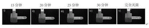
Step 1, sucking 1ml of biological ink with different sterilization procedures, placing the biological ink in a 2ml EP tube, keeping flat and standing for 10min at 26 ℃, and observing the flowing condition of the ink.
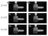
As shown in fig. 2, the conventional sterilization procedure set (example 2) exhibited significant fluidity, and was prone to structural collapse during printing, and could not be used for 3D bioprinting. The biological ink of other sterilization program groups has no obvious flow, and the extrusion characteristics can be further examined.
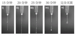
And 2, sucking the biological ink screened by the fluidity test by using a 1ml syringe, and observing the extrusion behavior of the ink at 26 ℃.
As shown in fig. 3, the extrusion shape of the conventional sterilization program group (example 2, complete sterilization) is in a droplet shape, and is easily diffused on a low-temperature platform, and the structure collapses during printing, and cannot form a stable gel scaffold, and thus cannot be used for constructing a 3D culture system; the extrusion form of the group (example 5) subjected to sterilization for 15min is in a curved uneven line shape, the bonding strength between layers is not enough in the printing process, the structure is unstable, and the line is easy to surround the needle point after being extruded to cause printing failure; the groups of 20min (example 4), 25min (example 1) and 30min (example 3) for sterilization have good extrusion forms, good adhesion between layers in the printing process, uniform extrusion of lines, regular distribution, stable gel scaffold structure and no collapse phenomenon.
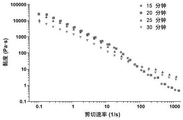
Step 3, taking a proper amount of biological ink for controlling the sterilization time to a platform of a rotary rheometer, and measuring for 0.1-1000 s at the temperature of 25 DEG C -1 Rheological behavior of the ink in the shear rate range.
As shown in fig. 4, the bio-ink still has good shear-thinning properties after sterilization. However, as the sterilization time is prolonged, the viscosity of the bio-ink is gradually decreased, and the shear thinning ability is gradually reduced.
2: volume ratio of ink to cell suspension
Step 1, taking biological ink which is sterilized for 20min, 25min and 30min, and mixing the biological ink and the cell suspension respectively in a ratio of 1: 1. 4: 1, and comparing the fluidity and the extrusion characteristics after uniformly mixing.
And 2, sucking 1ml of mixed bio-ink into a 2ml EP tube, horizontally placing at 26 ℃, standing for 10min, and observing the flowing condition of the ink.
As shown in fig. 5, 1: 1, the mixed biological ink shows obvious fluidity and is easy to cause structural collapse in the printing process; and 1: 4, the ink mixed in the proportion has no obvious fluidity for 20min and 25min, and the extrusion characteristics can be further examined.
And 3, sucking biological ink with different sterilization time by using a 1ml syringe, and observing the extrusion behavior of the ink at 26 ℃.
As shown in fig. 6, corresponding to the fluidity test, 1: the biological ink mixed according to the proportion of 1 is extruded to be in a liquid drop shape, is easy to diffuse on a low-temperature platform, collapses in the structure in the printing process, cannot form a stable gel support, and cannot be used for constructing a 3D culture system. And 1: 4, the ink mixed according to the proportion can be extruded out in a linear shape within 20min and 25min, the layers are well adhered in the printing process, lines are evenly extruded and are regularly distributed, and the gel support is stable in structure and free of collapse.
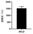
3: swelling Properties of 3D gel scaffolds
Step 1, placing a sterilized 25-min biological ink bio-printed 16-layer grid structure gel support in pure water, swelling and balancing at 37 ℃, and weighing W S And weighing again after freeze-drying l ,
The swelling ratio is calculated by the formula: SR (%) - (Ws | W ↓ l|/W ↓ l)×100%
As shown in fig. 8, the gel scaffold printed with bio-ink after sterilization has good swelling capacity, which indicates that the gel scaffold can have good nutrient transfer capacity, and is beneficial to the growth of cells in the scaffold.
4: proliferative capacity of HepG2 cells in 3D gel scaffolds
Step 1, after the HepG2 cells are amplified and cultured in a culture dish, 2.5X 10 cells are collected 7 HepG2 cells, at individual cells/ml, were resuspended in fresh medium. The medium consists of a basal medium (MEM) supplemented with 10% (v: v) fetal calf serum and 1% (v: v) non-essential amino acids. The non-essential amino acid is a mixed solution of 7 non-essential amino acids, such as 1320mg/l L-asparagine, 1470mg/l L-glutamic acid, 1330mg/l L-aspartic acid, 890mg/l L-alanine, 1150mg/l L-proline, 1050mg/l L-serine and 750mg/l glycine.
Step 2, 200 mu L of the density is addedThe cell suspension is mixed with 800 mu L of biological ink and loaded on a biological printer to start printing, and the printing parameters are that the printing environment temperature is 26 ℃, the printing speed is 3mm/s, and the printing platform temperature is 4 ℃. Immersing in 3% (w/v) CaCl after printing 2 Crosslinking for 30s in solution. Adding 1.5ml culture medium gently, placing back into incubator for culture at 37 deg.C and 5% (v: v) CO 2 。
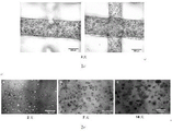
And 3, after printing, replacing the fresh culture medium every other day, and replacing every day after 6 days. And observing the cell growth under a cell imager on the 2 nd day, the 7 th day and the 14 th day, and measuring the cell viability by using a CCK-8 kit to obtain a cell proliferation curve graph.
As shown in FIG. 9-1, the cells grew uniformly in the scaffold with no apparent fusion between layers. As shown in 9-2, the cells grew well in the scaffold and significant cell aggregation could be formed within 14 days. As shown in FIG. 10, HepG2 cells in the 3D gel scaffold can still maintain high-level cell viability after being inoculated for 20 days, which indicates that the 3D culture system has stronger proliferation capacity in vitro, and the established 3D in vitro model can meet the requirement of in vitro long-term administration.
Comparative example 1: the difference from example 6 is that the effect of cell density on 3D culture was examined.
Step 1, after the HepG2 cells are amplified and cultured in a culture dish, 5 x 10 cells are collected respectively 6 1X 10 cells/ml 7 1.5X 10 cells/ml 7 2X 10 cells/ml 7 Individual cells/ml, 3X 10 7 Individual cell/ml, 4X 10 7 HepG2 cells, at individual cells/ml, were resuspended in fresh medium. The medium consists of a basal medium (MEM) supplemented with 10% (v: v) fetal calf serum and 1% (v: v) non-essential amino acids.
Step 2, mixing 200 μ L of the cell suspension with 800 μ L of bio-ink according to the ratio of 1: 4 proportion of the mixture, the final cell density is 1X 10 respectively 6 2X 10 cells/ml 6 Individual cells/ml, 3X 10 6 Individual cell/ml, 4X 10 6 Individual cell/ml, 6X 10 6 Individual cell/ml, 8X 10 6 Each cell/ml, loading on a biological printer and starting printing, wherein the printing parameters are that the printing environment temperature is 26The printing speed is 3mm/s, and the temperature of the printing platform is 4 ℃. Immersing in 3% (w/v) CaCl after printing 2 Crosslinking for 30s in solution. Adding 1.5ml culture medium gently, placing back into incubator for culture at 37 deg.C and 5% (v: v) CO 2 。
Step 3, start observing cell growth on day 2 and determine cell viability using CCK-8.
After comparison, the cell density is lower or higher, which causes a great deal of cell death during the culture process, so that 5X 10 with more proper density is selected 6 Individual cells/ml.
Comparative example 2: the difference from example 6 is that the influence of the printing speed on 3D culture was examined.
Step 1, after the HepG2 cells are amplified and cultured in a culture dish, 2.5 x 10 cells are collected 7 HepG2 cells, at individual cells/ml, were resuspended in fresh medium. The medium consists of a basal medium (MEM) supplemented with 10% (v: v) fetal calf serum and 1% (v: v) non-essential amino acids.
Step 2, mixing 200 μ L of the cell suspension with the density with 800 μ L of bio-ink respectively to obtain a final cell density of 5 × 10 6 And each cell/ml is loaded on a biological printer to start printing, and the printing parameters are that the printing environment temperature is 26 ℃, the printing platform temperature is 4 ℃, and the printing speed is 1mm/s, 3mm/s and 5mm/s respectively. Immersing in 3% (w/v) CaCl after printing 2 Crosslinking in solution for 30 s. Adding 1.5ml culture medium gently, placing back into incubator for culture at 37 deg.C and 5% (v: v) CO 2 。
Step 3, start observing cell growth on day 2 and determine cell viability using CCK-8.
After comparison, the extrusion pressure is increased due to the higher printing speed, and a large amount of cells are dead in the printing process; at a slower printing speed, the gel scaffold stays on the low-temperature platform for too long time, so that the cell activity is influenced, and therefore, a more proper 3mm/s is selected as a printing speed parameter.
Therefore, the analysis of the embodiment 6 and the comparative example 1/2 shows that the optimized bioprinting 3D culture system can well meet the requirements of formation and growth of a human hepatocyte model, is an ideal model for in vitro drug screening, and has good market value prospect.
While the invention has been described with reference to specific embodiments, it will be understood by those skilled in the art that the invention is not limited to the details of the foregoing description, and that various changes and modifications may be made without departing from the spirit and scope of the invention.