Disclosure of Invention
In order to solve the defects of the prior art, the invention provides a method for rapidly judging the survival condition of spores, which is based on an alpha glucosidase detection method, can rapidly judge the activity of the spores and can judge the activity of single spores.
The technical problem to be solved by the invention is realized by the following technical scheme:
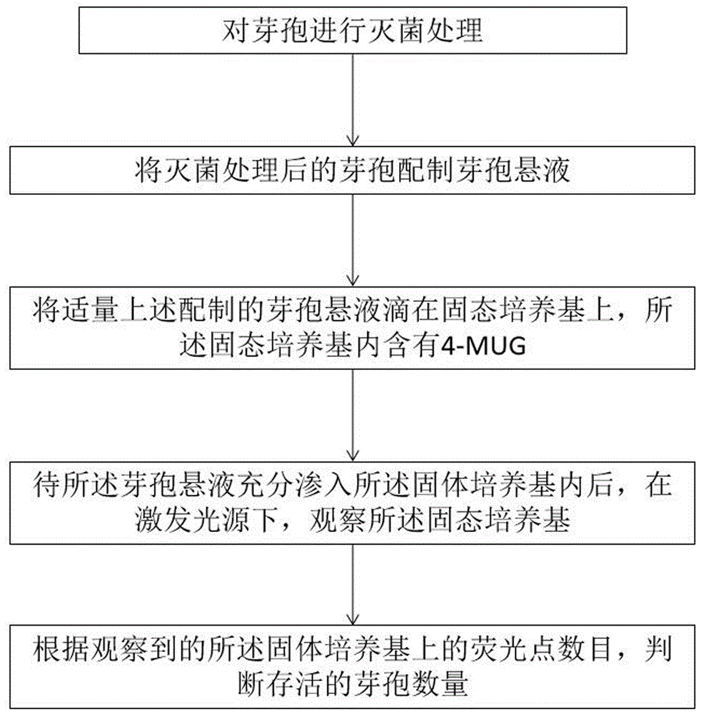
a method for rapidly judging spore survival condition comprises the following steps:
s100: sterilizing spores;
s200: preparing spore suspension from the sterilized spores;
s300: dropping a proper amount of the prepared spore suspension on a solid culture medium, wherein the solid culture medium contains 4-MUG;
s400: observing the solid culture medium under an excitation light source after the spore suspension is fully infiltrated into the solid culture medium;
s500: and judging the number of the surviving spores according to the observed number of the fluorescent spots on the solid culture medium.
Further, the spores include spores of one or more of bacillus atrophaeus (b. atrophaeus), bacillus subtilis (b. subtilis), bacillus stearothermophilus (g. stearothermophilus), bacillus pumilus (b. puminus), and the like.
Further, the sterilization treatment method for the spores is a common sterilization method such as a wet heat sterilization method, a dry heat sterilization method, a hydrogen peroxide sterilization method, an ethylene oxide sterilization method, or a low-temperature steam formaldehyde sterilization method.
Further, in step S100, the spores are loaded on a slide during sterilization.
Further, in step S200, the sterilized spores are placed in a buffer solution or a recovery solution, and the spore suspension is obtained after repeated washing, centrifugation, filtration and residue removal.
Further, the components of the solid medium include: agar, tryptone Soy Broth (Tryptic soy broth) and 4-MUG.
Further, before step S300, the method further includes the steps of: preparing the solid culture medium.
Further, the steps of preparing the solid medium are as follows:
mixing agar and trypticase soy peptone with water in proportion and sterilizing at high temperature to obtain a liquid culture medium;
dripping a proper amount of liquid culture medium on a glass slide, and cooling and solidifying to obtain the solid culture medium;
and dripping 4-MUG on the solid culture medium according to the proportion until the 4-MUG permeates into the solid culture medium.
Further, in step S400, the step of observing the solid medium under the excitation light source is as follows:
s401: arranging a spacer on the periphery of the observation area of the solid culture medium;
s402: covering a Polydimethylsiloxane (PDMS) cover plate on the spacer;
s403: and irradiating an observation area on the solid culture medium by using the excitation light source, and observing the solid culture medium in the observation area.
Further, the wavelength of the excitation light source is 340-380 nm.
The invention has the following beneficial effects: the method is based on an alpha glucosidase detection method to quickly judge the survival condition of spores, 4-MUG is firstly dripped on the solid culture medium, then the spore suspension is dripped on the solid medium, and the surviving spore is induced to germinate and grow by utilizing the alpha-glucosidase (if any) with the remaining activity and the nutrient substances in the solid medium, and the solid culture medium is utilized to limit the outward diffusion of 4-MU after 4-MUG is hydrolyzed by alpha-glucosidase, so that the hydrolysate 4-MU is gathered at the periphery of each surviving spore, by forming each light spot corresponding to each surviving spore under the excitation light source, the activity of the spore can be rapidly determined, and the activity of a single spore can also be judged.
Detailed Description
The invention is described in detail below with reference to the drawings, wherein examples of the embodiments are shown in the drawings, wherein like or similar reference numerals refer to like or similar elements or elements having like or similar functions throughout. The embodiments described below with reference to the drawings are illustrative and intended to be illustrative of the invention and are not to be construed as limiting the invention.
In the description of the present invention, it is to be understood that the terms "length", "width", "upper", "lower", "front", "rear", "left", "right", "vertical", "horizontal", "top", "bottom", "inner", "outer", and the like, indicate orientations or positional relationships based on those shown in the drawings, and are used only for convenience in describing the present invention and for simplicity in description, and do not indicate or imply that the devices or elements referred to must have a particular orientation, be constructed in a particular orientation, and be operated, and thus, are not to be construed as limiting the present invention.
Furthermore, the terms "first", "second", "third" are used for descriptive purposes only and are not to be construed as indicating or implying relative importance or implicitly indicating the number of technical features indicated. Thus, a feature defined as "first," "second," or "third" may explicitly or implicitly include one or more of that feature. In the description of the present invention, "a plurality" means two or more unless specifically defined otherwise.
In the present invention, unless otherwise expressly stated or limited, the terms "mounted," "connected," "secured," "disposed," and the like are to be construed broadly and can, for example, be fixedly connected, detachably connected, or integrally formed; can be mechanically or electrically connected; they may be directly connected or indirectly connected through intervening media, or may be connected through the interconnection of two elements or through the interaction of two elements. The specific meanings of the above terms in the present invention can be understood by those skilled in the art according to specific situations.
As shown in FIG. 1, a method for rapidly determining spore survival status comprises the following steps:
s100: and sterilizing the spores.
In this step S100, the spores include spores of one or more of bacillus atrophaeus (b. atrophaeus), bacillus subtilis (b. subtilis), bacillus stearothermophilus (g. stearothermophilus), bacillus pumilus (b. pumipius), and the like; the sterilization treatment of the spores may be, but not limited to, a wet heat sterilization method, a dry heat sterilization method, a hydrogen peroxide sterilization method, an ethylene oxide sterilization method, a low-temperature steam formaldehyde sterilization method, or other common sterilization methods.
The specific species of the spores will depend on the specific sterilization method, for example, if the sterilization method is ethylene oxide sterilization, the spores include spores of one or more of Bacillus atrophaeus ATCC 9372, NCTC 10073, NCIMB 8058, DSM 2277, NRRL B-4418, or CIP 77.18; if the sterilization treatment method is moist heat sterilization, the spores comprise spores of one or more of Bacillus stearothermophilus ATCC 7953(NCTC 10007, DSM 22 and CIP 52.81) or ATCC 12980 (as NRRL B-4419); if the sterilization treatment method is dry heat sterilization, the spores include spores of one or more of Bacillus atrophaeus CIP 77.18, NCIMB 8058, DSM 675, NRRL B-4418 and ATCC 9372 or Bacillus subtilis DSM 13019; if the sterilization process is low temperature steam formaldehyde sterilization, the spores comprise one or more of bacillus stearothermophilus NCIB 8224, DSM 6790, ATCC 10149, or ATCC 12980; if the sterilization process is a hydrogen peroxide sterilization process, the spores include one or more of bacillus stearothermophilus ATCC 7953 or SSI K31.
According to the regulation in the medical health-care product sterilization biological indicator: when wet heat sterilization or low-temperature steam formaldehyde sterilization is adopted, the viable bacteria amountNot less than 1.0 × 10 5 When ethylene oxide sterilization, hydrogen peroxide sterilization or dry heat sterilization is adopted, the viable bacteria amount is not less than 1.0 × 10 6 And (4) respectively.
Preferably, the spores are carried on a glass slide during sterilization.
S200: and preparing spore suspension by using the sterilized spores.
In step S200, the sterilized spores are placed in a buffer solution or a recovery solution, and the spore suspension is obtained after repeated washing, centrifugation, and residue removal.
The buffer solution is a phosphate buffer solution with a concentration of 0.03M and a pH of 7.2, and the specific components and preparation method of the phosphate buffer solution belong to the common knowledge in the field, so the detailed description is not given. The recovery solution comprises 0.1% of Tween 80 and the buffer solution.
S300: a proper amount of the spore suspension prepared above is dripped on a solid culture medium, and the solid culture medium contains 4-MUG.
In step S300, the amount of spore suspension dropped on the solid medium may depend on the specific detection requirement, and in the present case, only one or two drops of spore suspension need to be extracted and dropped on the solid medium.
The solid medium comprises agar, Tryptic soy peptone (TBS) and 4-MUG, wherein the agar is prepared according to 30g/L, the Tryptic soy peptone is prepared according to 15g/L, and the concentration of the 4-MUG in the solid medium is 50-500 mg/L.
Before this step S300, the method further includes the steps of: preparing the solid culture medium.
Wherein the solid medium is prepared by the following steps:
mixing agar and trypticase soy peptone with water in proportion and sterilizing at high temperature to obtain a liquid culture medium;
dripping a proper amount of liquid culture medium on a glass slide, and cooling and solidifying to obtain the solid culture medium;
and dripping 4-MUG on the solid culture medium according to the proportion until the 4-MUG permeates into the solid culture medium.
The amount of the liquid culture medium dropped on the glass slide can be determined according to specific detection requirements, and only one drop or two drops of the liquid culture medium need to be extracted and dropped on the glass slide.
S400: and after the spore suspension is fully infiltrated into the solid culture medium, observing the solid culture medium under an excitation light source.
In this step S400, the spore suspension on the solid medium is observed under an observation system. After absorbing each nutrient component in the solid culture medium, the surviving spores in the spore suspension germinate and grow, and synthesize sufficient alpha-glucosidase through metabolism, and then 4-MUG in the solid culture medium is hydrolyzed by residual alpha-glucosidase (if any) after sterilization and newly synthesized alpha-glucosidase of the surviving spores to generate 4-MU serving as a fluorescent indicator; 4-MU generates fluorescence at 340-380nm excitation light source.
Most preferably, the wavelength of the excitation light source is about 365 nm.
In step S400, the step of observing the spore suspension on the solid medium under the excitation light source is as follows:
s401: and arranging a spacer on the periphery of the observation area of the solid culture medium.
In this step S401, the spacer should surround the observation region, and the range of the observation region may include the whole region of the solid medium to which the spore suspension is dripped, or may include the local region of the solid medium to which the spore suspension is dripped, depending on the specific observation requirement.
In the scheme, the shock insulator is a rubber ring.
S403: and irradiating an observation area on the solid culture medium by using the excitation light source, and observing the solid culture medium in the observation area.
S500: and judging the number of the surviving spores according to the observed number of the fluorescent spots on the solid culture medium.
In this step S500, as shown in fig. 2, since 4-MUG is located in the solid medium, 4-MU formed by hydrolysis of 4-MUG by α -glucosidase is also limited by the solid medium, cannot diffuse outward, and then gathers on the periphery of each surviving spore, when observed under an excitation light source, a tester observes one light spot, one light spot represents one surviving spore, the number of surviving spores on the solid medium is determined according to the number of light spots, and then the total number of surviving spores in the whole sterilization process is calculated according to the dropping ratio of the spore suspension, so as to finally evaluate whether the sterilization process of the sterilization process is qualified.
The method is based on an alpha glucosidase detection method to quickly judge the survival condition of spores, and 4-MUG is firstly dripped on the solid culture medium, then the spore suspension is dripped on the solid medium, and the surviving spore is induced to germinate and grow by utilizing the alpha-glucosidase (if any) with the remaining activity and the nutrient substances in the solid medium, and the solid culture medium is utilized to limit the outward diffusion of 4-MU after 4-MUG is hydrolyzed by alpha-glucosidase, so that the hydrolysate 4-MU is gathered at the periphery of each surviving spore, by forming each light spot corresponding to each surviving spore under the excitation light source, the activity of the spore can be rapidly determined, and the activity of a single spore can also be judged.
EXAMPLE one (Sterilization with 3M 1292 pressure steam biological indicator)
In this example, 6 biological indicators (indicators) for pressure steam sterilization were selected from 3M 1292. Each indicator contained about 10 pieces of mushroom 6 Spores of Bacillus stearothermophilus. The 6 indicators were divided into 3 groups of 2. The biological indicator is put into a pressure steam sterilization resistance detector (resistance instrument) for sterilization treatment, wherein the sterilization temperature is 121 ℃, and the sterilization time is respectively 0min, and 3 groups of sterilization time are 8 min and 24 min. Treating at 121 deg.C for 0min without any treatment, and making into experimental control group; processing at 121 ℃ for 8 min to simulate the situation of incomplete sterilization; after treatment at 121 ℃ for 24 min, spores should theoretically be completely killed, thereby simulating the successful sterilization. The indicator being exposed at a location in the sterilization deviceAnd (4) cold spots. After the sterilization treatment, the samples were transferred to an ultraclean bench. Taking out the bacterial strips contained in the 3 groups of sterilized biological indicators by using sterilized tweezers, putting the bacterial strips into a sterile centrifuge tube, adding a proper amount of sterile recovery solution (0.1% Tween 80, 0.03M phosphate buffer solution, pH 7.2), fully shaking and beating the bacterial strips. The centrifuge tube was then placed in an ice bath to cool.
And then centrifuging the sample in the centrifugal tube (centrifugal force 2000 Xg, temperature 4 ℃, centrifuging time 20 min), and filtering the obtained supernatant with a 10 mu m microporous filter membrane (the diameter of the spore is 1-2 mu m, so that the crushed slag of the bacterial strips is blocked by the filter membrane in order to allow the spore to pass through the filter membrane), thereby obtaining the filtrate.
Adding a proper amount of sterile recovery liquid into filter residues (the main component is broken mushroom strip fibers) obtained after the centrifugal filtration, shaking uniformly, repeating the centrifugal filtration process again, further recovering filtrate containing heat damaged spores, repeating the process for 2-3 times, and collecting the filtrate obtained by each centrifugal filtration to obtain the spore suspension.
A drop of autoclaved liquid medium (agar 30g/L, trypticase soy peptone 15 g/L) was dropped on the slide glass, and after it solidified, a drop of 4-MUG with a concentration of 210mg/L was dropped on the solid medium, and later, it was just after it sufficiently infiltrated into the solid medium.
One drop of the spore suspension (containing thermally damaged spores) was added dropwise to the solid medium.
The number and size of the fluorescent spots were observed under an observation system. The observation system is composed of four parts: 1) a stereomicroscope (brand model may be: nikon SMZ 800); 2) a time gating camera mounted on the stereomicroscope (the brand model may be: photonics Research Systems, Salford, uk); 3) a xenon flash lamp at a 45 ° angle to the sample (brand model may be: PerkinElmer, Waltham, MA), and 4) a temperature-controlled microscope slide mount (brand model may be: thermal).
The parameters set by the viewing system are as follows: the xenon flash lamp has an excitation wavelength of 365nm and an excitation slit width of 10 nm. The wavelength of the emission observed by a stereomicroscope is 544nm, and the emission slit is 10 nm. The number and intensity of the spots were observed and the results are shown in Table 1.
TABLE 1
| Numbering
|
Conditions of treatment
|
Observation results
|
Conclusion
|
| A-1
|
121℃、0 min
|
Large overlapping fluorescence, no discernable "spot" presence
|
Is not sterilized
|
| A-2
|
121℃、8 min
|
A certain amount of scattered fluorescent dots
|
Incomplete sterilization
|
| A-3
|
121℃、24 min
|
No fluorescent spot
|
Completely eradicate |
EXAMPLE two (Sterilization with 3M 1295 Hydrogen peroxide biological indicator)
In this example, 6 biological indicators (indicators) for hydrogen peroxide sterilization were selected from 3M 1295. Bacteria per indicatorThe tablet contains about 10 6 Spores of Bacillus atrophaeus. The 6 indicators were divided into 3 groups of 2. The biological indicator was put into a hydrogen peroxide sterilization resistance detector (resistance meter) for sterilization treatment at 50 deg.C for 3 sets of 0 sec, 20 sec and 60 sec, respectively, and the hydrogen peroxide concentration was 1.74 mg/L. Treating at 50 deg.C for 0 sec without any treatment, and making into experimental control group; the treatment at 50 ℃ for 20 sec simulates the situation of incomplete sterilization; after 60 sec at 50 ℃, spores should theoretically be completely killed, thereby simulating the successful sterilization. The exposure site of the indicator is located at a cold spot of the sterilization apparatus. After the sterilization treatment, the samples were transferred to an ultraclean bench. Taking out the bacterial strips contained in the 3 groups of sterilized biological indicators by using sterilized forceps, placing the bacterial strips into a sterile centrifuge tube, adding a proper amount of sterile recovery liquid (0.1% Tween 80, 0.03M phosphate buffer solution, pH 7.2), fully shaking, and breaking the bacterial strips. The centrifuge tube was then placed in an ice bath to cool.
And then centrifuging the sample in the centrifugal tube (centrifugal force 2000 Xg, temperature 4 ℃, centrifuging time 20 min), and filtering the obtained supernatant with a 10 mu m microporous filter membrane (spore diameter is 1-2 mu m, so that in order to allow the spores to pass through the filter membrane, the broken slag of the bacterial strips is blocked, a filter membrane of more than 2 mu m is needed), so as to obtain filtrate.
Adding a proper amount of sterile recovery liquid into filter residues (the main component is broken mushroom strip fibers) obtained after the centrifugal filtration, shaking uniformly, repeating the centrifugal filtration process again, further recovering filtrate containing heat damaged spores, repeating the process for 2-3 times, and collecting the filtrate obtained by each centrifugal filtration to obtain the spore suspension.
A drop of autoclaved liquid medium (agar 30g/L, trypticase soy peptone 15 g/L) was dropped on the slide glass, and after it solidified, a drop of 4-MUG with a concentration of 210mg/L was dropped on the solid medium, and later, it was just after it sufficiently infiltrated into the solid medium.
One drop of the spore suspension (containing heat damaged spores) was added to the solid medium.
The number and size of the fluorescent spots were observed under an observation system. The observation system is composed of four parts: 1) a stereomicroscope (brand model may be: nikon SMZ 800); 2) a time gating camera mounted on the stereomicroscope (the brand model may be: photonics Research Systems, Salford, uk); 3) a xenon flash lamp at a 45 ° angle to the sample (brand model may be: PerkinElmer, Waltham, MA), and 4) a temperature-controlled microscope slide mount (brand model may be: thermal).
The parameters set by the viewing system are as follows: the xenon flash lamp has an excitation wavelength of 365nm and an excitation slit width of 10 nm. The wavelength of the emission observed by a stereomicroscope is 544nm, and the emission slit is 10 nm. The number and intensity of the spots were observed and the results are shown in Table 2.
TABLE 2
| Numbering
|
Conditions of treatment
|
Observation results
|
Conclusion
|
| B-1
|
50℃、1.74mg/L、0 sec
|
Large overlapping fluorescence, no discernable "spot" presence
|
Is not sterilized
|
| B-2
|
50℃、1.74mg/L 、20 sec
|
A certain amount of scattered fluorescent dots
|
Incomplete sterilization
|
| B-3
|
50℃、1.74mg/L、60 sec
|
No fluorescent spot
|
Completely eradicate |
EXAMPLE three (pressure steam sterilization with homemade spores of Bacillus subtilis)
This example selects Mesa, a new product of China ® 6 strips of Strip spores were divided into 3 groups of 2 strips. Sterilizing the strips in a pressure steam sterilization resistance detector (resistance instrument) at 121 deg.C for 3 groups (0 min, 8 min and 24 min). Treating at 121 deg.C for 0min without any treatment, and making into experimental control group; processing at 121 ℃ for 8 min to simulate the situation of incomplete sterilization; after treatment at 121 ℃ for 24 min, spores should theoretically be completely killed, thereby simulating the successful sterilization. The exposure site of the indicator is located at a cold spot of the sterilization apparatus. After the sterilization treatment, the samples were transferred to an ultraclean bench. Taking out the bacterial strips contained in the 3 groups of sterilized biological indicators by using sterilized tweezers, putting the bacterial strips into a sterile centrifuge tube, adding a proper amount of sterile recovery solution (0.1% Tween 80, 0.03M phosphate buffer solution, pH 7.2), fully shaking and beating the bacterial strips. The centrifuge tube was then placed in an ice bath to cool.
One drop of sterilized liquid medium (agar 30g/L, trypticase soy peptone 15 g/L) was dropped on the slide glass, and after it solidified, one drop of 4-MUG with a concentration of 210mg/L was dropped on the solid medium, and it was allowed to sufficiently permeate into the solid medium for a short time.
One drop of the spore suspension (containing thermally damaged spores) was added dropwise to the solid medium.
The number and size of the fluorescent spots were observed under an observation system. The observation system is composed of four parts: 1) a stereomicroscope (brand model may be: nikon SMZ 800); 2) a time gating camera mounted on the stereomicroscope (the brand model may be: photonics Research Systems, Salford, uk); 3) a xenon flash lamp at a 45 ° angle to the sample (brand model may be: PerkinElmer, Waltham, MA), and 4) a temperature-controlled microscope slide mount (brand model may be: thermal).
The parameters set by the viewing system are as follows: the xenon flash lamp has an excitation wavelength of 365nm and an excitation slit width of 10 nm. The wavelength of the emission observed by a stereomicroscope is 544nm, and the emission slit is 10 nm. The number of spots and the intensity were observed and the results are shown in Table 3.
TABLE 3
| Numbering
|
Conditions of treatment
|
Observation results
|
Conclusion
|
| C-1
|
121℃、0 min
|
Large overlapping fluorescence, no discernable "spot" presence
|
Is not sterilized
|
| C-2
|
121℃、8 min
|
A certain amount of scattered fluorescent dots
|
Incomplete sterilization
|
| C-3
|
121℃、24 min
|
No fluorescent spot
|
Completely eradicate |
Example four (Sterilization with hydrogen oxide Using spores of Bacillus atrophaeus)
This example selects Mesa, a new product of China ® 6 strips of Strip spores were divided into 3 groups of 2 strips. The bacterial strips were sterilized in a hydrogen peroxide sterilization resistance tester (resistance tester) at 50 deg.C for 3 sets of 0 sec, 20 sec and 60 sec, respectively, and hydrogen peroxide concentration was 1.74 mg/L. Treating at 50 deg.C for 0 sec without any treatment, and making into experimental control group; the treatment at 50 ℃ for 20 sec simulates the situation of incomplete sterilization; after 60 sec at 50 ℃, spores should theoretically be completely killed, thereby simulating the successful sterilization. The exposure site of the indicator is located at a cold spot of the sterilization apparatus. After the sterilization treatment, the samples were transferred to an ultraclean bench. Taking out the bacterial strips contained in the 3 groups of sterilized biological indicators by using sterilized tweezers, putting the bacterial strips into a sterile centrifuge tube, adding a proper amount of sterile recovery solution (0.1% Tween 80, 0.03M phosphate buffer solution, pH 7.2), fully shaking and beating the bacterial strips. The centrifuge tube was then placed in an ice bath to cool.
A drop of autoclaved liquid medium (agar 30g/L, trypticase soy peptone 15 g/L) was dropped on the slide glass, and after it solidified, a drop of 4-MUG with a concentration of 210mg/L was dropped on the solid medium, and later, it was just after it sufficiently infiltrated into the solid medium.
One drop of the spore suspension (containing thermally damaged spores) was added dropwise to the solid medium.
The number and size of the fluorescent spots were observed under an observation system. The observation system is composed of four parts: 1) a stereomicroscope (brand model may be: nikon SMZ 800); 2) a time gating camera mounted on the stereomicroscope (the brand model may be: photonics Research Systems, Salford, uk); 3) a xenon flash lamp at a 45 ° angle to the sample (brand model may be: PerkinElmer, Waltham, MA), and 4) a temperature-controlled microscope slide mount (brand model may be: thermal).
The parameters set by the viewing system are as follows: the xenon flash lamp has an excitation wavelength of 365nm and an excitation slit width of 10 nm. The wavelength of the emission observed by a stereomicroscope is 544nm, and the emission slit is 10 nm. The number of spots and the intensity were observed and the results are shown in Table 4.
TABLE 4
| Numbering
|
Conditions of treatment
|
Observation results
|
Conclusion
|
| D-1
|
50℃、1.74mg/L、0 sec
|
Large overlapping fluorescence, no discernable "spot" presence
|
Is not sterilized
|
| D-2
|
50℃、1.74mg/L 、20 sec
|
A certain amount of scattered fluorescent dots
|
Incomplete sterilization
|
| D-3
|
50℃、1.74mg/L、60 sec
|
No fluorescent spot
|
Completely eradicate |
Finally, it should be noted that the above embodiments are only used for illustrating the technical solutions of the embodiments of the present invention and not for limiting the same, and although the embodiments of the present invention are described in detail with reference to the preferred embodiments, it should be understood by those skilled in the art that modifications or equivalent substitutions can be made on the technical solutions of the embodiments of the present invention, and these modifications or equivalent substitutions cannot make the modified technical solutions depart from the scope of the technical solutions of the embodiments of the present invention.