CN115598024A - Cell grading counting device and method thereof - Google Patents
Cell grading counting device and method thereof Download PDFInfo
- Publication number
- CN115598024A CN115598024A CN202211430052.3A CN202211430052A CN115598024A CN 115598024 A CN115598024 A CN 115598024A CN 202211430052 A CN202211430052 A CN 202211430052A CN 115598024 A CN115598024 A CN 115598024A
- Authority
- CN
- China
- Prior art keywords
- fresnel lens
- grading
- counting
- cells
- cell
- Prior art date
- Legal status (The legal status is an assumption and is not a legal conclusion. Google has not performed a legal analysis and makes no representation as to the accuracy of the status listed.)
- Granted
Links
- 238000000034 method Methods 0.000 title claims abstract description 37
- 238000001514 detection method Methods 0.000 claims abstract description 35
- 230000006378 damage Effects 0.000 abstract description 4
- 210000004027 cell Anatomy 0.000 description 115
- 230000000694 effects Effects 0.000 description 7
- 239000000975 dye Substances 0.000 description 6
- 210000003743 erythrocyte Anatomy 0.000 description 6
- 210000000265 leukocyte Anatomy 0.000 description 6
- 230000008569 process Effects 0.000 description 5
- 238000000684 flow cytometry Methods 0.000 description 4
- 238000004043 dyeing Methods 0.000 description 3
- 238000010191 image analysis Methods 0.000 description 3
- 239000000463 material Substances 0.000 description 3
- 230000003287 optical effect Effects 0.000 description 3
- 238000004458 analytical method Methods 0.000 description 2
- 238000010586 diagram Methods 0.000 description 2
- 238000002795 fluorescence method Methods 0.000 description 2
- 230000004048 modification Effects 0.000 description 2
- 238000012986 modification Methods 0.000 description 2
- 238000004451 qualitative analysis Methods 0.000 description 2
- 238000004445 quantitative analysis Methods 0.000 description 2
- 238000002834 transmittance Methods 0.000 description 2
- 230000004075 alteration Effects 0.000 description 1
- 230000006907 apoptotic process Effects 0.000 description 1
- 230000005540 biological transmission Effects 0.000 description 1
- 230000005779 cell damage Effects 0.000 description 1
- 208000037887 cell injury Diseases 0.000 description 1
- 238000006243 chemical reaction Methods 0.000 description 1
- 230000007547 defect Effects 0.000 description 1
- 239000000835 fiber Substances 0.000 description 1
- 239000012530 fluid Substances 0.000 description 1
- 239000011521 glass Substances 0.000 description 1
- 230000006698 induction Effects 0.000 description 1
- 238000011527 multiparameter analysis Methods 0.000 description 1
- 239000002245 particle Substances 0.000 description 1
- 239000002861 polymer material Substances 0.000 description 1
- 238000002360 preparation method Methods 0.000 description 1
- 239000010453 quartz Substances 0.000 description 1
- 238000005070 sampling Methods 0.000 description 1
- 230000011218 segmentation Effects 0.000 description 1
- 238000000926 separation method Methods 0.000 description 1
- VYPSYNLAJGMNEJ-UHFFFAOYSA-N silicon dioxide Inorganic materials O=[Si]=O VYPSYNLAJGMNEJ-UHFFFAOYSA-N 0.000 description 1
- 239000000126 substance Substances 0.000 description 1
- 238000006467 substitution reaction Methods 0.000 description 1
Images



Classifications
-
- G—PHYSICS
- G01—MEASURING; TESTING
- G01N—INVESTIGATING OR ANALYSING MATERIALS BY DETERMINING THEIR CHEMICAL OR PHYSICAL PROPERTIES
- G01N15/00—Investigating characteristics of particles; Investigating permeability, pore-volume or surface-area of porous materials
- G01N15/02—Investigating particle size or size distribution
- G01N15/0205—Investigating particle size or size distribution by optical means
-
- G—PHYSICS
- G01—MEASURING; TESTING
- G01N—INVESTIGATING OR ANALYSING MATERIALS BY DETERMINING THEIR CHEMICAL OR PHYSICAL PROPERTIES
- G01N15/00—Investigating characteristics of particles; Investigating permeability, pore-volume or surface-area of porous materials
- G01N15/10—Investigating individual particles
-
- G—PHYSICS
- G01—MEASURING; TESTING
- G01N—INVESTIGATING OR ANALYSING MATERIALS BY DETERMINING THEIR CHEMICAL OR PHYSICAL PROPERTIES
- G01N15/00—Investigating characteristics of particles; Investigating permeability, pore-volume or surface-area of porous materials
- G01N15/01—Investigating characteristics of particles; Investigating permeability, pore-volume or surface-area of porous materials specially adapted for biological cells, e.g. blood cells
-
- G—PHYSICS
- G01—MEASURING; TESTING
- G01N—INVESTIGATING OR ANALYSING MATERIALS BY DETERMINING THEIR CHEMICAL OR PHYSICAL PROPERTIES
- G01N15/00—Investigating characteristics of particles; Investigating permeability, pore-volume or surface-area of porous materials
- G01N15/01—Investigating characteristics of particles; Investigating permeability, pore-volume or surface-area of porous materials specially adapted for biological cells, e.g. blood cells
- G01N2015/012—Red blood cells
-
- G—PHYSICS
- G01—MEASURING; TESTING
- G01N—INVESTIGATING OR ANALYSING MATERIALS BY DETERMINING THEIR CHEMICAL OR PHYSICAL PROPERTIES
- G01N15/00—Investigating characteristics of particles; Investigating permeability, pore-volume or surface-area of porous materials
- G01N15/01—Investigating characteristics of particles; Investigating permeability, pore-volume or surface-area of porous materials specially adapted for biological cells, e.g. blood cells
- G01N2015/016—White blood cells
-
- G—PHYSICS
- G01—MEASURING; TESTING
- G01N—INVESTIGATING OR ANALYSING MATERIALS BY DETERMINING THEIR CHEMICAL OR PHYSICAL PROPERTIES
- G01N15/00—Investigating characteristics of particles; Investigating permeability, pore-volume or surface-area of porous materials
- G01N15/01—Investigating characteristics of particles; Investigating permeability, pore-volume or surface-area of porous materials specially adapted for biological cells, e.g. blood cells
- G01N2015/018—Platelets
-
- G—PHYSICS
- G01—MEASURING; TESTING
- G01N—INVESTIGATING OR ANALYSING MATERIALS BY DETERMINING THEIR CHEMICAL OR PHYSICAL PROPERTIES
- G01N15/00—Investigating characteristics of particles; Investigating permeability, pore-volume or surface-area of porous materials
- G01N15/10—Investigating individual particles
- G01N2015/1006—Investigating individual particles for cytology
-
- G—PHYSICS
- G01—MEASURING; TESTING
- G01N—INVESTIGATING OR ANALYSING MATERIALS BY DETERMINING THEIR CHEMICAL OR PHYSICAL PROPERTIES
- G01N15/00—Investigating characteristics of particles; Investigating permeability, pore-volume or surface-area of porous materials
- G01N15/10—Investigating individual particles
- G01N2015/1022—Measurement of deformation of individual particles by non-optical means
-
- G—PHYSICS
- G01—MEASURING; TESTING
- G01N—INVESTIGATING OR ANALYSING MATERIALS BY DETERMINING THEIR CHEMICAL OR PHYSICAL PROPERTIES
- G01N15/00—Investigating characteristics of particles; Investigating permeability, pore-volume or surface-area of porous materials
- G01N15/10—Investigating individual particles
- G01N2015/1024—Counting particles by non-optical means
-
- Y—GENERAL TAGGING OF NEW TECHNOLOGICAL DEVELOPMENTS; GENERAL TAGGING OF CROSS-SECTIONAL TECHNOLOGIES SPANNING OVER SEVERAL SECTIONS OF THE IPC; TECHNICAL SUBJECTS COVERED BY FORMER USPC CROSS-REFERENCE ART COLLECTIONS [XRACs] AND DIGESTS
- Y02—TECHNOLOGIES OR APPLICATIONS FOR MITIGATION OR ADAPTATION AGAINST CLIMATE CHANGE
- Y02P—CLIMATE CHANGE MITIGATION TECHNOLOGIES IN THE PRODUCTION OR PROCESSING OF GOODS
- Y02P70/00—Climate change mitigation technologies in the production process for final industrial or consumer products
- Y02P70/50—Manufacturing or production processes characterised by the final manufactured product
Landscapes
- Chemical & Material Sciences (AREA)
- Dispersion Chemistry (AREA)
- Physics & Mathematics (AREA)
- Health & Medical Sciences (AREA)
- Life Sciences & Earth Sciences (AREA)
- Analytical Chemistry (AREA)
- Biochemistry (AREA)
- General Health & Medical Sciences (AREA)
- General Physics & Mathematics (AREA)
- Immunology (AREA)
- Pathology (AREA)
- Investigating Or Analysing Biological Materials (AREA)
Abstract
The invention relates to the technical field of photoelectric detection, and particularly provides a cell grading counting device and a cell grading counting method. The laser emits laser beams which are incident to cells to be counted in a grading mode in the cell channel and then are scattered, the scattered beams are collimated by the collimating lens and then are incident to the size grading counting unit for grading counting, the beams after grading counting are incident to the photoelectric detection unit, and size grading and the number of the cells to be counted in the grading mode are obtained by analyzing information received by the photoelectric detection unit. The technical scheme provided by the invention solves the problem that the traditional device can only count cells singly or classify the size of the cells, and realizes the statistical work of classifying and counting the types of the cells simultaneously on the basis of no damage to the cells.
Description
Technical Field
The invention relates to the technical field of photoelectric detection, in particular to a cell grading counting device and a cell grading counting method.
Background
Flow CytoMetry (FCM) is a biological technique used to count and sort microscopic particles suspended in a fluid. This technique can be used to perform a continuous multi-parameter analysis of individual cells flowing past an optical or electronic detector. The microfluidic chip technology integrates basic operation units of sample preparation, reaction, separation, detection and the like in biological, chemical and medical analysis processes into a micron-scale chip, and automatically completes the whole analysis process. In the technical fields of flow cytometry, microfluidics and the like, tens of thousands of cells are often required to be detected in one detection operation.
At present, methods for cell counting in flow cytometry and microfluidic chip technology mainly include fluorescence cell counting method, image recognition counting method and pulse counting method. The fluorescent cell counting method needs to dye cells, and counting statistics is carried out on different cells through the fluorescent method. The disadvantage of this procedure is that the dye may cause apoptosis, some dyes can only stain dead cells, and even if the dyes can maintain the activity of the cells, the cells need to be rinsed after the counting procedure, which in turn increases the probability of cell destruction and increases the workload. The image recognition counting method counts cells by image analysis, but the accuracy of counting depends on the accuracy of segmentation of image cells, and the cells cannot be simultaneously qualitatively and quantitatively analyzed accurately. The pulse counting method counts the number of cells through a pulse induction module, and when the activity difference of the cells is accumulated in a sample or the flow rate is high, the counting of the number of the cells is not in place easily.
The devices and methods in the prior art are not capable of accurately counting while differentiating cell size fractions. There is therefore a need to develop an apparatus and method for fractional cell counting that is accurate, rapid, and cost-effective and labor-efficient.
Disclosure of Invention
The invention aims to overcome the defects of the prior art and provides a cell grading counting device and a cell grading counting method.
In order to realize the purpose, the invention adopts the following specific technical scheme:
the invention provides a cell grading and counting device, comprising: the device comprises a laser, a cell channel, a collimating lens, a size grading counting unit and a photoelectric detection unit which are sequentially arranged along a light path; wherein,
the size grading counting unit comprises a Fresnel lens and a Fresnel lens ring group which are concentric; the number of the Fresnel lens rings in the Fresnel lens ring group is N, wherein N is more than or equal to 1;
the number of the photoelectric detectors in the photoelectric detection unit is N +1, and each photoelectric detector corresponds to the lens in the size grading counting unit one by one;
the laser device emits laser beams, the laser beams are incident to cells to be counted in a grading mode in the cell channel and are scattered after the cells to be counted in the grading mode, scattered beams are collimated into collimated beams through the collimating lens and then are incident to the size grading counting unit to be counted in the grading mode, the light beams after the grading counting are incident to the photoelectric detection unit, and size grading and the number of the cells to be counted in the grading mode are obtained through analyzing information received by the photoelectric detection unit.
Further, the Fresnel lens and the Fresnel lens ring group are both positive Fresnel lenses;
the Fresnel lens is a circular Fresnel lens;
the Fresnel lens ring set is sleeved outside the Fresnel lens, and the focal length of each Fresnel lens ring is the same as that of the Fresnel lens.
Further, an included angle between the fresnel lens and the fresnel lens ring group is greater than 0 ° and less than 90 °.
Further, the detection surface of the photodetector is arranged at the focal point of the corresponding lens and is tangent to the focal surface of the corresponding lens.
The cell grading counting method provided by the invention comprises the following steps:
s1, setting an inclination angle between the Fresnel lens and each Fresnel lens ring in the size grading counting unit;
s2, laser beams emitted by the laser sequentially pass through cells to be counted in a grading mode in the cell channel, the collimating lens and the size grading counting unit and then are incident to the photoelectric detection unit;
and S3, analyzing the information received by the photoelectric detection unit, and identifying the number of the photoelectric detectors receiving the information in the photoelectric detection unit, thereby realizing size grading and counting of the cells to be graded and counted.
The invention can obtain the following technical effects:
according to the cell grading counting device and the cell grading counting method, the size of the airy disk after the cells are scattered is identified, and the counting work of cell type division and counting is completed simultaneously on the basis of no damage to the cells. The cell grading counting device and the method thereof provided by the invention are simple and easy to operate and have high accuracy. The counting method avoids the counting statistics of the cells by a dyeing fluorescence method in the prior art, only dyeing can be carried out aiming at dead cells, and even if dyes capable of keeping the cell activity are used, the cells need to be rinsed after the counting process. And the problem that the image recognition counting method depends on image analysis to count the cells and cannot accurately perform qualitative and quantitative analysis on the cells at the same time is solved. Meanwhile, the problem that the cell number is not counted in place easily when the cell activity is poor and the cell is accumulated in a sample or the flow rate is high in a pulse counting method is solved.
Drawings
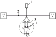
FIG. 1 is a first schematic view of a cell fractional count apparatus according to an embodiment of the present invention;
FIG. 2 is a schematic diagram of a size grading counting unit and a photodetecting unit according to an embodiment of the present invention;
FIG. 3 is a schematic diagram of a size grading counting unit according to an embodiment of the invention;
FIG. 4 is a second schematic view of a cell fractional counting apparatus according to an embodiment of the present invention;
FIG. 5 is a third schematic view of a cell fractional counting apparatus according to an embodiment of the present invention;
FIG. 6 is a flow chart of a cell fractional count method according to an embodiment of the present invention.
Wherein the reference numerals include:
the device comprises a laser 1, a cell channel 2, a collimating lens 3, a size grading counting unit 4, a Fresnel lens 4-1, a first Fresnel lens ring 4-2, a second Fresnel lens ring 4-3, a photoelectric detection unit 5, a first photoelectric detector 5-1, a second photoelectric detector 5-2 and a third photoelectric detector 5-3.
Detailed Description
Hereinafter, embodiments of the present invention will be described with reference to the accompanying drawings. In the following description, the same reference numerals are used for the same blocks. In the case of the same reference numerals, their names and functions are also the same. Therefore, detailed description thereof will not be repeated.
In order to make the objects, technical solutions and advantages of the present invention more apparent, the present invention will be described in further detail below with reference to the accompanying drawings and specific embodiments. It should be understood that the specific embodiments described herein are merely illustrative of the invention and do not limit the invention.
The specific operation of the present invention will be described in detail with reference to fig. 1 to 6:
the present invention provides a cell fractional counter, as shown in fig. 1, comprising: the device comprises a laser 1, a cell channel 2, a collimating lens 3, a size grading counting unit 4 and a photoelectric detection unit 5 which are arranged along a light path in sequence. As shown in fig. 2 and 3, in the embodiment of the present invention, the size grading counting unit 4 includes a fresnel lens 4-1 and a fresnel lens ring group concentrically arranged, the number of the fresnel lens rings is 2, and the fresnel lens rings are respectively a first fresnel lens ring 4-2 and a second fresnel lens ring 4-3 concentrically arranged. That is, N =2 is taken as an example for detailed description. The number of the photodetectors in the photodetector unit 5 is 3, and each photodetector corresponds to a lens in the size grading counting unit 4 one by one, that is, the number of the photodetectors in the embodiment of the present invention is also 3, and the photodetectors are respectively a first photodetector 5-1, a second photodetector 5-2, and a third photodetector 5-3. In the embodiment of the invention, the first photoelectric detector 5-1, the second photoelectric detector 5-2 and the third photoelectric detector 5-3 are respectively in one-to-one correspondence with the Fresnel lens 4-1, the first Fresnel lens ring 4-2 and the second Fresnel lens ring 4-3. The photodetection unit 5 in the embodiment of the present invention employs a highly sensitive photodetector to ensure accuracy and rapidity of size grading counting, such as a PMT photomultiplier tube and an APD photoelectric avalanche tube in the prior art, which is not limited in this respect and can be selected according to specific situations. The cell channel 2 in the embodiment of the invention is a cell channel in the prior art, and is usually made of quartz, glass or high polymer materials, the structure of the cell channel is a pipeline-shaped structure with the diameter of 20-500 mu m and a circular or rectangular section, and the material for making the cell channel needs to have good transmittance in ultraviolet-visible light-near infrared bands. The cell channel 2 in the examples of the invention has a transmittance of >50%.
In the embodiment of the invention, the number of lenses in the size grading counting unit 4 is related to the grading number of the cell size grades to be graded and distinguished. If the cell size needs to be classified into 4 grades, the value of N is 3, that is, if the cell size needs to be classified into M grades, the value of N is M-1. In the embodiment of the present invention, N is 2, and in the embodiment of the present invention, the number of lenses in the size grading counting unit 4 is 3. That is, the size gradation counting unit 4 includes three lenses, respectively: one fresnel lens 4-1, a first fresnel lens ring 4-2 and a second fresnel lens ring 4-3. The cells to be size-fractionated can be classified into three stages according to their sizes, and the examples of the present invention are described in detail with reference to 3 μm platelet cells, 7 μm red blood cells, and 11 μm white blood cells, which are to be size-fractionated and counted. The number of fresnel lens rings can be set according to actual conditions, and then the cells can be graded and counted according to the size of the cells, which is not limited in the embodiment of the invention.
The laser 1 emits laser beams, the laser beams are incident to cells to be counted in a grading mode in the cell channel 2 and scattered after the cells to be counted in the grading mode, and the scattered beams are incident to the size grading counting unit 4 for grading counting after being collimated into collimated beams by the collimating lens 3. The light beam after the graded counting is incident to the photoelectric detection unit 5, and the size grading and the number of the cells to be graded and counted are obtained by analyzing the information received by the photoelectric detection unit 5. Since scattered light beams scattered by cells to be classified and counted are scattered, light beams entering a size classification counting unit 4 and a photoelectric detection unit 5 of a subsequent light path are weaker, the technical scheme of the invention can improve the utilization rate of the scattered light beams and the accuracy of size classification and counting by increasing the collimating lens 3 to collimate the light beams.
The principle of the technical scheme in the embodiment of the invention is as follows: since the diameter of the airy disk formed by scattering the laser light through the cells to be sized and counted is proportional to the size of the cells, the diameter of the airy disk can be used as a representation of the size of the cells. The size of the cells to be sized and counted is different, and the size of the airy disk scattered after being incident on the cells is different. The larger the cell size, the larger the scattered airy disk size. According to the embodiment of the invention, the size grading unit comprises a Fresnel lens 4-1 and a Fresnel lens ring group, the center of the Fresnel lens 4-1 coincides with the center of the Fresnel lens ring group, when the diameter of a scattered Airy spot is larger, more Fresnel lens rings can be covered by the Airy spot, and the Fresnel lens 4-1 and the Fresnel lens ring group can generate a focus in space, a high-sensitivity photoelectric detector is placed at the focus corresponding to the Fresnel lens 4-1 and the Fresnel lens ring group, as the Fresnel lens rings are covered by the Airy spots with larger diameters, more photoelectric signals are correspondingly generated by the Airy spots with larger diameters, the larger cell diameter can be obtained according to the above principle, more photoelectric signals can be generated, and the corresponding cell size grade can be obtained by calculating the number of the photoelectric detectors for single sampling.
In the present example, the scattering angles of platelet cells of 3 μm, red blood cells of 7 μm and white blood cells of 11 μm were increased in this order. Scattered light beams of platelet cells are incident to a Fresnel lens 4-1 and then received by a first photoelectric detector 5-1; scattered light beams of the red blood cells are incident to the Fresnel lens 4-1 and the first Fresnel lens ring 4-2 and then received by the first photodetector 5-1 and the second photodetector 5-2; scattered light beams of white blood cells are incident on the Fresnel lens 4-1, the first Fresnel lens ring 4-2 and the second Fresnel lens ring 4-3 and then received by the first photodetector 5-1, the second photodetector 5-2 and the third photodetector 5-3. Therefore, when the photoelectric detector receives the signal, a cell passes through the photoelectric detector, and counting is carried out; the photodetector or photodetectors identifying the received signal can be ranked according to size. Finally, the size grading and the number of the cells to be graded and counted are obtained simultaneously.
The present invention further provides a preferred embodiment, as shown in fig. 4 and 5, the fresnel lens 4-1, the first fresnel lens ring 4-2 and the second fresnel lens ring 4-3 are all positive fresnel lenses; the Fresnel lens 4-1 is a circular Fresnel lens; the first Fresnel lens ring 4-2 and the second Fresnel lens ring 4-3 are sleeved outside the Fresnel lens 4-1, and the focal lengths of the Fresnel lens 4-1, the first Fresnel lens ring 4-2 and the second Fresnel lens ring 4-3 are the same. The detection surface of the photodetector is arranged at the focal point of the corresponding lens and is tangent to the focal surface of the corresponding lens. The detection surface of the first photoelectric detector 5-1 is arranged at the focus of the Fresnel lens 4-1, the detection surface of the second photoelectric detector 5-2 is arranged at the focus of the first Fresnel lens ring 4-2, and the third photoelectric detector 5-3 is arranged at the focus of the second Fresnel lens ring 4-3. The technical scheme adopting the principle has the advantages that the focuses of the Fresnel lens 4-1 and the Fresnel lens ring group are distributed on a half spherical surface behind the optical path of the size grading unit, so that the spatial position of the photoelectric detector is predictable, and the position for placing the photoelectric detector does not need to be debugged manually.
The invention also provides a preferable embodiment, the focal lengths of the Fresnel lens 4-1, the first Fresnel lens ring 4-2 and the second Fresnel lens ring 4-3 are equal, and when the centers of the Fresnel lens 4-1, the first Fresnel lens ring 4-2 and the second Fresnel lens ring 4-3 are superposed and form a space included angle which is larger than 0 degree and smaller than 90 degrees, the focuses of the Fresnel lens 4-1 and the Fresnel lens ring are distributed on a half spherical surface at the rear of the light path of the grading unit. As shown in FIG. 5, F 1 Is the focal point of the Fresnel lens 4-1; f 2 A first Fresnel lens ring 4-2 focus; f 3 The second fresnel lens ring 4-3 is at the focal point. More photodetectors can be placed in such a three-dimensional space, thereby allowing the accuracy of cell size grading to be improved.
The embodiment of the present invention further provides a cell fractional counting method, as shown in fig. 6, including the following steps:
s1, setting an inclination angle among the Fresnel lens 4-1, the first Fresnel lens ring 4-2 and the second Fresnel lens ring 4-3, wherein the inclination angle among the lenses is an included angle which is larger than 0 degree and smaller than 90 degrees. The focuses of the Fresnel lens 4-1, the first Fresnel lens ring 4-2 and the second Fresnel lens ring 4-3 are distributed on a half spherical surface behind the optical path of the grading unit, and more photodetectors can be placed in the three-dimensional space, so that the grading precision of the cell size is improved.
And S2, laser beams emitted by the laser 1 sequentially pass through the cells to be graded and counted in the cell channel 2, the collimating lens 3 and the size grading counting unit 4 and then are incident to the photoelectric detection unit 5.
The laser 1 emits a collimated laser beam, which irradiates the center of the cell channel 2 through a free space or a fiber transmission mode. The cells to be counted in stages move in the cell channel 2 in one direction in the form of single cells, and are irradiated with the laser beam emitted from the laser 1 while flowing, and a scattered beam is emitted, the scattering angle of which is proportional to the cell radius.
The vertical distance from the cell channel 2 to the collimating lens 3 is the focal length of the collimating lens 3, so that the light beam scattered by the cell can be collimated into a parallel light beam through the collimating lens 3, and the parallel light beam is transmitted back to the light path and irradiated onto the size grading counting unit 4. According to the embodiment of the invention, the size grading counting unit 4 comprises a Fresnel lens 4-1, a first Fresnel lens ring 4-2 and a second Fresnel lens ring 4-3, the Fresnel lens 4-1, the first Fresnel lens ring 4-2 and the second Fresnel lens ring 4-3 have the same focal length, the focal points of the Fresnel lens 4-1, the first Fresnel lens ring 4-2 and the second Fresnel lens ring 4-3 are distributed on a spherical space with the center of the Fresnel lens 4-1 as a spherical center and the focal length as a radius, and the detection surface of each photoelectric detector of the photoelectric detection unit 5 is arranged at the focal point of the corresponding Fresnel lens 4-1 and Fresnel lens ring group and is tangent to the focal surface of the corresponding lens, so that light beams passing through the corresponding lens are refracted into the corresponding photoelectric detector.
And S3, analyzing the information received by the photoelectric detection unit 5, and identifying the number of the photoelectric detectors receiving the information in the photoelectric detection unit 5, thereby realizing size grading and counting of the cells to be graded and counted.
In the present example, the scattering angles of platelet cells of 3 μm, red blood cells of 7 μm and white blood cells of 11 μm were increased in this order. Scattered light beams of platelet cells are incident to the Fresnel lens 4-1 and then received by the first photoelectric detector 5-1; scattered light beams of the red blood cells are incident to the Fresnel lens 4-1 and the first Fresnel lens ring 4-2 and then received by the first photodetector 5-1 and the second photodetector 5-2; scattered light beams of white blood cells are incident on the Fresnel lens 4-1, the first Fresnel lens ring 4-2 and the second Fresnel lens ring 4-3 and then received by the first photodetector 5-1, the second photodetector 5-2 and the third photodetector 5-3. Therefore, when the photoelectric detector receives the signal, a cell passes through the photoelectric detector, and counting is carried out; the photodetector or photodetectors identifying the received signal can be ranked according to size. Finally, size grading and number of the cells to be graded and counted are obtained. For example, if the photodetector receives a signal, a count is made, at which time one cell passes. Further, the photodetectors receiving the signals are identified, and when only the first photodetector 5-1 receives the signals, the cells passing through are platelet cells of 3 μm. Similarly, when the first photodetector 5-1 and the second photodetector 5-2 receive signals simultaneously, the passing cell is a red blood cell of 7 μm. When the first photodetector 5-1, the second photodetector 5-2, and the third photodetector 5-3 simultaneously receive the signals, the passing cells are white blood cells of 11 μm.
Therefore, the cell grading and counting device and the cell grading and counting method provided by the embodiment of the invention realize the statistical work of cell class division and counting on the basis of no damage to cells by marking the dimension of the airy disk after the cells are scattered. The counting method has the advantages that counting and counting of cells by a dyeing fluorescence method in the prior art are avoided, only dead cells can be dyed, even if dyes capable of keeping cell activity are used, the cells need to be rinsed after the counting process, and other processes are needed, so that the cell damage probability is increased, and the workload is increased. And the problem that the image recognition counting method depends on image analysis to count the cells and can not accurately perform qualitative and quantitative analysis on the cells at the same time is solved. Meanwhile, the problem that the cell number is not counted in place easily when the cell activity is poor and the cell is accumulated in a sample or the flow rate is high in a pulse counting method is solved.
In the description herein, references to the description of the term "one embodiment," "some embodiments," "an example," "a specific example," or "some examples," etc., mean that a particular feature, structure, material, or characteristic described in connection with the embodiment or example is included in at least one embodiment or example of the invention. In this specification, the schematic representations of the terms used above are not necessarily intended to refer to the same embodiment or example. Furthermore, the particular features, structures, materials, or characteristics described may be combined in any suitable manner in any one or more embodiments or examples. Moreover, various embodiments or examples and features of various embodiments or examples described in this specification can be combined and combined by one skilled in the art without being mutually inconsistent.
While embodiments of the present invention have been shown and described above, it should be understood that the above embodiments are exemplary and should not be taken as limiting the invention. Variations, modifications, substitutions and alterations of the above-described embodiments may be made by those of ordinary skill in the art without departing from the scope of the present invention.
The above embodiments of the present invention should not be construed as limiting the scope of the present invention. Any other corresponding changes and modifications made according to the technical idea of the present invention should be included in the protection scope of the claims of the present invention.
Claims (5)
1. A cell grading counting device is characterized by comprising a laser (1), a cell channel (2), a collimating lens (3), a size grading counting unit (4) and a photoelectric detection unit (5) which are sequentially arranged along a light path; wherein,
the size grading counting unit (4) comprises a concentric Fresnel lens (4-1) and a Fresnel lens ring group; the number of the Fresnel lens rings in the Fresnel lens ring group is N, wherein N is more than or equal to 1;
the number of the photoelectric detectors in the photoelectric detection unit (5) is N +1, and each photoelectric detector corresponds to the lens in the size grading counting unit (4) one by one;
the laser device (1) emits laser beams, the laser beams are incident to cells to be counted in a grading mode in the cell channel (2) and scattered after the cells to be counted in the grading mode are collimated by the collimating lens (3) into collimated light, then the collimated light beams are incident to the size grading counting unit (4) to be counted in the grading mode, the light beams after the grading counting are incident to the photoelectric detection unit (5), and the size grading and the number of the cells to be counted in the grading mode are obtained by analyzing information received by the photoelectric detection unit (5).
2. The cell fractional counting apparatus of claim 1,
the Fresnel lens (4-1) and the Fresnel lens ring group are both positive Fresnel lenses;
the Fresnel lens (4-1) is a circular Fresnel lens;
the Fresnel lens ring set is sleeved outside the Fresnel lens (4-1), and the focal length of each Fresnel lens ring is the same as that of the Fresnel lens (4-1).
3. The device according to claim 2, wherein the angle between the fresnel lens (4-1) and the fresnel lens ring set is greater than 0 ° and less than 90 °.
4. The cell fractional counting apparatus of claim 3, wherein the detection surface of the photodetector is disposed at the focal point of the corresponding lens and is tangent to the focal surface of the corresponding lens.
5. A cell fractional count method using the cell fractional count apparatus according to any one of claims 1 to 4, comprising the steps of:
s1, setting an inclination angle between each Fresnel lens ring and each Fresnel lens (4-1) in the size grading counting unit (4);
s2, laser beams emitted by the laser (1) sequentially pass through cells to be counted in a grading mode in the cell channel (2), the collimating lens (3) and the size grading counting unit (4) and then are incident to the photoelectric detection unit (5);
and S3, analyzing the information received by the photoelectric detection unit (5), and identifying the number of the photoelectric detectors receiving the information in the photoelectric detection unit (5), thereby realizing size grading and counting of the cells to be graded and counted.
Priority Applications (1)
| Application Number | Priority Date | Filing Date | Title |
|---|---|---|---|
| CN202211430052.3A CN115598024B (en) | 2022-11-16 | 2022-11-16 | Cell grading counting device and method thereof |
Applications Claiming Priority (1)
| Application Number | Priority Date | Filing Date | Title |
|---|---|---|---|
| CN202211430052.3A CN115598024B (en) | 2022-11-16 | 2022-11-16 | Cell grading counting device and method thereof |
Publications (2)
| Publication Number | Publication Date |
|---|---|
| CN115598024A true CN115598024A (en) | 2023-01-13 |
| CN115598024B CN115598024B (en) | 2023-03-14 |
Family
ID=84852635
Family Applications (1)
| Application Number | Title | Priority Date | Filing Date |
|---|---|---|---|
| CN202211430052.3A Active CN115598024B (en) | 2022-11-16 | 2022-11-16 | Cell grading counting device and method thereof |
Country Status (1)
| Country | Link |
|---|---|
| CN (1) | CN115598024B (en) |
Citations (8)
| Publication number | Priority date | Publication date | Assignee | Title |
|---|---|---|---|---|
| US4975237A (en) * | 1987-03-12 | 1990-12-04 | The Secretary Of State For Defence In Her Britannic Majesty's Government Of The United Kingdom Of Great Britain And Northern Ireland | Dynamic light scattering apparatus |
| US20130050838A1 (en) * | 2011-08-31 | 2013-02-28 | Sumitomo Electric Industries, Ltd. | Collimator device and laser light source |
| US20150212308A1 (en) * | 2012-04-13 | 2015-07-30 | Bioaxial Sas | Optical Measurement Method and Device |
| CN106680186A (en) * | 2016-11-02 | 2017-05-17 | 北京信息科技大学 | Multi-type scattered light detection system of flow cytometer |
| CN106769805A (en) * | 2017-01-22 | 2017-05-31 | 江西特康科技有限公司 | Cell count sorter |
| US20190234852A1 (en) * | 2016-10-11 | 2019-08-01 | Malvern Panalytical Limited | Particle characterisation instrument |
| WO2021247918A1 (en) * | 2020-06-03 | 2021-12-09 | Kinetic River Corp. | Configurable particle analyzer apparatuses and methods |
| US20220099546A1 (en) * | 2020-09-25 | 2022-03-31 | Applied Materials, Inc. | Method and apparatus for detection of particle size in a fluid |
-
2022
- 2022-11-16 CN CN202211430052.3A patent/CN115598024B/en active Active
Patent Citations (8)
| Publication number | Priority date | Publication date | Assignee | Title |
|---|---|---|---|---|
| US4975237A (en) * | 1987-03-12 | 1990-12-04 | The Secretary Of State For Defence In Her Britannic Majesty's Government Of The United Kingdom Of Great Britain And Northern Ireland | Dynamic light scattering apparatus |
| US20130050838A1 (en) * | 2011-08-31 | 2013-02-28 | Sumitomo Electric Industries, Ltd. | Collimator device and laser light source |
| US20150212308A1 (en) * | 2012-04-13 | 2015-07-30 | Bioaxial Sas | Optical Measurement Method and Device |
| US20190234852A1 (en) * | 2016-10-11 | 2019-08-01 | Malvern Panalytical Limited | Particle characterisation instrument |
| CN106680186A (en) * | 2016-11-02 | 2017-05-17 | 北京信息科技大学 | Multi-type scattered light detection system of flow cytometer |
| CN106769805A (en) * | 2017-01-22 | 2017-05-31 | 江西特康科技有限公司 | Cell count sorter |
| WO2021247918A1 (en) * | 2020-06-03 | 2021-12-09 | Kinetic River Corp. | Configurable particle analyzer apparatuses and methods |
| US20220099546A1 (en) * | 2020-09-25 | 2022-03-31 | Applied Materials, Inc. | Method and apparatus for detection of particle size in a fluid |
Non-Patent Citations (2)
| Title |
|---|
| 刘红等: "激光散射测量微粒尺寸的理论与技术", 《计量技术》 * |
| 牛憨笨等: "细胞内分子检测及成像技术研究", 《深圳大学学报(理工版)》 * |
Also Published As
| Publication number | Publication date |
|---|---|
| CN115598024B (en) | 2023-03-14 |
Similar Documents
| Publication | Publication Date | Title |
|---|---|---|
| US12209948B2 (en) | Methods of forming multi-color fluorescence-based flow cytometry panel | |
| US9372143B2 (en) | Scanning image flow cytometer | |
| US6592822B1 (en) | Multi-analyte diagnostic system and computer implemented process for same | |
| US10775292B2 (en) | Multiple-spot time-sequential cytometry | |
| US10234392B2 (en) | Optical engine for flow cytometer, flow cytometer system and methods of use | |
| US3705771A (en) | Photoanalysis apparatus | |
| US7492522B2 (en) | Optical detector for a particle sorting system | |
| CN107290265B (en) | Ultra-wide spectrum multi-channel laser flow cytometer | |
| CN110514575B (en) | Decoding device and method for quantum dot coding microspheres | |
| CN121877704A (en) | Discrimination system | |
| US20060192940A1 (en) | Modular flow cytometry system | |
| CN110312926B (en) | Method for analyzing a plurality of droplets and selecting specific droplets therefrom and related device | |
| US20240027447A1 (en) | Methods and aparatus for a mouse surface and intracellular flow cytometry immunophenotyping kit | |
| WO2023240165A2 (en) | Methods and apparatus for a twenty-five-color fluorescence-based assay and flow cytometry panel | |
| CN115598024B (en) | Cell grading counting device and method thereof | |
| CN108120666A (en) | For the Optical devices of blood cell analysis | |
| US20240027457A1 (en) | High parameter reagent panel and reagent kit for effective detection of aberrant cells in acute myeloid leukemia | |
| US8441637B2 (en) | Method and device of compensating scattering light signals generated by light interaction with particles or biological cells moving in fluid currents, such as in flow cytometry | |
| CN117607010A (en) | Full spectrum detection system and detection method based on flow cytometer | |
| JPS61221633A (en) | Flow cell and flow sight meter equipped with flow cell | |
| US20250199002A1 (en) | Accurate enumeration of leukocyte subsets in peripheral blood with a 15-color immunoprofiling panel using a spectral flow cytometer | |
| JPS59102139A (en) | Blood corpuscle counter | |
| US20240027448A1 (en) | B cell monitoring reagent panel and reagent kit for analyzing b cell subsets in anti-cd20 treated autoimmune patients | |
| US20240337581A1 (en) | Methods and aparatus for a twenty-five-color fluorescence-based assay and flow cytometry panel | |
| US8716026B2 (en) | Methods and systems for determining composition and completion of an experiment |
Legal Events
| Date | Code | Title | Description |
|---|---|---|---|
| PB01 | Publication | ||
| PB01 | Publication | ||
| SE01 | Entry into force of request for substantive examination | ||
| SE01 | Entry into force of request for substantive examination | ||
| GR01 | Patent grant | ||
| GR01 | Patent grant |